Embed Size (px)
Citation preview
20th ESICM Annual Congress – Berlin, Germany – 7–10 October 2007 S205
Poster SessionsResource management 0793-08020793EVALUATION OF COSTS DUE TO DELAYED PLACEMENT OF A DEFINITIVEPACEMAKER AFTER A TRANSITORY PACEMAKER.
J. Munoz, M. Aragones*, B. Hernandez, M. Delgado, R. Gomez, M. Arias, M. Zamora,A. MunozServicio de Cuidados Criticos, Hospital Regional Universitario Carlos Haya, Malaga, Spain
INTRODUCTION. Intensive medicine services have just 5-10% of hospital beds but theyconsume some 30% of available resources. The expenditure involved in a 13-bed Spanish ICUis estimated as follows: monthly cost of personnel, 153000 euros; other fixed costs, 7200 euros;variable costs (drugs and disposable material), 35000 euros. This gives a monthly total of 195200 euros, or 6500 euros per day, or 500 euros per patient/day, 20.8 euros per patient/hour ofICU stay.1 Our objetive were evaluated the costs associated with the placement of transitorypacemakers in ICU patients awaiting a definitive pacemaker.
METHODS. We undertook a prospective, observational, cohort study at Carlos Haya RegionalUniversity Hospital, Malaga, Spain, of patients who required emergency placement of a tran-sitory pacemaker between January 2001 and December 2006. The clinical variables studiedincluded the incidence and type of complications. We analysed the costs associated with fixedexpenditure and specific disposable material under the usual protocol: blood, coagulation andbiochemical assays; chest radiography; disposable material, venous introducer, transitory pace-makers; peripheral line; drugs, including antibiotics, DVT prophylaxis, gastric protection andsedation agents.
RESULTS. A total of 182 pacemakers were implanted. The mean age of the patients was 78years, with 63% men. The indications for implantation were: third degree AV block (64%), sec-ond degree AV block (10.4%), sinus disease (9.3%), drug intoxication (12%), hydroelectrolyticalterations (1.6%) and acute myocardial infarction (2.2%). The mean time to implantationof a definitive pacemaker was 50.7 hours (range, 8-150). Mortality was 2.7%. The 34% hadcomplications the type were: haematoma 13%, infections 2,7%, pacemaker dysfunction 12%,cardiac rupture 1,6%, pacemaker mobilization, agitation 45%. The incidence of agitation ismore frequently in old patients and was associated with an increased mean stay (p<0.05).Theestimated cost mean stay were: 50.7h/patient * 20.8 euros per hour * 182 patients = 191.930e and the cost special disposable material were: venous introducer 65.92 * 182 + transitorypacemaker 151.75 * 182 = 11.997 e + 27.618 e. This made a global cost of 231.545 e in ourstudy.
CONCLUSION. Delay in the placement of a definitive pacemaker is associated with consid-erable cost. The cost of this delay in our patients was calculated to be 231.545 e. Implantationof transitory pacemakers was associated with 45% of agitation, which are more likely the ageand the longer the delay to implantation of a definitive pacemaker (p<0.05).
REFERENCE(S). G. Carrasco, A. Pallares y L. Cabre. Cost of Quality in intensive medicine.Guidelines for clinical management. Med Intensiva. 2006;30(4):167-70.
0794THE COSTS OF INTENSIVE CARE IN THE NETHERLANDS WITH A SPECIALFOCUS ON MECHANICAL VENTILATION
S. S. Tan*1, L. Hakkaart1, M. Al1, C. Bouwmans1, M. Hoogendoorn2, P. Spronk3, J. Bakker4
1institute for Medical Technology Assessment, Erasmus MC University Medical Center,Rotterdam, 2Dept. of Intensive Care, Isala Clinics, Zwolle, 3Dept. of Intensive Care, GelreHospitals, Apeldoorn, 4Dept. of Intensive Care, Erasmus MC University Medical Center,Rotterdam, Netherlands
INTRODUCTION. Until now, there was neither an accurate estimate for the costs of intensivecare nor for the costs associated with mechanical ventilation for the Netherlands. To fill thesegaps, we calculated the actual costs of intensive care in mixed adult ICUs of three hospitals inthe Netherlands with and without mechanical ventilation.
METHODS. A retrospective cost analysis of 242 consecutive patients admitted to a 32-bed ICUat a university hospital was conducted. Furthermore, detailed data from two general hospitalswere obtained: one 10-bed ICU (general hospital 1) and one 22-bed ICU (general hospital 2). Amicro-costing methodology was applied, i.e. all relevant resources were identified and valuedat a detailed level (in prices of 2006), from a hospital perspective. Data on resource use ofdiagnostics, drugs, fluids, materials, admission and discharge was acquired from computerizedPatient Data Management System while the hotel- and nutrition costs were collected from therespective financial departments. Patient record forms were used to prospectively collect thetime for consultations of medical staff whereas the NEMS or TISS scores were applied tocalculate direct nursing time per patient per day. Labour costs of ICU staff (specialists, fellowsand nurses) were based on standardised costs per minute. They were estimated by dividingthe normative income of the respective group divided by the number of workable minutes peryear. The costs of medical specialists were based on the labour costs and the number of ICUdays per year. Capital and overhead costs were appointed to patients using a marginal mark-uppercentage.
RESULTS. The costs per ICU day weree 2,176 at the general hospital 1,e 1,753 at the generalhospital 2, and e 1,805 at the university hospital (P < 0.001). Based on the average of thesethree costs estimates, average daily costs of e 1,911 (SD 230) per ICU day were determined.Labour as well as capital plus overheads represented the most important cost drivers, eachaccounting for approximately 33% of total costs. Mechanically ventilated patients caused 29%higher costs than non-ventilated patients.
CONCLUSION. The magnitude of overall costs per ICU day is comparable for universityand general hospitals in the Netherlands. The increase in costs attributable to patients withmechanical ventilation versus patients without mechanical ventilation is substantial. Its sizeseems to be similar in the Netherlands (about 29%) and the US (about 32%)(a).
REFERENCE(S). (a) Dasta et al. Crit Care Med 2005;33(6):1266-1271.
GRANT ACKNOWLEDGEMENT. This study was funded by GlaxoSmithKline, Munich,Germany.
0795HOW CLINICAL ANALGO-SEDATION APPROACH COULD REACH THE ECO-NOMIC INTEREST IN INTENSIVE CARE? A PROSPECTIVE RANDOMISEDSTUDY.
F. S. Meurant*intensive care, kirchberg hospital, luxembourg, Luxembourg
INTRODUCTION. Optimal sedation regimen in critical ill patients reduces hospitalisationtime and has financial benefits. We compared morphine (Mo), sufentanil (S), fentanyl (F) andremifentanil (Re) analgesia added to a propofol (P) sedation regimen preventing NarcoticsInduced Hyperalgesia (NIH) by the simultaneous use of ketamine (K) and Magnesium (Mg).
METHODS. 180 critical ill intubated patients (Severity Acute Physiologic Score: 28+/-16 SD;mean age 58y, +/-14 SD; 28% female), suffering from acute renal and liver impairment, needingsedation for more than 7 days (+/-2d) were prospectively randomised in 4 groups (G). Sedation(P: 0.5-2 mg/kg/h; K: 0.5-1 µg/kg/min; Mg: 0.06 g/kg/d) was adjusted every hour in order toreach a Ramsay Score of 3 and to keep the patient calm, comfortable and cooperative. We addedin GMo, Mo (50-250 µg/kg/h), in GS, S (0.15-0.75 µg/kg/h), in GF, F (1.5-7.5 µg/kg/h) andin GRe, Re (0.05-0.25 µg/kg/min) keeping a Visual Analogic Scale (VAS) below 3. Weaningtime and global hospitalisation time were evaluated. NIH was checked by naloxon testing 4hours after extubation (1). Sedation cost / day and the global drug cost / stay were calculated.For statistical analyses a Shapiro-Wilk test, Wilcox and a Student T-test were performed.
RESULTS. The mean ventilation times were comparable in all groups (168+/-50 h SD). Perday, optimal analgo-sedation (Ramsay = 3; AVS<3) was achieved during 20h (+/-2) in GRecompared to 16h (+/-4) in GF, 15h (+/-5) in GS and 12h (+/- 4) in GMo. This was performedwith fewer infusion rate adjustments in GRe (0.20 changes/h, +/-0.1) than in GMo (0.85, +/-0.30), GS (0.48, +/-0.22), and GF (0.35, +/-0.20). Weaning Time in GRe was reduced (5min,+/-6) compared to GF (8h, +/-4), GS (10h, +/-5), and GMo (22h, +/-6) (p<0.05). Global hospi-talisation time in GRe was reduced compared to all groups: -2.8 d (+/-0.8) vs GMo (p<0.05),-1.8 d (+/-0.2) vs GS and -1.6d (+/-0.2) vs GF. 4 hours after extubation, NIH was higher in GMo(mean AVS 8, +/-2) than in GS and GF (5, +/-2) and than in GRe (3, +/-2) (p=0.05). Sedationcost/day (e100 +/-12/day) were in all groups comparable. Global drug cost/stay was higher inGMo (e3193, +/- 125) than in GS (e3080, +/- 55), GF (e2939, +/- 45) and GRe (e2650, +/-28) (p<0.05).
CONCLUSION. The choice of remifentanil permits a high flexibility while sedating the ICUpatient. Remifentanil has no risk of accumulation in case of renal and liver dysfunction andhas a high synergy with propofol. Therefore, remifentanil reduces weaning time and globalhospitalisation time saving money.
REFERENCE(S). (1) F. Meurant, “Does morphine, sufentanil, fentanyl and remifentanil in-duce hyperalgesia in the intensive care?” Intensive Care Medicine Volume 32 supplement 1,septembre 2006, S197; n.0759.
0796EVALUATION OF A STANDARDIZED PRESCRIPTION PROTOCOL OF ROU-TINEBLOOD TESTS AND BEDSIDE CHEST X-RAYS IN AN ICU: FINANCIALIMPACT
G. Prat*, S. Jaffuel, M. Lefevre, A. Renault, J. Tonnelier, E. L’Her, J. BolesMedical Intensive care unit, University hospital cavale blanche, Brest, France
INTRODUCTION. There are no defined rules for prescribing routine blood tests (RBT)in ICUpatients. Frequent tests induce anaemia (1), patient discomfort, increased nurse workload andadditional cost. The interest of daily routine chest radiography is debated (2). We performed aprospective study to assess the financial impact of an RBT and bedside chest X-rays protocol.
METHODS. Setting: 15-bed ICU in a university hospital. A help guide for prescribing RBTand bedside chest X-rays was definded according to the most frequent and usual clinical situ-ations; it was displayed in each room since 01/2006. Physicians prescribed next day RBT andchest X-rays accordingly, with possible adaptation to each clinical situation. Assessment: wecompared the total number and cost of each RBT and chest X-rays during years 2006 and 2005,year before guide redaction.
RESULTS. Results are shown in Table 1.
TABLE 1.2005 2006 p
Patients (n) 541 639 (+17%) /Mean age (years) 55,7 55,9 /Mean SAPS II 45,9 44,4 /Mortality (%) 25,3 24,8 /Haematological RBT (n/e) 16215 / 103019 7834 / 55078 (-47%) /Biochemical RBT (n/e) 58286 / 382926 28243/175247 (-54%) /Chest X-rays (n/e) 3778 / 80396 2203 / 46880 (-42%) /Global annual cost (e) 566341 277205 (-52%) /Cost per patient (e) 1046 433 (-59) <0, 01
CONCLUSION. The protocol-driven prescription of RBT and bedside chest X-rays was re-sponsible for a more than 50% drop in the number of exams prescribed and a 300,000e reducedcost in year 2006 compared to 2005. Apparently, no adverse effect was linked to this protocol.Such a standardization is very cost-effective and could be implemented in ICUs.
REFERENCE(S). (1)Alazia M & all. Ann Fr Anesth 1996; 15 (7): 1004-1007(2)Graat ME & all. Intensive Care Med 2005;20:238-246
S206 20th ESICM Annual Congress – Berlin, Germany – 7–10 October 2007
0797COST-CONSEQUENCE ANALYSIS OF REMIFENTANIL VS. CONVENTIONALSEDATION FOR PATIENTS WITH AN ANTICIPATED MECHANICAL VENTILA-TION DURATION OF 2-3 DAYS
M. J. Al1, L. Hakkaart1, S. S. Tan*1, P. G. Mulder2, R. Welte3, J. Bakker4
1Institute for Medical Technology Assessment, 2Department of Epidemiology and Biostatis-tics, Erasmus MC University Medical Center, Rotterdam, Netherlands, 3Reimbursementand Health Outcomes, GlaxoSmithKline, Munich, Germany, 4Department of Intensive Care,Erasmus MC University Medical Center, Rotterdam, NetherlandsINTRODUCTION. We estimated the duration of mechanical ventilation (MV), the lengthof stay (LOS) in the ICU and the direct medical costs of remifentanil-based sedation (RS)compared to conventional sedation (CS) in ICU patients with an anticipated MV time of 2-3days.METHODS. A Markov model was developed based on UltiSAFE, a Dutch centre-randomised,open-label trial which included patients with an anticipated MV duration of 2-3 days. Patientsreceived CS according to Dutch guidelines (predominantly fentanyl or morphine combinedwith midazolam or propofol) or RS (remifentanil, combined with propofol if required). Time atwhich the patient was eligible for weaning, extubation and discharge as well as the actual timeof weaning, extubation and discharge, plus all study drugs with all adjustments in dosage wererecorded. The model describes the patient flow in the ICU. 8 model states are distinguished:MV - maintenance, MV- eligible start weaning, MV - actual weaning started, MV- eligible forextubation, ICU - extubated, ICU - eligible for discharge, Discharged from ICU, and Death.At every hour, patients either stay at the current state, move to the next state or die. Transitionprobabilities and the costs of the study drugs were derived from UltiSAFE. All other costs wereestimated in a Dutch microcosting study conducted in one university and two general hospitalsin 2006. Only costs in the ICU were measured, from the hospital perspective with 2006 asreference year. According to the UltiSAFE target population, we only included those patients inthe analysis who started weaning within 72 hours of treatment start. A probabilistic sensitivityanalysis was performed to calculate confidence intervals (CI).RESULTS. In the remifentanil group the duration of MV and the LOS in the ICU were 23%and 12% shorter than in the conventional group, respectively. The savings associated with theshorter LOS more than offset the additional drug costs rendering approximatelye 1,300 savingsper patient.TABLE 1.
Conventional Remifentanil-based Difference CS vs RSsedation (CS) sedation (RS) (95% CI)
Duration of MV 3.55 days 2.75 days 0.8 (0.5 - 1.1) daysLOS in the ICU 6.44 days 5.69 days 0.8 (0.4 - 1.1) daysTotal costs e 12,332 e 11,007 e 1,325 (606 - 2,044)Mean values, all differences are statistically significant as shown by the 95% CI
CONCLUSION. Remifentanil-based sedation dominates conventional sedation for patientswith an anticipated MV time of 2-3 days: It decreases the LOS in the ICU, the total costs perpatient and the duration of MV which is a risk factor for ventilator-associated morbidity. Inaddition, the shorter LOS in the ICU enables a 12% increase in ICU capacity.GRANT ACKNOWLEDGEMENT: This study was funded by GlaxoSmithKline
0798DAILY COST OF ANTIMICROBIAL THERAPY IN CRITICALLY ILL PATIENTSWITH NOSOCOMIAL SEPSIS
D. M. Vandijck*1, S. I. Blot1, M. Depaemelaere2, S. Oeyen1, K. E. Colpaert1, L. Annemans3,R. R. Peleman4, F. M. Buyle5, S. O. Labeau2, J. M. Decruyenaere1
1Intensive Care Medicine, Ghent University Hospital, 2Healthcare, Hogeschool Gent, 3PublicHealth and Health Economics, Ghent University, 4Infectious Diseases, 5Pharmacy, GhentUniversity Hospital, Ghent, Belgium
INTRODUCTION. Nosocomial sepsis is associated with excess in morbidity, mortality, andeconomic burden, particularly in ICU patients. Although longer hospital stay is reported to bethe major socioeconomic consequence of nosocomial sepsis, data regarding extra costs due toantimicrobial treatment are scarce. This study analysed daily antimicrobial costs of nosocomialbloodstream infection (BSI) per infected patient.
METHODS. All patients (>18yrs) admitted to the ICU during a 4 year period (from 2003through 2006), who were diagnosed with a nosocomial BSI were included. Those patientsreceiving no antimicrobial treatment were excluded for further analysis. Only antimicrobialagents initiated for curative indications were recorded. The daily antibiotic cost per infectedpatient was calculated by multiplying box prices with the number of daily doses prescribed.Costs were calculated according to: (i) focus of infection, (ii) pathogen, and (iii) antimicrobialagent. Costs are given in euros, based on the 2006 prices of antimicrobial agents provided bythe hospital pharmacy.
RESULTS. Three-hundred eight patients who developed 446 episodes of nosocomial BSI(1.45 episodes/patient) were retained for analysis. Mean daily antibiotic cost was e114.25.Daily antibiotic cost was the most expensive for BSIs with unknown focus (e137.70), followedby catheter-related (e122.73), pulmonary (e112.80), abdominal (e98.00), wound (e89.21),urinary (e87.85), and other inciting focuses (e81.59), respectively. Coagulase-negative staphy-lococci were the most prevalent pathogens isolated. The treatment of BSIs caused by Candidaspp. was the most costly. The daily antibiotic cost per infected patient with multi-drug resistantBSI was about 50% more expensive compared to those without (e82.67 vs.e165.09, P<0.001).Among the total of 880 prescriptions, beta-lactam antibiotics accounted for about one-third ofthe overall daily cost of antimicrobial agents. Carbapenems, and mainly meropenem, accountedfor 40% of the overall beta-lactam daily cost, followed by piperacillin-tazobactam (24.3%) andceftazidime (12.7%). Among the non beta-lactams, glycopeptides were the most frequentlyprescribed antibiotics. Although only prescribed eight times, caspofungin accounted for thehighest overall cost.
CONCLUSION. Daily costs of antimicrobial therapy in critically ill patients with nosocomialsepsis represent an important part of healthcare costs, which can be considerably reduced byimplementing infection control measures and preventive strategies.
0799IMPACT OF A CHANGE IN PRACTICE IN FLUID ADMINISTRATION ON OUT-COMES AND COST
R. H. Gibbs*, M. SairIntensive Therapy Unit, Derriford Hospital, Plymouth, United Kingdom
INTRODUCTION. Controversy remains into which fluids should be used to resuscitate pa-tients in critical care. The SAFE trial demonstrated fluid resuscitation with either HumanAlbumin Solution (HAS) or Normal Saline produced similar outcomes in a general ICU popu-lation. The ratio of albumin used to NaCl 0.9% over the first 4 days in this trial was 1:1.4, whichwas less than previous studies comparing crystalloids to colloids had suggested. There is alsoa potential cost benefit to using crystalloids. In light of these results we reviewed our currentpractice of using both colloids and crystalloids for resuscitation. We adopted a crystalloid-basedresuscitation policy and reduced the use of colloids.
METHODS. We performed a retrospective analysis of fluids issued to the intensive care unitover a 60-month period. This consisted of two equal 30-month epochs before and after thechange in practice. Proportions were compared by means of chi-square test, continuous vari-ables by unpaired t-tests and non-parametric data by Mann-Whitney U-test.
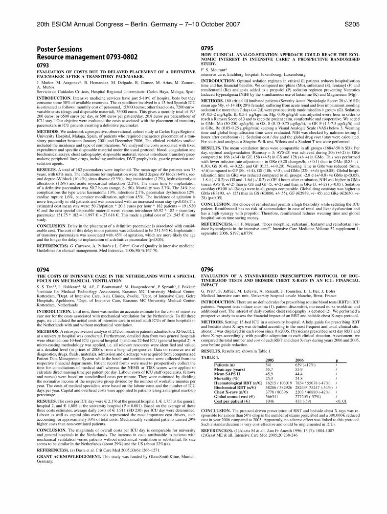
RESULTS. Fluid resuscitation based on crystalloids resulted in a 9.3% increase in total fluidused. Renal Replacement Therapy (RRT) was similar in both groups. There was a statisticallysignificant reduction in mortality in the group following the change in practice. There was anincrease in length of stay. Total cost reduction was 57% and fluid costs per patient day wasreduced by 62%.
TABLE 1.Outcomes and costs
Before After p valueAPACHE II 15.4±6.60 14.98±6.13 .39ITU mortality (%) 15.3 12.9 .01Length of stay ITU 4.32±6.61 4.38±6.42 .0001RRT (%) 4.6 5.3 .23Cost per patient day (£) 10.24 3.88Mean annual cost (£) 51135 21770
CONCLUSION. A crystalloid-based policy for fluid resuscitation is associated with improvedmortality and is cost-effective compared to using both crystalloids and colloids.
REFERENCE(S). A comparison of albumin and saline for fluid resuscitation in the intensivecare unit: The SAFE Study Investigators. New Engl J Med 2004, 350:2247-2256.
0800CORRELATION AMONG APACHE II, ICU OUTCOME AND COST: A ONE-YEARRETROSPECTIVE STUDY
A. Koumpos*, G. Xatzis, C. Kannavas, G. Anastasiadou, P. Pappas, C. Kontouli,G. Vlachogianni, G. Santis, V. Tsagalidou, A. LioliosIntensive Care Unit, General Hospital O Agios Dimitrios, Thessaloniki, Greece
INTRODUCTION. The aim of our study was to prove if there is any correlation amongAPACHE II, outcome and cost, in ICU patients (pts).
METHODS. Design: Retrospective cost-accounting analysis of each patient stay in ICU andcorrelation with ICU outcome and Acute Physiology and Chronic Health Evaluation (APACHE)II score. Setting: A 5-bedded medical/surgical adult ICU. Patients: 152 consecutive pts over1-yr period (January-December 2006). Measurements: Demographics were collected, includingage, gender, type of ICU admission, APACHE II and Predictive Death Rate (adjusted), ICUlength stay, outcome (mortality 22,4%), cost/day/pts (e1211,69±70), mean cost/stay for allpts (e9330,01±500), and total cost in ICU (e1.417.677,3). Costs were collected for human(medical, nursing, and support staff) and capital (laboratory examinations, drug/fluid/blood,equipment, supplies, diagnostic imaging) resources. Cost information was available on all 152pts (88 male-64 female) and was calculated by the financial service. The pts were divided intothree groups, according to primary reason for ICU admission: a) medical b) surgical (emer-gency) c) surgical (elective). The statistical analysis was performed with the statistical packageof SPSS 14.0.
RESULTS. 1) There is strong correlation (p<0,001) among APACHE II, Predicted DeathRate (adjusted), ICU outcome and cost. 2) The correlation among cost/day/pts, mortality andAPACHE II, is statistically significant (p<0,005) with logarithmic increase 3) 53,01% of theper day cost of ICU patients was the human resources cost.
TABLE 1.Type of Number of Mean Age Mortality % APACHE II Predictive Mean length Cost peradmission pts (range) (n) (mean) Death Rate of stay (days) day (e)Medical 58 59,93(14-93) 39,6%(23) 20,26 42,22% 11,19 1169,57Surgical 37 67,52(30-88) 24,3%(9) 13,24 24,73% 7,79 1318,6(emergency)Surgical 57 69,53(37-89) 3,5%(2) 8,93 10,96% 3,11 1031,44(elective)Total 152 65,73(14-93) 22,4% (34) 14,52 26,82% 7,7 1211,69
CONCLUSION. 1) APACHE II score is directly correlated to the ICU outcome and the perpatient cost. 2) APACHE II score helps calculate the ICU cost, the length of stay and predictthe outcome. 3) Although medical pts present with a higher APACHE II score than the surgical(emergency) pts, their ICU cost per day is significantly lower and their mortality is significantlyhigher (see table).
REFERENCE(S). Noseworthy TW, Konopad E, Shustack A, Johnston R, Grace. Cost ac-counting of adult intensive care: methods and human and capital inputs. Crit Care Med, 1996Jul;24 (7):1168-72
20th ESICM Annual Congress – Berlin, Germany – 7–10 October 2007 S207
0801ARE DAILY ROUTINE CHEST RADIOGRAPHS USEFUL IN CRITICALLY ILL,MECHANICALLY VENTILATED PATIENTS?
C. Clec’h*, P. Simon, A. Hamdi, P. Karoubi, J. Fosse, F. Gonzalez, F. Vincent, Y. CohenICU, Hopital Avicenne, Bobigny, France
INTRODUCTION. Whether chest radiographs (CXRs) in intensive care unit (ICU) mechan-ically ventilated patients should be routinely obtained or only if an abnormality is anticipatedremains debated. Our aim was to compare the diagnostic, therapeutic and outcome efficacy of arestrictive prescription of CXRs to that of a routine prescription, focusing on delayed diagnosesand treatments potentially related to the restrictive prescription.
METHODS. All patients admitted to the ICU of the Avicenne teaching hospital (Bobigny,France) and requiring mechanical ventilation (MV) were eligible. After 48 hours of MV,patients were randomly allocated to have daily routine CXRs (routine prescription group) orclinically indicated CXRs (restrictive prescription group). For each CXR performed, a question-naire was completed addressing the reason for the CXR, the new findings, and any subsequenttherapeutic intervention. The endpoints were the rates of new findings, the rates of new find-ings that prompted therapeutic interventions, the rate of delayed diagnoses in the restrictiveprescription group, and mortality.
RESULTS. Twenty-nine patients were included in the routine prescription group and 26 in therestrictive prescription group. Baseline characteristics were well balanced between groups. Therates of new findings and the rates of new findings that prompted a therapeutic intervention inthe restrictive prescription group and in the routine prescription group were 62 % vs 8 % (p< .0001), and 56 % vs 5.5 % (p < .0001), respectively. The rate of delayed diagnoses in therestrictive prescription group was 0.7 %. There was no difference in mortality.
CONCLUSION. A restrictive use of CXRs in ICU mechanically ventilated patients was asso-ciated with better diagnostic and therapeutic efficacies without impairing patients’ outcome.
0802DOES THE 20/80 PERCENT RULE APPLY TO INTENSIVE CARE?
I. A. Meynaar*, R. Schoenmakers, P. L. Tangkau, L. Dawson, M. van Spreuwel, S. SleeswijkVisserICU, Reinier de Graaf Hospital, Delft, Netherlands
INTRODUCTION. The so-called 20/80 percent rule roughly implies that 20% of cases require80% of efforts. We studied our ICU database to see if the 20/80 percent rule is valid in intensivecare. We hypothesized that 20% of patients with the longest length-of-stay in the ICU consumeapproximately 80% of ICU resources.
METHODS. The ICU is a 10-bed closed format mixed unit in a 500-bed general hospital. Dataare collected prospectively in our database of all admitted patients. All patients admitted to theICU between January 1st, 2004 and December 31st, 2006 were included in the study.
RESULTS. During the study period 2192 patients were admitted to the ICU. The ICU length-of-stay was 4 days or more in 437 patients (20%) and less than 4 days in 1755 patients (80%).Outcome and resource utilisation are presented in Table 1.
TABLE 1.20% of patients 80% of patients All Patients
Number of patients 437 1755 2192Age (mean/median) 67,2 / 70,5 66,1 / 69,7 66,3 / 69,9SAPS II (mean/median) 40,5 / 39 29,9 / 26 32,0 / 28APACHE II (mean/median) 18,2 / 18 12,8 / 11 13,9 / 12Hospital mortality 25,9% 13,8% 16,2%ICU treatment days 4524 (66%) 2337 (34%) 6861 (100%)Days on ventilator 2729 (87%) 695 (13%) 3424 (100%)Days on hemofiltration 499 (90%) 57 (10%) 556 (100%)Total TISS 142455 (62%) 86117 (38%) 228572 (100%)Estimated cost in 4,03 (70%) 1,77 (30%) 5,80 (100%)million euro’s
CONCLUSION. The so-called 20/80 percent rule is more or less applicable to intensive care.We found that 20% of patients are accountable for 66% of ICU treatment days, 87% of ventilatordays, 90% of hemofiltration days and 70% of ICU costs.
0288ARE MECHANICALLY VENTILATED PATIENTS A SUITABLE TARGET FORDECREASE BLOOD TRANSFUSIONS?
I. Tubau*1, R. Fernandez1, R. Ferrer1, I. Roig2, A. Artigas1
1Critical Care Center, 2Haemathology, Sabadell hospital, Sabadell, Spain
INTRODUCTION. Blood transfusion in ICU remains a daily routine despite its known side-effects, because of the lack of real alternatives. Our hypothesis was that transfusion rate mayshow differential trends depending on risk factors or physicians-induced changes. Our objectivewas to study the trend of transfusion in our Intensive Care Unit in the last three years and torecognize risk factors for transfusion.
METHODS. We retrospectively review our database of ICU patients from 2004 to 2006.We recorded diagnosis category, age, severity score on admission (Apache II), length of stay,hospital mortality, complications, need for ICU specific treatments and transfusion. Statisticalanalysis: Multivariate logistic analysis for the risk of transfusion. The yearly trend was assessedby Chi-square analysis in significant groups.
RESULTS. We admitted 2199 patients with a 26.2% rate of transfusion. Mean age (62.3±16.9yr), diagnosis category, length of ICU stay (8.4±10.5 days), rate of mechanical ventilation(MV) (50.2 %), and mortality rate adjusted for severity were unchanged during the period.Factors associated with transfusion by multivariate analysis were: vasoactive drugs (OR 2,4(1.8–3.0), p<0.001), acute renal failure (OR 3.3 (2.4–4.3), p<0.001), trauma (OR 3.0 (2.2–4.1), p<0.001), gastroscopy (OR 10 (5.5–18.7), p<0.001), MV plus arterial catheter (OR 2.3(1.8–2.9), p<0.001). The trend of transfusion only significantly decreased in this last group(absolute 11% reduction or a 20% relative reduction) (see Table).
TABLE 1.2004 2005 2006 p value
MV 140/287 (49%) 124/286 (43%) 106/279 (38%) 0.04Non MV 69/472 (15%) 69/442 (16%) 67/434 (15%) nsTotal 209/759 (28%) 193/728 (27%) 173/713 (24%) ns
CONCLUSION. In our ICU, the trend of transfusion in the last 3 years decreased 4%, butmostly restricted to mechanically ventilated patients.
Poster SessionsProfessional issues 0803-08160803WAITING ROOMS IN ITALIAN ICUS: A NATIONAL SURVEY
A. Giannini*1, G. Miccinesi2, S. Leoncino1, M. Munteanu1, S. Baraldi11Pediatric Intensive Care Unit, Fondazione Ospedale Maggiore Policlinico, Mangiagallie Regina Elena, Milan, 2Unit of Epidemiology, Centro per lo Studio e la PrevenzioneOncologica, Florence, Italy
INTRODUCTION. Families of critically ill patients spend a considerable amount of time inhospital, both beside their loved ones in ICU and outside the unit waiting to visit them or toreceive news. No published data are to date available on the provision of waiting rooms inItaly’s approximately 600 ICUs and on the facilities available to patients’ families and visitors.We investigated these aspects in the course of a national survey concerning visiting policies inItalian ICUs.
METHODS. An email questionnaire regarding visiting policies was sent to all 303 ICUs (gen-eral and specialized) in the Italian collaborative group GiViTI (Italian Group for the Evaluationof Interventions in Intensive Care Medicine); questions about waiting rooms and facilities forpatients’ families and visitors were also included.
RESULTS. The response rate was 85% (257/303). No waiting room was provided in 25% ofICUs. In other ICUs, families and visitors were provided with seats (64% of ICUs), armchairs(23%), lockers for personal effects (18%), magazines and books (12%), drinks machines (19%)and snack machines (12%). A bathroom was available to families and visitors in 43% of ICUs,use of the ICU’s kitchen in 2% and access to the hospital canteen in 22%. Median daily visitingtime was 60 minutes, ranging from 15 minutes to 18 hours (10th percentile: 30 minutes; 90thpercentile: 120 minutes).
CONCLUSION. Overall these data indicate that in Italian ICUs, alongside a clear tendency toapply restricted visiting policies, there is limited attention to the comfort of families of patientsin ICU. Comfort is one of five domains (in addition to assurance, proximity, information andsupport) associated with the needs of families who have critically ill loved ones [1, 2]. Oursurvey could contribute not only towards modifying current policies in favour of opening ICUsthat are still ‘closed’ but also to promoting more attentive and supportive care for the patient’sfamily [3].
REFERENCE(S). 1. Leske JS. Needs of relatives of critically ill patients. A follow up. HeartLung 1986;15:189-932. Bijttebier P et al. Needs of relatives of critical care patients: perceptions of relatives, physi-cians and nurses. Intensive Care Med 2001;27:160-53. Deitrick L et al. Evaluation and recommendations from a study of a critical-care waitingroom. J Healthc Qual. 2005;27:17-25.
GRANT ACKNOWLEDGEMENT. This study was supported by ABN (Associazione per ilBambino Nefropatico, Milan, Italy). We thank GiViTi for their valuable help.
S208 20th ESICM Annual Congress – Berlin, Germany – 7–10 October 2007
0804SATISFACTION WITH CARE IN THE INTENSIVE CARE UNIT: CAN WE RELYON PROXIES?
K. H. Stricker*1, O. Kimberger1, L. Brunner2, M. Jeitziner2, U. Mohr1, O. Rohrer2,H. U. Rothen2
1Department of Anesthesiology, 2Department of Intensive Care Medicine, University HospitalInselspital, Bern, Switzerland
INTRODUCTION. Quality of care has been recognized as an important aspect of quality man-agement, as recall of experiences may impair quality of life1. Many patients do not consciouslyremember their stay in the ICU. Therefore, next of kin are often approached to assess qualityof care. However, reliability of proxy assessment has been questioned, i.e. for quality of life2,as they may not adequately represent patients’ points of view.
METHODS. During a 2-month period, next of kin of patients in 23 ICUs were asked tocomplete a questionnaire on satisfaction with care3, including a single question and a visualanalogue scale (VAS) on overall satisfaction. If these patients themselves were oriented intime, person and place when leaving the ICU, they were asked to complete a short versionof the questionnaire. Answers were compared using Cohen’s Kappa and Pearson correlationcoefficient (for VAS). Analysis was done as suggested3 with rescaling of all questions to 0 -100 (worst - best care).
RESULTS. mean ± SD. 106 pairs of responses were available for analysis. Proxies’ age was57±15 years, 52% of respondents were spouses, 23% children, 5% parents and 20 % others.Patients’ age was 65±14 years, admission SAPS was 33±48 points, length of stay in the ICUwas 4.7±3.3 days and 67 % were emergencies.
TABLE 1.patient proxy kappa
courtesy, respect 88±16 84±16 0.31management of - pain 87±19 82±20 0.39- breathlessness 84±23 82±20 0.42- agitation 80±23 80±21 0.46coordination of care 86±17 81±18 0.37skills and competency of - nurses 87±17 85±15 0.26- doctors 89±15 84±17 0.36atmosphere in the ICU 84±17 77±21 0.22overall satisfaction:- single question 86±15 82±18 0.37- VAS 90±13 89±13 0.61
CONCLUSION. Next of kin and patients’ ratings of satisfaction with care were not significantydifferent, but the interrater reliability was only moderate. Since satisfaction with care could notbe assessed in individuals with impaired consciousness, good outcome may have lead to highsatisfaction, resulting in a ceiling effect: the majority of answers were given as excellent or verygood, and only very few ratings were moderate or poor.
REFERENCE(S). 1. Granja C, Crit Care 2005; 9: R96 – 109 2. Scales DC, Intensive CareMed 2006; 32: 1826-1831 3. Heyland DK, J Crit Care 2001; 16:142-149
GRANT ACKNOWLEDGEMENT. Supported by Astra-Zeneca Pharmaceuticals, Inselspitaland all 23 participating ICUs.
0805FAMILY SATISFACTION IN THE ICU: PRELIMINARY RESULTS OF A MULTI-CENTRE TRIAL
K. H. Stricker*1, O. Kimberger1, U. Mohr1, O. Rohrer2, M. Jeitziner2, L. Brunner2,H. U. Rothen2
1Department of Anesthesiology, 2Department of Intensive Care Medicine, University HospitalInselspital, Bern, Switzerland
INTRODUCTION. Family satisfaction has been recognized as an outcome measures in theICU1. Relatives of critically ill patients have particular needs and if these are not met, they mayaffect their long-term quality of life2. Therefore, sources of dissatisfaction need to be identifiedto initiate goal directed changes.
METHODS. In 23 ICUs, adult next of kin were asked to complete a questionnaire on satis-faction with care and with information /decision-making3, and a visual analogue scale (VAS,0-100: worst-best). Inclusion criteria were patients’ length of stay in the ICU > 2 days, andregular visits by the family. Questions were rescaled as suggested3 to 0-100.
RESULTS. (mean ± SD). 597 questionnaires were distributed, return rate was 66%. Respon-dents’ age was 55±15 years, 45% of respondents were spouses, 33% children, 5% parents an17 % others. Patients’ age was 67±14 years, their SAPS 37±32 points, and 7% of patientshad died when proxies responded. 41% of patients had a surgical diagnosis and 44% wereemergency admissions. 16 ICUs had < 12 bed (small ICU, 158 patients), 7 had > 12 beds (largeICU, 236 patients).
TABLE 1.Overall, best and least satisfaction, and distribution among small – large ICUs
small ICUs large ICUsoverall satisfaction with: - care 83±18 82±17- information and decision-making 72±28 75±23VAS: - care 88±15 90±11- information and decision-making 83±19 85±18best satisfaction*: respect and courtesy for patient 84±17 86±152nd best satisfaction*: respect and courtesy for family 82±18 85±152nd least satisfaction*: completeness of information 77±22 77±21least satisfaction*: waiting room atmosphere 60±27 65±24* Ranking according to all patients of all ICUs
CONCLUSION. In general, next of kin were satisfied with care as well as with informationand decision-making. However, large differences between different aspects were found andsome facets with need for improvement were identified (e.g. waiting room, information). Sat-isfaction was somewhat lower than in a previous study3 from Canada, and similar sources ofdissatisfaction were noted. No significant difference was found between small and large ICUs.
REFERENCE(S). 1. Berenholtz SM J Crit Care 2002; 17: 1-15 2. Azoulay E Am J Respir CritCare Med 2005; 171:987-94 3. Heyland DK Crit Care Med 2002;30: 1413-8
GRANT ACKNOWLEDGEMENT. Supported by Astra-Zeneca Pharmaceuticals, Inselspitaland all 23 participating units.
0806PATIENT’S VIEW ON ICU SERVICE QUALITY: MEASUREMENT BY DESCRIP-TIVE ITEMS VS. “GENERAL SATISFACTION” (PRELIMINARY RESULTS OFA QUESTIONNAIRE STUDY)
A. W. Schindler*, N. Schindler, A. Lueck, F. Enz, D. A. VagtsAnaesthesiology and Intensive Care Medicine, University of Rostock, Rostock, Germany
INTRODUCTION. Patients’ assessment of a received medical service is a measure of quality(1). „General satisfaction“ is a questionable measure of quality after threatening situationsbecause of considerable ceiling effects*(1, 2). We hypothesized that following an ICU staydescriptive items show less ceiling effects then „general satisfaction“ – if so, they were a moreappropriate measure of quality.
METHODS. 81 consecutive patients were interviewed 2-7 days after their ICU stay using aself-constructed questionnaire (ethic committee approval, written informed consent). „Generalsatisfaction“ as well as 31 descriptive items focussing on 3 different spheres of subjective expe-riences were ranked using a 4-point Likert scale (1=positive, 4=negative). The 3 spheres weresomatic (pain etc.) intrapersonal (emotions etc.) and interpersonal (communication etc.) experi-ences and they were described by 7, 14 and 10 items. SAPS 2 (initial and daily mean), TISS 28(daily mean) and length of stay were chosen as medical descriptors. Statistics: Wilcoxon signedrank test (p <0.05), percentual frequencies of rankings; mean±SD for medical descriptors.
RESULTS. Percentual distribution of the Likert ranks for „general satisfaction“ and the spheresof subjective experiences are presented in Table 1. „General satisfaction“ is ranked lower (i.e.more positively) than the descriptive items.SAPS 2initial 37±20; SAPSdaily mean 30±20; TISS 28 33±20; Length of stay: 4±7 days.
TABLE 1.Frequencies (%) of Likert ranks for “satisfaction” and grouped descriptive itemsRanking “satisfied” somatic sphere intrapersonal interpersonal(1=pos. ... 4=neg.) (1 item) (7 items) sphere sphere
(14 items) (10 items)1 80.3 31.6 51.4 67.82 17.2 24.3 15.9 17.43 2.5 19.1 13.8 5.94 0 21.3 15.9 5.9invalid answers 0 3.7 3.1 3.0
CONCLUSION. „General satisfaction“ received mainly best rankings, while the descriptiveitems did not, indicating a considerable ceiling effect in the former but not the latter. Therefore,patients’ assessment of ICU treatment as a medical service should be measured with descriptiveitems to produce meaningful results.
REFERENCE(S). 1) Der Anaesthesist 49:613, 2) Anesth Analg 87:1089*ceiling effects: best rankings, even despite objectively bad outcome
0807A PHARMACOECONOMIC MODEL FOR THE ANALYSIS OF PATIENT FLOWSAND SEDATION REGIMENS IN THE ICU
M. J. Al1, L. Hakkaart1, S. S. Tan*1, P. G. Mulder2, R. Welte3, J. Bakker4
1Institute for Medical Technology Assessment, 2Department of Epidemiology and Biostatis-tics, Erasmus MC University Medical Center, Rotterdam, Netherlands, 3Reimbursementand Health Outcomes, GlaxoSmithKline, Munich, Germany, 4Department of Intensive Care,Erasmus MC University Medical Center, Rotterdam, Netherlands
INTRODUCTION. Modelling allows the extrapolation of study findings to longer time pe-riods and the assessment of study subgroups. We developed a Markov model to simulate thepatient flow in the ICU with a focus on mechanical ventilation (MV). It was employed to com-pare remifentanil-based sedation (RS) vs. conventional sedation (CS) in critically ill patientsrequiring MV.
METHODS. A probabilistic Markov model was built to analyse the patient flow, the conse-quences and direct medical costs of different sedation regimen in the ICU. The model contains8 states: MV - maintenance, MV- eligible start weaning, MV - actual weaning started, MV-eligible for extubation, ICU - extubated, ICU - eligible for discharge, Discharged from ICU,and Death. At every hour, patients may move to a different state. Data to derive the respectivetransition probabilities were obtained from UltiSAFE (1), a Dutch open-label trial with 205critically ill patients, using time-to-event analyses. Patients received CS according to Dutchguidelines (predominantly fentanyl or morphine combined with midazolam or propofol) orRS (remifentanil, combined with propofol if required) for up to 10 days. Applying Weibullfunctions to fit the UltiSAFE data to our model allowed us to run the model for 28 days, i.e.beyond the UltiSAFE study period. Study drug costs were derived from UltiSAFE whereas allother costs were measured in a separate microcosting study, conducted in 3 Dutch mixed adultICUs. Resource utilisation was valued from a hospital perspective using prices of 2006.
RESULTS. Our model showed a high internal and external validity. CS led to 6.2, RS to 5.2days of mean MV duration, i.e. RS reduced the duration of MV by 1 day (95% CI 0.6 - 1.3days). Similarly, CS caused a mean length of stay in the ICU of 8.8 days compared to 7.9 daysfor RS rendering a difference of 0.9 days (95% CI 0.6 - 1.2 days). The total mean costs perpatient were e 17,502 in the CS group and e 16,093 in the RS group yielding cost-savings ofe 1,409 per patient (95% CI e 744 - 2,074) in the RS group.
CONCLUSION. A Markov model can be successfully used to simulate the patient flow in theICU and evaluate different sedation regimens. Compared to CS, RS seems to be the preferredregimen for patients with 5 - 6 days of MV. Its ability to decrease the length of stay not onlyovercompensates the additional drug costs of RS but also increases the ICU capacity. A shorterduration of MV might benefit the patient as several studies have shown a positive correlationbetween ventilator associated pneumonia and the duration of MV (2).
REFERENCE(S). (1) Bakker J et al., Inten Care Med 2006;32 (Suppl.1):S86(2) Vincent JL, Lancet 2003;361:2068-2077
GRANT ACKNOWLEDGEMENT. This study was funded by GlaxoSmithKline
20th ESICM Annual Congress – Berlin, Germany – 7–10 October 2007 S209
0808OVER ONE YEAR ON FROM “AN ACUTE PROBLEM”: IS IT NOW ACUTE ONCHRONIC?
N. J. Flint, J. A. Sadler*, D. C. Bouch, J. P. ThompsonAnaesthesia, Critical Care and Pain Management, Leicester Royal Infirmary, UniversityHospitals of Leicester, Leicester, United Kingdom
INTRODUCTION. The publication in 2005 of the National Confidential Enquiry into PatientOutcome and Death (NCEPOD) report entitled ‘An Acute Problem’ [1], reviewed the care ofmedical patients referred for Level 3 care. In light of their findings the report made recommen-dations regarding the provision of critical care including: pre-referral care and monitoring, thereferral procedure and the Intensive Care Unit (ICU) admission process. The aim of our auditwas to review the process for medical admissions to Level 3 care in the three adult ICUs acrossUHL, using the NCEPOD data as a benchmark.
METHODS. We audited medical patients admitted to Level 3 care over a three-month period.Case notes were reviewed and data collected on initial clerking and observations, observationplans, evidence of senior clinician involvement and the personnel involved in the referral andadmission process to ICU.
RESULTS. There were 104 admissions, 36% referred by Accident and Emergency (A&E),28% by general medicine and 36% by a medical speciality. 57% were male with a mean ageof 58 years; 43% were female with a mean age of 53 years. Admission data: A full historyand examination was present in 39% of patients on admission to hospital. Respiratory rate wasrecorded in 75% of patients. Observation plans were in place in 25%, with early warning scorecharts used in 16%. A consultant reviewed the patient within 24 hours of admission to hospitalin 42% of cases. Referral data: The consultant in charge of the patient was aware of the referralin 39% of cases, referring 21% of the total. A specialist registrar (SpR) referred 65%, a seniorhouse officer (SHO) 11% and a house officer 3%. ICU data: Referral was received by the ICUconsultant in 13% of cases, the SpR in 68%, and by the SHO in 19%. The ICU team reviewedall patients prior to admission. The ICU consultant was aware of admission in 89% of cases.The ICU consultant reviewed the patient within 12 hours in 78%, being present on admissionin 39% of cases.
CONCLUSION. The A&E clerking was satisfactory in less than 5% of cases; despite ac-counting for this referrals from medical wards had worse results for clerking than the report.Physiological monitoring, notably the recording of respiratory rate, was better than the NCE-POD report. Pre-ICU consultant input was comparable to NCEPOD; but A&E consultantinvolvement was markedly lower than other medical specialties. The ICU consultant input wasgenerally comparable to or better than the NCEPOD data. Poor documentation may well hidea greater level of senior input. One year after the report significant improvement still needs tobe made.
REFERENCE. 1. An Acute Problem? The 2005 Report of the National Confidential Enquiryinto Patient Outcome and Death. NCEPOD. London, 2005.
0809CLIENT-PROFESSIONAL-GAPS IN INTENSIVE CARE: PATIENTS’ EXPERI-ENCES AND EXPECTATIONS – AND STAFF’S ASSUMPTIONS (PRELIMINARYRESULTS OF A QUESTIONNAIRE STUDY)
A. W. Schindler*, N. Schindler, E. Friederike, A. Lueck, D. A. VagtsAnaesthesiology and Intensive Care Medicine, University of Rostock, Rostock, Germany
INTRODUCTION. When true patients’ experiences and expectations differ from what staffmembers assume about these experiences and expectations, suboptimal service quality is likelyto result (1). We investigated differences between true patients’ experiences and expectations andstaff members’ assumptions about these experiences and expectations – i.e. client-professional-gaps (cp-gaps).
METHODS. All Participants were recruited from two operative Intensive Care Units (ICUs).81 patients (local ethic committee approval, written informed consent) and 60 staff members(voluntary participation) were interviewed anonymously with an analog questionnaire; patientswere interviewed retrospectively. The questionnaires focussed on the real (patients) and theassumed (staff) patients’ experiences and expectations. „General satisfaction“ and further 31items representing patients’ experiences in the somatic (pain etc.), the intrapersonal (emotionsetc.) and the interpersonal sphere (communication etc.) were ranked on a 4-point Likert scale(1=positive, 4=negative). In addition, 61 dichotomous items (presence / absence of a negativeexperience) grouped into 5 categories could be chosen. 4 dimensions of expectations (somaticsphere, intrapersonal sphere, interpersonal sphere, medical competence) were ranked on a 4-point Likert scale with regard to their relative importance. Statistics: U-test (p<0.05) for Likertranks, number of chosen dichomotous items for each individual (means±SD).
RESULTS. Patients chose lower Likert ranks (1.8±1.1 vs. 2.8±0.9) and less dichotomousitems (6.2 vs. 25.3) compared to the staff members, indicating cp-gaps. The relative importanceof the 4 dimensions of expectations were ranked equally in patients and staff.
CONCLUSION. Our questionnaire detected a cp-gap for experiences but not for expectationswith real service being better than the staff members assumed. Maybe a similar cp-gap existsfor expectations but could not be measured with the coarse gradation used in our preliminaryquestionnaire. Gap detection can help to focus measures intended to improve medical servicequality.
REFERENCE. 1) Brown und Swartz, Journal of Marketing 53:92-98
0810DO AESTHETICS MATTER IN THE ICU?
B. S. Hansen*, V. Holst-Jaeger, E. SoereideICU, Division of Acute Care Medicine, Stavanger University Hospital, Stavanger, Norway
INTRODUCTION. When planning a new ICU it is important to bear in mind that the aestheticsof the ICU may have a large influence on the mental and physical health of both patients andnurses (Caspari, 2004). The aim of this study was to find out whether the aesthetics (space,light, sound, colours, and art) in a planned new ICU would be of importance to the nursesworking there, and if so, how?
METHODS. Using both a qualitative and quantitative approach, we designed a questionnairecontaining 42 items (using Likert scale) with 4 main areas (space/design, light, sound, art) andwith room for free comments in each area, also containing 3 open questions. The respondentswere 101 nurses working part- or full-time. We had a response rate of (77%) and collected 462free comments. These qualitative statements were collected and qualitative content analysiswas used for analysing the themes.
RESULTS. Enough space around the bed to provide easy access and good overview was con-sidered the most important factor for the staff. A need for sufficient storage room for essentialsupplies and equipment next to each bed was also rated as important. The respondents describedmany difficulties when space was too narrow, as mobilising ventilator-patients, avoiding cross-infections, preserving the patient’s rights to privacy and confidentiality etc. Sufficient naturaldaylight was also perceived as very important. Inadequat light influences the mood. Windowsare the visual link to the outside world (NHS Estates, 2003). The nurses believed bright, colour-ful, welcoming surroundings would affect the mood of both patients and others in a positiveway. Art was welcomed as something beautiful to look at, and stimulating for patients, familyand staff. Noise (incl noise from medical equipment) was said to cause discomfort both forpatients, family and staff, and made it difficult to concentrate. The respondents believed spaceand privacy (single bed rooms) for information exchange between staff is important as well asroom for private conversation between patient, family and medical staff. It was also expressedthat adequate space, light, and nice surroundings give both patient and families a feeling ofdignity, safety and respect.
CONCLUSION. Aesthetics do matter when planning a new ICU. Focus on space, light, noiseprevention and welcoming surroundings might increase the comfort for patients, families andstaff and even increase efficiency and safety in the ICU.
REFERENCE(S). Caspari, S. “The Golden Section, the aesthetic dimension- an ethical obli-gation, Abo Akademis forlag, 2004.”NHS Estates, HBN 57, “Facilities for critical care”,www.tso.co.uk/bookshop
0811COMMUNICATION WITH THE RELATIVES OF CRITICALLY ILL PATIENTS:AN AUDIT AND DISCUSSION
S. R. Humble*, S. J. Cole, P. Antoniewicz, J. R. ColvinIntensive Care, Ninewells Hospital, Dundee, United Kingdom
INTRODUCTION. Good communication is an essential part of medical and nursing care. Itfacilitates the diagnostic process and minimises the emotional distress experienced by patientsand their relatives. This study evaluated the effectiveness of communication from two perspec-tives. The results were compared against a national framework of recommended standards.
METHODS. We prospectively audited the frequency and location of interviews that occurredbetween ICU staff and patient’s relatives from Feb to Jun 2006. After discharge from ICU,the relatives were asked to complete a retrospective anonymous questionnaire regarding theirexperience.
RESULTS. Of the 106 admissions to ICU within the time period we studied data from 80patients. The remainder met the exclusion criteria. 30% of relatives were interviewed within15 minutes of arrival by nursing staff, while 28% were interviewed by medical staff within 60minutes. Nursing staff spoke to the relatives on 74% of days and medical staff spoke on 57% ofdays. 21% of patients received no visitors. 41 of the patient’s relatives were given questionnairesto complete, the results from these were very positive.
CONCLUSION. The needs of relatives include the opportunity to effectively appraise a sit-uation that is perceived as harmful, to assimilate the information and to formulate a copingstrategy. The overall impression a visitor forms of ICU is heavily influenced by the communi-cation skills of the staff and effective communicators are also less likely to receive complaints.The subjective nature of communication makes it difficult to quantify and difficult to mea-sure definitively. As a surrogate marker of quality, we assessed the quantity and frequencyof discussions. Arbitrary end points are unable to measure crucial elements of the interactionprocess or patient perspectives. It has been suggested that an assessment of Doctor and Patientperceptions should be used, based on the premise that subjective measurements may be moreuseful than objective ones in this context. However, these are highly complex to measure; ipsofacto very few studies have been completed in this area to support this assertion. High qualitycommunication is something to which we should all aspire. The strict use of inflexible targetsis not necessary to promote good communication, but serves as a benchmark to raise the profileof this issue. Emphasis should be placed on integrating a routine interview process into clinicalcare.
REFERENCE(S). 1. Jayaprakash V, Smith GB. Communication with relatives or visitors ofICU patients. RCoA. A compendium of audit recipes. Critical care services 10.92. Hagihara A, Tarumi K. Doctor and patient perceptions of the. . . quality of. . . communication.Scand J Caring Sci 2006; 20: 143-150
S210 20th ESICM Annual Congress – Berlin, Germany – 7–10 October 2007
0812USING THE “LIVERPOOL INTEGRATED CARE PATHWAY FOR THE DYINGPATIENT” (LCP) IN INTENSIVE CARE
J. Harper*1, L. Chapman2, M. Gambles3, J. Ellershaw3, D. Parsons1, E. Arthan1,L. McCrossan1
1Intensive Care Unit, 2Palliative Care Unit, Royal Liverpool University Hospital, 3PalliativeCare Unit, Marie Curie Palliative Care Institute, Liverpool, United Kingdom
INTRODUCTION. In this 1200 bed inner city tertiary referral hospital, 10% of patients whodie, die in the intensive care unit. 23% of patients admitted to ICU die; of these, 70% havetreatment withheld or withdrawn. It is recognised that approaches to dying patients are veryvariable between units and doctors(1). To improve the quality and consistency of care of patientsdying in intensive care, we adapted the LCP, originally designed for patients dying in hospicesand on hospital wards. The LCP is divided into 3 sections: initial assessment and care, ongoingcare, and care after death. It specifies physical, psychological, spiritual and social goals to bemet for each patient.
METHODS. The LCP used on wards was introduced to the intensive care unit. Action researchmethodology using focus group interviews led to modifications of the pathway to meet specificneeds of critically ill patients and their families. After introduction of the modified pathway, asurvey of staff satisfaction was performed using a validated questionnaire graded with a Likertscale.
RESULTS. In the first 6 months, 45/ 107 deaths were on the LCP. Patients were on the pathwaya median of 5 hours (range 1 – 215). Compliance with specific goals was > 80%, except forcontinuation of IV fluids (33%) and discontinuation of electronic monitoring (50%). 24% ofpatients were extubated. 44% of staff responded to the survey; median score for ‘improvedquality of care’ was 4/5.
CONCLUSION. Use of the LCP in our ICU improved documentation of the process of dyingon patients for whom treatment was withdrawn. Staff satisfaction with the pathway is high.
REFERENCE(S). Taylor Thompson B, Cox PN, Antonelli M, Carlet JM, Cassell J, Hill NS,Hinds CJ, Pimentel JM, Reinhart K, Thijs LG. Challenges in End-of-Life Care in the ICU:Statement of the 5th International Consensus Conference in Critical Care. Crit Care Med 2004;32: 1781–4.
0813RAPID RESPONSE SYSTEMS IN ITALY: STATE OF THE ART.
F. Rubulotta*1, G. Radeschi2, G. Rubulotta1, F. Lodo2, L. Ferla1, A. Gullo1
1Department of Anaesthesia and Intensive Care Medicine, Policlinico University Hospital,Catania, 2Department of Anaesthesia and Intensive Care Medicine, ASO S. Luigi Gonzagadi Orbassano, Torino, Italy
INTRODUCTION. In 2007, a manuscript containing Italian recommendations for implement-ing Rapid Response Systems (RRS) will be published. In April 2007 the Societa italiana dianestesia analgesia rianimazione e terapia intensiva (SIAARTI) the Italian resuscitation council(IRC) the Associazione anestesisti rianimatori ospedalieri italiani (AAROI) instituted a na-tional commission to implement RRS in Italy. The aim of the authors is to investigate baselinemanagement of hospital emergencies in Italy.
METHODS. A survey has been carried out using a national e-mail list enclosing all Italianhospitals with critical care beds (total 413 hospitals in the 2005 national AAROI register).Either general directors or physicians working in critical care areas were contacted.
RESULTS. 109 hospitals (26%) replied to this questionnaire. Most hospitals were located in thenorth of Italy (67%) and were public (49,5%) hospitals. The medical emergency team (MET)comprised a doctor in 42% of cases, a doctor and a nurse in 34%, but never only nurses. Theanaesthesiologist was the specialist most often in charge of MET teams (70,6%). BLS (60,5%)andor BLSD (57,8%) were the certificates of training owned by most people working in theafferent arm (alerting MET teams), while ALS 73,4%) was the training of most specialistsworking in the efferent arm of the hospital RRS. Most of the time the MET team was calledfor cardiac arrests (64%), and the report of these events were not systematically written in aregister (57%). No statistically significant differences were found among university, public ormixed hospitals, and between hospitals in the north, the centre and the south of Italy.
CONCLUSION. The afferent and the efferent arm of RRS in Italy do not receive a specificcertification. No protocols are followed in the way critical events are recorded, as a consequencemost crisis are not reported.
REFERENCE(S). 1. Hillman K, Chen J, Brown D. A clinical model for Health ServicesResearch-the Medical Emergency Team. J Crit Care. 2003 Sep;18(3):195-9.2. Hillman K, Chen J, Brown D, et al. Introduction of the medical emergency team (MET)system: a cluster-randomised controlled trial. Lancet. 2005 ;365(9477):2091-7.3. Galhotra S, DeVita MA, Simmons RL, et al. members of the Medical Emergency ResponseImprovement Team (MERIT) Committee. Impact of patient monitoring on the diurnal patternof medical emergency team activation. Crit Care Med. 2006;34(6):1700-6.
0814INTENSIVE CARE INPUT INTO END OF LIFE CARE IN A REGION OF THEUNITED KINGDOM
M. Thomas*, J. Gatward, A. PadkinIntensive Care Unit, Royal United Hospital, Bath, United Kingdom
INTRODUCTION. One in five Americans die using Intensive Care (Intensive Care or Coro-nary Care) services [1]. The same figure is not known for the UK, despite the acknowledgeddifferences in end of life care between European and US Intensive Care Units [2]. This studyinvestigated the percentage of people who die from a region of the UK having used generalICU services during their final hospital admission.
METHODS. We investigated a target population defined geographically as those living in thearea served by a single Primary Care Trust (PCT) in the UK. Patients from this area are treatedby two acute hospitals. Anonymous data was supplied by the UK Office of National Statisticson place of death for all those who died during 2004. The general ICU admissions databases ofboth acute hospitals were interrogated to obtain anonymous ICU and hospital mortality data forall patients from the target population admitted during 2004. The study did not include patientsadmitted to coronary care units.
RESULTS. The region had a population of 186,000. Of these, a total of 1687 people diedduring 2004. 902 (53%) died in the two acute hospitals. 59 of these patients (3.5% of totalpopulation deaths) were admitted to a general ICU during their final hospital stay.
CONCLUSION. In this UK region, 1 in 29 patients (3.5%) die having used general intensivecare services during their final hospital admission. This is substantially less than in the USA.The difference is unlikely to be accounted for by coronary care patients. If seen in a widerpopulation, it is likely to reflect a very different approach to ICU admission criteria and end oflife care.
REFERENCE(S). 1. Angus DC, Barnato AE, Linde-Zwirble WT, Weissfeld LA, Watson RS,Rickert T, et al. Use of intensive care at the end of life in the United States: an epidemiologicstudy. Crit Care Med 2004;32(3):638-43.2. Levy MM. Evaluating our end-of-life practice. Crit Care 2001;5(4):182-3.
0815DIPEX: PATIENTS’ AND RELATIVES’ EXPERIENCES OF INTENSIVE CARE,WWW.DIPEX.ORG
S. B. Prinjha*1, K. Rowan2
1Primary Healthcare, University of Oxford, Oxford, 2Primary Healthcare, ICNARC, London,United Kingdom
INTRODUCTION. There are very few qualitative studies that focus on both patients’ andrelatives’ experiences of intensive care. Most concentrate on fairly small numbers of patientsor carers and on specific problems, such as problems with mechanical ventilation, with com-munication or with a specific condition. Very little qualitative research has focussed on largernumbers of patients and carers or on their entire experience, from the patient’s ICU admission tohospital discharge and recovery. The DIPEx charity, based at the University of Oxford, runs anaward-winning multimedia website based on qualitative interview studies about patients’ andcarers’ experiences of health and illness. Interview clips from patients and carers are availablein video, audio and written formats. The DIPEx intensive care studies, funded by ICNARC,aimed to identify the things that mattered to patients who were admitted to intensive care andtheir relatives and close friends.
METHODS. 78 qualitative interviews were conducted across the UK between 2005 and 2007,and analysed. 40 in-depth interviews were conducted with ICU patients and 38 with the relativesand close friends of patients.
RESULTS. The interviews detailed different aspects of patients’ and relatives’ individual ex-periences, including: physical and emotional experiences, nursing care, information needs,communication, support, and affects on daily life. Over 50 main topics from the interviewswere illustrated with extracts in written, audio and video format. The DIPEx intensive care siteslinked patients’ and relatives’ experiences with evidence-based information and with a rangeof other useful resources, including support groups and links to other websites.
CONCLUSION. DIPEx aims to identify the questions that matter to people when they are illand is widely used to inform patients and carers, educate healthcare professionals, and providea patient-centred perspective to researchers and those who manage health services. Each DIPExintensive care study contains around 250 interview clips in video, audio and written formatsfrom patients and relatives who talk about their experiences from when the patient was admittedto ICU to recovery at home.
REFERENCE(S). Herxheimer A, McPherson A, Miller R, Shepperd S, Yaphe J, Ziebland S.Database of patients’ experiences (DIPEx): a multi-media approach to sharing experiences andinformation. Lancet. 2000; 355: 1540-1543.
GRANT ACKNOWLEDGEMENT. Intensive Care National Audit and Research Centre (IC-NARC)
20th ESICM Annual Congress – Berlin, Germany – 7–10 October 2007 S211
0816OUTDOOR ACTIVITY IN AN INTENSIVE CARE UNIT (ICU)
M. Bringas*, C. Munoz, E. Garcıa, R. Fernandez, M. de la Torre, M. Tolon, S. VazquezIntensive Care Unit, San Carlos Clinical Hospital, Madrid, Spain
INTRODUCTION. The purpose of this study was to analyse the overall outdoor activitydeveloped by the intensive care specialist.
METHODS. In our teaching hospital with a closed ICU model, a prospective descriptive studywas designed to evaluate all activities carry out by the intensivist outside the ICU during thelast nine months (July 2006-March 2007).Outdoor activity was defined as any intensivist intervention (treatment criteria, illness degreegraduation, special technics..) on hospitalized patient outside the ICU.Requirements for cardiac arrest are usually recorded in a unit independent protocol and wereexcluded from this study.We recorded patient demographics, admitting diagnoses, type and duration of activity, clinicaloutcome.
RESULTS. Outdoor activity was required in 132 patients, 59% being males. Mean age 76years old. The most common comorbility were ischemic cardiopaty (48%), pulmonary chronicillness (25%), high pressure (14%), diabetes (12%).Most frequent cause of intervention was respiratory failure (48%), 79% of the calls corre-sponded to medical department leaded by the emergency (38%). 40% of the requirementsreceived from surgical department were due to general surgery postoperative patients.
CONCLUSION. Mean age in our study shows that we assist an old population. Most of theICU specialist activities are patients hospitalized in medical departments. Respiratory failureis the leading cause of intervention. Most studies are required to evaluate the perceptive ofincremental needs for intensivist outside the ICU.
Poster SessionsLong-term outcome 0817-08250817MORTALITY, FUNCTIONAL STATUS AND QUALITY OF LIFE IN PATIENTS SUF-FERING CRANEOENCEPHALIC TRAUMATISM AT THE TIME OF DISCHARGEFROM INTENSIVE CARE UNIT AND ONE YEAR AFTER
M. Delange*, M. A. Prieto, M. J. Chaparro, E. Curiel, J. Munoz, B. HernandezIntensive Care Unit, Carlos Haya Hospital, Malaga, Spain
INTRODUCTION. Health problems in patients with craneoencephalic traumatism (CT) havea great personal, family and social life quality repercussion. The results reported for patientswith CT at the time of leaving Intensive Care Unit (UCI), are given in terms of mortality rate,but there is limited knowledge of their physical and functional status after being dischargedfrom hospital and even less of their quality of life later on. Our working group analyze theseaspects when the patient leaves the hospital and after one year out.
METHODS. We carried out a cohort study with the adult patients admitted to ICU in HospitalCarlos Haya at Malaga in the years 2003-2005 with CT. Demographic, epidemiologic andclinical pre/posthospital stay data were recorded. Validated measure scales were used GCS(Glasgow Coma Scale) GOS (Glasgow Outcome Scale), ISS (Injury Severity Score) and alife quality questionnaire PAEEC (Project for the Epidemiological Analysis of Critical Pa-tients), considering basic physiological activities, normal daily activities and emotional state atdischarge from hospital and one year after by phone contact.
RESULTS. Data from 417 patients with a mean age of 38.5±19.5 years old, 80.1% malewere studied. CT was caused by a traffic accident involving motorcycle (33.8%) or automo-bile (23.7%). A cranial scan showed evidence of brainstem dysfunction (63%), 24% requiredsurgery, and 57% suffered septic, respiratory or/and neurologic complications. First ISS was27(14-38), length of stay was 5 days(2-14) , days on mechanical ventilation 5(1-10); GCS atICU admission 7(4.5-10);GCS at ICU discharge 11(4-14); APACHE II: 16(12-22), GOS at timeof hospital discharge 3(2-3); GOS one year later 4(3-5). Mortality rate intra ICU was 24.7%and was associated with a GCS < 8 (p<0.0001, OR 5.2 (CI 95% 2.7-9.9). Multivariant analysisshowed as predictors of mortality in ICU the age (p<0.05), length of stay (p<0.0001), GCS atadmission (p<0.028), complications (p<0.0001), tracheotomy (p<0.005), days on mechanicalventilation (p<0.0001) and APACHE II (p<0.022). One year after discharge 260 of 314 pa-tients answered the questionnaire (82.8%): severe physiologic dysfunction, great dependencein activities of daily living and emotional disturbances were detected when leaving hospitaland marked as 48.5%, 81.5% and 59.6%, while one year after it was 16.1%, 25.4% and 33.9%,respectively.
CONCLUSION. TC are caused by traffic accidents, affect young adults, show, at admissionin ICU, severity parameters. Mortality in ICU is near 25%, and is associated with coma level,complications, prolonged mechanical ventilation, APACHE II, age or/and the need for a tra-cheotomy. One year after admission in ICU, survivors show a better GOS level and also betterphysiologic capacity, feeling better and more independent in their life style.
0818LONG TERM OUTCOMES AND FUNCTIONAL HEALTH STATUS OF ADULTCRITICAL ILL PATIENTS AT TWO YEARS AFTER ICU DISCHARGE
N. B. Da Silva*1, C. Teixeira2, J. J. H. Hass2, J. Maccari3, C. Cabral31Adult ICU Department, 2Adult ICU, 3Adult ICU, Hospital Moinhos de Vento, Porto Alegre,Brazil
INTRODUCTION. Severity of illness are predictors of early and late morbidity/mortality incritical ill patients. Late outcomes of those recovered from their acute illness are scarce inmedical literature. Our main objective was to evaluate survival and functional health status ofpatients at 2 years after discharge from the Intensive Care Unit (ICU).
METHODS. Observational and prospective study including all patients admitted from April-2003 to April-2004 in our 20 beds medical-surgical ICU. Patients clinical characteristics and themain diagnostic-therapeutic procedures were collected during ICU stay. Long-term outcomes,vital status and functional capacity (Karnofsky scale and Activities of Daily Living Scale (ADL)were evaluated by telephone interview two years after hospital discharge.
RESULTS. 788 patients were admitted in ICU during the study period. Mean ICU stay was 8.5+-13.2 days, mean APACHE II score 14.08 SD6.3; SOFA: 1.58 SD3.3; 38.4% on mechanicalventilation. 99 (12.6%) patients died in ICU and 141 (17.9%) died after ICU discharge in thenext two years. 215 patients with long-term complete interviews were included in the study,from the 548 ones that survive and were discharged. 67 patients were lost , 22 do not consentand decline the interview , and the others didn’t complete yet the follow-up. The main clinicalcharacteristics of the 215 included patients at ICU admission were: Age: 66±16 years; 66.28%males; APACHE II score at admission:11.6, TISS 24h score: 18.4 +-8.0; SOFA score at ICUdischarge: 0.56; 20.8% had infection; 30.7% need mechanical ventilation > 48 hours, and24.2% were on vasoactive drugs. These survivors patients profiles were less severe compared tothose of the whole ICU patients in these cohort. The functional health status was assessed justbefore ICU admission and 24 months after ICU discharge in all 215 patients. The mean Karnof-sky scale was 87.99 before and 80.7 two-years latter ; the mean ADL scale was 26.92 beforeand 22.80 after respectively. In their own subjective self evaluation those patients younger than70 years were 66% similar and 33% worst than their physical condition previous to the ICUadmission and those elderly than 70 years 35% similar and 60% worst respectively.
CONCLUSION. Adult patients discharged from the ICU recovered from their critical illnesswere in reasonable functional health status in long-term evaluation.
REFERENCE(S). 1)Henessy D et al. Chest 2005;127:1764-742)de Rooij SE; Govers A; et al. Intensive Care Medicine 2006;32:1039-44
0819INTENSIVE CARE AND LONG TERM MORTALITY AMONG CRITICALLY ILLPATIENTS: OUR PERSONAL EXPERIENCE
D. Chiumello*1, P. Bruzzone1, S. Azzari2, M. Botticelli2, E. Carlesso2, C. Carsenzola2,M. Chierichetti2, G. Motta2, F. Tallarini2, S. Terragni21Dipartimento di Anestesia e Rianimazione, Fondazione IRCCS-Ospedale Maggiore Poli-clinico, Mangiagalli, Regina Elena, 2Istituto di Anestesia e Rianimazione, Universita degliStudi, Milano, Italy
INTRODUCTION. Although ICU and hospital mortality are considered as a quality assurancetool in intensive care (1), low attention has been devoted to 6 and 12 months mortality. Aim ofthis study was to evaluate both ICU and long term mortality in a cohort of patients followingadmission to a general ICU.
METHODS. Prospective observational study of adult patients admitted to ICU who requiredmore than 48 hours of mechanical ventilation. Data were collected from November 2004 toMarch 2006.
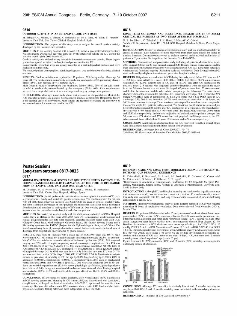
RESULTS. 101 patients (87 M) were included. Primary reasons of mechanical ventilation were:postoperative (27%), sepsis (19%), respiratory disease (ARDS, community pneumonia, hos-pital pneumonia, aspiration and other acute respiration failure) (27%), trauma (6%) and othercause (congestion heart failure, cardiac arrest, neuromuscular disease, liver disease) (21%).Baseline characteristics at ICU admission were: mean age 62±16 yrs, PaO2/FiO2 212±112mmHg, PEEP 7.2±4.2 cmH2O, Mean Airway Pressure 12.5±4.8 cmH2O, SAPS 45±16, SOFA7.8±3.6. Clinical characteristics were similar among different underlying disease groups. MeanICU stay was 20±17 days (range 3–91 days): we did not find any difference in outcome ac-cording to the length of ICU stay (more or less than 14 days). ICU, 6 months and 12 monthsmortality were related to patients’ age (> or <65 yrs).Figure 1 shows ICU (23%), 6 months (44%) and 12 months (50%) mortality, according to theunderlying disease at admission.
CONCLUSION. Although ICU mortality is relatively low, 6 and 12 months mortality arevery high. Both ICU, 6 and 12 months mortality were not related to the underlying disease atadmission.
REFERENCE(S). (1) Short et al, Crit Care Med 1999;27:51-57
S212 20th ESICM Annual Congress – Berlin, Germany – 7–10 October 2007
0820CLINICAL PREDICTORS AND OUTCOMES FOR PATIENTS REQUIRING LONG-TERM MECHANICAL VENTILATION IN THE ICU
F. V. C. De Marco*, M. M. Barbosa, A. S. Fantauzzi, G. P. T. Barros, A. L. G. Guimaraes,W. M. ImanishiICU, Hospital Vivalle, Sao Jose dos Campos, Brazil
INTRODUCTION. Prolonged ventilatory support in the ICU setting has a significant andgrowing impact on health care economics. As few as 5% to 10% of ICU patients require pro-longed mechanical ventilation, and this patient group consumes ≥ 50% of ICU patient daysand ICU resources. The aim of this study is to describe clinical predictors and the outcome ofthis patient population.
METHODS. We conducted a retrospective cohort study in order to identify independent riskfactors. We reviewed data from consecutive patients requiring mechanical ventilation fromApril 2003 to February 2007. We have defined long term patients those in need of mechani-cal ventilation for a period greater than seven days (LTMV). Epidemiological data, outcomeand ICU resource utilization were recorded. Apache II score was calculated (QuaTI softwaredatabase, Dixtal, Brazil). Statistical analysis was performed by univariate analysis (Fisher’sExact Test, Chi-Square) followed by multivariate stepwise logistic regression.
RESULTS. There were 195 consecutive patients (106 male). The mean age was 60±19. Themean APACHE II score was 18±10. The mean LOS was 9 ± 14. The frequency of LTMV was23% (n=44). By means of univariable analysis, risk factors for LTMV were male sex, creati-nine level ≥ 1.5 mg/dl at admission, presence of organic dysfunction at admission accordingto APACHE II definitions, hemoglobin level ≤ 7g/dl, APACHE II ≥ 25, use of pulmonaryartery catheter, dialysis therapy, enteral and parenteral nutritional support. In the multivariateanalysis only use of pulmonary artery catheter (OR 8.3; CI95% 2.3-30.1, p<0.001), dialysistherapy (OR 12.7; CI95% 3.1-52.7, p<0.001) and use of enteral nutritional support (OR 40.7;CI95% 13-127.8, p<0.001) were independent predictors of LTMV. The overall ICU mortalityrate was 40 %; it was 60% in patients with LTMV compared with 35% in patients withoutLTMV (p=0.013).
CONCLUSION. Patients requiring long-term mechanical ventilation have a higher mortalityrate and use of pulmonary artery catheter, dialysis therapy and enteral nutritional support areclinical predictors. If we could precisely identify patients at risk we could implement an institu-tional program to act in advance and improve clinical and financial outcomes in this vulnerablepopulation.
0821EVALUATION OF ICU PATIENTS HEALTH REALTED QUALITY OF LIFE BYPROXIES.
K. Waardal*, A. Muri, H. FlaattenKSK ICU, Haukeland University Hospital, Bergen, Norway
INTRODUCTION. Critically ill patients are during their ICU stay often not able to evaluatetheir Health Related Quality of Life (HRQoL) prior to the admission. This study is an evaluationof how accurate HRQoL assessments of ICU patients are made by proxies.
METHODS. Cohort study of 50 patients and their proxies (> 18 years) admitted to the generalICU in Haukeland University hospital in the period of 2004-2006. The proxies were asked tofill in the questionnaire SF-36, on behalf of the patient soon after ICU admission. In survivors,the patient also filled in the SF-36 when his og her cognitive function allowed it, after ICUdischarge.
RESULTS. Mean age of the patients was 58 ± 17 years, 35 males and 15 females. In the eightdimensions of the SF-36 there were no significant differences between the patient answer andtheir proxy assessments. The differences were greater when we compared male patients withfemale patients and their respective proxies. There is a trend that proxy assessments of femalepatient are not as coherent as proxies of male patients. This is further revealed when we lookat the gender of the proxies. Female proxies have a greater coherence in their assessments withtheir patient answers then male proxies have.
TABLE 1.Patients aswers vs their proxies assessments using the SF-36 (n=50)
Patients Proxies Delta 95% CIGeneral Health 75,8 69,7 6,1 -3,76 – 15,96PhysicalFunctioning 82,1 82,3 0,2 -9,96 – 10,36Role Physical 66,5 64,2 2,3 -14,37 – 18,97Role Emotional 80,0 84,0 4,0 -9,92 – 17,92Social Functioning 81,0 82,3 1,3 -9,05 – 11,65Body Pain 74,3 68,0 6,3 -5,36 – 17,96Vitality 57,5 55,2 2,3 -7,87 – 12,47Mental Health 77,1 78,6 1,5 -6,96 – 9,96
CONCLUSION. In our patients there are small but insignificant differences in the assessmentof HRQoL prior to the admission between the patient and his or her proxy. This allows us touse proxy evaluation of ICU patients HRQoL before ICU stay.
0822WITHDRAWAL OF LIFE SUSTAINING THERAPY DOES NOT REDUCE LENGTH-OF-STAY IN CRITICALLY ILL LEUKEMIA PATIENTS
J. L. Nates*, M. A. Cesta, C. Wakefield, K. PriceCritical Care, University of Texas M. D. Anderson Cancer Center, Houston, United States
INTRODUCTION. An increasing percentage of ICU deaths will follow decisions to with-hold or withdraw life sustaining therapies (LST)(1). The impact of these decisions on resourceutilization should be evaluated. We hypothesized that patients who had made these decisionswould have a shorter LOS than patients who did not.
METHODS. We performed a retrospective chart review of all the adult critically ill leukemiapatients who died in our oncological ICU between 9/01/04 to 8/31/05. An extensive chartreview including nurses, pharmacy and respiratory therapy records to determine data accuracywas performed. Data collected included but were not limited to demographics, overall ICUlength-of-stay (LOS), and hours until death after LST were withheld. Patients with ICU LOS<24 hours were excluded to avoid skewing the results by including patients that were admittedin a moribund state.
RESULTS. During the study period, 104 study group patients died in our ICU. The median agewas 61 (19-86), 58.4% were male. Thirty-one patients (29.8%) had LST withheld or withdrawn.The overall median ICU LOS was 3.9 days (0.1-39); patients that died without discontinuationof LST had a median LOS of 2.3 days (0.1-39) while patients with LST discontinuance had amedian LOS 6.3 days (0.9-24.7) (p=0.01). After exclusion of the 20 patients with ICU LOS <24hours, the median LOS was 5.2 days (1-39) for patients (n=54) who did not undergo terminalweaning and 6.5 days (1-24.7) for those who did (n=30) (p=0.35). Time until death followinginitiation of terminal weaning was median 134 minutes (6-2220).
CONCLUSION. Withdrawal or withholding LST was not associated with shortening LOS inpatients that survived the first 24 hours in ICU. These results seem to indicate that patient’send-of-life wishes are enforced at a very late stage in their disease process, and with no impacton their ICU resource utilization. A prospective study is necessary to further investigate thesimilar LOS.
REFERENCE(S). 1. Prendergast TJ, Luce JM. Increased incidence of withholding and with-drawal of life support from the critically ill. Am J Respir Crit Care Med. 1997;155:15-20.
GRANT ACKNOWLEDGEMENT. George B. Sweeney, Jr. Fellowship in Critical Care
0823ARDS IS NOT A DETERMINANT OF QUALITY OF LIFE OF SEVERE POLI-TRAUMA WITH THORACIC TRAUMA.
E. Gomes*1, R. Raimundez1, R. Carneiro1, A. Alegria1, I. Andre1, C. Dias2, A. Carneiro1
1Polivalent Intensive Care Unit, Hospital Geral de Santo Ant, 2Servico de Bioestatıstica einformatica Medica, Oporto University, Medicine, Porto, Portugal
INTRODUCTION. It was our objective to characterize thoracic trauma concerning epidemi-ology, baseline characteristics, ICU characteristics, mortality and quality of life at 6 months.
METHODS. All patients between 2004 and 2005 with thoracic trauma (AIS in the thorax supe-rior or equal to one) from the prospective registry collecting epidemiological data and severitydata using Trauma Severity Score System (TRISS) methodology. ICU process was evaluatedretrospectively. Mortality was defined as hospital mortality. Quality of life was measured usingEuroqol (EQ-5D) questionnaire at 6 months. Categorical variables were described as absoluteand relative frequencies; median and percentiles were used for continuous variables. PearsonQui Square test, and Mann-Whitney test were used for comparisons. Multiple logistic regres-sion was performed with mortality and EQ-Index as dependent variables and background, ICUvariables as independent variables. Stepwise Forward method was used.
RESULTS. 179 patients where analysed (45% of all trauma patients admitted in the same pe-riod). Median age was 41,42 years; trauma was blunt in 169 (94,4%); and road traffic accidentsthe main cause (67%). Median Abbreviated Injury Score (AIS) in the thorax was 3.16 (1.05),and 90% had Injury Severity Score (ISS) ≥15. In 93,7% a severe head trauma was also present.74,3% of the patients where ventilated during 9,12 days (+/-12,431). Pleural drainage was donein 30% of patients and thoracic surgery in 3%. Main complications during ICU stay wherepneumonia (25,4%) and ARDS (4,1%). Median ICU LOS was 6 days and hospital LOS was11 days. Mortality was 34,6%.EQ-5D at 6 months showed 31% of patients were not able to perform their usual activ-ities and 55,2% referred depression. Median EQ Index was 68% and EQ-VAS 70%. Lo-gistic regression for mortality showed ISS (OR:1.036; IC95%:1.004-1.069); female gender(OR:3.001; IC95%:1.065-8.455); penetrating injury (OR:1.153; IC95%:0.128 -10.360); andARDS (OR:2.390; IC95%:0.376 -15.169) -0.651;βas independent factors. Logistic regressionfor EQ-Index showed age (-9.202; IC95%:-21.322; -2.917) asβIC95%:-0.967;-0.336) and ven-tilation (independent factors).
CONCLUSION. In this severe trauma patients with thoracic trauma, mortality was determinedby the initial anatomical and physiological severity (RTS and ISS) and ARDS but those wherenot determinants of Quality of life.
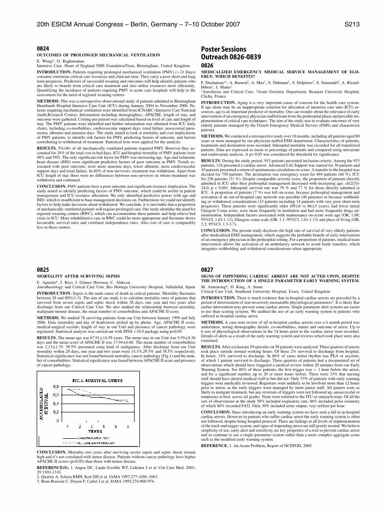
20th ESICM Annual Congress – Berlin, Germany – 7–10 October 2007 S213
0824OUTCOMES OF PROLONGED MECHANICAL VENTILATION
E. Wong*, G. RaghuramanIntensive Care, Heart of England NHS FoundationTrust, Birmingham, United Kingdom
INTRODUCTION. Patients requiring prolonged mechanical ventilation (PMV) (>21 Days)consume enormous critical care resources and clinician time. They carry a poor short and longterm prognosis. Predictors of successful weaning and outcomes will help identify patients whoare likely to benefit from critical care treatment and also utilise resources more efficiently.Quantifying the incidence of patients requiring PMV in acute care hospitals will help in theassessment for the need of regional weaning centres.
METHODS. This was a retrospective observational study of patients admitted to BirminghamHeartlands Hospital Intensive Care Unit (ICU) during January 2004 to November 2006. Pa-tients requiring mechanical ventilation were identified from ICNARC (Intensive Care NationalAudit,Research Centre). Information including demographics, APACHE, length of stay, andoutcome were gathered. Costing per patient was calculated based on level of care and length ofstay. The PMV patients were identified and further information was obtained from ICU dailycharts, including co-morbidities, cardiovascular support days, renal failure, nosocomial pneu-monia, albumin and anaemia days. The study aimed to look at mortality and cost implicationsof PMV patients, to identify risk factors for PMV, predicting factors of outcome, and factorscontributing to withdrawal of treatment. Statistical tests were applied for the analysis.
RESULTS. 5%(46) of all mechanically ventilated patients required PMV. However they ac-counted for 24% of the total cost in bed days. ICU and hospital mortality of PMV patients were48% and 59%. The only significant risk factor for PMV was increasing age. Age and ischaemicheart disease (IHD) were significant predictive factors of poor outcome in PMV. Trends as-sociated with poor outcome, were more anaemia days, lower albumin, more cardiovascularsupport days and renal failure. In 60% of non-survivors, treatment was withdrawn. Apart fromICU length of stay there were no differences between non-survivors in whom treatment waswithdrawn and continued.
CONCLUSION. PMV patients have a poor outcome and significant resource implication. Thestudy aimed to identify predicting factors of PMV outcome, which could be useful in patientmanagement and ICU resource allocation. We only identified predictive power with age andIHD, which is insufficient to base management decisions on. Furthermore we could not identifyfactors to help make decisions about withdrawal. We conclude, it is inevitable that a proportionof mechanically ventilated patients will require prolonged care. Our study identifies the need forregional weaning centres (RWC), which can accommodate these patients and help relieve bedcrisis in ICU. More rehabilitative care in RWC could be more appropriate and literature showsfavourable survival rates and ventilator independence rates. Also cost of care is comparablyless in these centres.
0825MORTALITY AFTER SURVIVING SEPSIS
E. Agustin*, J. Rico, J. Gomez Herreras, C. AldecoaAnesthesiology and Critical Care Unit, Rio Hortega University Hospital, Valladolid, Spain
INTRODUCTION. Sepsis is the main cause of death in critical patients. Mortality fluctuatesbetween 28 and 80%(1-3). The aim of our study is to calculate mortality rates of patients thatsurvived from severe sepsis and septic shock within 28 days, one year and two years afterdischarge from our Critical Care Unit. We also studied the relationship between mortality,malignant tumour disease, the mean number of comorbidities and APACHE II score.
METHODS. We studied 78 surviving patients from our Unit between January 1999 and July2006. Data (mortality and day of death)were picked up by phone. Age, APACHE II score,medical-surgical records, length of stay in our Unit and presence of cancer pathology wereregistered. Statistical analysis was carried out with SPSS v.10.0 package using p<0.05.
RESULTS. The mean age was 67.91±14.59 years. The mean stay in our Unit was 9.59±8.36days and the mean score of APACHE II was 17.94±6.60. The mean number of comorbiditieswas 2.13±1.35. 39.5% presented some kind of malignance. After discharge from our Unitmortality within 28 days, one year and two years were 14.1%,29.5% and 38.5% respectively.Statistical significance was not found between mortality, cancer pathology (Fig.1) and the num-ber of comorbidities. Statistical significance was found between APACHE II score and presenceof cancer pathology.
CONCLUSION. Mortality two years after surviving severe sepsis and septic shock remainhigh and it’s not correlated with tumor disease. Patients without cancer pathology have higherAPACHE II scores (p<0.05) than those with tumor disease.
REFERENCE(S). 1. Angus DC, Linde-Zwirble WT, Lidicker J et al. Crit Care Med. 2001;29:1303-1310.2. Quartin A, Schein RMH, Kett DH et al. JAMA 1997;277:1058–1063.3. Brun-Buisson C, Doyon F, Carlet J et al. JAMA 1995;274:968-974.
Poster SessionsOutreach 0826-08390826MEDICALIZED EMERGENCY MEDICAL SERVICE MANAGEMENT OF ELD-ERLY: WHICH BENEFITS?
F. Duchateau*1, A. Burnod1, A. Max1, S. Dahmani1, S. Delpierre2, S. Samandel2, A. Ricard-Hibon1, J. Mantz1
1Anesthesia and Critical Care, 2Acute Geriatric Department, Beaujon University Hospital,Clichy, France
INTRODUCTION. Aging is a very important cause of concern for the health care system.If age alone may be an inappropriate criterion for allocation of intensive care unit (ICU) re-sources, age is an important predictor of mortality. One can wonder about the relevance of earlyintervention of an emergency physician staffed team from the prehospital phase and possible im-plementation of critical care techniques. The aim of the study was to evaluate outcomes of veryelderly patients managed by the French Emergency Medical Service (EMS) and characterizepatients.
METHODS. We conducted a retrospective study over 18 months, including all patients aged 80years or more managed by our physician staffed EMS department. Characteristics of patients,treatments and destination were recorded. Inhospital mortality was recorded for all transferredpatients. Data are expressed as mean or percentage of patients and compared using univariateand multivariate analysis. A p< 0.05 was considered the threshold for significance.
RESULTS. During the study period, 933 patients presented inclusion criteria. Among the 933patients, 124 presented a cardiac arrest: Advanced Life Support was started for 36 patients and10 patients presented a return of spontaneous circulation on scene. A transfer to the hospital wasdecided for 750 patients. The destination was emergency room for 494 patients (66 %), ICUfor 236 patients (31 %). Despite comparable severity score, the proportion of patients directlyadmitted in ICU after their prehospital management decreased with increasing age: chi2[3]=24.0; p < 0.001. Inhospital survival rate was 79 % and 77 % for those directly admitted inICU. A proportion of patients (7 %) was left on-scene, because prehospital management andactivation of an out-of-hospital care network was possible (46 patients) or because withhold-ing or withdrawal considerations (23 patients including 14 patients with very poor short-termprognosis). These patients were significantly older (89±6 vs 86±5 years), had lower initialGlasgow Coma score, were more frequently in institution and had more frequently drugs ad-ministration. Independent factors associated with maintenance on-scene were age (OR, 1.08;95%CI, 1.03-1.13), Glasgow coma scale (OR, 1.1; 95%CI, 1.01-1.13) and place of living (OR,2.2; 95%CI, 1.3-3.7).
CONCLUSION. The present study discloses the high rate of survival of very elderly patientsafter medicalized EMS management, which suggests the probable benefit of early interventionof an emergency physician in the prehospital setting. For a proportion of patients, medical teamintervention allows the activation of an ambulatory network to avoid futile transfers, whichintegrates withholding and withdrawal considerations when appropriate.
0827SIGNS OF IMPENDING CARDIAC ARREST ARE NOT ACTED UPON, DESPITETHE INTRODUCTION OF A SINGLE PARAMETER EARLY WARNING SYSTEM.
M. Armstrong*, D. King, A. StoneCritical Care Unit, Southend University Hospital, Essex, United Kingdom
INTRODUCTION. There is much evidence that in-hospital cardiac arrests are preceded by aperiod of deterioration of non-invasively measurable physiological parameters1. It is likely thatearlier intervention may prevent some cardiac arrests. Single parameter alert systems are easierto use than scoring systems. We audited the use of an early warning system in patients whosuffered in-hospital cardiac arrest.
METHODS. A case-note review of all in-hospital cardiac arrests over a 6 month period wasundertaken, noting demographic details, co-morbidities, nature and outcome of arrest. Up to6 sets of physiological observations in the 24 hours prior to the cardiac arrest were recorded.Details of alerts as a result of the early warning system and reviews which took place were alsoexamined.
RESULTS. After exclusions 59 episodes on 58 patients were analysed. Three quarters of arreststook place outside normal working hours. Of these 2% survived to discharge from hospital.In hours, 14% survived to discharge. In 86% of cases initial rhythm was PEA or asystole,of which 1 patient survived to discharge. Three quarters of patients had a documented set ofobservations which should have triggered a medical review within 20 minutes from our EarlyWarning System. For 88% of these patients, the first trigger was > 1 hour before the arrest,and for a significant number, up to 20 or more hours before. There were 33% that nursingstaff should have alerted medical staff to but did not. Only 53% of patients with early warningtriggers were medically reviewed. Registrars were unlikely to be involved more than 12 hoursprior to arrest, as the early triggers were managed by more junior staff. All juniors were aslikely to instigate treatment, but any reversals of triggers were not followed up, unsuccessful ortemporary at best, across all grades. None were referred to the ITU or outreach team. Of all thesets of observations in the study 58% included respiratory rate, 86% included pulse oximetryof which 80% recorded FiO2. Only 30% included urine output, very seldom per hour.
CONCLUSION. Since introducing an early warning system we have seen a fall in in-hospitalcardiac arrests. However in patients who suffer cardiac arrest the early warning system is oftennot followed, despite being hospital protocol. There are failings at all levels of implementationof the track and trigger system, and signs of impending arrest are still poorly treated. We believesimplicity of use, early alert and sensitivity are key properties of a tool to prevent cardiac arrestand so continue to use a single parameter system rather than a more complex aggregate scoresuch as the modified early warning system.
REFERENCE. 1. An Acute Problem, Report of NCEPOD, 2005.

S214 20th ESICM Annual Congress – Berlin, Germany – 7–10 October 2007
0828IMPACT OF A CRITICAL CARE FOLLOW-UP SERVICE ON OUTCOME OF ICURE-ADMISSIONS
J. W. Zwaal*, S. Chitty, M. DeVille, E. Brophy, J. WelchIntensive Care, Kingston Hospital, Kingston Upon Thames, United Kingdom
INTRODUCTION. Re-admission into ICU is associated with high risk of mortality(1). Post-discharge follow-up of critical care patients may in the event of deterioration lead to moretimely re-admission to ICU which may result in better outcome of patients. We therefore set outto determine whether establishment of a critical care follow-up service could be demonstratedto lead to improved outcome in patients re-admitted to ICU.
METHODS. Re-admissions in the year before and after establishment of a critical care follow-up service were compared for re-admission rate, delay to re-admission, LOD scores at admissionand discharge, ICU and post-ICU mortality. In addition we compared both groups for differ-ences in the following risk factors for post-ICU mortality:age, sex, time of discharge,type ofadmission and co-morbidity. Differences between groups were tested by the Student-t test forcontinuous variables and by the method as described by Armitage(2) for proportions. P<0.05was considered statistically significant.
RESULTS. Complete datasets were obtained for 23 re-admissions.
TABLE 1.pre follow-up post follow-up P value
icu mortality(%) 33.3 26.9 p>0.62post-icu mortality(%) 25 15.4 0.55% re-admissions 4 6.4 p>0.13delay to re-admission(d) 6.4 12.6 0.17LOD admission 3.7 2.2 0.15LOD discharge 2.18 1.5 0.45age(yr) 53.9 60.5 0.40%male sex 58.3 84.6 p>0.13% co-morbidity 16.7 7.7 0.48% post acute surgery 8.3 23.1 p>0.31% night time discharge 0 7.7 p>0.31
CONCLUSION. Establishment of a critical care follow-up service led to a trend towardslower ICU and post-ICU mortality in patients re-admitted to ICU. An associated reduction inco-morbidity and severity of illness on admission and discharge may be responsible for thesefindings.
REFERENCE(S). 1. Chen,Liddy et al. Crit Care Med,26(11). November 1998, pp 1834-18412. Armitage P:Statistical Methods in Medical Research. Oxford: Blackwell;2002.
0829ASSESSMENT OF MEDICAL EMERGENCY TEAM CRITERIA IN THE EMER-GENCY ROOM: ASSOCIATION WITH SUBSEQUENT INTENSIVE CARE MOR-BIDITY AND MORTALITY
R. Etter*, R. Ludwig, F. Lersch, J. Takala, T. MerzDepartment of Intensive Care Medicine, University Hospital, Bern, Switzerland
INTRODUCTION. The survival of patients admitted to an emergency department is deter-mined mainly by the degree of acute illness at admission, the level and quality of the careprovided and the time interval until this care is delivered. Early recognition and prompt treat-ment of patients at risk of further deterioration may influence the course of illness and decreasemorbidity and mortality rates.
METHODS. We retrospectively analyzed 452 consecutive critically ill adult patients admittedto the emergency department from 1 January 2004 to 31 December 2004, who subsequentlywere referred to the ICU. Scores defined as the sum of organ failures based on the commonlyused medical emergency team calling criteria within the first hour (METinitial) and throughoutthe stay (METmax) in the emergency department were assessed as potential predictors. Mainoutcome measures were need for mechanical ventilation and hemodynamic instability duringthe subsequent ICU stay, and hospital mortality.
RESULTS. METinital and METmax both were significant predictors of hospital mortality,the need for mechanical ventilation and hemodynamic instability (Table 1). Hospital survivalranged from 96.2% (METinitial score =0) to 11.7% (METinitial score ≥4) and from 97.7%(METmax score =0) to 13.3% (METmax score ≥4) (log rank tests p<0.0001).
TABLE 1.p odds ratio lower CI (95%) upper CI (95%)
Hospital mortalityMETinitial <0.0001 3.392 2.534 4.540METmax <0.0001 3.867 2.816 5.312Need mech. ventil.METinitial <0.0001 4.151 3.530 4.652METmax <0.0001 4.292 3.151 5.846Hemodynamic instab.METinitial <0.0001 1.548 1.258 1.905METmax <0.0001 1.685 1.355 2.094
CONCLUSION. MET scores collected early after admission or throughout the stay in theemergency department allow for simple identification of patients at risk of unfavorable out-come during the subsequent ICU stay.
0830AN EARLY WARNING SCORE PREDICTED MORTALITY COMPARED TO OB-SERVED MORTALITY
I. J. Rechner*, A. Kapila, T. Even, M. OdellIntensive Care Medicine, Royal Berkshire hospital, Reading, United Kingdom
INTRODUCTION. Physiological deterioration is associated with worsened outcome.1 TheReading Early Warning Score (R-MEWS) is a track and trigger tool(range 0-15).
METHODS. The Outreach service used the R-MEWS to trigger referral. Data collected fromSept 2001 to Mar 2003 (2195 patients with 2339 episodes with re-referrals). A model predictingmortality by R-MEWS as the predictive parameter was developed using logistic regression.
RESULTS. Surgical patients made up 81%, and 29% were ICU discharges. For every 1 pointincrease in the R-MEWS, risk of death increased 1.53 fold (95% CI 1.45–1.61). A score of 2 to8 showed good prediction by the model of the observed mortality. With R-MEWS of 0, there isunder prediction and at a score of 1 over prediction. This despite N=214 and 611 for R-MEWS= 0 and 1 respectively.
CONCLUSION. Multiple factors influence mortality in the critically ill. However in a simplepredictive model just using R-MEWS, there is an association with poor outcome and increasingscore. Discrepancy at R-MEWS > 8 could be explained by a small sample size. R-MEWS of 0and 1 suggests there may possibly be recording error, with urine output being underestimatedor alternatively previous ICU admission might be a factor.
REFERENCE. 1. Goldhill DR, et al. Anaesthesia 2005;60(6):547-53
0831THE COST-EFFECTIVENESS OF CRITICAL CARE OUTREACH SERVICES FORREDUCING IN-HOSPITAL MORTALITY FOLLOWING DISCHARGE FROM ANINTENSIVE CARE UNIT
D. A. Harrison*1, R. D. Grieve2, K. M. Rowan1
1ICNARC, Intensive Care National Audit and Research Centre, 2Public Health and Policy,London School of Hygiene and Tropical Medicine, London, United Kingdom
INTRODUCTION. Critical care outreach services (CCOS) were introduced in 2000 as partof the modernisation of adult critical care in England but have never been subject to cost-effectiveness analysis (CEA). We conducted a CEA comparing CCOS to no CCOS using alarge, multicentre, matched-cohort analysis of patients discharged from intensive care units(ICUs).
METHODS. Fifty-two CCOS collected prospective data for one year on every patient visit.These data were linked to the Case Mix Programme, the national comparative audit of criticalcare, to identify patients receiving CCOS visits post-ICU discharge (cases). Each case wasmatched, where possible, to three controls: (1) a patient discharged from the same ICU in theyear before CCOS were introduced in that hospital; (2) a patient discharged from an ICU in ahospital with no CCOS during the study period; and (3) a patient discharged from the same ICUduring the study period that did not receive CCOS visits. Matching was based on: age (±10years); severe comorbidity; reason for ICU admission; source of admission/surgical status; IC-NARC physiology score (±10); length of ICU stay; and destination following ICU discharge.Hospital costs post-ICU discharge were compared for cases vs controls. For each case the num-ber of CCOS visits post-ICU discharge were multiplied by the cost per CCOS visit, calculatedas the total annual costs (predominately staffing) in each centre divided by the annual numberof CCOS visits. Days in ICU following original discharge and days in hospital were costedusing Healthcare Resource Groups for cases and controls. The incremental cost-effectivenessof CCOS vs no CCOS was reported as the cost per in-hospital death averted.
RESULTS. Of 5887 patients receiving CCOS visits post-ICU discharge, 1743 (30%), 4309(73%) and 1792 (30%), respectively, successfully matched to the three pools. In matches 1 and 2,CCOS visits were associated with lower mortality (risk ratio ∼0.86) and shorter post-ICU hos-pital stay (∆mean 2–3 days). The mean cost per CCOS visit was £115 (e169). Cases receiveda mean of 2.6–3.0 visits depending on matching, with an overall mean cost of £5129–£5470(e7527–e8027). The incremental cost of CCOS vs no CCOS was -£289 (-e424), -£34 (-e50)and +£275 (+e403) for the three matches, respectively. Cost-effectiveness acceptability curvesshowed that for matches 1 and 2 there was a high probability that CCOS were cost-effective;this result was robust to varying costing assumptions in sensitivity analyses.
CONCLUSION. While CEA ideally use effectiveness data from randomised controlled trials,randomised studies of this established intervention are now infeasible. These results, which useappropriate methods for analysing non-randomised data, suggest that CCOS visits post-ICUdischarge are associated with reduced length of stay and are cost-effective.
GRANT ACKNOWLEDGEMENT. NIHR Service Delivery & Organisation R&DProgramme (SDO/74/2004)
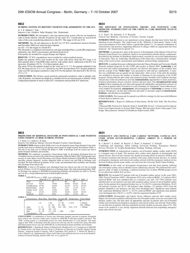
20th ESICM Annual Congress – Berlin, Germany – 7–10 October 2007 S215
0832SCORING SYSTEM TO IDENTIFY PATIENTS FOR ADMISSIONS TO THE ICU.
A. J. E. Dekkers*, TjanIntensive Care, Gelderse Vallei Hospital, Ede, Netherlands
INTRODUCTION. We developed a vital sign alarmscoring system (AS) for our hospital inorder to better identify critically ill patients on the ward. In a 3 month pilot we assessed thevalue of the score in predicting admission to the ICU. The range of AS was 0-30.
METHODS. The AS and admission to the ICU for 127 ICU consultations between Octoberand December 2006 were analyzed and evaluated.An AS > 8 is the trigger to consult ICU.The AS points is a weighted score based on vital signs including Pulse, systolic BP, temperature,respiratory rate, SaO2, consciousness and blood lactate level.Extra points are awarded for oxygen therapy and oliguria.
RESULTS. 127 ICU consultations were recorded for various medical reasons.Eighty-one patients (64%) were treated on the ward with advice from the ICU team. 6 ofthese patients died, 4 with DNR orders and two with cardiac arrest. Admission to the ICU wasnecessary 46 patients (36 %). 12 of these patients died.The mean AS for the patients treated on the ward was 6.7± 3.5. For patients admitted to theICU the mean AS was 11.4 ± 4.1. Sensitivity was 89% and specificity 93%. Positive predictivevalue was 0.87 and negative predictive value 0.94. For the patients who died in ICU the AS was15.4 ± 2.3.
CONCLUSION. The AS has a good sensitivity and positive predictive value to identify criti-cally ill patients. An alarmscore might be a valuable tool for an outreach team to identify vitallycompromised patients in need of early ICU consultation and possible ICU admission.
0833PREDICTION OF HOSPITAL OUTCOME IN POST-CRITICAL CARE PATIENTSUSING PATIENT AT RISK SCORING SYSTEMJ. J. Taylor, S. J. Andrews*, R. BabaIntensive Care Medicine, Mayday University Hospital, Croydon, United KingdomINTRODUCTION. Patient at Risk (PAR) score1 was developed by James Paget Hospital. It has tradi-tionally been used for identifying sick patients and is based on simple nursing observations (Table 1).The aim of our study was to evaluate the ability of ‘PAR’ at discharge from our critical care unit topredict hospital morbidity and mortality.METHODS. We conducted a prospective observational study on all patients discharged from our14-bedded critical care unit to the wards between September 2006 and March 2007. Demographic data,severity of acute illness (Acute Physiology and Chronic Health Evaluation [APACHE] II), admittingspeciality, primary diagnosis, systems supported while on critical care and PAR at discharge wererecorded. We then followed up patients on the wards, and determined their morbidity and mortalityafter discharge from critical care.RESULTS. A total of 159 patients were discharged from our critical care unit over the six-monthperiod. Of these, 16 (10.1%) patients died prior to discharge home. Our study demonstrated that ‘PAR’on discharge was superior to APACHE II in predicting morbidity and mortality (see table 2). Of note,age was also a strong influencing factor in final hospital outcome.TABLE 1.
APACHE II score vs ’PAR’ score at dischargeAPACHE 0-25 APACHE >25 PAR 0-1 PAR 2-3 PAR >3
Total 134 25 80 68 11Survival 92.5% 76.0% 95.0% 92.6% 36.4%Complications 47.0% 44.0% 42.5% 51.5% 45.5%Renal Failure 10.4% 8.0% 7.5% 13.2% 9.1%Cardiac event 4.8% 12.0% 5.0% 5.9% 9.1%Sepsis 43.3% 32.0% 35.0% 42.6% 36.4%GI Bleed 8.2% 8.0% 5.0% 13.2% 0%
TABLE 2.Conciousness Resp. Rate Heart Rate Systolic BP Temperature Urine Output
3 < 70 02 < 8 < 40 71 - 80 < 35.0 < 250mls1 41 - 50 81 - 100 250 - 500mls0 Alert 9 - 14 51 - 100 101 - 199 35.0 - 37.9 > 500mls1 Verbal 15 - 20 101 - 1102 Pain 21 - 29 111 - 130 > 200 > 38.03 Unresponsive >30 > 130
CONCLUSION. A combination of factors may determine hospital outcome in patients dischargedfrom critical care. Previous studies have focused on APACHE II affecting hospital outcome2. The‘PAR’ scoring system is a measurable process for early identification of a sick patient1. Our studysuggests that ‘PAR’ at discharge from the critical care unit may also be a useful tool, along with otherindividual patient factors (age and APACHE II), in determining hospital morbidity and mortality.REFERENCE(S). 1. Rutledge R, Fakhry S, Rutherford E, Muakkassa F et al, ‘Comparison of APACHEII, Trauma Score, and Injury Severity Score as Predictors of Outcome in Critically Injured TraumaPatients’, American Journal of Surgery, 166(3), September 1993, pp244-47. 2. Watson W, Mozley C,Cope J, Hart D et al, ‘Implementing a Nurse-Led Critical Care Outreach Service in an Acute Hospital’,Journal of Clinical Nursing, January 2006, Vol. 15 (1), pp105-10.
0834THE DIFFUSION OF INNOVATIONS THEORY AND INTENSIVE CAREMEDICINE: INTRODUCTION OF THE CRITICAL CARE RESPONSE TEAM INONTARIO.
A. C. Steel*, M. Salenieks, S. F. ReynoldsCritical Care Medicine, University of Toronto, Toronto, Canada
INTRODUCTION. Intensive care operational system changes develop more slowly than theinitiation of new therapies. The central tenet of the “diffusion of innovations” theory is thatsuccessful adoption of new ideas follows a predictable pattern (1). An optimal combination ofcharacteristics and practices supporting diffusion of change within an organization has beenproposed - the “framework for spread” (2).
METHODS. A retrospective study of the process of development of the Ontario Critical CareResponse Team Project was conducted. Analysis was performed to identify the key components,described by the Institute for Healthcare Improvement, essential for successful implementationof innovation. They are: leadership, identification of the better idea, communication, strength-ening of the social system, measurement and feedback, and knowledge management.
RESULTS. A multiprofessional collaboration from the Ottawa Hospital and University HealthNetwork outlined proposals for the CCRT pilot project in March 2004. Leadership at thelocal level and central management structure was derived. Description of the evidence baseand the concept of the “better idea” were presented to staff and key stakeholders. A time-line was established and an agenda for the initial pilot. After review of the pilot the timelinewas modified to increase the number of months of planning for the expansion of the CCRTprogram. During both the pilot study and expansion project monthly local feedback meetingsand province-wide teleconferences provide communication opportunities to share awareness,technical information, discuss concerns, barriers and new ideas. This strengthened the socialsystem of those adopting the new ideas and engaged all stakeholders and potential end users ofthe system in patient safety. Formation of a sub-committee of “champions” from within the net-work enabled development, management, and promulgation of knowledge to emerge as bestpractice. Prospective, on-line data collection and audit of outcomes improved measurement,feedback, and afforded accountability.
CONCLUSION. The framework for spread is an effective strategy for the successful diffusionof innovation in intensive care.
REFERENCE(S). 1 Rogers E. Diffusion of Innovations, 5th Ed. New York: The Free Press2005.2 Massoud MR, Nielson GA, Nolan K, Nolan T, Schall MW, Sevin C. A Framework For Spread:From Local Improvements to System-Wide Change. IHI Innovation Series white paper. Cam-bridge, Massachusetts: Institute for Healthcare Improvement; 2006.
0835EMERGENCY AND CRITICAL CARE CARDIAC NETWORK: CLINICAL OUT-COME AFTER OUT-OF-HOSPITAL CARDIAC ARREST IN A PERIOD OF6 YEARS
H. J. Busch*1, E. Kohls1, H. Reisch2, C. Bode1, F. Koberne2, T. Schwab1
1Cardiology and Angiology, Albert Ludwigs University Freiburg, 2Emergency MedicalServices Division, St. Josephs-Krankenhaus Freiburg, Freiburg, Germany
INTRODUCTION. In industrialized countries out-of-hospital sudden cardiac death (SCD)is a frequent cause of death. The outcome after cardiac arrest depends on demographic andlogistic factors. Survival rates remains low, despite increasing efforts in intensive medical care.To advance treatment and outcome in patients with acute cardiovascular diseases, we initiatean integrative emergency and critical care cardiac network with the emergency medical servicedivision Freiburg and the acute cardiac care unit from the University Heart Center Freiburg.
METHODS. In this study we investigated circumstances and data from patients sufferingout-of-hospital cardiac arrest to identify factors influencing outcome. We performed this retro-spective study of adults in Freiburg, a midsized German city of about 300.000 people servedby two physician staffed ALS services.
RESULTS. We included 675 patients with out of hospital cardiac arrests, in the years 2001-2006. From 675 patients (100%), 364 patients (54%) never achieved ROSC, 311 patients (46%)achieved ROSC and were admitted alive to a intensive care unit. 235 (76%) patients wereadmitted alive to our intensive care unit with assumed cardiac reasons of out of hospital cardiacarrest. In this patients first registered rhythm was in 114 (48,5%) ventricular fibrillation, 20,4%(48 patients) asystole and 36,7% (86 patients) other rhythms. 115 patients (49%) from thepatients admitted to our intensive care unit were discharged alive. Significant more patientswith ventricular fibrillation (68,3% vs. 25,5%) achieved ROSC and significant more patientswere discharged alive (58,4% vs. 22,9%) with first registered rhythm of VF.
CONCLUSION. Demographic and logistic factors influence outcome after cardiac arrest.Higher survival rates in patients suffering from out of hospital cardiac arrest are associated withprimary cardiac care. Our data show an appropriate outcome in patients after out-of-hospitalcardiac arrest treated in an integrative emergency and critical cardiac care network. Noteworthy,we can find also in patients with first registered asystolic rhythm an outcome about 23%. Thisseems to be seeded in a very strictly organized network between first responding emergencyteams and critical care unit.
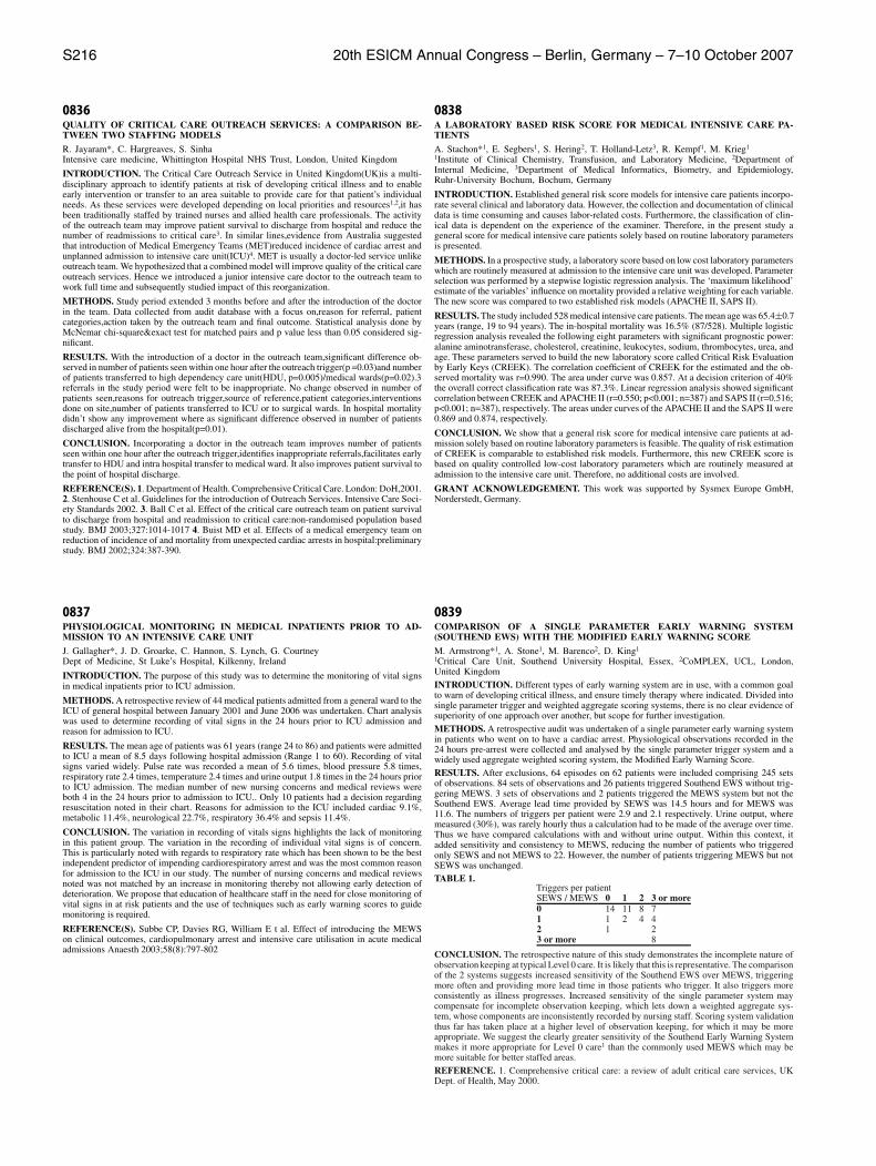
S216 20th ESICM Annual Congress – Berlin, Germany – 7–10 October 2007
0836QUALITY OF CRITICAL CARE OUTREACH SERVICES: A COMPARISON BE-TWEEN TWO STAFFING MODELS
R. Jayaram*, C. Hargreaves, S. SinhaIntensive care medicine, Whittington Hospital NHS Trust, London, United Kingdom
INTRODUCTION. The Critical Care Outreach Service in United Kingdom(UK)is a multi-disciplinary approach to identify patients at risk of developing critical illness and to enableearly intervention or transfer to an area suitable to provide care for that patient’s individualneeds. As these services were developed depending on local priorities and resources1,2,it hasbeen traditionally staffed by trained nurses and allied health care professionals. The activityof the outreach team may improve patient survival to discharge from hospital and reduce thenumber of readmissions to critical care3. In similar lines,evidence from Australia suggestedthat introduction of Medical Emergency Teams (MET)reduced incidence of cardiac arrest andunplanned admission to intensive care unit(ICU)4. MET is usually a doctor-led service unlikeoutreach team. We hypothesized that a combined model will improve quality of the critical careoutreach services. Hence we introduced a junior intensive care doctor to the outreach team towork full time and subsequently studied impact of this reorganization.
METHODS. Study period extended 3 months before and after the introduction of the doctorin the team. Data collected from audit database with a focus on,reason for referral, patientcategories,action taken by the outreach team and final outcome. Statistical analysis done byMcNemar chi-square&exact test for matched pairs and p value less than 0.05 considered sig-nificant.
RESULTS. With the introduction of a doctor in the outreach team,significant difference ob-served in number of patients seen within one hour after the outreach trigger(p =0.03)and numberof patients transferred to high dependency care unit(HDU, p=0.005)/medical wards(p=0.02).3referrals in the study period were felt to be inappropriate. No change observed in number ofpatients seen,reasons for outreach trigger,source of reference,patient categories,interventionsdone on site,number of patients transferred to ICU or to surgical wards. In hospital mortalitydidn’t show any improvement where as significant difference observed in number of patientsdischarged alive from the hospital(p=0.01).
CONCLUSION. Incorporating a doctor in the outreach team improves number of patientsseen within one hour after the outreach trigger,identifies inappropriate referrals,facilitates earlytransfer to HDU and intra hospital transfer to medical ward. It also improves patient survival tothe point of hospital discharge.
REFERENCE(S). 1. Department of Health. Comprehensive Critical Care. London: DoH,2001.2. Stenhouse C et al. Guidelines for the introduction of Outreach Services. Intensive Care Soci-ety Standards 2002. 3. Ball C et al. Effect of the critical care outreach team on patient survivalto discharge from hospital and readmission to critical care:non-randomised population basedstudy. BMJ 2003;327:1014-1017 4. Buist MD et al. Effects of a medical emergency team onreduction of incidence of and mortality from unexpected cardiac arrests in hospital:preliminarystudy. BMJ 2002;324:387-390.
0837PHYSIOLOGICAL MONITORING IN MEDICAL INPATIENTS PRIOR TO AD-MISSION TO AN INTENSIVE CARE UNIT
J. Gallagher*, J. D. Groarke, C. Hannon, S. Lynch, G. CourtneyDept of Medicine, St Luke’s Hospital, Kilkenny, Ireland
INTRODUCTION. The purpose of this study was to determine the monitoring of vital signsin medical inpatients prior to ICU admission.
METHODS. A retrospective review of 44 medical patients admitted from a general ward to theICU of general hospital between January 2001 and June 2006 was undertaken. Chart analysiswas used to determine recording of vital signs in the 24 hours prior to ICU admission andreason for admission to ICU.
RESULTS. The mean age of patients was 61 years (range 24 to 86) and patients were admittedto ICU a mean of 8.5 days following hospital admission (Range 1 to 60). Recording of vitalsigns varied widely. Pulse rate was recorded a mean of 5.6 times, blood pressure 5.8 times,respiratory rate 2.4 times, temperature 2.4 times and urine output 1.8 times in the 24 hours priorto ICU admission. The median number of new nursing concerns and medical reviews wereboth 4 in the 24 hours prior to admission to ICU.. Only 10 patients had a decision regardingresuscitation noted in their chart. Reasons for admission to the ICU included cardiac 9.1%,metabolic 11.4%, neurological 22.7%, respiratory 36.4% and sepsis 11.4%.
CONCLUSION. The variation in recording of vitals signs highlights the lack of monitoringin this patient group. The variation in the recording of individual vital signs is of concern.This is particularly noted with regards to respiratory rate which has been shown to be the bestindependent predictor of impending cardiorespiratory arrest and was the most common reasonfor admission to the ICU in our study. The number of nursing concerns and medical reviewsnoted was not matched by an increase in monitoring thereby not allowing early detection ofdeterioration. We propose that education of healthcare staff in the need for close monitoring ofvital signs in at risk patients and the use of techniques such as early warning scores to guidemonitoring is required.
REFERENCE(S). Subbe CP, Davies RG, William E t al. Effect of introducing the MEWSon clinical outcomes, cardiopulmonary arrest and intensive care utilisation in acute medicaladmissions Anaesth 2003;58(8):797-802
0838A LABORATORY BASED RISK SCORE FOR MEDICAL INTENSIVE CARE PA-TIENTS
A. Stachon*1, E. Segbers1, S. Hering2, T. Holland-Letz3, R. Kempf1, M. Krieg1
1Institute of Clinical Chemistry, Transfusion, and Laboratory Medicine, 2Department ofInternal Medicine, 3Department of Medical Informatics, Biometry, and Epidemiology,Ruhr-University Bochum, Bochum, Germany
INTRODUCTION. Established general risk score models for intensive care patients incorpo-rate several clinical and laboratory data. However, the collection and documentation of clinicaldata is time consuming and causes labor-related costs. Furthermore, the classification of clin-ical data is dependent on the experience of the examiner. Therefore, in the present study ageneral score for medical intensive care patients solely based on routine laboratory parametersis presented.
METHODS. In a prospective study, a laboratory score based on low cost laboratory parameterswhich are routinely measured at admission to the intensive care unit was developed. Parameterselection was performed by a stepwise logistic regression analysis. The ‘maximum likelihood’estimate of the variables’ influence on mortality provided a relative weighting for each variable.The new score was compared to two established risk models (APACHE II, SAPS II).
RESULTS. The study included 528 medical intensive care patients. The mean age was 65.4±0.7years (range, 19 to 94 years). The in-hospital mortality was 16.5% (87/528). Multiple logisticregression analysis revealed the following eight parameters with significant prognostic power:alanine aminotransferase, cholesterol, creatinine, leukocytes, sodium, thrombocytes, urea, andage. These parameters served to build the new laboratory score called Critical Risk Evaluationby Early Keys (CREEK). The correlation coefficient of CREEK for the estimated and the ob-served mortality was r=0.990. The area under curve was 0.857. At a decision criterion of 40%the overall correct classification rate was 87.3%. Linear regression analysis showed significantcorrelation between CREEK and APACHE II (r=0.550; p<0.001; n=387) and SAPS II (r=0.516;p<0.001; n=387), respectively. The areas under curves of the APACHE II and the SAPS II were0.869 and 0.874, respectively.
CONCLUSION. We show that a general risk score for medical intensive care patients at ad-mission solely based on routine laboratory parameters is feasible. The quality of risk estimationof CREEK is comparable to established risk models. Furthermore, this new CREEK score isbased on quality controlled low-cost laboratory parameters which are routinely measured atadmission to the intensive care unit. Therefore, no additional costs are involved.
GRANT ACKNOWLEDGEMENT. This work was supported by Sysmex Europe GmbH,Norderstedt, Germany.
0839COMPARISON OF A SINGLE PARAMETER EARLY WARNING SYSTEM(SOUTHEND EWS) WITH THE MODIFIED EARLY WARNING SCORE
M. Armstrong*1, A. Stone1, M. Barenco2, D. King1
1Critical Care Unit, Southend University Hospital, Essex, 2CoMPLEX, UCL, London,United KingdomINTRODUCTION. Different types of early warning system are in use, with a common goalto warn of developing critical illness, and ensure timely therapy where indicated. Divided intosingle parameter trigger and weighted aggregate scoring systems, there is no clear evidence ofsuperiority of one approach over another, but scope for further investigation.METHODS. A retrospective audit was undertaken of a single parameter early warning systemin patients who went on to have a cardiac arrest. Physiological observations recorded in the24 hours pre-arrest were collected and analysed by the single parameter trigger system and awidely used aggregate weighted scoring system, the Modified Early Warning Score.RESULTS. After exclusions, 64 episodes on 62 patients were included comprising 245 setsof observations. 84 sets of observations and 26 patients triggered Southend EWS without trig-gering MEWS. 3 sets of observations and 2 patients triggered the MEWS system but not theSouthend EWS. Average lead time provided by SEWS was 14.5 hours and for MEWS was11.6. The numbers of triggers per patient were 2.9 and 2.1 respectively. Urine output, wheremeasured (30%), was rarely hourly thus a calculation had to be made of the average over time.Thus we have compared calculations with and without urine output. Within this context, itadded sensitivity and consistency to MEWS, reducing the number of patients who triggeredonly SEWS and not MEWS to 22. However, the number of patients triggering MEWS but notSEWS was unchanged.TABLE 1.
Triggers per patientSEWS / MEWS 0 1 2 3 or more0 14 11 8 71 1 2 4 42 1 23 or more 8
CONCLUSION. The retrospective nature of this study demonstrates the incomplete nature ofobservation keeping at typical Level 0 care. It is likely that this is representative. The comparisonof the 2 systems suggests increased sensitivity of the Southend EWS over MEWS, triggeringmore often and providing more lead time in those patients who trigger. It also triggers moreconsistently as illness progresses. Increased sensitivity of the single parameter system maycompensate for incomplete observation keeping, which lets down a weighted aggregate sys-tem, whose components are inconsistently recorded by nursing staff. Scoring system validationthus far has taken place at a higher level of observation keeping, for which it may be moreappropriate. We suggest the clearly greater sensitivity of the Southend Early Warning Systemmakes it more appropriate for Level 0 care1 than the commonly used MEWS which may bemore suitable for better staffed areas.REFERENCE. 1. Comprehensive critical care: a review of adult critical care services, UKDept. of Health, May 2000.
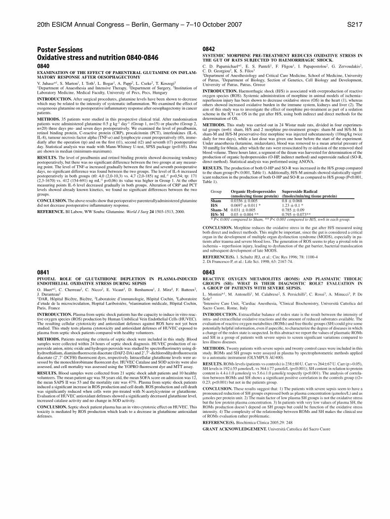
20th ESICM Annual Congress – Berlin, Germany – 7–10 October 2007 S217
Poster SessionsOxidative stress and nutrition 0840-08460840EXAMINATION OF THE EFFECT OF PARENTERAL GLUTAMINE ON INFLAM-MATORY RESPONSE AFTER OESOPHAGECTOMY
V. Juhasz*1, S. Marton1, I. Toth1, L. Bogar1, A. Papp2, L. Cseke2, T. Koszegi31Department of Anaesthesia and Intensive Therapy, 2Department of Surgery, 3Institution ofLaboratory Medicine, Medical Faculty, University of Pecs, Pecs, Hungary
INTRODUCTION. After surgical procedures, glutamine levels have been shown to decreasewhich may be related to the intensity of systematic inflammation. We examined the effect ofexogeneous glutamine on postoperative inflammatory response after oesophagectomy in cancerpatients.
METHODS. 35 patients were studied in this prospective clinical trial. After randomisationpatients were administered glutamine 0.5 g kg-1 day-1 (Group 1, n=15) or placebo (Group 2,n=20) three days pre- and seven days postoperatively. We examined the level of prealbumin,retinol binding protein, C-reactive protein (CRP), procalcitonin (PCT), interleukines (IL-6,IL-8), tumour necrosis factor alpha (TNF-α) and lymphocyte count preoperatively (t0), imme-diatly after the operation (tp) and on the first (t1), second (t2) and seventh (t7) postoperativeday. Statistical analysis was made with Mann-Whitney U-test, SPSS package (p<0.05). Dataare shown in median (minimum-maximum).
RESULTS. The level of prealbumin and retinol binding protein showed decreasing tendencypostoperatively, but there was no significant difference between the two groups at any measur-ing point. The level of TNF-α increased gradually between the first and seventh postoperativedays, no significant difference was found between the two groups. The level of IL-6 increasedpostoperatively in both groups (t0: 4,0 (2,0-10,3) vs. 4,7 (2,0-185) ng mL-1 p=0,54; tp: 171(2,3-1670) vs. 412 (154-691) ng mL-1 p=0,06) its value was higher in Group 1. At the othermeasuring points IL-6 level decreased gradually in both groups. Alteration of CRP and PCTlevels showed already known kinetics, we found no significant differences between the twogroups.
CONCLUSION. The above results show that perioperative parenterallyadministered glutaminedid not decrease postoperative inflammatory response.
REFERENCE. BI Labow, WW Souba: Glutamine. World J Surg 24 1503-1513, 2000.
0841PIVOTAL ROLE OF GLUTATHIONE DEPLETION IN PLASMA-INDUCEDENDOTHELIAL OXIDATIVE STRESS DURING SEPSIS
O. Huet*1, C. Cherreau2, C. Nicco2, E. Vicaut3, D. Benhamou1, J. Mira4, F. Batteux2,J. Duranteau1
1DAR, Hopital Bicetre, Bicetre, 2Laboratoire d’immunologie, Hopital Cochin, 3Laboratoired’etude de la microcirculation, Hopital Lariboisiere, 4reanimation medicale, Hopital Cochin,Paris, France
INTRODUCTION. Plasma from septic shock patients has the capacity to induce in vitro reac-tive oxygen species (ROS) production by Human Umbilical Vein Endothelial Cells (HUVEC).The resulting cellular cytotoxicity and antioxidant defenses against ROS have not yet beenstudied. This study tests plasma cytotoxicity and antioxidant defenses of HUVEC exposed toplasma from septic shock patients compared with healthy volunteers.
METHODS. Patients meeting the criteria of septic shock were included in this study. Bloodsamples were collected within 24 hours of septic shock diagnosis. HUVEC production of su-peroxide anion, nitric oxide and hydrogen peroxide was studied by spectrofluorimetry using di-hydroethidium, diaminofluorescein diacetate (DAF2-DA) and 2’,7’-dichlorodihydrofluoresceindiacetate (2’,7’-DCFH) fluorescent dyes, respectively. Intracellular glutathione levels were as-sessed by the monochlorobimane fluorecent dye. HUVEC Catalase and SOD activity were alsoassessed, and cell mortality was assessed using the YOPRO fluorescent dye and MTT assay.
RESULTS. Blood samples were collected from 21 septic shock adult patients and 10 healthyvolunteers. The mean patient age was 58 years old, the mean SOFA score on admission was 12,the mean SAPS II was 53 and the mortality rate was 47%. Plasma from septic shock patientsinduced a significant increase in ROS production and cell death. ROS production and cell deathwas significantly reduced when cells were pre-treated with N-acetylcysteine or glutathione.Evaluation of HUVEC antioxidant defenses showed a significantly decreased glutathione level,increased catalase activity and no change in SOD activity.
CONCLUSION. Septic shock patient plasma has an in vitro cytotoxic effect on HUVEC. Thistoxicity is mediated by ROS production which leads to a decrease in glutathione antioxidantdefenses.
0842SYSTEMIC MORPHINE PRE-TREATMENT REDUCES OXIDATIVE STRESS INTHE GUT OF RATS SUBJECTED TO HAEMORRHAGIC SHOCK.
C. D. Papamichael*1, E. S. Panteli1, F. Fligou1, I. Papapostolou2, G. Zervoudakis2,C. D. Georgiou2, K. S. Filos1
1Department of Anesthesiology and Critical Care Medicine, School of Medicine, Universityof Patras, 2Department of Biology, Section of Genetics, Cell Biology and Development,University of Patras, Patras, Greece
INTRODUCTION. Haemorrhagic shock (H/S) is associated with overproduction of reactiveoxygen species (ROS). Systemic administration of morphine in animal models of ischemia–reperfusion injury has been shown to decrease oxidative stress (OS) in the heart (1), whereasothers showed increased oxidative burden in the immune system, kidneys and liver (2). Theaim of this study was to investigate the effect of morphine pre-treatment as part of a sedationscheme in the ICU on OS in the gut after H/S, using both indirect and direct methods for thedetermination of OS.
METHODS. This study was carried out in 24 Wistar male rats, divided in four experimen-tal groups (n=6): sham, H/S and 2 morphine pre-treatment groups: sham-M and H/S-M. Insham-M and H/S-M preservative-free morphine was injected subcutaneously (10mg/kg twicedaily for two days), while a last dose was given one hour before the start of the experiment.Under anaesthesia (ketamine, midazolam), blood was removed to a mean arterial pressure of30 mmHg for 60min, after which the rats were resuscitated by re-infusion of the removed shedblood volume. Three hours later, a segment of the ileum was harvested for determination of theproduction of organic hydroperoxides (O-HP, indirect method) and superoxide radical (SO-R,direct method). Statistical analysis was performed using ANOVA.
RESULTS. The production of both O-HP and SO-R was increased in the H/S group comparedto the sham group (P< 0.001, Table 1). Additionally, H/S-M animals showed statistically signif-icant reduction in the production of both O-HP and SO-R as compared to H/S group (P<0.001,Table 1).
Group Organic Hydroperoxides Superoxide Radical(nmoles/mg tissue protein) (fmoles/min/mg tissue protein)
Sham 0.0356 ± 0.005 0.8 ± 0.068H/S 0.0697 ± 0.011 * 1.23 ± 0.1 *Sham- M 0.031 ± 0.005 0.785 ± 0.09H/S- M 0.03 ± 0.004 ** 0.795 ± 0.073*** P< 0.001 compared to Sham, ** P< 0.001 compared to H/S, n=6 in each group.
CONCLUSION. Morphine reduces the oxidative stress in the gut after H/S measured usingboth direct and indirect methods. This might be important, since the gut is considered a criticalorgan in the development of multiple organ dysfunction syndrome (MODS), especially in pa-tients after trauma and severe blood loss. The generation of ROS seems to play a pivotal role inischemia – reperfusion injury, leading to dysfunction of the gut barrier, bacterial translocationand subsequent development of late MODS.
REFERENCE(S). 1. Schultz JEJ, et al.: Circ Res 1996; 78: 1100-42. Di Francesco P, et al.: Life Sci. 1998; 63: 2167-74.
0843REACTIVE OXYGEN METABOLITES (ROMS) AND PLASMATIC THIOLICGROUPS (SH): WHAT IS THEIR DIAGNOSTIC ROLE? EVALUATION INA GROUP OF PATIENTS WITH SEVERE SEPSIS.
L. Montini*1, M. Antonelli1, M. Calabrese2, S. Persichilli3, C. Rossi3, A. Minucci3, P. DeSole3
1Intensive Care Unit, 2Cardiac Anesthesia, 3Clinical Biochemistry, Universita Cattolica delSacro Cuore, Rome, Italy
INTRODUCTION. Extracellular balance of redox state is the result between the intensity ofintra- and extracellular oxidative reactions and the amount of reduced substrates available. Theevaluation of reactive oxygen metabolites (ROMs) and free thiolic groups (SH) could give somepotentially helpful information, even if aspecific, to characterize the degree of diseases in whicha change of the redox state is suspected. In this abstract we report the values of plasmatic ROMsand SH in a group of patients with severe sepsis to screen significant variations compared toless illness diseases.
METHODS. Twenty patients with severe sepsis and twenty control cases were included in thisstudy. ROMs and SH groups were assayed in plasma by spectrophotometric methods appliedto a automatic instrument (OLYMPUS AU400).
RESULTS. ROMs levels (patients vs controls) is 238±80 U. Carr vs 264±67 U. Carr (p>0.05),SH levels is 192±55 µmoles/L vs 364±77 µmoli/L (p<0.001); SH content in relation to proteincontent is 4.4±1.0 µmoles/g vs 5.6±1.0 µmoli/g respectly (p<0.001). The analysis of correla-tion between ROMs and SH shows a significant positive correlation in the controls group (r2=0.23; p<0.001) but not in the patients group.
CONCLUSION. These results suggest that: 1) The patients with severe sepsis seem to have apronounced reduction of SH groups expressed both as plasma concentration (µmoles/L) and asµmoles per protein unit. 2) The main factor of low plasma SH groups is not the oxidative stressbut the low protein plasma concentration. 3) In patients with very low values of plasma SH, theROMs production doesn’t depend on SH groups but could be function of the oxidative stressintensity. 4) The complexity of the relationship between ROMs and SH makes the clinical useof ROMs evaluation rather problematic.
REFERENCE(S). Biochimica Clinica 2005,29: 248
GRANT ACKNOWLEDGEMENT. Universita Cattolica del Sacro Cuore
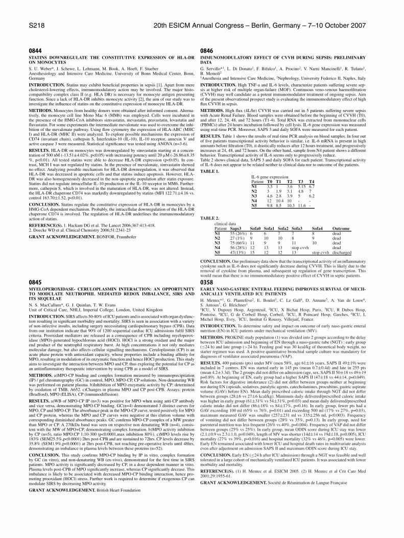
S218 20th ESICM Annual Congress – Berlin, Germany – 7–10 October 2007
0844STATINS DOWNREGULATE THE CONSTITUTIVE EXPRESSION OF HLA-DRON MONOCYTES
S. U. Weber*, J. Schewe, L. Lehmann, M. Book, A. Hoeft, F. StueberAnesthesiology and Intensive Care Medicine, University of Bonn Medical Center, Bonn,Germany
INTRODUCTION. Statins may exhibit beneficial properties in sepsis [1]. Apart from merecholesterol-lowering effects, immunomodulatory action may be involved. The major histo-compatibility complex class II (e.g. HLA DR) is necessary for monocyte antigen presentingfunction. Since a lack of HLA-DR inhibits monocyte activity [2], the aim of our study was toinvestigate the influence of statins on the constitutive expression of monocyte HLA-DR.
METHODS. Monocytes from healthy donors were obtained after informed consent. Alterna-tively, the monocyte cell line Mono Mac 6 (MM6) was employed. Cells were incubated inthe presence of the HMG-CoA inhibitors simvastatin, mevastatin, pravastatin, lovastatin andfluvastatin. For some experiments the intermediate mevalonate was used to overcome the inhi-bition of the mevalonate pathway. Using flow cytometry the expression of HLA-ABC (MHCI) and HLA-DR (MHC II) were analyzed. To explore possible mechanisms the expression ofCD74 (invariant chain), cathepsin S, Interleukin-10 (IL-10), IL-10 receptor, annexin V andactive caspase 3 were measured. Statistical significance was tested using ANOVA (n=3-6).
RESULTS. HLA-DR on monocytes was downregulated by simvastatin starting at a concen-tration of 500 nM (-13.51±4.02%, p<0.05) with increasing potency until 20 µM (-26.86±8.35%, p<0.01). All tested statins were able to decrease HLA-DR expression (p<0.05). In con-trast, MCH I was not regulated by statins. In the presence of mevalonate, simvastatin showedno effect. Analyzing possible mechanism for HLA-DR downregulation, it was observed thatHLA-DR was decreased in apoptotic cells and that statins induce apoptosis. However, HLA-DR was also homogeneously decreased in the non-apoptotic population after statin exposure.Statins did not regulate intracellular IL-10 production or the IL-10 receptor in MM6. Further-more, cathepsin S, which is involved in the maturation of HLA-DR, was not altered. Instead,the HLA-DR chaperone CD74 was markedly downregulated by statins (MFI 122.71±4.16 vs.control 163.70±1.52, p<0.01).
CONCLUSION. Statins regulate the constitutive expression of HLA-DR in monocytes by aHMG-CoA dependent mechanism. Probably, the intracellular downregulation of the HLA-DRchaperone CD74 is involved. The regulation of HLA-DR underlines the immunomodulatoryaction of statins.
REFERENCE(S). 1. Hackam DG et al. The Lancet 2006;367:413-418.2. Doecke WD et al. Clinical Chemistry 2006;51:2341-23
GRANT ACKNOWLEDGEMENT. BONFOR, Fraunhofer
0845MYELOPEROXIDASE- CERULOPLASMIN INTERACTION: AN OPPORTUNITYTO MODULATE NEUTROPHIL MEDIATED REDOX IMBALANCE, SIRS ANDITS SEQUELAE
N. S. MacCallum*, G. J. Quinlan, T. W. EvansUnit of Critical Care, NHLI, Imperial College, London, United Kingdom
INTRODUCTION. SIRS affects 50-80% of ICU patients and is associated with organ dysfunc-tion resulting in significant morbidity and mortality. SIRS is seen in association with a varietyof non-infective insults, including surgery necessitating cardiopulmonary bypass (CPB). Datafrom our institution indicate that 90% of 1200 sequential cardiac ICU admissions fulfil SIRScriteria. Prooxidant mediators are released as a consequence of CPB including myeloperox-idase (MPO)-generated hypochlorous acid (HOCl). HOCl is a strong oxidant and the majorend product of the neutrophil respiratory burst. At high concentrations it not only mediatesmolecular damage, but also affects cellular signalling mechanisms. Ceruloplasmin (CP) is anacute phase protein with antioxidant capacity, whose properties include a binding affinity forMPO, resulting in modulation of its enzymatic function and hence HOCl production. This studyaims to investigate the interaction between MPO and CP, thus exploring the potential for CP asan antiinflammatory therapeutic intervention by using CPB as a model of SIRS.
METHODS. a)MPO-CP binding and complex formation measured by immunoprecipitation(IP) / gel chromatography (GC) in control, MPO, MPO-CP, CP solutions. Non-denaturing WBwas performed on patient plasma. b)Inhibition of MPO enzymatic activity by CP: determinedby oxidation of TMB, A652. c)Changes in plasma MPO and CP post-CPB. Assays: protein(Bradford), MPO (ELISA), CP (immunodiffusion).
RESULTS. a)WB of MPO-CP IP (n=3) was positive for MPO when using anti-CP antibodyand vice versa, demonstrating MPO-CP binding. GC(n=4) demonstrated 3 distinct curves forMPO, CP and MPO-CP. The absorbance peak in the MPO-CP curve, tested positively for MPOand CP protein, whereas the MPO and CP curves were negative at this elution volume withcorresponding diminished absorbance peaks. GC confirms MPO-CP complex of a MW greaterthan MPO or CP. A 270kDa band was seen on respective non denaturing WB (n=4), consis-tent with the MW of MPO+CP, demonstrating complex formation. b)MPO activity inhibitionby CP (n=5), ratio MPO:CP 1:10-300 (p<0.0001,max inhibition 80%). c)MPO levels rise by183% (SEM25.5%,p<0.0001) 2hrs post-CPB and are sustained to 72hrs. CP levels decrease by35.8% (SEM1.9%,p<0.0001) at 2hrs post-CPB, not reaching pre-operative levels until 48hrs,demonstrating an imbalance in plasma levels between these proteins (n=52).
CONCLUSION. This study confirms MPO-CP binding by IP in vitro, complex formationby GC (in vitro), and non-denaturing WB (ex-vivo), demonstrated for the first time in SIRSpatients. MPO activity is significantly decreased by CP, in a dose dependent manner in vitro.Plasma levels post-CPB of MPO significantly increase, whereas CP significantly decease. Thisimbalance is likely to be associated with decreased MPO-CP binding interaction, hence pro-moting prooxidant (HOCl) stress. Further work is required to determine if exogenous CP canmodulate SIRS by decreasing MPO activity.
GRANT ACKNOWLEDGEMENT. British Heart Foundation
0846IMMUNOMODULATORY EFFECT OF CVVH DURING SEPSIS: PRELIMINARYDATA
G. Servillo*1, L. Di Donato1, F. Bifulco1, A. Procino2, V. Narni Mancinelli1, R. Tufano1,B. Memoli21Anesthesia and Intensive Care Medicine, 2Nephrology, University Federico II, Naples, Italy
INTRODUCTION. High TNF-a and IL-6 levels, characterize patients suffering severe sep-sis at higher risk of multiple organ-failure (MOF). Continuous veno-venous haemofiltration(CVVH) may well candidate as a potent immunomodulator treatment of ongoing sepsis. Aimof the present observational prospect study is evaluating the immunomodulatory effect of highflux CVVH in sepsis.
METHODS. High flux (4L/hr) CVVH was carried out in 5 patients suffering severe sepsiswith Acute Renal Failure. Blood samples were obtained before the beginning of CVVH (T0),and after 12, 24, 48, and 72 hours (T1-4). Total RNA was extracted from mononuclear cells(PBMCs) after 24 hours incubation followed by cell lysis. IL-6 gene expression was measuredusing real-time PCR. Moreover, SAPS 3 and daily SOFA were measured for each patient.
RESULTS. Table 1 shows the results of real-time PCR analysis on blood samples. In four outof five patients transcriptional activity behavior is similar, i.e. IL-6 mRNA is present in highamounts before filtration (T0), it drastically reduces after 12 hours treatment, and progressivelyincreases at 24, 48, and 72 hours. On the other hand, sample from N4 patient shows a differentbehavior. Transcriptional activity of IL-6 seems only to progressively reduce.Table 2 shows clinical data, SAPS 3 and daily SOFA for each patient. Transcriptional activityof IL-6 does not appear to be related neither to clinical data nor to outcome of the patients.
TABLE 1.IL-6 gene expressionPatient T0 T1 T2 T3 T4N1 3,5 1 3.6 5.15 6.7N2 3 1.9 3.1 4.8 7N3 4,6 2.8 3.9 5 6.2N4 12 10.4 10 - -N5 9.8 8.5 10.3 11.6 -
TABLE 2.clinical dataPatient Saps3 Sofa0 Sofa1 Sofa2 Sofa3 Sofa4 OutcomeN1 55 (26%) 6 6 7 7 8 deadN2 27 (1%) 9 10 10 8 9 deadN3 75 (66%) 11 9 9 11 10 deadN4 56 (28%) 12 13 13 stop cvvh deadN5 47(13%) 15 12 12 13 stop cvvh discharged
CONCLUSION. Our preliminary data show that the transcriptional activity of an inflammatorycytokyne such as IL-6 does not significantly decrease during CVVH. This is likely due to theremoval of cytokine from plasma, and subsequent up regulation of gene transcription. Thiswould mean that there is no immunomodulatory positive effect of CVVH in septic patients.
0358EARLY NASO-GASTRIC ENTERAL FEEDING IMPROVES SURVIVAL OF MECH-ANICALLY VENTILATED ICU PATIENTS
H. Mentec*1, G. Plantefeve2, E. Boulet3, C. Le Gall4, D. Annane5, A. Van de Louw6,S. Antoun7, G. Bleichner1
1ICU, V Dupouy Hosp, Argenteuil, 2ICU, X Bichat Hosp, Paris, 3ICU, R Dubos Hosp,Pontoise, 4ICU, G de Corbeil Hosp, Corbeil, 5ICU, R Poincare Hosp, Garches, 6ICU, LMichel Hosp, Evry, 7ICU, Institut G Roussy, Villejuif, France
INTRODUCTION. To determine safety and impact on outcome of early naso-gastric enteralnutrition (EN) in ICU patients under mechanical ventilation (MV).
METHODS. PROKINE study population (1) was divided into 2 groups according to the delaybetween ICU admission and beginning of EN through a naso-gastric tube (NGT) : early group(≤24 h) and late group (>24 h). Feeding goal was 30 kcal/kg of theoretical body weight, nostarter regimen was used. A positive quantitative bronchial sample culture was mandatory fordiagnosis of ventilator associated pneumonia (VAP).
RESULTS. 400 patients (pts) under MV (men 58%, age 61±16 years, SAPS II 49±19) wereincluded in 7 centres. EN was started early in 145 pts (mean 0.7±0.4d) and late in 255 pts(mean 4.2±3.3d). The 2 groups did not differ on admission (age, sex, SAPS II 50±18 vs 49±19p=0.89). At beginning of EN, early group had a higher SAPS II (47±18 vs 44±14, p=0.049).Risk factors for digestive intolerance (2) did not differ between groups neither at beginningnor during EN (opioids, sedatives, paralytic agents, catecholamines, procubitus, gastric aspiratevolume (GAV) before EN). Mean daily prescribed caloric intake through NGT did not differbetween groups (28±6 vs 27±6 kcal/kg). Minimum daily delivered/prescribed caloric intakewas higher in early group (61±31% vs 54±31%, p=0.03) and mean daily delivered/prescribedcaloric intake did not differ (86±18% vs 84±17%, p=0.16). In early group, fewer pts had aGAV exceeding 100 ml (65% vs 76%, p=0.01) and exceeding 500 ml (17% vs 27%, p=0.03),maximum measured GAV was smaller (257±231 ml vs 335±256 ml, p<0.003). Frequencyof vomiting did not differ between groups (28% vs 35%, p=0.13). In early group, need forparenteral nutrition was less frequent (26% vs 40%, p=0.004). Frequency of VAP did not differbetween groups (25% vs 25%). In early group, mean ODIN score during ICU stay was lower(2.1±0.9 vs 2.3±1.0, p=0.049), length of MV was shorter (14d±14 vs 19d±18, p=0.005), ICUmortality (27% vs 39%, p=0.016) and hospital mortality (32% vs 46%, p=0.005) were lower.Early EN remained associated with lower ICU and hospital death rates in multivariate analysiseven after adjustment on admission SAPS II and maximum ODIN score during ICU stay.
CONCLUSION. Early EN (≤24 h after ICU admission) through a NGT was feasible and welltolerated in a large cohort of mechanically ventilated ICU patients. It was associated with lowermorbidity and mortality.
REFERENCE(S). (1) H. Mentec et al. ESICM 2005. (2) H. Mentec et al Crit Care Med2001;29:1955-61.
GRANT ACKNOWLEDGEMENT. Societe de Reanimation de Langue Francaise
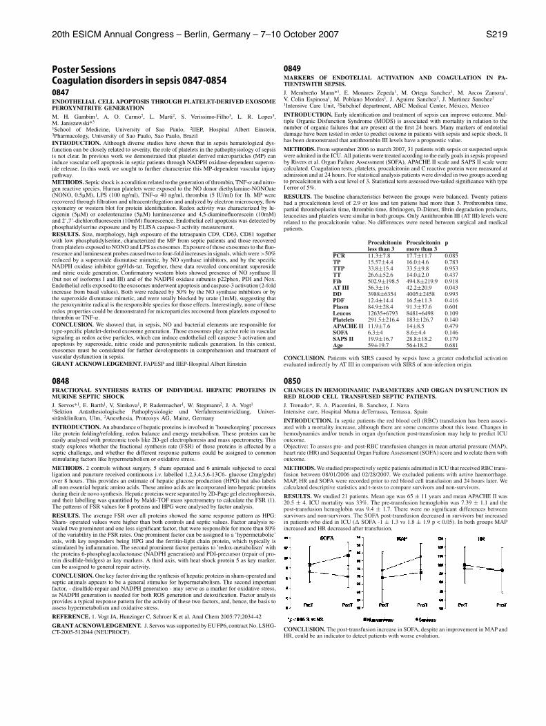
20th ESICM Annual Congress – Berlin, Germany – 7–10 October 2007 S219
Poster SessionsCoagulation disorders in sepsis 0847-08540847ENDOTHELIAL CELL APOPTOSIS THROUGH PLATELET-DERIVED EXOSOMEPEROXYNITRITE GENERATION
M. H. Gambim1, A. O. Carmo2, L. Marti2, S. Verissimo-Filho3, L. R. Lopes3,M. Janiszewski*3
1School of Medicine, University of Sao Paulo, 2IIEP, Hospital Albert Einstein,3Pharmacology, University of Sao Paulo, Sao Paulo, BrazilINTRODUCTION. Although diverse studies have shown that in sepsis hematological dys-function can be closely related to severity, the role of platelets in the pathophysiology of sepsisis not clear. In previous work we demonstrated that platelet derived microparticles (MP) caninduce vascular cell apoptosis in septic patients through NADPH oxidase-dependent superox-ide release. In this work we sought to further characterize this MP-dependent vascular injurypathway.METHODS. Septic shock is a condition related to the generation of thrombin, TNF-α and nitro-gen reactive species. Human platelets were exposed to the NO donor diethylamine-NONOate(NONO, 0.5µM), LPS (100 ng/ml), TNF-α 40 ng/ml, thrombin (5 IU/ml) for 1h. MP wererecovered through filtration and ultracentrifugation and analyzed by electron microscopy, flowcytometry or western blot for protein identification. Redox activity was characterized by lu-cigenin (5µM) or coelenterazine (5µM) luminescence and 4,5-diaminofluorescein (10mM)and 2’,7’-dichlorofluorescein (10mM) fluorescence. Endothelial cell apoptosis was detected byphosphatidylserine exposure and by ELISA caspase-3 activity measurement.RESULTS. Size, morphology, high exposure of the tetraspanin CD9, CD63, CD81 togetherwith low phosphatidylserine, characterized the MP from septic patients and those recoveredfrom platelets exposed to NONO and LPS as exosomes. Exposure of those exosomes to the fluo-rescence and luminescent probes caused two to four-fold increases in signals, which were >50%reduced by a superoxide dismutase mimetic, by NO synthase inhibitors, and by the specificNADPH oxidase inhibitor gp91ds-tat. Together, these data revealed concomitant superoxideand nitric oxide generation. Confirmatory western blots showed presence of NO synthase II(but not of isoforms I and III) and of the NADPH oxidase subunits p22phox, PDI and Nox.Endothelial cells exposed to the exosomes underwent apoptosis and caspase-3 activation (2-foldincrease from basal values). Both were reduced by 50% by the NO synthase inhibitors or bythe superoxide dismutase mimetic, and were totally blocked by urate (1mM), suggesting thatthe peroxynitrite radical is the responsible species for those effects. Interestingly, none of theseredox properties could be demonstrated for microparticles recovered from platelets exposed tothrombin or TNF-α.CONCLUSION. We showed that, in sepsis, NO and bacterial elements are responsible fortype-specific platelet-derived exosome generation. Those exosomes play active role in vascularsignaling as redox active particles, which can induce endothelial cell caspase-3 activation andapoptosis by superoxide, nitric oxide and peroxynitrite radicals generation. In this context,exosomes must be considered for further developments in comprehension and treatment ofvascular dysfunction in sepsis.GRANT ACKNOWLEDGEMENT. FAPESP and IIEP-Hospital Albert Einstein
0848FRACTIONAL SYNTHESIS RATES OF INDIVIDUAL HEPATIC PROTEINS INMURINE SEPTIC SHOCK
J. Servos*1, E. Barth1, V. Simkova1, P. Radermacher1, W. Stegmann2, J. A. Vogt11Sektion Anasthesiologische Pathophysiologie und Verfahrensentwicklung, Univer-sitatsklinikum, Ulm, 2Anesthesia, Proteosys AG, Mainz, Germany
INTRODUCTION. An abundance of hepatic proteins is involved in ’housekeeping’ processeslike protein folding/refolding, redox balance and energy metabolism. These proteins can beeasily analysed with proteomic tools like 2D-gel electrophoresis and mass spectrometry. Thisstudy explores whether the fractional synthesis rate (FSR) of these proteins is affected by aseptic challenge, and whether the different response patterns could be assigned to commonstimulating factors like hypermetabolism or oxidative stress.
METHODS. 2 controls without surgery, 5 sham operated and 6 animals subjected to cecalligation and puncture received continuous i.v. labelled 1,2,3,4,5,6-13C6- glucose (2mg/gxhr)over 8 hours. This provides an estimate of hepatic glucose production (HPG) but also labelsall non essential hepatic amino acids. These amino acids are incorporated into hepatic proteinsduring their de novo synthesis. Hepatic proteins were separated by 2D-Page gel electrophoresis,and their labelling was quantified by Maldi-TOF mass spectrometry to calculate the FSR (1).The patterns of FSR values for 8 proteins and HPG were analysed by factor analysis.
RESULTS. The average FSR over all proteins showed the same response pattern as HPG:Sham- operated values were higher than both controls and septic values. Factor analysis re-vealed two prominent and one less significant factor, that were responsible for more than 80%of the variability in the FSR rates. One prominent factor can be assigned to a ’hypermetabolic’axis, with key responders being HPG and the ferritin-light chain protein, which typically isstimulated by inflammation. The second prominent factor pertains to ’redox-metabolism’ withthe proteins 6-phosphoglucolactonase (NADPH generation) and PDI-precursor (repair of pro-tein disulfide-bridges) as key markers. A third axis, with heat shock protein 5 as key marker,can be assigned to general repair activity.
CONCLUSION. One key factor driving the synthesis of hepatic proteins in sham-operated andseptic animals appears to be a general stimulus for hypermetabolism. The second importantfactor, - disulfide-repair and NADPH generation - may serve as a marker for oxidative stress,as NADPH generation is needed for both ROS generation and detoxification. Factor analysisprovides a typical response pattern for the activity of these two factors, and, hence, the basis toassess hypermetabolism and oxidative stress.
REFERENCE. 1. Vogt JA, Hunzinger C, Schroer K et al. Anal Chem 2005:77;2034-42
GRANT ACKNOWLEDGEMENT. J. Servos was supported by EU FP6, contract No. LSHG-CT-2005-512044 (NEUPROCF).
0849MARKERS OF ENDOTELIAL ACTIVATION AND COAGULATION IN PA-TIENTSWITH SEPSIS.
J. Membreno Mann*1, E. Monares Zepeda1, M. Ortega Sanchez1, M. Arcos Zamora1,V. Colin Espinosa1, M. Poblano Morales1, J. Aguirre Sanchez2, J. Martinez Sanchez2
1Intensive Care Unit, 2Subchief department, ABC Medical Center, Mexico, Mexico
INTRODUCTION. Early identification and treatment of sepsis can improve outcome. Mul-tiple Organic Disfunction Syndrome (MODS) is associated with mortality in relation to thenumber of organic failures that are present at the first 24 hours. Many markers of endotelialdamage have been tested in order to predict outome in patients with sepsis and septic shock. Ithas been demonstrated that antithrombin III levels have a prognostic value.
METHODS. From september 2006 to march 2007, 31 patients with sepsis or suspected sepsiswere admited in the ICU. All patients were treated acording to the early goals in sepsis proposedby Rivers et al. Organ Failure Assessment (SOFA), APACHE II scale and SAPS II scale werecalculated. Coagulation tests, platelets, procalcitonin and C reactive protein were measured atadmission and at 24 hours. For statistical analysis patients were divided in two groups acordingto procalcitonin with a cut level of 3. Statistical tests assessed two-tailed significance with typeI error of 5%.
RESULTS. The baseline characteristics between the groups were balanced. Twenty patienshad a procalcitonin level of 2.9 or less and ten patiens had more than 3. Prothrombin time,partial thromboplastin time, thrombin time, fibrinogen, D-Dimer, fibrin degradation products,leucocites and platelets were similar in both groups. Only Antithrombin III (AT III) levels wererelated to the procalcitonin value. No differences were noted between surgical and medicalpatients.
Procalcitonin Procalcitonin pless than 3 more than 3
PCR 11.3±7.8 17.7±11.7 0.085TP 15.57±4.4 16.0±4.6 0.783TTP 33.8±15.4 33.5±9.8 0.953TT 26.6±52.6 14.0±2.0 0.437Fib 502.9±198.5 494.8±219.9 0.918AT III 56.3±16 42.2±20.9 0.043DD 3988±6354 4005±2458 0.993PDF 12.4±14.4 16.5±11.3 0.416Plasm 84.9±28.4 91.3±37.6 0.601Leucos 12635+6793 8481+6498 0.109Platelets 291.5±216.4 183±126.7 0.140APACHE II 11.9±7.6 14±8.5 0.479SOFA 6.3±4 8.6±4.4 0.146SAPS II 19.9±16.7 28.8±18.2 0.179Age 59±19.7 56±18.2 0.681
CONCLUSION. Patients with SIRS caused by sepsis have a greater endothelial activationevaluated indirectly by AT III in comparison with SIRS of non-infection origin.
0850CHANGES IN HEMODINAMIC PARAMETERS AND ORGAN DYSFUNCTION INRED BLOOD CELL TRANSFUSED SEPTIC PATIENTS.
J. Trenado*, E. A. Piacentini, B. Sanchez, J. NavaIntensive care, Hospital Mutua deTerrassa, Terrassa, Spain
INTRODUCTION. In septic patients the red blood cell (RBC) transfusion has been associ-ated with a mortality increase, although there are some concerns about this issue. Changes inhemodynamics and/or trends in organ dysfunction post-transfusion may help to predict ICUoutcome.Objective: To assess pre- and post-RBC transfusion changes in mean arterial pressure (MAP),heart rate (HR) and Sequential Organ Failure Assessment (SOFA) score and to relate them withoutcome.
METHODS. We studied prospectively septic patients admitted in ICU that received RBC trans-fusion between 08/01/2006 and 02/28/2007. We excluded patients with active haemorrhage.MAP, HR and SOFA were recorded prior to red blood cell transfusion and 24 hours later. Wecalculated descriptive statistics and t-tests to compare survivors and non-survivors.
RESULTS. We studied 21 patients. Mean age was 65 ± 11 years and mean APACHE II was20.5 ± 4. ICU mortality was 33%. The pre-transfusion hemoglobin was 7.39 ± 1.1 and thepost-transfusion hemoglobin was 9.4 ± 1.7. There were no significant differences betweensurvivors and non-survivors. The SOFA post-transfusion decreased in survivors but increasedin patients who died in ICU (∆ SOFA -1 ± 1.3 vs 1.8 ± 1.9 p < 0.05). In both groups MAPincreased and HR decreased after transfusion.
CONCLUSION. The post-transfusion increase in SOFA, despite an improvement in MAP andHR, could be an indicator to detect patients with worse evolution.
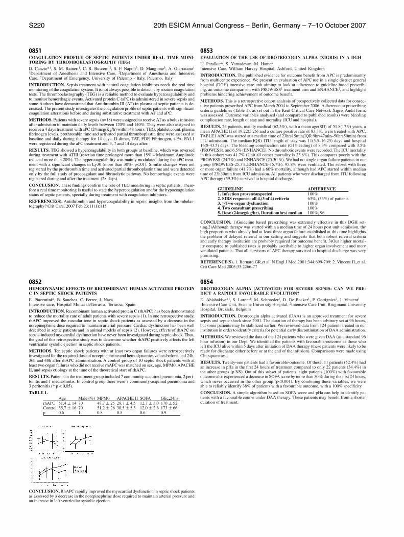
S220 20th ESICM Annual Congress – Berlin, Germany – 7–10 October 2007
0851COAGULATION PROFILE OF SEPTIC PATIENTS UNDER REAL TIME MONI-TORING BY THROMBOELASTOGRAPHY (TEG)
D. Canzio*1, S. M. Raineri1, C. R. Buscemi2, S. F. Napoli3, D. Mangione3, A. Giarratano1
1Department of Anesthesia and Intensive Care, 2Department of Anesthesia and IntensiveCare, 3Department of Emergency, University of Palermo - Italy, Palermo, Italy
INTRODUCTION. Sepsis treatment with natural coagulation inhibitors needs the real timemonitoring of the coagulation system. It is not always possible to detect it by routine coagulationtests. The thromboelastography (TEG) is a reliable method to evaluate hypercoagulability andto monitor hemorrhagic events. Activated protein C (aPC) is administered in severe sepsis andsome Authors have demostrated that Antithrombin III (AT) in plasma of septic patients is de-creased. The present study investigates the coagulation profile of septic patients with significantcoagulation alterations before and during substitutive treatment with AT and aPC.
METHODS. Patients with severe sepsis (n=18) were assigned to receive AT as a bolus infusionafter admission to maintain daily levels between 120% and 140%. They were also assigned toreceive a 4 days treatment with aPC (24 mcg/Kg/h) within 48 hours. TEG, platelet count, plasmafibrinogen levels, prothrombin time and activated partial thromboplastin time were assessed atbaseline and daily during therapy for 14 days. D-dimer, TAT, FDP, Fibrinogen, t-PA, PAI-1were registered during the aPC treatment and 3, 7 and 14 days after.
RESULTS. TEG showed a hypercoagulability in both groups at baseline, which was reversedduring treatment with ATIII (reaction time prolonged more than 15% – Maximum Amplitudereduced more than 20%). The hypercoagulability was mainly modulated during the aPC treat-ment with a significant changes in Ly30 (more than 30%- p<.01). Similar changes were notregistered by the prothrombin time and activated partial thromboplastin time and were detectedonly by the full study of procoagulant and fibrinolytic pathway. No hemorrhagic events wereregistered during and after the treatment (28 days).
CONCLUSION. These findings confirm the role of TEG monitoring in septic patients. There-fore a real time monitoring is useful to state the hypercoagulation and/or the hypocoagulationstatus of septic patients, specially during treatment with coagulation inhibitors.
REFERENCE(S). Antithrombin and hypercoagulability in sepsis: insights from thrombelas-tography? Crit Care. 2007 Feb 23;11(1):115
0852HEMODYNAMIC EFFECTS OF RECOMBINANT HUMAN ACTIVATED PROTEINC IN SEPTIC SHOCK PATIENTS
E. Piacentini*, B. Sanchez, C. Ferrer, J. NavaIntensive care, Hospital Mutua deTerrassa, Terrassa, Spain
INTRODUCTION. Recombinant human activated protein C (rhAPC) has been demonstratedto reduce the mortality rate of adult patients with severe sepsis (1). In one retrospective study,rhAPC improved the vascular tone in septic shock patients as assessed by a decrease in thenorepinephrine dose required to maintain arterial pressure. Cardiac dysfunction has been welldescribed in septic patients and in animal models of sepsis (2). However, effects of rhAPC onsepsis-induced myocardial dysfunction have never been investigated during septic shock. Thus,the goal of this retrospective study was to determine whether rhAPC positively affects the leftventricular systolic ejection in septic shock patients.
METHODS. Ten septic shock patients with at least two organ failures were retrospectivelyinvestigated for the required dose of norepinephrine and hemodynamics values before, and 24h,36h and 48h after rhAPC administration. A control group of 10 septic shock patients with atleast two organ failures who did not receive rhAPC was matched on sex, age, MPM0, APACHEII, and sepsis etiology at the time of the theoretical start of rhAPC.
RESULTS. Patients in the treatment group included 7 community-acquired pneumonia, 2 peri-tonitis and 1 mediastinitis. In control group there were 7 community-acquired pneumonia and3 peritonitis.(* p < 0,05).
TABLE 1.Age Male (%) MPM0 APACHE II SOFA Glic 24hs
rhAPC 51,4 ± 14 70 48,7 ± 25 28,7 ± 4,5 12,7 ± 3,0 170 ± 52Control 55,7 ± 16 70 51,2 ± 26 30,5 ± 5,3 12,0 ± 2,6 173 ± 66p 0.6 1 0.8 0.5 0.6 0.9
CONCLUSION. RhAPC rapidly improved the myocardial dysfunction in septic shock patientsas assessed by a decrease in the norepinephrine dose required to maintain arterial pressure andan increase in left ventricular systolic ejection.
0853EVALUATION OF THE USE OF DROTRECOGIN ALPHA (XIGRIS) IN A DGH
U. Paralkar*, S. Vamadevan, M. HamerIntensive Care, William Harvey Hospital, Ashford, United Kingdom
INTRODUCTION. The published evidence for outcome benefit from APC is predominantlyfrom multicentre experience. We present an evaluation of APC use in a single district generalhospital (DGH) intensive care unit aiming to look at adherence to guideline-based prescrib-ing, an outcome comparison with PROWESS1 treatment arm and ENHANCE2, and highlightproblems hindering achievement of outcome benefit.
METHODS. This is a retrospective cohort analysis of prospectively collected data for consec-utive patients prescribed APC from March 2004 to September 2006. Adherence to prescribingcriteria guidelines (Table 1), as set out in the Kent Critical Care Network Xigris Audit form,was assessed. Outcome variables analysed (and compared to published results) were bleedingcomplication rate, length of stay and mortality (ICU and hospital).
RESULTS. 24 patients, mainly medical (62,5%), with a mean age(SD) of 51.8(17.9) years, amean APACHE II of 19.22(5.26) and a culture positive rate of 83.3%, were treated with APC.TABLE1 APC was started at a median time of 23hrs15min(IQR 9hrs47min–50hrs50min) fromITU admission. The median(IQR) ITU length of stay was 11(5.5–16.25) days and hospital16(8-43.5) days. The bleeding complication rate (GI bleeding) of 8.3% compared with 3.5%(PROWESS), and 6.5% (ENHANCE). No thrombotic events were recorded. The ICU mortalityin this cohort was 41.7% (Unit all comer mortality is 23.8%). This compares poorly with thePROWESS (24.7%) and ENHANCE (25.30 %). We had no single organ failure patients in ourgroup (PROWESS-25.3%,ENHANCE-15.7%). 95.8% were ventilated. The subset with threeor more organ failure (41.7%) had a 60% mortality, although had APC started within mediantime of 23h30min from ICU admission. All patients who were discharged from ITU followingAPC therapy (59.3%) survived to hospital discharge.
GUIDELINE ADHERENCE1. Infection proven/suspected 100%2. SIRS response- all 4,(3 of 4) criteria 63%, (33%) of patients3. ≥Two organ dysfunction 100%4. Two consultant prescribing 100%5. Dose (24mcg/kg/hr), Duration(hrs) median 100%, 96
CONCLUSION. 1)Guideline based prescribing was extremely effective in this DGH set-ting.2)Although therapy was started within a median time of 24 hours post unit admission, thehigh proportion who already had at least three organ failure established at this time highlightsthe problem of delayed referral in our setting and suggests that both robust referral criteriaand early therapy institution are probably required for outcome benefit. 3)Our higher mortal-ity compared to published rates is probably ascribable to higher organ involvement and moreventilated patients. That all survivors of APC therapy survived to hospital discharge was verypromising.
REFERENCE(S). 1. Bernard GR,et al. N Engl J Med 2001;344:699-709: 2. Vincent JL,et al.Crit Care Med 2005;33:2266-77
0854DROTRECOGIN ALPHA (ACTIVATED) FOR SEVERE SEPSIS: CAN WE PRE-DICT A RAPIDLY FAVOURABLE EVOLUTION?
D. Ahishakiye*1, S. Lorent1, M. Schroeder1, D. De Backer1, P. Gottignies2, J. Vincent11Intensive Care Unit, Erasme University Hospital, 2Intensive Care Unit, Brugmann UniversityHospital, Brussels, Belgium
INTRODUCTION. Drotrecogin alpha activated (DAA) is an approved treatment for severesepsis and septic shock since 2001. The duration of therapy has been arbitrary set at 96 hours,but some patients may be stabilized earlier. We reviewed data from 124 patients treated in ourinstitution in order to identify criteria for potential early discontinuation of DAA administration.
METHODS. We reviewed the data of the 124 patients who were given DAA (as a standard 96hour infusion) in our Dept. We identified the patients with favourable-outcome as those wholeft the ICU alive within 5 days after initiation of DAA therapy (these patients were likely to beready for discharge either before or at the end of the infusion). Comparisons were made usingChi-square test.
RESULTS. Twenty-one patients had a favourable-outcome. Of these, 11 patients (52.4%) hadan increase in pHa in the first 24 hours of treatment compared to only 22 patients (34.4%) inthe other groups (p NS). Out of this subset of patients, eight patients (100%) with favourableoutcome also experienced a decrease in SOFA score by more than 50 % during the first 24 hours,which never occurred in the other group (p<0.001). By combining these variables, we wereable to reliably identify 38% of patients with a favourable outcome, with a 100% specificity.
CONCLUSION. A simple algorithm based on SOFA score and pHa can help to identify pa-tients with a favourable course under DAA therapy. These patients may benefit from a shorterduration of treatment.
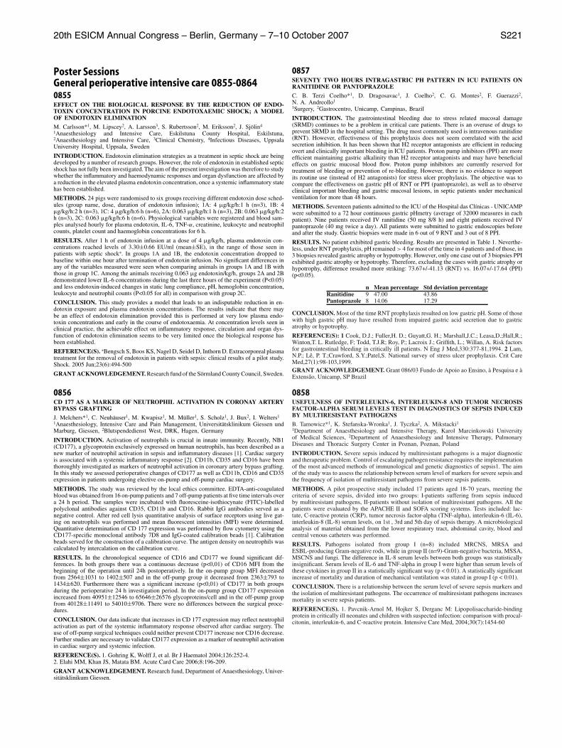
20th ESICM Annual Congress – Berlin, Germany – 7–10 October 2007 S221
Poster SessionsGeneral perioperative intensive care 0855-08640855EFFECT ON THE BIOLOGICAL RESPONSE BY THE REDUCTION OF ENDO-TOXIN CONCENTRATION IN PORCINE ENDOTOXAEMIC SHOCK; A MODELOF ENDOTOXIN ELIMINATION
M. Carlsson*1, M. Lipscey2, A. Larsson3, S. Rubertsson2, M. Eriksson2, J. Sjolin4
1Anaesthesiology and Intensive Care, Eskilstuna County Hospital, Eskilstuna,2Anaesthesiology and Intensive Care, 3Clinical Chemistry, 4Infectious Diseases, UppsalaUniversity Hospital, Uppsala, Sweden
INTRODUCTION. Endotoxin elimination strategies as a treatment in septic shock are beingdeveloped by a number of research groups. However, the role of endotoxin in established septicshock has not fully been investigated. The aim of the present investigation was therefore to studywhether the inflammatory and haemodynamic responses and organ dysfunction are affected bya reduction in the elevated plasma endotoxin concentration, once a systemic inflammatory statehas been established.
METHODS. 24 pigs were randomised to six groups receiving different endotoxin dose sched-ules (group name, dose, duration of endotoxin infusion); 1A: 4 µg/kg/h:1 h (n=3), 1B: 4µg/kg/h:2 h (n=3), 1C: 4 µg/kg/h:6 h (n=6), 2A: 0.063 µg/kg/h:1 h (n=3), 2B: 0.063 µg/kg/h:2h (n=3), 2C: 0.063 µg/kg/h:6 h (n=6). Physiological variables were registered and blood sam-ples analysed hourly for plasma endotoxin, IL-6, TNF-α, creatinine, leukocyte and neutrophilcounts, platelet count and haemoglobin concentrations for 6 h.
RESULTS. After 1 h of endotoxin infusion at a dose of 4 µg/kg/h, plasma endotoxin con-centrations reached levels of 3.30±0.66 EU/ml (mean±SE), in the range of those seen inpatients with septic shock∗. In groups 1A and 1B, the endotoxin concentration dropped tobaseline within one hour after termination of endotoxin infusion. No significant differences inany of the variables measured were seen when comparing animals in groups 1A and 1B withthose in group 1C. Among the animals receiving 0.063 µg endotoxin/kg/h, groups 2A and 2Bdemonstrated lower IL-6 concentrations during the last three hours of the experiment (P<0.05)and less endotoxin-induced changes in static lung compliance, pH, hemoglobin concentration,leukocyte and neutrophil counts (P<0.05 for all) in comparison with group 2C.
CONCLUSION. This study provides a model that leads to an indisputable reduction in en-dotoxin exposure and plasma endotoxin concentrations. The results indicate that there maybe an effect of endotoxin elimination provided this is performed at very low plasma endo-toxin concentrations and early in the course of endotoxaemia. At concentration levels seen inclinical practice, the achievable effect on inflammatory response, circulation and organ dys-function of endotoxin elimination seems to be very limited once the biological response hasbeen established.
REFERENCE(S). ∗Bengsch S, Boos KS, Nagel D, Seidel D, Inthorn D. Extracorporeal plasmatreatment for the removal of endotoxin in patients with sepsis: clinical results of a pilot study.Shock. 2005 Jun;23(6):494-500
GRANT ACKNOWLEDGEMENT. Research fund of the Sormland County Council, Sweden.
0856CD 177 AS A MARKER OF NEUTROPHIL ACTIVATION IN CORONAY ARTERYBYPASS GRAFTING
J. Melchers*1, C. Neuhauser1, M. Kwapisz1, M. Muller1, S. Scholz1, J. Bux2, I. Welters1
1Anaesthesiology, Intensive Care and Pain Management, Universitatsklinikum Giessen undMarburg, Giessen, 2Blutspendedienst West, DRK, Hagen, Germany
INTRODUCTION. Activation of neutrophils is crucial in innate immunity. Recently, NB1(CD177), a glycoprotein exclusively expressed on human neutrophils, has been described as anew marker of neutrophil activation in sepsis and inflammatory diseases [1]. Cardiac surgeryis associated with a systemic inflammatory response [2]. CD11b, CD35 and CD16 have beenthoroughly investigated as markers of neutrophil activation in coronary artery bypass grafting.In this study we assessed perioperative changes of CD177 as well as CD11b, CD16 and CD35expression in patients undergoing elective on-pump and off-pump cardiac surgery.
METHODS. The study was reviewed by the local ethics committee. EDTA-anti-coagulatedblood was obtained from 16 on-pump patients and 7 off-pump patients at five time intervals overa 24 h period. The samples were incubated with fluoresceine-isothiocynate (FITC)-labelledpolyclonal antibodies against CD35, CD11b and CD16. Rabbit IgG antibodies served as anegative control. After red cell lysis quantitative analysis of surface receptors using live gat-ing on neutrophils was performed and mean fluorescent intensities (MFI) were determined.Quantitative determination of CD 177 expression was performed by flow cytometry using theCD177-specific monoclonal antibody 7D8 and IgG-coated calibration beads [1]. Calibrationbeads served for the construction of a calibration curve. The antigen density on neutrophils wascalculated by intercalation on the calibration curve.
RESULTS. In the chronological sequence of CD16 and CD177 we found significant dif-ferences. In both groups there was a continuous decrease (p<0,01) of CD16 MFI from thebeginning of the operation until 24h postoperatively. In the on-pump group MFI decreasedfrom 2564±1031 to 1402±507 and in the off-pump group it decreased from 2363±793 to1434±620. Furthermore there was a significant increase (p<0,01) of CD177 in both groupsduring the perioperative 24 h investigation period. In the on-pump group CD177 expressionincreased from 40951±12546 to 65646±26576 glycoproteins/cell and in the off-pump groupfrom 40128±11491 to 54010±9706. There were no differences between the surgical proce-dures.
CONCLUSION. Our data indicate that increases in CD 177 expression may reflect neutrophilactivation as part of the systemic inflammatory response observed after cardiac surgery. Theuse of off-pump surgical techniques could neither prevent CD177 increase nor CD16 decrease.Further studies are necessary to validate CD177 expression as a marker of neutrophil activationin cardiac surgery and systemic infection.
REFERENCE(S). 1. Gohring K, Wolff J, et al. Br J Haematol 2004;126:252-4.2. Elahi MM, Khan JS, Matata BM. Acute Card Care 2006;8:196-209.
GRANT ACKNOWLEDGEMENT. Research fund, Department of Anaesthesiology, Univer-sitatsklinikum Giessen.
0857SEVENTY TWO HOURS INTRAGASTRIC PH PATTERN IN ICU PATIENTS ONRANITIDINE OR PANTOPRAZOLE
C. B. Terzi Coelho*1, D. Dragosavac1, J. Coelho2, C. G. Montes2, F. Guerazzi2,N. A. Andreollo1
1Surgery, 2Gastrocentro, Unicamp, Campinas, Brazil
INTRODUCTION. The gastrointestinal bleeding due to stress related mucosal damage(SRMD) continues to be a problem in critical care patients. There is an overuse of drugs toprevent SRMD in the hospital setting. The drug most commonly used is intravenous ranitidine(RNT). However, effectiveness of this prophylaxis does not seem correlated with the acidsecretion inhibition. It has been shown that H2 receptor antagonists are efficient in reducingovert and clinically important bleeding in ICU patients. Proton pump inhibitors (PPI) are moreefficient maintaining gastric alkalinity than H2 receptor antagonists and may have beneficialeffects on gastric mucosal blood flow. Proton pump inhibitors are currently reserved fortreatment of bleeding or prevention of re-bleeding. However, there is no evidence to supportits routine use (instead of H2 antagonists) for stress ulcer prophylaxis. The objective was tocompare the effectiveness on gastric pH of RNT or PPI (pantoprazole), as well as to observeclinical important bleeding and gastric mucosal lesions, in septic patients under mechanicalventilation for more than 48 hours.
METHODS. Seventeen patients admitted to the ICU of the Hospital das Clınicas - UNICAMPwere submitted to a 72 hour continuous gastric pHmetry (average of 32000 measures in eachpatient). Nine patients received IV ranitidine (50 mg 8/8 h) and eight patients received IVpantoprazole (40 mg twice a day). All patients were submitted to gastric endoscopies beforeand after the study. Gastric biopsies were made in 6 out of 9 RNT and 3 out of 8 PPI.
RESULTS. No patient exhibited gastric bleeding. Results are presented in Table 1. Neverthe-less, under RNT prophylaxis, pH remained > 4 for most of the time in 4 patients and of those, in3 biopsies revealed gastric atrophy or hypotrophy. However, only one case out of 3 biopsies PPIexhibited gastric atrophy or hypotrophy. Therefore, excluding the cases with gastric atrophy orhypotrophy, difference resulted more striking: 73.67+/-41.13 (RNT) vs. 16.07+/-17.64 (PPI)(p<0.05).
n Mean percentage Std deviation percentageRanitidine 9 47.00 43.86Pantoprazole 8 14.06 17.29
CONCLUSION. Most of the time RNT prophylaxis resulted on low gastric pH. Some of thosewith high gastric pH may have resulted from impaired gastric acid secretion due to gastricatrophy or hypotrophy.
REFERENCE(S): 1 Cook, D.J.; Fuller,H. D.; Guyatt,G. H.; Marshall,J.C.; Leasa,D.;Hall,R.;Winton,T. L. Rutledge, F; Todd, T.J.R; Roy, P.; Lacroix J.; Griffith, L.; Willan, A. Risk factorsfor gastrointestinal bleeding in critically ill patients. N Eng J Med,330:377-81,1994. 2 Lam,N.P.; Le, P. T.;Crawford, S.Y.;Patel,S. National survey of stress ulcer prophylaxis. Crit CareMed,27(1):98-103,1999.GRANT ACKNOWLEDGEMENT. Grant 086/03 Fundo de Apoio ao Ensino, a Pesquisa e aExtensao, Unicamp, SP Brazil
0858USEFULNESS OF INTERLEUKIN-6, INTERLEUKIN-8 AND TUMOR NECROSISFACTOR-ALPHA SERUM LEVELS TEST IN DIAGNOSTICS OF SEPSIS INDUCEDBY MULTIRESISTANT PATHOGENS
B. Tamowicz*1, K. Stefanska-Wronka1, J. Tyczka2, A. Mikstacki11Department of Anaesthesiology and Intensive Therapy, Karol Marcinkowski Universityof Medical Sciences, 2Department of Anaesthesiology and Intensive Therapy, PulmonaryDiseases and Thoracic Surgery Center in Poznan, Poznan, Poland
INTRODUCTION. Severe sepsis induced by multiresistant pathogens is a major diagnosticand therapeutic problem. Control of escalating pathogen resistance requires the implementationof the most advanced methods of immunological and genetic diagnostics of sepsis1. The aimof the study was to assess the relationship between serum level of markers for severe sepsis andthe frequency of isolation of multiresistant pathogens from severe sepsis patients.
METHODS. A pilot prospective study included 17 patients aged 18-70 years, meeting thecriteria of severe sepsis, divided into two groups: I-patients suffering from sepsis inducedby multiresistant pathogens, II-patients without isolation of multiresistant pathogens. All thepatients were evaluated by the APACHE II and SOFA scoring systems. Tests included: lac-tate, C-reactive protein (CRP), tumor necrosis factor-alpha (TNF-alpha), interleukin-6 (IL-6),interleukin-8 (IL-8) serum levels, on 1st , 3rd and 5th day of sepsis therapy. A microbiologicalanalysis of material obtained from the lower respiratory tract, abdominal cavity, blood andcentral venous catheters was performed.
RESULTS. Pathogens isolated from group I (n=8) included MRCNS, MRSA andESBL-producing Gram-negative rods, while in group II (n=9)-Gram-negative bacteria, MSSA,MSCNS and fungi. The difference in IL-8 serum levels between both groups was statisticallyinsignificant. Serum levels of IL-6 and TNF-alpha in group I were higher than serum levels ofthese cytokines in group II in a statistically significant way (p < 0.01). A statistically significantincrease of mortality and duration of mechanical ventilation was stated in group I (p < 0.01).
CONCLUSION. There is a relationship between the serum level of severe sepsis markers andthe isolation of multiresistant pathogens. The occurrence of multiresistant pathogens increasesmortality in severe sepsis patients.
REFERENCE(S). 1. Pavcnik-Arnol M, Hojker S, Derganc M: Lipopolisaccharide-bindingprotein in critically ill neonates and children with suspected infection: comparison with procal-citonin, interleukin-6, and C-reactive protein. Intensive Care Med, 2004;30(7):1454-60
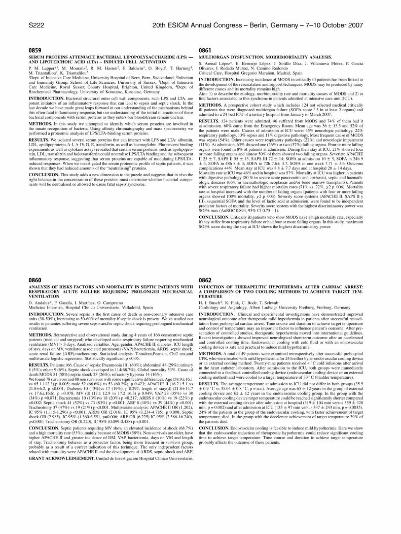
S222 20th ESICM Annual Congress – Berlin, Germany – 7–10 October 2007
0859SERUM PROTEINS ATTENUATE BACTERIAL LIPOPOLYSACCHARIDE (LPS) —AND LIPOTEICHOIC ACID (LTA) – INDUCED CELL ACTIVATION
P. M. Lepper*1, M. Mouratis2, R. M. Haston2, F. Baldwin3, O. Boyd3, T. Hartung4,M. Triantafilou2, K. Triantafilou2
1Dept. of Intensive Care Medicine, University Hospital of Bern, Bern, Switzerland, 2Infectionand Immunity Group, School of Life Sciences, University of Sussex, 3Dept. of IntensiveCare Medicine, Royal Sussex County Hospital, Brighton, United Kingdom, 4Dept. ofBiochemical Pharmacology, University of Konstanz, Konstanz, Germany
INTRODUCTION. Bacterial structural outer cell wall components, such LPS and LTA, arepotent initiators of an inflammatory response that can lead to sepsis and septic shock. In thelast decade we have made great leaps forward in our understanding of the mechanisms behindthis often-fatal inflammatory response, but our understanding of the initial interactions of thesebacterial components with serum proteins as they enters our bloodstream remain unclear.
METHODS. In this study we attempted to identify which serum proteins are involved inthe innate recognition of bacteria. Using affinity chromatography and mass spectrometry weperformed a proteomic analysis of LPS/LTA-binding serum proteins.
RESULTS. We isolated several serum proteins that can interact with LPS and LTA: albumin,LDL, apolipoproteins A-I, A-IV, D, E, transferrin, as well as haemoglobin. Fluorescent bindingexperiments as well as cytokine assays revealed that certain serum proteins, such as apolipopro-tein, LDL, transferrin and holotransferrin could neutralise LPS/LTA binding and the subsequentinflammatory response, suggesting that serum proteins are capable of modulating LPS/LTA-induced responses. When we investigated the serum proteomic profile of septic patients, it wasshown that they had reduced amounts of the “neutralising” proteins.
CONCLUSION. This study adds a new dimension to the puzzle and suggests that in vivo theright balance in the concentration of these proteins must determine whether bacterial compo-nents will be neutralised or allowed to cause fatal sepsis syndrome.
0860ANALYSIS OF RISKS FACTORS AND MORTALITY IN SEPTIC PATIENTS WITHRESPIRATORY ACUTE FAILURE REQUIRING PROLONGED MECHANICALVENTILATION
D. Andaluz*, F. Gandıa, I. Martınez, O. CampesinoMedicina Intensiva, Hospital Clınico Universitario, Valladolid, Spain
INTRODUCTION. Severe sepsis is the first cause of death in non-coronary intensive careunits (30-50%), increasing to 50-60% of mortality if septic shock is present. We’ve studied ourresults in patientes suffering severe sepsis and/or septic shock requiring prolongued mechanicalventilation.
METHODS. Retrospective and observational study during 4 years of 166 consecutive septicpatients (medical and surgycal) who developed acute respiratory failure requering mechanicalventilation (MV) ≥ 3 days. Analized variables: Age, gender, APACHE II, diabetes, ICU lengthof stay, days on MV, ventilator associated pneumonia (VAP),bacteriemia, ARDS, septic shock,acute renal failure (ARF),tracheotomy. Statistical analysis: T-student,Pearson, Chi2 test,andmultivariate logistic regression. Statistically significant p <0.05.
RESULTS. Patients:166. Cause of sepsis: Pneumonia 101 (60%); abdominal 48 (29%); urinary8 (5%); other: 9 (6%). Septic shock developed in 114(68.7%). Global mortality 53%. Cause ofdeath:MODS 51 (58%);septic shock 23 (26%); refractory hypoxia 14 (16%).We found 78 survivors and 88 non survivors with some significative differences: Age (58.9±16.1vs 65.1±12.3),p 0.005; male 52 (66.6%) vs 53 (60.2%), p 0.423; APACHE II (16.7±5.1 vs21.8±6.2, p <0.001; Diabetes 10 (13%)vs 17 (19%), p 0.297; length of stay(d) (21.8±14.7vs 17.6±16.6), p =0.078; MV (d) (17.1 12.9 vs 17.2 16.3) p 0.949; VAP 28 (35%) vs 30(34%) p =0.871; Bacteriemia 10 (13%)vs 18 (20%) p =0.217; ARDS 8 (10%) vs 19 (22%) p=0.002; Septic shock 41 (52%) vs 73 (83%) p <0.001; ARF 8 (10%) vs 39 (44%) p <0.001;Tracheotomy 37 (47%) vs 19 (21%) p <0.001. Multivariate analysis: APACHE II OR (1.202),IC 95% (1.115-1.296) p <0.001; ARDS OR (2.016), IC 95% (1.234-4.785), p 0.008; Septicshock OR (2.985), IC 95% (1.360-6.551, p=0.006; ARF OR (6.225) IC 95% (2.386-16.240),p<0.001; Tracheostomy OR (0.220), IC 95% (0.099-0.458) p <0.001.
CONCLUSION. Septic patients requiring MV show an elevated incidence of shock (68.7%)and a high mortality rate (53%), mainly because of MODS (58%). Non survivals are older, havehigher APACHE II and greater incidence of DM, VAP, bacteriemia, days on VM and lengthof stay. Tracheotomy behaves as a protector factor, being more frecuent in survivor group,probably as a result of a correct indication of this technique. The only independent factorsrelated with mortality were APACHE II and the development of ARDS, septic shock and ARF.
GRANT ACKNOWLEDGEMENT. Unidad de Investigacion Hospital Clınico Universitario.
0861MULTIORGAN DYSFUNCTION. MORBIMORTALITY ANALYSIS.
S. Arenal Lopez*, E. Bermejo Lopez, J. Sotillo Dıaz, J. Villanueva Florez, P. GarciaOlivares, I. Rodado Munoz, N. Camino RedondoCritical Care, Hospital Gregorio Maranon, Madrid, Spain
INTRODUCTION. Increasing incidence of MODS in critically ill patients has been linked tothe development of the resuscitation and support techniques. MODS may be produced by manydifferent causes and its mortality remains high.Aim: 1) to describe the etiology, morbimortality rate and mortality causes of MODS and 2) tofind factors associated to this syndrome in patients admitted at intensive care unit (ICU).
METHODS. A prospective cohort study which includes 124 not selected medical criticallyill patients that were diagnosed multiorgan failure (SOFA score 3 3 in at least 2 organs) andadmitted to a 24-bed ICU of a tertiary hospital from January to March 2007.
RESULTS. 124 patients were admitted, 46 suffered from MODS and 74% of them had itat admission. 60% came from the Emergency Room. Mean age was 56 ± 15.5 and 52% ofthe patients were male. Causes of admission at ICU were: 35% neurologic pathology, 22%respiratory pathology, 13% sepsis and 11% digestive pathology. Most frequent cause of MODSwas sepsis (54%). Other causes were respiratory pathology (22%) and neurologic impairment(11%). At admission, 63% showed one (26%) or two (37%) failing organs. Four or more failingorgans were found in 8% of patients at admission. During their stay at ICU, 21% showed fouror more failing organs and almost 50% of them showed two failing organs. Severity: APACHEII 25 ± 7, SAPS II 55 ± 15, SAPS III 72 ± 14; SOFA at admission 10 ± 3, SOFA in 24h 9± 4, SOFA in 48h 8 ± 3; SOFA in 72h 7.6± 3.7, SOFA in one week 7.71 ± 3.6. Outcomecomplications 44%. Mean stay at ICU was 8.8 ± 7.7 days and at hospital 20 ± 14 days.Mortality rate at ICU was 46% and in hospital was 57%. Mortality at ICU was higher in patientswith digestive pathology (80 % in severe acute pancreatitis and cirrhosis), septic and haemath-ologic diseases (66% in haemathologic neoplasias and/or bone marrow transplants). Patientswith severe respiratory failure had higher mortality rates (71% vs. 22%, χ2 p .006). Mortalityrate at hospital increased with the number of failing organs (patients with four or more failingorgans showed 100% mortality, χ2 p .003). Severity score systems (APACHE II, SAPS II yIII), sequential SOFA and the level of lactic acid at admission, were found to be independentpredictor factors of mortality. Severity socre system with the highest discriminatory power wasSOFA max (AuROC 0.894, 95% CI 0.75 – 1).
CONCLUSION. Critically ill patients who show MODS have a high mortality rate, especiallyif they suffer from respiratory failure or had four or more failing organs. In this study, maximumSOFA score during the stay at ICU shows the highest discriminatory power.
0862INDUCTION OF THERAPEUTIC HYPOTHERMIA AFTER CARDIAC ARREST:A COMPARISON OF TWO COOLING METHODS TO ACHIEVE TARGET TEM-PERATURE
H. J. Busch*, K. Fink, C. Bode, T. SchwabCardiology and Angiology, Albert Ludwigs University Freiburg, Freiburg, Germany
INTRODUCTION. Clinical and experimental investigations have demonstrated improvedneurological outcome after therapeutic mild hypothermia in patients after successful resusci-tation from prehospital cardiac arrest. Time course and duration to achieve target temperatureand control of temperature may an important factor to influence patient’s outcome. After pre-sentation of controlled studies, therapeutic hypothermia moved into international guidelines.Recent investigations showed improved neurological short-term outcome after an acceleratedand controlled cooling time. Endovascular cooling with cold fluid or with an endovascularcooling device is safe and practical to induce mild hypothermia.
METHODS. A total of 49 patients were examined retrospectively after successful prehospitalCPR, who were treated with mild hypothermia for 24 h either by an endovascular cooling deviceor an external cooling method. Twenty-nine patients received 4 ˚ C cold infusions after arrivalin the heart catheter laboratory. After admission to the ICU, both groups were immediatelyconnected to a feedback controlled cooling device (endovascular cooling device or an externalcooling method) and were cooled to a target temperature of 33 ˚ C (bladder temperature).
RESULTS. The average temperature at admission to ICU did not differ in both groups (35.5± 0.9 ˚ C vs 35.04 ± 0.8 ˚ C, p = n.s.). Average age was 65 ± 12 years in the group of externalcooling device and 62 ± 12 years in the endovascular cooling group. In the group with theendovascular cooling device target temperature could be reached significantly shorter comparedwith the external cooling device after admission at hospital (319 ± 104 min versus 559 ± 320min, p = 0.002) and after admission at ICU (155 ± 97 min versus 337 ± 243 min, p = 0.0035).24% of the patients in the group of the endovascular cooling, with faster achievement of targettemperature, died. In the group with the decelerate achievement of target temperature 39% ofthe patients died.
CONCLUSION. Endovascular cooling is feasible to induce mild hypothermia. Here we showthat the endovascular induction of therapeutic hypothermia could reduce significant coolingtime to achieve target temperature. Time course and duration to achieve target temperatureprobably affects the outcome of these patients.
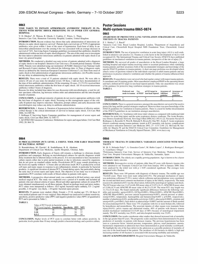
20th ESICM Annual Congress – Berlin, Germany – 7–10 October 2007 S223
0863TIME TAKEN TO INITIATE APPROPRIATE ANTIBIOTIC THERAPY IN PA-TIENTS WITH SEPTIC SHOCK PRESENTING TO AN INNER CITY GENERALHOSPITAL
V. D. Dhokia*, D. Watson, R. Ghosh, J. Coakley, C. Peters, L. ThamIntensive Care Unit, Homerton University Hospital, London, United Kingdom
INTRODUCTION. Recent evidence has shown that early administration of intravenous an-tibiotics in septic shock improves survival1. Patients had a survival rate of 79.9% if appropriateantibiotics were given within 1 hour of the onset of hypotension. Each hour of delay in an-timicrobial administration over the ensuing 6 hrs was associated with an average decrease insurvival of 7.6%. Surviving Sepsis Campaign guidelines recommend that antibiotics should beadministered within one hour of septic shock diagnosis2. The aim of this study was to investigatethe time taken to administer intravenous antibiotics in patients admitted with septic shock to aninner city general hospital.
METHODS. We conducted a detailed case note review of patients admitted with a diagnosisof septic shock to our hospital’s Intensive Care Unit over a 10 month period (January–October2006). Patients were identified using our institution’s ICNARC (Intensive Care National Auditand Research Centre) database. We collected the following data: (i) Time of diagnosis septicshock (as defined by the American-European Consensus Conference3). (ii) Time from onset ofseptic shock to first administration of appropriate intravenous antibiotics. (iii) Possible reasonsfor any delay in administering the antibiotics.
RESULTS. There were a total of 33 patients admitted with septic shock. We were able toretrieve 20 sets of case notes for detailed review. Of these, 2 did not have documentation ofthe time of first antibiotic administration. Of the remaining 18 cases, 13 (72.2%) received theirinitial intravenous antibiotics within the first hour of septic shock. All 18 received intravenousantibiotics within 5 hours of diagnosis.Reasons for delay included time taken for case discussion with microbiologists, a wait for cul-ture specimen collection, lag between written order and administration and the intra-hospitaltransfer of patients.
CONCLUSION. A significant minority of patients do not receive appropriate antibiotics withthe first hour of diagnosis of septic shock. Focusing on this management aspect of the criti-cally ill patient may improve outcomes. Education, prompt cultures and early discussion withmicrobiologists may reduce any delay in antibiotic administration.
REFERENCE(S). 1. Kumar A. Duration of hypotension before initiation of effective antimi-crobial therapy is the critical determinant of survival in human septic shock. Crit Care Med2006; 34:1589-96.2. Dellinger P. Surviving Sepsis Campaign guidelines for management of severe sepsis andseptic shock. Crit Care Med 2004; 32: 858-73.3. ACCP/SCCM Consensus Conference: Definitions for sepsis and organ failure. Crit Care Med1992; 20: 864-74.
0864IS PROCALCITONIN (PCT) LEVEL A USEFUL TOOL FOR EARLY DIAGNOSISOF BACTERIAL SEPSIS?
N. Ramakrishnan, M. Chavda*, R. Senthilkumar, B. K. AbrahamDepartment of Critical Care Medicine, Apollo Hospitals, Chennai, India
INTRODUCTION. Early diagnosis of Sepsis still remains a challenge to clinicians despiteguidelines and campaigns. Relying on microbiological cultures for specific diagnoses woulddelay treatment due to inherent delays in the process. It is not uncommon to have inconclusiveculture reports either due to prior partial treatment or due to infections caused by organismsthat do not grow on standard culture media. Procalcitonin (PCT) assay sounds promising asthe levels rise rapidly within 6– 12 hours after an infectious insult. PCT is produced by severalcell types and many organs in response to pro-inflammatory stimuli, in particular by bacterialproducts. A significant elevation of plasma PCT is found during sepsis, but particularly duringthe early days of severe sepsis and septic shock. The objective of our study was to evaluate ifquantitative PCT correlates with results of blood culture in patients with sepsis.
METHODS. A prospective observational study was conducted in 68-bed tertiary care mixedmedical surgical ICU. The study was conducted over a period of 6 months and included allpatients who met clinical criteria of sepsis. Blood and other appropriate cultures & serum PCTwere obtained simultaneously on all these patients. Based on package insert and prior studies,PCT values were interpreted as follows: <O.5 ng/ml: bacterial sepsis unlikely, 0.5- 2 ng/ml-possible, 2-10 ng/ml- very likely, >10 ng/ml- bacterial sepsis present.
RESULTS. 55 patients were studied, (Male: Female = 37:18, median age: 57). Of these 41were medical patients and 14 were surgical. Data obtained was analysed to evaluate sensitivity,specificity, positive predictive value (PPV) and negative predictive value (NPV) of quantitativePCT and is presented in Table 1.
TABLE 1.Value of PCT levels in bacterial sepsisPCT Level (ng/ml) Sensitivity Specificity PPV NPV>10 36 90 75 612-10 40 67 50 570.5-2 56 53 50 59
CONCLUSION. Higher levels of PCT seem to correlate better with culture positivity. Inparticular, levels greater than 10 may strongly predict a bacterial etiology and a positive culture.
Poster SessionsMulti-system trauma 0865-08740865AWARENESS OF PROTECTIVE LUNG VENTILATION STRATEGIES BY ANAES-THETISTS IN TRAUMA PATIENTS
P. J. Shirley*1, J. Burch2
1Intensive Care Unit, Royal London Hospital, London, 2Department of Anaesthesia andCritical Care, Chesterfield Royal Hospital NHS Foundation Trust, Chesterfield, UnitedKingdomINTRODUCTION. The use of protective ventilation in acute lung injury (ALI) is well estab-lished in intensive care practice [1]. Trauma as a risk factor in the development of ALI is wellrecognised [2]. The International Trauma and Critical Care Society (ITACCS) have publishedguidelines on mechanical ventilation in trauma patients, irrespective of the site of injury [3].METHODS. We surveyed all grades of anaesthetists at the Royal London Hospital, a largeteaching hospital and major trauma receiving centre to ascertain preferences when ventilatingtrauma patients and their awareness both of the recommended strategies and knowledge of thespecific ITACCS guidance in trauma patients. A short one page proforma was developed whichasked a series of questions related to the individuals exposure to adult trauma patients and anypreferences in practice related to ventilation and whether the pattern of trauma influenced theirapproach.RESULTS. 26 anaesthetists were surveyed who had regular contact with major trauma patients,8 consultants and 18 training grades. Most anaesthetists employed PEEP in the operating theatreand had a defined pCO2 target in non-head injured patients. Only one anaesthetist was awareof the guidance for protective lung ventilation strategies in trauma patients.TABLE 1.
Preferred vent Use pCO2 target in Awareness of Awareness ofmode in trauma of PEEP non-head injured ARDSnet ITACCS
guideline guidelinesCONSULTANT 6 4 8 7 1TRAINING GRADE 13 10 17 15 0
CONCLUSION. There is a general awareness amongst the anaesthetists surveyed of the need toprotect the lung and the general strategies employed. However there was poor knowledge of theITACCS guidelines for ventilation in trauma patients. There is a need for further disseminationof the ITACCS guidelines in our institution amongst anaesthetists.REFERENCE(S). [1]. Ventilation with lower tidal volumes as compared with traditional tidalvolumes for acute lung injury and the acute respiratory distress syndrome. The Acute Respira-tory Distress Syndrome Network. New Engl J Med 2000;342:1301-8. [2]. Navarrete-Navarro P,Rodriguez A, Reynolds N, West R, Habashi N, Rivera R, Chiu WC, Scalea T. Acute respiratorydistress syndrome among trauma patients: trends in ICU mortality, risk factors, complicationsand resource utilization Intensive care Medicine 2001; 27: 1133-1140. [3]. McCunn M, Sut-cliffe AJ, Mauritz W and the ITACCS Critical Care Committee. Guidelines for Managementof Mechanical Ventilation in Critically Injured Patients. 2004. www.itaccs.org
0866THORACIC TRAUMA IN GERIATRICS. VARIABLES ASSOCIATED WITH MOR-TALITY
M. M. S. Holanda Pena*1, A. Gonzalez-Castro1, M. Rubio Lopez1, J. Rodriguez-Borregan1,A. Castellanos Ortega2
1Polytrauma Intensive Care Unit, Service of Intensive Care Medicine, 2Pediatric IntensiveCare Unit, Hospital Universitario Marques de Valdecilla, Santander, Spain
INTRODUCTION. The elderly are a rapidly growing population. Age is known to be a factorin traumatic injury outcome.
METHODS. Retrospective review of patients older than 65 years with thoracic trauma whowere admitted to our Traumatic Critical Care Unit from January 1991 to January 2006. Datawere analysed by the [chi]2 test with p < 0.05 considered significant. The averages werecompared with t-Student.
RESULTS. There were 189 patients with diagnosis of thoracic trauma. The middle age was74±8,68 years. There were more male subjects(62.4%). The principal mechanism of injurywas pedestrian collisions(37,5%); motor vehicle collisions and precipitations were constitutedthe second and third most common mechanisms of injury in the elderly, respectively. The mainreason to be admitted in ICU was the observation, followed by neurological vigilance and shock.The GCS mean value was 11,67±4,68, ISS mean value of 21,07±16,13, APACHE II mean valueof 15,06±8,74 and APACHE III mean value of 46,23±31,69. The mean ICU stay length was11,73±23,18 days. We found statistically significant differences between the following vari-ables and mortality: age(p=0,0032), GCS(p<0,0001), ISS(p<0,0001), APACHE II(p<0,0001),APACHE III(p<0,0001), previous arterial hypertension(p=0,007), arterial pressure at admis-sion(p<0.001), temperature, haemoglobin and hematocrit(p=0,003 y p=0,004, respectively),number of platelets(p=0,02), prothrombin activity(p< 0,001), glucose(p<0.0069), creatinin andurea(p=0,04 y p=0.0041), base deficit at admission(p< 0,0001) and the amount of fluids neededfor resuscitation(p=0,014). There were 73% of patients with two or more broken ribs, followedby hemothorax and neumothorax. The associate injuries of other organs were traumatic braininjury(56,6%), pelvic trauma and abdominal trauma. 48% of patients with thoracic traumaneeded mechanical ventilation(12±15,69 days of MV) because of neurological impairmentmainly. The ICU mortality was 29.6%, and intra-hospital mortality was 33,8%.
CONCLUSION. Our results corroborate other studies that showed increased risk of mortalityin the age greater than 65 years. The prognostic scales used have been demonstrated to be goodpredictors of mortality in patients with thoracic trauma. The previous hypertension has beenseen as a protector factor. Arterial pressure, temperature, glucose, renal parameters, anemia,haemostasis impairment at admission have been demonstrated as good mortality scoreboards.We highlight the role of the base deficit in the admission as a possible predictor of mortality ofeasy use to the head-board of the patient. The incidence of rib fractures in elderly is high andincreasing number of rib fractures is associated with increasing morbidity and mortality.
GRANT ACKNOWLEDGEMENT. Hospital Universitario Marques de Valdecilla
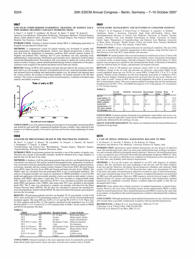
S224 20th ESICM Annual Congress – Berlin, Germany – 7–10 October 2007
0867LOW-TECH COMPUTERIZED IN-HOSPITAL TRACKING OF PATIENT LOCA-TION DURING MULTIPLE CASUALTY INCIDENTS (MCIS)
S. Einav*1, S. Schiff2, S. Goldberg3, M. Hersch1, R. Spira4, T. Zalut3, W. Schecter5
1Intensive Care Medicine, 2Information Technology, 3Emergency Medicine, 4General Surgery,Shaare Zedek Medical Centre, Jerusalem, Israel, 5General Surgery, San Francisco GeneralHospital, San Francisco, United States
INTRODUCTION. Tracking of patient location during MCIs is challenging particularly inhospitals not specialized in trauma care.
METHODS. A computerized system for patient tracking was developed (4 months full-time programmer). Diagnostic/therapeutic stations were defined prospectively to direct pa-tient flow from the department of emergency medicine (ED) onward; general, trauma, or-thopedic and pediatric EDs, radiology and CT, post anesthesia care unit, operating room,ophthalmology, otolaryngology, debridement, post traumatic stress, admission ward/secondarytransfer/discharged/deceased. Non-medical staff were trained to update the system with esti-mated severities of injury, patient identification/identifying features,composition of the physi-cian/nurse team assigned to the patient and patient arrival times per-station.
RESULTS. During 18 MCIs (842 patients) real-time data provided by the system to key hos-pital staff (e.g event manager, directors of hospital/social services, head nurse)enabled orderlyand informed decision-making and family/media updates (e.g. the patient load imposed uponthe various stations, the location of individual patients). No patient returned to the ED afterexiting it. The system is currently being used for research purposes, evaluation of hospital surgecapacity and quality assurance.
CONCLUSION. Low-tech. patient tracking systems improve Command&Control during MCIsand should be used until fully computerized methods become available. Data collected in thismanner is of sufficient quality to be used in real-time and for later analyses/planning for futureevents.
0868ANALYSIS OF PREVENTABLE DEATH IN THE POLYTRAUMA PATIENTS
M. Koo*1, S. Lopez2, E. Bassas1, M. Lacambra1, N. Fustran1, J. Garreta3, M. Garcia4,J. Dominguez5, T. Gracia1, A. Sabate1
1Anesthesiology and Critical Care, 2Bioesthadistic, 3Trauma Surgery, 4Digestive Surgery,5Angiorradiology, Bellvitge Hospital, Barcelona, Spain
INTRODUCTION. In polytrauma patients the mortality is one of the markers of quality care.Therefore the concept of preventable mortality should be obtained from a large database ofpatients that was the aim of our study.
METHODS. A database of all the polytrauma patients that arrived to our Hospital during oneyear period, was analyzed. The registry included demographical data, mechanism of trauma le-sion, resuscitative measures during the primary revision, diagnostic methods, graduated anatom-ical lesions, mortality and four scoring systems: Glasgow Coma Score (GSC), Revised TraumaScore (RTS), Injury Severity Score (ISS) and Trauma and Injury Severity Score (TRISS). TheTRISS value was calculated from the prehospital RTS in case of prehospital intubation. Theanalysis of hospital mortality was based on calculation of TRISS probability of survival (PS)model and application of the preventable mortality criteria was based on other author’s criteria.Patients with TRISS value minor o equal than 25% were classified as nonpreventable death(ND). Patients with a TRISS value between 20-50 % were classified as possibly preventabledeath (PPD) and patients with a TRISS value major than 50% were classified as preventabledeath (PD). The Z value was calculated to compare our mortality with that from the MajorTrauma Outcome Study (MTOS). The M value was calculated to measure the matching be-tween the populations studied with the MTOS. M value lower than 0.88 meaned a great disparitybetween the two groups.
RESULTS. 198 polytrauma patients arrived to our hospital, 29.1% were female. A total of 184(93%) were closed and 14 (7%) penetrating trauma. In 111 cases (55.8%) received prehospitaltreatment support. The mean ISS was 16.89 +/-11.23 and the PS of 0.95 +/- 0.19. There were91 (46%) patients with an ISS>15. The statistics calculated for this population was: Z =-5.05,M=0.95 and W =-7.15. A total of 22 (11.55%) patients died. In Table 1 are the results of TRISSand ISS for the patients who died.
TABLE 1.TRISS Classification Hospital length Cause of death99.9-96.9 (2 cases) PD 10 -11 days Broncoasp-Sepsis99.9 PD 24 days Multyorganic failure99.8 PD 29 days Sepsis acinetobacter99.8-99.6 (3 cases) PD 90 minutes-2 days Delayed treatment99.5 PD 1 day No treatment98.8-72.1(5cases) PD 180 minutes-6 day Brain death-Head inj.78.5-38.9 (2 cases) PD-PPD 90-120 minutes Hypovolemic shock34.5 PPD 1 day Arrythmia6.8-0.1 (6 cases) ND 5-75 minutes Cardiac arrest0.1 ND 5 days Brain death-Head inj.
CONCLUSION. Delayed treatment is the most important factor for potentially preventabledeath. Brain death, hypovolemic shock and sepsis were the most common causes of death.
0869VENTILATORY MANAGEMENT AND OUTCOMES IN COMATOSE PATIENTS
P. Pelosi*1, N. D. Ferguson2, F. Frutos-Vivar3, C. Putensen4, A. Anzueto5, A. Esteban3
1Ambiente, Salute e Sicurezza, Universita’ degli Studi dell’Insubria, Varese, Italy,2Interdepartmental Division of Critical Care Medicine, University of Toronto, Toronto,Canada, 3Intensive Care Unit, Hospital Universitario de Getafe, University of Getafe,Getafe, Spain, 4Department of Anaesthesiology and Intensive Care Medicine, Universityof Bonn, Bonn, Germany, 5Health Science Center and South Texas Veterans HealthcareSystem, University of Texas, San Antonio, United States
INTRODUCTION. Coma is a frequent indication for mechanical ventilation. The aim of thisstudy was to describe the ventilatory management of these patients, along with the course oftheir Intensive Care Unit (ICU) organ failures and outcomes.
METHODS. We studied patients who received mechanical ventilation for at least 12 hours withcoma as the reason for initiating ventilation (due to metabolic causes, intoxication, hemorrhagicor ischemic stroke, or brain trauma), who had a Glasgow Coma Score (GCS) below 14. Thesepatients were prospectively enrolled in the 2nd International Study of Mechanical Ventilationand were followed daily for 28 days, and thereafter for hospital outcomes.
RESULTS. A total of 780 patients met inclusion criteria (20% of all ventilated patients).Ventilatory settings in the 1st week and outcomes are shown by coma origin (Table). In theoverall population mean age was 54(18) yrs, mean SOFA 7(3)and GCS < 8 in 75 % of thepatients. Volume-cycled ventilation was the most frequently used mode of ventilation (70%),while Pressure Support Ventilation progressively increased after the first week. Patients evermet “ready to wean” criteria in 60% for the overall population (from 80% in intoxication to46% in hemorrhagic stroke). The most common method of weaning after the first spontaneousbreathing trial was PSV (75%). ALI/ARDS developed in 12% of the patients (7% in metabolicto 15% in brain trauma).
TABLE 1.Overall Metabolic Intoxication Hemorrhagic Ischemic Brain PPopulation Stroke Stroke Trauma
Pts (N) 780 104 124 260 102 190Lung Failure (%) 42 39 36 42 31 53 <0.01VT (ml/Kg PBW) 9(2) 9(2) 9(2) 10(2) 9(2) 9(2) <0.02PEEP (cmH2O) 4(3) 4(3) 4(3) 4(3) 4(3) 4(3) 0.78PaO2/FiO2 283(116) 278(125) 266(120) 286(114) 250(106) 310(110) <0.01Tracheostomy (%) 18 7 7 20 14 30 <0.01MV (days) median 4 4 2 5 4 6 <0.01ICUMortality (%) 35 30 17 48 37 29 <0.01
CONCLUSION. Comatose patients frequently have pulmonary organ failure, but receive ven-tilation with relatively high tidal volume and low PEEP. Airway management and outcome aresignificantly influenced by the aetiology of coma.
GRANT ACKNOWLEDGEMENT. The Authors are indebted to the International MechanicalVentilation Group Study
0870A CASE OF SPINAL EPIDURAL HAEMATOMA RELATED TO PRES
L. Di Donato*, G. Servillo, F. Bifulco, E. De Robertis, R. TufanoAnesthesia and Intensive Care Medicine, University Federico II, Naples, Italy
INTRODUCTION. Spontaneous spinal epidural haematomas are rare diseases of unknowncause. Elevated blood pressure values can cause acute epidural hemorrage, leading to an impres-sive and eventually difficult to distinguish spinal syndrome1. Moreover, elevated blood pressureis also a recognized cause of Posterior Reversible Encephalopathy Syndrome (PRES)2,3. Herewe describe a case report in which these two conditions are both present in the same patient, atthe same time, and, probably, both related to hypertension.
METHODS. A 56 year-old women was admitted to our ICU with diagnosis of epilepticseizures. She also developed neck pain irradiating to her left arm, and left sided weaknessin her arm and leg. Moreover, the patient showed progressive blindness and confusion. Highsystolic (210-200) and diastolic (115-135) blood pressure values were documented. CT scanof the brain and spinal cord performed at admission revealed no signs of brain haemorrhage,and a large extraspinal large lesion (C5-T2). Diagnosis of epidural haematoma was performedon MRI scan with gadolinium. Moreover, MRI sequences showed classical images of PRES.Medical therapy for seizures and hypertension was performed. After laminectomy, epiduralhaematoma was completely evacuated. Patient’s condition improved rapidly so that she wasdischarged after 6 days.
RESULTS. Some authors have related occurrence of epidural haematomas to arterial hyper-tension. However, the exact cause of bleeding remains mostly undetermined. PRES is mainlyrelated to arterial hypertension. Since epidural haematoma and PRES occurred at the same timein our patient, we hypothesize that arterial hypertension could have determined both conditionsin this case.
CONCLUSION. Although spontaneous spinal epidural haematomas are rare, clinicians shouldstill consider them as possible complications in patients with uncontrolled hypertension.
REFERENCE(S). 1) Kuker W et al. Acta Neurochir , 2000;142:777-85.2) Hynchey J et al. N Engl J Med, 1996;334:494-500.3) Servillo et al. Intensive Care Med, 2006; 32 (11)
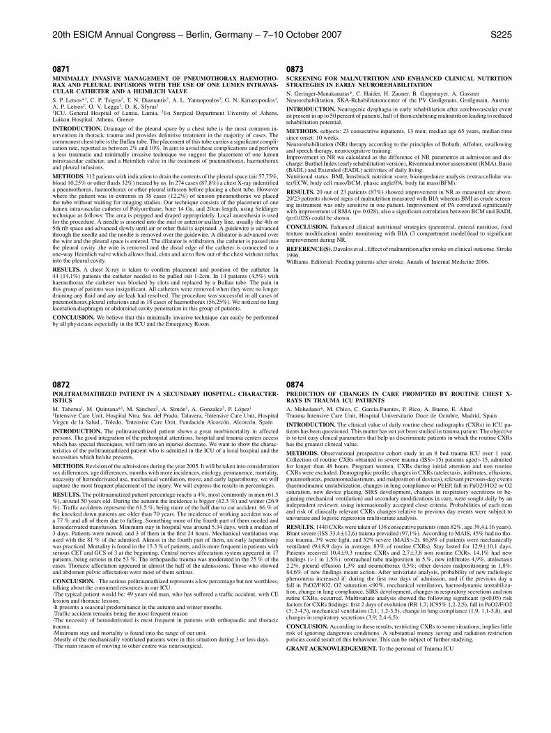
20th ESICM Annual Congress – Berlin, Germany – 7–10 October 2007 S225
0871MINIMALLY INVASIVE MANAGEMENT OF PNEUMOTHORAX HAEMOTHO-RAX AND PLEURAL INFUSIONS WITH THE USE OF ONE LUMEN INTRAVAS-CULAR CATHETER AND A HEIMLICH VALVE
S. P. Letsos*1, C. P. Tsigris2, T. N. Diamantis2, A. L. Yannopoulos2, G. N. Kiriazopoulos1,A. P. Letsos2, O. V. Legga1, D. K. Sfyras1
1ICU, General Hospital of Lamia, Lamia, 21st Surgical Department Uiversity of Athens,Laikon Hospital, Athens, Greece
INTRODUCTION. Drainage of the pleural space by a chest tube is the most common in-tervention in thoracic trauma and provides definitive treatment in the majority of cases. Thecommonest chest tube is the Bullau tube. The placement of this tube carries a significant compli-cation rate, reported as between 2% and 10%. In aim to avoid these complications and performa less traumatic and minimally invasive technique we suggest the placement of one lumenintravascular catheter, and a Heimlich valve in the treatment of pneumothorax, haemothoraxand pleural infusions.
METHODS. 312 patients with indication to drain the contents of the pleural space (air 57,75%,blood 10,25% or other fluids 32%) treated by us. In 274 cases (87,8%) a chest X-ray indentifieda pneumothorax, haemothorax or other pleural infusion before placing a chest tube. Howeverwhere the patient was in extremis in 38 cases (12,2%) of tension pneumothorax we placedthe tube without waiting for imaging studies. Our technique consists of the placement of onelumen intravascular catheter of Polyurethane, bore 14 Ga, and 20cm length, using Seldingertechnique as follows: The area is prepped and draped appropriately. Local anaesthesia is usedfor the procedure. A needle is inserted into the mid or anterior axillary line, usually the 4th or5th rib space and advanced slowly until air or other fluid is aspirated. A guidewire is advancedthrough the needle and the needle is removed over the guidewire. A dilatator is advanced overthe wire and the pleural space is entered. The dilatator is withdrawn, the catheter is passed intothe pleural cavity ,the wire is removed and the distal edge of the catheter is connected to aone-way Heimlich valve which allows fluid, clots and air to flow out of the chest without refluxinto the pleural cavity.
RESULTS. A chest X-ray is taken to confirm placement and position of the catheter. In44 (14,1%) patients the catheter needed to be pulled out 1-2cm. In 14 patients (4,5%) withhaemothorax the catheter was blocked by clots and replaced by a Bullau tube. The pain inthis group of patients was insignificant. All catheters were removed when they were no longerdraining any fluid and any air leak had resolved. The procedure was successful in all cases ofpneumothorax,pleural infusions and in 18 cases of haemothorax (56,25%). We noticed no lunglaceration,diaphragm or abdominal cavity penetration in this group of patients.
CONCLUSION. We believe that this minimally invasive technique can easily be performedby all physicians especially in the ICU and the Emergency Room.
0872POLITRAUMATHIZED PATIENT IN A SECUNDARY HOSPITAL: CHARACTER-ISTICS
M. Taberna1, M. Quintana*1, M. Sanchez2, A. Simon1, A. Gonzalez3, P. Lopez1
1Intensive Care Unit, Hospital Ntra. Sra. del Prado, Talavera, 2Intensive Care Unit, HospitalVirgen de la Salud., Toledo, 3Intensive Care Unit, Fundacion Alcorcon, Alcorcon, Spain
INTRODUCTION. The politraumathized patient shows a great morbimortality in affectedpersons. The good integration of the prehospital attentions, hospital and trauma centers accesswhich has special thecniques, will turn into an injuries decrease. We want to show the charac-teristics of the politraumathized patient who is admitted in the ICU of a local hospital and thenecessities which he/she presents.
METHODS. Revision of the admissions during the year 2005. It will be taken into considerationsex differences, age differences, months with more incidences, etiology, permanence, mortality,necessity of hemoderivated use, mechanical ventilation, move, and early laparothomy, we willcapture the most frequent placement of the injury. We will express the results in percentages.
RESULTS. The politraumatized patient percentage reachs a 4%, most commonly in men (61.5%), around 50 years old. During the autumn the incidence is bigger (42.3 %) and winter (26.9%). Traffic accidents represent the 61.5 %, being more of the half due to car accident. 66 % ofthe knocked down patients are older than 70 years. The incidence of working accident was ofa 77 % and all of them due to falling. Something more of the fourth part of them needed andhemoderivated transfusion. Minimum stay in hospital was around 5.34 days, with a median of3 days. Patients were moved, and 3 of them in the first 24 hours. Mechanical ventilation wasused with the 81 % of the admitted. Almost in the fourth part of them, an early laparathomywas practiced. Mortality is found in the 15.3 % of patients, and is more frequent in patients withserious CET and GCS of 3 at the beginning. Central nerves affectation system appeared in 17patients, being serious in the 53 %. The orthopaedic trauma was moderated in the 75 % of thecases. Thoracic affectation appeared in almost the half of the admissions. Those who showedand abdomen pelvic affectation were most of them serious.
CONCLUSION. · The serious politraumathized represents a low percentage but not worthless,talking about the consumed resources in our ICU.·The typical patient would be: 49 years old man, who has suffered a traffic accident, with CElession and thoracic lession.·It presents a seasonal predominance in the autumn and winter months.·Traffic accident remains being the most frequent reason·The necessity of hemoderivated is most frequent in patients with orthopaedic and thoracictrauma.·Minimum stay and mortality is found into the range of our unit.·Mostly of the mechanically ventilated patients were in this situation during 3 or less days.·The main reason of moving to other centre was neurosurgical.
0873SCREENING FOR MALNUTRITION AND ENHANCED CLINICAL NUTRITIONSTRATEGIES IN EARLY NEUROREHABILITATION
N. Geringer-Manakanatas*, C. Haider, H. Zauner, B. Gappmayer, A. GassnerNeurorehabilitation, SKA-Rehabilitationcenter of the PV Großgmain, Großgmain, Austria
INTRODUCTION. Neurogenic dysphagia in early rehabilitation after cerebrovascular eventist present in up to 50 percent of patients, half of them exhibiting malnutrition leading to reducedrehabilitation potential.
METHODS. subjects: 23 consecutive inpatients, 13 men; median age 65 years, median timesince onset: 10 weeks.Neurorehabilitation (NR) therapy according to the principles of Bobath, Affolter, swallowingand speech therapy, neurocognitive training.Improvement in NR wa calculated as the difference of NR parametres at admission and dis-charge: Barthel Index (early rehabilitation version), Rivermead motor assessment (RMA), Basic(BADL) and Extended (EADL) acitivities of daily living.Nutritional status: BMI, Innsbruck nutrition score, bioimpedance analysis (extraccellular wa-ter/ECW, body cell mass/BCM, phasic angle/PA, body fat mass/BFM).
RESULTS. 20 out of 23 patients (87%) showed improvement in NR as measured see above.20/23 patients showed signs of malnutrition measured with BIA whereas BMI as crude screen-ing instrument was only sensitive in one patient. Improvement of PA correlated significantlywith improvement of RMA (p= 0.028), also a significant correlation between BCM and BADL(p=0.028) could be shown.
CONCLUSION. Enhanced clinical nutritional strategies (parenteral, enteral nutrition, foodtexture modification) under monitoring with BIA (3 compartment model)lead to significantimprovement during NR.
REFERENCE(S). Davalos et al., Effect of malnutrition after stroke on clinical outcome. Stroke1996.Williams. Editorial: Feeding patients after stroke. Annals of Internal Medicine 2006.
0874PREDICTION OF CHANGES IN CARE PROMPTED BY ROUTINE CHEST X-RAYS IN TRAUMA ICU PATIENTS
A. Mohedano*, M. Chico, C. Garcia-Fuentes, P. Rico, A. Bueno, E. AltedTrauma Intensive Care Unit, Hospital Universitario Doce de Octubre, Madrid, Spain
INTRODUCTION. The clinical value of daily routine chest radiographs (CXRs) in ICU pa-tients has been questioned. This matter has not yet been studied in trauma patient. The objectiveis to test easy clinical parameters that help us discriminate patients in which the routine CXRshas the greatest clinical value.
METHODS. Observational prospective cohort study in an 8 bed trauma ICU over 1 year.Collection of routine CXRs obtained in severe trauma (ISS>15) patients aged>15, admittedfor longer than 48 hours. Pregnant women, CXRs during initial attention and non routineCXRs were excluded. Demographic profile, changes in CXRs (atelectasis, infiltrates, effusions,pneumothorax, pneumomediastinum, and malposition of devices), relevant previous-day events(haemodinamic unstabilization, changes in lung compliance or PEEP, fall in PaO2/FIO2 or O2saturation, new device placing, SIRS development, changes in respiratory secretions or be-ginning mechanical ventilation) and secondary modifications in care, were sought daily by anindependent reviewer, using internationally accepted close criteria. Probabilities of each itemand risk of clinically relevant CXRs changes relative to previous day events were subject tounivariate and logistic regression multivariate analysis.
RESULTS. 1440 CXRs were taken of 138 consecutive patients (men 82%, age 39,4±16 years).Blunt severe (ISS 33,4±12,6) trauma prevailed (97,1%). According to MAIS, 45% had no tho-rax trauma, 3% were light, and 52% severe (MAIS>2). 86,8% of patients were mechanicallyventilated (9±8,9 days in average, 83% of routine CXRs). Stay lasted for 12,9±10,1 days.Patients received 10,4±9,3 routine CXRs and 2,7±3,8 non routine CXRs. 14,1% had newfindings (>1 in 1,5%): orotracheal tube malposition in 5,%, new infiltrates 4,9%, atelectasis2.2%, pleural effusion 1,3% and neumothorax 0,5%; other devices malpositioning in 1,8%.84,6% of new findings meant action. After univariate analysis, probability of new radiologicphenomena increased if: during the first two days of admission, and if the previous day afall in PaO2/FIO2, O2 saturation <90%, mechanical ventilation, haemodynamic unstabiliza-tion, change in lung compliance, SIRS development, changes in respiratory secretions and nonrutine CXRs, occurred. Multivariate analysis showed the following significant (p<0,05) riskfactors for CXRs findings: first 2 days of evolution (RR 1,7; IC95% 1,2-2,5), fall in PaO2/FiO2(3; 2-4,5), mechanical ventilation (2,1; 1,2-3,5), change in lung compliance (1,9; 1,1-3,8), andchanges in respiratory secretions (3,9; 2,4-6,5).
CONCLUSION. According to these results, restricting CXRs to some situations, implies littlerisk of ignoring dangerous conditions. A substantial money saving and radiation restrictionpolicies could result of this behaviour. This can be subject of further studying.
GRANT ACKNOWLEDGEMENT. To the personal of Trauma ICU
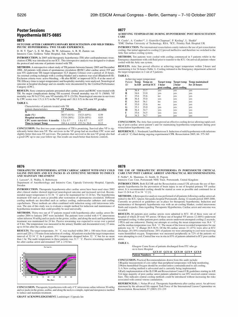
S226 20th ESICM Annual Congress – Berlin, Germany – 7–10 October 2007
Poster SessionsHypothermia 0875-08850875OUTCOME AFTER CARDIOPULMONARY RESUSCITATION AND MILD THERA-PEUTIC HYPOTHERMIA: TWO YEARS EXPERIENCE.
D. H. T. Tjan*, L. E. M. Haas, M. W. Adriaanse, A. R. H. Zanten vanIntensive Care, Gelderse Vallei Hospital, Ede, Netherlands
INTRODUCTION. In 2005 mild therapeutic hypothermia (TH) after cardiopulmonary resus-citation (CPR) was introduced in our ICU. This retrospective analysis was designed to evaluatethe protocol and outcome of patients treated with TH.
METHODS. A retrospective cohort study of TH patients between January 2005 and December2006. All patients with return of spontaneous circulation (ROSC) after cardiac arrest (VF andnon-VF) underwent TH (target temperature 32,5 degrees Celsius) over a period of 24 hours.An external cooling technique with a cooling blanket and a mattress was used (Blanketroll-II).Exclusion criteria were: active bleeding, >8h after ROSC or improved GCS>8 at the onset ofTH. Efficacy (time to target-temperature) and hospitality mortality were analysed. Neurologicaloutcome at hospital discharge and six-months were documented by the Cerebral PerformanceCategory (CPC).
RESULTS. Sixty comatose patients presented after cardiac arrest and ROSC were treated withTH. No major complications during TH occurred. Overall mortality was 63 % (38/60). VFmortality was 50 %(17/34), non-VF mortality 85 % (21/26). Neurological outcome in survivorsby CPC scores was 1.5 (± 0.7) in the VF group and 1.8(± 0.5) in the non VF group.
TABLE 1.Characteristics of patients treated with THpatient characteristics VF Patients Non-VF patients p-valueN 37 23male (%) 30/37 (81%) 15/23 (65%) 0.59Hospital mortality 17/34 (50%) 22/26 (85%) 0.03CPC score survivors 6 months 1.5± 0.7 1.8± 0.7 0.57Time to target Temp 184.4 ± 112.9 153.2± 26.0 0.13
CONCLUSION. Our first two-years experience of TH is promising. Survival after VF is sig-nificantly better then non-VF. The survivors in the VF group had an excellent CPC score andslightly better then non-VF survivors. The patients that survived in the non VF group also hada good CPC up to one year follow-up. Our results are much better than historic controls.
0876THERAPEUTIC HYPOTHERMIA AFTER CARDIAC ARREST WITH ONLY COLDSALINE INFUSION AND ICE PACKS IS AN EFFECTIVE METHOD TO INDUCEAND MAINTAIN TREATMENT
I. Larsson*, E. Wallin, S. RubertssonDepartment of Anesthesiology and Intensive Care, Uppsala University Hospital, Uppsala,Sweden
INTRODUCTION. Therapeutic hypothermia after cardiac arrest have been used since 2002after clinical studies showed improved neurological outcome and increased survival. Recom-mended target temperature is 32-34 ˚ C and to be maintained for 12-24 hrs. The hypothermiatreatment will start as soon as possible after restoration of spontaneous circulation. Differentcooling methods are described such as surface cooling, endovascular catheters and coolingcaps/helmets. These methods are often combined with induction using cold intravenous infu-sion. The aim of this study was to evaluate a simple method for induction and maintenance ofhypothermia treatment that could possibly be used universally.
METHODS. Thirty-two out of 37 patients treated with hypothermia after cardiac arrest De-cember 2004 to January 2007 were included. The patients were cooled with 4 oC intravenoussaline infusion 30 ml/kg and ice packs in the groins, axillas, and along the neck and hypothermiatreatment was maintained for 24 hrs. Passive rewarming was expected to occur over a periodof 8 hrs. The temperature was measured in the urinary bladder and documented every 15 minsup to 44 hrs after the cardiac arrest.
RESULTS. The target temperature, 34 ˚ C, was reached within 280 ± 180 mins from cardiacarrest and 220 ± 170 mins from induction of cooling. All patients reached the target temperatureinterval of 32-34 ˚ C. In 4 patients (8%) temperature dropped below 32 ˚ C but for no morethan 1 hr. The nadir temperature in these patients was 31.7 ˚ C. Passive rewarming started 26hrs after cardiac arrest and remained 7.85 ± 2.92 hrs.
CONCLUSION. Therapeutic hypothermia with only 4 oC intravenous saline infusion 30 ml/kgand ice packs in the groins, axillas, and along the neck is a simple, rapid and inexpensive methodto induce and maintain treatment.
GRANT ACKNOWLEDGEMENT. Landstinget i Uppsala lan
0877ACHIEVING TEMPERATURE DURING HYPOTHERMIC POST RESUSCITATIONCARE
N. Castle1, A. Combes*2, J. Granville-Chapman2, P. Keeling2, L. Shaikh2
1EMC Durban University of Technology, RSA, 2ICU, Frimley Park Hospital UK
INTRODUCTION. The international resuscitation comity indorses the use of pot resuscitationcooling. Our initial approach to cooling [1] proved ineffective and therefore we switched to theArtic Sun surface cooling system.
METHODS. Ten patients were cooled with cooling commenced in 5 patients whilst in theEmergency department with cold fluid prior to transfer to the ICU. On arrival all patients wherecooled with the Artic sun system.
RESULTS. Artic Sun proved effective at achieving target temperature within 2-hours andmaintaining it for 24-hours (Table 1). Cooling initiated in the Emergency department achievedthe target temperature in 2 out of 5 patients.
TABLE 1.Achieving target temperaturePatient Temp Temp on Target temp Target temp Temp maintainedID in ED arrival ICU 1 hour 2 hour for 24 hours
post cooling post coolingA 36.4 33.7 yes yes yesB 34.6 33.0 yes yes yesC 35.6 35.6 no yes yesD 36.4 35.0 yes yes yesE 36.0 36.1 no yes yesF 35.0 35.6 no yes yesG 35.1 35.0 yes yes yesH 36.4 34.7 yes yes yesI 35.6 34.5 yes yes yesJ 34.3 34.7 yes yes yes
CONCLUSION. The Artic Sun system proved an effective cooling device allowing rapid cool-ing of post-cardiac arrest patient’s and for maintaining hypothermia temperature during postresuscitation hypothermic care.
REFERENCE. 1. Nordmark J and Rubertsson S. Induction of mild hypothermia with infusionsof cold (4 ˚ C) fluid during ongoing experimental CPR. Resuscitation 2005; 66: 375-365
0878THE USE OF THERAPEUTIC HYPOTHERMIA IN PORTSMOUTH CRITICALCARE UNIT POST CARDIAC ARREST AND PRACTICAL RECOMMENDATIONS.
F. Noble*, K. Shammas, G. Smith, D. PogsonIntensive Care Unit, Portsmouth NHS Trust, Portsmouth, United Kingdom
INTRODUCTION. Both ILCOR and the Resuscitation council UK advocate the use of ther-apeutic hypothermia for the prevention of brain injury in out of hospital primary VF cardiacarrest. It is recommended cooling should be started as soon as possible and continued for atleast 12-24 h at 32 to 34 ˚ C [1].
METHODS. A Retrospective analysis was performed of all patients following cardiac arrest ad-mitted to the ICU, Queen Alexandra hospital Portsmouth, during 12-month period 2005-2006.Currently no protocol or guidelines are in place for therapeutic hypothermia. Induction andmaintenance of therapeutic hypothermia was with an air-cooling blanket, cooled intravenousfluids and icepacks. Data regarding Therapeutic Hypothermia, Cardiac arrest and outcome wasrecorded.
RESULTS. 66 patients post cardiac arrests were admitted to ICU. 40 of these were out ofhospital of which 20 were VF arrests. Of these out of hospital VF arrests 12 (60%) underwentattempted cooling. 6 other patients post cardiac arrests underwent attempted cooling (2 asystole,3 PEA and one unknown). During our attempts at cooling the mean temperature was 36.4 ˚ Cand the median temperature was 36.6 ˚ C. The mean of the lowest recorded temperature for ourpatients was 34 ˚ C (Range 26.9-36.9). Of the 66 cardiac arrests 31 (47%) were alive at ICUdischarge, 20 (30%) returned home. 28% of patients we were attempting to cool were receivingwarm humidified oxygen. Temperature was measured peripherally in 72% of the patients wewere attempting to cool. Central line was in situ in 83% of patients admitted to ICU post cardiacarrest.
TABLE 1.Glasgow Coma Score of patients discharged from ITU who goon to leave Hospital
GCS 15 GCS 14 GCS 10 GCS 9 GCS 8Patient Numbers 9 7 2 1 1
CONCLUSION. Practical Recommendations drawn from this audit include:1)Regular measurement of core rather than peripheral temperature will help monitoring.2)Warm humidified Oxygen should be avoided unless contraindicated to aid cooling.3) A New cooling method is advocated and is currently being implemented.4)Early implementation of the ILCOR and Resuscitation Council UK guidelines starting in A/E5)A large majority of post cardiac arrest patients admitted to our ITU received central venouslines. This indicates central cooling methods could be introduced without increasing the risksassociated with central venous cannulation.
REFERENCE(S). 1. Nolan JP et al. Therapeutic hypothermia after cardiac arrest. An advisorystatement by the advanced life support Task Force of the International Liason Copmmittee onResuscitation. Resucitation 57 (2003) 231-235
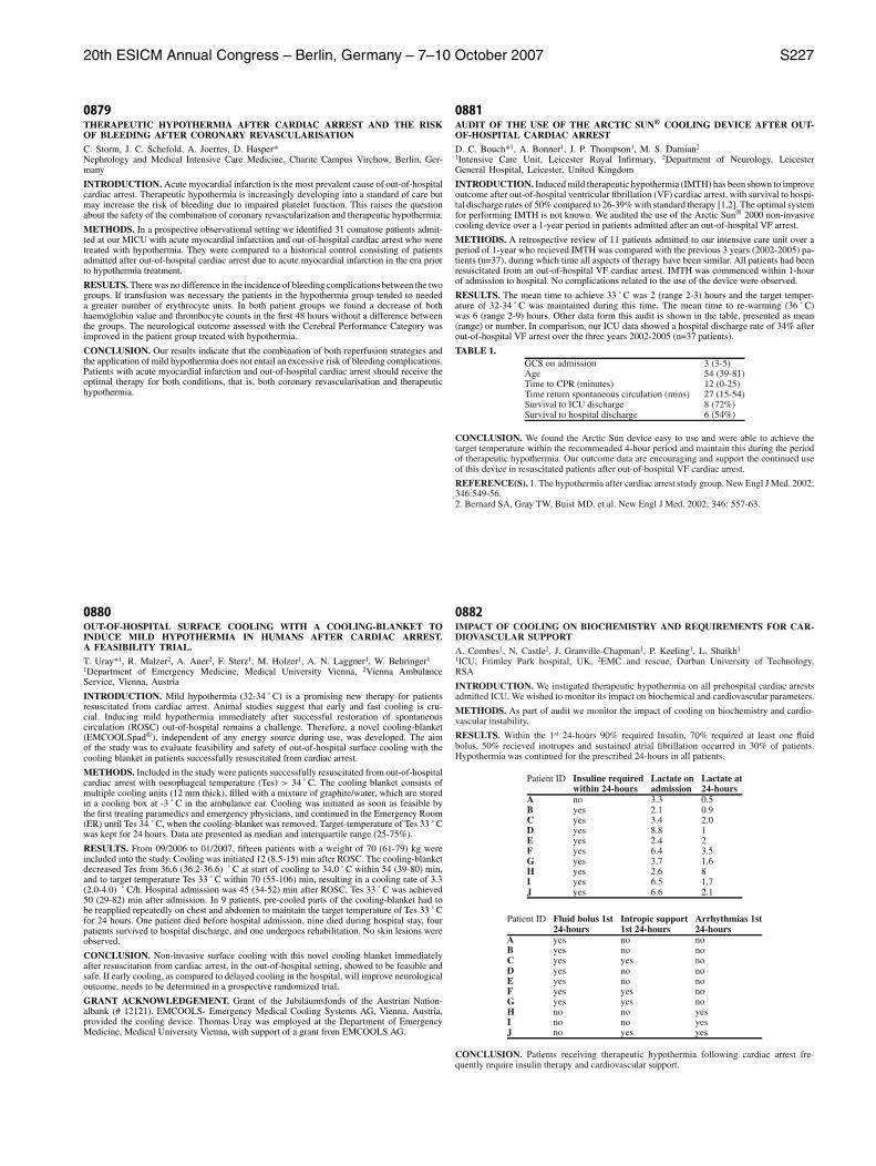
20th ESICM Annual Congress – Berlin, Germany – 7–10 October 2007 S227
0879THERAPEUTIC HYPOTHERMIA AFTER CARDIAC ARREST AND THE RISKOF BLEEDING AFTER CORONARY REVASCULARISATION
C. Storm, J. C. Schefold, A. Joerres, D. Hasper*Nephrology and Medical Intensive Care Medicine, Charite Campus Virchow, Berlin, Ger-many
INTRODUCTION. Acute myocardial infarction is the most prevalent cause of out-of-hospitalcardiac arrest. Therapeutic hypothermia is increasingly developing into a standard of care butmay increase the risk of bleeding due to impaired platelet function. This raises the questionabout the safety of the combination of coronary revascularization and therapeutic hypothermia.
METHODS. In a prospective observational setting we identified 31 comatose patients admit-ted at our MICU with acute myocardial infarction and out-of-hospital cardiac arrest who weretreated with hypothermia. They were compared to a historical control consisting of patientsadmitted after out-of-hospital cardiac arrest due to acute myocardial infarction in the era priorto hypothermia treatment.
RESULTS. There was no difference in the incidence of bleeding complications between the twogroups. If transfusion was necessary the patients in the hypothermia group tended to neededa greater number of erythrocyte units. In both patient groups we found a decrease of bothhaemoglobin value and thrombocyte counts in the first 48 hours without a difference betweenthe groups. The neurological outcome assessed with the Cerebral Performance Category wasimproved in the patient group treated with hypothermia.
CONCLUSION. Our results indicate that the combination of both reperfusion strategies andthe application of mild hypothermia does not entail an excessive risk of bleeding complications.Patients with acute myocardial infarction and out-of-hospital cardiac arrest should receive theoptimal therapy for both conditions, that is, both coronary revascularisation and therapeutichypothermia.
0880OUT-OF-HOSPITAL SURFACE COOLING WITH A COOLING-BLANKET TOINDUCE MILD HYPOTHERMIA IN HUMANS AFTER CARDIAC ARREST.A FEASIBILITY TRIAL.
T. Uray*1, R. Malzer2, A. Auer2, F. Sterz1, M. Holzer1, A. N. Laggner1, W. Behringer1
1Department of Emergency Medicine, Medical University Vienna, 2Vienna AmbulanceService, Vienna, Austria
INTRODUCTION. Mild hypothermia (32-34 ˚ C) is a promising new therapy for patientsresuscitated from cardiac arrest. Animal studies suggest that early and fast cooling is cru-cial. Inducing mild hypothermia immediately after successful restoration of spontaneouscirculation (ROSC) out-of-hospital remains a challenge. Therefore, a novel cooling-blanket(EMCOOLSpad®), independent of any energy source during use, was developed. The aimof the study was to evaluate feasibility and safety of out-of-hospital surface cooling with thecooling blanket in patients successfully resuscitated from cardiac arrest.
METHODS. Included in the study were patients successfully resuscitated from out-of-hospitalcardiac arrest with oesophageal temperature (Tes) > 34 ˚ C. The cooling blanket consists ofmultiple cooling units (12 mm thick), filled with a mixture of graphite/water, which are storedin a cooling box at -3 ˚ C in the ambulance car. Cooling was initiated as soon as feasible bythe first treating paramedics and emergency physicians, and continued in the Emergency Room(ER) until Tes 34 ˚ C, when the cooling-blanket was removed. Target-temperature of Tes 33 ˚ Cwas kept for 24 hours. Data are presented as median and interquartile range (25-75%).
RESULTS. From 09/2006 to 01/2007, fifteen patients with a weight of 70 (61-79) kg wereincluded into the study. Cooling was initiated 12 (8.5-15) min after ROSC. The cooling-blanketdecreased Tes from 36.6 (36.2-36.6) ˚ C at start of cooling to 34.0 ˚ C within 54 (39-80) min,and to target temperature Tes 33 ˚ C within 70 (55-106) min, resulting in a cooling rate of 3.3(2.0-4.0) ˚ C/h. Hospital admission was 45 (34-52) min after ROSC, Tes 33 ˚ C was achieved50 (29-82) min after admission. In 9 patients, pre-cooled parts of the cooling-blanket had tobe reapplied repeatedly on chest and abdomen to maintain the target temperature of Tes 33 ˚ Cfor 24 hours. One patient died before hospital admission, nine died during hospital stay, fourpatients survived to hospital discharge, and one undergoes rehabilitation. No skin lesions wereobserved.
CONCLUSION. Non-invasive surface cooling with this novel cooling blanket immediatelyafter resuscitation from cardiac arrest, in the out-of-hospital setting, showed to be feasible andsafe. If early cooling, as compared to delayed cooling in the hospital, will improve neurologicaloutcome, needs to be determined in a prospective randomized trial.
GRANT ACKNOWLEDGEMENT. Grant of the Jubilaumsfonds of the Austrian Nation-albank (# 12121). EMCOOLS- Emergency Medical Cooling Systems AG, Vienna, Austria,provided the cooling device. Thomas Uray was employed at the Department of EmergencyMedicine, Medical University Vienna, with support of a grant from EMCOOLS AG.
0881AUDIT OF THE USE OF THE ARCTIC SUN® COOLING DEVICE AFTER OUT-OF-HOSPITAL CARDIAC ARREST
D. C. Bouch*1, A. Bonner1, J. P. Thompson1, M. S. Damian2
1Intensive Care Unit, Leicester Royal Infirmary, 2Department of Neurology, LeicesterGeneral Hospital, Leicester, United Kingdom
INTRODUCTION. Induced mild therapeutic hypothermia (IMTH) has been shown to improveoutcome after out-of-hospital ventricular fibrillation (VF) cardiac arrest, with survival to hospi-tal discharge rates of 50% compared to 26-39% with standard therapy [1,2]. The optimal systemfor performing IMTH is not known. We audited the use of the Arctic Sun® 2000 non-invasivecooling device over a 1-year period in patients admitted after an out-of-hospital VF arrest.
METHODS. A retrospective review of 11 patients admitted to our intensive care unit over aperiod of 1-year who recieved IMTH was compared with the previous 3 years (2002-2005) pa-tients (n=37), during which time all aspects of therapy have been similar. All patients had beenresuscitated from an out-of-hospital VF cardiac arrest. IMTH was commenced within 1-hourof admission to hospital. No complications related to the use of the device were observed.
RESULTS. The mean time to achieve 33 ˚ C was 2 (range 2-3) hours and the target temper-ature of 32-34 ˚ C was maintained during this time. The mean time to re-warming (36 ˚ C)was 6 (range 2-9) hours. Other data form this audit is shown in the table, presented as mean(range) or number. In comparison, our ICU data showed a hospital discharge rate of 34% afterout-of-hospital VF arrest over the three years 2002-2005 (n=37 patients).
TABLE 1.GCS on admission 3 (3-5)Age 54 (39-81)Time to CPR (minutes) 12 (0-25)Time return spontaneous circulation (mins) 27 (15-54)Survival to ICU discharge 8 (72%)Survival to hospital discharge 6 (54%)
CONCLUSION. We found the Arctic Sun device easy to use and were able to achieve thetarget temperature within the recommended 4-hour period and maintain this during the periodof therapeutic hypothermia. Our outcome data are encouraging and support the continued useof this device in resuscitated patients after out-of-hospital VF cardiac arrest.
REFERENCE(S). 1. The hypothermia after cardiac arrest study group. New Engl J Med. 2002;346:549-56.2. Bernard SA, Gray TW, Buist MD, et al. New Engl J Med. 2002; 346: 557-63.
0882IMPACT OF COOLING ON BIOCHEMISTRY AND REQUIREMENTS FOR CAR-DIOVASCULAR SUPPORT
A. Combes1, N. Castle2, J. Granville-Chapman1, P. Keeling1, L. Shaikh1
1ICU, Frimley Park hospital, UK, 2EMC and rescue, Durban University of Technology,RSA
INTRODUCTION. We instigated therapeutic hypothermia on all prehospital cardiac arrestsadmitted ICU. We wished to monitor its impact on biochemical and cardiovascular parameters.
METHODS. As part of audit we monitor the impact of cooling on biochemistry and cardio-vascular instability.
RESULTS. Within the 1st 24-hours 90% required Insulin, 70% required at least one fluidbolus, 50% recieved inotropes and sustained atrial fibrillation occurred in 30% of patients.Hypothermia was continued for the prescribed 24-hours in all patients.
Patient ID Insuline required Lactate on Lactate atwithin 24-hours admission 24-hours
A no 3.3 0.5B yes 2.1 0.9C yes 3.4 2.0D yes 8.8 1E yes 2.4 2F yes 6.4 3.5G yes 3.7 1.6H yes 2.6 8I yes 6.5 1.7J yes 6.6 2.1
Patient ID Fluid bolus 1st Intropic support Arrhythmias 1st24-hours 1st 24-hours 24-hours
A yes no noB yes no noC yes yes noD yes no noE yes no noF yes yes noG yes yes noH no no yesI no no yesJ no yes yes
CONCLUSION. Patients receiving therapeutic hypothermia following cardiac arrest fre-quently require insulin therapy and cardiovascular support.
S228 20th ESICM Annual Congress – Berlin, Germany – 7–10 October 2007
0883MANAGEMENT OF CASE OF DEEP HYPOTHERMIC CARDIAC ARREST
M. Bhavsar*, M. SheridanIntensive Care Unit, Altnagelvin Area Hospital, Londonderry, United Kingdom
INTRODUCTION. Management of severe hypothermic cardiac arrest1 has always been verychallenging2. Victims can appear to be clinically dead because of marked depression of brainand cardiovascular function, and full resuscitation with intact neurological recovery is possible,although unusual.3 There have been reports of successful resuscitation in children with coretemperatures as low as 14.2 degrees Celsius4 and similarly in adults down to 13.7 degreesCelsius.5 We describe a prolonged but ultimately successful resuscitation from hypothermiccardiac arrest utilising available algorithms6 and various re-warming methods.
METHODS. A thirty five year-old male was found unconscious in his backyard at around11:00 where he may have been lying for more than twelve hours. He had a history of alcoholabuse and depression. Paramedics arrived and started cardiopulmonary resuscitation (CPR) ashe was in asystolic cardiac arrest. He was transferred to Altnagelvin Area Hospital where histemperature on arrival was noted to be 19.5 degrees Celsius. CPR was continued and effortsto re-warm the patient were started. The various methods employed for re-warming were withwarming blanket7, warm humidified oxygen, lavage through naso-gastric & urinary catheter,thoracic lavage8 through a chest tube drain and continuous venous- venous haemofiltration.
RESULTS. After five hours of continuous CPR and re-warming to a core temperature of 30degrees Celsius ventricular fibrillation was seen on the cardiac monitor. Following three shockshe did convert to sinus rhythm with return of spontaneous circulation (ROSC) with a systolicblood pressure of 90 mmHg. He had a stormy intensive care course, but at the end of 23 dayswas discharged to the ward and has recovered very well with good neurological outcome.
CONCLUSION. This case illustrates successful resuscitation and excellent outcome in a caseof severe and prolonged hypothermic cardiac arrest using aggressive CPR and re-warming indistrict general hospital without availability of cardiopulmonary bypass. 9.
REFERENCE(S). 1. Reuler JB. Ann Intern Med 1978; 89:519-527.2. Advanced challenges in resuscitation. Circulation 2000; 102(8)1-217-1-252.3. Schneider SM. Emerg Med Rep. 1992; 13:1-20.4. Dobson JA. J. Trauma 1996; 40:483-5.5. Gilbert M, Busund R, Skagseth A et al. Lancet 2000; Jan 29; 355 (9201):375-6.6. Advanced Cardiac Life Support Manual, Resuscitation Council (UK), 105-106.7. Kornberger E, Schwarz B, Mair P et al. Resuscitation 1999; Jul; 41(2); 105-11.8. Plaisier BR. Resuscitation. 2005 Jul; 66(1):99-104. Epub 2005; Apr 18.9. Walpoth BH, Walpoth-Aslan BN, Mattle HP et al. N Engl J Med 1997; 337: 1500-1505.
0884THERAPEUTIC HYPOTHERMIA BY PASSIVE WHOLE BODY COOLING AFTERPERINATAL ASPHYXIA
K. Daetwyler*, T. Riedel, D. Bachmann, B. P. WagnerPediatric Intensive Care Unit, University Hospital, Bern, Switzerland
INTRODUCTION. Active cooling of either total body or selective head has been shown to beneuroprotective after perinatal asphyxia in two large randomized studies. We speculated that byaccepting the spontaneous, so called endogenous neuroprotective hypothermic reaction of theasphyxiated newborn, mild hypothermia of 33.5 ± 0.5 ˚ C might be induced and maintained aseffective as active cooling with much less effort and equipment.
METHODS. From April 2005 until March 2007 all asphyxiated newborns were treated accord-ing to a strict protocol defining target rectal temperature, medication for sedation and analgesia,anticonvulsiva, blood pressure and ventilation goals and metabolic, coagulation and infectionmonitoring. Inclusion criteria were the same as in the NICHD study1: 10 min Apgar score of5 or less, severe acidosis at 1hour after birth (pH 7.0 or less or BE 16 mmol/l or more) ora continued need for ventilation 10 min after birth; plus clinical signs of moderate to severeencephalopathy (according to Sarnat stages) or seizures. Exclusion criteria were: GA < 35weeks and birth weight of < 2000g, severe malformations, inability to enroll by 6 hours of ageor therapy resistant shock, bleeding or respiratory failure. Cooling was achieved by turning offthe ambient heating systems and adding cold-packs at the infants head and chest if needed.
RESULTS. 9 patients were included and were cooled passively only. We defined hypother-mia start once rectal temperature was documented to have reached 35 ˚ C or less. This wasachieved on average 4.4 ± 1.8h (SD) after birth with an average temperature of 34.3 ± 0.9 ˚ C.1h into hypothermia target temperature was reached with 33.9 ± 0.9 ˚ C. From 1h to 72h ofhypothermia overall average rectal temperature was 33.6 ± 0.7 ˚ C. One patient showed cardiacarrhythmia due to the combination of hypothermia and hyperkaliemia of 7.2 mmol/l, easilytreated by calcium. This newborn as well as 3 others had therapy withdrawal within 48-72hbecause of unfavorable neurological prognosis estimated by EEG.
CONCLUSION. These preliminary data demonstrate the feasibility of passively achiving mildhypothermia in infants with hypoxic-ischemic encephalopathy. Compared to the NICHD1 study,target temperature was reached faster after birth and without initial hypothermic overshoot. Thisapproach appears to be simple and effective, allowing early start of hypothermia and earlierachievement of target temperature, which might further improve longterm outcome.
REFERENCE(S). 1NEJM 2005;353:1574-84
0885CAN HYPOTHERMIA BE THE SINGLE POSSIBLE METHOD OF EFFECTIVEICP CONTROL?
K. A. Popugaev*, I. A. Savin, A. S. Goriachev, A. V. Oshorov, A. P. Troitsky, T. A. Abramov,V. P. KulikovskyNICU, Research Neurosurgical, Moscow, Russian Federation
INTRODUCTION. Algorithm of intracranial hypertension (ICH) therapy is well-known forpatients with TBI or SAH. However there are some clinical situations when another priority ofchosen intracranial pressure (ICP) control method is necessary. Illustration of that is intracranialhypertension after complicated pituitary surgery.
METHODS. A case report of a 26-year old male with giant pituitary adenoma. He recoveredafter transcranial removal. CT-investigation revealed blood imbibition tumor remnants. Neuro-logical deterioration developed on the second postoperative day due to the increasing of brainedema, verified on CT-investigation. He was intubated and artificially ventilated. ICP monitor-ing was performed. Transcranial Doppler revealed accelerated velocity of blood flow. Therewere fluid-electrolytes disturbances with polyuria episodes with sequences of hypernatremiaand hypovolemia due to diabetes insipidus. Desmopressin acetate was successfully used forcorrection of diabetes insipidus.
RESULTS. ICP monitoring revealed severe intracranial hypertension (30 -45 torr). Sedationwith propofol (45 mkg/kg/min) and fentanyl (0,02 mkg/kg/min), relaxation with pipecuronium(0,5 mkg/kg/min), 300 elevated head bed-side did not decrease ICP. Lateral ventricles could notbe drainaged due to severe brain edema. Glucocorticosteroid therapy was ineffective becausedeterioration was determined by hemorrhagic insult. Hyperventilation was contra-indicated be-cause of vasospasm. Osmotherapy could not be used as there was a high risk of new episodes ofrebleeding and provocation of uncontrolled polyuria. Induced arterial hypertension to achieveadequate level of cerebral perfusion pressure was objectionable because this manoeuvre wouldincrease the probability of rebleeding. Surgical approach consisted in contra-indication of thedecompressive craniotomy. In this clinical situation mild hypothermia was the single possiblemethod of effective ICP control. Sedation and relaxation were continued. The patient wascooled to 33 – 340 C. During 2-hours period ICP was normalized to 10 - 15 torr, and then ICPwas stable. Hypothermia duration lasted 24 hours. Then patient was gradually rewarmed. Hewas at ICU during 14 days. He left hospital and patient returned to work 2 months later.
CONCLUSION. ICH after complicated pituitary surgery frequently requires different manage-ment than for patients with TBI or SAH. Mild hypothermia can be the single possible methodof effective ICP control in complicated pituitary surgery. Further investigations are necessary.
GRANT ACKNOWLEDGEMENT. We thank Professor Kadashev B.A. and Dr. Kutin M.A.for their neurosurgical support.
Poster SessionsPoisons 0886-08950886PHARMACOKINETIK / PHARMACODYNAMIC RELATIONSHIPS IN HUMANACUTE GAMMA-HYDROXYBUTYRATE INTOXICATIONS
B. Megarbane*1, V. Bloch1, M. Debray2, D. Fompeydie3, F. J. Baud1
1Reanimation Medicale et Toxicologique, Hopital Lariboisiere, 2INSERM U705, Faculte dePharmacie, 3Laboratoire de Toxicologie, Hopital Lariboisiere, Paris, France
INTRODUCTION. Incidence of gamma-hydroxybuturate (GHB) intoxication is increasingin relation to its use as recreational or illicit agent in drug-facilitated assaults. Pharmacoki-netic / pharmacodynamic (PK/PD) relationships have not been evaluated in this poisoning. Ourobjective was to study the relationship between the coma depth and plasma GHB concentration.
METHODS. Patient admitted in our ICU in 2006-2007 for a deep coma in the context of GHBintake were included. Coma depth was prospectively assessed using the Glasgow Coma Scale(GCS). Plasma GHB concentration was concomitantly measured using mass spectrometry(quantification threshold: <1 mg/l). The results were presented as median [25%-75% per-centiles]. The modeling of PK-PD relationships was performed using WinNonlin® software.This study was approved by our institutional ethic committee.
RESULTS. PK-PD relationships were studied in 4 patients (4M, age: 28 years [27-35], SAPSII: 41 [39-45]). Among them, two patients co-ingested ethanol (concentrations: 1.7 and 0.6g/l), and one MDMA. One patient declared a usual intake of GHB. Another one was victimof GHB-facilitated assault. The GCS was 3 in all patients. Plasma GHB concentration was152 mg/l [162-322]. During the course of poisoning, PK-PD relationships between coma depth(E, Emax=15, EO=3) and plasma GHB concentration (C) fit the sigmoidal Emax model E =Emax. Cn /[C50
n + Cn] + E0 (Hill coefficient (n): 9.9 [9.6-9.9], C50: 134 mg/l [116-165], R2:0.98 [0.95-0.99]). A maximal toxic effect (GCS: 3) was associated with a wide range of GHBconcentrations, suggesting the existence of a saturation of the GHB receptors in vivo. The highvalues of the Hill coefficient demonstrated that a small decrease in plasma GHB concentrationsnear the C50 was associated with a dramatic improvement in the level of consciousness (on/offcurves). PK/PD relationships varied in relation to the existence of a tolerance in case of chronicGHB consumption or pharmacodynamic interactions, such as co-ingestions.
CONCLUSION. Our results clearly showed that PK-PD relationships can be helpful to under-stand the individual variability of human response to GHB.
20th ESICM Annual Congress – Berlin, Germany – 7–10 October 2007 S229
0887A STUDY OF PROGNOSTIC FACTORS IN ORGANOPHOSPHOROUS AND CAR-BAMATE POISONING IN INDIA
H. S. Sunil*1, T. S. Deepak1, N. S. Shivakumar2
1MICU, M. S. Ramaiah Hospitals, Bangalore, India, 2M. S. Ramaiah Medical College andTeaching Hospital, Bangalore, India
INTRODUCTION. Insecticides are being used worldwide in increasing quantities for controlof insects which affect agriculture. Poisoning with insecticide is one of the major health hazardin the current scenario. In this study, an attempt has been made to study the different factorsthat may influence the outcome in a patient with OP or carbamate poisoning.
METHODS. 80 cases of Organophosphorous (OP) and Carbamate poisoning admitted toM. S. Ramaiah Hospital,Bangalore over a period of 1 1/2 years from July 2005 to December2006 were studied with respect to the probable prognostic factors. Details of the patients wereentered in a suitably designed data collection form. Guidelines were prepared for the manage-ment of these patients as per the hospital protocol based on standard text books and publishedliterature. Serum Cholinesterase was measured by colorimetric method using cholinesterasereagent kit(RAICHEM ®). Parameters studied were–type of insecticide, quantity, time for firstaid,co-morbid conditions, muscarinic/nicotinic features, serial serum cholinesterase, Bardingrading of severity, total dose and duration for atropinization,total quantity and duration ofatropine given,the events occuring due to insecticide poisoning including development of res-piratory insufficiency, intermediate syndrome, OPIDN, ventilator support and outcome.
RESULTS. Most patients were young,with agricultural background and had consumed theinsecticides with suicidal intention. OP was the most common group of insecticides andDimethoate was the commonest OP consumed. In this study,it was found that the occur-rence of adverse events due to OP/Carbamate poisoning was high in older patients(Age>40years),OP class of insecticide,presence of combined muscarinic and nicotinic features,severetoxicity according to Bardin Grading,presence of co-morbid conditions,delay in hospitalizationand first aid,delayed time for atropinization,low serum cholinesterase levels or a fall in serumcholinesterase levels. The quantity of poison consumed is probably not of prognostic valuefor the development of Intermediate Syndrome and Organophosphate Induced Delayed Neu-ropathy but may be of predictive value for development of Respiratory Insufficiency requiringventilator support.
CONCLUSION. Insecticide poisoning represents a substantial burden on an agricultural coun-try like ours. OP and Carbamates are among the commonly used insecticides. The present workwas focused to draw attention to the factors that affect the outcome in a patient exposed to OPcompound. Atropine and Pralidoxime are the mainstay in treatment of OP poisoning and earlyrecognition with prompt treatment of complications is of great significance in modifying themorbidity and mortality of OP poisoning. The role of mechanical ventilator support deserves aspecial mention to treat such complications and reduce mortality.
0888UNUSUAL PRESENTATION OF PENDIMETHALIN POISONING: A CASE RE-PORT
D. Govil*1, D. Aggarwal2, P. Kakar1, O. Prakash1, S. Gupta1, R. Bhayana1, S. Srinivasan1,D. Arora1, S. Das1, P. Govil11Department of Anesthesiology, Max Super Speciality Hospital, New Delhi, 2Departmentof Medicine, IDSP Dental College, Meerut, India
INTRODUCTION. Pendimethalin is a commonly used herbicide which is slightly toxic ifingested, inhaled or absorbed through the skin. Studies indicate that ingested pendimethalin islargely unabsorbed by the bloodstream and excreted through the feces. Pendimethalin whichdoes become absorbed into the bloodstream from the gastrointestinal tract is rapidly metab-olized in the kidneys and liver and is then excreted in the urine(1). Pendimethalin shares asimilar chemical structure with nitro compounds such as dinitrobenzene, which was previouslydemonstrated to cause methemoglobinemia in mammals. However, reports on pendimethalinpoisoning in humans are rare(2). Pendimethaline is marketed in India by Rallis India, as tradename of PENIDA 30% EC, being widely used for the control of weeds in wheat, musterd,groundnut, cotten, soyabean and a range of crops.
METHODS. We report an unusual presentation of pendimethalin poisoning in a 21 year oldfemale who presented to casualty in semiconscious state (GCS- E3V2M3) after suicidal in-gestion of about 300 ml of pendimethalin. Patient was having irregular respiration, cyanosiswas present and her oxygen saturation was 70% on room air, her blood gas analysis showedhypoxemia without any metabolic acidosis. The pupils were semi dilated and reaction to lightwas sluggish. There was no history of trauma, seizures, incontinence and vomiting. She wasintubated and put on mechanical ventilation following which her hypoxemia was corrected.Gastric lavage was done and supportive management was started. Within one hour she becamecomatose along with flaccid paralysis of all four limbs (power0/5). On fourth day she wastracheostomized and weaned off from the ventilator.
RESULTS. In her twenty days stay in the ICU the patient didnot regained consciousness , herflaccid paralysis persisted and she developed dysautonomia. As part of neurological workup aCT scan head, a MRI brain and spine and a CSF analysis were done and found to be normal.Rest of the routine investigations were normal except for a moderate rise in liver enzymes. Thepatient eventually died of secondary sepsis and multiorgan failure after forty days without anyneurological improvement.
CONCLUSION. The persistence of coma and flaccid paralysis is unique in the setting ofpendimethalin poisoning and has not been reported till date.
REFERENCE(S). 1. Zulalian, J. 1990. Study of the absorption, excretion, metabolism, andresidues in tissues of rats treated with carbon-14-labeled pendimethalin, Prowl herbicide. J.Agric. Food Chem. 38: 1743-54.2. Chuang CC, Wang ST, Yang CC, Deng JF. Clinical experience with pendimethalin (STOMP)poisoning in Taiwan. Vet Hum Toxicol.1998 Jun;40(3):149-50.
0889THE INTENSIVE CARE PHYSICIAN AS A CRIME SCENE INVESTIGATOR:CASE REPORT OF TWO COCAINE BODY PACKERS.
M. Guinot*1, J. Leal21Anesthesiology, 2Intensive care unit, Hospital Santo Espırito, Angra do Heroısmo, Portugal
INTRODUCTION. The process of swallowing or inserting packets of illegal drugs for thepurpose of evading law enforcement officers carries risks other than criminal charges andconsequences can be fatal. Individuals engaged in such actives are called “Body Packers” or“Mules”. The most frequent cause of death among body packers is acute drug intoxication dueto rupture of the package(s) within the gastrointestinal tract. In the event of a cocaine intoxica-tion due to rupture of a package, an emergency surgical exploration should be carried out. Thisreport takes into consideration the complications due to “body packers” cocaine absorption.In particular we present a case of two “body packers” with different outcomes: one fatal andanother non-fatal.
METHODS. Case ReportA 23 year old male with unknown medical history, traveling by plane from Cancun to Birm-ingham, started seizures after 7 hours of flight. He was treated on board with intravenousdiazepam. The pilot performed an emergency landing to transport the patient to the nearesthospital. After landing another passenger, a 27 year old female started convulsing and went intocardiopulmonary arrest with no response to CPR. The male patient was admitted to our hospitalpresenting tonic-chlonic seizures and opisthotomus, hypertension, tachycardia and hyperther-mia. He was sedated with midazolan, an endotracheal tube was placed and assisted ventilationwas initiated. Urine samples were positive for cocaine and ecstasy.
RESULTS. Considering all the facts (clinical and laboratory signs of cocaine intoxication,hours of flight and death of one passenger with similar clinical signs) the intensivist immedi-ately thought that he could be a drug smuggler with leakage of packets’contents. AbdominalCATscan showed several oval foreign bodies inside the patient’s stomach. Emergency surgery wasperformed and a total of 40 packets were extracted from the patient’s stomach and colon, oneof which was ruptured. Sedation and assisted ventilation were maintained for several hours andoverall outcome was good. The post-mortem examination of the female patient revealed 60similar packets, some ruptured.
CONCLUSION. Cocaine is a powerful sympathomimetic and central nervous system stimu-lant, an overdose of which causes primarily cardiac, neurological and psychiatric effects. Thecocaine body packer syndrome is seen in drug traffickers swallowing hermetically packed co-caine. The prompt identification of the syndrome and aggressive surgical treatment of one ofthe patients prevented his death. The death of the other patient was probably related to the factthat more packets were ruptured and higher cocaine levels occurred.
0890WATER-ELECTROLYTES DISTURBANCES AND THEIR TRANSFORMATIONAFTER CRANIOPHARYNGEOMAS REMOVAL IN PEDIATRICS
I. A. Savin*, A. S. Goriachev, K. A. Popugaev, A. V. Oshorov, T. A. Abramov, V. P. Ku-likovskyNICU, Research Neurosurgical, Moscow, Russian Federation
INTRODUCTION. Postoperative diabetes insipidus (DI) after craniopharyngeoma (CF) re-section in the majority of the patients is transient and reliably corrected with Desmopressinacetate. The removal of giant CF leads to polyhormonal insufficiency (PGI). 3 described post-operative water-electrolytes disturbances (WED) in patients with PGI differs from DI and theymay interchange.
METHODS. It was a prospective cohort study during the period 1990-05. 152 pediatric patientsunderwent removal of giant CF and uncommon WED were involved. All patients received astandard postoperative hormone therapy, desmopressin acetate if polyuria due to DI. Blood andurine samples were investigated once per 3 – 6 hours during all the period of WED. Hormonelevel was investigated twice a week. Fluid balance and urine output flow were calculated everyhour.
RESULTS. During postoperative days 1 – 2 all patients had a typical pattern of DI. Then DImanifestations either became other types of WED or DI co-existed with another abnormalities.There were 3 ways of these phenomena.1st variant developed in 32% of cases. It appeared in decreasing of urine output, arise of urineosmolarity and patients’ weight. Hyponatremia in blood samples was revealed. Cortisol levelwas 260±32 nmol/l. Thyroxin level was slightly decreased. SIADH was diagnosed. Therapywas corrected in accordance with recommendations of SIADH treatment.2nd variant developed in 41% of cases. Polyuria developed (urine output >1 ml/kg/h). Urineosmolarity and its sodium level were unstable. Plasma osmolarity was normal or decreased.Sodium level was normal or slightly decline. Cortisol and thyroxin levels were normal. Postop-erative DI coexisted with cerebral salt wasting syndrome in these cases. Desmopressin acetatewas combined with Fludrocortisone. This maneuver was successful.3rd variant developed in 27% of cases. Patients’ condition was grave and unstable. Urine out-put ranged from 0.5 to 5 ml/kg/h. Hypernatremia alternated with hyponatremia. Plasma andurine osmolarity was extremely unstable (260 – 340 mosm/l, 300 – 1500 mosm/l, respectively).Severe PGI was confirmed both clinically, and by laboratory findings of hormone levels: corti-sol<150 nmol/l, T3<1,2 nmol/l, T4<60 nmol/l. Arterial hypotension developed. Diffuse edemawas common. This type of WED is intimately connected with delayed secondary diencephalondamage. Patients of this group had the highest level of mortality. If therapy was effective theoutcome was favorable and all types of WED transformed to DI again during a period ofpostoperative days 15 – 30.
CONCLUSION. Typical feature of complicated postoperative period after removal of giant CFis PGI manifested in transformation of postoperative DI to other types of WED. Transformationof 4 determined variants of WED one into another depends on the variant of development orregress of PGI.
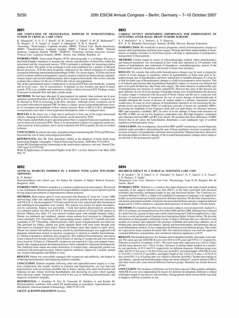
S230 20th ESICM Annual Congress – Berlin, Germany – 7–10 October 2007
0891THE USEFULNESS OF TRANSCRANIAL DOPPLER IN SUPRATENTORIALTUMOR IN CRITICAL CARE UNITS
K. Gharagozli1, S. G. U. I. Sharifi2, H. R. Jamaati3, A. Salimi4, S. M. R. Hashemian*5,M. Shojaee1, A. A. Asgari6, Z. Asgari2, S. Jabbarpour7, N. B. A. Danesh8
1Neurology, 2Neurosurgery, Loghman hospital, SBMU, 3Critical Care, Masih daneshvary,SBMU, 4Anesthesiology, Loghman hospital, SBMU, 5Critical Care, SBMU, 6Internalmedicine, Loghman hospital, SBMU, TEHRAN, 7Neurology, kerman university, kerman,8Critical Care, Loghman hospital, SBMU, Tehran, Iran (Islamic Republic of)
INTRODUCTION. Transcranial Doppler (TCD) ultrasonography is a technique that uses ahand-held Doppler transducer to measure the velocity and pulsatility of blood flow within theintracranial and the extracranial arteries. TCD is primarily a technique for measuring relativechanges in flow. The utility of the technique is now well established for a number of differentdisease processes. TCD has been frequently employed for the clinical evaluation of cerebralvasospasm following subarachnoid haemorrhage (SAH). To a lesser degree, TCD has also beenused to evaluate cerebral autoregulatory capacity, monitor cerebral circulation during cardiopul-monary bypass and carotid endarterectomies and to diagnose brain death. This review criticallyevaluates the evidence for the use of TCD in the critical care population.One of the supratentorial tumors complications is increased ICP that cause headache, vomitingand in severe cases , loss of consciousness. It depends on size, location and speed of tumorgrowth. TCD is an available and noninvasive technic to detect increased ICP. Findings consistof decrease in diastolic flow and spiky systolic peaks.
METHODS. We had been allocated 42 neurosurgical icu patients ,then estimate pre & postoperation cerebral blood flow & MCA,PCA,ACA velocities. In post operation, vasospasm canbe detected in TCD as increasing in the flow velocities. Although severe vasospasm can beassociated with reduced regional CBF. So there is a linear, inverse relationship between sever-ity of vasospasm and amplitude of flow velocity increase. Ratio of MCA to ICA ;distinguishvasospsm(ratio more than 3) from hyperemia.(hemispheric index or Lindegard ratio).
RESULTS. If the tumor size is large and its perfusion is from one of the major intracranialarteries, changing in blood flow in those arteries can be detected by TCD.After trauma and probably in post operation patient there is impaired brain auto regulation ,thiscan be observed as increased PSV. There is no documented evidence for impaired cerebrovas-cular reactivity in these patients, but it can be evaluated with TCD to predict the risk of cerebralischemia.
CONCLUSION. In critical care units, neurophysiological monitoring like TCD and EEG havedecreased the risk of many neurosurgical procedures.
REFERENCE(S). Kuo JR, Time dependent validity in the diagnosis of brain death usingtranscranial Doppler sonography. J Neurol Neurosurg Psychiatry. 2006 May;77(5):646-9.Jordan KG,Neurophysiologic monitoring in the neuroscience intensive care unit. Neurol Clin.1995 Aug;13(3):579-626.White H,Applications of transcranial Doppler in the ICU: a review. Intensive Care Med. 2006May 10
0892ATYPICAL DIABETES INSIPIDUS IN A PATIENT WITH GAINT PITUATORYADENOMA.
K. N. Reddy*Neuroanaesthesia and critical care, Sri Sathya Sai Institute of Higher Medical Sciences,Bangalore, India
INTRODUCTION. Diabetes insipidus is a common complication in neurosurgery. We presenta case of pituatory adenoma operated and developed diabetes insipidus in post operative periodhaving atypical presentation as hypernatremia without polyurea.
METHODS. Male patient aged 43 years presented with blurring of vision for 3 years. MRIshowed huge sellar and suprasellar tumor. Pre operatively patient had depressed sensoriumwith GCS of 11. On investigation T3,T4 and cortisol levels were reduced and other biochemicaland radiological investigations were normal. This patient was posted for tumor decompres-sion by craniotomy. Surgery was uneventful, 1 week later patient deteriorated in sensorium,on evaluation patient was dull with GCS 8, biochemical reduced T3,T4 and TSH, patient oneltroxin 100mcg once daily, CT scan showed residual tumor with multiple thalamic infarct.Patient was intubated and ventilated, patients serum sodium level increased to 160mmol/lt,serum osmolality 336.11mosm/kg with corresponding urinary sodium 51.5 mmol/lt and urineosmolality of 327.75mosm/kg, without polyurea, other causes of hypernatremia was ruled out.This persisted for 10days, patient was given adequate fluids with extra water through rylestube based on calculated water deficit. Patient developed sepsis then landed in septic shock.Patient was treated with antibiotic based on sensitivity and haemodynamic was supported withdopamine initially,then started on injection vasopressin as infusion to stabilise haemodynam-ics. Patient responded to antibiotic and vasopressin, This stabilised haemodynamic and sepsis,serum sodium levels became normal with in 8 hours of starting of vasopressin and even reachedlower levels by 24 hours to 128mmol/lt. vasopressin was tapered in 2 days and stopped, imme-diately after stopping patient developed polyurea which responded to intranasal desmopressin.This stabilized urine output and serum electrolytes to normal range, subsequently patient wascontinued with desmopressin spray. Patient required ventilatory support for 2 months, patientwas weaned subsequently and shifted to ward.
RESULTS. Patient was successfully managed with vasopressin and antibiotics, this helped instabilising haemodynamic and diagnosing diabetes insipidus.
CONCLUSION. Diabetes insipidus following sellar and suprasellar tumor surgery is a com-mon complication, this usually manifest as polyurea and hypernatremia, our case had onlyhypernatremia with out polyurea probably due to Sepsis causing early renal involvement andreducing out put. Tumor involving hypothalamus and destroying its centre which regulatethirst and urine output. Vasopressin was useful in this case in stabilizing haemodynamics anddiagnosis of diabetes insipidus.
REFERENCE(S). 1. Fumihiko N, Yuvi K, Tomoyuki H, Yukihino A, and Kinuko M.Myelodysplastic syndrome with central DI manifestating as hypodipsic, hypernatremia anddehydration; American journal of hematology: 2004;75:213-216.
GRANT ACKNOWLEDGEMENT. Institute
0893CARDIAC OUTPUT MONITORING IMPORTANCE FOR IMPROVEMENT OFOUTCOMES AFTER BASAL BRAIN TUMORS SURGERY
S. V. Madorskiy*, A. L. Parfenov, A. V. ZaharovaICU, N. N. Burdenko Neurosurgey Institute, RAMS, Moscow, Russian Federation
INTRODUCTION. We would like to discuss prognostic criteria of hemodynamics changes inpatients after hypothalamic and brain stem surgery. We hope that better understanding of mech-anisms of adaptive disorders in local brain lesions will help in optimization of postoperativemanagement of these patients.
METHODS. Cardiac output by means of echocardiographic method, others hemodynemicsand humoral parameters was investigated in first week after operation in 139 patients withtumors of hypothalamic area (adenomas of hypophysis, craniopharyngiomas, tumors of thebottom third ventricle) 148 patients with lesions of brain stem structures.
RESULTS. We consider that unfavorable hemodynamics changes may be used as prognosticcriteria of severe damage of regulatory centers in hypothalamus or brain stem such as hy-pophysotropic area of hypothalamus and dorso-medial part of medulla oblongata. It’s clear forus that favorable type of hemodynamics changes is a kind of postoperative stress reaction. Thisis of reduced type reaction after hypothalamo-hypophisis surgery, or reaction was delayed andgrew to its peak till third day after brain stem surgery. The main character of unfavorable typeof hemodynamic was decrease of cardiac output(CO). However the cause of this decrease arequite different. In case of severe damage of hypophysotropic area of hypothalamus the decreaseof cardiac output is connected with decreased of blood volume and the last is connected withdecrease of neurosecration of vasopressin. In case of severe damage of dorsomedial part ofmedulla oblongata the reason of decease of cardiac output is a primary neurogenic cardiacinsufficiency. In cause of severe damage of hypothalamic structures we see increasing the am-plitude power spectral density (PSD) of respiratory periodic of heard rate variability (HRV),decreasing the amplitude of low frequency peak and very high degree of coherence betweenHRV and respiratory variability (RV). In cause of severe damage of brain stem structures onthe PSD of HRV we can see low frequency components only. But high frequency componentand coherence between HRV and RV were absent. We postulate that these differences of HRVshowed that in all causes the hemodinamic disturbance a new inadequate type of cerebralregulation of hemodynamic forms.
CONCLUSION. Our investigations showed that HRV and RV monitoring is useful diagnosticmethods makes possible to determining the state of brain regulatory structures in patients afterexcision of tumors of hypothalamic and brain stem localization. Obtained data have allowed todevelop algorithms of intensive therapy of the revealed hemodynamics changes and to improvean outcome of the postoperative period.
0894DELIRIUM IMPACT IN A SURGICAL INTENSIVE CARE UNIT
R. B. Serafim*1, M. F. Dutra2, L. C. Vilarinho2, G. Santos2, N. C. Garcia2, J. E. Castro2,J. G. Pantoja3, F. Saddy1
1Respiratory Care Unit, 2Intensive Care Unit, 3Pneumology, Copa D Or Hospital, Rio deJaneiro, Brazil
INTRODUCTION. Delirium is a common but under-diagnosed and under-treated problemespecially in the surgical intensive care unit (SICU). It has been associated with increasedmorbidity and mortality, prolonged length of stay and functional decline. The Confusion As-sessment Method (CAM) is a simple tool (validated for portuguese language) to be applied forhealth care professionals previously trained for delirium detection. We describe characteristics,risk factors and related mortality of patients who presented delirium and also compared deliriumdiagnosed by CAM in relation to a question about presence of unclear thinks 24 hours before.
METHODS. Five hundred and fifty-four consecutive subjects were prospectively studied in aSICU of a tertiary care hospital between November 2005 and July 2006. Delirium was evaluatedin a daily basis by a group of nurses previously trained using the CAM tool applied twice a day,five days a week and also asked if patients have had unclear thinks 24 hours before. We describeinformation on demographics and initial severity of illness. Delirium data was collected as totalnumber of occurance and compared with: presence of bladder and central line catheters, use offeeding tube, prescription of narcoleptics, opioids and benzodiazepines; and laboratory data asusual inflammatory markers. It was compared with delirium or non-delirium groups. The resultsare expressed as mean standard deviation (SD). For statistical analysis was used chi-square forevaluated difference of proportion, and considered statistical significance p<0.05.
RESULTS. One hundred and sixty-five females and two hundred and fifty-nine males were stud-ied. The mean age and APACHE II score were 60.9±14.98 years and 9.95±5.17 respectively.Delirium occurred in 42 patients (7,0%). The mean onset after admission was 2.64±2.31daysand the mean duration was 1.70±1.16 days. Presence of unclear thinks resulted in a sensitiv-ity and specificity of 81% and 91% respectively for delirium diagnosis. Delirium group wereolder: 60,10±17,60 years vs 70.20±19,64 years (p=0.001); presented higher APACHE II score9.71±5.09 vs 12.95±5.27 (p=0,001); and higher length of stay 4.92±12.15 days vs 8.95±8.14days (p=0.038). Use of feeding tube was related to delirium (p=0,001). Neither prescription ofnarcoleptics, opioids and benzodiazepines drugs nor mean titulated C reactive protein (CRP-t)and white blood cells count showed difference between groups. Mortality was similar betweengroups.
CONCLUSION. The incidence of delirium was lower than expected. Older patients and higherAPACHE II score were independent risk factors for delirium development. Delirium is relatedwith higher length of stay but not with mortality. Asking patients about unclear thinks may helpto diagnose delirium in patients at risk.
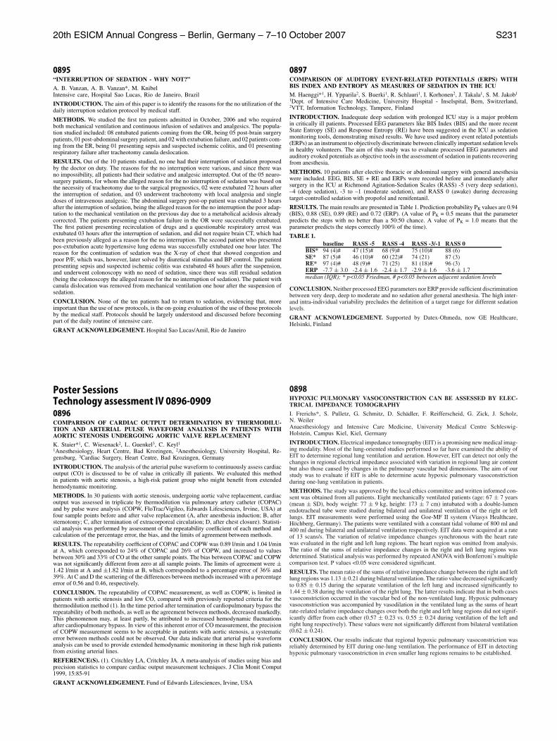
20th ESICM Annual Congress – Berlin, Germany – 7–10 October 2007 S231
0895“INTERRUPTION OF SEDATION - WHY NOT?”
A. B. Vanzan, A. B. Vanzan*, M. KnibelIntensive care, Hospital Sao Lucas, Rio de Janeiro, Brazil
INTRODUCTION. The aim of this paper is to identify the reasons for the no utilization of thedaily interruption sedation protocol by medical staff.
METHODS. We studied the first ten patients admitted in October, 2006 and who requiredboth mechanical ventilation and continuous infusion of sedatives and analgesics. The popula-tion studied included: 08 entubated patients coming from the OR, being 05 post-brain surgerypatients, 01 post-abdominal surgery patient, and 02 with extubation failure, and 02 patients com-ing from the ER, being 01 presenting sepsis and suspected ischemic colitis, and 01 presentingrespiratory failure after tracheotomy canula deslocation.
RESULTS. Out of the 10 patients studied, no one had their interruption of sedation proposedby the doctor on duty. The reasons for the no interruption were various, and since there wasno impossibility, all patients had their sedative and analgesic interrupted. Out of the 05 neuro-surgery patients, for whom the alleged reason for the no interruption of sedation was based onthe necessity of tracheotomy due to the surgical prognostics, 02 were extubated 72 hours afterthe interruption of sedation, and 03 underwent tracheotomy with local analgesia and singledoses of intravenous analgesic. The abdominal surgery post-op patient was extubated 3 hoursafter the interruption of sedation, being the alleged reason for the no interruption the poor adap-tation to the mechanical ventilation on the previous day due to a metabolical acidosis alreadycorrected. The patients presenting extubation failure in the OR were successfully extubated.The first patient presenting recirculation of drugs and a questionable respiratory arrest wasextubated 03 hours after the interruption of sedation, and did not require brain CT, which hadbeen previously alleged as a reason for the no interruption. The second patient who presentedpos-extubation acute hypertensive lung edema was successfully extubated one hour later. Thereason for the continuation of sedation was the X-ray of chest that showed congestion andpoor P/F, which was, however, later solved by diuretical stimulus and BP control. The patientpresenting sepsis and suspected ischemic colitis was extubated 48 hours after the suspension,and underwent colonoscopy with no need of sedation, since there was still residual sedation(being the colonoscopy the alleged reason for the no interruption of sedation). The patient withcanula dislocation was removed from mechanical ventilation one hour after the suspension ofsedation.
CONCLUSION. None of the ten patients had to return to sedation, evidencing that, moreimportant than the use of new protocols, is the on-going evaluation of the use of those protocolsby the medical staff. Protocols should be largely understood and discussed before becomingpart of the daily routine of intensive care.
GRANT ACKNOWLEDGEMENT. Hospital Sao Lucas/Amil, Rio de Janeiro
Poster SessionsTechnology assessment IV 0896-09090896COMPARISON OF CARDIAC OUTPUT DETERMINATION BY THERMODILU-TION AND ARTERIAL PULSE WAVEFORM ANALYSIS IN PATIENTS WITHAORTIC STENOSIS UNDERGOING AORTIC VALVE REPLACEMENT
K. Staier*1, C. Wiesenack2, L. Guenkel3, C. Keyl11Anesthesiology, Heart Centre, Bad Krozingen, 2Anesthesiology, University Hospital, Re-gensburg, 3Cardiac Surgery, Heart Centre, Bad Krozingen, Germany
INTRODUCTION. The analysis of the arterial pulse waveform to continuously assess cardiacoutput (CO) is discussed to be of value in critically ill patients. We evaluated this methodin patients with aortic stenosis, a high-risk patient group who might benefit from extendedhemodynamic monitoring.
METHODS. In 30 patients with aortic stenosis, undergoing aortic valve replacement, cardiacoutput was assessed in triplicate by thermodilution via pulmonary artery catheter (COPAC)and by pulse wave analysis (COPW, FloTrac/Vigileo, Edwards Lifesciences, Irvine, USA) atfour sample points before and after valve replacement (A, after anesthesia induction; B, aftersternotomy; C, after termination of extracorporeal circulation; D, after chest closure). Statisti-cal analysis was performed by assessment of the repeatability coefficient of each method andcalculation of the percentage error, the bias, and the limits of agreement between methods.
RESULTS. The repeatability coefficient of COPAC and COPW was 0.89 l/min and 1.04 l/minat A, which corresponded to 24% of COPAC and 26% of COPW, and increased to valuesbetween 30% and 33% of CO at the other sample points. The bias between COPAC and COPWwas not significantly different from zero at all sample points. The limits of agreement were ±1.42 l/min at A and ±1.82 l/min at B, which corresponded to a percentage error of 36% and39%. At C and D the scattering of the differences between methods increased with a percentageerror of 0.56 and 0.46, respectively.
CONCLUSION. The repeatability of COPAC measurement, as well as COPW, is limited inpatients with aortic stenosis and low CO, compared with previously reported criteria for thethermodilution method (1). In the time period after termination of cardiopulmonary bypass therepeatability of both methods, as well as the agreement between methods, decreased markedly.This phenomenon may, at least partly, be attributed to increased hemodynamic fluctuationsafter cardiopulmonary bypass. In view of this inherent error of CO measurement, the precisionof COPW measurement seems to be acceptable in patients with aortic stenosis, a systematicerror between methods could not be observed. Our data indicate that arterial pulse waveformanalysis can be used to provide extended hemodynamic monitoring in these high risk patientsfrom existing arterial lines.
REFERENCE(S). (1). Critchley LA, Critchley JA. A meta-analysis of studies using bias andprecision statistics to compare cardiac output measurement techniques. J Clin Monit Comput1999, 15:85-91
GRANT ACKNOWLEDGEMENT. Fund of Edwards Lifesciences, Irvine, USA
0897COMPARISON OF AUDITORY EVENT-RELATED POTENTIALS (ERPS) WITHBIS INDEX AND ENTROPY AS MEASURES OF SEDATION IN THE ICU
M. Haenggi*1, H. Ypparila2, S. Buerki1, R. Schlauri1, I. Korhonen2, J. Takala1, S. M. Jakob1
1Dept. of Intensive Care Medicine, University Hospital - Inselspital, Bern, Switzerland,2VTT, Information Technology, Tampere, Finland
INTRODUCTION. Inadequate deep sedation with prolonged ICU stay is a major problemin critically ill patients. Processed EEG parameters like BIS Index (BIS) and the more recentState Entropy (SE) and Response Entropy (RE) have been suggested in the ICU as sedationmonitoring tools, demonstrating mixed results. We have used auditory event related potentials(ERPs) as an instrument to objectively discriminate between clinically important sedation levelsin healthy volunteers. The aim of this study was to evaluate processed EEG parameters andauditory evoked potentials as objective tools in the assessment of sedation in patients recoveringfrom anesthesia.
METHODS. 10 patients after elective thoracic or abdominal surgery with general anesthesiawere included. EEG, BIS, SE + RE and ERPs were recorded before and immediately aftersurgery in the ICU at Richmond Agitation-Sedation Scales (RASS) -5 (very deep sedation),–4 (deep sedation), -3 to –1 (moderate sedation), and RASS 0 (awake) during decreasingtarget-controlled sedation with propofol and remifentanil.
RESULTS. The main results are presented in Table 1. Prediction probability PK values are 0.94(BIS), 0.88 (SE), 0.89 (RE) and 0.72 (ERP). (A value of PK = 0.5 means that the parameterpredicts the steps with no better than a 50:50 chance. A value of PK = 1.0 means that theparameter predicts the steps correctly 100% of the time).
TABLE 1.baseline RASS -5 RASS -4 RASS -3/-1 RASS 0
BIS* 94 (4)# 47 (15)# 68 (9)# 75 (10)# 88 (6)SE* 87 (5)# 46 (10)# 60 (22)# 74 (21) 87 (3)RE* 97 (4)# 48 (9)# 71 (25) 81 (18)# 96 (3)ERP -7.7 ± 3.0 -2.4 ± 1.6 -2.4 ± 1.7 -2.9 ± 1.6 -3.6 ± 1.7median (IQR); * p<0.05 Friedman, # p<0.05 between adjacent sedation levels
CONCLUSION. Neither processed EEG parameters nor ERP provide sufficient discriminationbetween very deep, deep to moderate and no sedation after general anesthesia. The high inter-and intra-individual variability precludes the definition of a target range for different sedationlevels.
GRANT ACKNOWLEDGEMENT. Supported by Datex-Ohmeda, now GE Healthcare,Helsinki, Finland
0898HYPOXIC PULMONARY VASOCONSTRICTION CAN BE ASSESSED BY ELEC-TRICAL IMPEDANCE TOMOGRAPHY
I. Frerichs*, S. Pulletz, G. Schmitz, D. Schadler, F. Reifferscheid, G. Zick, J. Scholz,N. WeilerAnaesthesiology and Intensive Care Medicine, University Medical Centre Schleswig-Holstein, Campus Kiel, Kiel, Germany
INTRODUCTION. Electrical impedance tomography (EIT) is a promising new medical imag-ing modality. Most of the lung-oriented studies performed so far have examined the ability ofEIT to determine regional lung ventilation and aeration. However, EIT can detect not only thechanges in regional electrical impedance associated with variation in regional lung air contentbut also those caused by changes in the pulmonary vascular bed dimensions. The aim of ourstudy was to evaluate if EIT is able to determine acute hypoxic pulmonary vasoconstrictionduring one-lung ventilation in patients.
METHODS. The study was approved by the local ethics committee and written informed con-sent was obtained from all patients. Eight mechanically ventilated patients (age: 67 ± 7 years(mean ± SD), body weight: 77 ± 9 kg, height: 173 ± 7 cm) intubated with a double-lumenendotracheal tube were studied during bilateral and unilateral ventilation of the right or leftlungs. EIT measurements were performed using the Goe-MF II system (Viasys Healthcare,Hochberg, Germany). The patients were ventilated with a constant tidal volume of 800 ml and400 ml during bilateral and unilateral ventilation respectively. EIT data were acquired at a rateof 13 scans/s. The variation of relative impedance changes synchronous with the heart ratewas evaluated in the right and left lung regions. The heart region was omitted from analysis.The ratio of the sums of relative impedance changes in the right and left lung regions wasdetermined. Statistical analysis was performed by repeated ANOVA with Bonferroni’s multiplecomparison test. P values <0.05 were considered significant.
RESULTS. The mean ratio of the sums of relative impedance change between the right and leftlung regions was 1.13 ± 0.21 during bilateral ventilation. The ratio value decreased significantlyto 0.85 ± 0.15 during the separate ventilation of the left lung and increased significantly to1.44 ± 0.38 during the ventilation of the right lung. The latter results indicate that in both casesvasoconstriction occurred in the vascular bed of the non-ventilated lung. Hypoxic pulmonaryvasoconstriction was accompanied by vasodilation in the ventilated lung as the sums of heartrate-related relative impedance changes over both the right and left lung regions did not signif-icantly differ from each other (0.57 ± 0.23 vs. 0.55 ± 0.24 during ventilation of the left andright lung respectively). These values were not significantly different from bilateral ventilation(0.62 ± 0.24).
CONCLUSION. Our results indicate that regional hypoxic pulmonary vasoconstriction wasreliably determined by EIT during one-lung ventilation. The performance of EIT in detectinghypoxic pulmonary vasoconstriction in even smaller lung regions remains to be established.
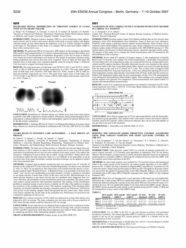
S232 20th ESICM Annual Congress – Berlin, Germany – 7–10 October 2007
0899DECREASED SPATIAL DISTRIBUTION OF VIBRATION ENERGY IN LUNGSWITH ACUTE HEART FAILURE
Z. Wang*1, R. P. Dellinger1, S. Trzeciak1, S. Jean1, K. N. Gruber2, R. Arnold2, J. E. Parrillo1
1Division of Critical Care Medicine, 2Emergency Department, Robert Wood Johnson Schoolof Medicine and Cooper University Hospital, Camden, United States
INTRODUCTION. Vibration response imaging (VRI) is a novel technology using sophisti-cated software and 34 surface skin sensors placed on the back to record, analyze and displayvibration energy of lung sounds during the respiratory cycle. It is a likely surrogate of airflowin the lungs (1). The purpose of this study is to compare VRI in acute heart failure (AHF) tothose with a normal chest x-ray.
METHODS. We performed VRI in 9 consecutive AHF subjects in the emergency department(ED) and ICU and compared them to 12 controls (without pulmonary disease). Recordings wereperformed over 20 second periods of respiration. Respiratory cycles free of noise or artifactswere chosen for analysis and images were analyzed. The images at maximum vibration energyduring inspiration were chosen and areas were compared. Areas of right and left lungs andregional areas of both lungs were calculated digitally using the program Image J. Statisticalt-test was used to compare total mean lung areas.
RESULTS. The total mean areas of both lungs were 56756.9± 8231.4 and 76170.2 ±3843.2(mean ± SD) in AHF patients and normals, respectively (p = 0.01). The mean upper regionareas of both lungs were 35220.6± 3497.9and 36423.5± 2398.3 (mean ± SD) in AHF pa-tient and normals, respectively (p = 0.11). The mean lower region areas of both lungs were17119.7±6461.1 and 39634.5± 4651.1 (mean ± SD) in AHF subjects and normals, respectively(p = 0.01). (Figure 1).
CONCLUSION. Distribution of vibration energy was shifted away from lower lung regionsin patients with AHF compared to normal subjects. Pulmonary edema predominating in lowerlung regions or pleural effusion (4 subjects had radiographic support for pleural effusion) maybe the physiologic explanation.
REFERENCE(S). 1. Dellinger RP, Jean S, Cinel I, et al. Regional distribution of acoustic-basedlung vibration as a function of mechanical ventilation mode. Crit Care. 2007 Feb 22;11(1):R26
0900ALARM RULES IN INTENSIVE CARE MONITORING – A DATA DRIVEN AP-PROACH
W. Sieben*1, S. Siebig2, C. Wrede2, M. Imhoff3, U. Gather1
1Department of Statistics, University of Dortmund, Dortmund, 2Department of InternalMedicine I, University Hospital Regensburg, Regensburg, 3Department for Medical Infor-matics, Biometrics and Epidemiology, Ruhr-University Bochum, Bochum, Germany
INTRODUCTION. The high rate of false positive or clinically not relevant alarms from pa-tient monitors is still a concern in critical care. False alarms are an issue even with the latestgeneration of monitoring devices and can cause mistrust in the alarm system. This reduces thecaregivers’ willingness to react to them, the so-called “crying wolf effect”. Better alarm rulesare needed to reduce the false alarm rate. Since it is very difficult, if not impossible, to set upuniversal rules from expert knowledge, machine learning techniques can be applied to deductrules from annotated data.
METHODS. Complete high-resolution monitoring data was acquired from 55 critically illpatients on the medical ICU of the University Hospital Regensburg. All alarms were annotatedas “true” or “false” by an intensivist. New alarm rules were learned from the data by a classi-fication method called “Random Forrest”. A Random Forrest is an ensemble of decision treeswhich “vote” for the class membership of an alarm situation. Usually, a situation is classifiedas not alarm relevant, if more than c=50% of the trees vote for this. Misclassification of analarm relevant situation as not alarm relevant may be considered more dangerous than viceversa. Therefore, the probability of such classification needs to be controlled. The alarm systemmust achieve certain sensitivity with maximum specificity. The analogy of this classificationproblem to statistical testing is used to choose a suitable cut off value c that enables us to adjustthe sensitivity of the resulting alarm system. The database comprising 2749 annotated alarmsituations was divided randomly into learning, estimation and test sets 1050 times. 1050 forestswere grown on the learning samples which incorporated the new alarm rules. These forestswere used to calculate the cut off value on the estimation sets. Performance of each forest wasevaluated by applying it to the test set.
RESULTS. For a chosen sensitivity of 95% which was achieved on average false alarms werereduced by 46% on average. The same evaluation was also done with a chosen sensitivity of98% where the false alarms could be reduced by 30% on average.
CONCLUSION. In our study the use of a Random Forrest approach led to a reduction of falsealarms from a standard patient monitor by 46% and 30% on average with a chosen sensitivityof 95% and 98% respectively. Such data driven approach may improve future alarms systemsby enhancing specificity while maintaining adequate sensitivity.
GRANT ACKNOWLEDGEMENT. Funded, in part, by the DFG (SFB 475).
0901VALIDATION OF NEW CARDIAC OUTPUT ULTRASOUND DILUTION METHODIN CARDIAC ICU PATIENTS
A. A. Eremenko*, P. N. SafarovCardiac ICU, National Research Center of Surgery Russian Academy of Medical Science,Moscow, Russian Federation
INTRODUCTION. Existing cardiac output (CO) dilution methods that are less invasive thanpulmonary artery (PA) thermodilution have limitations. Some of them use toxic substances(LiDCO); some require a dedicated arterial catheter inserted (Pulsion). None of them can beroutinely used in small children. First animal (rats, pigs, sheep) validation of a novel ultrasounddilution cardiac output (COud) method was presented at the 2006 ESICM meeting [1]. Thismethod utilizes the decrease in blood ultrasound velocity caused by injecting isotonic saline.The purpose of this study was to compare the COud measurements with CO measured by PAthermodilution (COt) in humans.
METHODS. Twelve adult ICU patients (10–bypass surgery; 2–after implantation of ImpellaRecover LP 5.0 System) were studied. For COud measurements, a disposable extracorporealAV loop filled with 3 ml of heparinized saline was connected between an existing radial arterycatheter and PA catheter introducer. Reusable ultrasound sensors were clamped on the arterialand venous limbs of the loop. A peristaltic pump (Nipro, Japan) was used to circulate the bloodfrom the artery to the vein at 8-12 ml/min for 5-7 min. Two to three (later averaged) COudmeasurements (HCP101, Transonic Systems Inc., USA) were obtained by injecting 20-25 ml ofbody temperature isotonic saline into the venous limb of the AV loop. At the end, the system wasflushed with heparinized saline until the next measurement session. Five COt measurementswere obtained by thermodilution (Nihon Koden, Japan). Three readings were averaged (largestand the smallest were ignored). For comparison, COud measurements were started within 3-5minutes after thermodilution measurements.
RESULTS. A total of 31 comparison measurements were obtained. Correlation was R2= 0.88.Linear regression was COud = 1.06 COt - 0.34 L/min. Bland-Altman test (Fig 1) did not showa significant bias (-0.01 L/min).
CONCLUSION. First clinical comparison of CO by ultrasound dilution with PA thermodilu-tion produced good agreement. This method works with central venous and arterial cathetersroutinely used in ICU patients, utilizes innocuous indicator, thus can be used with patients ofany age and at any time in the treatment.
REFERENCE(S). 1. Gleed RD, et al. ESICM, 2006; 32: S172.
0902REFINING THE ENHANCED MODEL PREDICTIVE CONTROL ALGORITHMUSING TIME VARIANT SAMPLING FOR TIGHT GLYCEMIC CONTROL INTHE ICU
S. J. Van Cromphaut*1, I. van den Heuvel1, D. Vlasselaers1, P. J. Wouters1, L. Chassin2,G. Wilinska2, R. Hovorka2, G. Van den Berghe1
1Intensive Care Medicine, University Hospital, Leuven, Belgium, 2Paediatrics, Addenbrooke’sHospital, Cambridge, United Kingdom
INTRODUCTION. Tight glycemic control (TGC) in critically ill patients significantly im-proves clinical outcome. (1,2) Currently used algorithms to implement TGC are not alwaysefficient. We developed a second version of a fully automated algorithm based on an enhancedmodel predictive controller (eMPC2), following the comparison between the first eMPC withthe routinely used insulin algorithm in the ICU.
METHODS. In a first study, patients were included for 72 consecutive hours and randomizedeither to the control group (n=10) treated by our routine insulin algorithm or to the eMPC1group (n=10). A follow-up study evaluated an adapted eMPC2 algorithm in 16 patients for 120consecutive hours. Target range for blood glucose (BG) was 80–110 mg/dl for all groups. Safetywas assessed by the number of hypoglycemic events (BG<40mg/dl). Efficacy was assessed bycalculating time-to-BG-target (BG<110mg/dl), hyperglycemic index (HGI) (3) and samplingfrequency (SF).
RESULTS. There were no hypoglycemic events and time-to-BG-target was not significantlydifferent in the 3 study groups. HGI was significantly higher at day 1 (p<0.05; Table 1), but notstatistically different among the 3 groups at any time point. HGI was lower than the target of 27mg/dl in all cases. SF was significantly higher at any time point in the eMPC1 group, whereasthe eMPC2 group showed the same pattern as the standard group (p<0.05; Table 1). HGI andSF remained low on day 4 and 5 of the eMPC2 study.
TABLE 1.HGI (mg/dl) HGI (mg/dl) HGI (mg/dl) SF (#/d) SF (#/d) SF (#/d)Standard eMPC1 eMPC2 Standard eMPC1 eMPC2
Day 1 11.4 (± 2.1) 14.4 (± 2.4) 11.0 (± 3.2) 12 (± 1) 18 (± 1)* 13 (± 1)Day 2 1.5 (± 1.0)* 1.4 (± 0.5)* 4.7 (± 1.1)* 8 (± 1)* 11 (± 1)£ 8 (± 1)*Day 3 1.1 (± 0.6)* 3.4 (± 1.2)* 3.9 (± 1.2)* 8 (± 2)* 12 (± 2)§ 7 (± 1)**p<0.05 vs. Standard day 1; £p<0.05 vs. Standard day 2;§p<0.05 vs. Standard day 3
CONCLUSION. Use of eMPC in the ICU is safe and efficient in patients with prolongedmechanical ventilation. The refined algorithm eMPC2 resulted in a decreased workload, com-parable to the use of our standard ICU insulin protocol. eMPC2 is a reliable tool for theimplementation of TGC in the ICU.
REFERENCE(S). 1 Van den Berghe G, NEJM 2001;345:1359-67. 2 Van den Berghe G, NEJM2006;354:449-61. 3 Vogelzang, CritCare 2004; 8:122
GRANT ACKNOWLEDGEMENT. EC 6th Framework Program, ref 506965 (Clinicip)
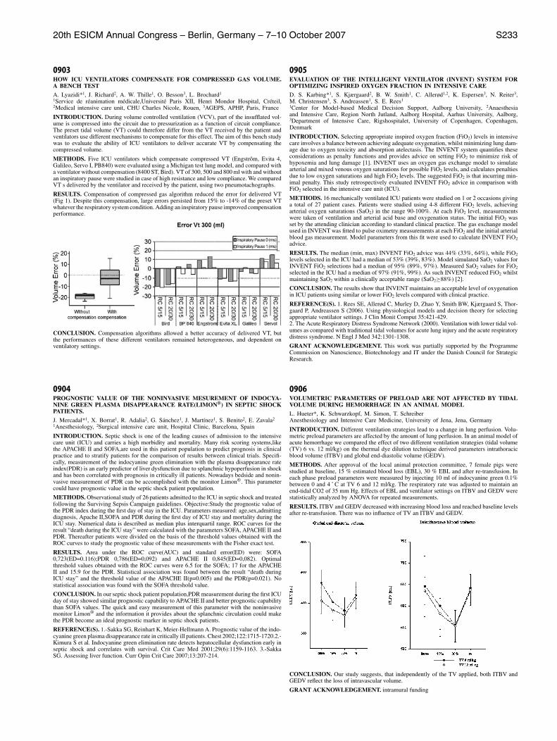
20th ESICM Annual Congress – Berlin, Germany – 7–10 October 2007 S233
0903HOW ICU VENTILATORS COMPENSATE FOR COMPRESSED GAS VOLUME.A BENCH TEST
A. Lyazidi*1, J. Richard2, A. W. Thille1, O. Besson3, L. Brochard1
1Service de reanimation medicale,Universite Paris XII, Henri Mondor Hospital, Creteil,2Medical intensive care unit, CHU Charles Nicole, Rouen, 3AGEPS, APHP, Paris, France
INTRODUCTION. During volume controlled ventilation (VCV), part of the insufflated vol-ume is compressed into the circuit due to pressurization as a function of circuit compliance.The preset tidal volume (VT) could therefore differ from the VT received by the patient andventilators use different mechanisms to compensate for this effect. The aim of this bench studywas to evaluate the ability of ICU ventilators to deliver accurate VT by compensating thecompressed volume.
METHODS. Five ICU ventilators which compensate compressed VT (Engstrom, Evita 4,Galileo, Servo I, PB840) were evaluated using a Michigan test lung model, and compared witha ventilator without compensation (8400 ST, Bird). VT of 300, 500 and 800 ml with and withoutan inspiratory pause were studied in case of high resistance and low compliance. We comparedVT s delivered by the ventilator and received by the patient, using two pneumotachographs.
RESULTS. Compensation of compressed gas algorithm reduced the error for delivered VT(Fig 1). Despite this compensation, large errors persisted from 15% to -14% of the preset VTwhatever the respiratory system condition. Adding an inspiratory pause improved compensationperformance.
CONCLUSION. Compensation algorithms allowed a better accuracy of delivered VT, butthe performances of these different ventilators remained heterogeneous, and dependent onventilatory settings.
0904PROGNOSTIC VALUE OF THE NONINVASIVE MESUREMENT OF INDOCYA-NINE GREEN PLASMA DISAPPEARANCE RATE(LIMON®) IN SEPTIC SHOCKPATIENTS.
J. Mercadal*1, X. Borrat1, R. Adalia2, G. Sanchez1, J. Martınez1, S. Benito2, E. Zavala2
1Anesthesiology, 2Surgical intensive care unit, Hospital Clinic, Barcelona, Spain
INTRODUCTION. Septic shock is one of the leading causes of admission to the intensivecare unit (ICU) and carries a high morbidity and mortality. Many risk scoring systems,likethe APACHE II and SOFA,are used in this patient population to predict prognosis in clinicalpractice and to stratify patients for the comparison of results between clinical trials. Specifi-cally, measurement of the indocyanine green elimination with the plasma disappearance rateindex(PDR) is an early predictor of liver dysfunction due to splanchnic hypoperfusion in shockand has been correlated with prognosis in critically ill patients. Nowadays bedside and nonin-vasive measurement of PDR can be accomplished with the monitor Limon®. This parametercould have prognostic value in the septic shock patient population.
METHODS. Observational study of 26 patients admitted to the ICU in septic shock and treatedfollowing the Surviving Sepsis Campaign guidelines. Objective:Study the prognostic value ofthe PDR index during the first day of stay in the ICU. Parameters measured: age,sex,admittingdiagnosis, Apache II,SOFA and PDR during the first day of ICU stay and mortality during theICU stay. Numerical data is described as median plus interquartil range. ROC curves for theresult “death during the ICU stay” were calculated with the parameters SOFA, APACHE II andPDR. Thereafter patients were divided on the basis of the threshold values obtained with theROC curves to study the prognostic value of these measurements with the Fisher exact test.
RESULTS. Area under the ROC curve(AUC) and standard error(ED) were: SOFA0,723(ED=0.116);PDR 0,786(ED=0,092) and APACHE II 0,845(ED=0,082). Optimalthreshold values obtained with the ROC curves were 6.5 for the SOFA; 17 for the APACHEII and 15.9 for the PDR. Statistical association was found between the result “death duringICU stay” and the threshold value of the APACHE II(p=0.005) and the PDR(p=0.021). Nostatistical association was found with the SOFA threshold value.
CONCLUSION. In our septic shock patient population,PDR measurement during the first ICUday of stay showed similar prognostic capability to APACHE II and better prognostic capabilitythan SOFA values. The quick and easy measurement of this parameter with the noninvasivemonitor Limon® and the information it provides about the splanchnic circulation could makethe PDR become an ideal prognostic marker in septic shock patients.
REFERENCE(S). 1.-Sakka SG, Reinhart K, Meier-Hellmann A. Prognostic value of the indo-cyanine green plasma disappearance rate in critically ill patients. Chest 2002;122:1715-1720.2.-Kimura S et al. Indocyanine green elimination rate detects hepatocellular dysfunction early inseptic shock and correlates with survival. Crit Care Med 2001;29(6):1159-1163. 3.-SakkaSG. Assessing liver function. Curr Opin Crit Care 2007;13:207-214.
0905EVALUATION OF THE INTELLIGENT VENTILATOR (INVENT) SYSTEM FOROPTIMIZING INSPIRED OXYGEN FRACTION IN INTENSIVE CARE
D. S. Karbing*1, S. Kjærgaard2, B. W. Smith1, C. Allerød1,2, K. Espersen3, N. Reiter3,M. Christensen3, S. Andreassen1, S. E. Rees1
1Center for Model-based Medical Decision Support, Aalborg University, 2Anaesthesiaand Intensive Care, Region North Jutland, Aalborg Hospital, Aarhus University, Aalborg,3Department of Intensive Care, Rigshospitalet, University of Copenhagen, Copenhagen,Denmark
INTRODUCTION. Selecting appropriate inspired oxygen fraction (FiO2) levels in intensivecare involves a balance between achieving adequate oxygenation, whilst minimizing lung dam-age due to oxygen toxicity and absorption atelectasis. The INVENT system quantifies theseconsiderations as penalty functions and provides advice on setting FiO2 to minimize risk ofhypoxemia and lung damage [1]. INVENT uses an oxygen gas exchange model to simulatearterial and mixed venous oxygen saturations for possible FiO2 levels, and calculates penaltiesdue to low oxygen saturations and high FiO2 levels. The suggested FiO2 is that incurring min-imal penalty. This study retrospectively evaluated INVENT FiO2 advice in comparison withFiO2 selected in the intensive care unit (ICU).
METHODS. 16 mechanically ventilated ICU patients were studied on 1 or 2 occasions givinga total of 27 patient cases. Patients were studied using 4-8 different FiO2 levels, achievingarterial oxygen saturations (SaO2) in the range 90-100%. At each FiO2 level, measurementswere taken of ventilation and arterial acid base and oxygenation status. The initial FiO2 wasset by the attending clinician according to standard clinical practice. The gas exchange modelused in INVENT was fitted to pulse oximetry measurements at each FiO2 and the initial arterialblood gas measurement. Model parameters from this fit were used to calculate INVENT FiO2advice.
RESULTS. The median (min, max) INVENT FiO2 advice was 44% (33%, 64%), while FiO2levels selected in the ICU had a median of 53% (39%, 83%). Model simulated SaO2 values forINVENT FiO2 selections had a median of 95% (89%, 97%). Measured SaO2 values for FiO2selected in the ICU had a median of 97% (91%, 99%). As such INVENT reduced FiO2 whilstmaintaining SaO2 within a clinically acceptable range (SaO2≥88%) [2].
CONCLUSION. The results show that INVENT maintains an acceptable level of oxygenationin ICU patients using similar or lower FiO2 levels compared with clinical practice.
REFERENCE(S). 1. Rees SE, Allerød C, Murley D, Zhao Y, Smith BW, Kjærgaard S, Thor-gaard P, Andreassen S (2006). Using physiological models and decision theory for selectingappropriate ventilator settings. J Clin Monit Comput 35:421-429.2. The Acute Respiratory Distress Syndrome Network (2000). Ventilation with lower tidal vol-umes as compared with traditional tidal volumes for acute lung injury and the acute respiratorydistress syndrome. N Engl J Med 342:1301-1308.
GRANT ACKNOWLEDGEMENT. This work was partially supported by the ProgrammeCommission on Nanoscience, Biotechnology and IT under the Danish Council for StrategicResearch.
0906VOLUMETRIC PARAMETERS OF PRELOAD ARE NOT AFFECTED BY TIDALVOLUME DURING HEMORRHAGE IN AN ANIMAL MODEL
L. Hueter*, K. Schwarzkopf, M. Simon, T. SchreiberAnesthesiology and Intensive Care Medicine, University of Jena, Jena, Germany
INTRODUCTION. Different ventilation strategies lead to a change in lung perfusion. Volu-metric preload parameters are affected by the amount of lung perfusion. In an animal model ofacute hemorrhage we compared the effect of two different ventilation strategies (tidal volume(TV) 6 vs. 12 ml/kg) on the thermal dye dilution technique derived parameters intrathoracicblood volume (ITBV) and global end-diastolic volume (GEDV).
METHODS. After approval of the local animal protection committee, 7 female pigs werestudied at baseline, 15 % estimated blood loss (EBL), 30 % EBL and after re-transfusion. Ineach phase preload parameters were measured by injecting 10 ml of indocyanine green 0.1%between 0 and 4 ˚ C at TV 6 and 12 ml/kg. The respiratory rate was adjusted to maintain anend-tidal CO2 of 35 mm Hg. Effects of EBL and ventilator settings on ITBV and GEDV werestatistically analyzed by ANOVA for repeated measurements.
RESULTS. ITBV and GEDV decreased with increasing blood loss and reached baseline levelsafter re-transfusion. There was no influence of TV an ITBV and GEDV.
CONCLUSION. Our study suggests, that independently of the TV applied, both ITBV andGEDV reflect the loss of intravascular volume.
GRANT ACKNOWLEDGEMENT. intramural funding
S234 20th ESICM Annual Congress – Berlin, Germany – 7–10 October 2007
0907MONITORING OF CARDIAC OUTPUT CHANGES INDUCED BY DIFFERENTDEGREES OF INTRA ABDOMINAL PRESSURE.
F. Franchi*1, G. Sbrana2, V. Tammi2, S. M. Romano3, R. Madonna2, P. Giomarelli1,S. Scolletta1
1Anesthesia and ICU, University of Siena, Siena, 2Anesthesia and ICU, Hospital ofMisericordia, Grosseto, 3Critical Care Unit, University of Florence, Florence, Italy
INTRODUCTION. Intra-abdominal hypertension (IAH) and abdominal compartment syn-drome (ACS) have been increasingly recognized in critically ill patients over the past decade.A prolonged IAH at greater than 25 mmHg can produce significant morbidity and mortality(1). Cardiac output (CO) can be influenced by IAH, and a low CO may lead to multiorgandysfunction and failure. The aim of this study was to compare three different CO monitoringmethods during different degrees of intra abdominal pressure.
METHODS. We investigated the PRAM (Pressure Recording Analytical Method) by compar-ing its CO (PRAM-CO) with paired measurements obtained by the Fick method (Fick-CO),and by the standard thermodilution (ThD-CO) during various CO changes in a swine model.PRAM is a pulse contour method that has been studied in humans and animals, and does notneed for calibrating factors (2). Ten pigs (mean body weight 39 ± 2.4 kg) were monitored witha pulmonary artery catheter and a femoral artery catheter at T0 (baseline), at T1, T2, and T3,(after 10, 20, and 30 minutes of IAP=15 mmHg, respectively), at T4 (IAP=20 mmHg), and at T5(IAP=30 mmHg). IAP was induced by pneumoperitoneum by CO2 insufflation. Bland-Altmanstatistical analysis was used.
RESULTS. 180 paired cardiac output values over a range of ThD-CO of 2.5 to 6.4 litre/minresulted. At T0 mean ThD-CO value was 5.5 litre/min. From T1 to T5 CO mean values were 5.0,4.6, 4.1, 3.6, 3.2 litre/min, respectively. The analysis of variance showed a significant decreaseof CO values from T0 to T5 (p<0.05). We found close agreements between the techniques.Mean bias between PRAM-CO and Fick-CO was +0.02 litre/min (precision of ± 0.38 litre/min;95% limits of agreement from -0.54 to +0.57). Mean bias between PRAM-CO and ThD-COwas +0.07 litre/min (precision of ± 0.56 litre/min; 95% limits of agreement from 0.44 to +0.67).Similar results between ThD-CO and Fick-CO were found.
CONCLUSION. We demonstrated that under different degrees of IAH, CO can be interchange-able calculated with invasive or minimally invasive monitoring systems. In this porcine modelPRAM seemed easy to use and provided CO continuously during the different measurements.This is the first study assessing PRAM in this clinical setting.
REFERENCE(S). (1) Melbrain ML, et al. Results from the International Conference of Ex-perts on Intra-abdominal Hypertension and Abdominal Compartment Syndrome. I. Definitions.Intensive Care Med 2006.(2) Scolletta S, et al. Pressure recording analytical method (PRAM) for measurement of cardiacoutput during various haemodynamic states. Br J Anaesth. 2005
0908DISTRIBUTION OF LUNG VIBRATIONS DURING USE OF VIBRATORYACAPELLA DEVICE
I. Cinel*1, S. Jean1, C. Tay1, I. Gratz2, R. P. Dellinger1
1Critical Care Medicine, 2Anesthesia, Cooper University Hospital, Camden, United States
INTRODUCTION. Vibratory devices work by oscillating vibrations that travel into the lungs.The Acapella® is a small hand-held vibratory device that combines the resistive features ofthe positive expiratory pressure (PEP) and the vibratory features of a flutter valve to mobilizesecretions in the airway. VRI is a novel dynamic imaging technique that measures vibrationenergy of lung sounds generated during respiration. In this study, our aim is to determine, usingthe VRI, what regions of the lungs receive the most vibrations when the Acapella is being used.
METHODS. A 20 second VRI recording was performed on a healthy volunteer during normalbreathing (first three breaths) and while using the Acapella device (last four breaths). The VRIrecordings were obtained in 20 second periods of respiration. Dynamic digital images andnumerical raw values for vibration energy are analyzed and compared any regions of interest.
RESULTS. There was an over sixty fold increase in total vibration intensity during expirationwhen Acapella was used. VRI images at maximal expiration during normal breathing (Fig. 1A)was compared to images obtained while using the Acapella (Fig. 1B). Areas of highest vibrationintensity are black. When the distribution of expiratory vibration is examined, it appears thatvibration from the acapella goes more to the lower lung regions.
CONCLUSION. VRI data clearly demonstrate that oscillating vibrations coming from theAcapella not only increase vibrations in the lungs, but preferencially affect the lower lungregions. The vibrations from this device can penetrate the dependent lung regions where atelec-tasis is likely to occur and mucus likely to collect.
0909AN ELECTRONIC ALERT TO IMPROVE GLUCOSE CONTROL IN A GENERALICU
E. Segal*, Y. Haviv-Yadid, D. Livingstone, A. Nini, L. Toderis, A. Axel, Y. Waheeb,A. PerelGeneral ICU, Dept. Anesthesiology, Sheba Medical Center, Ramat Gan, Israel
INTRODUCTION. Tight glucose control is an important component of modern critical care.1Different algorithms have been assessed with regard to their impact on morbidity and mortality.
METHODS. We used a novel computerized alert as a tool to improve the rate of patients inwhom glucose levels below 120mg/dl were measured, yet without an increase in the rate ofhypoglycemic episodes. The alert notified the clinical staff if a patient had not had a bloodglucose test in 3 hours. We analyzed retrospectively the glucose levels measured in the ICUwhich are recorded in the computerized patient record (MetaVision, IMDsoft, Israel). Controlcharts of the glucose levels were compared over three time periods: A baseline period (Baseline,Jan-Dec 2003), a period following the implementation of tight glucose control (Jan 2004-Dec2005), and a period following the implementation of the event (Event manager, Jan 2006-Dec2006).
RESULTS. There was a consistent and statistically significant increase in the rate of glucosemeasurements at a level of 80-120 mg/dl, over the three time periods (Figure). At the sametime there was a statistically significant decrease in the rate of glucose levels greater than 200mg/dl. There was no change in the occurrence of hypoglycemia episodes.
CONCLUSION. Using computerized alerts may impact clinical care and improve performanceof ICU teams.
REFERENCE(S). 1. Van den Berghe G at al N Engl J Med 2001; 345:1359-1367.
GRANT ACKNOWLEDGEMENT. Prof. Perel And Dr. Segal are members of the MAb ofIMDsoft
Poster SessionsProtective strategies in lung injury 0910-09230910HIGH FREQUENCY PERCUSSIVE VENTILATION ATTENUATES LUNG INJURYIN A RABBIT MODEL OF GASTRIC JUICE ASPIRATION
J. Allardet-Servent*1, F. Bregeon2, S. Delpierre2, J. Steinberg2, M. Payan3, S. Ravailhe2,L. Papazian1
1Reanimation Medicale, Hopital Sainte Marguerite, 2Laboratoire de Physiopathologie Res-piratoire, Faculte de Medecine, 3Service d’Anatomo-Pathologie, Hopital Sainte Marguerite,Marseille, FranceINTRODUCTION. Protective conventional mechanical ventilation (CMV), including tidalvolume (VT) reduction and sufficient positive end expiratory pressure (PEEP), decreased lungpathology and cellular inflammatory response in experimental lung injury. High frequencyoscillatory ventilation (HFOV) is as effective as protective CMV to improve lung function 1.More recently introduced in clinical practice, high frequency percussive ventilation (HFPV)combined advantages of both high frequency ventilation and CMV, but evidences concerningits protective character are still lacking. Thus, we investigate the effects of HFPV, as comparedwith HFOV and protective CMV, on lung injury course in a gastric aspiration model.METHODS. 43 rabbits were anesthetized and received intratracheal human gastric juice (1.5ml.kg-1 followed by subsequent 0.5 ml.kg-1 boli) to achieve a PaO2/FIO2 ratio ≤ 50 Torr. An-imals were ventilated for 4 hours after randomization in one of the following 4 groups: HFPV(median pressure 15 cmH2O), LVCMV (VT 6 mL.kg-1 and PEEP set to reach 15 cmH2OPplat), HFOV (mPaw 15 cmH2O), and a high volume control group HVCMV (VT 12 mL.kg-1and ZEEP).RESULTS. Static respiratory compliance increased after the ventilation period in HFPV,LVMCV, and HFOV groups, contrasting with the HVCMV group. PaO2/FIO2 improved sim-ilarly in the HFPV, LVCMV and HFOV groups, and remained lower in the HVCMV groupthan in the three others. Lung edema, myeloperoxydase and histological lung injury score werehigher in HVCMV group, but not different among all others.TABLE 1.
Data after 4 hours of ventilation in the four study groups (mean±SD)HFPV n=11 LVCMV n=11 HFOV n=11 HVCMV n=10
PaO2/FIO2, 210±161 248±167 219±130 77±47 *TorrLung weight, 3.1±0.6 2.7±0.5 3.0±1.2 4.5±1.4 *g.kg-1 body weightMyeloperoxydase, 0.8±0.4 1.1±0.8 1.1±0.9 2.6±0.9 *units.g-1 of tissuLung injury score 17.4±8.7 16.8±7.5 16.5±7.5 28.2±4.5 *% maximal damage* p < 0.05 vs three other groups (ANOVA)
CONCLUSION. HFPV, as HFOV and protective CMV, improves respiratory mechanics andoxygenation, and attenuates lung damage. HFPV should therefore be considered as an effec-tive protective ventilation strategy, deserving the need for prospective investigation on ARDSpatient’s outcome.REFERENCE(S). 1. Rotta, AT. Crit Care Med. 2001 Nov;29(11):2176-84.
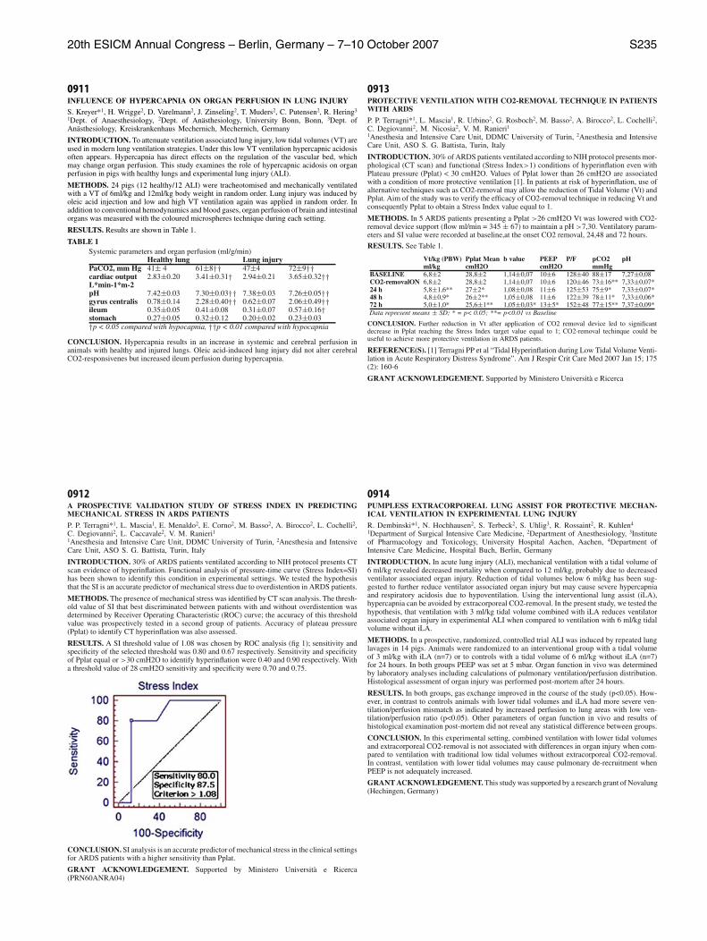
20th ESICM Annual Congress – Berlin, Germany – 7–10 October 2007 S235
0911INFLUENCE OF HYPERCAPNIA ON ORGAN PERFUSION IN LUNG INJURY
S. Kreyer*1, H. Wrigge2, D. Varelmann2, J. Zinseling2, T. Muders2, C. Putensen2, R. Hering3
1Dept. of Anaesthesiology, 2Dept. of Anasthesiology, University Bonn, Bonn, 3Dept. ofAnasthesiology, Kreiskrankenhaus Mechernich, Mechernich, Germany
INTRODUCTION. To attenuate ventilation associated lung injury, low tidal volumes (VT) areused in modern lung ventilation strategies. Under this low VT ventilation hypercapnic acidosisoften appears. Hypercapnia has direct effects on the regulation of the vascular bed, whichmay change organ perfusion. This study examines the role of hypercapnic acidosis on organperfusion in pigs with healthy lungs and experimental lung injury (ALI).
METHODS. 24 pigs (12 healthy/12 ALI) were tracheotomised and mechanically ventilatedwith a VT of 6ml/kg and 12ml/kg body weight in random order. Lung injury was induced byoleic acid injection and low and high VT ventilation again was applied in random order. Inaddition to conventional hemodynamics and blood gases, organ perfusion of brain and intestinalorgans was measured with the coloured microspheres technique during each setting.
RESULTS. Results are shown in Table 1.
TABLE 1Systemic parameters and organ perfusion (ml/g/min)
Healthy lung Lung injuryPaCO2, mm Hg 41± 4 61±8†† 47±4 72±9††cardiac output 2.83±0.20 3.41±0.31† 2.94±0.21 3.65±0.32††L*min-1*m-2pH 7.42±0.03 7.30±0.03†† 7.38±0.03 7.26±0.05††gyrus centralis 0.78±0.14 2.28±0.40†† 0.62±0.07 2.06±0.49††ileum 0.35±0.05 0.41±0.08 0.31±0.07 0.57±0.16†stomach 0.27±0.05 0.32±0.12 0.20±0.02 0.23±0.03†p < 0.05 compared with hypocapnia, ††p < 0.01 compared with hypocapnia
CONCLUSION. Hypercapnia results in an increase in systemic and cerebral perfusion inanimals with healthy and injured lungs. Oleic acid-induced lung injury did not alter cerebralCO2-responsivenes but increased ileum perfusion during hypercapnia.
0912A PROSPECTIVE VALIDATION STUDY OF STRESS INDEX IN PREDICTINGMECHANICAL STRESS IN ARDS PATIENTS
P. P. Terragni*1, L. Mascia1, E. Menaldo2, E. Corno2, M. Basso2, A. Birocco2, L. Cochelli2,C. Degiovanni2, L. Caccavale2, V. M. Ranieri11Anesthesia and Intensive Care Unit, DDMC University of Turin, 2Anesthesia and IntensiveCare Unit, ASO S. G. Battista, Turin, Italy
INTRODUCTION. 30% of ARDS patients ventilated according to NIH protocol presents CTscan evidence of hyperinflation. Functional analysis of pressure-time curve (Stress Index=SI)has been shown to identify this condition in experimental settings. We tested the hypothesisthat the SI is an accurate predictor of mechanical stress due to overdistention in ARDS patients.
METHODS. The presence of mechanical stress was identified by CT scan analysis. The thresh-old value of SI that best discriminated between patients with and without overdistention wasdetermined by Receiver Operating Characteristic (ROC) curve; the accuracy of this thresholdvalue was prospectively tested in a second group of patients. Accuracy of plateau pressure(Pplat) to identify CT hyperinflation was also assessed.
RESULTS. A SI threshold value of 1.08 was chosen by ROC analysis (fig 1); sensitivity andspecificity of the selected threshold was 0.80 and 0.67 respectively. Sensitivity and specificityof Pplat equal or >30 cmH2O to identify hyperinflation were 0.40 and 0.90 respectively. Witha threshold value of 28 cmH2O sensitivity and specificity were 0.70 and 0.75.
CONCLUSION. SI analysis is an accurate predictor of mechanical stress in the clinical settingsfor ARDS patients with a higher sensitivity than Pplat.
GRANT ACKNOWLEDGEMENT. Supported by Ministero Universita e Ricerca(PRN60ANRA04)
0913PROTECTIVE VENTILATION WITH CO2-REMOVAL TECHNIQUE IN PATIENTSWITH ARDS
P. P. Terragni*1, L. Mascia1, R. Urbino2, G. Rosboch2, M. Basso2, A. Birocco2, L. Cochelli2,C. Degiovanni2, M. Nicosia2, V. M. Ranieri11Anesthesia and Intensive Care Unit, DDMC University of Turin, 2Anesthesia and IntensiveCare Unit, ASO S. G. Battista, Turin, Italy
INTRODUCTION. 30% of ARDS patients ventilated according to NIH protocol presents mor-phological (CT scan) and functional (Stress Index>1) conditions of hyperinflation even withPlateau pressure (Pplat) < 30 cmH2O. Values of Pplat lower than 26 cmH2O are associatedwith a condition of more protective ventilation [1]. In patients at risk of hyperinflation, use ofalternative techniques such as CO2-removal may allow the reduction of Tidal Volume (Vt) andPplat. Aim of the study was to verify the efficacy of CO2-removal technique in reducing Vt andconsequently Pplat to obtain a Stress Index value equal to 1.
METHODS. In 5 ARDS patients presenting a Pplat >26 cmH2O Vt was lowered with CO2-removal device support (flow ml/min = 345 ± 67) to maintain a pH >7,30. Ventilatory param-eters and SI value were recorded at baseline,at the onset CO2 removal, 24,48 and 72 hours.
RESULTS. See Table 1.
Vt/kg (PBW) Pplat Mean b value PEEP P/F pCO2 pHml/kg cmH2O cmH2O mmHg
BASELINE 6,8±2 28,8±2 1,14±0,07 10±6 128±40 88±17 7,27±0,08CO2-removalON 6,8±2 28,8±2 1,14±0,07 10±6 120±46 73±16** 7,33±0,07*24 h 5,8±1,6** 27±2* 1,08±0,08 11±6 125±53 75±9* 7,33±0,07*48 h 4,8±0,9* 26±2** 1,05±0,08 11±6 122±39 78±11* 7,33±0,06*72 h 5,0±1,0* 25,6±1** 1,05±0,03* 13±5* 152±48 77±15** 7,37±0,09*Data represent means ± SD; * = p< 0.05; **= p<0.01 vs Baseline
CONCLUSION. Further reduction in Vt after application of CO2 removal device led to significantdecrease in Pplat reaching the Stress Index target value equal to 1; CO2-removal techinque could beuseful to achieve more protective ventilation in ARDS patients.
REFERENCE(S). [1] Terragni PP et al “Tidal Hyperinflation during Low Tidal Volume Venti-lation in Acute Respiratory Distress Syndrome”. Am J Respir Crit Care Med 2007 Jan 15; 175(2): 160-6
GRANT ACKNOWLEDGEMENT. Supported by Ministero Universita e Ricerca
0914PUMPLESS EXTRACORPOREAL LUNG ASSIST FOR PROTECTIVE MECHAN-ICAL VENTILATION IN EXPERIMENTAL LUNG INJURY
R. Dembinski*1, N. Hochhausen2, S. Terbeck2, S. Uhlig3, R. Rossaint2, R. Kuhlen4
1Department of Surgical Intensive Care Medicine, 2Department of Anesthesiology, 3Instituteof Pharmacology and Toxicology, University Hospital Aachen, Aachen, 4Department ofIntensive Care Medicine, Hospital Buch, Berlin, Germany
INTRODUCTION. In acute lung injury (ALI), mechanical ventilation with a tidal volume of6 ml/kg revealed decreased mortality when compared to 12 ml/kg, probably due to decreasedventilator associated organ injury. Reduction of tidal volumes below 6 ml/kg has been sug-gested to further reduce ventilator associated organ injury but may cause severe hypercapniaand respiratory acidosis due to hypoventilation. Using the interventional lung assist (iLA),hypercapnia can be avoided by extracorporeal CO2-removal. In the present study, we tested thehypothesis, that ventilation with 3 ml/kg tidal volume combined with iLA reduces ventilatorassociated organ injury in experimental ALI when compared to ventilation with 6 ml/kg tidalvolume without iLA.
METHODS. In a prospective, randomized, controlled trial ALI was induced by repeated lunglavages in 14 pigs. Animals were randomized to an interventional group with a tidal volumeof 3 ml/kg with iLA (n=7) or to controls with a tidal volume of 6 ml/kg without iLA (n=7)for 24 hours. In both groups PEEP was set at 5 mbar. Organ function in vivo was determinedby laboratory analyses including calculations of pulmonary ventilation/perfusion distribution.Histological assessment of organ injury was performed post-mortem after 24 hours.
RESULTS. In both groups, gas exchange improved in the course of the study (p<0.05). How-ever, in contrast to controls animals with lower tidal volumes and iLA had more severe ven-tilation/perfusion mismatch as indicated by increased perfusion to lung areas with low ven-tilation/perfusion ratio (p<0.05). Other parameters of organ function in vivo and results ofhistological examination post-mortem did not reveal any statistical difference between groups.
CONCLUSION. In this experimental setting, combined ventilation with lower tidal volumesand extracorporeal CO2-removal is not associated with differences in organ injury when com-pared to ventilation with traditional low tidal volumes without extracorporeal CO2-removal.In contrast, ventilation with lower tidal volumes may cause pulmonary de-recruitment whenPEEP is not adequately increased.
GRANT ACKNOWLEDGEMENT. This study was supported by a research grant of Novalung(Hechingen, Germany)
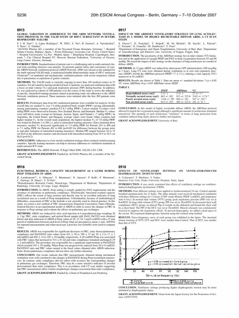
S236 20th ESICM Annual Congress – Berlin, Germany – 7–10 October 2007
0915GLOBAL VARIATION IN ADHERENCE TO THE ARDS NETWORK VENTILA-TION PROTOCOL IN THE VALID STUDY OF RSP-C SURFACTANT IN SEVERERESPIRATORY FAILURE
F. J. H. Taut*1, A. Lopez Rodrıguez2, W. Tillis3, S. Yus4, H. Joensen5, A. Yaroshetskiy6,V. Reus1, A. Gunther7
1ALTANA Pharma AG, a member of the Nycomed Group, Konstanz, Germany, 2, HospitalInfanta Cristina, Badajoz, Spain, 3, Peoria Pulmonary Associates Ltd., Peoria, United States,4, Hospital Universitario ’La Paz’, Madrid, Spain, 5, Bispebjerg Hospital, Copenhagen, Den-mark, 6, City Clinical Hospital N7, Moscow, Russian Federation, 7University of Giessen,Lung Center, Giessen, Germany
INTRODUCTION. Standardisation of patient care is a challenging task in multi-national clin-ical trials enrolling intensive care patients. To promote patient safety and consistency of care,the ARDS Network ventilation protocol (ARDS Network, 2000) was chosen as the standard forthe multi-national VALID study, a randomised double-blind mortality study of rSP-C surfactant(Venticute®) in intubated and mechanically ventilated patients with severe respiratory failuredue to pneumonia or aspiration of gastric contents.
METHODS. The VALID study is currently ongoing in more than 140 medical centers in 23countries. For all countries having enrolled at least 12 patients, we analysed ventilation data witha focus on tidal volume (VT) and peak inspiratory pressure (PIP) during baseline. In addition,VT was analysed in cohorts of 200 patients over the course of the study to assess the influenceof specific, intensified training measures aimed at promoting study site adherence to the ARDSNetwork ventilation protocol. These measures were initiated after the first 200 patients wereenrolled.
RESULTS. Preliminary data from 641 randomised patients were available for analysis. In thisoverall data set, median VT was 7.4 ml/kg predicted body weight (PBW) varying substantiallyamong participating countries (median 6.2-8.3 ml/kg PBW). Lowest median VTvalues (<6.6ml/kg PBW) were observed in Estonia, New Zealand, Russia, and Denmark. Median VT valueswere still below study average in Germany, Austria, Spain, and Finland (∼7 ml/kg PBW). InArgentina, the United States, and Hungary, average values were found. Other countries hadhigher median VT. In the overall study population, the highest median VT of 7.8 ml/kg PBWwas found for Patients 1 to 200, i.e. prior to intensified training measures. Since initiating thesemeasures, median VT decreased significantly to 7.4 ml/kg PBW for Patients 201 to 400 andto 7.1 ml/kg PBW for Patients 401 to 600 (p=0.003 for difference between the groups priorto and after initiation of intensified training measures). Median PIP ranged between 24 to 35cm H2O in the different countries and decreased with intensified training from 29.5 to 28.5 cmH2O (p=0.012).
CONCLUSION. Adherence to a low stretch ventilation strategy shows marked variation amongcountries. Specific training measures can help to increase adherence to ventilation standards inmulti-national ICU trials.
REFERENCE(S). The ARDS Network. N Engl J Med 2000; 342(18):1301-1308.
GRANT ACKNOWLEDGEMENT. Funded by ALTANA Pharma AG, a member of the Ny-comed Group.
0916FUNCTIONAL RESIDUAL CAPACITY MEASUREMENT AS A GUIDE DURINGPEEP TITRATION IN ARDS.
B. Lambermont*1, A. Ghuysen1, V. Mommens1, N. Janssen1, P. Kolh1, P. Morimont1,C. Garweg2, B. Ghaye3, V. D’Orio1
1Hemodynamics Research Centre, HemoLiege, 2Department of Medicine, 3Department ofRadiology, University of Liege, Liege, Belgium
INTRODUCTION. In ARDS, Peep setting is usually guided by FiO2 requirements and theexistence of alterations in pulmonary compliance. Theoretically, functional residual capacity(FRC) reflects the extent of lung aeration and thereby may be an excellent indicator of lungrecruitment area as a result of Peep manipulations. Unfortunately, because of methodologicaldifficulties, assessment of FRC at the bedside is not currently used in clinical practice. In thisstudy, we tested a new method of FRC measurement (Engstrom Carestation, Datex-Ohmeda,General Electric) in an experimental model of ARDS in order to assess the changes in FRC inresponse to Peep settings and evaluate the effects on pulmonary gas exchanges.
METHODS. ARDS was induced by oleic acid injection in 6 anaesthetized pigs weighing 26± 1 kg. FRC, static compliance, and arterial blood sample (pH, PaO2, PaCO2) were obtainedbefore and after induction of ARDS at Peep values of 20, 15, 10, 5 and 0 cmH2O with a 15 minperiod of stabilisation between each level of Peep. Data are presented as mean ± sem. ANOVAfor repeated measurements and when necessary a post hoc test (Scheffe) were used to comparevalues.
RESULTS. ARDS was responsible for significant decreases in FRC, static thoracopulmonarycompliance and PaO2/FiO2 ratio values from 421 ± 58 to 190 ± 37 ml, 28 ± 2 to 17 ± 1ml/cmH2O and 402 ± 14 to 105 ± 10 mmHg, respectively. A 20 cmH2O Peep was associatedwith FRC values that increased to 514 ± 81 ml and static compliance remained unchanged (19± 2 ml/cmH2O). The procedure was responsible for a significant improvement in PaO2/FiO2which reached 345 ± 29 mmHg. When Peep was progressively reduced from 20 to 0 cmH2O,PaO2/FiO2 ratio and FRC values turned to the basal values obtained after ARDS induction.Static thoracopulmonary compliance did not show any further alteration.
CONCLUSION. Our results indicate that FRC measurements obtained during mechanicalventilation were well correlated to the changes in PaO2/FiO2 during Peep recruitment manoeu-vres. In contrast, static compliance did not reflect with accuracy the corresponding changesin pulmonary gas exchanges. Therefore, FRC may be a more sensitive indicator of alveolarderecruitment than static compliance. This is in accordance with CT scan studies suggestingthat FRC measurement offers a better morphologic changes assessment than static compliance.
GRANT ACKNOWLEDGEMENT. Funded by a Grant of Fondation Leon Fredericq
0917IMPACT OF THE ARDSNET VENTILATORY STRATEGY ON LUNG ACTELEC-TASIS IN A MODEL OF HIGHLY RECRUITABLE DIFFUSE ARDS: A CT SCANSTUDY
S. Grasso*1, D. Franchini1, T. Stripoli1, M. De Michele1, M. Sacchi1, L. Falconi2,C. Ferrante2, G. Cinnella2, M. Dambrosio2, T. Fiore1
1Department of Emergency and Organ Trasplantation, University of Bari, Bari, 2Departmentof Anaesthesiology and Intensive care, University of Foggia, Foggia, Italy
INTRODUCTION. The peculiarity of the ARDSnet strategy lies in tidal volume (VT) limita-tion and in the application of enough PEEP and FiO2 to keep oxygenation between 55 and 80mmHg. We tested the impact of this strategy on the clearance of lung actelectasis in a model ofdiffuse ARDS.
METHODS. In 12 pigs,ARDS was induced by intravenous LPS administration (100 mcg/Kgin 1 hour). Lung CT scan were obtained during ventilation a) at zero end expiratory pres-sure (ZEEP); b)with the ARDSnet protocol (PEEP 7.7 ± 2.1); c)during a vital capacity (VC)maneuver at 45 cmH2O.
RESULTS. Results are shown in Table 1. Data are mean +/- standard deviation; *) p < 0.05VC versus ARDSnet; #) p < 0.05 ARDSnet versus ZEEP.
TABLE 1.ZEEP ARDSnet VC
Hyperinflated areas (cm2) 2 +/- 1 2 +/- 1 27 +/- 3 *Normally aerated areas (cm2) 662 +/- 262 833 +/- 325 # 3162 +/- 1174 *Poorly aerated areas (cm2) 1148 +/- 395 884 +/- 341 547 +/- 343 *Non aerated areas (cm2) 815 +/- 252 495 +/- 215 # 101 +/- 51 *
CONCLUSION. In this model of highly recruitable diffuse ARDS, the ARDSnet protocolallowed to match the oxygenation target although a significant amount of actelectasis remainedunresolved. The meaning of such actelectasis “tolerance” in terms of lung protection fromventilator induced lung injury deserves further investigation.
GRANT ACKNOWLEDGEMENT. University of Bari.
0918EFFECTS OF VENTILATORY SETTINGS ON VENTILATOR-INDUCEDDIAPHRAGMATIC DYSFUNCTION
A. Uchiyama*, T. Mashimo, Y. FujinoIntensive Care Unit, Osaka University Hospital, Suita, Japan
INTRODUCTION. It was rarely examined that effects of ventilatory settings on ventilator-induced diaphragmatic dysfunction (VIDD).
METHODS. Four different settings were applied to tracheostomized 24 rats. Control animalsbreathed spontaneously for 12 hours. The other animals were received mechanical ventilationwith three different settings for 12 hours (CMV mode 80/min, PEEP 1cmH2O, and inspiratorytime 0.3sec). In normal tidal volume (NTV) group, peak inspiratory pressure (PIP) was set at8cmH2O. In large tidal volume (LTV) group, PIP was set at 18cmH2O. In decreased chest wallcompliance (DCC) group, we placed 50g of weight on the abdomen and bound the chest wallby a chest band. The PIP of the DCC was set at 16cmH2O. Muscle relaxants was continuouslyreceived to suppress the inspiratory efforts. To prevent hypocapnia, we added a dead space tothe circuit. We examined diaphragmatic function using the isolated strip method.
RESULTS. Force-frequency curve of each group was exhibited in the figure. The maximaltetanic tensions of NTV, LTV and DCC were smaller than Control. That of DCC was smallerthan that of NTV.
CONCLUSION. Ventilatory settings producing higher diaphragmatic tension may be moreattributed to diaphragmatic injury.
GRANT ACKNOWLEDGEMENT. Grant from the Japan Society for the Promotion of Sci-ence (185917019)
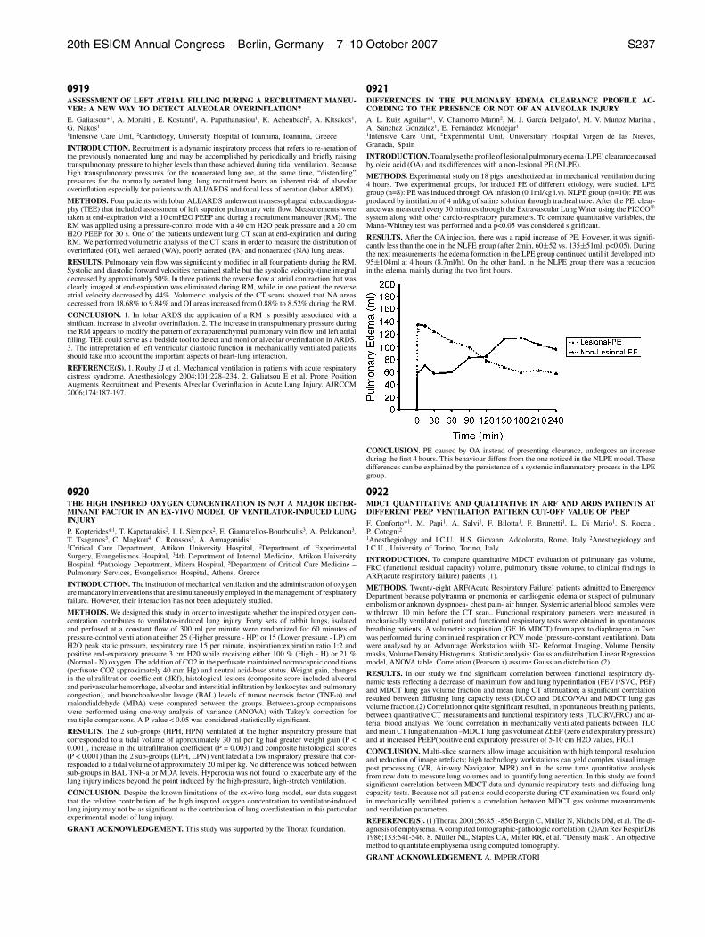
20th ESICM Annual Congress – Berlin, Germany – 7–10 October 2007 S237
0919ASSESSMENT OF LEFT ATRIAL FILLING DURING A RECRUITMENT MANEU-VER: A NEW WAY TO DETECT ALVEOLAR OVERINFLATION?
E. Galiatsou*1, A. Moraiti1, E. Kostanti1, A. Papathanasiou1, K. Achenbach2, A. Kitsakos1,G. Nakos1
1Intensive Care Unit, 2Cardiology, University Hospital of Ioannina, Ioannina, Greece
INTRODUCTION. Recruitment is a dynamic inspiratory process that refers to re-aeration ofthe previously nonaerated lung and may be accomplished by periodically and briefly raisingtranspulmonary pressure to higher levels than those achieved during tidal ventilation. Becausehigh transpulmonary pressures for the nonaerated lung are, at the same time, “distending”pressures for the normally aerated lung, lung recruitment bears an inherent risk of alveolaroverinflation especially for patients with ALI/ARDS and focal loss of aeration (lobar ARDS).
METHODS. Four patients with lobar ALI/ARDS underwent transesophageal echocardiogra-phy (TEE) that included assessment of left superior pulmonary vein flow. Measurements weretaken at end-expiration with a 10 cmH2O PEEP and during a recruitment maneuver (RM). TheRM was applied using a pressure-control mode with a 40 cm H2O peak pressure and a 20 cmH2O PEEP for 30 s. One of the patients undewent lung CT scan at end-expiration and duringRM. We performed volumetric analysis of the CT scans in order to measure the distribution ofoverinflated (OI), well aerated (WA), poorly aerated (PA) and nonaerated (NA) lung areas.
RESULTS. Pulmonary vein flow was significantly modified in all four patients during the RM.Systolic and diastolic forward velocities remained stable but the systolic velocity-time integraldecreased by approximately 50%. In three patients the reverse flow at atrial contraction that wasclearly imaged at end-expiration was eliminated during RM, while in one patient the reverseatrial velocity decreased by 44%. Volumeric analysis of the CT scans showed that NA areasdecreased from 18.68% to 9.84% and OI areas increased from 0.88% to 8.52% during the RM.
CONCLUSION. 1. In lobar ARDS the application of a RM is possibly associated with asinificant increase in alveolar overinflation. 2. The increase in transpulmonary pressure duringthe RM appears to modify the pattern of extraparenchymal pulmonary vein flow and left atrialfilling. TEE could serve as a bedside tool to detect and monitor alveolar overinflation in ARDS.3. The intrepretation of left ventricular diastolic function in mechanicallly ventilated patientsshould take into account the important aspects of heart-lung interaction.
REFERENCE(S). 1. Rouby JJ et al. Mechanical ventilation in patients with acute respiratorydistress syndrome. Anesthesiology 2004;101:228–234. 2. Galiatsou E et al. Prone PositionAugments Recruitment and Prevents Alveolar Overinflation in Acute Lung Injury. AJRCCM2006;174:187-197.
0920THE HIGH INSPIRED OXYGEN CONCENTRATION IS NOT A MAJOR DETER-MINANT FACTOR IN AN EX-VIVO MODEL OF VENTILATOR-INDUCED LUNGINJURY
P. Kopterides*1, T. Kapetanakis2, I. I. Siempos2, E. Giamarellos-Bourboulis3, A. Pelekanou3,T. Tsaganos3, C. Magkou4, C. Roussos5, A. Armaganidis1
1Critical Care Department, Attikon University Hospital, 2Department of ExperimentalSurgery, Evangelismos Hospital, 34th Department of Internal Medicine, Attikon UniversityHospital, 4Pathology Department, Mitera Hospital, 5Department of Critical Care Medicine –Pulmonary Services, Evangelismos Hospital, Athens, Greece
INTRODUCTION. The institution of mechanical ventilation and the administration of oxygenare mandatory interventions that are simultaneously employed in the management of respiratoryfailure. However, their interaction has not been adequately studied.
METHODS. We designed this study in order to investigate whether the inspired oxygen con-centration contributes to ventilator-induced lung injury. Forty sets of rabbit lungs, isolatedand perfused at a constant flow of 300 ml per minute were randomized for 60 minutes ofpressure-control ventilation at either 25 (Higher pressure - HP) or 15 (Lower pressure - LP) cmH2O peak static pressure, respiratory rate 15 per minute, inspiration:expiration ratio 1:2 andpositive end-expiratory pressure 3 cm H20 while receiving either 100 % (High - H) or 21 %(Normal - N) oxygen. The addition of CO2 in the perfusate maintained normocapnic conditions(perfusate CO2 approximately 40 mm Hg) and neutral acid-base status. Weight gain, changesin the ultrafiltration coefficient (dKf), histological lesions (composite score included alveoraland perivascular hemorrhage, alveolar and interstitial infiltration by leukocytes and pulmonarycongestion), and bronchoalveolar lavage (BAL) levels of tumor necrosis factor (TNF-a) andmalondialdehyde (MDA) were compared between the groups. Between-group comparisonswere performed using one-way analysis of variance (ANOVA) with Tukey’s correction formultiple comparisons. A P value < 0.05 was considered statistically significant.
RESULTS. The 2 sub-groups (HPH, HPN) ventilated at the higher inspiratory pressure thatcorresponded to a tidal volume of approximately 30 ml per kg had greater weight gain (P <0.001), increase in the ultrafiltration coefficient (P = 0.003) and composite histological scores(P < 0.001) than the 2 sub-groups (LPH, LPN) ventilated at a low inspiratory pressure that cor-responded to a tidal volume of approximately 20 ml per kg. No difference was noticed betweensub-groups in BAL TNF-a or MDA levels. Hyperoxia was not found to exacerbate any of thelung injury indices beyond the point induced by the high-pressure, high-stretch ventilation.
CONCLUSION. Despite the known limitations of the ex-vivo lung model, our data suggestthat the relative contribution of the high inspired oxygen concentration to ventilator-inducedlung injury may not be as significant as the contribution of lung overdistention in this particularexperimental model of lung injury.
GRANT ACKNOWLEDGEMENT. This study was supported by the Thorax foundation.
0921DIFFERENCES IN THE PULMONARY EDEMA CLEARANCE PROFILE AC-CORDING TO THE PRESENCE OR NOT OF AN ALVEOLAR INJURY
A. L. Ruiz Aguilar*1, V. Chamorro Marın2, M. J. Garcıa Delgado1, M. V. Munoz Marina1,A. Sanchez Gonzalez1, E. Fernandez Mondejar1
1Intensive Care Unit, 2Experimental Unit, Universitary Hospital Virgen de las Nieves,Granada, Spain
INTRODUCTION. To analyse the profile of lesional pulmonary edema (LPE) clearance causedby oleic acid (OA) and its differences with a non-lesional PE (NLPE).
METHODS. Experimental study on 18 pigs, anesthetized an in mechanical ventilation during4 hours. Two experimental groups, for induced PE of different etiology, were studied. LPEgroup (n=8): PE was induced through OA infusion (0.1ml/kg i.v). NLPE group (n=10): PE wasproduced by instilation of 4 ml/kg of saline solution through tracheal tube. After the PE, clear-ance was measured every 30 minutes through the Extravascular Lung Water using the PICCO®
system along with other cardio-respiratory parameters. To compare quantitative variables, theMann-Whitney test was performed and a p<0.05 was considered significant.
RESULTS. After the OA injection, there was a rapid increase of PE. However, it was signifi-cantly less than the one in the NLPE group (after 2min, 60±52 vs. 135±51ml; p<0.05). Duringthe next measurements the edema formation in the LPE group continued until it developed into95±104ml at 4 hours (8.7ml/h). On the other hand, in the NLPE group there was a reductionin the edema, mainly during the two first hours.
CONCLUSION. PE caused by OA instead of presenting clearance, undergoes an increaseduring the first 4 hours. This behaviour differs from the one noticed in the NLPE model. Thesedifferences can be explained by the persistence of a systemic inflammatory process in the LPEgroup.
0922MDCT QUANTITATIVE AND QUALITATIVE IN ARF AND ARDS PATIENTS ATDIFFERENT PEEP VENTILATION PATTERN CUT-OFF VALUE OF PEEP
F. Conforto*1, M. Papi1, A. Salvi1, F. Bilotta1, F. Brunetti1, L. Di Mario1, S. Rocca1,P. Cotogni21Anesthegiology and I.C.U., H.S. Giovanni Addolorata, Rome, Italy 2Anesthegiology andI.C.U., University of Torino, Torino, Italy
INTRODUCTION. To compare quantitative MDCT evaluation of pulmunary gas volume,FRC (functional residual capacity) volume, pulmonary tissue volume, to clinical findings inARF(acute respiratory failure) patients (1).
METHODS. Twenty-eight ARF(Acute Respiratory Failure) patients admitted to EmergencyDepartment because polytrauma or pnemonia or cardiogenic edema or suspect of pulmunaryembolism or anknown dyspnoea- chest pain- air hunger. Systemic arterial blood samples werewithdrawn 10 min before the CT scan.. Functional respiratory pameters were measured inmechanically ventilated patient and functional respiratory tests were obtained in spontaneousbreathing patients. A volumetric acquisition (GE 16 MDCT) from apex to diaphragma in 7secwas performed during continued respiration or PCV mode (pressure-constant ventilation). Datawere analysed by an Advantage Workstation wiith 3D- Reformat Imaging, Volume Densitymasks, Volume Density Histograms. Statistic analysis: Gaussian distribution Linear Regressionmodel, ANOVA table. Correlation (Pearson r) assume Gaussian distribution (2).
RESULTS. In our study we find significant correlation between functional respiratory dy-namic tests reflecting a decrease of maximum flow and lung hyperinflation (FEV1/SVC, PEF)and MDCT lung gas volume fraction and mean lung CT attenuation; a significant correlationresulted between diffusing lung capacity tests (DLCO and DLCO/VA) and MDCT lung gasvolume fraction.(2) Correlation not quite significant resulted, in spontaneous breathing patients,between quantitative CT measuraments and functional respiratory tests (TLC,RV,FRC) and ar-terial blood analysis. We found correlation in mechanically ventilated patients between TLCand mean CT lung attenuation –MDCT lung gas volume at ZEEP (zero end expiratory pressure)and at increased PEEP(positive end expiratory pressure) of 5-10 cm H2O values, FIG.1.
CONCLUSION. Multi-slice scanners allow image acquisition with high temporal resolutionand reduction of image artefacts; high technology workstations can yeld complex visual imagepost processing (VR, Air-way Navigator, MPR) and in the same time quantitative analysisfrom row data to measure lung volumes and to quantify lung aereation. In this study we foundsignificant correlation between MDCT data and dynamic respiratory tests and diffusing lungcapacity tests. Because not all patients could cooperate during CT examination we found onlyin mechanically ventilated patients a correlation between MDCT gas volume measuramentsand ventilation parameters.
REFERENCE(S). (1)Thorax 2001;56:851-856 Bergin C, Muller N, Nichols DM, et al. The di-agnosis of emphysema. A computed tomographic-pathologic correlation. (2)Am Rev Respir Dis1986;133:541-546. 8. Muller NL, Staples CA, Miller RR, et al. “Density mask”. An objectivemethod to quantitate emphysema using computed tomography.
GRANT ACKNOWLEDGEMENT. A. IMPERATORI
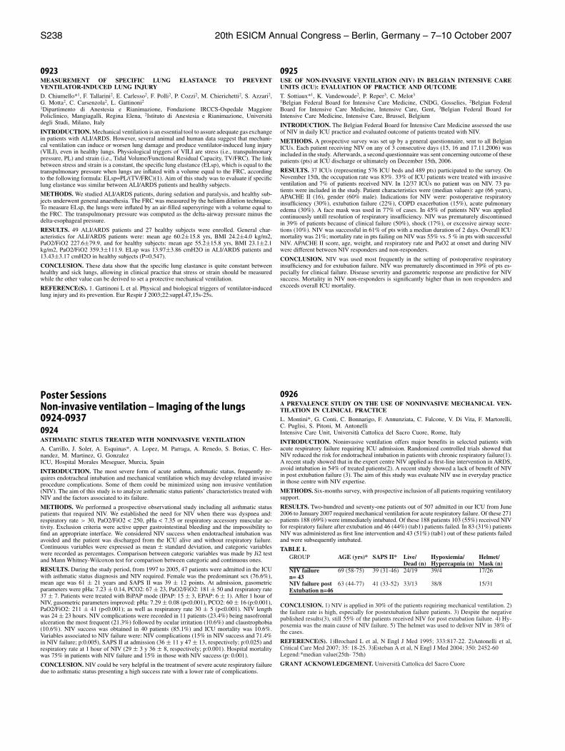
S238 20th ESICM Annual Congress – Berlin, Germany – 7–10 October 2007
0923MEASUREMENT OF SPECIFIC LUNG ELASTANCE TO PREVENTVENTILATOR-INDUCED LUNG INJURY
D. Chiumello*1, F. Tallarini2, E. Carlesso2, F. Polli2, P. Cozzi2, M. Chierichetti2, S. Azzari2,G. Motta2, C. Carsenzola2, L. Gattinoni21Dipartimento di Anestesia e Rianimazione, Fondazione IRCCS-Ospedale MaggiorePoliclinico, Mangiagalli, Regina Elena, 2Istituto di Anestesia e Rianimazione, Universitadegli Studi, Milano, Italy
INTRODUCTION. Mechanical ventilation is an essential tool to assure adequate gas exchangein patients with ALI/ARDS. However, several animal and human data suggest that mechani-cal ventilation can induce or worsen lung damage and produce ventilator-induced lung injury(VILI), even in healthy lungs. Physiological triggers of VILI are stress (i.e., transpulmonarypressure, PL) and strain (i.e., Tidal Volume/Functional Residual Capacity, TV/FRC). The linkbetween stress and strain is a constant, the specific lung elastance (ELsp), which is equal to thetranspulmonary pressure when lungs are inflated with a volume equal to the FRC, accordingto the following formula: ELsp=PL/(TV/FRC)(1). Aim of this study was to evaluate if specificlung elastance was similar between ALI/ARDS patients and healthy subjects.
METHODS. We studied ALI/ARDS patients, during sedation and paralysis, and healthy sub-jects underwent general anaesthesia. The FRC was measured by the helium dilution technique.To measure ELsp, the lungs were inflated by an air-filled supersyringe with a volume equal tothe FRC. The transpulmonary pressure was computed as the delta-airway pressure minus thedelta-esophageal pressure.
RESULTS. 49 ALI/ARDS patients and 27 healthy subjects were enrolled. General char-acteristics for ALI/ARDS patients were: mean age 60.2±15.8 yrs, BMI 24.2±4.0 kg/m2,PaO2/FiO2 227.6±79.9, and for healthy subjects: mean age 55.2±15.8 yrs, BMI 23.1±2.1kg/m2, PaO2/FiO2 359.3±111.9. ELsp was 13.97±3.86 cmH2O in ALI/ARDS patients and13.43±3.17 cmH2O in healthy subjects (P=0.547).
CONCLUSION. These data show that the specific lung elastance is quite constant betweenhealthy and sick lungs, allowing in clinical practice that stress or strain should be measuredwhile the other value can be derived to set a protective mechanical ventilation.
REFERENCE(S). 1. Gattinoni L et al. Physical and biological triggers of ventilator-inducedlung injury and its prevention. Eur Respir J 2003;22:suppl.47,15s-25s.
Poster SessionsNon-invasive ventilation – Imaging of the lungs0924-09370924ASTHMATIC STATUS TREATED WITH NONINVASIVE VENTILATION
A. Carrillo, J. Soler, A. Esquinas*, A. Lopez, M. Parraga, A. Renedo, S. Botias, C. Her-nandez, M. Martinez, G. GonzalezICU, Hospital Morales Meseguer, Murcia, Spain
INTRODUCTION. The most severe form of acute asthma, asthmatic status, frequently re-quires endotracheal intubation and mechanical ventilation which may develop related invasiveprocedure complications. Some of them could be minimized using non invasive ventilation(NIV). The aim of this study is to analyze asthmatic status patients’ characteristics treated withNIV and the factors associated to its failure.
METHODS. We performed a prospective observational study including all asthmatic statuspatients that required NIV. We established the need for NIV when there was dyspnea and:respiratory rate > 30, PaO2/FiO2 < 250, pHa < 7.35 or respiratory accessory muscular ac-tivity. Exclusion criteria were active upper gastrointestinal bleeding and the impossibility tofind an appropriate interface. We considered NIV success when endotracheal intubation wasavoided and the patient was discharged from the ICU alive and without respiratory failure.Continuous variables were expressed as mean ± standard deviation, and categoric variableswere recorded as percentages. Comparison between categoric variables was made by Ji2 testand Mann Whitney-Wilcoxon test for comparison between categoric and continuous ones.
RESULTS. During the study period, from 1997 to 2005, 47 patients were admitted in the ICUwith asthmatic status diagnosis and NIV required. Female was the predominant sex (76.6%),mean age was 61 ± 21 years and SAPS II was 39 ± 12 points. At admission, gasometricparameters were pHa: 7.23 ± 0.14, PCO2: 67 ± 23, PaO2/FiO2: 181 ± 50 and respiratory rate37 ± 7. Patients were treated with BiPAP mode (IPAP: 15 ± 3, EPAP: 6 ± 1). After 1 hour ofNIV, gasometric parameters improved: pHa: 7.29 ± 0.08 (p<0.001), PCO2: 60 ± 16 (p:0.001),PaO2/FiO2: 211 ± 41 (p<0.001); as well as respiratory rate 30 ± 5 (p<0.001). NIV lengthwas 24 ± 23 hours. NIV complications were recorded in 11 patients (23.4%) being nasofrontalulceration the most frequent (21.3%) followed by ocular irritation (10.6%) and claustrophobia(10.6%). NIV success was obtained in 40 patients (85.1%) and ICU mortality was 10.6%.Variables associated to NIV failure were: NIV complications (15% in NIV success and 71.4%in NIV failure; p:0.005), SAPS II at admission (36 ± 11 y 47 ± 13, respectively; p:0.025) andrespiratory rate at 1 hour of NIV (29 ± 3 y 36 ± 8, respectively; p:0.001). Hospital mortalitywas 75% in patients with NIV failure and 15% in those with NIV success (p: 0.001).
CONCLUSION. NIV could be very helpful in the treatment of severe acute respiratory failuredue to asthmatic status presenting a high success rate with a lower rate of complications.
0925USE OF NON-INVASIVE VENTILATION (NIV) IN BELGIAN INTENSIVE CAREUNITS (ICU): EVALUATION OF PRACTICE AND OUTCOME
T. Sottiaux*1, K. Vandewoude2, P. Reper3, C. Melot31Belgian Federal Board for Intensive Care Medicine, CNDG, Gosselies, 2Belgian FederalBoard for Intensive Care Medicine, Intensive Care, Gent, 3Belgian Federal Board forIntensive Care Medicine, Intensive Care, Brussel, Belgium
INTRODUCTION. The Belgian Federal Board for Intensive Care Medicine assessed the useof NIV in daily ICU practice and evaluated outcome of patients treated with NIV.
METHODS. A prospective survey was set up by a general questionnaire, sent to all BelgianICUs. Each patient receiving NIV on any of 3 consecutive days (15, 16 and 17.11.2006) wasincluded in the study. Afterwards, a second questionnaire was sent concerning outcome of thesepatients (pts) at ICU discharge or ultimately on December 15th, 2006.
RESULTS. 37 ICUs (representing 576 ICU beds and 489 pts) participated to the survey. OnNovember 15th, the occupation rate was 83%. 33% of ICU patients were treated with invasiveventilation and 7% of patients received NIV. In 12/37 ICUs no patient was on NIV. 73 pa-tients were included in the study. Patient characteristics were (median values): age (66 years),APACHE II (16), gender (60% male). Indications for NIV were: postoperative respiratoryinsufficiency (30%), extubation failure (22%), COPD exacerbation (15%), acute pulmonaryedema (30%). A face mask was used in 77% of cases. In 45% of patients NIV was appliedcontinuously untill resolution of respiratory insufficiency. NIV was prematurely discontinuedin 39% of patients because of clinical failure (50%), shock (17%), or excessive airway secre-tions (10%). NIV was successful in 61% of pts with a median duration of 2 days. Overall ICUmortality was 21%; mortality rate in pts failing on NIV was 55% vs. 5 % in pts with successfulNIV. APACHE II score, age, weight, and respiratory rate and PaO2 at onset and during NIVwere different between NIV responders and non-responders.
CONCLUSION. NIV was used most frequently in the setting of postoperative respiratoryinsufficiency and for extubation failure. NIV was prematurely discontinued in 39% of pts es-pecially for clinical failure. Disease severity and gazometric response are predictive for NIVsuccess. Mortality in NIV non-responders is significantly higher than in non responders andexceeds overall ICU mortality.
0926A PREVALENCE STUDY ON THE USE OF NONINVASIVE MECHANICAL VEN-TILATION IN CLINICAL PRACTICE
L. Montini*, G. Conti, C. Bonnarigo, F. Annunziata, C. Falcone, V. Di Vita, F. Martorelli,C. Puglisi, S. Pitoni, M. AntonelliIntensive Care Unit, Universita Cattolica del Sacro Cuore, Rome, Italy
INTRODUCTION. Noninvasive ventilation offers major benefits in selected patients withacute respiratory failure requiring ICU admission. Randomized controlled trials showed thatNIV reduced the risk for endotracheal intubation in patients with chronic respiratory failure(1).A recent study showed that in the expert centre NIV applied as first-line intervention in ARDS,avoid intubation in 54% of treated patients(2). A recent study showed a lack of benefit of NIVin post extubation failure (3). The aim of this study was evaluate NIV use in everyday practicein those centre with NIV expertise.
METHODS. Six-months survey, with prospective inclusion of all patients requiring ventilatorysupport.
RESULTS. Two-hundred and seventy-one patients out of 507 admitted in our ICU from June2006 to January 2007 required mechanical ventilation for acute respiratory failure. Of these 271patients 188 (69%) were immediately intubated. Of these 188 patients 103 (55%) received NIVfor respiratory failure after extubation and 46 (44%) (tab1) patients failed. In 83 (31%) patientsNIV was administered as first line intervention and 43 (51%) (tab1) out of these patients failedand were subsequently intubated.
TABLE 1.GROUP AGE (yrs)* SAPS II* Live/ Hypoxiemia/ Helmet/
Dead (n) Hypercapnia (n) Mask (n)NIV failure 69 (58-75) 39 (31-46) 24/19 39/4 17/26n= 43NIV failure post 63 (44-77) 41 (33-52) 33/13 38/8 15/31Extubation n=46
CONCLUSION. 1) NIV is applied in 30% of the patients requiring mechanical ventilation. 2)the failure rate is high, especially for postextubation failure patients. 3) Despite the negativepublished results(3), still 55% of the patients received NIV for post extubation failure. 4) Hy-poxemia was the main cause of NIV failure. 5) The helmet was used to deliver NIV in 38% ofthe cases.
REFERENCE(S). 1)Brochard L et al, N Engl J Med 1995; 333:817-22. 2)Antonelli et al,Critical Care Med 2007; 35: 18-25. 3)Esteban A et al, N Engl J Med 2004; 350: 2452-60Legend:*median value(25th- 75th)
GRANT ACKNOWLEDGEMENT. Universita Cattolica del Sacro Cuore
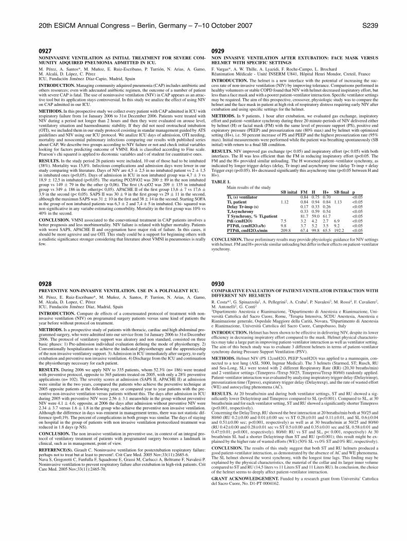
20th ESICM Annual Congress – Berlin, Germany – 7–10 October 2007 S239
0927NONINVASIVE VENTILATION AS INITIAL TREATMENT FOR SEVERE COM-MUNITY ADQUIRED PNEUMONIA ADMITTED IN ICU.
M. Perez, A. Santos*, M. Munoz, E. Ruiz-Escribano, P. Turrion, N. Arias, A. Gamo,M. Alcala, D. Lopez, C. PerezICU, Fundacion Jimenez Dıaz-Capio, Madrid, Spain
INTRODUCTION. Managing community adquired pneumonia (CAP) includes antibiotic andothers resources; even with adecuated anitbiotic regimen, the outcome of a number of patientwith severe CAP is fatal. The use of noninvasive ventilation (NIV) in CAP appears as an atrac-tive tool but its application stays controversial. In this study we analize the effect of using NIVon CAP admitted in our ICU.
METHODS. In this prospective study we collect every patient with CAP admitted in ICU withrespiratory failure from 1st January 2006 to 31st December 2006. Patients were treated withNIV during a period not longer than 2 hours and then they were evaluated on arouse level,ventilatory situation and haemodinamic stability. If they did not need orotracheal intubation(OTI), we included them in our study protocol cosisting in standar management guided by ATSguidelines and NIV using our ICU protocol. We analize ICU days of admission, OTI needing,mortality and nosocomial pulmonary infections comparing our results with published seriesabout CAP. We describe two groups according to NIV failure or not and check initial variableslooking for factors predicting outcome of VMNI. Risk is classified according to Fine scale.Pearson’s chi squared is applied to dicotomic variables and t of student for cuantitative ones.
RESULTS. In the study period 26 patients were included, 10 out of those had to be intubated(38%). Mortality was 15,8%. Infectious complications and admission days were lower in ourstudy comparing with literature. Days of NIV are 4,5 ± 2,5 in no intubated patient vs 2 ± 1,5in intubated ones (p<0,05). Days of admission in ICU in non intubated group was 4,7 ± 3 vs18,9 ± 12,5 in intubated (p<0,05). The starting PO2/FiO2 was 200 ± 89 in the non intubatedgroup vs 149 ± 79 in the the other (p 0,06). The first (A-a)O2 was 209 ± 135 in intubatedgroup vs 349 ± 186 in the other(p< 0,05). APACHE II of the first group 13,6 ± 7 vs 17,6 ±3,9 in the second (p< 0,05). SAPS II was 30 ± 9 in the first group vs 29 ± 11 in the second,although the maximun SAPS was 31 ± 10 in the first and 38 ± 14 in the second. Starting SOFAin the group of non intubated patients was 6,3 ± 2 and 7,4 ± 5 in intubated. Chi- squared wasnon significative in any variabe estimating comorbility. Mortality in the first group was 10% vs40% in the second.
CONCLUSION. VMNI associated to the conevntional treatment in CAP patients involves abetter prognosis and less morbimortality. NIV failure is related with higher mortality. Patientswith worst SAPS, APACHE II and oxygenation have major risk of failure. In this cases, itshould be more agresive and use OTI. This study could be a support for beginning others witha stadistic significance stronger considering that literature about VMNI in pneumonies is reallyfew.
0928PREVENTIVE NON-INVASIVE VENTILATION. USE IN A POLIVALENT ICU.
M. Perez, E. Ruiz-Escribano*, M. Munoz, A. Santos, P. Turrion, N. Arias, A. Gamo,M. Alcala, D. Lopez, C. PerezICU, Fundacion Jimenez Dıaz, Madrid, Spain
INTRODUCTION. Compare de effects of a consensuated protocol of treatment with non-invasive ventilation (NIV) on programated surgery patients versus same kind of patients theyear before without protocol on treatment.
METHODS. Is a prospective study of patients with thoracic, cardiac and high abdominal pro-grammed surgery, who were admitted into our service from 1st January 2006 to 31st December2006. The protocol of ventilatory support was aleatory and non standard, consisted on threebasic phases: 1) Pre-admission individual evaluation defining the mode of physiotherapy. 2)Conventionally hospitalization to achieve the indicated physiotherapy and the apprenticeshipof the non invasive ventilatory support. 3) Admission in ICU immediately after surgery, to earlyextubation and preventive non invasive ventilation. 4) Discharge from the ICU and continuationthe physiotherapy necessary for each patient.
RESULTS. During 2006 we apply NIV to 335 patients, whom 52.3% (n= 186) were treatedwith preventive protocol, opposite to 365 patients treated on 2005, with only a 28% preventiveapplications (n= 102). The severity scores at admission (SAPS II, APACHE II) at admissionwere similar in the two years, compared the patients who achieve the preventive technique at2005 opposite patients at the following year, or compared year to year patients with the pre-ventive non-invasive ventilation versus patients without this. The days after admission in ICUduring 2005 with preventive NIV were 2.56 ± 3.1 meanwhile in the group without preventiveNIV were 4.1 ± 4.6; opposite, at 2006 the days after admission without preventive NIV were2.34 ± 3.7 versus 1.6 ± 1.8 in the group who achieve the preventive non invasive ventilation.Although the difference in days was eminent in management terms, there was not statistic dif-ference (p=0,19). The percent of complications in both groups was similar. The days of stayingon hospital in the group of patients with non invasive ventilation protocolized treatment wasreduced in 1.8 days (p NS).
CONCLUSION. The non invasive ventilation in preventive use, in context of an integral pro-tocol of ventilatory treatment of patients with programated surgery becomes a landmark inclinical, such as in management, point of view.
REFERENCE(S). Girault C. Noninvasive ventilation for postextubation respiratory failure:perhaps not to treat but at least to prevent!. Crit Care Med. 2005 Nov;33(11):2685-6.Nava S, Gregoretti C, Fanfulla F, Squadrone E, Grassi M, Carlucci A, Beltrame F, Navalesi P.Noninvasive ventilation to prevent respiratory failure after extubation in high-risk patients. CritCare Med. 2005 Nov;33(11):2465-70.
0929NON INVASIVE VENTILATION AFTER EXTUBATION: FACE MASK VERSUSHELMET WITH SPECIFIC SETTINGS
F. Vargas*, A. W. Thille, A. Lyazidi, F. Roche-Campo, L. BrochardReanimation Medicale - Unite INSERM U841, Hopital Henri Mondor, Creteil, France
INTRODUCTION. The helmet is a new interface with the potential of increasing the suc-cess rate of non-invasive ventilation (NIV) by improving tolerance. Comparisons performed inhealthy volunteers or stable COPD found that NIV with helmet decreased inspiratory effort, butless than a face mask and with a poorer patient-ventilator interaction. Specific ventilator settingsmay be required. The aim of this prospective, crossover, physiologic study was to compare thehelmet and the face mask in patient at high risk of respiratory distress requiring early NIV afterextubation and using specific settings for the helmet.
METHODS. In 9 patients, 1 hour after extubation, we evaluated gas exchange, inspiratoryeffort and patient–ventilator synchrony during three 20 minute-periods of NIV delivered eitherby helmet (H) or facial mask (FM) with the same level of pressure support (PS), positive endexpiratory pressure (PEEP) and pressurization rate (80% max) and by helmet with optimizedsetting (H+), i.e. 50 percent increase of PS and PEEP and the highest pressurization rate (95%max). Initial measurements were performed while the patient was breathing spontaneously (SBinitial) with return to a final SB condition.
RESULTS. NIV improved gas exchange (p< 0.05) and inspiratory effort (p< 0.05) with bothinterfaces. The H was less efficient than the FM in reducing inspiratory effort (p<0.05. TheFM and the H+ provided similar unloading. The H worsened patient–ventilator synchrony, asindicated by longer trigger delays (delay, Tr-insp) and asynchrony time (delay Tr-insp + delayTrigger exp) (p<0.05). H+ decreased significantly this asynchrony time (p<0.05 between H andH+).
TABLE 1.Main results of the study
SB inital FM H H+ SB final pTi, (s) ventilator 0.84 0.75 0.70 <0.05Ti, patient 1.12 0.84 0.94 0.84 1.13 <0.05Delay Tr-insp (s) 0.17 0.33 0.26 <0.05T,Asynchrony 0.33 0.59 0.54 <0.05T Synchrony, % Ti,patient 81.7 59.0 61.7 <0.05Pdi (cmH2O) 7.5 3.2 4.2 2.7 6.9 <0.05PTPdi, (cmH2O.s/b) 9.8 3.7 5.2 3.5 9.2 <0.05PTPdi, cmH2O.s/min 209.8 67.4 99.8 65.5 192.2 <0.05
CONCLUSION. These preliminary results may provide physiologic guidance for NIV settingswith helmet. FM and H+ provide similar unloading but differ in their effects on patient-ventilatorsynchrony.
0930COMPARATIVE EVALUATION OF PATIENT-VENTILATOR INTERACTION WITHDIFFERENT NIV HELMETS
R. Costa*1, G. Spinazzola2, A. Pellegrini2, A. Craba2, P. Navalesi3, M. Rossi4, F. Cavaliere2,M. Antonelli2, G. Conti21Dipartimento Anestesia e Rianimazione, 2Dipartimento di Anestesia e Rianimazione, Uni-versita Cattolica del Sacro Cuore, Rome, 3Terapia Intensiva, SCDU Anestesia, Anestesia eRianimazione generale, Ospedale Maggiore della Carita, Novara, 4Dipartimento di Anestesiae Rianimazione, Universita Cattolica del Sacro Cuore, Campobasso, Italy
INTRODUCTION. Helmet has been shown to be effective in delivering NIV, despite its lowerefficiency in decreasing inspiratory effort compared to the mask. Helmet physical characteris-tics may take a large part in improving patient-ventilator interaction as well as ventilator setting.The aim of this bench study was to evaluate 3 different helmets in terms of patient-ventilatorsynchrony during Pressure Support Ventilation (PSV).
METHODS. Helmet NIV (PS 12cmH2O, PEEP 5cmH2O) was applied to a mannequin, con-nected to a test lung (ASL 5000, Ingmar Medical). The 3 helmets (Starmed, ST; Rusch, RUand Sea-Long, SL) were tested with 2 different Respiratory Rate (RR) (20,30 breaths/min)and 2 ventilator settings (Timepress /Trexp 50/25, Timepress/Trexp 80/60) randomly applied.Patient-ventilator interaction was evaluated by analyzing inspiratory trigger delay (Delaytrinsp),pressurization time (Tpress), expiratory trigger delay (Delaytrexp), and the rate of wasted effort(WE) and autocycling phenomena (AC).
RESULTS. At 20 breaths/min and during both ventilator settings, ST and RU showed a sig-nificantly lower Delaytrinsp and Timepress compared to SL (p<0.001). Compared to SL, at 30breaths/min and for each ventilator setting, ST and RU showed a significantly shorter Timepress(p<0.001, respectively).Concerning the DelayTrexp, RU showed the best interaction at 20 breaths/min both at 50/25 and80/60 (RU 0.2±0.00 and 0.01±0.00 sec vs ST 0.28±0.01 and 0.11±0.01, and SL 0.6±0.04and 0.51±0.00 sec; p<0.001, respectively) as well as at 30 breaths/min at 50/25 and 80/60(RU 0.42±0.00 and 0.28±0.01 sec vs ST 0.5±0.00 and 0.35±0.01 sec and SL 0.58±0.01 and0.47±0.01; p<0.001, respectively). 80/60: RU vs ST and SL, p< 0.001, respectively) At 30breath/min SL had a shorter Delaytrinsp than ST and RU (p<0.001); this result might be ex-plained by the higher rate of wasted efforts (WE) (50% SL vs 0% ST and 0% RU, respectively).
CONCLUSION. The results of this study suggest that both ST and RU helmets produced agood patient-ventilator interaction, as demonstrated by the absence of AC and WE phenomena.The SL helmet showed the worst synchrony, with the longest time lags. This finding may beexplained by the physical characteristics, the material of the collar and its larger inner volumecompared to ST and RU (14,5 liters vs 11 Liters ST and 11 Liters RU). In conclusion, the choiceof the helmet seems to deeply affect patient-ventilator interaction.
GRANT ACKNOWLEDGEMENT. Funded by a research grant from Universita’ Cattolicadel Sacro Cuore, No. D1-PT 0004162.
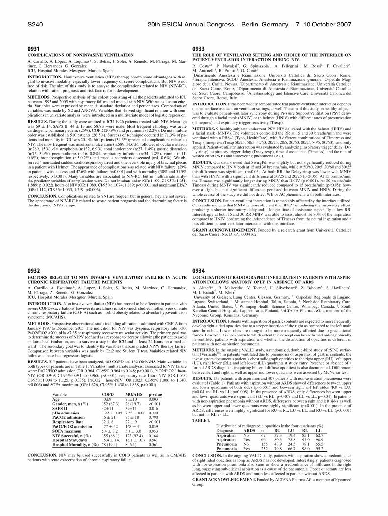
S240 20th ESICM Annual Congress – Berlin, Germany – 7–10 October 2007
0931COMPLICATIONS OF NONINVASIVE VENTILATION
A. Carrillo, A. Lopez, A. Esquinas*, S. Botias, J. Soler, A. Renedo, M. Parraga, M. Mar-tinez, C. Hernandez, G. GonzalezICU, Hospital Morales Meseguer, Murcia, Spain
INTRODUCTION. Noninvasive ventilation (NIV) therapy shows some advantages with re-gard to invasive modality, especially lower frequency of severe complications. But NIV is notfree of risk. The aim of this study is to analyze the complications related to NIV (NIV-RC),relation with patient prognosis and risk factors for it development.
METHODS. Prospective analysis of the cohort consisting of all the pacients admitted to ICUbetween 1995 and 2005 with respiratory failure and treated with NIV. Without exclusion crite-ria. Variables were expressed by mean ± standard deviation and percentages. Comparison ofvariables was made by X2 and ANOVA. Variables that showed significant relation with com-plications in univariate analysis, were introduced in a multivariate model of logistic regression.
RESULTS. During the study were amitted in ICU 1926 patients treated with NIV. Mean agewas 69 ± 14, SAPS II: 44 ± 13 and 58.8% were male. Most frequent ethiology was acutecardiogenic pulmonary edema (25%), COPD (20.9%) and pneumonia (12.2%). Do not intubateorder was established in 510 patients (26.5%). Success of technique occurred in 71.3% of pa-tients and mortality in ICU was 20.1%. 661 patients (34.3%) presented complications related toNIV. The most frequent was nasofrontal ulceration (n:589, 30.6%), followed of ocular irritation(n:289, 15%), claustrophobia (n:132, 6.9%), total intolerance (n:27, 1.4%), gastric distension(n:75, 3.9%), pneumothorax (n:16, 0.8%), respiratory infection (n:34, 1.8%), vomits (n:11,0.6%), bronchoaspiration (n:3,0.2%) and mucous secretions desecated (n:4, 0.6%). We ob-served 4 nonwaited sudden cardiorespiratory arrest and one reversible injury of brachial plexusin a patient with Helmet. The appearance of complications was related with NIV failure. (29%in patients with success and 47.6% with failure; p<0.001) and with mortality (30% and 51.5%respectively, p<0.001). Many variables are associated to NIV-RC, but in multivariate analy-sis, predictor variables of complication were: Do not intubate order (OR:1.409, CI-95%:1.051,1.889; p:0.022), hours of NIV (OR:1.089, CI-95%: 1.074, 1.089; p<0.001) and maximum EPAP(OR:1.112, CI-95%:1.033, 1.219; p:0.006).
CONCLUSION. Complications related to VNI are frequent but in general they are not severe.The appearance of NIV-RC is related to worse patient prognosis and the determining factor isthe duration of NIV therapy.
0932FACTORS RELATED TO NON INVASIVE VENTILATORY FAILURE IN ACUTECHRONIC RESPIRATORY FAILURE PATIENTS
A. Carrillo, A. Esquinas*, A. Lopez, J. Soler, S. Botias, M. Martinez, C. Hernandez,M. Parraga, A. Renedo, G. GonzalezICU, Hospital Morales Meseguer, Murcia, Spain
INTRODUCTION. Non invasive ventilation (NIV) has proved to be effective in patients withsevere COPD exacerbations, however its usefulness is not so much studied in other types of acutechronic respiratory failure (CRF-A) such as morbid obesity related to alveolar hypoventilationsyndrome (MO/AHS).
METHODS. Prospective observational study including all patients admitted with CRF-A fromJanuary 1997 to December 2005. The indication for NIV was dyspnea, respiratory rate >30,PaO2/FiO2 <200, pHa <7.35 or respiratory accessory muscular activity. The primary goal wasto determine the success of NPPV (defined as a response to therapy allowing the patient to avoidendotracheal intubation, and to survive a stay in the ICU and at least 24 hours on a medicalward). The secondary goal was to identify the variables that can predict NPPV therapy failure.Comparison between variables was made by Chi2 and Student T test. Variables related NIVfailre was made bua regression logistic.
RESULTS. 535 patients have been analyzed, 403 COPD and 132 OM/AHS. Main variables inboth types of patients are in Table 1: Variables, multivariate analysis, associated to NIV failurewere: PaO2/FiO2 admission (OR:0.964, CI-95%:0.964 to 0.948; p<0.001), PaO2/FiO2 1 hour-NIV (OR:0.949, CI-95%:0.931 to 0.965; p<0.001), respiratory rate 1hour-NIV (OR:1.063,CI-95%:1.004 to 1.125, p:0.035), PaCO2 1 hour-NIV (OR:1.023, CI-95%:1.006 to 1.040,p:0.006) and SOFA maximum (OR:1.626, CI-95%:1.438 to 1.838, p<0.001).
Variable COPD MO/AHS p-valueAge 70±9 73±10 0.003Gender, men, n (%) 352 (87.3) 26 (19.7) <0.001SAPS II 42±11 39±11 0.016pHa admission 7.22 ± 0.09 7.22 ± 0.08 0.320PaCO2 admission 76 ± 21 75 ± 18 0.790Respiratory Rate 32 ± 8 27 ± 9 <0.001PaO2/FiO2 admission 177 ± 42 168 ± 41 0.039SOFA maximun 5.4 ± 3.2 5.3 ± 3.0 0.953NIV Succesful, n (%) 355 (88.1) 122 (92.4) 0.164Hospital Stay, days 15.4 ± 14.1 16.1 ± 10.7 0.561Hospital Mortality, n (%) 78 (19.4) 8 (6.1) 0.561
CONCLUSION. NIV may be used successfully in COPD patients as well as in OM/AHSpatients with acute exacerbation of chronic respiratory failure.
0933THE ROLE OF VENTILATOR SETTING AND CHOICE OF THE INTERFACE ONPATIENT-VENTILATOR INTERACTION DURING NIV.
R. Costa*1, P. Navalesi2, G. Spinazzola3, A. Pellegrini3, M. Rossi4, F. Cavaliere5,M. Antonelli3, R. Proietti3, G. Conti31Dipartimento Anestesia e Rianimazione, Universita Cattolica del Sacro Cuore, Rome,2Terapia Intensiva, SCDU Anestesia, Anestesia e Rianimazione generale, Ospedale Mag-giore della Carita, Novara, 3Dipartimento di Anestesia e Rianimazione, Universita Cattolicadel Sacro Cuore, Rome, 4Dipartimento di Anestesia e Rianimazione, Universita Cattolicadel Sacro Cuore, Campobasso, 5Anesthesiology and Intensive Care, Universita Cattolica delSacro Cuore, Rome, Italy
INTRODUCTION. It has been widely demonstrated that patient-ventilator interaction dependson the interface used and on ventilator settings, as well. The aim of this study on healthy subjectswas to evaluate patient-ventilator synchrony during Pressure Support Ventilation (PSV) deliv-ered through a facial mask (MNIV) or an helmet (HNIV) with different rates of pressurization(Timepress) and expiratory trigger sensitivity (Trexp).
METHODS. 9 healthy subjects underwent PSV NIV delivered with the helmet (HNIV) anda facial mask (MNIV). The volunteers controlled the RR at 15 and 30 breaths/min and wereventilated with a PB840 (Tyco, HealthCare), with 9 different combinations of Timepress andTrexp (Timepress /Trexp 50/25, 50/5, 50/60, 20/25, 20/5, 20/60, 80/25, 80/5, 80/60), randomlyapplied. Patient-ventilator interaction was evaluated by analyzing inspiratory trigger delay (De-laytrinsp), expiratory trigger delay (Delaytrexp), time of assistance (Timeass), and the rate ofwasted effort (WE) and autocycling phenomena (AC).
RESULTS. Our data showed that SwingPdi was slightly but not significantly reduced duringMNIV compared to HNIV both at 15 and 30 breaths/min, while at 50/60, 20/5, 20/60 and 80/25this difference was significant (p<0.05). At both RR, the Delaytrinsp was lower with MNIVthan with HNIV, with a significant difference at 50/25 and 20/25 (p<0.05). At 15 breaths/min,the Timeass was significantly longer during MNIV than HNIV (p<0.001). At 30 breaths/minTimeass during MNIV was significantly reduced compared to 15 breaths/min (p<0.05); how-ever a slight but not significant difference persisted between MNIV and HNIV. During thewhole course of the study we did not detect WE or AC phenomena with both interfaces.
CONCLUSION. Patient-ventilator interaction is remarkably affected by the interface utilized.Our results indicate that MNIV is more efficient than HNIV in reducing the inspiratory effort,producing a shorter inspiratory delay and a longer time of assistance especially at low RR.Interestingly at both 15 and 30 RR MNIV was able to assist almost the 80% of the inspirationcompared to HNIV, confirming the independence of Timeass from the neural inspiration and aless efficient patient-ventilator interaction with this interface.
GRANT ACKNOWLEDGEMENT. Funded by a research grant from Universita’ Cattolicadel Sacro Cuore, No. D1-PT 0004162.
0934LOCALISATION OF RADIOGRAPHIC INFILTRATES IN PATIENTS WITH ASPIR-ATION FOLLOWS ANATOMY ONLY IN ABSENCE OF ARDS
A. Althoff*1, R. Malacrida2, V. Toome3, H. Silverboard4, Z. Bshouty5, S. Hovilheto6,M. I. Brandt7, M. Klein7
1Unversity of Giessen, Lung Center, Giessen, Germany, 2, Ospedale Regionale di Lugano,Lugano, Switzerland, 3, Mustamae Hospital, Tallin, Estonia, 4, Northside Respiratory Care,Atlanta, United States, 5, Winnipeg Health Science Centre, Winnipeg, Canada, 6, SouthKarelian Central Hospital„ Lappeenranta, Finland, 7ALTANA Pharma AG, a member of theNycomed Group, Konstanz, Germany
INTRODUCTION. Patients with aspiration of gastric contents are expected to more frequentlydevelop right-sided opacities due to a steeper insertion of the right as compared to the left mainstem bronchus. Lower lobes are thought to be more frequently affected due to gravitationalforces. However, it is not known to which extent this concept can be confirmed radiographicallyin ventilated patients with aspiration and whether the distribution of opacities is different inpatients with non-aspiration pneumonia.
METHODS. In the ongoing VALID study, a randomised, double-blind study of rSP-C surfac-tant (Venticute®) in patients ventilated due to pneumonia or aspiration of gastric contents, theinvestigators document a patient’s chest radiograph opacities to the right upper (RU), left upper(LU), right lower (RL), and left lower (LL) quadrants at study entry. Presence or absence of aformal ARDS diagnosis (requiring bilateral diffuse opacities) is also documented. Differencesbetween left and right as well as upper and lower quadrants were assessed by McNemar test.RESULTS. 133 patients with aspiration and 407 patients with non-aspiration pneumonia wereevaluated (Table 1). Patients with aspiration without ARDS showed differences between upperand lower quadrants of both sides (p<0.001) and between right and left sides (RU vs LU;p=0.04 and RL vs LL; p=0.008). In the presence of ARDS, only differences between upperand lower quadrants were significant (RU vs RL; p=0.007 and LU vs LL; p=0.04). In patientswith non-aspiration pneumonia without ARDS, differences between right and left sides as wellas between upper and lower quadrants were highly significant (p<0.001). In the presence ofARDS, differences were highly significant for RU vs RL, LU vs LL, and RU vs LU (p<0.001)but not for RL vs LL.
TABLE 1.Distribution of radiographic opacities in the four quadrants (%)Diagnosis ARDS n RU LU RL LLAspiration No 67 37.3 19.4 85.1 62.7Aspiration Yes 66 80.3 75.8 97.0 90.9Pneumonia No 155 43.9 24.5 78.1 55.5Pneumonia Yes 252 79.8 66.7 98.0 95.2
CONCLUSION. In the ongoing VALID study, patients with aspiration show a predominanceof right sided opacities as long as ARDS has not developed. Interestingly, patients diagnosedwith non-aspiration pneumonia also seem to show a predominance of infiltrates in the rightlung, suggesting sub-clinical aspiration as a cause of the pneumonia. Upper quadrants are lessaffected in patients with ARDS and much less affected in patients without ARDS.GRANT ACKNOWLEDGEMENT. Funded by ALTANA Pharma AG, a member of NycomedGroup.
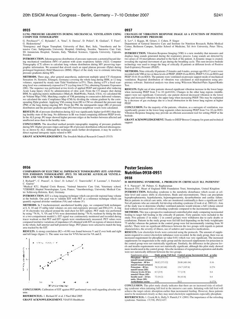
20th ESICM Annual Congress – Berlin, Germany – 7–10 October 2007 S241
0935LUNG PRESSURE GRADIENTS DURING MECHANICAL VENTILATION USINGCOMPUTER TOMOGRAPHY
G. Perchiazzi*1, C. Rylander2, A. Vena3, S. Derosa1, D. Polieri1, R. Giuliani1, T. Fiore1,G. Hedenstierna4
1Emergency and Organ Transplant, University of Bari, Bari, Italy, 2Anesthesia and In-tensive Care, Sahlgrenska University Hospital, Goteborg, Sweden, 3Intensive Care Unit,SS Annunziata Hospital, Taranto, Italy, 4Medical Sciences, Uppsala University, Uppsala,Sweden
INTRODUCTION. Inhomogeneous distribution of pressure represents a potential hazard dur-ing mechanical ventilation (MV) of patient with acute respiratory failure (ALI). ComputerTomography (CT) allows to calculate lung volume distribution during MV preserving topo-graphic information. We assumed that alveoli reach an equal plateau pressure (Pplat) duringprolonged Inspiratory Hold Maneuvers (IHM). Object of the study was to estimate intra-lungpressure gradients during MV.
METHODS. Three pigs, after general anaesthesia, underwent multiple spiral CT (SomatomSensation 16, Siemens, Erlangen, Germany) covering the whole lung during IHM, at 12 lungvolumes, separated by steady state Tidal Ventilation (ssTV). Then, during ssTV a fixed scan-ning level was continuously exposed to CT (cycling time 0.75s), obtaining Dynamic Exposures(DE). The sequence was performed at two levels of applied PEEP and repeated after inducingAcute Lung Injury (ALI) by administration of oleic acid. From the CT images shot duringIHM, by applying matrix mathematics (MatLab, MathWorks, Natick, USA), we calculated theair Volume Map (VM). Matching couples of VM pertaining to consecutive delivered volumes,we calculated the static Compliance Map (CM) by dividing the volume gradient by the corre-sponding Pplat gradient. Applying VM coming from DE to CM we obtained the pressure map(PM) of the lung during ongoing MV. From the PM, the interquartile range (IR) of pressuredistribution and the pressure gradient map (PG) between neighbour voxels were calculated.
RESULTS. IR of pressure distribution during ALI was significantly higher (p<0.001) than dur-ing healthy state of the lung. No difference in IR was found comparing different PEEP levels.In the ALI group, PG maps showed higher pressure ridges at the borders between affected andunaffected areas than in the healthy state.
CONCLUSION. The described method permits topographic mapping of pressure gradientsduring MV. Higher pressure ridges and larger IR of pressure distribution reflected inhomogene-ity as shown by ALI. Although the technique needs further development, it may be useful todetect regional iatrogenic injury related to MV.
GRANT ACKNOWLEDGEMENT. The Swedish Medical Research Council (5315)
0936COMPARISON OF ELECTRICAL IMPEDANCE TOMOGRAPHY (EIT) AND POSI-TON EMISSION TOMOGRAPHY (PET) TO MEASURE ALVEOLAR VENTILA-TION AND VOLUME IN NORMAL PIGS
J. Richard*1, C. Pouzot2, A. Gros1, D. Lebars3, C. Tourevieille3, F. Lavenne3, I. Frerichs4,C. Guerin1
1Medical ICU, Hopital Croix Rousse, 2Animal Intensive Care Unit, Veterinary school,3CERMEP, Hopital Neurologique, Lyon, France, 4Anesthesiology, University Medical Cen-tre Schleswig-Holstein, Kiel, Germany
INTRODUCTION. EIT is a lung imaging technique which can provide regional lung volumeat the bedside. Our goal was to validate EIT with PET as a reference technique which canquantify regional alveolar ventilation (VA) and volume (V).
METHODS. In 5 tracheotomized, anesthetized healthy pigs, we compared both techniquesat 6, 8, 10 and 15 ml/kg tidal volumes on zero end-expiratory pressure and FIO221%. A ringof 16 electrodes was placed around the mid-chest for EIT capture. PET study was performedby using 13N-N2. V, VA and V/VA were determined during 13N-N2 washout by fitting the datato a two-compartment model(1). EIT signal was continuously monitored and recorded duringtracer washout so that PET and EIT signals were simultaneously measured. PET values werecompared torelative variations of impedance (Z) obtained with EIT on regions of interest drawnin the whole, half anterior and half posterior lungs. PET planes were selected to match the lungarea tracked by the EIT.
RESULTS. A strong correlation (R2>=0.90) was found between V and Z over both and rightand left lungs (figure 1). The same was true for V/VA but not for VA and Z.
CONCLUSION. Calibration of EIT against PET performed very well regarding alveolar vol-ume.REFERENCE(S). 1. Richard JC et al. J Nucl Med 2005
GRANT ACKNOWLEDGEMENT. VIASYS Healthcare
0937CHANGES OF VIBRATION RESPONSE IMAGE AS A FUNCTION OF POSITIVEEND-EXPIRATORY PRESSURE
S. Lev*, I. Kagan, M. Grinev, J. Cohen, P. SingerDepartment of General Intensive Care and Institute for Nutrition Research, Rabin MedicalCenter, Beilinson Campus, Sackler School of Medicine, Tel Aviv University, Petav Tikva,Israel
INTRODUCTION. Vibration Response Imaging (VRI) is a new modality that measures anddisplays lung sounds generated during the respiratory process. Lung sounds are detected bytwo arrays of 18 microphones attached to the back of the patient. A dynamic image is created,revealing the regional movement of gas during the breathing cycle. This non-invasive bedsidetechnology was used to image the lung of critically ill patients at different levels of PositiveEnd-Expiratory Pressure (PEEP).
METHODS. Nine critically ill patients (5 females and 4 males, average age 69±17 years) wererecorded with VRI at two or three levels of PEEP: ZEEP (0 cm H2O), PEEP 5 (5-6 cm H2O) andPEEP 10 (8-10 cm H2O). The patients were ventilated on pressure support mode of mechanicalventilation. Regional distribution of vibration was calculated at mid-inspiration using pro-prietary software. Statistical analysis was done using Wilcoxon Matched-Pairs Signed-RanksTest.
RESULTS. Eight out of nine patients showed significant vibration increase in the lower lungswhen increasing PEEP from 5 to 10 (p=0.016). Changes in the other lung regions (middle,upper) were not significant. Conversely, one patient showed decreased vibration in the lowerlungs and increased vibration in the upper lung when increasing PEEP. This may be explainedby a decrease of gas exchange due to a local obstruction in the lower lung regions at higherPEEP.
CONCLUSION. For the majority of the patients, vibration, as a surrogate of ventilation, wassignificantly greater in the lower lung segments when increasing PEEP from 5 to 10 cm H2O.Vibration Response Imaging may provide an efficient assessment tool for setting PEEP at thebedside.
GRANT ACKNOWLEDGEMENT. Thanks to DEEP Breeze Company for grant and technicalsupport
Poster SessionsNutrition 0938-09510938IS REFEEDING SYNDROME, A PROBLEM IN CRITICALLY ILL PATIENTS?
P. S. Narayan*, M. Palmer, G. RaghuramanResearch ITU, Heart of England NHS Foundation Trust, birmingham, United KingdomINTRODUCTION. Refeeding syndrome is the metabolic disturbance which occurs at cel-lular level and causes shifts in electrolytes, fluids and micronutrients. These can precipitatehypophosphataemia, hypokalaemia, hypomagnesaemia, dysarrhythmias and cardiac failure.But,in patients in critical care units, who are monitored continually,is there a significant risk?Not all patients who are enterally fed develop refeeding syndrome (Crook et al, 2001)(1). Aimof the study was to determine whether ventilated patients would tolerate a full volume enteralfeed without increased risks of refeeding syndrome compared to the incremental feeding.METHODS. This was a prospective randomised controlled pilot study comparing incrementalfeeding to target full feeding in the critically ill patients. Forty patients were included in thestudy. Five patients (3 in study / 2 in control groups) were withdrawn due to early deaths orextubations. Patients in the study group were fed full feed depending on the body weight(upto75 mls/hr) whereas the patients in the control group were fed incrementally (starting from 30mls/hr). There were no significant differences between the two groups with regards to patientcharacteristics, the severity of illness, use of sedative and vasoactive medications.RESULTS. Low electrolyte levels were corrected using the protocols. The amount of supple-ments required to correct electrolyte imbalance were recorded. In the study group, there was anincreased requirement for phosphate (p-value 0.01) which was very significant. The increasedsupplements for magnesium in the study group and the increased supplements for potassium inthe control group were not statistically significant. Similarly, the difference in the glucose lev-els and insulin requirements were not statistically significant, although this pilot study showedmore insulin need in the control group. Also,the incidence of regurgitation,aspiration and deathswere not statistically different between the two groups.
Supplements given Study group Full feed Control group Incremental feed p-valuePhosphate - 48 hrs 24.70 [676.47] 6.66 [94.11] 0.01mmol/l mean[variance]Magnesium - 48 hrs 38.82 [373.52] 28.88 [481.04] 0.16mmol/l mean[variance]Potassium - 48 hrs 78.24 [83.68] 134.7 [107.8] 0.274mmol/l mean [SD]Glucose levels-24 hrs 8.5 [2.9] 7.7 [1.9] 0.4mmols/l Mean (SD)Insulin need - 24 hrs 72 [54.2] 56 [43.8] 0.4units, Mean (SD)Arrhythmias yes/no 1/16 5/13 0.1774
CONCLUSION. This pilot study clearly indicates that there are no increased risks of refeed-ing syndrome when initiating full feed in the intensive care units. Initiating with full feed willachieve the target caloric absorption earlier than incremental feeding. However, these patientsneed to be monitored closely as they may need electrolyte supplements especially phosphate.REFERENCE(S). 1. Crook,M.A. Hally,V. Panteli,J.V. (2001) The importance of the refeedingsyndrome. Nutrition. 17(7/8). P632-637.
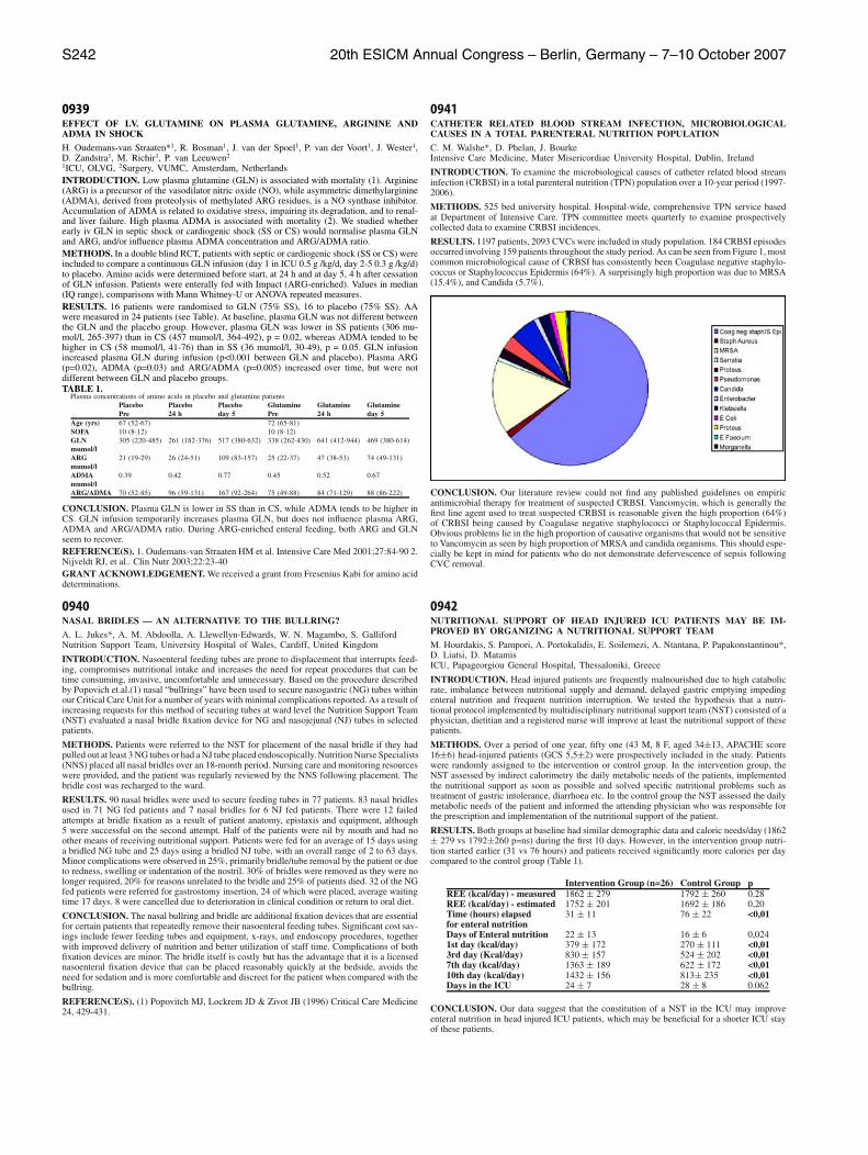
S242 20th ESICM Annual Congress – Berlin, Germany – 7–10 October 2007
0939EFFECT OF I.V. GLUTAMINE ON PLASMA GLUTAMINE, ARGININE ANDADMA IN SHOCK
H. Oudemans-van Straaten*1, R. Bosman1, J. van der Spoel1, P. van der Voort1, J. Wester1,D. Zandstra1, M. Richir1, P. van Leeuwen2
1ICU, OLVG, 2Surgery, VUMC, Amsterdam, NetherlandsINTRODUCTION. Low plasma glutamine (GLN) is associated with mortality (1). Arginine(ARG) is a precursor of the vasodilator nitric oxide (NO), while asymmetric dimethylarginine(ADMA), derived from proteolysis of methylated ARG residues, is a NO synthase inhibitor.Accumulation of ADMA is related to oxidative stress, impairing its degradation, and to renal-and liver failure. High plasma ADMA is associated with mortality (2). We studied whetherearly iv GLN in septic shock or cardiogenic shock (SS or CS) would normalise plasma GLNand ARG, and/or influence plasma ADMA concentration and ARG/ADMA ratio.METHODS. In a double blind RCT, patients with septic or cardiogenic shock (SS or CS) wereincluded to compare a continuous GLN infusion (day 1 in ICU 0.5 g /kg/d, day 2-5 0.3 g /kg/d)to placebo. Amino acids were determined before start, at 24 h and at day 5, 4 h after cessationof GLN infusion. Patients were enterally fed with Impact (ARG-enriched). Values in median(IQ range), comparisons with Mann Whitney-U or ANOVA repeated measures.RESULTS. 16 patients were randomised to GLN (75% SS), 16 to placebo (75% SS). AAwere measured in 24 patients (see Table). At baseline, plasma GLN was not different betweenthe GLN and the placebo group. However, plasma GLN was lower in SS patients (306 mu-mol/l, 265-397) than in CS (457 mumol/l, 364-492), p = 0.02, whereas ADMA tended to behigher in CS (58 mumol/l, 41-76) than in SS (36 mumol/l, 30-49), p = 0.05. GLN infusionincreased plasma GLN during infusion (p<0.001 between GLN and placebo). Plasma ARG(p=0.02), ADMA (p=0.03) and ARG/ADMA (p=0.005) increased over time, but were notdifferent between GLN and placebo groups.TABLE 1.
Plasma concentrations of amino acids in placebo and glutamine patientsPlacebo Placebo Placebo Glutamine Glutamine GlutaminePre 24 h day 5 Pre 24 h day 5
Age (yrs) 67 (52-67) 72 (65-81)SOFA 10 (8-12) 10 (8-12)GLN 305 (220-485) 261 (182-376) 517 (380-632) 338 (262-430) 641 (412-944) 469 (380-614)mumol/lARG 21 (19-29) 26 (24-51) 109 (83-157) 25 (22-37) 47 (38-53) 74 (49-131)mumol/lADMA 0.39 0.42 0.77 0.45 0.52 0.67mumol/lARG/ADMA 70 (52-85) 96 (59-131) 167 (92-264) 75 (49-88) 84 (71-129) 88 (86-222)
CONCLUSION. Plasma GLN is lower in SS than in CS, while ADMA tends to be higher inCS. GLN infusion temporarily increases plasma GLN, but does not influence plasma ARG,ADMA and ARG/ADMA ratio. During ARG-enriched enteral feeding, both ARG and GLNseem to recover.REFERENCE(S). 1. Oudemans-van Straaten HM et al. Intensive Care Med 2001;27:84-90 2.Nijveldt RJ, et al.. Clin Nutr 2003;22:23-40GRANT ACKNOWLEDGEMENT. We received a grant from Fresenius Kabi for amino aciddeterminations.
0940NASAL BRIDLES — AN ALTERNATIVE TO THE BULLRING?
A. L. Jukes*, A. M. Abdoolla, A. Llewellyn-Edwards, W. N. Magambo, S. GallifordNutrition Support Team, University Hospital of Wales, Cardiff, United Kingdom
INTRODUCTION. Nasoenteral feeding tubes are prone to displacement that interrupts feed-ing, compromises nutritional intake and increases the need for repeat procedures that can betime consuming, invasive, uncomfortable and unnecessary. Based on the procedure describedby Popovich et.al.(1) nasal “bullrings” have been used to secure nasogastric (NG) tubes withinour Critical Care Unit for a number of years with minimal complications reported. As a result ofincreasing requests for this method of securing tubes at ward level the Nutrition Support Team(NST) evaluated a nasal bridle fixation device for NG and nasojejunal (NJ) tubes in selectedpatients.
METHODS. Patients were referred to the NST for placement of the nasal bridle if they hadpulled out at least 3 NG tubes or had a NJ tube placed endoscopically. Nutrition Nurse Specialists(NNS) placed all nasal bridles over an 18-month period. Nursing care and monitoring resourceswere provided, and the patient was regularly reviewed by the NNS following placement. Thebridle cost was recharged to the ward.
RESULTS. 90 nasal bridles were used to secure feeding tubes in 77 patients. 83 nasal bridlesused in 71 NG fed patients and 7 nasal bridles for 6 NJ fed patients. There were 12 failedattempts at bridle fixation as a result of patient anatomy, epistaxis and equipment, although5 were successful on the second attempt. Half of the patients were nil by mouth and had noother means of receiving nutritional support. Patients were fed for an average of 15 days usinga bridled NG tube and 25 days using a bridled NJ tube, with an overall range of 2 to 63 days.Minor complications were observed in 25%, primarily bridle/tube removal by the patient or dueto redness, swelling or indentation of the nostril. 30% of bridles were removed as they were nolonger required, 20% for reasons unrelated to the bridle and 25% of patients died. 32 of the NGfed patients were referred for gastrostomy insertion, 24 of which were placed, average waitingtime 17 days. 8 were cancelled due to deterioration in clinical condition or return to oral diet.
CONCLUSION. The nasal bullring and bridle are additional fixation devices that are essentialfor certain patients that repeatedly remove their nasoenteral feeding tubes. Significant cost sav-ings include fewer feeding tubes and equipment, x-rays, and endoscopy procedures, togetherwith improved delivery of nutrition and better utilization of staff time. Complications of bothfixation devices are minor. The bridle itself is costly but has the advantage that it is a licensednasoenteral fixation device that can be placed reasonably quickly at the bedside, avoids theneed for sedation and is more comfortable and discreet for the patient when compared with thebullring.
REFERENCE(S). (1) Popovitch MJ, Lockrem JD & Zivot JB (1996) Critical Care Medicine24, 429-431.
0941CATHETER RELATED BLOOD STREAM INFECTION, MICROBIOLOGICALCAUSES IN A TOTAL PARENTERAL NUTRITION POPULATION
C. M. Walshe*, D. Phelan, J. BourkeIntensive Care Medicine, Mater Misericordiae University Hospital, Dublin, Ireland
INTRODUCTION. To examine the microbiological causes of catheter related blood streaminfection (CRBSI) in a total parenteral nutrition (TPN) population over a 10-year period (1997-2006).
METHODS. 525 bed university hospital. Hospital-wide, comprehensive TPN service basedat Department of Intensive Care. TPN committee meets quarterly to examine prospectivelycollected data to examine CRBSI incidences.
RESULTS. 1197 patients, 2093 CVCs were included in study population. 184 CRBSI episodesoccurred involving 159 patients throughout the study period. As can be seen from Figure 1, mostcommon microbiological cause of CRBSI has consistently been Coagulase negative staphylo-coccus or Staphylococcus Epidermis (64%). A surprisingly high proportion was due to MRSA(15.4%), and Candida (5.7%).
CONCLUSION. Our literature review could not find any published guidelines on empiricantimicrobial therapy for treatment of suspected CRBSI. Vancomycin, which is generally thefirst line agent used to treat suspected CRBSI is reasonable given the high proportion (64%)of CRBSI being caused by Coagulase negative staphylococci or Staphylococcal Epidermis.Obvious problems lie in the high proportion of causative organisms that would not be sensitiveto Vancomycin as seen by high proportion of MRSA and candida organisms. This should espe-cially be kept in mind for patients who do not demonstrate defervescence of sepsis followingCVC removal.
0942NUTRITIONAL SUPPORT OF HEAD INJURED ICU PATIENTS MAY BE IM-PROVED BY ORGANIZING A NUTRITIONAL SUPPORT TEAM
M. Hourdakis, S. Pampori, A. Portokalidis, E. Soilemezi, A. Ntantana, P. Papakonstantinou*,D. Liatsi, D. MatamisICU, Papageorgiou General Hospital, Thessaloniki, Greece
INTRODUCTION. Head injured patients are frequently malnourished due to high catabolicrate, imbalance between nutritional supply and demand, delayed gastric emptying impedingenteral nutrition and frequent nutrition interruption. We tested the hypothesis that a nutri-tional protocol implemented by multidisciplinary nutritional support team (NST) consisted of aphysician, dietitian and a registered nurse will improve at least the nutritional support of thesepatients.
METHODS. Over a period of one year, fifty one (43 M, 8 F, aged 34±13, APACHE score16±6) head-injured patients (GCS 5,5±2) were prospectively included in the study. Patientswere randomly assigned to the intervention or control group. In the intervention group, theNST assessed by indirect calorimetry the daily metabolic needs of the patients, implementedthe nutritional support as soon as possible and solved specific nutritional problems such astreatment of gastric intolerance, diarrhoea etc. In the control group the NST assessed the dailymetabolic needs of the patient and informed the attending physician who was responsible forthe prescription and implementation of the nutritional support of the patient.
RESULTS. Both groups at baseline had similar demographic data and caloric needs/day (1862± 279 vs 1792±260 p=ns) during the first 10 days. However, in the intervention group nutri-tion started earlier (31 vs 76 hours) and patients received significantly more calories per daycompared to the control group (Table 1).
Intervention Group (n=26) Control Group pREE (kcal/day) - measured 1862 ± 279 1792 ± 260 0.28REE (kcal/day) - estimated 1752 ± 201 1692 ± 186 0,20Time (hours) elapsed 31 ± 11 76 ± 22 <0,01for enteral nutritionDays of Enteral nutrition 22 ± 13 16 ± 6 0,0241st day (kcal/day) 379 ± 172 270 ± 111 <0,013rd day (Kcal/day) 830 ± 157 524 ± 202 <0,017th day (kcal/day) 1363 ± 189 622 ± 172 <0,0110th day (kcal/day) 1432 ± 156 813± 235 <0,01Days in the ICU 24 ± 7 28 ± 8 0.062
CONCLUSION. Our data suggest that the constitution of a NST in the ICU may improveenteral nutrition in head injured ICU patients, which may be beneficial for a shorter ICU stayof these patients.
20th ESICM Annual Congress – Berlin, Germany – 7–10 October 2007 S243
0943INCIDENCE AND PATTERNS OF ABNORMAL LIVER FUNCTION TESTS ONTHE GENERAL INTENSIVE CARE UNIT
S. J. Thomson*1, M. L. Cowan2, P. O. Collinson3, M. Grounds1, T. M. Rahman1
1Department of Intensive Care, 2Department of Gastroenterology and Hepatology,3Department of Chemical Pathology, St George’s NHS Trust, London, United Kingdom
INTRODUCTION. The interpretation of deranged liver function tests (LFT) is a commonclinical problem faced by critical care physicians. There is little descriptive data in the literaturewhich adequately illustrates the nature of the problem. We undertook a retrospective analysis ofdata within our institution to identify the trends in LFT and ascertain the frequency of abnormalresults.
METHODS. The results of all LFT requests by the General Intensive Care Unit of a largeteaching hospital in South London, between 1st December 2006 and 28th February 2007 wereobtained from the Chemical Pathology department.
RESULTS. During the three months studied 1655 sets of LFT were performed on 355 pa-tients representing 1472 patient-days. All patients had daily LFT checked as part of a routine‘ITU profile’. LFT were checked twice in 152 patient-days, three times in 12 and four timesonce. Bilirubin was above the normal range in 36% of tests, alanine transaminase (ALT) 27%,alkaline phosphatase (AKP) 21% and gamma-glutaryl transferase (gammaGT) 45% (table). Av-erage bilirubin rose from 19.8mumo/l on admission to 48.7mumo/l on day 13 and fell graduallythereafter. ALT peaked on day 5 (52.7 IU/l, 91.61 IU/l, p<0.01). AKP and gammaGT increasedlater after admission to a peak on days 17 and 13 respectively and then plateaued. Values ofAKP and gammaGT were correlated (r2=0.51) whilst there was only very weak correlationbetween ALT and AKP or gammaGT.
Bilirubin ALT AKP gammaGTNormal range 1053 (64%) 1211 (73%) 1305 (79%) 906 (55%)1-2xULN 394 (24%) 249 (16%) 244 (15%) 325 (20%)2-5xULN 145 (9%) 122 (7%) 101 (6%) 263 (16%)5-10xULN 24 (1%) 34 (2%) 5 (<1%) 105 (6%)>10xULN 28 (2%) 40 (2%) 53 (3%)Distribution of LFT in 1655 tests performed in three months on the General ICU
CONCLUSION. We have demonstrated that abnormal LFT are a significant problem on theGeneral Intensive Care Unit. They are checked daily on a routine basis and in 10% of caseswere repeated on the same day. Our data suggest two patterns of abnormal LFT, peaking atdifferent times during the ITU stay, correlating with a clinical distinction between ‘hepatitic’and ‘cholestatic’ liver dysfunction. The causative factors behind abnormal LFT are likely to bemultifactorial and may be related to pre-existing liver conditions, the acute illness promptingadmission to the ITU, diseases evolving during the ITU stay or critical care therapeutic inter-ventions. The cost of performing each ‘set’ of LFT was £4 (∼ e6, $8) equating to £26,480(e39,720, $52,960) per year. Given the high prevalence of LFT outside of the normal range,prospective studies to investigate their clinical significance and causes are required.
0944LOW ALBUMIN IS UBIQUITOUS ON THE GENERAL INTENSIVE CARE UNITAND IS ASSOCIATED WITH A LONGER LENGTH OF STAY BUT A LOWERRISK OF DEATH
S. J. Thomson*1, M. L. Cowan2, P. O. Collinson3, M. Grounds1, T. M. Rahman1
1Department of Intensive Care, 2Department of Gastroenterology and Hepatology,3Department of Chemical Pathology, St George’s NHS Trust, London, United Kingdom
INTRODUCTION. Serum albumin level falls with acute and chronic illness and albuminlevels outside of the normal range are common in the critically ill. Serum albumin is routinelytested daily but the significance of abnormal values is not clear. We planned to define the rangeof serum albumin at admission and trends during stay on the General Intensive Care Unit andassociation with length of stay and 30 day mortality.
METHODS. Levels of serum albumin in patients admitted to the General Intensive Care Unitof a large teaching hospital in South London, between 1st December 2006 and 28th Febru-ary 2007 were obtained from the Chemical Pathology department. Mortality statistics wereobtained from the hospital electronic patient record.
RESULTS. 1655 blood tests were performed during this three month period with an averageserum albumin level of 18.6g/l. Only 35 (2%) were greater than 35g/l, the lower limit of thenormal range. A total of 355 patients, 57 of whom died within 30 days (16%), had a firstadmission to the general ICU during the study period. The average age was 58.4yrs (SD 18.1),55% were male and the mean length of stay was 3.9 days (range 1-26 days). The median albu-min on admission was 22g/dl. Levels of albumin progressively declined during the ICU stay.Patients with albumin less than 22g/dl on admission were older (60.7 yrs v 55.8 yrs, p<0.05).An albumin less than 22g/dl at the time of admission was associated with a longer length ofstay (4.6 days v 3.1 days p<0.001). Albumin greater than 22g/dl at the time of admission wasassociated with a greater 30 day mortality (OR 2.1; 1.17-3.77).
CONCLUSION. Serum albumin in the critically ill patient is almost invariably below the lowerlevel of what is considered the normal range and falls further as the length of time spent onthe ICU increases. Lower levels of albumin at the time of admission may reflect chronic illnessprior to presentation since they are more common in older patients and result in an increasedICU stay. The statistics suggesting that higher levels of albumin are associated with greatermortality may reflect the poor outcome associated with sudden catastrophic illness. The causesof low albumin are multifactorial and prospective studies are needed to elucidate these and theirconsequences.
0945IS THERE ANY DIFFERENCE IN NUTRITIONAL SUPPORT BETWEEN MEDI-CAL AND NON-MEDICAL ICU PATIENTS?
L. A. Trindade*1, J. M. Pereira1, L. Aguiar1, A. Taveira Gomes2
1Unidade de Cuidados Intensivos Polivalente da Urgencia, 2Servico de Cirurgia, Hospital SJoao, Oporto, Portugal
INTRODUCTION. Early nutrition in the critically ill patient will improve outcome and fa-cilitate more rapid rehabilitation. Recent data suggest that nutritional support (NS) is a veryimportant part of the management of the critically ill patient, whether the acute illness begins asa medical or a surgical process. The goals of our study were to quantify the NS administrationand to compare NS in medical patients (MP) and non-medical patients (NMP)(trauma andsurgical).
METHODS. Prospective analysis of ICU patients admitted consecutively. Exclusion criteriawere: NS for <2 days, age <16 years and length-of-stay (LOS) < 3 days. Patients were fol-lowed for the first 7 days of NS. Data collection included: demographic data, NS (route, targetvolume and delivered volume), reasons for interruption and suspension of NS. Chi-square,Mann-Whitney U and T-test were used in statistical analysis.
RESULTS. Fifty patients were included: 26 were MP (52%)and 24 NMP (48%). A total of327 nutrition days (ND) were analyzed: MP 166 days and NMP 161 days. Mean age of MPwas higher than NMP (64+11.3 vs. 49.3+22.3; p=0.006). Most patients were male (61.5% vs.87.5%; p= 0.037) without differences regarding weight, SAPS II score on admission, targetvolume of NS and vasopressors use. NMP were put on sedatives more frequently than MP(p<0.05). NS was started earlier in MP (mean: 29.8 h +14.6 vs. 38.9 h +14.9; p=0.035), mainlyby enteral route (92.3% vs. 79.2%; p=ns). Target volume of NS was achieved in 70% of thepatients (MP 73.1% vs. NMP 66.7%; p=ns). MP received > 90% of what was prescribed in80% of ND and NMP in 76.6% (p=ns). NS was interrupted in 29 ND in MP and 33 in NMP(p=ns), mainly due to gastrointestinal dysfunction (47.4% vs. 57.6%; p=ns). GI dysfunctionwas responsible for reduction of enteral feeding in 8% of ND in MP and in 9.8% in NMP(p=ns). There was no difference in ICU LOS and mortality between the 2 groups (p=ns).
CONCLUSION. Although NS was started earlier in MP with statistical significance, mostof the patients received > 90% of prescribed NS in both groups. No statistically significantdifference was found between MP and NMP regarding: route feeding, target dose achievementand NS interruption or reduction.
0946HOW IS IRON PRESCRIBED IN FRENCH INTENSIVE CARE UNITS?
S. Lasocki*, M. Garnier, L. Ley, R. Bronchard, J. Desmonts, P. MontraversDepartement d’anesthesie reanimation chirurgicale, APHP, CHU Bichat, universite Paris 7,paris, France
INTRODUCTION. Blood loss and iron deficiency (ID) are important factors of intensive careanemia(1). However, neither recommendation nor study focusing on iron treatment exists. Theaim of this study was to evaluate French intensivists’ (I) practices of iron supplementation.
METHODS. We collected 128 questionnaires (containing 19 items) from French I, workingin mixed (45%), surgical (31%) or medical (24%) intensive care units. They where separatedin 2 groups ((P) prescribing iron ≥1 time / week; or not (NP), ≤ 1 time / month). Comparisonswere done using Mann-Whitney or Chi-2 tests, as appropriate. Iron treatments (IV or oral)efficacy and tolerance were quoted from 1 to 10 and compared by a Wilcoxon rank test. p<0.05significant.
RESULTS. All I (73% males, mean age 36±8 yrs) considered having anemic patients and 94%having patients with ID. 72% of I judged ID frequent (concerning >10% of their patients).73% diagnosed ID using biology tests; with iron tests for 90% and blood numeration for 84%.Only 25% of I were P. 20% declared using IV iron, 35% oral iron and 44% both. 20% usedblood transfusion for ID correction. Treatment was ended after a calculate dose or duration in29%, after anemia or iron tests normalization in 32% or was ended by the discharged physi-cian (29%). IV iron was considered more efficient (7±2 vs 6±2, p<0.001) and tolerated (7±2vs 6±2, p<0.001) than oral iron. There was no characteristics difference between P and NP(according to age, sexe, ICU type, hospital type). P answered more often having ID patients(p=0.04), and having more patients with ID (p=0.03). But, P did not performed biology tests forID diagnosis more often than NP (p=0.26) ; and P did not consider treatment efficacy (p=0.68et 0.17 for IV and oral) or tolerance (p=0.32 et 0.06 for IV and oral) higher than NP.
CONCLUSION. Intensive care anemia and ID are considered to be frequent by the Frenchintensivists interrogated, and a large proportion of them (3/3) declared using biology tests toassess ID. Iron treatments are judged efficient and quite well tolerated. However, only 25 % ofI are frequently prescribing iron treatments. The efficacy and the tolerance of these treatmentsdo not seem to influence the doctor choice, since they are not different for P and NP. Thisunderscore that ID is a clinically relevant problem but, to date, insufficiently explored.
REFERENCE(S). (1) von Ahsen Crit Care Med 1999 ;27:2630-9
S244 20th ESICM Annual Congress – Berlin, Germany – 7–10 October 2007
0947MINIMISING RISK
S. E. M. Scutt*, A. L. JukesNutrition and Dietetics, University Hospital of Wales, CARDIFF, United Kingdom
INTRODUCTION. Enteral tube feeding is the preferred method of nutrition support in thecritically ill patient. It is more physiological, cheaper and associated with fewer complicationsthan parenteral nutrition. The majority of patients that receive enteral nutrition are fed usinga nasogastric tube (NG). The main clinical risk associated is of NG tube misplacement. TheTrust guidelines for NG feeding were revised as a result of the 2005 National Patient SafetyAlert “Reducing the harm caused by misplaced nasogastric feeding tubes.” The aim of thisreview was to identify measures used within the Critical Care Unit targetted at reducing therisks associated with enteral nutrition and to assess compliance with these strategies.
METHODS. The potential risks of enteral feeding were identified as misplaced nasogastricfeeding tubes and incorrect feed prescription or administration. Documentation employed toreduce these risks was identified within the standardised fluid prescription chart and nursingcare plan, both of which are completed daily. Both documents were reviewed on each patientfor the previous 1 to 5 days. The feed administration set was examined to determine when itwas set up. The feed infusion was checked against the feed prescription.
RESULTS. 15 patients and their notes were reviewed giving a total of 54 patient days. Themajority (73%) were receiving NG feeding and 13% were fed via a gastrostomy tube. Afluid prescription chart is written daily and has a section for enteral feed. This was prescribedcorrectly on 85% of occasions (9% not documented, 6% incorrectly). The correct feed wasadministered in 100% of enterally fed patients on the day of review. The nursing care planhas a section to record enteral tube type, length at nose and confirm tube position check oneach shift. The external NG tube length was correctly documented in 16% of cases (72% hadno documentation, 12% ticked box without documentation of method). The method used tocheck tube position was correctly documented in 8% of cases (34% had no documentation,58% ticked box without documentation of method). In the Critical Care Unit enteral nutritionis delivered continuously and although reported as standard practice, the administration set wasonly labelled with the time and date of renewal in 20% of cases, potentially increasing risk ofmicrobial contamination or unecessary set renewal.
CONCLUSION. Standardised documentation for medical and nursing staff within CriticalCare encompasses strategies to promote practice to minimise the risks associated with enteraltube feeding. However, compliance in practice is poor or poorly documented. Education andtraining regarding procedures and appropriate documentation should improve compliance anda review will be undertaken in 6 months.
REFERENCE(S). NPSA(2005)Reducing the harm caused by misplaced nasogastric feedingtubes. Patient Safety Alert 05.
0948BEDSIDE PLACEMENT OF SMALL BOWEL FEEDING TUBES
N. A. White, F. Swinton*, K. DonovanCritical Care, Royal Bournemouth Hospital, Bournemouth, United Kingdom
INTRODUCTION. Enteral feeding is the preferred route of feeding for the majority of ICUpatients. Often gastric feeding fails to meet feeding requirements due to delayed gastric empty-ing caused by drugs or pathology 1. Small bowel tube feeding has been advocated as a way ofovercoming problems with gastric emptying however placement of small bowel feeding tubescan be difficult and time consuming 2 leading to delay in instigation of feed and failure to reachcalorific requirements. We present our bedside method of small bowel feeding tube placementwhich has proved rapid and successful.
METHODS. Following a review of the literature one of the authors (NW) developed a tech-nique for placement of small bowel feeding tubes. Patients were placed 45 degrees right lateraland 30 degrees head up. A size 8 french 110 cm flocare® polyurothane feeding tube with guidewire (manufacturer Nutricia Healthcare SA) was inserted to 70 cm. 30 minutes after adminis-tration of 10mg of metoclopramide and 250 mg of erythromycin iv the tube was advanced to100cm at the nose over at least five minutes. The wire was left in situ and and an X-Ray wastaken to confirm position.NW has been using this technique successfully for over a year. Here we present details of thelast 5 patients on whom this technique was used.
RESULTS. All 5 insertions were successful with position confirmed radiologically in eachcase (see image). The mean time from start of procedure to confirmation of placement was 91.8minutes.
CONCLUSION. We believe with this technique small bowel feeding tubes can be quickly,easily and reliably placed at the bedside in ICU.
REFERENCE(S). 1. Mentec H et al. Upper digestive intolerance of entereal nutrition in criti-cally ill patients: Frequency, risk factors, and complications. Crit Care Med 2001; 29:1955-612. Neumann DA, Delegge MH. Gastric vs small bowel feeding in the intensive care unit: aprospective comparison of efficacy. Crit Care Med 2002; 30:1436-38.
0949THE USE OF AN ENTERAL DIET ENRICHED WITH ARGININE IS ASSOCI-ATED WITH INCREASED TOXICITY ON HUMAN LYMPHOCYTES OF PATIENTSWITH SEVERE SEPSIS
A. Pontes-Arruda*1, F. Longo2
1Intensive Care Department, Fernandes Tavora Hospital, Fortaleza, Brazil, 2Laboratoryof Cellular and Molecular Biology, International Center for Genetic Engineering andBiotechnology, Trieste, Italy
INTRODUCTION. The use of an enteral nutrition with eicosapentaenoic acid (EPA fromfish oil) and gamma-linolenic acid (GLA from Borage Oil) but not enhanced with argininehas been associated with improved outcomes in patients with severe sepsis and septic shock(Pontes-Arruda et al, 2006). The use of arginine enhanced diets, particularly in patients withsevere sepsis, remains controversial. The level of toxicity of a diet to human lymphocytes maybe evaluated by its capacity to increase cell death by apoptosis and necrosis (Cury-Boaventuraet al., 2004). The aim of this study is to compare the toxicity of the EPA+GLA diet with anEPA+Arginine diet on human lymphocytes obtained from patients with severe sepsis.
METHODS. 45 patients with severe sepsis requiring mechanical ventilation were randomizedto receive either: an EPA+GLA diet (Oxepa, Abbott Labs), a high lipid diet not enriched withEPA or GLA used as control (Pulmocare, Abbott Labs) or an EPA+Arginine enhanced diet(Impact, Novartis). Caloric goals were calculated using the Harris-Benedict equation x 1.3. Thefirst day patients achieved 75% of BEE x 1.3 was considered as study day 1. Blood samples werecollected at baseline, Study Day 4 and Study Day 7 to access lymphocyte proliferation, and cellmembrane integrity (a marker of necrosis) and DNA fragmentation (a marker of apoptosis) inaccordance with the protocol previously described by Cury-Boaventura et al.
RESULTS. On study day 4, the EPA+arginine enhanced diet was associated with a significantdecrease in lymphocyte proliferation as compared with the EPA+GLA and control diets (44% x93% x 65%, EPA+arginine, EPA+GLA and control diet respectively, p<0.001), increased DNAfragmentation (46% x 17% x 34%, p<0.001) and increased loss of cell membrane integrity(65% x 83% x 70%, p<0.001). Similar results were observed on study day 7.
CONCLUSION. This study demonstrates for the first time that an arginine enhanced enteraldiet is associated with increased human lymphocyte toxicity as well as compromised lympho-cyte proliferation in patients with severe sepsis. This mechanism may help explain, in part,some of the negative associations with arginine and sepsis (Heyland et al. 2005).
REFERENCE(S). 1-Pontes Arruda et al. Crit Care Med (2006)34:23252-Cury-Boaventura et al. Clinical Nutrition (2004) 23:7213-Heyland et al. Intensive Care Med (2005) 31:501
GRANT ACKNOWLEDGEMENT. A. Pontes-Arruda received the 2006 Stoutenbeek Awardresearch grant.
0950EFFECT OF EARLY NUTRITION SUPPORT IN BURNS CHILDREN
Y. W. Erpuleva*1, A. U. Lecmanov2
1ICU, Moscow Children’s Hospital 9 of Speransky G.N., 2ICU, Moscow Institut of Children’ssurgery, Moskow, Russian Federation
INTRODUCTION. The aim of our study was to study the specific features in the developmentof protein disorders, to evaluate the effect of enteral nutrition and to discover and gastrointesti-nal complications associated with early enteral nutrition support by children with severe burnedcondition.
METHODS. We studied 64 pediatric patients aged from 6 months to 5 years old with burninjury of more than 15% of total body surface area (TBSA). Starting from the 1st day after burnshock, the patients received a total parenteral nutrition and enteral tube infusion of Regidronsolution + pectin supplement (1% solution). From the 2nd burn shock day, the children receivednutritional support combining a parenteral nutrition and a partial enteral tube nutrition withpeptide formulae (in 50% concentration on the 1st day). On the following 3-5 days, providedthe enteral nutrition product was well-tolerated, the portion of parenteral nutrition was reducedand the portion of enteral nutrition increased. Enteral feeding was administered into the stomachby nasogastric tube or by jejunostomy feeding tube. Statistical analysis of data was made using“Statistica”, the Student’s t-test was used for mean values in the sample.
RESULTS. Monitoring of total protein and its fractions over time demonstrated that hypopro-teinemia was as high as 48,4-54,1 g/L and maintained at that level for up to 2-3 weeks. Onthe 1st-2nd day post-burn albumin was decreased by 25% in all the children with severe burns.Within the following 2 weeks, we observed a persisting hypodysproteinemia, hypoalbumine-mia that resolved on adequate nutrition at 4-5 weeks. We see complications as gastroesophagalreflux in 5% patients, tube displacement in 4%, gastrointestinal complications associated withenteral nutrition therapy - diarrhea in 32,3 %, constipation in 14%, vomiting in 5,5%, bleedingin 6,7% cases.
CONCLUSION. We considered the persistent hypoproteinemia after severe burns to be theresult of pronounced catabolic protein decay, and also a decreased synthetic function of theliver. Elevation of the patient’s head of bed greater than 30 degree, to use jejunostomy feedingtube to the ligament Treiz to prevent mechanical complications. To provide a required diet andcompensate the energy and protein deficiency in burn children, it is necessary to administer (atthe earliest possible stage) a combined parenteral and enteral nutrition support with increasinglactose-free formula and peptide formulae reduce gastrointestinal complications.
20th ESICM Annual Congress – Berlin, Germany – 7–10 October 2007 S245
0951EARLY IV GLUTAMINE IN SHOCK PATIENTS, CLINICAL RESULTS OFA PILOT STUDY
H. Oudemans-van Straaten*1, R. Bosman1, J. van der Spoel1, P. van der Voort1, J. Wester1,D. Zandstra1, M. Richir2, P. van Leeuwen2
1ICU, OLVG, 2Surgery, VUMC, Amsterdam, Netherlands
INTRODUCTION. Early iv glutamine has beneficial effects in animal studies on outcomeof septic shock and myocardial ischemia/reperfusion injury. In preparation of a larger studyaiming to determine whether early iv GLN in patients with shock reduces organ failure, weperformed this pilot study to evaluate GLN plasma concentrations and clinical safety.
METHODS. In a double blind RCT, including patients with septic or cardiogenic shock, acontinuous GLN infusion (day 1 in ICU 0.5 g /kg/d, day 2-5 0.3 g /kg/d) was compared toplacebo. SOFA scores and outcome were measured. This pilot study was powered for the effectof iv GLN on plasma concentrations of amino-acids (reported separately). Clinical endpointswere measured to evaluate safety of this nutro-pharmacological intervention. Patients wereenterally fed with Impact (ARG-enriched). Values, presented in median (IQR) or percentage,were compared with Mann-Whitney U or Fischer’s Exact test.
RESULTS. 16 patients were randomised to GLN, 16 to placebo. In both groups 75% of patientshad septic shock.
Placebo Glutamine pAge (yrs) 67 (52-72) 72 (65-81) 0.07APACHE II score 26 (22-35) 26 (20-31) 0.60GLN pre (µµµmol/l) 383 (305-577) 324 (254-409) 0.21SOFA score 10 (8-12) 10 (8-12) 0.81delta SOFA score 2 (0-2) 2 (0-2) 0.37Insuline (U/day) 61 (36-94) 84 (42-115) 0.35LOS ICU (days) 4.1 (2-10) 6.6 (4-15) 0.22LOS Hospital (days) 25 (15-34) 15 (7-38) 0.42Predicted mortality (%) 55 (0.37-0.79) 65 (0.39-0.75) 0.76Hospital mortality (%) 50% 44% 1.00
CONCLUSION. This pilot study on the safety of early iv GLN as nutro-pharmaceutical inpatients with shock did neither show clinical benefit nor adverse effects. The results justify asuitably powered clinical trial.
Poster SessionsElectrolytes, acid-base, endocrinology 0952-09650952FLUID RESUSCITATION IN THE CRITICALLY ILL PATIENTS: ROLE IN THEDEFINITION OF ACID-BASE DISORDERS IN ACCORDING TO PHYSICAL-CHEMICAL APPROACH
C. Rosati*, R. OggioniICU, Ospedale Mugello Azienda Sanitaria Firenze, Borgo S. Lorenzo, Italy
INTRODUCTION. Critically ill patients share complex disorders of the acid-base balancewhich are the results of peculiar physiopathological mechanisms and often of fluid resus-citation (infusion volume and acid-base property). Stewart approach, defining the acid-baseproperties of different resuscitation fluids, elucidates the effects of their administration. Aimof our study was to assess a possible variation of SBE, SID and SIG in relation to the type offluids used for resuscitation in ICU.
METHODS. We collected data of 73 patients admitted in ICU in the space of six months.We measured pH, PaO2, PaCO2, sodium, potassium, chlorine, magnesium, calcium, lactates,haemoglobin, blood urea nitrogen, creatinine, phosphate and albumin. We calculated standardbicarbonate, SBE, the anion gap, AG corrected for albumin, the apparent SID, the effective SIDand the SIG. Of each patient we registered the daily fluidtherapy.
RESULTS. We found a significant decrease in SID (p<0,05) in relation to a positive waterbalance, that confirms the tendency to dilutional acidosis during overfilling. The administrationof crystalloids, in particular Ringer Lactate, caused a decrease in SIG and an increase in SBE,which defines a tendency of alkalization because of Atot dilution and SID of the solution (28mEq/l). The administration of colloids, as 6% hydroxyethyl starch, showed an increase in SIGand a decrease in SIDe. Albumin administration reveals an increase in SIG and a decrease inSID and in SBE. The definition of effective SID of solutions explains the acidifying effect ofhydroxyethyl starch (SIDe 0 mEq/l) and albumin, weak non volatile acid, which with its netnegative charge, antagonizes the dilutional alkalosis.
CONCLUSION. Fluid resuscitation with Ringer Lactate decreases or eliminates the risks ofmetabolic acidosis correlated to the infusion of great volumes of saline solution, unlike col-loids administration, which has a net acidifying effect. Stewart approach still now helps us tounderstand.
0953PREDICTION OF CHANGES IN SERUM SODIUM LEVELS IN RESPONSE TOINFUSION THERAPY: AN ANALYSIS OF CURRENTLY PROPOSED FORMULAS
G. Lindner*1, C. Schwarz2, N. Kneidinger1, L. Kramer1, C. Madl1, W. Druml31Intensive Care Unit 13H1, Medical University of Vienna, Vienna, 2Department for Nephrol-ogy, Krankenhaus der Elisabethinen Linz, Linz, 3Department for Nephrology and Dialysis,Medical University of Vienna, Vienna, Austria
INTRODUCTION. Dysnatremias are common electrolyte disorders in critically ill patients.Both, hyponatremia as well as hypernatremia are associated with an excess mortality. Infusiontherapy often is guided by various formulas being proposed to assess the influence of infusionsolution administration on serum sodium levels. The aim of the study was to test the accuracyand applicability of currently available formulas for the prediction of serum sodium in clinicalpractice.
METHODS. In a retrospective analysis, the following data were extracted from a database ofcritically ill patients with hypernatremia: demographic and prognostic factors: age on admis-sion, height and weight, ICU length of stay (LOS), gender, cause of admission classified byorgan system, type of admission, SAPS II scores on admission and ICU mortality; and on adaily basis: routine blood lab, including electrolytes, parameters of renal function; urinalysis;administered medication, including infusions, enteral and parenteral nutrition; total fluid inputand output, total sodium and potassium input and output. The following formulas were an-alyzed: Tonicity balance, Adrogue-Madias formula, Barsoum-Levine formula, Kurtz-Nguyenformula. Additionally, we proposed a new formula, based on electrolyte-free-water-clearance(EFWC). T-tests were used to compare the predictions of the diverse formulas.
RESULTS. 661 patient days in the CCU were analyzed. The Barsoum-Levine formula showedthe highest accuracy in predicting serum sodium, was the only one yielding non-significantdifferences compared to measured serum sodium (p=0.64). Mean deviation of the Barsoum-Levine formula was 3.3 mmol/L, in one case 21 mmol/L. EFWC-based formula was the secondmost appropriate formula with mean deviation of 3.6 mmol/L (min=0, max=51) (p<0.01). Theother formulas resulted in even higher significant differences from measured serum sodium.
CONCLUSION. All formulas at test did not correctly predict serum sodium levels after infu-sion therapy in the critically ill and thus, should not be used for guiding infusion therapy at anICU.
0954SEVERE HYPERLACTATEMIA WITH NORMAL BASE EXCESS
G. Tuhay, M. C. Pein, D. F. Masevicius, D. Olmos Kutscherauer, M. Moseinco, E. Es-tenssoro, A. Dubin*Servicio de Terapia Intensiva, Sanatorio Otamendi y Miroli, Buenos Aires, Argentina
INTRODUCTION. Despite normal pH, PCO2, HCO3- and base excess (BE), critically ill pa-
tients frequently have complex and severe acid-base disorders. Our hypothesis was that severehyperlactatemia might be hidden by the presence of alkalinizing processes that could normalizeBE.
METHODS. We prospectively studied 1448 patients on admission. Arterial blood gases andelectrolytes were determined. Then, HCO3
-, BE, anion gap (AGAP), strong ion difference (SID)and strong ion gap were calculated. We included patients with severe hyperlactatemia (≥ 4.0mmol/l, grouped into normal or low BE (> or < -3.0 mmol/l).
RESULTS. 140 patients (9.7%) had severe hyperlactatemia. However, BE was normal in 30(20.3%) of them. The DeltaAGAP-DeltaHCO3
- was higher in patients with normal BE (10 ± 6vs. -1 ± 4 mmol/l, p < 0.0001).
pH PCO2 HCO3< BE anion GAP Cl- SID SIGmmHg mmol/l mmol/l mmol/l mmol/l mmol/l mmol/l
low BE 7.28±0.10 36±11 17±4 -9±4 23±5 107±7 27±5 13±6normal BE 7.41±0.07* 39±12 24±4* -0±4* 21±5 100±6* 36±5* 11±5* p < 0.0001
CONCLUSION. Critically ill patients frequently present severe hyperlactatemia with normalpH, HCO3
− and BE due to associated hypochloremic metabolic alkalosis. A comprehensiveevaluation of metabolic acid-base status requires Cl- and AGAP determinations.
S246 20th ESICM Annual Congress – Berlin, Germany – 7–10 October 2007
0955HYPERNATREMIA IN CRITICALLY ILL PATIENTS – PATHOPHYSIOLOGY ANDIMPACT ON OUTCOME
G. Lindner*1, G. C. Funk2, N. Kneidinger1, C. Schwarz3, B. Schneeweiss1, C. Madl1,L. Kramer1, W. Druml41Intensive Care Unit 13H1, Medical University of Vienna, 2Department for Respiratoryand Critical Care Medicine, Sozialmedizinisches Zentrum Baumgartner Hohe, Vienna,3Department for Nephrology, Krankenhaus der Elisabethinen Linz, Linz, 4Department forNephrology and Dialysis, Medical University of Vienna, Vienna, Austria
INTRODUCTION. Hypernatremia is often seen in critically ill patients, thus the aim of thestudy was threefold: we A.) evaluated the prevalence of hypernatremia in the ICU, B.) identifiedthe causes leading to hypernatremia and C.) tried to determine the impact of hypernatremia onmortality and ICU-length of stay.
METHODS. In this retrospective cohort analysis we included all patients admitted to a medicalICU during a 35 months observation period. Demographic factors, main diagnosis, length ofICU stay and SAPS II score on admission were collected for all patients. Additionally, dailylaboratory findings, administered infusions, enteral and parenteral nutrition and total fluid/sodium input and output were assessed for all patients with serum sodium levels exceeding149mmol/L. Cox’s proportional hazards regression models were used for data analysis.
RESULTS. Of 981 consecutive patients 90 (9%) had hypernatremia, 21 on admission to theICU (2 %) and 69 (7%; 77% of hypernatremic patients) developing during the ICU stay. Mor-tality was increased in patients with hypernatremia (39% and 43% vs 24%, p=0.0067 andp=0.0479). Hypernatremia was an independent risk factor for mortality and associated withincreased length of stay (20±16 vs 8±10 days, p<0.0001). The main mechanisms were anincreased renal free water loss (50% of cases; induced by loop diuretics, osmotic diuresiscaused by urea/ glucose) and a positive sodium balance (35%). Combined mechanisms wereresponsible in 34% of cases.
CONCLUSION. Hypernatremia is a common phenomenon in the ICU, with most cases devel-oping after admission. Mortality was massively increased in patients with hypernatremia andso was ICU-length of stay in patients with ICU-acquired hypernatremia. Thus, hypernatremiamust be viewed as a serious electrolyte derangement which should strictly be avoided and/ orvigorously treated in any critically ill patient.
0956SERUM SODIUM LEVELS BEFORE, DURING AND AFTER ICU ADMISSIONAND THE ASSOCIATION WITH OUTCOME.
N. Koopmans*, A. de Niet, B. G. Loef, J. G. Zijlstra, J. G. Regtien, L. P. H. J. Aarts, H.Delwig, M. W. N. NijstenIntensive Care Unit, University Medical Center Groningen, University of Groningen,Groningen, Netherlands
INTRODUCTION. Electrolyte disturbances in the ICU are common. Serum sodium balancecan be disturbed by many factors, including intravenous fluid infusion, drug therapy and dia-betes insipidus. We investigated the incidence and prevalence of abnormal serum sodium levelsin ICU patients and their relation to patient outcome.
METHODS. Over a 6-year period all sodium measurements of adults who were admitted toour ICU for at least 4 days were analyzed. Sodium levels were assessed before ICU-admission(5 to 2 days), during ICU-stay (day 1 through 7) and after ICU-discharge (2 to 5 days). 30-daysurvival was used as outcome measure.
RESULTS. We included 6177 patients. Sodium levels before ICU-admission were availablein 2047 cases. Before ICU-admission hyponatremia (<135 mmol/L) and hypernatremia (>145mmol/L) were present in 27% and 4% of the patients respectively. Also during ICU-stay hy-ponatremia was more frequently observed than hypernatremia (17% and 10% respectively).However, after ICU-discharge the incidence of hyponatremia increased again to 32% whilethe incidence of hypernatremia decreased to 6%. There were 1173 non-survivors (19%). Theincidence of hyponatremia during ICU-stay was similar in both non-survivors and survivors.However, the incidence of hypernatremia was significantly higher in non-survivors (17% versus8%).
CONCLUSION. During ICU-stay, the incidence of hypernatremia increased, especially inpatients with a poor outcome. Hypernatriemia, as a potentially modifiable outcome parameter,deserves further study.
REFERENCE(S). Mandal AK, et al. Predictive factors for high mortality in hypernatremicpatients. Am J Emerg Med. 1997 Mar;15(2):130-2Polderman KH, et al. Hypernatremia in the intensive care unit: an indicator of quality of care?Crit Care Med. 1999 Jun;27(6):1105-8.
0957DOES SEVERE METABOLIC AND HYPERCAPNIC ACIDOSIS HAVE THE SAMEPROGNOSIS IN ICU?
B. Jung*1, S. Jaber1, J. Lefrant2, C. Le Goff3, P. Corne4, O. Jonquet4, C. Guervilly5,L. Papazian5, B. Allaouchiche3
1Departement of Anesthesia and Intensive Care B, Saint Eloi Hospital, Montpellier,2Reanimation Chirurgicale, CHU Caremeau, Nımes, 3Departement of Anesthesia and Inten-sive Care, CHU E. Herriot, Lyon, 4Medical ICU, Gui de Chauliac Hospital, Montpellier,5Medical ICU, AP-HM, Marseille, FranceINTRODUCTION. Acidosis seems to be associated with a worsen prognosis in ICU patients(1). The aim of our study was to evaluate the outcome of severe acidosis (pH<7.20) and tocompare patients with a metabolic acidosis vs a hypercapnic acidosis.METHODS. Prospective multiple-center observational study. Inclusion criteria: patients whowere admitted or who developed a severe acidosis (pH<7.20) within their 24th hours of admis-sion. We compare demographic data, blood gases at day 0 and day 1 and outcome with t test,Mann-Withney or Chi-2 when appropriated. p<0.05 was statistically significant.RESULTS. From Nov 2006 to March 2007, we prospectively included all patients who pre-sented a severe acidosis (pH<7.20) in their 24th hours of hospitalization. Thirteen patients hada mixt acidosis and were not included in the analysis. Fifty patients who developed a metabolicacidosis were then compared to 37 who were in hypercapnic acidosis. Patients who presented ahypercapnic acidosis were less severe (SAPSII 51±16 vs 66±25 and SOFA 7±2.9 vs 10±4.7respectively, p<0.01), older (69±15 vs 59±17, p<0.01) and admitted mainly for medical reason(84% vs 62, p<0.05)than those admitted with metabolic acidosis. All patients who presenteda metabolic acidosis had an elevated corrected anion gap (with albuminemia). Length of stayof survivors was not different between the 2 groups. Mortality of metabolic acidosis was notdifferent than hypercapnic acidosis (54% vs 43%, p=0.54).TABLE 1.
Blood gases in metabolic and hypercapnic patientsn=87 Metabolic Ventilatory
acidosis (n=50) acidosis (n=37)Age 59 ± 17 69 ± 15**pH at D0 7,08 ± 0,14 7,15 ± 0,08**pH at D1 7,26 ± 0,13 7,32 ± 0,13Delta pH (D1-D0) 0,18 ± 0,19 0,16 ± 0,16Bicarbonate at D0 11 ± 5 29 ± 7**Bicarbonate at D1 18 ± 6 29 ± 7**PaCO2 at D0 35 ± 12 82 ± 23**PaCO2 at D1 34 ± 8 59 ± 22**Lactate at D0 9,8 ± 7,7 2,3 ± 2**Lactate at D1 5,9 ± 6,9 2,1 ± 2**** p<0.01
CONCLUSION. Mortality of very severe acidemic patients in ICU is about 50%. Our pre-liminary results suggest that in spite of a more severe initial acidosis in metabolic patients, wecould not find any difference of mortality between metabolic and hypercapnic patients.REFERENCE(S). (1) (Gunnerson and coll, Curr Op Crit Care 2003)
0958DISTRIBUTION OF METABOLIC ACIDOSIS IN CRITICALLY ILL PATIENTSFOLLOWING THE STEWART APPROACH
M. E. van Welsem, P. H. J. vd Voort*, H. M. Oudemans-van Straaten, R. J. Bosman,J. P. J. Wester, J. I. vd Spoel, D. F. ZandstraIntensive Care, Onze Lieve Vrouwe Gasthuis, Amsterdam, Netherlands
INTRODUCTION. Metabolic acidosis is a common and complex disorder in the severelyill patient. Traditionally, the assessment of metabolic acidosis relies on the measurement ofbase excess. The Stewart approach states three independent variables dictating acid-base sta-tus: pCO2, total weak anion concentration and strong ion difference (SID). We performed aprospective observational cohort study to detect the distribution of metabolic acidosis followingthe Stewart approach.
METHODS. Consecutive patients admitted at the ICU were screened for pure metabolicacidosis (pH<7.30 and pCO2<45 mmHg). Blood and urine samples were taken. SIDa =(Na++Mg2++2iCa2++K+)-(Cl-+lactate-), normal value 42 meEq/l. SIDe = (bicarbonate +albumin x (0,123 x pH-0,631)+ phosphate/(10 x pH-0,47)). SIG= SIDa – SIDe, reflectingunmeasured anions. Pearson’s r-square was calculated.
RESULTS. Data were collected from 54 patients. Mean APACHE II score was 30+/-9, meanSOFA 11+/-4. 24 patients were admitted after resuscitation, 14 with sepsis and 14 for otherreasons. 36 were male. Mean age was 61+/-16 yrs. Mean SIDa was 33.5+/-5.4 meq/l. SIDawas below 42 in 52 out of 54 patients. The 2 patients with SIDa above 42 had ketoacidosisand metformine intoxication. SIG was greater than zero in all patients (mean 13.1+/-5). SIGshowed an inverse correlation (r2=0.32) with pH. SIDa did not show a linear correlation withpH (r2=0.01) but Cl-/Na+ ratio did (r2=0.38) and lactate did (r2=0.64). Serum creatinin andSIG were correlated (r2=0.38), indicating the presence of unmeasured anions in acute renalfailure partly explaining the acidosis. 39% had hyperchloraemia (above 106 mmol/l) explainingthe low SIDa.
CONCLUSION. This study shows that critical illness-related metabolic acidosis is most of-ten a combination of decreased SIDa (largely the result of saline infusion) and increased SIG(indicating the presence of unmeasured anions). Renal failure is in part the explanation ofunmeasured anions. The results stress the importance using resuscitation fluids with a higherSIDa, eg a lower Cl-/Na+ ratio.
20th ESICM Annual Congress – Berlin, Germany – 7–10 October 2007 S247
0959COMPARISON OF TWO CENTRAL LABORATORY MEASUREMENTS OFELECTROLYTE CONCENTRATIONS ON CALCULATION OF THE STEWARTPARAMETERS FOR ACID-BASE DIAGNOSIS.
B. Nguyen*1, G. Gueret2, J. Abalain3, K. Gabor2, J. Carre3, C. Arvieux2
1Anesthesiology and intensive care unit, HIA Clermont Tonnerre, Brest, France,2Anesthesiology and intensive care department, University Hospital, Brest, France ,3Biochemistry and molecular biology, University Hospital, Brest, France
INTRODUCTION. Acid base diagnosis can be oriented by Stewart method (1) modifiedby Fencl (2). Three parameters are calculated: SIDa (apparent Strong Ion Difference), SIDe(effective SID), and SIG (Strong Ion Gap). However, biochemical measurements have somevariabilities. We hypothesize that different measurement technologies could significantly mod-ify parameters of the Stewart method.
METHODS. 180 consecutive routine blood samples from cardiac surgery patients were anony-mously studied during a period of three months. After centrifugation, plasma ionogram, bi-carbonate and albumin concentrations were simultaneously measured by two different centrallaboratory automated blood chemistry analysers [Modular® (ROCHE) (M) and LX20® (BECK-MAN) (L). SIDa, SIDe and SIG were then calculated. All data were compared with a Studentt-test for paired values and the Bland-Altman method.
RESULTS.
TABLE 1.Mean value, mean differences, limits of agreement and range
Mean value Mean value P Mean r2 inf limit of sup limit of inf supwith M with L difference agreement agreement range range
Na (mmol/L) 138.1±3.5 136.7±3.3 <0.001 +1.4 0.75 -2.1 +4.8 -4.0 +5.0Cl (mmol/L) 106.5±4.7 105±4.5 <0.001 +1.7 0.87 -1,6 +5.0 -3.0 +7.0K (mmol/L) 4.7±1.1 4.7±1.1 0.46 0 0.99 -0.3 +0.3 -0.5 +1.0HCO3 (mmol/L 22±2.9 24.1±2.8 <0.001 -2.1 0.71 -5.3 +1.1 -6.0 +2.0Albumine 400.8±90 340.9±82 <0.001 +4 0.94 +1 +7 -49 +163(µµµmol/L)SIDa (mEq/L) 39.8±3 39.9±3.4 0.51 -0.1 0.51 -4.9 +4.6 -10.4 +6.3SIDe(mEq/L) 31.4±4 32.3±3.8 <0.001 -0.9 0.83 -4.1 +2.3 -5.0 +3.3SIG (mEq/L) 8.4±2.9 7.6±3.2 <0.001 +0.8 0.25 -5.0 +6.5 -9.1 +9.4M: Modular ® (ROCHE), L: LX20 ® (BECKMAN), inf: inferior, sup: superior
CONCLUSION. Even if correlations between both central laboratory analysers were accept-able for electrolyte and albumine concentration measurements, there are significant differencesbetween the two measurement methods for most electrolytes except for K+. Difference is highfor SIDa, SIDe, and SIG between both biochemical analysers. This must be taken into accountfor the interpretation of acid-base disorders with the Stewart method.
REFERENCE(S). [1] Stewart PA. Can J Physiol Pharmacol 1983;61:1444–61.[2] Fencl V, Leith DE. Respir Physiol 1993;91:1–16.
0960ETIOLOGY OF METABOLIC ACIDOSIS IN THE INTENSIVE CARE UNIT USINGFLOW CHARTS
M. E. van Welsem, P. H. J. vd Voort*, H. M. Oudemans-van Straaten, R. J. Bosman,J. P. J. Wester, J. I. vd Spoel, D. F. ZandstraIntensive Care, Onze Lieve Vrouwe Gasthuis, Amsterdam, Netherlands
INTRODUCTION. Decisive steps to categorise metabolic acidosis in ICU patients are notalways precisely taken. Justified therapy might therefore be withheld. In a prospective obser-vational cohort we studied the etiology using systematic categorisation models.
METHODS. Included were patients with pure metabolic acidosis (PH<7.30 and PCO2<45).Blood and urine samples were taken to calculate serum and urine anion gaps. All patients weresystematically reviewed with Halperin and Goldstein flow charts to categorise the metabolicacidosis.
RESULTS. In 54 patients mean APACHE II score was 30+/-9, mean SOFA 11+/-4. 24 patientswere admitted after resuscitation, 14 with sepsis, 14 for other reasons. 36 were male. Mean agewas 61+/-16 yrs.
CONCLUSION. Most patients had combined L-lactic and other acidosis. Urine analysis ap-pears crucial in identifying different causes of metabolic acidosis. All patients had a positiveurine aniongap, suggesting type 1 renal tubular acidosis (with low urine osmol gap) or un-measured urine anions (with high urine osmol gap), eg ketoacid anions, drug metabolites orradiologic contrast.
0961EFFECT OF PLASMA EXCHANGE (PE): THROMBOTIC THROMBOCYTOPENICPURPURA VERSUS (TTP) HEMOLYTIC AND UREMIC SYNDROME (HUS)
K. Clabault*1, A. Thannberger2, I. Hauchard1, F. Soulis1, H. Tilly2, G. Bonmarchand1,J. Richard1
1Medical Intensive Care Unit, Rouen University Hospital, 2Oncology and Hematology,Centre Henri Becquerel, ROUEN, France
INTRODUCTION. Thrombotic microangiopathy (TMA)is commonly differentiated based onclinical grounds in TTP (which is dominated by neurologic abnormalities) or HUS (which isdominated by renal dysfunction). Recent studies have demonstrated that severe deficiency ofthe protease (ADAMTS13) that cleaves von Willebrandt Factor causes the developpement ofTTP but not HUS, and is often associated with autoimmmune inhibitors of ADAMTS13. In thiscontext, time course of TMA during plasma exchange could depend on underlying diagnosis.
METHODS. We analyzed retrospectively daily platelets count evolution during PE therapyin a subset of patients considered at admission as TMA for 6 years. Patients were included ifthey presented TMA (excepted complication of pregnancy or bone marrow transplantion). TTP,HUS and TTP-HUS were classified a posteriori by two experts: for this classification purpose,patients medical records, admission laboratory finding, ADAMTS13 activity when availablewere display to experts: TTP (neurological impairment, ADAMTS13 activity<5%+/-inhibitor),HUS (renal impairment, normal ADAMTS13 activity), or TTP-HUS if overlap.
RESULTS. Thirty patients underwent 506 PE for 33 TMA. Twenty-eight patients were in-cluded, 2 presented relapses (recurrence after complete remission), 7 presented acute exacer-bation(fall in platelets count after initial improvment).Ten TMA were classified HUS, 11 TTP and 10 HUS-TTP.Platelets count increases significantly on day 2 in TTP versus day5 in HUS. There is no signif-icantly increase of platelets count in HUS-TTP group during PE.More exacerbations were observed in TTP (7/11) versus HUS (0/10). Thrombosis complicationsare frequent in our serie: 8/11TTP, 2/10 HUS.
CONCLUSION. Our study suggests than PE are effective not only in TTP but also in HUS.Platelets count increases later in HUS, exacerbations are more frequent in TTP.Thrombotic complications appear to be particularly frequent, and often associated with anexacerbation of TTP. Systematic research of such complications may be recommended if TTPexacerbation is observed without any cause.
0962ENDOGENOUS VASOPRESSIN AND COPEPTIN RESPONSE IN MULTIPLETRAUMA PATIENTS
S. Jochberger*1, I. Westermann1, M. Duenser2, G. Luckner1, V. Mayr1, V. Wenzel1,P. Innerhofer1, W. Voelckel1, W. Hasibeder3, N. Morgenthaler4
1Department of Anesthesiology and Critical Care Medicine, Innsbruck Medical University,Innsbruck, Austria, 2Department of Intensive Care Medicine, University HospitalBern, Bern, Switzerland, 3Department of Anesthesiology and Critical Care Medicine,Krankenhaus der Barmherzigen Schwestern, Ried i. Innkreis, Austria, 4Department ofResearch, B.R.A.H.M.S., Henningsdorf, Germany
INTRODUCTION. Endogenous arginine vasopressin (AVP) levels in multiple trauma patientsare unknown. AVP is considered to play an important role in severe hemorrhage and may beused as a rescue vasopressor in uncontrolled hemorrhagic shock.
METHODS. In this prospective study, 87 multiple trauma patients (Injury Severity Score >15)were enrolled. On admission to the emergency department (ED), demographic, clinical andlaboratory data were documented and blood was sampled for determination of AVP (radio-immunosassay) and copeptin, a stable fragment of the AVP pre-cursor (immunoluminometricassay). In patients requiring intensive care unit (ICU) therapy, blood and data sampling wasrepeated at 4, 6, and 24 hours after ED admission. Linear logistic and mixed effects regressionanalyses were used for statistical analysis.
RESULTS. On ED admission, AVP (43.2±84.9 pmol/L) and copeptin (155.1±185.5 pmol/L)were significantly increased. Plethysmographic oxygen saturation was the only parameter inde-pendently associated with AVP (regression coefficient -0.126, CI95% -0.237 to –0.014, p=0.03).No correlation was observed between AVP and survival (p=0.62), hemodynamic variables (sys-tolic arterial pressure, p=0.24; mean arterial pressure, p=0.59; diastolic arterial pressure, p=0.74;central venous pressure, p=0.36), or brain trauma (p=0.46). In ICU patients, AVP decreasedduring the first 24 hours (p<0.001), and was independently associated with heart rate (p=0.02)and blood glucose (p=0.009). Copeptin concentrations were correlated with AVP (r2=0.718,p<0.001).
CONCLUSION. In conclusion, AVP was significantly increased in multiple trauma patientsand seems to be an integral part of the neuroendocrine response to severe injury. In ICU patients,AVP decreased to moderately elevated levels within 24 hrs after ED admission.
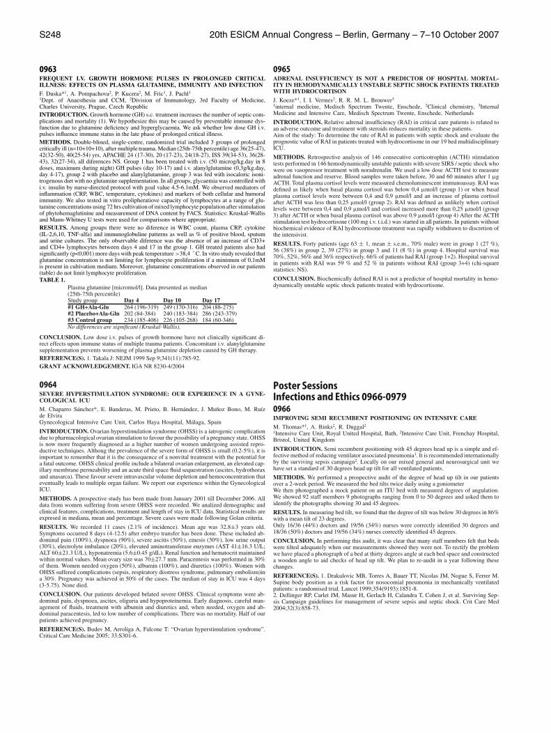
S248 20th ESICM Annual Congress – Berlin, Germany – 7–10 October 2007
0963FREQUENT I.V. GROWTH HORMONE PULSES IN PROLONGED CRITICALILLNESS: EFFECTS ON PLASMA GLUTAMINE, IMMUNITY AND INFECTION
F. Duska*1, A. Pompachova2, P. Kucera2, M. Fric1, J. Pachl11Dept. of Anaesthesia and CCM, 2Division of Immunology, 3rd Faculty of Medicine,Charles University, Prague, Czech RepublicINTRODUCTION. Growth hormone (GH) s.c. treatment increases the number of septic com-plications and mortality (1). We hypothesize this may be caused by preventable immune dys-function due to glutamine deficiency and hyperglycaemia. We ask whether low dose GH i.v.pulses influence immune status in the late phase of prolonged critical illness.METHODS. Double-blined, single-centre, randomized trial included 3 groups of prolongedcritically ill (n=10+10+10), after multiple trauma. Median (25th-75th percentile) age 36(25-47),42(32-50), 40(25-54) yrs, APACHE 24 (17-30), 20 (17-23), 24(18-27), ISS 39(34-53), 36(28-43), 32(27-34), all diferences NS. Group 1 has been treated with i.v. (50 microg/kg.day in 8doses, maximum during night) GH pulses (day 10-17) and i.v. alanylglutamine (0,3g/kg.day,day 4-17), group 2 with placebo and alanylglutamine, group 3 was fed with isocaloric isoni-trogenous diet with no glutamine supplementation. In all groups, glycaemia was controlled withi.v. insulin by nurse-directed protocol with goal value 4.5-6.1mM. We observed mediators ofinflammation (CRP, WBC, temperature, cytokines) and markers of both cellular and humoralimmunity. We also tested in vitro prolipheratiove capacity of lymphocytes at a range of glu-tamine concentrations using 72 hrs cultivation of mixed lymphocyte population after stimulationof phytohemaglutinine and measurement of DNA content by FACS. Statistics: Kruskal-Wallisand Mann-Whitney U tests were used for comparisons where appropriate.RESULTS. Among groups there were no diference in WBC count, plasma CRP, cytokine(IL-2,6,10, TNF-alfa) and immunoglobuline patterns as well as % of positive blood, sputumand urine cultures. The only observable diference was the absence of an increase of CD3+and CD4+ lymphocytes between days 4 and 17 in the group 1. GH treated patients also hadsignificantly (p<0,001) more days with peak temperature >38,4 ˚ C. In vitro study revealed thatglutamine concentration is not limiting for lymphocyte proliferation if a minimum of 0,1mMis present in cultivation medium. Moreover, glutamine concentrations observed in our patients(table) do not limit lymphocyte proliferation.TABLE 1.
Plasma glutamine [micromol/l]. Data presented as median(25th-75th percentile)Study group Day 4 Day 10 Day 17#1 GH+Ala-Gln 264 (196-319) 249 (170-316) 204 (88-275)#2 Placebo+Ala-Gln 202 (84-384) 240 (183-384) 286 (243-379)#3 Control group 234 (185-406) 226 (105-268) 184 (60-346)No differences are significant (Kruskal-Wallis).
CONCLUSION. Low dose i.v. pulses of growth hormone have not clinically significant di-rect effects upon immune status of multiple trauma patients. Concomitant i.v. alanylglutaminesupplementation prevents worsening of plasma glutamine depletion caused by GH therapy.REFERENCE(S). 1. Takala J: NEJM 1999 Sep 9;341(11):785-92.GRANT ACKNOWLEDGEMENT. IGA NR 8230-4/2004
0964SEVERE HYPERSTIMULATION SYNDROME: OUR EXPERIENCE IN A GYNE-COLOGICAL ICU
M. Chaparro Sanchez*, E. Banderas, M. Prieto, B. Hernandez, J. Munoz Bono, M. Ruızde ElviraGynecological Intensive Care Unit, Carlos Haya Hospital, Malaga, Spain
INTRODUCTION. Ovarian hyperstimulation syndorme (OHSS) is a iatrogenic complicationdue to pharmacological ovarian stimulation to favour the possibility of a pregnancy state. OHSSis now more frequently diagnosed as a higher number of women undergoing assisted repro-ductive techniques. Althoug the prevalence of the severe form of OHSS is small (0.2-5%), it isimportant to remember that it is the consequence of a nonvital treatment with the potential fora fatal outcome. OHSS clinical profile include a bilateral ovarian enlargement, an elevated cap-illary membrane permeability and an acute third space fluid sequestration (ascites, hydrothoraxand anasarca). These favour severe intravascular volume depletion and hemoconcentration thateventually leads to multiple organ failure. We report our experience within the GynecologicalICU.
METHODS. A prospective study has been made from January 2001 till December 2006. Alldata from women suffering from severe OHSS were recorded. We analized demographic andclinical features, complications, treatment and length of stay in ICU data. Statistical results areexpresed in mediana, mean and percentage. Severe cases were made following Golan criteria.
RESULTS. We recorded 11 cases (2.1% of incidence). Mean age was 32.8±3 years old.Symptoms occurred 8 days (4-12.5) after embryo transfer has been done. These included ab-dominal pain (100%), dyspnoea (90%), severe ascitis (50%), emesis (30%), low urine output(30%), electrolyte imbalance (20%), elevated aminotransferase enzymes (AST 41±16.3 U/L;ALT 60±21.3 U/L), hyponatremia (5.6±0.45 g/dL). Renal function and hematocrit maintainedwithin normal values. Mean ovary size was 70±27.7 mm. Paracentesis was performed in 30%of them. Women needed oxygen (50%), albumin (100%), and diuretics (100%). Women withOHSS suffered complications (sepsis, respiratory disstress syndrome, pulmonary embolism)ina 30%. Pregnancy was achieved in 50% of the cases. The median of stay in ICU was 4 days(3-5.75). None died.
CONCLUSION. Our patients developed belated severe OHSS. Clinical symptoms were ab-dominal pain, dyspnoea, ascites, oliguria and hypoproteinemia. Early diagnosis, careful man-agement of fluids, treatment with albumin and diuretics and, when needed, oxygen and ab-dominal paracentesis, led to low number of complications. There was no mortality. Half of ourpatients achieved pregnancy.
REFERENCE(S). Budev M, Arroliga A, Falcone T: “Ovarian hyperstimulation syndrome”.Critical Care Medicine 2005; 33:S301-6.
0965ADRENAL INSUFFICIENCY IS NOT A PREDICTOR OF HOSPITAL MORTAL-ITY IN HEMODYNAMICALLY UNSTABLE SEPTIC SHOCK PATIENTS TREATEDWITH HYDROCORTISON
J. Koeze*1, I. I. Vermes2, R. R. M. L. Brouwer3
1internal medicine, Medisch Spectrum Twente, Enschede, 2Clinical chemistry, 3InternalMedicine and Intensive Care, Medisch Spectrum Twente, Enschede, Netherlands
INTRODUCTION. Relative adrenal insufficiency (RAI) in critical care patients is related toan adverse outcome and treatment with steroids reduces mortality in these patients.Aim of the study: To determine the rate of RAI in patients with septic shock and evaluate theprognostic value of RAI in patients treated with hydrocortisone in our 19 bed multidisciplinaryICU.
METHODS. Retrospective analysis of 146 consecutive corticotrophin (ACTH) stimulationtests performed in 146 hemodynamically unstable patients with severe SIRS / septic shock whowere on vasopressor treatment with noradrenalin. We used a low dose ACTH test to measureadrenal function and reserve. Blood samples were taken before, 30 and 60 minutes after 1 µgACTH. Total plasma cortisol levels were measured chemoluminescent immunoassay. RAI wasdefined as likely when basal plasma cortisol was below 0,4 µmol/l (group 1) or when basalplasma cortisol levels were between 0,4 and 0,9 µmol/l and an increase of plasma cortisolafter ACTH was less than 0,25 µmol/l (group 2). RAI was defined as unlikely when cortisollevels were between 0,4 and 0,9 µmol/l and cortisol increased more than 0,25 µmol/l (group3) after ACTH or when basal plasma cortisol was above 0.9 µmol/l (group 4) After the ACTHstimulation test hydrocortisone (100 mg i.v. t.i.d.) was started in all patients. In patients withoutbiochemical evidence of RAI hydrocortisone treatment was rapidly withdrawn to discretion ofthe intensivist.
RESULTS. Forty patients (age 63 ± 1, mean ± s.e.m., 70% male) were in group 1 (27 %),56 (38%) in group 2, 39 (27%) in group 3 and 11 (8 %) in group 4. Hospital survival was70%, 52%, 56% and 36% respectively. 66% of patients had RAI (group 1+2). Hospital survivalin patients with RAI was 59 % and 52 % in patients without RAI (group 3+4) (chi-squarestatistics: NS).
CONCLUSION. Biochemically defined RAI is not a predictor of hospital mortality in hemo-dynamically unstable septic shock patients treated with hydrocortisone.
Poster SessionsInfections and Ethics 0966-09790966IMPROVING SEMI RECUMBENT POSITIONING ON INTENSIVE CARE
M. Thomas*1, A. Binks2, R. Duggal21Intensive Care Unit, Royal United Hospital, Bath, 2Intensive Care Unit, Frenchay Hospital,Bristol, United Kingdom
INTRODUCTION. Semi recumbent positioning with 45 degrees head up is a simple and ef-fective method of reducing ventilator associated pneumonia1. It is recommended internationallyby the surviving sepsis campaign2. Locally on our mixed general and neurosurgical unit wehave set a standard of 30 degrees head up tilt for all ventilated patients.
METHODS. We performed a prospective audit of the degree of head up tilt in our patientsover a 2-week period. We measured the bed tilts twice daily using a goniometerWe then photographed a mock patient on an ITU bed with measured degrees of angulation.We showed 92 staff members 9 photographs ranging from 0 to 50 degrees and asked them toidentify the photographs showing 30 and 45 degrees.
RESULTS. In measuring bed tilt, we found that the degree of tilt was below 30 degrees in 86%with a mean tilt of 23 degrees.Only 16/36 (44%) doctors and 19/56 (34%) nurses were correctly identified 30 degrees and18/36 (50%) doctors and 19/56 (34%) nurses correctly identified 45 degrees.
CONCLUSION. In performing this audit, it was clear that many staff members felt that bedswere tilted adequately when our measurements showed they were not. To rectify the problemwe have placed a photograph of a bed at thirty degrees angle at each bed space and constructeda wooden angle to aid checks of head up tilt. We plan to re-audit in a year following thesechanges.
REFERENCE(S). 1. Drakulovic MB, Torres A, Bauer TT, Nicolas JM, Nogue S, Ferrer M.Supine body position as a risk factor for nosocomial pneumonia in mechanically ventilatedpatients: a randomised trial. Lancet 1999;354(9193):1851-8.2. Dellinger RP, Carlet JM, Masur H, Gerlach H, Calandra T, Cohen J, et al. Surviving Sep-sis Campaign guidelines for management of severe sepsis and septic shock. Crit Care Med2004;32(3):858-73.
20th ESICM Annual Congress – Berlin, Germany – 7–10 October 2007 S249
0967COLISTIN SERUM CONCENTRATIONS AFTER INTRAVENOUS ADMINISTRA-TION IN CRITICALLY ILL PATIENTS
N. Markou*1, S. Markantonis2, E. Dimitrakis2, D. Panidis2, E. Boutzouka3, S. Karatzas3,H. Apostolakos4, G. Baltopoulos5
1ICU-B, KAT Hospital, 2Laboratory of Biopharmaceutics and Pharmacokinetics, AthensUniversity, Faculty of Pharmacy, 3Athens University School of Nursing ICU, 4ICU-B, KATHOSPITAL, 5Athens University School of Nursing ICU, KAT Hospital, Athens, Greece
INTRODUCTION. No reliable pharmacokinetic data are available for colistin in critically illpatients. We assessed steady-state serum concentrations of colistin sulfate (which is the activeform of the drug) after IV administration of colistin methanesulfonate (CMS) in critically illpatients with stable renal function.
METHODS. We performed a prospective, open-labeled, uncontrolled study in critically illpatients with stable renal function who were receiving CMS as part of a treatment regimenfor sepsis from multi-resistant Gram (-) bacilli. After intravenous administration of 225 mgCMS (exception 1 patient 150 mg) every 8 or 12 hours for at least 2 days, blood samples werecollected just before initiation of infusion on the sampling day and at 0.16, 1, 2, 4, 6 and 8 hours,thereafter. Serum concentrations of colistin sulfate were determined by high-performance liquidchromatography.
RESULTS. We enrolled 14 non-consecutive patients aged 62 ± 19.2 years (APACHE II scoreon admission 17.1 ± 6.0). At steady-state mean (± S.D.) colistin maximum and minimumconcentrations were 2.93 ± 1.24 mg/L and 1.03 ± 0.44 mg/L, while the total body clearance,volume of distribution and half-life were 13.6 ± 5.85 L/h, 139.9 ± 60.3 L and 7.4 ± 1.7 hours,respectively. 78.6% of patients responded to treatment. No adverse effects attributed to colistinwere observed.
CONCLUSION. In critically ill patients, colistin serum levels were similar to those found incystic fibrosis patients treated with substantially lower doses of CMS (1) and the mean half-lifewas longer. CMS dosage regimens administered may be inappropriate, as these produce colistinconcentrations which would probably give suboptimal Cmax/MIC ratios for many strains ofGram (-) bacilli.
REFERENCE(S). Li J, et al. Steady-state pharmacokinetics of intravenous colistin methene-sulfonate in patients with cystic fibrosis. J Antimicrob Chemother 2003 ; 52:987-992
0968IMMUNODEPRESSION AND ICU-ADQUIRED INFECTIONS
M. Palomar*1, F. Alvarez Lerma2, P. Olaechea3, J. Otal4, J. Ballus5, C. Lomas4, R. Alcaraz4,A. Arenzana6, R. Sierra7
1ICU, H. Vall d’Hebron, 2ICU, H del Mar, Barcelona, 3ICU, H Galdakano, Galdakano,4ICU, H. Vall d’Hebron, 5ICU, H Bellvitge, Barcelona, 6ICU, H Macarena, Sevilla, 7ICU,H Puerta del Mar, Cadiz, Spain
INTRODUCTION. Immunodepressed patients (ID) who had higher prevalence in ICUs,present more risk of infections. The aim of this study was to evaluate the demography andincidence of ICU-acquired infections (I-ICU) related to invasive procedures in patients withclinical or pharmacological ID, compared to the rest of ICU hospitalized patients.
METHODS. A multicentre prospective study from April to July 2005 was performed in 102UCIs of 93 Spanish hospitals. The number of patients admitted to ICU > 24h accordingto established criteria (immunodeficiency, neutropenia and immunosuppression) was studied.Demographic characteristics, infections developed during ICU stay (I-ICU), the diagnosis ac-cording to the CDC’s criteria and expressed in rate by 100 patients or for 1000 days, etiologyand resistance patterns were analyzed.
RESULTS. Among 11,461 studied patients, 799 (6,97%) presented ID. Characteristics areshown in Table 1. Invasive procedures: Mechanical ventilation (MV): 41,7/61,2%; Centralvenous catheter (CVC): 71,4 / 91,2 %; Parenteral Nutrition (PN):12,7 / 29,1 %; Extrarenaldepuration (ERD): 3,4/10,3 %. Nosocomial infections and outcome in table 2. The incidenceof multiresistant bacteria infection/colonization was: ESBL 0,79 /1,75 %; MRSA 1,8/ 2,6 %;Acinetobacter 1,7 / 4 %; multiresistant P aeruginosa 0,9 /1,7 %. There were differences ininfection etiologies: BGN more frequently in ID, mainly A baumnannii 8,1/18,3 % and alsohave higher resistance patterns. ID required more antibiotics before ICU admission 28,3/56,5% and during ICU stay 56,9/ 88,8%. The number of antibiotics/patient was 2,1 / 3 respectively.
TABLE 1.Patients N Age APACHE II Coronary Medical Surgical Trauma
Total 11.461 61,4 14,1 27,2 38,1 24,5 10ID 799/6,9% 56,8 20,1 4,5 6 9,9 26,5
TABLE 2.I-ICU % ID o/oo NAP % UI % PB+CB % LOS (d) Mortality %patients
Total 14,2 19,4 6,1 14,1 3,3 7,3 10,8ID 25 25,2 10,8 4,8 6 9,9 26,5
CONCLUSION. Patients with ID, 7 % of the population, presented more risk factors, medicalpathology and higher severity. Predisposition to more antibiotic-resistant organism infectionwas confirmed, being necessary a superior antibiotic usage.
0969CURRENT INFECTION CONTROL RECOMMENDATION ARE NOT ALWAYSNECESSARY TO REDUCE MRSA INFECTION RATES IN ICU
D. Harji*1, M. Manji2, D. Mortiboy3, T. Whitehouse2
1Medical Student, University of Birmingham, 2Department of Anaesthesia and CriticalCare, 3Department of Microbiology, University Hospital Birmingham, Birmingham, UnitedKingdom
INTRODUCTION. There are recommendations for control and prevention of Methicillin-Resistant Staphylococcus Aureus (MRSA) (1). These recommendations however, are basedon large series and case reports rather than randomised trials (2). Of those recommendations,only two (handwashing and adequate staffing) are reliably carried out in our ICU as evenconventional ‘deep cleaning’ has been shown to be unreliable (3).
METHODS. All patients admitted to ICU at University Hospital Birmingham between June2002 and May 2006 were retrospectively studied so that any microbiological sample whereMRSA was cultured was correlated with the date of ICU admission. Isolation of MRSA wasclassed as prior to-, during-, or after- an ICU stay. Isolates dated within 48 hours of a changeof site within the hospital were counted as being acquired in the previous site.
RESULTS. The number of patients admitted to ICU already infected/colonised with MRSA in2002 was 265 and 172 new cases occurred. By 2005 the number of new cases in ICU had fallensignificantly to 52 despite the number of patients admitted to the ICU already carrying MRSAwas 243 (p=0.001, 4*2 Chi squared = 60.05 Degrees of Freedom = 3).
CONCLUSION. There has been a steady decline in the number of primary MRSA infectionoccurring in our ICU whilst the number of cases admitted has remained constant. Colonisationpressure from patients admitted to ICU is an independent predictor for MRSA acquisition onITU. The reasons for our decline in MRSA infection remain unclear as full recommendationsto inhibit MRSA spread can not be implemented.
REFERENCE(S). 1. Coia, JE et al. J Hosp Infect 63 Suppl 1:S1-442. Cooper, BS et al. BMJ 329:5333. French, GL et al. J Hosp Infect 57:31-37
0970PREVENTION OF SPREAD OF METHICILLIN-RESISTANT STAPHYLOCOCCUSAUREUS IN A CARDIAC SURGERY CENTER
A. Mastoraki*, E. Douka, I. Kriaras, S. Mastoraki, G. Stravopodis, S. GeroulanosSurgical Intensive Care Unit, Onassis Cardiac Surgery Center, Kalithea, Athens, Greece
INTRODUCTION. In Greece Methicillin-Resistant Staphylococcus aureus (MRSA) preva-lence ranges from 11.1% to 90.9% of the Staphylococci isolated. In our institution prophylacticmeasures include the mandatory screening of all newly employed health care workers andpatients admitted from other hospitals. The aim of this survey was to elucidate the efficacy ofthe above preventing strategy.
METHODS. A prospective quality control study was conducted from 1997 to 2005. Patientsadmitted from other hospitals remained isolated for 48 hours and were screened for MRSAby nasal swabs. Infected patients were treated with a glycopeptide in combination with ri-fampin and carriers with mupirocin. Furthermore all newly employed health care workers werescreened for MRSA and carriers were treated with mupirocin.
RESULTS. Throughout the 9-year study period among 62313 hospitalized patients 15270 wereattended in the Department of Cardiac Surgery. Total infection rate was 5.4%. MRSA preva-lence was 0.37% (n: 56). Infection associated with MRSA was identified in 30 patients (0.2%).In 10 patients MRSA was detected in tracheal secretions, in 4 patients in swabs taken fromdonor site infection and in 4 patients from superficial sternal surgical wound. In 10 patientsthe pathogen was isolated from cultures of the surgical site drainage. The remaining 2 patientswere defined as having severe sepsis. Mortality rate was 30%.
CONCLUSION. The prompt determination, isolation and appropriate treatment of MRSA pa-tients admitted from other institutions combined with the detection and elimination of carriersamong new health care workers prevented further spread of the pathogen.
S250 20th ESICM Annual Congress – Berlin, Germany – 7–10 October 2007
0971EXPERIENCE IMPLEMENTING A VANCOMYCIN INFUSION POLICY ON ANINTENSIVE CARE UNIT
H. W. J. Twamley*, E. Boxall, L. Hantom, G. L. ThomasIntensive Care, Salford Royal NHS Foundation Trust, Manchester, United Kingdom
INTRODUCTION. Intravenous vancomycin remains the drug of choice for many infectionsin the intensive care unit (ICU), including methicillin-resistant staphylococcus aureus. Con-ventional b.d. doseage regimes may not achieve sustained concentrations above the minimuminhibitory concentration. Continuous infusions have been shown to lower mortality in suscepti-ble staphylococcus aureus infections compared with b.d dosage. We describe an audit followingthe sucessful introduction of a vancomycin infusion protocol.
PROTOCOL. All patients receive a vancomycin loading dose,adjusted for weight and renalfunction. A continuous infusion of standard concentration is then commenced. Concentrationof infusion depends on whether venous access is peripheral (5mg/ml) or central (10mg/ml).A serum vancomycin level is requested with routine morning blood tests. The target range is15-25 mg/l. The daily dose is titrated by altering the infusion rate.
AUDIT. 30 consecutive patients who received > 24 hrs vancomycin were reviewed. Resultsare presented as morning vancomycin level post load and initial infusion(level 1) and thenconsecutive daily morning levels (levels 2,3,4,5,6)
RESULTS. Mean vancomycin levels were consistently within the target range, although thestandard deviations were large (Table 1). Problems identified with the protocol were:(1)Inadequate loading dose in patients with renal failure. (2)Standard concentrations givenperipherally or centrally were confused by staff. (3)Patients already on vancomycin b.d beforeICU admission were inappropriately converted to the infusion. Unusual outlying levels werefrequently, but not always, due to poor protocol compliance. Following the audit, the loadingdose regimen was altered, the protocol was re-written to clarify problem areas, and a programof staff education was implemented. We are currently completing the audit cycle.
TABLE 1.Daily Vancomycin Levels (mg/l)
Level 1 Level 2 Level 3 Level 4 Level 5 Level 6Mean 15.1 18.1 21.0 21.3 22.4 21.2Standard dev 6.2 7.1 8.5 6.1 4.0 5.0Range 4.8-31.6 9-36.5 7.5-38.8 8-28.5 14.5-30.1 15.5-29.6
CONCLUSION. A vancomycin infusion protocol was sucessfully introduced on our ICU. Theprotocol titrates the rate of infusion of vancomycin formulated as a standard concentration. Theprotocol maintains therapeutic levels within the desired target range for most patients. Outly-ing vancomycin levels are typically the result of non-compliance with the protocol. Effectiveongoing education resolves many of the problems associated with the regimen which, onceunderstood, is readily implemented.
REFERENCE(S). Wysokl et al Antimicrobial Agents and Chemotherapy(2001)45;9:2460-2467; Rello et al Crit Care Med(2005)33;9:1983-7
0972CLINICAL EVALUATION OF EXECUTION OF EARLY GOAL DIRECTED THER-APY IN SEPTIC SHOCK
X. Wang*, Z. HeAnesthesia and Intensive care, Ren Ji Hospital, Medical school of Shanghai Jiao TongUniversity, Shanghai, China
INTRODUCTION. To evaluate the efficacy of early goal directed therapy(EGDT) insepticshock.
METHODS. Two hundred and three patients with septic shock were assigned into treatmentgroup (n=98) and Control group (n=105). According to the state of organ function at thebeginning of treatment and multiple Organ dysfunction syndrome(MODS) evaluation scores,each group was categorized into three strata: Stratum A (mild organ dysfunction), stratum B(medium organ dysfunction) and stratum C (severeorgandysfunction). Mortality and incidenceof organ dysfunction in each group were analyzed.
RESULTS. At stratum A, themortality and incidence of organ dysfunction in treatment groupwere significantly lower than those of control group: 27.78% (1554 cases) vs. 37.50% (1848cases), 31.48% (1754 cases) vs. 43.75% (2148 cases), both P<0.05. There was no significantdifference between treatment group and control group in patients of stratum B: 75.86% (2229cases) vs. 76.92% (2026 cases), 55.17% (1629 cases) vs. 57.69% (1526 cases) and stratum C:93.33% (1415 cases) vs. 96.77% (3031 cases), 40.00% (615 cases) vs. 41.93% (1331 cases),all P>0.05.
CONCLUSION. In the earlier Period of septic shock, EGDT can remarkably decrease thepatients′mortality and incidence of organ dysfunction, but cannot improve survival rate andprognosis in patients in advanced stage of septic shock.
REFERENCE(S). 1 Rivers E, Nguyen B, Havstad S, et al. Early goal directed therapy in thetreatment of severe sepsis and septic shock. N Engl J Med,2001,345(19):1368-1377.2 Dellinger RP, Carlet JM, Masur H, et al. Surviving sepsis Campaign guidelines for manage-ment of severe sepsis and septic shock. J Crit Care Med, 2004,32(3):858-873.
GRANT ACKNOWLEDGEMENT. The funds of shanghai healthy bureau(03-77-20)
0973TAP WATER FILTERS REDUCE THE RISC OF LEGIONELLA INFECTIONS INICU PATIENTS
J. Bisgaard*1, T. Gilsaa1, S. Uldum2
1Intensive Care Unit, Fredericia and Kolding Hospitals, Kolding, 2Microbiology and Diag-nostics, Statens Serum Institut, Copenhagen, Denmark
INTRODUCTION. Legionella in the hospital water supply carries a serious risk of health-careacquired infections to immunocompromised patients, e.g. in intensive care units. Thereforepathogen-free water should be provided for high risk patients. Various approaches to eliminateLegionella have been tested, none of them being 100% efficient and none without negative sideeffects. The latest product introduced, point-of-use water filters which can be easily mountedon water taps, seems to have the required advantages.
METHODS. We prospectively studied the ability of Pall-Aquasafe Faucet Water Filters (PallCorporation) to eliminate Legionella from tap water in our ICU. 30 water samples were col-lected from 5 faucets over a 12 week period. Five samples, 1 from each faucet, were collectedbefore installing water filters and again prior to removing them after 4 weeks. This routine wasrepeated 3 times. All samples were cultured for Legionella spp. and number of colony formingunits (CFU) per litre was determined.
RESULTS. 15 filtered and 15 unfiltered water samples were collected. In filtered water weisolated Legionella at low levels in 2 of 15 samples. In contrast, Legionella was isolated from14 out of 15 samples of unfiltered water (Fig. 1). One of the positive samples from filteredwater was due to an easily recognizable leak from the connector upstream of the filter. Theother contaminated filter system showed no obvious signs of malfunction.
TABLE 1.Pall Filter CFU per Litre No Filter CFU per Litre
1. period 0 - 0 - 5 - 0 - 0 550 - 850 - 5.000 - 6.000 - 4002. period 5 - 0 - 0 - 0 - 0 6.000 - 54.000 - 15 - 4.000 - 5153. period 0 - 0 - 0 - 0 - 0 41.000 - 700 - 50 - 0 - 50
CONCLUSION. The Pall-Aquasafe Faucet Filter effectively reduces the concentration of Le-gionella species in tap water in the ICU, but close attention should be paid to the integrity ofthe system. This system has the potential for reducing ICU-aquired Legionella infections.
GRANT ACKNOWLEDGEMENT. Filters provided free of charge from Pall Corporation
0974DO WE NEED ACTIVATED PROTEIN C?
E. M. Lafuente*1, M. J. Fernandes1, F. Gil2, F. Assuncao2, J. Brandao3, A. Marinho3,A. Silva3
1UCIP, Hospital Padre Americo - Vale do Sousa EPE, Penafiel, 2UCIP, Hospital S. Marcos,Braga, 3Servico Cuidados Intensivos, HGSA, Porto, Portugal
INTRODUCTION. Therapy with activated protein C (APC) (24 µg/Kg/minute/96 h) has beenassociated to a decrease of mortality and ameliorate of organ dysfunction/failure in patientswith severe sepsis. Objective: Evaluate the impact on the outcome of activated protein C inacute ill patients with severe sepsis.
METHODS. Design: Multicentric, prospective, observational cohort study conducted duringone year and half. We selected patients admitted to 3 polivalent ICU. Inclusion criteria: allpatients treated with APC. We collected for each patient: demographics data, SAPS II andSOFA score and number of organ failure at admission, when we started the treatment, and atthe end of the treatment. We also collected intrahospital mortality, 28 day mortality rate andsurvival according to the moment of prescription of activated protein C (< 12 hours; 12-24hours and later 24 hours after admission).
RESULTS. We enrolled 37 patients: age of 53 ± 17,2, SAPS II score 42,9 ± 14,6 SOFA 7,9± 2,8, with a length of stay 14,8 ± 9,9, mortality rate – 18,9% and at 28o day – 32,4%. Mainclinical presentation: severe sepsis with ARDS after community acquired pneumonia (21,6%).Time to started the treatment: less than 12 hours 21 patients (56, 8%) with a mortality rate 19%;12-24 hrs 12 patients (32, 4%) with a mortality rate 50%, and after 24 hours 4 patients (10,8%)with a mortality rate 50%.Adverse effects: Three patients (8,1%) stop the perfusion because of hemorrhagic complica-tions.Survival analysis revealed as depicted in the plot:
CONCLUSION. Although the controversy of clinical application activated protein C in thissample there we found different patterns of clinical response according to the prescription time,nevertheless the survival rate was not related with the time of activated protein-c prescription.
20th ESICM Annual Congress – Berlin, Germany – 7–10 October 2007 S251
0975PRELIMINARY RESULTS OF A CANDIDA SCORE FOR EARLY ANTIFUNGALTREATMENT IN CRITICAL CARE PATIENTS.
M. Santos, D. Thielman, C. Teles, F. Gutierrez, R. C. Costa Filho*ICU, Hospital Pr, Rio de Janeiro, Brazil
INTRODUCTION. The incidence of infections caused by Candida species in critical care hassubstantially increased in recent years. Recently, Candida infections have been increasing spe-cially in critical care patients. Invasive candidiasis has been associated with severe sepsis, septicshock and multiple organ failure with a clinical scenario very similar to bacterial infections.This infection is a diagnosis challenge with an estimated mortality rate of 40%.Objective: To evaluate the impact of a Candida Score to early antifungal treatment in a generalcritical care unit.
METHODS. The Candidemia incidence rate ratio from July until December 2005 was com-pared to the same period in 2006 after the implementation of Candida Score System (SoftwareStata 8.0). Surveillance cultures of urine, tracheal and digestive samples from each patient wereobtained weekly. According to Leon (1), patients with at least two positive sites and sepsis orwith total parenteral nutrition or a recently surgical intervention, received antifungal treatment(fluconazole or Caspofungin) - preemptive treatment.
RESULTS. The incidence after the Candida Score implementation was reduced from 1,91(2/1049 patients day) to 0,92 (1/1081 patients day) with no statistical significance (p = 0,3).
CONCLUSION. In this preliminary report, the Candida Score seems to be a good tool toreduce the incidence of Candida infections in a general critical care unit. In a large population,the use of Candida Score system may assist in identify critical patients candidates to preemptiveantifungal treatment.
REFERENCE(S). (1)Leon C; Ruiz-Santana S; Saavedra P et al. Crit Care Med 2006; 34:730-734
0976VENTILATOR ASSOCIATED PNEUMONIA IN A MEDICAL ICU
R. Gimeno*1, C. Cortes1, I. Madrid1, L. Arias2, M. Garcia3, P. Ramirez1, J. Botella1,J. Cunat11Medicina Intensiva, Hospital Universitari La FE, Valencia, 2Medicina Intensiva, Hospitalde la Ribera, Alzira, 3Medicina Intensiva, Hospital de Sagunto, Sagunto, Spain
INTRODUCTION. An important indicator of the quality in medical assistance is the control ofnosocomial infection (NI). Intensive Care Units (ICU) are areas with elevated NI rates. Manyof these NI are associated with the use of external devices: mechanical ventilation, urinarycatheters and central venous catheters. The aim of this study was to know the incidence ofventilator associated pneumonia (VAP) in our ICU, the trends in the infective flora and the useof antibiotics in two periods of time: the first period (A): from November 1.994 to January1.995 and the second period (B): from May to July 2.006.
METHODS. During three months in A and three in B, the mechanical ventilation associatedinfection rates were determinated following the recomendations of the National NosocomialInfection Surveillance System (NNIS) in the USA in 308 patients in A and 256 in B, admittedto our 21 beds ICU. We also determined the the most frequent microorganisms recovered andthe specific antibiotics used.
RESULTS. The VAP rates were 28.8 pneumonia per 1000 mechanical ventilation days in Aand 16.7 in B. Pseudomonas aeruginosa and Escherichia coli were the microorganisms recov-ered most frequently in A; Acinetobacter baumannii and Enterobacter aerogenes in B. Amongthe antibiotics, we used third generation cephalosporins, quinolones and macrolides in A andsulbactam-ampicillin, amikacin and linezolid in B.
CONCLUSION. 1)During the last year we have detected a significant decreasing tendency inthe VAP rates in our ICU. 2)This nosocomial infection surveillance study has allowed us toknow the most frequent microorganisms causing VAP in our ICU: currently is Acinetobacterbaumannii; eleven years ago was Pseudomonas aeruginosa. According to this, we can start anappropiate empirical antibiotherapy. 3)The use of NNIS rates is advisable because it allowsus to know the impact of VAP in our unit. We can also perform comparative studies betweenperiods of time in our unit and comparative studies with other units of similar characteristics.
REFERENCE(S). 1. National Nosocomial Infections Surveillance (NNIS) System Report,data summary from January 1992 through June 2004, issued October 2004. Am J Infect Con-trol 2004; 32:470-485.2. Barrios A, Martın JL, Jannone R, et al. Estudio prospectivo de la infeccion nosocomial enuna UCI medica. Una propuesta para el uso generalizado de las tasas del National NosocomialInfection Surveillance System. Rev Clin Esp 1998; 198:284-288.
GRANT ACKNOWLEDGEMENT. ICU La Fe hospital.
0977WHAT BOTHERS THE PATIENT IN AN INTENSIVE CARE UNIT? A INTERDIS-CIPLINARY PERSPECTIVE
M. E. Lugarinho*, P. C. Galhardo, P. P. SouzaIntensive Care Unit, Hospital de Clınicas Mario Lioni, Rio de Janeiro, Brazil
INTRODUCTION. Our intensive care unit (ICU) was one of the first to iniciate a humanizationprogram in daily routine in 1996. The program suffered changes, the ICU grew up in numberof beds and complexity, and had great renewal of the members of our interdisciplinary group.To improve our knowledge and quality of care, we evaluated the stress factors for the patientsfrom our staff members perspective, putting them in the patients place.
METHODS. Between October and December of 2005, a research form was used. The follow-ing itens were analysed: profile of the interviewed, evaluation of the environment of the ICUand the stress factors for the patients. The results were compared with the survey form filled bythe patients after ICU discharge.
RESULTS. The entire ICU team answered the research (n=75). The mean age is 29,98 years(SD 6,50), 61.33% of female, 62.70% married, 38.70% protestants and 37% catholics and ICUprofessional experience of 4,96 years (SD 4,78). Our ICU is noisy for 73,33%, very illuminatedfor 73,33%, easy-going for 72%, organized for 49,33%. According to the team, factors thatbothers the patient are: anxiety (77.33%), fear (70.66%), loneliness (70.66%), noise (68.67%),bed bath (65.33%), lack of privacy (58.66%), distortion of time perceptions (50.66%). Thepatients (n=71) described as main complaints after ICU discharged: anxiety (59.15%), sleep-lessness (52.11%), lack perception of time (50.70%), pain (42.25%), noise (33.80%), loneliness(32.39%), fear (22.53%), bed bath (19.71%) and lack of privacy (11.26%).
CONCLUSION. The study showed differences of ICU team opinions and the patients com-plaints. When the team is placed in the patients perspective they may experience a better viewof how harmfull is an ICU and how much we can do to improve it. This is our daily challenge:take care with quality, respect and affection.
0978FAMILY NEEDS OF CRITICAL CARE TRAUMA PATIENTS IN CYPRUS AS-SESSED WITH QUALITATIVE METHODOLOGY
G. Samoutis1, T. Kyprianou*2, A. Loutsiou-Ladd3, L. Palazis2, C. Lionis4, A. Filalithis4
1NGO, INTENSIVE CARE FORUM, 2INTENSIVE CARE UNIT, NICOSIA GENERALHOSPITAL, 3Dept of Psychology, Universiy of Cyprus, NICOSIA, Cyprus, 4Clinic of Socialand Family Medicine, University of Crete, Heraklion, Greece
INTRODUCTION. The relatives of critically ill patients have increased needs in this crisisperiod. Family centered quality improvement interventions can be implemented effectivelyafter capturing their experiences, recognizing their needs and evaluating how they should bemet/being met. This subject has not received adequate attention with regard to qualitativeapproach. A quantitative study from our group has shown that certain (dominant) coping mech-anisms may protect mental health / QOL. The aim of this study was to explore critical carefamily needs in Cyprus and discuss potential interventions.
METHODS. A descriptive qualitative research design with focus groups interviews from ICUpatients’ caregivers was used to explore family experiences and needs. Three focus groups wereused to elicit the experiences and needs of families with a relative in ICU at the time of hospi-talization and after discharge to the community. A number of 18 relatives (10 male/8 female,age 16-60) from different families were participated. Qualitative data analysis was based onthe 5-steps framework approach (familiarization, identifying a thematic framework, indexing,mapping and interpretation).
RESULTS. Four major categories of needs were emerged from the focus groups: 1) infor-mation and communication, 2) support - human and structures, 3) assurance –to be assuredabout the quality of care received 4) proximity-able to be physically close to their ICU patient.Information and communication was far more prominent need than the others. The majorityof needs were perceived as not being adequately met and the main resources expected to meettheir needs were physicians, nurses and the hospital environment.
CONCLUSION. The findings of this study indicate important areas of unmet needs of familieshaving patients in critical care, that require relevant family-centered quality improvement inter-ventions, aiming to improve family care and modulate recruitment/use of coping mechanisms.
GRANT ACKNOWLEDGEMENT. AROGI project is co-funded by Research PromotionFoundation, Cyprus.
S252 20th ESICM Annual Congress – Berlin, Germany – 7–10 October 2007
0979IS IT POSSIBLE TO ARRIVE AT 0% AVOIDABLE LOSSES IN ORGAN DONA-TION?
N. Masnou*1, T. Pont2, R. M. Gracia3, P. Salamero1
1Transplant Coordination Management, Vall d’Hebron Hospital, 2Transplant CoordinationManagement, Vall d’Hebron Hospital, 3Intensive Care Service, Vall d’Hebron Hospital,Barcelona, Spain
INTRODUCTION. Donation rates have not kept pace with demand, resulting in a criticaldeficit of available healthy organs. Donor detection is influenced by the legal system, familyrefusal and underreporting caused by erroneous knowledge of donation criteria and lack offamiliarity with the procedure.The aim of this study is evaluate possible donor losses in the Vall d’Hebron University Hospitalduring 2002-2006) and compare two detection systems (Using GSC detection follow-up andSpanish Quality Guarantee Program) introduced in our hospital.
METHODS. All deaths in critical care units (ICU) were reviewed, with emphasis on neuro-logical cases. We contrasted this deaths with pre-existing coordination registers to ascertaindiscrepancies in protocols (donor maintenance, brain death diagnosis (BDD), family informa-tion).
RESULTS. 1928 deaths were included. Neurological cases 482, of which 250 were braindeaths. 99% were detected by the Transplant Coordinator (CT). 145 (58%) became real donors.50 (20%) were rejected due to medical contraindications, 40 (16%) because of family refusal. 5(2%) maintenance problems and 5 (2%) other. Only in 9 (3.6%) cases did we find discrepanciesin protocols which led to the losses of donors (2 delay in donor maintenance, 2 erroneousmedical contraindications, 1 BDD and 4 cases in family information).
CONCLUSION. 1. It is indispensable for all clinicians involved in the process to know andcarry out the protocols2. In spite of having two excellent protocols in place avoidable losses still exist
Poster SessionsImproving (micro) circulation 0980-09930980CLINICAL PROFILE AND PROGNOSIS OF PATIENTS WITH ACUTE COR-ONARY SYNDROME AND NORMAL CORONARY ARTERIES
A. del Castillo*1, E. Gomez2, L. Castellano3, L. Lopez3, V. Valentin4
1Intensive Care Medicine, Dr Peset Hospital, Valencia, 2Intensive Care Medicine, 3InternalMedicine Department, Sagunt Hospital, Sagunt, 4Intensive Care Medicine, Dr Peset Hospital,Valencia, Spain
INTRODUCTION. The purpose of this study was to examine the main clinical characteris-tics of patients with Acute Coronary Syndrome (ACS) admitted in Intensive Care Unit (ICU)with normal coronariography, to analyze the morbility and mortality of these patients in thefollow-up and to evaluate which variables are related to it.
METHODS. Retrospective study of 53 patients admitted in two ICUs from 2000 to 2006with diagnosis of ACS (Acute Myocardial Infarction [AMI] or Unstable Angina [UA]), whounderwent coronariography during their admission which ruled out the presence of significantlesions in coronary arteries (normal coronariography=NC). Demographic, clinical, and echocar-diographic variables were registered in both groups. Patients from NC group were matched byage (± 5 years), sex and diagnosis of ACS (AMI/UA) with a same number of patients admittedin ICU during same period of time, and with significant lesions on coronariography (abnormalcoronariography=AC). Demographic and clinical variables were compared in both groups, aswell as the incidence of a composite end-point of cardiovascular death, readmission for ACS orheart failure at 12 months. Statistical analysis was performed with 15.0 SPSS statistical pack-age, using chi square for categorical variables, t-test for quantitative variables, Kaplan-Meiercurves and Cox regression.
RESULTS. Among 53 patients with NC, mean age was 57±12 and 51 % were women. 64 %of patients had AMI which corresponded to ST-Elevation in 19 % cases. AC patients under-went revascularization before discharge in 95 % of cases. There was no difference comparingcardiovascular risk factors between both groups. In the multivariate model, the only two in-dependent predictive variables of NC were lower CPK levels in patients with AMI in the NCgroup(633±429 vs 1584±1903; p<0.02) and the presence of segmental contractility alterationsby echocardiography, less observed in patients with NC than in AC patients (37.8% vs 62.2%;p=0.016; Odds Ratio=2.8; 95% CI=1.26-6.33). In the 12-month-follow-up, the occurrence ofthe composite end-point was similar in both groups (23% in NC vs 19% in AC; p=ns).
CONCLUSION. In our study, demographic characteristics of patients with NC were similarto patients with AC. However, patients with NC had fewer segmental contractility alterationsby echocardiography and lower levels of CPK in AMI subgroup than in patients with AC. Theprognosis at 1 year was similar in the two groups with no significant difference in the incidenceof cardiovascular death, readmission for ACS or heart failure, which could have been influencedby the relative short follow-up, the limited sample power and the initial high revascularizationrate in the AC patients.
0981PROTECTIVE EFFECTS OF HEMODILUTION AND ISCHEMIC PRECONDI-TIONING AGAINST REPERFUSION INJURY IN PIG HEART
R. X. Wang*, N. Y. Hang, J. D. SunAnesthesia and Intensive care, Ren Ji Hospital,Medical school of Shanghai Jiao TongUniversity, Shanghai, China
INTRODUCTION. We investigated the protective effects of isovolumetric hemodilution andischemia preconditioning against myocardial injury due to reperfusion through a pig myocardialischemia reperfusion model.
METHODS. Eighteen mini-pigs were used in an acute myocardial ischemia model. They wererandomly divided into three groups, with 6 pigs in each group: the control (group I, n=6),ischemia pre-treatment (group II, n=6), ischemia pretreatment plus blood dilution (group III,n=6). Cardiac output (CO), Sv02 and blood flow of coronary artery were measure, and then theoxygen supply to the myocardium and oxygen consumption were calculated. The MDA, SODactivity, CPK and CK-MB were also determined 20 min and 60 min respectively before andafter the forceps clamping. A sample was taken from the left atrial appendage to determine therate of expression of heat shock proteins Hsp70 mRNA.
RESULTS. When ischemia lasted for 20 min, the heart rate (HR) of group I and II slowed downdramatically while that of group III accelerated slightly. The MAP, CI and SVR of all threegroups significantly decreased from control values(P<0.01), but the magnitude of decrease ingroups II and III were lower than those of group I (P<0.05). Also the SVR of group III wassignificantly lower than group II(P<0.05). After reperfusion at 20min and 60 min, the HR ofgroups I and II was significantly lower than that of the controls (P<0.01). The HR of group IIIwas significantly higher than that of groups I and II(P<0.05), but was not significantly higherthan control value(P>0.05). The decreased MAP and CI of groups II and III were significantlylower than those of group I (P<0.05). In addition the SVR of group III was lower than groups Ior II(P<0.05). The values of CPK, CPK-MB of all three groups were significantly higher thanthe controls after reperfusion at 20min and 60min (P<0.05-0.01 respectively). However groupsII and III increased less than those of group I. The values of CPK and CPK-MB in group IIIwere lower than that of group II(P<0.05). The MDA value of group I was dramatically higherthan that of groups II and III after reperfusion at 20min and 60min (P<0.05), while the value ofgroup III was lower than that of group II after reperfusion at 60min (P<0.05). The expressionof Hsp70 mRNA in group II and III was higher than that of group I P<0.05,also, Hsp70 mRNAin group III was higher than group II(P<0.05) after reperfusion at 20min and 60min.
CONCLUSION. Isovolumetric hemodilution can increases the protection of preconditioningagainst the ischemic myocardial injury caused by reperfusion.
REFERENCE(S). 1 Murry CE, Jennings RB, Reimer KA: preconditioning with ischemia:A delay of lethal cell injury in ischemic myocardium. Circulation 1986; 74:1124-36
GRANT ACKNOWLEDGEMENT. Fund of Assistance to the shanghai health bureau
0982DOUBLE AND TRIPLE ANTIPLATELET THERAPY ARE EQUALLY EFFECTIVEBEFORE CORONARY INTERVENTION IN NON ST- ACUTE CORONARY SYN-DROME
R. Blancas*1, M. Chana2, M. Quintana3, J. Anon4, L. Marina5, P. Jimenez6, F. Prieto7,C. Martın8, E. Nevado2, C. Martın Delgado2
1Intensive Care Unit, Hospital General La Mancha-Centro, Alcazar de San Juan, 2IntensiveCare Unit, Hospital General La Mancha-Centro, Alcazar de San Juan, 3Intensive Care Unit,Hospital Nta. Sra. del Prado, Talavera de la Reina, 4Intensive Care Unit, Hospital Virgen dela Luz, Cuenca, 5Intensive Care Unit, Hospital Virgen de la Salud, Toledo, 6Intensive CareUnit, Hospital de Hellın, Hellın, 7Intensive Care Unit, Hospital Sta. Barbara, Puertollano,8Intensive Care Unit, Hospital Provincial de Toledo, Toledo, Spain
INTRODUCTION. Introduction of new invasive strategies and antiplatetelet treatment for nonST-elevation acute coronary syndrome (NSTACS) are emerging as a current concern. Benefitof these combinations of antiplatelet medications is not verified before invasive proceduresare performed, when clinical status is unstable, and when the time of invasive procedures isuncertain. We describe early outcome comparing double and triple antiplatelet treatment withdata from the a regional registry in Spain.
METHODS. The Castilla-La Mancha NSTACS Registry includes high-risk (considered as thepresence of ST-segment changes or elevation of biomarkers of cardiac damage) patients ad-mitted to intensive care and coronary units, during a twelve months period, from seven publichospitals. Antiplatelet therapy was choosen following institution protocols. We selected forthe study those patients receiving one of the following therapies: Double (DA)-aspirine plusIIb/IIIa glycoprotein inhibitor- or triple (TA) antiplatelet treatment -aspirine plus clopidogrelplus IIb/IIIa glycoprotein inhibitor-. Both treatments were compared for the following vari-ables: chest pain plus ST-segment changes, reelevation of markers of cardiac damage (reAMI),shock and death. The variables were recorded and evaluated during the stay of the patients inthe intensive care and coronary units, before cardiac catheterism. Statistical analysis: variableswere compared by the Chi-square and Fisher exact test. P< 0,05 was considered statisticallysignificant.
RESULTS. For hundred and eighty-two patients were registered. Three hundred and thirty-four patients were included in the study because they were treated wiht one of the antiplateletcombination studied. Median age was 66 for DA and 70 for TA groups,respectively. Ratemale/female was 1,63. DA and TA were institued in 127 and 207 patients, respectively. Noantiplatelet combination (DA or TA) was associated with a better outcome during intensive careand coronary stay. Variables and results are shown in Table 1.
DA N (%) TA N (%) pChest pain and ST-segment changes 11 (8,6) 9 (4,4) 0,174reIAM 2 (1,6) 4 (2) 1Shock 5 (4) 1 (0,5) 0,31Death 3 (2,4) 4 (2) 1
CONCLUSION. TA offers no benefit over DA, in high risk NSTACS, before coronary in-terventions. Optimal antiplatelet therapy has to be stablished to improve clinal satatus beforecoronary angiography is performed.
20th ESICM Annual Congress – Berlin, Germany – 7–10 October 2007 S253
0983DESFLURANE — IMPROVED SPLANCHNIC MICROVASCULAR OXYGENATIONAND HAEMODYNAMIC STABILITY COMPARED WITH SEVOFLURANE
C. Beck*1, I. Schwartges1, M. Stephan1, L. A. Schwarte2, O. Picker1
1Department of Anaesthesiology, University Hospital Dusseldorf, Dusseldorf, Germany,2Department of Anaesthesiology, VU University Medical Center, Amsterdam, Netherlands
INTRODUCTION. Maintenance of adequate microcirculatory haemoglobin oxygenation[µHbO2] is crucial for the integrity of the splanchnic mucosa [1]. Although the negativeeffects of volatile anaesthetics on systemic macro-haemodynamics are well known theirmicrocirculatory effects on oxygenation of the splanchnic mucosa remain unclear.
METHODS. Chronically instrumented dogs (Foxhounds, 30±1 kg, n=5) were randomized andanaesthetized either with sevoflurane [SEV] or desflurane [DES]. The animals were mechan-ically ventilated (FiO2 0.3, etCO2 35 mmHg) and the concentration of the respective volatileanaesthetic was gradually increased from 1.0 to 1.5 and finally to 2.0 MAC, each concentrationmaintained for 30 minutes. The µHbO2 was continuously measured by reflectance spectropho-tometry in the gastric mucosa [2]. Systemic haemodynamics (cardiac output [CO], mean arterialpressure [MAP]) were continuously recorded. Oxygen delivery [DO2] was calculated from in-termittently obtained arterial blood samples (i.e., O2-content) and CO. Statistics: Means±SEM,ANOVA, p<0.05.
RESULTS. During anaesthesia with DES µHbO2 was significantly higher even at 1 MACcompared with SEV (67±3 vs. 57±1%), without significant difference in DO2 (19.5±3.2 vs.14.4±1.3 mlkg-1min-1). DO2 decreased significantly though with both agents at higher con-centrations (2.0 MAC: DES 15.5±1.4; SEV 8.2±1.0 mlkg-1min-1). µHbO2 however, remainedalmost unchanged with DES (63±2%) even at 2.0 MAC, in contrast to SEV, where it wassignificantly reduced (41±5%).
CONCLUSION. Desflurane preserves splanchnic oxygenation and provides better haemody-namic stability compared with sevoflurane even at higher concentrations. Sevoflurane exertsits negative effect both on systemic oxygen delivery and regional microvascular oxygenation.Thus the use of desflurane could be favorable in patients at risk for splanchnic hypoxia.
REFERENCE(S). [1] Sato, N et al. (1988) J Clin Gastroentrol 10:13-8[2] Scheeren, TWL et al. (2002) Crit Care Med 30:881-7
0984THE EFFECT OF INTRAVENOUS MAGNESIUM SULFATE ON EXTUBATIONTIME AND ACUTE POSTOPERATIVE PAIN IN ELECTIVE CORONARY ARTERYBYPASS SURGERY
A. Dabbagh*1, R. Ferasatkish2, E. Hydarpur2, H. Elyasi1, G. Mollasadeghi2, M. S. Alavi2,A. Moghadamnia2, Z. S. Faritus2, M. Z. Totnchi2, R. Farahzadi21Anesthesia Dept, Shaheed Beheshti Medical University, Tehran, Iran, 2Anesthesia Dept,Shaheed Rajaei Heart Center, Tehran, Iran (Islamic Republic of)
INTRODUCTION. The delay time in extubation and acute postoperative pain are amongthe major concerns in CABG patients undergoing cardiac bypass. In this study the effect ofmagnesium sulphate solution on postoperative pain scores and delay in extubation in electiveCABG was assessed.
METHODS. In a double-blind, randomized, placebo-controlled clinical trial, 218 patientsscheduled for elective CABG were selected and randomly assigned into 2 groups. After match-ing, intravenous magnesium sulphate was administered for one group, intraoperatively andplacebo to the 2nd group. All the cases were handled similarly regarding anesthesia and surgery.
RESULTS. Pain in the first group (MgSO4) was significantly less after the operation (Table 1).Also the magnesium sulphate patients were extubated sooner compared to the placebo group(Table 2).
TABLE 1.Pain scores in the two groups (based on a maximum VAS score of 10)
Magnesium Sulphate Placebo P value (for t test)6th 3.5 (0.6) 5.2 (0.7) <0.000112th 3.1 (0.5) 4.7 (0.6) <0.000118th 2.8 (0.6) 4.3 (0.4) <0.000124th 2.6 (0.4) 3.6 (0.5) <0.0001*Data are presented as mean± (standard deviation)
TABLE 2.Postoperative time (in minutes) between patient entry to the ICU and extubationMagnesium Sulphate Placebo P value (for t test)378 (36) 468 (42) <0.0001*Data are presented as mean± (standard deviation)
CONCLUSION. The results demonstrated significantly decreased post operative extubationtime and acute pain scores due to intravenous magnesium sulfate infusion in elective CABG.
REFERENCE(S). 1-Buvanendran A, et al. Intrathecal magnesium prolongs fentanylanalgesia: a prospective, randomized controlled trial. Anesth Analg. 2002; 96 (5): 1533-6.2- Terui K, et al. The effect of magnesium on coagulation in parturients with pretermlabor. Anesthesiology. 2002; 96:1065-7. 3- Alina MG, et al. Neuro-protection by high dosemagnesium in CABG patients: preliminary data. Anesthesiology. 2001; 95: 331-50. 4-Durmus M, But AK, Erdem TB, Ozpolat Z, Ersoy MO. The effects of magnesium sulphateon sevoflurane minimum alveolar concentrations and haemodynamic responses. Eur JAnaesthesiol. 2006; 23(1):54-9
0985ASSESMENT OF THE MICROCIRCULATION IN PATIENTS WITH PULMONARYHYPERTENSION BY ORTHOGONAL POLARIZATION SPECTRAL (OPS) IM-AGING IN THE PEDIATRIC ICU
A. P. C. Top*1, P. Schouwenberg2, C. Ince3, D. Tibboel21Intensive Care for Pediatric Surgery, ErasmusMC-Sophia Children’s hospital, 2IntensiveCare for Pediatric Surgery, ErasmusMC-Sphia Children’s hospital, Rotterdam, 3Physiology,Academical Medical Center, Amsterdam, Netherlands
INTRODUCTION. Pulmonary hypertension is a life threatening clinical condition, well knownin patients with congenital diaphragmatic hernia (CDH). Despite protocolized treatment modal-ities, combining artificial ventilation, sedation, vaso-active drugs, and nitric oxide inhalation,this condition leads to the need for Extracorporeal membrane oxygenation (ECMO) in about50% of the cases.The aim of our study was to assess the microcirculation in CDH-patients and to evaluate theeffect of the different therapeutic modalities, like inhaled nitric oxide (iNO) and extracorporealmembrane oxygenation (ECMO), using orthogonal polarization spectral (OPS) imaging1.
METHODS. All consecutive patients diagnosed with CDH were evaluated for pulmonaryhypertension by clinical parameters like oxygenation index and cardiac ultrasound. Their mi-crocirculation was studied by measuring the microvascular network of the buccal mucosa.Serial measurements were done, starting as soon as possible after admission until the mechan-ical ventilation was stopped. Therefore we used a CYTOSCAN E-II Backfocus type device(Cytometrics, Philadelphia, PA). During analysis the functional capillary density (FCD) wasdetermined.
RESULTS. 36 patients were included. In 10 patients we were able to obtain images before NOwas started and evaluate the effect of NO. No change in buccal microcirculation was found afterstart of NO, regardless to the fact of the patient being a responder or a non-responder to NO.23 patients (64%) received ECMO. Mean FCD in patients who did not received ECMO washigher (FCD = 5,5 +/- 0,8 cm/cm2) during the first days of admission in comparison to patientsreceiving ECMO, measured before ECMO (FCD = 4,4 +/- 1,0 cm/cm2). In all survivors, themicrocirculation normalized before they were weaned off the ventilator.
CONCLUSION. This technique has a high feasibility. Microcirculatory parameters in patientswho need ECMO seem to be worse in comparison to those who survive without ECMO. Consis-tent to the fact that iNO could not be demonstrated to have effect on systemic blood pressure orsystemic regional blood flow2, we found no effect on the systemic microcirculation followingiNO.
REFERENCE(S). 1 Lindert J, Redlin M, Kuppe H,Habazettl H, Pries AR. OPS imaging ofhuman microcirculation: a short technical report. J Vasc Res. 2002 Jul-Aug;39(4):368-722 Cannon RO 3rd, Schechter AN, Panza JA, Ognibene FP, Pease-Fye ME, Waclawiw MA, Shel-hamer JH, Gladwin MT. Effects of inhaled nitric oxide on regional blood flow are consistentwith intravascular nitric oxide delivery. J Clin Invest. 2001 Jul;108(2):279-87.
0986SUBLINGUAL MICROCIRCULATORY IMPROVEMENT WITH FLUID LOADINGIN PRELOAD-DEPENDENT ICU PATIENTS
S. Deruddre*1, J. Pottecher1, J. Georger2, X. Repesse1, D. Benhamou1, E. Vicaut3, J. Teboul2,J. Duranteau1
1Department of Anesthesia and Critical Care Medicine, 2Medical Intensive Care, Bice-tre University Hospital, Le Kremlin-Bicetre, 3Laboratoire d’Etude de la Microcirculation,Fernand Widal University Hospital, Paris, France
INTRODUCTION. Respiratory changes in pulse pressure (∆Pp, reflecting preload-dependency) are now widely accepted as accurate predictive indicators of macrocirculatoryresponse to FL (>15% increase in cardiac index) in ICU patientsa. However, relation betweenFL and microcirculatory parameters has not been yet investigated. The aim of this study was toassess sublingual microcirculatory changes induced by FL in preload-dependent ICU patients.
METHODS. After approval from our local institutionnal review board, six mechanicaly ven-tilated, preload-dependent patients (∆Pp > 13%) under deep sedation and in sinus cardiacrythm were prospectively included. FL consisted in 500 mL bolus of either cristalloids orcolloids over 15 min. Before (Base) and after FL, microcirculatory videos were obtained onthe sublingual site with orthogonal polarized spectral imaging (MicroScan®) and analyzed off-line with dedicated expert software (AVA®). Validated microcirculatory parameters of smallvessels (10-25µm) were then recorded: proportion of non-perfused microvessels (%NF) andmicrocirculatory flow index (MFI)b. Values are median[interquartile range] and analyzed withnon-parametric Wilcoxon test.
RESULTS. In all Included patients (age: 62[50-67], APACHEII: 23[22-40]), FL correctedpreload-dependency (∆PpBase: 14%[14-24] vs. ∆PpFL: 8,5%[7-12], p = 0,018) and increasedmean arterial pressure (MAPBase: 66,5mmHg[64-67] vs. MAPFL: 79,5mmHg[69-91], p < 0,01).FL also greatly improved microcirculatory parameters (see figure).
CONCLUSION. In preload-dependent ICU patients, treating hypovolemia with FL improvessublingual microcirculation.
REFERENCE(S). aMichard F. Am J Respir Crit Care Med 2000;162: 134-138.bBoerma E.C. Critical Care 2005;9: R601-R608.
S254 20th ESICM Annual Congress – Berlin, Germany – 7–10 October 2007
0987CAN NEAR INFRARED SPECTROSCOPY DETECT FUNTIONAL MICROCIRCU-LATION ABNORMALITIES IN SEPTIC PATIENTS
J. F. Georger*, A. Chaari, C. Richard, J. L. TeboulMedical Intensive Care, Bicetre University Hospital, Le Kremlin Bicetre, France
INTRODUCTION. The near infrared spectrometry (NIRS) assesses the haemoglobin satu-ration in the tissue (StO2). Performance of a dynamic test resulting in a transient regionalischemia can provide additional information on the functional integrity of the local microcircu-lation (washout effect) through the analysis of the slope of the StO2 re-ascension after occlusionrelease.
OBJECTIVE. To examine whether 1) baseline StO2 and the StO2 re-ascension slope are alteredinsevere sepsis and septic shock patients in comparison with healthy volunteers, and 2) thesenew regional perfusion variables correlate with classical systemic perfusion variables.
METHODS. We included 27 patients in severe sepsis (n=3) and septic shock (n=24) withinthe first 48 hours after ICU admission. Cardiac index (CI) was obtained from a transpulmonarythermodilution device (PiCCO", Pulsion Germany). The thenar muscle StO2was continuouslymeasured whit Inspectra" StO2 model 640 (Hutchinson Technology) and after transient forearmischemia induced by pneumatic cuff inflation (220 mmHg) until StO2 went down to 40%. Therate of StO2 re-ascension after cuff deflation was calculated. We also compared the first mea-surements of StO2 and the rate of StO2 re-ascension in the septic patients with those collectedin 9 healthy volunteers.
RESULTS. Overall, 115 sets of measurements were obtained in the 27 patients. The meanSAPS2, CI and MAP were 57 ± 21 , 3.48 ± 1.31L/min/m2 and 76 ± 18mmHg, respectively.In comparison with healthy subjects StO2 was not different in septic patients (79 ± 5%) andin healthy subjects (80 ± 12%). In contrast, the StO2 re-ascension slope was lower in septicpatients (0.77 ± 0.91%.sec-1) than in controls (1.95 ± 0.34%.sec-1) (p=0.004). StO2 weaklycorrelated with CI (r2=0.13 , p<0.001) and with MAP (r2=0.05, p<0.005). StO2 re-ascensionslope weakly correlated with CI (r2=0.15 , p<0.001) and with MAP (r2=0.21 , p<001). However,CI was lower (p<70 % (n=87) (CI= 3.76 ± 1.23 L/min.m2).
CONCLUSION. On average, septic patients are not characterized by lower than normal StO2.Low values of StO2are generally associated with low CI. Slow StO2re-ascension slopes af-ter inducing transient regional using forearm cuff inflation seem to better detect functionalmicrocirculation abnormalities in particular when StO2 is in the normal range.
0988KETANSERIN PRESERVES MICROCIRCULATORY PERFUSION IN HYPERTEN-SION AFTER EXTRACORPOREAL CIRCULATION
P. W. G. Elbers*1, A. Ozdemir1, M. Van Iterson1, E. Van Dongen1, C. Ince2
1Anesthesiology, Intensive Care and Pain Management, St. Antonius Hospital, Nieuwegein,2Clinical Physiology, Academic Medical Center, Amsterdam, Netherlands
INTRODUCTION. Up to 60% of patients undergoing extracorporeal circulation (ECC) de-velop early postoperative hypertension. This should be treated to avoid suture disruption,myocardial ischemia and aortic dissection. Intravenous administration of Ketanserin (K), aserotonin and alpha-1 receptor antagonist may be useful in this setting. It causes a drop in bloodpressure (BP) and systemic vascular resistance while cardiac index increases slightly. However,low blood pressure may compromise tissue perfusion. Therefore we aimed to evaluate the effectof K on the microcirculation.
METHODS. After local ethics committee approval, we included 6 mechanically ventilated pa-tients who developed a systolic BP > 110 mmHg within 2 hours after ECC for cardiac surgery.They were given a 15 mg/kg IV bolus of K. Five minutes before and 10 minutes after K admin-istration, global hemodyamic variables were recorded. In addition, we used Side Stream DarkField Imaging to record video clips of the microcirculation at three different sublingual sites.This technique uses green light which is absorbed by hemoglobin [1]. Clips were stored undera random number and later analyzed off line by an investigator unaware of the randomization.Vessels were classified by diameter as either small (<20mu) or large (>20mu). For each clipand each vessel type we determined vessel density and microcirculatory flow index (MFI). Bothhave been validated previously [2,3]. For vessel density, we drew 3 horizontal and 3 verticalequidistant lines. Flow in each vessel crossing these lines was scored semi-quantitatively: 0=noflow, 1=intermittent flow, 2=sluggish flow, 3=continuous flow. Perfused vessel density wasdefined as the number of crossings with flow scores greater than 1. For MFI, we divided thescreen in four equal quadrants. For each quadrant and vessel type, flow was scored as above.MFI is the sum of these flow scores divided by the number of quadrants in which the vesseltype is visible. Results are reported as mean±standard deviation.
RESULTS. Following K administration, systolic BP decreased significantly (129±9 vs.100±15 mmHg, p<0.01). At the level of the microcirculation, MFI did not change significantly.However, there was a significant increase in mean perfused vessel density for large vessels(1.23±0.63 to 1.70±79 mm-1, p=0.017), but not for small vessels (5.59±2.60 to 5.87±1.22mm-1, p=0.72).
CONCLUSION. In hypertension following ECC, K effectively lowers arterial blood pressure.This may be caused by increased perfusion of large microcirculatory vessels. Perfusion ofsmall vessels remained unaltered. This suggests that tissue perfusion was not compromised byK which may render its hypotensive effect acceptable.
REFERENCE(S). [1] Ince C. Crit Care 2005, 8(S1):72. [2] De Backer D et al. Am Heart J2004, 147(1):91-99. [3] Boerma EC et al. Crit Care 2005, 9(6):R601-606.
0989INDUCED REGIONAL HYPERTHERMIA IS ASSOCIATED WITH CHANGES ININDOCYANINE GREEN-PLASMA DISAPPEARANCE RATE
C. Lojewski*1, C. Cho2, T. Kerner1, J. Gellermann2, B. Hildebrandt3, P. Wust2, M. Deja1
1Anesthesiology and Critical Care Medicine, Charite’ Universitatsmedizin Berlin, Ger-many, Campus Virchow- Klinikum and Campus Charite Mitte, 2Department of Radiology,Charite Universitatsmedizin Berlin, 3Medical Clinic for Hemtaology and Oncology, ChariteUniversitatsmedizin, Campus Virchow-Klinikum, Berlin, GermanyINTRODUCTION. Whole body and regional hyperthermia in combination with chemotherapyis used for treatment of various malignant lesions. The aim of this study was to determine indo-cyanine green disappearance rate (ICG-PDR) as a surrogate parameter for splanchnic perfusionused as prognostic tool in critically ill patients.METHODS. 35 patients underwent regional body hyperthermia for pelvic or abdominal tumorsor metastasis. Intratumoral hyperthermia of 42 ˚ C with a plateau time of 60 min. was inducedusing an annular phased array applicator (BSD 2000; BSD Med. Corp., Salt Lake City, Utah,USA). Cardiac output (CO), blood volume and ICG-PDR rate was measured at three time points(baseline [T0], 30 minutes after reaching plateau of hyperthermia [T1] and 60 minutes aftercompleting hyperthermia [T2]) using pulse dye-densitometry (DDG Analyser, Nihon KohdenCorp. Tokyo/Japan). Liver enzymes (aspartate aminotransferase [ASAT], alanine aminotrans-ferase [ALAT]) were measured before (T0) and two days (D2) after hyperthermia. Statistics:Friedman-Test; Logistic Regression.RESULTS. During plateau of regional hyperthermia all patients showed a hyperdynamic re-sponse (Table 1). During hyperthermia, at T1, ICG-PDR remained within the normal range(18-25 %/min) but was decreased compared to baseline whereas it completely recovered afterfinishing hyperthermia at T2 (Table 1). Transaminases were maximal 1.5 fold elevated on T0and did not change at the follow up on D2.
T0 T1 T2Cardiac output (l/min) 5.2 +/- 0.3 11.08 +/- 0.5 */# 6.6 +/- 0.54Heart rate (bpm) 73 +/- 2 102 +/- 3 */# 79 +/- 2ICG-PDR (%/min) 24.1 +/- 1 20.8 +/- 1.2 */# 24.2 +/- 1.3Blood volume (ml/kg) 76.4 +/- 3.3 78.3 +/- 3.3 77.4 +/- 4Temperature ( ˚ C) Rectal: 37.2 +/- 0.1 Rectal: 40 +/- 0.3 Oral: 36.5 +/- 0.1T0, T1, T2 (* p< 0.05 vs T0, # p< 0.05 vs T2)
CONCLUSION. Patients undergoing induced regional hyperthermia develop hyperdynamiccirculation which did not differ to induced whole body hyperthermia. The decline in ICG-PDRas a surrogate parameter for splanchnic perfusion might indicate a temporary redistribution ofblood flow during regional hyperthermia.REFERENCE(S). Deja, Maria, Hildebrandt, Bert, Ahlers, Olaf, et al: Goal-Directed Therapyof Cardiac Preload in Induced Whole-Body Hyperthermia. Chest 2005 128: 580-586 Sakka,Samir G., Reinhart, Konrad, Meier-Hellmann, Andreas: Prognostic Value of the IndocyanineGreen Plasma Disappearance Rate in Critically Ill Patients. Chest 2002 122: 1715-1720GRANT ACKNOWLEDGEMENT. The study was supported by the “SparkassenstiftungBerlin”.
0990INCREASED INTRATHORACIC PRESSURE AND ITS EFFECT ON HEPATICFLOW IN PATIENTS WITH DIFFERENT THORACIC COMPLIANCE
A. Graziani*1, F. Morgagni2, M. Chiesa3, S. Bonarelli3, B. Pratico4, F. Stefanini51Medicina interna, Ospedale Degli Infermi, Faenza, 2Medicina urgenza, 3Terapia Intensiva,4Medicina Interna, Ospedale M Bufalini, cesena, 5Medicina Interna, Ospedale Infermi,Faenza, Italy
INTRODUCTION. Pulmonary compliance influences the transmission of respiratory positivepressure to the surrounding vascular structures. We evaluate the effect of Continuous Posi-tive Airways Pressure (CPAP) on hepatic flow in patients with severe Chronic ObstructivePulmonary Disease (COPD) and patients with Obstructive Sleep Apnea (OSA).
METHODS. We studied eight COPD patients (2 female; 6 male; age 62,3+/-6,5 yrs) with ansevere obstructive pulmonary pattern at the spirometry (FEV 1 0,38 +/- 4,3 %) and eigtht OSApatients (1 female; 6 male: age 58,7 +/- 5,4 yrs) with a normal spirometric pattern, but high BMI(33,1 +/- 3,6). Each patient were investigated in a supine position before and after applicationof Continuous Positive Airways Pressure (CPAP) of 10 cmH2O by nasal mask. The study wasperformed using a sonographic equipment with multi probe (convex 3,5-5 MHz; sector 2,5-3,5MHz) and colordoppler capability (Hitachi H 21). Portal Vein Velocity (PV), assessed near theliver hilum, was used as a measure of Portal Flow and left intrahepatic branch Resistivity Index(RI) was used as a measure of Hepatic Artery Flow. Measures were repeated twice for eachvalue of intrathoracic pressure by two different examiners and the mean value was given forthe statistical analysis. Results are given as mean +/- SD. Data were evaluated by paired t testand a value of P < 0,05 was taken as statistically significant.
RESULTS. In COPD patients CPAP determines a reduction of PV 28,3 +/- 6,1 cm/sec vs 18,7+/- 4,2 cm/sec (P < 0,01) and an increase in RI 0,65 +/- 0,03 vs 0,81 +/- 0,04 (P < 0,05). InOSAS patients there was not significant variation: PV 22,5 +/- 8,0 vs 25,4 +/- 5,3 cm/sec, RI0,74 +/- 0,05 vs 0,79 +/- 0,06.
CONCLUSION. CPAP is largely used for acute and chronical diseases and its haemodynam-ical effects are different in each clinical setting. COPD patients have an increased pulmonarycompliance that allows a greater transmission of the airways pressure to the surrounding vas-cular structure. Increased intrathoracic pressure ed diaphragm descent determine the recuctionof Hepatic flow. In obese OSA patient thoracic cage is less compliant and these effects are lesspronounced.
20th ESICM Annual Congress – Berlin, Germany – 7–10 October 2007 S255
0991TISSULAR OXYGEN SATURATION IN SEPTIC SHOCK
M. Leone*, B. Messignac, S. Figueirido, J. Albanese, C. MartinAnesthesiology and Critical Care, AP-HM, CHU Nord, Marseille, France
INTRODUCTION. Despite the correction of macrohemodynamic parameters (CVP, MAP,urine output, SvO2), the mortality of septic shock remains elevated. Defect of microcirculationis probably the motor of sepsis. This study was aimed at assessing the oxygen tissular saturation.
METHODS. Eighteen patients (age: 57 ± 14 yrs, SAPS II: 47 [38 – 53]) with septic shock inwhom macrocirculatory goals (CVP, urine output, MAP, SvcO2) have been achieved were in-cluded. Clinical and biological parameters of microcirculation (mottling, capillary refill, serumlactates) were assessed. In the same time, thenar muscle tissular oxygen saturation (StiO2)was measured using the monitor Inspectra (Tissue Spectrometer model 325). The results wereanalyzed according to mortality. Non parametric t test or Chi-2 were performed for statisticalanalysis.
RESULTS. Results are summarized in Table 1.
TABLE 1.
Clinical and biological parametersSurvivors (n = 11) Non Survivors (n = 7)
MAP (mm Hg) mean (SD) 87 (14) 96 (18) 0.1Urine output (mL/h)mean (SD) 133 (115) 67 (70) 0.07Mottling, No (%) 0 (0) 3 (43) 0.2Capillary filling > 2 s, No 1 (9) 2 (28) 0.9SvcO2 (%) mean (SD) 82 (8) 79 (4) 0.2StiO2 > SvcO2, No (%) 6 (54) 2 (28) 0.8Lactate (mmol/L) mean (SD) 2.6 (1.4) 3.6 (1.9) 0.1
CONCLUSION. Muscle thenar oxygen saturation seems to be of interest to predict outcomeof the patients with septic shock. Management of the patients according to its value should beassessed in further studies.
0992ACUTE PULMONARY EMBOLISM AT ICU: THROMBOLYTIC THERAPY ANDMORTALITY
N. Camino*, C. Sotillo, P. Garcia-Olivares, S. Arenal, J. Benitez, P. Santa Teresa, A. JaspeIntensive Care Unit, Hospital Gregorio Maranon, Madrid, Spain
INTRODUCTION. Massive and submassive pulmonary embolism (PE) is a life-threateningemergency and use to require intensive care unit (ICU) treatment. Anticoagulation and measuresto attain hemodynamic stability are the primary treatment. Thrombolytic therapy is usually re-served for patients with clinically serious or massive PE. The objective of the present study wasto estimate acute PE outcome at ICU and to identify factors for predicting mortality.
METHODS. we retrospectively registered all consecutive episodes of acute PE admitted at ourICU, from 2000 to 2006. The following data were collected: demographic data, risk factors,previous deep vein thrombosis, clinical presentation, radiological severity by spiral computedtomography (emboli located in main, lobar or segmental, or sub segmental pulmonary arteries),troponin T, APACHE II, shock or right ventricular failure at presentation, PE severity (mas-sive, sub massive, non-massive), administration of anticoagulation or thrombolytic therapy,complications of treatment and mortality at ICU.
RESULTS. 43 patients were included in this study. Median age was 69 years (24-87) and 53%were male. Main risk factor for acute PE was immobilization (37.2%), 7% take oral contracep-tives, 2.3% had obesity, 2.3% had a diagnosed tumour and 51.2% patients had unknown riskfactors. 13 patients had concomitant deep vein thrombosis. Clinical presentation was: dyspnoea(67.4%), chest pain (39.5%), syncope (25.6%) and hypotension (2.3%). Regarding PE exten-sion, 65.1% had central emboli, 18.6% lobar or segmental and 2.3% sub segmental. Troponin Twas increased in 76.5%. Mean APACHE II score at ICU 5.7. 40.5% were categorized as massivePE and 59.5% as±admission was 12.8 submassive. Thrombolytic therapy was administered in25 patients and only 1 patient developed complications (retroperitoneal haemorrhage). Mor-tality rate was 16.3%, being higher in massive PE (RR=1.7; 95% CI, 1.14-2.53; p=0.001) orin patients that developed shock at ICU (RR=1.77; 95% CI, 1.15-2.73; p=0.001). APACHE IIscore was a predictor of mortality (OR=1.28; 95% CI, 1.001-1.65; p=0.49).
CONCLUSION. acute PE is a frequent pathology at ICU, responsible of high mortality rates,specially in patients with massive PE or patients that developed shock during ICU stay. Fac-tors that predict this mortality are those related to illness severity at admission. Thrombolytictherapy is a safe and effective treatment.
0993CEREBRAL BLOOD FLOW AS A PREDICTOR OF CENTRAL HEMODYNAMICSDISTURBANCES IN PREECLAMTIC PARTURIENTS
E. Shifman*, A. Ivshin, E. GumenukIntensive care department, Republican Perinatal Center, Petrozavodsk, Russian Federation
INTRODUCTION. Analysis of cerebral haemodynamics in pregnant patients with preeclamp-sia represents an area of special interest. The goal of this study was to estimate the strength ofrelationship between parameters of central hemodynamics and cerebral blood flow in patientswith preeclampsia.
METHODS. After local ethic committee approval, 52 women with severe preeclampsia, agesof 16-37 years (mean age 25.2±5.5 years) were recruited for this prospective study. Cere-bral hemodynamic parameters were studied with transcranial dopplerography (TDG). We usedtranstemporal approach and the estimated following parameters of blood flow: peak systolicvelocity (PSV), end-diastolic velocity (EDV), time averaged velocity (TAV), systolic-diastolicratio (S/D), pulsative index and resistance index (RI). The following central hemodynamicsparameters were estimated by echocardiography: stroke volume (SV), cardiac output (CO),cardiac index (CI), end-diastolic volume (EDV) and total peripheral resistance (TPR). Pear-son’s correlation coefficient (r) was calculated to estimate the strength and direction of a linearrelationship between recorded parameters.
RESULTS. Statistical analysis of collected data revealed high correlation between parametersof cerebral blood flow and central hemodynamics. Very high positive linear correlation wasfound between CO and PSV (r = 0,81); between CO and EDV (r = 0,77); between CO andTAV (r = 0,78). Relatively high positive correlation was found between CI and PSV (r = 0,72);between CI and EDV (r = 0,60); between CI and TAV (r = 0,66). High negative linear correlationwas revealed between TPR and several parameters of cerebral blood flow (PSV – TPR: r =-0,90 ; EDV – TPR: r = -0,83 ; PSV – TPR: r = -0,84).
CONCLUSION. Cerebral blood flow monitoring can be used to predict disturbances of cen-tral hemodynamics in pregnant women with preeclampsia. TDG can be considered to be aneffective method of assessment of severity of this pregnancy complication.
Poster SessionsEvaluation of cardiac function II 0994-10070994TRANSESOPHAGEAL ECHOCARDIOGRAM (TEE) IN THE INTENSIVE CAREUNIT (ICU)
J. M. Lopez-Perez*, S. Fojon-Polanco, M. Mourelo-Farina, F. Mosteiro-Pereira, J. Pastor-Benavent, A. Lopez-Baz, J. Garcıa-PardoIntensive Care Unit, Hospital Universitario Juan Canalejo, A Coruna, Spain
INTRODUCTION. The TEE is a diagnostic technique that enables the clinician to know theheart function and the diagnosis of cardiac lesions. The aim of this study was to analyze theusefulness of TEE in critically ill patients.
METHODS. The study included the patients who had been admitted to our department since1-1-2005 to 31-12-2006 and a TEE was bedside performed by staff with training in echocar-diography and TEE.
RESULTS. A total sum of patients 611 (445 males and 166 females) was included. Their meanage was 55.44±16. The reason for their admission was shock (36%), polytraumatism (18%),sepsis (16%), cardiac failure (12%), respiratory insufficiency (10%) and others (8%). The 61%of all studies were carried out in the day of the admission. Additionally 24% carried out fromthe first day to the end of the week. The main diagnosis obtained with TEE were: segmental wallmotion abnormality and/or wall thickening (20%), systolic dysfunction of Left Ventricle and/orRight Ventricle (12%), moderate pericardial effusion and/or cardiac tamponade (6%), endo-carditis (5%), severe mitral or aortic regurgitation (4%), hypovolemia (4%), aorta dissection(4%), traumatic rupture of the thoracic aorta (2%), prosthetic valve dysfunction (2%), dynamicleft ventricular outflow tract obstruction (2%), intracavitary thrombus (2%), aortic and/or mitralstenosis (1,5%), minor abnormalities (13,5%) and normal studies (22%).After the TEE was done, new diagnosis emerged in 358 patients (58,5%) and in 328 patients(53,6%) the treatment was modified, requiring surgery 105. No relevant complications werefound.
CONCLUSION. 1.- The TEE is a bedside diagnostic technique that offers crucial data for thetreatment of the critically ill patient.2.- The TEE offered new diagnosis in more than 50% of the patients.3.- The echocardiography should be part of the training in the intensive care unit medical staff.4.- The TEE does not present severe complications when skilled personnel performed it.
S256 20th ESICM Annual Congress – Berlin, Germany – 7–10 October 2007
0995DIFFERENTIAL ELECTROCARDIOGRAPHIC PATTERN IN THE ACUTE PHASEOF TAKO-TSUBO SYNDROME COMPARED TO ANTERIOR MYOCARDIAL IN-FARCTION
A. Rossello*1, M. Riera1, R. Amezaga1, A. Carrillo1, M. Fiol1, C. Palomeque2
1Intensive Care and Coronary Unit, 2Cardiologia, Son Dureta University Hospital, Palma,Spain
INTRODUCTION. Electrocardiographic (ECG) changes of Tako-Tsubo syndrome (TTS) aresimilar to the changes of anterior myocardial infarction (AMI). In order to adequate the treat-ment it is important to look for differential ECG patterns.
METHODS. 10 patients (p.) (100% were female) with diagnostic criteria of Tako-Tsubosyndrome (angina, “ischemic” ECG changes, normal coronariography and typical ventriculog-raphy) were compared to 29 p. (20% female) with an AMI and anterior wall involvement dueto an occlusion of the left anterior descending coronary artery below the first diagonal branch.The mean age of the STT group of patients was 70±6 years old compared to 53±13 years oldin the AMI group (p=0,0006). Statistical tests used were: T-test for quantitative variables andFisher test for categorical variables. The odds ratio (OR) and a confidence interval (IC) of 95%were also calculated.
RESULTS. See Table 1.
TABLE 1.ECG pattern in the acute phase
TTS AMI OR CI p(n=10) (n=29) 95%
↑ST and + T precordial leads 30% (3) 79% (23) 0,11 0,02-0,57 0,008↑ST and +/- T precordial leads 20% (2) 0% (0) 10,92 0,71-167,74 0,061- T precord leads and inferior wall 20% (2) 0% (0) 10,92 0,71-167,74 0,061↑ST + T precord and inferior wall 40% (4) 17% (5) 3,20 0,65-15,70 0,197Mirror pattern 0% (0) 45% (13) 0,03 0,001-0,96 0,016
CONCLUSION. The typical ECG pattern of the hyperacute myocardial infarction (↑STsegment and + T wave) is not frequently observed in the TTS. Other features of the TTS thatcan help with the differential diagnosis are the lack of a mirror pattern, the female sex and theage of the patient.
0996LEVOSIMENDAN, COMPARED TO PLACEBO, PROVIDES RAPID, CONSISTENT,AND SUSTAINED SYMPTOMATIC IMPROVEMENT: REVIVE II
J. R. Teerlink*1, J. B. Young2, R. Thakkar3, J. E. Salon3, L. Delgado-Herrera3, B. Huang3,J. Lancaster3, A. M. Kitten3, R. J. Padley3
1, SFVAMC/Univ of California - San Francisco, San Francisco, 2, Cleveland ClinicFoundation, Cleveland, 3, Abbott, Abbott Park, United States
INTRODUCTION. Levosimendan, a vasodilator and enhancer of cardiac contractility, im-proves both patient global assessment for heart failure (PGA) and dyspnea when analyzed atindividual time points. The potential benefit of levosimendan within individual patients wasevaluated at both 24 hours and 5 days to determine the sustainability of symptomatic improve-ment.
METHODS. REVIVE II enrolled 600 patients with acute decompensated heart failure (ADHF)and dyspnea at rest. Patients assessed their change in PGA and dyspnea, relative to baseline, onseparate 7-point categorical scales (from markedly worse to markedly improved) at 6, 24, and48 hours, and Days 3 and 5. To test the consistency of patient responses for PGA and dyspnea,a post hoc analysis was conducted: patients indicating moderate or marked improvement atboth 24 hours and 5 days were classified as “Improved”, all other patients were considered“Unimproved.”
RESULTS. For PGA, 48% more patients treated with levosimendan (144/295, 49%) wereclassified as “Improved,” at both 24 hours and on Day 5, compared with placebo (98/297, 33%;p<0.001). The results for dyspnea were similar (138/295, 47%; and 115/296, 39%, respectively;p=0.053).
CONCLUSION. Levosimendan provides symptomatic improvement in patients with ADHFduring an early period of hospitalization, a critical period impacting patients’ ultimate outcome.Furthermore, these initial benefits are maintained more often in patients receiving levosimendancompared to placebo.
GRANT ACKNOWLEDGEMENT. Abbott & Orion Pharma.
0997CARDIAC FUNCTION INDEX (CFI) COMPARED TO OTHER CARDIAC PER-FORMANCE MEASUREMENTS IN CRITICALLY ILL PATIENTS
M. Zandberg*1, P. H. van der Voort21Internal Medicine, Medical Centre Leeuwarden, Leeuwarden, 2Intensive Care, Onze LieveVrouwe Gasthuis, Amsterdam, Netherlands
INTRODUCTION. Several minimally invasive devices estimate cardiac performance. TheCardiac Function Index (CFI), derived by transpulmonary thermodilution (Pulse-induced Con-tour Cardiac Output (PiCCO), PMS), is an index of cardiac systolic function, calculated fromthe Cardiac Index (CI) and the Global End-Diastolic Volume Index (GEDVI). CFI is dependenton filling status of the (left) cardiac ventricle. Other parameters of cardiac performance likePAR score and dP/dt max are dependent on pressures and cardiac contractility, respectively. Westudied the correlation of CFI with dP/dt max and PAR score. We hypothesise that these indica-tors of cardiac performance in ICU patients can not be compared, because of their dependencyon various cardiac parameters.
METHODS. Consecutive patients with a PiCCO system for haemodynamic monitoring onour ICU were included in this study. PiCCO scores CI, CFI (i.e. CI/GEDVI x 1000) and dP/dtmax were noted at time of calibration. PAR scores (i.e.(central venous pressure/mean arterialpressure)x heart rate) were calculated at the same moment. All variables were evaluated fortheir correlation using Pearson’s correlation coefficient.
RESULTS. 41 measurements were taken of 21 mechanically ventilated patients. Reasons foradmission were septic shock, major surgery, heart failure or other. Cardiac indexes ranged from1.92 to 5.79 l/min/m2, and GEDVI ranged from 520 to 1321 ml/m2. CFI, ranged from 2.83 to7.78 l/min. Non-significant correlations were found between CFI and PAR (r2=0.04), CI andPAR (r2=0.0009), PAR and dP/dt max (r2=0.003). Significant but relatively weak correlationswere found between CFI and dP/dt max (r2=0.32)(Fig.1) and CI and dP/dt max (r2=0.30). Inthe 21 individual patients (with only the first recording used) the same results were found: acorrelation coefficient (r2) for CFI and dP/dt max was 0.14 and CI and dP/dt max 0.34.
CONCLUSION. CFI and CI are weakly correlated with dP/dt max, CFI and PAR score aswell as dP/dt max and PAR are not correlated However, as the CFI is only useful when there isno right heart failure, which can be frequently seen in (septic) ICU patients, this indicator forcardiac performance might be used solely with knowledge of existing or new right heart failure.
0998MONITORING CARDIAC OUTPUT USING ARTERIAL BLOOD PRESSURE;FLOTRACTM
R. de Wilde*, B. F. Geerts, J. R. C. Jansen, P. C. M. van den BergIntensive care, Leiden University Medical Center, Leiden, Netherlands
INTRODUCTION. Cardiac output (CO) is an important variable to evaluate hemodynamiccondition of ICU patients or the reaction to changes in drug therapy. In this study CO ismonitored with a pulmonary artery catheter COTD, CCO and FloTracTM. In this study accu-racy of changes in cardiac output is analysed with crosstabulation and intraclass correlation ofreliability (ICC).
METHODS. In 30 patients after cardiothoracic surgery, the cardiac output (CO) was monitoredon predefined moments after operation (1, 2, 4, 8, 12 and 24 hr). In this study we comparedchanges in cardiac output between an arterial pressure based algorithm; FloTracTM (APCO)(Edwards Lifesciences, Irvine, Ca, USA) and data collected with intermittent bolus cardiacoutput (COTD) and continuous (CCO) using a pulmonary artery catheter (139HF75P CCOcatheter/Vigilance, Edwards Lifesciences, Irvine, Ca, USA). Each bolus cardiac output wascalculated as an average of three measurements taken over approximately 5 minutes. APCOvalues were determined over the same time interval as COTD. CCO values were taken im-mediately prior to bolus determinations, and also present a 5 minutes average. Negative enpositive changes in CO, were dichotomized and calculated in cross tabulation as percentagesof agreement (accuracy). Intraclass correlation coefficient was assessed to test agreement inrepeated measurements (two-way mixed, random effect model, absolute agreement).
RESULTS. 168 matched sets of data were available for statistical analysis from 30 cardiotho-racic patients (heart valve surgery with CABG and/or left ventricle remodelling with heart valvesurgery). Female/male ratio = 6:24, mean age ±SD = 67.2 ± 9.0 years [range 42-78years],mean body mass index ±SD = 26.4 ± 3.8 kg/m [Range 16.9 – 32.3kg/m]. COTD values duringthe observation period ranged 1.5 to 8.7 L/min. Accuracy of CO changes between COTD andCCO was 89.3%, between COTD and APCO 74.4%. Intraclass correlation was 0.806, CI95%= 0.675–0.876 (COTD - CCO), and 0.668 CI95% = 0.448-0.787 (COTD - APCO).
CONCLUSION. Changes in CO are well observed with both monitoring systems, COTD,CCO as APCO with FloTracTM. The agreement to detect cardiac output changes using thesemonitoring sytems is substantial and almost perfect.We found strong associated values between accuracy (cross tabulation) and ICC.
GRANT ACKNOWLEDGEMENT. This study was supported by Edwards Lifesciences, LLC.
20th ESICM Annual Congress – Berlin, Germany – 7–10 October 2007 S257
0999THE HEMODYNAMIC IMPACT OF A LOADING DOSE PRECEDING A 24-HOURCONTINUOUS INFUSION OF LEVOSIMENDAN IN PATIENTS WITH ACUTEHEART FAILURE
W. Zhang1, R. Pradhan*1, R. Thakkar2, W. Awni11Clinical Pharmacokinetics, 2Levosimendan Clinical Team, Abbott, Abbott Park, UnitedStates
INTRODUCTION. Levosimendan is an intravenous treatment under evaluation for acute heartfailure (AHF). The elimination half-life of levosimendan is about 1 hour and steady state isachieved in about 4-5 hours following the start of a continuous infusion. A loading dose alongwith continuous infusion has been used to reduce the time taken to reach PK steady state. Apopulation pharmacokinetic and pharmacodynamic (PK/PD) model was developed to evaluatethe effect of a levosimendan loading dose vs. no loading dose on hemodynamic variables.
METHODS. A population PK/PD model was constructed using data in patients with NYHAClasses II-IV congestive heart failure from eight clinical studies. The combined dataset in-cluded infusion of various doses (0.25 – 70.6 mg), rates (0.05 – 0.6 mcg/kg/min) and durations(5 min – 7 days), with or without an initial loading dose. Relationships of the exposures of lev-osimendan and its metabolites and the hemodynamic response variables including pulmonarycapillary wedge pressure (PCWP), heart rate (HR) and systolic blood pressure (SBP) were usedto simulate the hemodynamic response at infusion dosing regimens of 0.2 mcg/kg/min for 24hours with and without loading infusion dose of 12 mcg/kg (administered over 10 minutes).
RESULTS. The maximum predicted hemodynamic response difference due to the loading in-fusion dose of 12 mcg/kg was -1.5 mmHg, -1.4 mmHg and +1.5 bpm for PCWP, SBP and HRat 1, 1 and 2 hours, respectively. At 6 hours, the hemodynamic response differences betweenthe two regimens (with and without loading dose) were negligible.
CONCLUSION. Simulations predict that a loading dose prior to a continuous infusion hasminimal effects on PCWP, HR or SBP, during the first 6 hours after dosing compared to acontinuous infusion not preceded by a loading dose.
1000LEVOSIMENDAN POPULATION PHARMACOKINETIC/PHARMACODYNAMICMODEL PREDICTS HEMODYNAMIC RESPONSES IN PATIENTS WITH ACUTEHEART FAILURE
R. Pradhan*1, P. Noertersheuser2, W. Zhang1, L. Delgado-Herrera3, R. Thakkar3, W. Awni11Clinical Pharmacokinetics, Abbott, Abbott Park, United States, 2Clinical Pharmacology andPharmacometrics, Abbott, Ludwigshafen, Germany, 3Levosimendan Clinical Team, Abbott,Abbott Park, United States
INTRODUCTION. Levosimendan is an intravenous treatment under evaluation for acute heartfailure (AHF). A population pharmacokinetic/pharmacodynamic (PK/PD) model was devel-oped for levosimendan and hemodynamic data from patients with AHF in 2 Phase 3 studies(REVIVE II, N=600 and SURVIVE, N=1327) were used to validate the model.
METHODS. A population PK/PD model was constructed using data in patients with NYHAclasses II-IV congestive heart failure from eight Phase 2 clinical studies. The combined datasetincluded infusion of various doses (0.25 – 70.6 mg), rates (0.05 – 0.6 mcg/kg/min) and dura-tions (5 min – 7 days), with or without an initial loading dose. The model was used to predictthe hemodynamic responses for the validation dataset based on patient’s dosing history andcovariates. The hemodynamic response variables were heart rate (HR), systolic blood pressure(SBP) (all averaged using time–windows of 12 hours each over 120 hours). In REVIVE IIand SURVIVE trials (the validation dataset) loading doses were 6-12 mcg/kg for 10 minutes,followed by 0.1 mcg/kg/min for 50 minutes, then 0.2 mcg/kg/min for the remaining 23 hours.
RESULTS. The mean predicted time courses of HR and SBP responses followed a similar trendas the mean observed time courses of HR and SBP responses for the REVIVE II trial. The meanpredicted and observed HR changes from baseline slowly increased and reached steady state byDay 3. The mean predicted and observed peak HR responses were approximately 7 beats/min(bpm). The mean predicted and observed SBP changes from baseline rapidly decreased withinthe first 24 hours and reached the maximal effect by Day 2. The mean predicted and observedpeak SBP responses were approximately -7 mmHg. Similarly, the model provided reasonablepredictions for HR and SBP in the SURVIVE trial. The mean predicted and observed peakHR responses were approximately 5 to 6 bpm. The mean predicted and observed peak SBPresponses were approximately -5 to -6 mmHg.
CONCLUSION. The model validation exercise showed that the PK-PD model developed us-ing the Phase II data was successful in predicting the average time course and magnitude ofhemodynamic responses (HR and SBP) from 2 Phase 3trials in patients with AHF.
1001TRANSIENT SYSTEMIC LOW FLOW STATE INDUCES CARDIAC DYSFUNC-TION AND INTERLEUKIN-6 RELEASE
L. C. Azevedo*1, A. T. Maciel1, D. T. Noritomi1, M. Park2, G. P. Schettino1
1Intensive Care Laboratory, Hospital Sırio-Libanes, 2Intensive Care Unit, University of SaoPaulo, Sao Paulo, Brazil
INTRODUCTION. Inflammatory response has been implicated in the pathogenesis of lowcardiac output state. The purpose of this study was to evaluate hemodynamic and metabolicevolution after a transient low flow state and to assess the role of inflammatory parameters inthis scenario.
METHODS. Ten Large White pigs (43 ± 5 kg) were instrumented with arterial and pulmonarycatheters and had a latex balloon inserted anterior to the heart. Pigs were randomized to tran-sient obstructive shock (n=5) or control group (n=5). Shock group had the balloon inflatedwith 620 ± 344 ml of air to keep the mean arterial blood pressure (ABPm) 45-55 mmHg for 1hour. Animals were followed for 6 hours and IL-6 concentrations were measured in plasma byELISA.
RESULTS. During shock ABPm was 49 ± 4 mmHg, SvO2 was 34 ± 8%, heart rate was 173± 36 bpm and stroke volume (SV) was 18 ± 12 ml/min/beat.
TABLE 1.Hemodynamic, Metabolic and Inflammatory Parameters After Cardiac TamponadeData Baseline Post Shock 3 hours 6 hours p valueABPm (mmHg) shock 111 ± 19 76 ± 13 *& 82 ±10 *& 72 ± 7 *& <0.001 #ABPm (mmHg) control 111 ± 16 115 ± 9 102 ± 7 97 ± 8 <0.001 $SV (ml/min/beat) shock 65 ± 9 22 ± 5 *& 38 ± 12 & 36 ± 8 & <0.001 #SV (ml/min/beat) control 59 ± 16 59 ± 13 54 ± 9 45 ± 12 <0.001 $SvO2 (%) shock 73 ± 9 65 ± 8 55 ± 15 & 56 ± 13 & 0.023 #SvO2 (%) control 75 ± 7 72 ± 6 68 ± 5 & 62 ± 6 & 0.002 $Temp. (oC) shock 36.8 ± 0.6 37.5 ± 0.4 39.6 ± 0.7 *& 39.7 ± 0.6 *& <0.001 #Temp. (oC) control 36.5 ± 0.3 37.0 ± 0.6 38.0 ± 0.6 38.4 ± 0.4 <0.001 $IL-6 (pg/ml) shock 189 ± 124 - 389 ± 78 * 422 ± 242 * <0.001 #IL-6 (pg/ml) control 93 ± 63 - 135 ± 55 118 ± 101 0.088 $ANOVA between groups # within groups $, *p < 0.05 vs ctrl, & p < 0.05 vs baseline
CONCLUSION. In this model, transient cardiac tamponade caused persistent cardiovasculardysfunction. Hyperthermia and IL-6 release after obstructive shock may be associated withinflammatory response after transitory hypotension.
GRANT ACKNOWLEDGEMENT. Research and Education Institute, HospitalSırio-Libanes.
1002RELIABILITY OF TRANSPULMONARY INDICATOR DILUTION TECHNIQUEIN CRITICALLY ILL PATIENTS
J. Kozieras*, T. Pohl, S. G. SakkaAnaesthesiology and Intenisive Care Medicine, University Medical Center at Jena, Jena,Germany
INTRODUCTION. In comparison to the thermo-dye dilution technique (TD) and pulmonaryartery catheterisation (PAC), single transpulmonary thermodilution (ST) enables less invasiveassessment of hemodynamic parameters, e.g. cardiac output (CO), global enddiastolic volume(GEDV), intrathoracic blood volume (ITBV) and extravascular lung water index (EVLWI).Previous data showed that EVLWI can be determined sufficiently accurate by single transpul-monary thermodilution using GEDV as underlying parameter. In this study, we analyzed theinfluence of EVLWI levels on accuracy of CO measurement by transpulmonary thermodilutionand furthermore whether CO has influence on the reliability of EVLWI assessment by singlethermodilution when compared to the double indicator technique.
METHODS. In this retrospective study, we analyzed 2 populations of critically ill patients.In 57 patients (38 male, 19 female, age 18-79 years) with the clinical indication for PAC andthermo-dye dilution we analyzed 572 simultaneous thermodilution measurements (TD, PAK).The difference between transpulmonary (COTD) and pulmonary artery CO (COPAC) wasrelated to the respective EVLWI values. In another 209 patients (139 male;, 70 female;, age10-88 years) single thermodilution derived EVLWI (EVLWIST) was compared with the clinicalreference standard of EVLWI (EVLWITD) depending on CO. For each hemodynamic mea-surement, 15-17ml of 2% indocyanine green (4-6 ˚ C) was injected by hand central venouslyand not coordinated with the respiratory cycle. The femoral artery thermo-fiberoptic catheterand pulmonary artery catheter were connected to a computer system (COLD-Z021, PulsionMedical Systems, Munich, Germany). Statistical analysis was performed by linear regression(SPSS for Windows, Version 12.0).
RESULTS. There was no significant relation between differences in CO measurement byPAC and TD and EVLWI values: COPAC - COTD = 0.004 x EVLWI - 0.35 (l/min) (r=0.016,p=0.69). Furthermore, CO was not correlated with differences in EVLWI by the two techniques:EVLWITD - EVLWIST = 0.031 x CO - 0.48 (ml/m2) (r=0.057, p=0.42).
CONCLUSION. EVLWI level has no significant influence on the accuracy of CO assessmentby transpulmonary thermodilution and estimation of EVLWI by the single transpulmonarythermodilution is accurate, independently from CO.
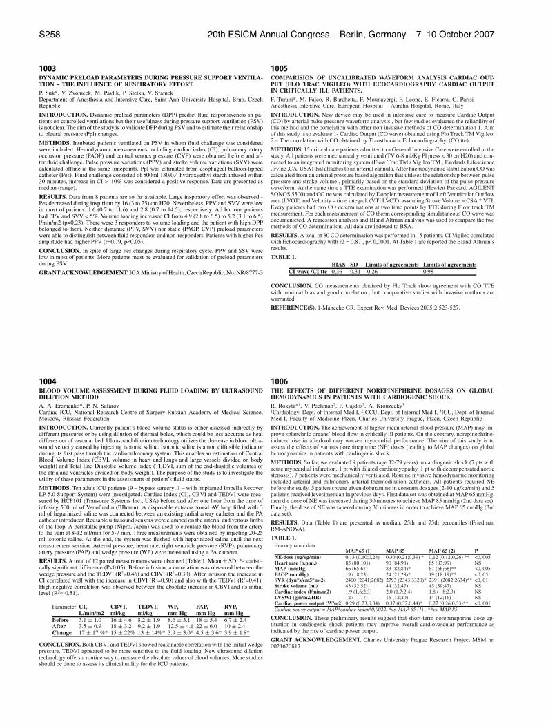
S258 20th ESICM Annual Congress – Berlin, Germany – 7–10 October 2007
1003DYNAMIC PRELOAD PARAMETERS DURING PRESSURE SUPPORT VENTILA-TION – THE INFLUENCE OF RESPIRATORY EFFORT
P. Suk*, V. Zvonicek, M. Pavlik, P. Stetka, V. SramekDepartment of Anesthesia and Intensive Care, Saint Ann University Hospital, Brno, CzechRepublic
INTRODUCTION. Dynamic preload parameters (DPP) predict fluid responsiveness in pa-tients on controlled ventilations but their usefulness during pressure support ventilation (PSV)is not clear. The aim of the study is to validate DPP during PSV and to estimate their relationshipto pleural pressure (Ppl) changes.
METHODS. Intubated patients ventilated on PSV in whom fluid challenge was consideredwere included. Hemodynamic measurements including cardiac index (CI), pulmonary arteryocclusion pressure (PAOP) and central venous pressure (CVP) were obtained before and af-ter fluid challenge. Pulse pressure variations (PPV) and stroke volume variations (SVV) werecalculated offline at the same timepoints. Ppl was estimated from esophageal balloon-tippedcatheter (Pes). Fluid challenge consisted of 500ml 130/0.4 hydroxyethyl starch infused within30 minutes, increase in CI > 10% was considered a positive response. Data are presented asmedian (range).
RESULTS. Data from 8 patients are so far available. Large inspiratory effort was observed -Pes decreased during inspirium by 16 (3 to 25) cm H20. Nevertheless, PPV and SVV were lowin most of patients: 1.6 (0.7 to 11.6) and 2.8 (0.7 to 14.5), respectively. All but one patientshad PPV and SVV < 5%. Volume loading increased CI from 4.9 (2.8 to 6.5) to 5.2 (3.1 to 6.5)l/min/m2 (p=0.23). There were 3 responders to volume loading and the patient with high DPPbelonged to them. Neither dynamic (PPV, SVV) nor static (PAOP, CVP) preload parameterswere able to distinguish between fluid responders and non-responders. Patients with higher Pesamplitude had higher PPV (r=0.79, p<0.05).
CONCLUSION. In spite of large Pes changes during respiratory cycle, PPV and SSV werelow in most of patients. More patients must be evaluated for validation of preload parametersduring PSV.
GRANT ACKNOWLEDGEMENT. IGA Ministry of Health, Czech Republic, No. NR/8777-3
1004BLOOD VOLUME ASSESSMENT DURING FLUID LOADING BY ULTRASOUNDDILUTION METHOD
A. A. Eremenko*, P. N. SafarovCardiac ICU, National Research Centre of Surgery Russian Academy of Medical Science,Moscow, Russian Federation
INTRODUCTION. Currently patient’s blood volume status is either assessed indirectly bydifferent pressures or by using dilution of thermal bolus, which could be less accurate as heatdiffuses out of vascular bed. Ultrasound dilution technology utilizes the decrease in blood ultra-sound velocity caused by injecting isotonic saline. Isotonic saline is a non diffusible indicatorduring its first pass though the cardiopulmonary system. This enables an estimation of CentralBlood Volume Index (CBVI, volume in heart and lungs and large vessels divided on bodyweight) and Total End Diastolic Volume Index (TEDVI, sum of the end-diastolic volumes ofthe atria and ventricles divided on body weight). The purpose of the study is to investigate theutility of these parameters in the assessment of patient’s fluid status.
METHODS. Ten adult ICU patients (9 – bypass surgery; 1 – with implanted Impella RecoverLP 5.0 Support System) were investigated. Cardiac index (CI), CBVI and TEDVI were mea-sured by HCP101 (Transonic Systems Inc., USA) before and after one hour from the time ofinfusing 500 ml of Venofundin (BBraun). A disposable extracorporeal AV loop filled with 3ml of heparinized saline was connected between an existing radial artery catheter and the PAcatheter introducer. Reusable ultrasound sensors were clamped on the arterial and venous limbsof the loop. A peristaltic pump (Nipro, Japan) was used to circulate the blood from the arteryto the vein at 8-12 ml/min for 5-7 min. Three measurements were obtained by injecting 20-25ml isotonic saline. At the end, the system was flushed with heparinized saline until the nextmeasurement session. Arterial pressure, heart rate, right ventricle pressure (RVP), pulmonaryartery pressure (PAP) and wedge pressure (WP) were measured using a PA catheter.
RESULTS. A total of 12 paired measurements were obtained (Table 1, Mean ± SD, *- statisti-cally significant difference (P<0.05). Before infusion, a correlation was observed between thewedge pressure and the TEDVI (R2=0.46) and CBVI (R2=0.33). After infusion the increase inCI correlated well with the increase in CBVI (R2=0.50) and also with the TEDVI (R2=0.41).High negative correlation was observed between the absolute increase in CBVI and its initiallevel (R2=-0.51).
Parameter CI, CBVI, TEDVI, WP, PAP, RVP,L/min/m2 ml/kg ml/kg mm Hg mm Hg mm Hg
Before 3.1 ± 1.0 16 ± 4.6 8.2 ± 1.9 8.6 ± 3.1 18 ± 5.4 6.7 ± 2.4After 3.5 ± 0.9 18 ± 3.2 9.2 ± 1.9 12.5 ± 4.1 22 ± 6.0 10 ± 2.4Change 17 ± 17 %* 15 ± 22% 13 ± 14%* 3.9 ± 3.0* 4.5 ± 3.6* 3.9 ± 1.8*
CONCLUSION. Both CBVI and TEDVI showed reasonable correlation with the initial wedgepressure. TEDVI appeared to be more sensitive to the fluid loading. New ultrasound dilutiontechnology offers a routine way to measure the absolute values of blood volumes. More studiesshould be done to assess its clinical utility for the ICU patients.
1005COMPARISION OF UNCALIBRATED WAVEFORM ANALYSIS CARDIAC OUT-PUT (FLO TRAC VIGILEO) WITH ECOCARDIOGRAPHY CARDIAC OUTPUTIN CRITICALLY ILL PATIENTS.
F. Turani*, M. Falco, R. Barchetta, F. Mounayergi, F. Leone, E. Ficarra, C. ParisiAnesthesia Intensive Care, European Hospital – Aurelia Hospital, Rome, Italy
INTRODUCTION. New device may be used in intensive care to measure Cardiac Output(CO) by arterial pulse pressure waveform analysis , but few studies evaluated the reliability ofthis method and the correlation with other non invasive methods of CO determination 1. Aimof this study is to evaluate 1- Cardiac Output (CO wave) obtained using Flo Track TM Vigileo.2 – The correlation with CO obtained by Transthoracic Echocardiography. (CO tte).
METHODS. 15 critical care patients admitted to a General Intensive Care were enrolled in thestudy. All patients were mechanically ventilated (TV 6-8 ml/Kg Pl press < 30 cmH20) and con-nected to an integrated monitoring system (Flow Trac TM / Vigileo TM , Ewdards Lifescience,Irvine ,CA, USA) that attaches to an arterial cannula. After haemodynamic stabilization CO wascalculated from an arterial pressure based algorithm that utilises the relationship between pulsepressure and stroke volume , primarily based on the standard deviation of the pulse pressurewaveform. At the same time a TTE examination was performed (Hewlett Packard, AGILENTSONOS 5500) and CO tte was calculated by Doppler measurement of Left Ventricular Outflowarea (LVOT) and Velocity – time integral. (VTI LVOT), assuming Stroke Volume = CSA * VTI.Every patients had two CO determinations at two time points by TTE during Flow track TMmeasurement. For each measurement of CO therm corresponding simulataneous CO wave wasdocumenteted. A regression analysis and Bland Altman analysis was used to compare the twomethods of CO determination. All data are indexed to BSA.
RESULTS. A total of 30 CO determination was performed in 15 patients. CI Vigileo correlatedwith Echocardiography with r2 = 0.87 , p< 0,0001. At Table 1 are reported the Bland Altman’sresults.
TABLE 1.BIAS SD Limits of agreements Limits of agreements
CI wave /CI tte 0,36 0,31 -0,26 0,98
CONCLUSION. CO measurements obtained by Flo Track show agreement with CO TTEwith minimal bias and good correlation , but comparative studies with invasive methods arewarranted.
REFERENCE(S). 1-Manecke GR. Expert Rev. Med. Devices 2005;2:523-527.
1006THE EFFECTS OF DIFFERENT NOREPINEPHRINE DOSAGES ON GLOBALHEMODYNAMICS IN PATIENTS WITH CARDIOGENIC SHOCK.
R. Rokyta*1, V. Pechman2, P. Gajdos2, A. Krouzecky3
1Cardiology, Dept. of Internal Med I, 2ICCU, Dept. of Internal Med I, 3ICU, Dept. of InternalMed I, Faculty of Medicine Plzen, Charles University Prague, Plzen, Czech Republic
INTRODUCTION. The achievement of higher mean arterial blood pressure (MAP) may im-prove splanchnic organs’ blood flow in critically ill patients. On the contrary, norepinephrine-induced rise in afterload may worsen myocardial performance. The aim of this study is toassess the effects of various norepinephrine (NE) doses (leading to MAP changes) on globalhemodynamics in patients with cardiogenic shock.
METHODS. So far, we evaluated 9 patients (age 32-79 years) in cardiogenic shock (7 pts withacute myocardial infarction, 1 pt with dilated cardiomyopathy, 1 pt with decompensated aorticstenosis). 7 patients were mechanically ventilated. Routine invasive hemodynamic monitoringincluded arterial and pulmonary arterial thermodilution catheters. All patients required NEbefore the study. 5 patients were given dobutamine in constant dosages (2-10 ug/kg/min) and 5patients received levosimendan in previous days. First data set was obtained at MAP 65 mmHg,then the dose of NE was increased during 30 minutes to achieve MAP 85 mmHg (2nd data set).Finally, the dose of NE was tapered during 30 minutes in order to achieve MAP 65 mmHg (3rddata set).
RESULTS. Data (Table 1) are presented as median, 25th and 75th percentiles (FriedmanRM-ANOVA).
TABLE 1.Hemodynamic data
MAP 65 (1) MAP 85 MAP 65 (2) PNE-dose (ug/kg/min) 0,13 (0,10;0,24) 0,30 (0,21;0,39) * 0,12 (0,12;0,26) ** <0, 005Heart rate (b.p.m.) 85 (80;101) 90 (84;98) 85 (83;99) NSMAP (mmHg) 66 (65;67) 83 (82;84)* 67 (66;68)** <0, 005PAOP (mmHg) 19 (18;23) 24 (21;28)* 19 (18;19)** <0, 05SVR (dyn*s/cm5*m-2) 2400 (2041;2682) 2793 (2343;3320)* 2391 (2082;2634)** <0, 01Stroke volume (ml) 43 (32;52) 44 (32;47) 45 (39;47) NSCardiac index (l/min/m2) 1,9 (1,6;2,3) 2,0 (1,7;2,4) 1,8 (1,8;2,3) NSLVSWI (gm/m2/HR) 12 (11;17) 16 (12;20) 14 (12;16) NSCardiac power output (W/m2) 0,29 (0,23;0,34) 0,37 (0,32;0,44)* 0,27 (0,26;0,33)** <0, 001Cardiac power output = MAP*cardiac index*0,0022. *vs. MAP 65 (1) , **vs. MAP 85
CONCLUSION. These preliminary results suggest that short-term norepinephrine dose up-titration in cardiogenic shock patients may improve overall cardiovascular performance asindicated by the rise of cardiac power output.
GRANT ACKNOWLEDGEMENT. Charles University Prague Research Project MSM nr.0021620817
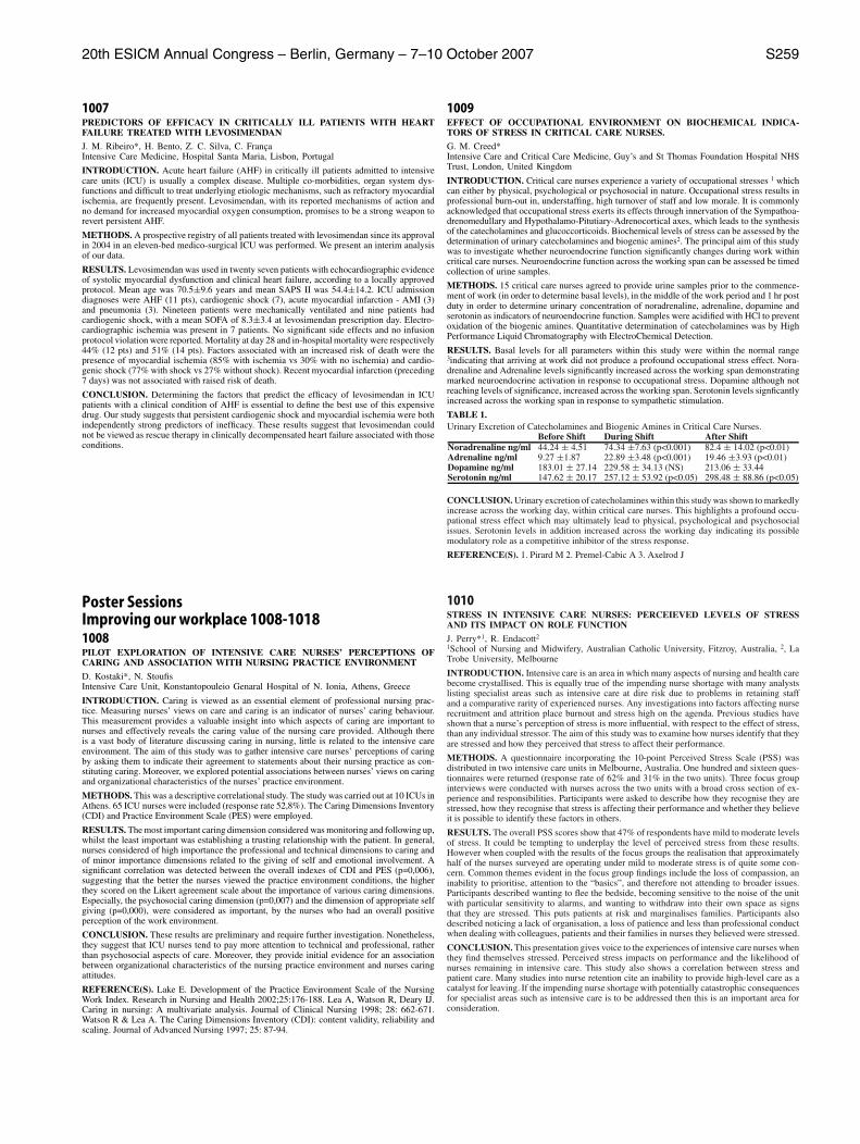
20th ESICM Annual Congress – Berlin, Germany – 7–10 October 2007 S259
1007PREDICTORS OF EFFICACY IN CRITICALLY ILL PATIENTS WITH HEARTFAILURE TREATED WITH LEVOSIMENDAN
J. M. Ribeiro*, H. Bento, Z. C. Silva, C. FrancaIntensive Care Medicine, Hospital Santa Maria, Lisbon, Portugal
INTRODUCTION. Acute heart failure (AHF) in critically ill patients admitted to intensivecare units (ICU) is usually a complex disease. Multiple co-morbidities, organ system dys-functions and difficult to treat underlying etiologic mechanisms, such as refractory myocardialischemia, are frequently present. Levosimendan, with its reported mechanisms of action andno demand for increased myocardial oxygen consumption, promises to be a strong weapon torevert persistent AHF.
METHODS. A prospective registry of all patients treated with levosimendan since its approvalin 2004 in an eleven-bed medico-surgical ICU was performed. We present an interim analysisof our data.
RESULTS. Levosimendan was used in twenty seven patients with echocardiographic evidenceof systolic myocardial dysfunction and clinical heart failure, according to a locally approvedprotocol. Mean age was 70.5±9.6 years and mean SAPS II was 54.4±14.2. ICU admissiondiagnoses were AHF (11 pts), cardiogenic shock (7), acute myocardial infarction - AMI (3)and pneumonia (3). Nineteen patients were mechanically ventilated and nine patients hadcardiogenic shock, with a mean SOFA of 8.3±3.4 at levosimendan prescription day. Electro-cardiographic ischemia was present in 7 patients. No significant side effects and no infusionprotocol violation were reported. Mortality at day 28 and in-hospital mortality were respectively44% (12 pts) and 51% (14 pts). Factors associated with an increased risk of death were thepresence of myocardial ischemia (85% with ischemia vs 30% with no ischemia) and cardio-genic shock (77% with shock vs 27% without shock). Recent myocardial infarction (preceding7 days) was not associated with raised risk of death.
CONCLUSION. Determining the factors that predict the efficacy of levosimendan in ICUpatients with a clinical condition of AHF is essential to define the best use of this expensivedrug. Our study suggests that persistent cardiogenic shock and myocardial ischemia were bothindependently strong predictors of inefficacy. These results suggest that levosimendan couldnot be viewed as rescue therapy in clinically decompensated heart failure associated with thoseconditions.
Poster SessionsImproving our workplace 1008-10181008PILOT EXPLORATION OF INTENSIVE CARE NURSES’ PERCEPTIONS OFCARING AND ASSOCIATION WITH NURSING PRACTICE ENVIRONMENT
D. Kostaki*, N. StoufisIntensive Care Unit, Konstantopouleio Genaral Hospital of N. Ionia, Athens, Greece
INTRODUCTION. Caring is viewed as an essential element of professional nursing prac-tice. Measuring nurses’ views on care and caring is an indicator of nurses’ caring behaviour.This measurement provides a valuable insight into which aspects of caring are important tonurses and effectively reveals the caring value of the nursing care provided. Although thereis a vast body of literature discussing caring in nursing, little is related to the intensive careenvironment. The aim of this study was to gather intensive care nurses’ perceptions of caringby asking them to indicate their agreement to statements about their nursing practice as con-stituting caring. Moreover, we explored potential associations between nurses’ views on caringand organizational characteristics of the nurses’ practice environment.
METHODS. This was a descriptive correlational study. The study was carried out at 10 ICUs inAthens. 65 ICU nurses were included (response rate 52,8%). The Caring Dimensions Inventory(CDI) and Practice Environment Scale (PES) were employed.
RESULTS. The most important caring dimension considered was monitoring and following up,whilst the least important was establishing a trusting relationship with the patient. In general,nurses considered of high importance the professional and technical dimensions to caring andof minor importance dimensions related to the giving of self and emotional involvement. Asignificant correlation was detected between the overall indexes of CDI and PES (p=0,006),suggesting that the better the nurses viewed the practice environment conditions, the higherthey scored on the Likert agreement scale about the importance of various caring dimensions.Especially, the psychosocial caring dimension (p=0,007) and the dimension of appropriate selfgiving (p=0,000), were considered as important, by the nurses who had an overall positiveperception of the work environment.
CONCLUSION. These results are preliminary and require further investigation. Nonetheless,they suggest that ICU nurses tend to pay more attention to technical and professional, ratherthan psychosocial aspects of care. Moreover, they provide initial evidence for an associationbetween organizational characteristics of the nursing practice environment and nurses caringattitudes.
REFERENCE(S). Lake E. Development of the Practice Environment Scale of the NursingWork Index. Research in Nursing and Health 2002;25:176-188. Lea A, Watson R, Deary IJ.Caring in nursing: A multivariate analysis. Journal of Clinical Nursing 1998; 28: 662-671.Watson R & Lea A. The Caring Dimensions Inventory (CDI): content validity, reliability andscaling. Journal of Advanced Nursing 1997; 25: 87-94.
1009EFFECT OF OCCUPATIONAL ENVIRONMENT ON BIOCHEMICAL INDICA-TORS OF STRESS IN CRITICAL CARE NURSES.
G. M. Creed*Intensive Care and Critical Care Medicine, Guy’s and St Thomas Foundation Hospital NHSTrust, London, United Kingdom
INTRODUCTION. Critical care nurses experience a variety of occupational stresses 1 whichcan either by physical, psychological or psychosocial in nature. Occupational stress results inprofessional burn-out in, understaffing, high turnover of staff and low morale. It is commonlyacknowledged that occupational stress exerts its effects through innervation of the Sympathoa-drenomedullary and Hypothalamo-Pitutiary-Adrenocortical axes, which leads to the synthesisof the catecholamines and glucoccorticoids. Biochemical levels of stress can be assessed by thedetermination of urinary catecholamines and biogenic amines2. The principal aim of this studywas to investigate whether neuroendocrine function significantly changes during work withincritical care nurses. Neuroendocrine function across the working span can be assessed be timedcollection of urine samples.
METHODS. 15 critical care nurses agreed to provide urine samples prior to the commence-ment of work (in order to determine basal levels), in the middle of the work period and 1 hr postduty in order to determine urinary concentration of noradrenaline, adrenaline, dopamine andserotonin as indicators of neuroendocrine function. Samples were acidified with HCl to preventoxidation of the biogenic amines. Quantitative determination of catecholamines was by HighPerformance Liquid Chromatography with ElectroChemical Detection.
RESULTS. Basal levels for all parameters within this study were within the normal range3indicating that arriving at work did not produce a profound occupational stress effect. Nora-drenaline and Adrenaline levels significantly increased across the working span demonstratingmarked neuroendocrine activation in response to occupational stress. Dopamine although notreaching levels of significance, increased across the working span. Serotonin levels signficantlyincreased across the working span in response to sympathetic stimulation.
TABLE 1.Urinary Excretion of Catecholamines and Biogenic Amines in Critical Care Nurses.
Before Shift During Shift After ShiftNoradrenaline ng/ml 44.24 ± 4.51 74.34 ±7.63 (p<0.001) 82.4 ± 14.02 (p<0.01)Adrenaline ng/ml 9.27 ±1.87 22.89 ±3.48 (p<0.001) 19.46 ±3.93 (p<0.01)Dopamine ng/ml 183.01 ± 27.14 229.58 ± 34.13 (NS) 213.06 ± 33.44Serotonin ng/ml 147.62 ± 20.17 257.12 ± 53.92 (p<0.05) 298.48 ± 88.86 (p<0.05)
CONCLUSION. Urinary excretion of catecholamines within this study was shown to markedlyincrease across the working day, within critical care nurses. This highlights a profound occu-pational stress effect which may ultimately lead to physical, psychological and psychosocialissues. Serotonin levels in addition increased across the working day indicating its possiblemodulatory role as a competitive inhibitor of the stress response.
REFERENCE(S). 1. Pirard M 2. Premel-Cabic A 3. Axelrod J
1010STRESS IN INTENSIVE CARE NURSES: PERCEIEVED LEVELS OF STRESSAND ITS IMPACT ON ROLE FUNCTION
J. Perry*1, R. Endacott21School of Nursing and Midwifery, Australian Catholic University, Fitzroy, Australia, 2, LaTrobe University, Melbourne
INTRODUCTION. Intensive care is an area in which many aspects of nursing and health carebecome crystallised. This is equally true of the impending nurse shortage with many analystslisting specialist areas such as intensive care at dire risk due to problems in retaining staffand a comparative rarity of experienced nurses. Any investigations into factors affecting nurserecruitment and attrition place burnout and stress high on the agenda. Previous studies haveshown that a nurse’s perception of stress is more influential, with respect to the effect of stress,than any individual stressor. The aim of this study was to examine how nurses identify that theyare stressed and how they perceived that stress to affect their performance.
METHODS. A questionnaire incorporating the 10-point Perceived Stress Scale (PSS) wasdistributed in two intensive care units in Melbourne, Australia. One hundred and sixteen ques-tionnaires were returned (response rate of 62% and 31% in the two units). Three focus groupinterviews were conducted with nurses across the two units with a broad cross section of ex-perience and responsibilities. Participants were asked to describe how they recognise they arestressed, how they recognise that stress is affecting their performance and whether they believeit is possible to identify these factors in others.
RESULTS. The overall PSS scores show that 47% of respondents have mild to moderate levelsof stress. It could be tempting to underplay the level of perceived stress from these results.However when coupled with the results of the focus groups the realisation that approximatelyhalf of the nurses surveyed are operating under mild to moderate stress is of quite some con-cern. Common themes evident in the focus group findings include the loss of compassion, aninability to prioritise, attention to the “basics”, and therefore not attending to broader issues.Participants described wanting to flee the bedside, becoming sensitive to the noise of the unitwith particular sensitivity to alarms, and wanting to withdraw into their own space as signsthat they are stressed. This puts patients at risk and marginalises families. Participants alsodescribed noticing a lack of organisation, a loss of patience and less than professional conductwhen dealing with colleagues, patients and their families in nurses they believed were stressed.
CONCLUSION. This presentation gives voice to the experiences of intensive care nurses whenthey find themselves stressed. Perceived stress impacts on performance and the likelihood ofnurses remaining in intensive care. This study also shows a correlation between stress andpatient care. Many studies into nurse retention cite an inability to provide high-level care as acatalyst for leaving. If the impending nurse shortage with potentially catastrophic consequencesfor specialist areas such as intensive care is to be addressed then this is an important area forconsideration.
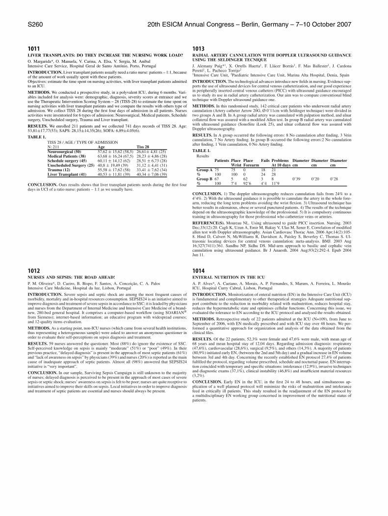
S260 20th ESICM Annual Congress – Berlin, Germany – 7–10 October 2007
1011LIVER TRANSPLANTS: DO THEY INCREASE THE NURSING WORK LOAD?
O. Margarida*, O. Manuela, V. Carina, A. Elsa, V. Sergia, M. AnibalIntensive Care Service, Hospital Geral de Santo Antonio, Porto, Portugal
INTRODUCTION. Liver transplant patients usually need a ratio nurse: patients – 1:1, becauseof the amount of work usually spent with these patients.Objectives: estimate the time spent on nursing activities, with liver transplant patients admittedto an ICU.
METHODS. We conducted a prospective study, in a polyvalent ICU, during 6 months. Vari-ables included for analysis were: demographic, diagnosis, severity scores at entrance and weuse the Therapeutic Intervention Scoring System – 28 (TISS-28) to estimate the time spent onnursing activities with liver transplant patients and we compare the results with others type ofadmission. We collect TISS 28 during the first four days of admission in all patients. Nursesactivities were inventoried for 6 types of admission: Neurosurgical, Medical patients, Schedulesurgery, Unscheduled surgery, Trauma and Liver transplant.
RESULTS. We enrolled 211 patients and we collected 741 days records of TISS 28. Age:53,81±17,77(53); SAPS: 28,15±14,35(26); SOFA: 6,89±4.05(6).
TABLE 1.TISS 28 / AGE / TYPE OF ADMISSIONN: 211 Age Tiss 28Neurosurgical (50) 57,62 ± 15,62 (58,5) 26,61± 4,81 (25)Medical Patients (38) 63,68 ± 16,24 (67,5) 28,23 ± 4,86 (28)Schedule surgery (45) 60,11 ± 14,12 (62) 28,51 ± 6,73 (28)Unscheduled Surgery (25) 40,8 ± 19,49 (39) 31,12 ± 4,41 (31)Trauma (12) 55,58 ± 17,62 (58) 33,41 ± 7,62 (34)Liver Transplant (41) 40,53 ± 11,81 (39) 40,34 ± 7,06 (39)
CONCLUSION. Ours results shows that liver transplant patients needs during the first fourdays in UCI of a ratio nurse: patients – 1:1 as we usually have.
1012NURSES AND SEPSIS: THE ROAD AHEAD!
P. M. Oliveira*, D. Caeiro, B. Bispo, F. Santos, A. Conceicao, C. A. PalosIntensive Care Medicine, Hospital da luz, Lisbon, Portugal
INTRODUCTION. Severe sepsis and septic shock are among the most frequent causes ofmorbidity, mortality and in-hospital resources consumption. SEPSIS24 is an initiative aimed toimprove diagnosis and treatment of severe sepsis in accordance to SSC; it is leaded by physiciansand nurses from the Department of Internal Medicine and Intensive Care Medicine of a brand-new, 280-bed general hospital. It comprises a computer-based workflow (using SOARIAN®
from Siemens); internet-based information; an educative program with widespread courses;and 12-quality items evaluation.
METHODS. As a starting point, non-ICU nurses (which came from several health institutions,thus representing a heterogeneous sample) were asked to answer an anonymous questioner inorder to evaluate their self-perceptions on sepsis diagnosis and treatment.
RESULTS. 59 nurses answered the questioner. Most (88%) do ignore the existence of SSC.Self-perceived knowledge on sepsis is mainly “moderate” (51%) or “poor” (49%). In theirprevious practice, “delayed diagnosis” is present in the approach of most septic patients (61%)and “lack of awareness on sepsis” by physicians (39%) and nurses (20%) is reported as the maincause of inadequate approach of septic patients. Almost all (98%) answered that SEPSIS24initiative is “very important”.
CONCLUSION. In our sample, Surviving Sepsis Campaign is still unknown to the majorityof nurses; delayed diagnosis is perceived to be present in the approach of most cases of severesepsis or septic shock; nurses’ awareness on sepsis is felt to be poor; nurses are quite receptive toinitiatives aimed to improve their skills on sepsis. Local initiatives in order to improve diagnosisand treatment of septic patients are essential and nurses should always be present.
1013RADIAL ARTERY CANNULATION WITH DOPPLER ULTRASOUND GUIDANCEUSING THE SELDINGER TECNIQUE
J. Alemany Puig*1, X. Ortells Huerta1, F. Llacer Borras1, F. Mas Ballester1, J. CardonaPereto1, L. Pacheco Torrijo2
1Intensive Care Unit, 2Paediatric Intensive Care Unit, Marina Alta Hospital, Denia, Spain
INTRODUCTION. The technological advances introduce new fields in nursing. Evidence sup-ports the use of ultrasound devices for central venous catheterization, and our good experiencein peripherally inserted central venous catheters (PICC) with ultrasound guidance encouragedus to study its use in radial artery catheterization. Our aim was to compare conventional blindtechnique with Doppler ultrasound guidance one.
METHODS. In this randomised study, 142 critical care patients who underwent radial arterycannulation (Artery catheter Arrow 20G, Ø 0’11cm with Seldinger technique) were divided intwo groups A and B. In A group radial artery was cannulated with palpation method, and ulnarcollateral flow was assured with a modified Allen test. In group B radial artery was cannulatedwith ultrasound guidance (SonoSite iLook 25), and ulnar collateral flow was assured withDoppler ultrasonography.
RESULTS. In A group occurred the following errors: 8 No cannulation after finding, 3 Veincannulation, 7 No Artery finding. In group B occurred the following errors:2 No cannulationafter finding, 1 Vein cannulation, 0 No Artery finding.
TABLE 1.Results
Patients Place Place Fails Problems Diameter Diameter DiameterWrist Forearm At 10 days cm cm cm
Group A 75 75 0 18 21% 100 100 0 24 28Group B 67 5 62 3 8 0’39 0’20 0’28% 100 7’4 92’6 4’4 11’9
CONCLUSION. 1) The doppler ultrasonography reduces cannulation fails from 24% to a4’4%. 2) With the ultrasound guidance it is possible to cannulate the artery in the whole fore-arm, reducing the long term problems avoiding the wrist flexion. 3) Ultrasound technique hasbetter results in edematous, obese or several punctured patients. 4) The results of the techniquedepend on the ultrasonographic knowledge of the professional. 5) It is compulsory continuoustraining in ultrasonography for those professional who catheterize veins or arteries.
REFERENCE(S). Moureau NL. Using ultrasound to guide PICC insertion. Nursing. 2003Dec;33(12):20. Cagli K, Uzun A, Emir M, Bakuy V, Ulas M, Sener E. Correlation of modifiedallen test with Doppler ultrasonography. Asian Cardiovasc Thorac Ann. 2006 Apr;14(2):105-8. Hind D, Calvert N, McWilliams R, Davidson A, Paisley S, Beverley C, Thomas S. Ul-trasonic locating devices for central venous cannulation: meta-analysis. BMJ. 2003 Aug16;327(7411):361. Sandhu NP, Sidhu DS. Mid-arm approach to basilic and cephalic veincannulation using ultrasound guidance. Br J Anaesth. 2004 Aug;93(2):292-4. Epub 2004Jun 11.
1014ENTERAL NUTRITION IN THE ICU
A. P. Alves*, A. Caetano, A. Morais, A. P. Fernandes, S. Marum, A. Ferreira, L. MouraoICU, Hospital Curry Cabral, Lisbon, Portugal
INTRODUCTION. Monitorization of enteral nutrition (EN) in the Intensive Care Unit (ICU)is fundamental and complimentary to other therapeutical strategies Adequate nutritional sup-port contribute to the reduction in morbidity related with malnutrition, reduces hospital stay,reduces the hypermetabolic state and optimises cellular functions. Concerning this issue, weevaluated the tolerance to EN according to the ICU protocol and analysed the results obtained.
METHODS. Retrospective study of 22 patients admitted at the ICU (N=109), from June toSeptember of 2006, with EN medically prescribed and with ICU stay over 48 hours. We per-formed a quantitative approach for organization and analysis of the data obtained from theclinical files.
RESULTS. Of the 22 patients, 52,3% were female and 47,6% were male, with mean age of68 years and mean hospital stay of 12,04 days. Regarding admission diagnosis: respiratory(47,6%), cardiovascular (28,6%), surgical (9,5%), and others (14,3%). A majority of patients(80,9%) initiated early EN, (between the 2nd and 5th day) and a gradual increase in EN volumebetween 3rd and 4th day. Concerning the recently established EN protocol 27,4% of patientsfulfilled the protocol, regarding volume prescribed, schedule and nocturnal pause. EN interrup-tion coincided with temporary and specific situations: intolerance (12,9%), invasive techniquesand diagnostic exams (37,1%), clinical instability (46,8%) and insufficient material resources(3,2%).
CONCLUSION. Early EN in the ICU, in the first 24 to 48 hours, and simultaneous ap-plication of a well planned protocol will minimize the risks of malnutrition and intolerancefeed in critically ill patients. This study resulted in the readjustment of the EN protocol bya multidisciplinary EN working group concerned in improvement of the nutritional status ofpatients.
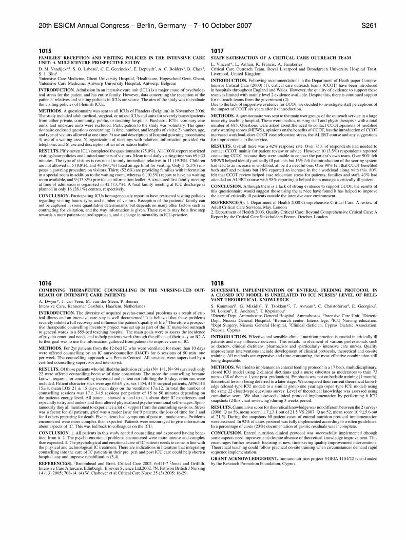
20th ESICM Annual Congress – Berlin, Germany – 7–10 October 2007 S261
1015FAMILIES’ RECEPTION AND VISITING POLICIES IN THE INTENSIVE CAREUNIT: A MULTICENTRE PROSPECTIVE STUDY
D. M. Vandijck*1, S. O. Labeau2, C. E. Geerinckx2, E. Depuydt2, A. C. Bolders3, B. Claes3,S. I. Blot11Intensive Care Medicine, Ghent University Hospital, 2Healthcare, Hogeschool Gent, Ghent,3Intensive Care Medicine, Antwerp University Hospital, Antwerp, Belgium
INTRODUCTION. Admission in an intensive care unit (ICU) is a major cause of psycholog-ical stress for the patient and his entire family. However, data concerning the reception of thepatients’ relatives and visiting policies in ICUs are scarce. The aim of the study was to evaluatethe visiting policies of Flemish ICUs.
METHODS. A questionnaire was sent to all ICUs of Flanders (Belgium) in November 2006.The study included adult medical, surgical, or mixed ICUs and units for severely burned patientsfrom either private, community, public, or teaching hospitals. Paediatric ICUs, coronary careunits, and mid-care units were excluded. Participation to the study was voluntary. The ques-tionnaire enclosed questions concerning; 1) time, number, and lengths of visits; 2) number, age,and type of visitors allowed at one time; 3) use and description of hospital gowning procedures;4) use of a waiting area; 5) organization of meetings with relatives, information provided viatelephone; and 6) use and description of an information leaflet.
RESULTS. Fifty-seven ICUs completed the questionnaire (75.0%). All (100%) report restrictedvisiting-hour policies and limited numbers of visitors. Mean total daily visiting time was 69±33minutes. The type of visitors is restricted to only immediate relatives in 11 (19.3%). Childrenare not allowed in 5 (8.8%), and 46 (80.7%) fixed an age limit for visiting. Only 3 (5.3%) im-poses a gowning procedure on visitors. Thirty (52.6%) are providing families with informationin a special room in addition to the waiting room, whereas 6 (10.5%) report to have no waitingroom available, and 9 (15.8%) provide an information leaflet. A structured first family meetingat time of admission is organized in 42 (73.7%). A final family meeting at ICU discharge isplanned in only 16 (28.1%) centres, respectively.
CONCLUSION. Participating ICUs homogeneously report to have restricted visiting policiesregarding visiting hours, type, and number of visitors. Reception of the patients’ family cannot be captured in some quantitative determinants, but depends on many other factors such ascontracting for visitation, and the way information is given. These results may be a first steptowards a more patient-centred approach, and a change in mentality in ICU-practice.
1016COMBINING THERAPEUTIC COUNSELLING IN THE NURSING-LED OUT-REACH OF INTENSIVE CARE PATIENTS
A. Dwyer*, J. van Veen, M. van der Steen, P. BennerIntensive Care, Kennemer Gasthuis, Haarlem, Netherlands
INTRODUCTION. The diversity of acquired psycho-emotional problems as a result of crit-ical illness and an intensive care stay is well documented1 It is believed that these problemsseverely inhibit a full recovery and influence the patient’s quality of life 2 Therefore a prospec-tive therapeutic counselling inventory project was set up as part of the IC nurse-led outreachto general wards in a 855-bed teaching hospital. The main goals were to assess the incidenceof psycho-emotional needs and to help patients work through the effects of their stay on IC. Afurther goal was to use the information gathered from patients to improve care on IC.
METHODS. For 2yr patients from the 12-bed IC who were ventilated for more than 10 dayswere offered counselling by an IC nurse/counsellor (BACP) for 6 sessions of 50 min. oneper week. The counselling approach was Person-Centred. All sessions were supervised by acertified counselling supervisor and intensivist.
RESULTS. Of those patients who fulfilled the inclusion criteria (N= 141, N= 94 survived) only22 were offered counselling because of time constraints. The more the counselling becameknown, requests for counselling increased and patient not fulfilling the 10-days criterium wereincluded. Patient characteristics were age 63±9 yrs, sex 11M, 41% surgical patients, APACHE15±8, mean LOS 21 ± 15 days, mean days on the ventilator 17±12. In total the number ofcounselling sessions was 171; 3-15 sessions per patient of 15 to 50 minutes depending onthe patients energy level. All patients showed a need to talk about their IC experiences andespecially to try and understand their altered physical and psycho-emotional self-images. Spon-taneously they all mentioned to experience a lot of support from the counseling sessions. Stresswas a factor for all patients, grief was a major issue for 9 patients, the loss of time for 3 andfor 4 others preparing for death. Five patients had symptoms of post traumatic stress. Problemsencountered were more complex than expected. Patients were encouraged to give informationabout aspects of IC. This was fed back to colleagues on the ICU.
CONCLUSION. 1. All patients in this study needed counselling and expressed having bene-fited from it. 2. The psycho-emotional problems encountered were more intense and complexthan expected. 3. The psychological and emotional care of IC patients needs to come in line withthe physical and technological IC treatment. There are indications in literature that integratingcounselling into the care of IC patients in their pre, peri and post ICU care could help shortenhospital stay and improve rehabilitation (3,4).
REFERENCE(S). 1Broomhead and Brett, Critical Care 2002. 6:411-7 2Jones and Griffith.Intensive Care Aftercare. Edinburgh: Elsevier Science Ltd,2002. 3N. Pattison British J Nursing14 (13) 2005; 708-14. (4) W. Chaboyer et al Critical Care Nurse 25 (3) 2005; 16-29.
1017STAFF SATISFACTION OF A CRITICAL CARE OUTREACH TEAM
L. Vincent*, L. Arthan, K. Francis, A. FazakerleyCritical Care Outreach Team, Royal Liverpool and Broadgreen University Hospital Trust,Liverpool, United Kingdom
INTRODUCTION. Following recommendations in the Department of Heath paper Compre-hensive Critical Care (2000) (1), critical care outreach teams (CCOT) have been introducedin hospitals throughout England and Wales. However, the quality of evidence to support theseteams is limited with mainly level 2 evidence available. Despite this, there is continued supportfor outreach teams from the government (2).Due to the lack of supportive evidence for CCOT we decided to investigate staff perceptions ofthe impact of CCOT six years after its introduction.
METHODS. A questionnaire was sent to the main user groups of the outreach service in a largeinner city teaching hospital. These were medics, nursing staff and physiotherapists with a totalnumber of 685. Questions were asked about:the need to contact CCOT,opinions of modifiedearly warning scores (MEWS), opinions on the benefits of CCOT, has the introduction of CCOTincreased workload, does CCOT ease relocation stress, the ALERT course and any suggestionsfor improvements to the service.
RESULTS. Overall there was a 42% response rate. Over 75% of respondents had needed tocontact CCOT, mainly for patient review or advice. However 10 (3.5%) respondents reportedcontacting CCOT because they were unable to contact the patient’s own team. Over 90% feltMEWS helped identify critically ill patients but 16% felt the introduction of the scoring systemhad lead to an increase in workload, all be it a needful one. Over 90% felt that CCOT benefitedboth staff and patients but 18% reported an increase in their workload along with this. 80%felt that CCOT review helped ease relocation stress for patients, families and staff. 43% hadattended an ALERT course with 98% reporting it helped them manage a critically ill patient.
CONCLUSION. Although there is a lack of strong evidence to support CCOT, the results ofthis questionnaire would suggest those using the service have found it has helped to improvethe care of critically ill patients outside the intensive care environment.
REFERENCE(S). 1. Department of Health 2000 Comprehensive Critical Care: A review ofAdult Critical Care Services. May. London2. Department of Health 2003. Quality Critical Care: Beyond Comprehensive Critical Care: AReport by the Critical Care Stakeholders Forum. October. London
1018SUCCESSFUL IMPLEMENTATION OF ENTERAL FEEDING PROTOCOL INA CLOSED ICU MODEL IS UNRELATED TO ICU NURSES’ LEVEL OF RELE-VANT THEORETICAL KNOWLEDGE
S. Kountouri1, G. Mixidis2, Y. Tsokkou*2, T. Avraam3, C. Christoforou4, E. Georgiou5,M. Loizou6, E. Andreou7, T. Kyprianou2
1Dietetic Dept, Ammohostos General Hospital, Ammohostos, 2Intensive Care Unit, 3DieteticDept, Nicosia General Hospital, 4Research center, Intercollege, 5ICU Nursing education,6Dept Surgery, Nicosia General Hospital, 7Clinical dietician, Cyprus Dietetic Association,Nicosia, Cyprus
INTRODUCTION. Effective and sensible clinical nutrition practice is crucial in critically illpatients and may influence outcome. This entails involvement of various professionals suchas doctors, clinical dietitians, pharmacists and -particularly- intensive care nurses. Qualityimprovement interventions include development of clinical protocols, theoretical and on-sitetraining. All methods are expensive and time-consuming, the most effective combination stillbeing disputable.
METHODS. We tried to implement an enteral feeding protocol in a 17 beds, multidisciplinary,closed ICU model using 2 clinical dietitians and a nurse educator as moderators to train 73nurses (38 of them < 1 year post-graduation). Emphasis was put on bedside training (1 month),theoretical lessons being deferred to a later stage. We compared their current theoretical knowl-edge (closed-type ICU model) to a similar group one year ago (open-type ICU model) usingthe same 22 closed-type questions survey. Level of theoretical knowledge was assessed usingcumulative score. We also assessed clinical protocol implementation by performing 6 ICUsnapshots (24hrs chart reviewing) during 3 weeks period.
RESULTS. Cumulative score for theoretical knowledge was not different between the 2 surveys(2006: Q no 56, mean score 11.7±3.1 out of 21.5 VS 2007: Q no 52, mean score 10.9±5.6 outof 21.5). During the snapshots 60 patient-cases of enteral nutrition protocol implementationwere assessed. In 92% of cases protocol was fully implemented according to written guidelines.In a percentage of cases (23%) documentation of gastric residuals was incomplete.
CONCLUSION. Enteral nutrition clinical protocol was successfully implemented (thoughsome aspects need improvement) despite absence of theoretical knowledge improvement. Thisencourages further research focusing at new, time-saving quality improvement interventions.Theoretical teaching could follow practical on-site training when circumstances demand rapidsequence implementation.
GRANT ACKNOWLEDGEMENT. Immunonutrition project YGEIA 1104/22 is co-fundedby the Research Promotion Foundation, Cyprus.
S262 20th ESICM Annual Congress – Berlin, Germany – 7–10 October 2007
Poster SessionsPhysical activity in ICU patients 1019-10241019DOES PHYSIOTHERAPY LED EARLY MOBILISATION AFFECT LENGTH OFSTAY ON ICU
D. J. McWilliams*, K. P. PantelidesPhysiotherapy, Manchester Royal Infirmary, Manchester, United KingdomINTRODUCTION. The negative effects of mechanical ventilation and the associated bed restare well documented. Muscle wasting and weakness are prominent, which may take months torecover and are more likely with prolonged ICU stays and longer hours of ventilation [1]. Tocounteract these effects there has been an increasing move towards early rehabilitation on ICU.This study aimed to determine the effect of early mobilisation on length of stay on ICU.METHODS. The study was carried out on 65 patients admitted to ICU for >24 hours over athree month period. Physiotherapists documented patients’ rehabilitation status daily, allowingidentification of when patients began mobilisation, defined as sitting on the edge of the bed orout in a chair. A target of day 5 was agreed locally, for those not mobilised by this point limitingfactors were identified via a retrospective analysis of the patients’ notes. Data were analysedusing Wilcoxon Signed Ranks Test.RESULTS. No differences were identified between the three groups analysed in terms of ageor APACHE scores(see Table 1). 26% of patients were mobilised by day 5 on ICU and had amedian length of stay of 4 days. Patients who met the criteria for mobilisation but were notmobilised had a significantly increased length of stay with a median of 9 days (p< 0.001), whilstthose who did not meet the criteria for mobilisation had a median length of stay of 16.5 days.TABLE 1.
N Age APACHE II APACHEPred. Mortality
Mobilised on or before day 5 17 55.5 15.6 27.7Met criteria but not mobilised 14 56.6 17.9 27.5Not appropriate to mobilise 34 57 16.2 25.4All Subjects 65 56.5 16.5 26.4
TABLE 2.Length of stay on ICU median (Interquartile range)
Met standard Did not meetfor Mobilisation standard for mobilisation
Mobilsed on or before Day 5 4 (3-6)Not mobilised by Day 5 9 (5.5-19.25) 16.5 (9.5-26.5)P <0.001
CONCLUSION. Early mobilisation can significantly decrease length of stay on ICU, althoughadequate staffing is required in order to maximise the effectiveness of these interventions.REFERENCE(S). 1. Jones C, Griffiths RD (2000) Identifying post intensive care patients whomay need physical rehabilitation. Clin Intensive Care 11:35-38
1020ELECTRICAL MUSCLE STIMULATION HAS A SYSTEMIC EFFECT ON THEMICROCIRCULATION OF ICU PATIENTS
V. Gerovasili, E. Karatzanos, E. Papadopoulos, P. Renieris, A. Bouchla, V. Markaki,D. Zervakis, C. Routsi, S. Nanas*First critical care medicine departement, Evaggelismos Hospital, National and KapodestrianUniversity of Athens, Athens, Greece
INTRODUCTION. Recent studies have shown that electrical muscle stimulation (EMS) maybe an alternative to active exercise in COPD and CHF patients with myopathy. A commonfinding in ICU patients is critical illness polyneuromyopathy (CIPNM). The role of EMS forthe prevention of CIPNM in ICU patients has not been studied yet. The aim of our study wasto assess the acute effect of EMS of lower extremities on the thenar muscle microcirculation inICU patients using Near Infrared Spectroscopy (NIRS).
METHODS. Eleven ICU patients (?6/?5),(age:56±18years)(APACHE admissionscore:15±6)(SOFA admission score 7±2),10 patients on mechanical ventilation (6 pts at VCmode/ 3 pts at PS/1 patient at T-piece) and 1 patient on venturi mask, underwent a 45-minsession of EMS of vastus lateralis, vastus medialis and peroneus longus muscles of bothlower extremities. The stimulator delivered biphasic, symmetric impulses of 45 Hz, 230µsecpulse duration ,12 sec on and 6 sec off and an intensity of 40-80mA able to cause visiblecontractions. Systolic blood pressure(SBP) and heart rate(HR) were measured before and aftereach session in all patients. In eight patients arterial and central venous blood samples wereretrieved before and after the session. Routine laboratory parameters were also recorded.Tissue oxygen saturation (StO2%) was assessed with NIRS at the thenar muscle before, duringand after 3 min occlusion of the brachial artery before the beginning and after the completionof the EMS session.
RESULTS. StO2% did not differ significantly before and after the EMS session(88±7%vs.89±9%). Oxygen consumption rate (%/min) during occlusion of the brachialartery differed significantly before the beginning and at the end of the session(19±6%/minvs.23±7%/min,p=0.004). Reperfusion rate (%/min) differed significantly before the begginingand at the end of the session (305±123%/min vs.378±139%/min, p=0.007). SBP and HRincreased significantly at the end of the session (92bpm±16 vs 97bpm±15 p=0.002 and135mmHg±23 vs 140±22mmHg, p=0.02 respectively).
CONCLUSION. The data presented suggest that EMS may have a systemic effect on micro-circulation. These results may encourage further studies to explore the potential use of EMS asa possible preventive and rehabilitation tool in ICU patients with CIPNM.
1021EVALUATION OF MOTOR PHYSICAL THERAPY IN INTRA-ABDOMINAL PRES-SURE
A. T. Guiera*1, E. Aquim1, D. Higa1, W. Vicentini1, G. Vasconcelos1, M. C. Oliveira2,A. Rea2
1UTI, Profisio, 2UTI, CEPETI, Curitiba, Brazil
INTRODUCTION. A number of different factors such as advances in the surgical techniquesin abdominal surgeries, the great number of automobilistic accidents and the increase of woundsdue to firearms and edged weapons contribute to the higher incidence of patients with elevatedintra-abdominal pressure (IAP). Elevated intra-abadominal pressure might have a negative im-pact with temporary or lasting side-effects on different organs and systems. Motor physicaltherapy has an important role in the rehabilitation of ICU patients, also the total amount oftime the patient is restricted to bed is another factor that should be taken into account. Physicaltherapy stimulates early ambulation, benefits both the muscular and cardiorespiratory systemsand increases psychosocial integration. In addition, pre and post-operatory rest result in neuro-humoral dysfunction, skeletal mescle atrophy due to insufficient perfusion and malnutrition, allof which explain the reduction of physical capacity. The aim os this study was to evaluate therepercussion of motor physical therapy of the lower limbs in the intra-abdominal pressure.
METHODS. Our study was conducted in the ICUs of the Hospital Vita Curitiba and Hospitaldo Trabalhador, both located in the city of Curitiba, PR, Brazil. Patiente included in the studywere those who required monitorization of intra-abdominal pressure. Data collection was abo-tained through a data sheet with all the pertinent variables (IAP, MAP, CF, pulse oximetry) andthe time interval that should be followed. Four different tretment protocols were compared: hipflexion, elevation of headrest, triple flexion and prevention of deep-vein thrombosis. Statisticalanalysis was performed with the aid of Praimer software.
RESULTS. We evaluated 30 patients, 23 were male and 7 female. Mean age at admission was44.4 years, ranging from 21 to 76. Off all patients, 80% were using mechanical ventilation, meanAPACHE index was 8.43, RAMSAY 5.30 and Glasgow 6.43. In this group 23.33% eventualydied. Statistical analysis showed no significant differences prior, during or after institution ofmotor physical therapy. Only the avlues of IAP in the protocol of elevation of headrest at 0o
and at 75o showed significat differences (p<0,05). All the remaning results were statisticallynon-significant.
CONCLUSION. Our results showed that there were no significant changes in IAP and hemod-inamical variables, thus proving that physical exercises perfomed in theses patients have nodeleterious effects. Conversely, IAP values only increased when headrest was elevated at a75o, suggesting that one should be more cautions when sitting poatients with elevated intra-abdominal pressure.
1022EARLY PHYSICAL ACTIVITY IN PATIENTS UNDERGOING MECHANICAL VEN-TILATION IN ICU
G. Bourdin1, J. Barbier2, J. Burle2, G. Durante2, S. Passant2, B. Vincent2, F. Bayle2,S. Lemasson2, J. Richard2, C. Guerin*2
1Pneumology, 2Medical ICU, Hopital Croix Rousse, Lyon, France
INTRODUCTION. The goal of this study is to describe our practice in rehabilitating patientson invasive mechanical ventilation in our ICU.
METHODS. Our program aims to actively mobilize ICU patients out of their bed for physicalactivities as chair-sitting for 1 to 2 hours once or twice a day, tilting-up for 45 minutes on a tablewith or without arms supported, walking in the corridor for 15 minutes. Patients are eligibleif > 18 years, invasive mechanical ventilation and ICU stay > 7 days. Patients are not eligi-ble if sedation, agitation, confusion, shock or respiratory distress, renal replacement therapy,scheduled extubation. The program is indicated by the physician in charge and performed bythe physiotherapist on a daily basis. Anthropometric characteristics of the patients, duration ofmechanical ventilation before program onset, physiologic data before and after a rehabilitationsession and adverse events were prospectively gathered during this prospective study.
RESULTS. Over 5 months, 225 patients were admitted and 22 patients included, among them20 (13 men), mean±SD age 62±15 years, body mass index 27±5 were analyzed. The durationof ICU stay amounted to 531 days for the whole. A contra-indication to the program was presentin 230 days: sedation (n=78 days), shock (59), renal support (48), transfer out of ICU for aprocedure (17), acute respiratory distress (11), scheduled extubation (9), cognitive alteration(8). Among the 424 rehabilitation acts performed (1.4 per day), 270 were analysed. The dura-tion of invasive mechanical ventilation and the time from ICU admission to rehabilitation onsetaveraged 7±8 days. Moving patients to a chair was the most frequent rehabilitation procedure(n=144, 53%) followed by tilting-up arms supported (67), walking (37) and tilting-up armsunsupported (22). 31% of interventions (n=83) were performed during invasive mechanicalventilation. During chair-sitting, heart and respiratory rates significantly declined (105±18 vs102±17 and 26±7 vs 25±7 min-1, respectively, p<0.05) and SpO2 significantly increased(95.9±3 vs 96.4±3%, p=0.02). Tilting-up arms unsupported, arms supported and walking wereassociated with significant increase of heart rate (102±10 vs 117±17, 97±13 vs 110±17,97±13 vs 104±18 min-1 p<0.001) and respiratory rate (26±5 vs 29±7, 23±7 vs 28±8, 23±6vs 29±8 min-1, p<0.01) and significant reduction in SpO2 (98±2 vs 97±2, 97±3 vs 96±3,97±3 vs 96±3%, p=0.02, 0.004 et 0.001, respectively). An adverse event was observed in 13cases: loss of muscular tone (n=7), hypoxemia (4), unscheduled extubation (1) and orthostaticarterial hypotension (1).
CONCLUSION. Present rehabilitation program was safely applied to ICU patients once sta-bilization of their acute episode was reached. Further studies are required to test whether suchapproach may improve patient with chronic respiratory failure.
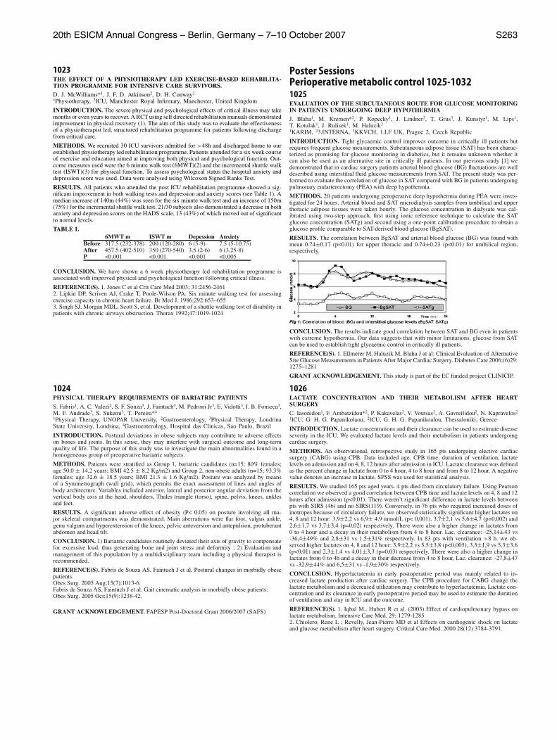
20th ESICM Annual Congress – Berlin, Germany – 7–10 October 2007 S263
1023THE EFFECT OF A PHYSIOTHERAPY LED EXERCISE-BASED REHABILITA-TION PROGRAMME FOR INTENSIVE CARE SURVIVORS.
D. J. McWilliams*1, J. F. D. Atkinson2, D. H. Conway2
1Physiotherapy, 2ICU, Manchester Royal Infirmary, Manchester, United Kingdom
INTRODUCTION. The severe physical and psychological effects of critical illness may takemonths or even years to recover. A RCT using self directed rehabilitation manuals demonstratedimprovement in physical recovery (1). The aim of this study was to evaluate the effectivenessof a physiotherapist led, structured rehabilitation programme for patients following dischargefrom critical care.
METHODS. We recruited 30 ICU survivors admitted for >48h and discharged home to ourestablished physiotherapy led rehabilitation programme. Patients attended for a six week courseof exercise and education aimed at improving both physical and psychological function. Out-come measures used were the 6 minute walk test (6MWT)(2) and the incremental shuttle walktest (ISWT)(3) for physical function. To assess psychological status the hospital anxiety anddepression score was used. Data were analysed using Wilcoxon Signed Ranks Test.
RESULTS. All patients who attended the post ICU rehabilitation programme showed a sig-nificant improvement in both walking tests and depression and anxiety scores (see Table 1). Amedian increase of 140m (44%) was seen for the six minute walk test and an increase of 150m(75%) for the incremental shuttle walk test. 21/30 subjects also demonstrated a decrease in bothanxiety and depression scores on the HADS scale, 13 (43%) of which moved out of significantto normal levels.
TABLE 1.6MWT m ISWT m Depession Anxiety
Before 317.5 (232-378) 200 (120-280) 6 (5-9) 7.5 (5-10.75)After 457.5 (402-510) 350 (270-540) 3.5 (2-6) 6 (3.25-8)P <0.001 <0.001 <0.001 <0.005
CONCLUSION. We have shown a 6 week physiotherapy led rehabilitation programme isassociated with improved physical and psychological function following critical illness.
REFERENCE(S). 1. Jones C et al Crit Care Med 2003; 31:2456-24612. Lipkin DP, Scriven AJ, Crake T, Poole-Wilson PA. Six minute walking test for assessingexercise capacity in chronic heart failure. Br Med J. 1986;292:653–6553. Singh SJ, Morgan MDL, Scott S, et al. Development of a shuttle walking test of disability inpatients with chronic airways obstruction. Thorax 1992;47:1019-1024
1024PHYSICAL THERAPY REQUIREMENTS OF BARIATRIC PATIENTS
S. Fabris1, A. C. Valezi2, S. F. Souza3, J. Faintuch4, M. Pedroni Jr3, E. Vidotti3, I. B. Fonseca3,M. F. Andrade3, S. Sukemi3, T. Pereira*4
1Physical Therapy, UNOPAR University, 2Gastroenterology, 3Physical Therapy, LondrinaState University, Londrina, 4Gastroenterology, Hospital das Clinicas, Sao Paulo, Brazil
INTRODUCTION. Postural deviations in obese subjects may contribute to adverse effectson bones and joints. In this sense, they may interfere with surgical outcome and long-termquality of life. The purpose of this study was to investigate the main abnormalities found in ahomogeneous group of preoperative bariatric subjects.
METHODS. Patients were stratified as Group 1, bariatric candidates (n=15; 80% females;age 50.0 ± 14.2 years; BMI 42.5 ± 8.2 Kg/m2) and Group 2, non-obese adults (n=15; 93.3%females; age 32.6 ± 18.5 years; BMI 21.3 ± 1.6 Kg/m2). Posture was analyzed by meansof a Symmetrograph (wall grid), which permits the exact assessment of lines and angles ofbody architecture. Variables included anterior, lateral and posterior angular deviation from thevertical body axis at the head, shoulders, Thales triangle (torso), spine, pelvis, knees, anklesand feet.
RESULTS. A significant adverse effect of obesity (P< 0.05) on posture involving all ma-jor skeletal compartments was demonstrated. Main aberrations were flat foot, valgus ankle,genu valgum and hyperextension of the knees, pelvic anteversion and antepulsion, protuberantabdomen and head tilt.
CONCLUSION. 1) Bariatric candidates routinely deviated their axis of gravity to compensatefor excessive load, thus generating bone and joint stress and deformity ; 2) Evaluation andmanagement of this population by a multidisciplinary team including a physical therapist isrecommended.
REFERENCE(S). Fabris de Souza AS, Faintuch J et al. Postural changes in morbidly obesepatients.Obes Surg. 2005 Aug;15(7):1013-6.Fabris de Souza AS, Faintuch J et al. Gait cinematic analysis in morbidly obese patients.Obes Surg. 2005 Oct;15(9):1238-42.
GRANT ACKNOWLEDGEMENT. FAPESP Post-Doctoral Grant 2006/2007 (SAFS)
Poster SessionsPerioperative metabolic control 1025-10321025EVALUATION OF THE SUBCUTANEOUS ROUTE FOR GLUCOSE MONITORINGIN PATIENTS UNDERGOING DEEP HYPOTHERMIA
J. Blaha1, M. Kremen*2, P. Kopecky1, J. Lindner3, T. Grus3, J. Kunstyr1, M. Lips1,T. Kotulak1, J. Rulisek1, M. Haluzik2
1KARIM, 23.INTERNA, 3KKVCH, 1.LF UK, Prague 2, Czech Republic
INTRODUCTION. Tight glycaemic control improves outcome in critically ill patients butrequires frequent glucose measurements. Subcutaneous adipose tissue (SAT) has been charac-terized as promising for glucose monitoring in diabetics, but it remains unknown whether itcan also be used as an alternative site in critically ill patients. In our previous study [1] wedemonstrated that in cardiac surgery patients arterial blood glucose (BG) fluctuations are welldescribed using interstitial fluid glucose measurements from SAT. The present study was per-formed to evaluate the correlation of glucose in SAT compared with BG in patients undergoingpulmonary endarterectomy (PEA) with deep hypothermia.
METHODS. 20 patients undergoing peroperative deep hypothermia during PEA were inves-tigated for 24 hours. Arterial blood and SAT microdialysis samples from umbilical and upperthoracic adipose tissues were taken hourly. The glucose concentration in dialysate was cal-ibrated using two-step approach, first using ionic reference technique to calculate the SATglucose concentration (SATg) and second using a one-point calibration procedure to obtain aglucose profile comparable to SAT-derived blood glucose (BgSAT).
RESULTS. The correlation between BgSAT and arterial blood glucose (BG) was found withmean 0.74±0.17 (p<0.01) for upper thoracic and 0.74±0.23 (p<0.01) for umbilical region,respectively.
CONCLUSION. The results indicate good correlation between SAT and BG even in patientswith extreme hypothermia. Our data suggests that with minor limitations, glucose from SATcan be used to establish tight glycaemic control in critically ill patients.
REFERENCE(S). 1. Ellmerer M, Haluzik M, Blaha J at al: Clinical Evaluation of AlternativeSite Glucose Measurements in Patients After Major Cardiac Surgery. Diabetes Care 2006;(6)29:1275–1281
GRANT ACKNOWLEDGEMENT. This study is part of the EC funded project CLINICIP.
1026LACTATE CONCENTRATION AND THEIR METABOLISM AFTER HEARTSURGERY
C. Iasonidou1, F. Ambatzidou*2, P. Kakavelas2, V. Voutsas2, A. Gavreilidou2, N. Kapravelos2
1ICU, G. H. G. Papanikolaou, 2ICU, G. H. G. Papanikoalou, Thessaloniki, Greece
INTRODUCTION. Lactate concentrations and their clearance can be used to estimate diseaseseverity in the ICU. We evaluated lactate levels and their metabolism in patients undergoingcardiac surgery.
METHODS. An observational, retrospective study in 165 pts undergoing elective cardiacsurgery (CABG) using CPB. Data included age, CPB time, duration of ventilation, lactatelevels on admission and on 4, 8, 12 hours after admission in ICU. Lactate clearance was definedas the percent change in lactate from 0 to 4 hour, 4 to 8 hour and from 8 to 12 hour. A negativevalue denotes an increase in lactate. SPSS was used for statistical analysis.
RESULTS. We studied 165 pts aged years. 4 pts died from circulatory failure. Using Pearsoncorrelation we observed a good correlation between CPB time and lactate levels on 4, 8 and 12hours after admission (p<0,01). There weren’t significant difference in lactate levels betweenpts with SIRS (46) and no SIRS(119). Conversely, in 76 pts who required increased doses ofinotropes because of circulatory failure, we observed statistically significant higher lactates on4, 8 and 12 hour: 3,9±2,2 vs 6,9± 4,9 mmol/L (p< 0,001), 3,7±2,1 vs 5,6±4,7 (p=0,002) and2,6±1,7 vs 3,7±3,4 (p=0,02) respectively. There were also a higher change in lactates from0 to 4 hour and a decay in their metabolism from 4 to 8 hour. Lac. clearance: -25,14±43 vs-36,4±49% and 2,8±31 vs 1,5±31% respectively. In 83 pts with ventilation >8 h. we ob-served higher lactates on 4, 8 and 12 hour: 3,9±2,2 vs 5,5±3,8 (p<0,005), 3,5±1,9 vs 5,3±3,6(p<0,01) and 2,3±1,4 vs 4,01±3,3 (p=0,03) respectively. There were also a higher change inlactates from 0 to 4h and a decay in their decrease from 4 to 8 hour, Lac. clearance: -27,8±47vs -32,9±44% and 6,5±31 vs -1,9±30% respectively.
CONCLUSION. Hyperlactatemia in early postoperative period was mainly related to in-creased lactate production after cardiac surgery. The CPB procedure for CABG change thelactate metabolism and a decreased utilization may contribute to hyperlactatemia. Lactate con-centration and its clearance in early postoperative period may be used to estimate the durationof ventilation and stay in ICU and the outcome.
REFERENCE(S). 1. Iqbal M., Hubert R et al. (2003) Effect of cardiopulmonary bypass onlactate metabolism. Intensive Care Med. 29: 1279-12852. Chiolero, Rene L ; Revelly, Jean-Pierre MD et al Effects on cardiogenic shock on lactateand glucose metabolism after heart surgery. Critical Care Med. 2000 28(12) 3784-3791.
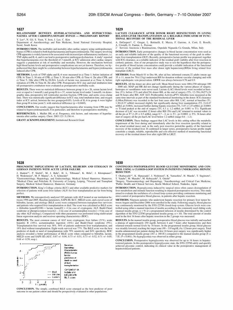
S264 20th ESICM Annual Congress – Berlin, Germany – 7–10 October 2007
1027RELATIONSHIP BETWEEN HYPERLACTATAEMIA AND HYPERCYTOKI-NAEMIA AFTER CARDIOPULMONARY BYPASS — PRELIMINARY REPORT
Y. Lee*, N. Gil, S. Yoon, Y. Jeon, J. Lee, C. KimDepartment of Anesthesiology and Pain Medicine, Seoul National University Hospital,Seoul, South Korea
INTRODUCTION. The morbidity and mortality after cardiac surgery using cardiopulmonarybypass (CPB) is related to both hyperlactataemia and hypercytokinaemia. The surgery involvingCPB apparatus is widely known to be associated with increased pro-inflammatory mediators, i.e.TNF-alpha and IL-6, and it can lead to postoperative multiorgan dysfunction. A study1 reportedthat hyperlactataemia over the threshold of 3 mmol/L at ICU admission after cardiac surgerysuggests a population at risk of morbidity and mortality. However, the mechanism betweenhigh blood lactate levels and postoperative complications are not yet known. Therefore, we hy-pothesized that there might be a relationship between hyperlactataemia and hypercytokinaemiainduced after CPB.
METHODS. Levels of TNF-alpha and IL-6 were measured in a) Time 1; before initiation ofCPB, b) Time 2; 30 min of CPB, c) Time 3; 30 min after CPB, d) Time 4; 2hr after CPB, ande) Time 5; 24hr after CPB by ELISA. Levels of lactate was measured at a) Time A; beforeinitiation of CPB, b) Time B; 2hr after CPB. Postoperative ICU stay and total intubation timetill postoperative extubation were evaluated as postoperative morbidity scale.
RESULTS. There were no statistical differences between group A (n = 26, serum lactate levelover or equal to 3 mmol/L) and group B (n = 37, serum lactate level under 3 mmol/L) in demo-graphic data, preoperative left ventricular ejection fraction, CPB time, and aortic cross-clamptime. There was statistically significant difference in ICU stay, total intubation time, and oxygenindex at ICU arrival, between the two groups(p<0.05). Levels of IL-6 in group A were higherthan group B in time point 5, with statistical difference (p = 0.0469).
CONCLUSION. Our results suggest that hyperlactataemia after weaning from CPB may berelated to hypercytokinaemia(IL-6 in particular), and therefore to postoperative morbidity.
REFERENCE(S). 1. Maillet JM, et al. Frequency, risk factors, and outcomes of hyperlac-tatemia after cardiac surgery. Chest. 2003.123: 5;1361-6.
GRANT ACKNOWLEDGEMENT. Institutional Research Grant
1028PROGNOSTIC IMPLICATIONS OF LACTATE, BILIRUBIN AND ETIOLOGY INGERMAN PATIENTS WITH ACUTE LIVER FAILURE
J. Hadem*1, P. Stiefel1, M. J. Bahr1, H. L. Tillmann2, K. Rifai1, J. Klempnauer3,H. Wedemeyer1, M. P. Manns1, A. S. Schneider1
1Gastroenterology, Hepatology, and Endocrinology, Medical School Hannover, Hannover,2Medical Clinic and Policlinic II, University of Leipzig, Leipzig, 3Visceral and TransplantSurgery, Medical School Hannover, Hannover, Germany
INTRODUCTION. King’s College criteria (KCC) and other available predictive markers forselection of patients with acute liver failure (ALF) for liver transplantation are far from beingperfect.
METHODS. We retrospectively analyzed 102 patients with ALF treated at our institution be-tween 1996 and 2005. Baseline parameters, SAPS-III, KCC, MELD score, and a novel score ofbilirubin, lactate, and etiology (BiLE score) were compared between transplant-free survivorsand patients who required liver transplantation or died. The score was calculated as BiLE score= (bilirubin [µmol/l]/100) + lactate [mmol/l] + 4 [in case of cryptogenic ALF, Budd-Chiarisyndrome or phenprocoumon toxicity] – 2 [in case of acetaminophen toxicity] + 0 [in case ofany other ALF etiology]. Comparison with other parameters was performed using multivariatelinear regression analysis and receiver operating characteristics (ROC).
RESULTS. The most common causes of ALF were cryptogenic liver failure (21%), acutehepatitis B (18%), acetaminophen ingestion (16%) and Budd-Chiari syndrome (9%).Transplantation-free survival was 38%, 44% of patients underwent liver transplantation, and18% died without transplantation. Eight-week-survival was 77%. The BiLE score was the bestpredictor of death or need of transplantation with 79% sensitivity and 84% specificity. ROCanalysis revealed a better performance of BiLE score when compared to bilirubin, lactate,MELD score and SAPS III (AUC: 0.87 +/- 0.04, 0.73 +/- 0.51, 0.73 +/- 0.52, 0.71 +/- 0.05,0.68 +/- 0.59, resp.).
CONCLUSION. The simple, combined BiLE score emerged as the best predictor of pooroutcome in our patient cohort and should be prospectively evaluated in other populations.
1029LACTATE CLEARANCE AFTER DONOR RIGHT HEPATECTOMY IN LIVINGRELATED LIVER TRANSPLANTATION AS A RELIABLE INDICATOR OF FUNC-TIONAL RECOVERY OF THE RESIDUAL LIVER
A. DeGasperi*, C. Grugni, L. Bettinelli, E. Mazza, F. Garrone, A. Corti, E. Roselli,A. Cristalli, G. Fantini, L. Perrone2 ˚ Servizio Anestesia e Rianimazione, Ospedale Niguarda Ca Granda, Milan, Italy
INTRODUCTION. Early postoperative changes in blood lactate concentration were used asan early and reliable indicator of the quality of the functional recovery of the graft in ortho-topic liver transplantation (OLT). Recently, perioperative lactate profile was proposed, togetherwith ICG clearance, as a reliable indicator of the residual graft viability after liver resection incirrhotic patients. Aim of our prospective study was to test the hypothesis that the periopera-tive profile of blood lactate concentration could provide a reliable indication of the functionalrecovery of the residual liver mass after donor right hepatectomy(RH)in living related livertransplant (LRLT).
METHODS. From March 01 to Dec 06, after ad hoc informed consent,32 adults (mean age35 ± 11, mean bw 70±12 kg) underwent RH for donation without vascular clamping and withright suprahepatic vein preservation. GRWR was always between 0.78 and 0.9.
RESULTS. All the donor are alive and well. Mean blood losses were 400±350 ml(range 100– 1000 ml). MAP and HR did not change significantly during the various phases of surgery.Inotropes or vasodilators were never used. Lactate (LAC) blood levels were recorded at base-line (T1), before (T3) and after(T4) donor RH, at the end of surgery (T5), 12 (T6),18(T7),24and 36 hours after RH. AST ALT, Prothrombin Activity,aPTT, bilirubin were measured at thesame time intervals as part of the standard biochemical monitoring in the donor. ICG clearance(LiMon,Pulsion) was recorded in the last four cases after RH. LAC, normal at baseline (T1,1.30±0.37 mMol) increased slightly but significantly during liver manipulation (T2, 2±0.47mMol, p< 0.004), increased further during hepatic resection (T4, 3.45 ±1.25 mMol, p< 0.0001vs T1)and peaked at the end of surgery (T5, 4.3 ± 1.2 mMol, p< 0.001 vs T1). Significantdecrease of LAC was recorded 12 hours after RH (T6) and LAC returned to normal values inall the pts at T7 (1,29 ± 0.5 mMol, range 0.7 – 2; ns vs T1 ; p< 0.0001 vs T5). 36 hours afterstart of surgery all the pts had LAC level below 1.2 mMol (range 0.6 – 1.2).
CONCLUSION. These findings suggest that LAC levels in this setting reflect the metabolicimpairment of the liver during and immediately after the liver resection (possibly related tothe actual residual mass) and, in the early post resective period,the quality of the functionalrecovery of the residual liver. If confirmed in larger series, postoperative lactate profile mightconstitute a simple, reliable, reproducible and cost effective method of monitoring functionalrecovery of the residual donor liver after right DH in LRLT.
1030CONTINUOUS POSTOPERATIVE BLOOD GLUCOSE MONITORING AND CON-TROL USING A CLOSED-LOOP SYSTEM IN PATIENTS UNDERGOING HEPATICRESECTION
T. Okabayashi*1, K. Hanazaaki1, I. Nishimori2, K. Yamashita3, H. Maeda1, T. Sugimoto1,T. Yatabe3, M. Manabe3, M. Kobayashi4, S. Onishi21Surgery, 2Gastroenterology and Hepatology, 3Anesthesiology and Critical Care Medicine,4Public Health and Clinical Servises, Kochi Medical School, Nankoku, Japan
INTRODUCTION. Hyperglycemia induced by surgical stress often causes dysregulation ofliver metabolism and immune function resulting in impaired postoperative recovery. This studyaimed to evaluate the usefulness of a closed-loop system providing continuous monitoring andstrict control of postoperative blood glucose, in patients after hepatic resection.
METHODS. Nineteen patients who underwent hepatic resection for primary liver tumor be-tween August and December 2006 were enrolled in the study. Following surgery, blood glucosewas continuously monitored by the STG-22TM closed-loop system. Glucose levels were con-trolled using either a manual injection of insulin according to the commonly used sliding scale(manual insulin group, n = 9) or a programmed infusion of insulin determined by the controlalgorithm of the STG-22TM (programmed insulin group, n = 10). The total amount of insulinused in the first 16 hours after hepatic resection in the 2 groups was measured.
RESULTS. In the manual insulin group, postoperative blood glucose rose initially and reacheda plateau of approximately 250 mg/dL between 4 and 7 hours after hepatectomy and thenreturned towards normal levels by 16 hours. In the programmed insulin group, blood glucosewas steadily lowered, reaching the target zone (90 – 110 mg/dL) by 12 hours post surgery. Totalinsulin administered per patient during the first 16 hours post surgery was significantly higherin the programmed insulin group (183 ± 188 IU) compared to the manual insulin group (8 ±7 IU; P < 0.001). No hypoglycemia was observed in either group.
CONCLUSION. Postoperative hyperglycemia was observed for up to 16 hours in hepatec-tomized patients. In this postoperative hyperglycemic state, the STG-22TM safely and quicklyachieved glycemic control, indicating its clinical value in the postoperative management ofhepatectomized patients.
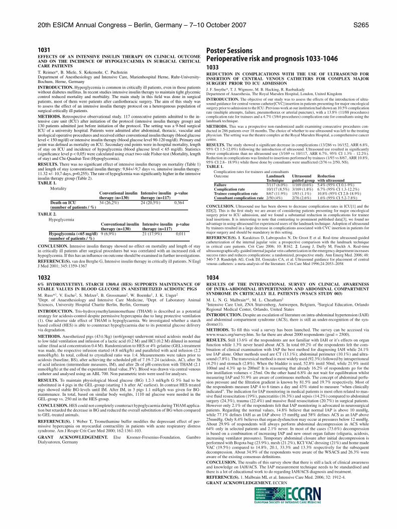
20th ESICM Annual Congress – Berlin, Germany – 7–10 October 2007 S265
1031EFFECTS OF AN INTENSIVE INSULIN THERAPY ON CLINICAL OUTCOMEAND ON THE INCIDENCE OF HYPOGLYCAEMIA IN SURGICAL CRITICALCARE PATIENTS
T. Reimer*, B. Miele, S. Kokemohr, C. PuchsteinDepartment of Anesthesiology and Intensive Care, Marienhospital Herne, Ruhr-University-Bochum, Herne, GermanyINTRODUCTION. Hyperglycemia is common in critically ill patients, even in those patientswithout diabetes mellitus. In recent studies intensive insulin therapy to maintain tight glycemiccontrol reduced mortality and morbidity. The main study in this field was done in surgicalpatients, most of them were patients after cardiothoracic surgery. The aim of this study wasto assess the effect of an intensive insulin therapy protocol on a heterogenous population ofsurgical critically ill patients.METHODS. Retrospective observational study. 117 consecutive patients admitted to the in-tensive care unit (ICU) after initiation of the protocol (intensive insulin therapy group) and130 patients admitted just before initiation of the protocol. The setting was a 9-bed surgicalICU of a university hospital. Patients were admitted after abdominal, thoracic, vascular andurological operative procedures and received either conventional insulin therapy (blood glucoselevel < 150 mg/dl) or intensive insulin therapy (blood glucose level 90-120 mg/dl). Primary endpoint was defined as mortality on ICU. Secondary end points were in-hospital mortality, lengthof stay on ICU and incidence of hypoglycemia (blood glucose level < 65 mg/dl). Statistics(significance level p < 0,05) were calculated using exact two-side Fisher-test (Mortality, lengthof stay) and Chi-Quadrat-Test (Hypoglycemia).RESULTS. There was no significant effect of intensive insulin therapy on mortality (Table 1)and length of stay (conventional insulin therapy: 9,84+/-9,7 days vs. intensive insulin therapy:11.32 +/- 10,7 days, p=0,255). The rate of hypoglcemia was significantly higher in the intensiveinsulin therapy group (Table 2).TABLE 1.
MortalityConventional insulin Intensive insulin p-valuetherapy (n=130) therapy (n=117)
Death on ICU 34 (26,2%) 24 (20,5%) 0,364(number of patients / %)
TABLE 2.Hypoglycemia
Conventional insulin Intensive insulin p-valuetherapy (n=130) therapy (n=117)
Hypoglycemia (<65 mg/dl) 9 (6,9%) 21 (17,9%) 0,011(number of patients / %)
CONCLUSION. Intensive insulin therapy showed no effect on mortality and length of stayin critically ill patients after surgical procedures but was correlated with an increased risk ofhypoglycemia. If this has an influence on outcome should be examined in further investigations.REFERENCE(S). van den Berghe G, Intensive insulin therapy in critically ill patients. N EnglJ Med 2001; 345:1359-1367
10326% HYDROXYETHYL STARCH 130/0.4 (HES) SUPPORTS MAINTENANCE OFSTABLE VALUES IN BLOOD GLUCOSE IN ANESTHETIZED ACIDOTIC PIGS
M. Russ*1, V. Esche2, S. Melzer2, B. Grossmann2, W. Boemke1, J. K. Unger2
1Dept. of Anaesthesiology and Intensive Care Medicine, 2Dept. of Laboratory AnimalSciences, University Hospital Charite Berlin, Berlin, Germany
INTRODUCTION. Tris-hydroxymethylaminomethane (THAM) is described as a potentialstrategy for acidosis-control despite permissive hypercapnia due to lung protective ventilation(1). One adverse side effect of THAM is hypoglycaemia. We investigated whether a starchbased colloid (HES) is able to counteract hypoglycaemia due to its potential glucose deliveryvia degradation.
METHODS. Anesthetized pigs (43±3kg) (n=6/group) underwent mixed acidosis model dueto low tidal ventilation and infusion of a lactic acid (0.2 M) and HCl (0.2 M) diluted in normalsaline (final acid concentration 0.4 M). Randomization to HES or 4% gelatine (GEL)-treatmentwas made, the respective infusion started (4.8 ml/kg/h) and paralleled with acid infusion (2.2mmol/kg/h). In total, colloid to crystalloid ratio was 1:4. Measurements were taken prior toacidosis (baseline, BS), after achieving the scheduled pH of 7.19-7.24 (acidosis, AC), after 3hof acid infusion (intermediate measures, IM), and after 2h of pH-correction with THAM (2.1mmol/kg/h) at the end of the experiment (final value, FV). Blood was drawn via central venouscatheter and analysed using an ABL 700. Non parametric tests were used for analyses.
RESULTS. To maintain physiological blood glucose (BG) 1.2-3 ml/kg/h G 5% had to besubstituted in 4 pigs in the GEL-group (starting 1 h after AC earliest). In contrast HES treatedpigs showed stable BG-levels until IM. After IM, in 3 pigs 1.1 ml/kg/h was given for BG-maintenance. In total, based on similar body weights, 1110 ml glucose were needed in theGEL-group vs. 250 ml in the HES-group.
CONCLUSION. HES could not completely counteract hypoglycaemia during THAM applica-tion but retarded the decrease in BG and reduced the overall substitution of BG when comparedto GEL-treated animals.
REFERENCE(S). 1 Weber T, Tromethamine buffer modifies the depressant effect of per-missive hypercapnia on myocardial contractility in patients with acute respiratory distresssyndrome. Am J Respir Crit Care Med 2000; 162:1361-103.
GRANT ACKNOWLEDGEMENT. Else Kroener-Fresenius-Foundation, GambroDialysatoren, Germany
Poster SessionsPerioperative risk and prognosis 1033-10461033REDUCTION IN COMPLICATIONS WITH THE USE OF ULTRASOUND FORINSERTION OF CENTRAL VENOUS CATHETERS FOR COMPLEX MAJORSURGERY PRIOR TO ICU ADMISSIONJ. F. Smythe*, T. J. Wigmore, M. B. Hacking, R. RaobaikadyDepartment of Anaesthesia, The Royal Marsden Hospital, London, United KingdomINTRODUCTION. The objective of our study was to assess the effects of the introduction of ultra-sound guidance for central venous catheter[CVC] insertion in patients presenting for major oncologicalsurgery prior to admission to the ICU. Previous work at our institution had shown an 10.5% complicationrate (multiple attempts, failure, pneumothorax or arterial puncture), with a 13.8% (11/88 procedures)complication rate for trainees and a 4.7% (3/64 procedures) complication rate for consultants using thelandmark technique.METHODS. This was a prospective non-randomized audit examining consecutive procedures con-ducted in 286 patients over 18 months. The choice of whether to use ultrasound was left to the treatingphysician. The setting was the theatre complex at the Royal Marsden Hospital, a comprehensive cancercentre.RESULTS. The study showed a significant decrease in complications (13/286 vs 16/152, ARR 6.8%,95% CI 1.5-12.0%) following the introduction of ultrasound. Ultrasound use resulted in significantlyfewer complications than no ultrasound use (3/169 vs 10/117, ARR 6.7%, 95% CI 1.3% - 12.2%).Reduction in complications was limited to insertions performed by trainees (1/93 vs 8/67, ARR 10.8%,95% CI 2.8– 18.9%) while those done by consultants were unaffected (2/76 vs 2/50, NS).TABLE 1.
Complication rates for trainees and consultantsOutcome Landmark Ultrasound Reduction
Technique guided group with ultrasoundFailure 7/117 (6.0%) 1/169 (0.6%) 5.4% (95% CI 0.1-9%)Complication rate 10/117 (8.5%) 3/169 (1.8%) 6.7% (95% CI 1.3-12.2%)Trainee complication rate 8/67 (11.9%) 1/93 (1.1%) 10.8% (95% CI 2.8-18.9%)Consultant complication rate 2/50 (4%) 2/76 (2.6%) 1.6% (95% CI 5.2-7.8%)
CONCLUSION. Ultrasound use has been shown to decrease complication rates in ICU[1] and theED[2]. This is the first study we are aware of considering patients presenting for major oncologicalsurgery prior to ICU admission, and we found a substantial reduction in complications for traineelead insertions. It is interesting to note that contrasting to prominent published data[3], we found noadvantage in using ultrasound for experienced users of the landmark technique. Adoption of ultrasoundby trainees resulted in a large decrease in complications associated with CVC insertion in patients formajor surgery and should be mandatory in this setting.REFERENCE(S). 1. Karakitsos D, Labropoulos N, De Groot E et al. Real-time ultrasound-guidedcatheterization of the internal jugular vein: a prospective comparison with the landmark techniquein critical care patients. Crit Care 2006; 10: R162. 2. Leung J, Duffy M, Finckh A. Real-timeultrasonographically-guided internal jugular vein catheterization in the emergency department increasessuccess rates and reduces complications: a randomized, prospective study. Ann Emerg Med. 2006; 48:540-7 3. Randolph AG, Cook DJ, Gonzales CA, et al. Ultrasound guidance for placement of centralvenous catheters: a meta-analysis of the literature. Crit Care Med 1996;24:2053–2058
1034RESULTS OF THE INTERNATIONAL SURVEY ON CLINICAL AWARENESSOF INTRA-ABDOMINAL HYPERTENSION AND ABDOMINAL COMPARTMENTSYNDROME IN CRITICALLY ILL PATIENTS (WSACS STUDY 003)
M. L. N. G. Malbrain*1, M. L. Cheatham2
1Intensive Care Unit, ZNA Stuivenberg, Antwerpen, Belgium, 2Surgical Education, OrlandoRegional Medical Center, Orlando, United StatesINTRODUCTION. Despite an escalation of literature on intra-abdominal hypertension (IAH)and abdominal compartment syndrome (ACS), there is still an under-recognition of the syn-drome(1).METHODS. To fill this void a survey has been launched. The survey can be accessed viawww.wsacs.org/survey.htm. So far there are about 2000 respondents (goal = 2500).RESULTS. Still 13.6% of the respondents are not familiar with IAH or it’s effects on organfunction while 1.3% never heard about ACS. In total 69.2% of the respondents felt the com-bination of clinical examination with IAP the best method for diagnosing IAH while 24.1%use IAP alone. Other methods used are CT (13.1%); abdominal perimeter (10.1%) and ultra-sound (7.8%). The transvesical method is most widely used (92.3%) followed by intraperitoneal(4.2%) and stomach (2.8%). When the bladder is used, 52.8% instil 50ml, while 21.9% instil100ml and 4.3% up to 200ml! It is reassuring that already 16.2% of respondents go for thelow instillation volumes < 25ml. On the other hand 6.8% do not wait for equilibration whilstmeasuring IAP and 51.9% are aware of continuous methods. The concept of abdominal perfu-sion pressure and the filtration gradient is known by 81.5% and 19.7% respectively. Most ofthe respondents measure IAP 4 to 6 times a day and 43% stated to measure “when clinicallyindicated”. The indication for IAP monitoring in medical patients is most often related to mas-sive fluid resuscitation (19%), pancreatitis (16.3%) and sepsis (14.2%) compared to abdominalsurgery (24.3%), trauma (22.4%) and massive fluid resuscitation (20.7%) in surgical patients.However only 2.1% of the respondents felt that IAP monitoring is advocated in neurosurgicalpatients. Regarding the normal values, 14.8% believe that normal IAP is above 10 mmHg,while 77.1% defines IAH as an IAP above 15 mmHg and 58% defines ACS as an IAP above25mmHg. Only 8.4% believes that organ dysfunction may occur at pressures below 12 mmHg.About 29.9% of respondents will always perform abdominal decompression in ACS while64% only in selected patients and 2.1% never. In most of the cases (73.6%) decompressionis based on a combination of increasing IAP and new onset organ failure (oliguria, acidosis,increasing ventilator pressures). Temporary abdominal closure after initial decompression isperformed with Bogota bag (23.9%), mesh (21.2%), KCI VAC dressing (21%) and home madeVAC (19.5%) compared to 14.8%, 20.1, 33.3% and 13.3% respectively for the subsequentdecompression. About 34.9% of the respondents were aware of the WSACS and 26.3% wereaware of the existing consensus definitions.CONCLUSION. The results of this survey show that there is still a lack of clinical awarenessand knowledge on IAH/ACS. The IAP measurement technique needs to be standardised andthere is a lot of educational work to do regarding IAH/ACS diagnosis and treatment.REFERENCE(S). 1. Malbrain ML et al. Intensive Care Med. 2006; 32: 1912-4.GRANT ACKNOWLEDGEMENT. ECCRN
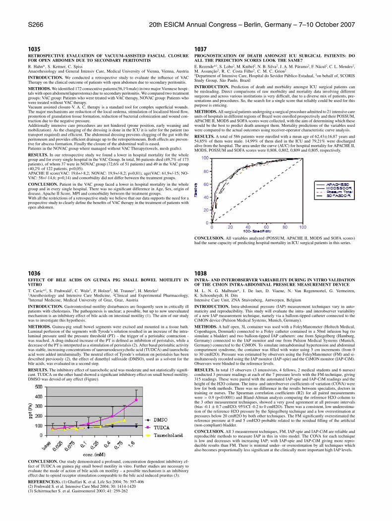
S266 20th ESICM Annual Congress – Berlin, Germany – 7–10 October 2007
1035RETROSPECTIVE EVALUATION OF VACUUM-ASSISTED FASCIAL CLOSUREFOR OPEN ABDOMEN DUE TO SECONDARY PERITONITIS
R. Hahn*, S. Kettner, C. SpissAnaesthesiology and General Intensiv Care, Medical University of Vienna, Vienna, Austria
INTRODUCTION. We conducted a retrospective study to evaluate the influence of VACTherapy on the clinical outcome of patients with open abdomen due to secondary peritonitis.
METHODS. We identified 172 consecutive patients(56,1%male) in two major Viennese hospi-tals with open abdomen(laparostoma) due to secondary peritonitis. We compared two treatmentgroups: VAC group: Patients who were treated with VAC therapy, NOVAC group: Patients whowere treated without VAC therapy.Vacuum assisted closure-V. A. C. therapy is a standard tool for complex superficial wounds.The major mechanisms are reduction of the local oedema, stimulation of localized blood flow,promotion of granulation tissue formation, reduction of bacterial colonization and wound con-traction due to the negative pressure.Additionally intensive care procedures are not hindered (prone position, early weaning andmobilization). As the changing of the dressing is done in the ICU it is safer for the patient (notransport required) and efficient. The abdominal dressing prevents clogging of the gut with theperitoneum and provides efficient drainage up to the retroperitoneum. Both effects are preven-tive for abscess formation. Finally the closure of the abdominal wall is eased.Patients in the NOVAC group where managed without VAC Therapy(towels, mesh grafts).
RESULTS. In our retrospective study we found a lower in hospital mortality for the wholegroup and for every single hospital in the VAC Group. In total, 86 patients died (49,7% of 173patients), of whom 37 were in NOVAC group (72,6% of 51 patients) and 49 in the VAC group(40,2% of 122 patients, p<0,05).APACHE II score(VAC: 19,6+/-8,2; NOVAC: 19,9+/-8,2; p=0,81), age(VAC: 61,9+/-15; NO-VAC: 58+/-14,6; p=0,14) and comorbidity did not differ between the treatment groups.
CONCLUSION. Patient in the VAC group faced a lower in hospital mortality in the wholegroup and in every single hospital. There was no significant difference in Age, Sex, origin ofdisease, Apache II Score, MPI and comorbidity between the treatment groups.With all the restrictions of a retrospective study we believe that our data supports the need for aprospective study to clearly define the benefits of VAC therapy in the treatment of patients withopen abdomen.
1036EFFECT OF BILE ACIDS ON GUINEA PIG SMALL BOWEL MOTILITY INVITRO
T. Cavic*1, S. Fruhwald1, C. Wulz1, P. Holzer2, M. Trauner3, H. Metzler1
1Anesthesiology and Intensive Care Medicine, 2Clinical and Experimental Pharmacology,3Internal Medicine, Medical University of Graz, Graz, Austria
INTRODUCTION. Gastrointestinal motility disturbances are frequently seen in critically illpatients with cholestasis. The pathogenesis is unclear; a possible, but up to now unevaluatedmechanism is an inhibitory effect of bile acids on intestinal motility (1). The aim of our studywas to investigate this hypothesis.
METHODS. Guinea-pig small bowel segments were excised and mounted in a tissue bath.Luminal perfusion of the segments with Tyrode’s solution resulted in an increase of the intra-luminal pressure until the pressure threshold (PT) – the trigger of a peristaltic contraction -was reached. A drug-induced increase of the PT is defined as inhibition of peristalsis, while adecrease of the PT is interpreted as a stimulation of peristalsis (2). After basal peristaltic activitywas stable, increasing concentrations of tauroursodesoxycholic acid (TUDCA) and taurocholicacid were added intraluminally. The neutral effect of Tyrode’s solution on peristalsis has beendescribed previously (2), the effect of dimethyl sulfoxide (DMSO), used as a solvent for thebile acids, was evaluated in a second step.
RESULTS. The inhibitory effect of taurocholic acid was moderate and not statistically signifi-cant. TUDCA on the other hand showed a significant inhibitory effect on small bowel motility.DMSO was devoid of any effect (Figure).
CONCLUSION. Our study demonstrated a profound, concentration dependent inhibitory ef-fect of TUDCA on guinea pig small bowel motility in vitro. Further studies are necessary toevaluate the mode of action of bile acids on motility – a possible mechanism is an inhibitoryeffect due to opioid receptor stimulation comparable to the bile acid induced pruritus (3).
REFERENCE(S). (1) Ghaffari K. et al. Life Sci 2004; 76: 397-406(2) Fruhwald S. et al. Intensive Care Med 2004; 30: 1414-1420(3) Schirrmacher S. et al. Gastroenterol 2003; 41: 259-262
1037PROGNOSTICATION OF DEATH AMONGST ICU SURGICAL PATIENTS: DOALL THE PREDICTION SCORES LOOK THE SAME?
E. Rezende*1, S. Lobo2, M. Knibel2, N. B. Silva2, J. A. M. Paramo2, F. Nacul2, C. L. Mendes2,M. Assuncao2, R. C. Costa Filho2, C. M. C. Grion2
1Department of Intensive Care, Hospital do Sevidor Publico Estadual, 2on behalf of, SCORISStudy Group, Sao Paulo, Brazil
INTRODUCTION. Prediction of death and morbidity amongst ICU surgical patients canbe misleading. Direct comparisons of raw morbidity and mortality data involving differentsurgeons and across various institutions is very difficult, due to a diverse mix of patients, pre-sentations and procedures. So, the search for a single score that reliably could be used for thispurpose is enticing.
METHODS. All surgical patients undergoing a surgical procedure admitted to 21 intensive careunits of hospitals in different regions of Brazil were enrolled prospectively and their POSSUM,APACHE II, MODS and SOFA scores were collected, with the aim of determining which thesewould be the best to predict death amongst them. Mortality predictions of the variables usedwere compared to the actual outcomes using receiver-operator characteristic curve analysis.
RESULTS. A total of 586 patients were enrolled with a mean age of 62,43±16,87 years and54,85% of them were male. 14,99% of them died in the ICU and 79,21% were dischargedalive from the hospital. The area under the curve (AUC) for hospital mortality for APACHE II,MODS, POSSUM and SOFA scores were 0,808, 0,802, 0,809 and 0,805, respectively.
CONCLUSION. All variables analyzed (POSSUM, APACHE II, MODS and SOFA scores)had the same capacity of predicting hospital mortality in ICU surgical patients in this series.
1038INTRA- AND INTEROBSERVER VARIABILITY DURING IN VITRO VALIDATIONOF THE CIMON INTRA-ABDOMINAL PRESSURE MEASUREMENT DEVICE
M. L. N. G. Malbrain*, I. De laet, D. Viaene, N. Van Regenmortel, G. Vermeiren,K. Schoonheydt, H. DitsIntensive Care Unit, ZNA Stuivenberg, Antwerpen, Belgium
INTRODUCTION. Intra-abdominal pressure (IAP) measurement techniques vary in auto-maticity and reproducibility. This study will evaluate the intra- and interobserver variabilityof a new IAP measurement technique, namely via a balloon-tipped catheter connected to theCiMON device (Pulsion Medical Systems, Munich, Germany).
METHODS. A half open, 3L container was used with a FoleyManometer (Holtech Medical,Copenhagen, Denmark) connected to a Foley catheter contained in a 50ml infusion bag (tosimulate a bladder) and two balloon-tipped IAP catheters: one from Spiegelberg (Hamburg,Germany) connected to the IAP monitor and one from Pulsion Medical Systems (Munich,Germany) connected to the CiMON. To simulate intraabdominal hypertension and abdominalcompartment syndrome, the container was filled with water using 5 cm increments (from 0to 30 cmH2O). Pressure was estimated by observers using the FoleyManometer (FM) and si-multaneously recorded using the IAP monitor (IAP-spie) and the CiMON monitor (IAP-CiM).Observers were blinded to the reference levels.
RESULTS. In total 15 observers (3 intensivists, 4 fellows, 2 medical students and 6 nurses)conducted 3 pressure readings at each of the 7 pressure levels with the FM-technique, giving315 readings. These were paired with the automated IAP-spie and IAP-CiM readings and theheight of the H2O column. The intra- and interobserver coefficients of variation (COVA) werelow for both methods. There was no difference in the results between specialists, doctors intraining or nurses. The Spearman correlation coefficients (R2) for all paired measurementswere > 0.9 (p<0.0001) and Bland-Altman analysis comparing the reference H2O column tothe 3 other measurement techniques, showed a very good agreement at all pressure intervals(bias -0.1 ± 0.7 cmH2O; 95%CI -0.2 to 0 cmH2O). There was a consistent, low underestima-tion of the reference H2O pressure by the Spiegelberg technique and a low overestimation atpressures below 20 cmH2O by both other techniques. The FM significantly overestimated thereference pressure at 0 and 5 cmH2O probable related to the residual filling of the artificial(non-compliant) bladder.
CONCLUSION. All 3 measurement techniques, FM, IAP-spie and IAP-CiM are reliable andreproducible methods to measure IAP in this in vitro model. The COVA for each techniqueis low and decreases with increasing IAP; with IAP-spie and IAP-CiM giving more repro-ducible results than FM. There is minimal under- or overestimation by all techniques whichalso becomes proportionally less significant at the clinically more important high IAP levels.
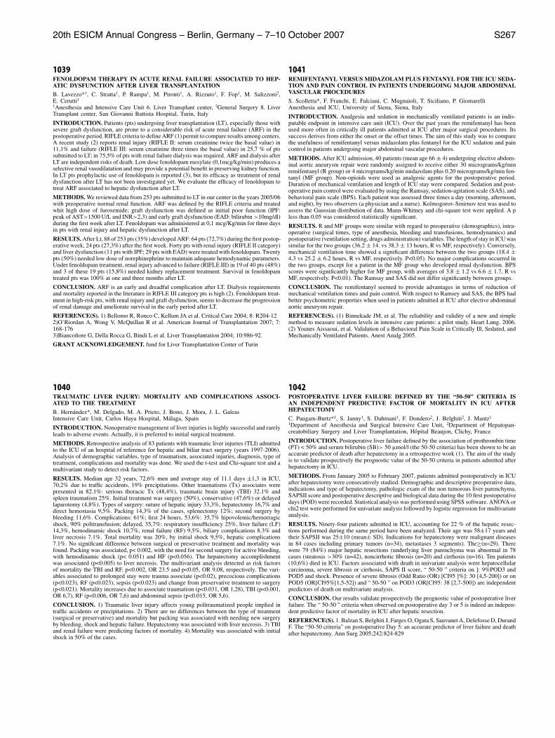
20th ESICM Annual Congress – Berlin, Germany – 7–10 October 2007 S267
1039FENOLDOPAM THERAPY IN ACUTE RENAL FAILURE ASSOCIATED TO HEP-ATIC DYSFUNCTION AFTER LIVER TRANSPLANTATION
B. Lavezzo*1, C. Stratta1, P. Rampa1, M. Pironti1, A. Rizzuto1, F. Fop1, M. Salizzoni2,E. Cerutti11Anesthesia and Intensive Care Unit 6. Liver Transplant center, 2General Surgery 8. LiverTransplant center, San Giovanni Battista Hospital, Turin, Italy
INTRODUCTION. Patients (pts) undergoing liver transplantation (LT), especially those withsevere graft dysfunction, are prone to a considerable risk of acute renal failure (ARF) in thepostoperative period. RIFLE criteria to define ARF (1) permit to compare results among centers.A recent study (2) reports renal injury (RIFLE II: serum creatinine twice the basal value) in11,1% and failure (RIFLE III: serum creatinine three times the basal value) in 25,7 % of ptssubmitted to LT; in 75,5% of pts with renal failure dialysis was required. ARF and dialysis afterLT are independent risks of death. Low dose fenoldopam mesylate (0,1mcg/kg/min) produces aselective renal vasodilatation and may provide a potential benefit in preserving kidney function.In LT pts prophylactic use of fenoldopam is reported (3), but its efficacy as treatment of renaldysfunction after LT has not been investigated yet. We evaluate the efficacy of fenoldopam totreat ARF associated to hepatic dysfunction after LT.
METHODS. We reviewed data from 253 pts submitted to LT in our center in the years 2005/06with preoperative normal renal function. ARF was defined by the RIFLE criteria and treatedwhit high dose of furosemide; graft dysfunction was defined as initial poor function (IPF:peak of AST>1500 U/L and INR>2,3) and early graft dysfunction (EAD: bilirubin >10mg/dl)during the first week after LT. Fenoldopam was administered at 0,1 mcg/Kg/min for three daysin pts with renal injury and hepatic dysfunction after LT.
RESULTS. After Lt, 88 of 253 pts (35%) developed ARF: 64 pts (72,7%) during the first postop-erative week, 24 pts (27,3%) after the first week. Forty pts with renal injury (RIFLE II category)and liver dysfunction (11 pts with IPF; 29 pts with EAD) were treated with fenoldopam. Twentypts (50%) needed low dose of norephinephrine to maintain adequate hemodynamic parameters.Under fenoldopam treatment, renal injury advanced to failure (RIFLE III) in 19 of 40 pts (48%)and 3 of these 19 pts (15,8%) needed kidney replacement treatment. Survival in fenoldopamtreated pts was 100% at one and three months after LT.
CONCLUSION. ARF is an early and dreadful complication after LT. Dialysis requirementsand mortality reported in the literature in RIFLE III category pts is high (2). Fenoldopam treat-ment in high-risk pts, with renal injury and graft dysfunction, seems to decrease the progressionof renal damage and ameliorate survival in the early period after LT.
REFERENCE(S). 1) Bellomo R, Ronco C, Kellum JA et al. Critical Care 2004; 8: R204-122)O’Riordan A, Wong V, McQuillan R et al. American Journal of Transplantation 2007; 7:168-1763)Biancofiore G, Della Rocca G, Bindi L et al. Liver Transplantation 2004; 10:986-92.
GRANT ACKNOWLEDGEMENT. fund for Liver Transplantation Center of Turin
1040TRAUMATIC LIVER INJURY: MORTALITY AND COMPLICATIONS ASSOCI-ATED TO THE TREATMENT
B. Hernandez*, M. Delgado, M. A. Prieto, J. Bono, J. Mora, J. L. GaleasIntensive Care Unit, Carlos Haya Hospital, Malaga, Spain
INTRODUCTION. Nonoperative management of liver injuries is highly successful and rarelyleads to adverse events. Actually, it is preferred to initial surgical treatment.
METHODS. Retrospective analysis of 83 patients with traumatic liver injuries (TLI) admittedto the ICU of an hospital of reference for hepatic and biliar tract surgery (years 1997-2006).Analysis of demographic variables, type of traumatism, associated injuries, diagnosis, type oftreatment, complications and mortality was done. We used the t-test and Chi-square test and amultivariant study to detect risk factors.
RESULTS. Median age 32 years, 72,6% men and average stay of 11.1 days ±1,3 in ICU,70,2% due to traffic accidents, 19% precipitations. Other traumatisms (Tx) associates werepresented in 82.1%: serious thoracic Tx (48,4%), traumatic brain injury (TBI) 32.1% andspleen traumatism 25%. Initial treatment was surgery (50%), conservative (47,6%) or delayedlaparotomy (4,8%). Types of surgery: suture of hepatic injury 33,3%, hepatectomy 16,7% anddirect hemostasia 9,5%. Packing 14,3% of the cases, splenectomy 12%; second surgery bybleeding 11,6%. Complications: 61%; first 24 hours, 53,6%: 35,7% hipovolemic/hemorragicshock, 90% politransfusion; delayed, 35,7%: respiratory insufficiency 25%, liver failure (LF)14,3%, hemodinamic shock 10,7%, renal failure (RF) 9,5%, biliary complications 8.3% andliver necrosis 7.1%. Total mortality was 20%, by initial shock 9,5%, hepatic complications7.1%. No significant difference between surgical or preservative treatment and mortality wasfound. Packing was associated, p< 0.002, with the need for second surgery for active bleeding,with hemodinamic shock (p< 0.051) and HF (p<0.056). The hepatectomy accomplishmentwas associated (p<0.005) to liver necrosis. The multivariant analysis detected as risk factorsof mortality the TBI and RF, p<0.002, OR 23.5 and p<0.05, OR 9,08, respectively. The vari-ables associated to prolonged stay were trauma associate (p<0.02), precocious complications(p<0.023), RF (p<0.023), sepsis (p<0.023) and change from preservative treatment to surgery(p<0.021). Mortality increases due to associate traumatism (p<0.031, OR 1,28), TBI (p<0.001,OR 6,7), RF (p<0.006, OR 7,6) and abdominal sepsis (p<0.015, OR 5,6).
CONCLUSION. 1) Traumatic liver injury affects young politraumatized people implied intraffic accidents or precipitations. 2) There are no differences between the type of treatment(surgical or preservative) and mortality but packing was associated with needing new surgeryby bleeding, shock and hepatic failure. Hepatectomy was associated with liver necrosis. 3) TBIand renal failure were predicting factors of mortality. 4) Mortality was associated with initialshock in 50% of the cases.
1041REMIFENTANYL VERSUS MIDAZOLAM PLUS FENTANYL FOR THE ICU SEDA-TION AND PAIN CONTROL IN PATIENTS UNDERGOING MAJOR ABDOMINALVASCULAR PROCEDURES
S. Scolletta*, F. Franchi, E. Falciani, C. Mugnaioli, T. Siciliano, P. GiomarelliAnesthesia and ICU, University of Siena, Siena, Italy
INTRODUCTION. Analgesia and sedation in mechanically ventilated patients is an indis-putable endpoint in intensive care unit (ICU). Over the past years the remifentanyl has beenused more often in critically ill patients admitted at ICU after major surgical procedures. Itssuccess derives from either the onset or the offset times. The aim of this study was to comparethe usefulness of remifentanyl versus midazolam plus fentanyl for the ICU sedation and paincontrol in patients undergoing major abdominal vascular procedures.
METHODS. After ICU admission, 40 patients (mean age 66 ± 4) undergoing elective abdom-inal aortic aneurysm repair were randomly assigned to receive either 30 micrograms/kg/minremifentanyl (R group) or 4 micrograms/kg/min midazolam plus 0.20 micrograms/kg/min fen-tanyl (MF group). Non-opioids were used as analgesic agents for the postoperative period.Duration of mechanical ventilation and length of ICU stay were compared. Sedation and post-operative pain control were evaluated by using the Ramsay, sedation-agitation scale (SAS), andbehavioral pain scale (BPS). Each patient was assessed three times a day (morning, afternoon,and night), by two observers (a physician and a nurse). Kolmogorov-Smirnov test was used toassess the Gaussian distribution of data. Mann-Whitney and chi-square test were applied. A pless than 0.05 was considered statistically significant.
RESULTS. R and MF groups were similar with regard to preoperative (demographics), intra-operative (surgical times, type of anesthesia, bleeding and transfusions, hemodynamics) andpostoperative (ventilation setting, drugs administration) variables. The length of stay in ICU wassimilar for the two groups (36.2 ± 14. vs 38.3 ± 13 hours, R vs MF, respectively). Conversely,mechanical ventilation time showed a significant difference between the two groups (18.4 ±4.3 vs 25.2 ± 6.2 hours, R vs MF, respectively. P<0.05). No major complications occurred inthe two groups, except for a patient in the MF group who developed renal dysfunction. BPSscores were significantly higher for MF group, with averages of 3.8 ± 1.2 vs 6.6 ± 1.7, R vsMF, respectively. P<0.01). The Ramsay and SAS did not differ significantly between groups.
CONCLUSION. The remifentanyl seemed to provide advantages in terms of reduction ofmechanical ventilation times and pain control. With respect to Ramsey and SAS, the BPS hadbetter psychometric properties when used in patients admitted at ICU after elective abdominalaortic aneurysm repair.
REFERENCE(S). (1) Binnekade JM, et al. The reliability and validity of a new and simplemethod to measure sedation levels in intensive care patients: a pilot study. Heart Lung. 2006.(2) Younes Aissaoui, et al. Validation of a Behavioral Pain Scale in Critically Ill, Sedated, andMechanically Ventilated Patients. Anest Analg 2005.
1042POSTOPERATIVE LIVER FAILURE DEFINED BY THE “50-50” CRITERIA ISAN INDEPENDENT PREDICTIVE FACTOR OF MORTALITY IN ICU AFTERHEPATECTOMY
C. Paugam-Burtz*1, S. Janny1, S. Dahmani1, F. Dondero2, J. Belghiti2, J. Mantz1
1Department of Anesthesia and Surgical Intensive Care Unit, 2Department of Hepatopan-creatobiliary Surgery and Liver Transplantation, Hopital Beaujon, Clichy, France
INTRODUCTION. Postoperative liver failure defined by the association of prothrombin time(PT) < 50% and serum bilirubin (SB)> 50 µmol/l (the 50-50 criteria) has been shown to be anaccurate predictor of death after hepatectomy in a retrospective work (1). The aim of the studyis to validate prospectively the prognostic value of the 50-50 criteria in patients admitted afterhepatectomy in ICU.
METHODS. From January 2005 to February 2007, patients admitted postoperatively in ICUafter hepatectomy were consecutively studied. Demographic and descriptive preoperative data,indications and type of hepatectomy, pathologic exam of the non tumorous liver parenchyma,SAPSII score and postoperative descriptive and biological data during the 10 first postoperativedays (POD) were recorded. Statistical analysis was performed using SPSS software. ANOVA orchi2 test were performed for univariate analysis followed by logistic regression for multivariateanalysis.
RESULTS. Ninety-four patients admitted in ICU, accounting for 22 % of the hepatic resec-tions performed during the same period have been analyzed. Their age was 58±17 years andtheir SAPSII was 25±10 (mean± SD). Indications for hepatectomy were malignant diseasesin 84 cases including primary tumors (n=34), metastases 3 segments). The≥(n=29). Therewere 79 (84%) major hepatic resections (underlying liver parenchyma was abnormal in 78cases (steatosis >30% (n=42), noncirrhotic fibrosis (n=20) and cirrhosis (n=16). Ten patients(10,6%) died in ICU. Factors associated with death in univariate analysis were hepatocellularcarcinoma, severe fibrosis or cirrhosis, SAPS II score, “ 50-50 ” criteria on ]: 9%POD3 andPOD5 and shock. Presence of severe fibrosis (Odd Ratio (OR) [CI95 ]%]: 30 [4,5-200]) or onPOD5 (OR[CI95%[1,5-52]) and “ 50-50 ” on POD3 (OR[CI95: 38 [2,7-500]) are independentpredictors of death on multivariate analysis.
CONCLUSION. Our results validate prospectively the prognostic value of postoperative liverfailure. The “ 50-50 ” criteria when observed on postoperative day 3 or 5 is indeed an indepen-dent predictive factor of mortality in ICU after hepatic resection.
REFERENCE(S). 1. Balzan S, Belghiti J, Farges O, Ogata S, Sauvanet A, Delefosse D, DurandF. The “50-50 criteria” on postoperative Day 5: an accurate predictor of liver failure and deathafter hepatectomy. Ann Surg 2005;242:824-829
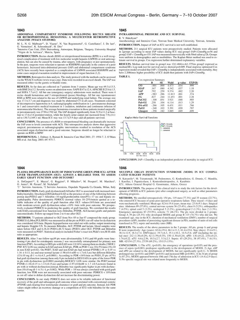
S268 20th ESICM Annual Congress – Berlin, Germany – 7–10 October 2007
1043ABDOMINAL COMPARTMENT SYNDROME FOLLOWING RECTUS SHEATHOR RETROPERITONEAL HEMATOMA: A MULTICENTER RETROSPECTIVEANALYSIS (WSACS STUDY002)
M. L. N. G. Malbrain*1, D. Viaene1, N. Van Regenmortel1, G. Castellanos2, I. De laet1,G. Vermeiren1, K. Schoonheydt1, H. Dits1
1Intensive Care Unit, ZNA Stuivenberg, Antwerpen, Belgium, 2Surgery, University Hospital“Virgen de la Arrixaca”, Murcia, Spain
INTRODUCTION. Rectus sheath (RSH) or retroperitoneal hematoma (RPH) is a well recog-nised complication of treatment with low molecular weight heparin (LMWH) or oral anticoag-ulation, but can also be caused by trauma, after surgery, with pregnancy or just spontaneously.In the past, surgeons were reluctant to operate on RSH/RPH especially in the context of a poorhemostasis. Increased intra-abdominal pressure (IAP) and abdominal compartment syndrome(ACS) have recently been reported as a complication of LMWH associated RSH/RPH and insome cases surgical evacuation resulted in improvement of organ function (1,2).
METHODS. Retrospective data analysis. The study protocol with the methods can be accessedvia the WSACS website (www.wsacs.org). Data were recorded in an excel sheath. The IAP wasmeasured either via the gastric or bladder route.
RESULTS. So far data are collected on 7 patients from 3 centres. Mean age was 69.3±13.5,with BMI 24±3.2. Severity scores on admission were: SAPS II 47±12.4, APACHE II 25.6±1.5,and SOFA 7.7±4.2. All but one (emergency surgery) admissions were medical. There were 4rectus sheath hematomas and 3 retroperitoneal (psoas) bleedings. All but one (spontaneous)RSH or RPH were related to the use of LMWH and underlying renal failure. The average sizewas 17.3±3.7 cm and diagnosis was made by abdominal CT in all cases. Treatment consistedof decompressive laparotomy in 4, radioangiographic embolisation in 1, percutaneous drainagein 1 and conservative medical treatment in 1 (sedation, intubation and continuous infusion ofneuromuscular blockers). The average hematoma evacuation in those patients treated surgicallyor percutaneously was 2730±1702 ml. The IAP dropped significantly from 32.9±11.2 at base-line to 17.6±3.6 postintervention, while the hourly urine output rate increased from 7.9±14.1ml to 130.7±169.1 ml. Mean ICU stay was 13.7±5.9 days and all patients survived.
CONCLUSION. The presence of a RSH or retroperitoneal bleeding can dramatically increasethe IAP even to levels consistent with ACS. This retrospective analysis shows that aggressivesurgical, percutaneous or radioangiographic interventions resulted in resolution of the ACSassociated organ dysfunction and a good outcome. Surgeons should no longer be reluctant tooperate on RSH or RPH!
REFERENCE(S). 1. Dabney A, Bastani B. Intensive Care Med 2001; 27: 1954-7. 2. O’MaraMS et al. Am Surg. 2003; 69: 975-7.
1044PLASMA DISAPPEARANCE RATE OF INDOCYANINE GREEN (PDR ICG) AFTERLIVER TRANSPLANTATION (OLT): ALWAYS A RELIABLE TOOL TO ASSESSEARLY GRAFT FUNCTION AND OUTCOME?
A. DeGasperi*1, E. Mazza2, G. Reggiori2, P. Mihailov2, M. Prosperi2, M. Marchesi2,O. Amici2, M. Pavani2, M. Nichelatti212 ˚ Servizio Anestesia, 22 Servizio Anestesia, Ospedale Niguarda Ca Granda, Milan, Italy
INTRODUCTION. Early graft dysfunction(EAD)after OLT is associated with increased mor-bidity/mortality. Deschenes(DES)defined EAD as the presence of one of the following betweenpostop day(POD)2 and 7: bilirubin>10 mg/dl,prothrombin time(PT)>17 sec and hepatic en-cephalopathy. Pulse densitometric PDRICG (normal values 18–25%/min)is quoted as a re-liable indicator of the quality of graft function after OLT: values<16%/min are associatedwith moderate-severe graft dysfunction,values <10% /min with poor outcome. We prospec-tively evaluated PDRICG in predicting the quality of graft function. We correlated the resultswith EAD,conventional liver function tests (bilirubin, PTINR, ALT,lactate),grafts and patientsoutcome(death). Follow-up ranged from 2 to 6 mo after OLT.
METHODS. 73 patients admitted to OLT from Nov 05 to Jan 07 composed the study group.PDRICG (LiMon,PULSION) was measured in all the pts on POD1:cut-off value for dysfunctionwas considered 16%/min. Hepatic hypoperfusion was ruled out with cardiac output monitoringand hepatic Doppler US. To measure Bilirubin, ALT, PTINR and lactate, blood samples weretaken before OLT and 6,18,24 POD1),48,72 hours (POD3) after OLT. PTINR and Bilirubinwere measured on POD7. Statistical analysis included Fisher’s exact test,Wald’s test,M-H oddsratio as appropriate.
RESULTS. After 3 mo follow-up,69 pts were alive(mortality 5.4%),and 68 grafts were func-tioning:4 pts died for extrahepatic reasons,1 was successfully retransplanted for primary nonfunction (PNF). According to DES,pts with EAD were 12(16%):among them no deaths,2 MOF,1PNF. ICU LOS was similar in EAD and non-EAD pts. PDR on POD 1 was 10±5%/min (19±10in non EAD p<0.02). On POD7, EAD and non-EAD pts had normal PTINR(1.19 ± 0.19 vs1.16 ± 0.13, ns) and near normal lactate (1.98±0.4 mM vs 1.7±0.3,ns) but different Bilirubin(15±10 mg dl-1 vs 6±4.5, p<0.0001). According to PDR <16%/min on POD1,35 pts (47%)had graft dysfunction (among them only 8 pts included in DES EAD):in spite of the lower PDRin pts with dysfunction (p<0.0001),mortality,MOF,ICU LOS were similar. On POD7,normalPTINR (1.18±0.15 vs 1.13±0.15,ns) and lactate (1.97±0.8mM vs 1.7 ±0.5,ns)were found inpts with or without dysfunction;instead, Bilirubin was significantly higher in pts with dysfunc-tion (10±8 mg dl-1 vs 5± 4, p< 0.002). While PDR > 18 was always correlated with good graftfunction, low PDR were not necessarily associated with poor outcome: PDRICG < 10%/minas cut off value to define dysfunction did not increase the discriminating power.
CONCLUSION. In our study PDRICG does not seem to be reliable indicator of functionalrecovery of the graft and subsequent graft outcome when correlated with conventional syntetic(PTINR) and clearing liver tests(lactate clearance) or graft and pts outcome. Instead, low PDRvalues might reflect an escretory damage or a competition of ICG with bilirubin for the samecarrier.
1045INTRAABDOMINAL PRESSURE AND ICU SURVIVAL
R. V. Hakobyan*Anesthesiology and Intensive Care, Yerevan State Medical University, Yerevan, Armenia
INTRODUCTION. Impact of IAP on ICU survival is not well established.
METHODS. 211 surgical ICU patients were prospectively studied. Patients were allocatedin 2groups according to mean IAP values during ICU stay:group1-IAP<12mmHg,n=56 andgroup2-IAP>12mmHg,n=155. IAP was measured transvesically with 50ml saline q12h. Groupswere uniform in demographics and co-morbidities. The Kaplan-Meier method was used to es-timate survival in groups. Cox regression further determined explanatory variables.
RESULTS. Median survival time in group1.was 12(1.484)vs.6(1.375)in group2 expresed asmedian(SE). Log-rank test for survival curves showed p=0.009. Final stepwise optimized Coxregression model revealed that IAP>12mmHg is independently associated with mortality, theyhave 2.208times higher possibility of ICU death then patients with IAP<12mmHg.
TABLE 1.Cox regression Summary
B SE Wald p value EXP(B)UO -.001 .000 4.473 .034 .999MAP .167 .080 4.342 .037 1.18IAP .792 .259 9.374 .002 2.20APP -.983 .286 11.813 .001 .374FG .827 .244 11.451 .001 2.28PaCO2 -.100 .051 3.921 .048 .905PalvO2 .258 .104 6.114 .013 1.29DA-aO2 -.266 .105 6.431 .011 .766PaO2 -.240 .103 5.391 .020 .787Glasgow -.358 .182 3.885 .049 .699
CONCLUSION. IAP>12mmHg is an independent predictor of mortality in surgical ICU.
1046MULTIPLE ORGAN DYSFUNCTION SYNDROME (MODS) IN ICU COMPLI-CATED SURGERY PATIENTS
N. Katsarelis*, M. Tsirantonaki, M. Pedonomos, G. Koukoulitsios, G. Dounis, C. Mandila,I. Karellas, I. Papanicolaou, I. Koutsodimitropoulos, A. KarabinisICU, Athens General Hospital G. Gennimatas, Athens, Greece
INTRODUCTION. The purpose of this clinical trial is to study the risk factors for the devel-opment of MODS in ICU patients (pts) after complicated surgery, as well as other parametersaffecting prognosis.
METHODS. We enrolled retrospectively 154 pts, 119 men (77.3%) and 35 women (22.7%),who entered ICU because of acute post-operative respiratory failure. They stayed >4 days andwere mechanically ventilated. Mean age: 62.6±19.4 years, mean stay: 23.4±9.1 days. Surgicalsites: Abdomen 89 (57.8%), central nervous system 36 (23.4%), chest 8 (5.2%), orthopaedics7 (4.5%), spinal cord 5 (3.2%), urological 5 (3.2%), gynecological 2 (1.3%), face 2 (1.3%).Emergency operations 83 (53.9%), eclectic 71 (46.1%). The pts were divided in 2 groups:Group A 39 pts (25.3%) who developed MODS and group B 115 (74.7%) who did not. Weexamined: age, stay in the ICU, duration of mechanical ventilation (DMV), number of surgicalprocedures (nSP), number of preexisting significant disease (n PD), number of units of red cellstransfused (n TU), sepsis and mortality rates (MR).
RESULTS. The results of the above parameters in the 3 groups: All pts, group A and groupB were respectively: Age (years): 62.6±19.4, 66.1±11.3, 61.4±23.6. Stay (days): 23.4±9.1,38.3±12.9, 18.3±7.4. DMV(days): 19.7±6.3, 35.1±10.2, 14.5±5.6. nSP (including the ini-tial one): 212 (1.38±0.29), 62 (1.59±0.35), 150 (1.30±0.24). nPD: 1.81±0.22, 2.01±0.23,1.74±0.22. nTU: 6.6±2.56, 10.3±2.7, 5.3±2.3. Sepsis: 45 (29.2%), 38 (97.4%), 7 (6.1%).MR: 42/154 (27.3%), 27/39 (69.2%), 15/115 (13%).
CONCLUSION. 1) The nTU, (p<0.05), the emergency of operations (p<0.05) and the pres-ence of sepsis (p<0.001) predispose significantly to the development of MODS. 2) Age, nSPand nPD are related to the development of MODS, but not significantly (p<0.1). 3) MODSprolonged stay (p<0.01) and DMV (p<0.01) and increased MR (p<0.001). 4) In 34 pts of groupA (87.2%), MODS appeared between 10th and 17th day of admission in ICU (14.4±2.9 days).5) No specific surgical site was related more frequently to MODS.
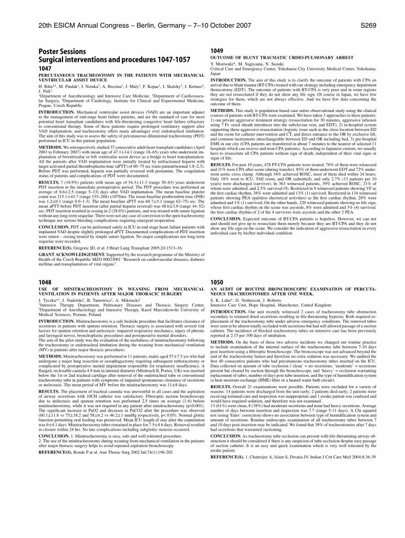
20th ESICM Annual Congress – Berlin, Germany – 7–10 October 2007 S269
Poster SessionsSurgical interventions and procedures 1047-10571047PERCUTANEOUS TRACHEOSTOMY IN THE PATIENTS WITH MECHANICALVENTRICULAR ASSIST DEVICE
H. Riha*1, M. Pindak1, I. Netuka2, A. Brezina1, J. Maly2, F. Kopac1, I. Skalsky2, J. Kettner3,J. Pirk2
1Department of Anesthesiology and Intensive Care Medicine, 2Department of Cardiovascu-lar Surgery, 3Department of Cardiology, Institute for Clinical and Experimental Medicine,Prague, Czech Republic
INTRODUCTION. Mechanical ventricular assist devices (VAD) are an important adjunctto the management of end-stage heart failure patients, and are the standard of care for mostpotential heart transplant candidates with life-threatening congestive heart failure refractoryto conventional therapy. Some of these patients require prolonged ventilatory support afterVAD implantation, and tracheostomy offers many advantages over endotracheal intubation.The aim of this study was to assess the safety of percutaneous dilatational tracheostomy (PDT)performed in ICU in this patient population.
METHODS. We retrospectively studied 37 consecutive adult heart transplant candidates (April2003 to February 2007) with mean age of 47.1±14.1 (range 18–65) years who underwent im-plantation of biventricular or left ventricular assist device as a bridge to heart transplantation.All the patients after VAD implantation were initially treated by unfractioned heparin withtarget activated partial thromboplastin time (aPTT) of 60–75 sec (ratio patient/normal 1.8–2.3).Before PDT was performed, heparin was partially reversed with protamine. The coagulationstatus of patients and complications of PDT were documented.
RESULTS. 7 (18.9%) patients with mean age of 54.3±11.3 (range 30–63) years underwentPDT insertion in the immediate postoperative period. The PDT procedure was performed anaverage of 8.6±2.5 (range 5–13) days after VAD implantation. The mean baseline plateletcount was 215.1±45.7 (range 155–288) x109/liter. The mean baseline prothrombin time (INR)was 1.2±0.1 (range 0.9–1.3). The mean baseline aPTT was 68.7±5.1 (range 62–75) sec. Themean aPTT before PDT insertion (after partial heparin reversal) was 48.6±3.0 (range 44–52)sec. PDT insertion resulted in oozing in 2 (28.6%) patients, and was treated with suture ligationwithout any long-term sequelae. There were not any case of conversion to the open tracheostomytechnique nor serious bleeding complications requiring emergent reoperation.
CONCLUSION. PDT can be performed safely in ICU in end-stage heart failure patients withimplanted VAD despite slightly prolonged aPTT. Documented complications of PDT insertionwere minor – oozing treated by simple suture ligation. No major complications nor long-termsequelae were recorded.
REFERENCE(S). Gregoric ID, et al. J Heart Lung Transplant 2005;24:1513–16.
GRANT ACKNOWLEDGEMENT. Supported by the research programme of the Ministry ofHealth of the Czech Republic MZO 00023001 “Research on cardiovascular diseases, diabetesmellitus and transplantation of vital organs”.
1048USE OF MINITRACHEOSTOMY IN WEANING FROM MECHANICALVENTILATION IN PATIENTS AFTER MAJOR THORACIC SURGERY
J. Tyczka*1, J. Nadolski1, B. Tamowicz2, A. Mikstacki21Intensive Therapy Department, Pulmonary Diseases and Thoracic Surgery Center,2Department of Anesthesiology and Intensive Therapy, Karol Marcinkowski University ofMedical Sciences, Poznan, Poland
INTRODUCTION. Minitracheostomy is a safe bedside procedure that facilitates clearance ofsecretions in patients with sputum retention. Thoracic surgery is associated with several riskfactors for sputum retention and atelectasis: impaired respiratory mechanics, injury of phrenicand laryngeal nerves, bronchoplastic procedures and postoperative mental disorders.The aim of the pilot study was the evaluation of the usefulness of minitracheostomy followingthe tracheostomy or endotracheal intubation during the weaning from mechanical ventilation(MV) in patients after major thoracic procedures.
METHODS. Minitracheostomy was performed in 11 patients, males aged 57±7.5 yrs who hadundergone a major lung resection or oesophagectomy requiring subsequent rethoracotomy orcomplicated by postoperative mental impairment responsible for respiratory insufficiency. Aflanged, reclosable cannula 4.0 mm in internal diameter (Minitrach II, Portex, UK) was insertedbelow the 1st or 2nd tracheal cartilage after removal of the endotracheal tube or conventionaltracheostomy tube in patients with symptoms of impaired spontaneous clearance of secretionsor atelectasis. The mean period of MV before the minitracheostomy was 11±8 days.
RESULTS. The placement of tracheal cannula was uneventful in all cases and the aspirationof airway secretions with 10CH catheter was satisfactory. Fiberoptic suction bronchoscopydue to atelectasis and sputum retention was performed 2,5 times on average (1-6) beforeminitracheostomy, while it was not required in any patient after minitracheostomy (p<0.001).The significant increase in PaO2 and decrease in PaCO2 after the procedure was observed(60.1±11.8 vs 75±18.2 and 58±6.2 vs 46.2±1 mmHg respectively, p< 0.05). Normal glotticfunction permitting oral feeding was preserved. Mean ICU length of stay after the cannulationwas 6±4.1 days. Minitracheostomy tubes remained in place for 7.5±4.6 days. Removal resultedin closure within 24 hrs. No late complications including subglottic stenosis occurred.
CONCLUSION. 1. Minitracheostomy is easy, safe and well tolerated procedure.2. The use of the minitracheostomy during weaning from mechanical ventilation in the patientsafter major thoracic surgery helps to avoid repeated aspiration bronchoscopy.
REFERENCE(S). Bonde P at al. Ann Thorac Surg 2002 Jul;74(1):196-202
1049OUTCOME OF BLUNT TRAUMATIC CRDIO-PULMONARY ARREST
Y. Moriwaki*, M. Sugiyama, N. SuzukiCritical Care and Emergency Center, Yokohama City University Medical Center, Yokohama,Japan
INTRODUCTION. The aim of this study is to clarify the outcome of patients with CPA onarrival due to blunt trauma (BT-CPA) treated with our strategy including emergency departmentthoracotomy (EDT). The outcome of patients with BT-CPA is very poor and in some regionsthey are not resuscitated if they do not show any life sign. Of course in Japan, we have fewstrategies for them, which are not always effective. And we have few data concerning theoutcome of them.
METHODS. This study is population based case series observational study using the clinicalcourses of patients with BT-CPA were examined. We have taken 3 approaches to these patients;1) our private aggressive treatment strategy (resuscitation for 30 minutes, aggressive infusionusing 7 Fr. sized sheath introducer into the subclavian vein, and EDT), 2) in-hospital systemsupporting these aggressive resuscitation (logistic issue such as the close location between EDand the room for catheter intervention and CT, and direct entrance to the OR by exclusive lift,and common instruments interchangeable between ED and OR including bed, 3) pre-hospitalEMS in our city (CPA patients are transferred in about 7 minutes to the nearest of selected 11hospitals which can receive and treat CPA patients). According to Japanese custom, we usuallyhave to resuscitate all CPA patients without sign of death, independent of their vital signs orsigns of life.
RESULTS. For past 10 years, 478 PT-CPA patients were treated. 76% of them were witnessedand 21% were CPA after scene (during transfer). 85% of them underwent EDT and 72% under-went aortic cross clamp. Although 34% achieved ROSC, most of them died within 24 hours.Only 18% went to ICU, TAE room, and OR (admitted), and only 2.7% (13 patients per 10years) were discharged (survivor). In 363 witnessed patients, 39% achieved ROSC, 21% ofwhom were admitted, and 2.5% survived (9). Restricted in 8 witnessed patients showing VF asthe first cardiac rhythm, 38% were admitted and 13% (1) survived. Restricted in 134 witnessedpatients showing PEA (pulsless electorical activities) as the first cardiac rhythm, 28% wereadmitted and 1% (1) survived. On the other hands, 220 witnessed patients showing no life sign,whose first cardiac rhythm on the scene was asystole, 8% were admitted and 3% (4) survived.the first cardiac rhythm of 2 of the 4 survivors were asystole and the other 2 PEA.
CONCLUSION. Expected outcome of BT-CPA patients is hopeless. However, we can notand should not give up to resuscitate them merely because they are BT-CPA and they do notshow any life sign on the scene. We consider the indication of aggressive resuscitation in everyindividual case by his/her individual condition.
1050AN AUDIT OF ROUTINE BRONCHOSCOPIC EXAMINATION OF PERCUTA-NEOUS TRACHEOSTOMIES AFTER ONE WEEK.
S. K. Laha*, D. Nethercott, J. RobertsIntensive Care Unit, Hope Hospital, Manchester, United Kingdom
INTRODUCTION. Our unit recently witnessed 2 cases of tracheostomy tube obstructionsecondary to retained dried secretions resulting in life-threatening hypoxia. Both required re-placement of the tracheostomy tube under adverse emergency conditions. The removed tubeswere seen to be almost totally occluded with secretions but had still allowed passage of a suctioncatheter. The incidence of blocked tracheostomy tubes on intensive care has been previouslyreported at 2.15 per 100 days of intubation.
METHODS. On the basis of these two adverse incidents we changed our routine practiceto include examination of the internal surface of the tracheostomy tube between 7-10 dayspost insertion using a fibreoptic bronchoscope. The broncoscope was not advanced beyond theend of the tracheostomy lumen and therefore no extra sedation was necessary. We audited thefirst 40 consecutive patients who had percutaneous tracheostomy tubes inserted on the ICU.Data collected on amount of tube occlusion (‘clean’ = no secretions, ‘moderate’ = secretionspresent but cleared by suction through the bronchoscope, and ‘heavy’ = occlusion warrantingreplacement of tube), number of days post tube insertion, and the type of humidification system(a heat-moisture exchange (HME) filter or a heated water bath circuit).
RESULTS. Overall 21 examinations were possible. Patients were excluded for a variety ofreasons: 14 patients were discharged from the unit early; 2 patients died early; 2 patients werereceiving terminal care and inspection was inappropriate and 1 awake patient was confused andwould have required sedation, and therefore was not examined.13 (61%) were clean, 8 (38%) had moderate secretions and none had heavy secretions. Averagenumber of days between insertion and inspection was 7.7 (range 5-11 days). A Chi squaredtest (using Yates’ correction) shows no association between type of humidification system andamount of secretions. Routine endoscopic examination of all tracheostomy tubes between 7and 10 days post-insertion may be indicated. We found that 38% of tracheostomies after 7 dayshad secretions that warranted suctioning.
CONCLUSION. As tracheostomy tube occlusion can present with life-threatening airway ob-struction it should be considered if there is any suspicion of tube occlusion despite easy passageof suction catheter. It is an easy and quick examination which is very well tolerated by theawake patient.
REFERENCE(S). 1. Chatterjee A, Islam S, Divatia JV. Indian J Crit Care Med 2004;8:36-39
S270 20th ESICM Annual Congress – Berlin, Germany – 7–10 October 2007
1051LIVER TRANSPLANTATION IN ADULTS
M. Bispo1, P. Marcelino1, S. Silva1, C. Pereira1, S. Marum*1, A. P. Fernandes1, A. Freire2,A. Martins2, E. Barroso2, L. Mourao1
1ICU, 2UT, Hospital Curry Cabral, Lisbon, Portugal
INTRODUCTION. Characterize and analyze the post liver transplantation evolution in theintensive-care unit (ICU) and identify potential risk factors for major complications.
METHODS. Retrospective study of data of 194 postoperative liver transplantations in the ICU,between January 2005 and February 2007. Forty-four perioperative variables were surveyedand a multivariate analysis attempted to identify risk factors independently associated to themajor early postoperative complications.
RESULTS. The most frequent primary diagnosis were familial amyloidotic polyneuropathy(FAP) (35,6%), hepatocellular carcinoma (19,1%) and alcoholic liver disease (17,5%). Se-quential (domino) liver transplantations were carried out in 40 procedures. The most frequentindication for retransplantation was hepatic artery thrombosis (60%). ICU stay was 5,7 days. Themost challenging complications were hemorrhagic events requiring blood transfusion (19,6%),acute renal failure requiring dialysis (9,3%) and thrombotic events (8,3%). Sixty nine percentof the thrombotic events occurred in FAP patients. The overall mortality was 4,6% and themost common cause of death was hemorrhagic shock. After logistic regression, independentfactors predicting early hemorrhagic events included primary diagnosis of cirrhosis with severehepatic failure (Child-Pugh C), intraoperative bleeding with hemodynamic instability and alonger cold ischemic time (non sequential liver transplantation). Independent factors predictingearly thrombotic events included primary diagnosis of AFP, transaminases elevation more than24 hours after transplantation and a 1,5 fold (50%) rise in serum lactate within a 24 hour period.
CONCLUSION. Awareness of potential risk factors for the major post hepatic transplant com-plications in the ICU should contribute to lowering short-term morbidity and mortality. Thesequential (domino) transplantation enlarges the donor pool, decreases cold ischemic time andwas in this study independently associated to a lower risk of early hemorrhagic complications.The FAP group was independently associated to an increased risk for thrombotic complications.
1052THIRTY-DAY MORTALITY AFTER ESOPHAGEAL RESECTION IS REDUCEDFOLLOWING INTRODUCTION OF A CLOSED FORMAT INTENSIVIST LEDICU
L. B. Koppert*1, I. A. Meynaar2, M. W. J. M. Wouters3, P. L. Tangkau2, P. W. de Graaf1,T. Karsten1, E. W. Steyerberg4, L. P. S. Stassen1
1Surgery, 2ICU, Reinier de Graaf Gasthuis, Delft, 3Surgery, The Netherlands Cancer Institute,Antoni van Leeuwenhoek Hospital, Amsterdam, 4Public Health, Erasmus MC, Rotterdam,Netherlands
INTRODUCTION. Surgical resection is the only curative treatment in patients with esophagealcancer, but is associated with considerable postoperative morbidity and mortality. The short-term surgical outcome depends on patient characteristics, surgical approach (transhiatal ortransthoracic), preoperative treatment as well as hospital characteristics. In this single centerstudy, we analyzed surgical mortality in esophageal cancer patients before and after introductionof a closed format intensivist led ICU.
METHODS. The Reinier de Graaf Gasthuis is a large non-academic hospital. The surgical teamperformed 100 esophagectomies between 1990 and 2004. The ICU organisation was changedfrom an open format ICU to a closed format intensivist led ICU starting January 1st, 2001.We compared 30-day mortality in patients operated on before 2001 (n=47, 1990-2000, cohort1) and since 2001 (n=53, 2001-2004, cohort 2) with correction for case-mix in multivariablelogistic regression analysis as described in recent literature1.
RESULTS. Results are presented in Table 1. Univariate analysis showed a 12.5 times decreasedrisk for 30-day mortality as compared to the risk of cohort 1 (Odds Ratio 0.08, 95%CI 0.01-0.67). Multivariable analysis with adjustment for age, comorbidity, preoperative treatment andhospital volume (per period) confirmed this reduction (OR 0.075, 95%CI 0.01-0.63).
TABLE 1.Cohort 1 Cohort 2 P-value/1990-2000 2001-2004 Odds ration=47 n=53 (95%CI)
Age (mean ± SD) 61.6 ± 11.6 61.1 ± 8.7 0.8 a
Comorbidity 28 (60%) 35 (66%) 0.5 b
Preoperative chemotherapy 5 (11%) 20 (38%) 0.004 b
Transhiatal approach 33 (70%) 47 (89%) 0.06 b
ICU length of stay 9.9 ± 12.1 8.9 ± 10.9 0.7 a
(mean ± SD)30-day mortality 9 (19%) 1 (2%) OR 0.08 (0.01-0.67) c
a Independent samples T-test. b Chi-square test. c Multivariable log reg analysis
CONCLUSION. Closed format intensivist led ICU organisation significantly improved 30-daymortality after esophagectomy for cancer.
REFERENCE(S). 1EW Steyerberg, BA Neville, LB Koppert, VEPP Lemmens, HW Tilanus,JWW Coebergh, JC Weeks, CC, Earle. Surgical mortality in patients with esophageal cancer:development and validation of a simple risk score.J Clin Oncol. 2006 Sep 10;24(26):4277-84.
1053HYPERAMYLASEMIA AFTER AORTIC ANEURYSM REPAIR
P. A. Vernikos*, K. Salatas, N. K. Archontoulis, A. Mega, J. Pavleas, A. Rigas, G. Tho-mopoulos, K. RigasICU, Laiko General Hospital, Athens, Greece
INTRODUCTION. Acute pancreatitis after surgical treatment of aortic aneurysm is a rarecomplication with a high mortality rate, while hyperamylasemia without clinical signs of pan-creatitis is more frequent but self-limited complication.We analyzed retrospectively the incidence of hyperamylasemia after aortic aneurysm repair, toconfirm its use as a prognostic indicator regarding clinical course and outcome.
METHODS. We reviewed 32 patients (30 males/2 females), who were admitted in ICU, from9/2004 to 9/2006, after abdominal or thoraco-abdominal aortic aneurysm (TAA) repair. Amongthese patients, 15 (46%) had elevated levels of serum amylase (Group I) and 17 (54%) hadnormal values (Group II). Serum amylase, lactic acid, blood tests, liver and renal function tests,were recorded the 1st and 3rd day of ICU hospitalization as well as their correlation with thepatient outcome.
RESULTS. In both groups age, APACHE II score and the percentage of ruptured aneurysmswere found at the same range (Table 1). Also at the same range were the values of Hemoglobin(Hb) and the lactic acid (Lac). Group I (with hyperamylasemia) was associated with highermean values of aspartic aminotransferase(AST), alanine tranferase(ALT) and Urea, than groupII (Table 2). The following days all these values tent to normalization except serum amylaselevels which continued to rise till 3rd day and were normalized after 4th to17th day. None of thepatients developed acute pancreatitis and no signs of embolisation were noticed. The overallmortality in Group I was 53% and in Group II. 30%.
TABLE 1.Number Age Apache II Ruptured Survivors
Group I 15 (TAA 2) 74.87 18.93 7 (47%) 7 (47%)Group II 17 (TAA 2) 72.65 16.35 8 (47%) 12 (70%)
TABLE 2.Amylas Amylas SGOT SGPT Urea Cr Hb Bil Lacday 1 day 2 day 1 day 1 day 1 day 1 day 1 day 1 day 1
Group I 325.87 504.29 176.53 102.40 106.40 2.71 10.49 1.73 2.81Group II 104.53 111.88 59.35 49.35 68.35 2.14 10.45 1.49 2.71
CONCLUSION. Hyperamylasemia after aortic aneurysm repair can be attributed in per-operative trauma during retroperitoneal dissection, shock, thromboembolic episodes or is-chemia/reperfusion injury. Hyperamylasemia is a frequent self-limited complication, but whenis accompanied with elevated values of serum transaninases and urea nitrogen, then is associatedwith high mortality rate.
1054ABDOMINAL AORTIC ANEURYSM: OPEN OR ENDOVASCULAR REPAIR?
A. V. Ristow1, A. C. Filho*2, H. Simon2, C. B. Ramos2, R. Chicrala1, A. Vescovi1,R. Coelho2, D. Bastos2, J. A. M. Paramo2
1Centervasc, 2ICU, Clinica Sorocaba, Rio de Janeiro, Brazil
INTRODUCTION. Abdominal Aortic Aneurysm (AAA) has a markedly progressive increas-ing incidence, probably due to an aging population and a high incidence of smoking. It seemsthat 85% of AAA > 6cm of diameter suffer rupture until 2 years. Recent endovascular tech-niques of AAA repair (EVAR) contribute to a diminishing mortality, mainly among patientswith important comorbidities. The aim of this study was to compare mortality and length ofstay (LOS) at Intensive Care Unit(ICU), between open aortic repair (OAR) versus EVAR.
METHODS. Observational retrospective study with immediate post-operative patients ad-mitted at ICU of a private hospital in Rio de Janeiro from May of 2004 thru March 2007.Demographics, clinical and laboratorial data were collected. We divided patients into twogroups, according to technique (OAR or EVAR). Data were compared using SPSS for windowsversion 10. For categorical data we used ANOVA, and for numerical data Student T Test.
RESULTS. 28 patients were studied,16 patients in OAR group with mean age of 67,68 ± 9,59years. 75% were men (n = 12), 25% women (n = 4). EVAR group had 18 patients with mean ageof 72,01 ± 7,96 years. 94,44 % were men(n = 17), 5,56% women (n = 2). There were no statisticdifference in incidence of Systemic Hypertension (87,5% X 100%), Diabetes Mellitus (43,76%X 33,33%), Chronic Pulmonary Obstructive Disease (25% X 16,67%) and Dislipidemy (25%X 16,67%). LOS at ICU was significantly lower for EVAR group (1,11 ± 0,76 days X 3,69 ±2,06 days, p < 0,05). EVAR group mortality was 0% X 12,5% (n=2) in OAR group.
CONCLUSION. EVAR was the best technique related to a significant shorter LOS at ICU andalthough not statistically significant,showed lower mortality rates.
REFERENCE(S). 1. Endovascular aortic repair: has there been an improvement in deviceperformance ? British Journal of Surgery - Supplement. 88 Supplement 1:49,May 2001.2. Endovascular Stent Graft Repair of Abdominal and Thoracic Aortic Aneurysms: A Ten YearExperience With 817 Patients. Annals of Surgery.238(4):586-595,October2003.3. Norris,Edward J M. D., M. B. A.,F. A. H. A. Perioperative Management of the Patient Under-going Aortic Vascular Surgery. ASA Refresher Courses in Anesthesiology.33(1):187-202,2005.
20th ESICM Annual Congress – Berlin, Germany – 7–10 October 2007 S271
1055COMPLEX VASCULAR SURGICAL PROCEDURES IN WOMEN. MORBIDITYAND MORTALITY
A. V. Ristow1, A. C. Filho*2, R. Chicrala1, H. Simon2, D. Bastos2, B. Massiere1, A. Vescovi1,J. Morgado2, J. A. M. Paramo2
1Centervasc, 2ICU, Clinica Sorocaba, Rio de Janeiro, Brazil
INTRODUCTION. Cardiovascular disease is one of the main mortality causes of the world.Mortality of women submitted to cardiac surgery is higher than men. The aim of this study isto analyze if women patients submitted to vascular surgical procedures in a private hospital inRio de Janeiro, Brazil show different mortality when compared with men.
METHODS. Observational retrospective study with immediate post-operative patients admit-ted at ICU of a tertiary private hospital in Rio de Janeiro from May of 2004 to March of2007. Demographics, clinical and laboratorial data were collected. We divided patients into twogroups, according to gender (Male or Female). Data were compared using SPSS for windowsversion 10. For categorical data we used ANOVA, and for numerical data Student T Test.
RESULTS. 205 patients were studied. Male group had 145 patients with mean age of 70,38± 10 years. Female group had 60 patients with mean age of 69,47 ± 13,87 years. There wereno statistic difference in incidence of Systemic Hypertension (96,67% X 90,34%), DiabetesMellitus (55% X 42,76%), Chronic Obstructive Pulmonary Disease (10% X 12,41%), IschemicHeart Disease (20% X 26,21%). Female group showed more Chronic Renal Failure (6,67% X17,93%, p < 0,05). Mortality was higher in male group (8,33% X 2,76%, p < 0,05). Length ofstay at ICU was significant higher in male group (4,97 ± 2,33 days X 1,92 ± 3,32 days, p <0,05). Mean APACHE II in male group was 15,52 and 14,33 in female group.
CONCLUSION. In our population, male group showed greater mortality than female groupdespite of greater incidence of Chronic Renal Failure in female group. LOS at ICU was higherin male group too.
REFERENCE(S). 1. Gender-associated differences in plaque phenotype of patients undergo-ing carotid endarterectomy.J Vasc Surg. 2007 Feb;45(2):289-96 Hellings WE, Pasterkamp G, Verhoeven BA, De KleijnDP, De Vries JP, Seldenrijk KA, van den Broek T, Moll FL.2. An institutional approach to maintenance of excellent outcomes for carotid endarterectomyin a setting with moderate-to-low procedure volume.J Am Coll Surg. 2006 Nov;203(5):634-41. Epub 2006 Sep 20. Sullivan HG, Cogbill TH, FischerGG, Hoppe CM, Lambert PJ, Mathiason MA, Glenz CJ, Kilburg CJ.3. Effect of age and gender on restenosis after carotid endarterectomy.Ann Vasc Surg. 2006 Sep;20(5):602-8. Epub 2006 Oct 4. Hugl B, Oldenburg WA, NeuhauserB, Hakaim AG.
1056SURGICAL INTERVENTIONS IN ICU PATIENTS: ARE THEY SAFE?
P. A. D. Duarte*1, A. C. Jorge1, A. A. Luiz1, R. Nomelini1, M. Campos1, C. S. O. Bredt2,G. L. Bredt Jr2, C. Mroginski1, L. M. Ramos2, F. L. Motter2
1General ICU, 2Internal Medicine, University Hospital - Universidade Estadual do Oeste doParan, Cascavel, Brazil
INTRODUCTION. Critically ill patients frequently need surgical procedures due to differentproblems, including medical complications not directly related to the cause of admission. Suchprocedures have a high risk of mortality and complications, even due to patient’s condition orby surgical risk itself. OBJECTIVE: To study incidence of major surgical procedures in patientsalready interned at ICU, performed after more than 24 hours of admission; as well as to analyzethe impact of such procedures in ICU outcome and mortality.
METHODS. Retrospective cohort study. All patients admitted in ICU since 2004 july to 2006may which were submitted to major surgical procedures after > 24 hours were studied. Itwere excluded cases in that the admission intent was to prepare the patient to operation (pre-operative optimization); it were included re-operations. It were also excluded surgical electivetracheostomies and gastrostomies, as well as operations directly related to the primary causeand soon after ICU admission (ex, orthopedic trauma surgeries). It was made analysis withdescriptive statistics, and comparison with univariated analysis of variance (anova), with t-test.
RESULTS. During the 23 month study period, there were 798 admissions in the general adultICU (mean age: 51.9; APACHE II: 18.2; 60,8% male). 48 major surgical procedures (SP) wereperformed in 44 patients: 17 on digestive system, 10 neurosurgery, 06 vascular, 04 orthopedic,03 cardiac, 03 thoracic, 02 plastic, 02 tocogynecologic and 01 urologic. All of them with gen-eral anesthesia, at operation room. Mean age: 44.2; male: 68.9%. There was no death due totransport, during surgical procedures or directly related to them.
SP Non-SP pAPACHE II 19.2 18.2 0.487ICU Mortality (%) 62.2 39.7 0.005ICU length (days) 16.2 06.1 <0.0001MV length (days) 13.0 04.1 <0.0001
CONCLUSION. Major emergency surgical procedures performed in ICU are safe, howeverICU mortality, ICU length and MV length are very high, probably due to the complexity andthe clinical seriousness of the ICU patients. Admission APACHE II did not predict mortality inthis group of patients.
1057INTERMITTENT CLAUDICATION: ENDOVASCULAR TREATMENT X OPEN IN-FERIOR LIMB REVASCULARIZATION
A. V. Ristow1, A. C. Filho*2, M. Moraes3, R. Chicrala3, D. Bastos2, B. Massiere3, R. Coelho2,H. Simon2, J. A. M. Paramo2
1Centervasc, , 2ICU, 3Centervasc, Clinica Sorocaba, Rio de Janeiro, Brazil
INTRODUCTION. Peripheral arterial disease is a common manifestation of atherosclerosis,and its prevalence increases with age and the presence of cardiovascular risk factors. Cigarettesmoking and diabetes mellitus are the strongest risk factors. Hypertension, dyslipidemia, andhyperhomocysteinemia also significantly increase the risk of peripheral arterial disease. Surgi-cal treatment markedly improved in recent years, mainly because development of endovasculartechiniques like subintimal angioplasty. The aim of this study was to compare mortality andlength of stay (LOS) at Intensive Care Unit (ICU), between open inferior limb revascularization(OLR) or endovascular inferior limb revascularization (ELR).
METHODS. Observational retrospective study with immediate post-operative patients admit-ted at ICU of a tertiary private hospital in Rio de Janeiro from May of 2004 to March of2007. Demographics, clinical and laboratorial data were collected. We divided patients into twogroups, according to technique (OLR or ELR). Data were compared using SPSS for windowsversion10. For categorical data we used ANOVA, and for numerical data Student T Test.
RESULTS. 63 patients were studied. OLR group had 32 patients with mean age of 70,83 ±8,04 years. 78,13% of men (n=25), 21,87% of women (n=7). ELR group had 31 patients withmean age of 66 ± 14,38 years. 70,97% of men (n=22), 29,03% of women (n=9). There wereno statistic difference in incidence of Systemic Hypertension (93,75% X 83,87%), DiabetesMellitus (71,88% X 61,29%), Ischemic Heart Disease (34,38% X 19,35%), Chronic RenalFailure (28,13% X 16,13%) and Dislipidemy (18,75% X 12,9%). Mortality was higher in OLRgroup, but without significant difference (15,63% X 3,23%). LOS at ICU was higher in OLR3,09 ± 4,84 days X 1,84 ± 4,05.
CONCLUSION. In our population, both techniques did not show statistic differences. Bothshowed safety and seems to be excellent options to improve wellness of patients suffering ofintermittent claudication.
REFERENCE(S). 1. Review of mortality and cardiovascular event rates in patients enrolledin clinical trials for claudication therapies.Vasc Med. 2006 Nov;11(3):141-5. Review. Brass EP, Hiatt WR.2. Clinical practice. Intermittent claudication.N Engl J Med. 2007 Mar 22;356(12):1241-50. Review. White C.3. Intermittent claudication–diagnosis and treatment]Tidsskr Nor Laegeforen. 2007 Jan 18;127(2):167-70. Review. Norwegian. Pedersen G, LaxdalE, Jonung T, Aune S.


