Embed Size (px)
Citation preview
HAL Id: halsde-00582731https://hal.archives-ouvertes.fr/halsde-00582731
Submitted on 13 Jul 2011
HAL is a multi-disciplinary open accessarchive for the deposit and dissemination of sci-entific research documents, whether they are pub-lished or not. The documents may come fromteaching and research institutions in France orabroad, or from public or private research centers.
L’archive ouverte pluridisciplinaire HAL, estdestinée au dépôt et à la diffusion de documentsscientifiques de niveau recherche, publiés ou non,émanant des établissements d’enseignement et derecherche français ou étrangers, des laboratoirespublics ou privés.
Physiological responses of the scleractinian coralPocillopora damicornis to bacterial stress from Vibrio
corallilyticusJeremie Vidal-Dupiol, Ophélie Ladrière, Anne-Leila Meistertzheim, Laurent
Fouré, Mehdi Adjeroud, Guillaume Mitta
To cite this version:Jeremie Vidal-Dupiol, Ophélie Ladrière, Anne-Leila Meistertzheim, Laurent Fouré, Mehdi Adjeroud,et al.. Physiological responses of the scleractinian coral Pocillopora damicornis to bacterial stressfrom Vibrio corallilyticus. Journal of Experimental Biology, The Company of Biologists, 2011, 214(9), pp.1533-1545. �10.1242/jeb.053165�. �halsde-00582731�
1
INTRODUCTIONCoral reef ecosystems are among the most biologically diverse andcomplex marine ecosystems worldwide. In addition to theirbiological and ecological importance, coral reefs support majoreconomic and physical functions (e.g. food production, tourism,biotechnology development and coast protection) that are essentialfor many countries (Lesser, 2004). These ecosystems are principallysupported by small, colonial and calcifying organisms, thehermatypic scleractinian corals, which enter into mutualisticsymbioses with microalgae of the genus Symbiodinium,dinoflagellates that are also referred to as zooxanthellae (Lesser,2004).
Because of global climate change and anthropogenic pressure,coral reef ecosystems have been increasingly confronted withsevere natural and anthropogenic disturbances over the past threedecades (Hughes et al., 2003; Ward and Lafferty, 2004; Donner etal., 2005; Hoegh-Guldberg et al., 2007; Bourne et al., 2009). A recentglobal assessment of coral reef health showed that approximately19% of coral reefs were irremediably degraded with no sign of
recovery, 15% presented symptoms of an imminent risk of collapseand another 20% were at risk of becoming critically affected in thenext few decades (Wilkinson, 2008). Coral diseases are among themajor factors in coral reef degradation; their impacts have severelyincreased in recent decades, in apparent association with globalclimate change (Weil et al., 2006; Bourne et al., 2009). Indeed, ithas been suggested that high temperatures influence the outcomeof bacterial infections by lowering the disease resistance of coralsand/or increasing pathogen growth, virulence and infectivity (Wardet al., 2007; Rodriguez-Lanetty et al., 2009).
To date, 18 coral diseases have been identified (Sutherland et al.,2004; Willis et al., 2004; Harvell et al., 2007). Among the well-characterized coral infectious diseases, several have been shown tobe caused by members of the Vibrionaceae family (Kushmaro etal., 2001; Ben-Haim et al., 2003a; Sussman et al., 2008). Bacteriabelonging to the genus Vibrio are ubiquitously distributed in aquaticenvironments worldwide, from brackish water to deep-seaenvironments. Kushmaro et al. demonstrated that the bacteriaVibrio shiloi infects and triggers bleaching of the coral Oculina
Page nos Page total Colour pages: Facing pages: Issue Ms order
The Journal of Experimental Biology 214, 000-000© 2011. Published by The Company of Biologists Ltddoi:10.1242/jeb.053165
RESEARCH ARTICLE
Physiological responses of the scleractinian coral Pocillopora damicornis tobacterial stress from Vibrio coralliilyticus
Jérémie Vidal-Dupiol1,*, Ophélie Ladrière2,*, Anne-Leila Meistertzheim1, Laurent Fouré3, Mehdi Adjeroud4
and Guillaume Mitta1,†
1UMR 5244, CNRS UPVD EPHE, Université de Perpignan Via Domitia, 52 Avenue Paul Alduy, 66860 Perpignan Cedex, France,2Unité d’écologie marine, Laboratoire d’écologie animale et ecotoxicologie, Université de Liège, Allée du 6 août, 15, Bat. B6C, 4000
Liege, Belgium, 3Aquarium du Cap d’Agde, 11 rue des 2 frères, 34300 Cap d’Agde, France and 4Institut de Recherche pour leDéveloppement, Unité 227 CoRéUs2 “Biocomplexité des écosystèmes coralliens de l’Indo-Pacifique”, bp A5, 98848 Nouméa
Cedex, Nouvelle-Calédonie*These authors contributed equally to this work
†Author for correspondence ([email protected])
Accepted 19 January 2011
SUMMARYAs the effects of climate change have become increasingly visible over the past three decades, coral reefs have suffered from anumber of natural and anthropogenic disturbances that have caused a critical decline in coral populations. Among thesedisturbances are coral diseases, which have appeared with increasing frequency and severity, often in correlation with increasesin water temperature. Although the crucial role played by Vibrio species in coral disease has been widely documented, thescientific community does not yet fully understand the infection process of Vibrio or its impact on coral physiology andimmunology. Here, we investigated the physiological and transcriptomic responses of a major reef-building coral, Pocilloporadamicornis, when exposed to a specific pathogen (Vibrio coralliilyticus) under virulent (increasing water temperature) and non-virulent (constant low temperature) conditions. The infection process was examined by electron microscopy and quantitativereverse-transcription PCR, and coral health was monitored by visual observations and measurements of zooxanthellar density.The results obtained suggest that coral tissue invasion occurs upon increasing water temperature only. Transcriptomic variationswere investigated using a suppression–subtractive–hybridization approach, and the expression levels of six candidate immune-related genes were examined during bacterial exposure. These genes correspond to three lectin-like molecules putativelyinvolved in the recognition of pathogens, two metal-binding proteins putatively involved in antibacterial response and one cysteinprotease inhibitor. The transcription patterns of these selected genes provide new insights into the responses of coral coloniesto virulent versus non-virulent bacteria.
Supplementary material available online at http://jeb.biologists.org/cgi/content/full/214/??/????/DC1
Key words: Pocillopora damicornis, Vibrio coralliilyticus, biomarker, coral disease, global change, vibriosis.
2
patagonica (Kushmaro et al., 1996; Kushmaro et al., 1997;Kushmaro et al., 1998; Kushmaro et al., 2001). The same researchgroup showed that the infectivity of this bacteria is temperaturedependent and occurs only after an increase in seawater temperature(Kushmaro et al., 1998). A similar temperature-dependent virulencewas observed in interactions between Vibrio coralliilyticus and thescleractinian coral Pocillopora damicornis (Ben-Haim et al., 2003b),and the involvement of Vibrio species has been suggested in twoother coral diseases: yellow blotch/band disease (Cervino et al.,2004; Cervino et al., 2008) and rapid tissue necrosis (Luna et al.,2007). Among these Vibrio-related diseases, the bleaching diseaseof O. patagonica caused by the infection of V. shiloi has beencharacterized to the greatest degree (for a review, see Rosenberg,2004). In brief, V. shiloi is attracted by chemotaxis and adheres to-galactoside-containing receptors on coral cells. This adhesionprocess is specific and dependent on temperature: no adhesion occursat low temperatures (16–20°C), whereas V. shiloi actively adhereto their target at temperatures between 25 and 30°C. After contact,the bacteria penetrate and multiply within the coral cells, and beginproducing two sets of factors: extracellular proline-rich toxins(referred to as toxin P) (Banin et al., 2001) that block photosynthesis,and enzymes that trigger the lysis of zooxanthellae.
However, although Vibrio species have been shown to play crucialroles in several coral diseases, our knowledge of the effects of Vibrioinfection on coral physiology remains incomplete. Accordingly, thepresent study used a global transcriptomic approach to examine thephysiological responses of coral colonies when confronted withbacterial stress and/or infection in a realistic ecological context.Specifically, we studied the interaction between the coral P.damicornis and its specific pathogenic bacteria, V. coralliilyticus.
Pocillopora damicornis has a widespread distribution in the Indo-Pacific region (Veron, 2000), is highly sensitive to a wide range ofnatural and anthropogenic disturbances, and is a good sentinelspecies for use in monitoring coral reef health (Gates et al., 1992;Ben-Haim and Rosenberg, 2002; Stimson et al., 2002; Hashimotoet al., 2004; Vidal-Dupiol et al., 2009). Vibrio coralliilyticus is aspecific pathogen of P. damicornis, and its virulence is temperaturedependent; the bacterium has been shown to trigger coral bleachingat moderate temperatures (24–25°C) and coral tissue lysis at highertemperatures (26–29°C) (Ben-Haim Rozenblat and Rosenberg,2004). Consequently, the P. damicornis–V. coralliilyticus modelallows us to compare coral responses to virulent and non-virulentbacteria.
Here, we exposed coral to a consistent supply of bacteria at either25°C (non-virulent) or under conditions of a gradual temperatureincrease (from 25 to 32.5°C; induction of virulence) and examinedtranscriptomic modifications using subtractive hybridization. Thedifferentially regulated genes identified through this approachincluded several thought to be involved in immune processes.Quantitative reverse-transcription PCR (qRT-PCR) was used tofollow the expression levels of a number of selected genes at varioustime points after the onset of bacterial stress or infection.
MATERIALS AND METHODSBiological materials
The Pocillopora damicornis (Linnaeus 1758) isolate used in thisstudy was harvested in Lombock, Indonesia (CITES no.06832/VI/SATS/LN/2001) and maintained at the Cap d’AgdeAquarium, France. For experimental procedures, nubbins (10g;7�6cm height�diameter) were propagated by cutting branchesfrom the parent colony and physiologically stabilizing them for2months at 25°C.
The coral pathogen Vibrio coralliilyticus strain YB1 (Ben-Haimet al., 2003a) (CIP 107925, Institut Pasteur, Paris, France) was usedto challenge or infect P. damicornis (Ben-Haim and Rosenberg,2002). Vibrio coralliilyticus was cultured in 2216 Marine Brothmedium (catalog no. 279110; BD Difco, Town, State, Country)under aerobic conditions with shaking (150rpm) at 30°C for routineuse, or at the temperature in the coral-containing tank for the Tband Cb groups (see Experimental infection).
Experiments aimed at determining which cells (host or symbiont)expressed the candidate genes (see Identification of host or symbiontgenes) utilized three zooxanthellar isolates corresponding to clonalcultures of zooxanthellae from the B, C and D clades (BURR CultureCollection, University of Buffalo; culture IDs Flap3, Pd44b andMF2.2b, respectively). The representative zooxanthellae werecultured in 250ml screw-top polycarbonate cell-culture flasks(Corning, Town, State, Country) in filtered seawater (pore size,0.2m) enriched with 1� Guillard’s (F/2) marine water enrichmentsolution (Sigma-Aldrich, Town, State, Country) and supplementedwith 1� antibiotic/antimycotic stabilized suspension (Sigma-Aldrich). The zooxanthellae were grown at 25±1°C under anirradiance of 70molphotonsm–2s–1 provided by daylightfluorescent tubes (Sylvania, Town, State, Country) set to a 12h:12hlight:dark photoperiod. Stock cultures were transferred monthly, andcells were used in the stationary phase.
Experimental infectionBacterial challenge was induced by constantly supplying V.coralliilyticus at a relatively low temperature (25°C; non-virulent),whereas infection was induced by constantly supplying the bacteriaand systematically increasing the water temperature from 25 to32.5°C (thereby activating bacterial virulence).
Bacterial challenge, bacterial infection and their respectivecontrols were established in four independent tanks: (1) constantlow temperature (25°C) and bacterial supply (Cb); (2) gradualtemperature increase from 25 to 32.5°C and regular bacterial supply(Tb); (3) constant low temperature without bacteria (C); and (4)gradual temperature increase from 25 to 32.5°C in the absence ofbacteria (T). For experiments, nubbins of P. damicornis wererandomly placed in 120l tanks (N27 per tank). After a 14-dayacclimatization at 25°C, the C and Cb treatments were maintainedat this temperature whereas the temperatures of the T and Tbtreatments were gradually increased by 1.5°C every 3days until thewater temperature reached 32.5°C (days 3, 6, 9, 12 and 15). Thishigher temperature was maintained until the corals bleached or lysed.In the Cb and Tb treatments, bacteria (103cellsml–1 of tank water)were supplied by balneation (Ben-Haim Rozenblat and Rosenberg,2004). The first bacterial supply was initiated on day 1 and repeatedevery 3days (days 1, 4, 7, 10, 13, 16 and 19). The utilized culturesof V. coralliilyticus were prepared at 25°C for the Cb treatment andat the temperature corresponding to the tank temperature for the Tbtreatment. For the Tb and T treatments, beginning on day 3, thetemperature was increased by +1.5°C every 3days until it reached32.5°C.
Three nubbins were randomly sampled from each tank every3days (days 0, 3, 9, 12, 15 and 18). Five 0.5-cm apices were cutfrom each nubbin; three were immediately frozen and stored in liquidnitrogen for later measurement of zooxanthellar density and twowere fixed directly in 2.5% glutaraldehyde and subjected to electronmicroscopy.
The tank temperature was controlled using an aquarium heater(600W, Schego, Town, State, Country) connected to an electronicthermostat (Hobby Biotherm Professional, Town, State, Country).
J. Vidal-Dupiol and others

3Coral response to vibriosis
Illumination was supplied at an irradiance of250molphotonsm–2s–1 (quantum meter; QMSW-SS, ApogeeInstruments Inc., Town, State, Country) by metal halide lamps(400W, Iwasaki 6500 Kelvin, Town, State, Country) set to a12h:12h light:dark photoperiod. All other seawater parameters(salinity 36, pH8.3…????) were maintained constant over time andbetween tanks. A constant water flow was maintained within eachtank using a water pump (1300lh–1, IDRA, Town, State, Country).Seawater was continuously recycled at a rate of 10.8 tank volumesper hour by coupling the action of a biological filter and an Aquavieprotein skimmer (EPS 600, Town, State, Country), and the waterwas renewed (2% tank volume per day) with natural filteredMediterranean seawater heated to 25°C. To avoid the growth ofbacterial blooms, the water was continuously treated using a UVCfilter (5W, AquaCristal Series II, JBL, Town, State, Country).During bacterial balneation, all equipment known to remove or killbacteria (e.g. the protein skimmer and UVC filter) were inactivatedfor the first 4h after treatment, allowing the bacteria to adhere tothe coral tissues.
In order to monitor coral health, physiological (e.g. zooxanthellardensity measure) and visual (e.g. coral color, tissue lysis appearance,opened or closed polyps) measurements were performed throughoutthe experiments. Zooxanthellar density was used as a measurementof coral health and the stability/breakdown of symbiosis. Coraltissues were extracted using a water pick (Johannes and Wiebe,1970) in 50ml of filtered seawater (0.2m pore size), homogenizedwith a Potter grinder and fixed with 2% formaldehyde.Zooxanthellae were counted under a light microscope using ahemocytometer, and algal densities were standardized per skeletalsurface area, as described previously (Stimson and Kinzie, 1991).Every day, two colonies per tank (the same colonies each day) werephotographed using a consistent camera setup, and both bleachingand lytic events were thoroughly examined.
Bacteria and the infectious processIn order to follow the infectious process in P. damicornis tissues,the presence of V. coralliilyticus was investigated by transmissionelectron microscopy (TEM). Two ultrathin slices were prepared fromcoral fragments fixed in 2.5% glutaraldehyde in filtered seawater(0.2m pore size). Sections were prepared as described previously(Ladrière et al., 2008) and observed with a Jeol JEM 100-SXtransmission electron microscope (Town, State, Country) at anaccelerating voltage of 80kV.
qRT-PCR was used to quantify the amount of V. coralliilyticuspresent in P. damicornis tissues. Total RNA was extracted fromeach sample (see Subtractive cDNA library construction) and100ng of total RNA were reverse transcribed using hexamer
primers and a RevertAidTM kit (Fermentas International Inc., Town,State, Country). qRT-PCR was performed with 4l cDNA (1/100dilution) in a total volume of 10l using a LightCycler 480 System(Roche Diagnostics, Town, State, Country), as described below (seeReal-time analysis). Oligonucleotide primers were designed basedon the aligned sequences from eight different V. coralliilyticus 16SrRNAs (GenBank accession nos EF094886, DQ079633,GQ406805.1, GQ406804.1, GQ406802.1, GQ406801.1,GQ406800.1 and GQ406798.1; National Center for BiotechnologyInformation database) and the 28S rRNA of P. damicornis (Vidal-Dupiol et al., 2009) (Table1). RNA extracted from the V.coralliilyticus culture used for infection was taken as the positivecontrol. The qRT-PCR products were cloned and sequenced (seeDNA sequencing and sequence analysis).
Subtractive cDNA library constructionVisual and physiological monitoring was used to select earlybacterially stressed or infected samples, and subtractive hybridizationwas used to identify genes that were differentially expressed underthe non-virulent and virulent conditions. For each set of three nubbins,tissues were extracted with a water pick in 800ml of filtered seawater(0.2m pore size), and both total RNA and mRNA were purified(Vidal-Dupiol et al., 2009). Forward and reverse suppression–subtractive–hybridization (SSH) libraries were constructed using thePCR-Select cDNA Subtraction kit (Clontech, Town, State, Country).Tester and driver cDNA were prepared using 2g of Poly(A)+ RNA,and enzyme digestion, adaptor ligation, hybridization and PCRamplification were performed according to the manufacturer’sinstructions (Clontech). The resulting PCR products were cloned intopCR4-TOPO using a TOPO TA cloning kit (Invitrogen, Town, State,Country) and transformed into One Shot TOP10 chemically competentEscherichia coli cells (Invitrogen).
DNA sequencing and sequence analysisFor each library, 384 clones were randomly selected and single-pass sequenced by GATC Biotech, using an ABI 3730xl apparatus.Vector and adaptor sequences were trimmed from all sequencesusing SequencherTM software (Gene Codes Corp., Town, State,Country). High-quality expressed sequence tags (ESTs) (>100bp)were assembled into clusters or identified as unique sequences, andused for database searches with the BLASTX and BLASTNprograms. Specific domain searches were performed using theInterProscan software. ESTs displaying significant similarities witha gene of known function were assigned to the appropriate functionalclass. The various EST sequences have been submitted to the dbESTdatabase; in an effort to minimize redundancy, sequences displaying100% identity with one another were submitted as a single sequence.
Table 1. Combinations of forward and reverse primers used in real-time PCR expression analysis
Gene name Forward primer (5�r 3�) Reverse primer (5�r 3�)
Concanavalin CATCTCCGTCCCAAAC CAACTGCATAATGAACCCACystatin B ATGGCCTTCCTTCTTTG AGGTGTATGTGGGTGACFerritin AGAATATGCGGGGAGG CCTGGACCAACACGAGMajor basic nuclear protein GGTACAGCAAACTGCG TTGGAAACGTCCGACCPdC-lectin ATTGGCAGAACGGAAG GGGAGGAGACCTGGTAP-selectin CAATGTTATCATTACCGACCC GTCTGTTCCAGTCCAGGSelenium binding protein TACGCCTTCCGGTAAC GTTCTACCCTGACCTGG60S ribosomal protein L40A CGACTGAGAGGAGGAGC CTCATTTGGACATTCCCGT60S ribosomal protein L22 TGATGTGTCCATTGATCGTC CATAAGTAGTTTGTGCAGAGG60S acidic ribosomal phosphoprotein P0 GCTACTGTTGGGTAGCC CTCTCCATTCTCGTATGGTVibrio coralliilyticus 16S rRNA TTAAGTAGACCGCCTGGG GATGTCAAGAGTAGGTAAGGTTCPocillopora damicornis 28S rRNA CGGGTGAGAATCCTGT CTGAAGGCCATCCTCAA
4
qRT-PCR analysesqRT-PCR was used to analyze the expression profiles of selectedgenes. Total RNA was extracted, and this total RNA (2.5g) wasreverse transcribed using oligo dT primers and the Superscript IIenzyme (Invitrogen). The resulting cDNA products were purifiedusing a Nucleotrap Gel Extraction Trial kit (Clontech), and qRT-PCR was performed with 2.5l of purified cDNA (diluted 1:50 inwater) in a total volume of 10l containing 1� LightCycler® 480SYBR Green I Master Mix (Roche Diagnostics) and 70nmoll–1 ofeach primer. The primers, which were designed using Light CyclerProbe Design Software version 1.0 (Roche Diagnostics), arepresented in Table1. Amplification was performed using aLightCycler 480 System (Roche Diagnostics) and the followingreaction conditions: activation of the Thermo-Start® DNApolymerase at 95°C for 15min, followed by 45 cycles of denaturationat 95°C for 30s and annealing/extension at 60°C for 1min. Meltingcurve profiles (95 to 70°C decreasing by 0.5°C every 10s) wereassayed to ensure that we were looking at a single product. Eachrun included a positive cDNA control that was sampled at thebeginning of the experiment (day 0) and also from each amplificationplate; this positive control was also used as the calibrator sample,and blank controls (water) were included for each primer pair. ThePCR products were resolved by electrophoresis, the bands wereisolated directly from agarose gels and DNA was extracted usingthe Gel Extraction PCR Purification System V (Promega). Theresulting qRT-PCR products were single-pass sequenced asdescribed above.
For each reaction, the crossing point (Cp) was determined usingthe second derivative maximum method applied using LightCycler Software version 3.3 (Roche Diagnostics). The PCRefficiency (E) of each primer pair was calculated by determiningthe slope of standard curves obtained from serial-dilution analysisof cDNAs pooled from all experimental samples, as described byYuan et al. (Yuan et al., 2006). The individual qRT-PCRefficiencies for the target or reference genes were calculated usingthe formula: E10(–1/slope). For each candidate gene, the level oftranscription was normalized using the mean geometrictranscription rate of three reference sequences encoding ribosomalprotein genes from the host, P. damicornis: 60S ribosomal proteinL22, 60S ribosomal protein L40A and 60S acidic ribosomalphosphoprotein P0 (GenBank accession nos HO112261,HO112283 and HO112666, respectively). The normalized relativequantities (NRQs) were calculated as described by Hellemans(Hellemans, 2007), using the equation:
where Etarget is the amplification efficiency of the gene of interest;Eref is the amplification efficiency of the reference gene;Cp,refCp,ref(calibrator)–Cp,ref(sample); and Cp,targetCp,target(calibrator)–Cp,target(sample).
Identification of host or symbiont genesTo determine which organism (host or symbiont) expressed thecandidate genes, cross-PCR experiments were performed on DNAand RNA extracted from the holobiont (host plus symbiont) andfrom pure cultured zooxanthellae, as previously described (Vidal-Dupiol et al., 2009). Briefly, PCR amplifications were performedwith oligonucleotides amplifying the targets, the gene encoding thesmall ribosomal subunit RNA Symbiodinium spp. (Rowan and
N ,RQ =Etarget
ΔCp,target
Erefi
ΔCp,ref i
i=1
3
∏3
Powers, 1991) and the cDNA encoding the major basic nuclearprotein (MBNP) of Symbiodinium spp. (Table1).
Statistical analysisBecause conditions of normality and homoscedasticity were notsatisfied, within-set variations in zooxanthellar density wereanalyzed with the non-parametric Kruskal–Wallis (KW) test usingGraphPad InStat 3 (Supplier, Town, State, Country). Variations ingene expression over time were analyzed with a maximum normedresidual test (Grubbs, 1969; Stefansky, 1972) using JMP (SASInstitute, Inc., Town, State, Country). Analyses were performed onqRT-PCR results obtained for a double-passed pool of three nubbinsfrom the same treatment at each sampling date. Differences wereconsidered statistically significant at P<0.05.
RESULTSCoral health and infection status
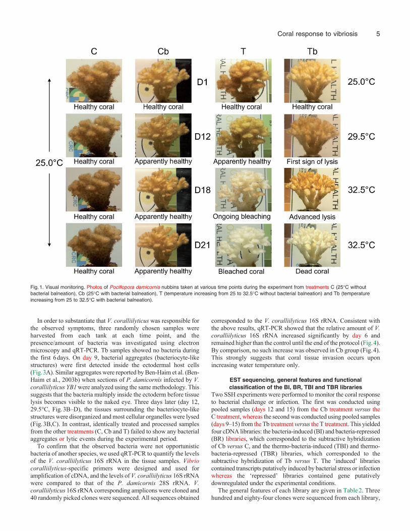
In order to investigate the effect of bacterial challenge and/orinfection, we designed two sets of experiments. The first setexamined the effects of non-virulent bacterial challenge by V.coralliilyticus. A constant temperature of 25°C was used, asexposure of P. damicornis to V. coralliilyticus at this temperaturewas previously shown to trigger bleaching without tissue lysis (Ben-Haim Rozenblat and Rosenberg, 2004). Tanks were maintained at25°C with (Cb) and without (C) bacteria (103bacteriaml–1). In thesecond set of experiments, which examined the effects of infectionby virulent bacteria, a similar setup was used, except that the tankswith (Tb) and without (T) bacteria were subjected to a gradualtemperature increase from 25 to 32.5°C. At each time point, thebacteria added to Tb were cultured at the tank temperature.
Coral health and the stability/breakdown of symbiosis wereexamined by analysis of zooxanthellar density and visual monitoring.For the C and Cb treatments, the zooxanthellar densities (cellscm–2
of skeleton) remained stable throughout the experiment(mean±s.e.m.: C1.07�106±2.35�105 andCb5.18�105±2.89�105cellscm–2). There was no significantdifference in zooxanthellar density between C and Cb (KW,P>0.05), indicating that bleaching did not occur under theseexperimental conditions. For the T and Tb treatments, thezooxanthellar densities remained stable over the first 15 days(mean±s.e.m.: T6.21�105±1.61�105 and Tb4.36�105±1.69�105cellscm–2), but dropped to 6.26�103±5.05�103 and1.72�105±6.23�104cellscm–2, respectively, by day 18. Thisdecrease was only significant for the Tb treatment (KW; P0.0535and P0.0147 for T and Tb, respectively).
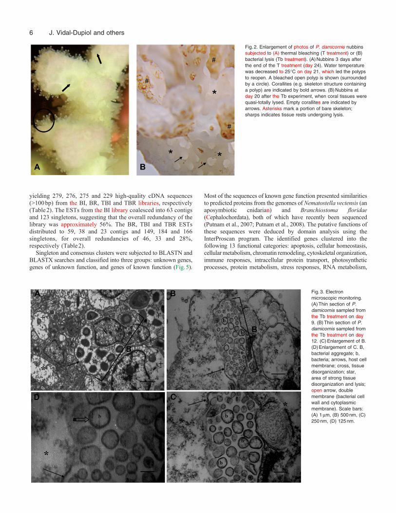
Visual monitoring was used to follow the occurrence of symptomssuch as tissue lysis and/or bleaching. In the C and Cb treatments,the nubbins appeared healthy throughout the duration of theexperiment (Fig.1). In the tank subjected to only increasedtemperature (T treatment), polyps were closed at day 15, bleachingbecame visible in the upper portion of nubbins at day 16 andbleaching was complete by day 18 (Fig.1). These bleached coralswere pale but still alive; when the tank temperature was returnedto 25°C, the polyps reopened (Fig.2). In the Tb treatment, however,tissue lysis was first observed on day 12; lytic plaques were observedin the coral tissues and calcified skeletons (seen at the beginningas small white spots) became evident on the nubbins (Fig.2). Thiseffect continued until lysis was complete (Fig.1; day 21). In orderto confirm that corals subjected to bacterial infection were dead,the tank temperature was returned to 25°C. After several days atthis temperature, polyps did not reopen and the skeleton began tobe colonized by algae.
J. Vidal-Dupiol and others
5Coral response to vibriosis
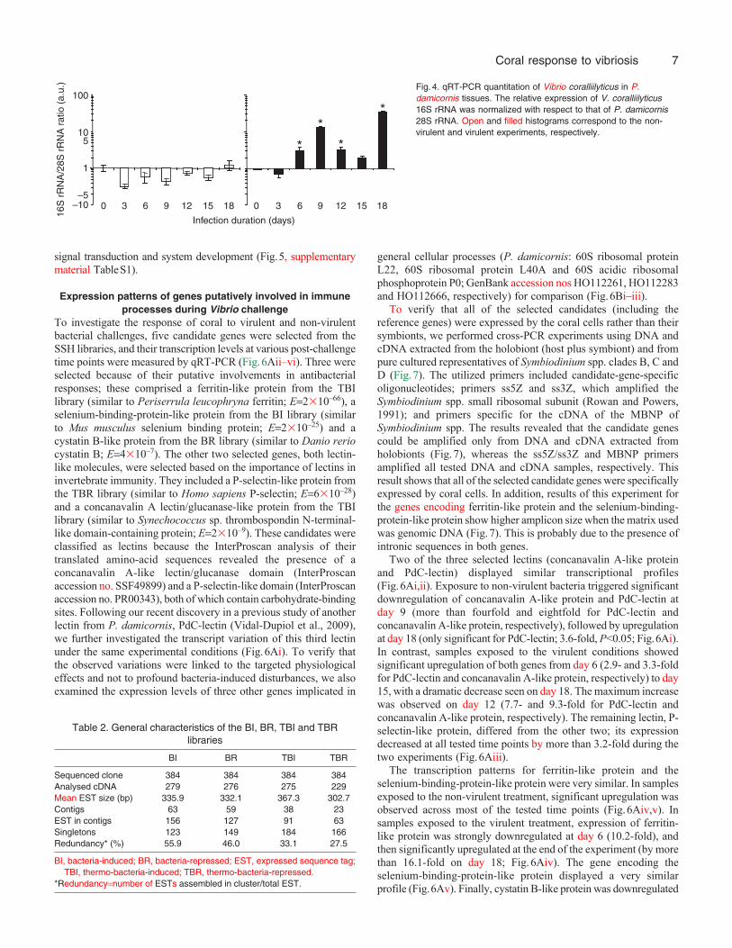
In order to substantiate that V. coralliilyticus was responsible forthe observed symptoms, three randomly chosen samples wereharvested from each tank at each time point, and thepresence/amount of bacteria was investigated using electronmicroscopy and qRT-PCR. Tb samples showed no bacteria duringthe first 6days. On day 9, bacterial aggregates (bacteriocyte-likestructures) were first detected inside the ectodermal host cells(Fig.3A). Similar aggregates were reported by Ben-Haim et al. (Ben-Haim et al., 2003b) when sections of P. damicornis infected by V.coralliilyticus YB1 were analyzed using the same methodology. Thissuggests that the bacteria multiply inside the ectoderm before tissuelysis becomes visible to the naked eye. Three days later (day 12,29.5°C, Fig.3B–D), the tissues surrounding the bacteriocyte-likestructures were disorganized and most cellular organelles were lysed(Fig.3B,C). In contrast, identically treated and processed samplesfrom the other treatments (C, Cb and T) failed to show any bacterialaggregates or lytic events during the experimental period.
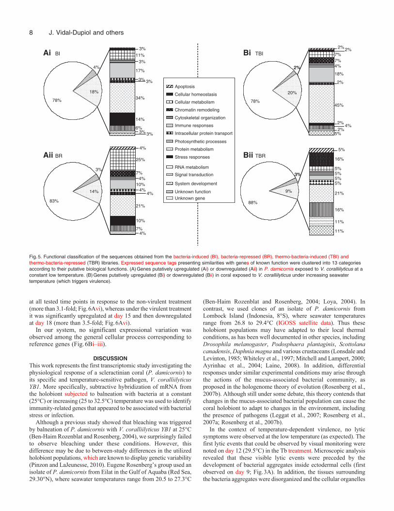
To confirm that the observed bacteria were not opportunisticbacteria of another species, we used qRT-PCR to quantify the levelsof the V. coralliilyticus 16S rRNA in the tissue samples. Vibriocoralliilyticus-specific primers were designed and used foramplification of cDNA, and the levels of V. coralliilyticus 16S rRNAwere compared to that of the P. damicornis 28S rRNA. V.coralliilyticus 16S rRNA corresponding amplicons were cloned and40 randomly picked clones were sequenced. All sequences obtained
corresponded to the V. coralliilyticus 16S rRNA. Consistent withthe above results, qRT-PCR showed that the relative amount of V.coralliilyticus 16S rRNA increased significantly by day 6 andremained higher than the control until the end of the protocol (Fig.4).By comparison, no such increase was observed in Cb group (Fig.4).This strongly suggests that coral tissue invasion occurs uponincreasing water temperature only.
EST sequencing, general features and functionalclassification of the BI, BR, TBI and TBR libraries
Two SSH experiments were performed to monitor the coral responseto bacterial challenge or infection. The first was conducted usingpooled samples (days 12 and 15) from the Cb treatment versus theC treatment, whereas the second was conducted using pooled samples(days 9–15) from the Tb treatment versus the T treatment. This yieldedfour cDNA libraries: the bacteria-induced (BI) and bacteria-repressed(BR) libraries, which corresponded to the subtractive hybridizationof Cb versus C, and the thermo-bacteria-induced (TBI) and thermo-bacteria-repressed (TBR) libraries, which corresponded to thesubtractive hybridization of Tb versus T. The ‘induced’ librariescontained transcripts putatively induced by bacterial stress or infectionwhereas the ‘repressed’ libraries contained gene putativelydownregulated under the experimental conditions.
The general features of each library are given in Table2. Threehundred and eighty-four clones were sequenced from each library,
Fig.1. Visual monitoring. Photos of Pocillopora damicornis nubbins taken at various time points during the experiment from treatments C (25°C withoutbacterial balneation), Cb (25°C with bacterial balneation), T (temperature increasing from 25 to 32.5°C without bacterial balneation) and Tb (temperatureincreasing from 25 to 32.5°C with bacterial balneation).
6
yielding 279, 276, 275 and 229 high-quality cDNA sequences(>100bp) from the BI, BR, TBI and TBR libraries, respectively(Table2). The ESTs from the BI library coalesced into 63 contigsand 123 singletons, suggesting that the overall redundancy of thelibrary was approximately 56%. The BR, TBI and TBR ESTsdistributed to 59, 38 and 23 contigs and 149, 184 and 166singletons, for overall redundancies of 46, 33 and 28%,respectively (Table2).
Singleton and consensus clusters were subjected to BLASTN andBLASTX searches and classified into three groups: unknown genes,genes of unknown function, and genes of known function (Fig.5).
Most of the sequences of known gene function presented similaritiesto predicted proteins from the genomes of Nematostella vectensis (anaposymbiotic cnidarian) and Branchiostoma floridae(Cephalochordata), both of which have recently been sequenced(Putnam et al., 2007; Putnam et al., 2008). The putative functions ofthese sequences were deduced by domain analysis using theInterProscan program. The identified genes clustered into thefollowing 13 functional categories: apoptosis, cellular homeostasis,cellular metabolism, chromatin remodeling, cytoskeletal organization,immune responses, intracellular protein transport, photosyntheticprocesses, protein metabolism, stress responses, RNA metabolism,
J. Vidal-Dupiol and others
Fig.2. Enlargement of photos of P. damicornis nubbinssubjected to (A) thermal bleaching (T treatment) or (B)bacterial lysis (Tb treatment). (A)Nubbins 3 days afterthe end of the T treatment (day 24). Water temperaturewas decreased to 25°C on day 21, which led the polypsto reopen. A bleached open polyp is shown (surroundedby a circle). Corallites (e.g. skeleton structure containinga polyp) are indicated by bold arrows. (B)Nubbins atday 20 after the Tb experiment, when coral tissues werequasi-totally lysed. Empty corallites are indicated byarrows. Asterisks mark a portion of bare skeleton;sharps indicates tissue rests undergoing lysis.
Fig.3. Electronmicroscopic monitoring.(A)Thin section of P.damicornis sampled fromthe Tb treatment on day9. (B)Thin section of P.damicornis sampled fromthe Tb treatment on day12. (C)Enlargement of B.(D)Enlargement of C. B,bacterial aggregate; b,bacteria; arrows, host cellmembrane; cross, tissuedisorganization; star,area of strong tissuedisorganization and lysis;open arrow, doublemembrane (bacterial cellwall and cytoplasmicmembrane). Scale bars:(A) 1m, (B) 500nm, (C)250nm, (D) 125nm.
7Coral response to vibriosis
signal transduction and system development (Fig.5, supplementarymaterial TableS1).
Expression patterns of genes putatively involved in immuneprocesses during Vibrio challenge
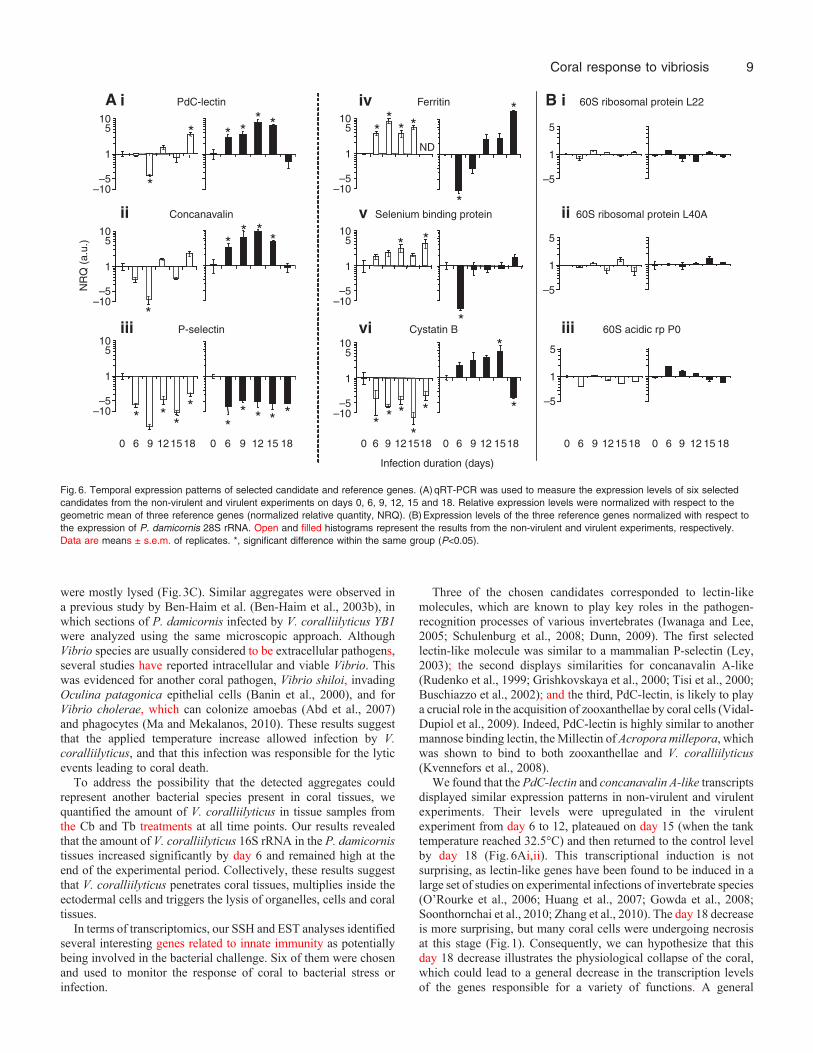
To investigate the response of coral to virulent and non-virulentbacterial challenges, five candidate genes were selected from theSSH libraries, and their transcription levels at various post-challengetime points were measured by qRT-PCR (Fig.6Aii–vi). Three wereselected because of their putative involvements in antibacterialresponses; these comprised a ferritin-like protein from the TBIlibrary (similar to Periserrula leucophryna ferritin; E2�10–66), aselenium-binding-protein-like protein from the BI library (similarto Mus musculus selenium binding protein; E2�10–25) and acystatin B-like protein from the BR library (similar to Danio reriocystatin B; E4�10–7). The other two selected genes, both lectin-like molecules, were selected based on the importance of lectins ininvertebrate immunity. They included a P-selectin-like protein fromthe TBR library (similar to Homo sapiens P-selectin; E6�10–28)and a concanavalin A lectin/glucanase-like protein from the TBIlibrary (similar to Synechococcus sp. thrombospondin N-terminal-like domain-containing protein; E2�10–9). These candidates wereclassified as lectins because the InterProscan analysis of theirtranslated amino-acid sequences revealed the presence of aconcanavalin A-like lectin/glucanase domain (InterProscanaccession no. SSF49899) and a P-selectin-like domain (InterProscanaccession no. PR00343), both of which contain carbohydrate-bindingsites. Following our recent discovery in a previous study of anotherlectin from P. damicornis, PdC-lectin (Vidal-Dupiol et al., 2009),we further investigated the transcript variation of this third lectinunder the same experimental conditions (Fig.6Ai). To verify thatthe observed variations were linked to the targeted physiologicaleffects and not to profound bacteria-induced disturbances, we alsoexamined the expression levels of three other genes implicated in
general cellular processes (P. damicornis: 60S ribosomal proteinL22, 60S ribosomal protein L40A and 60S acidic ribosomalphosphoprotein P0; GenBank accession nos HO112261, HO112283and HO112666, respectively) for comparison (Fig.6Bi–iii).
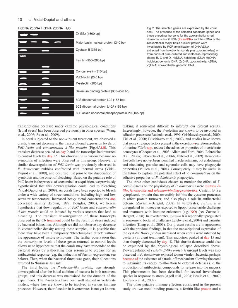
To verify that all of the selected candidates (including thereference genes) were expressed by the coral cells rather than theirsymbionts, we performed cross-PCR experiments using DNA andcDNA extracted from the holobiont (host plus symbiont) and frompure cultured representatives of Symbiodinium spp. clades B, C andD (Fig.7). The utilized primers included candidate-gene-specificoligonucleotides; primers ss5Z and ss3Z, which amplified theSymbiodinium spp. small ribosomal subunit (Rowan and Powers,1991); and primers specific for the cDNA of the MBNP ofSymbiodinium spp. The results revealed that the candidate genescould be amplified only from DNA and cDNA extracted fromholobionts (Fig.7), whereas the ss5Z/ss3Z and MBNP primersamplified all tested DNA and cDNA samples, respectively. Thisresult shows that all of the selected candidate genes were specificallyexpressed by coral cells. In addition, results of this experiment forthe genes encoding ferritin-like protein and the selenium-binding-protein-like protein show higher amplicon size when the matrix usedwas genomic DNA (Fig.7). This is probably due to the presence ofintronic sequences in both genes.
Two of the three selected lectins (concanavalin A-like proteinand PdC-lectin) displayed similar transcriptional profiles(Fig.6Ai,ii). Exposure to non-virulent bacteria triggered significantdownregulation of concanavalin A-like protein and PdC-lectin atday 9 (more than fourfold and eightfold for PdC-lectin andconcanavalin A-like protein, respectively), followed by upregulationat day 18 (only significant for PdC-lectin; 3.6-fold, P<0.05; Fig.6Ai).In contrast, samples exposed to the virulent conditions showedsignificant upregulation of both genes from day 6 (2.9- and 3.3-foldfor PdC-lectin and concanavalin A-like protein, respectively) to day15, with a dramatic decrease seen on day 18. The maximum increasewas observed on day 12 (7.7- and 9.3-fold for PdC-lectin andconcanavalin A-like protein, respectively). The remaining lectin, P-selectin-like protein, differed from the other two; its expressiondecreased at all tested time points by more than 3.2-fold during thetwo experiments (Fig.6Aiii).
The transcription patterns for ferritin-like protein and theselenium-binding-protein-like protein were very similar. In samplesexposed to the non-virulent treatment, significant upregulation wasobserved across most of the tested time points (Fig.6Aiv,v). Insamples exposed to the virulent treatment, expression of ferritin-like protein was strongly downregulated at day 6 (10.2-fold), andthen significantly upregulated at the end of the experiment (by morethan 16.1-fold on day 18; Fig.6Aiv). The gene encoding theselenium-binding-protein-like protein displayed a very similarprofile (Fig.6Av). Finally, cystatin B-like protein was downregulated
16S
rR
NA
/28S
rR
NA
rat
io (
a.u.
)
Infection duration (days)
9 12 15 180 3 6
1
10
100
–10 0 3 6 9 12 15 18–5
5
**
*
*
Fig.4. qRT-PCR quantitation of Vibrio coralliilyticus in P.damicornis tissues. The relative expression of V. coralliilyticus16S rRNA was normalized with respect to that of P. damicornis28S rRNA. Open and filled histograms correspond to the non-virulent and virulent experiments, respectively.
Table 2. General characteristics of the BI, BR, TBI and TBRlibraries
BI BR TBI TBR
Sequenced clone 384 384 384 384Analysed cDNA 279 276 275 229Mean EST size (bp) 335.9 332.1 367.3 302.7Contigs 63 59 38 23EST in contigs 156 127 91 63Singletons 123 149 184 166Redundancy* (%) 55.9 46.0 33.1 27.5
BI, bacteria-induced; BR, bacteria-repressed; EST, expressed sequence tag;TBI, thermo-bacteria-induced; TBR, thermo-bacteria-repressed.
*Redundancynumber of ESTs assembled in cluster/total EST.
8
at all tested time points in response to the non-virulent treatment(more than 3.1-fold; Fig.6Avi), whereas under the virulent treatmentit was significantly upregulated at day 15 and then downregulatedat day 18 (more than 3.5-fold; Fig.6Avi).
In our system, no significant expressional variation wasobserved among the general cellular process corresponding toreference genes (Fig.6Bi–iii).
DISCUSSIONThis work represents the first transcriptomic study investigating thephysiological response of a scleractinian coral (P. damicornis) toits specific and temperature-sensitive pathogen, V. coralliilyticusYB1. More specifically, subtractive hybridization of mRNA fromthe holobiont subjected to balneation with bacteria at a constant(25°C) or increasing (25 to 32.5°C) temperature was used to identifyimmunity-related genes that appeared to be associated with bacterialstress or infection.
Although a previous study showed that bleaching was triggeredby balneation of P. damicornis with V. coralliilyticus YB1 at 25°C(Ben-Haim Rozenblat and Rosenberg, 2004), we surprisingly failedto observe bleaching under these conditions. However, thisdifference may be due to between-study differences in the utilizedholobiont populations, which are known to display genetic variability(Pinzon and LaJeunesse, 2010). Eugene Rosenberg’s group used anisolate of P. damicornis from Eilat in the Gulf of Aquaba (Red Sea,29.30°N), where seawater temperatures range from 20.5 to 27.3°C
(Ben-Haim Rozenblat and Rosenberg, 2004; Loya, 2004). Incontrast, we used clones of an isolate of P. damicornis fromLombock Island (Indonesia, 8°S), where seawater temperaturesrange from 26.8 to 29.4°C (IGOSS satellite data). Thus theseholobiont populations may have adapted to their local thermalconditions, as has been well documented in other species, includingDrosophila melanogaster, Podosphaera plantaginis, Scottolanacanadensis, Daphnia magna and various crustaceans (Lonsdale andLevinton, 1985; Whiteley et al., 1997; Mitchell and Lampert, 2000;Ayrinhac et al., 2004; Laine, 2008). In addition, differentialresponses under similar experimental conditions may arise throughthe actions of the mucus-associated bacterial community, asproposed in the hologenome theory of evolution (Rosenberg et al.,2007b). Although still under some debate, this theory contends thatchanges in the mucus-associated bacterial population can cause thecoral holobiont to adapt to changes in the environment, includingthe presence of pathogens (Leggat et al., 2007; Rosenberg et al.,2007a; Rosenberg et al., 2007b).
In the context of temperature-dependent virulence, no lyticsymptoms were observed at the low temperature (as expected). Thefirst lytic events that could be observed by visual monitoring werenoted on day 12 (29.5°C) in the Tb treatment. Microscopic analysisrevealed that these visible lytic events were preceded by thedevelopment of bacterial aggregates inside ectodermal cells (firstobserved on day 9; Fig.3A). In addition, the tissues surroundingthe bacteria aggregates were disorganized and the cellular organelles
J. Vidal-Dupiol and others
2% 2%TBI
78%
20%
2%
88%
3%
9%
TBR
Bi
Bii
78%
18%
83%
14%
Apoptosis
Cellular homeostasis
Cellular metabolism
Chromatin remodeling
Cytoskeletal organization
Immune responses
Intracellular protein transport
Photosynthetic processes
Protein metabolism
Stress responses
System development
Unknown geneUnknown function
Signal transduction
RNA metabolism
78%
20%
2%
88%
3%
9%
78%
20%
7%7%4%
18%
2%
45%
2%4%
2%5%
78%
20%
88%
9%
88%
9%
5%
16%
5%
5%
21%
16%
11%
5%
5%
11%
78%
18%
83%
14%
78%
18%
78%
4%
18%
3%11%
17%
3%
34%
14%
6%3%
3%
3%
3%
83%
14%
83%
3%
14%
4%
25%
7%4%
21%
10%
7%4%
4%4%
10%
BI
BR
Ai
Aii
Fig.5. Functional classification of the sequences obtained from the bacteria-induced (BI), bacteria-repressed (BR), thermo-bacteria-induced (TBI) andthermo-bacteria-repressed (TBR) libraries. Expressed sequence tags presenting similarities with genes of known function were clustered into 13 categoriesaccording to their putative biological functions. (A)Genes putatively upregulated (Ai) or downregulated (Aii) in P. damicornis exposed to V. coralliilyticus at aconstant low temperature. (B)Genes putatively upregulated (Bi) or downregulated (Bii) in coral exposed to V. coralliilyticus under increasing seawatertemperature (which triggers virulence).
9Coral response to vibriosis
were mostly lysed (Fig.3C). Similar aggregates were observed ina previous study by Ben-Haim et al. (Ben-Haim et al., 2003b), inwhich sections of P. damicornis infected by V. coralliilyticus YB1were analyzed using the same microscopic approach. AlthoughVibrio species are usually considered to be extracellular pathogens,several studies have reported intracellular and viable Vibrio. Thiswas evidenced for another coral pathogen, Vibrio shiloi, invadingOculina patagonica epithelial cells (Banin et al., 2000), and forVibrio cholerae, which can colonize amoebas (Abd et al., 2007)and phagocytes (Ma and Mekalanos, 2010). These results suggestthat the applied temperature increase allowed infection by V.coralliilyticus, and that this infection was responsible for the lyticevents leading to coral death.
To address the possibility that the detected aggregates couldrepresent another bacterial species present in coral tissues, wequantified the amount of V. coralliilyticus in tissue samples fromthe Cb and Tb treatments at all time points. Our results revealedthat the amount of V. coralliilyticus 16S rRNA in the P. damicornistissues increased significantly by day 6 and remained high at theend of the experimental period. Collectively, these results suggestthat V. coralliilyticus penetrates coral tissues, multiplies inside theectodermal cells and triggers the lysis of organelles, cells and coraltissues.
In terms of transcriptomics, our SSH and EST analyses identifiedseveral interesting genes related to innate immunity as potentiallybeing involved in the bacterial challenge. Six of them were chosenand used to monitor the response of coral to bacterial stress orinfection.
Three of the chosen candidates corresponded to lectin-likemolecules, which are known to play key roles in the pathogen-recognition processes of various invertebrates (Iwanaga and Lee,2005; Schulenburg et al., 2008; Dunn, 2009). The first selectedlectin-like molecule was similar to a mammalian P-selectin (Ley,2003); the second displays similarities for concanavalin A-like(Rudenko et al., 1999; Grishkovskaya et al., 2000; Tisi et al., 2000;Buschiazzo et al., 2002); and the third, PdC-lectin, is likely to playa crucial role in the acquisition of zooxanthellae by coral cells (Vidal-Dupiol et al., 2009). Indeed, PdC-lectin is highly similar to anothermannose binding lectin, the Millectin of Acropora millepora, whichwas shown to bind to both zooxanthellae and V. coralliilyticus(Kvennefors et al., 2008).
We found that the PdC-lectin and concanavalin A-like transcriptsdisplayed similar expression patterns in non-virulent and virulentexperiments. Their levels were upregulated in the virulentexperiment from day 6 to 12, plateaued on day 15 (when the tanktemperature reached 32.5°C) and then returned to the control levelby day 18 (Fig.6Ai,ii). This transcriptional induction is notsurprising, as lectin-like genes have been found to be induced in alarge set of studies on experimental infections of invertebrate species(O’Rourke et al., 2006; Huang et al., 2007; Gowda et al., 2008;Soonthornchai et al., 2010; Zhang et al., 2010). The day 18 decreaseis more surprising, but many coral cells were undergoing necrosisat this stage (Fig.1). Consequently, we can hypothesize that thisday 18 decrease illustrates the physiological collapse of the coral,which could lead to a general decrease in the transcription levelsof the genes responsible for a variety of functions. A general
Infection duration (days)
NR
Q (
a.u.
)
Concanavalin
Ferritin
Cystatin B
Selenium binding protein
P-selectin
PdC-lectin
ii
iii
i
–10
1
105
–5 *
*
*–10
1
105
–5
* ** *
** *
* * *
* * * *
*
*
*
*
*
*
–10
1
105
–5
ND*
** *
1
5
–5
1
5
–5
1
5
–5
60S ribosomal protein L22
60S ribosomal protein L40A
60S acidic rp P0
BA
* ** * * **
**
0 6 9 121518 0 6 9 12 15 18
v
vi
iv
–10
1
105
–5
–10
1
105
–5
–10
1
105
–5
0 6 9 121518 0 6 9 12 1518
ii
iii
i
0 6 9 121518 0 6 9 12 15 18
Fig.6. Temporal expression patterns of selected candidate and reference genes. (A)qRT-PCR was used to measure the expression levels of six selectedcandidates from the non-virulent and virulent experiments on days 0, 6, 9, 12, 15 and 18. Relative expression levels were normalized with respect to thegeometric mean of three reference genes (normalized relative quantity, NRQ). (B)Expression levels of the three reference genes normalized with respect tothe expression of P. damicornis 28S rRNA. Open and filled histograms represent the results from the non-virulent and virulent experiments, respectively.Data are means ± s.e.m. of replicates. *, significant difference within the same group (P<0.05).
10
transcriptional decrease under extreme physiological conditions(lethal stress) has been observed previously in other species (Wanget al., 2006; Su et al., 2009).
In coral subjected to the non-virulent treatment, we observed adrastic transient decrease in the transcriptional expression levels ofPdC-lectin and concanavalin A-like protein (Fig.6Ai,ii). Thistransient decrease peaked on day 9 and the transcripts had returnedto control levels by day 12. This observation is curious because nosymptoms of infection were observed in this group. However, asimilar downregulation of PdC-lectin was previously observed inP. damicornis nubbins confronted with thermal stress (Vidal-Dupiol et al., 2009), and occurred just prior to the dissociation ofsymbiosis and the onset of bleaching. Based on the putative role ofPdC-lectin in the process of zooxanthellar acquisition, we previouslyhypothesized that this downregulation could lead to bleaching(Vidal-Dupiol et al., 2009). As corals have been reported to bleachunder a wide variety of stress conditions, including high and lowseawater temperature, increased heavy metal concentrations anddecreased salinity (Brown, 1997; Douglas, 2003), we hereinhypothesize that downregulation of PdC-lectin and concanavalinA-like protein could be induced by various stresses that lead tobleaching. The transient downregulation of these transcriptsobserved in the Cb treatment could be the result of stress inducedby bacterial balneation. Although we did not observe any decreasein zooxanthellar density among these samples, it is possible thatthere may have been a temporary ‘bleaching-like effect’ withoutthe appearance of visible symptoms. The further observation thatthe transcription levels of these genes returned to control levelsallows us to hypothesize that the corals may have responded to thebacterial stress by reallocating their resources to prepare for anantibacterial response (e.g. the induction of ferritin expression; seebelow). Then, when the bacterial threat was gone, their allocationsreturned to ‘business as usual’.
The third lectin, the P-selectin–like protein, was stronglydownregulated after the initial addition of bacteria in both treatmentgroups, and this decrease was maintained for the duration of theexperiments. The P-selectins have been well studied in vertebratemodels, where they are known to be involved in various immuneprocesses. However, their function in invertebrates is not yet known,
making it somewhat difficult to interpret our present results.Interestingly, however, the P-selectins are known to be involved inadhesion processes (Rudenko et al., 1999; Grishkovskaya et al., 2000;Tisi et al., 2000; Buschiazzo et al., 2002), and studies have shownthat some virulence factors present in the excretion–secretion productsof marine Vibrio spp. reduced the adhesive properties of invertebratehemocytes (Choquet et al., 2003; Allam and Ford, 2006; Labreucheet al., 2006a; Labreuche et al., 2006b; Mateo et al., 2009). Hemocyte-like cells have not yet been identified in scleractinians, but endodermaland circulating granular and agranular cells may have phagocyticproperties (Mullen et al., 2004). Consequently, it may be useful inthe future to explore the potential effect of V. coralliilyticus on theadhesive properties of P. damicornis phagocytes.
The three other candidates chosen to monitor the effect of V.coralliilyticus on the physiology of P. damicornis were cystatin B-like, ferritin-like and selenium-binding-protein-like. Cystatin B is acytoplasmic protein that reversibly inhibits the cysteine proteasesto affect protein turnover, and also plays a role in antibacterialdefense (Zavasnik-Bergant, 2008). In vertebrates, cystatin B isupregulated in monocytes exposed to lipopolysaccharide challengeand treatment with immune enhancers (e.g. NO) (see Zavasnik-Bergant, 2008). In invertebrates, cystatin B is reportedly upregulatedin response to bacterial challenge (Lefebvre et al., 2004) and parasiticinfection (Kang et al., 2006). Our present results are in agreementwith the previous findings, in that the transcriptional expression ofthe cystatin B-like protein increased when corals were infected bybacteria (virulent treatment). This induction peaked at day 15 andthen sharply decreased by day 18. This drastic decrease could alsobe explained by the physiological collapse described above.Downregulation of cystatin B-like protein transcript levels was alsoobserved in P. damicornis exposed to non-virulent bacteria, perhapsbecause of the existence of a trade-off mechanism allowing the coralto maximize its energy re-allocation to external defenses (i.e. theproduction of antibacterial compounds for release into the mucus).This phenomenon has been described for several invertebratespecies in response to stress (Agell et al., 2004; Brulle et al., 2007;Lourenco et al., 2009).
The other putative immune effectors considered in the presentstudy are two metal-binding proteins, a ferritin-like protein and a
J. Vidal-Dupiol and others
HgDNA ZgDNA HcDNA ZcDNA H2O
Cystatin B (355 bp)
Selenium binding protein (650–270 bp)
Zx SSz (1600 bp)
Ferritin (950–265 bp)
Concanavalin (310 bp)
P-selectin (205 bp)
PdC-lectin (240 bp)
Major basic nuclear protein (240 bp)
60S ribosomal protein L22 (155 bp)
60S ribosomal protein L40A (159 bp)
60S acidic ribosomal phosphoprotein P0 (165 bp)
Fig.7. The selected genes are expressed by the coralhost. The presence of the selected candidate genes andthose encoding the gene for the zooxanthellar smallribosomal subunit RNA (Zx ssRNA) and the cDNA of thezooxanthellar major basic nuclear protein wereinvestigated by PCR amplification of DNA/cDNAextracted from holobionts (corals plus zooxanthellae) orfrom pools of pure cultured zooxanthellae representingclades B, C and D. HcDNA, holobiont cDNA; HgDNA,holobiont genomic DNA; ZcDNA, zooxanthellar cDNA;ZgDNA, zooxanthellar genomic DNA.
11Coral response to vibriosis
selenium-binding-protein-like protein. Ferritins, which can sequesteriron, play dual roles in detoxification and iron storage (Arosio etal., 2009; Arosio and Levi, 2010), and the selenium-binding proteincovalently binds selenium (Bansal et al., 1989; Jeong et al., 2009).Interestingly, iron appears to be essential for bacterial growth(Doherty, 2007), especially for Vibrio species (Wright et al., 1981;Tolmasky and Crosa, 1991; Wyckoff et al., 2007), and selenium isa trace element crucial for the survival of all living organismsthrough the formation of selenocysteine, a modified amino acidlargely implicated in antioxidant defense (e.g. glutathioneperoxidase, thioredoxin reductase) (Burk et al., 2003; Stadtman,1996). Upregulation of ferritin and selenium-binding-protein byinfected host cells can trigger iron and selenium sequestration,thereby reducing the amount of these essential trace elementsnecessary for bacterial growth. Infection-induced iron and seleniumsequestration by ferritin and selenium-binding-protein have beenobserved in several other invertebrate–bacteria interactions (Becket al., 2002; Song et al., 2006; Altincicek et al., 2008; Li et al.,2008; Kong et al., 2010; Simão et al., 2010), and the crucial roleof ferritin in antibacterial defense was recently demonstrated in astudy showing that the injection of recombinant ferritin into Vibrioharveyi-infected shrimp (Peneaus monodon) increased the hostsurvival rate (Maiti et al., 2010).
Under non-virulent conditions, both ferritin-like and selenium-binding-protein-like were upregulated, perhaps reflecting thatbacteria have been detected and the coral is preparing its defensesby sequestering iron and selenium. Under our virulent experimentalconditions, the expression of ferritin-like and selenium-binding-protein-like corresponding genes was significantly downregulatedduring the early stage of infection, but upregulated near the end ofthe infection. This could illustrate that V. coralliilyticus is able toinhibit the intracellular immune response during the initial stagesof infection. A similar phenomenon was observed in abalonefollowing infection by V. harveyi (Travers et al., 2009). Theupregulation of ferritin-like and selenium-binding-protein-like geneslater in the process of infection could limit bacterial infection bysequestering iron and selenium. In addition, zooxanthellae underthermal stress have been shown to suffer photo-inhibition leadingto ROS over-production (Weis, 2008). Thus, the overexpression offerritin and selenium-binding-protein might also participate in ROSdetoxification. Nevertheless, this late increase seems insufficient toovercome the Vibrio infection process, at least under ourexperimental conditions.
In conclusion, the present study represents the first molecularexamination of the response evoked by a scleractinian coral whenconfronted by its pathogenic Vibrio (V. coralliilyticus). Several genesthat appeared to be involved in the coral immune response wereidentified, and their expression patterns were studied in coralsexposed to virulent and non-virulent bacteria. The results revealedclear differences for some of the candidate genes, which could proveuseful as functional biomarkers for measuring coral health andimmunity in monitoring programs and public aquariums. Futurestudies will be required to fully examine the molecular and cellularmechanisms involved in this process, but our study also suggestsseveral new hypotheses concerning the effect of V. coralliilyticuson the physiology of P. damicornis.
ACKNOWLEDGEMENTSThis work was supported by the Centre National pour la Recherche Scientifique(CNRS). O.L. is a PhD student of the National Fund for Scientific Research(FNRS-Fonds National de la Recherche Scientifique, Belgium). The authors areindebted to Marc Manetti for his help with the experimental procedures, CélineCosseau for many helpful discussions and Andrew Carroll for English
proofreading and helpful discussions. We thank Alain Pigno and Boris Rota fromthe Cap d’Agde Public Aquarium for their help with the project, and JérômeBossier for helping with the statistical analyses. We also thank the Department ofEnvironmental Sciences and Management (Prof. J. P. Thome) and the Dean ofthe Faculty of Sciences (Prof. J. M. Bouquegneau), both of the University of Liege,for financial support. Finally, the authors thank Mary-Alice Coffroth for allowing usto use cultures from the BURR Culture Collection.
REFERENCESAbd, H., Saeed, A., Weintraub, A., Nair, G. B. and Sandström, G. (2007). Vibrio
cholerae O1 strains are facultative intracellular bacteria, able to survive and multiplysymbiotically inside the aquatic free-living amoeba Acanthamoeba castellanii. FEMSMicrobiol. Ecol. 60, 33-39.
Agell, G., Turon, X., De Caralt, S., López-Legentil, S. and Uriz, M. J. (2004).Molecular and organism biomarkers of copper pollution in the ascidianPseudodistoma crucigaster. Mar. Pollut. Bull. 48, 759-767.
Allam, B. and Ford, S. E. (2006). Effects of the pathogenic Vibrio tapetis on defencefactors of susceptible and non-susceptible bivalve species: I. Haemocyte changesfollowing in vitro challenge. Fish Shellfish Immunol. 20, 374-383.
Altincicek, B., Knorr, E. and Vilcinskas, A. (2008). Beetle immunity: identification ofimmune-inducible genes from the model insect Tribolium castaneum. Dev. Comp.Immunol. 32, 585-595.
Arosio, P. and Levi, S. (2010). Cytosolic and mitochondrial ferritins in the regulationof cellular iron homeostasis and oxidative damage. Biochim. Biophys. Acta 1800,783-792.
Arosio, P., Ingrassia, R. and Cavadini, P. (2009). Ferritins: a family of molecules foriron storage, antioxidation and more. Biochim. Biophys. Acta 1790, 589-599.
Ayrinhac, A., Debat, V., Gibert, P., Kister, A.-G., Legout, H., Moreteau, B.,Vergilino, R. and David, J. R. (2004). Cold adaptation in geographical populationsof Drosophila melanogaster: phenotypic plasticity is more important than geneticvariability. Funct. Ecol. 18, 700-706.
Banin, E., Israely, T., Kushmaro, A., Loya, Y., Orr, E. and Rosenberg, E. (2000).Penetration of the coral-bleaching bacterium Vibrio shiloi into Oculina patagonica.Appl. Environ. Microbiol. 66, 3031-3036.
Banin, E., Sanjay, K. H., Naider, F. and Rosenberg, E. (2001). A proline rich peptidefrom the coral pathogen Vibrio shiloi that inhibits photosynthesis of zooxanthellae.Appl. Environ. Microbiol. 67, 1536-1541.
Bansal, M. P., Oborn, C. J., Danielson, K. G. and Medina, D. (1989). Evidence fortwo selenium-binding proteins distinct from glutathione peroxidase in mouse liver.Carcinogenesis 10, 541-546.
Beck, G., Ellis, T. W., Habicht, G. S., Schluter, S. F. and Marchalonis, J. J. (2002).Evolution of the acute phase response: iron release by echinoderm (Asterias forbesi)coelomocytes, and cloning of an echinoderm ferritin molecule. Dev. Comp. Immunol.26, 11-26.
Ben-Haim, Y. and Rosenberg, E. (2002). A novel Vibrio sp. pathogen of the coralPocillopora damicornis. Mar. Biol. 141, 47-55.
Ben-Haim, Y., Thompson, F. L., Thompson, C. C., Cnockaert, M. C., Hoste, B.,Swings, J. and Rosenberg, E. (2003a). Vibrio coralliilyticus sp. nov., a temperature-dependent pathogen of the coral Pocillopora damicornis. Int. J. Syst. Evol. Microbiol.53, 309-315.
Ben-Haim, Y., Zicherman-Keren, M. and Rosenberg, E. (2003b). Temperature-regulated bleaching and lysis of the coral Pocillopora damicornis by the novelpathogen Vibrio coralliilyticus. Appl. Environ. Microbiol. 69, 4236-4242.
Ben-Haim Rozenblat, Y. and Rosenberg, E. (2004). Temperature-regulatedbleaching and tissue lysis of Pocillopora damicornis by the novel pathogen Vibriocoralliilyticus. In Coral Health and Disease (ed. E. Rosenberg and Y. Loya), pp. 301-324. New-York: Spinger-Verlag.
Bourne, D. G., Garren, M., Work, T. M., Rosenberg, E., Smith, G. W. and Harvell,C. D. (2009). Microbial disease and the coral holobiont. Trends Microbiol. 17, 554-562.
Brown, B. E. (1997). Coral bleaching: causes and consequences. Coral Reefs 16,S129-S138.
Brulle, F., Mitta, G., Leroux, R., Lemière, S., Leprêtre, A. and Vandenbulcke, F.(2007). The strong induction of metallothionein gene following cadmium exposuretransiently affects the expression of many genes in Eisenia fetida: a trade-offmechanism? Comp. Biochem. Physiol. 144C, 334-341.
Burk, R. F., Hill, K. E. and Motley, A. K. (2003). Selenoprotein metabolism andfunction: evidence for more than one function for selenoprotein P. J. Nutr. 133,1517-1520.
Buschiazzo, A., Amaya, M. F., Cremona, M. L., Frasch, A. C. and Alzari, P. M.(2002). The crystal structure and mode of action of trans-sialidase, a key enzyme inTrypanosoma cruzi pathogenesis. Mol. Cell 10, 757-768.
Cervino, J. M., Hayes, R. L., Polson, S. W., Polson, S. C., Goreau, T. J., Martinez,R. J. and Smith, G. W. (2004). Relationship of Vibrio species infection and elevatedtemperatures to yellow blotch/band disease in Caribbean corals. Appl. Environ.Microbiol. 70, 6855-6864.
Cervino, J. M., Thompson, F. L., Gomez-Gil, B., Lorence, E. A., Goreau, T. J.,Hayes, M. L., Winiarski-Cervino, K. B., Smith, D. J., Hughen, K. and Bartels, E.(2008). The Vibrio core group induces yellow band disease in Caribbean and Indo-Pacific reef-building corals. J. Appl. Microbiol. 105, 1658-1671.
Choquet, G., Soudant, P., Lambert, C., Nicolas, J.-L. and Paillard, C. (2003).Reduction of adhesion properties of Ruditapes philippinarum hemocytes exposed toVibrio tapetis. Dis. Aquat. Org. 57, 109-116.
Doherty, C. P. (2007). Host-pathogen interactions: the role of iron. J. Nutr. 137, 1341-1344.
Donner, S. D., Skirving, W. J., Little, C. M., Oppenheimer, M. and Hoegh-Guldberg, O. V. E. (2005). Global assessment of coral bleaching and required ratesof adaptation under climate change. Glob. Change Biol. 11, 2251-2265.
Douglas, A. E. (2003). Coral bleaching-how and why? Mar. Pollut. Bull. 46, 385-392.
12
Dunn, S. R. (2009). Immunorecognition and immunoreceptors in the Cnidaria.Invertebrate Surviv. J. 6, 7-14.
Gates, R. D., Baghdasarian, G. and Muscatine, L. (1992). Temperature stresscaused host cell detachent in symbiotic cnidarians: implications for coral bleaching.Biol. Bull. 182, 324-332.
Gowda, N. M., Goswami, U. and Islam Khan, M. (2008). T-antigen binding lectin withantibacterial activity from marine invertebrate, sea cucumber (Holothuria scabra):possible involvement in differential recognition of bacteria. J. Invertebr. Pathol. 99,141-145.
Grishkovskaya, I., Avvakumov, G. V., Sklenar, G., Dales, D., Hammond, G. L. andMuller, Y. A. (2000). Crystal structure of human sex hormone-binding globulin:steroid transport by a laminin G-like domain. EMBO J. 19, 504-512.
Grubbs, F. E. (1969). Procedures for detecting outlying observations in samples.Technometrics 11, 1-21.
Harvell, D., Jordán-Dahlgren, E., Merkel, S., Rosenberg, E., Raymundo, L., Smith,G., Weil, E. and Willis, B. (2007). Coral disease, environmental drivers, and thebalance between coral and microbial associates. Oceanography 20, 172-195.
Hashimoto, K., Shibuno, T., Murayama-Kayano, E., Tanaka, H. and Kayano, T.(2004). Isolation and characterization of stress-responsive genes from thescleractinian coral Pocillopora damicornis. Coral Reefs 23, 485-491.
Hellemans, J., Mortier, G., De Paepe, A., Speleman, F. and Vandesompele, J.(2007). qBase relative quantification framework and software for management andautomated analysis of real-time quantitative PCR data. Genome Biol. 8, R19.
Hoegh-Guldberg, O., Mumby, P. J., Hooten, A. J., Steneck, R. S., Greenfield, P.,Gomez, E., Harvell, C. D., Sale, P. F., Edwards, A. J., Caldeira, K. et al. (2007).Coral reefs under rapid climate change and ocean acidification. Science 318, 1737-1742.
Huang, G., Liu, H., Han, Y., Fan, L., Zhang, Q., Liu, J., Yu, X., Zhang, L., Chen, S.,Dong, M. et al. (2007). Profile of acute immune response in Chinese amphioxusupon Staphylococcus aureus and Vibrio parahaemolyticus infection. Dev. Comp.Immunol. 31, 1013-1023.
Hughes, T., Baird, A., Bellwood, D., Card, M., Connolly, S., Folke, C., Grosberg,R., Guldberg, H., Jackson, J., Kleypas, J. et al. (2003). Climate change, humanimpacts, and the resilience of coral reefs. Science 301, 929-933.
IGOSS (2010). IGOSS time series SST, from 1981 to 2002.Iwanaga, S. and Lee, B. R. (2005). Recent advances in the innate immunity of
invertebrate animals. J. Biochem. Mol. Biol. 38, 128-150.Jeong, J.-Y., Wang, Y. and Sytkowski, A. J. (2009). Human selenium binding
protein-1 (hSP56) interacts with VDU1 in a selenium-dependent manner. Biochem.Biophys. Res. Commun. 379, 583-588.
Johannes, R. E. and Wiebe, W. J. (1970). Method for determination of coral tissuebiomass and composition. Limnol. Oceanogr. 15, 822-824.
Kang, Y.-S., Kim, Y.-M., Park, K.-I., Kim Cho, S., Choi, K.-S. and Cho, M. (2006).Analysis of EST and lectin expressions in hemocytes of Manila clams (Ruditapesphilippinarum) (Bivalvia: Mollusca) infected with Perkinsus olseni. Dev. Comp.Immunol. 30, 1119-1131.
Kong, P., Wang, L., Zhang, H., Zhou, Z., Qiu, L., Gai, Y. and Song, L. (2010). Twonovel secreted ferritins involved in immune defense of Chinese mitten crab Eriocheirsinensis. Fish Shellfish Immunol. 28, 604-612.
Kushmaro, A., Loya, Y., Fine, M. and Rosenberg, E. (1996). Bacterial infection andbleaching. Nature 380, 396.
Kushmaro, A., Rosenberg, E., Fine, M. and Loya, Y. (1997). Bleaching of the coralOculina patagonica by Vibrio AK-1. Mar. Ecol. Prog. Ser. 147, 159-165.
Kushmaro, A., Rosenberg, E., Fine, M., Ben Haim, H. and Loya, Y. (1998). Effect oftemperature on bleaching of the coral Oculina patagonica by Vibrio AK-1. Mar. Ecol.Prog. Ser. 171, 131-137.
Kushmaro, A., Banin, E., Loya, Y., Stackebrandt, E. and Rosenberg, E. (2001).Vibrio shiloi sp. nov., the causative agent of bleaching of the coral Oculinapatagonica. Int. J. Syst. Evol. Microbiol. 51, 1383-1388.
Kvennefors, E. C. E., Leggat, W., Hoegh-Guldberg, O., Degnan, B. M. and Barnes,A. C. (2008). An ancient and variable mannose-binding lectin from the coralAcropora millepora binds both pathogens and symbionts. Dev. Comp. Immunol. 32,1582-1592.
Labreuche, Y., Lambert, C., Soudant, P., Boulo, V., Huvet, A. and Nicolas, J.-L.(2006a). Cellular and molecular hemocyte responses of the Pacific oyster,Crassostrea gigas, following bacterial infection with Vibrio aestuarianus strain 01/32.Microbes Infect. 8, 2715-2724.
Labreuche, Y., Soudant, P., Gonçalves, M., Lambert, C. and Nicolas, J.-L. (2006b).Effects of extracellular products from the pathogenic Vibrio aestuarianus strain 01/32on lethality and cellular immune responses of the oyster Crassostrea gigas. Dev.Comp. Immunol. 30, 367-379.
Ladrière, O., Compère, P., Decloux, N., Vandewalle, P. and Poulicek, M. (2008).Morphological alterations of zooxanthellae in bleached cnidarian hosts. Cahiers Biol.Mar. 49, 215-227.
Laine, A.-L. (2008). Temperature-mediated patterns of local adaptation in a naturalplant pathogen metapopulation. Ecol. Lett. 11, 327-337.
Lefebvre, C., Cocquerelle, C., Vandenbulcke, F., Hot, D., Huot, L., Lemoine, Y.and Salzet, M. (2004). Transcriptomic analysis in the leech Theromyzon tessulatum:involvement of cystatin B in innate immunity. Biochem. J. 380, 617-625.
Leggat, W., Ainsworth, T., Bythell, J., Dove, S., Gates, R., Hoegh-Guldberg, O.,Iglesias-Prieto, R. and Yellowlees, D. (2007). The hologenome theory disregardsthe coral holobiont. Nat. Rev. Microbiol. 5. doi:10.1038/nrmicro1635-c1
Lesser, M. P. (2004). Experimental biology of coral reef ecosystems. J. Exp. Mar. Biol.Ecol. 300, 217-252.
Ley, K. (2003). The role of selectins in inflammation and disease. Trends Mol. Med. 9,263-268.
Li, M., Saren, G. and Zhang, S. (2008). Identification and expression of a ferritinhomolog in amphioxus Branchiostoma belcheri: evidence for its dual role in immuneresponse and iron metabolism. Comp. Biochem. Physiol. 150B, 263-270.
Lonsdale, D. J. and Levinton, J. S. (1985). Latitudinal differentiation in copepodgrowth: an adaptation to temperature. Ecology 66, 1397-1407.
Lourenco, A. P., Martins, J. R., Bitondi, M. M. G. and Simoes, Z. L. P. (2009).Trade-off between immune stimulation and expression of storage protein genesArch. Insect Biochem. Physiol. 71, 70-87.
Loya, Y. (2004). The coral reefs of Eilat-past, present and future: three decades ofcoral comunity structure studies. In Coral Health and Disease (ed. E. Rosenberg andY. Loya), pp. 1-29. Berlin, Heidelberg: Spinger-Verlag.
Luna, G. M., Biavasco, F. and Danovaro, R. (2007). Bacteria associated with therapid tissue necrosis of stony corals. Environ. Microbiol. 9, 1851-1857.
Ma, A. T. and Mekalanos, J. J. (2010). In vivo actin cross-linking induced by Vibriocholerae type VI secretion system is associated with intestinal inflammation. Proc.Natl. Acad. Sci. USA 107, 4365-4370.
Maiti, B., Khushiramani, R., Tyagi, A., Karunasagar, I. and Karunasagar, I. (2010).Recombinant ferritin protein protects Penaeus monodon infected by pathogenicVibrio harveyi. Dis. Aquat. Org. 88, 99-105.
Mateo, D. R., Siah, A., Araya, M. T., Berthe, F. C. J., Johnson, G. R. andGreenwood, S. J. (2009). Differential in vivo response of soft-shell clam hemocytesagainst two strains of Vibrio splendidus: changes in cell structure, numbers andadherence. J. Invertebr. Pathol. 102, 50-56.
Mitchell, S. E. and Lampert, W. (2000). Temperature adaptation in a geographicallywidespread zooplankter, Daphnia magna. J. Evol. Biol. 13, 371-382.
Mullen, K. M., Peters, E. C. and Harvell, C. D. (2004). Coral resistance to disease. InCoral Health and Disease (ed. E. Rosenberg and Y. Loya), pp. 377-399. Berlin,Heidelberg: Spinger-Verlag.
O’Rourke, D., Baban, D., Demidova, M., Mott, R. and Hodgkin, J. (2006). Genomicclusters, putative pathogen recognition molecules, and antimicrobial genes areinduced by infection of C. elegans with M. nematophilum. Genome Res. 16, 1005-1016.
Pinzon, H. and LaJeunesse, T. C. (2010). Species delimitation of common reef coralsin the genus Pocillopora using nucleotide sequence phylogenies, population geneticsand symbiosis ecology. Mol. Ecol. 20, 311-325.
Putnam, N. H., Srivastava, M., Hellsten, U., Dirks, B., Chapman, J., Salamov, A.,Terry, A., Shapiro, H., Lindquist, E., Kapitonov, V. V. et al. (2007). Sea anemonegenome reveals ancestral eumetazoan gene repertoire and genomic organization.Science 317, 86-94.
Putnam, N. H., Butts, T., Ferrier, D. E. K., Furlong, R. F., Hellsten, U., Kawashima,T., Robinson-Rechavi, M., Shoguchi, E., Terry, A., Yu, J.-K. et al. (2008). Theamphioxus genome and the evolution of the chordate karyotype. Nature 453, 1064-1071.
Rodriguez-Lanetty, M., Harii, S. and Hoegh-Guldberg, O. (2009). Early molecularresponses of coral larvae to hyperthermal stress. Mol. Ecol. 18, 5101-5114.
Rosenberg, E. (2004). The bacterial disease hypothesis of coral bleaching. In CoralHealth and Disease (ed. E. Rosenberg and Y. Loya), pp. 377-399. Berlin,Heidelberg: Spinger-Verlag.
Rosenberg, E., Koren, O., Reshef, L., Efrony, R. and Zilber-Rosenberg, I. (2007a).The hologenome theory disregards the coral holobiont: reply from Rosenberg et al.Nat. Rev. Microbiol. 5, doi:10.1038/nrmicro1635-c2
Rosenberg, E., Koren, O., Reshef, L., Efrony, R. and Zilber-Rosenberg, I. (2007b).The role of microorganisms in coral health, disease and evolution. Nat. Rev.Microbiol. 5, 355-362.
Rowan, R. and Powers, D. A. (1991). Molecular genetic identification of symbioticsdinoflagellates (zooxanthellae). Mar. Ecol. Prog. Ser. 71, 65-73.
Rudenko, G., Nguyen, T., Chelliah, Y., Südhof, T. C. and Deisenhofer, J. (1999).Regulation of LNS domain function by alternative splicing: the structure of the ligand-binding domain of Neurexin I . Cell 99, 93-101.
Schulenburg, H., Hoeppner, M. P., Weiner, J. R. and Bornberg-Bauer, E. (2008).Specificity of the innate immune system and diversity of C-type lectin domain (CTLD)proteins in the nematode Caenorhabditis elegans. Immunobiology 213, 237-250.
Simão, M., Leite, R., Rocha, C. and Cancela, M. (2010). Changes in bioturbation ofiron biogeochemistry and in molecular response of the clam Ruditapes decussatesupon Perkinsus olseni infection. Arch. Environ. Contam. Toxicol. 59, 433-443.
Song, L., Zou, H., Chang, Y., Xu, W. and Wu, L. (2006). The cDNA cloning andmRNA expression of a potential selenium-binding protein gene in the scallopChlamys farreri. Dev. Comp. Immunol. 30, 265-273.
Soonthornchai, W., Rungrassamee, W., Karoonuthaisiri, N., Jarayabhand, P.,Klinbunga, S., Söderhäll, K. and Jiravanichpaisal, P. (2010). Expression ofimmune-related genes in the digestive organ of shrimp, Penaeus monodon, after anoral infection by Vibrio harveyi. Dev. Comp. Immunol. 34, 19-28.
Stadtman, T. C. (1996). Selenocysteine. Annu. Rev. Biochem. 65, 83-100.Stefansky, W. (1972). Rejecting outliers in factorial designs. Technometrics 14, 469-
479.Stimson, J. and Kinzie, R. A., III (1991). The temporal pattern and rate of release of
zooxanthellae from the reef coral Pocillopora damicornis (Linnaeus) under nitrogen-enrichment and control conditions. J. Exp. Mar. Biol. Ecol. 153, 63-74.
Stimson, J., Sakai, K. and Sembali, H. (2002). Interspecific comparison of thesymbiotic relationship in corals with high and low rates of bleaching-inducedmortality. Coral Reefs 21, 409-421.
Su, J., Yang, C., Xiong, F., Wang, Y. and Zhu, Z. (2009). Toll-like receptor 4signaling pathway can be triggered by grass carp reovirus and Aeromonashydrophila infection in rare minnow Gobiocypris rarus. Fish Shellfish Immunol. 27,33-39.
Sussman, M., Willis, B. L., Victor, S. and Bourne, D. G. (2008). Coral pathogensidentified for white syndrome (WS) epizootics in the Indo-Pacific. Plos ONE 3, e2393.
Sutherland, K. P., Porter, J. W. and Torres, C. (2004). Disease and immunity inCaribbean and Indo-Pacific zooxanthellate corals. Mar. Ecol. Prog. Ser. 266, 273-302.
Tisi, D., Talts, J. F., Timpl, R. and Hohenester, E. (2000). Structure of the C-terminallaminin G-like domain pair of the laminin 2 chain harbouring binding sites for -dystroglycan and heparin. EMBO J. 19, 1432-1440.
J. Vidal-Dupiol and others
13Coral response to vibriosis
Tolmasky, M. E. and Crosa, J. H. (1991). Regulation of plasmid-mediated irontransport and virulence in Vibrio anguillarum. Biometals 4, 33-35.
Travers, M. A., Le Bouffant, R., Friedman, C. S., Buzin, F., Cougard, B., Huchette,S., Koken, M. and Paillard, C. (2009). Pathogenic Vibrio harveyi in contrast to non-pathogenic strains, intervenes with the p38 MAPK pathway to avoid an abalonehaemocyte immune response. J. Cell. Biochem. 106, 152-160.
Veron, J. E. N. (2000). Corals of the World. Townsville, Australia: Australian Instituteof Marine Science.
Vidal-Dupiol, J., Adjeroud, M., Roger, E., Foure, L., Duval, D., Mone, Y., Ferrier-Pages, C., Tambutte, E., Tambutte, S., Zoccola, D. et al. (2009). Coral bleachingunder thermal stress: putative involvement of host/symbiont recognition mechanisms.BMC Physiol. 9, 14.
Wang, B., Li, F., Dong, B., Zhang, X., Zhang, C. and Xiang, J. (2006). Discovery ofthe genes in response to White Spot Syndrome Virus (WSSV) infection inFenneropenaeus chinensis through cDNA microarray. Mar. Biotechnol. 8, 491-500.
Ward, J. R. and Lafferty, K. D. (2004). The elusive baseline of marine disease: arediseases in ocean ecosystems increasing? PLoS Biol. 2, e120.
Ward, J. R., Kim, K. and Harvell, C. D. (2007). Temperature affects coral diseaseresistance and pathogen growth. Mar. Ecol. Prog. Ser. 329, 115-121.
Weil, E., Smith, G. W. and Gil-Agudelo, D. L. (2006). Status and progress in coralreef disease research. Dis. Aquat. Org. 69, 1-7.
Weis, V. M. (2008). Cellular mechanisms of Cnidarian bleaching: stress causes thecollapse of symbiosis. J. Exp. Biol. 211, 3059-3066.
Whiteley, N. M., Taylor, E. W. and El Haj, A. J. (1997). Seasonal and latitudinaladaptation to temperature in crustaceans. J. Therm. Biol. 22, 419-427.
Wilkinson, C. (ed.) (2008). Status of Coral Reefs of the World, 296 pp. Townsville,Australia: Global Coral Reef Monitoring Network and Reef and Rainforest ResearchCenter.
Willis, B. L., Page, C. A. and Dinsdale, E. A. (2004). Coral diseases on the greatbarrier reef. In Coral Health and Disease (ed. E. Rosenberg and Y. Loya), pp. 69-104. New York: Springer-Verlag.
Wright, A. C., Simpson, L. M. and Oliver, J. D. (1981). Role of iron in thepathogenesis of Vibrio vulnificus infections. Infect. Immun. 34, 503-507.
Wyckoff, E., Mey, A. and Payne, S. (2007). Iron acquisition in Vibrio cholerae.Biometals 20, 405-416.
Yuan, J. S., Reed, A., Chen, F. and Stewart, C. N., Jr (2006). Statistical analysis ofreal-time PCR data. BMC Bioinformatics 7, 85-97.
Zavasnik-Bergant, T. (2008). Cystatin protease inhibitors and immune functions.Front. Biosci. 13, 4625-4637.
Zhang, H., Kong, P., Wang, L., Zhou, Z., Yang, J., Zhang, Y., Qiu, L. and Song, L.(2010). Cflec-5, a pattern recognition receptor in scallop Chlamys farreriagglutinating yeast Pichia pastoris. Fish Shellfish Immunol. 29, 149-156.


