Embed Size (px)
Citation preview

Europ.J.Hort.Sci., 79 (6). S. 327–334, 2014, ISSN 1611-4426. © Verlag Eugen Ulmer KG, Stuttgart
Physiological Characteristics and Pathogenicity of eight Neofabraea Isolates from Apples in Czechia
B. Hortova1),2), D. Novotny1) and T. Erban1)
(1)Crop Research Institute, Praha, Czech Republic and 2)Czech University of Life Sciences Prague, Faculty ofAgrobiology, Food and Natural Resources, Praha, Czech Republic)
E
Summary
We obtained seven isolates of Neofabraea alba and oneof N. perennans from apples in Czechia. All of theNeofabraea isolates showed variable growth on 2.0 %malt-extract agar (MEA) over a wide range of pHs,from 3.0 to 10.0, and pH 2.0 suppressed growth. Thetemperature optimum was observed at 18 and 20 °C;growth was suppressed at 30 °C. Five isolates exhib-ited optimal growth at 20 and three at 18 °C. Isolategrowth was similarly low at 25 and 5 °C. The mostfavorable medium for the growth of the Neofabraeaisolates was oatmeal agar (OA). On 2.0 % MEA, pota-to-dextrose agar (PDA) and OA isolates formed fungus
colonies with a mostly regular margin and abundantaerial mycelium. Fungus-submerged mycelium formedon potato-carrot agar and 0.1 % MEA with a twig ofapple tree with a few aerial hyphae in the middle ofcolonies. In general, cv. ‘Rubin’ was the most suscepti-ble and cv. ‘Sampion’ the most resistant apple fruit tothe Neofabraea isolates. Cv. ‘Rubinola’ was more sus-ceptible than cv. ‘Rubin’ to Neofabraea infection onbranches and the formation of cankers. These resultsare valuable to further mycological and pathologicalresearch on the fungus and disease, including the de-velopment of measures for disease management.
Key words. Neofabraea alba – Neofabraea perennans – apple – bull's-eye rot – pathogen
Introduction
One of the major causes of the postharvest decay ofapples is bull's-eye rot caused by species of Neofabraea(Dermateaceae, Helotiales), and the postharvest losses offruits can reach more than 40 % (SHOLBERG and HAAG 1996;SPOTTS et al. 1999). The genus Neofabraea includes six de-scribed species, including N. alba (E. J. Guthrie) Verkley(anamorph: Phlyctema vagabunda Desm.), N. eucalyptiCheewangkoon & Crous (anamorph not known), N.krawtzewii (Petr.) Verkley, also known as N. populi (G. E.Thompson) (anamorph: Cryptosporiopsis sp.), N. mali-corticis H. S. Jacks. (anamorph: C. curvispora (Peck)),N. perennans Kienholz (anamorph: C. perennans (Zellerand Childs) Wollenw.) and Neofabraea sp. (anamorph:C. kienholzii Seifert, Spotts and Levesque). While N. euca-lypti and N. krawtzewii do not infect pome fruits, the re-maining four described species, N. alba, N. malicorticis,N. perennans and C. kienholzii, cause bull's-eye rot of ap-ples (VERKLEY 1999; CHEEWANGKOON et al. 2009; SPOTTS etal. 2009).
Neofabraea species, especially N. perennans and N. mal-icorticis, can produce perennial canker and anthracnoseon the branches and twigs of apple trees. N. alba is con-
sidered a weaker parasite of apple tree branches; it cre-ates smaller cankers than N. perennans and lives as asaprophyte on the dead bark of fruit trees (BOMPEIX andBONDOUX 1974; VERKLEY 1999). Perennial cankers serve asthe source of the inoculum for apple fruit infection. Fruit canbecome infected anytime between petal fall and harvest,and disease symptoms appear several months after harvest(generally after 3–4 months in cold storage) (SPOTTS 1990).Cultivars cultivated in Czechia differ in their suscepti-bility to storage disease and branch cankers (BLAZEK
2004; BLAZEK et al. 2006; KRUCZYNSKA and RUTKOWSKI
2006). Commercial apple production in Czechia is stilldominated by well-known cultivars, such as ‘Golden Deli-cious’, ‘Rubin’ and ‘Sampion’, but over the past ten years,a large increase of resistant cultivars, such as ‘Rubinola’and ‘Topaz’ (BUCHTOVA 2013), have appeared.
The physiological characteristics of the fungi as wellas the influence of culture media are crucial for under-standing their growth on substrates, such as apple fruitsor twigs (KIM et al. 2005). Moreover, the colonial mor-phology of the genus Neofabraea on different media isincompletely known (VERKLEY 1999). Among the majorphysiological factors affecting the ecology of spoilagefungi are the pH of the medium and the temperature.
urop.J.Hort.Sci. 6/2014
328 Hortova et al.: Physiological Characteristics and Pathogenicity of Neofabraea
pH can affect fungal growth directly by its action onthe cell surfaces or indirectly by its effect on the avail-ability of nutrients (PARDO et al. 2006; ABUBAKAR et al.2013).
The objective of this study was to isolate Neofabraeaspecies from apples and to provide information on variousenvironmental factors, including the temperature, pH andeffects of the culture media on mycelial growth. More-over, we tested the pathogenicity of eight Neofabraea iso-lates. This information will be valuable to further myco-logical and pathological research on the fungus and itsdisease, including the development of measures fordisease management.
Materials and Methods
Collection and storage of isolates
Three apple cultivars with bull's-eye rot symptoms, ‘GoldenDelicious’, ‘Jonagold’, and ‘Melrose’, were used in thestudy. The apples originated from the Research andBreeding Institute of Pomology at Holovousy in Czechia.Direct tissue isolations were obtained from fruit that hadbeen surface sterilized in 96 % ethanol and further rinsedwith sterile distilled water. The skin of a decayed applewas removed and a small section from the rotting fleshwas plated on 2.0 % malt-extract agar (MEA; Cat. No.70145, Sigma-Aldrich, St. Louis, USA). Plates were incu-bated at 20 °C for 5 d, after which the Neofabraea-like cul-tures were transferred to fresh 2.0 % MEA. The isolates havebeen deposited in the Collection of Phytopathogenic Fungi(CPPF), Crop Research Institute in Prague in Czechia.
Identification of Neofabraea species
Eight isolates of Neofabraea have been identified basedon genetic analysis of the β-tubulin gene. Total genomicDNA was extracted from pure Neofabraea cultures usingan Ultra Clean Microbial DNA Isolation Kit (Cat. No.12224, MoBio Laboratories, Carlsbad, USA) accordingto the manufacturer's instructions. Amplification of DNAwas performed using species-specific primers accordingto the protocol of GARIEPY et al. (2003). The PCR reactionwas conducted in a thermocycler from MJ ResearchPTC 200 (GMI, Ramsey, USA) and contained 0.2 mMdNTPs, 2.5 mM MgCl2, 0.4 μM of Neofab-upTub-100,0.4 μM of the appropriate species-specific reverse primer,and 1 U Taq DNA polymerase (Cat. No. M1241, Promega,Madison, USA) in a 10x PCR buffer (Cat. No. M1241,Promega). PCR products were run on 2 % agarose gels(Cat No. 50005, Seakem Cambrex, Walkersville, USA),stained with ethidiumbromide, and visualized by a302 nm UV transilluminationator. Band sizes from thePCR reaction were estimated on the basis of a 100 bpDNA Ladder marker (Cat. No. G2101, Promega). Usedpositive controls were: N. alba – CBS 102871, N. peren-
nans – CBS 453.64 (CBS – Centraalbureau voor Schim-melcultures).
Physiological characterization of isolates
Species of the genus Neofabraea were observed undervarious culture conditions. The macromorphological fea-tures (radial growth, morphology and colour of the colo-nies) of isolates were evaluated. MEA (2.0 %) was pre-pared and adjusted to different pH levels in the range of2.0 and 10.0 with 0.1 M NaOH or 0.1 M HCl using a pHmeter from Mettler Toledo S20 SevenEasy™ pH (Mettler-Toledo, Ohio, USA). The inoculum was a 5 mM diameterdisc transferred from a colony onto 2.0 % MEA. Theplates were incubated at 20 °C in the dark. The effect ofdifferent temperatures on mycelial growth was evaluatedon 2.0 % MEA. The inoculum was a 5 mM diameter disctransferred from a colony onto 2.0 % MEA (Cat. No.70145, Sigma-Aldrich). The inoculated plates were incu-bated at 5, 10, 15, 18, 20, 25, 30, 35, 40 and 45 °C. Theeffect of different culture media was determined on 2.0 %MEA, potato-dextrose agar (Cat. No. M096, PDA, HiMedia,Mumbai, India), potato-carrot agar (PCA, Cat. No. M696,HiMedia), oatmeal agar (OA; Cat. No. M397, HiMedia)and 0.1 % malt-extract agar (MEA; Sigma-Aldrich, Cat.No. 70145) with an apple tree twig. Inoculations were in-cubated at 20 °C in the dark. The inoculum was a 5 mMdiameter disc transferred from a colony onto 2.0 % MEA.Colony diameters were measured after 21 d of cultiva-tion and colony colours were classified according to theMethuen handbook of colour (KORNERUP and WANSCHER
1978). Colony photographs were taken with a digitalcamera (Olympus 510, Tokyo, Japan). Colony diameterswere measured along two axes perpendicular to one an-other at 7, 14 and 21 d after inoculations, and five repli-cates were maintained in each treatment for pH, temper-ature and culture media.
Apple fruit inoculation for testing of pathogenicity of Neofabraea species
For testing pathogenicity we used the apple cultivars ‘Gol-den Delicious’, ‘Rubin’, ‘Rubinola’, ‘Florina’ and ‘Sampion’harvested at commercial maturity by Pomona (Tesetice,Czechia) and maintained at 4 °C. The apples were used forexperiments within 15 d of harvesting. Only undamagedand disease-free fruits were used in the experiments. Inoc-ulation tests were performed according to the proceduresdescribed by GUTHRIE (1959), with slight modification.Apples were first rinsed in sterile ddH2O. A 5 mM diame-ter mycelial plug from the margin of a 21-day-old cultureon 2.0 % MEA was transferred under the fruit cuticle, andthe wound was wrapped with Parafilm (Cat. No. P7793,Sigma-Aldrich). Inoculated fruits were maintained incold storage at 4 °C, and the size of the lesions was ratedafter 5 weeks. Control inoculations were performed bythe same method using a sterile plug of 2.0 % MEA.
Europ.J.Hort.Sci. 6/2014

Hortova et al.: Physiological Characteristics and Pathogenicity of Neofabraea 329
Apple branch inoculation for testing of pathogenicity of Neofabraea species
Two cultivars, ‘Rubin’ and ‘Rubinola’, were selected. In-oculations were performed in May. Inoculation testswere performed according to the procedures describedby HENRIQUEZ et al. (2006), with slight modification. Thesection of the branch was first surface disinfected with96 % ethanol. A 5 mM diameter mycelial plug from themargin of a 21-day-old culture on 2.0 % MEA wasplaced under a V-shaped cut made into the wood to thedepth of the secondary xylem. Control inoculationswere performed by the same method using a sterile plugof 2.0 % MEA. After inoculation, the wounds werewrapped with Parafilm, which was removed 1 monthlater. Canker progression was observed after 2 and 6months.
Statistical analyses
The data were analysed using a nested analysis of vari-ance with pH, temperature, culture media and cultivarsas main groups and isolates as subgroups with randomeffects, the group means were compared by Tukey’s HSDtest. All analyses were performed with XLStat2006(Addinsoft, New York, USA).
Results
Identification of isolates
Eight Neofabraea isolates were obtained from 27 selectedapples with significant postharvest decay symptoms. DNAfragments of 358 and 300 bp were obtained for N. albaand N. perennans, respectively. Molecular identificationrevealed that seven out of eight isolates were Neofabraeaalba (denoted as Na1 – Na7), and one species was identifiedas N. perenans (denoted as Np8). The identification fromapples was as follows: Na1 and Na7 – ‘Jonagold’, Na2 –‘Melrose’, Na3, Na4, Na5, Na6 and Np8 – ‘Golden Delicious’.
Evaluation of physiological data
Diameters were measured after 7, 14 and 21 d; for pres-entation, the data were selected from observations after21 d. The remaining data after 7 and 14 d showed asimilar trend to 21 d, but the data are not shown.
Effect of pH
The seven N.alba isolates and one isolate N. perennansshowed growth on 2.0 % MEA over a wide range ofpHs, from 3.0 to 10.0 (Table 1). The results of optimal pH
Table 1. Effect of pH and temperature on the mycelial growth of Neofabraea isolates.
Fungal Isolates
Na1 Na2 Na3 Na4 Na5 Na6 Na7 Np8 Mean
pH2 0.0 0.0 0.0 0.0 0.0 0.0 0.0 0.0 0.0
3 31.4 ± 5.0 f 25.6 ± 1.6 b 28.5 ± 1.3 c 22.2 ± 1.5 a 25.3 ± 1.3 d 25.7 ± 1.4 a 21.4 ± 0.9 a 28.0 ± 1.1 e 26.0 B
4 53.0 ± 3.2 f 30.6 ± 3.4 b 33.2 ± 3.5 c 22.1 ± 1.6 a 27.0 ± 4.1 d 24.5 ± 1.9 a 22.4 ± 1.5 a 32.8 ± 1.2 e 30.7 CD
5 46.4 ± 4.6 f 21.1 ± 1.0 b 33.5 ± 1.7 c 20.9 ± 1.2 a 38.7 ± 1.1 d 21.6 ± 1.9 a 23.7 ± 0.9 a 36.9 ± 3.9 e 30.4 CD
6 56.4 ± 1.1 f 26.2 ± 2.7 b 31.3 ± 2.3 c 18.7 ± 1.2 a 40.3 ± 0.8 d 21.6 ± 1.9 a 21.4 ± 2.2 a 37.6 ± 7.2 e 31.7 D
7 35.9 ± 2.9 f 30.7 ± 6.1 b 37.2 ± 10.5 c 18.3 ± 0.6 a 35.3 ± 1.2 d 18.3 ± 2.2 a 16.8 ± 0.6 a 40.8 ± 2.5 e 29.2 C
8 37.3 ± 1.6 f 32.9 ± 2.0 b 21.8 ± 3.2 c 16.7 ± 1.3 a 27.1 ± 1.6 d 15.8 ± 0.6 a 14.7 ± 1.4 a 43.3 ± 2.7 e 26.2 B
9 26.7 ± 1.7 f 19.5 ± 3.6 b 23.9 ± 1.5 c 14.8 ± 1.2 a 32.4 ± 1.4 d 14.2 ± 2.5 a 14.2 ± 2.5 a 38.7 ± 4.0 e 23.1 A
10 30.2 ± 2.7 f 21.4 ± 0.9 b 25.3 ± 2.6 c 13.6 ± 0.9 a 35.8 ± 1.0 d 13.1 ± 1.0 a 13.0 ± 0.9 a 36.6 ± 0.9 e 23.6 A
Temperature5 °C 14.9 ± 2.1 f 16.4 ± 2.1 cd 14.5 ± 1.5 bc 15.0 ± 0.8 a 11.5 ± 0.9 e 18.1 ± 1.0 ab 15.6 ± 0.9bc 13.3 ± 0.8 d 14.9 B
10 °C 36.8 ± 3.5 f 19.7 ± 0.9 cd 24.4 ± 0.9 bc 20.0 ± 1.2 a 26.7 ± 2.2 e 20.7 ± 0.9 ab 22.7 ± 1.4bc 27.0 ± 7.0 d 24.8 D
15 °C 20.7 ± 1.7 f 25.6 ± 3.0 cd 16.7 ± 1.5 bc 19.4 ± 0.8 a 18.8 ± 0.9 e 19.7 ± 1.0 ab 22.8 ± 0.9bc 22.8 ± 0.8 d 20.8 C
18 °C 51.5 ± 2.0 f 35.0 ± 2.1 cd 25.6 ± 1.5 bc 28.7 ± 1.7 a 51.0 ± 3.1 e 30.6 ± 1.4 ab 32.1 ± 2.0bc 44.6 ± 3.5 d 37.4 E
20 °C 54.3 ± 1.7 f 35.7 ± 1.4 cd 43.6 ± 3.3 bc 32.9 ± 1.9 a 48.3 ± 2.5 e 29.5 ± 1.0 ab 34.8 ± 1.5bc 41.5 ± 5.0 d 40.1 F
25 °C 21.9 ± 2.9 f 13.4 ± 1.7 cd 9.7 ± 3.1 bc 8.0 ± 3.0 a 14.0 ± 2.0 e 14.0 ± 2.4 ab 7.8 ± 2.4bc 7.3 ± 2.7 d 12.0 A
30 °C 0.0 0.0 0.0 0.0 0.0 0.0 0.0 0.0 0.0
Colony diameter (mm) was assessed after 21 d. Data represent the mean of five replicates ± standard deviation. For each attribute, the mean values with the same lowercase letters among isolates in the same row are not significantly different at the 5 % level of proba-bility (Tukey HSD test); those with the same uppercase letters among the pH values in the same column are not significantly different.
Europ.J.Hort.Sci. 6/2014
330 Hortova et al.: Physiological Characteristics and Pathogenicity of Neofabraea
screening indicated that studied Neofabraea isolates haveextremely broad optimum on 2.0 % MEA, with the excep-tion of the most acidic pH of 2.0 – the only pH tested atwhich mycelium was unable to grow for all Neofabraeaisolates. Nested ANOVA analysis (F63,256 = 50; P < 0.0001)showed significant effects from both independent varia-bles (fungal isolates: F = 305; P < 0.0001; pH: F = 46;P < 0.0001) and their interaction (F = 135; P < 0.0001)on the mycelial growth of Neofabraea isolates. The radialgrowth of isolates was very variable for each pH.
Effect of temperature
The rate of mycelial growth of the N. alba and N. peren-nans isolates followed similar trends in response tochanges in temperature (Table 1); however, the differ-ence in the growth rate of the isolates at different tem-peratures was statistically significant. The nested ANOVAmodel (F47,192 = 109; P < 0.0001) showed significant in-fluences from both independent variables (fungal isolates:F = 78; P < 0,0001; temperature: F = 778; P < 0.0001)and their interaction (F = 20; P < 0.0001) on the mycelialgrowth of N. alba and N. perennans isolates. The temper-ature optima were 18 and 20 °C. Of eight tested isolates,five showed the highest rate at 20 °C (isolates: Na1, Na2,Na3, Na4 and Na7). Isolates Na5, Na6 and species Np8showed the highest growth rate at 18 °C. The slowest ra-dial growth was generally observed at 25 °C, but isolatesNa1 and Na5 showed the slowest growth rate at 5 °C.21 d after incubation, the lowest growth rate (7.3 mM)was recorded for species Np8, whereas isolate Na1 showedthe highest growth rate (54.3 mM) at 20 °C. Tempera-tures 30 °C and higher suppressed growth totally and allthe isolates were presumably dead.
Effect of different culture media
The radial mycelial growth rates of the N. alba andN. perennans isolates were significantly affected by theculture media. Nested ANOVA (F39,200 = 75; P < 0,0001)showed significant effects from both independent variables(fungal isolates: F = 155; P < 0,0001; medium: F = 275;
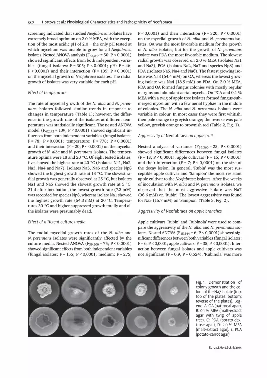
P < 0,0001) and their interaction (F = 320; P < 0,0001)on the mycelial growth of N. alba and N. perennans iso-lates. OA was the most favorable medium for the growthof N. alba isolates, but for the growth of N. perennansisolate was PDA the most favorable medium. The slowestradial growth was observed on 2.0 % MEA (isolates Na1and Na3), PCA (isolates Na2, Na7 and species Np8) andPDA (isolates Na5, Na4 and Na6). The fastest growing iso-late was Na3 (64.4 mM) on OA, whereas the lowest grow-ing isolate was Na4 (18.9 mM) on PDA. On 2.0 % MEA,PDA and OA formed fungus colonies with mostly regularmargins and abundant aerial mycelia. On PCA and 0.1 %MEA with a twig of apple tree isolates formed fungus-sub-merged mycelium with a few aerial hyphae in the middleof colonies. The N. alba and N. perennans isolates werevariable in colour. In most cases they were first whitish,then pale orange to greyish orange; the reverse was paleyellow, greyish orange to brownish red (Table 2, Fig. 1).
Aggressivity of Neofabraea on apple fruit
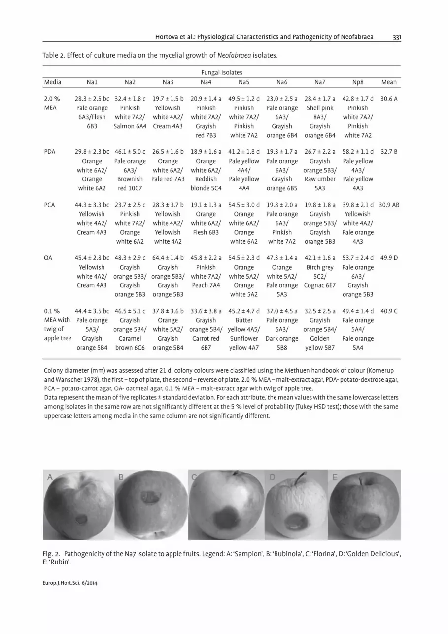
Nested analysis of variance (F39,360 = 25, P < 0,0001)showed significant differences between fungal isolates(F = 18; P < 0,0001), apple cultivars (F = 16; P < 0,0001)and their interaction (F = 7; P < 0,0001) on the size ofthe decay lesion. In general, ‘Rubin’ was the most sus-ceptible apple cultivar and ‘Sampion’ the most resistantapple cultivar to the Neofabraea isolates. After five weeksof inoculation with N. alba and N. perennans isolates, weobserved that the most aggressive isolate was Na7(36.6 mM) on ‘Rubin’. The lowest aggressivity was foundfor Na5 (15.7 mM) on ‘Sampion’ (Table 3, Fig. 2).
Aggressivity of Neofabraea on apple branches
Apple cultivars ‘Rubin’ and ‘Rubinola’ were used to com-pare the aggressivity of the N. alba and N. perennans iso-lates. Nested ANOVA (F15,144 = 6; P < 0,0001) showed sig-nificant differences between both variables (fungal isolates:F = 6, P < 0,0001; apple cultivars: F = 35; P < 0,0001). Inter-action between fungal isolates and apple cultivars wasnot significant (F = 0,9, P = 0,524). ‘Rubinola’ was more
Fig. 1. Demonstration ofcolony growth and the co-lour of the Na7 isolate (top:top of the plates; bottom:reverse of the plates). Leg-end: A: OA (oat-meal agar),B: 0.1 % MEA (malt-extractagar with twig of appletree), C: PDA (potato-dex-trose agar), D: 2.0 % MEA(malt-extract agar), E: PCA(potato-carrot agar).
Europ.J.Hort.Sci. 6/2014
Hortova et al.: Physiological Characteristics and Pathogenicity of Neofabraea 331
Table 2. Effect of culture media on the mycelial growth of Neofabraea isolates.
Fungal Isolates
Media Na1 Na2 Na3 Na4 Na5 Na6 Na7 Np8 Mean
2.0 % MEA
28.3 ± 2.5 bc 32.4 ± 1.8 c 19.7 ± 1.5 b 20.9 ± 1.4 a 49.5 ± 1.2 d 23.0 ± 2.5 a 28.4 ± 1.7 a 42.8 ± 1.7 d 30.6 A
Pale orange 6A3/Flesh
6B3
Pinkish white 7A2/Salmon 6A4
Yellowish white 4A2/Cream 4A3
Pinkish white 7A2/
Grayish red 7B3
Pinkish white 7A2/
Pinkish white 7A2
Pale orange6A3/
Grayish orange 6B4
Shell pink 8A3/
Grayish orange 6B4
Pinkish white 7A2/
Pinkish white 7A2
PDA 29.8 ± 2.3 bc 46.1 ± 5.0 c 26.5 ± 1.6 b 18.9 ± 1.6 a 41.2 ± 1.8 d 19.3 ± 1.7 a 26.7 ± 2.2 a 58.2 ± 1.1 d 32.7 B
Orange white 6A2/
Orange white 6A2
Pale orange 6A3/
Brownish red 10C7
Orange white 6A2/
Pale red 7A3
Orange white 6A2/
Reddish blonde 5C4
Pale yellow 4A4/
Pale yellow 4A4
Pale orange 6A3/
Grayish orange 6B5
Grayish orange 5B3/Raw umber
5A3
Pale yellow 4A3/
Pale yellow 4A3
PCA 44.3 ± 3.3 bc 23.7 ± 2.5 c 28.3 ± 3.7 b 19.1 ± 1.3 a 54.5 ± 3.0 d 19.8 ± 2.0 a 19.8 ± 1.8 a 39.8 ± 2.1 d 30.9 AB
Yellowish white 4A2/Cream 4A3
Pinkish white 7A2/
Orange white 6A2
Yellowish white 4A2/Yellowish white 4A2
Orange white 6A2/Flesh 6B3
Orange white 6A2/
Orange white 6A2
Pale orange6A3/
Pinkish white 7A2
Grayish orange 5B3/
Grayish orange 5B3
Yellowish white 4A2/Pale orange
4A3
OA 45.4 ± 2.8 bc 48.3 ± 2.9 c 64.4 ± 1.4 b 45.8 ± 2.2 a 54.5 ± 2.3 d 47.3 ± 1.4 a 42.1 ± 1.6 a 53.7 ± 2.4 d 49.9 D
Yellowish white 4A2/Cream 4A3
Grayish orange 5B3/
Grayish orange 5B3
Grayish orange 5B3/
Grayish orange 5B3
Pinkish white 7A2/Peach 7A4
Orange white 5A2/
Orange white 5A2
Orange white 5A2/Pale orange
5A3
Birch grey 5C2/
Cognac 6E7
Pale orange 6A3/
Grayish orange 5B3
0.1 % MEA with twig of apple tree
44.4 ± 3.5 bc 46.5 ± 5.1 c 37.8 ± 3.6 b 33.6 ± 3.8 a 45.2 ± 4.7 d 37.0 ± 4.5 a 32.5 ± 2.5 a 49.4 ± 1.4 d 40.9 C
Pale orange 5A3/
Grayish orange 5B4
Grayish orange 5B4/
Caramel brown 6C6
Orange white 5A2/
Grayish orange 5B4
Grayish orange 5B4/
Carrot red 6B7
Butter yellow 4A5/Sunflower yellow 4A7
Pale orange5A3/
Dark orange 5B8
Grayish orange 5B4/
Golden yellow 5B7
Pale orange 5A4/
Pale orange 5A4
Colony diameter (mm) was assessed after 21 d, colony colours were classified using the Methuen handbook of colour (Kornerup and Wanscher 1978), the first – top of plate, the second – reverse of plate. 2.0 % MEA – malt-extract agar, PDA- potato-dextrose agar, PCA – potato-carrot agar, OA- oatmeal agar, 0.1 % MEA – malt-extract agar with twig of apple tree.Data represent the mean of five replicates ± standard deviation. For each attribute, the mean values with the same lowercase letters among isolates in the same row are not significantly different at the 5 % level of probability (Tukey HSD test); those with the same uppercase letters among media in the same column are not significantly different.
Fig. 2. Pathogenicity of the Na7 isolate to apple fruits. Legend: A: ‘Sampion’, B: ‘Rubinola’, C: ‘Florina’, D: ‘Golden Delicious’,E: ‘Rubin’.
Europ.J.Hort.Sci. 6/2014

332 Hortova et al.: Physiological Characteristics and Pathogenicity of Neofabraea
susceptible than ‘Rubin’ to Neofabraea infection on branchesand the formation of cankers. Six months after inocula-tion, the most aggressive isolate was Na7 (17.6 mM) on‘Rubinola’, whereas the least aggressive isolates were Na1and Na2 (11.4 mM) on ‘Rubin’ (Table 4, Fig. 3).
Discussion
In this study, all seven isolates of N. alba and one isolateof N. perennans were able to grow over the pH range of 3to 10; pH 10 was the most basic pH tested; however, the
isolates were not able to grow at pH 2. Our results areconsistent with earlier reports for N. malicorticis andN. perennans (MILLER 1932); for N. alba and N. malicor-ticis (SENULA 1985) and for Sphareopsis pyriputrescens(KIM et al. 2005). The most acidic pH at which phytopath-ogenic fungi of fruit are able to grow is generally aroundpH 3.0, as was found for N. malicorticis and N. perennans(MILLER 1932) and N. alba and N. malicorticis (SENULA
1985), and pH 3.3 for Sphareopsis pyriputrescens (KIM etal. 2005). However, N. malicorticis and N. perennans wereable grow at a very basic pH up to 11.8; pH 12.0 sup-pressed growth totally (MILLER 1932). The optimal radial
Table 3. Lesion diameters (mm) in infected fruit inoculated with Neofabraea isolates.
Applecultivars
Fungal Isolates
Na1 Na2 Na3 Na4 Na5 Na6 Na7 Np8 Mean
‘Golden Delicious’
30.3 ± 9.8 b 25.7 ± 9.2 b 30.0 ± 3.7 c 29.3 ± 4.4 c 19.1 ± 2.3 a 28.7 ± 7.3 c 30.4 ± 4.0 d 20.0 ± 2.6 ab 26.7 B
‘Florina’ 19.2 ± 1.9 b 20.2 ± 2.0 b 35.1 ± 6.0 c 27.8 ± 2.3 c 16.2 ± 1.2 a 30.0 ± 5.2 c 35.4 ± 4.3 d 16.3 ± 1.2 a 25.0 B
‘Rubin’ 23.2 ± 3.3 b 30.4 ± 2.0 b 29.5 ± 4.4 c 30.9 ± 2.9 c 20.7 ± 2.5 a 34.2 ± 1.8 c 36.6 ± 3.1 d 28.3 ± 3.0 ab 29.2 C
‘Rubinola’ 19.1 ± 2.6 b 18.6 ± 0.6 b 20.1 ± 2.5 c 21.9 ± 5.7 c 18.5 ± 1.3 a 24.4 ± 1.5 c 24.5 ± 3.2 d 18.7 ± 1.1 ab 20.7 A
‘Sampion’ 16.0 ± 1.8 b 15.8 ± 0.9 b 22.0 ± 1.7 c 24.2 ± 2.7 c 15.7 ± 0.8 a 21.3 ± 3.5 c 24.7 ± 3.4 d 15.8 ± 1.3 ab 19.4 A
Lesion diameters (mm) were assessed after 5 weeks of incubation at 4 °C. Data represent the mean of ten replicates ± standard deviation. For each attribute, the mean values with the same lowercase letters among isolates in the same row are not significantly different at the 5 % level of probability (Tukey HSD test); those with the same uppercase letters among apple cultivars in the same column are not significantly different.
Table 4. Length of cankers (mm) induced by Neofabraea isolates on branches of apple trees.
Applecultivars
Fungal Isolates
Na1 Na2 Na3 Na4 Na5 Na6 Na7 Np8 Mean
‘Rubin’ 11.4 ± 1.0 ab 11.4 ± 1.3 a 14.8 ± 0.5 c 14.7 ± 1.6 c 14.9 ± 1.2 c 14.9 ± 1.0 c 14.6 ± 2.2 c 14.8 ± 1.8 bc 13.9 A
‘Rubinola’ 14.7 ± 5.5 ab 14.6 ± 2.5 a 16.3 ± 2.1 c 16.6 ± 1.7 c 15.9 ± 1.1 c 17.1 ± 2.3 c 17.6 ± 2.4 c 15.7 ± 2.1 bc 16.0 B
Length of cankers (mm) was assessed after 6 month of inoculation. Data represent the mean of ten replicates ± standard deviation. For each attribute, the mean values with the same lowercase letters among isolates in the same row are not significantly different at the 5 % level of probability (Tukey HSD test); those with the same uppercase letters among apple cultivars in the same column are not significantly different.
Fig. 3. Pathogenicity of the Na7 isolate to apple tree branches. Legend: A: ‘Rubinola’, B: ‘Rubin’.
Europ.J.Hort.Sci. 6/2014

Hortova et al.: Physiological Characteristics and Pathogenicity of Neofabraea 333
growth of Neofabraea isolates was very variable for eachpH value. The fastest radial growth of N. alba isolates wasin a range of pH 3.0 to 6.0, except for isolate Na2, whichshowed the highest growth activity at pH 8.0 but also hadincreased growth at pH 4.0. N. perennans seems to differfrom N. alba isolates because its optimal pH was pH 8.0.It can be generalized that the optimal range for thegrowth of Neofabraea fungi on apple fruit is at low tomid-acidic pH.
The optimum temperature for the growth of Neo-fabraea isolates was over the range of 18 °C to 20 °C.Earlier temperature optima were found to be 18–20 °Cand 18–22 °C for N. alba and N. malicorticis, respectively(SENULA 1985). MILLER (1932) monitored the colonygrowth of N. perennans and N. malicorticis from 0 °C to15 °C. High growth activity was recorded at 15 °C butboth N. perennans and N. malicorticis were capable ofgrowth at 0 °C (MILLER 1932). KIM et al. (2005) studiedSphaeropsis rot of apple and pear and stated that animportant characteristic for a fungus that is a postharvestpathogen on apple is its ability to grow at fruit storagetemperatures that are typically in the range of –1 to 4 °C.Interestingly, the fungal isolates we tested showed similargrowth at 5 and 25 °C when 25 °C was the highest tem-perature at which the isolates were able to grow. ThatNeofabraea could grow at 5 °C supports the statementthat bull's-eye rot caused by this Neofabraea isolate can-not be prevented by the low temperatures used to storeapples. Future studies can test the growth of the isolatesin temperatures from –1 to 5 °C in detail to confirm ear-lier results by KIM et al. (2005) and MILLER (1932).
All the tested isolates exhibited the fastest growthactivity on OA. Studies on the growth of N. alba on AEA(apple extract agar), Czapek agar, MEA and PDA revealedthat the most suitable medium was PDA (BORECKA andBRYK 1982); in contrast, we found that PDA was the leastsuitable medium for isolates Na5, Na4 and Na6. A varia-tion in the colonial morphology of N. malicorticis wasdocumented by VERKLEY (1999), who tested OA, 2.0 %MEA and CHA (cherry agar) and found that the bestmedium for the growth of N. malicorticis is OA. The mostsuitable medium for the growth of N. perennans is V8-agar(BOGO et al. 2008). Neofabraea produce highly colouredmycelia, which differ on various media (KIENHOLZ 1939).N. alba is characterized by cream- to cinnamon-colouredcolonies, which reverse with a reddish tinge on V8-agarand are saffron to orange on MEA, CHA and CMA (VERKLEY
1999). N. perennans creates white to orange-pink andbrown colonies (KIENHOLZ 1939). BOGO et al. (2008) de-scribed N. perennans colonies with gray-green to slightlypink colours and observed a major variability in colour onthe reverse of the colonies; the reverse ranged from red-dish yellow to red purple colours. A similar variability inthe coloration of the N. alba colonies was also noticedby BORECKA and BRYK (1982); isolates of N. alba formedcolonies that were white-cream, beige or beige-pink. Itis evident that Neofabraea isolates are variable in both
colony coloration and growth activity on different culti-vation media and between different isolates.
The most frequent occurrence of bull's-eye rot causedby N. alba on apple fruit is due to the saprophytic growthof the fungus on the apple trees. Low pathogenicity andthe formation of small cankers that did not produce sporeswere observed (CORKE 1956). We found that N. alba iso-lates were pathogenic to branches of apple trees similarto N. perennans, as did HENRIQUEZ et al. (2006), who ex-amined the induction of cankers by N. alba and N. peren-nans on pear and apple trees. We also observed differ-ences between apple cultivars. ‘Rubinola’ apple scab-re-sistant cultivar was more susceptible to the induction ofcankers by Neofabraea isolates than ‘Rubin’. Other applescab-resistant cultivars, ‘Topaz’ and ‘Freedom’, werefound to be more susceptible to N. alba and N. malicorticiscankers (BRYK and KRUCZYNSKA 2005). Cultivation of scab-resistant apples is important in terms of economy, ecologyand toxicology; resistant cultivars are therefore currentlypreferred in apple orchards (BRYK and KRUCZYNSKA 2005;KRUCZYNSKA and RUTKOWSKI 2006). We suggest that theresistant cultivars are susceptible to Neofabraea cankers.Thus it is necessary to keep the danger of these pathogensin mind.
Isolates of N. alba manifest large differences in theirpathogenicity; some isolates are capable of attacking onlyapple fruits, whereas others attack only the branches ofapple trees (BORECKA and BRYK 1982). All Neofabraea iso-lates were pathogenic to apple fruits, and differencesbetween the isolates were found. The most aggressive iso-late was Na7, whereas the least aggressive were Na5 andNp8. Results similar to ours were observed by MILLER
(1932), who examined bull's-eye rot caused by N. peren-nans and N. malicorticis, both of which are pathogens ofapple fruits. Moreover he determined that a wide varia-bility exists between isolates. SOTO-ALVEAR et al. (2013)studied the occurrence and pathogenicity of N. alba inChile. They found that this species is dominant but thedifferences in lesion size were not statistically significant.We found significant differences between cultivars. ‘Rubin’was the most sensitive to inoculation with Neofabraeaisolates, and this cultivar is generally considered to bevery susceptible to storage diseases (BLAZEK et al. 2006).A relatively resistant cultivar to bull's-eye rot was ‘Rubi-nola’. This finding is consistent with BRYK and KRUCZYNSKA
(2005), who examined postharvest disease on applesresistant to scab. ‘Golden Delicious’ showed increasedresistance to this disease (SPOTTS et al. 1999), but in ourstudy, it exhibited only medium resistance to bull's eye rot.
Acknowledgments
This research was supported by the Ministry of Agri-culture of the Czech Republic (http://eagri.cz) via theGrant No. RO0414. The authors thank Radmila Repovaand Martin Markovic for the valuable assistance.
Europ.J.Hort.Sci. 6/2014

334 Hortova et al.: Physiological Characteristics and Pathogenicity of Neofabraea
References
ABUBAKAR, A., H.A. SUBERU, I.M. BELLO, R. ABDULKADIR, O.A.DAUDU and A.A. LATEEF 2013: Effect of pH on mycelialgrowth and sporulation of Aspergillus parasiticus. J.Plant. Sci. 1, 64–67.
BLAZEK, J. 2004: Response to diseases in new apple culti-vars from the Czech Republic. J. Fruit. Ornam. Plant.Res. 12, 241–250.
BLAZEK, J., J. KLOUTVOROVA and J. KRELINOVA 2006: Inci-dence of storage diseases on apples of selected cultivarsand advanced selections grown with and without fun-gicide treatments. Hort. Sci. 33, 87–94.
BOGO, A., M.A. MAFFIOLETTI, R.M.V. SANHUEZA and R.T.CASA 2008: Caracterizacao morfologica de isolados deCryptosporiopsis perennans em diferentes meios de cul-tura. Trop. Plant. Pathol. 33, 248–251 (in Portugues).
BOMPEIX, G. and P. BONDOUX 1974: Etude experimentale dupouvoir pathogene du Pezicula alba Guthrie et du Peziculamalicorticis (Jack.) Nann. sur rammeaux du pommier.Ann. Phytopathol. 6, 1–11 (in French).
BORECKA, H. and H. BRYK 1982: Porownanie patogenicz-nosci, wzrostu, zarodnikowania i cech morfologicz-nych roznych izolatow grzyba Pezicula alba Guthrie<Gloeosporium album Osterw.>. Acta. Agrobot. 35, 225–234 (in Polish).
BRYK, H. and D. KRUCZYNSKA 2005: Wystepowanie chorobprzechowalniczych na jablkach odmian parchoodpor-nych. Acta. Agrobot. 58, 205–212 (in Polish).
BUCHTOVA, I. 2013: Situacni a vyhledova zprava – ovoce:rijen 2013. Ministry of Agriculture of the Czech Re-public, Prague, Czechia (In Czech).
CHEEWANGKOON, R., J.Z. GROENEWALD, B.A. SUMMERELL, K.D.HYDE, C. TO-ANUN and P.W. CROUS 2009: Myrtaceae, acache of fungal biodiversity. Persoonia 23, 55–85.
CORKE, A.T.K. 1956: Bitter rot of apples II. Seasonal varia-tions in the development and sporulation of cankers ofGloeosporium spp. inoculated into apple branches. J.Hort. Sci. 31, 272–283.
GARIEPY, T.D., C.A. LEVESQUE and S.N. DE JONG and J.E.Rahe2003: Species specific identification of the Neofabraeapathogen complex associated with pome fruits usingPCR and multiplex DNA amplification. Mycol. Res. 107,528–536.
GUTHRIE, E.J. 1959: The occurrence of Pezicula alba sp. nov.and P. malicorticis, the perfect states of Gloeosporiumalbum and G. perennans, in England. Trans. Br. Mycol.Soc. 42, 502–506.
HENRIQUEZ, J.L., D. SUGAR and R.A. SPOTTS 2006: Inductionof cankers on pear tree branches by Neofabraea alba andN. perennans, and fungicide effects on conidial produc-tion on cankers. Plant. Dis. 90, 481–486.
KIENHOLZ, J.R. 1939: Comparative study of the apple an-thracnose and perennial canker fungi. J. Agric. Res. 59,635–665.
KIM, Y.K., C.L. XIAO and J.D. ROGERS 2005: Influence ofculture media and environmental factors on mycelialgrowth and pycnidial production of Sphaeropsis pyripu-trescens. Mycologia 97, 25–32.
KORNERUP, A. and J.H. WANSCHER 1978: Methuen handbookof colour. 3rd edition. Eyre Methuen, London, UnitedKingdom.
KRUCZYNSKA, D.E. and K.P. RUTKOWSKI 2006: Quality andstorage of Czech scab resistant apple cultivars. Phyto-pathol. Pol. 39, 53–61.
MILLER, E.V. 1932: Some physiological studies of Gloeos-porium perennans and Neofabraea malicorticis. J. Agric.Res. 45, 65–77.
PARDO, E., S. MARIN, A.J. RAMOS and V. SANCHIS 2006: Eco-physiology of ochratoxigenic Aspergillus ochraceus andPenicillium verrucosum isolates. Predictive models forfungal spoilage prevention – a review. Food. Addit. Con-tam. 23, 398–410.
SENULA, A. 1985: Untersuchungen zur Morpohologie undPhysiologie von Cryptosporiopsis malicorticis (Cordl).Nannf. und Phlyctaena vagabunda Desm. Arch. Phyto-pathol. Plant. Prot. 21, 273–286 (In German).
SHOLBERG, P.L. and P.D. HAAG 1996: Incidence of post-harvest pathogens of stored apples in British Columbia.Can. J. Plant. Pathol. 18, 81–85.
SOTO-ALVEAR, S., M. LOLAS, I.M. ROSALES, E.R. CHAVEZ andB.A. LATORRE 2013: Characterization of the bull's eye rotof apple in Chile. Plant. Dis. 97, 485–490.
SPOTTS, R.A. 1990: Bull's eye rot. In: JONES A.L. and H.S.ALDWINCKLE (eds.): Compendium of apple and peardiseases. Am. Phytopath. Soc., St. Paul (MN, USA), 56–56.
SPOTTS, R.A., L.A. CERVANTES and E.A. MIELKE 1999: Varia-bility in postharvest decay among apple cultivars. Plant.Dis. 83, 1051–1054.
SPOTTS, R.A., K.A. SEIFERT, K.M WALLIS, D. SUGAR, C.L. XIAO,M. SERDANI and J.L. HENRIQUEZ 2009: Description ofCryptosporiopsis kienholzii and species profiles ofNeofabraea in major pome fruit growing districts in thePacific Northwest USA. Mycol. Res. 113, 1301–1311.
VERKLEY, G.J.M. 1999: A monograph of Pezicula and itsanamorphs. Stud. Mycol. 44, 1–176.
Received 06/26/2014 / Accepted 09/23/2014
Addresses of authors: Bronislava Hortova, Crop ResearchInstitute, Drnovska 507/73, Praha 6–Ruzyne, CZ-16106,Czech Republic and Czech University of Life SciencesPrague, Faculty of Agrobiology, Food and Natural Re-sources, Kamycka 129, Praha 6–Suchdol, CZ-16521, CzechRepublic, and David Novotny and Tomas Erban (corre-sponding author), Crop Research Institute, BiologicallyActive Substances in Crop Protection, Laboratory of Pro-teomics, Drnovska 507/73, Praha 6–Ruzyne, CZ-16106,Czech Republic, e-mail (corresponding author): [email protected].
Europ.J.Hort.Sci. 6/2014

![Innere und äußere Peripherie am Beispiel Tschechiens [Inner and outer periphery: Example of Czechia]](https://img.dokumen.tips/doc/110x75/6343f044f474639c9b042436/innere-und-aeussere-peripherie-am-beispiel-tschechiens-inner-and-outer-periphery.jpg)