Embed Size (px)
Citation preview
materials
Article
Physicochemical Properties of Two Generations of MTA-BasedRoot Canal Sealers
Sawsan Abu Zeid 1,2,*, Hadeel Yaseen Edrees 1 , Abeer Abdulaziz Mokeem Saleh 1 and Osama S. Alothmani 1
�����������������
Citation: Abu Zeid, S.; Edrees, H.Y.;
Mokeem Saleh, A.A.; Alothmani, O.S.
Physicochemical Properties of Two
Generations of MTA-Based Root
Canal Sealers. Materials 2021, 14, 5911.
https://doi.org/10.3390/ma14205911
Academic Editor: Alessandro Vichi
Received: 31 August 2021
Accepted: 29 September 2021
Published: 9 October 2021
Publisher’s Note: MDPI stays neutral
with regard to jurisdictional claims in
published maps and institutional affil-
iations.
Copyright: © 2021 by the authors.
Licensee MDPI, Basel, Switzerland.
This article is an open access article
distributed under the terms and
conditions of the Creative Commons
Attribution (CC BY) license (https://
creativecommons.org/licenses/by/
4.0/).
1 Department of Endodontics, Faculty of Dentistry, King Abdulaziz University, Jeddah 21589, Saudi Arabia;[email protected] (H.Y.E.); [email protected] (A.A.M.S.); [email protected] (O.S.A.)
2 Department of Endodontics, Faculty of Dentistry, Cairo University, Cairo 12613, Egypt* Correspondence: [email protected]
Abstract: This study evaluated the physicochemical properties and the effect of solubility on the sur-face morphology and composition of the root canal sealers MTA-Bioseal, MTA-Fillapex, and Adseal.Discs (n = 10) of freshly mixed sealer were prepared and then analyzed by Fourier transform in-frared (FTIR) spectroscopy and scanning electron microscopy/energy-dispersive X-ray spectroscopy(SEM/EDX). The discs were immersed for 1, 7, 14, and 28 days in deionized water. The solubility%; pH change of the solution; and released calcium, phosphate, and silicon were measured for eachperiod. The flowability and film thickness were also evaluated. Changes in the surface morphologyand composition after 28 days of immersion were evaluated by SEM/EDX. The data were statisticallyanalyzed by one-way ANOVA at p < 0.05. The FTIR and EDX results revealed similar compositionsof MTA-Bioseal and MTA-Fillapex, but with different concentrations. The two MTA-based sealershad higher solution alkalinity (pH > 10) than Adseal (pH ≈ 8.5). MTA-Fillapex exhibited the highestsolubility % and the largest calcium and silicon ion release. MTA-Bioseal had the highest phosphateion release. After 28 days, the sealer surfaces showed large micropores, with larger pores in MTA-Fillapex. Adseal had an intermediate flowability but exhibited the greatest film thickness. Finally, thehighest solubility and largest amount of silicon release was exhibited by MTA-Fillapex, which mightpredispose it to the development of large micropores, compromising the apical seal of obturation.
Keywords: MTA-based root canal sealer; physical and chemical properties; pH; solubility; releasingelement
1. Introduction
Fostering a fluid-tight apical seal throughout the root filling after instrumentation iscrucial for a favorable long-term outcome of root canal treatment. Because gutta-perchalacks adhesiveness, a root canal sealer must be used to fill the minute spaces betweenthe gutta-percha and the canal wall to provide a three-dimensional seal of the root canalsystem [1].
A wide variety of root canal sealers is commercially available, and these possessdifferent compositions and physicochemical properties. A sealer’s performance dependson its composition and physical and chemical properties. Owing to the favorable cohe-sive strength, biological behavior, and osteogenic potential of mineral trioxide aggregate(MTA) [2], several MTA-based root canal sealers have been formulated to utilize theseadvantages. MTA-Fillapex was the first generation of MTA-based root canal sealer and waslaunched by Angelus (Angelus, Londrina, Brazil) in 2010 as a paste–paste formulation [3].According to the suggested ideal requirements for an endodontic sealer proposed by Gross-man [4], to achieve three-dimensional obturation, the sealer should be insoluble in tissuefluid, inhibit bacterial growth, and properly flow along the dentinal tubules when it is firstapplied. In previous studies, MTA-Fillapex showed a high solubility that exceeded theacceptable limit [5,6]. Another study showed that MTA-Fillapex was cytotoxic for 4 weeksafter its application, which was attributed to its high dissolution rate [7].
Materials 2021, 14, 5911. https://doi.org/10.3390/ma14205911 https://www.mdpi.com/journal/materials

Materials 2021, 14, 5911 2 of 12
A new MTA-based sealer, MTA-Bioseal, has been recently introduced by ITENAClinical (Paris, France). The manufacturer claims it exhibits limited expansion duringsetting, low solubility when it contacts tissue fluid, and optimal flowability [8]. However,no available studies have reported its physical and chemical properties.
The current study evaluated the physicochemical properties (solubility %; pH changes;released calcium, phosphate, and silicon ions; flowability; and film thickness) and the effectof solubility on surface morphology and composition of two generations of MTA-basedroot canal sealers (MTA-Fillapex and MTA-Bioseal) and compared them with an epoxyresin-based sealer (Adseal, META Biomed Co., Chungbuk, Korea), which was consideredas a control. The null hypothesis was that there would be no significant difference amongthe tested sealers for any of the parameters assessed.
2. Materials and Methods
The study design and protocol were approved by the Ethics Committee of the Facultyof Dentistry, King Abdulaziz University (#216-01-21).
2.1. Sample Preparation
According to ISO 6876 and ANSI/ADA Specification No. 57 for root canal filling [9,10],a fresh mix of each sealer was prepared in accordance with the manufacturer’s instructionsand inserted into a polyethylene mold (10 mm diameter, 3 mm high). Discs (n = 10/sealer)were wrapped with moistened gauze and incubated at 37 ◦C and 100% humidity untilmaterial hardening.
2.2. Fourier Transform Infrared (FTIR) Analysis
One disc of each sealer was analyzed by FTIR spectroscopy (Vertex 80v, Bruker, Karl-sruhe, Germany) to determine the composition. The spectra were obtained at 4000–400 cm−1
and 4 nm resolution.
2.3. Solubility
After complete hardening, each disc was weighed (W0) using an electric balance(#ZSA210, Scientech, Boulder, CO, USA), placed in a vial containing 10 mL of deionizedwater, and incubated at 37 ◦C and 100% humidity. After each immersion period (1, 7,14, and 28 days), the discs were removed and dried on blotting paper overnight, thenreweighed (Wt1, Wt7, Wt14, and Wt28). The solubility percentage (%) was calculated bythe following Equation (1) [11]:
Solubility % =W0−Wt1
W0× 100 (1)
2.4. pH Changes
After each immersion period, the solution was evaluated for pH changes at 25 ◦C usinga pH meter (Jenway 3510 pH meter, Bibby Scientific Ltd., Stone, UK) initially calibratedwith standard pH 4.0 and 7.0 solutions [5].
2.5. Released Elements
After each immersion period, the deionized water was analyzed for the amount ofreleased calcium (Ca2+), phosphorus (P3−), and silicon (Si4+) ions, which were respectivelyanalyzed using an EDTA titration method [12], a colorimetric method with a spectropho-tometer (Jenway 6705 UV/Vis spectrophotometer, Stone, UK) [13,14], and inductivelycoupled plasma optical emission spectroscopy (Agilent 5100, Santa Clara, CA, USA).
2.6. Scanning Electron Microscopy (SEM) and Energy-Dispersive X-ray (EDX) Analysis
The set discs were analyzed by SEM/EDX (Octane pro, 7.2/15252, EDAX, AmetekMaterial Analysis Division, Mahwah, NJ, USA) to determine the surface morphology andcomposition of each sealer before immersion in deionized water. At the end of the final
Materials 2021, 14, 5911 3 of 12
immersion period (i.e., after 28 days), the discs were reexamined to determine the surfaceand composition changes consequent to solubility. The microporosities in each image weremeasured using ImageJ software, a Java-based image processing program, (version 1.44,64-bit Java 1.8.0_112, National Institutes of Health, Bethesda, MD, USA).
2.7. Flowability and Film Thickness
The flowability test was conducted based on ISO 6876/2001 for dental root canalsealing material [10]. One drop of 0.05 ± 0.005 volumes of each mixed sealer (n = 5) wasapplied onto a glass slab (35 × 35 × 6 mm3) [15]. After 3 min, it was covered by anotherglass slab weighing 20 mg, and an additional weight of 100 g was placed on the top ofthe spreading sealer. The two glass slabs containing the sealer and the 100 g weight wereincubated for 10 min at 37 ◦C and 100% humidity. After removing the weight and theupper glass slab, the dimensions of the circular sample were measured using a digitalcaliper (Cole-Parmer Canada Inc., Montreal, QC, Canada). In cases where the obtainedcircle was not uniform or if the dimension exceeded 1 mm, the test was repeated.
After finishing the flowability test, the thickness of the double slab containing the setsealer (Ts) was measured by a digital caliper. The thickness of an empty double slab (T0)was also measured. The sealer film thickness was calculated as Ts–T0.
2.8. Statistical Analysis
The recorded data (solubility %; pH; released Ca2+, PO43−, and Si4+; and EDX) were
statistically analyzed by one-way ANOVA and the post hoc Tukey HSD test using SPSSsoftware (version 20.0; SPSS, Inc., Chicago, IL, USA). Comparisons of the sealers wereanalyzed at a 5% significance level.
3. Results3.1. FTIR Analysis
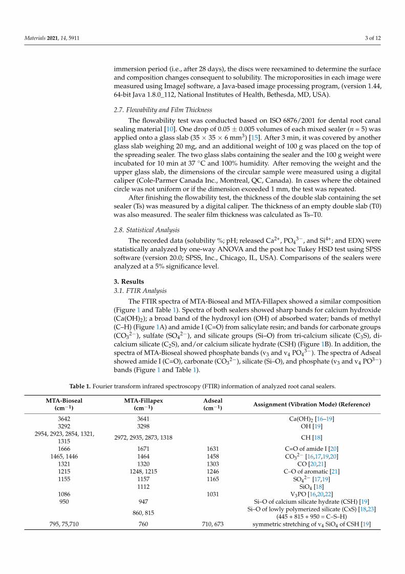
The FTIR spectra of MTA-Bioseal and MTA-Fillapex showed a similar composition(Figure 1 and Table 1). Spectra of both sealers showed sharp bands for calcium hydroxide(Ca(OH)2); a broad band of the hydroxyl ion (OH) of absorbed water; bands of methyl(C–H) (Figure 1A) and amide I (C=O) from salicylate resin; and bands for carbonate groups(CO3
2−), sulfate (SO42−), and silicate groups (Si–O) from tri-calcium silicate (C3S), di-
calcium silicate (C2S), and/or calcium silicate hydrate (CSH) (Figure 1B). In addition, thespectra of MTA-Bioseal showed phosphate bands (v3 and v4 PO4
3−). The spectra of Adsealshowed amide I (C=O), carbonate (CO3
2−), silicate (Si–O), and phosphate (v3 and v4 PO3−)bands (Figure 1 and Table 1).
Table 1. Fourier transform infrared spectroscopy (FTIR) information of analyzed root canal sealers.
MTA-Bioseal(cm−1)
MTA-Fillapex(cm−1)
Adseal(cm−1) Assignment (Vibration Mode) (Reference)
3642 3641 Ca(OH)2 [16–19]3292 3298 OH [19]
2954, 2923, 2854, 1321,1315 2972, 2935, 2873, 1318 CH [18]
1666 1671 1631 C=O of amide I [20]1465, 1446 1464 1458 CO3
2− [16,17,19,20]1321 1320 1303 CO [20,21]1215 1248, 1215 1246 C–O of aromatic [21]1155 1157 1165 SO4
2− [17,19]1112 SiO4 [18]
1086 1031 V3PO [16,20,22]950 947 Si–O of calcium silicate hydrate (CSH) [19]
860, 815 Si–O of lowly polymerized silicate (CxS) [18,23](445 + 815 + 950 = C–S–H)
795, 75,710 760 710, 673 symmetric stretching of v4 SiO4 of CSH [19]
Materials 2021, 14, 5911 4 of 12
Table 1. Cont.
MTA-Bioseal(cm−1)
MTA-Fillapex(cm−1)
Adseal(cm−1) Assignment (Vibration Mode) (Reference)
701 701, 690 CO3 of aragonite [24]618, 568 v4PO [22]
592 SiO42− bending of C3S [16–18]
465 464 500 SiO42− bending of C2S [18]
440 428 412 O–Si–O of CSH [17,18]
Materials 2021, 14, x FOR PEER REVIEW 4 of 13
Figure 1. FTIR spectra of hydrated root canal sealers showing the composition of each sealer. At region 4000–2000 cm−1 (A), the spectra detected bands of calcium hydroxide (Ca(OH)2), hydroxyl ion of absorbent water (OH), methyl (C–H). At region 2000-400 cm−1 (B), the spectra detected amide I (C=O) of salicylate resin, carbonate (CO32−), sulfate (SO42−), phosphate (PO4), and silicate group (SiO4) of calcium silicate.
Table 1. Fourier transform infrared spectroscopy (FTIR) information of analyzed root canal sealers.
MTA-Bioseal (cm−1)
MTA-Fillapex (cm−1)
Adseal (cm−1) Assignment (Vibration Mode) (Reference)
3642 3641 Ca(OH)2 [16–19] 3292 3298 OH [19]
2954, 2923, 2854, 1321, 1315 2972, 2935, 2873, 1318 CH [18]
1666 1671 1631 C=O of amide I [20] 1465, 1446 1464 1458 CO32− [16,17,19,20]
1321 1320 1303 CO [20,21] 1215 1248, 1215 1246 C–O of aromatic [21]
Figure 1. FTIR spectra of hydrated root canal sealers showing the composition of each sealer. At region 4000–2000 cm−1 (A),the spectra detected bands of calcium hydroxide (Ca(OH)2), hydroxyl ion of absorbent water (OH), methyl (C–H). At region2000-400 cm−1 (B), the spectra detected amide I (C=O) of salicylate resin, carbonate (CO3
2−), sulfate (SO42−), phosphate
(PO4), and silicate group (SiO4) of calcium silicate.
Materials 2021, 14, 5911 5 of 12
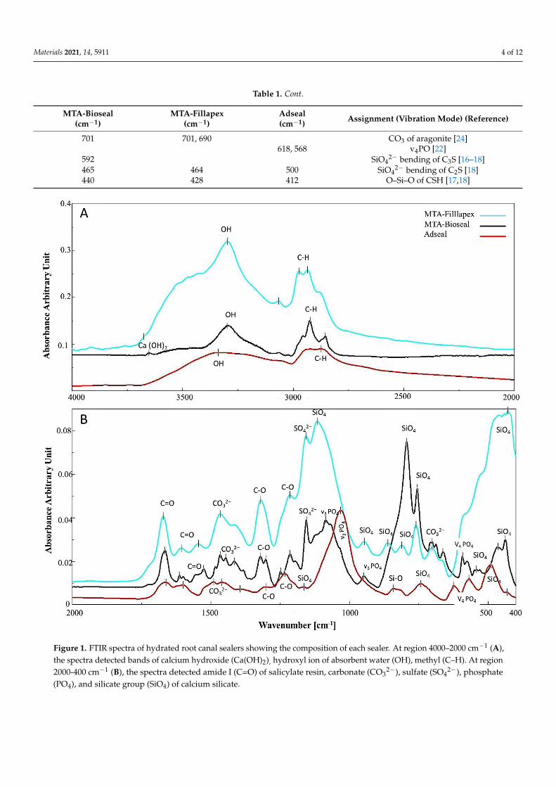
3.2. Solubility %
Both of the MTA-based sealers demonstrated an increased mean solubility % overtime, with no significant difference (p > 0.05) between them. Adseal showed a significantlylower solubility % compared with the MTA-based sealers (p < 0.001) (Figure 2A).
Materials 2021, 14, x FOR PEER REVIEW 6 of 13
Figure 2. Histograms representing the mean values of solubility % (A), pH changes (B), Ca2+ ion release (C), PO43− ion release (D), and Si4+ ion release (E) of the root canal sealers over the immersion times of the experiment. * indicates the highest significant value (at p < 0.001). † indicates the lowest significant value (at p < 0.001). ≠ indicates no significant dif-ference between sealers of the same symbol (p > 0.05).
Figure 2. Histograms representing the mean values of solubility % (A), pH changes (B), Ca2+ ion release (C), PO43− ion
release (D), and Si4+ ion release (E) of the root canal sealers over the immersion times of the experiment. * indicates thehighest significant value (at p < 0.001). † indicates the lowest significant value (at p < 0.001). 6= indicates no significantdifference between sealers of the same symbol (p > 0.05).
Materials 2021, 14, 5911 6 of 12
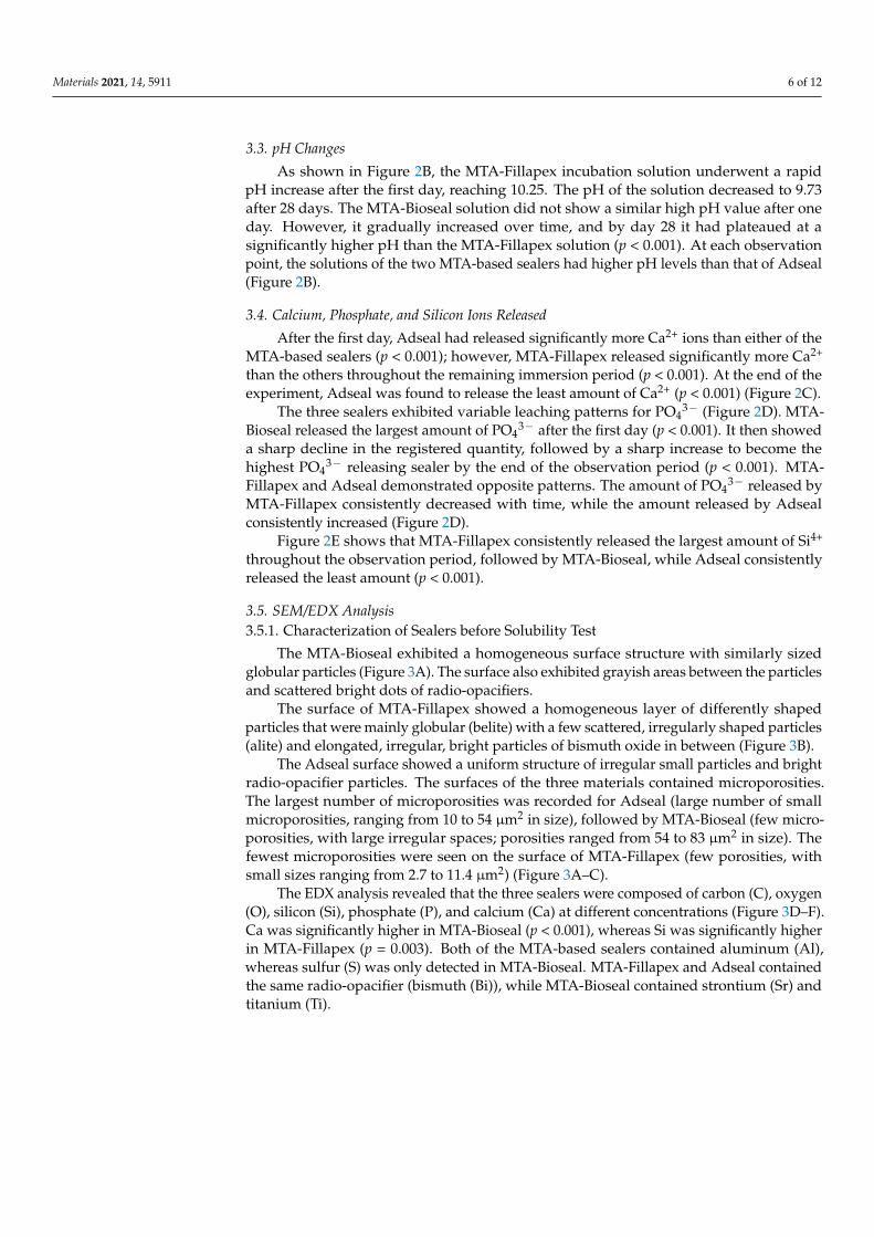
3.3. pH Changes
As shown in Figure 2B, the MTA-Fillapex incubation solution underwent a rapidpH increase after the first day, reaching 10.25. The pH of the solution decreased to 9.73after 28 days. The MTA-Bioseal solution did not show a similar high pH value after oneday. However, it gradually increased over time, and by day 28 it had plateaued at asignificantly higher pH than the MTA-Fillapex solution (p < 0.001). At each observationpoint, the solutions of the two MTA-based sealers had higher pH levels than that of Adseal(Figure 2B).
3.4. Calcium, Phosphate, and Silicon Ions Released
After the first day, Adseal had released significantly more Ca2+ ions than either of theMTA-based sealers (p < 0.001); however, MTA-Fillapex released significantly more Ca2+
than the others throughout the remaining immersion period (p < 0.001). At the end of theexperiment, Adseal was found to release the least amount of Ca2+ (p < 0.001) (Figure 2C).
The three sealers exhibited variable leaching patterns for PO43− (Figure 2D). MTA-
Bioseal released the largest amount of PO43− after the first day (p < 0.001). It then showed
a sharp decline in the registered quantity, followed by a sharp increase to become thehighest PO4
3− releasing sealer by the end of the observation period (p < 0.001). MTA-Fillapex and Adseal demonstrated opposite patterns. The amount of PO4
3− released byMTA-Fillapex consistently decreased with time, while the amount released by Adsealconsistently increased (Figure 2D).
Figure 2E shows that MTA-Fillapex consistently released the largest amount of Si4+
throughout the observation period, followed by MTA-Bioseal, while Adseal consistentlyreleased the least amount (p < 0.001).
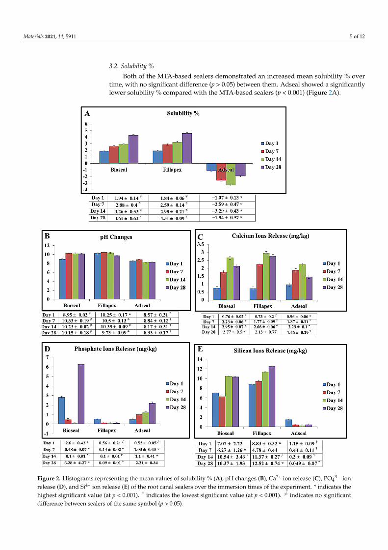
3.5. SEM/EDX Analysis3.5.1. Characterization of Sealers before Solubility Test
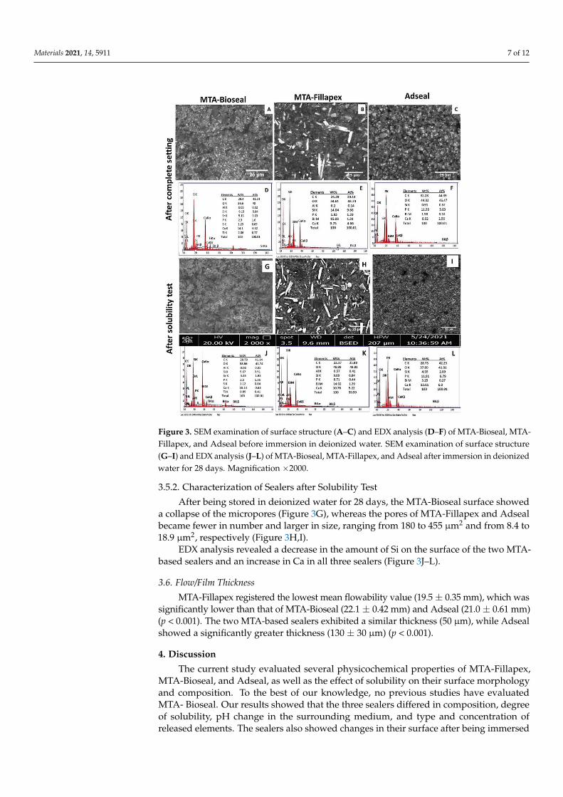
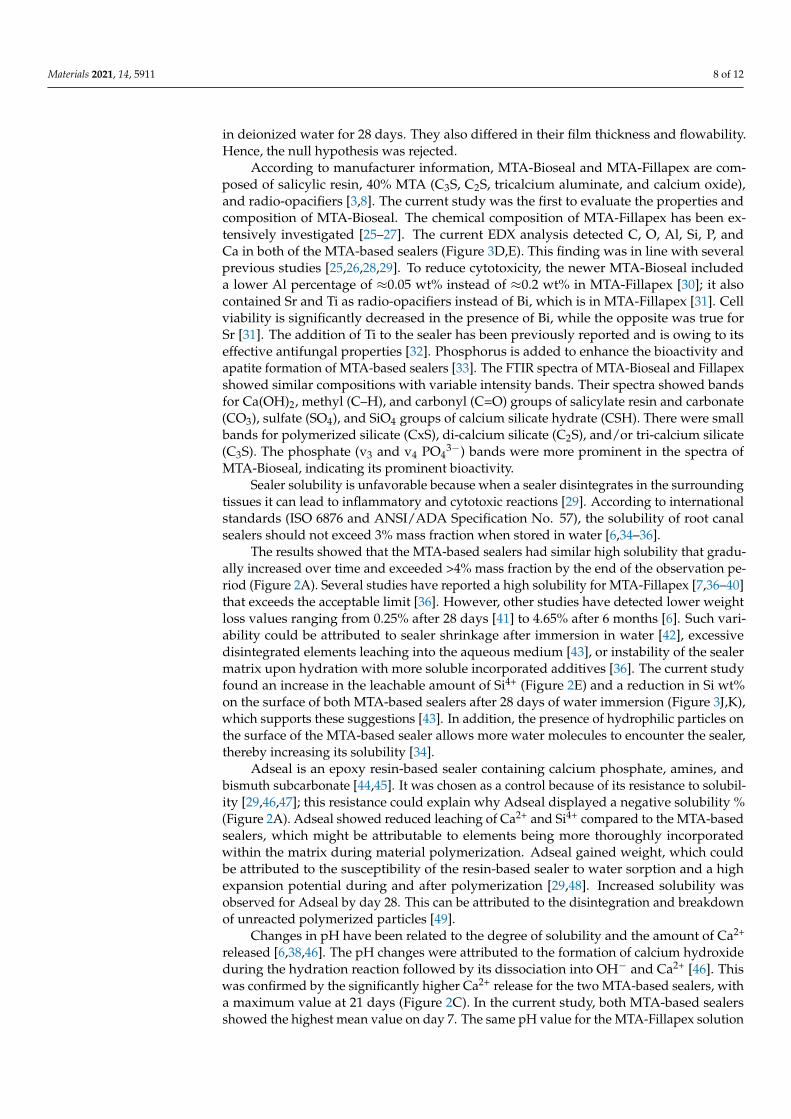
The MTA-Bioseal exhibited a homogeneous surface structure with similarly sizedglobular particles (Figure 3A). The surface also exhibited grayish areas between the particlesand scattered bright dots of radio-opacifiers.
The surface of MTA-Fillapex showed a homogeneous layer of differently shapedparticles that were mainly globular (belite) with a few scattered, irregularly shaped particles(alite) and elongated, irregular, bright particles of bismuth oxide in between (Figure 3B).
The Adseal surface showed a uniform structure of irregular small particles and brightradio-opacifier particles. The surfaces of the three materials contained microporosities.The largest number of microporosities was recorded for Adseal (large number of smallmicroporosities, ranging from 10 to 54 µm2 in size), followed by MTA-Bioseal (few micro-porosities, with large irregular spaces; porosities ranged from 54 to 83 µm2 in size). Thefewest microporosities were seen on the surface of MTA-Fillapex (few porosities, withsmall sizes ranging from 2.7 to 11.4 µm2) (Figure 3A–C).
The EDX analysis revealed that the three sealers were composed of carbon (C), oxygen(O), silicon (Si), phosphate (P), and calcium (Ca) at different concentrations (Figure 3D–F).Ca was significantly higher in MTA-Bioseal (p < 0.001), whereas Si was significantly higherin MTA-Fillapex (p = 0.003). Both of the MTA-based sealers contained aluminum (Al),whereas sulfur (S) was only detected in MTA-Bioseal. MTA-Fillapex and Adseal containedthe same radio-opacifier (bismuth (Bi)), while MTA-Bioseal contained strontium (Sr) andtitanium (Ti).
Materials 2021, 14, 5911 7 of 12Materials 2021, 14, x FOR PEER REVIEW 8 of 13
Figure 3. SEM examination of surface structure (A–C) and EDX analysis (D–F) of MTA-Bioseal, MTA-Fillapex, and Adseal before immersion in deionized water. SEM examination of surface struc-ture (G–I) and EDX analysis (J–L) of MTA-Bioseal, MTA-Fillapex, and Adseal after immersion in deionized water for 28 days. Magnification ×2000.
3.6. Flow/Film Thickness MTA-Fillapex registered the lowest mean flowability value (19.5 ± 0.35 mm), which
was significantly lower than that of MTA-Bioseal (22.1 ± 0.42 mm) and Adseal (21.0 ± 0.61 mm) (p < 0.001). The two MTA-based sealers exhibited a similar thickness (50 µm), while Adseal showed a significantly greater thickness (130 ± 30 µm) (p < 0.001).
4. Discussion The current study evaluated several physicochemical properties of MTA-Fillapex,
MTA-Bioseal, and Adseal, as well as the effect of solubility on their surface morphology and composition. To the best of our knowledge, no previous studies have evaluated MTA- Bioseal. Our results showed that the three sealers differed in composition, degree of solu-bility, pH change in the surrounding medium, and type and concentration of released elements. The sealers also showed changes in their surface after being immersed in deion-ized water for 28 days. They also differed in their film thickness and flowability. Hence, the null hypothesis was rejected.
According to manufacturer information, MTA-Bioseal and MTA-Fillapex are com-posed of salicylic resin, 40% MTA (C3S, C2S, tricalcium aluminate, and calcium oxide), and radio-opacifiers [3,8]. The current study was the first to evaluate the properties and compo-sition of MTA-Bioseal. The chemical composition of MTA-Fillapex has been extensively in-vestigated [25–27]. The current EDX analysis detected C, O, Al, Si, P, and Ca in both of the MTA-based sealers (Figure 3D–E). This finding was in line with several previous studies
Figure 3. SEM examination of surface structure (A–C) and EDX analysis (D–F) of MTA-Bioseal, MTA-Fillapex, and Adseal before immersion in deionized water. SEM examination of surface structure(G–I) and EDX analysis (J–L) of MTA-Bioseal, MTA-Fillapex, and Adseal after immersion in deionizedwater for 28 days. Magnification ×2000.
3.5.2. Characterization of Sealers after Solubility Test
After being stored in deionized water for 28 days, the MTA-Bioseal surface showeda collapse of the micropores (Figure 3G), whereas the pores of MTA-Fillapex and Adsealbecame fewer in number and larger in size, ranging from 180 to 455 µm2 and from 8.4 to18.9 µm2, respectively (Figure 3H,I).
EDX analysis revealed a decrease in the amount of Si on the surface of the two MTA-based sealers and an increase in Ca in all three sealers (Figure 3J–L).
3.6. Flow/Film Thickness
MTA-Fillapex registered the lowest mean flowability value (19.5 ± 0.35 mm), which wassignificantly lower than that of MTA-Bioseal (22.1 ± 0.42 mm) and Adseal (21.0 ± 0.61 mm)(p < 0.001). The two MTA-based sealers exhibited a similar thickness (50 µm), while Adsealshowed a significantly greater thickness (130 ± 30 µm) (p < 0.001).
4. Discussion
The current study evaluated several physicochemical properties of MTA-Fillapex,MTA-Bioseal, and Adseal, as well as the effect of solubility on their surface morphologyand composition. To the best of our knowledge, no previous studies have evaluatedMTA- Bioseal. Our results showed that the three sealers differed in composition, degreeof solubility, pH change in the surrounding medium, and type and concentration ofreleased elements. The sealers also showed changes in their surface after being immersed
Materials 2021, 14, 5911 8 of 12
in deionized water for 28 days. They also differed in their film thickness and flowability.Hence, the null hypothesis was rejected.
According to manufacturer information, MTA-Bioseal and MTA-Fillapex are com-posed of salicylic resin, 40% MTA (C3S, C2S, tricalcium aluminate, and calcium oxide),and radio-opacifiers [3,8]. The current study was the first to evaluate the properties andcomposition of MTA-Bioseal. The chemical composition of MTA-Fillapex has been ex-tensively investigated [25–27]. The current EDX analysis detected C, O, Al, Si, P, andCa in both of the MTA-based sealers (Figure 3D,E). This finding was in line with severalprevious studies [25,26,28,29]. To reduce cytotoxicity, the newer MTA-Bioseal includeda lower Al percentage of ≈0.05 wt% instead of ≈0.2 wt% in MTA-Fillapex [30]; it alsocontained Sr and Ti as radio-opacifiers instead of Bi, which is in MTA-Fillapex [31]. Cellviability is significantly decreased in the presence of Bi, while the opposite was true forSr [31]. The addition of Ti to the sealer has been previously reported and is owing to itseffective antifungal properties [32]. Phosphorus is added to enhance the bioactivity andapatite formation of MTA-based sealers [33]. The FTIR spectra of MTA-Bioseal and Fillapexshowed similar compositions with variable intensity bands. Their spectra showed bandsfor Ca(OH)2, methyl (C–H), and carbonyl (C=O) groups of salicylate resin and carbonate(CO3), sulfate (SO4), and SiO4 groups of calcium silicate hydrate (CSH). There were smallbands for polymerized silicate (CxS), di-calcium silicate (C2S), and/or tri-calcium silicate(C3S). The phosphate (v3 and v4 PO4
3−) bands were more prominent in the spectra ofMTA-Bioseal, indicating its prominent bioactivity.
Sealer solubility is unfavorable because when a sealer disintegrates in the surroundingtissues it can lead to inflammatory and cytotoxic reactions [29]. According to internationalstandards (ISO 6876 and ANSI/ADA Specification No. 57), the solubility of root canalsealers should not exceed 3% mass fraction when stored in water [6,34–36].
The results showed that the MTA-based sealers had similar high solubility that gradu-ally increased over time and exceeded >4% mass fraction by the end of the observation pe-riod (Figure 2A). Several studies have reported a high solubility for MTA-Fillapex [7,36–40]that exceeds the acceptable limit [36]. However, other studies have detected lower weightloss values ranging from 0.25% after 28 days [41] to 4.65% after 6 months [6]. Such vari-ability could be attributed to sealer shrinkage after immersion in water [42], excessivedisintegrated elements leaching into the aqueous medium [43], or instability of the sealermatrix upon hydration with more soluble incorporated additives [36]. The current studyfound an increase in the leachable amount of Si4+ (Figure 2E) and a reduction in Si wt%on the surface of both MTA-based sealers after 28 days of water immersion (Figure 3J,K),which supports these suggestions [43]. In addition, the presence of hydrophilic particles onthe surface of the MTA-based sealer allows more water molecules to encounter the sealer,thereby increasing its solubility [34].
Adseal is an epoxy resin-based sealer containing calcium phosphate, amines, andbismuth subcarbonate [44,45]. It was chosen as a control because of its resistance to solubil-ity [29,46,47]; this resistance could explain why Adseal displayed a negative solubility %(Figure 2A). Adseal showed reduced leaching of Ca2+ and Si4+ compared to the MTA-basedsealers, which might be attributable to elements being more thoroughly incorporatedwithin the matrix during material polymerization. Adseal gained weight, which couldbe attributed to the susceptibility of the resin-based sealer to water sorption and a highexpansion potential during and after polymerization [29,48]. Increased solubility wasobserved for Adseal by day 28. This can be attributed to the disintegration and breakdownof unreacted polymerized particles [49].
Changes in pH have been related to the degree of solubility and the amount of Ca2+
released [6,38,46]. The pH changes were attributed to the formation of calcium hydroxideduring the hydration reaction followed by its dissociation into OH− and Ca2+ [46]. Thiswas confirmed by the significantly higher Ca2+ release for the two MTA-based sealers, witha maximum value at 21 days (Figure 2C). In the current study, both MTA-based sealersshowed the highest mean value on day 7. The same pH value for the MTA-Fillapex solution
Materials 2021, 14, 5911 9 of 12
was previously recorded in several studies [6,25,38,39,42], while a lower pH value (7.7–9.39)was recorded by others [27,50]. This suggests that pH changes are related to time [38].Among the experimental periods, the MTA-Bioseal solution showed a higher pH value(Figure 2B) than that stated by the manufacturer (pH = 9) [8].
The high alkalinity of the MTA-based sealer solutions may be attributed to the poz-zolanic reaction and the formation of Ca(OH)2 during the hydration reaction. Ca(OH)2dissociates into OH− and Ca2+, which promote antibacterial ability and osteogenic poten-tial, respectively [6,51–53]. However, the prolonged alkalinity of the MTA-based sealersolutions might be considered as a source of cytotoxicity, leading to protein destructionand enzymatic cell membrane denaturation [54]. This adverse effect could be of clinicalconcern, as our results showed an increase in Ca wt% on the sealers’ surfaces (Figure 3J,K).Such accumulation might lead to cytotoxic events. A higher calcium content released byMTA-Fillapex compared with epoxy resin has been previously reported [40,50]. Siboni et al.reported that the maximum calcium content released by MTA-Fillapex was detected withinthe first 3 days, while epoxy resin (AHplus) did not exhibit calcium release at all [27].The Adseal solution was weakly alkaline (Figure 2B), in contrast to the nearly neutral pH(≈7.5) [29] or acidic pH (≤6.5) [55] previously recorded. The lower pH changes induced byAdseal might be related to its lower solubility and reduced Ca2+ release.
The prolonged release of Ca2+, PO43−, and Si4+ results in degradation of the sealer’s
surface. SEM/EDX analysis revealed a decrease in the Si wt% (Figure 3D,E,J,K), withlarge micropores detected in both of the MTA-based sealers. MTA-Fillapex released asignificant amount of Ca2+ and Si4+. This has been previously reported [52]. MTA-Biosealdemonstrated the greatest PO4
3− release (Figure 2C–E), which might be due to its higher Pcontent compared with MTA-Fillapex, as detected by EDX. The marked increase in PO4
3−
by the end of the observation period might have been due to its lack of attachment withinthe set sealer. This amount of PO4
3− release might enhance its bioactivity [33]. Conversely,Adseal showed greater PO4
3− release, which increased over time. This finding might bedue to its greater P content, as detected by EDX.
The FTIR analysis identified PO43− in the spectra of MTA-Bioseal and Adseal at 1086
and 1031 cm−1, respectively [16,20,22]. The largest PO43− release was exhibited by MTA-
Bioseal, followed by Adseal (Figure 2D). It appeared that PO43− was not well incorporated
into the CSH structure of MTA-Bioseal; hence, it was easily released into the aqueousmedium. Adseal is mainly composed of calcium phosphate [44]; thus, after polymerization,PO4
3− became incorporated within the sealer and was slowly released. The presenceof PO4
3− within the sealer seems to enhance its bioactivity. This finding corroboratedprevious results [56].
The surfaces of the three materials contained microporosities, with the largest sizesin MTA-Fillapex. MTA-Fillapex exhibits a homogeneous surface with various sizes ofporosities [38]. This may be related to the setting characteristics and the formation ofa polymerized silicate phase [37]. Previous reports have shown that MTA-Fillapex isunable to set, even after 1 month [29]. Here, the FTIR spectra confirmed the presence ofunhydrated calcium silicate (C3S and C2S) particles and little polymerized calcium silicate(CxS), with a low content of polymerized calcium silicate (CSH) [29]. Furthermore, therewas low intensity of the SiO4 band at 900–800 cm−1 [19]. The presence of unhydratedsilicate phase is responsible for the excessive Si4+ release and for the large micropores onthe MTA-Fillapex surface. It is assumed that these micropores can hold water from thesurrounding environment, allowing bacterial colonization [57]. Whether this would impactthe long-term outcome of endodontic treatment warrants further clinical investigation.
Although improved flowability facilitates a sealer’s penetration into canal irregulari-ties, excessive flow has been considered as a risk factor for extrusion and can potentiallyprovoke inflammatory and cytotoxic reactions [41]. According to the ISO standard, thethree tested sealers met the adequate flow specification (>17 mm) [10], with MTA-Biosealregistering the highest flowability, followed by Adseal and MTA-Fillapex. Previous studieshave reported a wide range of MTA-Fillapex flowabilities (22–34 mm) [29,36,41,58]. Such
Materials 2021, 14, 5911 10 of 12
high flowability could be due to a prolonged setting time [25,37] or a high resin/MTA ratiowhen used from freshly opened tubes [41].
Regarding film thickness, both MTA-Bioseal and Fillapex complied with the ISOstandard (50 µm) [10], but Adseal had a high value (130 ± 30 µm). Previous studies havereported thick films for MTA-Fillapex (75 ± 12 µm) [36,42] and Adseal (0.083 mm) [44].The flowability and film thickness of sealers may be influenced by their composition, smallparticle size, and setting characteristics [58]. The greater film thickness of Adseal can beattributed to its expansion after polymerization [29,48].
5. Conclusions
The three sealers differed in their composition, degree of solubility, induced pHchanges in the surrounding medium, type and concentration of released elements, surfacechanges upon immersion in deionized water over 28 days, film thickness, and flowability.The two MTA-based sealers exhibited high solution alkalinity and released a consider-able amount of Ca2+, which is conducive to osteogenic behavior. The greater solubilityand Si4+ release exhibited by MTA-Fillapex might have led to the development of largemicropores on its surface, which would compromise the apical sealing of the root canalsystem. This could be a clinical concern jeopardizing the long-term outcome of root canaltreatment. Hence, clinicians should maximize efforts to limit contact of MTA-Fillapexwith the surrounding periapical tissues. Further investigations are needed to evaluate thesetting characteristics of MTA-based root canal sealers.
Despite the meticulous approach adopted in this study, the lack of moist conditionsprovided by dentinal tubule fluids, which aids in the setting reaction of MTA-based sealers,limits the extrapolation of our results to the clinical setting.
Author Contributions: Conceptualization, S.A.Z.; data curation, S.A.Z.; formal analysis, S.A.Z.,H.Y.E., A.A.M.S. and O.S.A.; funding acquisition, H.Y.E., A.A.M.S. and O.S.A.; investigation, S.A.Z.and H.Y.E.; methodology, S.A.Z. and H.Y.E.; resources, S.A.Z., H.Y.E., A.A.M.S. and O.S.A.; supervi-sion, S.A.Z.; validation, S.A.Z.; writing—original draft, S.A.Z.; writing—review and editing, H.Y.E.,A.A.M.S. and O.S.A. All authors have read and agreed to the published version of the manuscript.
Funding: This research received no external funding.
Institutional Review Board Statement: Not applicable.
Informed Consent Statement: Not applicable.
Data Availability Statement: Data available in a publicly accessible repository.
Conflicts of Interest: The authors declare no conflict of interest.
References1. Ørstavik, D. Materials used for root canal obturation: Technical, biological and clinical testing. Endod. Top. 2005, 12, 25–38.
[CrossRef]2. Parirokh, M.; Torabinejad, M. Mineral trioxide aggregate: A comprehensive literature review-part I: Chemical, physical, and
antibacterial properties. J. Endod. 2010, 36, 16–27. [CrossRef]3. Angulus Science and Technology. MTA-Fillapex Endodontic Sealer, Scientific Profile. 2011. Available online: http://www.
angelusdental.com/img/arquivos/mta_fillapex_technical_profile_download.pdf (accessed on 22 August 2021).4. Johnson, W.T.; Kulild, J.C.; Tay, F. Obturation of the cleaned and shaped root canal system. In Pathways of the Pulp, 11th ed.;
Elsevier: Amsterdam, The Netherlands, 2016; pp. 280–322.5. Abu Zeid, S.T.H.; Saleh, A.A.Y.M. Solubility, pH Changes and Releasing Elements of Different Bioceramic and Mineral Trioxide
Aggregate Root Canal Sealers Comparative Study. Trauma Treat 2015, 4, 1–4.6. Urban, K.; Neuhaus, J.; Donnermeyer, D.; Schäfer, E.; Dammaschke, T. Solubility and pH value of 3 different root canal sealers: A
long-term investigation. J. Endod. 2018, 44, 1736–1740. [CrossRef] [PubMed]7. Da Silva, E.J.N.L.; Accorsi-Mendonça, T.; Pedrosa, A.C.; Granjeiro, J.M.; Zaia, A.A. Long-term cytotoxicity, pH and dissolution
rate of AH Plus and MTA Fillapex. Brazil. Dent. J. 2016, 27, 419–423. [CrossRef]8. ITENA Clinical Product. MTA-Bioseal, White Paper—Dental Sky. 2018. Available online: WP_MTABIOSEAL.pdf(dentex.ro)
(accessed on 20 August 2021).
Materials 2021, 14, 5911 11 of 12
9. American National Standards Institute. American Dental Association Specification no. 57 for endodontic filling materials. J. Am.Dent. Assoc. 2000, 108, 88.
10. International Standardization Organization. ISO 6876: Dental Root Canal Sealing Materials; International Organization forStandardization: Geneva, Swizerland, 2012.
11. McMichen, F.; Pearson, G.; Rahbaran, S.; Gulabivala, K. A comparative study of selected physical properties of five root-canalsealers. Int. Endod. J. 2003, 36, 629–635. [CrossRef]
12. Kim, J.; Vipulanandan, C. Effect of pH, sulfate and sodium on the EDTA titration of calcium. Cem. Concr. Res. 2003, 33, 621–627.[CrossRef]
13. Mussa, S.B.; Elferjani, H.S.; Haroun, F.A.; Abdelnabi, F.F. Determination of available nitrate, phosphate and sulfate in soil samples.Int. J. PharmTech Res. 2009, 1, 598–604.
14. Olsen, S.R.; Sommers, L.E. Phosphorus. In Methods of Soil Analysis: Part 2 Chemical and Microbiological Properties, 2nd ed.; AmericanSociety of Agronomy: Madison, WI, USA, 1983; pp. 403–430.
15. de Miranda Candeiro, G.T.; Correia, F.C.; Duarte, M.A.H.; Ribeiro-Siqueira, D.C.; Gavini, G. Evaluation of radiopacity, pH, releaseof calcium ions, and flow of a bioceramic root canal sealer. J. Endod. 2012, 38, 842–845. [CrossRef]
16. Ahmadi, S.M.; Behnamghader, A.; Sharifipoor, S.; Farsadzadeh, B. Effect of nano flourhydroxyapatite (nFHA) addition on theacellular bioactivity of MTA cement: An in vitro assessment. In Proceedings of the 4th International Conference on Nanostructures(ICNS4), Kish Island, Iran, 12–14 March 2012; pp. 12–14.
17. Gandolfi, M.G.; Taddei, P.; Tinti, A.; Prati, C. Apatite-forming ability (bioactivity) of ProRoot MTA. Int. Endod. J. 2010, 43, 917–929.[CrossRef]
18. Okamura, T.; Chen, L.; Tsumano, N.; Ikeda, C.; Komasa, S.; Tominaga, K.; Hashimoto, Y. Biocompatibility of a High-Plasticity,Calcium Silicate-Based, Ready-to-Use Material. Materials 2020, 13, 4770. [CrossRef]
19. Ylmén, R.; Jäglid, U.; Steenari, B.-M.; Panas, I. Early hydration and setting of Portland cement monitored by IR, SEM and Vicattechniques. Cem. Concr. Res. 2009, 39, 433–439. [CrossRef]
20. Boskey, A.; Camacho, N.P. FT-IR imaging of native and tissue-engineered bone and cartilage. Biomaterials 2007, 28, 2465–2478.[CrossRef]
21. Delgado, A.H.; Young, A.M. Modelling ATR-FTIR Spectra of Dental Bonding Systems to Investigate Composition and Polymeri-sation Kinetics. Materials 2021, 14, 760. [CrossRef]
22. Jayasree, R.; Kumar, T.S.; Kavya, K.P.S.; Nankar, P.; Mukesh, D. Self setting bone cement formulations based on egg shell derivedtetracalcium phosphate bioceramics. Bioceram. Dev. Appl. 2015, 5, 2.
23. Radwan, M.; Nagi, S.M.; Abd El-Hamid, H. Physico-mechanical characteristics of tri-calcium silicate pastes as dentin substituteand interface analysis in class II cavities: Effect of CaCl2 and SBF solutions. Heliyon 2019, 5, e01975. [CrossRef]
24. Trezza, M.A. Hydration study of ordinary portland cement in the presence of zinc ions. Mater. Res. 2007, 10, 331–334. [CrossRef]25. Benezra, M.K.; Wismayer, P.S.; Camilleri, J. Influence of environment on testing of hydraulic sealers. Sci. Rep. 2017, 7, 1–11.26. Sampaio, F.C.; Alencar, A.H.G.D.; Guedes, O.A.; Veloso, H.H.P.; Santos, T.O.D.; Estrela, C. Chemical elements characterization of
root canal sealers using scanning electron microscopy and energy dispersive X-ray analysis. Oral Health Dent. Manag. 2014, 13,27–34. [PubMed]
27. Siboni, F.; Taddei, P.; Zamparini, F.; Prati, C.; Gandolfi, M.G. Properties of BioRoot RCS, a tricalcium silicate endodontic sealermodified with povidone and polycarboxylate. Int. Endod. J. 2017, 50, e120–e136. [CrossRef] [PubMed]
28. Reszka, P.; Nowicka, A.; Lipski, M.; Dura, W.; Drozdzik, A.; Wozniak, K. A comparative chemical study of calcium silicate-containing and epoxy resin-based root canal sealers. BioMed Res. Int. 2016, 2016, 9808432. [CrossRef]
29. Lee, J.K.; Kwak, S.W.; Ha, J.-H.; Lee, W.; Kim, H.-C. Physicochemical properties of epoxy resin-based and bioceramic-based rootcanal sealers. Bioinorg. Chem. Appl. 2017, 2017, 2582849. [CrossRef]
30. Drukteinis, S.; Camilleri, J. Bioceramic Materials in Clinical Endodontics; Springer: Berlin/Heidelberg, Germany, 2021.31. Antonijevic, D.; Despotovic, A.; Biocanin, V.; Miloševic, M.; Trišic, D.; Lazovic, V.; Zogovic, N.; Milašin, J.; Ilic, D. Influence of the
addition of different radiopacifiers and bioactive nano-hydroxyapatite on physicochemical and biological properties of calciumsilicate based endodontic ceramic. Ceram Int. 2021, 47, 28913–28923. [CrossRef]
32. Raura, N.; Garg, A.; Arora, A.; Roma, M. Nanoparticle technology and its implications in endodontics: A review. Biomater. Res.2020, 24, 1–8. [CrossRef] [PubMed]
33. Al-Sanabani, J.S.; Madfa, A.A.; Al-Sanabani, F.A. Application of calcium phosphate materials in dentistry. Int. J. Biomater. 2013,2013, 876132. [CrossRef]
34. Poggio, C.; Dagna, A.; Ceci, M.; Meravini, M.-V.; Colombo, M.; Pietrocola, G. Solubility and pH of bioceramic root canal sealers:A comparative study. J. Clin. Exp. Dent. 2017, 9, e1189. [CrossRef] [PubMed]
35. Borges, Á.H.; Pedro, F.L.; Miranda, C.E.; Semenoff-Segundo, A.; Pécora, J.D.; Cruz Filho, A.M. Comparative study of physico-chemical properties of MTA-based and Portland cements. Acta Odontol. Latinoam. 2010, 23, 175–181. [PubMed]
36. Viapiana, R.; Flumignan, D.; Guerreiro-Tanomaru, J.; Camilleri, J.; Tanomaru-Filho, M. Physicochemical and mechanical propertiesof zirconium oxide and niobium oxide modified P ortland cement-based experimental endodontic sealers. Int. Endod. J. 2014, 47,437–448. [CrossRef] [PubMed]
Materials 2021, 14, 5911 12 of 12
37. Amoroso-Silva, P.A.; Guimarães, B.M.; Marciano, M.A.; Duarte, M.A.H.; Cavenago, B.C.; Ordinola-Zapata, R.; De Almeida, M.M.;De Moraes, I.G. Microscopic analysis of the quality of obturation and physical properties of MTA F illapex. Microsc. Res. Tech.2014, 77, 1031–1036. [CrossRef]
38. Borges, Á.H.; Dorileo, O.; Gonçales, M.C.; Villa, R.D.; Borba, A.M.; Semenoff, T.A.D.V.; Guedes, O.A.; Estrela, C.R.A.; Bandéca,M.C. Physicochemical properties and surfaces morphologies evaluation of MTA FillApex and AH plus. Sci. World J. 2014, 2014,589732. [CrossRef]
39. Faria-Júnior, N.; Tanomaru-Filho, M.; Berbert, F.L.C.V.; Guerreiro-Tanomaru, J. Antibiofilm activity, pH and solubility ofendodontic sealers. Int. Endod. J. 2013, 46, 755–762. [CrossRef]
40. Jafari, F.; Jafari, S. Composition and physicochemical properties of calcium silicate based sealers: A review article. J. Clin. Exp.Dent. 2017, 9, e1249. [CrossRef] [PubMed]
41. Vitti, R.P.; Prati, C.; Silva, E.J.N.L.; Sinhoreti, M.A.C.; Zanchi, C.H.; Silva, M.G.D.S.E.; Guedes, O.A.; Estrela, C.R.A.; Bandéca, M.C.Physical properties of MTA Fillapex sealer. J. Endod. 2013, 39, 915–918. [CrossRef] [PubMed]
42. Zhou, H.-M.; Shen, Y.; Zheng, W.; Li, L.; Zheng, Y.-F. Haapasalo, M. Physical properties of 5 root canal sealers. J. Endod. 2013, 39,1281–1286. [CrossRef] [PubMed]
43. Tagger, M.; Tagger, E.; Kfir, A. Release of calcium and hydroxyl ions from set endodontic sealers containing calcium hydroxide. J.Endod. 1988, 14, 588–591. [CrossRef]
44. Cardona Hidalgo, J.C.; González Carreño, J.M.; Avendaño Rueda, J.C. Physicochemical properties of two epoxy resin-basedsealants: Topseal® and AdSeal™. A comparative study. Rev. Fac. Odontol. Univ. Antioq. 2019, 31, 68–76.
45. Marciano, M.A.; Guimarães, B.M.; Ordinola-Zapata, R.; Bramante, C.M.; Cavenago, B.C.; Garcia, R.B.; Bernardineli, N.; Andrade,F.; Moraes, I.G.; Duarte, M.A.H. Physical properties and interfacial adaptation of three epoxy resin–based sealers. J. Endod. 2011,37, 1417–1421. [CrossRef]
46. Huang, T.-H.; Kao, C.-T. pH measurement of root canal sealers. J. Endod. 1998, 24, 236–238. [CrossRef]47. Schäfer, E.; Zandbiglari, T. Solubility of root canal sealers in water and artificial saliva. Int. Endod. J. 2003, 36, 660–669. [CrossRef]
[PubMed]48. Carvalho-Júnior, J.R.; Guimarães, L.F.L.; Correr-Sobrinho, L.; Pécora, J.D.; Sousa-Neto, M.D. Evaluation of solubility, disintegra-
tion, and dimensional alterations of a glass ionomer root canal sealer. Brazil. Dent. J. 2003, 14, 114–118. [CrossRef]49. Azadi, N.; Fallahdoost, A.; Mehrvarzfar, P.; Rakhshan, H.; Rakhshan, V. A four-week solubility assessment of AH-26 and four
new root canal sealers. Dent. Res. J. 2012, 9, 31.50. Kuga, M.C.; Faria, G.; Weckwerth, P.H.; Duarte, M.A.H.; Campos, E.A.D.; Só, M.V.R.; Voila, K.S. Evaluation of the pH, calcium
release and antibacterial activity of MTA Fillapex. Rev. Odontol. UNESP 2013, 42, 330–335. [CrossRef]51. Desai, S.; Chandler, N. Calcium hydroxide–based root canal sealers: A review. J. Endod. 2009, 35, 475–480. [CrossRef]52. McHugh, C.P.; Zhang, P.; Michalek, S.; Eleazer, P.D. pH required to kill Enterococcus faecalis in vitro. J. Endod. 2004, 30, 218–219.
[CrossRef] [PubMed]53. Edrees, H.Y.; Abu Zeid, S.T.; Atta, H.M.; AlQriqri, M.A. Induction of osteogenic differentiation of mesenchymal stem cells by
bioceramic root repair material. Materials 2019, 12, 2311. [CrossRef]54. Yoshino, P.; Nishiyama, C.K.; Modena, K.C.D.S.; Santos, C.F.; Sipert, C.R. In vitro cytotoxicity of white MTA, MTA Fillapex® and
Portland cement on human periodontal ligament fibroblasts. Brazil. Dent. J. 2013, 24, 111–116. [CrossRef] [PubMed]55. Song, Y.-S.; Choi, Y.; Lim, M.-J.; Yu, M.-K.; Hong, C.-U.; Lee, K.-W.; Min, K.-S. In vitro evaluation of a newly produced resin-based
endodontic sealer. Restor. Dent. Endod. 2016, 41, 189. [CrossRef] [PubMed]56. Xuereb, M.; Vella, P.; Damidot, D.; Sammut, C.V.; Camilleri, J. In situ assessment of the setting of tricalcium silicate–based sealers
using a dentin pressure model. J. Endod. 2015, 41, 111–124. [CrossRef] [PubMed]57. Gandolfi, M.; Siboni, F.; Prati, C. Properties of a novel polysiloxane-guttapercha calcium silicate-bioglass-containing root canal
sealer. Dent. Mater. 2016, 32, e113–e126. [CrossRef]58. Silva, E.J.; Rosa, T.P.; Herrera, D.R.; Jacinto, R.C.; Gomes, B.P.; Zaia, A.A. Evaluation of cytotoxicity and physicochemical
properties of calcium silicate-based endodontic sealer MTA Fillapex. J. Endod. 2013, 39, 274–277. [CrossRef] [PubMed]


