Embed Size (px)
Citation preview
Course: IMB Part II
E-content-Online class 23/7/20
Photosynthesis
Light reaction
Photosynthesis is the most fundamental process in plants and some bacteria. It is the
single most important physic-biochemical process in the world on which the existence of life on
earth depends. It is the ability of plants only, to utilise the solar energy to produce carbon containing
organic compound (carbohydrate) from inorganic matter by photosynthesis. Plants produce
carbohydrate with the help of light, chlorophyll, water and CO2, , evolving oxygen. Therefore, plants
are producers which produce food for the consumers and not only this; they also produce oxygen at
the same time.
T he overall process can be simply written as:-
Light energy
6 CO2 + 6 H2O---------------- C6H12O6 + 6O2
Chlorophyll
Ruben, Randall and Kamen (1941) demonstrated that isolated chloroplasts evolved oxygen when
they were illuminated in the presence of suitable electron acceptor ferricyanide.They demonstrated
by experiment that oxygen evolved in photosynthesis comes from water and not from carbon
dioxide. They modified this equation on the basis of their experiments using radio labelled oxygen 18O.They confirmed that when carbon dioxide with isotopic 18O was given, the oxygen produced in
the photosynthesis was not 18O2, but when H218O was given to the chloroplasts, the oxygen was
found labelled with 18O2.Therefore, Ruben and Kamen confirmed that oxygen evolved in the
photosynthesis comes from water and not from CO2.
2H2O18 + CO2 --- (Chlorophyll & Light) -------------> (CH2 O) + H2O + O2
18
Carbohydrate
2H2O + CO218--- (chlorophyll & light) ----------------->O2 + (CH2O
18) +H2O18
Carbohydrate
Thus, the overall equation of Photosynthesis was modified as –
Light energy
6CO2 + 12H2O -----------C6H12O6+ 6H2O+ 6O2
Chlorophyll
There are requirements of light, chlorophyll, water and carbon dioxide. The solar light energy is
received by the chlorophyll, which in turn utilise water and CO2 and produces carbohydrate.
Blackman (1905) divided the entire process of photosynthesis into two component reactions,termed
light and dark reactions. The ability of isolated chloroplasts to carry out both these reactions was
shown by Arnon et al. (1954).Therefore, both the reactions take place as given below:-
1. First that takes place in presence of light, called as Light reaction. This reaction is light dependent
and light is utilised by the chlorophyll molecules. As a result of getting light energy, chlorophyll
molecules get excited and transfer the excitons to other chlorophyll molecules. Water is being
splitted in this reaction to produce oxygen. Besides the production of O2, in the light reaction ATP
and NADPH2 are also produced, which are called as assimilatory power.
2. Second reaction does not require light. In this reaction, ATP, NADPH2 (produced in the light reaction) and CO2 from the atmosphere are used to synthesize carbohydrate. This reaction is known as dark reaction or Blackman reaction or carbon dioxide fixation pathway or Calvin cycle. Operation of light and dark reactions in photosynthesis was experimentally proved by Warburg (1919) in his experiments as given below- Evidence from intermittent light-
Warburg (1919) used unicellular green algae Chlorella vulgaris and Scenedesmus obliquus in his
experiments. In one set of experiment, he exposed it to continuous light along with other required
factors; and in another set, exposed to intermittent(light followed by dark periods) flashes of light of
about 1/16 seconds. He measured the rate of photosynthesis in both the cases per unit time. It was
found that the rate of photosynthesis was greater in intermittent light than that in continuous light.
This indicated that a given amount of light is more efficiently used up if a dark period follows the
light period. It was concluded that photosynthesis includes two types of reactions, one the light
dependent and another light independent. The product of light reaction is used up in the dark to
synthesize carbohydrate.
There are some more evidences which prove the existence of two types of reactions in
photosynthesis-
Evidence from temperature coefficient-
The temperature coefficient of a reaction is the ratio of the rate of a reaction at a given temperature
and at a temperature 100 C lower. It is denoted by the symbol Q10. For a photochemical reaction
(light reaction) this value is found almost unity. i.e. Q10 =1, as it shows no increase in the rate with
the increase in temperature.
For the dark reaction Q10 is always equal to 2 or 3.Blackman demonstrated that if a plant is well
illuminated and CO2 is in sufficient quantity, Q10 is always equal to 2 or more than that. This indicates
that there must be a chemical reaction phase which is influenced by the temperature.This must be a
dark reaction phase. In low intensity light, the Q10 is almost unity which shows that there is at least
one reaction controlled by light and in this condition the light becomes limiting, which limits dark
reaction to occur. It is further clear that both light and dark reactions are independent but are
interlinked.
3. Evidence from carbon dioxide reduction in dark –
Melvin Calvin exposed an alga chlorella to light in absence of CO2 and then transferred them
immediately to dark with the supply of C14O2 .The carbon of CO2 was already labelled by using
radioactive isotope of carbon (C14).It was found that 14CO2 in dark was fixed into carbohydrate. This
indicates that CO2 is reduced to carbohydrate in dark and this phase is purely chemical or dark phase
of photosynthesis.
Hill reaction- Robert Hill (1937) and Hill and Scarisbrick (1939)-demonstrated that isolated
chloroplasts when exposed to light in presence of water, liberate O2. Hill observed that if some
oxidants (ferric salt / Fe3+) were present in the medium, they got reduced to ferrous forms
(Fe2+).Other compounds, such as ferricyanide, quinines, dichlorophenol indophenols (DCIP) can also
be reduced by illuminated chloroplasts. This reaction is called as Hill reaction, and the used oxidants
are called as Hill oxidant. In the above reaction water is splitted to liberate O2 in presence of
chlorophyll, light and an oxidant. Quantitative studies of Hill reaction confirmed that it was a
photochemical oxidation of water.
Light reaction machinery
Photosynthetic apparatus- The chloroplast
Experiments done by Park and Pon (1963) with isolated chloroplasts confirmed that the chloroplasts are the site of photosynthesis. They found that the water soluble proteinaceous non pigmented stroma portion participated in dark reactions whereas the water –insoluble pigmented portion containing thylakoids carried out light reactions. Their results localized the Hill reaction and photophosphorylation in photosynthetic lamellae, and CO2 fixation reactions in the stroma region. The chloroplasts are known as photosynthetic apparatus, which are the site of photosynthesis. They are the highly ordered complex structure that floats freely in the cytoplasm. Its existence was shown in 1679 by Leeuwenhoek, but it was identified as a separate structure in 1837. Sachs (1862) observed starch grains inside the chloroplasts during photosynthesis and this confirmed its role as seat in photosynthesis. They occur in the cytoplasm of all the green cells of plants except autotrophic prokaryotes .The chloroplasts are small, green, discoid or ellipsoidal .The normal size of chloroplast in higher plants is 4-10μ in diameter and 1-3μ in thickness. The chloroplast is enclosed with a highly permeable outer membrane and a nearly impermeable inner membrane separated by a narrow inters membranous space. The inner membrane encloses the stroma, a concentrated solution of enzymes, including those required for carbohydrate synthesis. A chloroplast usually contains 10-100 grana .Thylakoids membrane arises from invaginations in the inner membrane of developing chloroplasts and therefore resembles mitochondrial cristae. The thylakoids membrane contains protein complexes involved in the harvesting light energy, transporting electrons, and synthesizing ATP. In photosynthetic bacteria, the machinery for the light reactions is located in the plasma membrane, which often forms invaginations or multilamellar structures resembling grana. Usually they are found in mesophyll cells in the palisade parenchyma and vegetative green cells of
lower plants. Shape and size of the chloroplasts greatly vary in algae, where they are found in
different shapes such as cup shaped, horse shoe shaped, spiral, reticulate etc. The general structure
of chloroplast is a double membrane structure having lamellar structures inside, known as
thylakoids. The thylakoid is a single highly folded vesicle, although in most organisms it appears to
consist of stacks of disc like sacs named grana, which are interconnected by unstacked stroma
lamellae, called stroma lamellae. The lamellae forming the granum are called as granum lamellae.
Both the kinds of lamellae show different composition of pigments.
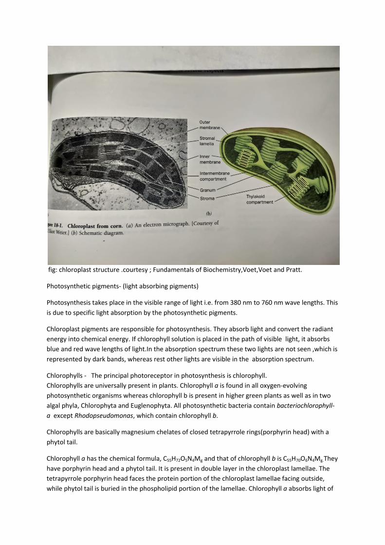
fig: chloroplast structure .courtesy ; Fundamentals of Biochemistry,Voet,Voet and Pratt.
Photosynthetic pigments- (light absorbing pigments)
Photosynthesis takes place in the visible range of light i.e. from 380 nm to 760 nm wave lengths. This
is due to specific light absorption by the photosynthetic pigments.
Chloroplast pigments are responsible for photosynthesis. They absorb light and convert the radiant
energy into chemical energy. If chlorophyll solution is placed in the path of visible light, it absorbs
blue and red wave lengths of light.In the absorption spectrum these two lights are not seen ,which is
represented by dark bands, whereas rest other lights are visible in the absorption spectrum.
Chlorophylls - The principal photoreceptor in photosynthesis is chlorophyll.
Chlorophylls are universally present in plants. Chlorophyll a is found in all oxygen-evolving
photosynthetic organisms whereas chlorophyll b is present in higher green plants as well as in two
algal phyla, Chlorophyta and Euglenophyta. All photosynthetic bacteria contain bacteriochlorophyll-
a except Rhodopseudomonas, which contain chlorophyll b.
Chlorophylls are basically magnesium chelates of closed tetrapyrrole rings(porphyrin head) with a
phytol tail.
Chlorophyll a has the chemical formula, C55H72O5N4Mg and that of chlorophyll b is C55H70O6N4Mg.They
have porphyrin head and a phytol tail. It is present in double layer in the chloroplast lamellae. The
tetrapyrrole porphyrin head faces the protein portion of the chloroplast lamellae facing outside,
while phytol tail is buried in the phospholipid portion of the lamellae. Chlorophyll a absorbs light of
maximum 662 nm in red region and 430 nm in the blue region. In plants the maximum absorption
peak of chlorophyll a is obtained at 683 nm. Depending upon the maximum absorption peaks viz.,
670-673, 680-683, 695-705 nm, a wide range of chlorophylls are recognized. Chlorophyll b im ether
solution absorbs maximum at 644 nm in red region and 455 nm in blue region.
Caroteinoids-These are secondary pigments which are present in very low quantity as compared to
chlorophylls. They are present in close association of chlorophylls. Most of the caroteinoids are
yellow or orange in colour. They are arranged in between the chlorophyll molecules in the thylakoid
lamellae. There are two major types of caroteinoids, carotenes and xanthophylls. Both are soluble in
organic solvents. There are different types of carotenes: α carotene, β carotene, γ carotene.
α carotenes are found in all green plants, β carotene is present in many leaves and certain algae,
whereas γ carotene is present in green sulphur bacteria. Carotenes absorb light of wavelength below
500 nm.
Xanthophylls are oxygenated derivatives of carotenes. They are also of different types such as,
lutein, zeaxanthin, violaxanthin and some others.
Phycobilins- These are major pigment constituents in Cyanobacteria (Cyanophyceae/blue green
algae) and red algae. These are water soluble and are localised in small granules attached to the
lamellae. These are of two types:
(a) Phycocyanin- ‘
“c” type is found in Cyanophyceae and “r” type is found in Rhodophyceae
(b)Phycoerythrin-
Group of Chlorophyll molecules act as a light harvesting antennas. These antenna chlorophylls pass
the energy of absorbed photons (units of light) from molecule to molecule until it reaches the
photosynthetic reaction centre. The concept of antenna complex was physically observed by Park
and Biggins (1964), who called them Quantasomes. Quantasomes are the hemispherical bodies of
about 18X16X10 nm size, present as a monolayer on the lamellae. Each quantasome was found to p
contain about 230 chlorophyll molecules (160 chl a and 70 chl b),48 caroteinoids 46 quinone
compounds and many other phospholipids, sterols and lipids. They are arranged in highly packed
manner in the thylakoid.
Light reaction has two phases, the photo- physical phase and the photo-chemical phase.
Emerson effect-
Robert Emerson (1958) led the concept on photosynthesis more advanced. He calculated the
quantum yield (number of oxygen bubbles produced per quantum of light absorbed) and found that
reduction of one molecule of CO2 to carbohydrate and liberation of one molecule of O2 requires
minimum 8 quanta of light. Thus, the quantum requirement of photosynthesis is 8 quanta.
Emerson et al., (1957) exposed Chlorella, a unicellular alga, to monochromatic light (only one wave length of light) at a time and measured the quantum yield. A graph of quantum yield in terms of O2 evolution was plotted against different wavelengths of light from 380-760 nm. Purpose of this study was to determine the wave length at which the photochemical production of O2 was vigorous. The

yield curve was constant in most of the wave lengths but it showed a dip in the region of 440-520 nm, which is exclusively absorbed by the caroteinoids. The curve remained constant in the region of 660-680 nm, but suddenly dropped in the region above 680 nm(red region).The fall in the quantum yield in the red region of spectrum is called as Red drop or Emerson effect. The phenomenon of Red drop was discovered by Emerson and Lewis (1943).
Emerson enhancement effect -
In further experiments, Emerson and his co workers (1957, 58), extended the previous experiment
by giving additional shorter wave length of light (red light) along with longer wave length of light
(i.e., far red beyond 680 nm).The monochromatic light of longer wave length ,which was inefficient
in the previous experiment, when supplemented with shorter wave length of light, enhanced the
photosynthetic yield and recovered the red drop. The enhancement in photosynthetic yield by the
combined effect of short and long wave lengths of light is called as the Emerson enhancement
effect.
Fig. Emerson enhancement effect. (Courtesy: Plant Physiology, H.N.Srivastava. Pradeep Publ.
Jalandhar)
The value of Emerson’s effect can be calculated by the formula:
Enhancement =
Rate in (short wave length + Long wave length) Rate in short wave length
Rate in long wave length
Emerson’s experiment showed that there exists two light reactions- one carried
by the short wave length absorbing Chlorophyll a ,and the other by the long wave length absorbing
form of chl a . This was concluded that two separate groups of pigments carry different light
reactions in photosynthesis. The reason of Red drop was due to the failure of pigments absorbing
shorter wave length beyond 680 nm. When the long wave length light was supplemented with short
wave length, both the pigment systems started functioning, and this resulted in enhancement of
yield.
Further researches carried by Emerson, indicated that there exist two pigment systems/Photo
systems (PS):
Photo system I (PS I) - The light absorbing PS I consists of pigments absorbing at longer wave lengths
of light. PS I consists of 200 Chlorophylls, 50 caroteinoids, one molecule of P700, one Cyt.f , one
Plastocyanin(pc), two Cyt.b 563, FRS(ferredoxin reducing substance), one or two membrane bound
ferredoxin molecules. It includes major amount of Chl a, lesser amount of Chl b., caroteinoids,
xanthophylls and phycobilins. The reaction centre of this PS I is P700 (the Chl a molecule which
absorbs light of 700 nm).It contains two iron containing proteins similar to ferredoxin, called Fe-S
proteins. These are primary electron acceptors of PS I.
PS I is active both in red and far red lights. It carries the reduction of NADP+. PS I is associated with
cyclic electron transport and are located in the stroma lamellae.
The PS I can be defined as a pigment protein complex capable of light induced generation of weak
oxidant that can oxidize plastocyanin (a cu containing protein) and a strong reductant,that reduces
ferredoxin ( 2Fe-2S containing protein).
Photo system II (PS II) - PS II includes pigments absorbing shorter wave lengths of light- Chl a, Chl b,
and caroteinoids. PS II consists of 200 chlorophylls, 50 caroteinoids, a molecule of P680 , a primary
electron acceptor Q , a plastoquinone (PQ), 4 PQ equivalents, 4 Mn molecules bound to one or more
proteins, 2 Cyt b 559, one Cyt b6, and chloride. It’s Chl a is now called Chl a II. Core complex of PS II
consists of P680 as its reaction centre, two or more electron donors acting on oxidizing side of
complex, an intermediate electron donor (pheophytin) and two bound quinones (QA and QB) acting
as primary and secondary electron acceptors of PS II, respectively.
PS II is concerned with generation of strong oxidant and weak reductant coupled with the release of
oxygen. PS II complex is active in far red (beyond 680 nm) light. It carries out evolution of O2 (photo
oxidation of water) and Hill reaction in the presence of Hill oxidants. The PS II is involved in non
cyclic electron transport and reduces reaction centre of PS I. The PS II complex is located only in the
appressed regions of grana thylakoids.
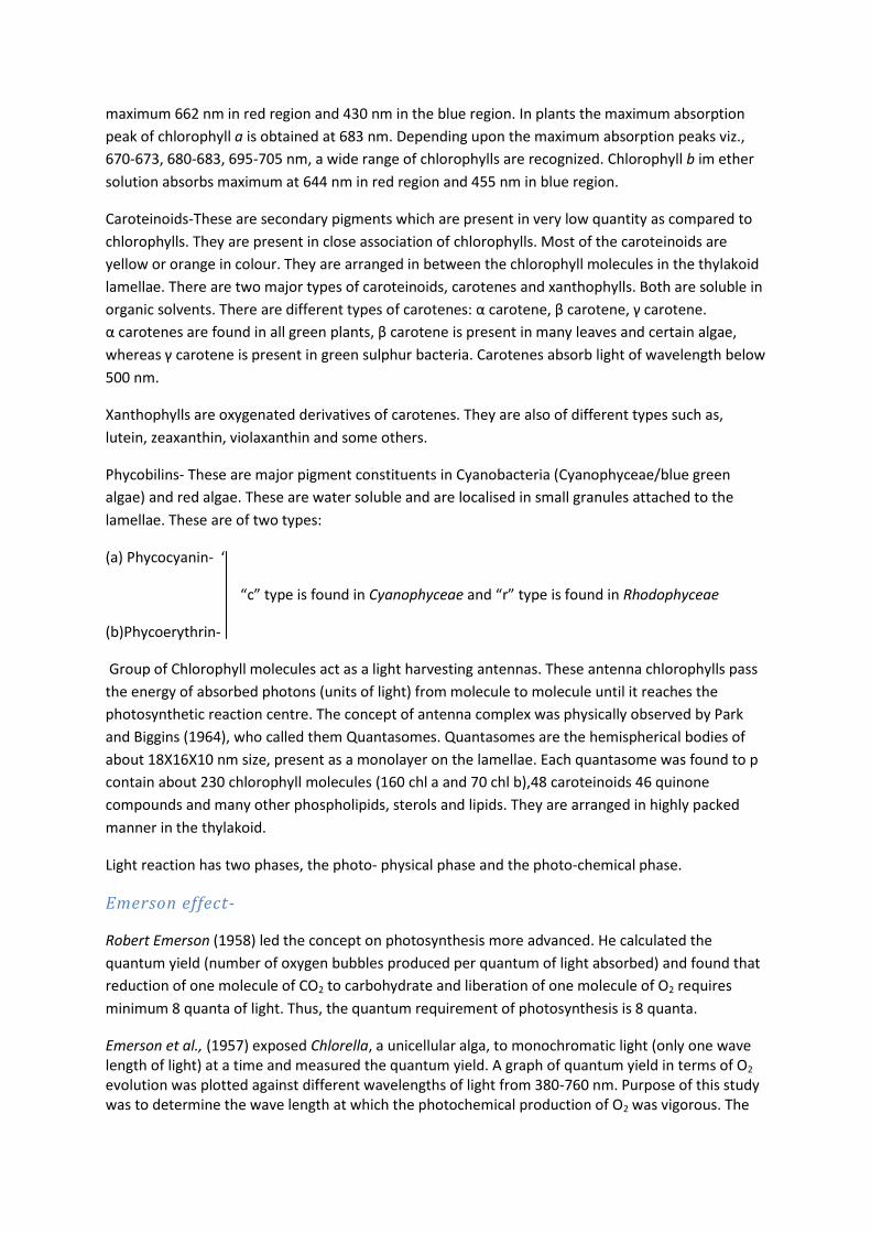
Photo-physical phase –
This phase actually includes the striking of photons and it’s receiving by the pigment molecule. The
normal state of an atom/molecule is known as ground state. When a chlorophyll molecule absorbs a
photon, one of its electrons is promoted from its ground state to the excited state. It’s outer valence
electron is pushed into a high energy orbit and the molecule comes into excited singlet state. The
excited state is unstable having half life of 10 12 seconds. The electron tends to fall back in one of the
several ways. It may release energy in the form of radiation and come to its ground state. This
release of energy in the form of light radiation is known as fluorescence. Fluorescence accounts for
the dissipation of only 3 to 6 % of the light energy absorbed by living plants. It may come to next
higher energy level by losing some of energy in the form of heat. This state is again untenable having
half life of 10 9 seconds, called triplet state. The electro may fall back to ground state from triplet
state by losing radiant energy called phosphorescence.
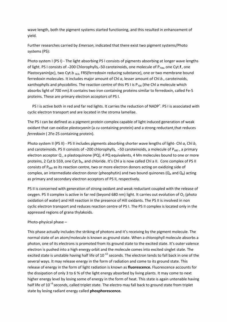
Fig (a)
Fig (b)
Fig (a) Energy diagram indicating the electronic states of chlorophyll and their most important
modes of interconversion. (b)Excitation energy trapping by the photosynthetic reaction centre.
(courtesy: Fundamentals of Biochemistry ; Voet,Voet and Pratt.John Wiley & sons,Inc)
Transfer of exciton (Resonance energy transfer) - When one molecule gets excited it transfers its
energy to the next adjacent chlorophyll molecule and the initial molecule returns to ground state.
The molecule which receives energy gets excited and all the process repeats what happened with
the first chlorophyll molecule. This process continues till the energy finally reaches the reaction
centre (P 700 in PS I and P680 in PS II). Light energy is funnelled to photosynthetic reaction centres
through exciton transfer among antenna pigments. This process is called as Resonance energy
transfer.
Photo-chemical phase –
This phase includes generation of assimilatory power consequent upon the transfer of electrons.
When photon energy reaches the reaction centres of both the photo systems ,electrons present in
the excited states are donated to the respective adjacent receivers.
The first photo-chemical reaction in the photosynthesis is evolution of O2. This is related with PS II
complex. The light energy harvested by the antenna molecules reaches to the reaction centre P 680.
The P680 gets excited and transfer electron to Pheophytin and gets oxidised. P 680 returns to normal
ground state by pulling electrons from water. Water is splitted by photo oxidation as below:
2H2O --------------------- O2 + 4 H + + 4e
Two molecules of water are oxidized to evolve one molecule of O2. In this process hydrogen is not
produced, rather it is carried by PQ, which reduce NADP+.
In the photo oxidation of water (Hill reaction), Mn ++ and Cl are required.
Both the Photo systems are interconnected through a series of electron carriers, which transfer
electrons through them. Transfer of electrons in photo systems takes place in two manners-One is
non cyclic electron transport and another is cyclic electron transport.
Non cyclic electron transport & non cyclic photophosphorylation –
Non cyclic electron transport in PS II is coordinated with the ATP generation during the transfer of electrons. Since, the path of electron transport is linear and it produces ATP during the process it is called NON cyclic photophosphorylation.
This involves role of PS II. Steps involved in non cyclic path are as follows:
The reaction centre P680 gets excited after receiving light energy from antenna molecules. Its
electron is donated to pheophytin –an intermediate electron acceptor. Pheophytin is a modified
chlorophyll a molecule in which two H atoms are replaced by central Mg 2+.
P 680 becomes oxidized by transferring electrons to pheophytin and becomes reduced by receiving
electrons from photo oxidation of water via an unknown compound “Z”.
Water is photo oxidized and O2 is liberated as stated above. The oxygen evolving system involves M
(an intermediate compound), Z-an electron donor to P 680 , P680 and Q the primary acceptor of PS II.
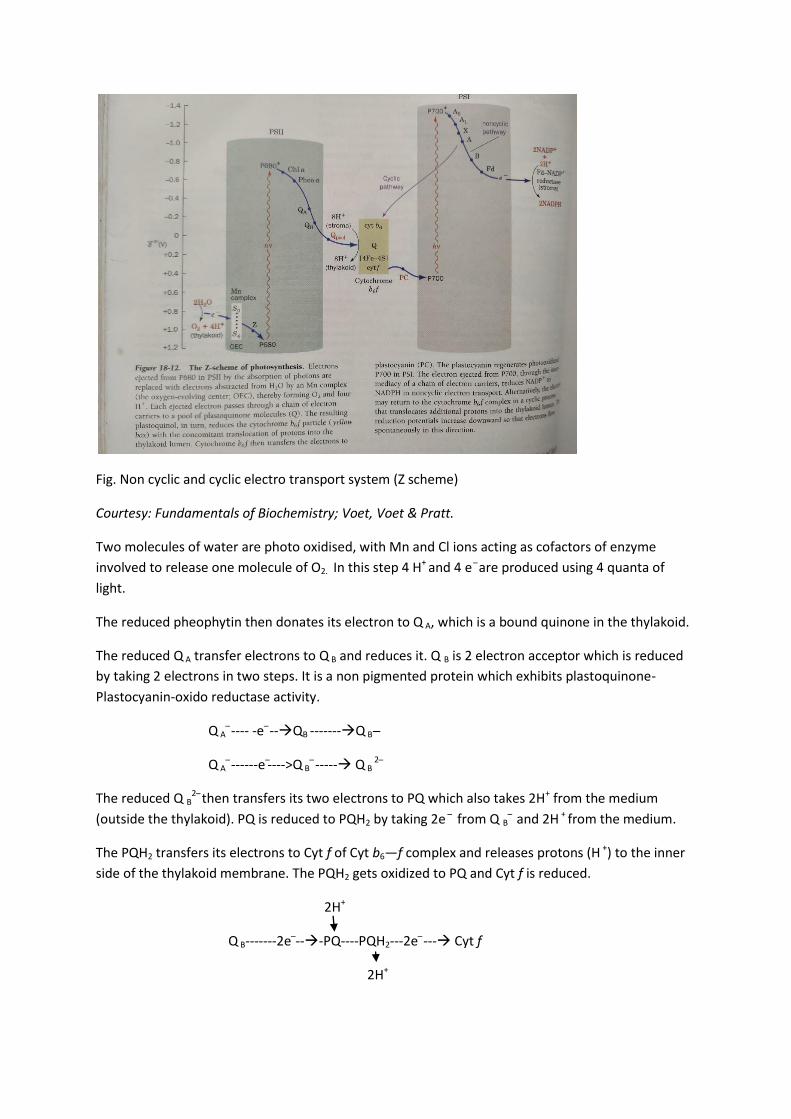
Fig. Non cyclic and cyclic electro transport system (Z scheme)
Courtesy: Fundamentals of Biochemistry; Voet, Voet & Pratt.
Two molecules of water are photo oxidised, with Mn and Cl ions acting as cofactors of enzyme
involved to release one molecule of O2. In this step 4 H+ and 4 e are produced using 4 quanta of
light.
The reduced pheophytin then donates its electron to Q A, which is a bound quinone in the thylakoid.
The reduced Q A transfer electrons to Q B and reduces it. Q B is 2 electron acceptor which is reduced
by taking 2 electrons in two steps. It is a non pigmented protein which exhibits plastoquinone-
Plastocyanin-oxido reductase activity.
Q A ---- -e --QB -------Q B
Q A ------e ---->Q B
----- Q B 2
The reduced Q B2 then transfers its two electrons to PQ which also takes 2H+ from the medium
(outside the thylakoid). PQ is reduced to PQH2 by taking 2e from Q B and 2H + from the medium.
The PQH2 transfers its electrons to Cyt f of Cyt b6—f complex and releases protons (H +) to the inner
side of the thylakoid membrane. The PQH2 gets oxidized to PQ and Cyt f is reduced.
2H+
Q B-------2e ---PQ----PQH2---2e --- Cyt f
2H+
Now, the reduced Cyt f transfers its electron to Plastocyanin (a copper containing protein; PC).
The Cyt f is oxidized and PC is reduced.
Cyt f-------e ----- PC----------e ------ PS I
PS I –Complex
In the photo system I the radiant energy is harvested by its antenna complex and the excitons are
finally transferred to its reaction centre P700. As a result P 700 gets excited and gives its electron to an
unknown compound A 1. P 700 gets oxidized which is reduced by electrons donated by PC.
The reduced A 1 passes its electron to primary electron acceptor PSI-A 2 ,a protein with Fe-S centre.
The reduced A2 then transfers its electron to secondary electron acceptor PSI-A 3, an unknown
compound.
The reduced A 3 then transfers its electron to ferredoxin ( Fd ) present at the outer surface of
thylakoid membrane. As a result ferredoxin is reduced and A 3 is oxidized.
The reduced ferredoxin finally transfers its electron to NADP +, which receives protons( H +) from the
medium and gets reduced to NADPH. This reaction is catalyzed by an enzyme Fd-NADP reductase.
Ferredoxin + NADP + + H +----------Fd-NADP reductase--Ferredoxin (ox.) + NADPH
Significance: Non cyclic electron transport is a linear sequence of electron transfer where NADP+ is
reduced by PS I complex. The PSI is reduced by PS II and ultimately PS II is reduced by electrons
coming from photo-oxidation of water. Since the path of electron is linear, this is called non cyclic .
It releases considerable number of protons (H +) which generates proton motive force to produce
ATP. Hence, the non cyclic electron transport is referred to as non cyclic photophosphorylation.
PS I is the producer of assimilatory power, i.e., NADPH and ATP.
Cyclic electron transport & cyclic photophosphorylation-
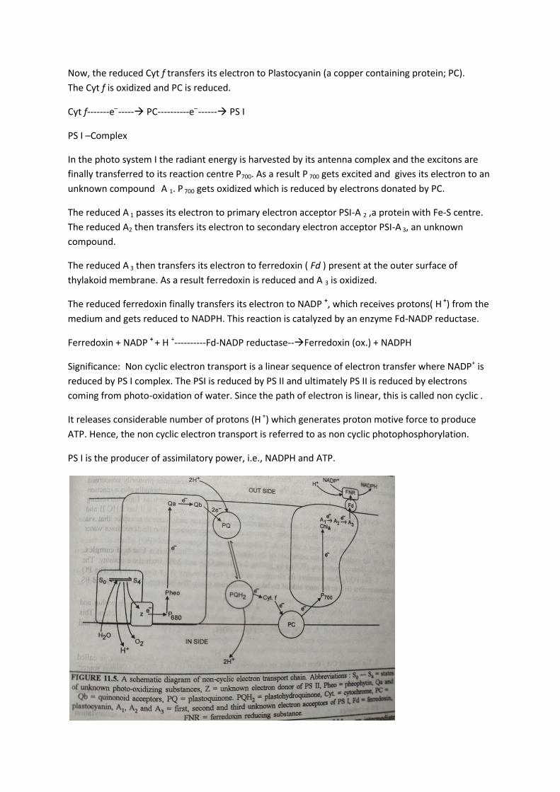
In addition to non cyclic ETS in plants, there exists cyclic electron transport in certain conditions. When there is paucity of CO2 in chloroplast and NADPH starts accumulating, cyclic ETS is favoured in addition to the Non cyclic one. The purpose of cyclic photophosphorylation is more production of ATP. In this condition, electrons released by P 700 are cycled back to PS I via a series of intermediate electron carriers, therefore this is called cyclic ETS. Since, ATP is also generated in this process, it is referred to as cyclic photophosphorylation. Following steps are involved in the cyclic ETS: 1. The antenna chlorophyll molecules receive light energy and get excited, which transfer their exciton to the reaction centre P700. The P700 gets excited and transfer its electron to ferredoxin reducing it via intermediate carriers A1(Chl a), A2(Fe-S), and A3 (P430). 2. The reduced ferredoxin, unable to reduce NADP +, passes its electron to PQ via Cyt b 6 (Cyt b 563). 3. The reduced Cyt b 6 transfers its electron to PQ. The PQ is a hydrogen carrier which gets electrons from Cyt b 6 (in cyclic electron transport) and Q B (in non cyclic ETS) and protons H + from the outer medium to get reduced to PQH2. 4. The PQH2 transfers its electrons to Cyt f and 2H+ to the inner side of thylakoid membrane. 5. The reduced Cyt transfers its electron to Plastocyanin (PC).The PC gets reduced and Cyt f becomes oxidized. 6. The reduced PC moves and donates its electron to oxidised reaction centre of PS I (P700).
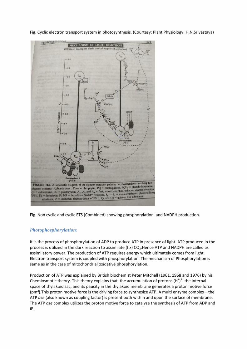
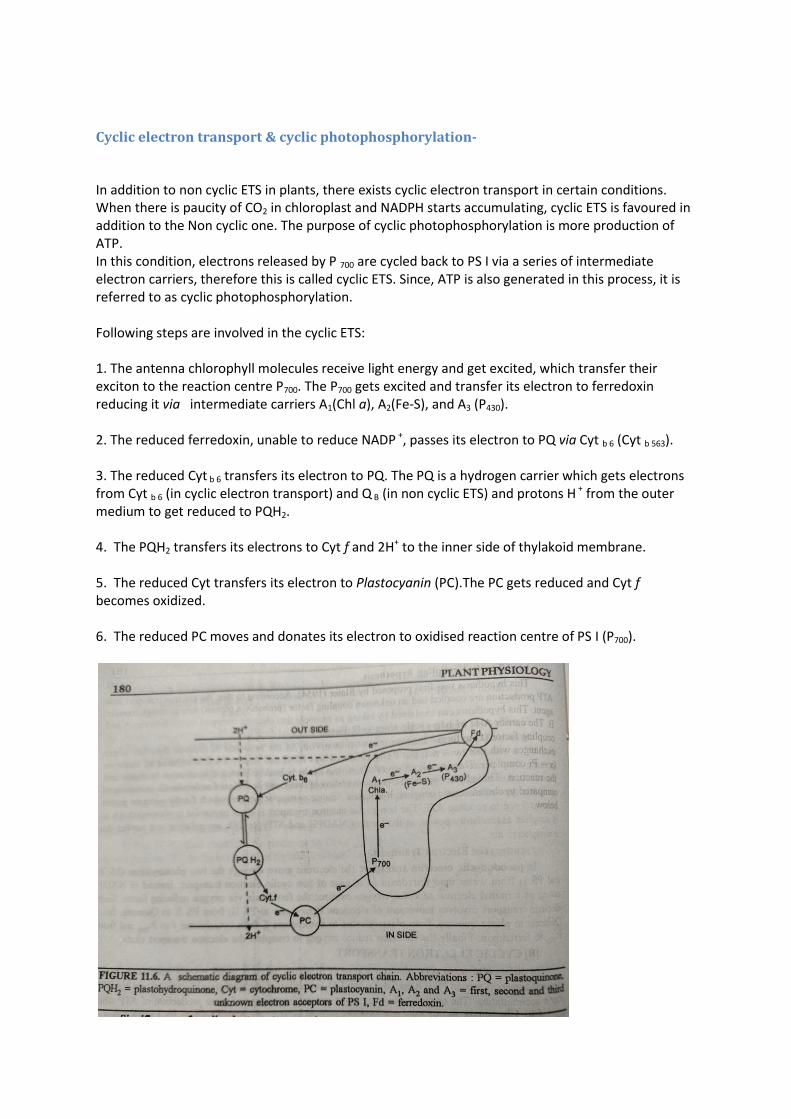
Fig. Cyclic electron transport system in photosynthesis. (Courtesy: Plant Physiology; H.N.Srivastava)
Fig. Non cyclic and cyclic ETS (Combined) showing phosphorylation and NADPH production.
Photophosphorylation:
It is the process of phosphorylation of ADP to produce ATP in presence of light. ATP produced in the process is utilized in the dark reaction to assimilate (fix) CO2.Hence ATP and NADPH are called as assimilatory power. The production of ATP requires energy which ultimately comes from light. Electron transport system is coupled with phosphorylation. The mechanism of Phosphorylation is same as in the case of mitochondrial oxidative phosphorylation. Production of ATP was explained by British biochemist Peter Mitchell (1961, 1968 and 1976) by his Chemiosmotic theory. This theory explains that the accumulation of protons (H+) in the internal space of thylakoid sac, and its paucity in the thylakoid membrane generates a proton motive force (pmf).This proton motive force is the driving force to synthesize ATP. A multi enzyme complex—the ATP ase (also known as coupling factor) is present both within and upon the surface of membrane. The ATP ase complex utilizes the proton motive force to catalyze the synthesis of ATP from ADP and iP.
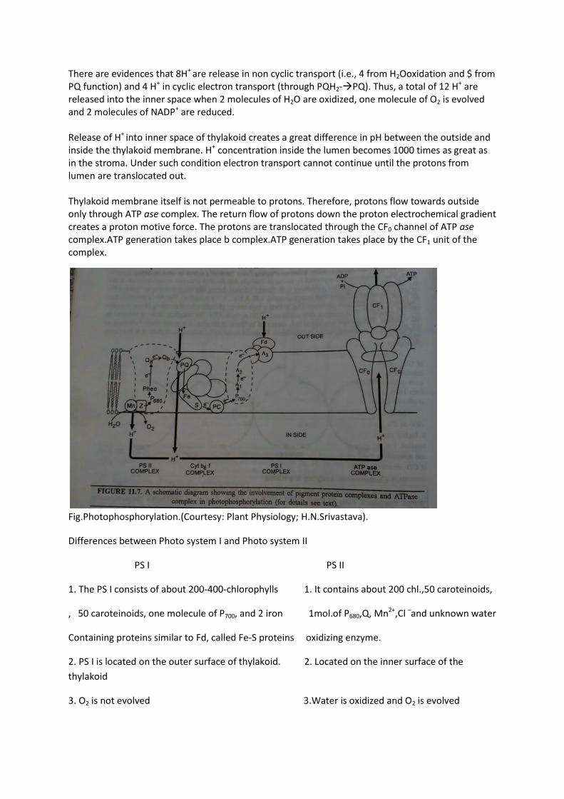
There are evidences that 8H+ are release in non cyclic transport (i.e., 4 from H2Ooxidation and $ from PQ function) and 4 H+ in cyclic electron transport (through PQH2-PQ). Thus, a total of 12 H+ are released into the inner space when 2 molecules of H2O are oxidized, one molecule of O2 is evolved and 2 molecules of NADP+ are reduced. Release of H+ into inner space of thylakoid creates a great difference in pH between the outside and inside the thylakoid membrane. H+ concentration inside the lumen becomes 1000 times as great as in the stroma. Under such condition electron transport cannot continue until the protons from lumen are translocated out. Thylakoid membrane itself is not permeable to protons. Therefore, protons flow towards outside only through ATP ase complex. The return flow of protons down the proton electrochemical gradient creates a proton motive force. The protons are translocated through the CF0 channel of ATP ase complex.ATP generation takes place b complex.ATP generation takes place by the CF1 unit of the complex.
Fig.Photophosphorylation.(Courtesy: Plant Physiology; H.N.Srivastava).
Differences between Photo system I and Photo system II
PS I PS II
1. The PS I consists of about 200-400-chlorophylls 1. It contains about 200 chl.,50 caroteinoids,
, 50 caroteinoids, one molecule of P700, and 2 iron 1mol.of P680,Q, Mn2+,Cl and unknown water
Containing proteins similar to Fd, called Fe-S proteins oxidizing enzyme.
2. PS I is located on the outer surface of thylakoid. 2. Located on the inner surface of the
thylakoid
3. O2 is not evolved 3.Water is oxidized and O2 is evolved

4. It produces a strong reductant which reduces 4. It donates electrons to PS I when NADP+ is
NADP+ to NADPH+ H+ reduced.
5. It involves both cyclic and non-cyclic phospho- 5. It is involved only in non-cyclic photophos-
rylation phorylation.
6. On ultracentrifugation these appear as lighter 6. On ultracentrifugation these appear as
fraction. heavier fraction.
-----------------------------------------------------------------------------------------------------------------


