Embed Size (px)
Citation preview

OPEN
ORIGINAL ARTICLE
Cyanobacterial photosynthesis under sulfidicconditions: insights from the isolate Leptolyngbyasp. strain hensonii
Trinity L Hamilton1,5, Judith M Klatt2,3, Dirk de Beer2 and Jennifer L Macalady4
1Department of Biological Sciences, University of Cincinnati, Cincinnati, OH 45221, USA; 2Max-PlanckInstitute for Marine Microbiology, Bremen, Germany; 3Geomicrobiology Lab, Department of Earth andEnvironmental Sciences, University of Michigan, Ann Arbor, MI, USA and 4Department of Geosciences andthe Penn State Astrobiology Research Center (PSARC), Pennsylvania State University, University Park,PA 16802, USA
We report the isolation of a pinnacle-forming cyanobacterium isolated from a microbial mat coveringthe sediment surface at Little Salt Spring—a flooded sinkhole in Florida with a perennially microoxicand sulfidic water column. The draft genome of the isolate encodes all of the enzymatic machinerynecessary for both oxygenic and anoxygenic photosynthesis, as well as genes for methylatinghopanoids at the C-2 position. The physiological response of the isolate to H2S is complex: (i) noinduction time is necessary for anoxygenic photosynthesis; (ii) rates of anoxygenic photosynthesisare regulated by both H2S and irradiance; (iii) O2 production is inhibited by H2S concentrations as lowas 1 μM and the recovery rate of oxygenic photosynthesis is dependent on irradiance; (iv) under theoptimal light conditions for oxygenic photosynthesis, rates of anoxygenic photosynthesis are nearlydouble those of oxygenic photosynthesis. We hypothesize that the specific adaptation mechanismsof the isolate to H2S emerged from a close spatial interaction with sulfate-reducing bacteria. The newisolate, Leptolyngbya sp. strain hensonii, is not closely related to other well-characterizedCyanobacteria that can perform anoxygenic photosynthesis, which further highlights the need tocharacterize the diversity and biogeography of metabolically versatile Cyanobacteria. The isolate willbe an ideal model organism for exploring the adaptation of Cyanobacteria to sulfidic conditions.The ISME Journal (2018) 12, 568–584; doi:10.1038/ismej.2017.193; published online 12 January 2018
Introduction
Cyanobacteria are the only chlorophototrophs thatcarry out oxygenic photosynthesis and presumablyprovided the first significant source of O2 on earlyEarth. The evolution of oxygenic photosynthesis inancient Cyanobacteria transformed Earth, ultimatelyproviding conditions that ushered in complex multi-cellular life forms. However, even after the firstglobal rise of atmospheric O2 during the GreatOxidation Event, concentrations in ocean surfacewaters remained low and sulfidic conditions were
common throughout much of the Proterozoic(Canfield, 1998; Meyer and Kump, 2008), particu-larly in restricted basins and along productivecontinental margins (Scott et al., 2008; Lyons et al.,2009; Poulton et al., 2010; Poulton and Canfield,2011). Multiple phylogenetic analyses suggest thatthe less complex, anoxygenic modes of photosynth-esis evolved before oxygenic photosynthesis(Blankenship, 2001; Xiong and Bauer, 2002;Sadekar et al., 2006; Bryant and Liu, 2013). Anoxy-genic phototrophs use one reaction center, whichmay be either type 1 or type 2, and do not evolveoxygen. Anoxygenic photosynthesis relies on asupply of reducing equivalents from reduced sulfurcompounds, organic acids, hydrogen, nitrite,arsenite or Fe(II) to drive CO2 reduction. Despitethe ubiquity of H2O as an electron donor for oxygenicphotosynthesis, observations of extant Cyanobacteriacapable of performing anoxygenic photosynthesishave been documented in environments wheresulfide is present in the photic zone and in a handfulof pure cultures (Cohen et al., 1975a, b; Padan, 1979;de Wit and van Gemerden, 1987; Garcia-Pichel andCastenholz, 1990; Klatt et al., 2015a).
Correspondence: TL Hamilton, Department of Plant and MicrobialBiology, University of Minnesota, Room 218 Cargill Building, StPaul, MN 55108, USA.E-mail: [email protected] JM Klatt, Department of Earth & Environmental Sciences,University of Michigan, 2004 CC Little Building, Ann Arbor, MI48109, USA.E-mail: [email protected] address: Department of Plant and Microbial Biology,University of Minnesota, Room 218 Cargill Building, St Paul,MN 55108, USA.Received 26 January 2017; revised 1 September 2017; accepted 9October 2017; published online 12 January 2018
The ISME Journal (2018) 12, 568–584www.nature.com/ismej

The capability to perform anoxygenic photosynth-esis has been considered a relic of cyanobacterialancestors living before the evolution of oxygenicphotosynthesis (Oren et al., 1977; Padan, 1979). Analternative hypothesis is that photosynthetic versati-lity in Cyanobacteria represents an intermediate stateduring the evolution and fine-tuning of oxygenicphotosynthesis, in which Cyanobacteria used eitherH2O or H2S in sulfidic photic zones such as thosepresent at continental margins throughout most ofEarth’s history (Hamilton et al., 2016). In extantCyanobacteria, the ability to perform anoxygenicphotosynthesis, while not common, is widespreadamong phylogenetically diverse Cyanobacteria (Millerand Bebout, 2004). To date, all characterized Cyano-bacteria that perform anoxygenic photosynthesisencode a sulfide quinone oxidoreductase (SQR),which oxidizes sulfide to sulfur and transfers electronsto photosystem I (PSI) (Arieli et al., 1994; Theissenet al., 2003). SQRs can also have other functionsincluding sulfide detoxification. There is evidencethat SQRs have been transferred horizontally(Theissen et al., 2003), and genes encoding multipletypes of SQRs are often found within the samegenome. For SQR sequences in general, there is onlyrough correlation between the topology of phyloge-netic trees of 16S rRNA and SQR sequences from thesame organism (Pham et al., 2008). The widespreadtaxonomic distribution of Cyanobacteria capable ofperforming anoxygenic photosynthesis in the absenceof detectable heritability of this trait supports theemergence of this physiology multiple times throughhorizontal gene transfer. Still, these observations donot discern if SQR, or more specifically, anoxygenicphotosynthesis, is an ancestral trait in Cyanobacteria.
Regardless, little of the physiology and ecology ofancient Cyanobacteria can be gleaned from the fossilrecord. Today Cyanobacteria are key primary pro-ducers in laminated mats of varying morphologies.Sulfur cycling is crucially important in these mats,which also host sulfate-reducing organisms andother anoxygenic phototrophs. Similar communitiesdominated by Cyanobacteria are thought to havebeen present in ancient stromatolites (Walter, 1976).Other fossil information including lipid, chlorophyll,and carotenoid biomarkers provide clues aboutancient microbial community structure and physiol-ogy, but their interpretation is complicated by thediverse organisms that produce them. For instance,hopanoids methylated at the C-2 position wereoriginally thought to be synthesized exclusively byCyanobacteria (Summons et al., 1999). More recentstudies indicate that other organisms also producethese lipids, including anoxygenic phototrophs inproteobacterial clades (Rashby et al., 2007; Welanderet al., 2010). The study of extant Cyanobacteria thatmake stromatolitic structures and lipid biomarkersunder microoxic, sulfidic conditions may yield theinsights necessary to interpret biosignatures in therock record as well as to understand the physiologyand ecology of ancient Cyanobacteria.
Cyanobacterial isolates of known purity capable ofanoxygenic photosynthesis are rare (Cohen et al.,1975a,b; de Wit and van Gemerden, 1987; Klatt et al.,2015a) and the regulation of photosynthetic modesappears to vary among them (Cohen et al., 1975a,b;Klatt et al., 2015a). Biochemical characterization sofar has only identified a single enzyme needed forH2S-driven anoxygenic photosynthesis: a SQR, pro-viding electrons to PSI via the plastoquinone pool(PQ). Recent observations suggest that the light-independent enzyme kinetics of SQR control therates of anoxygenic photosynthesis in Cyanobacteriawhen the sulfide concentration is low, whereas athigher levels of sulfide, light intensity dictates theupper limit of anoxygenic photosynthesis rates (Klattet al., 2015a, 2016a). These observations are, how-ever, complicated by the variability of specificadaptations to fluctuating sulfide concentrationsand irradiance in the environment, particularly inmicrobial mats, and by our lack of understanding ofthe mechanism of sulfide inhibition of PSII (Garcia-Pichel and Castenholz, 1990; Klatt et al., 2015b). Theaffinity of SQR to both H2S and PQ, for instance,varies substantially among the few studied Cyano-bacteria (de Wit and van Gemerden, 1987;Castenholz et al., 1991; Griesbeck et al., 2000; Klattet al., 2015a, 2016a). Overall, there are many gaps inour understanding of how environmental factorsinteract to determine the balance of oxygenic andanoxygenic photosynthesis in metabolically versatileCyanobacteria.
Little Salt Spring (LSS), a flooded sinkhole inFlorida, hosts a seasonal bloom of red pinnacle matsdominated by Cyanobacteria and green sulfur bac-teria (GSB) (Hamilton et al., 2017). The water columnis sulfidic and despite the abundance of Cyanobac-teria in the mat, only a small increase in oxygen(0.2 μM) has been observed in the water columnduring midday (de Beer et al., 2017). The matcontains abundant hopanoids, including a signifi-cant fraction methylated at the C-2 position(Hamilton et al., 2017). Here, we report the isolationand draft genome of a metabolically versatile,pinnacle-forming cyanobacterium from LSS. Weexamined whether the isolate is closely related toother Cyanobacteria capable of both types of photo-synthesis and if the isolate could be the source of2-methyl hopanoids in the pinnacle mats. We usedmicrosensors to determine if the isolate can performboth types of photosynthesis, and developed a modelof photosynthetic electron transport to explorepotential regulatory mechanisms.
Materials and methods
Sample collectionRed pinnacle mat samples were collected by diversin June of 2012 from LSS, a 78-m diameter sinkholelake located in Sarasota County, FL (lat. 27°04′30″N,long. 82°14′00″W). The geology and hydrology of the
Adaptation of Cyanobacteria to sulfidic conditionsTL Hamilton et al
569
The ISME Journal

sinkhole have been described previously (Zarikianet al., 2005) as well as the spring geochemistry andmicrobiology of the red pinnacle mat (Hamiltonet al., 2017). Diver-collected pinnacle mats from thewater-sediment interface at ~ 14-m were placed intosterile 50-ml conical tubes, overlaid with springwater collected from the same location as the matsample, and stored in the dark at 4 °C. Spring waterwas also collected at the water-sediment interfacewith syringes and immediately analyzed for sulfideconcentration as they were brought to the surface.Dissolved sulfide concentration was measured witha portable spectrophotometer (Hach Co., Loveland,CO, USA), using methylene blue for total sulfide(Hach method 690, detection limit ~ 0.2 μM). Sulfideanalyses were conducted in duplicate and werewithin 5% of each other. The total dissolved sulfideconcentration was 21.6 μM at the mat–sedimentinterface at the time of sample collection. Photo-synthetically active radiation (PAR) was measured atthe surface of the red pinnacle mat at the time ofcollection using a scalar quantum PAR sensor (LiCorLI-193S) attached to a LI-COR LI-1400 data logger(LI-COR Biosciences, Lincoln, NE, USA). PAR at thesurface of the red pinnacle mat was 65 μmolphotonsm− 2 s− 1 at the time of sample collection.Dissolved oxygen was measured in situ using amulitparameter YSI 6600 sonde (YSI Inc., YellowSprings, OH, USA) with a luminescent-based dis-solved oxygen sensor (ROX). The sonde was cali-brated daily according to the manufacturer’s protocoland a detection limit of 0.3 μM was determined forthe ROX sensor. In addition, in situ oxygen micro-sensor measurements were recorded in November of2014 (de Beer et al., 2017).
Enrichment, isolation and microscopySamples of red pinnacle mat were homogenized anda small aliquot (~500 μl) was added to BG11 media(Rippka et al., 1979) supplemented with 25mM
HEPES (B-HEPES) and adjusted to pH 7.2. Enrich-ment cultures were maintained in 60ml of liquidmedia in 125-ml conical flasks at 100 r.p.m. at 28 °Cunder either a day–night cycle or continuouslyilluminated with 100 μmol photonsm− 2 s− 1 undercool white fluorescent lamps. An axenic culture wasachieved using a dilution series in liquid mediawhere the highest dilution that showed growth wastaken as the inoculum for the next dilution. Lightmicroscopy was performed periodically to visuallyexamine enrichments for purity. The dilution toextinction strategy was continued until light micro-scopy indicated the presence of a single morphotypeand sequencing of the 16S rRNA gene returned asingle phylotype. In addition, to test for hetero-trophic contaminants, samples of the axenic culturewere plated on LB agar medium supplemented with1% tryptone and 0.5% yeast extract. No colonieswere observed on the plates after incubation in thedark for 5 days. Growth of the isolate was monitored
with chlorophyll a concentration determined spec-trophotometrically using the absorption at 665 nm ofa methanol extract and an extinction coefficient of0.075ml μg− 1 (made from a filtered 2-ml culturesubsample) (De Marsac and Houmard, 1988) orprotein concentration using the Bradford assay(Bradford, 1976) with bovine serum albumin(Sigma-Aldrich, St Louis, MO, USA) as the standard.The isolate is filamentous and forms biofilms andpinnacles.
The isolate was imaged using using an OlympusBX53 digital microscope (Olympus, Tokyo, Japan)and an Olympus DP73 digital camera with Cellsensdigital image software (Olympus American Inc.,Center Valley, PA, USA).
Nucleic acid analysesSamples of biofilm (~1.5ml) were harvested bycentrifugation, the excess media removed by decant-ing and cell pellets frozen immediately (−20 °C) orsubjected to nucleic acid extraction. Genomic DNAwas extracted as described previously (Boyd et al.,2007). Quality of extracted DNA was assessed on anagarose gel (1%) using the HiLo DNA Marker(Bionexus, Oakland, CA, USA) and visualized byethidium bromide staining and using a NanoDropND-1000 spectrophotometer (NanoDrop Technolo-gies, Wilmington, DE, USA). To check for purity/contaminants, 16S small subunit RNA genes wereamplified with bacterial domain primers 27F and1492R (Lane, 1991) as described previously(Hamilton et al., 2017). Reactions were performedin triplicate, purified using a QIAquick PCR Purifica-tion Kit (Qiagen, Valencia, CA, USA) and sequencedat the Genomics Core Facility of the Huck Institutesof the Life Sciences at the Penn State University.Sequences were assembled and manually checkedusing using Bio-Edit (v.7.2.5), and checked forchimeras using CHIMERA_CHECK (Cole et al.,2003). Putative chimeras were excluded from sub-sequent analyses. A single 16S rRNA gene sequencewas recovered indicating the culture was pure.
Genomic sequencing, assembly and completenessA draft genome of the isolate was generated fromgenomic DNA extracted as described above. PurifiedDNA was sequenced with on an Ion Torrent PersonalGenome Machine according to the Ion Torrentprotocol at the Penn State University sequencingfacility. Specific details are provided in the Supple-mentary Online Material (SOM). The resulting readswere assembled with Newbler assembler version 2.6(Roche Diagnostics, Basel, Switzerland) resulting in77 contiguous reads (contigs) of at least 500 bp withan average read depth of ~ 100× . Contigs wereannotated using RAST (http://rast.nmpdr.org; Azizet al., 2008) and manually curated. Genome com-pleteness was evaluated using conserved house-keeping genes (Supplemetary Table S1) and
Adaptation of Cyanobacteria to sulfidic conditionsTL Hamilton et al
570
The ISME Journal

phylogenetic marker genes identified with Phyla-AMPHORA (Wang and Wu, 2013). The draft genomeof the LSS cyanobacterium (scaffolds larger than1000 bp in length) was submitted to the IntegratedMicrobial Genomes Expert Review automated pipe-line from Joint Genomes Institute for annotation ofgenes and pathways (IMG accession number:2708742396). In addition, raw reads and assembledscaffolds have been submitted to NCBI (project:PRJNA355315).
Phylogenetic analysisThe 16S rRNA gene sequence from the LSS isolatewas compared to full-length or near full-lengthsequences in public databases using BLASTN(Altschul et al., 1997). Sequences were added to anexisting 16S rRNA alignment in ARB (Ludwig et al.,2004), and manually refined. Maximum likelihoodtrees were constructed using PhyML (Guindon et al.,2010) with 1000 bootstrap replicates using thegeneral time-reversible model and substitution para-meters estimated from the data. The resulting treeswere viewed and edited using iTOL (http://itol.embl.de/)(Letunic and Bork, 2016).
For gene-specific phylogenetic analyses, full-length sequences were identified in the genomeusing functional annotation and BLASTX, translatedand verified by BLASTP. Reference datasets werepopulated by detecting homologs in IMG genomicdatabases by BLASTP (Altschul et al., 1997). Proteinsequences were aligned with MUSCLE (Edgar, 2004)and redundancy in the alignments was reducedthrough the Decrease Redundancy Program (http://web.expasy.org/decrease_redundancy/). Maximumlikelihood trees were constructed using PhyML(Guindon et al., 2010) with the LG+gamma model,4 gamma rate categories, 10 random starting trees,NNI branch swapping and substitution parametersestimated from the data. The resulting trees wereviewed and edited using iTOL (http://itol.embl.de/)(Letunic and Bork, 2016).
Microsensor measurementsRates of oxygenic and anoxygenic photosynthesisdependent on varying irradiance and H2S concentra-tion were determined in biofilms of the isolate usingmicrosensors. Biofilms of the isolate were grown onpresterilized glass fiber filters. For the measure-ments, single filters were transferred into 500mlcustom-made aquarium (Supplementary Figure S1).The aquarium was equipped with a horizontallystretched polyester fibrous web that separated abottom and a top reservoir filled with B-HEPESmedia similar to the setup described in Klatt et al.,2015b (Supplementary Figure S1). The filter wasplaced on top of the web and held in place with pins.When desired, sulfide was added from a neutralizedNa2S stock solution (pH 7–7.5) to both bottom andtop reservoir to ensure that the biofilm had sulfide
supplied from both directions. A circular flow of thewater column above the biofilm was achieved bystreaming N2 gas onto the paraffin–oil surface.Illumination in the visible range was provided by ahalogen lamp (Schott KL-2500, Mainz, Germany)mounted above the aquarium. The incident irradi-ance at the surface of the biofilm was determinedwith a submerged cosine-corrected quantum sensorconnected to a LI-250A light meter (both LI-CORBiosciences).
O2, pH and H2S microsensors with a tip diameter of10–30 μm and response time of o1 s were built,calibrated and used as described previously(Revsbech, 1989; Jeroschewski et al., 1996; de Beeret al., 1997). The microsensor tips were positioned atthe biofilm surface and always separated by o50 μmduring simultaneous O2, pH and H2S measurements.All measurements were performed at room tempera-ture. Volumetric rates of gross oxygenic photosynth-esis (GOP) were estimated based on the dynamics ofoxygen concentration after a light-dark shift asdescribed previously (Revsbech and Jørgensen,1986). Analogously, volumetric rates of gross anoxy-genic photosynthesis (GAP) were calculated from thedynamics of H2S concentration and pH directly after alight-dark transition (i.e., light-driven Stot consumptionrates) (Klatt et al., 2016a, b).
Before each measurement in the presence ofsulfide, GOP as a function of irradiance (9–289 μmolphotonsm−2 s−1) was quantified (photosynthesis-over-irradiance-curve (PI-curve)). Irradiance was thenadjusted to a specific value chosen from the PI-curveand GOP was determined again, before addition ofsulfide. Following injection of sulfide into the watercolumn, the local H2S concentration, GOP and GAP inthe surface of the biofilm were monitored untilcomplete depletion of sulfide and recovery of GOP.After recovery of GOP, the PSII inhibitor, 3-(3,4-dichlorphenyl)-1,1-dimethylurea (DCMU; Sigma-Aldrich, St Louis, MO, USA) was added to the watercolumn (final concentration: 10 μM) during exposureto light and before the readdition of sulfide. GOP andGAP in the biofilm were again monitored untilcomplete depletion of sulfide. This procedure wasrepeated at four light intensities (23, 36, 137 and180 μmol photonsm−2 s−1, n=2 for each intensity) onfresh biofilm samples.
To compare GAP and GOP, all reactant consump-tion/production rates were converted to theoreticalelectron transport rates as described previously(Klatt et al., 2015a; 2016a). Briefly, assumingzero-valent sulfur is the primary product of cyano-bacterial anoxygenic photosynthesis, rates oftotal sulfide (Stot = ∑(H2S,HS−,S2− )) consumptionwere multiplied by a factor of 2 to account for twoelectrons per consumed sulfide that are theoreticallytransported and used for NADP+ reduction.Analogously, the rates of O2 production were multi-plied by a factor of 4. To allow for the comparison ofresults obtained in different biofilm samples, wenormalized GOP and GAP to GOPmax, the maximum
Adaptation of Cyanobacteria to sulfidic conditionsTL Hamilton et al
571
The ISME Journal

rate of GOP electron transport, in the respectivebiofilm sample.
CO2 photoassimilationThe contribution of PSII-independent anoxygenicphotoassimilation of CO2 with H2S as the electrondonor was determined in assays containing the PSIIinhibitor DCMU. All experiments were carried out at30 °C in B-HEPES media at pH 7.2. Exponentiallygrowing cells were harvested by centrifugation,resuspended in fresh O2-free media and introduced(10-ml suspensions) into O2-free 20-ml cappedserum vials. Vials were kept in the dark and purgedwith N2 to remove any remaining O2. NaH13CO3
(Cambridge Isotope Laboratories Inc., Andover, MA,USA) was added to each vial at a final concentrationof 100 μM. To assess the PSII-independent anoxy-genic CO2 assimilation, vials (n=3 for each treat-ment) were amended with 100 μM neutralized Na2Sand 10 μM DCMU (Sigma-Aldrich). To analyze theeffects of Na2S or DCMU alone on CO2 photoassimi-lation, vials (n=3 for each treatment) were amendedwith 100 μM Na2S or 10 μM DCMU. Natural abun-dance controls (n=3 vials) received unlabeledNaHCO3 (100 μM). Vials were incubated at a lightintensity of 100 μmol photonsm−2 s− 1 under coolwhite fluorescent lamps. Following incubation, cellswere harvested by centrifugation and freeze-dried.Dried samples were treated with concentrated HCl(1 M) to remove excess carbonate, washed withdeionized H2O to remove excess acid and freeze-dried again. Samples were weighed and placed into
tin dishes, sealed and analyzed via an automatedelemental analyzer (FlashEA, 1112 series) coupled toa Delta Plus Advantage mass spectrometer (FinniganDeltaplusXP) (both from Thermo Scientific) asdescribed previosuly (Klatt et al., 2015a). All assayswere performed in triplicate.
Electron transport modelWe combined previously described models of (i) themain electron transport chain components in photo-synthetically versatile Cyanobacteria (Klatt et al.,2015a) (Figure 1) and (ii) the inhibition kinetics ofthe oxygen evolving complex (OEC) by H2S (Klattet al., 2015b) (Figure 1) to fit our experimental data ofGOP and GAP. Briefly, the model of the oxygenicand anoxygenic electron transport chain focuses onthe PQ where both pathways intersect (Figure 1). Inthis model the affinity of PSII components and SQRtowards PQ, as well as light harvested in both PSIand PSII, represent the main regulatory parametersof photosynthetic activity. Inhibitory effects of H2Son oxygenic photosynthesis were not considered inthis model because such effects were not observed inthe specific study organism of Klatt et al., 2015a. Toaccount for inhibitory effects of H2S on activity of theLSS cyanobacterium, we extended the model byintroducing the interference of H2S with the watersplitting reaction proposed by Klatt et al. (2015b).Briefly, according to this model H2S binds to anintermediate of the OEC that is formed at a rate thatis determined by the light intensity (Figure 1).
Figure 1 Proposed model for the kinetic control of the redox reactions involved in oxygenic and anoxygenic photosynthesis by strainhensonii. Gray arrows represent reactions involved only in anoxygenic photosynthesis; black arrows are those reactions that are involvedonly in oxygenic photosynthesis. E is the photon flux; PSII, PSII* and PSIId are photosystem II in the ground, excited and degraded/inactive state, respectively; OEC is the oxygen evolving complex. OECox is an intermediate formed during H2O oxidation that is inhibitedby H2S. OEC:H2S is the inhibited form of this intermediate; SQR is the sulfide quinone oxidoreductase that couples the oxidation of sulfideto the reduction of the oxidized part of the plastoquinone pool (PQox), which yields zero-valent sulfur and reduced PQ (PQred). ‘ET’ isrepresentative of any intermediate electron transport chain component between PQ and photosystem I (PSI) that serves as the electronacceptor for an unidentified sulfide oxidase (‘USO’). ‘ET’ could be cyt b6f, plastocyanin or cytochrome c553. The PSI reaction center canreceive electrons from the reduced intermediate ‘ET’. In the alternative version of the model that does not involve ‘USO’, PQ directlyreduces PSI. These electrons are used to reduce NADP+ to NADPH, which serves as the electron donor during CO2 fixation. Definitions ofthe process rates (ki) are given in Table 2. Rates that were introduced to specifically explain the photosynthetic activity patterns in strainhensonii and that are not based on previously described models (Klatt et al., 2015a, b) are highlighted in bold (kD, kR, kPQ, kAP2, kCO2 ).Details of the model are provided in the SOM.
Adaptation of Cyanobacteria to sulfidic conditionsTL Hamilton et al
572
The ISME Journal

Implementation into a numerical model using thedeSolve package in R (Soetaert et al., 2010; http://cran.r-project.org) allowed us to test hypothesesabout the regulation of photosynthesis and to fit theexperimental data. We introduced additional path-ways (rates highlighted in bold in Figure 1) when-ever the observed rates could not be explained basedon the previously existing models. The fundamentalconcepts of the models described previously, such asa constant total PQ pool with variable oxidized andreduced fraction (Klatt et al., 2015a), were main-tained in the implementation process. Details of theformulation of process rate laws are provided in theSOM.
Results and discussion
Isolation of Leptolyngbya sp. strain hensoniiSamples of red pinnacle mat collected from thesediment–water interface in LSS were used forisolation by serial dilution. Multiple transfers ofthe red filaments making up the majority of themat biomass resulted in an axenic culture of acyanobacterium which contains chlorophyll a, phy-cocyanin, phycoerythrin and allophycocyanin.The isolate is red colored, filamentous(Supplementary Figure S2), motile and forms pinna-cles in pure culture.
The 16S rRNA gene sequence of the isolate isidentical to a sequence recovered from the redpinnacle mat in LSS (KP728185; Hamilton et al.,2017). Based on BLASTN analyses against all non-redundant nucleotide sequences in the NCBI-NTdatabase, the isolate is closely related to an unculturedclone from copper mine water (KF287742; 95%sequence identity) and copper mine tailings(JQ769661; 95% sequence identity). The 16S rRNAgene sequence of the isolate also shared 94% sequenceidentity with clones recovered from the benthic zoneof an east Antarctic Lake (DQ181675, DQ181685).
The phylogenetic position of the isolate wasevaluated by comparing a 1293 bp fragment of the16S rRNA gene sequence with closely relatedCyanobacteria for which draft or full genomes areavailable (Figure 2). In this analysis, the 16S rRNAsequence of the isolate formed a monophyleticbranch (bootstrap value 0.91) with the most closelyrelated sequence—Leptolyngbya sp. strain JSC1within subsection III. Leptolyngbya sp. strain JSC1was isolated from a ferrous iron-rich hot spring withcircumneutral pH in Yellowstone National Park(Brown et al., 2010). The LSS isolate is also closelyrelated to Geitlerinema sp. PCC 7407 and Oscillator-iales cyanobacterium UVFP2 (~94% sequence iden-tity)(Figure 2). A highly enriched culture ofOscillatoriales cyanobacterium UVFP2 was obtainedfrom Fuente Podrida, a sulfide-rich spring close tothe Cabriel River in Valencia, Spain (Camacho et al.,2005), while the isolation source of Geitlerinema sp.PCC 7407 has not been published. The pinnacle-
forming LSS cyanobacterium strain was namedLeptolyngbya sp. strain hensonii, for its growth habitresembling the fur of Jim Henson’s famous puppets.We acknowledge that Leptolyngbya are polyphyletic;however, a systematic nomenclature for Cyanobac-teria has not been published (Komárek, 2016) andthe description of Leptolyngbya is consistent withour isolate—long filaments with solitary or coiledclusters and fine mats.
Genome featuresThe draft genome of strain hensonii contains 77contigs and 5 940 030 bp with an average GC contentof 52.3% (Table 1). The draft genome encodes 61tRNAs and 5627 protein coding genes (Table 1),including all of the conserved housekeeping genes(Supplementary Table S1) and 96% of the phylum-specific marker genes identified with Phyla-AMPHORA. All of these metrics suggest that thegenome is nearly complete.
The genome encodes the enzymes necessary foraerobic photoautotrophic growth including Form IRuBisCO and a complete Calvin–Benson cycle; twohigh affinity terminal oxidases, a cytochrome coxidase and a bd-type quinol oxidase; PSI and PSII;chlorophyll biosynthesis pathway enzymes; and acytochrome b6f complex. The bd-type quinol oxidaseand cytochrome c oxidase differ in their affinity foroxygen (0.35 vs 1.0 μM) (Pils and Schmetter, 2001),and the former is postulated to be expressed underlow oxygen conditions (Hart et al., 2005). Genesencoding a succinate dehydrogenase (sdhABC), anF-type ATPase and a NAD(P)H:quinone oxidoreduc-tase (NDH) are also present. Thus, hensonii isexpected to be capable of aerobic respiration undervariable O2 concentrations. In the environmenthensonii is indeed exposed to fluctuating O2 over adiel cycle (de Beer et al., 1997). O2 is, however, notavailable from the water column in situ but isexclusively produced by oxygenic photosynthesis,with hensonii as a main source. Under in situconditions in LSS the enzymatic machineryfor aerobic respiration likely serves to maintaincellular redox balance, with the terminal oxidases,for instance, serving as electron valves forthe photosynthetic electron transport reactions(Supplementary Figure S3). This implies that term-inal oxidases would thus never be used in thepresence of the inhibitory H2S (Beauchamp et al.,1984; Cooper and Brown, 2008). The genome alsoencodes the enzymatic machinery necessary forassimilatory nitrate reduction and assimilatory sul-fate reduction and nitrogen fixation via a Mo-dependent nitrogenase.
Three enzymes integral to photosynthesis—copro-porphyrinogen III oxidase, heme oxygenase and Mg-protoporphyrin IX monomethylester cyclase—require oxygen for activity. However, oxygen levelsat the water depth hosting the red pinnacle mats inLSS reach only 0.2 μM oxygen (de Beer et al., 2017).
Adaptation of Cyanobacteria to sulfidic conditionsTL Hamilton et al
573
The ISME Journal

Genes encoding alternative forms of these enzymeshave been observed in genomes of Cyanobacteriafrom diverse environments (Panek and O’Brian,2002) and, in the cyanobacterium Synechocystis sp.PCC 6803, the alternative forms of these enzymes areexpressed under low oxygen conditions (Aoki et al.,
2011). Consistent with environmental conditions inthe natural habitat of the isolate, we found homologsof both the aerobic and anaerobic forms of theseenzymes in the strain hensonii genome. MultiplepsbA genes (which encode a subunit of PSII) havebeen observed in the genomes of sulfide-tolerant
Figure 2 Maximum likelihood phylogenetic 16S rRNA gene tree of closely related Cyanobacteria and Leptolyngbya sp. strain hensonii.Accession numbers are provided in parentheses. Circles represent bootstrap support values 485 based on 1000 bootstrap samplings.
Adaptation of Cyanobacteria to sulfidic conditionsTL Hamilton et al
574
The ISME Journal

and/or sulfide-using Cyanobacteria (Grim and Dick,2016). These additional copies of psbA facilitateoxygenic photosynthesis under conditions of varyingoxygen and light (Mohamed et al., 1993). In thestrain hensonii draft genome, we observed the copiesof the canonical oxygenic group 4 psbA (Cardonaet al., 2015) as well as a group 3 psbA, which havebeen recovered from cyanobacterial genomic binsfrom a low-oxygen cyanobacterial mat in the MiddleIsland Sinkhole (Voorhies et al., 2012), and a group 2psbA. Transcripts of group 2 psbA have beenobserved under microaerobic conditions in culturesof Synechocystis PCC 6803, Anabaena PCC 7120 andThermosynechococcus elongatus (Sicora et al.,2009).
The draft genome of Leptolyngbya sp. strainhensonii encodes a single SQR—specifically anSQR type F. SQR catalyzes the oxidation of sulfideto zero-valent sulfur and may have a physiologicalrole in both energy transduction and sulfide detox-ification. SQR has also been implicated in anoyx-genic photosynthetic activity in Cyanobacteria(Shahak et al., 1998) (see Figure 1). SQR sequencescan be divided into seven classes (A, B, C, D, E, F orX) and a single genome can encode multiple SQRhomologs (Gregersen et al., 2011). SQRA are typi-cally found in Cyanobacteria, Proteobacteria andAquificaceae. SQRD and SQRX form two paralogousclades—SQRD homologs are encoded by strains ofGSB, Proteobacteria and Actinobacteria (Gregersonet al., 2011), while SQRX homologs are encoded byGSB. No representative of SQRC has been demon-strated to oxidize sulfide. SQRB homologs are oftenrecovered from eukaryotes, while SQRE catalyzessulfide oxidation in the archaeon Acidianus ambiv-alens (Brito et al., 2009). SQRF homologs arecommonly observed in the genomes of in GSB,Proteobacteria, Aquificaceae and Cyanobacteria(Supplementary Figure S5); however, an SQRF froma Cyanobacteria has not been characterized. In theGSB Chlorobaculum tepidum, SQRF is important forgrowth at high sulfide concentration (≥4mM) (Chanet al., 2009; Holkenbrink et al., 2011). The hensoniiSQRF is only distantly related (25% sequenceidentity) to the Type A SQR sequences of Apha-nothece halophytica and Geitlerinema sp. PCC 9228(formerly Oscillatoria limnetica), which have been
implicated in anoxygenic photosynthesis in theseisolates (Cohen et al., 1986).
Red pinnacle mats collected from LSS containelevated concentrations of hopanoids, includingthose methylated at the C-2 position (Hamiltonet al., 2017). The isolate genome encodes homologsof two enzymes that are presumably necessary forthe biosynthesis of 2-methylhopanoids—a squalene–hopene cyclase and a radical SAM methylase (HpnP)(Welander et al., 2010). The translated HpnP homo-log branches with other cyanobacterial HpnPsequences (Supplementary Figure S6) and is iden-tical to the hpnP transcript recovered from LSS(Hamilton et al., 2017). These results suggest that theisolate is a source of 2-methyl hopanoids in LSS.Several lines of evidence suggest that anoxic condi-tions favor the production of 2-methyl hopanoids: (1)an increased abundance in Proterozoic rocks com-pared to Phanerozoic rocks (Summons et al., 1999);(2) increased abundance in rocks recording oceanicanoxic events in the Phanerozoic (Knoll et al., 2007;Talbot et al., 2008; Cao et al., 2009; Kasprak et al.,2015); and (3) higher abundance of hpnP genes inenvironments where anoxic conditions prevail (Ricciet al., 2013). The recovery of 2-methyl hopanoidsfrom anoxic mats in LSS is consistent with theselines of evidence, and the isolation of a cyanobacter-ium with the genetic machinery to synthesize2-methyl hopanoids will facilitate future studiesaimed at determining their functions in adaptationand/or metabolism.
Strain hensonii performs anoxygenic photosynthesisBecause the Leptolyngbya sp. strain hensonii livesunder anoxic and sulfidic conditions in situ (de Beeret al., 2017) and the genome encodes at least oneSQR protein, we hypothesized that the isolate couldperform anoxygenic photosynthesis. Indeed, in thepresence of sulfide and the PSII inhibitor DCMU, theisolate assimilated 5.7 (±0.71) μmol Cmg dryweight− 1 suggesting anoxgenic photosynthetic activ-ity (Figure 3). The capability to perform anoxygenicphotosynthesis was confirmed by microsensor-basedmeasurements in the absence and presence of DCMU(Figure 4). In fact, microsensor-based measurementsindicate that no induction time is necessary foranoxygenic photosynthesis (Figure 4), even if strainhensonii had been grown aerobically before expo-sure to sulfide. This is in contrast to other character-ized Cyanobacteria capable of performinganoxygenic photosynthesis that require ~ 2 h in thepresence of sulfide before performing this activity(Oren and Paden, 1978; Cohen et al., 1986; Klattet al., 2015a). Oxygenic and anoxygenic photosynth-esis were never observed to occur simultaneouslyduring our experiments because oxygenic photo-synthesis was inhibited by H2S concentrations of1 μM or lower based on the detection limit of thespecific H2S sensor used (Figure 4).
Table 1 Statistics for the Leptolyngbya sp. strain hensonii draftgenome
Scaffolds 77Longest scaffold (bp) 544 817
General informationTotal bp 5 940 030N50 (bp) 137 782
CharacteristicsG+C 52.32tRNA 61Protein coding genes 5627
Adaptation of Cyanobacteria to sulfidic conditionsTL Hamilton et al
575
The ISME Journal

Regulation of anoxygenic photosynthesisIn addition to oxygenic photosynthesis, severalCyanobacteria are capable of using sulfide as anelectron donor, that is, performing anoxygenicphotosynthesis using only PSI (Cohen et al.,1975a, b; de Wit and van Gemerden, 1987); however,the mechanism for regulating anoxygenic photosyn-thetic activity is different between isolates (Cohenet al., 1986; Garcia-Pichel and Castenholz, 1990;Klatt et al., 2015a; 2016a). Below we report specificactivity patterns dependent on H2S concentrationand irradiance of strain hensonii in laboratoryexperiments and discuss plausible regulationmechanisms (see the SOM for additional detailsand discussion).
Light and H2S concentrationAnoxygenic photosynthesis was regulated by bothirradiance and H2S concentration—GAP increasedwith increasing H2S concentration until a light-dependent maximum (GAPmax; dashed horizontallines in Figure 5) was reached. The initial increase ofGAP over low H2S concentrations and the saturationeffect at higher concentrations resembled H2S-dependent Michaelis–Menten kinetics. The initialslope of the increase and the maximum GAP werealso light-dependent. All patterns were strictlydependent on H2S concentration and were notaffected by the temporal dynamics of exposure tosulfide (see H2S dynamics in Figures 4b and c). Asexpected, the inhibition of GOP by DCMU did nothave an effect on GAP (compare open and closedsymbols in Figure 5) because GOP was also inhibitedby H2S.
H2S dependency suggests kinetic regulation of GAPThe increase of GAP with H2S until a light-dependent maximum (GAPmax; Figure 5) is consis-tent with previous observations (Cohen et al., 1986;Garcia-Pichel and Castenholz, 1990; Klatt et al.,
2015a, 2016a) and can be explained using apreviously described model of the anoxygenicphotosynthetic electron transport reactions (Klattet al., 2015a). According to the model, the H2Soxidation rate by SQR is concentration-dependentand SQR donates electrons to the PQ. Reoxidation ofPQ is governed by light harvested in PSI. Thus, H2Soxidation proceeds at a rate that depends on theaffinity of SQR for H2S and the oxidized part of thePQ pool (kAP; Figure 1; Table 2; SOM) and isconsequently governed by H2S concentration andthe availability of oxidized PQ.
Light dependency suggests multiple sulfide-oxidizing enzymes. Irradiance had two effects onGAP: (a) it determined GAPmax and (b) it affected theinitial slope of GAP increase with H2S concentration(Figure 5). The first effect can be explained byconsidering that rates of H2S oxidation can onlyincrease with H2S concentration until PSI becomes abottleneck for electron transport reactions (Klattet al., 2015a). Specifically, the light energy harvestedin PSI dictates the maximum electron transport ratein the irradiance range below light saturation (ktot;Figure 1; Table 2; SOM), which also representsGAPmax (Figure 5).
Intriguingly, the light-dependent slope of theincrease in GAP with H2S concentration could notbe explained using the previously described modelfor anoxygenic photosynthesis in Cyanobacteria.Different light-dependent slopes of GAP have beenobserved in a cyanobacterium, but these could beexplained by GAP and GOP occurring simulta-neously, with the two photosynthetic modes com-peting for the PQ pool (Klatt et al., 2016a). However,GAP and GOP are not performed concurrently instrain hensonii, suggesting light must have morecomplex, previously unconsidered, effects on GAP.
To fit our data with the model of the anoxygenicphotosynthetic electron transport reactions, we hadto suspend a basic assumption: a steady pool of asingle sulfide-oxidizing enzyme. Specifically, wehad to make one of two plausible assumptionsinstead: (a) The isolate is equipped with one ormultiple types of SQR and the abundance of theseenzymes is dependent on irradiance. For instance, if,at higher light intensities the synthesis of SQR isupregulated and thus has a higher vmax (see kAP_B inTable 2; SOM), the result would be more active SQRsand an increased maximum rate of H2S oxidation,manifested in a steeper initial slope in GAP (graylines in Figure 5 for model output). (b) There are twotypes of sulfide-oxidizing enzymes (SQR and uni-dentified sulfide oxidase ‘USO’ in Figure 1 andSupplementary Figure S4) with different affinitiesfor H2S, with SQR donating electrons to the PQ pooland ‘USO’ donating electrons into the electrontransport chain at some other level, most likely tocytochrome b6f or plastocyanin (or cytochrome c553),which are encoded in the genome of hensonii (see‘USO’ and kAP2 in Figure 1 and Table 2; black lines in
Figure 3 Inorganic carbon assimilation by Leptolyngbya sp.strain hensonii. Error bars obtained from triplicate measurements.
Adaptation of Cyanobacteria to sulfidic conditionsTL Hamilton et al
576
The ISME Journal
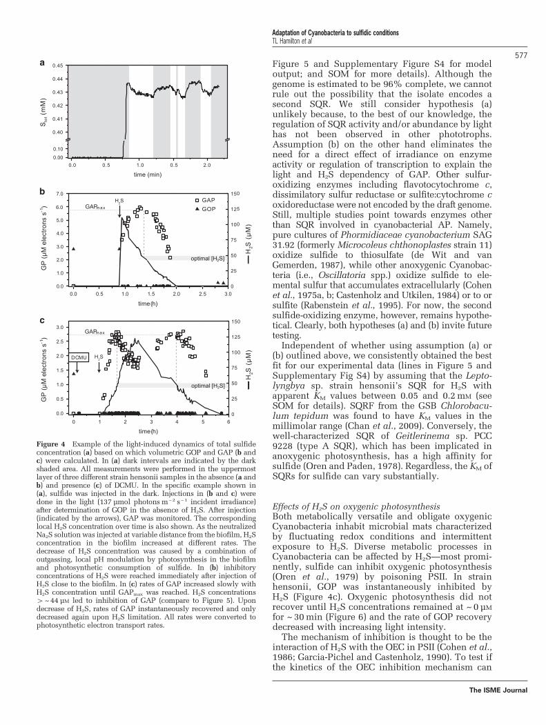
Figure 5 and Supplementary Figure S4 for modeloutput; and SOM for more details). Although thegenome is estimated to be 96% complete, we cannotrule out the possibility that the isolate encodes asecond SQR. We still consider hypothesis (a)unlikely because, to the best of our knowledge, theregulation of SQR activity and/or abundance by lighthas not been observed in other phototrophs.Assumption (b) on the other hand eliminates theneed for a direct effect of irradiance on enzymeactivity or regulation of transcription to explain thelight and H2S dependency of GAP. Other sulfur-oxidizing enzymes including flavotocytochrome c,dissimilatory sulfur reductase or sulfite:cytochrome coxidoreductase were not encoded by the draft genome.Still, multiple studies point towards enzymes otherthan SQR involved in cyanobacterial AP. Namely,pure cultures of Phormidiaceae cyanobacterium SAG31.92 (formerly Microcoleus chthonoplastes strain 11)oxidize sulfide to thiosulfate (de Wit and vanGemerden, 1987), while other anoxygenic Cyanobac-teria (i.e., Oscillatoria spp.) oxidize sulfide to ele-mental sulfur that accumulates extracellularly (Cohenet al., 1975a, b; Castenholz and Utkilen, 1984) or to orsulfite (Rabenstein et al., 1995). For now, the secondsulfide-oxidizing enzyme, however, remains hypothe-tical. Clearly, both hypotheses (a) and (b) invite futuretesting.
Independent of whether using assumption (a) or(b) outlined above, we consistently obtained the bestfit for our experimental data (lines in Figure 5 andSupplementary Fig S4) by assuming that the Lepto-lyngbya sp. strain hensonii’s SQR for H2S withapparent KM values between 0.05 and 0.2mM (seeSOM for details). SQRF from the GSB Chlorobacu-lum tepidum was found to have KM values in themillimolar range (Chan et al., 2009). Conversely, thewell-characterized SQR of Geitlerinema sp. PCC9228 (type A SQR), which has been implicated inanoxygenic photosynthesis, has a high affinity forsulfide (Oren and Paden, 1978). Regardless, the KM ofSQRs for sulfide can vary substantially.
Effects of H2S on oxygenic photosynthesisBoth metabolically versatile and obligate oxygenicCyanobacteria inhabit microbial mats characterizedby fluctuating redox conditions and intermittentexposure to H2S. Diverse metabolic processes inCyanobacteria can be affected by H2S—most promi-nently, sulfide can inhibit oxygenic photosynthesis(Oren et al., 1979) by poisoning PSII. In strainhensonii, GOP was instantaneously inhibited byH2S (Figure 4c). Oxygenic photosynthesis did notrecover until H2S concentrations remained at ~ 0 μM
for ~ 30min (Figure 6) and the rate of GOP recoverydecreased with increasing light intensity.
The mechanism of inhibition is thought to be theinteraction of H2S with the OEC in PSII (Cohen et al.,1986; Garcia-Pichel and Castenholz, 1990). To test ifthe kinetics of the OEC inhibition mechanism can
Figure 4 Example of the light-induced dynamics of total sulfideconcentration (a) based on which volumetric GOP and GAP (b andc) were calculated. In (a) dark intervals are indicated by the darkshaded area. All measurements were performed in the uppermostlayer of three different strain hensonii samples in the absence (a andb) and presence (c) of DCMU. In the specific example shown in(a), sulfide was injected in the dark. Injections in (b and c) weredone in the light (137 μmol photons m−2 s−1 incident irradiance)after determination of GOP in the absence of H2S. After injection(indicated by the arrows), GAP was monitored. The correspondinglocal H2S concentration over time is also shown. As the neutralizedNa2S solution was injected at variable distance from the biofilm, H2Sconcentration in the biofilm increased at different rates. Thedecrease of H2S concentration was caused by a combination ofoutgassing, local pH modulation by photosynthesis in the biofilmand photosynthetic consumption of sulfide. In (b) inhibitoryconcentrations of H2S were reached immediately after injection ofH2S close to the biofilm. In (c) rates of GAP increased slowly withH2S concentration until GAPmax was reached. H2S concentrations4~44 μM led to inhibition of GAP (compare to Figure 5). Upondecrease of H2S, rates of GAP instantaneously recovered and onlydecreased again upon H2S limitation. All rates were converted tophotosynthetic electron transport rates.
Adaptation of Cyanobacteria to sulfidic conditionsTL Hamilton et al
577
The ISME Journal

explain the delayed recovery of oxygenic photo-synthesis, we used a previously described model ofOEC inhibition (Klatt et al., 2015b). We found thatthe light-independent ~ 30min delay of GOP recov-ery in strain hensonii can be understood by assum-ing that H2S only slowly dissociated from the OECeven after external H2S was depleted—that is, theback reaction to an active non-inhibited OEC (kS2 inFigure 1 and Table 2) is slow. As soon as non-inhibited OEC is available, oxygenic photosynthesiscan resume.
To explore the light dependency of the recoveryrate of GOP, we introduced degradation and repairrates of PSII (D1 subunit) into the model (kD and kR,respectively, in Figure 1; Table 2; see SOM for moredetails). We assumed that the rate of degradation isdependent on light intensity and the level of OECinhibition. This is because excitation energy har-vested in PSII cannot be used efficiently for photo-chemical reactions if a part of the OEC pool isinhibited. The ‘unused’ fraction of energy isexpected to enhance degradation. In other words,H2S inhibition of the OEC enhances photoinhibition.Upon reinstatement of the complete pool of unin-hibited OEC, light intensity becomes the only factorcontrolling recovery of GOP. If the light intensity ishigh, the rate of PSII degradation will still substan-tially exceed the rate of PSII repair, causing slowrecovery. In contrast, low light intensities will allowfor a rapid decrease in photoinhibition rates andconsequently a fast recovery of oxygenicphotosynthesis.
The assumptions that (i) the 30-min delay inrecovery is caused by OEC inhibition kinetics and
(ii) the recovery rate of GOP depends on OECinhibition are not independent—both are caused byan interplay between the kinetics of OEC inhibitionand photoinhibition reactions based on the modeldepicted in Figure 1 (and described in the SOM). Theresults of the implementation of this concept into thenumerical model are in remarkable agreement withthe experimental data (lines in Figure 5). Thus, wepropose that GOP inhibition is solely controlled byinhibition kinetics and does not invoke additionalregulatory mechanisms, such as H2S-driven degrada-tion of PSII and a delayed resynthesis of PSII. Still,future studies of this physiology including transcrip-tomic studies are necessary to fully elucidate themechanism of GOP inhibition.
Effect of H2S on reactions downstream of PSIBesides the direct regulatory effects on the initialoxidation reactions of oxygenic and anoxygenicphotosynthesis, our data suggest that sulfide alsoaffects reactions downstream of PSI, likely reactionsof the Calvin cycle. H2S appears to both enhance andinhibit these reactions, with the balance betweenthese contrasting effects depending on light and H2Sconditions.
Inhibition of anoxygenic photosynthesis at non-optimal H2S concentrations was previously observedby Cohen et al. (1986). Intriguingly, in Leptolyngbyasp. strain hensonii, the inhibition was light-dependent. During exposure to the optimal lightintensity for GOP (137 μmol photonsm− 2 s−1),GAPmax was reached at ~ 44 μM H2S, followed by apronounced decrease in GAP with increasing H2Sconcentration (Figure 5). The pronounced decreaseof GAP was, however, not observed during exposureto lower light intensities (Figure 5). Because light hasan effect on this inhibition, a simple substrateinhibition of SQR cannot account for the decreasein GAP. Using our model, we found that light-dependent inhibition by H2S can best be explainedby assuming that H2S inhibits a reaction downstreamof PSI (e.g., kCO2 in Figure 1 and Table 2, see lines inFigure 5 for model output and SOM for more details),which only has a role when the maximum GAP is notexclusively controlled by irradiance, that is, at lightintensities where the rate of CO2 fixation limits theoverall electron transport rate.
The enhancement of reaction rates downstream ofPSI became apparent in the observation that rates ofanoxygenic photosynthesis can exceed the rates ofoxygenic photosynthesis (Figure 7, note that GAP inFigure 5 is up to 200% of GOPmax). During exposureto 36 μmol photons m− 2 s− 1, GAP did not exceedGOP at any H2S concentration (Figure 5 andFigure 7). However, the maximum GAP (in electrons)was roughly two times higher than GOPmax duringexposure to 137 and 180 μmol photonsm− 2 s− 1
(Figure 7). An enhancement of photosynthetic ratesby sulfide was confirmed by 13C-bicarbonate incuba-tions in the absence and presence of DCMU
Figure 5 Volumetric gross rates of anoxygenic photosynthesisdependent on H2S concentration measured at 36 and 137 μmolphotons m− 2 s−1 in the absence and presence of DCMU. Thevalues are normalized to the maximum electron transport rate ofoxygenic photosynthesis at the optimal irradiance 137 μmolphotons m− 2 s−1 (GOPmax, measured before the injection of sulfide;Figure 4). The dotted horizontal gray lines indicate the light-dependent maximum rate of GAP at optimal H2S concentration(GAPmax). The solid and dashed gray lines represent the output ofthe model that does not consider the presence of an ‘USO’, butlight-dependent changes of SQR activity as described by Figure 1and in Table 2. The black lines are the output of the model built onthe assumption of an ‘USO’.
Adaptation of Cyanobacteria to sulfidic conditionsTL Hamilton et al
578
The ISME Journal

(Figure 3). In the presence of sulfide (withoutDCMU), Leptolyngbya sp. strain hensonii incorpo-rated higher amounts of 13C-bicarbonate (12.9(±1.61) μmol C assimilatedmg dry weight− 1) com-pared to cells that received no sulfide (9.07 (±0.92)μmol C assimilated mg dry weight − 1) (Figure 3). Thelower assimilation in cells that received both DCMUand sulfide is presumably because DCMU preventedthe switch to oxygenic photosynthesis upon deple-tion of sulfide.
We again used our model to identify the mostlikely mechanism for the enhancement of GAP. We
found the best agreement with our experimental databy assuming that (i) H2S upregulates rates down-stream of PSI (γ in kCO2 in Table 2 and Figure 1; SOMfor more details) and (ii) the increase in electrontransport rate is further supported by excitationenergy transfer from PSII to PSI, which is regulatedby the redox state of the PQ pool (β in ktot in Table 2;Figure 1; SOM for more details). Thus, H2S hasno enhancing effect at low light intensities becauselight harvested in PSI limits electron transportrates (Figure 7). Around the optimal light intensityGOP becomes rate limited by CO2 fixation reactions
Table 2 Definition of the rate laws governing the redox reactions shown in Figure 1
Expression Description
kE ¼ f IIEPSII The rate of generation of an excited catalytic Chlorophyll a (Chl a) dimer in photosystem II (PSII*). Itdepends on irradiance (E), the availability of the ground state Chl a in PSII (PSII) and the absorbancecross-section factor fII that describes the efficiency of conversion of the externally available photon flux (E)into a volumetric rate of excitation
kOP ¼ nOPPSII OECOECþkOEC
PQoxPQoxþKPSII
The rate of PQ reduction by PSII, that is, the rate of oxygenic photosynthetic electron transport. It dependson the availability of the excited catalytic Chl a dimer in PSII (PSII*), non-inhibited oxygen evolvingcomplex (OEC) and oxidized plastoquinone (PQox). This process results in the formation of a highlyreactive oxidized oxygen evolving complex (OECox), reduced plastoquinone (PQred) and regeneration ofground state Chl a in PSII (PSII)
kO2 ¼ nO2OECox
OECoxþKO2The rate of O2 release from the OEC, which depends on the availability of oxidized OEC (OECox)
kD ¼ nD EEþKD1
PSII�PSII�þKD2
The rate of PSII degradation by photoinhibition. It depends on the availability of the excited catalytic Chla dimer in PSII (PSII*) and the irradiance (E). To account for light-dependent efficiency of photoinhibitionthe rate saturates at high light intensities
kR ¼ nR PSIIdPSIIdþKR
The rate of repair of the partially degraded, non-active PSII (PSIId)
kS1 ¼ k OECox H2S½ � The rate of OECox inhibition by H2S. This rate depends on the availability of the intermediate formedduring OEC oxidation (OECox) and yields OEC:H2S, which refers to H2S being bound to OECox
kS2 ¼ s OECox : H2S½ � The rate of OECox release from OECox:H2S, that is, the rate of deinhibition
kAP ¼ vmaxH2S½ �
KMþ H2S½ �PQox
KSQRþPQoxThe rate of H2S oxidation coupled to PQox reduction by SQR, that is, the rate of anoxygenic photosyntheticelectron transport. This process results in the formation of zero-valent sulfur and PQred
kAPB ¼ vmaxaH2S½ �
KMþ H2S½ �PQox
KSQRþPQoxAssuming that a hypothetical additional sulfide-oxidizing enzyme (see ‘USO’ in kAP2) exists or that twoSQRs exist that are expressed dependent on the light intensity (not shown), this rate is kAP and dependsexclusively on the H2S concentration ([H2S]) and the availability of PQox
a ¼ 1þ a0 EEþKa
� �Assuming that the activity of SQR is directly regulated by the light intensity the maximum rate of H2Soxidation this rate is kAP_B. In this rate νmax, additionally depends on a factor α that increases withirradiance (E)
kAP2 ¼ vmax2H2S½ �
KM2þ H2S½ �0EC0
oxK 0USO0 þ0EC0
oxThe rate of H2S oxidation coupled to the reduction of another electron transport chain component (‘EC’),such as oxidized cytochrome c, by a hypothetical sulfide oxidizing enzyme ‘USO’. This rate is onlyincluded in the model when assuming that the activity of SQR is not directly regulated by the lightintensity (see description of kAP and kAP_B)
kPQ ¼ nC0EC0
ox0EC0
oxþKC1
PQredPQredþKPQ
The rate of PQred oxidation coupled to the reduction of any electron transport chain component (‘EC’).This process results in the reformation of PQox, which is available again for the reduction by SQR or PSII
ktot ¼ f I b E NADPþNADPþþKN
0EC0red
0EC0redþKC2
b ¼ 1þ b0 f IIPQredPQtot
� �The rate of NADP+ reduction coupled to the oxidation of the unidentified electron transport component‘EC’. This rate depends on the availability of reduced ‘EC’, NADP+, irradiance (IPSI in μmolphotons m−2 s−1), and the absorbance cross-section factor fI that describes the efficiency of conversion ofthe externally available photon flux (E) into a volumetric rate of excitation in PSI. fI is increased byexcitation energy transfer from PSII to PSI, which is described by the transfer factor β. β depends on theredox state of the plastoquinone pool where PQred refers to the reduced part of the total PQ pool (PQtot)
kCO2 ¼ nCO2 g dNADPH
NADPHþKCO2
g ¼ 1þ g0H2S½ �
H2S½ �þKE
d ¼ 1� d0H2S½ �
H2S½ �þK I
The rate of CO2 fixation coupled to NADPH oxidation, which depends on the maximum rate of CO2
reduction (nCO2 ) and the availability of NADPH. nCO2 is enhanced when H2S available, which is describedby the factor γ. At high H2S concentrations (KEooKI) nCO2 decreases again, which is described by thefactor δ
Adaptation of Cyanobacteria to sulfidic conditionsTL Hamilton et al
579
The ISME Journal

in the Calvin cycle (Sukenik et al., 1987; Cardolet al., 2011) and enhancement and inhibition cantake effect (see lines in Figures 5 and 7 for modeloutput).
Based on these data, we propose that H2S has tworegulatory effects downstream of PSI: It enhancesphotosynthetic rates at saturating light intensities ifconcentrations of H2S are below 44 μM. Above thisthreshold, inhibitory effects outweigh the enhancingeffect of H2S on reactions downstream of PSI. Themechanisms behind enhancement and inhibitionwarrant further research.
Summary: complex response based on simplemechanismsThe physiological responses of hensonii to H2S arecomplex: (i) No induction time is necessary foranoxygenic photosynthesis, which suggests that thesulfide-oxidizing machinery is constitutivelyexpressed. (ii) The rates of anoxygenic photosynth-esis are regulated by both H2S and irradiance.Specifically, rates of anoxygenic photosynthesisincrease with H2S at a light-dependent slope untillight limitation occurs or until inhibitory effects ofH2S occur, which are more pronounced at higherirradiance. (iii) Under the optimal light conditions,rates of anoxygenic photosynthesis are nearly doublethat of oxygenic photosynthesis. We suggest that (ii)and (iii) can be explained based on concertedresponses of multiple elements involved in oxygenicand anoxygenic photosynthesis: the kinetics ofsulfide oxidation by SQR and an ‘USO’, enhancedexcitation energy transfer from PSII to PSI uponexposure to sulfide, and enhancing and inhibitoryeffects of sulfide on reactions downstream of PSI,most likely in the Calvin cycle. (iv) O2 production isinhibited by H2S concentrations o1 μM and remainsinhibited for ~ 30min even after depletion of sulfide,wherein the recovery rate of oxygenic photosynth-esis after this lag phase is dependent on irradiance.Intriguingly, these observations can be explained byconsidering the kinetics of OEC inhibition and
relaxation, and the kinetics of photoinhibition, thatis, PSII/D1 degradation and repair. Therefore, theactivity patterns of strain hensonii in response tosulfide and irradiance are thus likely based onrelatively simple, instantaneous mechanisms thatdo not necessarily involve adjustments of theenzyme equipment.
Ecophysiology of strain hensoniiIn pure culture, strain hensonii requires no induc-tion time to perform anoxygenic photosynthesis andconsumes sulfide until oxygenic photosynthesis isno longer inhibited. The switch between oxygenicand anoxygenic photosynthesis in strain hensonii issubject to complex regulatory pathways, but essen-tially depends on light and sulfide. In this respect,our results are consistent with previous character-izations of Cyanobacteria inhabiting sulfidic envir-onments. However, the sluggish recovery (~30-mindelay) of oxygenic photosynthesis following deple-tion of H2S is not consistent with the observedsuccess of the isolate in situ. Based on the abundanceof 16S rRNA gene sequences affiliated with hensoniirecovered from LSS mat (Hamilton et al., 2017) andthe physiology of the strain described here, it islikely that this organism has a key role in shapingthis highly dynamic mat microenvironment.
In situ, the cyanobacterial layer of the LSS mattransitions elegantly between photosynthetic modesover the diel light cycle: In the early morning
Figure 6 Recovery of the volumetric GOP after the depletion ofH2S by strain hensonii (at time point 0 h) during exposure to 23(triangles) and 137 (squares) μmol photons m− 2 s−1. The blacklines represent the output of the simulation of the experimentaldata based on the model described in Figure 1 and in Table 2.
Figure 7 Volumetric GOP with lines representing the results ofthe model described by Figure 1 and in Table 2. The resultantphotosynthesis-over-irradiance-curves (PI-curve) for GOP isdivided into operational irradiance ranges based on the plausiblerate-limiting steps of oxygenic photosynthetic electron transport.The experimental data were fitted with the model of Eilers andPeeters (1988) for PI-curves by nonlinear regression (data notshown) to determine the optimal light intensity (137 μmolphotons m− 2 s−1) and the biomass-dependent GOP at this optimallight intensity (GOPmax). Normalization of all rates from eachseparate measurement to GOPmax revealed that the activity relativeto GOPmax was highly reproducible and independent of thebiomass in the surface layer. For GOP, the average of replicatemeasurements in each biofilm sample is shown (n=3–7; error barsare standard deviation). Values for GAP shown here are theaverage maximum rates at optimal H2S concentration (GAPmax, seeFigure 5) in the presence and absence of DCMU (n=4).
Adaptation of Cyanobacteria to sulfidic conditionsTL Hamilton et al
580
The ISME Journal

anoxygenic photosynthesis dominates. In the evening,the cyanobacterial layer transitions back to anoxygenicphotosynthesis. To illustrate that the physiology ofstrain hensonii is not consistent with in situ observa-tions, we used our model to simulate the activity of theisolate in LSS over a diel cycle (Figure 8). In ourhypothetical biofilm, we assumed that irradiance inthe late morning is high enough for complete deple-tion of H2S in the uppermost layers because sulfidesupply from underneath is capped by cyanobacterialanoxygenic photosynthesis in deeper layers as hasbeen observed in natural systems including LSS (Klattet al., 2016b; de Beer et al., 2017). Our model predictsthat when sulfide becomes locally depleted in theuppermost layer due to anoxygenic photosynthesis indeeper layers, there is a delay between anoxygenicand oxygenic photosynthetic activity because strainhensonii cannot switch instantaneously from anoxy-genic to oxygenic photosynthesis. It seems highlyunlikely that a cyanobacterium exhibiting 30min ofphotosynthetic inactivity at high light is competitivein the environment and the lag was not observedin situ in LSS (de Beer et al., 2017).
To understand how strain hensonii can still besuccessful in the environment, we need to considerthat anoxygenic photosynthesis in the cyanobacteriallayer of the LSS mat is fueled by three sources ofH2S: diffusion from underlying sediment, diffusionfrom the water column and locally produced H2Swithin the cyanobacterial layer of the mat (de Beeret al., 2017). Locally produced sulfide could crypti-cally fuel cyanobacterial anoxygenic photosynthesis(de Beer et al., 2017). This means that anoxygenicphotosynthesis at high light could be operationaleven though H2S concentrations approach o1 μM—
concentrations low enough to allow for oxygenicphotosynthesis to start. Thus, strain hensonii couldremain photosynthetically active throughout thephotoperiod. This implies a very close beneficialinteraction with sulfate reducing bacteria. The resultis a cyanobacterial dominated mat in a delicatelypoised environment, the productivity of which islargely controlled by local sulfate reduction.
Microbial mat systems likely represent hotspots ofevolution including sulfur cycling processes andphotosynthesis (Nisbet and Sleep, 2001). Even theearliest oxygenic phototrophs were likely exposed tointermittently sulfidic conditions in the immediatemicroenvironment despite largely ferruginous condi-tions in the oceans during the Archean and much ofthe Proterozoic (Lyons et al., 2014). Early Cyanobac-teria might have had to develop strategies to copewith H2S toxicity that have been refined over thefollowing billions of years (Castenholz, 1977; Garlicket al., 1977; Oren et al., 1979; Cohen et al., 1986;Miller and Bebout, 2004). In LSS and in the hensoniiisolate, these strategies are not necessary—the cyano-bacterial part of the mat performs anoxygenic photo-synthesis until enough sulfide is consumed to enableoxygenic photosynthesis, whereas the Chlorobi-dominated deeper mat continuously performs anoxy-genic photosynthesis due to sulfide production fromlocally closely associated sulfate reducing organisms.The physiology of the hensonii strain is consistentwith ecological success in this environment: (i) noinduction time is necessary for anoxygenic photo-synthesis; (ii) rates of anoxygenic photosynthesis areregulated by both H2S and irradiance; (iii) O2
production is inhibited by H2S concentrations aslow as 1 μM and the recovery rate of oxygenicphotosynthesis is dependent on irradiance; (iv) ratesof anoxygenic photosynthesis can be nearly doublethose of oxygenic photosynthesis. While the evolu-tionary history of metabolically versatile Cyanobac-teria remains unknown, our data highlight thepossibility of coevolution of sulfate reduction andcyanobacterial anoxygenic photosynthesis in micro-bial mat systems where local sulfur cycling is fueledby a dense biofilm population.
Conflict of Interest
The authors declare no conflict of interest.
AcknowledgementsSampling at Little Salt Spring was carried out in cooperationwith divers T White (Pennsylvania State University),K Broad (University of Miami/RSMAS), S Koski (Universityof Miami/RSMAS), R Riera-Gomez (University of Miami/RSMAS) and C Coy (Florida Aquarium). We are grateful toS Koski for help with field operations. We are grateful to thetechnicians of the microsensor group and staff at the MaxPlanck Institute for Marine Microbiology in Bremen,Germany for microsensor construction. We thank L Pole-recky and D Bryant for fruitful discussions. We thankA Czaja, A Gangidine and C Schuler for assistance withmicroscopy. This project was funded by grants to JLM fromthe National Science Foundation (NSF EAR-0525503) andthe NASA Astrobiology Institute (PSARC, NNA04CC06A),and by the PSU Science Diving Program. Microsensor workwas carried out, while JLM was a Fellow at the HanseWissenschaftskolleg (HWK), Delmenhorst, Germany. TLHgraciously acknowledges support from the NAI PostdoctoralProgram and the University of Cincinnati.
Figure 8 Simulation of irradiance and H2S dynamics and thecorresponding photosynthetic activity over a diel cycle in theupper layer of a Leptolyngbya sp. strain hensonii-dominatedmicrobial mat, using the model described in Figure 1 and Table 2.
Adaptation of Cyanobacteria to sulfidic conditionsTL Hamilton et al
581
The ISME Journal

ReferencesAltschul SF, Madden TL, Schäffer AA, Zhang J, Zhang Z,
Miller W et al. (1997). Gapped BLAST and PSI-BLAST:a new generation of protein database search programs.Nucleic Acids Res 25: 3389–3402.
Aoki R, Goto T, Fujita Y. (2011). A heme oxygenaseisoform is essential for aerobic growth in the cyano-bacterium Synechocystis sp. PCC 6803. Modes ofdifferential operation of two isoforms/enzymes toadapt to low oxygen environments in cyanobacteria.Plant Cell Physiol 52: 1744–1756.
Arieli B, Shahak Y, Taglicht D, Hauska G, Padan E. (1994).Purification and characterization of sulfide-quinonereductase, a novel enzyme driving anoxygenic photo-synthesis in Oscillatoria limnetica. J Biol Chem 269:5705–5711.
Aziz RK, Bartels D, Best AA, DeJongh M, Disz T,Edwards RA et al. (2008). The RAST Server: rapidannotations using subsystems technology. BMC Geno-mics 9: 75.
Beauchamp RO, Bus JS, Popp JA, Boreiko CJ, AndjelkovichDA. (1984). A critical review of the literature onhydrogen sulfide toxicity. Crit Rev Toxicol 13: 25–97.
Blankenship RE. (2001). Molecular evidence for theearly evolution of photosynthesis. Trends Plant Sci 6:4–6.
Boyd ES, Jackson RA, Encarnacion G, Zahn JA, Beard T,Leavitt WD et al. (2007). Isolation, characterization,and ecology of sulfur-respiring crenarchaea inhabitingacid-sulfate-chloride-containing geothermal springs inYellowstone National Park. Appl Env Microbiol 73:6669–6677.
Bradford M. (1976). A rapid and sensitive method forquantification of microgram quantities of proteinutilizing the principle of protein-dye binding. AnalBiochem 72: 248–254.
Brito JA, Sousa FL, Stelter M, Bandeiras TM, Vonrhein C,Teixeira M et al. (2009). Structural and functionalinsights into sulfide:quinone oxidoreductase. Biochem48: 5613–5622.
Brown I, Bryant DA, Casamatta D, Thomas-Keprta KL,Sarkisova SA, Shen G et al. (2010). Polyphasiccharacterization of a thermotolerant siderophilicfilamentous cyanobacterium that produces intracellu-lar iron deposits. Appl Env Microbiol 76: 6664–6672.
Bryant DA, Liu ZF. (2013). Green bacteria: insights intogreen bacterial evolution through genomic analyses. In:Beatty JT (ed) Genome Evolution of PhotosyntheticBacteria. Advances in Botanical Research. AcademicPress: San Diego, CA, USA, Vol 66, pp 99–150.
Camacho A, Rochera C, Silvestre JJ, Vicente E, Hahn MW.(2005). Spatial dominance and inorganic carbonassimilation by conspicuous autotrophic biofilms in aphysical and chemical gradient of a cold sulfurousspring: The role of differential ecological strategies.FEMS Microbiol Ecol 50: 172–184.
Canfield DE. (1998). A new model for Proterozoic oceanchemistry. Nature 396: 450–453.
Cao C, Love GD, Hays LE, Wang W, Shen S, Summons RE.(2009). Biogeochemical evidence for a euxinic oceanand ecological disturbance presaging the end-Permianmass extinction event. Earth Planet Sci Lett 288:188–201.
Cardol P, Forti G, Finazzi G. (2011). Regulation of electrontransport in microalgae. Biochim Biophys Acta 1807:912–918.
Cardona T, Murray JW, Rutherford AW. (2015). Originand evolution of water oxidation before the lastcommon ancestor of the Cyanobacteria. Mol Biol Evol32: 1310–1328.
Castenholz RW. (1977). The effect of sulfide on the blue-green algae of hot springs II. Yellowstone NationalPark. Microb Ecol 3: 79–105.
Castenholz RW, Utkilen HC. (1984). Physiology of sulfidetolerance in a thermophilic Oscillatoria. Arch Micro-biol 138: 299–305.
Castenholz RW, Jørgensen BB, D’Amelio E, Bauld J. (1991).Photosynthetic and behavioral versatility of the cya-nobacterium Oscillatoria boryana in a sulfide-richmicrobial mat. FEMS Microbio Lett 86: 43–57.
Chan L-K, Morgan-Kiss RM, Hanson TE. (2009). Functionalanalysis of three sulfide:quinone oxidoreductasehomologs in Chlorobaculum tepidum. J Bacteriol 191:1026–1034.
Cohen Y, Jørgensen BB, Paden E, Shilo M. (1975a).Sulfide dependent anoxygenic photosynthesis in thecyanobacterium Oscillatoria limnetica. Nature 257:489–492.
Cohen Y, Jørgensen BB, Revsbech NP, Poplawski R. (1986).Adaptation to hydrogen sulfide of oxygenic andanoxygenic photosynthesis among cyanobacteria. ApplEnv Microbiol 51: 398–407.
Cohen Y, Padan E, Shilo M. (1975b). Facultative anoxy-genic photosynthesis in the cyanobacterium Oscilla-toria limnetica. J Bacteriol 123: 855–861.
Cole JR, Chai B, Marsh TL, Farris RJ, Wang Q, Kulam SAet al. (2003). The Ribosomal Database Project (RDP-II):previewing a new autoaligner that allows regularupdates and the new prokaryotic taxonomy. NucleicAcids Res 31: 442–443.
Cooper CE, Brown GC. (2008). The inhibition of mitochon-drial cytochrome oxidase by the gases carbon mon-oxide, nitric oxide, hydrogen cyanide and hydrogensulfide: chemical mechanism and physiological sig-nificance. J Bioenerg Biomembr 40: 533–539.
de Beer D, Schramm A, Santegoeds CM, Kühl M. (1997).A nitrite microsensor for profiling environmentalbiofilms. Appl Env Microbiol 63: 973–977.
de Beer D, Weber M, Chennu A, Hamilton TL, Lott C,Macalady JL et al. (2017). Oxygenic and anoxygenicphotosynthesis in a microbial mat from an anoxicspring, Little Salt Spring. Env Microbiol 19: 1251–1265.
de Wit R, van Gemerden H. (1987). Oxidation of sulfide tothiosulfate byMicrocoleus chtonoplastes. FEMS Micro-bial Ecol 45: 7–13.
De Marsac NT, Houmard J. (1988). Complementary chro-matic adaptation: physiological conditions and actionspectra. Meth Enzymol 167: 318–328.
Edgar RC. (2004). MUSCLE: multiple sequence alignmentwith high accuracy and high throughput. Nucl AcidsRes 19: 1792–1797.
Eilers PHC, Peeters JCH. (1988). A model for therelationship between light intensity and the rate ofphotosynthesis in phytoplankton. Ecol Modell 42:199–215.
Garcia-Pichel F, Castenholz RW. (1990). Comparativeanoxygenic photosynthetic capacity in 7 strains of athermophilic cyanobacterium. Arch Microbiol 153:344–351.
Garlick S, Oren A, Padan E. (1977). Occurrence offacultative anoxygenic photosynthesis among filamen-tous and unicellular cyanobacteria. Appl Env Micro-biol 129: 623–629.
Adaptation of Cyanobacteria to sulfidic conditionsTL Hamilton et al
582
The ISME Journal

Gregersen LH, Bryant DA, Frigaard N-U. (2011). Mechan-isms and evolution of oxidative sulfur metabolism ingreen sulfur bacteria. Front. Microbiol 2: 116.
Griesbeck C, Hauska G, Schütz M. (2000). Biologicalsulfide-oxidation: sulfide-quinone reductase (SQR),the primary reaction. In: Pandalai SG (ed)Recent Research Developments in MicrobiologyVol 4. Research Signpost: Trivandrum, India,pp 179–203.
Grim SL, Dick GJ. (2016). Photosynthetic versatility inthe genome of Geitlerinema sp. PCC 9228 (formerlyOscillatoria limnetica ‘Solar Lake’), a model anoxy-genic photosynthetic cyanobacterium. Front Microbiol7: 1546.
Guindon S, Dufayard JF, Lefort V, Anisimova M,Hordijk W, Gascuel O. (2010). New algorithms andmethods to estimate maximum-likelihood phylogenies:Assessing the performance of PhyML 3.0. Syst Biol 59:307–321.
Hamilton TL, Bryant DA, Macalady JL. (2016). The role ofbiology in planetary evolution: Cyanobacterial primaryproduction in low oxygen Proterozoic oceans. EnvironMicrobiol 18: 325–340.
Hamilton TL, Welander P, Albrecht HL, Fulton JM,Schaperdoth I, Bird L et al. (2017). Microbial commu-nities and organic biomarkers in a Proterozoic-analogsinkhole environment. Geobiology 15: 784–797.
Hart SE, Schlarb-Ridley BG, Bendall DS, Howe CJ. (2005).Terminal oxidases of cyanobacteria. Biochem SocTrans 33: 832–835.
Holkenbrink C, Ocón Barbas S, Mellerup A, Otaki H,Frigaard N-U. (2011). Sulfur globule oxidation ingreen sulfur bacteria is dependent on the dissimilatorysulfite reductase system.Microbiology 157: 1229–1239.
Jeroschewski P, Steuckart C, Kühl M. (1996). Anamperometric microsensor for the determinationof H2S in aquatic environments. Anal Chem 68:4351–4357.
Kasprak AH, Sepulveda J, Price-Waldman R, Williford KH,Schoepfer SD, Haggart Ward PD et al. (2015). Episodicphotic zone euxinia in the northeastern PanthalassicOcean during the end-Triassic extinction. Geology 43:307–310.
Klatt JM, Al-Najjar MAA, Yilmaz P, Lavik G, de Beer D,Polerecky L. (2015a). Anoxygenic photosynthesis con-trols oxygenic photosynthesis in a cyanobacteriumfrom a sulfidic springs. Appl Env Microbiol 81:2025–2031.
Klatt JM, de Beer D, Häusler S, Polerecky L. (2016a).Cyanobacteria in sulfidic spring microbial mats canperform oxygenic and anoxygenic photosynthesissimultaneously during an entire diurnal period. FrontMicrobiol 7: 1973.
Klatt JM, Haas S, Yilmaz P, de Beer D, Polerecky L. (2015b).Hydrogen sulfide can inhibit and enhance oxygenicphotosynthesis in a cyanobacterium from sulphidicsprings. Environ Microbiol 17: 3301–3313.
Klatt JM, Meyer S, Häusler S, Macalady JL, de Beer D,Polerecky L. (2016b). Structure and function of naturalsulphide-oxidizing microbial mats under dynamicinput of light and chemical energy. ISME J 10:921–933.
Knoll AH, Summons RE, Waldbauer JR, Zumberge JE.(2007). The geological succession of primary producersin the oceans. In: Falkowski P, Knoll AH (eds) TheEvolution of Primary Producers in the Sea. AcademicPress: Boston, MA, USA, pp 133–164.
Komárek J. (2016). A polyphasic approach for thetaxonomy of cyanobacteria: principles and applica-tions. Eur J Phycol 51: 346–353.
Lane DJ. (1991). 16S/23S rRNA sequencing. In: Stackeb-randt E, Goodfellow M (eds) Nucleic Acid Techniquesin Bacterial Systematics. Wiley: Chichester, UK, pp115–147.
Letunic I, Bork P. (2016). Interactive tree of life (iTOL) v3:an online tool for the display and annotation ofphylogenetic and other trees. Nucleic Acids Res 44:W242–W245.
Ludwig W, Strunk O, Westram R, Richter L, Meier H,Yadhukumar A et al. (2004). ARB: a software environmentfor sequence data. Nucleic Acids Res 32: 1363–1371.
Lyons TW, Anbar AD, Severmann S, Scott C, Gill BC.(2009). Tracking euxinia in the ancient ocean: amultiproxy perspective and Proterozoic case study.Annu Rev Earth Planet Sci 37: 507–534.
Lyons TW, Reihard CT, Planavsky NJ. (2014). The rise ofoxygen in Earth’s early ocean and atmosphere. Nature506: 307–315.
Meyer KM, Kump LR. (2008). Oceanic euxinia in Earthhistory: causes and consequences. Annu Rev EarthPlanet Sci 36: 251–288.
Miller SR, Bebout BM. (2004). Variation in sulfidetolerance of photosystem II in phylogenetically diverseCyanobacteria from sulfidic habitats. Appl Env Micro-biol 70: 736–744.
Mohamed A, Eriksson J, Osiewacz HD, Jansson C. (1993).Differential expression of the psbA genes in thecyanobacterium Synechocystis 6803. Mol Gen Genet238: 161–168.
Nisbet EG, Sleep NH. (2001). The habitat and nature ofearly life. Nature 409: 1083–1091.
Oren A, Padan E, Avron M. (1977). Quantum yields foroxygenic and anoxygenic photosynthesis in the cya-nobacterium Oscillatoria limnetica. Proc Natl Acad SciUSA 74: 2152–2156.
Oren A, Paden E. (1978). Induction of anaerobic, photo-autotrophic growth in the cyanobacterium Oscillatorialimnetica. J Bacteriol 133: 558–563.
Oren A, Padan E, Malkin S. (1979). Sulfide inhibition ofphotosystem II in Cyanobacteria (blue-green algae) andtobacco chloroplasts. Biochim Biophys Acta 546:270–279.
Padan E. (1979). Facultative anoxygenic photosynthesis incyanobacteria. Annu Rev Plant Physiol 30: 27–40.
Panek H, O’Brian M. (2002). A whole genome view ofprokaryotic haem biosynthesis. Microbiology 148:2273–2282.
Pham VH, Yong J-J, Park S-J, Yoon D-N, Chung W-H,Rhee S-K. (2008). Molecular analysis of thediversity of the sulfide: quinone reductase (sqr) genein sediment environments. Microbiology 154:3112–3121.
Pils D, Schmetter G. (2001). Characterization of threebioenergetically active respiratory terminal oxidases inthe cyanobacterium Synechocystis sp. strain PCC 6803.FEMS Microbiol Lett 203: 217–222.
Poulton SW, Canfield DE. (2011). Ferruginous conditions:a dominant feature of the ocean through Earth’shistory. Elements 7: 107–112.
Poulton SW, Fralick PW, Canfield DE. (2010). Spatialvariability in oceanic redox structure 1.8 billionyears ago. Nat Geosci 3: 486–490.
Rabenstein A, Rethmeier J, Fischer U. (1995). Sulphite asintermediate sulphur compound in anaerobic sulphide
Adaptation of Cyanobacteria to sulfidic conditionsTL Hamilton et al
583
The ISME Journal

oxidation to thiosulphate by marine cyanobacteria.Z Naturforsch C 50: 769–774.
Rashby SE, Sessions AL, Summons RE, Newman DK.(2007). Biosynthesis of 2-methylbacteriohopanepolyolsby an anoxygenic phototroph. Proc Natl Acad Sci USA104: 15099–15104.
Revsbech NP. (1989). An oxygen microsensor with a guardcathode. Limnol Oceanogr 34: 474–478.
Revsbech NP, Jørgensen BB. (1986). Microelectrodes—theiruse in microbial ecology. Adv Microb Ecol 9: 293–352.
Ricci JN, Coleman ML, Welander PV, Sessions AL,Summons RE, Spear JR et al. (2013). Di-verse capacityfor 2-methylhopanoid production correlates with aspecific ecological niche. ISME J 8: 675–684.
Rippka R, Denulles Y, Waterburry JB, Herdman M,Stanier RY. (1979). Generic assignment, strain historiesand properties of pure cultures of cyanobacteria. J GenMicrobiol 111: 1–61.
Sadekar S, Raymond J, Blankenship RE. (2006). Conserva-tion of distantly related membrane proteins: photo-synthetic reaction centers share a common structuralcore. Mol Biol Evol 23: 2001–2007.
Scott C, Lyons TW, Bekker A, Shen Y, Poulton SW, Chu Xet al. (2008). Tracing the stepwise oxygenation of theProterozoic ocean. Nature 452: 456–459.
Shahak Y, Schütz M, Bronstein M, Griesbeck C, Hauska G,Padan E. (1998). Sulfide-dependent anoxygenic photo-synthesis in prokaryotes: sulfide-quinone reductase(SQR), the initial step. In: Peschek GA, Löffelhardt WL,Schmetterer G (eds) The Phototrophic Prokaryotes.Kluwer Academic/Plenum Publishers: New York, NY,USA, pp 217–228.
Sicora CI, Ho FM, Salminen T, Styring S, Aro E-M. (2009).Transcription of a ‘Silent’ cyanobacterial psbA gene isinduced by microaerobic conditions. Biochim BiophysActa 1787: 105–112.
Soetaert KER, Petzoldt T, Setzer RW. (2010). Solvingdifferential equations in R: Package deSolve. J StatSoftw 33: 1–25.
Sukenik A, Bennett J, Falkowski P. (1987). Light-saturatedphotosynthesis—limitation by electron transportor carbon fixation? Biochim Biophys Acta 891:205–215.
Summons RE, Jahnke LL, Hope JM, Logan GA. (1999).2-Methylhopanoids as biomarkers for cyanobacterialoxygenic photosynthesis. Nature 400: 554–557.
Talbot HM, Summons RE, Jahnke L, Cockell CS, Rohmer M,Farrimond P. (2008). Cyanobacterial bacteriohopanepo-lyol signatures from cultures and natural environmentalsettings. Org Geochem 39: 232–263.
Theissen U, Hoffmeister M, Grieshaber M, Martin W.(2003). Single eubacterial origin of eukaryotic sulfide:quinone oxidoreductase, a mitochondrial enzymeconserved from the early evolution of eukaryotesduring anoxic and sulfidic times. Mol Biol Evol 20:1564–1574.
Voorhies AA, Biddanda BA, Kendall ST, Jain S,Marcus DN, Nold SC et al. (2012). Cyanobacterial lifeat low O2: community genomics and function revealmetabolic versatility and extremely low diversity in aGreat Lakes sinkhole mat. Geobiology 10: 250–267.
Walter MR. (1976). Stromatolites. Elsevier: New York, NY,USA.
Wang Z, Wu M. (2013). A phylum-level bacterial phylo-genetic marker database. Mol Biol Evol 30: 1258–1262.
Welander PV, Coleman ML, Sessions AL, Summons RE,Newman DK. (2010). Identification of a methylaserequired for 2-methylhopanoid production and impli-cations for the interpretation of sedimentary hopanes.Proc Natl Acad Sci USA 107: 8537–8542.
Xiong J, Bauer CE. (2002). Complex evolution of photo-synthesis. Annu Rev Plant Biol 53: 503–521.
Zarikian ACA, Swart PK, Gifford JA, Blackwelder PL.(2005). Holocene paleohydrology of Little Salt Spring,Florida, based on ostracod assemblages and stableisotopes. Palaeogeogr Palaeoclimatol Palaeoecol 225:134–156.
This work is licensed under a CreativeCommons Attribution 4.0 International
License. The images or other third party material inthis article are included in the article’s CreativeCommons license, unless indicated otherwise in thecredit line; if the material is not included under theCreative Commons license, users will need to obtainpermission from the license holder to reproduce thematerial. To view a copy of this license, visit http://creativecommons.org/licenses/by/4.0/
© The Author(s) 2018
Supplementary Information accompanies this paper on The ISME Journal website (http://www.nature.com/ismej)
Adaptation of Cyanobacteria to sulfidic conditionsTL Hamilton et al
584
The ISME Journal


