Embed Size (px)
Citation preview
REVIEW / SYNTHESE
Cyanobacterial bioactive molecules — an overviewof their toxic properties
Pranita Jaiswal, Pawan Kumar Singh, and Radha Prasanna
Abstract: Allelopathic interactions involving cyanobacteria are being increasingly explored for the pharmaceutical and en-vironmental significance of the bioactive molecules. Among the toxic compounds produced by cyanobacteria, the biosyn-thetic pathways, regulatory mechanisms, and genes involved are well understood, in relation to biotoxins, whereas thecytotoxins are less investigated. A range of laboratory methods have been developed to detect and identify biotoxins inwater as well as the causal organisms; these methods vary greatly in their degree of sophistication and the informationthey provide. Direct molecular probes are also available to detect and (or) differentiate toxic and nontoxic species from en-vironmental samples. This review collates the information available on the diverse types of toxic bioactive molecules pro-duced by cyanobacteria and provides pointers for effective exploitation of these biologically and industrially significantprokaryotes.
Key words: cyanobacteria, bioactive molecules, cyanotoxins, NRP (non-ribosomal peptide), biocontrol agent.
Resume : Les effets allelopathiques des cyoanobacteries sont de plus en explores pour identifier les molecules bioactivesimportantes d’un point de vue pharmaceutique ou environnemental. Parmi les composes toxiques produits par les cyano-bacteries, les biotoxines sont bien connues quant aux voies, aux mecanismes regulateurs et aux genes impliques dans leurbiosynthese, alors que les cytotoxines sont moins etudiees. Une variete de methodes de laboratoire ont ete developpeesafin de detecter et d’identifier les biotoxines de l’eau et les agents qui en sont responsables; elles different grandementquant a leur degre de sophistication et a l’information qu’elles generent. Des sondes moleculaires directes sont aussi dispo-nibles afin de detecter et differencier les especes toxiques des especes non toxiques d’echantillons environnementaux. Cetterevue collige l’information disponible sur les differents types de molecules bioactives toxiques produites par les cyanobac-teries et fournit des pistes pour exploiter de facon efficace ces procaryotes significatifs d’un point de vue biologique et in-dustriel.
Mots-cles : cyanobacteries, molecules bioactives, cyanotoxines, peptide non ribosomal, agent de controle biologique.
[Traduit par la Redaction]
Introduction
Cyanobacteria are an ancient group of simple micro-organisms, with characteristics in common with both bacte-ria and algae. They resemble bacteria in their prokaryoticcellular organization but exhibit oxygen-evolving photo-synthesis similar to algae and higher plants. They are em-ployed as model systems for understanding variousphysiological processes, besides serving as important linksin the evolutionary and (or) phylogenetic classification of
organisms. They were the first organisms to carry out oxy-genic photosynthesis and played a major part in the oxygen-ation of the atmosphere of earth. Their remarkableecological diversity combined with very simple metabolicrequirements is responsible for their success as a group in awide range of aquatic habitats. Also, their unique physio-logical characteristics and high adaptive ability under awide range of environmental conditions leads to their prolif-eration as excessive masses in aquatic habitats, often domi-nating other aquatic flora and fauna. The production ofwater blooms is a widespread phenomenon, which has beenreported from different parts of world, and poses a consider-able threat not only to the flora and fauna but also signifi-cantly to the health and welfare of human beings.
Biochemical processes in living cells are of 3 main types:basal (providing energy and raw materials for cell func-tions), synthetic (involved in replication and vegetativegrowth), and secondary (involved in the utilization of sub-strates to give a variety of products often specific for agiven organism). Among these processes, the maximumchemical diversity is observed in the secondary metabolites,
Received 1 December 2007. Revision received 2 April 2008.Accepted 11 April 2008. Published on the NRC Research PressWeb site at cjm.nrc.ca on 8 August 2008.
P. Jaiswal and R. Prasanna. Centre for Conservation andUtilization of Blue Green Algae (CCUBGA), Division ofMicrobiology, Indian Agricultural Research Institute (IARI),New Delhi 110012, India.P.K. Singh.1 Department of Botany, Banaras Hindu University,Varanasi, India.
1Corresponding author (e-mail: [email protected]).
701
Can. J. Microbiol. 54: 701–717 (2008) doi:10.1139/W08-034 # 2008 NRC Canada
such as cyanotoxins, which not only provide heterogeneitybut also serve as inter-/intra-species level markers for organ-isms. A heavy water bloom formed under adequate light andtemperature can also serve as a rich source of secondarymetabolites of novel chemical and molecular structures(Zingone and Enevoldsen 2000; Welker and Von Dohren2006; Volk 2007). Many of these biomolecules havepharmaceutical importance and include hepatotoxic (liverdamaging), neurotoxic (nerve damaging), cytotoxic (celldamaging) compounds, and toxins responsible for allergicreactions (Carmichael 1994; Volk and Mundt 2006). In re-cent years, there has been a tremendous enhancement in ourknowledge regarding their biological significance, especiallythose produced by cyanobacteria, as they have proved to beexciting molecules with immuno-modulatory, bioregulatory,and therapeutic potential (Namikoshi and Rinehart 1996;Burja et al. 2001; Singh et al. 2005b).
The first report on cyanobacterial toxins dates back toMay 1878 when George Francis of Australia reported thatthe blue-green alga Nodularia spumigena formed a thickscum-like green oil paint on the Murray River, and itsgrowth rendered water ‘‘unwholesome’’ for cattle and otheranimals drinking at the surface, bringing on a rapid andsometimes terrible death. Since then, there have been severalreports on cyanobacterial toxins and their ecological andeconomic impacts, besides the sociocultural implications(Sivonen 1990; Carmichael 1994; Ray and Bagchi 2001;Ghasemi et al. 2003; Agrawal et al. 2005; Wiegand andPflugmacher 2005; Volk 2006). Until the late 1990s, therehave been no definite reports on human death due to cyano-toxins. Pouria et al. (1998), however, reported that 126 pa-tients suffered from toxic hepatitis due to the use ofcontaminated water for haemodialysis, among which 60died. Immunoassays confirmed lethal doses of cyanotoxinsin the liver of the patients. The toxicity level of any waterbody contaminated with cyanobacteria depends on variousfactors, e.g., cellular concentration, type of toxins, biomassconcentration, mode of exposure, and susceptibility of vic-tim, notably age, sex, weight, and species (Carmichael1994).
Despite the availability of information on cyanotoxins, acomprehensive evaluation of various facets of their produc-tion, i.e., environmental factors, detection techniques, andgenetic basis, is not currently available to our knowledge.The major focus of our review is therefore to compile andanalyse the available information on cyanotoxins and dis-cuss their ecological significance.
Factors affecting toxicityToxin production by cyanobacteria appears highly vari-
able both within and between blooms (Codd and Bell 1985)and toxicity not only varies between strains but amongclones of same isolates (Carmichael 1994; Utkilen andGjolme 1992). In addition, few strains produce 3 or moretoxins with the relative proportions being influenced by en-vironmental factors (Wicks and Thiel 1990; Carmichael1994; Utkilen and Gjolme 1992).
Growth phaseCulture age significantly affects toxin production in cya-
nobacteria. Gentile reported for the first time in 1971 aboutleakage of toxin at mid-exponential phase of growth of Mi-crocystis aeruginosa (Gentile 1971). Since then, there havebeen many reports regarding optimum toxin production andrelease by M. aeruginosa at late exponential phase ofgrowth (Codd and Poon 1988; Carmichael 1994). Similar re-sults were obtained by Ray and Bagchi (2001) on Oscillato-ria sp. They observed that algicide started appearing in themedium at mid-exponential phase, which showed a positiverelation with biomass yield. The differential concentration ofalgicide in the medium and inside the cells led them to con-clude that the compound is secreted by an efflux mechanismrather than leaking out of cells. Dias et al. (2002) reportedthat in Aphanizomenon sp. strain LMECYA 31, the amountof extracellular toxin increased with culture time, indicatingthat toxins are released in water through cell lysis and maybe expected to remain in water upon collapse of the toxicbloom or removable by water treatment. Patterson and Bolis(1993) reported a rapid decrease in the scytophycin contentof Scytonema ocellatum in newly inoculated cultures, whichsuggests that scytophycin is continuously metabolized. Ra-pala et al. (1997) reported that in Anabaena, culture age isthe most important factor causing the release of toxins. Mi-crocystin and anatoxin-a are largely retained within the cellwhen the conditions are favorable for growth. The amountof microcystin in the culture increases during the logarith-mic growth phase and is highest in the late logarithmicphase. Maximum anatoxin-a concentration was found duringthe logarithmic phase of growth (Sivonen 1996). Volk(2007) reported variation in exometabolites excreted by Nos-toc insulare with culture age. During linear growth a non-toxic compound was excreted in the medium, whereasduring stationary phase, antimicrobial and cytotoxic exome-tabolites were also present in the extracellular medium.
Nutritional factorsToxin production in cyanobacteria is affected by various
nutritional factors like nitrogen (N) and phosphorus (P) con-centration. Codd and Poon (1988) found 10 times less toxinin M. aeruginosa cultures as compared with reference cellswhen the nitrogen source was removed. Watanabe and Oishi(1985) also observed a slight reduction in toxin productionwith lower nitrogen levels, and Sivonen (1990) showed a di-rect relationship between toxin production and nitrate con-centration in Oscillatoria agardhii. Since M. aeruginosaand O. agardhii are non-N2 fixers, the stimulation in toxinproduction in the presence of enhanced levels of inorganiccombined nitrogen sources can be directly correlated withthe peptide nature of their toxins.
Toxin production is favoured by a low level of phos-phorus present in the medium (Watanabe and Oishi 1985;Sivonen 1990). Sivonen (1990) reported that lower levels ofphosphorus are needed for toxin production and a saturationlevel of 0.4 mg P/L was recorded in this study. Utkilen andGjolme (1992) reported that phosphate-limiting conditionshad no effect on toxin production by M. aeruginosa. Rapalaet al. (1993) reported no significant variation in the produc-tion of anatoxin-a due to the concentration of P in the me-dium. On the other hand, Oh et al. (2000) observed thatmore P in the culture medium stimulates growth and toxinproduction by M. aeruginosa. The role of environmental
702 Can. J. Microbiol. Vol. 54, 2008
# 2008 NRC Canada
factors, such as pH, light and temperature, and P levels, onthe growth and production of biocidal compounds byAnabaena sp. and Calothrix sp. was also investigated(Radhakrishnan 2006). The diameter of the inhibition zonewas largest when extracellular filtrates of cultures of Ana-baena sp. and Calothrix sp. grown at a 2-fold higher con-centration of P (1.4 mg/L compared with 0.7 mg/L in BG11 medium) were employed in disc diffusion assays usingcyanobacteria and phytopathogenic fungi as test organisms.Repka et al. (2004) also reported maximum toxin productionby a cyanobacterium in 13-day-old culture of Anabaena sp.strain 90 in the presence of 2.6 mg/L phosphate concentra-tion. However, Ray and Bagchi (2001) reported that algicideproduction by Oscillatoria latevirens was negatively regu-lated with phosphate. They also analysed the effect of sul-phur, magnesium, calcium, and hydrogen on growth andsecondary metabolite production by cyanobacteria and foundthat although S did not show any effect on growth and algi-cide production, a decline in magnesium concentration(within the range that permitted growth) enhances the algi-cide production and its inhibitory activity. However, calciumwas required by the strain for growth, although it did nothave any effect on algicidal activity. Dias et al. (2002) re-ported that changing phosphate levels results in a change inthe type of toxin produced by cyanobacterium Aphanizome-non sp. Evidently, P level has a significant influence on thetoxin production by cyanobacteria, but a differential re-sponse is observed among different cyanobacterial strainsand genera. This may be attributed to the quantitative andqualitative differences in the specific requirements of thestrains, and no general relationship can be attributed interms of the influence of nutrients, especially phosphorus.
Environmental factorsMost of the preliminary work regarding the influence of
environmental factors on toxin production by cyanobacteriawas done using M. aeruginosa (Watanabe and Oishi 1985;Van der Westhuizen et al. 1986; Sivonen 1990). Watanabeand Oishi (1985) found that light intensity is the primaryfactor for toxin production, and low light intensity sup-pressed its production. They observed a 4-fold increase inM. aeruginosa toxicity when light intensity was increasedfrom 7.53 to 30.1 mE�m–2�s–1 (Van der Westhuizen and Eloff1985). Van der Westhuizen et al. (1986) observed lowertoxin production at very low and high light intensities withM. aeruginosa. However, Wicks and Thiel (1990) reportedonly small differences in toxicity at light intensities of 37and 270 mE�m–2�s–1. A positive correlation was found be-tween solar radiation and toxicity of M. aeruginosa undernatural conditions. On the contrary, Codd and Poon (1988)reported that light intensities of 5–50 mE�m–2�s–1 had no sig-nificant influence on toxin production in a strain ofM. aeruginosa. Utkilen and Gjolme (1992) reported an in-crease in toxin production rate up to 40 mE�m–2�s–1, and anyfurther increase in light intensity resulted in a decrease intoxicity. They found that the toxin produced by this strainis a small peptide, and the ratio of toxin to protein increasesup to 40 mE�m–2�s–1. But in contrast to toxicity, this ratio isalmost unaffected by any further increase in light intensity.Hence, they concluded that toxin synthesis increased fasterthan general protein synthesis at light intensities between 20
and 40 mE�m–2�s–1. They showed that both toxicity and theratio of toxin to protein were slightly enhanced by both redand green light as compared with white light. However,they concluded, a change in light quality had a minor effecton toxicity but may directly affect growth. The decrease intoxin production at high light intensities may also becaused by an accumulation of polysaccharides (Utkilen andGjolme 1992). Radhakrishnan (2006) observed that highlight intensity (5000 lx or 90–100 mmol photons�m–2�s–1
and temperature (40 ± 2 8C) enhanced the algicidal andfungicidal activity of the extracellular filtrates of the Ana-baena sp. and Calothrix sp. Such contradictory reports re-garding the effect of light intensities were explained bySivonen (1990) who attributed them to differences in lightsources, differences in culture media used, and differencesin toxin detection method used. It can also be concludedthat the differential behavior of a strain and its optimallight requirements for growth may have a direct correlationwith toxicity.
The optimum temperature for the production of cyano-bacterial toxins also shows conflicting results. Gorham(1964) found that the toxicity of M. aeruginosa to mice wasgreater after growth at 25 8C than at 20 8C, although it de-creased at 30 8C to an intermediate level. Van derWesthuizen and Eloff (1983, 1985) and Watanabe and Oishi(1985) reported a decrease in the toxicity of Microcystis iso-lates with increasing growth temperature. Ressom et al.(1994) found that most strains of M. aeruginosa produceless toxin at upper and lower limits of temperature optimafor the species. The formation of cyclic peptides by Micro-cystis was significantly reduced at a temperature range of30–35 8C (Gorham 1964; Watanabe and Oishi 1985). How-ever, the optimum temperature for the formation of anatoxin(Peary and Gorham 1966), saxitoxin (Gentile and Maloney1969), and cyclic peptide hepatotoxin by O. agardhii(Sivonen 1990) was the same as the optimum temperaturefor growth of the strain. Sivonen (1990) reported that toxinproduction by a strain of O. agardhii was maximum at25 8C, whereas another strain produced a similar amount oftoxin at temperatures between 15 and 25 8C. The lowestamount of toxin produced by both O. agardhii strains wasat 30 8C. Similar results indicating a lack of significant in-fluence of light intensity or temperature and nitrate concen-tration on algicide production were obtained by Jaiswal etal. (2007). In general, a temperature range of 20–25 8C isbest suited for toxin production in most cyanobacterialstrains (Codd and Poon 1988; Rapala et al. 1997; Rapalaand Sivonen 1998). Toxin production at different tempera-tures seems to be strain dependent.
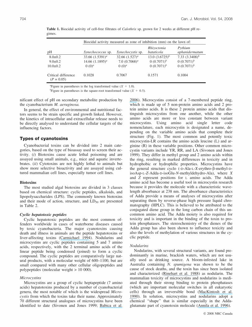
Toxin production by cyanobacteria is also influenced bypH of the medium (Van der Westhuizen and Eloff 1983;Ray and Bagchi 2001). Van der Westhuizen and Eloff(1983) reported that although the cyanobacterium M. aerugi-nosa exhibited a maximum growth rate at pH 9, the cellswere more toxic at higher and lower pH values, where theygrew more slowly. Ray and Bagchi (2001) observed a nega-tive regulation of pH and toxin production in O. latevirens.A pH of 8 was found to be most optimal (Table 1) for Cal-othrix sp. in terms of growth and biocidal activity against 2cyanobacterial and 2 fungal strains (Radhakrishnan 2006).However, Jaiswal et al. (2007) showed that there is no sig-
Jaiswal et al. 703
# 2008 NRC Canada
nificant effect of pH on secondary metabolite production bythe cyanobacterium M. aeruginosa.
In general, the effect of environmental and nutritional fac-tors seems to be strain specific and growth linked. However,the kinetics of intracellular and extracellular release needs tobe directly analysed to understand the cellular targets of theinfluencing factors.
Types of cyanotoxinsCyanobacterial toxins can be divided into 2 main cate-
gories, based on the type of bioassay used to screen their ac-tivity. (i) Biotoxins cause acute lethal poisoning and areassayed using small animals, e.g., mice and aquatic inverte-brates. (ii) Cytotoxins are not highly lethal to animals butshow more selective bioactivity and are assayed using cul-tured mammalian cell lines, especially tumor cell lines.
BiotoxinsThe most studied algal biotoxins are divided in 3 classes
based on chemical structure: cyclic peptides, alkaloids, andlipopolysaccharides (LPS). The commonly known biotoxinsand their mode of action, structure, and LD50 are presentedin Table 2.
Cyclic hepatotoxic peptidesCyclic hepatotoxic peptides are the most common of-
fenders worldwide in cases of waterborne diseases causedby toxic cyanobacteria. The major cyanotoxins causingdeath and illness in animals are the peptide hepatotoxins orliver-affecting toxins (Carmichael 1994). Nodularins andmicrocystins are cyclic peptides containing 5 and 7 aminoacids, respectively, with the 2 terminal amino acids of thelinear peptide being condensed (joined) to form a cycliccompound. The cyclic peptides are comparatively large nat-ural products, with a molecular weight of 600–1100, but aresmall compared with many other cellular oligopeptides andpolypeptides (molecular weight > 10 000).
MicrocystinsMicrocystins are a group of cyclic heptapeptide (7 amino
acids) hepatotoxins produced by a number of cyanobacterialgenera, the most notable of which is the widespread Micro-cystis from which the toxins take their name. Approximately70 different structural analogues of microcystins have beenidentified to date (Sivonen and Jones 1999; Babica et al.
2006). Microcystins consist of a 7-membered peptide ring,which is made up of 5 non-protein amino acids and 2 pro-tein amino acids. It is these 2 protein amino acids that dis-tinguish microcystins from one another, while the otheramino acids are more or less constant between variantmicrocystins. Using amino acid single letter codenomenclature, each microcystin is designated a name, de-pending on the variable amino acids that complete theirstructure (Fig. 1). The most common and potently toxicmicrocystin-LR contains the amino acids leucine (L) and ar-ginine (R) in these variable positions. Other common micro-cystin variants include YR, RR, and LA (Sivonen and Jones1999). They differ in methyl group and 2 amino acids withinthe ring, resulting in marked differences in toxicity and inhydrophobic or hydrophilic properties. Microcystins havethe general structure cyclo (-D-Ala-L-X-erythro-b-methyl-D-isoAsp-L-Z-Adda-D-isoGlu-N-methyldehydro-Ala), where Xand Z represent positions for L amino acids. The Addaamino acid has become a useful tool in microcystin researchbecause it provides the molecule with a characteristic wave-length absorbance at 238 nm. The absorbance characteristicsof Adda provide a means of analyses of microcystins afterseparating them by reverse-phase high pressure liquid chro-matography (HPLC). This is believed to be attributed to theconjugated diene group in the long carbon chain of this un-common amino acid. The Adda moiety is also required fortoxicity and is important in the binding of the toxin to pro-tein phosphatases. The stereochemistry of the dienes of theAdda group has also been shown to influence toxicity andalso the levels of methylation of various structures in the cy-clic peptide.
NodularinsNodularins, with several structural variants, are found pre-
dominantly in marine, brackish waters, which are not usu-ally used as drinking source. A bloom-infested lake inAustralia containing N. spumigena was shown to be thecause of stock deaths, and the toxin has since been isolatedand characterized (Rinehart et al. 1988) as nodularin. Themammalian toxicity of microcystins and nodularins is medi-ated through their strong binding to protein phosphatases(which are important molecular switches in all eukaryoticcells) through an irreversible bond (MacKintosh et al.1990). In solution, microcystins and nodularins adopt achemical ‘‘shape’’ that is similar especially in the Adda-glutamate part of cyanotoxin molecule (Annila et al. 1996).
Table 1. Biocidal activity of cell-free filtrates of Calothrix sp. grown for 2 weeks at different pH re-gimes.
Biocidal activity measured as zone of inhibition (mm) on the lawn of:
pH Synechococcus sp. Synechocystis sp.Rhizoctoniabataticola
Pythiumaphanidermatum
8.0±0.2 33.66 (1.5391)a 32.66 (1.527)a 13.0 (3.6725)b 7.33 (3.3408)b
9.0±0.2 14.66 (1.1895)a 7.0 (0.7068)a 0.(0.7071)b 0.(0.7071)b
10.0±0.2 0.(0)a 0.(0)a 0.(0.7071)b 0.(0.7071)b
Critical difference(P = 0.05)
0.1028 0.7067 0.1571 0.1004
aFigure in parentheses is the log transformed value (X + 1.0).bFigure in parentheses is the square-root transformed value (X + 0.5).
704 Can. J. Microbiol. Vol. 54, 2008
# 2008 NRC Canada
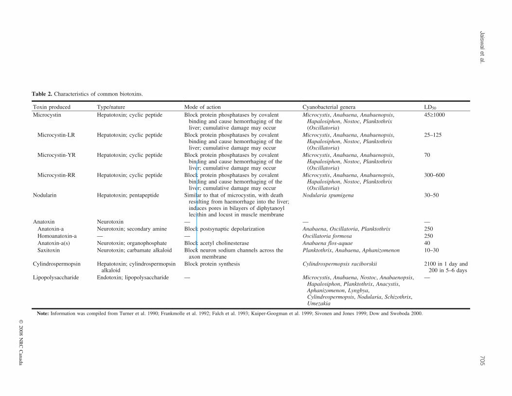
Table 2. Characteristics of common biotoxins.
Toxin produced Type/nature Mode of action Cyanobacterial genera LD50
Microcystin Hepatotoxin; cyclic peptide Block protein phosphatases by covalentbinding and cause hemorrhaging of theliver; cumulative damage may occur
Microcystis, Anabaena, Anabaenopsis,Hapalosiphon, Nostoc, Planktothrix(Oscillatoria)
45‡1000
Microcystin-LR Hepatotoxin; cyclic peptide Block protein phosphatases by covalentbinding and cause hemorrhaging of theliver; cumulative damage may occur
Microcystis, Anabaena, Anabaenopsis,Hapalosiphon, Nostoc, Planktothrix(Oscillatoria)
25–125
Microcystin-YR Hepatotoxin; cyclic peptide Block protein phosphatases by covalentbinding and cause hemorrhaging of theliver; cumulative damage may occur
Microcystis, Anabaena, Anabaenopsis,Hapalosiphon, Nostoc, Planktothrix(Oscillatoria)
70
Microcystin-RR Hepatotoxin; cyclic peptide Block protein phosphatases by covalentbinding and cause hemorrhaging of theliver; cumulative damage may occur
Microcystis, Anabaena, Anabaenopsis,Hapalosiphon, Nostoc, Planktothrix(Oscillatoria)
300–600
Nodularin Hepatotoxin; pentapeptide Similar to that of microcystin, with deathresulting from haemorrhage into the liver;induces pores in bilayers of diphytanoyllecithin and locust in muscle membrane
Nodularia spumigena 30–50
Anatoxin Neurotoxin — — —Anatoxin-a Neurotoxin; secondary amine Block postsynaptic depolarization Anabaena, Oscillatoria, Planktothrix 250Homoanatoxin-a — — Oscillatoria formosa 250Anatoxin-a(s) Neurotoxin; organophosphate Block acetyl cholinesterase Anabaena flos-aquae 40Saxitoxin Neurotoxin; carbamate alkaloid Block neuron sodium channels across the
axon membranePlanktothrix, Anabaena, Aphanizomenon 10–30
Cylindrospermopsin Hepatotoxin; cylindrospermopsinalkaloid
Block protein synthesis Cylindrospermopsis raciborskii 2100 in 1 day and200 in 5–6 days
Lipopolysaccharide Endotoxin; lipopolysaccharide — Microcystis, Anabaena, Nostoc, Anabaenopsis,Hapalosiphon, Planktothrix, Anacystis,Aphanizomenon, Lyngbya,Cylindrospermopsis, Nodularia, Schizothrix,Umezakia
—
Note: Information was compiled from Turner et al. 1990; Frankmolle et al. 1992; Falch et al. 1993; Kuiper-Googman et al. 1999; Sivonen and Jones 1999; Dow and Swoboda 2000.
Jaiswalet
al.705
#2008
NR
CC
anada
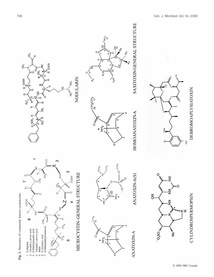
Fig
.1.
Stru
ctur
esof
com
mon
lykn
own
cyan
otox
ins.
706 Can. J. Microbiol. Vol. 54, 2008
# 2008 NRC Canada
Alkaloid toxinsAlkaloids vary in their chemical structure and stabilities,
and depending on the type, they can affect the nervous sys-tem, skin liver, or gastrointestinal tract. Some of the moststudied algal toxins belonging to alkaloid class includeanatoxin-a, anatoxin-a(s), homo-anatoxin-a, saxitoxin, andcylindrospermopsin.
AnatoxinsThe anatoxins are a group of neurotoxic alkaloids. They
are low molecular weight secondary amines produced by anumber of cyanobacterial genera, including Anabaena,Oscillatoria, and Aphanizomenon. The toxicity of thesecompounds (LD50) varies from 20 mg/kg (by mass, intra-peritoneally in mice) for anatoxin-a(s) to 200–250 mg/kg foranatoxin-a and homoanatoxin-a, making them more toxicthan many microcystins. Anatoxin-a is produced by somestrains of Anabaena. It is a structural analogue of cocaineand the neurotransmitter acetylcholine and binds to nicotinicacetylcholine receptors. Death occurs by respiratory failurewithin minutes to few hours depending upon the species(Carmichael 1994; Hunter 1995). Homo-anatoxin-a is a po-tent neuromuscular blocking agent that causes severe para-lysis, convulsions, and death by respiratory arrest. Thistoxin has been purified from Oscillatoria formosa.Anatoxin-a(s) is an organophosphate (Fig. 1) produced bystrains of Anabaena flos-aquae (Matsunaga et al. 1989).This is the only known naturally occurring organophosphate,which functions as cholinesterase.
SaxitoxinsLike anatoxins, the saxitoxins (STX) are neurotoxic alka-
loids, which are also known as PSPs (paralytic shellfish poi-sons) owing to their occurrence and association withseafood. Saxitoxins and their analogues are produced byAnabaena and Aphanizomenon (Sawyer et al. 1968). Theyhave also been recorded from Lyngbya wollei (Carmichaelet al. 1997). They act on the sodium ion channel of the ex-citable membranes of the nerve cells and block them, thuscausing their neurotoxic effects like paralysis or respiratoryfailure (Hunter 1995). However, to date no PSP-like illnesshas been reported in humans from consumption of drinkingwater. There are a number of STX variants generally div-ided into groups based on their structure or organism of ori-gin. The single sulphated STXs are known as gonyautoxinsand the doubly sulphated STXs are known as C-toxins.There are also decarbamyl STXs (dcSTX) and a group ofSTX variants, so far found only in L. wollei, known asLyngbya-wollei-toxins. STXs are highly toxic with LD50s aslow as 10 mg/kg (intraperitoneally in mice). (See Fig. 1 forthe general structure of STX.)
CylindrospermopsinCylindrospermopsin is a hepatotoxic alkaloid (Fig. 1) that
has been isolated from cultures of Cylindrospermopsis raci-borskii (Ohtani et al. 1992). It blocks protein synthesis, thefirst clinical symptoms being kidney and liver failure. Incontrast to pure toxin, the crude extract of the organismalso causes injury to lungs, adrenal glands, and intestines,indicating the presence of further unknown toxin in the or-ganism. This toxin has been identified in an outbreak ofacute hepato-enteritis and renal damage among an aboriginal
population in Australia (Falconer 1996). Cylindrospermopsinhas also been characterized from Aphanizomenon ovalispo-rum in Israel (Banker et al. 1997) and Umezakia natans inJapan (Harada et al. 1994). Its LD50 (intraperitoneally inmice) of 200 mg/kg ranks it as a relatively potent cyano-bacterial toxin.
LPS toxins (endotoxin)Weise and co-workers (1970) were the first to isolate LPS
from the cyanobacterium Anacystis nidulans, which exhibitsboth pyrogenic and toxic effects (Weckesser et al. 1979).But unlike Gram-negative bacteria, cyanobacterial LPS lacksany phosphate in the lipid A core (Keletis and Sykora 1982).Cyanobacterial LPSs are found to be 10 times less toxicthan other bacterial LPSs, e.g., those produced by Salmon-ella sp. (Codd 1984), and reports of human illness due tothese toxins exist (Codd and Poon 1988). There is consider-able diversity among the cyanobacteria in the chemical com-position; however, the differences are largely related tophylogeny.
CytotoxinsAlthough neurotoxins and hepatotoxins are more studied
due to their lethality and ubiquity, cytotoxins are equally im-portant due to their antialgal, antimycotic, and antibacterialactivity and some are active against cell tissue lines andhave moderate antitumor activity (Gerwick et al. 1994;Patterson et al. 1994). Some common cyanotoxins, theirmode of action, their structure, and the organism producingthem are listed in Table 2.
Cytotoxins are interesting compounds because they aremore targeted and can be exploited as antitumor, antiviral,or antibiotic compounds (Gross 2003). In general most cyto-toxins have a strong cytostatic activity or an ability to selec-tively inhibit tumors. However, they are highlyheterogeneous in their chemical structure and in terms ofthe qualitative and quantitative aspects of damage inflicted.The majority of them are cyclic or aliphatic peptides withprotease inhibition activity, e.g., micropeptins, cyano-peptides, cyanopeptolins, all of which have been isolatedfrom several planktonic species belonging to diverse colo-nial and filamentous genera — Microcystis, Anabaena, andPlanktothrix (Moore 1996; Namikoshi and Rinehart 1996).Scytophycins represent another well-investigated group ofcytotoxins that have been mostly isolated from branchedcyanobacteria, e.g., Scytonema, Tolypothrix, and Cylindro-spermum muscicola (Jung et al. 1991). Among this group ofmacrolytic lactones, tolytoxin has been investigated indepth. It causes severe cytotoxic effect through the depolari-zation of cell microfilaments (Patterson and Carmeli 1992).Another group of promising compounds are the crypto-phycins, which are lipophilic compounds mainly isolatedfrom Nostoc strains (Trimurtulu et al. 1994). The cytotoxiceffects of methanolic extracts belonging to several soil iso-lates of the genera Anabaena, Nodularia, Cylindrospermum,Tolypothrix, and Trichormus have also been shown on mam-malian cell lines (Hrouzek et al. 2005). Similar studies withmicrocystins have shown that these compounds are capableof initiating apoptosis in hapatocytes (Botha et al. 2004), be-sides their well-investigated effects on increased proteinphosphorylation due to protein phosphatase inhibition ofmammalian cell lines (Carmichael 1994).
Jaiswal et al. 707
# 2008 NRC Canada
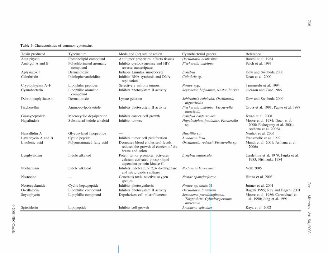
Table 3. Characteristics of common cytotoxins.
Toxin produced Type/nature Mode and (or) site of action Cyanobacterial genera ReferenceAcutiphycin Phospholipid compound Antitumor properties, affects tissues Oscillatoria acutissima Barchi et al. 1984Ambigol A and B Polychlorinated aromatic
compoundInhibits cyclooxygenase and HIV
reverse transcriptaseFischerella ambigua Falch et al. 1993
Aplysiatoxin Dermatotoxic Induces Limulus amoebocyte Lyngbya Dow and Swoboda 2000Calothrixin Indolophenanthridine Inhibits RNA synthesis and DNA
replicationCalothrix sp. Doan et al. 2000
Cryptophycins A–F Lipophilic peptides Selectively inhibits tumors Nostoc spp. Trimurtulu et al. 1994Cyanobacterin Lipophilic aromatic
compoundInhibits photosystem II activity Scytonema hofmannii, Nostoc linckia Gleason and Case 1986
Debromoaplysiatoxin Dermatotoxic Lysate gelation Schizothrix calcicola, Oscillatorianigroviridis
Dow and Swoboda 2000
Fischerellin Aminoacylpolyketide Inhibits photosystem II activity Fischerella ambigua, Fischerellamuscicola
Gross et al. 1991; Papke et al. 1997
Grassypeptolide Macrocyclic depsipeptide Inhibits cancer cell growth Lyngbya confervoides Kwan et al. 2008Hapalindole Substituted indole alkaloid Inhibits tumors Hapalosiphon fontinalis, Fischerella
sp.Moore et al. 1984; Doan et al.
2000; Etchegaray et al. 2004;Asthana et al. 2006b
Hassallidin A Glycosylated lipopeptide — Hassillia sp. Neuhof et al. 2005Laxaphycin A and B Cyclic peptide Inhibits tumor cell proliferation Anabaena laxa Frankmolle et al. 1992Linolenic acid Polyunsaturated fatty acid Decreases blood cholesterol levels,
reduces the growth of cancers of thebreast and colon
Oscillatoria redekei, Fischerella sp. Mundt et al. 2001; Asthana et al.2006a
Lyngbyatoxin Indole alkaloid Potent tumor promoter, activatescalcium-activated phospholipid-dependent protein kinase C
Lyngbya majuscula Cardellina et al. 1979; Fujiki et al.1983; Nishizuka 1984
Norharmane Indole alkaloid Inhibits indoleamine 2,3- dioxygenaseand nitric oxide synthase
Nodularia harveyana Volk 2005
Nostocine — Generates toxic reactive oxygenspecies
Nostoc spongiaeforme Hirata et al. 2003
Nostocyclamide Cyclic heptapeptide Inhibits photosynthesis Nostoc sp. strain 31 Juttner et al. 2001Oscillatorin Lipophilic compound Inhibits photosystem II activity Oscillatoria latevirens Bagchi 1995; Ray and Bagchi 2001Scytophycin Lipophilic compound Depolarizes cell microfilaments Scytonema pseudohofmanni,
Tolypothrix, Cylindrospermummuscicola
Moore et al. 1986; Carmichael etal. 1990; Jung et al. 1991
Spiroidesin Lipopeptide Inhibits cell growth Anabaena spiroides Kaya et al. 2002
708C
an.J.
Microbiol.
Vol.
54,2008
#2008
NR
CC
anada
The cytotoxins that have been isolated from cyanobacteriaare listed in Table 3 along with their major characteristicsand modes of action.
Detection techniques for cyanotoxinsThe physical appearance of cyanobacterial bloom does
not reveal its toxicity, and diagnosis of cyanotoxicosis is dif-ficult primarily because many blooms are not hazardous atall times.
The development of more sensitive separation and detec-tion methods will help in reducing the consequences of thesepoisons in our food and water supply. A range of other as-say procedures have been investigated, which range fromlow cost and relatively simple methods to highly sensitiveand sophisticated methods. Detection of toxin depends uponthe type of information required and facilities available.
Toxicity tests and bioassay
Mouse bioassayThere have been many biological detection methods de-
veloped for detecting cyanotoxins that use bioactivity of tox-ins, e.g., potent hepatotoxicity, neurotoxicity, cytotoxicityenzymatic activity, and immunological interactions. Mousebioassay was typically the first test for toxicity used inscreening water bloom materials and laboratory cultures orcell extracts. It is also considered as the standard procedurefor establishing the LD50. It is still preferred over othermethods because of the easy availability of laboratory miceand the low cost. In this method, adult mice are injectedintraperitoneally with the sample to be assayed and are keptunder observation. Generally, toxic symptoms are visiblewithin 24 h. This method is able to detect the toxin withinminutes (to a few hours) and can possibly determine thetype of toxin present, i.e., hepatotoxin, neurotoxin, etc. Thedisadvantages of this method are the nonspecific inferenceand (or) result obtained, its inability to detect low levels oftoxin, and its inability to distinguish between homologues oftoxins. Mouse bioassay remains the primary means of as-sessing function and potency of cyanotoxins, but this is nowslowly being replaced by other bioassays — chemical andimmunological methods. Further, there is increasing opposi-tion in many countries to the use of animals for any form oftoxicity testing.
Alternative bioassayA number of alternative bioassay methods have been de-
vised for detection of cyanotoxins, e.g., by using Daphniasp. and Artemia salina (Kiviranta et al. 1991). Turell andMiddlebrook (1988) suggested a bioassay that involvedmicroinjection of the toxin into mosquitoes. The use of bothadult and larvae stages has been investigated as potentialbioassay agents. Both methods are relatively simple but nothave been widely adopted owing to difficulties in handlingthis organism (Turell and Middlebrook 1988; Kiviranta etal. 1991). The other test organism, which can detect micro-cystins successfully in the bloom samples, is the fruit flyDrosophila melanogaster (Swoboda and Dow 1994). Theseorganisms are easy to maintain in the laboratory with nospecial equipment required. The flies were, however, notsensitive to neurotoxic Aphanizomenon (Swoboda and Dow
1994). McElhiney et al. (1998) reported that the locust bio-assay was able to detect saxitoxin successfully in a range ofsamples that included cyanobacteria and shellfish, but thebioassay was not found sensitive to microcystin-LR andanatoxin-a.
Biochemical assaysThe protein phosphatase inhibition assay is a sensitive
screening method for microcystin and nodularin, which usesthe biochemical activity of these enzymes. One version isbased on the quantification of 32P-phosphate released fromradiolabelled substrate (Lambert et al. 1994) by the activityof protein phosphatase enzyme (PP1 and PP2). Althoughthis method is very popular, the only disadvantage is theuse of radioactive chemicals, which requires specialized andwell-equipped laboratory facilities. Carmichael (1994) useda colorimetric protein phosphatase inhibition assay, whichavoids the complications of using radioactive materials.Ward et al. (1997) also reported this method is an effectiveand reliable means of screening water samples.
Immunological detectionThe Enzyme-Linked Immunosorbant Assay (ELISA) tech-
nique is currently the most promising for rapid samplescreening of microcystins owing to its sensitivity, specific-ity, and ease of operation. Chu et al. (1989) developed anELISA technique based on polyclonal antisera in rabbitagainst bovine albumin conjugated to microcystin-LR,which proved to be a better immunogen. The raised antibod-ies had good cross-reactivities with microcystin-RR, -YR,and nodularin and less with microcystin-LY and -LA. Ap-proximately 50% binding occurred at a toxin concentrationof 1 ng/mL, which is appropriate for water quality testing.Nagata et al. (1995) produced monoclonal antibodies againstmicrocystin-LR, which showed cross-reactivity withmicrocystin-RR, -YR, -LA, and several other derivatives.Although the epitope of this antibody is not clear, the impor-tance of the Adda moiety for antibody binding has beenindicated.
Analytical methodsThe physicochemical properties of the cyanotoxins can be
used for detection and identification. The molecular weight,chromophores, and reactivities of the functional groups inthe molecules are generally used for the chemical analysesof the cyanotoxins. The majority of analytical methods havebeen developed primarily for microcystins but nodularinscan also be easily identified by the same method because ofthe similar physicochemical properties.
Thin layer chromatography (TLC) is the most basic tech-nique that has been used to purify hepatotoxins, but it is notvery sensitive and specific; hence, over time, it has beensuperceded by HPLC. Typical HPLC analysis uses areverse-phase C18 silica column, and separation is achievedover a gradient of acetonitrile and water, both containing0.05% trifluoroacetic acid. However, this method relies onretention time for identification and microcystin standardsare required (Harada 1996). Recent advances in detectorhardware can now provide high-resolution spectra that de-tect very slight variations in chemical composition and canbe used in conjunction with advanced spectral matching
Jaiswal et al. 709
# 2008 NRC Canada
software. Further confirmation may be done by liquidchromatography – mass spectrometry, which enablessimultaneous separation and identification of microcystinsfrom a mixture of samples (Kondo et al. 1992). A more ad-vanced technique — frit-fast atom bombardment liquidchromatography – mass spectrometry (which uses a micro-bore column (0.3 mm internal diameter)) — can identifynanogram levels in water and biological samples (Kondo etal. 1995, 1996). The structural analyses of hepatotoxins canbe carried out by fast atom bombardment mass spectrometryand proton nuclear magnetic resonance (Kusumi 1996). Re-cently, matrix-assisted laser desorption ionization massspectrometry has shown to be a rapid and sensitive analyti-cal method for the detection of cyanobacterial toxins (Dowet al. 1994).
Molecular detectionTraditionally, microscopic identification of cyanobacteria
alone or in combination with direct analysis of toxin hasbeen used for detection of toxic cyanobacteria. However,some cyanobacteria may be genetically capable of producingtoxins but do not produce under all conditions and some donot produce toxins all. Hence, scientists are focusing ongenes that can differentiate taxonomic groups of cyano-bacteria involved in toxin production and those that are nottoxic, i.e., genes that can separate toxic and nontoxic strains(Neilan et al. 1995; Bloch et al. 1996; Rouhianen et al.2004). Bloch et al. (1996) have looked for conserved regionsof genome, such as those encoding for phycocyanin sub-units, that might be used to classify the toxigenic species.Neilan et al. (1995) designed a general primer to the phyco-cyanin operon (cpc genes) and developed a PCR that al-lowed amplification of the pycocyanin (PC) gene containingthe intergenic spacer from cyanobacteria commonly associ-ated with toxic bloom sample. They showed that the DNAprofile obtained after restriction digestion with 9 enzymeswas specific to various taxonomic levels of cyanobacteria.This kind of gene probe, which can distinguish among toxicand nontoxic genera and (or) strains, can prove to be verybeneficial in detecting toxic cyanobacterial bloom or screen-ing cyanobacteria for toxicity. Neilan et al. (1995), using re-striction length polymorphism, probed a short tandemlyrepeated DNA sequence found in the chromosome of Ana-baena sp. PCC 7120 and found that it can distinguish ahepatotoxic Anabaena isolate from neurotoxin-producingstrains. Baker et al. (2001) showed from their studies thatbloom component can be identified and monitored for toxi-genicity by PCR more effectively than by other methods,like microscopy and mouse bioassay. They used PCR ampli-fication of a phycocyanin intergenic spacer region betweenthe genes for b and a subunits for detection of cyano-bacterial genera. Microcystin production was determined byPCR amplification of gene of microcystin biosynthesis andthe potential for saxitoxin production was determined byPCR amplification of a region of 16S rRNA gene of Ana-baena circinalis. They also showed that toxicity and celltype present in a bloom changes over a period of time.Fergusson and Saint (2000) developed an Anabaenacircinalis-specific PCR assay targeting the rpoCI gene to de-tect the organism directly from environmental samples.Wilson et al. (2000) developed a PCR assay targeting a re-
gion of rpoCI gene unique to Cylindrospermopsis racibor-skii for specific identification of the strain from bothpurified genomic DNA and environmental samples.
Genetic basis of toxin production
At present, 2 molecular systems are known to be involvedin cyanobacterial toxicity: nonribosomal peptides and Type Ipolyketide synthases. Most of the work involving this aspectof toxin production has been done using Microcystis (Tillettet al. 2001; Ouellette et al. 2006), and the expression ofthese genes is known to be regulated by complex mecha-nisms and is influenced by environmental factors.
Biosynthesis of microcystins has been studied in all the 3main microcystin-producing genera occurring in fresh water,i.e., Microcystis, Anabaena, and Planktothrix (Tillett et al.2001; Christiansen et al. 2003, 2006; Rouhianen et al.2004). Microcystins are synthesized nonribosomally by agiant enzyme complex comprising peptide synthetase, poly-ketide synthases (PKSs), and additional modifying enzymes(Arment and Carmichael 1996; Tillett et al. 2001;Christiansen et al. 2003; Rouhianen et al. 2004). Nonribo-somal peptide synthetases catalyse the formation of peptideby a thio-template mechanism. They are involved in the syn-thesis of linear, cyclic, and branched cyclic peptides, includ-ing potent drugs such as penicillin, vancomycin, andcyclosporin (Konz and Marahiel 1999).
The gene cluster encoding the microcystin synthetasecomplex has now been identified and sequenced (Nishizawaet al. 1999; Tillett et al. 2001). This 55-kb gene cluster(mcyABCDEFGHIJ) consists of 6 open reading frames(ORFs) of a mixed nonribosomal peptide synthetase –polyketide synthase nature (mcyA–mcyE and mcyG) and 4smaller ORFs with putative precursor and tailoring functions(mcyF and mcyH–mcyJ). The mcyABCDEFGHIJ genes aretranscribed as 2 polycistronic operons, mcyABC andmcyDEFGHIJ, from a central bi-directional promoter be-tween mcyA and mcyD. Kaebernick et al. (2002) were ableto identify the start sites for all the mcy genes except formcyB and mcyC. Baker et al. (2002) also suggested the useof PCR-based methods for direct detection and identificationof strains present and their toxigenicity. Vaitomaa et al.(2003) used quantitative RT–PCR analyses to show thatmcyE gene copy numbers can be used as surrogates for arough estimate of toxigenic cell numbers of hepatotoxicMicrocystis and Anabaena spp. This can provide a simplemethod for detecting toxic blooms in diverse areas and (or)in a large number of samples. Toxic Microcystis strainsoften produce several isoforms of cyclic hepatotoxin micro-cystin that have been attributed to relaxed substrate specif-icity and adenylation domains. Mikalsen et al. (2003)showed that this variability was also caused by genetic var-iation in a microcystin synthetase gene. Moffitt and Neilan(2004) sequenced and characterized a complete gene clusterencoding the enzymatic machinery required for the bio-synthesis of nodularin in N. spumigena NSOR 10.Kaebernick et al. (2000) studied the effect of light on micro-cystin synthetase production and concluded that both mcyBand mcyD transcript level increased under high light inten-sities and red light, blue light and certain artificial stress fac-tors (methylogen and NaCl) caused reduction in transcript
710 Can. J. Microbiol. Vol. 54, 2008
# 2008 NRC Canada
levels. Tonk et al. (2005) also reported that microcystincomposition of the cyanobacterium Planktothrix agardhiichanges to a more toxic variant with increasing light inten-sity. Kurmayer et al. (2004) reported the occurrence ofPlanktothrix strains containing all mcy genes but lackingtoxic hepatopeptide microcystin. Christiansen et al. (2006)analyzed 29 strains and showed that transposons inactivatebiosynthesis of nonribosomal peptide microcystin. Two de-letions spanning 400 bp (in mcyB; one strain) and 1869 bp(in mcyHA; 3 strains) and 3 insertions spanning 1429 bp (inmcyD; 8 strains), 1433 bp (in mcyEG; one strain), and1433 bp (in mcyA; one strain) were identified, indicatingthat a mutation resulted in the inactivation of microcystinbiosynthesis. Transcriptional analyses of the mcy gene clus-ter will help to increase our understanding of microcystinsynthetase regulation and toxin biosynthesis and will pro-vide useful insight into other nonribosomal systems.
Ecological implications of cyanobacterialsecondary metabolites
Cyanobacteria represent an untapped bioresource for a di-verse range of secondary metabolites, some of which showunique similarities to plant and animal products (Namikoshiand Rinehart 1996; Bagchi and Ray 2001; Agrawal et al.2005; Singh et al. 2005a; Capper et al. 2006; Dahms et al.2006; Volk 2006). Toxins produced by bacteria, algae, and(or) cyanobacteria can be considered allelochemicals, sincethey inhibit one or more groups of organisms and help toprovide a competitive advantage to the producer, whichmay be important factors contributing to formation and (or)maintenance of cyanobacterial blooms (Gross 2003; Legrandet al. 2003; Prasanna and Jaiswal 2006; Leflaive and Ten-Hage 2007).
AllelochemicalsThe term allelopathy is derived from 2 separate words (al-
lelon, which means ‘‘of each other,’’ and pathos, whichmeans ‘‘to suffer’’), and it denotes the production of specificbiomolecules by one plant or bacterial species that can in-duce suffering in, or give benefit to, another plant or bac-terial species. In simple terms, allelopathy refers to thechemical inhibition of one species by another. These bio-molecules, or allelochemicals, produced by one species arereleased into the environment and subsequently influencethe growth and development of neighbouring species.Allelochemicals in general have evolved to inhibit manybiochemical targets present on the surface of or within thecells (Skovgaard et al. 2003; Suikkanen et al. 2005). Manysuch chemicals, including antibiotics, inhibit components ofprotein synthesis. Both positive and negative allelochemicalincidents are known to be involved in the control of fresh-water bloom sequence (Keating 1977; Gross 1999). Schlegelet al. (1999) isolated cyanobacterial strains form diversehabitats, spanning the continents of Australia and Asia, thatexhibited allelopathic activity against green algae andcyanobacteria. Gross (1999) reported the production ofallelochemicals with a strong effect against several algae(including cyanobacteria). Jaiswal et al. (2005) reportedallelopathic activity of M. aeruginosa against a unicellularcyanobacterium. The colonial blue-green alga Merismopedia
tenuissima has been reported (Blomquist 1996) to dominatean acidic Swedish lake due to the release of allelochemicals.Hence, the prolific growth of cyanobacteria as blooms innutrient-rich water bodies, concomitant with eutrophication,can be considered a result of their invasiveness and produc-tion of bioactive molecules, which provides them the advant-age of eliminating other competitive flora and fauna. Thisrepresents an appropriate example of how allelopathy, nu-trient mobilization, and resource competition act synergisti-cally in providing a selective advantage to these prokaryotes.
Aquatic allelochemicals often target multiple physio-logical processes. A filamentous cyanobacterium, Scytonemahofmannii (UTEX 2349), has been reported to produce anallelochemical cyanobacterin, which inhibited the growth ofcyanobacteria (Gleason and Paulson 1984), eukaryotic algae(Gleason and Baxa 1986), and higher plants (Gleason andCase 1986). Non-photosynthetic microorganisms were notaffected by the cyanobacterin. Similar observations weremade in Oscillatoria (Bagchi et al. 1990; Chauhan et al.1992) and Fischerella (Gross et al. 1991), Nostoc (Juttner etal. 2001), where allelochemicals released by the cyano-bacteria inhibited growth of cyanobacteria and chlorophytesbut not heterotrophic organisms. The most common mode ofaction of these metabolites is directed towards the inhibitionof photosynthesis by affecting photosystem II activity(Bagchi et al. 1990; Bagchi 1995; Srivastava et al. 1998,2001). Smith and Doan (1999) also reported that bioactivecompounds produced by cyanobacteria (allelochemicals)show a diverse range of biological activities and affectmany biochemical processes within the cell, especially theoxygenic photosynthetic processes. Such chemicals arelikely to be involved in the regulation of natural populationsand are potentially useful as biochemical tools, i.e., as herbi-cidal and biocidal agents. Bagchi and Ray (2001) reportedthat a planktonic cyanobacterial extract showed photosystemII-inhibiting (herbicidal) activity, besides allelopathic activ-ity, which might play a role in competitive ability of thestrain in nature. Doan et al. (2000) reported alkaloids fromFischerella and Calothrix that inhibited RNA synthesis ofother organisms, indicating another mode of action ofcyanobacterial secondary metabolites. A bloom-formingcyanobacterium Anabaena flos-aquae has been reported tobe involved in chemical signaling with the competing phyto-plankton. This cyanobacterium produces anatoxin andmicrocystin-LR, which inhibit the growth of chlorophyteChlamydomonas reinhardtii, whereas the extracellular com-pound produced by the chlorophyte causes an increase inanatoxin content but no effect on microcystin-LR. (Kearnsand Hunter 2001). The exudates from Microcystis have alsobeen reported to inhibit the photosynthesis in the dinoflagel-late Peridium gatunnse by interfering with its internal car-bonic anhydrase activity (Sukenik et al. 2002). However,inhibition was reported in a strain that did not producemicrocystin-LR, which is indicative of the role of someother compound. The compound released by the Microcystissp. resulted in oxidative stress in the dinoflagellate and acti-vated certain protein kinases (Vardi et al. 2002), whileP. gatunnse caused cell lysis in Microcystis owing to a lossof buoyancy and a dramatic increase in mcyB. These 2 or-ganisms present excellent examples of complex allelopathicinteractions.
Jaiswal et al. 711
# 2008 NRC Canada
Multiple biotic and abiotic factors determine the strengthof allelopathic interaction. Frequently, the impact of exces-sive or limiting nutrients has been shown to affect the over-all production of allelochemicals and their effect on targetorganisms (Von Elert and Juttner 1997; Ray and Bagchi2001; Gross 2003; Hirata et al. 2003; Volk 2007). Jaiswalet al. (2007) showed that doubling the concentration ofphosphorus in the basal medium resulted in increasedgrowth and algicidal activity (measured in the form of dia-meter of inhibition zone) of M. aeruginosa. However, no ac-tivity was recorded when phosphate concentration wasreduced to half (50% of that present in the basal medium).In the same study, they also reported that in M. aeruginosacultures at all stages showed algicidal activity; however,maximum inhibitory action was recorded in 15- to 20-day-old cultures.
Suikkanen et al. (2004) found that N. spumigena wasmore allelopathic in exponential phase than in stationaryphase, whereas the culture filtrates were more hepatotoxicin stationary phase.
Bioactive compoundsCyanobacteria have also drawn much attention as a pro-
spective and rich bioresource of biologically active com-pounds that can be agriculturally useful. Toxic waterblooms comprising genera, such as Microcystis, Anabaena,and Nostoc, produce a diverse array of bioactive compoundsexhibiting antibiotic, algicidal, antifungal, cytotoxic,immuno-suppressive, and enzyme-inhibiting activities(Mundt et al. 2001). Terrestrial cyanobacteria, such as Ana-baena laxa, produce antifungal cyclic peptides, such as lax-aphycin A and B (Frankmolle et al. 1992). Kulik (1995)reported that extracts from the cyanobacterium Nostoc mus-corum Agardh inhibited in vitro growth of fungal plantpathogens, such as Sclerotinia sclerotiorum (Cottony rot ofvegetables and flowers) and Rhizoctonia solani (root andstem rots). Another antifungal glycosylated lipopeptide hasbeen reported (Neuhof et al. 2005) from the cyanobacteriumTolypothrix (Basionym Hassallia) that showed antifungalactivity against Aspergillus fumigatus and Candida albicans.The antifungal compound had been reported to contain bothfatty acid and carbohydrate moieties. A number of com-pounds exhibiting fungicidal activity against specific agri-culturally important fungi have been isolated and patented(Moore et al. 1989; Patterson et al. 1995; Hagmann andJuttner 1996; Moore 1996; Nagle and Wedge 2002; Volkand Furkert 2006). Pushparaj et al. (2000) showed that anacetone extract of Nodularia harveyana inhibited growth ofthe free-living root knot nematode Cephaloboides oxycerca.Such biomolecules produced by cyanobacteria can be devel-oped into safe formulations against many pathogen and (or)parasites.
The terrestrial cyanophyte Fischerella ambigua has beenreported (Falch et al. 1993) to produce 2 highly halogenatedcompounds with antibacterial, antifungal, and molluscicidalactivity. Kiviratna and Abdel-Hameed (1994) reported acompound from the cyanobacterium Oscillatoria agardhiithat is toxic to larvae of yellow fever mosquito Aedu ae-gypti. Casamatta and Wickstrom (2000) observed that theexudates of M. aeruginosa were inhibitory towards bacterialplankton communities. Recently Asthana et al. (2006a) re-
ported antibacterial activity of a methanolic extract of acyanobacterium Fischerella sp. that was further identifiedas linolinic acid (Asthana et al. 2006b). Antimicrobial andother bioactive compounds produced by cyanobacteria maybe released extracellularly to surroundings either actively orpassively or by autolysis. The biosynthetic pathways leadingto production of these novel compounds and finally their re-lease to the environment is not very well understood.Knowledge of biosynthetic pathways and factors leading tothe release of these biomolecules may prove to be of tre-mendous importance for mankind.
The chemical compounds isolated from cyanobacteria arealso of biotechnological interest, especially for clinical ap-plications, because of their antibiotic, algicidal, and cyto-toxic properties (Borowitzka 1995) and, hence, could beused as biocontrol agents of bacterial and fungal pathogens.In addition to neurotoxins and hepatotoxins, cyanobacteriaare known to produce several antibacterial compounds(Carmeli et al. 1990), antifungal compounds (Frankmolle etal. 1992), antiviral agents, anticancerous and (or) anti-neoplastic agents (Smith et al. 1994; Nianjun et al. 2004),and compounds useful in the treatment of HIV (Gerwick etal. 1994; Harrigan et al. 1998). Cyanobacterial metaboliteshave the potential for use in antifouling technology. The iso-lation of biogenic compounds and the determination of theirstructure may provide leads for future development of envi-ronmentally friendly antifouling paints (Dahms et al. 2006).
Cyanobacteria represent a vast bioresource of biologicallyactive compounds that may find tremendous applications inagriculture and pharmaceutical industry. So far, only a fewstrains have been commercially exploited, and extensive re-search is needed to unfold many unknown facets hidden in-side these small microorganisms, which through theiromnipresence and adaptation to diverse environment, can beexploited for diverse applications.
AcknowledgementsThe authors are thankful to the Council of Scientific and
Industrial Research (CSIR) and the Indian Council of Agri-cultural Research, New Delhi, India, for providing financialassistance, in the form of projects. The facilities required forcarrying out this investigation provided by the Centre forConservation and Utilization of Blue-Green Algae, IARI,New Delhi, are gratefully acknowledged.
ReferencesAgrawal, M.K., Bagchi, D., and Bagchi, S.N. 2005. Cysteine and
serine protease-mediated proteolysis in body homogenate of azooplankter, Moina macrocopa, is inhibited by the toxic cyano-bacterium, Microcystis aeruginosa PCC7806. Comp. Biochem.Physiol. Biochem. Mol. Biol. 141: 33–41. doi:10.1016/j.cbpc.2005.01.002.
Annila, A., Lehtimaki, J., Maltila, K., Eriksson, J.E., Sivonen, K.,Rantala, T.T., and Brakenbery, J. 1996. Solution structure ofnodularin — an inhibitor of serine/threonine specific proteinphosphatases. J. Biol. Chem. 271: 16695–16702. PMID:8663277.
Arment, A.R., and Carmichael, W.W. 1996. Evidence that micro-cysin is a thiol template product. J. Phycol. 32: 591–597.doi:10.1111/j.0022-3646.1996.00591.x.
Asthana, R.K., Srivastava, A., Kayastha, A.M., Nath, G., and
712 Can. J. Microbiol. Vol. 54, 2008
# 2008 NRC Canada
Singh, S.P. 2006a. Antibacterial potential of g-linolinic acid fromFischerella sp. colonizing neem bark. J. Appl. Phycol. 22: 443–448.
Asthana, R.K., Srivastava, A., Singh, A.P., and Singh, S.P. 2006b.Identification of antimicrobial entity from the cyanobacteriumFischerella sp. isolated from bark of Azadiracta indica (Neem)tree. World J. Microbiol. Biotechnol. 18: 33–39.
Babica, P., Blaha, L., and Marsalek, B. 2006. Exploring the role ofmicrocystins — a review of effects on photoautotrophic organ-isms. J. Phycol. 42: 9–20. doi:10.1111/j.1529-8817.2006.00176.x.
Bagchi, S.N. 1995. Structure and site of action of an algicide from acyanobacterium, Oscillatoria late-virens. J. Appl. Phycol. 10: 1–9.
Bagchi, S.N., and Ray, S. 2001. Extraction and purification of analgicidal metabolites from a cyanobacterium Oscillatoria latevi-rens. Indian J. Microbiol. 41: 163–167.
Bagchi, S.N., Palod, A., and Chauhan, V.S. 1990. Algicidal proper-ties of a bloom forming alga, Oscillatoria sp. J. Basic Microbiol.30: 21–29. doi:10.1002/jobm.3620300106.
Baker, J.A., Neilan, B.A., Entsch, B., and McKay, D.B. 2001. Iden-tification of cyanobacteria and their toxigenicity in environmen-tal samples by rapid molecular analysis. Environ. Toxicol. 16:472–482. doi:10.1002/tox.10010. PMID:11769244.
Baker, J.A., Entsch, B., Neilan, B.A., and Mckay, D.B. 2002. Mon-itoring changing toxigenicity of a cyanobacterial bloom by mo-lecular methods. Appl. Environ. Microbiol. 68: 6070–6076.doi:10.1128/AEM.68.12.6070-6076.2002. PMID:12450830.
Banker, P.D., Carmeli, S., Hadas, O., Teisch, B., Porat, R., andSukenik, A. 1997. Identification of cylindrospermopsin in Apha-nizomenon ovalisporum (Cyanophyceae) isolated from lake Kin-neret. Isr. J. Phycol. 33: 613–616.
Barchi, J.J., Jr., Moore, R.E., and Patterson, G.M.L. 1984. Acuti-phycin and 20,21-didehydroacutiphycin, new antineoplasticagents from the cyanophyte Oscillatoria acutissima. J. Am.Chem. Soc. 106: 8193–8197. doi:10.1021/ja00338a031.
Bloch, C.J.S., Blackburn, S.I., Neilan, B.A., and Grewe, P.M. 1996.Genetic characterization of strains of cyanobacteria using PCR–RFLP of the cpcBA intergenic spacer and flanking regions. J.Phycol. 32: 445–451.
Blomquist, P. 1996. Late summer phytoplankton responses to ex-perimental manipulations of nutrients and grazing in unlimedand limed lake Njupfatet, central Sweden. Arch. Hydrobiol.137: 425–455.
Borowitzka, M.A. 1995. Microalgae as a source of pharmaceuticalsand other biologically active compounds. J. Appl. Phycol. 7: 3–15.doi:10.1007/BF00003544.
Botha, N., Gehringer, M.M., Downing, T.G., Venter, M., andShephard, E.G. 2004. The role of microcystin-LR in the induc-tion of apoptosis and oxidative stress in a CaCo2 cells. Toxicon,43: 85–92. doi:10.1016/j.toxicon.2003.10.025. PMID:15037033.
Burja, A.M., Banaigs, B., Abou-Mansour, E., Burgess, J.G., andWright, P.C. 2001. Marine cyanobacteria — a prolific source ofnatural products. Tetrahedron, 57: 9347–9377. doi:10.1016/S0040-4020(01)00931-0.
Capper, A., Cruz-Rivera, E., Paul, V.J., and Tibbetts, I.R. 2006.Chemical deterrence of a marine cyanobacterium againstsympatric and non-sympatric consumers. Hydrobiologia, 553:319–326. doi:10.1007/s10750-005-1129-x.
Cardellina, J.A., II, Marner, F.J., and Moore, R.E. 1979. Seaweeddermatitis: structure of lyngbyatoxin A. Science, 204: 193–195.doi:10.1126/science.107586. PMID:107586.
Carmeli, S., Moore, R.E., and Patterson, G.M.L. 1990. Tolytoxinand new scytophycins from three species of Scytonema. J. Nat.Prod. 53: 1533–1542. doi:10.1021/np50072a021. PMID:2128517.
Carmichael, W.W. 1994. The toxins of cyanobacteria. Sci. Am.270: 78–86. PMID:8284661.
Carmichael, W.W., Mahmood, N.A., and Hyde, E.G. 1990. Naturaltoxins from cyanobacteria (blue green algae). In Marine toxins,origin, structure and molecular pharmacology. Vol. 418. Editedby S. Hall and G. Strichaetz. American Chemical Society, Wa-shington, DC. pp. 87–106.
Carmichael, W.W., Evans, W.R., Yin, Q.Q., Bell, P., andMocauklowski, E. 1997. Evidence for paralytic shellfish poisonin the freshwater cyanobacterium Lyngbya wollei. (Farlow exGomont) comb. nov. Appl. Environ. Microbiol. 63: 3104–3110.PMID:9251196.
Casamatta, D.A., and Wickstrom, C.E. 2000. Sensitivity of twodisjunct bacterioplankton communities to exudates from thecyanobacterium Microcystis aeruginosa. Microb. Ecol. 41: 64–73.
Chauhan, V.S., Marwah, J.B., and Bagchi, S.N. 1992. Effect ofantibiotic from Oscillatoria sp. on phytoplankton, higher plantsand mice. New Phytol. 120: 251–257. doi:10.1111/j.1469-8137.1992.tb05661.x.
Christiansen, G., Fastner, J., Erhand, M., Borner, T., and Dittmann,E. 2003. Microcystin biosynthesis in Planktothrix genes, evolu-tion and manipulation. J. Bacteriol. 185: 564–572. doi:10.1128/JB.185.2.564-572.2003. PMID:12511503.
Christiansen, G., Kurmayer, R., Liu, Q., and Borner, T. 2006. Trans-poson inactivate biosynthesis of non-ribosomal peptide micro-cystin in naturally occurring Planktothrix spp. Appl. Environ.Microbiol. 72: 117–123. doi:10.1128/AEM.72.1.117-123.2006.PMID:16391033.
Chu, F.S., Huang, X., Wei, R.D., and Carmichael, W.W. 1989. Pro-duction and characterization of antibodies against microcystins.Appl. Environ. Microbiol. 55: 1928–1933. PMID:2506810.
Codd, G.A. 1984. Toxins of freshwater cyanobacteria. Microbiol.Sci. 1: 48–52. PMID:6444183.
Codd, G., and Bell, S.G. 1985. Eutrophication and toxic cyano-bacteria in water. Sci. Technol. 21: 1–13.
Codd, G.A., and Poon, G.K. 1988. Cyanobacterial toxins. In Bio-chemistry of the algae and cyanobacteria. Proceedings of the Phy-tochemistry Society of Europe. Vol. 28. Edited by L.J. Rogers andJ.R. Gallon. Oxford University Press, Oxford. pp. 283–296.
Dahms, H.U., Ying, X., and Pfeiffer, C. 2006. Antifouling potentialof cyanobacteria: a minireview. Biofouling, 22: 317–327. doi:10.1080/08927010600967261. PMID:17110355.
Dias, E., Pereira, P., and Franca, S. 2002. Production of paralyticshellfish toxins by Aphanizomenon sp. LMECYA 31 (cyano-bacteria). J. Phycol. 38: 305–308.
Doan, N.T., Rickards, R.W., Rothschild, J.M., and Smith, G.D.2000. Allelopathic actions of the alkaloid 12-epi-haplindole Eisonitrile and calothrixin A from cyanobacteria of the generaFischerella and Calothrix. J. Appl. Phycol. 12: 409–416.doi:10.1023/A:1008170007044.
Dow, C.S., and Swoboda, U. 2000. Cyanotoxins. In The ecology ofcyanobacteria. Edited by B.A. Whitton and M. Potts. KluwerAcademic Publishers, Netherlands. pp. 613–632.
Dow, C.S., Swoboda, U.K., and Firth, P. 1994. Release and degra-dation of microcystin during a Microcystis aeruginosa bloom ina fresh water reservoir. In Detection methods for cyanobacterialtoxins. Proceedings of the First International Symposium on De-tection Methods for Cyanobacterial (Blue Green Algal) Toxins.27–29 September 1993. Edited by G.A. Codd, T.M. Jeffries,C.W. Keevil, and E. Potter. The Royal Society of Chemistry,Cambridge. pp. 158–160.
Etchegaray, A., Rabello, E., Dieckmann, R., Moon, D.H., Fiore,M.F., Dohren, H.V., Tsai, S., and Neilan, B.A. 2004. Algicideproduction by the filamentous cyanobacterium FischerellaCENA 19. J. Appl. Phycol. 16: 237–243. doi:10.1023/B:JAPH.0000048509.77816.5e.
Jaiswal et al. 713
# 2008 NRC Canada
Falch, B.S., Konig, M.K., Wrigh, D., and Sticher, O. 1993. Ambi-gol A and B: new biologically active polychlorinated aromaticcompounds from the terrestrial blue green alga Fischerella am-bigua. J. Org. Chem. 58: 6570–6575. doi:10.1021/jo00076a013.
Falconer, I. 1996. Potential impact on human health of toxic cyano-bacteria. Phycologia, 35: 74–79.
Fergusson, K.M., and Saint, C.P. 2000. Molecular phylogeny ofAnabaena circinalis and its identification in environmental sam-ples by PCR. Appl. Environ. Microbiol. 66: 4145–4148. doi:10.1128/AEM.66.9.4145-4148.2000. PMID:10966445.
Frankmolle, W.P., Larsen, L.K., Caplan, F.R., Patterson, G.M.L.,Knubel, G., and Moore, R.E. 1992. Antifungal cyclic peptidesfrom the terrestrial blue green alga Anabaena laxa. J. Antibiot.(Tokyo), 45: 1458–1466. PMID:1429232.
Fujiki, H., Sugimura, T., and Moore, R.E. 1983. New classes of en-vironmental tumor promoters: indole alkaloids and polyacetates.Environ. Health Perspect. 50: 85–90. doi:10.2307/3429537.PMID:6409606.
Gentile, J.H. 1971. Blue green algal toxins. In Microbial toxins.Vol. 7. Edited by S. Kadis, A. Ciegler, and S. Ajl. AcademicPress, New York. pp. 27–66.
Gentile, J.H., and Maloney, T.E. 1969. Toxicity and environmentalrequirements of a strain of Aphanizomenon flos-aquae (L.)Ralfs. Can. J. Microbiol. 15: 165–173. doi:10.1139/m69-028.PMID:5764275.
Gerwick, W.H., Roberts, M.A., Proteau, P.J., and Chen, J.L. 1994.Screening cultured marine microalgae for anticancer type activ-ity. J. Appl. Phycol. 6: 143–149. doi:10.1007/BF02186068.
Ghasemi, Y., Tabatabai, Y., Shokravi, S., Soltani, N., and Zarrini, G.2003. Antifungal and antibacterial activity of paddy-fields cyano-bacteria from the north of Iran. J. Sci. IR Iran, 14: 1016–1104.
Gleason, F.K., and Baxa, C.A. 1986. Activity of the natural algi-cide, cyanobacterin, on eukaryotic microorganisms. FEMS Mi-crobiol. Lett. 68: 77–81.
Gleason, F.K., and Case, D.E. 1986. Activity of the natural algicide,cyanobacterin, on angiosperms. Plant Physiol. 80: 834–838.PMID:16664727.
Gleason, F.K., and Paulson, J.L. 1984. Site of action of natural al-gicida, cyanobacterin in blue green alga, Synechococcus sp.Arch. Microbiol. 138: 273–277. doi:10.1007/BF00402134.
Gorham, P.R. 1964. Toxic algae. In Algae and man. Edited by D.F.Jackson. Plenum Publishing Corp., New York. pp. 307–336.
Gross, E. 1999. Allelopathy in benthic and littoral areas: case stu-dies on allelochemicals of benthic cyanobacteria and submergedmacrophytes. In Principles and practices in plant ecology: allelo-chemical interactions. Edited by Inderjit, K.M.M. Dakshini, andC.L. Foy. CRC Press, Boca Raton, Florida, USA. pp. 179–199.
Gross, E.M. 2003. Allelopathy of aquatic autotrophs. Crit. Rev.Plant Sci. 22: 313–339.
Gross, E.M., Wolk, C.P., and Juttner, F. 1991. Fischerellin, a newallelochemical from the freshwater cyanobacterium Fischerellamuscicola. J. Phycol. 27: 686–692.
Hagmann, L., and Juttner, F. 1996. Fischerellin A, a novel photo-system-II inhibiting allelochemical of the cyanobacterium Fischer-ella muscicola with antifungal and herbicidal activity. TetrahedronLett. 37: 6539–6542. doi:10.1016/0040-4039(96)01445-1.
Harada, K. 1996. Chemistry and detection of microcystins. In ToxicMicrocystis. Edited by M.F. Watanabe, H. Harada, W.W. Carmi-chael, and H. Fujiki. CRC Press, London, UK. pp. 79–102.
Harada, K.I., Ohtani, I., Iwamota, K., Suzuki, M., Watanabe, M.F.,and Terao, K. 1994. Isolation of cylindrospermopsin from acyanobacterium Umezakia natans and its screening method.Toxicon, 32: 73–84. doi:10.1016/0041-0101(94)90023-X.PMID:9237339.
Harrigan, G.G., Luesch, H., Yoshida, W.Y., Moore, R.E., Nagle,D.G., Paul, V.J., et al. 1998. Symplostatin 1: a dolastatin 10analog from the marine cyanobacterium, Symploca hydnoides. J.Nat. Prod. 61: 1075–1077. doi:10.1021/np980321c. PMID:9748368.
Hirata, K., Yashitomi, S., Dwi, S., Iwabe, O., Mahakhant, A.,Polchai, J., and Mivamota, K. 2003. Bioactivities of nostocineA produced by a freshwater cyanobacterium Nostoc spongiae-forme TISTR 8169. J. Biosci. Bioeng. 95: 512–517. PMID:16233448.
Hrouzek, P., Kopecky, J., Salat, J., Marsalek, B., and Lukesova, L.2005. Cytotoxic effect of soil cyanobacterial extracts to mammalcell lines YAC-1 and WEHI. Czech. Phycol. 5: 79–90.
Hunter, P.R. 1995. Cyanobacterial toxins and their potential risk todrinking water supplies. Microbiol. Eur. 3: 8–10.
Jaiswal, P., Prasanna, R., and Singh, P.K. 2005. Isolation andscreening of rice field cyanobacteria exhibiting algicidal activ-ity. Asian J. Microbiol. Biotechnol. Environ. Sci. 7: 367–373.
Jaiswal, P., Prasanna, R., and Singh, P.K. 2007. Factors influencingalgicide production by Microcystis sp., and its effect on selectedcyanobacteria. In Advances in applied phycology. Edited byR.K. Gupta and V.D. Pandey. Daya Publishing House, NewDelhi. pp. 75–84.
Jung, J.H., Moore, R.E., and Patterson, G.M.L. 1991. Scytophycinsfrom a blue green alga belonging to the Nostocaceae. Phyto-chemistry, 30: 3615–3616. doi:10.1016/0031-9422(91)80077-E.
Juttner, F., Todororova, A.K., Walch, N., and Von Philipsborn, W.2001. Nostocyclamide M: a cyanobacterial cyclic peptide with al-lelopathic activity from Nostoc 31. Phytochemistry, 57: 613–619.doi:10.1016/S0031-9422(00)00470-2. PMID:11394868.
Kaebernick, M., Neilan, B.A., Berner, T., and Dittmann, T. 2000.Light and transcriptional response of the microcystin biosynth-esis gene clustur. Appl. Environ. Microbiol. 66: 3387–3392.doi:10.1128/AEM.66.8.3387-3392.2000. PMID:10919796.
Kaebernick, M., Dittmann, E., Berner, T., and Neilan, B.A. 2002.Multiple alternate transcripts direct the biosynthesis of micro-cystin, a cyanobacterial nonribosomal peptide. Appl. Environ.Microbiol. 68: 449–455. doi:10.1128/AEM.68.2.449-455.2002.PMID:11823177.
Kaya, K., Mahakhant, A., Keovara, L., Sano, T., Kubo, T., andTakaji, H. 2002. Spiroidesin, a novel lipopeptide from thecyanobacterium Anabaena spiroides that inhibits cell growth ofthe cyanobacterium Microcystis aeruginosa. J. Nat. Prod. 65:920–921. doi:10.1021/np010660x. PMID:12088439.
Kearns, K.D., and Hunter, M.D. 2001. Toxin producing Anabaenaflos aquae induces settling of Chlamydomonas reinhardtii, acompeting motile alga. Microb. Ecol. 42: 80–86. PMID:12035083.
Keating, K.I. 1977. Allelopathic influence on blue-green bloom se-quence in a eutrophic lake. Science, 196: 885–887. doi:10.1126/science.196.4292.885. PMID:17821808.
Keletis, G., and Sykora, J.L. 1982. Production and properties of cy-anobacterial endotoxins. Appl. Environ. Microbiol. 43: 104–109.PMID:6798930.
Kiviratna, J., and Abdel-Hameed, A. 1994. Toxicity of blue greenalga Oscillatoria agardhii to the mosquito Ades aegypti andShrimp Artemia salina. World J. Microbiol. Biotechnol. 10:517–520.
Kiviranta, J., Sivonen, K., and Niemela, S.I. 1991. Detection oftoxicity of cyanobacteria by Artemia salina bioassay. Environ.Toxicol. Water Qual. 6: 423–436. doi:10.1002/tox.2530060407.
Kondo, F., Ikai, Y., Oka, H., Ishikawa, N., Watanabe, M.F.,Watanabe, M., Harada, K.-I., and Suzuki, M. 1992. Separationand identification of microcystins in cyanobacteria by frit-fast
714 Can. J. Microbiol. Vol. 54, 2008
# 2008 NRC Canada
atom bombardment liquid chromatography/mass spectrometry.Toxicon, 30: 227–237. doi:10.1016/0041-0101(92)90865-3.PMID:1529459.
Kondo, F., Ikai, Y., Oka, H., Matsumoto, H., Yamada, S.,Ishikawa, N., et al. 1995. Reliable and sensitive method fordetermination of microcystins in complicated matrices by frit-fast atom bombardment liquid chromatography/mass spectro-metry. Nat. Toxins, 3: 41–49. doi:10.1002/nt.2620030109.PMID:7749582.
Kondo, F., Matsumoto, H., Yamada, S., Ishikawa, N., Ito, N.,Nagata, S., et al. 1996. Detection and identification of meta-bolites of microcystins formed in vivo in mouse and rat livers.Chem. Res. Toxicol. 9: 1355–1359. doi:10.1021/tx960085a.PMID:8951240.
Konz, D., and Marahiel, M.A. 1999. How do peptide synthetasesgenerate structural diversity? Chem. Biol. 6: R39–R48. doi:10.1016/S1074-5521(99)80002-7. PMID:10021423.
Kuiper-Googman, T., Falconer, I., and Fitzgerald, J. 1999. Humanhealth aspects. In Toxic cyanobacteria in water: a guide to their pub-lic health consequences, monitoring and management. Edited by I.Chorus and J. Bartram. Published on behalf of the World Health Or-ganization by E. Spon and F.N. Spon, London, UK. pp. 113–154.
Kulik, M.M. 1995. The potential for using cyanobacteria (bluegreen algae) and algae in the biological control of plant patho-genic bacteria and fungi. Eur. J. Plant Pathol. 101: 585–599.doi:10.1007/BF01874863.
Kurmayer, R., Christiansen, G., Fastner, J., and Borner, T. 2004.Abundance of active and inactive microcystin genotype in popu-lations of toxic cyanobacterium Planktothrix. spp. Environ.Microbiol. 6: 831–841. doi:10.1111/j.1462-2920.2004.00626.x.PMID:15250885.
Kusumi, K. 1996. Toxicology of microcystins. In Toxic Microcys-tis. Edited by M.F. Watanabe, H. Harada, W.W. Carmichael,and H. Fujiki. CRC Press, London, UK. pp. 149–174.
Kwan, J.C., Rocca, J.R., Abboud, K.A., Paul, V.J., and Luesch, H.2008. Total structure determination of grassypeptolide, a newmarine cyanobacterial cytotoxins. Org. Lett. 10: 789–792.doi:10.1021/ol702946d. PMID:18220404.
Lambert, T.W., Boland, M.P., Holmes, C.F.B., and Hrudey, S.E.1994. Quantification of microcystin hepatotoxins in water at en-vironmentally relevant concentrations with the protein phospha-tase bioassay. Environ. Sci. Technol. 28: 753–755. doi:10.1021/es00053a032.
Leflaive, J., and Ten-Hage, L. 2007. Algal and cyanobacterial sec-ondary metabolites in freshwater: a comparison of allelopathiccompounds and toxins. Freshw. Biol. 52: 199–214. doi:10.1111/j.1365-2427.2006.01689.x.
Legrand, C., Rengefors, K., Fistarol, G.O., and Graneli, E. 2003.Allelopathy in phytoplankton: biochemical, ecological and evo-lutionary aspects. Phycologia, 42: 406–419.
MacKintosh, C., Beattie, K.A., Klump, S., Cohen, P., and Codd,G.A. 1990. Cyanobacterial microcystein-LR is a potent and spe-cific inhibitor of protein phosphatases 1 and 2A from both mam-mals and higher plants. FEBS Lett. 264: 187–192. doi:10.1016/0014-5793(90)80245-E. PMID:2162782.
Matsunaga, S., Moore, R.E., Niemczura, W.P., and Carmichael,W.W. 1989. Anatoxin-a(s), a potent anticholinesterase fromAnabaena flos-aquae. J. Am. Chem. Soc. 111: 8021–8023.doi:10.1021/ja00202a057.
McElhiney, J., Lawton, L.A., Edwards, C., and Gallacher, S. 1998.Development of bioassay employing the desert locust (Schisto-cera gregaria) for the detection of saxitoxin and related com-pounds in cyanobacteria and shellfish. Toxicon, 36: 417–420.doi:10.1016/S0041-0101(97)00077-9. PMID:9620590.
Mikalsen, B., Boisen, G., Skulberg, O.M., Fastner, J., Davis, W.,Gabrielsen, T.M., Rudi, K., and Jacobsen, K.S. 2003. Naturalvariation in microcystin synthetase operon mcyABC and im-pact on microcystin production in Microcystis strains. J. Bac-teriol. 185: 2774–2785. doi:10.1128/JB.185.9.2774-2785.2003.PMID:12700256.
Moffitt, M.C., and Neilan, B.A. 2004. Characterization of nodu-larin synthatase gene clustur and proposed theory of theevolution of cyanobacterial hepatotoxin. Appl. Environ. Micro-biol. 70: 6353–6362. doi:10.1128/AEM.70.11.6353-6362.2004.PMID:15528492.
Moore, R.E. 1996. Cyclic peptides and depsipeptides from cyano-bacteria: a review. J. Ind. Microbiol. 16: 134–143. doi:10.1007/BF01570074. PMID:8730577.
Moore, R.E., Cheuk, C., Yang, X.G., and Pattreson, G.M.L. 1984.Hapalindoles: new alkaloids from blue green alga Hapalosiphonfontinalis. J. Am. Chem. Soc. 106: 6456–6457. doi:10.1021/ja00333a079.
Moore, R.E., Patterson, G.M.L., Entzeroth, M., Morimoto, H.,Suganuma, M., Hakii, H., Fujiki, H., and Sugimura, T. 1986.Binding studies of [H-3] lyngbyatoxin A and [H-3] debromo-plysiatoxin to the phorbol ester receptor in a mouse epidermalparticulate fraction. Carcinogenesis, 7: 641–644. doi:10.1093/carcin/7.4.641. PMID:3698197.
Moore, R.E., Furusawa, E., Norton, T.R., Patterson, G.M.L., andMynderse, J.S. 1989. Scytophycins. U.S. Patent No. 4,863,955.
Mundt, S., Kreitlow, S., Nowotny, A., and Effmert, U. 2001. Bio-chemical and pharmacological investigations of selected cyano-bacteria. Int. J. Hyg. Environ. Health, 203: 327–334. doi:10.1078/1438-4639-00045. PMID:11434213.
Nagata, S., Soutome, H., Tsutsumi, T., Hasegawa, A., Sekizima,M., Sugamata, M., and Uena, Y. 1995. Novel monoclonal anti-bodies against microcystin and their protective activity for hepa-totoxicity. Nat. Toxins, 3: 78–86. doi:10.1002/nt.2620030204.PMID:7613739.
Nagle, D.G., and Wedge, D.E. 2002. Antifungal properties ofcyanobacteria and algae: ecological and agricultural implica-tions. In Chemical ecology of plants: allelopathy in aquatic andterrestrial ecosystems, Birkhauser. Edited by Inderjit and A.U.Malik. Verlag, Switzerland. pp. 7–32.
Namikoshi, M., and Rinehart, K.L. 1996. Bioactive compoundsproduced by cyanobacteria. J. Ind. Microbiol. Biotechnol. 17:373–384.
Neilan, B.A., Jacobs, D., and Goodman, A.E. 1995. Genetic diver-sity and phylogeny of toxic cyanobacteria determined by DNApolymorphisms within the phycocyanin locus. Appl. Environ.Microbiol. 61: 3875–3883.
Neuhof, T., Schmieder, P., Preussel, K., Dieckmann, R., Pham, H.,Bartl, F., and Von Dohren, H. 2005. Hassallidin A, a glyco-sylated lipopeptide with antifungal activity from the cyano-bacterium Hassallia sp. J. Nat. Prod. 68: 695–700. doi:10.1021/np049671r. PMID:15921412.
Nianjun, X., Fan, X., Yan, X., and Tseng, C.K. 2004. Screeningmarine algae from China for their antitumor activities. J. Appl.Phycol. 16: 451–456.
Nishizawa, T., Asayama, M., Fujii, K., Harada, K., and Shirai, M.1999. Genetic analysis of the peptide synthetase genes for a cyc-lic heptapeptide microcystin in Microcystis spp. J. Biochem.(Tokyo), 126: 520–529.
Nishizuka, Y. 1984. The role of protein kinase C in cell surfacetransduction and tumor promotion. Nature, 308: 693–698.doi:10.1038/308693a0. PMID:6232463.
Oh, H.M., Lee, S.J., Jang, M.H., and Yoon, B.D. 2000. Microcystinproduction by Microcystis aeruginosa in a phosphorus limited
Jaiswal et al. 715
# 2008 NRC Canada
chemostat. Appl. Environ. Microbiol. 66: 176–179. PMID:10618220.
Ohtani, I., Moore, R.E., and Runnegar, M.T.C. 1992. Cylindro-spermopsin: a potent hepatotoxin from the blue green alga Cylin-drospermopsis raciborskii. J. Am. Chem. Soc. 114: 7942–7944.
Ouellette, A.J., Handy, S.M., and Wilhelm, S.W. 2006. Toxic Mi-crocystis is widespread in Lake Erie: PCR detection of toxingenes and molecular characterization of associated cyano-bacterial communities. Microb. Ecol. 178: 421–427.
Papke, U., Gross, E.M., and Francke, W. 1997. Isolation, identifi-cation and determination of the absolute configuration of Fish-cherellin B. A new algicide from the freshwater cyanobacteriumFischerella miscicola (Thuret.). Tetrahedron Lett. 38: 379–382.doi:10.1016/S0040-4039(96)02284-8.
Patterson, G.M.L., and Bolis, C.M. 1993. Regulation of scyto-phycin accumulation in cultures of Scytonema ocellatum. I. Phy-sical factors. Appl. Microbiol. Biotechnol. 40: 375–381.
Patterson, G.M.L., and Carmeli, S. 1992. Biological effects of toly-toxin (6-hydroxy-7-O-methyl-scytophycin b) a potent bioactivemetabolite from cyanobacteria. Arch. Microbiol. 157: 406–410.doi:10.1007/BF00249096. PMID:1510566.
Patterson, G.M.L., Larsen, L.K., and Moore, R.E. 1994. Bioactivenatural products from blue green algae. J. Appl. Phycol. 6:151–157. doi:10.1007/BF02186069.
Patterson, G.M.L., Moore, R.E., Carmeli, S., Smith, C.D., andKimura, L.H. 1995. Scytophycin compounds, composition andmethods for their production and use. U.S. Patent No. 5,493,933.
Peary, J.A., and Gorham, P.R. 1966. Influence of light and tem-perature on growth and toxin production by Anabaena flos-aquae. J. Phycol. 2: 3.
Pouria, S., deAndrade, A., Barbosa, J., Caavalcanti, R.L., Barreto,V.T.S., Ward, C.J., et al. 1998. Fatal microcystin intoxication inhaemodialysis unit in Caruaru, Brazil. Lancet, 352: 21–26.doi:10.1016/S0140-6736(97)12285-1. PMID:9800741.
Prasanna, R., and Jaiswal, P. 2006. Algal toxins — boon or bane tomankind. Sci. Cult. 72: 9–10.
Pushparaj, B., Pelosi, E., and Caroppo, S. 2000. Effect ofNodularia harveyana biomass on the incidence of root-knotnematode (Meloidogyne incognita) in tomato. J. Appl. Phycol.12: 489–492. doi:10.1023/A:1008158711743.
Radhakrishnan, B. 2006. Potential of cyanotoxin as an antifungalagent. M.Sc. thesis, Division of Microbiology, Indian Agricul-tural Research Institute, New Delhi.
Rapala, J., and Sivonen, K. 1998. Assessment of environmental con-dition that favour hepatotoxic and neurotoxic Anabaena sp. strainin cultured under light limitation at different temperature. Microb.Ecol. 36: 181–192. doi:10.1007/s002489900105. PMID:9688780.
Rapala, J., Sivonen, K., Luukkainen, R., and Niemela, S.I. 1993.Anatoxin-a concentration in Anabaena and Aphanizomenen un-der different environmental conditions and comparison ofgrowth by toxic and non-toxic Anabaena strains — a laboratorystudy. J. Appl. Phycol. 5: 581–591. doi:10.1007/BF02184637.
Rapala, J., Sivonen, K., Lyra, C., and Niemela, S.I. 1997. Variationof microcystin, cyanobacterial hepatotoxin in Anabaena spp. asa function of growth stimuli. Appl. Environ. Microbiol. 64:2206–2212.
Ray, S., and Bagchi, S.N. 2001. Nutrients and pH regulate algicideaccumulation in cultures of cyanobacterium Oscillatoria latevi-rens. New Phytol. 149: 455–460. doi:10.1046/j.1469-8137.2001.00061.x.
Repka, S., Meyerhofer, M., Von Brockel, K., and Sivonen, K.2004. Associations of cyanobacterial toxin nodularin with envir-onmental factors and zooplanktons in the Baltic sea. Microb.Ecol. 47: 350–358. PMID:14681737.
Ressom, R., Soong, F.S., Filzgerald, J., Turezynowicz, L.E.I.,Saadi, O., Roder, D., Maynard, T., and Falconer, I. 1994.Health effects of toxic cyanobacterial (Blue Green Algae).National Health and Medical Research Council (NHMRC),Australia, Canberra Australia. Australian Government Publish-ing Service.
Rinehart, K.L., Harada, K., Namikoshi, M., Chen, C., Harvis, C.A.,Munro, M.H.G., et al. 1988. Nodularin, microcystin and the con-figuration of Adda. J. Am. Chem. Soc. 110: 8557–8558. doi:10.1021/ja00233a049.
Rouhianen, L., Vakkilainen, T., Siemer, B.L., Buikema, W.,Haselkorn, R., and Sivonen, K. 2004. Genes coding for hepato-toxic heptapeptide (microcystins) in the cyanobacterium (Ana-baena strain 90). Appl. Environ. Microbiol. 70: 686–692.PMID:14766543.
Sawyer, P.J., Gentile, J.H., and Sasner, J.J., Jr. 1968. Demonstra-tion of a toxin from Aphanizomenon flos-aquae (L.) Ralfs. Can.J. Microbiol. 14: 1199–1204. PMID:5724889.
Schlegel, I., Doan, N.T., de Chazal, N., and Smith, G.D. 1999. Anti-biotic activity of new cyanobacterial isolates from Australia, Asiaagainst greenalgae andcyanobacteria. J.Appl.Phycol.10: 471–479.
Singh, D.P., Tyagi, M.B., Thakur, J.K., and Kumar, A. 2005a. Anti-algal activity of a hepatotoxin-producing cyanobacterium, Mi-crocystis aeruginosa. World J. Microbiol. Biotechnol. 17: 15–22.
Singh, S., Kate, B.N., and Banerjee, U.C. 2005b. Bioactive com-pounds from cyanobacteria and microalgae: an overview. Crit.Rev. Biotechnol. 25: 73–95. doi:10.1080/07388550500248498.PMID:16294828.
Sivonen, K. 1990. Effects of light, temperature, nitrate, ortho-phosphate and bacteria on growth and hepatotoxin productionby Oscillatoria agardhii strains. Appl. Environ. Microbiol. 56:2658–2666. PMID:2125814.
Sivonen, K. 1996. Cyanobacterial toxins and toxins production.Phycologia, 35: 12–29.
Sivonen, K., and Jones, G. 1999. Cyanobacterial toxins. In Toxiccyanobacteria in water: a guide to their public health conse-quences, monitoring and management. Edited by I. Chorus andJ. Bartram. Published on behalf of the World Health Organiza-tion by E. Spon and F.N. Spon, London, UK. pp. 41–111.
Skovgaard, A., Legrand, C., Hansen, P.J., and Graneli, E. 2003. Ef-fects of nutrient limitation on food uptake in the toxic hapto-phyte Prymnesium parvum. Aquat. Microb. Ecol. 31: 259–265.doi:10.3354/ame031259.
Smith, G.D., and Doan, N.T. 1999. Cyanobacterial metabolites withbioactivity against photosynthesis in cyanobacteria, algae andhigher plants. J. Appl. Phycol. 11: 337–344. doi:10.1023/A:1008115818348.
Smith, C.D., Zhang, X., Mooberry, S.L., and Moore, R.E. 1994.Crytophycin: a new anitmicrotubule agent active against drug-resistant cells. Cancer Res. 54: 3779–3784. PMID:7913408.
Srivastava, A., Juttner, F., and Strasser, R.J. 1998. Action of theallelochemical, fischerellin A on photosystem II. Biochim. Bio-phys. Acta, 1364: 326–336. PMID:9630706.
Srivastava, R., Ray, S., and Bagchi, S.N. 2001. Action of an algi-cide from a cyanobacterium, Oscillatoria latevirens, on photo-system II. J. Exp. Biol. 39: 1268–1273.
Suikkanen, S., Fistaral, G.O., and Graneli, E. 2004. Allelopathic ef-fects of the Baltic cyanobacteria Nodularia spumigena, Aphani-zomenon flos aquae and Anabaena lemmermannit on algalmonoculture. J. Exp. Mar. Biol. Ecol. 308: 85–101. doi:10.1016/j.jembe.2004.02.012.
Suikkanen, S., Fistarol, G.O., and Graneli, E. 2005. Effect of cya-nobacterial allelochemicals on a natural plankton community.Mar. Ecol. Prog. Ser. 287: 1–9. doi:10.3354/meps287001.
716 Can. J. Microbiol. Vol. 54, 2008
# 2008 NRC Canada
Sukenik, A., Eshkol, R., Livne, A., Hadas, O., Rom, M., Tchernov,D., Vardi, A., and Kaplan, A. 2002. Inhibition of growth andphotosynthesis of the dinoflagellate Peridinium gatunense byMicrocystis sp. (cyanobacteria): a novel allelopathic mechanism.Limnol. Oceanogr. 47: 1656–1663.
Swoboda, U.K., and Dow, C.S. 1994. Expression of cyanotoxins inenvironmental biomass containing species of Oscillatoria. InDetection methods for cyanobacterial toxins. Proceedings of theFirst International Symposium on Detection Methods for Cyano-bacterial (Blue Green Algal) Toxins. 27–29 September 1993.Edited by G.A. Codd, T.M. Jeffries, C.W. Keevil, and E. Potter.The Royal Society of Chemistry, Cambridge. pp. 164–167.
Tillett, D., Parker, D.L., and Neilan, B.A. 2001. Detection of toxigeni-city by a probe for the microcystin synthetase A gene (mcyA) of thecyanobacterial genus Microcystis: comparison of toxicities with16S RNA and phycocyanin operon (phycocyanin intergenicspacer) phylogenies. Appl. Environ. Microbiol. 67: 2810–2818.doi:10.1128/AEM.67.6.2810-2818.2001. PMID:11375198.
Tonk, L., Visser, P.M., Christiansen, G., Dittmann, E., Snelder,E.F.M., Wiedner, C., Mur, L.R., and Huisman, J. 2005. Themicrocystin composition of the cyanobacterium Planktothrixagardhi changes toward a more toxic variant with increasinglight intensity. Appl. Environ. Microbiol. 71: 5777–5781.
Trimurtulu, G., Ohtani, I., Patterson, G.M.L., Moore, R.E., Corbett,T.H., Valeriote, F.A., and Demchik, L. 1994. Total structures ofcryptophycins, potent antitumor desipeptides from blue green algaNostoc sp. strain GSV 224. J. Am. Chem. Soc. 116: 4729–4737.doi:10.1021/ja00090a020.
Turell, M.J., and Middlebrook, J.L. 1988. Mosquito inoculation: analternative bioassay for toxins. Toxicon, 26: 1089–1094. doi:10.1016/0041-0101(88)90208-5. PMID:2907687.
Turner, P.C., Gammie, A.J., Hollinrake, K., and Codd, G.A. 1990.Pneumonia associated with contact with cyanobacteria. BMJ,300: 1440–1441. PMID:2116198.
Utkilen, H., and Gjolme, N. 1992. Toxin production by Microcystisaeruginosa as a function of light in continuous cultures and its eco-logical significance. Appl. Environ. Microbiol. 58: 1321–1325.PMID:16348697.
Vaitomaa, J., Rantala, A., Halinen, K., Rouhianen, L., Tallberg, P.,Mokelke, L., and Sivonen, K. 2003. Quantitative real time PCRfor determination of microcystin and Anabaena in lakes. Appl.Environ. Microbiol. 69: 7289–7297. doi:10.1128/AEM.69.12.7289-7297.2003. PMID:14660378.
Van der Westhuizen, A.J., and Eloff, J.N. 1983. Effect of cultureage and pH of culture medium on growth and toxicity of theblue-green alga Microcystis aeruginosa. Z. Pflanzenphysiol.110: 157–163.
Van der Westhuizen, A.J., and Eloff, J.N. 1985. Effect of tempera-ture and light on toxicity and growth of the blue-green algaMicrocystis aeruginosa (UV-006). Planta, 163: 55–59.
Van der Westhuizen, A.J., Eloff, J.N., and Kruger, G.H.J. 1986. Ef-fect of temperature and light (fluence rate) on the compositionof the toxin of the cyanobacterium, Microcystis aeruginosa(UV-006). Arch. Hydrobiol. 108: 145–154.
Vardi, A., Schatz, D., Beeri, K., Motro, U., Sukenik, A., Levine,A., and Kaplan, A. 2002. Dinoflagellate–cyanobacterium com-munication may determine the composition of phytoplanktonassemblage in a mesotrophic lake. Curr. Biol. 12: 1767–1772.doi:10.1016/S0960-9822(02)01217-4. PMID:12401172.
Volk, R.B. 2005. Screening of microalgal culture media for thepresence of algicidal compounds and isolation and identificationof two bioactive metabolites, excreted by the cyanobacteriaNostoc insulare and Nodularia harveyana. J. Appl. Phycol. 17:339–347. doi:10.1007/s10811-005-7292-7.
Volk, R.B. 2006. Antialgal activity of several cyanobacterialexometabolites. J. Appl. Phycol. 18: 145–151. doi:10.1007/s10811-006-9085-z.
Volk, R.B. 2007. Studies on culture age versus exometabolite pro-duction in batch cultures of the cyanobacterium Nostoc imsu-lare. J. Appl. Phycol. 19: 491–495.
Volk, R.B., and Furkert, F.H. 2006. Anti-algal, antibacterial andantifungal activity of two metabolites produced by cyanobacteriaduring growth. Microbiol. Res. 161: 180–186. doi:10.1016/j.micres.2005.08.005. PMID:16427523.
Volk, R.B., and Mundt, S. 2006. Cytotoxic and non-cytotoxic exo-metabolites of the cyanobacterium Nostoc insulare. J. Appl.Phycol. 19: 55–62.
Von Elert, E., and Juttner, F. 1997. Phosphorus limitation and notlight controls the extracellular release of allelopathic compoundsby Trichormus doliolum. Phycologia, 42: 1796–1802.
Ward, C.J., Beattie, K.A., Lee, E.Y.C., and Codd, G.A. 1997.Colorimetric protein phosphatase inhibition assay of laboratorystrains and natural blooms of cyanobacteria: comparisons with highperformance liquid chromatographic analysis. FEMS Microbiol.Lett. 153: 465–473. doi:10.1111/j.1574-6968.1997.tb12611.x.PMID:9271876.
Watanabe, M.F., and Oishi, S. 1985. Effects of environmentalfactors on toxicity of a cyanobacterium (Microcystis aerugi-nosa) under culture conditions. Appl. Environ. Microbiol. 49:1342–1344. PMID:3923932.
Weckesser, J., Drews, G., and Mayer, H. 1979. Lipopolysaccharides ofphotosynthetic prokaryotes. Annu. Rev. Microbiol. 33: 215–239.doi:10.1146/annurev.mi.33.100179.001243. PMID:115382.
Weise, G., Drews, G., Jann, B., and Jann, K. 1970. Identificationand analysis of lipopolysaccharides in cell walls of the blue-green algae Anacystis nidulans. Arch. Microbiol. 71: 89–98.
Welker, M., and Von Dohren, H. 2006. Cyanobacterial peptides —nature’s own combinational biosynthesis. FEMS Microbiol.Rev. 30: 530–563. doi:10.1111/j.1574-6976.2006.00022.x.PMID:16774586.
Wicks, J.R., and Thiel, P.G. 1990. Environmental factors affectingthe production of peptide toxins in floating scums of the cyano-bacterium Microcystis aeruginosa in a hypertrophic Africanreservoir. Environ. Sci. Technol. 24: 1413–1418. doi:10.1021/es00079a017.
Wiegand, C., and Pflugmacher, S. 2005. Ecotoxicological effects ofselected cyanobacterial secondary metabolites a short review.Toxicol. Appl. Pharmacol. 203: 201–218. doi:10.1016/j.taap.2004.11.002. PMID:15737675.
Wilson, K.M., Schembri, A., Baker, P.D., and Saint, C.P. 2000.Molecular characterization of the toxic cyanobacterium Cylin-drospermum raciborskii and design of a species specific PCR.Appl. Environ. Microbiol. 66: 332–338. PMID:10618244.
Zingone, A.O., and Enevoldsen, H. 2000. The diversity of harmful al-gal blooms: a challenge for science and management. Ocean Coast.Manag. 43: 725–748. doi:10.1016/S0964-5691(00)00056-9.
Jaiswal et al. 717
# 2008 NRC Canada


