Embed Size (px)
Citation preview
Phosphorylation of Calcineurin at a Novel Serine-ProlineRich Region Orchestrates Hyphal Growth and Virulencein Aspergillus fumigatusPraveen R. Juvvadi1, Christopher Gehrke1, Jarrod R. Fortwendel1,2¤, Frederic Lamoth1,
Erik J. Soderblom3, Erik C. Cook4, Michael A. Hast5, Yohannes G. Asfaw6, M. Arthur Moseley3,
Trevor P. Creamer4, William J. Steinbach1,2*
1 Department of Pediatrics, Division of Pediatric Infectious Diseases, Duke University Medical Center, Durham, North Carolina, United States of America, 2 Department of
Molecular Genetics and Microbiology, Duke University Medical Center, Durham, North Carolina, United States of America, 3 Duke Proteomics Facility, Institute for Genome
Sciences and Policy, Duke University, Durham, North Carolina, United States of America, 4 Department of Molecular and Cellular Biochemistry and Center for Structural
Biology, University of Kentucky, Lexington, Kentucky, United States of America, 5 Department of Biochemistry, Duke University Medical Center, Durham, North Carolina,
United States of America, 6 Division of Laboratory Animal Resources, Duke University Medical Center, Durham, North Carolina, United States of America
Abstract
The fungus Aspergillus fumigatus is a leading infectious killer in immunocompromised patients. Calcineurin, a calmodulin(CaM)-dependent protein phosphatase comprised of calcineurin A (CnaA) and calcineurin B (CnaB) subunits, localizes at thehyphal tips and septa to direct A. fumigatus invasion and virulence. Here we identified a novel serine-proline rich region(SPRR) located between two conserved CnaA domains, the CnaB-binding helix and the CaM-binding domain, that isevolutionarily conserved and unique to filamentous fungi and also completely absent in human calcineurin.Phosphopeptide enrichment and tandem mass spectrometry revealed the phosphorylation of A. fumigatus CnaA in vivoat four clustered serine residues (S406, S408, S410 and S413) in the SPRR. Mutation of the SPRR serine residues to blockphosphorylation led to significant hyphal growth and virulence defects, indicating the requirement of calcineurinphosphorylation at the SPRR for its activity and function. Complementation analyses of the A. fumigatus DcnaA strain withcnaA homologs from the pathogenic basidiomycete Cryptococcus neoformans, the pathogenic zygomycete Mucorcircinelloides, the closely related filamentous fungi Neurospora crassa, and the plant pathogen Magnaporthe grisea, revealedfilamentous fungal-specific phosphorylation of CnaA in the SPRR and SPRR homology-dependent restoration of hyphalgrowth. Surprisingly, circular dichroism studies revealed that, despite proximity to the CaM-binding domain of CnaA,phosphorylation of the SPRR does not alter protein folding following CaM binding. Furthermore, mutational analyses in thecatalytic domain, CnaB-binding helix, and the CaM-binding domains revealed that while the conserved PxIxIT substratebinding motif in CnaA is indispensable for septal localization, CaM is required for its function at the hyphal septum but notfor septal localization. We defined an evolutionarily conserved novel mode of calcineurin regulation by phosphorylation infilamentous fungi in a region absent in humans. These findings suggest the possibility of harnessing this unique SPRR forinnovative antifungal drug design to combat invasive aspergillosis.
Citation: Juvvadi PR, Gehrke C, Fortwendel JR, Lamoth F, Soderblom EJ, et al. (2013) Phosphorylation of Calcineurin at a Novel Serine-Proline Rich RegionOrchestrates Hyphal Growth and Virulence in Aspergillus fumigatus. PLoS Pathog 9(8): e1003564. doi:10.1371/journal.ppat.1003564
Editor: Leah E. Cowen, University of Toronto, Canada
Received April 5, 2013; Accepted July 2, 2013; Published August 22, 2013
Copyright: � 2013 Juvvadi et al. This is an open-access article distributed under the terms of the Creative Commons Attribution License, which permitsunrestricted use, distribution, and reproduction in any medium, provided the original author and source are credited.
Funding: WJS and PRJ were funded by NIH/NIAID 1 R56 AI077648-01A2 and 1 R21 AI097541-01A1. TPC and ECC were funded by NSF MCB-0843551. JRF wassupported by the Molecular Mycology and Pathogenesis Training Program grant at Duke University (5T32-AI052080). FL is funded by the Swiss National ScienceFoundation (PASMP3-142746). The funders had no role in study design, data collection and analysis, decision to publish, or preparation of the manuscript.
Competing Interests: The authors have declared that no competing interests exist.
* E-mail: [email protected]
¤ Current address: Department of Microbiology and Immunology, University of South Alabama, Mobile, Alabama, United States of America.
Introduction
Invasive fungal infections are a leading cause of death in
immunocompromised patients [1]. With a 40–60% mortality rate,
invasive aspergillosis, caused by the filamentous fungus Aspergillus
fumigatus, is the most frequent fungal cause of mortality [2].
Through both genetic and pharmacologic inhibition, we have
established that the conserved phosphatase calcineurin is necessary
for invasive fungal disease [3,4]. Although currently available
calcineurin inhibitors FK506 and cyclosporine A are active in vitro
against A. fumigatus [5], they are also immunosuppressive in the
host, limiting therapeutic effectiveness. Our goal is to translate
fungal biology into tangible clinical benefit by identifying targets
that specifically inhibit fungal calcineurin, resulting in fungal
killing without suppressing the immune system of the host.
Calcineurin is a Ca2+/calmodulin (CaM)-dependent protein
phosphatase comprised of a catalytic A and regulatory B subunit
heterodimeric complex [6]. Calcineurin is activated after Ca2+/
CaM binds to calcineurin A at the CaM-binding domain
(CaMBD), adjacent to the calcineurin B binding helix (CnBBH)
in its regulatory domain and displaces the auto-inhibitory domain
(AID) [6,7].
PLOS Pathogens | www.plospathogens.org 1 August 2013 | Volume 9 | Issue 8 | e1003564
Although calcineurin is conserved from yeasts to human, it
exhibits diverse roles in different cell types, evidenced by
modulating immune responses, impacting muscle development,
neuronal plasticity and cell death in mammalian cells [8–11], and
influencing cation homeostasis, morphogenesis, cell wall integrity,
mating, and stress responses in yeasts [12–15]. In the fission yeast
Schizosaccharomyces pombe, calcineurin participates in morphogenesis
by affecting septal positioning, spindle body organization, and
membrane trafficking [16,17]. In the pathogenic yeasts Candida
albicans and Cryptococcus neoformans, calcineurin regulates growth at
alkaline pH and elevated temperature, membrane stress, and
virulence [18–20]. In filamentous fungi, calcineurin is important
for cell cycle progression, hyphal branching, stress adaptation,
sclerotial development and formation of the infectious appresso-
rium in a plant pathogen [21–25].
As a protein phosphatase, calcineurin is known to dephosphor-
ylate specific substrates [26]. However, few reports have focused
on phosphorylation of calcineurin as a mechanism of its own
activation. King and Huang [27] first reported that bovine brain
calcineurin contains sub-stoichiometric amounts of covalently
bound phosphate, suggesting calcineurin regulation by phosphor-
ylation. While bovine calcineurin phosphorylation by CK1 yielded
no change in resultant activity [28], its phosphorylation by CaM
Kinase II and PKC in the CaMBD (S411) inactivated it and
decreased its affinity for substrates [29–31]. Although this
phosphorylation was inhibited upon CaM binding [29], it did
not significantly alter the binding of CaM [32]. Recently,
calcineurin from S. pombe was shown to be activated after
phosphorylation by the check point kinase Cds1 at the similarly
positioned serine residue within the CaMBD (S459), and at
another site at the C-terminus (S521) [33].
We and others have previously determined that calcineurin is
required for hyphal growth and virulence of A. fumigatus [3,34]. We
subsequently showed that the calcineurin complex (CnaA and
CnaB) localizes at both the hyphal tips and septa to direct proper
hyphal growth and regular septum formation, and that the
regulatory subunit (CnaB) is essential for activation of the catalytic
subunit (CnaA) in vivo [35,36].
Here we performed mutational analyses in the functional
domains of A. fumigatus CnaA to investigate those required for
hyphal growth, CnaA septal localization, phosphatase function,
and virulence. We uncovered six novel findings, including (i) the
linker between the CnBBH and CaMBD, contains a region unique
to filamentous fungi (completely absent in humans), that is rich in
serine and proline residues (404-PTSVSPSAPSPPLP-417; desig-
nated ‘‘SPRR’’ for Serine Proline Rich Region) and is phosphor-
ylated in vivo at all 4 clustered serine residues (S406, S408, S410
and S413), (ii) complementation of the A. fumigatus DcnaA mutant
strain with calcineurin A homologs from other fungi defined a
filamentous fungal-specific phosphorylation of the SPRR in CnaA,
suggesting its evolutionarily conserved importance in fungal
hyphal growth, (iii) GSK-3b, CK1, CDK1 and MAP kinase as
potential kinases that phosphorylate the SPRR, implicating their
role in the regulation of A. fumigatus CnaA, (iv) mutations in the
SPRR did not affect septal localization of CnaA but resulted in
significant hyphal growth and virulence defects, implicating the
importance of calcineurin phosphorylation for its function in A.
fumigatus and its possibility as a new antifungal target, (v) CaM is
not required for septal localization of CnaA but is required for its
function at the hyphal septum, and (vi) the PxIxIT substrate
binding motif in CnaA is required for its localization at the hyphal
septum.
Results
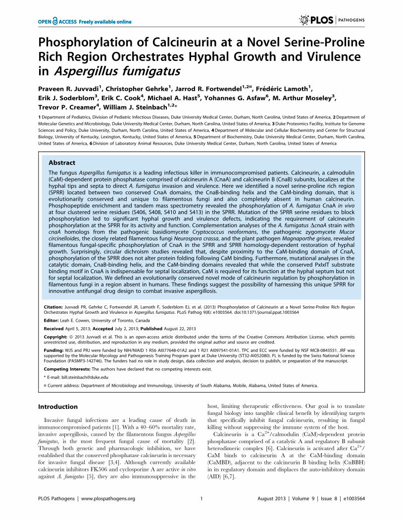
Truncations of A. fumigatus CnaA revealed importantdomains required for its function and septal localization
To characterize domains required for CnaA activity and septal
localization, we generated A. fumigatus strains expressing a series of
truncated cnaA cDNAs (cnaA-T1, cnaA-T2, cnaA-T3 and cnaA-T4)
under the control of its native promoter in the DcnaA mutant strain
(Figure 1A). While the expression of cnaA-T1, containing only the
catalytic domain (1–347 aa), did not complement the hyphal
growth defect of the DcnaA mutant strain and mislocalized CnaA
in the cytoplasm (Figure 1B and 1C), expression of cnaA-T2 that
included the CnBBH region (1–400 aa) showed partial growth
recovery, indicating that this fragment may bind to CnaB in vivo
and partially function by less efficiently localizing at the septum
(Figure 1B and 1C). However, expression of cnaA-T3 (1–425 aa),
containing the linker region spanning 23 aa between the CnBBH
and CaMBD (Figure 1A; indicated in red), completely restored
hyphal growth and efficiently localized CnaA at the septum
(Figure 1B and 1C). This indicated that the CaMBD and AID are
not required for septal targeting of CnaA. Complete hyphal
growth recovery observed in the CnaA-T3 strain also suggested
the possibility of a constitutively active calcineurin due to the
absence of the AID. Expression of cnaA-T4, including the CaMBD
but not the AID (1–458 aa), also completely restored hyphal
growth and properly localized CnaA at the septa (Figure 1B and
1C). Expression of all the constructs was confirmed by Western
analysis (Figure 1D).
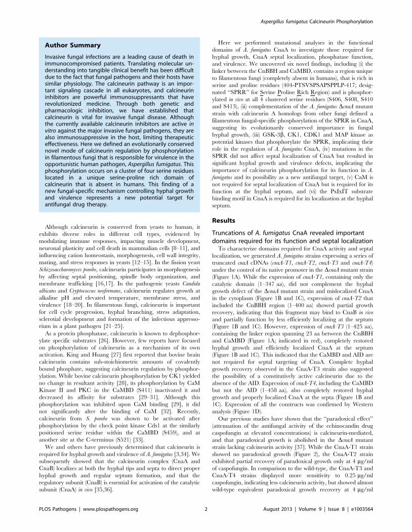
Our previous studies have shown that the ‘‘paradoxical effect’’
(attenuation of the antifungal activity of the echinocandin drug
caspofungin at elevated concentrations) is calcineurin-mediated,
and that paradoxical growth is abolished in the DcnaA mutant
strain lacking calcineurin activity [37]. While the CnaA-T1 strain
showed no paradoxical growth (Figure 2), the CnaA-T2 strain
exhibited partial recovery of paradoxical growth only at 4 mg/ml
of caspofungin. In comparison to the wild-type, the CnaA-T3 and
CnaA-T4 strains displayed more sensitivity to 0.25 mg/ml
caspofungin, indicating less calcineurin activity, but showed almost
wild-type equivalent paradoxical growth recovery at 4 mg/ml
Author Summary
Invasive fungal infections are a leading cause of death inimmunocompromised patients. Translating molecular un-derstanding into tangible clinical benefit has been difficultdue to the fact that fungal pathogens and their hosts havesimilar physiology. The calcineurin pathway is an impor-tant signaling cascade in all eukaryotes, and calcineurininhibitors are powerful immunosuppressants that haverevolutionized medicine. Through both genetic andpharmacologic inhibition, we have established thatcalcineurin is vital for invasive fungal disease. Althoughthe currently available calcineurin inhibitors are active invitro against the major invasive fungal pathogens, they arealso immunosuppressive in the host, limiting therapeuticeffectiveness. Here we defined an evolutionarily conservednovel mode of calcineurin regulation by phosphorylationin filamentous fungi that is responsible for virulence in theopportunistic human pathogen, Aspergillus fumigatus. Thisphosphorylation occurs on a cluster of four serine residueslocated in a unique serine-proline rich domain ofcalcineurin that is absent in humans. This finding of anew fungal-specific mechanism controlling hyphal growthand virulence represents a new potential target forantifungal drug therapy.
Aspergillus fumigatus Calcineurin Phosphorylation
PLOS Pathogens | www.plospathogens.org 2 August 2013 | Volume 9 | Issue 8 | e1003564
caspofungin. Concordant with these findings, the CnaA-T1 and
CnaA-T2 strains (Figure 1E) showed a significant reduction in
calcineurin activity (86% and 80%, respectively), and the CnaA-T3
strain showed only a 28% decrease in activity. Inclusion of the
CaMBD in the CnaA-T4 strain restored wild-type level of calcineurin
activity (Figure 1E). The growth restoration of the CnaA-T3 and
CnaA-T4 strains may also be attributed to constitutively active
calcineurin due to the truncation of the C-terminal autoinhibitory
domain. Taken together, these results indicated that the major
determinants/residues for hyphal growth restoration in the CnaA-T3
and CnaA-T4 strains and CnaA septal targeting may be present in
this newly described linker region between the CnBBH and the
CaMBD of CnaA. However, it is possible that targeting CnaA to the
hyphal septum occurs either independently or by binding of the linker
region to other unknown protein(s).
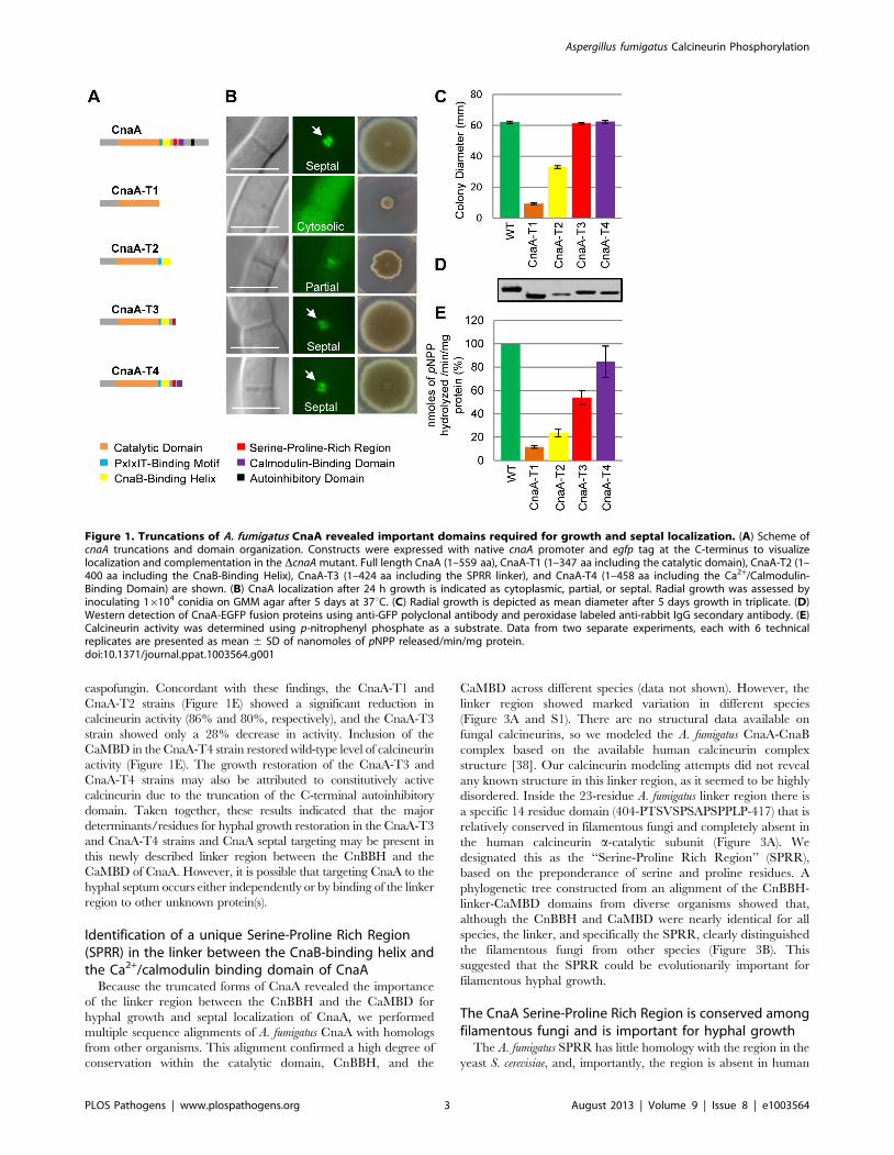
Identification of a unique Serine-Proline Rich Region(SPRR) in the linker between the CnaB-binding helix andthe Ca2+/calmodulin binding domain of CnaA
Because the truncated forms of CnaA revealed the importance
of the linker region between the CnBBH and the CaMBD for
hyphal growth and septal localization of CnaA, we performed
multiple sequence alignments of A. fumigatus CnaA with homologs
from other organisms. This alignment confirmed a high degree of
conservation within the catalytic domain, CnBBH, and the
CaMBD across different species (data not shown). However, the
linker region showed marked variation in different species
(Figure 3A and S1). There are no structural data available on
fungal calcineurins, so we modeled the A. fumigatus CnaA-CnaB
complex based on the available human calcineurin complex
structure [38]. Our calcineurin modeling attempts did not reveal
any known structure in this linker region, as it seemed to be highly
disordered. Inside the 23-residue A. fumigatus linker region there is
a specific 14 residue domain (404-PTSVSPSAPSPPLP-417) that is
relatively conserved in filamentous fungi and completely absent in
the human calcineurin a-catalytic subunit (Figure 3A). We
designated this as the ‘‘Serine-Proline Rich Region’’ (SPRR),
based on the preponderance of serine and proline residues. A
phylogenetic tree constructed from an alignment of the CnBBH-
linker-CaMBD domains from diverse organisms showed that,
although the CnBBH and CaMBD were nearly identical for all
species, the linker, and specifically the SPRR, clearly distinguished
the filamentous fungi from other species (Figure 3B). This
suggested that the SPRR could be evolutionarily important for
filamentous hyphal growth.
The CnaA Serine-Proline Rich Region is conserved amongfilamentous fungi and is important for hyphal growth
The A. fumigatus SPRR has little homology with the region in the
yeast S. cerevisiae, and, importantly, the region is absent in human
Figure 1. Truncations of A. fumigatus CnaA revealed important domains required for growth and septal localization. (A) Scheme ofcnaA truncations and domain organization. Constructs were expressed with native cnaA promoter and egfp tag at the C-terminus to visualizelocalization and complementation in the DcnaA mutant. Full length CnaA (1–559 aa), CnaA-T1 (1–347 aa including the catalytic domain), CnaA-T2 (1–400 aa including the CnaB-Binding Helix), CnaA-T3 (1–424 aa including the SPRR linker), and CnaA-T4 (1–458 aa including the Ca2+/Calmodulin-Binding Domain) are shown. (B) CnaA localization after 24 h growth is indicated as cytoplasmic, partial, or septal. Radial growth was assessed byinoculating 16104 conidia on GMM agar after 5 days at 37uC. (C) Radial growth is depicted as mean diameter after 5 days growth in triplicate. (D)Western detection of CnaA-EGFP fusion proteins using anti-GFP polyclonal antibody and peroxidase labeled anti-rabbit IgG secondary antibody. (E)Calcineurin activity was determined using p-nitrophenyl phosphate as a substrate. Data from two separate experiments, each with 6 technicalreplicates are presented as mean 6 SD of nanomoles of pNPP released/min/mg protein.doi:10.1371/journal.ppat.1003564.g001
Aspergillus fumigatus Calcineurin Phosphorylation
PLOS Pathogens | www.plospathogens.org 3 August 2013 | Volume 9 | Issue 8 | e1003564
Figure 2. Sensitivity of the CnaA truncation strains to Caspofungin. CnaA truncation expression strains were cultured on GMM agar in thepresence of varying concentrations of the cell wall inhibitor caspofungin for 5 days. The CnaA-T1 strain, which lacks all the functional domains ofCnaA, exhibits a severe growth defect and lacks caspofungin-mediated paradoxical growth due to a lack of calcineurin activity. In comparison to theWT the CnaA-T3 and CnaA-T4 strains show more sensitivity to 0.25 mg/ml caspofungin but recovery of paradoxical growth at 2 and 4 mg/mlcaspofungin. The CnaA-T2 strain, without the SPRR, shows paradoxical growth only at 4 mg/ml caspofungin.doi:10.1371/journal.ppat.1003564.g002
Figure 3. Conservation of a unique Serine-Proline Rich Region in filamentous fungi. (A) Alignment of the SPRR among filamentousascomycete fungi, yeasts, other fungi, and human. Conservation of serine and proline residues within the SPRR among filamentous fungi suggeststheir importance for hyphal growth; hyphal growth appears related to the potential ability for phosphorylation in the SPRR domain (box).Importantly, there is no homology in humans. (B) Phylogenic analysis of the CnaA region including the CnBBH, linker domain with the SPRR, and theCaMBD performed on the Phylogeny.fr platform. Sequences aligned with MUSCLE (v3.7) configured for highest accuracy. Phylogenetic tree wasreconstructed using the maximum likelihood method implemented in the PhyML program (v3.0 aLRT) and graphically represented with TreeDyn(v198.3).doi:10.1371/journal.ppat.1003564.g003
Aspergillus fumigatus Calcineurin Phosphorylation
PLOS Pathogens | www.plospathogens.org 4 August 2013 | Volume 9 | Issue 8 | e1003564
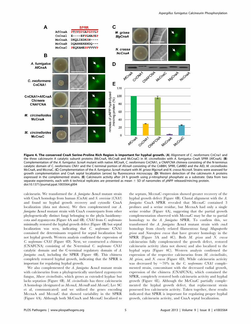
calcineurin. We transformed the A. fumigatus DcnaA mutant strain
with CnaA homologs from human (CnAa) and S. cerevisiae (CNA1)
and found no hyphal growth recovery and cytosolic CnaA
localization (data not shown). We then complemented our A.
fumigatus DcnaA mutant strain with CnaA counterparts from other
phylogenetically distinct fungi belonging to the phyla basidiomy-
cota and zygomycota (Figure 4A and 4B). CNA1 from C. neoformans
minimally restored the hyphal growth defect (Figure 4B) but septal
localization was seen, indicating that C. neoformans CNA1
contained the determinants required for septal localization but
not hyphal growth. Western analysis confirmed the expression of
C. neoformans CNA1 (Figure 4D). Next, we constructed a chimera
(CNAFCNA) consisting of the N-terminal C. neoformans CNA1
catalytic domain and the C-terminal regulatory domain of A.
fumigatus cnaA, including the SPRR (Figure 4B). This chimera
completely restored hyphal growth, indicating that the SPRR is
important for regulating hyphal growth.
We also complemented the A. fumigatus DcnaA mutant strain
with calcineurins from a phylogenetically unrelated zygomycete
fungus, Mucor circinelloides, which grows as extended hyphae but
lacks septation (Figure 4B). M. circinelloides has three calcineurin
A homologs (designated as MccnaA, MccnaB and MccnaC; Lee SC
et al, communicated) and we utilized the genes encoding
MccnaA and MccnaC that showed variability in the SPRR
(Figure 4A). Although both McCnaA and MccnaC localized to
the septum, MccnaC expression showed greater recovery of the
hyphal growth defect (Figure 4B). Clustal alignment with the A.
fumigatus CnaA SPRR revealed that MccnaC contained 3
prolines and a serine residue, but MccnaA had only a single
serine residue (Figure 4A), suggesting that the partial growth
complementation observed with MccnaC may be due to partial
homology to the A. fumigatus SPRR. To confirm this, we
transformed the A. fumigatus DcnaA mutant strain with cnaA
homologs from closely related filamentous fungi Magnaporthe
grisea and Neurospora crassa that have greater homology in the
SPRR (Figure 3A and 4C). Both M. grisea and N. crassa
calcineurins fully complemented the growth defect, restored
calcineurin activity (data not shown) and also localized to the
hyphal septa (Figure 4C). Western analysis confirmed the
expression of the respective calcineurins from M. circinelloides,
M. grisea, and N. crassa (Figure 4D). While calcineurin activity
was decreased by ,70% in the C. neoformans CNA1 comple-
mented strain, concomitant with the decreased radial growth,
expression of the chimera (CNAFCNA), which contained the
SPRR, completely restored both calcineurin activity and hyphal
growth (Figure 4E). Although the McCnaC partially comple-
mented the hyphal growth defect, that replacement strain
possessed less calcineurin activity. Taken together, these results
indicated that SPRR is important for regulating proper hyphal
growth, calcineurin activity, and CnaA septal localization.
Figure 4. The conserved CnaA Serine-Proline Rich Region is important for hyphal growth. (A) Alignment of C. neoformans CnCna1 andthe three calcineurin A catalytic subunit proteins (McCnaA, McCnaB and McCnaC) in M. circinelloides with A. fumigatus CnaA SPRR (AfCnaA). (B)Complementation of the A. fumigatus DcnaA mutant with native AfCnaA, C. neoformans CnCNA1, a CNAFCNA chimera consisting of the N-terminuscatalytic domain of C. neoformans CNA1 and the C-terminal portion of AfcnaA consisting of the CnBBH, SPRR, CaMBD and the AID, M. circinelloidesMcCnaA, and MccnaC. (C) Complementation of the A. fumigatus DcnaA mutant with M. grisea MgcnaA and N. crassa NccnaA. Strains were assessed forgrowth complementation and CnaA septal localization (arrow) by fluorescence microscopy. (D) Western detection of the calcineurin A proteinsexpressed in the complemented strains. (E) Calcineurin activity after 24 h growth using p-nitrophenyl phosphate as a substrate. Data from twoseparate experiments, each with 6 technical replicates are presented as mean 6 SD of nanomoles of pNPP released/min/mg protein.doi:10.1371/journal.ppat.1003564.g004
Aspergillus fumigatus Calcineurin Phosphorylation
PLOS Pathogens | www.plospathogens.org 5 August 2013 | Volume 9 | Issue 8 | e1003564
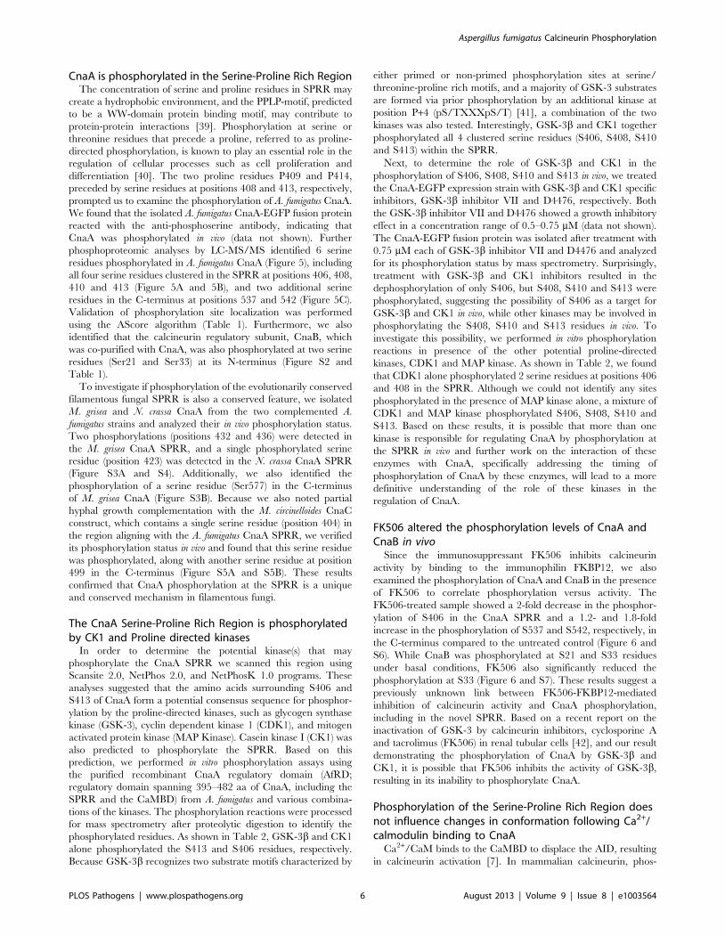
CnaA is phosphorylated in the Serine-Proline Rich RegionThe concentration of serine and proline residues in SPRR may
create a hydrophobic environment, and the PPLP-motif, predicted
to be a WW-domain protein binding motif, may contribute to
protein-protein interactions [39]. Phosphorylation at serine or
threonine residues that precede a proline, referred to as proline-
directed phosphorylation, is known to play an essential role in the
regulation of cellular processes such as cell proliferation and
differentiation [40]. The two proline residues P409 and P414,
preceded by serine residues at positions 408 and 413, respectively,
prompted us to examine the phosphorylation of A. fumigatus CnaA.
We found that the isolated A. fumigatus CnaA-EGFP fusion protein
reacted with the anti-phosphoserine antibody, indicating that
CnaA was phosphorylated in vivo (data not shown). Further
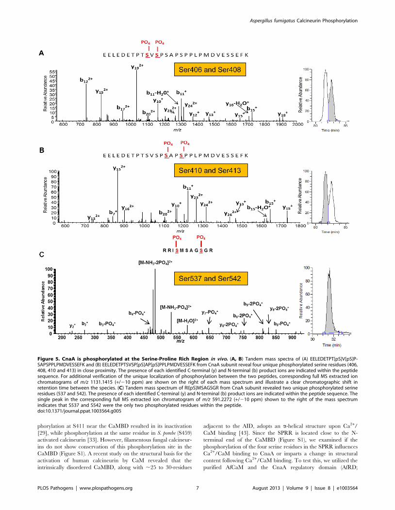
phosphoproteomic analyses by LC-MS/MS identified 6 serine
residues phosphorylated in A. fumigatus CnaA (Figure 5), including
all four serine residues clustered in the SPRR at positions 406, 408,
410 and 413 (Figure 5A and 5B), and two additional serine
residues in the C-terminus at positions 537 and 542 (Figure 5C).
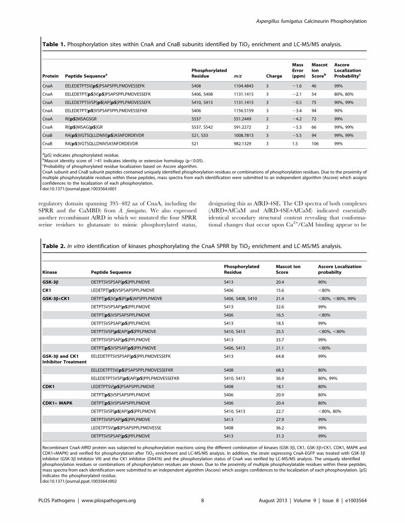
Validation of phosphorylation site localization was performed
using the AScore algorithm (Table 1). Furthermore, we also
identified that the calcineurin regulatory subunit, CnaB, which
was co-purified with CnaA, was also phosphorylated at two serine
residues (Ser21 and Ser33) at its N-terminus (Figure S2 and
Table 1).
To investigate if phosphorylation of the evolutionarily conserved
filamentous fungal SPRR is also a conserved feature, we isolated
M. grisea and N. crassa CnaA from the two complemented A.
fumigatus strains and analyzed their in vivo phosphorylation status.
Two phosphorylations (positions 432 and 436) were detected in
the M. grisea CnaA SPRR, and a single phosphorylated serine
residue (position 423) was detected in the N. crassa CnaA SPRR
(Figure S3A and S4). Additionally, we also identified the
phosphorylation of a serine residue (Ser577) in the C-terminus
of M. grisea CnaA (Figure S3B). Because we also noted partial
hyphal growth complementation with the M. circinelloides CnaC
construct, which contains a single serine residue (position 404) in
the region aligning with the A. fumigatus CnaA SPRR, we verified
its phosphorylation status in vivo and found that this serine residue
was phosphorylated, along with another serine residue at position
499 in the C-terminus (Figure S5A and S5B). These results
confirmed that CnaA phosphorylation at the SPRR is a unique
and conserved mechanism in filamentous fungi.
The CnaA Serine-Proline Rich Region is phosphorylatedby CK1 and Proline directed kinases
In order to determine the potential kinase(s) that may
phosphorylate the CnaA SPRR we scanned this region using
Scansite 2.0, NetPhos 2.0, and NetPhosK 1.0 programs. These
analyses suggested that the amino acids surrounding S406 and
S413 of CnaA form a potential consensus sequence for phosphor-
ylation by the proline-directed kinases, such as glycogen synthase
kinase (GSK-3), cyclin dependent kinase 1 (CDK1), and mitogen
activated protein kinase (MAP Kinase). Casein kinase I (CK1) was
also predicted to phosphorylate the SPRR. Based on this
prediction, we performed in vitro phosphorylation assays using
the purified recombinant CnaA regulatory domain (AfRD;
regulatory domain spanning 395–482 aa of CnaA, including the
SPRR and the CaMBD) from A. fumigatus and various combina-
tions of the kinases. The phosphorylation reactions were processed
for mass spectrometry after proteolytic digestion to identify the
phosphorylated residues. As shown in Table 2, GSK-3b and CK1
alone phosphorylated the S413 and S406 residues, respectively.
Because GSK-3b recognizes two substrate motifs characterized by
either primed or non-primed phosphorylation sites at serine/
threonine-proline rich motifs, and a majority of GSK-3 substrates
are formed via prior phosphorylation by an additional kinase at
position P+4 (pS/TXXXpS/T) [41], a combination of the two
kinases was also tested. Interestingly, GSK-3b and CK1 together
phosphorylated all 4 clustered serine residues (S406, S408, S410
and S413) within the SPRR.
Next, to determine the role of GSK-3b and CK1 in the
phosphorylation of S406, S408, S410 and S413 in vivo, we treated
the CnaA-EGFP expression strain with GSK-3b and CK1 specific
inhibitors, GSK-3b inhibitor VII and D4476, respectively. Both
the GSK-3b inhibitor VII and D4476 showed a growth inhibitory
effect in a concentration range of 0.5–0.75 mM (data not shown).
The CnaA-EGFP fusion protein was isolated after treatment with
0.75 mM each of GSK-3b inhibitor VII and D4476 and analyzed
for its phosphorylation status by mass spectrometry. Surprisingly,
treatment with GSK-3b and CK1 inhibitors resulted in the
dephosphorylation of only S406, but S408, S410 and S413 were
phosphorylated, suggesting the possibility of S406 as a target for
GSK-3b and CK1 in vivo, while other kinases may be involved in
phosphorylating the S408, S410 and S413 residues in vivo. To
investigate this possibility, we performed in vitro phosphorylation
reactions in presence of the other potential proline-directed
kinases, CDK1 and MAP kinase. As shown in Table 2, we found
that CDK1 alone phosphorylated 2 serine residues at positions 406
and 408 in the SPRR. Although we could not identify any sites
phosphorylated in the presence of MAP kinase alone, a mixture of
CDK1 and MAP kinase phosphorylated S406, S408, S410 and
S413. Based on these results, it is possible that more than one
kinase is responsible for regulating CnaA by phosphorylation at
the SPRR in vivo and further work on the interaction of these
enzymes with CnaA, specifically addressing the timing of
phosphorylation of CnaA by these enzymes, will lead to a more
definitive understanding of the role of these kinases in the
regulation of CnaA.
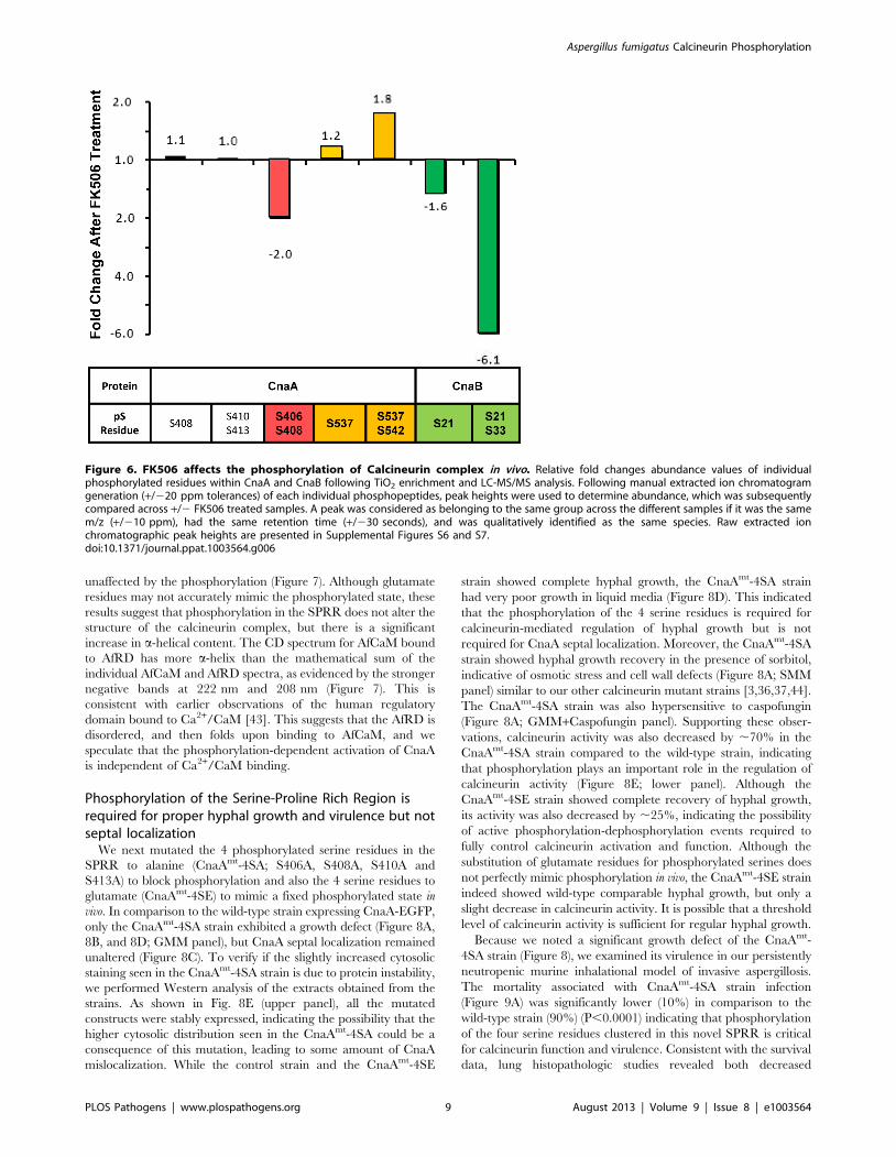
FK506 altered the phosphorylation levels of CnaA andCnaB in vivo
Since the immunosuppressant FK506 inhibits calcineurin
activity by binding to the immunophilin FKBP12, we also
examined the phosphorylation of CnaA and CnaB in the presence
of FK506 to correlate phosphorylation versus activity. The
FK506-treated sample showed a 2-fold decrease in the phosphor-
ylation of S406 in the CnaA SPRR and a 1.2- and 1.8-fold
increase in the phosphorylation of S537 and S542, respectively, in
the C-terminus compared to the untreated control (Figure 6 and
S6). While CnaB was phosphorylated at S21 and S33 residues
under basal conditions, FK506 also significantly reduced the
phosphorylation at S33 (Figure 6 and S7). These results suggest a
previously unknown link between FK506-FKBP12-mediated
inhibition of calcineurin activity and CnaA phosphorylation,
including in the novel SPRR. Based on a recent report on the
inactivation of GSK-3 by calcineurin inhibitors, cyclosporine A
and tacrolimus (FK506) in renal tubular cells [42], and our result
demonstrating the phosphorylation of CnaA by GSK-3b and
CK1, it is possible that FK506 inhibits the activity of GSK-3b,
resulting in its inability to phosphorylate CnaA.
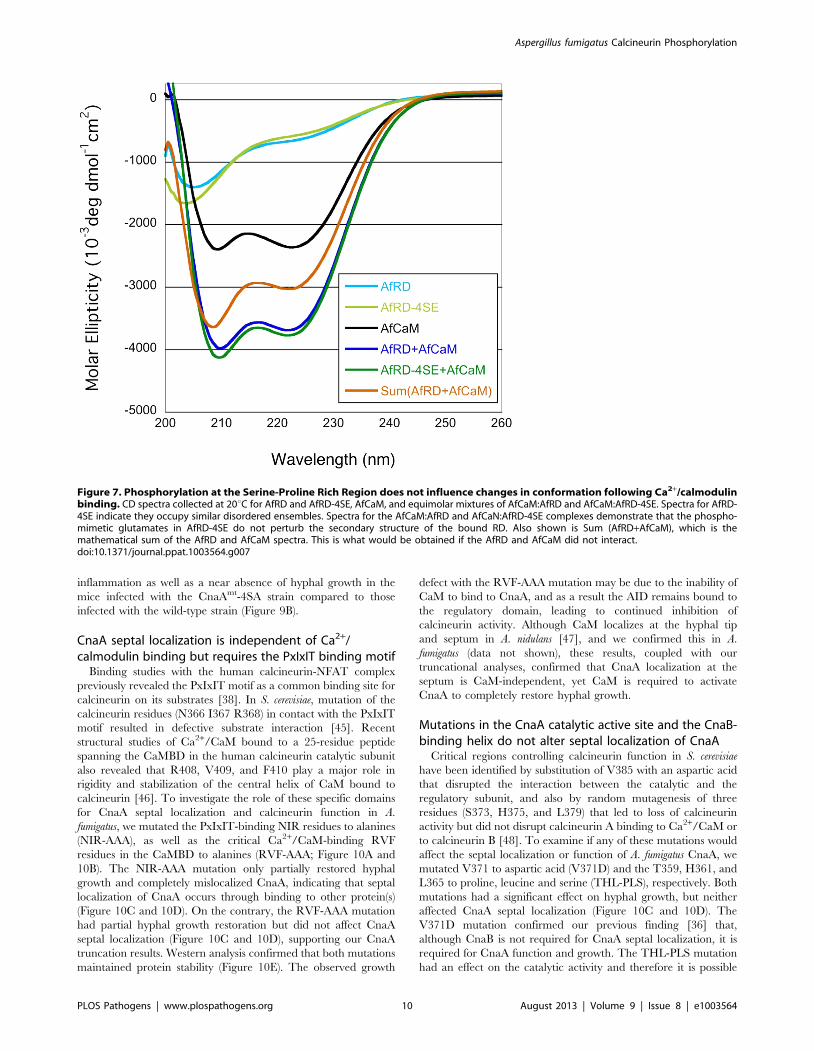
Phosphorylation of the Serine-Proline Rich Region doesnot influence changes in conformation following Ca2+/calmodulin binding to CnaA
Ca2+/CaM binds to the CaMBD to displace the AID, resulting
in calcineurin activation [7]. In mammalian calcineurin, phos-
Aspergillus fumigatus Calcineurin Phosphorylation
PLOS Pathogens | www.plospathogens.org 6 August 2013 | Volume 9 | Issue 8 | e1003564
phorylation at S411 near the CaMBD resulted in its inactivation
[29], while phosphorylation at the same residue in S. pombe (S459)
activated calcineurin [33]. However, filamentous fungal calcineur-
ins do not show conservation of this phosphorylation site in the
CaMBD (Figure S1). A recent study on the structural basis for the
activation of human calcineurin by CaM revealed that the
intrinsically disordered CaMBD, along with ,25 to 30-residues
adjacent to the AID, adopts an a-helical structure upon Ca2+/
CaM binding [43]. Since the SPRR is located close to the N-
terminal end of the CaMBD (Figure S1), we examined if the
phosphorylation of the four serine residues in the SPRR influences
Ca2+/CaM binding to CnaA or imparts a change in structural
content following Ca2+/CaM binding. To test this, we utilized the
purified AfCaM and the CnaA regulatory domain (AfRD;
Figure 5. CnaA is phosphorylated at the Serine-Proline Rich Region in vivo. (A, B) Tandem mass spectra of (A) EELEDETPT[pS]V[pS]P-SAPSPPLPMDVESSEFK and (B) EELEDETPTSVSP[pS]AP[pS]PPLPMDVESSEFK from CnaA subunit reveal four unique phosphorylated serine residues (406,408, 410 and 413) in close proximity. The presence of each identified C-terminal (y) and N-terminal (b) product ions are indicated within the peptidesequence. For additional verification of the unique localization of phosphorylation between the two peptides, corresponding full MS extracted ionchromatograms of m/z 1131.1415 (+/210 ppm) are shown on the right of each mass spectrum and illustrate a clear chromatographic shift inretention time between the species. (C) Tandem mass spectrum of RI[pS]MSAGSGR from CnaA subunit revealed two unique phosphorylated serineresidues (537 and 542). The presence of each identified C-terminal (y) and N-terminal (b) product ions are indicated within the peptide sequence. Thesingle peak in the corresponding full MS extracted ion chromatogram of m/z 591.2272 (+/210 ppm) shown to the right of the mass spectrumindicates that S537 and S542 were the only two phosphorylated residues within the peptide.doi:10.1371/journal.ppat.1003564.g005
Aspergillus fumigatus Calcineurin Phosphorylation
PLOS Pathogens | www.plospathogens.org 7 August 2013 | Volume 9 | Issue 8 | e1003564
regulatory domain spanning 395–482 aa of CnaA, including the
SPRR and the CaMBD) from A. fumigatus. We also expressed
another recombinant AfRD in which we mutated the four SPRR
serine residues to glutamate to mimic phosphorylated status,
designating this as AfRD-4SE. The CD spectra of both complexes
(AfRD+AfCaM and AfRD-4SE+AfCaM) indicated essentially
identical secondary structural content revealing that conforma-
tional changes that occur upon Ca2+/CaM binding appear to be
Table 1. Phosphorylation sites within CnaA and CnaB subunits identified by TiO2 enrichment and LC-MS/MS analysis.
Protein Peptide SequenceaPhosphorylatedResidue m/z Charge
MassError(ppm)
MascotIonScoreb
AscoreLocalizationProbabilityc
CnaA EELEDETPTSV[pS]PSAPSPPLPMDVESSEFK S408 1104.4843 3 21.6 46 99%
CnaA EELEDETPT[pS]V[pS]PSAPSPPLPMDVESSEFK S406, S408 1131.1415 3 22.1 54 80%, 80%
CnaA EELEDETPTSVSP[pS]AP[pS]PPLPMDVESSEFK S410, S413 1131.1415 3 20.5 75 90%, 99%
CnaA EELEDETPT[pS]VSPSAPSPPLPMDVESSEFKR S406 1156.5159 3 23.4 94 90%
CnaA RI[pS]MSAGSGR S537 551.2449 2 24.2 72 99%
CnaA RI[pS]MSAG[pS]GR S537, S542 591.2272 2 25.3 66 99%, 99%
CnaB RA[pS]VGTSQLLDNIV[pS]ASNFDRDEVDR S21, S33 1008.7813 3 25.5 94 99%, 99%
CnaB RA[pS]VGTSQLLDNIVSASNFDRDEVDR S21 982.1329 3 1.5 106 99%
a[pS] indicates phosphorylated residue.bMascot identity score of .41 indicates identity or extensive homology (p,0.05).cProbability of phosphorylated residue localization based on Ascore algorithm.CnaA subunit and CnaB subunit peptides contained uniquely identified phosphorylation residues or combinations of phosphorylation residues. Due to the proximity ofmultiple phosphorylatable residues within these peptides, mass spectra from each identification were submitted to an independent algorithm (Ascore) which assignsconfidences to the localization of each phosphorylation.doi:10.1371/journal.ppat.1003564.t001
Table 2. In vitro identification of kinases phosphorylating the CnaA SPRR by TiO2 enrichment and LC-MS/MS analysis.
Kinase Peptide SequencePhosphorylatedResidue
Mascot IonScore
Ascore Localizationprobabilty
GSK-3b DETPTSVSPSAP[pS]PPLPMDVE S413 20.4 90%
CK1 LEDETPT[pS]VSPSAPSPPLPMDVE S406 15.6 ,80%
GSK-3b+CK1 DETPT[pS]V[pS]P[pS]APSPPLPMDVE S406, S408, S410 21.4 ,80%, ,80%, 99%
DETPTSVSPSAP[pS]PPLPMDVE S413 32.6 99%
DETPT[pS]VSPSAPSPPLPMDVE S406 16.5 ,80%
DETPTSVSPSAP[pS]PPLPMDVE S413 18.5 99%
DETPTSVSP[pS]AP[pS]PPLPMDVE S410, S413 25.5 ,80%, ,80%
DETPTSVSPSAP[pS]PPLPMDVE S413 33.7 99%
DETPT[pS]VSPSAP[pS]PPLPMDVE S406, S413 21.1 ,80%
GSK-3b and CK1Inhibitor Treatment
EELEDETPTSVSPSAP[pS]PPLPMDVESSEFK S413 64.8 99%
EELEDETPTSV[pS]PSAPSPPLPMDVESSEFKR S408 68.3 80%
EELEDETPTSVSP[pS]AP[pS]PPLPMDVESSEFKR S410, S413 36.9 80%, 99%
CDK1 LEDETPTSV[pS]PSAPSPPLPMDVE S408 18.1 80%
DETPT[pS]VSPSAPSPPLPMDVE S406 20.9 80%
CDK1+ MAPK DETPT[pS]VSPSAPSPPLPMDVE S406 20.4 80%
DETPTSVSP[pS]AP[pS]PPLPMDVE S410, S413 22.7 ,80%, 80%
DETPTSVSPSAP[pS]PPLPMDVE S413 27.9 99%
LEDETPTSV[pS]PSAPSPPLPMDVESSE S408 36.2 99%
DETPTSVSPSAP[pS]PPLPMDVE S413 31.3 99%
Recombinant CnaA-AfRD protein was subjected to phosphorylation reactions using the different combination of kinases (GSK-3b, CK1, GSK-3b+CK1, CDK1, MAPK andCDK1+MAPK) and verified for phosphorylation after TiO2 enrichment and LC-MS/MS analysis. In addition, the strain expressing CnaA-EGFP was treated with GSK-3binhibitor (GSK-3b Inhibitor VII) and the CK1 inhibitor (D4476) and the phosphorylation status of CnaA was verified by LC-MS/MS analysis. The uniquely identifiedphosphorylation residues or combinations of phosphorylation residues are shown. Due to the proximity of multiple phosphorylatable residues within these peptides,mass spectra from each identification were submitted to an independent algorithm (Ascore) which assigns confidences to the localization of each phosphorylation. [pS]indicates the phosphorylated residue.doi:10.1371/journal.ppat.1003564.t002
Aspergillus fumigatus Calcineurin Phosphorylation
PLOS Pathogens | www.plospathogens.org 8 August 2013 | Volume 9 | Issue 8 | e1003564
unaffected by the phosphorylation (Figure 7). Although glutamate
residues may not accurately mimic the phosphorylated state, these
results suggest that phosphorylation in the SPRR does not alter the
structure of the calcineurin complex, but there is a significant
increase in a-helical content. The CD spectrum for AfCaM bound
to AfRD has more a-helix than the mathematical sum of the
individual AfCaM and AfRD spectra, as evidenced by the stronger
negative bands at 222 nm and 208 nm (Figure 7). This is
consistent with earlier observations of the human regulatory
domain bound to Ca2+/CaM [43]. This suggests that the AfRD is
disordered, and then folds upon binding to AfCaM, and we
speculate that the phosphorylation-dependent activation of CnaA
is independent of Ca2+/CaM binding.
Phosphorylation of the Serine-Proline Rich Region isrequired for proper hyphal growth and virulence but notseptal localization
We next mutated the 4 phosphorylated serine residues in the
SPRR to alanine (CnaAmt-4SA; S406A, S408A, S410A and
S413A) to block phosphorylation and also the 4 serine residues to
glutamate (CnaAmt-4SE) to mimic a fixed phosphorylated state in
vivo. In comparison to the wild-type strain expressing CnaA-EGFP,
only the CnaAmt-4SA strain exhibited a growth defect (Figure 8A,
8B, and 8D; GMM panel), but CnaA septal localization remained
unaltered (Figure 8C). To verify if the slightly increased cytosolic
staining seen in the CnaAmt-4SA strain is due to protein instability,
we performed Western analysis of the extracts obtained from the
strains. As shown in Fig. 8E (upper panel), all the mutated
constructs were stably expressed, indicating the possibility that the
higher cytosolic distribution seen in the CnaAmt-4SA could be a
consequence of this mutation, leading to some amount of CnaA
mislocalization. While the control strain and the CnaAmt-4SE
strain showed complete hyphal growth, the CnaAmt-4SA strain
had very poor growth in liquid media (Figure 8D). This indicated
that the phosphorylation of the 4 serine residues is required for
calcineurin-mediated regulation of hyphal growth but is not
required for CnaA septal localization. Moreover, the CnaAmt-4SA
strain showed hyphal growth recovery in the presence of sorbitol,
indicative of osmotic stress and cell wall defects (Figure 8A; SMM
panel) similar to our other calcineurin mutant strains [3,36,37,44].
The CnaAmt-4SA strain was also hypersensitive to caspofungin
(Figure 8A; GMM+Caspofungin panel). Supporting these obser-
vations, calcineurin activity was also decreased by ,70% in the
CnaAmt-4SA strain compared to the wild-type strain, indicating
that phosphorylation plays an important role in the regulation of
calcineurin activity (Figure 8E; lower panel). Although the
CnaAmt-4SE strain showed complete recovery of hyphal growth,
its activity was also decreased by ,25%, indicating the possibility
of active phosphorylation-dephosphorylation events required to
fully control calcineurin activation and function. Although the
substitution of glutamate residues for phosphorylated serines does
not perfectly mimic phosphorylation in vivo, the CnaAmt-4SE strain
indeed showed wild-type comparable hyphal growth, but only a
slight decrease in calcineurin activity. It is possible that a threshold
level of calcineurin activity is sufficient for regular hyphal growth.
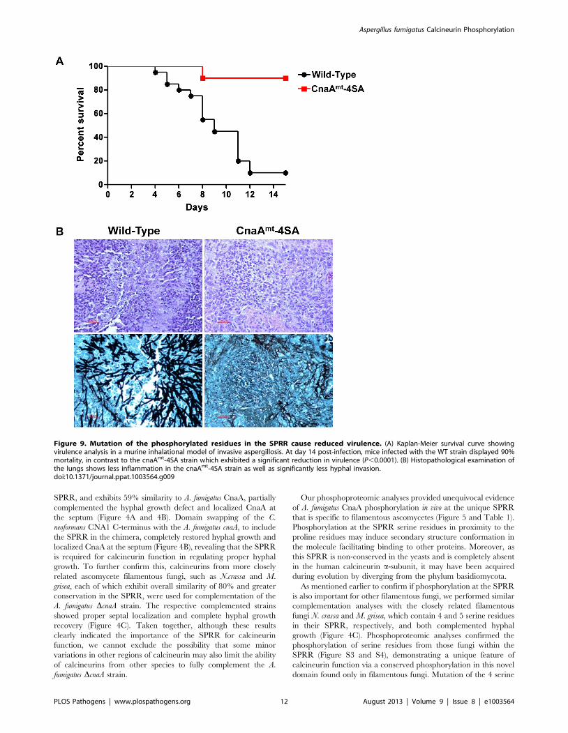
Because we noted a significant growth defect of the CnaAmt-
4SA strain (Figure 8), we examined its virulence in our persistently
neutropenic murine inhalational model of invasive aspergillosis.
The mortality associated with CnaAmt-4SA strain infection
(Figure 9A) was significantly lower (10%) in comparison to the
wild-type strain (90%) (P,0.0001) indicating that phosphorylation
of the four serine residues clustered in this novel SPRR is critical
for calcineurin function and virulence. Consistent with the survival
data, lung histopathologic studies revealed both decreased
Figure 6. FK506 affects the phosphorylation of Calcineurin complex in vivo. Relative fold changes abundance values of individualphosphorylated residues within CnaA and CnaB following TiO2 enrichment and LC-MS/MS analysis. Following manual extracted ion chromatogramgeneration (+/220 ppm tolerances) of each individual phosphopeptides, peak heights were used to determine abundance, which was subsequentlycompared across +/2 FK506 treated samples. A peak was considered as belonging to the same group across the different samples if it was the samem/z (+/210 ppm), had the same retention time (+/230 seconds), and was qualitatively identified as the same species. Raw extracted ionchromatographic peak heights are presented in Supplemental Figures S6 and S7.doi:10.1371/journal.ppat.1003564.g006
Aspergillus fumigatus Calcineurin Phosphorylation
PLOS Pathogens | www.plospathogens.org 9 August 2013 | Volume 9 | Issue 8 | e1003564
inflammation as well as a near absence of hyphal growth in the
mice infected with the CnaAmt-4SA strain compared to those
infected with the wild-type strain (Figure 9B).
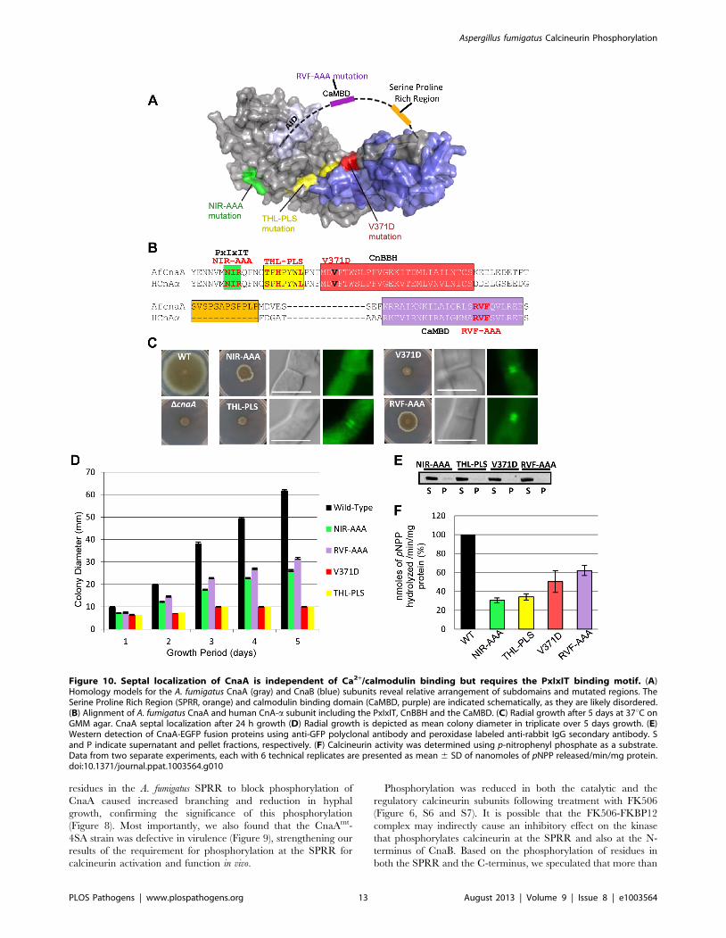
CnaA septal localization is independent of Ca2+/calmodulin binding but requires the PxIxIT binding motif
Binding studies with the human calcineurin-NFAT complex
previously revealed the PxIxIT motif as a common binding site for
calcineurin on its substrates [38]. In S. cerevisiae, mutation of the
calcineurin residues (N366 I367 R368) in contact with the PxIxIT
motif resulted in defective substrate interaction [45]. Recent
structural studies of Ca2+/CaM bound to a 25-residue peptide
spanning the CaMBD in the human calcineurin catalytic subunit
also revealed that R408, V409, and F410 play a major role in
rigidity and stabilization of the central helix of CaM bound to
calcineurin [46]. To investigate the role of these specific domains
for CnaA septal localization and calcineurin function in A.
fumigatus, we mutated the PxIxIT-binding NIR residues to alanines
(NIR-AAA), as well as the critical Ca2+/CaM-binding RVF
residues in the CaMBD to alanines (RVF-AAA; Figure 10A and
10B). The NIR-AAA mutation only partially restored hyphal
growth and completely mislocalized CnaA, indicating that septal
localization of CnaA occurs through binding to other protein(s)
(Figure 10C and 10D). On the contrary, the RVF-AAA mutation
had partial hyphal growth restoration but did not affect CnaA
septal localization (Figure 10C and 10D), supporting our CnaA
truncation results. Western analysis confirmed that both mutations
maintained protein stability (Figure 10E). The observed growth
defect with the RVF-AAA mutation may be due to the inability of
CaM to bind to CnaA, and as a result the AID remains bound to
the regulatory domain, leading to continued inhibition of
calcineurin activity. Although CaM localizes at the hyphal tip
and septum in A. nidulans [47], and we confirmed this in A.
fumigatus (data not shown), these results, coupled with our
truncational analyses, confirmed that CnaA localization at the
septum is CaM-independent, yet CaM is required to activate
CnaA to completely restore hyphal growth.
Mutations in the CnaA catalytic active site and the CnaB-binding helix do not alter septal localization of CnaA
Critical regions controlling calcineurin function in S. cerevisiae
have been identified by substitution of V385 with an aspartic acid
that disrupted the interaction between the catalytic and the
regulatory subunit, and also by random mutagenesis of three
residues (S373, H375, and L379) that led to loss of calcineurin
activity but did not disrupt calcineurin A binding to Ca2+/CaM or
to calcineurin B [48]. To examine if any of these mutations would
affect the septal localization or function of A. fumigatus CnaA, we
mutated V371 to aspartic acid (V371D) and the T359, H361, and
L365 to proline, leucine and serine (THL-PLS), respectively. Both
mutations had a significant effect on hyphal growth, but neither
affected CnaA septal localization (Figure 10C and 10D). The
V371D mutation confirmed our previous finding [36] that,
although CnaB is not required for CnaA septal localization, it is
required for CnaA function and growth. The THL-PLS mutation
had an effect on the catalytic activity and therefore it is possible
Figure 7. Phosphorylation at the Serine-Proline Rich Region does not influence changes in conformation following Ca2+/calmodulinbinding. CD spectra collected at 20uC for AfRD and AfRD-4SE, AfCaM, and equimolar mixtures of AfCaM:AfRD and AfCaM:AfRD-4SE. Spectra for AfRD-4SE indicate they occupy similar disordered ensembles. Spectra for the AfCaM:AfRD and AfCaN:AfRD-4SE complexes demonstrate that the phospho-mimetic glutamates in AfRD-4SE do not perturb the secondary structure of the bound RD. Also shown is Sum (AfRD+AfCaM), which is themathematical sum of the AfRD and AfCaM spectra. This is what would be obtained if the AfRD and AfCaM did not interact.doi:10.1371/journal.ppat.1003564.g007
Aspergillus fumigatus Calcineurin Phosphorylation
PLOS Pathogens | www.plospathogens.org 10 August 2013 | Volume 9 | Issue 8 | e1003564
that although CnaA is localized at the hyphal septum it is
catalytically inactive. We confirmed the stability of each mutation
by Western analysis (Figure 10E). The reduction in calcineurin
activity due to these mutations (Figure 10F) and the lack of
paradoxical growth recovery (Figure S8) established that catalytic
site residues and CnaB-binding activity of CnaA do not influence
its septal localization, yet catalytically active calcineurin is required
at the hyphal septum to direct proper hyphal growth.
Discussion
Calcineurin inhibitors are promising new antifungal candidates
due to their unique mode of action from other antifungal classes
(e.g., polyenes, triazoles, echinocandins), efficacy against emerging
resistant strains, and synergism with existing antifungals [4].
However, currently-used calcineurin inhibitors complex with
immunophilins leading to host immunosuppression [49]. Although
calcineurin has been well studied in several organisms and its
functional domains described, few studies have focused on
mutations in its key domains in vivo, and none have examined
phosphorylation as a mechanism of calcineurin function in a
human pathogen. By deleting the C-terminal regulatory domains
of CnaA which led to progressive defects in hyphal growth
(Figure 1 and 2), we identified a unique fungal-specific 23 residue
linker domain between the CnBBH and the CaMBD, containing
the novel and evolutionarily conserved SPRR (Figure 3A).
Inclusion of the SPRR showed full recovery of hyphal growth,
concomitant increase in calcineurin activity, and clear localization
of CnaA to the hyphal septum (Figure 4).
To evaluate the conservation of this linker region, we examined
it in 22 eukaryotes selected based on divergence and to include
model organisms and pathogens affecting both humans and plants
(Figure S1). Phylogenic analysis showed that the linker containing
the SPRR clearly distinguished the filamentous fungi (Figure 3B).
Based on the CnaA-CnaB molecular model we created
(Figure 10A), the SPRR is present outside the core binding region
between CnaA and CnaB, and the preponderance of proline and
serine residues in this linker creates a hydrophobic environment
which could lead to binding to other as of yet unknown proteins.
To determine the importance of the SPRR for CnaA function in
vivo, we performed complementation tests in our A. fumigatus DcnaA
mutant strain with cnaA homologs from human, S.cerevisiae, C.
neoformans and M. circinelloides, all of which lacked similarity within
the SPRR (Figure 4A and 4B). While neither human nor S.cerevisiae
CNA, which possess an overall 56% and 50% similarity to A.
fumigatus CnaA, respectively, complemented the hyphal growth
defect (data not show), C. neoformans CNA1, which exhibits 67%
similarity to A. fumigatus CnaA, localized to the septum but did not
restore hyphal growth (Figure 4B). Interestingly, only M.
circinelloides CnaC, which had partial similarity to the A. fumigatus
Figure 8. Phosphorylation of CnaA is required for proper hyphal growth. (A) Radial growth after 5 days at 37uC on GMM agar and GMMsupplemented with 1.2 M sorbitol (SMM) to assess growth remediation. Wild-type, cnaAmt-4SA, and cnaAmt-4SE strains (16104 conidia each) werecultured on GMM in varying concentrations of caspofungin for 5 days. The cnaAmt-4SA strain, defective in phosphorylation, exhibits a growth defectand lacks caspofungin-mediated paradoxical growth due to lack of calcineurin activity. (B) Radial growth is depicted as mean colony diameter intriplicate during 1–5 days growth period. (C) Localization of the CnaA mutated constructs by fluorescence microscopy after 24 h growth. (D) Growthreduction of the cnaAmt-4SA in comparison to the wild-type strain after inoculating 16106 conidia into GMM liquid and growth for 48 h at 37uC. (E)Calcineurin activity after 24 h growth using p-nitrophenyl phosphate as a substrate. Data from two separate experiments, each with 6 technicalreplicates are presented as mean 6 SD of nanomoles of pNPP released/min/mg protein.doi:10.1371/journal.ppat.1003564.g008
Aspergillus fumigatus Calcineurin Phosphorylation
PLOS Pathogens | www.plospathogens.org 11 August 2013 | Volume 9 | Issue 8 | e1003564
SPRR, and exhibits 59% similarity to A. fumigatus CnaA, partially
complemented the hyphal growth defect and localized CnaA at
the septum (Figure 4A and 4B). Domain swapping of the C.
neoformans CNA1 C-terminus with the A. fumigatus cnaA, to include
the SPRR in the chimera, completely restored hyphal growth and
localized CnaA at the septum (Figure 4B), revealing that the SPRR
is required for calcineurin function in regulating proper hyphal
growth. To further confirm this, calcineurins from more closely
related ascomycete filamentous fungi, such as N.crassa and M.
grisea, each of which exhibit overall similarity of 80% and greater
conservation in the SPRR, were used for complementation of the
A. fumigatus DcnaA strain. The respective complemented strains
showed proper septal localization and complete hyphal growth
recovery (Figure 4C). Taken together, although these results
clearly indicated the importance of the SPRR for calcineurin
function, we cannot exclude the possibility that some minor
variations in other regions of calcineurin may also limit the ability
of calcineurins from other species to fully complement the A.
fumigatus DcnaA strain.
Our phosphoproteomic analyses provided unequivocal evidence
of A. fumigatus CnaA phosphorylation in vivo at the unique SPRR
that is specific to filamentous ascomycetes (Figure 5 and Table 1).
Phosphorylation at the SPRR serine residues in proximity to the
proline residues may induce secondary structure conformation in
the molecule facilitating binding to other proteins. Moreover, as
this SPRR is non-conserved in the yeasts and is completely absent
in the human calcineurin a-subunit, it may have been acquired
during evolution by diverging from the phylum basidiomycota.
As mentioned earlier to confirm if phosphorylation at the SPRR
is also important for other filamentous fungi, we performed similar
complementation analyses with the closely related filamentous
fungi N. crassa and M. grisea, which contain 4 and 5 serine residues
in their SPRR, respectively, and both complemented hyphal
growth (Figure 4C). Phosphoproteomic analyses confirmed the
phosphorylation of serine residues from those fungi within the
SPRR (Figure S3 and S4), demonstrating a unique feature of
calcineurin function via a conserved phosphorylation in this novel
domain found only in filamentous fungi. Mutation of the 4 serine
Figure 9. Mutation of the phosphorylated residues in the SPRR cause reduced virulence. (A) Kaplan-Meier survival curve showingvirulence analysis in a murine inhalational model of invasive aspergillosis. At day 14 post-infection, mice infected with the WT strain displayed 90%mortality, in contrast to the cnaAmt-4SA strain which exhibited a significant reduction in virulence (P,0.0001). (B) Histopathological examination ofthe lungs shows less inflammation in the cnaAmt-4SA strain as well as significantly less hyphal invasion.doi:10.1371/journal.ppat.1003564.g009
Aspergillus fumigatus Calcineurin Phosphorylation
PLOS Pathogens | www.plospathogens.org 12 August 2013 | Volume 9 | Issue 8 | e1003564
residues in the A. fumigatus SPRR to block phosphorylation of
CnaA caused increased branching and reduction in hyphal
growth, confirming the significance of this phosphorylation
(Figure 8). Most importantly, we also found that the CnaAmt-
4SA strain was defective in virulence (Figure 9), strengthening our
results of the requirement for phosphorylation at the SPRR for
calcineurin activation and function in vivo.
Phosphorylation was reduced in both the catalytic and the
regulatory calcineurin subunits following treatment with FK506
(Figure 6, S6 and S7). It is possible that the FK506-FKBP12
complex may indirectly cause an inhibitory effect on the kinase
that phosphorylates calcineurin at the SPRR and also at the N-
terminus of CnaB. Based on the phosphorylation of residues in
both the SPRR and the C-terminus, we speculated that more than
Figure 10. Septal localization of CnaA is independent of Ca2+/calmodulin binding but requires the PxIxIT binding motif. (A)Homology models for the A. fumigatus CnaA (gray) and CnaB (blue) subunits reveal relative arrangement of subdomains and mutated regions. TheSerine Proline Rich Region (SPRR, orange) and calmodulin binding domain (CaMBD, purple) are indicated schematically, as they are likely disordered.(B) Alignment of A. fumigatus CnaA and human CnA-a subunit including the PxIxIT, CnBBH and the CaMBD. (C) Radial growth after 5 days at 37uC onGMM agar. CnaA septal localization after 24 h growth (D) Radial growth is depicted as mean colony diameter in triplicate over 5 days growth. (E)Western detection of CnaA-EGFP fusion proteins using anti-GFP polyclonal antibody and peroxidase labeled anti-rabbit IgG secondary antibody. Sand P indicate supernatant and pellet fractions, respectively. (F) Calcineurin activity was determined using p-nitrophenyl phosphate as a substrate.Data from two separate experiments, each with 6 technical replicates are presented as mean 6 SD of nanomoles of pNPP released/min/mg protein.doi:10.1371/journal.ppat.1003564.g010
Aspergillus fumigatus Calcineurin Phosphorylation
PLOS Pathogens | www.plospathogens.org 13 August 2013 | Volume 9 | Issue 8 | e1003564
one kinase is responsible. Although mammalian calcineurin was
shown to be phosphorylated at S411 in the CaMBD by PKC and
CaM kinase II [29–31], and the same residue at position S459 in
S. pombe was phosphorylated by Cds1 check point kinase [33], this
serine residue is not conserved among filamentous fungal
calcineurins. Based on our identified phosphorylation sites in A.
fumigatus CnaA and the SPRR, we were able to predict the
phosphorylation of this region by potential proline-directed kinases
such as GSK-3, CDK1 and MAP kinase. By in vitro phosphory-
lation assays, using the enzymes GSK-3b and CK1, we identified
that all 4 serine residue in the SPRR were phosphorylated.
Additionally, S413 and S408 are both flanked by downstream
proline residues, which represent typical GSK-3 phosphorylation
sites. While GSK-3b alone phosphorylated S413 in the SPRR,
CK1 alone phosphorylated S406, and a combination with GSK-
3b led to phosphorylation of other serine residues (S408 and
S410), revealing that the prephosphorylation of S406 residue by
CK1 may trigger the subsequent phosphorylation of S408 and
S410. Interestingly, a previous study showed that the yeast Mck1
protein kinase belonging to the GSK-3 kinase family stimulated
calcineurin activity by phosphorylating Rcn1, and in the absence
of GSK-3 kinase, calcineurin activity was fully inhibited revealing
a allosteric mechanism of calcineurin regulation by Rcn1 [50]. We
further validated our in vitro phosphorylation data by examining
the phosphorylation status of CnaA in vivo after treating with
inhibitors for GSK-3b and CK1, which confirmed the absence of
phosphorylation at S406 only but not the other serine residues,
leading to the notion that other kinases may also phosphorylate the
CnaA SPRR in vivo. The observed phosphorylation of CnaA
SPRR by CDK1 and MAP kinase in vitro strengthens this
possibility. However, further analyses are required to specifically
understand how these enzymes actually regulate CnaA.
Since the CaMBD is in close proximity to the SPRR, we
hypothesized that phosphorylation in the SPRR caused confor-
mational changes or altered binding between Ca2+/CaM and the
calcineurin complex. Circular dichroism studies revealed that
conformational changes that occur after Ca2+/CaM binding
remained unaffected between unphosphorylated and phosphomi-
metic SPRR constructs (Figure 7), indicating that CaM-mediated
activation of calcineurin is independent of SPRR phosphorylation.
This is intriguing when considering that calcineurin phosphory-
lation at S411 in Rat slightly decreased the affinity of calcineurin
for Ca2+/CaM, causing its inactivation [29], and phosphorylation
at the same residue (S459) in S. pombe activated calcineurin [33].
Surprisingly, even though Ca2+/CaM is well known to bind to
calcineurin and localize at the hyphal tips and septum [47], the
septal localization of CnaA was not impaired by the deletion of the
C-terminus (Figure 1), indicating that CaM is not involved in
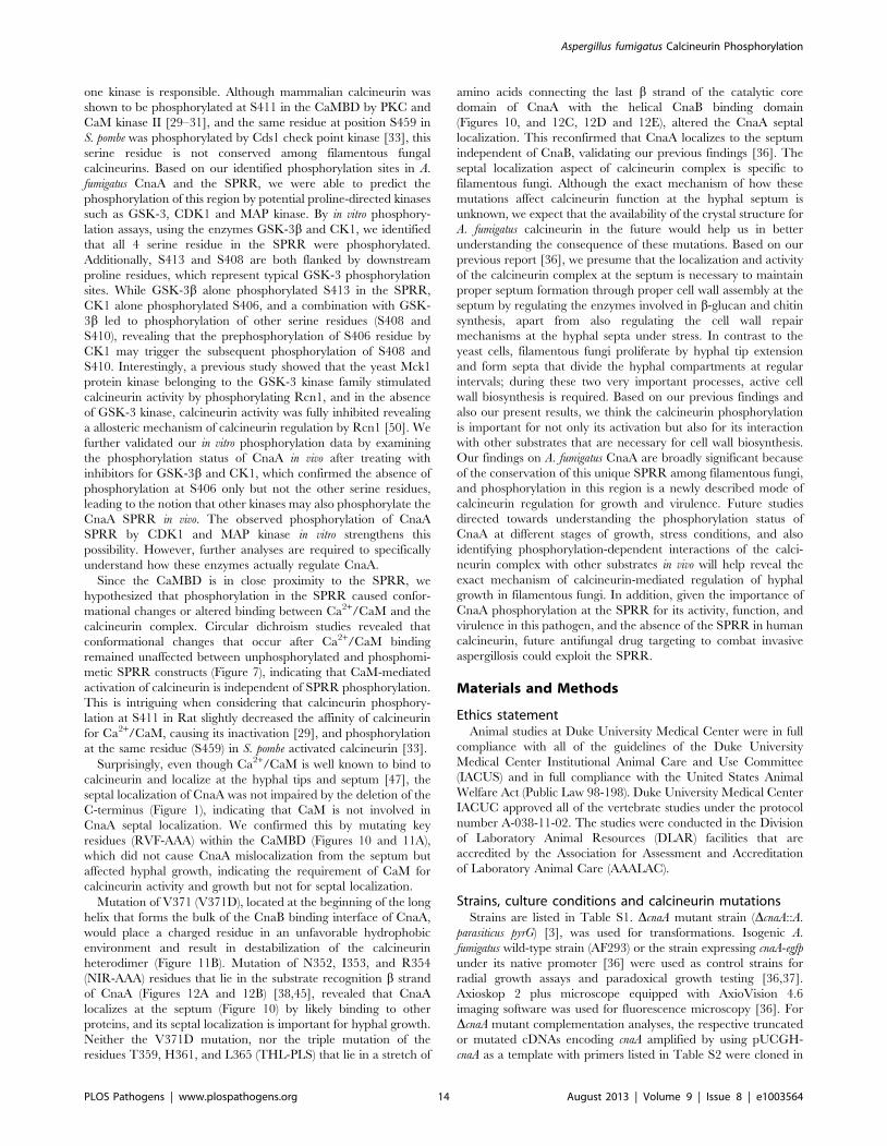
CnaA septal localization. We confirmed this by mutating key
residues (RVF-AAA) within the CaMBD (Figures 10 and 11A),
which did not cause CnaA mislocalization from the septum but
affected hyphal growth, indicating the requirement of CaM for
calcineurin activity and growth but not for septal localization.
Mutation of V371 (V371D), located at the beginning of the long
helix that forms the bulk of the CnaB binding interface of CnaA,
would place a charged residue in an unfavorable hydrophobic
environment and result in destabilization of the calcineurin
heterodimer (Figure 11B). Mutation of N352, I353, and R354
(NIR-AAA) residues that lie in the substrate recognition b strand
of CnaA (Figures 12A and 12B) [38,45], revealed that CnaA
localizes at the septum (Figure 10) by likely binding to other
proteins, and its septal localization is important for hyphal growth.
Neither the V371D mutation, nor the triple mutation of the
residues T359, H361, and L365 (THL-PLS) that lie in a stretch of
amino acids connecting the last b strand of the catalytic core
domain of CnaA with the helical CnaB binding domain
(Figures 10, and 12C, 12D and 12E), altered the CnaA septal
localization. This reconfirmed that CnaA localizes to the septum
independent of CnaB, validating our previous findings [36]. The
septal localization aspect of calcineurin complex is specific to
filamentous fungi. Although the exact mechanism of how these
mutations affect calcineurin function at the hyphal septum is
unknown, we expect that the availability of the crystal structure for
A. fumigatus calcineurin in the future would help us in better
understanding the consequence of these mutations. Based on our
previous report [36], we presume that the localization and activity
of the calcineurin complex at the septum is necessary to maintain
proper septum formation through proper cell wall assembly at the
septum by regulating the enzymes involved in b-glucan and chitin
synthesis, apart from also regulating the cell wall repair
mechanisms at the hyphal septa under stress. In contrast to the
yeast cells, filamentous fungi proliferate by hyphal tip extension
and form septa that divide the hyphal compartments at regular
intervals; during these two very important processes, active cell
wall biosynthesis is required. Based on our previous findings and
also our present results, we think the calcineurin phosphorylation
is important for not only its activation but also for its interaction
with other substrates that are necessary for cell wall biosynthesis.
Our findings on A. fumigatus CnaA are broadly significant because
of the conservation of this unique SPRR among filamentous fungi,
and phosphorylation in this region is a newly described mode of
calcineurin regulation for growth and virulence. Future studies
directed towards understanding the phosphorylation status of
CnaA at different stages of growth, stress conditions, and also
identifying phosphorylation-dependent interactions of the calci-
neurin complex with other substrates in vivo will help reveal the
exact mechanism of calcineurin-mediated regulation of hyphal
growth in filamentous fungi. In addition, given the importance of
CnaA phosphorylation at the SPRR for its activity, function, and
virulence in this pathogen, and the absence of the SPRR in human
calcineurin, future antifungal drug targeting to combat invasive
aspergillosis could exploit the SPRR.
Materials and Methods
Ethics statementAnimal studies at Duke University Medical Center were in full
compliance with all of the guidelines of the Duke University
Medical Center Institutional Animal Care and Use Committee
(IACUS) and in full compliance with the United States Animal
Welfare Act (Public Law 98-198). Duke University Medical Center
IACUC approved all of the vertebrate studies under the protocol
number A-038-11-02. The studies were conducted in the Division
of Laboratory Animal Resources (DLAR) facilities that are
accredited by the Association for Assessment and Accreditation
of Laboratory Animal Care (AAALAC).
Strains, culture conditions and calcineurin mutationsStrains are listed in Table S1. DcnaA mutant strain (DcnaA::A.
parasiticus pyrG) [3], was used for transformations. Isogenic A.
fumigatus wild-type strain (AF293) or the strain expressing cnaA-egfp
under its native promoter [36] were used as control strains for
radial growth assays and paradoxical growth testing [36,37].
Axioskop 2 plus microscope equipped with AxioVision 4.6
imaging software was used for fluorescence microscopy [36]. For
DcnaA mutant complementation analyses, the respective truncated
or mutated cDNAs encoding cnaA amplified by using pUCGH-
cnaA as a template with primers listed in Table S2 were cloned in
Aspergillus fumigatus Calcineurin Phosphorylation
PLOS Pathogens | www.plospathogens.org 14 August 2013 | Volume 9 | Issue 8 | e1003564
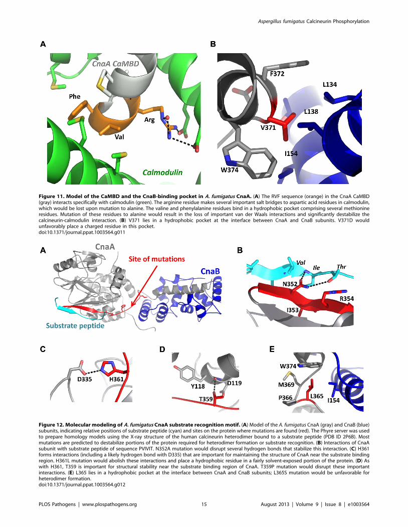
Figure 11. Model of the CaMBD and the CnaB-binding pocket in A. fumigatus CnaA. (A) The RVF sequence (orange) in the CnaA CaMBD(gray) interacts specifically with calmodulin (green). The arginine residue makes several important salt bridges to aspartic acid residues in calmodulin,which would be lost upon mutation to alanine. The valine and phenylalanine residues bind in a hydrophobic pocket comprising several methionineresidues. Mutation of these residues to alanine would result in the loss of important van der Waals interactions and significantly destabilize thecalcineurin-calmodulin interaction. (B) V371 lies in a hydrophobic pocket at the interface between CnaA and CnaB subunits. V371D wouldunfavorably place a charged residue in this pocket.doi:10.1371/journal.ppat.1003564.g011
Figure 12. Molecular modeling of A. fumigatus CnaA substrate recognition motif. (A) Model of the A. fumigatus CnaA (gray) and CnaB (blue)subunits, indicating relative positions of substrate peptide (cyan) and sites on the protein where mutations are found (red). The Phyre server was usedto prepare homology models using the X-ray structure of the human calcineurin heterodimer bound to a substrate peptide (PDB ID 2P6B). Mostmutations are predicted to destabilize portions of the protein required for heterodimer formation or substrate recognition. (B) Interactions of CnaAsubunit with substrate peptide of sequence PVIVIT. N352A mutation would disrupt several hydrogen bonds that stabilize this interaction. (C) H361forms interactions (including a likely hydrogen bond with D335) that are important for maintaining the structure of CnaA near the substrate bindingregion. H361L mutation would abolish these interactions and place a hydrophobic residue in a fairly solvent-exposed portion of the protein. (D) Aswith H361, T359 is important for structural stability near the substrate binding region of CnaA. T359P mutation would disrupt these importantinteractions. (E) L365 lies in a hydrophobic pocket at the interface between CnaA and CnaB subunits; L365S mutation would be unfavorable forheterodimer formation.doi:10.1371/journal.ppat.1003564.g012
Aspergillus fumigatus Calcineurin Phosphorylation
PLOS Pathogens | www.plospathogens.org 15 August 2013 | Volume 9 | Issue 8 | e1003564
the plasmid pUCGH-cnaApromo and transformants selected in the
presence of hygromycin B [36] were verified by Southern analysis.
Cloning of calcineurin A genes from other organismsThe calcineurin A encoding cDNAs from C. neoformans, M. grisea,
N. crassa and M. circinelloides were amplified from respective cDNA
libraries. All genes were amplified using the respective templates
and primers listed in Table S2 and cloned into the pUCGH vector
as previously described [36]. For cloning human CNA-a subunit
the plasmid, pET15b CnA CnB (obtained from Addgene), was
used as a template. S. cerevisiae genomic DNA was used as a
template to amplify S. cerevisiae CNA1. The plasmids were
sequenced to verify for accuracy prior to their transformation
into the A. fumigatus DcnaA mutant strain. Transformants were
selected in the presence of hygromycin B (150 mg/ml).
Protein extraction for Western analysis and calcineurinactivity
Preparation of cell extracts and Western detection were
performed as described earlier [36]. Cell extracts from 24 h
cultures were assayed for calcineurin phosphatase activity [36]
using p-nitrophenyl phosphate as substrate at 405 nm. The
difference of absorbance values between the amounts of p-
nitrophenol released in the strains versus the DcnaA DcnaB double
mutant control strain represented the phosphatase activity
mediated by calcineurin. Each experiment consisted of two
biologic replicates, with each assay consisting of 6 technical
replicates; data are presented as mean 6 SD of nanomoles of
pNPP released/min/mg protein.
Calcineurin A purification and nano-flow LiquidChromatography Electrospray Ionization Tandem MassSpectrometry (LC-MS/MS) analysis
Total cell lysates were extracted and normalized to contain
,10 mg protein in each sample before GFP-TrapH affinity
purification (Chromotek) and processed for TiO2 phosphopeptide
enrichment and mass spectrometry as previously described [51].
The dried phospho-peptide enriched samples were resuspended in
10 ml of 2% acetonitrile, 0.1% formic acid, 10 mM citric acid and
subjected to chromatographic separation on a Waters NanoAquity
UPLC equipped with a 1.7 mm BEH130 C18 75 mm
I.D.6250 mm reversed-phase column. The mobile phase consist-
ed of (A) 0.1% formic acid in water and (B) 0.1% formic acid in
acetonitrile. Following a 5 ml injection, peptides were trapped for
5 min on a 5 mm Symmetry C18 180 mm I.D.620 mm column at
20 ml/min in 99.9% A. The analytical column was held at 5% B
for 5 min then switched in-line and a linear elution gradient of 5%
B to 40% B was performed over 90 min at 300 nl/min. The
analytical column was connected to a fused silica PicoTip emitter
(New Objective, Cambridge, MA) with a 10 mm tip orifice and
coupled to an LTQ-Orbitrap XL mass spectrometer. In some
experiments the analytical column was connected to a fused silica
PicoTip emitter (New Objective, Cambridge, MA) with a 10 mm
tip orifice and was coupled to a Waters Synapt G2 QToF mass
spectrometer through an electrospray interface operating in a
data-dependent mode of acquisition. The instrument was set to
acquire a precursor MS scan in the Orbitrap from m/z 400–2000
with r = 60,000 at m/z 400 and a target AGC setting of 1e6 ions.
In a data-dependent mode of acquisition, MS/MS spectra of the
three most abundant precursor ions were acquired in the Orbitrap
with r = 7500 at m/z with a target AGC setting of 2e5 ions. Max
fill times were set to 1000 ms for full MS scans and 500 ms for
MS/MS scans with minimum MS/MS triggering thresholds of
5000 counts. For all experiments, fragmentation occurred in the
LTQ linear ion trap with a CID energy setting of 35% and a
dynamic exclusion of 60 s was employed for previously fragment-
ed precursor ions. When using the Waters Synapt G2 QToF mass
spectrometer through an electrospray interface operating in a
data-dependent mode of acquisition the instrument was set to
acquire a precursor MS scan from m/z 50–2000 with MS/MS
spectra acquired for the three most abundant precursor ions. For
all experiments, charge dependent CID energy settings were
employed and a 120 s dynamic exclusion was employed for
previously fragmented precursor ions.
Qualitative identifications and selected ionchromatograms from raw LC-MS/MS data
Raw LC-MS/MS data files were processed in Mascot distiller
(Matrix Science) and then submitted to independent Mascot
database searches (Matrix Science) against a SwissProt (fungus
taxonomy) containing both forward and reverse entries of each
protein. Search tolerances for LTQ-Orbitrap XL data were
10 ppm for precursor ions and 0.02 Da for product ions, and for
Synapt G2 data were 10 ppm for precursor and 0.04 Da for
product ions using trypsin specificity with up to two missed
cleavages. Carbamidomethylation (+57.0214 Da on C) was set as
a fixed modification, whereas oxidation (+15.9949 Da on M) and
phosphorylation (+79.9663 Da on S, T, and Y) were considered a
variable modification. All searched spectra were imported into
Scaffold (Proteome Software) and protein confidence thresholds
were set using a Bayesian statistical algorithm based on the
PeptideProphet and ProteinProphet algorithms which yielded a
peptide and protein false discovery rate of 1%. Phosphopeptide
intensities were obtained by generating selected ion chromato-
grams (20 ppm window around most abundant charge state of
precursor ion with seven point Boxcar smoothing) from raw LC-
MS data. MS response at peak apex was used for quantitating
abundance.
A. fumigatus calcineurin A-AfRD, calmodulin and circulardichroism
E. coli optimized genes for A. fumigatus calcineurin (AfRD;
regulatory domain from 395–482 aa of CnaA including the SPRR
and the CaMBD) and calmodulin (AfCaM) were synthesized and
ligated into a pUC57. The genes were digested and ligated into the
pET303/CT-His vector using XhoI and XbaI. The mutant
AfRD4Ser-Glu (AfRD-4SE), containing mutations S14E, S16E,
S18E, and S21E, was made using a QuikChange Site-Directed
Mutagenesis Kit. AfRD and AfRD-4SE were transformed into E.
coli BL-21 (DE3) cells for expression, and purified on a Ni2+/NTA
column followed by a calmodulin-sepharose column. AfCaM was
purified on a 2-trifluoromethyl-10-aminopropylphenothiazine
(TAPP)-sepharose column. Protein concentrations were deter-
mined by bicinchoninic acid assay. Circular dichroism (CD)
experiments were performed using a Jasco J-810 spectropolarim-
eter. Sample buffers were composed of 20 mM Tris, 200 mM
NaCl, 4 mM EGTA, pH 7.5, and 20 mM CaCl2. Samples
contained AfRD, AfCaM, or equimolar concentrations of AfRD
and AfCaM. The CD experiments shown were all performed in
the presence of an excess of calcium in order to determine the
effects of the phospho-mimics on the AfRD conformation when
bound by AfCaM. The concentrations of either protein alone per
sample ranged from 10–20 mM. In the sample that contained both
of AfRD and AfCaM, the total protein concentration per sample
ranged from 10–25 mM. Spectra were collected in quartz 1 mm
pathlength cuvettes. Samples were scanned from 200–260 nm in
Aspergillus fumigatus Calcineurin Phosphorylation
PLOS Pathogens | www.plospathogens.org 16 August 2013 | Volume 9 | Issue 8 | e1003564
0.5 nm increments at a scanning speed of 50 nm/sec and each
spectrum is the average of 4 scans. The raw CD data (in
millidegrees) was converted to molar ellipticity. The amount of
secondary structure was determined using the CONTIN/LL
deconvolution program.
In vitro phosphorylation assaysIn vitro phosphorylation reactions with GSK-3b, CK1, CDK1/
cyclinB and MAP kinase (New England Biolabs) contained 10 mg
of recombinant CnaA-AfRD protein, reaction buffer supplied by
the manufacturer, and 500 mM ATP in total volume of 50 ml.
GSK-3b (2500 U), CK1 (5000 U), CDK1 (100 U) and MAP
kinase (500 U) were used for each reaction. The reactions were
performed either with single enzymes or combinations of the
different enzymes. The reactions were incubated at 30uC for 4 h
and processed for mass spectral analysis following digestion with
Glu-C and TiO2 phosphopeptide enrichment.
Effect of kinase inhibitors on phosphorylation of CnaA invivo
To determine the effect of GSK-3b and CK1 inhibitors on the
phosphorylation status of CnaA in vivo, the CnaA-EGFP expression
strain was grown in presence of 0.75 mM each of the GSK-3binhibitor VII (calbiochem) and D4476 (abcam) for a period of 24 h
and the isolated CnaA-EGFP fusion protein was subjected to
phospho enrichment and mass spectrometry as described earlier [51].
Aspergillus fumigatus calcineurin modelingHomology models for the A. fumigatus calcineurin A and B
subunits were prepared using the Phyre server. A model of the
fungal heterodimer was constructed by superimposing the
individually created homology models onto the X-ray structure
of the human calcineurin heterodimer bound to a substrate
peptide (PDB ID 2P6B) [38]. Superpositions and analysis of
mutations were performed using PyMol, Coot and Molprobity.
Due to the high level of sequence conservation with the
mammalian calcineurin structure already determined, the models
are largely superimposable at the Ca backbone level and exhibit
good packing overall. We employed this model of A. fumigatus
calcineurin to propose a structural basis for the disruption of
calcineurin activity observed for the mutants described in the
current study.
Murine inhalational model of invasive aspergillosisSix-week-old CD1 male mice (mean weight 22.5 g) were
immunosuppressed with both cyclophosphamide and triamcinolone
acetonide as previously described [52]. Two groups of 20
immunosuppressed, unanesthetized mice each inhaled an aerosol-
ized suspension of either AF293 wild-type or CnaAmt-4SA strain
[52]. Survival was plotted on a Kaplan-Meier curve and log rank
was used for pair-wise comparison of survival with statistical
significance defined as a two-tailed p,0.05. Histopathological
examination of the lungs was performed in two mice in each group
that were euthanized on day +7 of infection. Lungs were embedded
in 10% neutral buffered formalin and subsequently sectioned and
stained with Gomori methenamine silver and hematoxylin-eosin for
assessment of histological signs of infection. The animal model and
experiments were conducted in accordance with the Animal Care
and Use Program of the Duke University Medical Center.
Supporting Information
Figure S1 Clustal alignment of the calcineurin Aproteins from 24 different species of the region includ-
ing the CnBBH and the CaMBD. While there was clear
homology observed in the CnBBH and the CaMBD, the linker
that separates these conserved domains exhibited variation.
Notably, filamentous fungal species contained several serine and
proline residues in this linker. AfcnaA-Aspergillus fumigatus CnaA;
AmCnaA-Allomyces macrogynus; BcCnaA-Botrytis cinerea CnaA;
BchycnaA-Batrachochytrium dendrobatidis CnaA; CaCnaA-Candida
albicans CnaA; CcCnaA-Coprinopsis cinerea CnaA; CeCnaA-Caenor-
habditis elegans CnaA; CiCnaA-Coccidioides immitis; CnCnaA-Crypto-
coccus neoformans CnaA; DdCnaA-Dictyostelium discoideum CnaA;
FvCnaA-Fusarium verticillioides CnaA; HcCnaA-Histoplasma capsula-
tum CnaA; HcnaA-Homo sapiens CnaA; MgCnaA-Magnaporthe grisea
CnaA; MygCnaA-Microsporum gypseum CnaA; MvCnaA-Mortierella
verticillata CnaA; NcCnaA-Neurospora crassa CnaA; PbCnaA-Para-
coccidiodes brasilensis CnaA; PtCnaA- Puccinia triticina CnaA;
RoCnaA-Rhizopus oryzae CnaA; ScCnaA-Saccharomyces cerevisiae
CnaA; SpCnaA-Schizosaccharomyces pombe CnaA; TrCnaA-Tricho-
phyton rubrum CnaA; UmCnaA-Ustilago maydis CnaA.
(PDF)
Figure S2 Phosphorylation of the A. fumigatus CnaBregulatory subunit. Annotated tandem mass spectrum of
RA[pS]VGTSQLLDNIV[pS]ASNFDRDEVDR (3+ m/z
1008.7813) from CnaB revealed two unique phosphorylated
serine residues (Ser21 and Ser33) localized with .99% confidence
using AScore. The presence of each identified C-terminal (y) and
N-terminal (b) product ions are indicated within the peptide
sequence.
(PDF)
Figure S3 Tandem mass spectra of phosphorylatedpeptides identified from Magnaporthe grisea calci-neurin. Phosphorylation in the SPRR (A) and the C-terminus
(B) are shown. Annotated tandem mass spectra showing assigned
fragment ion assignments from Mascot database searches
imported into Scaffold are shown. For each qualitative identifica-
tion, spectra were subjected to Ascore localization to assign
percent confidences of amino acid specific assignments. An
extracted ion chromatogram (EIC) of the precursor m/z (+/
220 ppm) of the chromatographic elution profile of each
phosphorylated peptide is presented to the right of each annotated
fragment ion spectra. An asterisk in the EIC plot indicated the
peak which was chosen for subsequent qualitative identification.
(PDF)
Figure S4 Tandem mass spectra of phosphorylatedpeptides identified from Neurospora crassa calcineurin.Annotated tandem mass spectra showing assigned fragment ion
assignments from Mascot database searches imported into Scaffold
are shown. For each qualitative identification, spectra were
subjected to Ascore localization to assign percent confidences of
amino acid specific assignments. An extracted ion chromatogram
(EIC) of the precursor m/z (+/220 ppm) of the chromatographic
elution profile of each phosphorylated peptide is presented to the
right of each annotated fragment ion spectra. An asterisk in the
EIC plot indicated the peak which was chosen for subsequent
qualitative identification.
(PDF)
Figure S5 Tandem mass spectra of phosphorylatedpeptides identified from Mucor circinelloides calci-neurin. Phosphorylation in the SPRR (A) and the C-terminus
(B) are shown. Annotated tandem mass spectra showing assigned
fragment ion assignments from Mascot database searches
imported into Scaffold are shown. For each qualitative identifica-
tion, spectra were subjected to Ascore localization to assign
Aspergillus fumigatus Calcineurin Phosphorylation
PLOS Pathogens | www.plospathogens.org 17 August 2013 | Volume 9 | Issue 8 | e1003564
percent confidences of amino acid specific assignments. An
extracted ion chromatogram (EIC) of the precursor m/z (+/
220 ppm) of the chromatographic elution profile of each
phosphorylated peptide is presented to the right of each annotated
fragment ion spectra. An asterisk in the EIC plot indicated the
peak which was chosen for subsequent qualitative identification.
(PDF)
Figure S6 Extracted ion chromatogram (EIC) peakheight measurements of CnaA following TiO2 enrich-ment and LC-MS/MS analysis. Manual EIC generation was
performed for each precursor ion (+/220 ppm tolerance) which
was qualitatively identified as a phosphorylated peptide from
CnaA and resulting peak height was used as a measure of intensity.
To ensure the same precursor ion was being compared across the
+FK506, 2FK506, and Control samples, a peak was required to
have the same m/z (+/210 ppm), have the same retention time
(+/230 seconds), and was qualitatively identified as the same
species in Mascot database searches.
(PDF)
Figure S7 Extracted ion chromatogram (EIC) peakheight measurements of CnaB following TiO2 enrich-ment and LC-MS/MS analysis. Manual EIC generation was
performed for each precursor ion (+/220 ppm tolerance) which
was qualitatively identified as a phosphorylated peptide from
CnaB and resulting peak height was used as a measure of intensity.
To ensure the same precursor ion was being compared across the
+FK506, 2FK506, and Control samples, a peak was required to
have the same m/z (+/210 ppm), have the same retention time
(+/230 seconds), and was qualitatively identified as the same
species in Mascot database searches.
(PDF)
Figure S8 CnaA mutation strains cultured on GMMagar in the presence of varying concentrations of the cell
wall inhibitor caspofungin for 5 days. The THL-PLS
mutation (catalytic residues) and the V371D mutation (CnBBH
residue) in CnaA completely abolish calcineurin-mediated para-
doxical growth. The NIR-AAA (PxIxIT Binding Motif) and the
RVF-AAA (CaMBD residues) mutations do show slight paradox-
ical growth at 2 mg/ml and 4 mg/ml of caspofungin.
(PDF)
Table S1 Strains used in the present study. All the strains
listed were constructed in the Af293 derived isogenic Af293.1
strain with pyrG auxotrophy. The DcnaA strain previously
constructed in the Af293.1 background by utilizing the pyrG
marker gene was transformed with the various cnaA constructs
either under the control of its native promoter or the otef promoter
with gfp tag and hygromycin (hph) resistance marker gene.
(DOCX)
Table S2 Primers and constructs. All the primers used for
construction of the various cnaA.
(DOCX)
Acknowledgments
We thank Dr. Ralph Dean (North Carolina State University), Dr. Daniel J.
Ebbole (Texas A&M University), Dr. John H. McCusker (Duke
University), Dr. Joseph Heitman (Duke Univeristy and Dr. Soo Chan
Lee (Duke University) for providing the DNA for amplification of
calcineurin A subunits from M. grisea, N. crassa, S. cerevisiae, C. neoformans
and M. circinelloides.
Author Contributions
Conceived and designed the experiments: PRJ WJS. Performed the
experiments: PRJ CG JRF FL ECC. Analyzed the data: PRJ CG JRF FL
ECC EJS YGA. Contributed reagents/materials/analysis tools: EJS MAM
YGA TPC. Wrote the paper: PRJ WJS. Performed molecular modeling:
MAH.
References
1. Kontoyiannis DP, Marr KA, Park BJ, Alexander BD, Anaissie EJ, et al. (2010)
Prospective Surveillance for Invasive Fungal Infections in Hematopoietic Stem
Cell Transplant Recipients, 2001–2006: Overview of the Transplant-Associated
Infection Surveillance Network (TRANSNET) Database. Clinical Infectious
Diseases 50: 1091–1100.
2. Steinbach WJ, Marr KA, Anaissie EJ, Azie N, Quan S-P, et al. (2012) Clinical
epidemiology of 960 patients with invasive aspergillosis from the PATH Alliance
registry. Journal of Infection 65: 453–464.
3. Steinbach WJ, Cramer RA, Perfect BZ, Asfaw YG, Sauer TC, et al. (2006)
Calcineurin Controls Growth, Morphology, and Pathogenicity in Aspergillus
fumigatus. Eukaryotic Cell 5: 1091–1103.
4. Steinbach WJ, Reedy JL, Cramer RA, Perfect JR, Heitman J (2007) Harnessing
calcineurin as a novel anti-infective agent against invasive fungal infections. Nat
Rev Micro 5: 418–430.
5. Steinbach WJ, Schell WA, Blankenship JR, Onyewu C, Heitman J, et al. (2004)
In Vitro Interactions between Antifungals and Immunosuppressants against
Aspergillus fumigatus. Antimicrobial Agents and Chemotherapy 48: 1664–1669.
6. Klee CB, Crouch TH, Krinks MH (1979) Calcineurin: a calcium- and
calmodulin-binding protein of the nervous system. Proceedings of the National
Academy of Sciences 76: 6270–6273.
7. Rusnak F, Mertz P (2000) Calcineurin: Form and Function. Physiological
Reviews 80: 1483–1521.
8. Clipstone NA, Crabtree GR (1992) Identification of calcineurin as a key
signalling enzyme in T-lymphocyte activation. Nature 357: 695–697.
9. Molkentin JD, Lu J-R, Antos CL, Markham B, Richardson J, et al. (1998) A
Calcineurin-Dependent Transcriptional Pathway for Cardiac Hypertrophy. Cell
93: 215–228.
10. Yakel JL (1997) Calcineurin regulation of synaptic function: from ion channels to
transmitter release and gene transcription. Trends in Pharmacological Sciences
18: 124–134.
11. Wang H-G, Pathan N, Ethell IM, Krajewski S, Yamaguchi Y, et al. (1999)
Ca2+-Induced Apoptosis Through Calcineurin Dephosphorylation of BAD.
Science 284: 339–343.
12. Arino J, Ramos J, Sychrova H (2010) Alkali Metal Cation Transport and
Homeostasis in Yeasts. Microbiology and Molecular Biology Reviews 74: 95–120.
13. Levin DE (2011) Regulation of Cell Wall Biogenesis in Saccharomyces cerevisiae:
The Cell Wall Integrity Signaling Pathway. Genetics 189: 1145–1175.
14. Withee JL, Mulholland J, Jeng R, Cyert MS (1997) An essential role of the yeast
pheromone-induced Ca2+ signal is to activate calcineurin. Molecular Biology of
the Cell 8: 263–277.
15. Cyert MS (2003) Calcineurin signaling in Saccharomyces cerevisiae: how yeast
go crazy in response to stress. Biochemical and Biophysical Research
Communications 311: 1143–1150.
16. Lu Y, Sugiura R, Yada T, Cheng H, Sio SO, et al. (2002) Calcineurin is
implicated in the regulation of the septation initiation network in fission yeast.
Genes to Cells 7: 1009–1019.
17. Miyatake M, Kuno T, Kita A, Katsura K, Takegawa K, et al. (2007) Valproic
Acid Affects Membrane Trafficking and Cell-Wall Integrity in Fission Yeast.
Genetics 175: 1695–1705.
18. Fox DS, Heitman J (2002) Good fungi gone bad: The corruption of calcineurin.
BioEssays 24: 894–903.
19. Brown SM, Campbell LT, Lodge JK (2007) Cryptococcus neoformans, a fungus
under stress. Current Opinion in Microbiology 10: 320–325.
20. Reedy JL, Filler SG, Heitman J (2010) Elucidating the Candida albicans
calcineurin signaling cascade controlling stress response and virulence. Fungal
Genetics and Biology 47: 107–116.
21. Kahl CR, Means AR (2003) Regulation of Cell Cycle Progression by Calcium/
Calmodulin-Dependent Pathways. Endocrine Reviews 24: 719–736.
22. Juvvadi PR, Kuroki Y, Arioka M, Nakajima H, Kitamoto K (2003) Functional
analysis of the calcineurin-encoding gene cnaA from Aspergillus oryzae:
evidence for its putative role in stress adaptation. Archives of Microbiology
179: 416–422.
23. Kothe GO, Free SJ (1998) Calcineurin Subunit B Is Required for Normal
Vegetative Growth inNeurospora crassa. Fungal Genetics and Biology 23: 248–258.
24. Harel A, Bercovich S, Yarden O (2006) Calcineurin Is Required for Sclerotial
Development and Pathogenicity of Sclerotinia sclerotiorum in an Oxalic Acid-
Independent Manner. Molecular Plant-Microbe Interactions 19: 682–693.
25. Choi JH, Yang Seon Kim, Yong-Hwan Lee (2009) Functional Analysis of
MCNA, a Gene Encoding a Catalytic Subunit of Calcineurin, in the Rice Blast
Fungus Magnaporthe oryzae. J Microbiol Biotechnol 19: 11–16.
Aspergillus fumigatus Calcineurin Phosphorylation
PLOS Pathogens | www.plospathogens.org 18 August 2013 | Volume 9 | Issue 8 | e1003564
26. Li H, Rao A, Hogan PG (2011) Interaction of calcineurin with substrates and
targeting proteins. Trends in Cell Biology 21: 91–103.27. King MM, Huang CY (1984) The calmodulin-dependent activation and
deactivation of the phosphoprotein phosphatase, calcineurin, and the effect of
nucleotides, pyrophosphate, and divalent metal ions. Identification of calcineurinas a Zn and Fe metalloenzyme. Journal of Biological Chemistry 259: 8847–8856.
28. Singh TJ, Wang JH (1987) Phosphorylation of calcineurin by glycogen synthase(casein) kinase-1. Biochemistry and Cell Biology 65: 917–921.
29. Hashimoto Y, King MM, Soderling TR (1988) Regulatory interactions of
calmodulin-binding proteins: phosphorylation of calcineurin by autophosphory-lated Ca2+/calmodulin-dependent protein kinase II. Proceedings of the
National Academy of Sciences 85: 7001–7005.30. Martensen TM, Martin BM, Kincaid RL (1989) Identification of the site on
calcineurin phosphorylated by calcium/CaM-dependent kinase II: modificationof the CaM-binding domain. Biochemistry 28: 9243–9247.
31. Hashimoto Y, Soderling TR (1989) Regulation of calcineurin by phosphoryla-
tion. Identification of the regulatory site phosphorylated by Ca2+/calmodulin-dependent protein kinase II and protein kinase C. Journal of Biological
Chemistry 264: 16524–16529.32. Calalb MB, Kincaid RL, Soderling TR (1990) Phosphorylation of calcineurin:
Effect on calmodulin binding. Biochemical and Biophysical Research Commu-
nications 172: 551–556.33. Kume K, Koyano T, Kanai M, Toda T, Hirata D (2011) Calcineurin ensures a
link between the DNA replication checkpoint and microtubule-dependentpolarized growth. Nat Cell Biol 13: 234–242.
34. Ferreira MEdS, Heinekamp T, Hartl A, Brakhage AA, Semighini CP, et al.(2007) Functional characterization of the Aspergillus fumigatus calcineurin.
Fungal Genetics and Biology 44: 219–230.
35. Juvvadi PR, Fortwendel JR, Pinchai N, Perfect BZ, Heitman J, et al. (2008)Calcineurin Localizes to the Hyphal Septum in Aspergillus fumigatus:
Implications for Septum Formation and Conidiophore Development. Eukary-otic Cell 7: 1606–1610.
36. Juvvadi PR, Fortwendel JR, Rogg LE, Burns KA, Randell SH, et al. (2011)
Localization and activity of the calcineurin catalytic and regulatory subunitcomplex at the septum is essential for hyphal elongation and proper septation in
Aspergillus fumigatus. Molecular Microbiology 82: 1235–1259.37. Fortwendel JR, Juvvadi PR, Perfect BZ, Rogg LE, Perfect JR, et al. (2010)
Transcriptional Regulation of Chitin Synthases by Calcineurin ControlsParadoxical Growth of Aspergillus fumigatus in Response to Caspofungin.
Antimicrobial Agents and Chemotherapy 54: 1555–1563.
38. Li H, Zhang L, Rao A, Harrison SC, Hogan PG (2007) Structure of Calcineurinin Complex with PVIVIT Peptide: Portrait of a Low-affinity Signalling
Interaction. Journal of Molecular Biology 369: 1296–1306.
39. Kay BK, Williamson MP, Sudol M (2000) The importance of being proline: the
interaction of proline-rich motifs in signaling proteins with their cognatedomains. The FASEB Journal 14: 231–241.
40. Lu KP, Liou Y-C, Zhou XZ (2002) Pinning down proline-directed phosphor-
ylation signaling. Trends in Cell Biology 12: 164–172.41. Cohen P, Frame S (2001) The renaissance of GSK3. Nat Rev Mol Cell Biol 2:
769–776.42. Berzal S, Alique M, Ruiz-Ortega M, Egido J, Ortiz A, et al. (2012) GSK3, Snail,
and Adhesion Molecule Regulation by Cyclosporine A in Renal Tubular Cells.
Toxicological Sciences 127: 425–437.43. Rumi-Masante J, Rusinga FI, Lester TE, Dunlap TB, Williams TD, et al. (2012)
Structural Basis for Activation of Calcineurin by Calmodulin. Journal ofMolecular Biology 415: 307–317.
44. Fortwendel JR, Juvvadi PR, Pinchai N, Perfect BZ, Alspaugh JA, et al. (2009)Differential Effects of Inhibiting Chitin and 1,3-b-d-Glucan Synthesis in Ras and
Calcineurin Mutants of Aspergillus fumigatus. Antimicrobial Agents and
Chemotherapy 53: 476–482.45. Roy J, Li H, Hogan PG, Cyert MS (2007) A Conserved Docking Site Modulates
Substrate Affinity for Calcineurin, Signaling Output, and In Vivo Function.Molecular Cell 25: 889–901.
46. Ye Q, Wang H, Zheng J, Wei Q, Jia Z (2008) The complex structure of
calmodulin bound to a calcineurin peptide. Proteins: Structure, Function, andBioinformatics 73: 19–27.
47. Chen S, Song Y, Cao J, Wang G, Wei H, et al. (2010) Localization and functionof calmodulin in live-cells of Aspergillus nidulans. Fungal Genetics and Biology
47: 268–278.48. Jiang B, Cyert MS (1999) Identification of a Novel Region Critical for
Calcineurin Function in Vivo and in Vitro. Journal of Biological Chemistry 274:
18543–18551.49. Ho S, Clipstone N, Timmermann L, Northrop J, Graef I, et al. (1996) The
Mechanism of Action of Cyclosporin A and FK506. Clinical Immunology andImmunopathology 80: S40–S45.
50. Hilioti Z, Gallagher DA, Low-Nam ST, Ramaswamy P, Gajer P, et al. (2004)
GSK-3 kinases enhance calcineurin signaling by phosphorylation of RCNs.Genes & Development 18: 35–47.
51. Juvvadi PR, Belina D, Soderblom EJ, Moseley MA, Steinbach WJ (2013)Filamentous fungal-specific septin AspE is phosphorylated in vivo and interacts
with actin, tubulin and other septins in the human pathogen Aspergillusfumigatus. Biochemical and Biophysical Research Communications 431: 547–
553.
52. Steinbach WJ, Benjamin Jr DK, Trasi SA, Miller JL, Schell WA, et al. (2004)Value of an inhalational model of invasive aspergillosis. Medical Mycology 42:
417–425.
Aspergillus fumigatus Calcineurin Phosphorylation
PLOS Pathogens | www.plospathogens.org 19 August 2013 | Volume 9 | Issue 8 | e1003564


