Embed Size (px)
Citation preview
Molecular Biology of the CellVol. 14, 2163–2180, May 2003
Depletion of a Polo-like Kinase in Candida albicansActivates Cyclase-dependent Hyphal-like GrowthCatherine Bachewich,*† David Y. Thomas,‡ and Malcolm Whiteway*§
*Health Sector, Biotechnology Research Institute, National Research Council of Canada, Montreal,Quebec, H4P 2R2, Canada; and Departments of ‡Biochemistry and § Biology, McGill University,Montreal, Quebec, H3G 1Y6, Canada
Submitted May 5, 2002; Revised November 15, 2002; Accepted January 23, 2003Monitoring Editor: Douglas Koshland
Morphogenesis in the fungal pathogen Candida albicans is an important virulence-determiningfactor, as a dimorphic switch between yeast and hyphal growth forms can increase pathogenesis.We identified CaCDC5, a cell cycle regulatory polo-like kinase (PLK) in C. albicans and demon-strate that shutting off its expression induced cell cycle defects and dramatic changes in morphol-ogy. Cells lacking CaCdc5p were blocked early in nuclear division with very short spindles andunseparated chromatin. GFP-tagged CaCdc5p localized to unseparated spindle pole bodies, thespindle, and chromatin, consistent with a role in spindle elongation at an earlier point in the cellcycle than that described for the homologue Cdc5p in yeast. Strikingly, the cell cycle defects wereaccompanied by the formation of hyphal-like filaments under yeast growth conditions. Filamentgrowth was determinate, as the filaments started to die after 24 h. The filaments resembledserum-induced hyphae with respect to morphology, organization of cytoplasmic microtubules,localization of nuclei, and expression of hyphal-specific components. Filament formation requiredCaCDC35, but not EFG1 or CPH1. Similar defects in spindle elongation and a correspondinginduction of filaments occurred when yeast cells were exposed to hydroxyurea. Because CaCdc5pdoes not appear to act as a direct repressor of hyphal growth, the data suggest that a target ofCaCdc5p function is associated with hyphal-like development. Thus, an internal, cell cycle–relatedcue can activate hyphal regulatory networks in Candida.
INTRODUCTION
Morphogenesis of fungal pathogens is intimately linkedwith virulence, supporting the need for a comprehensiveunderstanding of the regulation of fungal cell morphology.Candida albicans, a common fungal pathogen of humans,exists in yeast, pseudohyphal and hyphal growth forms, andcan enhance virulence in part by exploiting the hyphalgrowth mode (Lo et al., 1997). The environmentally stimu-lated yeast to hyphal transition in Candida is mediated byseveral signaling pathways (Whiteway, 2000), the most char-acterized of which involve MAP kinase and cAMP-basedsignaling. However, several factors influence hyphal forma-tion independent of the known pathways in Candida (White-way, 2000; Liu, 2001), and hyphal growth involves regula-tion beyond the level of transcription (Torralba and Heath,2001), supporting the existence of additional regulatory net-
works and reinforcing the complexity of the response sys-tems.
In the model fungus Saccharomyces cerevisiae, morphogen-esis is tightly coordinated with cell cycle progression. Budgrowth and the corresponding polarization of actin, synthe-sis of DNA, and duplication of spindle pole bodies occurs atthe G1/S transition, whereas elongated or pseudohyphalgrowth is associated with a block in G2 mediated by Swe1p-dependent negative regulation of Cdc28p (Kron and Gow,1995; Rua et al., 2001). Polar or isometric growth of buds canbe maintained by overexpression of G1 or mitotic cyclins,respectively (Lew and Reed, 1995). In filamentous and di-morphic fungi, the connection between cell cycle factors andtrue hyphal growth is less clear. Hyphae continue to growwhether their apical-most nuclei are in interphase or mitosis(Kron and Gow, 1995), and it was recently demonstratedthat the duration of cell cycle stages was similar in yeast,germlings, and apical hyphal cells of C. albicans (Hazan et al.,2002). However, the coordination between nuclear localiza-tion, division, and septation with the initiation and mainte-nance of hyphal growth in Candida requires a relationshipbetween aspects of the cell cycle and hyphal growth. Forexample, during the yeast to hyphal transition, the nucleus
Article published online ahead of print. Mol. Biol. Cell 10.1091/mbc.02–05–0076. Article and publication date are at www.molbiol-cell.org/cgi/doi/10.1091/mbc.02–05–0076.
† Corresponding author. E-mail address: [email protected].
© 2003 by The American Society for Cell Biology 2163

and septins move to a hyphal specific position out in thedeveloping germ tube before the first mitosis, after whichone daughter nucleus migrates with the growing tip, whilethe other moves back into the mother yeast cell (Gale et al.,2001; Sudbery, 2001). Septation follows each nuclear divi-sion in established hyphae, creating uni-nucleate subapicalcompartments of similar length that remain arrested in G0or G1 until branching takes place (Kron and Gow, 1995). Afew cell cycle factors have been characterized in Candida;these include the G1 cyclin CLN1, which is required formaintaining hyphal growth (Loeb et al., 1999) and the CDC2-related kinase CRK1, which can promote hyphal formation(Chen et al., 2000). In addition, a forkhead transcriptionfactor that regulates B-cyclin gene expression is required forhyphal growth in Candida (Bensen et al., 2002).
The polo-like kinases (PLKs) comprise a family of cellcycle regulators with the potential to influence hyphal mor-phogenesis in Candida, because they function at variousstages during the initiation and progression through mitosisand are required for septation/cytokinesis (reviewed inNigg, 1998; Alexandru et al., 2001; Toyoshima-Morimoto etal., 2001). Furthermore, the PLK homologue Cdc5p in S.cerevisiae physically interacts with septins and Swe1p (Bar-tholomew et al., 2001; Song and Lee, 2001) and can alter cellmorphology by generating elongated buds upon overex-pression (Song et al., 2000).
To explore the relationship between cell cycle factors andhyphal morphogenesis in C. albicans, we investigated therole of a PLK homologue, CaCDC5. We demonstrate thatCaCdc5p is required for spindle elongation and that generepression under yeast growth conditions leads to dramatichyphal-like growth. Similar inhibition of spindle elongationand a corresponding induction of filaments with hydroxyu-rea suggest there is an endogenous mechanism in place toconnect aspects of the cell cycle and the hyphal signalingnetworks in C. albicans.
MATERIALS AND METHODS
Strains, Oligos, and MediaStrains and oligos used in the investigation are listed in Table 1.Cells were grown at 30°C in 0.67% yeast nitrogen base containing2% sodium succinate (SS medium) or 2% glucose (SD medium) forinduction and repression of the Candida PCK1 promoter, respec-tively (Leuker et al., 1997). Control strains including RM1000 andCB102 were grown in the same media supplemented with uridineand histidine as required. To repress CaCDC5 expression from thePCK1 promotor, yeast cells were grown for 24 h in SS medium,washed with dH2O, and then diluted into SD medium to an OD600
of 0.25. SD medium with or without methionine and cysteine wasused to repress and induce expression, respectively, from the Can-dida MET promotor (Care et al., 1999). Hyphal-inducing conditionsinvolved the addition of 10% heat-inactivated fetal calf serum(GIBCO/Invitrogen, Burlington, Ontario) to growth medium andincubation at 37°C. For hydroxyurea (HU) treatment, cells of strainSC5314, CB110, CR216, JKC19, HLC52, and HLC54 were grownovernight in yeast extract/peptone/dextrose medium (YPD) or SDmedium and then diluted to an OD600 of 0.4 in fresh mediumcontaining 200 mM HU (Sigma Chemical Co., St. Louis, MO). Over-expression of CaCdc5p-GFP was performed in S medium contain-ing 2% casaminoacids (Leuker et al., 1997).
Transformation and Southern and NorthernAnalysesCells were transformed using lithium acetate (Chen et al., 1992), andDNA and RNA were extracted according to Rose et al. (1990) andKohrer and Domdey (1991), respectively. Southern analysis wasperformed using the DIG Hybridization System (Roche Diagnostics,Mannheim, Germany). To determine the level of PCK1::CaCDC5expression, cells from strains RM1000, CB102, and CB104 weregrown to an OD600 of 0.8 in SS media at 30°C or to an OD600 of 0.2followed by washing and transferring to SD media. Strain CB104was collected after 4, 7, or 24 h in SD medium, whereas strainsRM1000 and CB102 were collected at 4 h, because these dividingyeast cells entered stationary phase with longer incubation periods.Total RNA, 20 �g, was analyzed using a 32P-labeled (T7 QuickPrime Kit, Amersham Pharmacia Biotech, Piscataway, NJ) PCRproduct of CaCDC5 and hybridization conditions described by Sri-kantha and Soll (1993). 32P- labeled PCR fragments from HWP1,DDR48, ECE1, and ALS1 were used to probe total RNA from strainsCB102, CB104, SC5314 grown in SD medium, and SC5314 grown inSD medium plus 200 mM HU. An ACT1 probe (Rocha et al., 2001)was used as a loading control. Northern blots were visualized witha phosphoimager.
Cloning and Plasmid and Strain ProductionTo regulate expression of CaCDC5, a strain containing a single copyof CaCDC5 under control of the PCK1 promotor was created. Se-quence data for C. albicans was obtained from the Stanford GenomeTechnology Center website at http://www-sequence.stanford.edu/group/candida. Sequencing of C. albicans was accomplished withthe support of the NIDR and the Burroughs Wellcome Fund. Theopen reading frame plus 800 and 1500 base pairs of 5� and 3�flanking sequence, respectively, of CaCDC5 was PCR amplifiedfrom genomic DNA derived from strain SC5314 using primers CB1Fand CB1R and cloned into the KpnI/PstI sites of vector pTZ18R,creating plasmid pCB100. Primers CB3F and CB3R reverse-ampli-fied the flanking and vector sequences from pCB100, into which theBamHI/BglII URA3 blaster cassette (Fonzi and Irwin, 1993) wascloned, creating plasmid pCB101. Primers CB4F and CB4R were alsoused to amplify the flanking DNA and vector sequence frompCB100, into which a NotI/BamHI PCR-amplified CaHIS1 ORF wascloned, creating plasmid pCB102. The URA3-containing deletionconstruct was liberated from pCB101 with KpnI and PstI and trans-formed into strain RM1000. Transformants were screened by PCRand Southern analyses to confirm the complete elimination of onecopy of CaCDC5 and creation of strain CB100. CB100 was plated on5-fluoorotic acid (5-FOA) to select for the URA3- strain CB101. TheORF of CaCDC5 plus 300 base pairs of terminator sequence was PCRamplified from genomic DNA using primers CB5F and CB5R andcloned into the KpnI site following the PCK1 promotor in the URA3blaster-containing plasmid pJA24 (a kind gift from J. Ash, derivedfrom plasmid p5921 from Fonzi and Irwin, 1993), creating plasmidpCB103. pCB103 was cut at a unique XhoI site within the PCK1promotor sequence to direct integration at the PCK1 promotor locusin strain CB101, creating strain CB102. The empty plasmid wastransformed as a control, creating control strain CB103. The HIS1-containing deletion construct was liberated from pCB102 with SphIand SacI and transformed into strain CB102 to replace the secondendogenous copy of CaCDC5 while in the presence of succinate,creating strain CB104. Removal of the URA3 marker by plating onto5-FOA resulted in strain CB105. All strains were analyzed by PCRand Southern analyses to confirm correct integration of transform-ing DNA and replacement of both endogenous copies of CaCDC5(our unpublished results), and all PCR-derived clones were se-quenced. For additional controls, strain RM1000 was transformedwith plasmid pRM100, which contains Candida URA3 and HIS1,producing strain CB400. Strain CB401 consisted of one of severaltransformants from strain CB102 which did not correctly integrate
C. Bachewich et al.
Molecular Biology of the Cell2164

the HIS1 knockout cassette, remaining heterozygous for CaCDC5but HIS1�. These transformants behaved identically to strainsRM1000 and CB102 (supplemented with uridine and histidine)when switching between glucose and succinate-containing medium(our unpublished results).
To regulate expression of CaCDC5 in another strain using a dif-ferent promotor, strain CAI4 was transformed with the URA3-baseddeletion construct from plasmid pCB101, to create strain CB106.CB107 was created by plating CB106 on 5-FOA. A 3�-truncated copyof CaCDC5, lacking 1000 base pairs upstream from the stop codon,was PCR amplified with primers CB6F and CB6R and cloned intothe PstI/BamHI sites following the MET promotor in plasmid pCa-DIS (Care et al., 1999), creating plasmid pCB106. The plasmid wascut with ClaI to direct integration at the remaining endogenous copyof CaCDC5 in strain CB107. The resulting strain, CB108, contained afull-length copy of CaCDC5 under control of the MET promotor anda 3� truncated copy with no terminal processing sequence. As a
control, strain CB107 was transformed with empty pCaDIS plasmid,creating strain CB109.
To regulate expression of CaCDC5 in cacdc35�/cacdc35� andefg1�/efg1�, cph1�/cph1� mutant backgrounds, strains CR276 andHLC69, respectively, were transformed with the URA3-containingknockout cassette liberated from plasmid pCB101. Colonies het-erozygous for CaCDC5 were grown on 5-FOA and then transformedwith ClaI-cut plasmid pCB106 to allow integration at the remainingendogenous copy of CaCDC5, resulting in strains CB303 and CB305.Analysis by Southern and Northern analyses confirmed the correctintegration of constructs and proper regulation of the remainingcopy of CaCDC5 under control of the MET promotor (our unpub-lished results).
A GFP-tagged �-tubulin strain was created by PCR-amplifying a5�-truncated copy of CaTUB1, containing 1000 base pairs upstreamfrom the STOP codon and 300 base pairs of terminator sequence,from genomic DNA isolated from strain SC5314 with primers
Table 1. Candida albicans strains and oligos used in this study
Genotype Source
StrainSC5314 CaCDC5/CaCDC5 URA3/URA3 HIS1/HIS1 Fonzi and Irwin (1993)CA14 ura3�::imm434/ura3�::1 imm434 Fonzi and Irwin, 1993RM1000 ura3�::imm434/ura3�::1 imm434 his1�::hisG/his1�::hisG Negredo et al. (1997)JCK19 cph1�::hisG/cph1�::hisG ura3�::imm434/ura3�::1 imm424 Lo et al., (1997)HLC52 efg1�::hisG/efg1�::hisG URA3-hisG�::imm434/ura3�::1 imm434 Lo et al., 1997HLC54 HLC52 cph1�::hisG/cph1�::hisG Lo et al., 1997HLC69 cph1�::hisG/cph1�::hisG efg1�::hisG/efg1�::hisG ura3�:: imm434/ura3�::1
imm434Lo et al., 1997
CR216 CAI4 cdc35�::hisG-URA3-hisG/cdc35�::hisG Rocha et al., 2001CR276 cdc35�::hisG/cdc35�::hisG Rocha et al., 2001CB102 cacdc5�::hisG/CaCDC5 PCK1::CaCDC5-URA3 This studyCB104 cacdc5�::hisG/cacdc5�::HIS1 PCK1::CaCDC5-URA3 This studyCB105 cacdc5�::hisG/cacdc5�::HIS1 PCK1::CaCDC5-hisG This studyCB108 CAI4 cacdc5�::hisG/MET::CaCDC5-URA3 This studyCB109 cacdc5�::hisG/CaCDC5 MET::(URA3) This studyCB110 RM1000 (TUB1-GFP-URA3) This studyCB112 CB105 (TUB1-GFP-URA3) This studyCB113 CB105 (CDC12-GFP-URA3) This studyCB115 RM1000 (PCK1::CaCDC5-GFP-URA3) This studyCB116 RM1000 (CaCDC5-GFP-URA3) This studyCB303 CB108 cdc35�::hisG/cdc35�::hisG This studyCB305 CB108 cph1�::hisG/cph1�::hisG efg1�::hisG/efg1�::hisG This studyCB400 RM1000 (pRM100 URA3�, HIS1�) This studyCB401 CB102 (pCB102 HIS1�) This study
OligosCB1F (KpnI) TCGAGCAGGACCAATTGCCB1R (PstI) CTGTGGTGGGTGAAGCGACB3F (BamHI) CGAAGCGCCGACATATCACB3R (BamHI) CATGAAAATGTTCCGGCB4F (Not1) CGAAGCGCCGACATATCACB4R (BglII) CATGAAAATGTTCCGGCB5F (KpnI) TGATATGTCGGCGCTTCGCB5R (KpnI) GCTTTGCAAAATGCATGTTCCCB6F (PstI) TGATATGTCGGCGCTTCGCB6R (BamHI) GGAAATTCACTGGCATCAACCB50F (BglII) CCAAAGGGAGGAGAAGAACB50R (SacI) ACAAATAAATAAATCGCTCGGCB51F (SpeI) GTAGTATGGTTGATCGTGTTCCB51R (XhoI) ATATTCTTCTTCTTCTTCAGGGAAAGAATCAGTCB43F (SpeI) AGACAACGCTACTTGATATTTCB43R (XhoI) AGCTTCTTTAAATGCTTTTTTCCB44F (XhoI) ATGAGTAAAGGAGAAGAACTTTTCCB44R (SpeI) TTATTTGTATAGTTCATCCATGCC
CaCdc5p Influences Hyphal-like Growth
Vol. 14, May 2003 2165

CB50F and CB50R. The PCR fragment was cloned into the BglII/SacIsites of plasmid p5921 (Fonzi and Irwin, 1993), creating plasmidpCB104. Primers CB51F and CB51R were designed to bind imme-diately up and downstream of the STOP codon, respectively, andto reverse-amplify the plasmid sequence, into which a XhoI/SpeIPCR fragment (produced with primers CB44F and CB44R) of GFP(Morschhauser et al., 1998) was cloned, creating plasmid pCB105.pCB105 was digested with BstEII for directed integration at theendogenous TUB1 locus, creating full-length 3�-tagged TUB1-GFP,under control of its own promotor, and a 5�-truncated copy. pCB105was integrated into strains RM1000, CB101, and CB105 to createstrains CB110, CB111, and CB112, respectively.
CaCdc5p was tagged with GFP according to the same protocol. A5�-truncated BamHI/PstI fragment of CaCDC5, containing 1000 basepairs upstream of the STOP codon and 300 base pairs of terminatorsequence, was cut from plasmid pCB100 and cloned into plasmidp5921, creating plasmid pCB107. Primers CB43F and CB43R an-nealed immediately upstream and downstream of the STOP codonof 5�-truncated CaCDC5 in plasmid pCB107 and reverse amplifiedthe plasmid, into which the XhoI/SpeI-containing GFP PCR frag-ment was cloned, creating plasmid pCB108. pCB108 was cut atBstEII for site-directed integration at the endogenous CaCDC5 locusin strain RM1000, creating a 3�-tagged copy of CaCDC5-GFP and a5�-truncated copy in strain CB114. PCK1::CaCDC5-GFP was con-structed in a similar way, using primers CB43F and CB43R toreverse amplify the gene plus vector sequence from plasmidpCB103, into which the XhoI/SpeI-containing GFP PCR fragmentwas cloned, creating plasmid pCB109. pCB109 was integrated at thePCK1 promotor in strain RM1000, creating strain CB115. CDC12-GFP in plasmid pVEC, a kind gift from Dr. Ursula Oberholzer, wastransformed into strain CB105 to create strain CB113.
Cell Staining and MicroscopyNuclei and septa were visualized by fixing cells in 70% ethanol for1 h, followed by incubation in 1 �g/ml 4�,6�diamidino-2-phenylin-dole dihydrochloride (DAPI, Sigma) for 20 min and 1 �g/ml cal-cofluor white (Sigma) for 10 min. Immunofluorescence was per-formed by fixing cells in an equal volume of double-strengthfixative solution, containing 8% paraformaldehyde (Sigma) in 80mM PIPES buffer, pH 6.8, 5% DMSO (Sigma), 10 �g/ml leupeptin(Sigma), 4 mM AEBSF (Roche Diagnostics), and 1 �M aprotinin(Roche Diagnostics), for 30 min, followed by washing with 1�PIPES buffer, pH 6.8 (40 mM). For immunolocalization of �-tubulin,cell walls were then digested for 10 min at 37°C with 10 �g/mlzymolase (ICN Biomedicals, Aurora, OH) in 40 mM PIPES buffer,pH 6.8, 5% BSA (Sigma), and protease inhibitors described above.Membranes were permeabilized with 0.1% Nonidet P-40 (BDH,Poole, England) in 40 mM PIPES, pH 6.8, for 5 min. Immunolocal-ization of 16B1-F10 (Marot-Leblond et al., 2000) omitted these twosteps. Cells were incubated in 1/100 dilution of monoclonal anti–�tubulin clone B-5–1-2 (Sigma), or 1/500 dilution of Mab 16B-F10, in1� PIPES buffer, 0.05% sodium azide (Sigma), and 5% BSA over-night. After washing in PIPES buffer, cells were incubated in 1/100dilution of donkey anti-mouse FITC-coupled secondary antibody(Sigma) for 2 h, washed, and stained with DAPI and calcofluorwhite as described above. Nonspecific binding of the secondaryantibody was investigated by preparing a paired sample withoutprimary antibody. A signal was not detected (our unpublishedresults). To determine cell viability, unfixed cells were stained with10 �g/ml propidium iodide (Sigma) and immediately visualized.
Cells were examined on a Leitz Aristoplan microscope using 10�,40�, or 100� (1.32 NA) objectives with Nomarski differential inter-ference contrast (DIC) or epifluorescence optics and on a LeicaDMIRE2 inverted microscope using a 100� (1.32 NA) objective withphase contrast and fluorescence optics, using the appropriate filtersets.
Flow CytometryCells were prepared for FACS analysis according to Lew et al.(1992), with some modifications. Cells (5 � 106–1 � 107 cells/ml)were fixed overnight in 70% ethanol, washed with 0.2 M Tris buffer,pH 7.5, treated with 0.8 mg/ml RNaseA (Pharmacia, Piscataway,NJ) for 2 h at 37°C, washed, and incubated in 50 �g/ml propidiumiodide (Sigma) overnight. Cells were analyzed with a Becton-Dick-inson FAC-Scan. Results are based on 10,000–20,000 nongatedevents.
RESULTS
C. albicans Contains a Polo-like Kinase, CaCDC5A single gene resembling a polo-like kinase was identified inthe Stanford C. albicans sequencing data base (http://sequence-www.stanford.edu/group/candida), and cloned.CaCDC5 is 50% identical to CDC5 from S. cerevisiae andcontains the conserved domains of PLKs, including the car-boxy-terminal polo box, and the amino terminal catalyticdomain. CaCDC5 contains the GeGGFArC motif in subdo-main I of the catalytic domain, comparable to other PLKs.However, in place of the conserved ExxT motif betweensubdomain VII and VIII of the catalytic region in other PLKs,CaCDC5 contains an SxxT sequence, resembling kinases likeMEK. CaCDC5 contains potential destruction box sequencesin the amino-terminus (RSQPLQPLN and KEKLSALCK),similar to those in CDC5 (RSKLVHTPI, REKLSALCK) of S.cerevisiae (Shirayama et al., 1998).
Repression of CaCDC5 Induces Yeast Cells to Growinto FilamentsA strain containing a single copy of CaCDC5 under control ofthe regulatable PCK1 promotor (Leuker et al., 1997) was createdto manipulate CaCDC5 expression (see MATERIALS ANDMETHODS). Northern analysis demonstrated that CaCDC5was overexpressed in SS-inducing medium and repressed inSD repressing medium (Figure 1). To confirm that the effects ofrepressing CaCDC5 were not due to changes in carbon source,
Table 2. Proportion of cells (%) in a single, budded, or elongatedmorphology upon incubation in SD medium
Hours
Single Buddeda Elongatedb
CB104 RM1000 CB104 RM1000 CB104 RM1000
0 88.4 99.5 11.6 0.5 0 03 2.0 4.4 26.0 83.2 72.0 10.65 0 7.9 0 91.1 100 08 0 47.0 2.5 53.0 97.5 0
Cells from strains CB104 (cacdc5�::hisG/cacdc5�::HIS1/PCK1p::CaCDC5)and strain RM1000 (CaCDC5/CaCDC5) were grown at 30°C in SDmedium and fixed after 3, 5, and 8 h. More than 200 cells were countedin each time point.a Budded cells include those cells containing a bud equal to orsmaller than the mother yeast cell in size and budded pseudohyphalcells.b Elongated cells contain an elongated secondary extension with alength greater than the diameter of the mother yeast cell or equal tothe mother cell but more narrow.
C. Bachewich et al.
Molecular Biology of the Cell2166

the heterozygote strain CB102 and parental strain RM1000were subjected to identical changes in medium. In addition, asecond strain containing a single copy of CaCDC5 under con-trol of the CaMET promotor (Care et al., 1999) in strain CAI4was created for comparison of phenotype.
Under yeast growth conditions at 30°C, overexpressingCaCDC5 in the PCK1-regulated strain (CB104) did not resultin any gross changes in phenotype, because the morpholo-gies of yeast cells and colonies were normal (Figure 2A). Incontrast, shutting off CaCDC5 expression by streaking ontosolid SD medium induced dramatic changes in morphologyand proliferation. Yeast cells changed shape into filaments,which became highly elongate but did not branch out toform mycelial colonies. Repressing CaCDC5 expression inliquid medium at 30°C also induced the formation of fila-ments. By 3 h, the majority of cells contained an elongatedextension, which continued to grow in a polarized manner,
creating hyphal-like filaments (Figure 2B; Table 2). The fila-ments were initially wider in diameter (Figure 2, B and D)and grew at a mean rate of one third of that of serum-induced hyphae incubated at 37°C (0.13 � 0.01 �m/min,n � 45, vs. 0.41 � 0.01 �m/min, n � 32), although thepresence of some longer filaments indicated an ability togrow at rates approaching that of serum-induced hyphae.Different environmental conditions and activation mecha-nisms could account for such differences in growth charac-teristics. In contrast to strain CB104, the heterozygote strain(our unpublished results) and parental strain (Figure 2B)grew as yeast under repressing conditions. The cells dem-onstrated a transient pseudohyphal growth stage uponswitching from SS to SD medium but resumed normal yeastgrowth and morphology by 7 h (Figure 2B). The heterozy-gote strain appeared more pseudohyphal than the parentalstrain and contained some elongated cells, suggesting agene-dosage effect. Repressing CaCDC5 with the MET pro-motor also produced filaments (Figure 2D), supporting thatthe phenotype is due to manipulation of CaCDC5.
The relative absence of branching and mycelial colonyformation suggested that filament growth was determinate.In support of this, transferring filaments from repressingmedium back to inducing medium after 24 h allowed somereversion to yeast growth, but many cells remained trappedin a filamentous state. Several CaCDC5-repressed filamentsbecame highly vacuolated and approximately half of thecells stained with propidium iodide after 24 h of repression(our unpublished results), indicating that the cells weredying. Therefore yeast cells switch to a new active growthmode upon repression of CaCDC5, but the cells loose viabil-ity at a later time, suggesting that either CaCDC5 is essential,or maintenance of the growth mode and the signals thatgenerate it eventually become toxic to the cells.
CaCdc5p Is Not a Direct Negative Regulator ofHyphal Growth like Tup1p or Nrg1pBecause the depletion of CaCdc5p results in the productionof hyphal-like filaments, the possibility that CaCdc5p is adirect negative regulator of hyphal growth comparable toNrg1p or Tup1p (Braun and Johnson, 1997; Braun et al., 2001;Munir et al., 2001) was investigated by analyzing CaCDC5mRNA levels in yeast and serum-induced hyphae and de-termining whether overexpression of CaCdc5p could inhibitserum-induced hyphal growth. In contrast to that observedwith Nrg1p or Tup1p, CaCDC5 mRNA expression did notchange between yeast and hyphal cells (Figure 1B), andserum-induced hyphal growth was not inhibited byCaCdc5p overexpression in strain CB115 (our unpublishedresults). Therefore CaCdc5p does not appear to act likegeneral repressors of serum-induced hyphal growth, sug-gesting that an aspect of CaCdc5p function is involved ingenerating and/or transmitting a signal to activate the hy-phal-like growth mode.
Filament Initiation upon Repression of CaCDC5 IsAssociated with an Early Block in Nuclear DivisionBecause CaCdc5p did not appear to be a direct negativeregulator of hyphal growth, the filaments could be formingin response to a cell cycle defect induced by repression ofCaCDC5. PLKs are essential for nuclear division, as compro-
Figure 1. (A) Northern analysis of CaCDC5 expression under control ofthe Candida PCK1 promotor. Strains CB104 (cacdc5�::hisG/cacdc5�::HIS1/PCK1::CaCDC5), CB102 (cacdc5�::hisG/CaCDC5 PCK1::CaCDC5) andRM1000 (CaCDC5/CaCDC5) were grown in SD medium for 1, 2, 4, or 7 h,or in SS medium. Total RNA, 20 �g, was analyzed with a CaCDC5-specificprobe, followed by an ACT1 probe to compare RNA loading. CaCDC5 isoverexpressed in SS medium and repressed in SD medium at 1–7 h ofincubation, although some leakiness is present at 7 h. (B) Northern anal-ysis of CaCDC5 expression in cells grown in YPD at 30°C without serumor at 37°C with serum for 30 and 60 min.
CaCdc5p Influences Hyphal-like Growth
Vol. 14, May 2003 2167

mised PLK function results in a block at G2/M in mostorganisms (Llamazares et al., 1991; Okhura et al., 1995; Laneand Nigg, 1996), or at anaphase B in S. cerevisiae (Kitada etal., 1993). To determine whether defects in nuclear divisionaccompanied filament formation upon depletion ofCaCdc5p, cells incubated in SD medium were fixed at sev-eral time points and stained with DAPI. At 3 h in repressingmedium, control yeast cells were budding, dividing, andundergoing mitosis (Figures 2 and 3). In contrast, nuclei inthe majority of CaCDC5-repressed cells did not divide dur-ing the formation of filaments up to 5 h, after which nucleiin �45% of the cell population escaped the division block(Table 3; Figure 3). The ability of nuclei to divide couldreflect leakiness in the regulated expression of CaCDC5. At8 h, when the control yeast strains CB102 (our unpublished
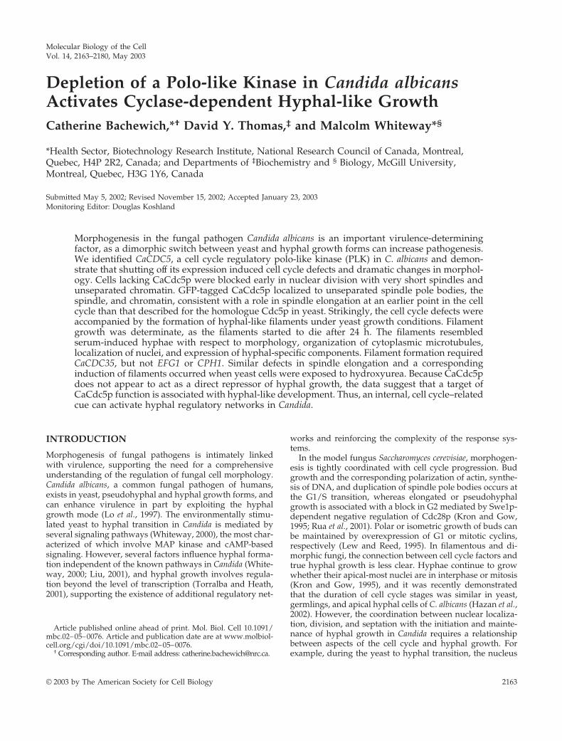
results) and RM1000 had undergone four or five rounds ofnuclear division, 65% of CaCDC5 repressed cells containedtwo nuclei, whereas 28% of the cells contained fragmentedDNA (Table 3; Figure 3). Only 3.5% of the cells containedthree nuclei. Fragmentation of DNA progressed up to 24 h ofrepression, preventing an accurate quantification of nuclei atthe later time point. The filaments were 28.9 � 1.5 �m (SEM;n � 45) in length at 5 h of repression, when approximatelyhalf of the cells had escaped the block in nuclear division,and 62.1 � 2.6 �m (SEM; n � 45) after 8 h, when the majorityof cells contained two nuclei. In contrast, the nucleus inserum-induced hyphae at 37°C divided approximately ev-ery 1.5 h, corresponding to hyphal lengths of 37.3 � 1.1 �m(SEM; n � 32) and 92.8 � 2.4 �m (SEM; n � 32) for the firstand second rounds of division, respectively (Figure 2B; Ta-
Figure 2. Repressing CaCDC5 expression results in the formation of filamentsunder conditions favoring yeast growth. (A) Strains CB104 (cacdc5�::hisG/cacdc5�::HIS1/PCK1::CaCDC5) and CB102 (cacdc5�::hisG/CaCDC5 PCK1:: CaCDC5) wereincubated for 24 h on solid SD or SS medium. Bar, 30 �m. (B) Time course analysisof the formation of filaments in liquid SD medium. Strains CB104 and RM1000were grown in SS medium (0 h), washed, diluted into SD medium, and fixed in70% ethanol after 3, 9, and 24 h of incubation at 30°C. Bars, 10 �m. (C) Filamentformation upon repressing CaCDC5 with the MET promotor. Strains CB108(cacdc5�::hisG/MET:: CaCDC5) and CB109 (cacdc5�::hisG/MET::) were grown inSD medium lacking methionine for promotor induction, then diluted to an OD600of 0.25 in SD medium containing methionine and cysteine to repress the promotorfor 24 h. Bar, 10 �m. (D) Hyphal growth in strain RM1000 incubated in thepresence of 10% serum at 37°C for 4 h. Bar, 10 �m.
C. Bachewich et al.
Molecular Biology of the Cell2168
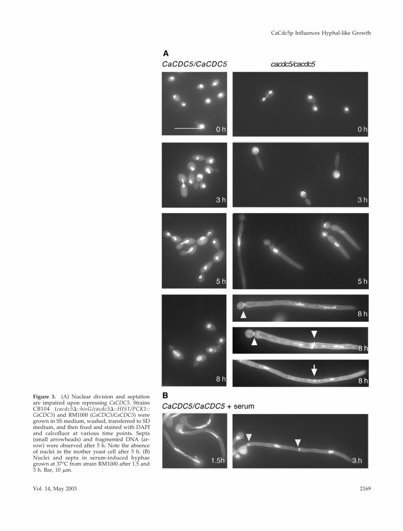
Figure 3. (A) Nuclear division and septationare impaired upon repressing CaCDC5. StrainsCB104 (cacdc5�::hisG/cacdc5�::HIS1/PCK1::CaCDC5) and RM1000 (CaCDC5/CaCDC5) weregrown in SS medium, washed, transferred to SDmedium, and then fixed and stained with DAPIand calcofluor at various time points. Septa(small arrowheads) and fragmented DNA (ar-row) were observed after 5 h. Note the absenceof nuclei in the mother yeast cell after 5 h. (B)Nuclei and septa in serum-induced hyphaegrown at 37°C from strain RM1000 after 1.5 and3 h. Bar, 10 �m.
CaCdc5p Influences Hyphal-like Growth
Vol. 14, May 2003 2169

ble 3). The lack of similar coordination between filamentlength and nuclear division in CaCDC5-repressed cells sug-gests that the block in nuclear division is due to repressionof CaCDC5 and not the initiation of a hyphal-like growthmode. Although the nucleus eventually escapes the block indivision, normal rates of mitosis are not resumed and DNAfragmentation occurs over time while filamentous growthcontinues. These results suggest CaCDC5 is required for theearly stages of nuclear division and chromatin separation,
similar to its counterparts in Schizosaccharomyces pombe andhigher organisms, but in contrast to that in S. cerevisiae.
Staining the CaCDC5-repressed cells with calcofluorwhite demonstrated that septa or chitin deposition didnot occur until later time points, consistent with whennuclei started to escape the block in division (Figure 3;Table 4). Taken together with the inhibition of chromatinseparation, this observation suggests that CaCDC5-re-pressed cells were blocked at an early stage in the cell
Table 4. Proportion of CaCDC5-repressed cells and serum-induced hyphae containing septa
Strain
No. of septa Positioning of septaa
0 1 2 Neck Neck and tube Tube
Hours in SDCB104
0 (n � 242) 100 0 0 0 0 03 (n � 212) 98.1 1.9 0 100 0 05 (n � 227) 86.3 10.6 3.1 77.4 22.6 08 (n � 217) 40.3 50.0 9.7 83.8 16.2 0
Hours in serum 37°CRM1000
0 (n � 100) 100 0 0 0 0 01.5 (n � 106) 98.9 1.2 0 0 0 1003.0 (n � 59) 19.0 47.0 34.0 17.6 41.2 40.7
Values are expressed as percentage. Cells from strain CB104 and RM1000 were incubated in SD medium at 30°C and in SD medium plus 10%serum at 37°C, respectively, for the indicated times, fixed, and stained with calcofluor.a Represents % of total number of cells containing septa.
Table 3. Proportion of cells demonstrating nuclear division and positioning upon incubation in SD medium or serum
Strain
No. of nuclei Position of nuclei
1 2 3 �3 Fraga MotherMother
and tube Tube
Hours in SDCB104
0 (n � 242) 100 0 0 0 0 100 0 03 (n � 212) 92.0 8.0 0 0 0 68.0 8.0 24.05 (n � 319) 55.0 45.0 0 0 0 1.0 36.7 62.48 (n � 197) 2.5 65.5 3.6 0 28.4 0 1.5 70.0
RM1000b
0 (n � 200) 100 0 0 0 0 — — —3 (n � 236) 88.6 11.4 0 0 0 — — —5 (n � 238) 89.5 10.5 0 0 0 — — —8 (n � 225) 86.2 13.8 0 0 0 — — —
Hours in serum at 37°CRM1000
0 (n � 100) 100 0 0 0 0 100 0 01.5 (n � 106) 52.0 48.0 0 0 0 33.3 34.5 32.23.0 (n � 59) 0 20 58 22.0 0 0 100 0
Values are expressed as percentage. Cells from strain CB104 or RM1000 were incubated in SD medium at 30°C for the indicated times, fixed,and stained with DAPI. Cells from strain RM1000 were incubated in SD medium plus 10% serum at 37°C for the indicated time points andfixed and stained with DAPI.a Fragmentation of nuclei prevented an accurate quantification of the total number of nuclei per cell after 8 h of repression.b Cells containing 2 nuclei were doublets.
C. Bachewich et al.
Molecular Biology of the Cell2170
cycle, preceding septation, and that filament formationcorrelated with this early block.
CaCdc5p Is Required for Spindle ElongationTub1p-GFP spindle patterns were analyzed to determinethe specific stage in nuclear division at which CaCdc5p isrequired. Tub1p was tagged with GFP in strains RM1000,CB102, and CB104, and the resulting strains (CB110,CB111, and CB112, respectively) responded to repressingmedium in a manner similar to the nontagged strains. TheGFP-tagged CaCDC5-regulated strain (CB112), however,was somewhat more sensitive to the absence of CaCdc5pthan strain CB104, as the filaments were shorter in lengthat the various time points. The majority of cells fromstrain CB112 depleted of CaCdc5p for 3 h were elongatedor large doublets (Figure 4A; Table 5) and containedspindles in the form of distinct spots or two spots side byside, corresponding to unseparated spindle pole bodies orvery short spindles in S-G2 phases of the cell cycle (Bartonand Gull, 1988; Hazan et al., 2002). Despite the elongatedand large doublet morphology of cells at 3 h, only 2.2% ofthe population contained an extended mitotic spindle.After 4 –7 h, the majority of filamentous cells still con-
tained spot or short bar-like spindles, but an increasingproportion of cells contained a slightly longer bar-likespindle, probably representative of G2/M, and 6% of thefilaments contained an extended mitotic spindle (Figure4A; Table 5). Spindle orientation was also disturbed inseveral cells, regardless of whether the nucleus was in themother yeast cell or in the filament. Filaments that sur-vived after 24 h of CaCDC5 repression contained extensivecytoplasmic microtubule arrays resembling those in se-rum-induced hyphae of Candida (Figure 4C) and hyphaeof other filamentous fungi (Han et al., 2001; Hazan et al.,2002), although the intensity of the Tub1p-GFP signal wasgreater in CaCdc5p-depleted cells at 30°C compared withserum-induced hyphae at 37°C. The remaining dead cellseither did not stain or contained a diffuse signal. Similarpatterns of spindles and cytoplasmic microtubules wereobserved using immunofluorescence with anti–� tubulinantibody in the nontagged strains incubated in repressingmedium for 3 h (Figure 4B) or 24 h (our unpublishedresults), respectively, indicating that the microtubule pat-terns were not artifacts of GFP-tagged tubulin. Immuno-fluorescence demonstrated that cytoplasmic microtubulessimilar to those in established hyphae were present in the
Figure 4. Spindle elongation is blocked in CaCDC5-repressed filaments. (A) Tub1p-GFP was visualized instrains CB110 (CaCDC5/CaCDC5, TUB1-GFP) and CB112 (cacdc5::hisG�/cacdc5�:: HIS1/PCK1::CaCDC5, TUB1-GFP) grown in SD medium for 3, 5, and 24 h. Bar, 10 �m. (B) Paired images of immunolocalized �-tubulin andDAPI-stained DNA in strains CB104 (cacdc5�::hisG/cacdc5�:: HIS1/PCK1::CaCDC5) and RM1000 (CaCDC5/CaCDC5) grown in SD medium for 3 h. Note the cytoplasmic microtubules in the filaments. The short spindlein strain CB104 is indicated by an arrow. Bar, 5 �m. (C) Microtubule organization in serum-induced hyphae.Cells of strain CB110 (CaCDC5/CaCDC5, TUB1-GFP) were incubated in SD medium containing 10% fetal calfserum at 37°C for 2 h to induce hyphal formation and visualize microtubules. Bar, 10 �m.
CaCdc5p Influences Hyphal-like Growth
Vol. 14, May 2003 2171

CaCdc5p-depleted filaments as early as 3 h of repression(Figure 4B). Their relative absence in the GFP-taggedstrain at early time points is likely a reflection of theamplified signal with immunofluorescence. In contrast,control yeast cells of strain CB110 grown at 30°C werecycling at the different time points and demonstrated theexpected spindle patterns (Figure 4, A and B; Table 5). Forexample, of the proportion of cells demonstrating a largebudded morphology, the majority contained a mitoticspindle (Table 5). Slightly elongated cells were present atearly time points but were pseudohyphal intermediatessince they eventually budded. When the control strainCB110 was grown in serum at 37°C for 2–3 h to induce hyphalgrowth, nuclei in the apical regions of hyphae were cycling,with 8% containing mitotic spindles (Table 5). The decrease inthe proportion of CaCdc5p-depleted cells containing spot-likespindles and the corresponding increase in cells containingslightly longer bar spindles at a later time point (28% at 7 hcompared with 13% in serum-induced hyphae) supports thenotion that the majority of CaCDC5-repressed cells were inhib-ited at an early stage in spindle elongation but eventuallybegan to leak through the block.
CaCdc5p Localizes to the Spindle Pole Bodies,Spindle, DNA, and Bud Neck in YeastPLKs localize to spindle pole bodies/centrosomes, thespindle, chromosomes, and sites of cytokinesis in diverseorganisms (Glover et al., 1998; Song et al., 2000). The local-ization of CaCdc5p-GFP was analyzed by integratingPCK1::CaCDC5-GFP into strain RM1000 and overexpressingthe protein with synthetic medium containing 2% casami-
noacids. The spindle, spindle pole bodies, DNA, and budneck clearly demonstrated a signal (Figure 5, A–C), consis-tent with known PLK localizations. Similarly, a strain con-taining CaCdc5p-GFP under control of its endogenous pro-motor demonstrated identical localizations but with aweaker signal (Figure 5, D–I). Intriguingly, CaCdc5p-GFPunder control of its own promotor localized to unsepa-rated spindle pole bodies in cells with very small buds(Figure 5, E and G), suggesting a function for CaCdc5p inthe early stages of spindle elongation.
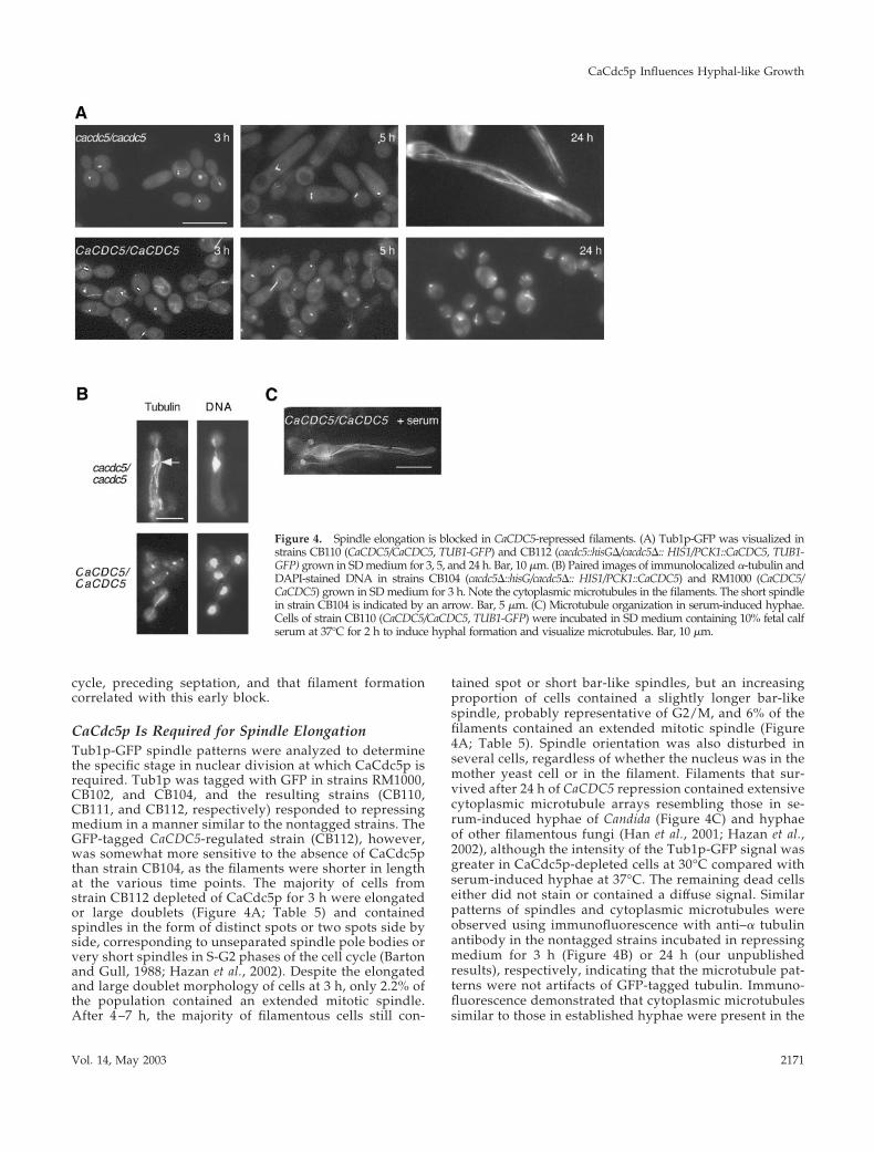
The DNA Synthesis Inhibitor Hydroxyurea InducesSimilar Filament Formation and Impairs SpindleElongationWe and other groups (Bai et al., 2002; Hazan et al., 2002)found that exposing yeast cells of C. albicans to HU pro-duced filaments under yeast growth conditions. Becausethe filaments closely resembled those of CaCDC5-re-pressed cells, we analyzed the filaments further to iden-tify common perturbed features and therefore possiblemechanisms involved in triggering filament initiation. Af-ter 2–3 h in 200 mM HU at 30°C, cells from strain SC5314were elongated and developed into filaments resemblingthose of CaCDC5-repressed cells (Figure 6A). By 24 h, thefilaments were shorter in length than the CaCdc5p-de-pleted filaments and more uniformly vacuolated. The re-sponse was cell density dependent, as yeast cells with anOD600 greater than 0.6 did not form filaments upon incu-bation in HU. DAPI staining (Figure 6A) and Tub1p-GFP(Figure 6B) patterns in cells from strains SC5314 andCB110, respectively, exposed to HU demonstrated that
Table 5. Spindle elongation and cell morphology in cells depleted of CaCdc5p, exposed to HU or treated with serum
StrainHours in
SD
Spindle elongation Cell Morphology
S/earlyG2a G2/Mb Mc
Single orsmall bud
LargeBud Elongated
CB112 3 (n�226) 92.0 5.8 2.2 31.0 11.0 58.04 (n�267) 84.0 9.7 6.3 0 17.0 83.07 (n�186) 66.0 28.0 5.9 0 0 100
CB1103 (n�236) 87.0 1.8 11.2 63.0 24.0 134 (n�180) 85.0 4.6 10.4 76.0 18.9 5.67 (n�190) 89.5 1.0 9.5 87.8 12.2 0
Hours in HUCB110 3 (n�274) 97.7 2.3 0 0 5.4 94.6
7 (n�397) 81.0 18 1.0 0 0 100Hours in serum
at 37°CCB110 3 (n� 75) 81.3 10.7 8.0 0 0 100
Values are expressed as percentages.a Unseparated spindle pole bodies and early spindles represented by a spot or two spots side by side.b Short bar spindles equivalent to 3 to 4 spots in length.c Extended mitotic spindles, greater than 4 spots in length.
C. Bachewich et al.
Molecular Biology of the Cell2172

nuclear division and spindle elongation were blocked in away similar to that of CaCDC5-repressed cells, althoughthe HU-induced filaments retained the short spindle for alonger period (Table 5). FACS analysis demonstrated thatHU-exposed cells were blocked in S phase but progressedto a G2 (4n) content of DNA, whereas CaCDC5-repressedcells progressed through S phase but were subsequentlyblocked in G2 (Figure 6C). Some fragmentation of chro-matin occurred in both conditions at later time points(Figure 6C). These results demonstrate that impairedspindle elongation is a common, early defect in HU-treated and CaCDC5-repressed cells, and thus an aspect ofspindle function may be linked to filament formation.
CaCDC5-repressed and HU-exposed FilamentsDemonstrate Hyphal Characteristics and ExpressFactors Normally Regulated by HyphalTranscription PathwaysThe filaments described here demonstrated several hy-phal-like characteristics. A distinguishing feature of hy-phal development in C. albicans is the migration of thenucleus into the germ tube before mitosis (Sudbery, 2001).Nuclei in both CaCDC5-repressed and HU-exposed cellsdemonstrated this behavior (Figures 3 and 6; Table 3),where the nucleus moved out of the mother cell and intothe filament. The filaments also contained extensive cyto-
Figure 5. CaCdc5p localizes to the spindle pole bod-ies, spindle, DNA, and bud neck. Strain CB115 (A–C)containing PCK1:: CaCDC5-GFP was grown in S me-dium containing 2% casaminoacids for overexpression.Strain CB116 (CaCDC5-GFP-URA3) (D–I) demonstratessimilar localization patterns. Note the staining of aspindle-like structure overlaying chromatin (arrow inE) and spindle pole bodies and chromatin in smallbudded cells (arrow heads in E and G). Bars, 10 �m
CaCdc5p Influences Hyphal-like Growth
Vol. 14, May 2003 2173
plasmic microtubules, comparable to serum-induced hy-phae.
Further evidence for similarity between the filaments andtrue hyphae was obtained by investigating the expression ofa protein that is specifically induced by the hyphal-generat-ing condition of serum and high temperature, using theMAb 16B1-F10 (Marot-Leblond et al., 2000). Immunolocal-ization with 16B1-F10 demonstrated that the antigen wasproduced by several filaments depleted of CaCdc5p for 7 hor exposed to HU for 24 h (Figure 7A). The filaments did notstain along their entire length, as seen with serum-induced
hyphae, indicating the antigen was not expressed immedi-ately upon filament initiation.
Northern analysis of cells depleted of CaCdc5p for 4, 7, or 24 hor exposed to HU for 6 h revealed the expression of factorsnormally induced by the hyphal signaling pathways in responseto serum, including HWP1 (Sharkey et al., 1999) and DDR48 (Laneet al., 2001). The factors were not all induced at similar stages offilament development (Figure 7B), and certain other hyphal-spe-cific factors, including ECE1 and ALS1, were not expressed (ourunpublished results). Therefore, repression of CaCDC5 and expo-sure to HU leads to the formation of filaments and the activation
Figure 6. Hydroxyurea induces filament formation under yeast growth conditions and im-pairs spindle elongation. (A) Strain SC5314 was grown in YPD medium overnight and thendiluted to an OD600 of 0.4 in fresh YPD containing 200 mM HU. Cells were fixed at 6 h andstained with DAPI and calcofluor. (B) Strain CB110 (CaCDC5/CaCDC5, TUB1-GFP) was incu-bated in 200 mM HU for 3, 7, and 24 h. Note the similarity to filaments of CaCDC5-repressedcells (Figures 2B, 3A, and 4A). Bar, 10 �m. (C) FACS analysis demonstrating S phase and G2phase blocks in HU-treated and CaCDC5-repressed cells, respectively. Cells of strains CB104(cacdc5::hisG�/cacdc5�::HIS1/PCK1::CaCDC5) and RM1000 (CaCDC5/CaCDC5) were transferredfrom SS to SD medium, collected at the indicated time points, and processed for FACS analysis.Strain SC5314 was grown in YPD containing 200 mM HU and processed for FACS analysis.Note the fragmentation of DNA at later time points of 9 and 6 h.
C. Bachewich et al.
Molecular Biology of the Cell2174

of aspects of the hyphal transcription program, suggesting a linkbetween spindle function and hyphal development.
Filament Formation Requires CaCDC35, but notEFG1 or CPH1Several hyphal signaling pathways in C. albicans convergeon the transcription factors Efg1p and Cph1p (Ernst, 2000).The absence of both these factors prevents hyphal formationunder most conditions (Lo et al., 1997), whereas CaCdc35p is
required for hyphal formation under all conditions tested(Rocha et al., 2001). To determine whether these factors arerequired for the formation of filaments described here,strains lacking CPH1, EFG1, and CaCDC35 were depleted ofCaCdc5p or exposed to HU. After 7 h of CaCDC5 repressionor exposure to HU, filaments were able to form normally inthe double mutant lacking both Efg1p and Cph1p (Figure8).Efg1p and Cph1p single mutants (strains JCK19 and
Figure 7. Filaments producedby repression of CaCDC5 or expo-sure to HU are similar to hyphaein expression of a serum-inducedantigen and RNA. (A) Serum-induced hyphae from strainSC5314, filaments from strainCB104 (cacdc5�::hisG/cacdc5�::HIS1/PCK1::CaCDC5) depleted ofCaCdc5p for 7 h, and filamentsfrom strain SC5314 incubated inHU for 24 h were fixed and pro-cessed for immunolocalizationof MAb16B1-F10. Bar, 10 �m. (B)Northern analysis demonstratingthe filament-induced expression ofRNA normally induced by the hy-phal-generating conditions of se-rum and high temperature. TotalRNA, 20 �g, from strain SC5314incubated in YPD with 200 mMHU for 6 h, SC5314 withoutHU, CB104 (cacdc5::hisG�/cacdc5�::HIS1/PCK1::CaCDC5) grown in SDmedium for 24, 7, and 4 h,and CB102 (cacdc5�::hisG/CaCDC5PCK1:: CaCDC5) grown in SD me-dium for 4 h was hybridized withprobes specific for HWP1, DDR48,and ACT1 as a loading control.
CaCdc5p Influences Hyphal-like Growth
Vol. 14, May 2003 2175

HLC52) also formed filaments in response to HU (our un-published results). However, the absence of CaCdc35p se-verely compromised filamentous growth in both conditions.The cells treated with HU or depleted of CaCdc5p lookedidentical and resembled large budded yeast with some iso-tropically enlarged daughter cells containing a distal polar-ized evagination. After 24 h, filamentous growth was stillseverely compromised, but more cells contained abnormalshapes and a short polarized extension in the CaCDC5-repression vs. HU-treated condition (our unpublished re-sults). This difference likely reflects the more toxic effects ofhydroxyurea and inhibition of DNA synthesis, causing celldeath more quickly than when CaCDC5 is repressed. Theresults suggest that absence of CaCdc5p and exposure to HUact through a similar pathway to produce hyphal-like fila-ments and communicate with the hyphal signaling networksat the level of CaCdc35p.
DISCUSSION
Regulation of hyphal formation in C. albicans involves sig-naling pathways that ultimately converge to control both theexpression of hyphal-specific genes and the activation/re-cruitment of mechanical factors required for hyphal devel-opment. We show that the polo-like kinase homologueCaCDC5 is required for spindle elongation and that perturb-ing spindle function through repression of CaCDC5 or ex-posure to HU is associated with activation of the transcrip-tion program and machinery required for hyphal-likeformation. Thus a mechanism is in place in C. albicans toallow communication between an internal, cell cycle–relatedcue and hyphal development.
CaCDC5 Is Required for Spindle Formation during SPhaseCaCDC5, along with PLKA from Aspergillus nidulans(Bachewich and Osmani, unpublished results) are the firstcharacterized polo-like kinase homologues in hyphal-pro-ducing fungi. The short spindles and unseparated chromatinthat result from repression of CaCDC5 and the localization ofCaCdc5p to unseparated spindle pole bodies in cells withsmall buds support a role for CaCdc5p in the early stages ofspindle elongation. Such a role is consistent with knownPLK functions in most other organisms, but the demonstra-tion that a PLK is required as early as S phase has not beenpreviously reported. In contrast to higher organisms and S.pombe, spindle initiation occurs during S phase in both C.albicans (Barton and Gull, 1988; Hazan et al., 2002) and S.cerevisiae (Winey and O’Toole, 2001). Several lines of evi-dence suggest a requirement for Cdc5p in spindle formationand DNA replication during S phase in S. cerevisiae (Hardyand Pautz, 1996; Cheng et al., 1997; Bartholemew et al., 2001),but the cdc5–1 mutant arrests with elongated spindles inmitosis with partially separated chromatin (Kitada et al.,1993) and early spindle defects associated with compro-mised Cdc5p function have yet to be reported. ThereforeCaCdc5p acts earlier in the cell cycle and/or has somedifferent functions than Cdc5p.
Figure 8. Filamentous growth in cells depleted of CaCdc5p or exposedto HU require CaCdc35p, but not Efg1p and Cph1p. Cells from strainsCB108 (cacdc5�::hisG/MET:: CaCDC5-URA3), CB305 (cacdc5�::hisG/MET::CaCDC5-URA3 cph1�::hisG/cph1�::hisG efg1�::hisG/efg1�::hisG) and CB303(cacdc5�::hisG/MET:: CaCDC5-URA3 cdc35�::hisG/cdc35�::hisG) were incu-bated in SD medium methionine for overexpression of MET:: CaCDC5,or SD medium � methionine and cysteine to repress MET:: CaCDC5expression for 7 h. Cells from strains SC5314 (�/�), HLC54(cph1�::hisG/cph1�::hisG efg1�::hisG/efg1�::hisG -URA3-hisG) and CR216(cdc35�::hisG/cdc35�::hisG-URA3-hisG) were grown in SD medium, di-luted to an OD600 of 0.4 in fresh medium containing 200 mM HU, andincubated for 7 h. Cells were fixed in 70% EtOH before collection ofimages. Bar, 10 �m.
C. Bachewich et al.
Molecular Biology of the Cell2176
Depletion of CaCdc5p Induces a Dramatic Switch inMorphology from Yeast Cells to Actively GrowingFilaments with Hyphal Characteristics in theAbsence of Serum or High TemperatureThe development of filaments upon repression of CaCDC5 isconsistent with a switch to a hyphal-like growth mode, asopposed to the terminal phenotype of slowly expiring elon-gated buds, because the filaments were actively extending atone third of the rate of serum-induced hyphae incubated atthe higher temperature of 37°C, and demonstrated severalhyphal characteristics, including movement of the nucleusout of the yeast cell into the filament, development of anextensive organization of cytoplasmic microtubules, and ex-pression of factors lying downstream of the hyphal signalingpathways. In filamentous fungi, mutations in a variety ofgenes result in wide diameter hyphae (Harris et al., 1997;Kaminskyj and Hamer, 1998; Momany et al., 1999), support-ing that the wide filaments described here are closely relatedto true hyphae. The filaments are also not analogous toelongated buds of S. cerevisiae that form in response to aninability to deposit a septum (Jimenez et al., 1998), becausethe early block in nuclear division and spindle elongation,and ability to produce filaments with HU, support the no-tion that filaments emerge during an early stage in the cellcycle, before the timing of septation. Septin rings, visualizedwith Cdc12p-GFP, appeared normal in CaCdc5p-depletedcells at 7 h and were localized at the yeast/filament junction(our unpublished results), supporting that septin-related de-fects did not generate the filamentous growth describedhere. The localization of septin rings at the neck of thefilaments is consistent with filament emergence during lateS/G2 of the cell cycle and is comparable to the localization ofthe first septa in serum-induced hyphae, which initiatedgrowth later than G1/S (Hazan et al., 2002).
The eventual death of the filaments suggests that CaCDC5is essential. This possibility exists despite the fact that activehyphal-like growth occurs upon gene repression, becausehyphae of filamentous fungi can grow for a determinateperiod of time in the absence of nuclear division. However,at a later point the nucleus must divide in order for growthto continue. For example, mutations in some essential cellcycle genes such as nimXcdc2p34 in A. nidulans do not preventspore germination and determinate hyphal growth(d’Enfert, 1997), and blocking nuclear division in establishedhyphae of C. albicans also does not prevent hyphal growth(Yokoyama et al., 1990). Therefore, depletion of CaCdc5pand the associated block in nuclear division trigger a changein growth mode to a hyphal-like state, which continues forsome time, but eventually the cells loose viability in theabsence of proper nuclear division.
Intriguingly, several hyphal characteristics appeared atdifferent stages in the development of CaCDC5-repressedfilaments. Increased expression of DDR48, movement of thenucleus into the filament, and initiation of a cytoplasmicmicrotubule network were observed as early as 3–4 h afterrepressing CaCDC5. HWP1 expression was delayed but nor-mally does not increase in serum-induced hyphae until laterin development, after 60 min (Nantel et al., 2002). Microarrayanalysis of the filaments indicates the expression of addi-tional factors that are normally induced by serum and reg-ulated by the hyphal signaling pathways (our unpublishedresults). The differences in timing of gene expression could
be due to the fact that an internal cue, as opposed to serumand high temperature, initiated the hyphal-like growth pro-cess, and different forks in the signaling networks wereutilized. Indeed, there are some differences in the expressionof transcripts (Nantel et al., 2002) and utilization of knowncomponents of the hyphal signaling pathways in hyphaeproduced under different environmental conditions (Giu-sani et al., 2002). Delayed expression of certain genes, espe-cially surface or secreted factors that are not required forhyphal development but turn on as a consequence of hyphalgrowth, could also reflect differences in growth rate, feed-back regulation from the developing hypha to the transcrip-tional pathways, and/or the involvement of factors requiredfor initiation vs. maintenance of hyphal growth, examples ofwhich have been identified (Nantel et al., 2002). These resultssuggest that internal signaling, as opposed to external envi-ronmental cues such as serum, may activate some transcrip-tional and other regulatory aspects governing hyphal initi-ation.
Defects in Spindle Elongation and theCorresponding Generation of Filaments in CaCDC5-repressed and HU-exposed Cells Suggest a Linkbetween Spindle Function and Activation of HyphalGrowthCaCdc5p did not act as a direct negative regulator of hyphalformation like Tup1p (Braun and Johnson, 1997) or Nrg1p(Braun et al., 2001; Munir et al., 2001), suggesting the induc-tion of hyphal-like growth upon CaCdc5p depletion couldbe due to a CaCdc5p-dependent cell cycle–related function.The high degree of similarity between HU-induced andCaCDC5-repressed filaments suggests that filamentousgrowth was triggered by the same cue. A common feature ofCaCDC5 repression and HU treatment is impaired spindleelongation, suggesting a link between spindle function andthe hyphal regulatory program. Consistent with this, repres-sion of DpbIIp, a subunit of DNA polymerase, results indeterminate filament formation in C. albicans (Backen et al.,2000), and defects in DNA synthesis can inhibit spindleelongation. In addition, nocodazole induces similar fila-ments in C. albicans that can be partially suppressed bydeletion of the spindle checkpoint factor MAD2 (Bai et al.,2002), suggesting filamentous growth is partially, but notfully, dependent on the kinetochore attachment/spindle as-sembly branch of the spindle checkpoint. Activation of thisbranch of the spindle checkpoint alone, however, is notsufficient to induce filament formation because deletion of ahomologue of the centromere protein CENP-A in C. albicansdid not result in filamentous growth (Sanyal and Carbon,2002). Although the data support the idea that spindle func-tion is involved in the cue leading to activation of hyphal-like growth, we cannot rule out other pathways or someadditional role for CaCdc5p itself, because polo-like kinasescan be negatively regulated by spindle and DNA damagecheckpoints (Sanchez et al., 1999; Smits et al., 2000; Hu et al.,2001) and nocodazole and HU activate spindle checkpoints(Hu et al., 2001; Garber and Rine, 2002). The nature of theinternal signal and mechanism of transmission to the hyphalregulatory pathways is currently under investigation.
The morphogenic effects of inhibiting different stages inthe cell cycle of Candida are not known, but repression of
CaCdc5p Influences Hyphal-like Growth
Vol. 14, May 2003 2177
another essential cell cycle factor in C. albicans, CaCdc42p,resulted in isometric growth under yeast growth conditions(Ushinsky et al., 2002). In addition, applying differentstresses to yeast cells of Candida at 30°C did not elicit thefilamentous response described here (Martchenko andWhiteway, unpublished results; Enjalbert et al., 2003), indi-cating the phenotype is not a reaction to general cell stress.
Activation of Filament Formation Is Dependent onCaCdc35p but not Efg1p or Cph1pThe formation of both CaCDC5-repressed and HU-exposedfilaments is dependent on CaCdc35p but not Efg1p/Cph1p,suggesting that depletion of CaCdc5p and exposure to HUmay act through similar pathways leading to hyphal-likeformation, and communication with the hyphal signalingnetworks occurs at the level of CaCdc35p. The facts thatCaCdc35p is predicted to act upstream of Efg1p (Rocha et al.,2001), Efg1p in turn was not required for filament formation,and the filaments expressed HWP1, a factor lying down-stream of Efg1p (Sharkey et al., 1999), suggest that additionalpathways feed into and out of CaCdc35p for filament for-mation. Transcript profiling of the cacdc35/cacdc35 strain(Harcus, Nantel, and Whiteway, unpublished results) alsosupports a role for CaCdc35p outside of Efg1p regulation.
Function of a Link between Spindle Elongation andHyphal DevelopmentThe recent demonstration that initiation of hyphal growth inC. albicans is not limited to one cell cycle phase suggests thatthe cell cycle is not a direct regulator of hyphal growth(Hazan et al., 2002). The ability to induce hyphal-like forma-tion upon depletion of CaCdc5p and perturbation of spindlefunction in yeast cells, however, suggests that there is aconnection between aspects of the cell cycle and the hyphalsignaling machinery. This regulatory relationship could ex-ist as a type of checkpoint, perhaps with the durationand/or extent of spindle formation being monitored. Inter-estingly, blocks in S phase can prevent hyphal growth in A.nidulans, whereas mutations in some other essential cellcycle factors do not prevent short-term hyphal growth(d’Enfert, 1997). Regardless of variations in different organ-isms, the demonstration that lack of CaCdc5p and perturba-tion of spindle function influences hyphal-like growth andtranscription in Candida indicates that hyphal-like growthcan be activated by internal, cell cycle–related cues, as op-posed to external signals like serum, and introduces a newlevel within the hyphal signaling networks of C. albicans.
ACKNOWLEDGMENTS
We thank all members of the Whiteway lab for discussions andassistance, Dr. A. Marot-Leblond for Mab16B1-F10, Lucie Bourgetfor assistance with the FACS analysis, and Ursula Oberholzer,James MaGee, Josee Ash, Peter Sudbery, Joachim Morschhauser,and William Fonzi for plasmids. This work was supported in partby a Natural Sciences and Engineering Research Council of CanadaPostdoctoral Fellowship and Visiting Fellowship to C.B. and by theNational Research Council Genomics Health Initiative to M.W. Se-quence data for C. albicans was obtained from the Stanford GenomeTechnology Center website at http://www-sequence.stanford.edu/group/candida. Sequencing of C. albicans was accomplished withthe support of the NIDR and the Burroughs Wellcome Fund.
REFERENCES
Alexandru, G., Uhlmann, F., Mechtler, K., Poupart, M., andNasmyth, K. (2001). Phosphorylation of the cohesin subunit Scc1 byPolo/Cdc5 kinase regulates sister chromatid separation in yeast.Cell 105, 459–472.
Backen, A.C., Broadbent, I.D., Fetherston, R.W., Rosamond, J.D.C.,Schnell, N.F., and Stark, M.J.R. (2000). Evaluation of the CaMALpromotor for regulated expression of genes in Candida albicans.Yeast 16, 1121–1129.
Bai, C., Ramanan, N., Wang, Y.M., and Wang, Y. (2002). Spindleassembly checkpoint component CaMad2p is indispensable for Can-dida albicans survival and virulence in mice. Mol. Microbiol. 45,31–44.
Bartholomew, C.R., Woo, S.H., Chung, Y.S., Jones, C., and Hardy, C.(2001). Cdc5 interacts with the Wee1 kinase in budding yeast. Mol.Cell. Biol. 21, 4949–4959.
Barton, R., and Gull. K. (1988). Variation in cytoplasmic microtubuleorganization and spindle length between two forms of the dimor-phic fungus Candida albicans. J. Cell Sci. 91, 211–220.
Bensen, E.S., Filler, S.G., and Berman, J. (2002). A forkhead tran-scription factor is important for true hyphal growth as well as yeastmorphogenesis in Candida albicans. Eukaryotic Cell 1, 787–798.
Braun, B.R., and Johnson, A.D. (1997). Control of filament formationin Candida albicans by the transcriptional repressor TUP1. Science277, 105–109.
Braun, B.R., Kadosh, D., and Johnson, A.D. (2001). NRG1, a repres-sor of filamentous growth in C. albicans, is down-regulated duringfilament induction. EMBO J. 20, 4753–4761.
Care, R.S., Trevethick, J., Binley, K.M., and Sudbery, P.E. (1999). TheMET3 promotor: a new tool for Candida albicans molecular genetics.Mol. Microbiol. 34, 792–798.
Chen, J., Zhou, S., Wang, Q., Chen, X., Pan, T., and Liu, H. (2000).Crk1, a novel Cdc2-related protein kinase, is required for hyphaldevelopment and virulence in Candida albicans. Mol. Cell. Biol. 20,8596–8708.
Chen, D.C., Yang, B.C., and Kuo, T.T. (1992). One-step transforma-tion of yeast. Curr. Genet. 21, 83–84.
Cheng, L., Collyer, T., and Hardy, C.F. (1997). Cell cycle regulationof DNA replication initiator factor Dbf4p. Mol. Cell. Biol. 19, 4270–4278.
d’Enfert, C. (1997). Fungal spore germination: insights from themolecular genetics of Aspergillus nidulans and Neurospora crassa.Fungal Genet. Biol. 21, 163–172.
Ernst, J.F. (2000). Transcription factors in Candida albicans-environ-mental control of morphogenesis. Microbiology 146, 1763–1764.
Enjalbert, B., Nantel, A., and Whiteway, M. (2003). Stress-inducedgene expression in Candida albicans: absence of a general stressresponse. Mol. Biol. Cell 14, 1460–1467.
Fonzi, W.A., and Irwin, M.Y. (1993). Isogenic strain constructionand gene mapping in Candida albicans. Genetics 134, 717–728.
Gale, C., Gerami-Nejad, M., McClellan, M., Vandoninck, S., Long-tine, M.S., and Berman, J. (2001). Candida albicans Int1p interactswith the septin ring in yeast and hyphal cells. Mol. Biol. Cell 12,3538–3549.
Garber, P.M., and Rine, J. (2002). Overlapping roles of the spindleassembly and DNA damage checkpoints in the cell-cycle responseto altered chromosomes in Saccharomyces cerevisiae. Genetics 161,521–534.
Giusani, A.D., Vinces, M., and Kumamoto, C.A. (2002). Invasivefilamentous growth of Candida albicans is promoted by Czf1p-de-
C. Bachewich et al.
Molecular Biology of the Cell2178

pendent relief of Efg1p-mediated repression. Genetics 160, 1749–1753.
Glover, D.M., Hagan. I.M., and Tavares, A.A.M. (1998). Polo-likekinases: a team that plays throughout mitosis. Genes Dev. 12, 3777–3787.
Han, G., Liu, B., Zhang, J., Zuo, W., Morris, N.R., and Xiang, X.(2001). The Aspergillus cytoplasmic dynein heavy chain and NUDFlocalize to microtubule ends and affect microtubule dynamics. Curr.Biol. 11, 719–724.
Hardy, C.F., and Pautz, A. (1996). A novel role for Cdc5 in DNAreplication. Mol. Cell. Biol. 16, 6775–6782.
Harris, S.D., Hamer. L., Sharpless, K.E., and Hamer, J.E. (1997). TheAspergillus nidulans sepA gene encodes an FH1/2 protein involved incytokinesis and the maintenance of cellular polarity. EMBO J. 16,3474–3483.
Hazan, I., Sepulveda-Becerra, M., and Liu, H. (2002). Hyphal elon-gation is regulated independently of cell cycle in Candida albicans.Mol. Biol. Cell 13, 134–145.
Hu, F., Wang, Y., Liu, D., Li, Y., Qin, J., and Elledge, S.J. (2001).Regulation of the Bub2/Bfa1 GAP complex by Cdc5 and cell cyclecheckpoints. Cell 107, 655–665.
Jimenez, J., Cid, V.J., Cenamor, R., Yuste, M., Molero, G., Nombela,C., and Sanchez, M. (1998). Morphogenesis beyond cytokinetic ar-rest in Saccharomyces cerevisiae. J. Cell Biol. 143, 1617–1634.
Kaminskyj, S.G.W., and Hamer. J.E. (1998). hyp loci control cellpattern formation in vegetative mycelium of Aspergillus nidulans.Genetics 148, 669–680.
Kitada, K., Johnson, A.L., Johnston, L.H., and Sugino, A. (1993). Amulticopy suppressor gene of the Saccharomyces cerevisiae G1 cellcycle mutant gene dbf4 encodes a protein kinase and is defined asCDC5. Mol. Cell. Biol. 13, 4445–4457.
Kohrer, K., and Domdey, H. (1991). Preparation of high molecularweight RNA. Methods Enzymol. 194, 398–405.
Kron, S.J., and Gow, N.A.R. (1995). Budding yeast morphogenesis:signaling, cytoskeleton, and cell cycle. Curr. Opin. Cell Biol. 7,845–855.
Lane, H.A., and Nigg, E.A. (1996). Antibody microinjection revealsan essential role for human polo-like kinase 1 (Plk1) in functionalmaturation of mitotic centrosomes. J. Cell Biol. 135, 1701–1713.
Lane, S., Birse, C., Zhou, S., Matson, R., and Liu, H. (2001). DNAarray studies demonstrate convergent regulation of virulence fac-tors by Cph1, Cph2, and Efg1 in Candida. J. Biol. Chem. 276, 48988–48996.
Leuker, C.E., Sonneborn, A., Delbruck, S., and Ernst, F. (1997).Sequence and promotor regulation of the PCK1 gene encodingphosphoenolpyruvate carboxykinase of the fungal pathogen Can-dida albicans. Gene 192, 235–240.
Lew, D.J., Marini, N.J., Reed, S.I. (1992). Different G1 cyclins controlthe timing of cell cycle commitment in mother and daughter cells ofthe budding yeast S. cerevisiae. Cell 69, 317–327.
Lew, D.J., and Reed, S.I. (1995). A cell cycle checkpoint monitors cellmorphogenesis in budding yeast. J. Cell Biol. 129, 739–749.
Liu, H. (2001). Transcriptional control of dimorphism in Candidaalbicans. Curr. Opin. Microbiol. 4, 728–735.
Llamazares, S., Moreira, A., Tavares, A., Girdham, C., Spruce, B.A.,Gonzales, C., Karess, R.E., Glover, D.M., and Sunkel, C.E. (1991).polo encodes a protein kinase homolog required for mitosis in Dro-sophila. Genes Dev. 5, 2153–2165.
Lo, H., Kohler, J.R., DiDomenico, B., Loebnberg, D., Cacciapuoti, A.,and Fink, G.R. (1997). Nonfilamentous C. albicans mutants are avir-ulent. Cell 90, 939–949.
Loeb, J.D.J., Sepulveda-Becerra, M., Hazan, I., and Liu, H. (1999). AG1 cyclin is necessary for maintenance of filamentous growth inCandida albicans. Mol. Cell. Biol. 19, 4019–4027.
Marot-Leblond, A., Grimaud, L., Nail, S., Bouterige, S., Apaire-Marchais, V., Sullivan, D.J., and Robert, R. (2000). New monoclonalantibody specific for Candida albicans germ tube. J. Clin. Microbiol.38, 61–67.
Momany, M., Westfall. P., and Abramowsky, G. (1999). Aspergil-lus nidulans swo mutants show defects in polarity establishment,polarity maintenance, and hyphal morphogenesis. Genetics 151,557–567.
Morschhauser, J., Michel, S., and Hacker, J. (1998). Expression of achromasomally integrated single copy GFP gene in Candida albicansand its use as a reporter of gene regulation. Mol. Gen. Genet. 257,412–420.
Munir, A. et al. (2001). NRG1 represses yeast-hypha morphogenesisand hypha specific gene expression in Candida albicans. EMBO J. 20,4742–4752.
Nantel A. et al. (2002). Transcription profiling of Candida albicanscells undergoing the yeast-to-hyphal transition. Mol. Biol. Cell. 13,3452–65.
Nigg, E.A. (1998). Polo-like kinases: positive regulators of cell divi-sion from start to finish. Curr. Opin. Cell Biol. 10, 776–783.
Negrado, A., Gil. C., Pla, J., and Nombela, C. (1997). Cloning anal-ysis and one step disruption of the ARG5,6 gene of Candida albicans.Microbiology 143, 297–302.
Okhura, H., Hagan, I.M., and Glover, D.M. (1995). The conservedSchizosaccharomyces pombe kinase plo1, required to form a bipolarspindle, the actin ring, and septum, can drive septum formation inG1 and G2 cells. Genes Dev. 9, 1059–1073.
Rocha, C.R., Schroppel, K., Harcus, D., Marcil, A., Dignard, D.,Taylor, B.N., Thomas, D.Y., Whiteway, M., and Leberer, E. (2001).Signaling through adenylyl cyclase is essential for hyphal growthand virulence in the pathogenic fungus Candida albicans. Mol. Biol.Cell 12, 3631–3643.
Rose, M.D., Winston, F., and Hieter, P. (1990). Methods in YeastGenetics: A Laboratory Course Manual. Cold Spring Harbor, NY:Cold Spring Harbor Laboratory Press.
Rua, D., Tobe, B., and Kron, S.J. (2001). Cell cycle control of yeastfilamentous growth. Curr. Opin. Microbiol. 4, 720–727.
Sanchez, Y., Bachant, J., Wang, H., Hu, F., Liu, D., Tetzlaff, M., andElledge, S.J. (1999). Control of the DNA damage checkpoint by Chk1and Rad53 protein kinases through distinct mechanisms. Science286, 1166–1171.
Sanyal, A., and Carbon, J. (2002). The CENP-A homolog CaCse4p inthe pathogenic yeast Candida albicans is a centromere protein essen-tial for chromosome transmission. Proc. Natl. Acad. Sci. USA 99,12969–12974.
Sharkey, L.L., McNemar, M.D., Saporito-Irwin, S.M., Sypherd, P.S.,and Fonzi, W.A. (1999). HWP1 functions in the morphological de-velopment of Candida albicans downstream of EFG1, TUP1, andRBF1. J. Bacteriol. 181, 5273–5279.
Shirayama, M., Zachariae, W., Ciosk, R. Nasmyth, K. 1998. Thepolo-like kinase cdc5p and the WD repeat protein Cdc20/fizzy areregulators and substrates of the anaphase-promoting complex inSaccharomyces cerevisiae. EMBO J. 17, 1336–1349.
Smits, V.A.J., Klommaker, R., Arnaud, L., Rijksen, G., Nigg, E.A.,and Medema, R.H. (2000). Polo-like kinase is a target of the DNAdamage checkpoint. Nat. Cell Biol. 2, 672–676.
Song, S., Grenfall, T.Z., Garfield, S., Erikson, R.L., and Lee, K.S.(2000). Essential function of the polo box of Cdc5 in subcellular
CaCdc5p Influences Hyphal-like Growth
Vol. 14, May 2003 2179
localization and induction of cytokinetic structures. Mol. Cell. Biol.20, 286–298.
Song, S., and Lee, K.S. (2001). A novel function of Saccharomycescerevisiae CDC5 in cytokinesis. J. Cell Biol. 152, 451–469.
Srikantha, T., and Soll, D.R. (1993). A white-specific gene in thewhite-opaque switching system of Candida albicans. Gene 131, 53–60.
Sudbery, P.E. (2001). The germ tubes of Candida albicans hyphae andpseudohyphae show different patterns of septin ring localization.Mol. Microbiol. 41, 19–31.
Torralba, S., and Heath, IB. (2001). Cytoskeletal and Ca2� regulationof hyphal tip growth and initiation. Curr. Top. Dev. Biol. 51, 135–187.
Toyoshima-Morimoto, F., Taniguchi, E., Shinya, N., Iwanatsu, A.,and Nishida, E. (2001). Polo-like kinase 1 phosphorylates cyclin Band targets it to the nucleus during prophase. Nature 410, 215–220.
Ushinsky, S.C., Harcus, D., Ash, J., Dignard, D., Marcil. A., Morch-hauser, J., Thomas, D.Y., Whiteway, M., and Leberer, E. (2002).CDC42 is required for polarized growth in human pathogen Candidaalbicans. Eukaryotic Cell 1, 95–104.
Whiteway, M. (2000). Transcriptional control of cell type and mor-phogenesis in Candida albicans. Curr. Opin. Microbiol. 3, 582–588.
Winey, M., and O’Toole, E.T. (2001). The spindle cycle in buddingyeast. Nat. Cell Biol. 3, E23–E27.
Yokoyama, K., Kaji, H., Nishimura, K., and Miyaji, M. (1990). Therole of microfilaments and microtubules in apical growth and di-morphism of Candida albicans. J. Gen. Microbiol. 136, 1067–1075.
C. Bachewich et al.
Molecular Biology of the Cell2180


