Embed Size (px)
Citation preview
MATBIO-1577; No. of pages: 19; 4C:
Article
Peptide gels of fcomposition andprobing cell-cellinteractions in v
J.C. Ashwortha, b, J.L. Thompsona, J.R. Ja d e
0022-2836/© 2019 Publish
Please cite this article as:for probing cell-cell and c
ully-definedmechanics forand cell-matrixitro
amesa, C.E. Slater a, S. Pijuan-Galitó a, c,K. Lis-Slimak , R.J. Holley , K.A. Meade , A. Thompson f, K.P. Arkill f, M. Tassieri g,A.J. Wrighth, G. Farnieb, i and C.L.R. Merrya
a - Stem Cell Glycobiology Group, Division of Cancer & Stem Cells, School of Medicine, University of Nottingham, UKb - Manchester Cancer Research Centre, Division of Molecular & Clinical Cancer Sciences, University of Manchester, UKc - Laboratory of Biophysics and Surface Analysis, School of Pharmacy, University of Nottingham, UKd - Stem Cell and Neurotherapies Group, University of Manchester, UKe - Office of Business Relations, Faculty of Biology, Medicine and Health, University of Manchester, UKf - Division of Cancer & Stem Cells, School of Medicine, University of Nottingham, UKg - Division of Biomedical Engineering, School of Engineering, University of Glasgow, UKh - Optics and Photonics Research Group, Faculty of Engineering, University of Nottingham, UKi - SGC, Botnar Research Centre, NDORMS, University of Oxford, UK
Correspondence to C.L.R. Merry: Corresponding author [email protected]://doi.org/10.1016/j.matbio.2019.06.009
Abstract
Current materials used for in vitro 3D cell culture are often limited by their poor similarity to human tissue,batch-to-batch variability and complexity of composition and manufacture. Here, we present a “blank slate”culture environment based on a self-assembling peptide gel free from matrix motifs. The gel can becustomised by incorporating matrix components selected to match the target tissue, with independent controlof mechanical properties. Therefore the matrix components are restricted to those specifically added, or thosesynthesised by encapsulated cells. The flexible 3D culture platform provides full control over biochemical andphysical properties, allowing the impact of biochemical composition and tissue mechanics to be separatelyevaluated in vitro. Here, we demonstrate that the peptide gels support the growth of a range of cells includinghuman induced pluripotent stem cells and human cancer cell lines. Furthermore, we present proof-of-conceptthat the peptide gels can be used to build disease-relevant models. Controlling the peptide gelatorconcentration allows peptide gel stiffness to be matched to normal breast (b1 kPa) or breast tumour tissue(N1 kPa), with higher stiffness favouring the viability of breast cancer cells over normal breast cells. In parallel,the peptide gels may be modified with matrix components relevant to human breast, such as collagen I andhyaluronan. The choice and concentration of these additions affect the size, shape and organisation of breastepithelial cell structures formed in co-culture with fibroblasts. This system therefore provides a means ofunravelling the individual influences of matrix, mechanical properties and cell-cell interactions in cancer andother diseases.
© 2019 Published by Elsevier B.V.
Introduction
In many research areas, but particularly in cancerresearch and disease modelling, there is an increas-ing emphasis on the use of biomaterials to grow cellsin 3D [1–3]. It is now well-understood that culturingmost cells on 2D surfaces results in inferior
ed by Elsevier B.V.
J.C. Ashworth, J.L. Thompson, J.R. Jamesell-matrix interactions in ..., Matrix Biology
physiological conditions affecting cell morphology,phenotype and cell-matrix interactions [4–6]. As aresult, there is a growing body of literature focussedon the development of biomaterials as biomimeticculture platforms, to produce more tissue-realisticcell behaviour in vitro. It has become clear that thereis unlikely to be a one-size-fits-all solution, with 3D in
Matrix Biology. (xxxx) xx, xxx
, et al., Peptide gels of fully-defined composition and mechanics, https://doi.org/10.1016/j.matbio.2019.06.009
2 Peptide gels for probing interactions in vitro
vitro culture environments requiring the samecapacity for variability and specificity as providedby natural in vivo matrix microenvironments [1].Therefore, the major hurdle still to be overcome isthe provision of a system that is both highly tunableand reproducible in composition and mechanicalproperties.Materials for 3D culture may broadly be separated
into natural and artificially derived materials [7].Natural materials, most notably collagen gels andMatrigel™, are the most established, with a longhistory of use in tissue culture for applicationsranging from cell migration and invasion studies toregenerative medicine [8–10]. Since these materialsare commonly based on matrix proteins, cells grownon or within them are able to adhere and grow to formtissue-realistic structures [9,11]. However, the bio-logical origin of natural materials also results inbatch-to-batch variability and uncertainty in compo-sition, most particularly for Matrigel™, a basementmembrane extract derived from Engelbreth-Holm-Swarm mouse sarcoma [12]. For this reason, and toprovide opportunities for adding functionality, therehas been a shift in focus towards more highlydefined, synthetic alternatives [7].Apart from a few notable exceptions [13,14], there
have thus far been few models designed specificallyto allow customisable matrix composition. Similarly,many models have a matrix component (commonlycollagen, laminin or hyaluronan) as an essential partof their make-up, making it hard to discriminateendogenous matrix production by encapsulatedcells from the 3D matrix itself. Here, we present theoptimisation of a self-assembling peptide gel as thebasis for a 3D culture platform with user-controlledcomposition, mechanical properties, and cell-cellinteractions. Based on a short, octapeptide gelator[15], the raw materials required for the peptide gelscan be produced reliably and rapidly and are widelycommercially available. By defining a protocol thatenables independent control over mechanical andbiochemical properties, we aim to provide a platformtechnology suitable for studies decoupling theinfluences of matrix stiffness and composition oncell behaviour. Here, we demonstrate the applicationof the peptide gel to investigate the role of matrixstiffness and functionalisation on a model of breastcancer.
Results
Controlled gelation produces a fully-definedenvironment
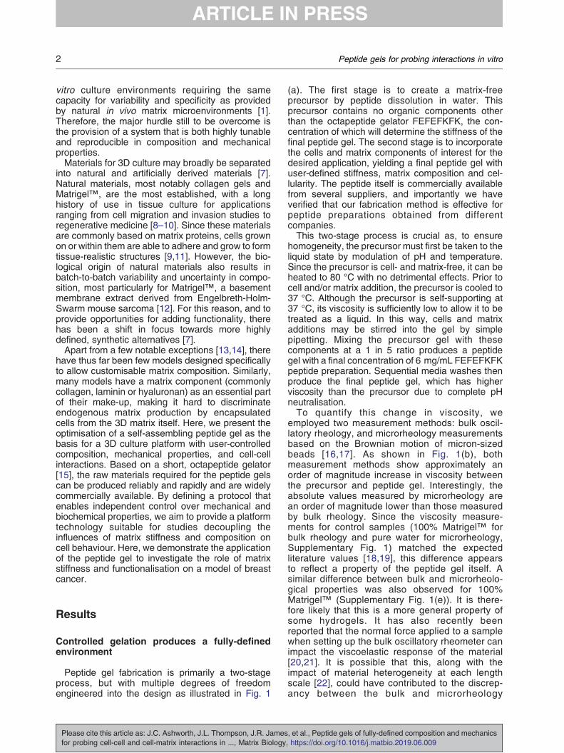
Peptide gel fabrication is primarily a two-stageprocess, but with multiple degrees of freedomengineered into the design as illustrated in Fig. 1
Please cite this article as: J.C. Ashworth, J.L. Thompson, J.R. Jamesfor probing cell-cell and cell-matrix interactions in ..., Matrix Biology
(a). The first stage is to create a matrix-freeprecursor by peptide dissolution in water. Thisprecursor contains no organic components otherthan the octapeptide gelator FEFEFKFK, the con-centration of which will determine the stiffness of thefinal peptide gel. The second stage is to incorporatethe cells and matrix components of interest for thedesired application, yielding a final peptide gel withuser-defined stiffness, matrix composition and cel-lularity. The peptide itself is commercially availablefrom several suppliers, and importantly we haveverified that our fabrication method is effective forpeptide preparations obtained from differentcompanies.This two-stage process is crucial as, to ensure
homogeneity, the precursor must first be taken to theliquid state by modulation of pH and temperature.Since the precursor is cell- and matrix-free, it can beheated to 80 °C with no detrimental effects. Prior tocell and/or matrix addition, the precursor is cooled to37 °C. Although the precursor is self-supporting at37 °C, its viscosity is sufficiently low to allow it to betreated as a liquid. In this way, cells and matrixadditions may be stirred into the gel by simplepipetting. Mixing the precursor gel with thesecomponents at a 1 in 5 ratio produces a peptidegel with a final concentration of 6 mg/mL FEFEFKFKpeptide preparation. Sequential media washes thenproduce the final peptide gel, which has higherviscosity than the precursor due to complete pHneutralisation.To quantify this change in viscosity, we
employed two measurement methods: bulk oscil-latory rheology, and microrheology measurementsbased on the Brownian motion of micron-sizedbeads [16,17]. As shown in Fig. 1(b), bothmeasurement methods show approximately anorder of magnitude increase in viscosity betweenthe precursor and peptide gel. Interestingly, theabsolute values measured by microrheology arean order of magnitude lower than those measuredby bulk rheology. Since the viscosity measure-ments for control samples (100% Matrigel™ forbulk rheology and pure water for microrheology,Supplementary Fig. 1) matched the expectedliterature values [18,19], this difference appearsto reflect a property of the peptide gel itself. Asimilar difference between bulk and microrheolo-gical properties was also observed for 100%Matrigel™ (Supplementary Fig. 1(e)). It is there-fore likely that this is a more general property ofsome hydrogels. It has also recently beenreported that the normal force applied to a samplewhen setting up the bulk oscillatory rheometer canimpact the viscoelastic response of the material[20,21]. It is possible that this, along with theimpact of material heterogeneity at each lengthscale [22], could have contributed to the discrep-ancy between the bulk and microrheology
, et al., Peptide gels of fully-defined composition and mechanics, https://doi.org/10.1016/j.matbio.2019.06.009
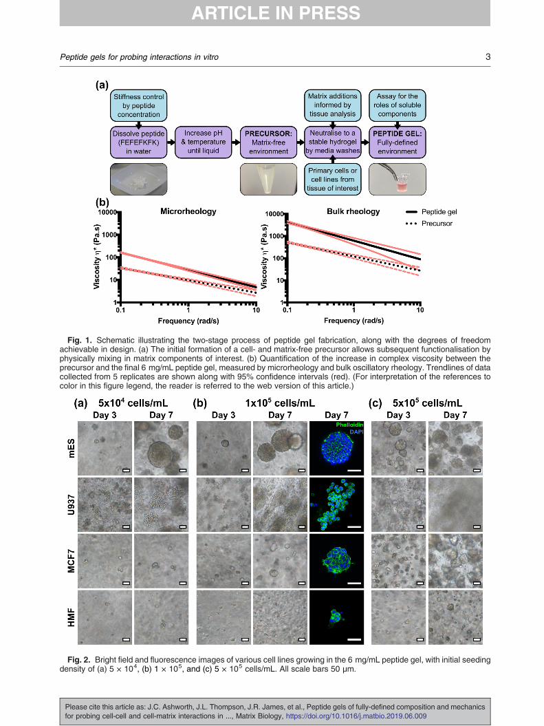
Fig. 2. Bright field and fluorescence images of various cell lines growing in the 6 mg/mL peptide gel, with initial seedingdensity of (a) 5 × 10⁴, (b) 1 × 10⁵, and (c) 5 × 10⁵ cells/mL. All scale bars 50 μm.
Fig. 1. Schematic illustrating the two-stage process of peptide gel fabrication, along with the degrees of freedomachievable in design. (a) The initial formation of a cell- and matrix-free precursor allows subsequent functionalisation byphysically mixing in matrix components of interest. (b) Quantification of the increase in complex viscosity between theprecursor and the final 6 mg/mL peptide gel, measured by microrheology and bulk oscillatory rheology. Trendlines of datacollected from 5 replicates are shown along with 95% confidence intervals (red). (For interpretation of the references tocolor in this figure legend, the reader is referred to the web version of this article.)
3Peptide gels for probing interactions in vitro
Please cite this article as: J.C. Ashworth, J.L. Thompson, J.R. James, et al., Peptide gels of fully-defined composition and mechanicsfor probing cell-cell and cell-matrix interactions in ..., Matrix Biology, https://doi.org/10.1016/j.matbio.2019.06.009
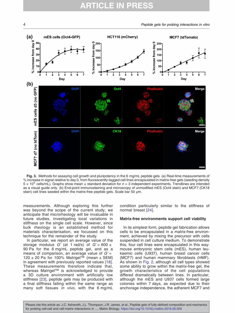
Fig. 3. Methods for assaying cell growth and pluripotency in the 6 mg/mL peptide gels. (a) Real-time measurements of% increase in signal relative to day 0, from fluorescently-tagged cell lines encapsulated in matrix-free gels (seeding density5 × 105 cells/mL). Graphs show mean ± standard deviation for n = 3 independent experiments. Trendlines are intendedas a visual guide only. (b) End-point immunostaining and microscopy of unmodified mES (Oct4 stain) and MCF7 (CK18stain) cell lines seeded within the matrix-free peptide gels. Scale bar 50 μm.
4 Peptide gels for probing interactions in vitro
measurements. Although exploring this furtherwas beyond the scope of the current study, weanticipate that microrheology will be invaluable infuture studies, investigating local variations instiffness on the single cell scale. However, sincebulk rheology is an established method formaterials characterisation, we focussed on thistechnique for the remainder of the study.In particular, we report an average value of the
storage modulus G' (at 1 rad/s) of G' = 600 ±90 Pa for the 6 mg/mL peptide gel, and as ameans of comparison, an average value of G' =120 ± 20 Pa for 100% Matrigel™ (mean ± SEM)in agreement with previously reported values [18].These measurements therefore indicate that,whereas Matrigel™ is acknowledged to providea 3D culture environment with artificially lowstiffness [23], peptide gels may be produced witha final stiffness falling within the same range asmany soft tissues in vivo, with the 6 mg/mL
Please cite this article as: J.C. Ashworth, J.L. Thompson, J.R. Jamesfor probing cell-cell and cell-matrix interactions in ..., Matrix Biology
condition particularly similar to the stiffness ofnormal breast [24].
Matrix-free environments support cell viability
In its simplest form, peptide gel fabrication allowscells to be encapsulated in a matrix-free environ-ment, achieved by mixing the precursor with cellssuspended in cell culture medium. To demonstratethis, four cell lines were encapsulated in this way:mouse embryonic stem cells (mES), human leu-kaemic cells (U937), human breast cancer cells(MCF7) and human mammary fibroblasts (HMF).As shown in Fig. 2, although all cell types showedsome ability to grow within the matrix-free gel, thegrowth characteristics of the cell populationsdiffered dramatically between lines. In particular,although the mES and U937 cells formed largecolonies within 7 days, as expected due to theiranchorage independence, the adherent MCF7 and
, et al., Peptide gels of fully-defined composition and mechanics, https://doi.org/10.1016/j.matbio.2019.06.009
5Peptide gels for probing interactions in vitro
HMF cell lines formed smaller colonies at a slowerrate.To test the effect of seeding density on cell growth,
each cell line was also suspended at three seedingdensities: 5 × 10⁴, 1 × 10⁵ and 5 × 10⁵ cells/mLwithin the peptide gel. At the lowest seeding density,mES and U937 both formed defined colonies withclearly distinguishable boundaries, with mES cellsforming a larger number of smaller colonies at highseeding density, and U937 rapidly colonising theentire gel with separate colonies merging together.MCF7 cells also formed approximately sphericalcolonies at this seeding density, similar in morphol-ogy to the mES cell colonies. HMF showed limitedproliferation at all seeding densities, forming smallrounded clusters rather than the classic elongatemorphology typical of fibroblasts [25].The optical transparency of the peptide gels also
allows quantitative read-outs of fluorescence (re-porters or constitutively expressed) allowing real-time measurement of viable cell number. Fig. 3(a)shows the increase in fluorescence signal relative tothe value at day 0 for three cell lines: mES with anOct4-GFP reporter, mCherry-HCT116 colorectalcancer cells, and tdTomato-MCF7 breast cancercells. Each cell line gave a distinct growth profile.The fluorescence values for both HCT116 andMCF7 increased steadily up to day 7, and whilst anapproximately linear increase in fluorescence overtime was observed for MCF7, the increase forHCT116 became even more pronounced towardsday 7. The Oct4-GFP construct in the mES cells isactive in pluripotent cells and will switch off as thecells differentiate and Oct4 is down-regulated.
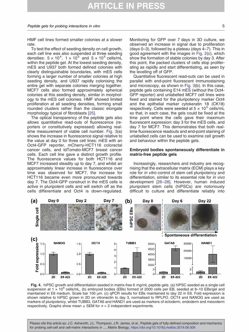
Fig. 4. hiPSC growth and differentiation seeded in matrix-fresuspension at 1 × 10⁶ cells/mL, (b) embryoid bodies (EBs) fomaintained in E6 medium. Scale bar 100 μm. (c) qPCR resulshown relative to hiPSC grown in 2D on vitronectin to day 3markers of pluripotency, whilst TUBB3, GATA6 and HAND1 arrespectively. Graphs show mean ± SEM for n = 2 independen
Please cite this article as: J.C. Ashworth, J.L. Thompson, J.R. Jamesfor probing cell-cell and cell-matrix interactions in ..., Matrix Biology
Monitoring for GFP over 7 days in 3D culture, weobserved an increase in signal due to proliferation(days 0–3), followed by a plateau (days 4–7). This ingood agreement with the images in Fig. 2(c), whichshow the formation of stable colonies by day 3. Afterthis point, the packed clusters of cells stop prolifer-ating as rapidly and start differentiating, as seen bythe levelling off of GFP.Quantitative fluorescent read-outs can be used in
parallel with end-point fluorescent immunostainingand microscopy, as shown in Fig. 3(b). In this case,peptide gels containing E14 mES (without the Oct4-GFP reporter) and unlabelled MCF7 cell lines werefixed and stained for the pluripotency marker Oct4and the epithelial marker cytokeratin 18 (CK18)respectively. Cells were seeded at 5 × 10⁵ cells/mLso that, in each case, the gels could be fixed at thetime point where the cells gave their maximumfluorescent expression: day 3 for the mES cells, andday 7 for MCF7. This demonstrates that both real-time fluorescence readouts and end-point staining ofunlabelled cells can be used to examine cell growthand behaviour within the peptide gels.
Embryoid bodies spontaneously differentiate inmatrix-free peptide gels
Increasingly, researchers and industry are recog-nising that the extracellular matrix (ECM) plays a keyrole for in vitro control of stem cell pluripotency anddifferentiation, similar to its essential role for in vivodevelopment [26–28]. However, human inducedpluripotent stem cells (hiPSCs) are notoriouslydifficult to culture and differentiate reliably into
e 6 mg/mL peptide gels. (a) hiPSC seeded as a single cellrmed of 2000 cells per EB, seeded at 8–10 EBs/gel andts for EBs maintained to day 22 in E6. Fold expression is, normalised to RPLPO. OCT4 and NANOG are used ase used as markers of ectoderm, endoderm and mesodermt experiments.
, et al., Peptide gels of fully-defined composition and mechanics, https://doi.org/10.1016/j.matbio.2019.06.009
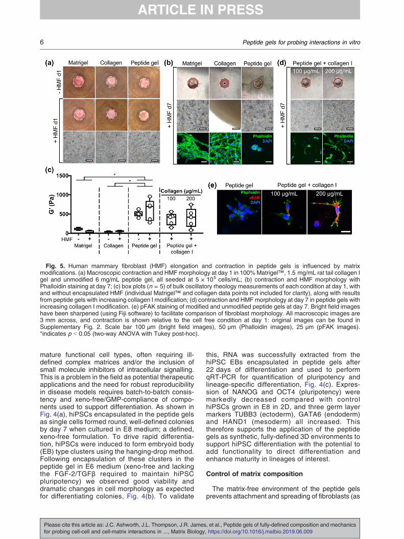
Fig. 5. Human mammary fibroblast (HMF) elongation and contraction in peptide gels is influenced by matrixmodifications. (a) Macroscopic contraction and HMFmorphology at day 1 in 100%Matrigel™, 1.5 mg/mL rat tail collagen Igel and unmodified 6 mg/mL peptide gel, all seeded at 5 × 10⁵ cells/mL; (b) contraction and HMF morphology withPhalloidin staining at day 7; (c) box plots (n = 5) of bulk oscillatory rheology measurements of each condition at day 1, withand without encapsulated HMF (individual Matrigel™ and collagen data points not included for clarity), along with resultsfrom peptide gels with increasing collagen I modification; (d) contraction and HMFmorphology at day 7 in peptide gels withincreasing collagen I modification. (e) pFAK staining of modified and unmodified peptide gels at day 7. Bright field imageshave been sharpened (using Fiji software) to facilitate comparison of fibroblast morphology. All macroscopic images are3 mm across, and contraction is shown relative to the cell free condition at day 1: original images can be found inSupplementary Fig. 2. Scale bar 100 μm (bright field images), 50 μm (Phalloidin images), 25 μm (pFAK images).*indicates p b 0.05 (two-way ANOVA with Tukey post-hoc).
6 Peptide gels for probing interactions in vitro
mature functional cell types, often requiring ill-defined complex matrices and/or the inclusion ofsmall molecule inhibitors of intracellular signalling.This is a problem in the field as potential therapeuticapplications and the need for robust reproducibilityin disease models requires batch-to-batch consis-tency and xeno-free/GMP-compliance of compo-nents used to support differentiation. As shown inFig. 4(a), hiPSCs encapsulated in the peptide gelsas single cells formed round, well-defined coloniesby day 7 when cultured in E8 medium; a defined,xeno-free formulation. To drive rapid differentia-tion, hiPSCs were induced to form embryoid body(EB) type clusters using the hanging-drop method.Following encapsulation of these clusters in thepeptide gel in E6 medium (xeno-free and lackingthe FGF-2/TGFβ required to maintain hiPSCpluripotency) we observed good viability anddramatic changes in cell morphology as expectedfor differentiating colonies, Fig. 4(b). To validate
Please cite this article as: J.C. Ashworth, J.L. Thompson, J.R. Jamesfor probing cell-cell and cell-matrix interactions in ..., Matrix Biology
this, RNA was successfully extracted from thehiPSC EBs encapsulated in peptide gels after22 days of differentiation and used to performqRT-PCR for quantification of pluripotency andlineage-specific differentiation, Fig. 4(c). Expres-sion of NANOG and OCT4 (pluripotency) weremarkedly decreased compared with controlhiPSCs grown in E8 in 2D, and three germ layermarkers TUBB3 (ectoderm), GATA6 (endoderm)and HAND1 (mesoderm) all increased. Thistherefore supports the application of the peptidegels as synthetic, fully-defined 3D environments tosupport hiPSC differentiation with the potential toadd functionality to direct differentiation andenhance maturity in lineages of interest.
Control of matrix composition
The matrix-free environment of the peptide gelsprevents attachment and spreading of fibroblasts (as
, et al., Peptide gels of fully-defined composition and mechanics, https://doi.org/10.1016/j.matbio.2019.06.009
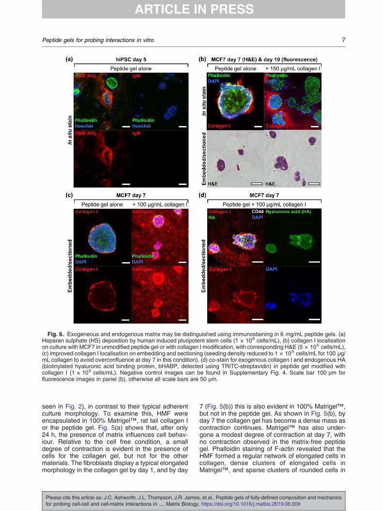
Fig. 6. Exogeneous and endogenous matrix may be distinguished using immunostaining in 6 mg/mL peptide gels. (a)Heparan sulphate (HS) deposition by human induced pluripotent stem cells (1 × 10⁶ cells/mL), (b) collagen I localisationon culture with MCF7 in unmodified peptide gel or with collagen I modification, with corresponding H&E (5 × 10⁵ cells/mL),(c) improved collagen I localisation on embedding and sectioning (seeding density reduced to 1 × 10⁵ cells/mL for 100 μg/mL collagen to avoid overconfluence at day 7 in this condition), (d) co-stain for exogenous collagen I and endogenous HA(biotinylated hyaluronic acid binding protein, bHABP, detected using TRITC-streptavidin) in peptide gel modified withcollagen I (1 × 10⁵ cells/mL). Negative control images can be found in Supplementary Fig. 4. Scale bar 100 μm forfluorescence images in panel (b), otherwise all scale bars are 50 μm.
7Peptide gels for probing interactions in vitro
seen in Fig. 2), in contrast to their typical adherentculture morphology. To examine this, HMF wereencapsulated in 100% Matrigel™, rat tail collagen Ior the peptide gel. Fig. 5(a) shows that, after only24 h, the presence of matrix influences cell behav-iour. Relative to the cell free condition, a smalldegree of contraction is evident in the presence ofcells for the collagen gel, but not for the othermaterials. The fibroblasts display a typical elongatedmorphology in the collagen gel by day 1, and by day
Please cite this article as: J.C. Ashworth, J.L. Thompson, J.R. Jamesfor probing cell-cell and cell-matrix interactions in ..., Matrix Biology
7 (Fig. 5(b)) this is also evident in 100% Matrigel™,but not in the peptide gel. As shown in Fig. 5(b), byday 7 the collagen gel has become a dense mass ascontraction continues. Matrigel™ has also under-gone a modest degree of contraction at day 7, withno contraction observed in the matrix-free peptidegel. Phalloidin staining of F-actin revealed that theHMF formed a regular network of elongated cells incollagen, dense clusters of elongated cells inMatrigel™, and sparse clusters of rounded cells in
, et al., Peptide gels of fully-defined composition and mechanics, https://doi.org/10.1016/j.matbio.2019.06.009
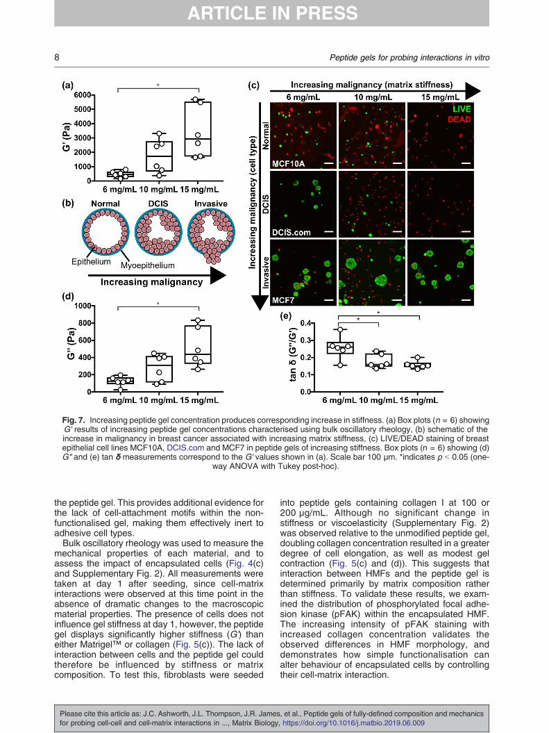
Fig. 7. Increasing peptide gel concentration produces corresponding increase in stiffness. (a) Box plots (n = 6) showingG' results of increasing peptide gel concentrations characterised using bulk oscillatory rheology, (b) schematic of theincrease in malignancy in breast cancer associated with increasing matrix stiffness, (c) LIVE/DEAD staining of breastepithelial cell lines MCF10A, DCIS.com and MCF7 in peptide gels of increasing stiffness. Box plots (n = 6) showing (d)G" and (e) tan δ measurements correspond to the G' values shown in (a). Scale bar 100 μm. *indicates p b 0.05 (one-
way ANOVA with Tukey post-hoc).
8 Peptide gels for probing interactions in vitro
the peptide gel. This provides additional evidence forthe lack of cell-attachment motifs within the non-functionalised gel, making them effectively inert toadhesive cell types.Bulk oscillatory rheology was used to measure the
mechanical properties of each material, and toassess the impact of encapsulated cells (Fig. 4(c)and Supplementary Fig. 2). All measurements weretaken at day 1 after seeding, since cell-matrixinteractions were observed at this time point in theabsence of dramatic changes to the macroscopicmaterial properties. The presence of cells does notinfluence gel stiffness at day 1, however, the peptidegel displays significantly higher stiffness (G') thaneither Matrigel™ or collagen (Fig. 5(c)). The lack ofinteraction between cells and the peptide gel couldtherefore be influenced by stiffness or matrixcomposition. To test this, fibroblasts were seeded
Please cite this article as: J.C. Ashworth, J.L. Thompson, J.R. Jamesfor probing cell-cell and cell-matrix interactions in ..., Matrix Biology
into peptide gels containing collagen I at 100 or200 μg/mL. Although no significant change instiffness or viscoelasticity (Supplementary Fig. 2)was observed relative to the unmodified peptide gel,doubling collagen concentration resulted in a greaterdegree of cell elongation, as well as modest gelcontraction (Fig. 5(c) and (d)). This suggests thatinteraction between HMFs and the peptide gel isdetermined primarily by matrix composition ratherthan stiffness. To validate these results, we exam-ined the distribution of phosphorylated focal adhe-sion kinase (pFAK) within the encapsulated HMF.The increasing intensity of pFAK staining withincreased collagen concentration validates theobserved differences in HMF morphology, anddemonstrates how simple functionalisation canalter behaviour of encapsulated cells by controllingtheir cell-matrix interaction.
, et al., Peptide gels of fully-defined composition and mechanics, https://doi.org/10.1016/j.matbio.2019.06.009
9Peptide gels for probing interactions in vitro
Distinguishing exogenous and endogenous ma-trix
The ability to discriminate and visualise matrixfunctionalisation (exogenous) as opposed to cell-deposited matrix (endogenous) is crucial for appli-cation of the peptide gels to the study of cell-matrixinteractions. Immunostaining and microscopy canbe used to detect heparan sulphate deposition byhiPSC (Fig. 6(a)) and collagen I deposition by MCF7(Fig. 6(b), upper panel) seeded in matrix-free peptidegels, highlighting predominantly cell-associated butnon-uniform distribution in the 3D cultures.When we applied the same technique to
visualise collagen I in a modified peptide gel,again confocal microscopy revealed a positivecollagen I signal, although this showed somespatial heterogeneity as displayed in the single z-slice in Fig. 6(b), upper right panel. It was notpossible to distinguish using this techniquewhether this heterogeneity was a true reflectionof the collagen I localisation within the gel, or alimitation of the in situ (whole gel) staining andimaging method. To clarify this, gels were em-bedded in agar blocks to allow sectioning. As afirst test, the agar blocks were paraffin-embeddedto allow microtomy and haematoxylin and eosin(H&E) staining (lower panel, Fig. 6(b)). Thismethod was successful in revealing the cross-sectional structure of cell clusters, whilst preserv-ing gel integrity. To facilitate immunostaining ofcollagen I throughout the gel, the agar blockswere thick-sectioned at 500 μm using a vibra-tome, rather than paraffin-embedding. This alter-native approach meant that the resulting hydratedsections could be immunostained directly, avoid-ing the rehydration and antigen retrieval stagesnecessary following paraffin-embedding (see Sup-plementary Fig. 3). As shown in Fig. 6(c), thismethod was successful for immunostaining bothendogenous and exogenous collagen I. Multiplematrix components could be visualised clearly invibratome-generated sections (Fig. 6(d)), wherethe same MCF7 structures grown in peptide gelwith collagen I additions showed endogenouslydeposited hyaluronic acid (HA). We observed aclear distinction between the relatively homoge-neous distribution of collagen I (exogenousmatrix) and the cell-localised deposition of HA(endogenous matrix). Increased intensity of colla-gen I staining was observed around cell clusters,likely to be the result of both endogenous collagenI production (see Fig. 6(c)) and localised interac-tions of the cells with the exogenous collagen.Again, the peptide gel model provides the oppor-tunity to access and probe these events, assayingchanges in matrix composition and organisationdriven by reciprocal interaction with encapsulatedcells.
Please cite this article as: J.C. Ashworth, J.L. Thompson, J.R. Jamesfor probing cell-cell and cell-matrix interactions in ..., Matrix Biology
Independent control of matrix stiffness
A benefit of the peptide gels is that theirmechanical properties may be altered with nochange in matrix composition, simply by control-ling the amount of peptide preparation added tothe precursor. We used this approach to createpeptide gels with G' values spanning an order ofmagnitude, from 500 Pa to 5 kPa (Fig. 7(a)).Importantly, this range is relevant to humantissue: increasing the peptide gel concentrationfrom 6 to 15 mg/mL yields a change in G'representative of the increasing tissue stiffnessassociated with tumourigenesis in breast cancer,Fig. 7(b) [29]. This raised the interesting questionof how human breast cell lines representative ofdifferent stages of malignancy would respond toculture under this range of stiffness conditions.Fig. 7(c) shows the results of a cell viability stainfollowing 7 days culture of three cell lines in eachpeptide gel concentration: MCF10A (non-tumouri-genic, normal breast), MCF10DCIS.com (ductalcarcinoma in situ, pre-invasive) and MCF7 (inva-sive breast cancer). Interestingly, the more malig-nant the cell type, the greater its viability in thepeptide gels, with only MCF7 forming stableclusters across all conditions. Limited DCIS.comacinar growth was also seen in the lowest peptidegel stiffness. Otherwise, no acini were observed,and very few viable MCF10A or DCIS.com weredetected in the highest stiffness 15 mg/mL gels.Matched quantification of gel stiffness and cellviability within a representative experiment (Sup-plementary Fig. 5) confirmed that changes instiffness with peptide concentration were constantbetween the cell lines, and therefore that thechanges in viability are cell-type specific re-sponses to their mechanical environment. Inaddition to the effect of matrix stiffness, it is alsoof note that the viscoelastic response of the gels,as quantified by the loss modulus G" and the G"/G' ratio tan δ (Fig. 7(d, e)), also changes withpeptide concentration. In particular, tan δ shows asignificant decrease in magnitude with increasingpeptide concentration. This indicates a decreasingrelative contribution from the viscous (energyloss) material response: a factor recently identi-fied as crucial for determining the extent of matrixremodelling [30]. Even in the absence of relevantmatrix proteins and glycosaminoglycans, thepeptide gels may therefore be used to probe theeffect of mechanical environment on cell behav-iour and relative survival, as demonstrated usingcell models of breast cancer progression.
Enhancing cell viability using multicellular models
Another degree of freedom in peptide gel designis the ability to add more than one cell type in co-
, et al., Peptide gels of fully-defined composition and mechanics, https://doi.org/10.1016/j.matbio.2019.06.009
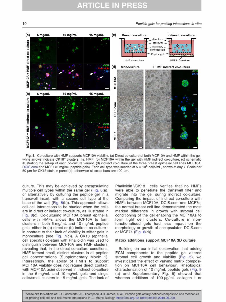
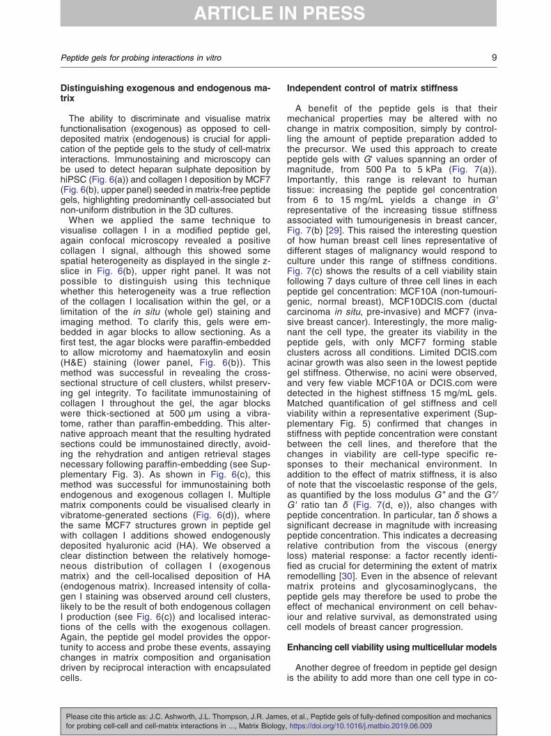
Fig. 8. Co-culture with HMF supports MCF10A viability. (a) Direct co-culture of both MCF10A and HMF within the gel,white arrows indicate CK18− clusters, i.e. HMF, (b) MCF10A within the gel with HMF indirect co-culture, (c) schematicillustrating the set-up of each co-culture variant, (d) indirect co-culture of the three breast epithelial cell lines MCF10A,DCIS.com and MCF7 (6 mg/mL peptide gels). Each cell type was seeded at 5 × 10⁵ cells/mL, shown at day 7. Scale bar50 μm for CK18 stain in panel (d), otherwise all scale bars are 100 μm.
10 Peptide gels for probing interactions in vitro
culture. This may be achieved by encapsulatingmultiple cell types within the same gel (Fig. 8(a))or alternatively by culturing the peptide gel in atranswell insert, with a second cell type at thebase of the well (Fig. 8(b)). This approach allowscell-cell interactions to be studied when the cellsare in direct or indirect co-culture, as illustrated inFig. 8(c). Co-culturing MCF10A breast epithelialcells with HMFs allows the MCF10A to formclusters in both 6 mg/mL and 10 mg/mL peptidegels, either in (a) direct or (b) indirect co-culture -in contrast to their lack of viability in stiffer gels inmonoculture (see Fig. 7(c)). A CK18 (epithelialcell specific) co-stain with Phalloidin was used todistinguish between MCF10A and HMF clusters,revealing that, in the direct co-culture conditions,HMF formed small, distinct clusters in all peptidegel concentrations (Supplementary Movie 1).Interestingly, the ability of HMFs to supportMCF10A viability does not require direct contact,with MCF10A acini observed in indirect co-culturein the 6 mg/mL and 10 mg/mL gels and singlecells/small clusters in 15 mg/mL gels. The lack of
Please cite this article as: J.C. Ashworth, J.L. Thompson, J.R. Jamesfor probing cell-cell and cell-matrix interactions in ..., Matrix Biology
Phalloidin+/CK18− cells verifies that no HMFswere able to penetrate the transwell filter andmigrate into the gel during indirect co-culture.Comparing the impact of indirect co-culture withHMFs between MCF10A, DCIS.com and MCF7s,the normal breast cell line demonstrated the mostmarked difference in growth with stromal cellconditioning of the gel enabling the MCF10As toform tight cell clusters. Co-culture in non-functionalised gels had less impact on themorphology or growth of encapsulated DCIS.comor MCF7s (Fig. 8(d)).
Matrix additions support MCF10A 3D culture
Building on our initial observation that addingECM components to the peptide gel alteredstromal cell growth and viability (Fig. 5), weinvestigated the effect of varying matrix composi-tion on MCF10A cell behaviour. Rheologicalcharacterisation of 10 mg/mL peptide gels (Fig. 9(a) and Supplementary Fig. 6) showed thatwhereas additions of 100 μg/mL collagen I or
, et al., Peptide gels of fully-defined composition and mechanics, https://doi.org/10.1016/j.matbio.2019.06.009
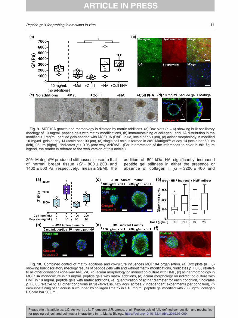
Fig. 9. MCF10A growth and morphology is dictated by matrix additions. (a) Box plots (n = 6) showing bulk oscillatoryrheology of 10 mg/mL peptide gels with matrix modifications, (b) immunostaining of collagen I and HA distribution in themodified 10 mg/mL peptide gels seeded with MCF10A (DAPI, blue, scale bar 50 μm), (c) acinar morphology in modified10 mg/mL gels at day 14 (scale bar 100 μm), (d) single cell acinus formed in 20% Matrigel™ at day 14 (scale bar 50 μm(left), 25 μm (right)). *indicates p b 0.05 (one-way ANOVA). (For interpretation of the references to color in this figurelegend, the reader is referred to the web version of this article.)
11Peptide gels for probing interactions in vitro
20% Matrigel™ produced stiffnesses closer to thatof normal breast tissue (G' = 800 ± 200 and1400 ± 500 Pa respectively, mean ± SEM), the
Fig. 10. Combined control of matrix additions and co-cultushowing bulk oscillatory rheology results of peptide gels with anto all other conditions (one-way ANOVA), (b) acinar morphologMCF10A monoculture in 10 mg/mL peptide gels with matrix aHMF in 10 mg/mL peptide gels with matrix additions, (e) quanp b 0.05 relative to all other conditions (Kruskal-Wallis, N25 aimmunostaining of an acinus surrounded by collagen I matrix inI. Scale bar 50 μm.
Please cite this article as: J.C. Ashworth, J.L. Thompson, J.R. Jamesfor probing cell-cell and cell-matrix interactions in ..., Matrix Biology
addition of 804 kDa HA significantly increasedpeptide gel stiffness in either the presence orabsence of collagen I (G' = 3200 ± 400 and
re influences MCF10A organisation. (a) Box plots (n = 6)d without matrix modifications, *indicates p b 0.05 relativey on indirect co-culture with HMF, (c) acinar morphology indditions, (d) acinar morphology on indirect co-culture withtification of acinar diameter for each condition, *indicatescini across 2 independent experiments per condition), (f)a 10 mg/mL peptide gel modified with 200 μg/mL collagen
, et al., Peptide gels of fully-defined composition and mechanics, https://doi.org/10.1016/j.matbio.2019.06.009
12 Peptide gels for probing interactions in vitro
3000 ± 700 Pa respectively). Interestingly, pep-tide gels with HA modifications alone showedbehaviour closer to an elastic solid than the othermodified conditions, as characterised by a signif-icant decrease in tan δ (Supplementary Fig. 6(b)).Immunostaining (or use of an HA binding peptide)enables visualisation of the matrix componentsadded to the gel, demonstrating that both compo-nents are homogeneously distributed throughoutthe gel (Fig. 9(b)).In contrast to the lack of growth observed in the
unmodified peptide gel, MCF10A 3D growth andthe formation of cell clusters was supported in allmodified conditions (Fig. 9(c)). However, only the20% Matrigel™ condition was able to promoteformation of the classic polarised single-cell layeracini typically observed in 100% Matrigel™(Supplementary Fig. 7), with peripheral collagenIV deposition (indicative of a nascent basementmembrane) by day 14 (Fig. 9(d)). This highlightsthat additional or alternative influences, beyondfunctionalisation with collagen I and HA, arerequired for this complex cell behaviour, inagreement with previously published studies[31,32].We next investigated how independent control
of matrix stiffness, composition and co-cultureconditioning could alter MCF10A organisation.Functionalisation with collagen I significantlydecreased the stiffness of a 10 mg/mL peptidegel (Fig. 10(a) and Supplementary Fig. 6).Therefore, a 6 mg/mL peptide gel with equalstiffness to the collagen-containing conditionswas included as a matrix-free control. After14 days of culture, immunostaining revealed thatneither indirect co-culture with HMFs (Fig. 10(b))nor the presence of collagen I (Fig. 10(c)) wassufficient to produce organised acinar structure,indicated here by the lack of focused cleavedcaspase 3 staining in the core of MCF10Aclusters.However, where applied in combination,collagen I and HMF co-culture produced largeacini, with evidence in the 100 μg/mL collagen Icondition of organised cleaved caspase 3 stain-ing, an early stage of lumen formation (Fig. 10(d)).Quantification of acinar diameter (Fig. 10(e))revealed that HMF co-culture produced a signifi-cant increase in acinar size in peptide gelscontaining 100 μg/mL collagen I. A significantincrease in diameter was also observed onincreasing collagen concentration from 100 to200 μg/mL, although the acini formed in the200 μg/mL condition were dense with no centrallumen, as shown in Fig. 10(b).Interestingly, although it is clear that additional
or alternative matrix functionalisation is requiredto promote further acini maturation, the MCF10Aswere able to organise and re-engineer theirsurrounding matrix, indicating reciprocal interac-
Please cite this article as: J.C. Ashworth, J.L. Thompson, J.R. Jamesfor probing cell-cell and cell-matrix interactions in ..., Matrix Biology
tions between the encapsulated cells and theirlocal environment. After 14 days MCF10A culturein a peptide gel containing 200 μg/mL collagen I,Fig. 10(f), the epithelial cells appear to distort thecollagen, with collagen excluded entirely from thecell cluster and surrounding the acinus at itsperiphery.
Experimental procedures
Cell line maintenance
The human mammary fibroblast cell line HMFU19(a gift from Professor Mike O'Hare, Ludwig Institute,London, UK), leukemia cell line U937 (DSMZGmBH) and colorectal cancer line mCherry-HCT116 (a gift from Prof. Anna Grabowska, Univer-sity of Nottingham) were cultured in HMF cell culturemedium: RPMI-1640 with 10% fetal bovine serum(FBS) and 1% L-glutamine. The murine embryonicstem cell line E14TG2a and Oct4-GFP reporter line(both gifts from Prof. Austin Smith, University ofCambridge, UK) were maintained on tissue cultureflasks coated with 0.1% gelatin (G1890 Sigma), inknockout Dulbecco's modified Eagle medium(DMEM) with 10% FBS (HyClone), 1% Non Essen-tial Amino Acids, 1% L-glutamine, 0.1% β mercap-toethanol, and leukemia inhibitory factor (LIF,ESG1107 Millipore). The breast cancer cell lineMCF7 and the modified tdTomato-MCF7 (a gift fromProf. Anna Grabowska, University of Nottingham)were maintained in high glucose DMEM with 10%FBS and 1% L-glutamine. The breast cell lineMCF10DCIS.com (Asterand) was cultured in ad-vanced DMEM with 5% horse serum and 1% L-Glutamine. MCF10A were maintained in DMEM/F12(D8062 Sigma) with 5% horse serum, 1% L-Glutamine, 10 μg/mL insulin (I9278 Sigma),0.5 mg/mL hydrocortisone (50237 Tocris), 20 ng/mL epidermal growth factor (ABC016 Source Bio-sciences) and 100 ng/mL cholera toxin (C8052Sigma). All cell lines were obtained from ATCCunless specified.
hiPSC culture and EB formation
For human induced pluripotent stem cell exper-iments (hiPSC), REBL-PAT (non-disease) cellswere used as established and characterisedpreviously [49]. hiPSC were maintained in Essen-tial 8 medium (E8) on tissue culture flasks coatedwith recombinant vitronectin peptide (VTN-N)following manufacturer's instructions. For hiPSCpassage, cells incubated with TrypLE Expresswere collected in E8 supplemented with 10 μM Y-27632 ROCK inhibitor (72304, Stem Cell Tech-nologies, UK). The hanging drop method used to
, et al., Peptide gels of fully-defined composition and mechanics, https://doi.org/10.1016/j.matbio.2019.06.009
13Peptide gels for probing interactions in vitro
generate embryoid bodies (EBs) from hiPSCs wasadapted from [50]. hiPSCs were harvested 48 hafter seeding and resuspended in E8 with 10 μMY-27632 and 4 mg/mL polyvinyl alcohol (Sigma,UK). 20 μL droplets containing 2000 cells/dropletwere pipetted onto the lid of a 10 cm petri dishcontaining 10 mL PBS to maintain hydration. TheEBs were formed for 24 h at 37 °C, then collectedin DMEM. EBs were allowed to sediment at thebottom of a 15 ml falcon tube for 10–15 min at37 °C and were subsequently cultured in peptidegels maintained in Essential 6 medium (E6) toallow spontaneous differentiation.All cell lines were maintained in antibiotic-free
conditions, at 37 °C and 5% CO2 in a humidifiedatmosphere. All media components were obtainedfrom Gibco, UK unless specified.
Precursor formation
A commercially available peptide preparation inpowder form was used as the source of theoctapeptide gelator (FEFEFKFK, Phe-Glu-Phe-Glu-Phe-Lys-Phe-Lys). As part of this study we usedpeptide sourced from Cambridge Research Bio-chemicals (batch 32597) although we also verifiedthe fabrication method using a second peptidesource (Pepceuticals, UK). To form each precursor,a mass of between 7.5 and 18.75 mg peptidepreparation was dissolved in 800 μL sterile water(W3500 Sigma), using a 3 min vortex step followedby centrifugation (3 min at 1000 rpm) and a 2 hincubation at 80 °C. After incubation, 0.5 M NaOH(S2770 Sigma) was added incrementally to the gelsuntil optically clear. Gels were vortexed, buffered byaddition of 100 μL 10× PBS (70011 Gibco), andincubated at 80 °C overnight. The resulting precur-sors could be stored at 4 °C until required.
Peptide gel formation
Prior to peptide gel formation, each precursor washeated at 80 °C until liquid to ensure homogeneity,before transferring to a 37 °C water bath. Peptide gelformation was then induced by pH neutralisation onaddition of cell culture medium. A final volume of1.25 mL was obtained from each preparation, byadding 250 μL of cell culture medium to a precursorvolume of 1 mL. The end concentration of peptidepreparation therefore ranged between 6 and 15 mg/mL. Medium was thoroughly mixed with the precursorby gentle (reverse) pipetting, before plating at 100 μLper well into a 96-well plate, or at 200 μL per well intoa hanging insert within a 24-well plate (MCRP24H48Millipore). The wells were then flooded with cellculture medium and incubated at 37 °C and 5% CO2in a humidified atmosphere. Sequential media chang-es (at least two) over the next 24 h ensured completeneutralisation and therefore gelation.
Please cite this article as: J.C. Ashworth, J.L. Thompson, J.R. Jamesfor probing cell-cell and cell-matrix interactions in ..., Matrix Biology
For cell encapsulation, the 250 μL volume of cellculture medium was prepared as a cell suspensionat 5× the intended final seeding density, to allow forthe dilution factor on mixing with the precursor.Trypsin-EDTA (0.25%), or TrypLE Express in thecase of REBL-PAT, was used to detach all adherentcell lines from 2D culture at sub-confluence. Cellswere resuspended in 250 μL cell culture medium at adensity between 2.5 × 10⁵ and 5 × 10⁶ cells/mL,giving final seeding densities in the peptide gelbetween 5 × 10⁴ and 1 × 10⁶ cells/mL.
Peptide gel formation with matrix modifications
Modified peptide gels were created using themethod above, by incorporating matrix additionsinto the 250 μL volume added to the precursor. Forcollagen I additions, rat tail collagen I (A10483Gibco) was neutralised directly before use with 1 MNaOH according to manufacturer instructions, anddiluted with sterile water and 10× PBS to aconcentration of 0.5–1.5 mg/mL. For hyaluronicacid (HA) additions, streptococcal HA polymer withmolecular weight 804 kDa (HA804 Iduron) wasreconstituted in PBS at 0.5 mg/mL and sterilisedusing a 0.2 μm syringe filter. Corning Matrigel™(354234 Fisher Scientific) was used for the 20%Matrigel™ condition. All matrix preparations werekept on ice. Modified peptide gels were created bypreparing a 250 μL volume containing each matrixcomponent at 5× the desired final concentration(diluted with cell culture medium if necessary), andmixing with 1 mL precursor as described above.Cells were incorporated into this 250 μL volume at5× the desired final seeding density as described.
Matrigel™ and collagen gels
Neutralised rat tail collagen I was prepared at1.5 mg/mL as described above, and plated at200 μL per well into a 24-well plate hanging insert.Corning Matrigel™ was plated in the same way. Allsolutions were kept on ice during use. To seed cellsinto these gels, a cell pellet was prepared andsuspended in either the neutralised collagen solutionor in pure Matrigel™, giving a final seeding density of5 × 105 cells/mL. Matrigel™ and collagen gels wereincubated at 37 °C and 5% CO2 in a humidifiedatmosphere for 30 min to allow gelation, beforeflooding the wells with cell culture medium.
Bulk oscillatory rheology
Peptide gel samples were prepared for bulkrheology as described above, by plating at 200 μLper well into 24-well plate hanging inserts, andincubating overnight as described above. At day 1after seeding, samples were removed from theinserts with a scalpel and mounted onto a Physica
, et al., Peptide gels of fully-defined composition and mechanics, https://doi.org/10.1016/j.matbio.2019.06.009
14 Peptide gels for probing interactions in vitro
MCR 301 rheometer (Anton Paar) with Peltier plateset to 37 °C. The linear viscoelastic region wasdetermined for each sample condition by carryingout an amplitude sweep from 0.1 to 100% strain at1 rad/s. Following this, a constant strain of 0.5% wasused to obtain frequency sweeps from 0.1 to100 rad/s, as well as 5 min time sweeps at aconstant frequency of 1 rad/s. The same testswere carried out on the precursor samples, whichcould be pipetted directly onto the rheometer plate.All tests were carried out using an 8 mm diameterparallel plate set-up with a spacing of 1 mm.
Microrheology
The microrheological properties of both precur-sor and peptide gel were tested by measuring theBrownian motion of 2 μm diameter polystyrenebeads (19814 Polysciences) embedded into thesamples. For the precursor, beads were sus-pended at a final concentration of 2 × 10⁵ beads/mL by incorporating them with the addition of 10×PBS. This relatively low bead concentration waschosen to avoid clustering and to ensure that onlyone bead was present in the field of view for theduration of the experiment. After a standardovernight incubation at 80 °C, precursor sampleswere equilibrated for 1 h at 37 °C and 5% CO2 ina humidified atmosphere prior to transfer into an8-well coverslip (80821 IBIDI) at 200 μL/well fortesting. For the peptide gel, beads were added tothe gel by suspension into the 250 μL of cellculture medium used for neutralisation, at a finalconcentration of 2 × 10⁵ beads/mL. Peptide gelswere seeded into the 8-well coverslips andincubated at 37 °C and 5% CO2 in a humidifiedatmosphere overnight. The beads were imaged inwide-field transmission with a 100× oil-immersionobjective lens (numerical aperture = 1.3) using aninverted microscope (Eclipse Ti-S, Nikon) and aCMOS camera (Optimos, QImaging). The trajec-tories of 5 individual beads per condition weretracked using a centre of mass algorithm. Foreach bead at least 50,000 frames were recordedat a frame rate of 600 frames per second(exposure time 1500 μs), this high frame ratewas achieved by imaging a small region ofinterest. For some measurements, the number offrames was limited by the bead diffusing out of thefield of view (for such gels the adoption of arelatively low laser power for optically trapping thebead of interest could be considered in futurestudies). In-house LabVIEW programs (LabVIEW,2013, National Instruments, USA) were used for(i) bead tracking, (ii) trajectory conversion to meansquared displacement, and (iii) for extracting thecomplex viscosity of the gel immediately sur-rounding the beads [51]. The experimental set-upwas verified by taking control measurements of
Please cite this article as: J.C. Ashworth, J.L. Thompson, J.R. Jamesfor probing cell-cell and cell-matrix interactions in ..., Matrix Biology
the viscosity of water (Supplementary Fig. 1). Forthese experiments, a 1064 nm continuous wavelaser (Ventus 1064, Laser Quantum Ltd., UK)operating at spatial Gaussian mode (TEM00) wasused to optically trap beads suspended in waterwith a laser power b5 mW. 500,000 frames wererecorded at a frame rate of 600 fps.
Live cell imaging and detection
Fluorescence signal from peptide gels containingfluorescently labelled cells was detected using aFluostar Omega Plate Reader (BMG LabTech). AnEclipse TI-S microscope (Nikon) was used for brightfield imaging during culture. For quantification ofcell cluster diameter, all regions containing cellclusters were imaged and Fiji software was used formanual diameter measurement [52]. For LIVE/DEAD staining, peptide gels were washed withPBS, removed from their hanging inserts, andincubated for 15 min in a solution of 40 μMEthidiumhomodimer and 20 μM calcein AM (L3224 Fisher) inPBS. A Leica TCS SPE laser scanning confocalmicroscope was used for acquisition of fluores-cence images.
Immunofluorescence staining
After washing in PBS, and removal fromhanging inserts if necessary, peptide gels wereincubated for 1 h in paraformaldehyde (Poly-sciences) diluted to 4% (v/v) in PBS. Sampleswere washed in PBS in preparation for immuno-fluorescence staining. Samples were incubated inblocking buffer, consisting of 0.1% Triton X-100and 0.5% bovine serum albumin (Sigma) for 1 h,and incubated overnight at 4 °C with a solution ofprimary antibody in blocking buffer: Oct4 (83932Cell Signalling Technologies (CST), 1:400), CK18(53981582 Thermofisher, 1:50–1:100), rabbit IgG(PP64, Chemicon International, 1:1000), pFAK(Tyr397, 44-624G Thermofisher, 1:100), collagenI (ab34710 AbCam, 1:100–1:500), collagen IV(ab6311 AbCam, 1:200), cleaved caspase 3(9661 CST, 1:400) or CD44 (3570 CST, 1:400).After further washes in blocking buffer, sampleswere incubated overnight at 4 °C with a solutionof secondary antibody in blocking buffer (a21042/a21050/a11010/a11029/a11034 Invitrogen,1:400). For HA staining, biotinylated hyaluronicacid binding protein (bHABP, AMS.HKD-BC41AMSBio, 1:100) was added with the primaryantibody, and TRITC-streptavidin (Stratech) withthe secondary antibody. Samples were incubatedin a 300 nM DAPI solution (D3571 Invitrogen) for1 h at room temperature prior to imaging. Forheparan sulphate staining (10e4, 370255-1 AMS-Bio, 1:100), 10% goat serum (Sigma) in PBS wassubstituted as blocking buffer, with Hoechst
, et al., Peptide gels of fully-defined composition and mechanics, https://doi.org/10.1016/j.matbio.2019.06.009
15Peptide gels for probing interactions in vitro
substituted in place of DAPI counterstain. WherePhalloidin was used for F-Actin staining (F432/R415, Thermofisher, 1:1000), this was addedeither alone or with the secondary antibody, asa solution in blocking buffer as described above.
Embedding and sectioning
Peptide gels were embedded in a 2–4% solution ofagar (SLS) in distilled water, and set for severalhours at 4 °C prior to further processing. Agar blockswere sectioned using a Leica Vibratome at athickness of 500 μm. Agar slices were stored inPBS, and were stained using the same immunoflu-orescence techniques described above. Alternative-ly, the agar blocks were transferred to a tissueprocessor and set in a paraffin block. 10 μm sliceswere sectioned onto SuperFrost slides (ThermoScientific, UK) using a microtome. After drying at37 °C, slides were dewaxed in xylene and rehy-drated in an ethanol series to allow staining withHaematoxylin and Eosin (3 min each). Slides werewashed in running water at each stage, andincubated for 20 s in acid alcohol and 1 min inScott's tap water between stains. Slides weredehydrated, cleared in xylene, and coverslippedusing DPX mounting medium (Thermo Scientific,UK).
Quantitative reverse transcription PCR (qRT-PCR)
Total RNA was extracted and purified from cellswithin peptide gels using the Nucleospin RNA kit(Machery Nagel) according to the manufacturer'sinstructions. 700 μg of RNA was reverse-transcribedto cDNA using SuperScript™ IIIReverse Transcrip-tase, following the manufacturer's instructions. TheGoTaq qPCRMaster Mix (Promega) was used to runthe PCR reaction on a LightCycler® 480 (Roche).Human RPLPO and HSP90AB1 were used asreference genes for normalisation and hiPSCscultured in 2D were used to calculate relativeexpression using the ΔΔCt method. Primers usedare listed in Supplementary Table 1.
Statistics
Prism v.7.0d and SPSS v.24 (IBM) were used forstatistical analysis. One-way or two-way ANOVAwith Tukey HSD post-hoc test were used asappropriate where measurements were normallydistributed, verified by the Shapiro-Wilk test. In theone case where the data were not normallydistributed (the acini diameter measurements inFig. 10), a non-parametric Kruskal-Wallis test wasused with Bonferroni correction for multiple compar-isons. Statistical significance for all tests wasdeclared at p b 0.05.
Please cite this article as: J.C. Ashworth, J.L. Thompson, J.R. Jamesfor probing cell-cell and cell-matrix interactions in ..., Matrix Biology
Discussion
An increasing awareness of the importance ofmatrix components in regulating cell behaviour hasnecessitated the continued improvement of in vitro3D models of human tissue. As recently highlightedin a high-profile technology feature, the ECMgoverns a surprising number of cellular functions,which must be adequately modelled in vitro to betterunderstand development, differentiation and theprogression to disease [1]. A major hurdle to thishas been unpicking the multitude of influencesexerted by the matrix on neighbouring cells. Theself-assembled peptide gel presented here allowsthe independent control of two critical factors: matrixcomposition and bulk stiffness. This in turn facilitatesthe application of customised (“bespoke”) gels tomimic multiple matrix microenvironments tailored forspecific applications.Self-assembling peptide gels are intermediates
between natural and synthetic materials; chemicallysynthesised, but formed from natural building blocks,with a biomimetic, fibrillar nanostructure [23,33].Here, the octapeptide gelator sequence FEFEFKFKwas chosen due to the high biocompatibility dem-onstrated by gelation at pH 7 at 37 °C [15]. Previousstudies have explored the application of similar gelsfor regenerative medicine applications [15,34] how-ever, to our knowledge they have not yet been usedto create fully-defined matrices for cell encapsula-tion. By exploiting the pH-dependent viscosity of thegels, we could physically incorporate cells and/ormatrix components into the precursor, and theseremained homogeneously distributed on finalgelation.Previously, decoupling of matrix stiffness and
composition to detect their respective influence oncell behaviour has often been conducted in thepresence of a complex matrix environment. Forinstance, ribose-mediated collagen cross-linkingwas used to increase the stiffness of a collagen-rBM (reconstituted basement membrane) compos-ite, demonstrating that ErbB2 signalling was neces-sary to promote mammary epithelial invasion in stiffmatrices [9]. In an alternative approach, rBM wascombined with alginate gels to determine theinterplay between matrix stiffness and rBM concen-tration, with greater epithelial cell malignancy ob-served in stiffer matrices only when rBMconcentration was held constant [18]. A similarlyelegant approach combined collagen with metha-crylated gelatin, allowing independent control ofcollagen concentration and matrix stiffness [35].Using this system, the authors discovered thatMDA MB 231 breast cancer cell invasion was bestsupported by matrices with low stiffness but highcollagen concentration. By using a non matrix-derived self-assembling peptide gel as a startingpoint, the method described here is distinct from
, et al., Peptide gels of fully-defined composition and mechanics, https://doi.org/10.1016/j.matbio.2019.06.009
16 Peptide gels for probing interactions in vitro
these approaches in that it not only allows indepen-dent control of matrix and mechanical properties, butalso allows the inclusion of selected matrix compo-nents, specific for the application. This flexibility ispermitted by the two-stage fabrication method; thefirst stage creates a matrix-free precursor to definethe stiffness, the second stage defines thecomposition.We were able to demonstrate the increase in
viscosity during gelation of the matrix-free gel usingtwo distinct measurement techniques: bulk oscilla-tory rheology, and microrheological measurementsbased on the Brownian motion of micron sizedbeads embedded in the gel. Indeed, despite theabsolute measurement values obtained from the twomethods differing by approximately an order ofmagnitude (likely due to the different length scalesexplored by the two techniques), the relativeincrease in viscosity on gelation was found to bevery similar between the two experimental proce-dures. Nonetheless, bulk rheology is a well-established experimental method, allowing us tocompare the viscoelastic properties of the peptidegel with those of other established 3D cell cultureplatforms. Whereas naturally derived gels, such asMatrigel™/rBM and collagen tend to be far less stiffthan the tissues they are used to mimic [23], we areable to control the peptide gel storage modulus in therange 500–5000 Pa, which covers a wide range of invivo tissue stiffnesses, such as brain and breast[23,29,36]. It is important to note, however, that bulkrheology is not well-suited to measuring cell-inducedchanges in gel mechanical properties during culture.This is because it cannot measure local materialchanges at the length scale of a single cell.Therefore, we are developing a novel microrheologymethod, combined with the ability to optically trap abead in the case of low modulus gels [37–39], tomeasure the gels' mechanical properties at a cell-scale; particularly at the cell-matrix interface, as cellsre-engineer their microenvironment.In good agreement with our current understanding
of cell-matrix interactions, anchorage- independentcells proliferate within unmodified peptide gels, withthe lack of matrix attachment motifs effectivelyenabling the cells to form structures similar tothose seen in suspension culture. Adhesive celltypes, such as fibroblasts, additionally require matrixcomponents, such as collagen I, to achieve theircharacteristic morphology. Importantly, the gel for-mulation method presented here allows for biochem-ical functionalisation, whilst also providing controlconditions with matched peptide gel stiffness.Although cell adhesion motifs have classicallybeen considered necessary for interactions betweenmatrix stiffness and cell behaviour [40], we have alsoshown a clear link between stiffness and cellresponse in the absence of cell binding sequences.It has recently been demonstrated that cells encap-
Please cite this article as: J.C. Ashworth, J.L. Thompson, J.R. Jamesfor probing cell-cell and cell-matrix interactions in ..., Matrix Biology
sulated in 3D materials rapidly synthesise their ownmatrix, with initial matrix stiffness implicated as a keyfactor determining the extent of this early matrixdeposition [30,41]. Importantly, the peptide gelsallow independent assessment of the effects ofbiochemical functionalisation and of the mechanicalenvironment initially presented to encapsulatedcells.In trying to create artificial culture environments
where the control of biochemical and physicalproperties is required, researchers often combinenaturally derived and synthetic elements. This canbe seen in the chemical modification of naturalmaterials, e.g. hyaluronic acid with thiol modifica-tions to allow cross-linking, or in the incorporation ofbiological components into synthetically producedhydrogels [23] as well as the current study. Aparticularly successful approach is that taken bythe Lutolf group, functionalising synthetic polyethyl-ene glycol (PEG) hydrogels with laminin-111 toproduce highly complex tissue models, including thehuman intestine [13]. Matrix remodelling by encap-sulated cells can be regulated and reported by theinclusion of matrix metalloproteinase (MMP) cleav-able cross-links or reporters [23]. Within the peptidegels, encapsulated cells appear to readily organiseand remodel their surrounding matrix without theneed to engineer in specific cleavage sites. Theability to image both endogenous and exogenousmatrix within the fully-synthetic system is a signifi-cant advantage here and will enable monitoring ofmatrix reorganisation as cells grow, differentiate andmigrate. Another feature of importance to cellbiologists is the optical transparency of the peptidegel that enables simple assessment of cell growth inreal-time by microscopy. The ability to use automat-ed plate readers to read-out endogenous fluores-cence as wel l as end-point analysis byimmunostaining and fluorescent microscopy is likelyto be useful for high-throughput analyses andapplications such as toxicity screening or biomarkeridentification.The ability to investigate the impact of stromal
cells in co-culture with epithelial cells is particu-larly valuable when studying the microenviron-mental control of cancer initiation, growth andmetastasis. Bidirectional cross-talk is thought topromote cancer progression, with exosomal-mediated signalling between neighbouring cellslikely to play an important role [42–44]. Stromalcells can additionally alter the hormone-dependence of nearby epithelia: for instanceestrogen treatment of uterine epithelial cellsincreased their proliferation only when in culturewith stromal cells [45]. For some effects, directcell contact appears to be unnecessary, with theexposure of epithelial cancer cells to stromal-conditioned media sufficient to alter their sensitiv-ity to chemotherapy and radiation [46]. In the
, et al., Peptide gels of fully-defined composition and mechanics, https://doi.org/10.1016/j.matbio.2019.06.009
17Peptide gels for probing interactions in vitro
current study, we were able to demonstratedifferences in the behaviour of encapsulatedcells when in the presence of direct or indirectstromal cell co-culture, with the flexibility of theculture system providing a useful test environmentin which to study the regulation of cancer cells bytheir microenvironment.As demonstrated in multiple studies, a benefit of
using short peptides to create hydrogels for cellencapsulation is the ability to simply and cheaplyengineer in covalently immobilised peptidic function-al motifs e.g. matrix-derived cell adhesion se-quences (RGD, IKVAV etc.) A less reductionistapproach can also be achieved by functionalisationwith complex sequences, combining cell attachmentand proteolytic motifs [47]. These approaches haveclear benefits for mechanistic investigations andadditionally highlight the potential for using peptidegels to move to fully synthetic xeno-free, matrix-inspired 3D culture. In the current study, we chose toincorporate full-length proteins and glycans toenable the visualisation of cell-mediated changesin matrix organisation, detailing cell contraction andimmunocytochemical imaging of matrix organisationand synthesis. Importantly, by using “naked” matrixfree gels, we can also detail matrix deposition byencapsulated cells. This is particularly valuablewhen studying glycans such as heparan sulphatewhere the conservation of structure between speciesmakes it impossible to differentiate glycosaminogly-cans deposited by encapsulated (human) cells fromthose present in complex animal-derived matrices(e.g. Matrigel™). Cell-deposited matrices havethemselves been used for 3D culture [5] and thestudy of differential ECM deposition under differentconditions, such as stromal activation in the pres-ence of cancer cells [48] is increasingly studied toidentify potential targets for novel therapeutic strat-egies. The use of indirect stromal cell co-culture toeffectively condition the peptide gels will hopefullyprove useful in these studies.
Conclusions
In summary, we present the optimisation of awell-established, simple and relatively inexpen-sive peptide gel for the study of cell-matrixinteractions in a wide variety of cell types. Byeliminating or significantly reducing the need foranimal-derived components e.g. Matrigel™, thissynthetic gel also helps researchers move awayfrom the batch-to-batch variability associated withtheir use, and addresses the need to replace,refine and reduce the use of animals in research.The cell encapsulation protocol has been specif-ically designed to ensure that reliable, reproduc-ible 3D culture is achievable within a standard cellculture laboratory setting with independent control
Please cite this article as: J.C. Ashworth, J.L. Thompson, J.R. Jamesfor probing cell-cell and cell-matrix interactions in ..., Matrix Biology
of the biochemical and mechanical influences ofthe matrix microenvironment. In this study, as wellas demonstrating broad applicability across mul-tiple adhesive and non-adhesive cell types, wehave demonstrated how the peptide gel can beapplied to unpick the role of extracellular regula-tion on the behaviour of cell lines used to modelthe progression from normal breast to breastcancer. We hope the peptide gels will be ofinterest to the matrix biology community, with theoptimised protocol and commercially availableprecursors ensuring that the technology is ap-proachable for any cell culture laboratory.Supplementary data to this article can be found
online at https://doi.org/10.1016/j.matbio.2019.06.009.
CRediT authorship contribution state-ment
J.C. Ashworth:Conceptualization, Formal analy-sis, Investigation.J.L. Thompson:Investigation.J.R.James:Investigation.C.E. Slater:Investigation.S.Pijuan-Galitó:Supervision.K. Lis-Slimak:Investiga-tion.R.J. Holley:Conceptualization.K.A. Meade:Conceptualization.A. Thompson:Supervision.K.P.Arkill:Methodology, Supervision.M. Tassieri:For-mal analysis.A.J. Wright:Conceptualization, Formalanalysis, Funding acquisition.G. Farnie:Conceptu-alization, Funding acquisition, Writing - review &editing.C.L.R. Merry:Conceptualization, Fundingacquisition, Writing - review & editing.
Acknowledgements
The research described was funded by theNational Centre for the Replacement, Refinementand Reduction of Animals in Research (NC3Rs)grant NC/N001583/1 (J.A., G.F., C.L.R.M.), andsupported by the EPSRC/BBSRC/MRC (Engi-neering and Physical Sciences Research Coun-cil /Biotechnology and Biological SciencesResearch Council/Medical Research Council)joint grants EP/R035067/1, EP/R035156/1 andEP/R035563/1 (M.T., A.J.W., C.L.R.M.), MRCgrant MR/P003214/1 (K.P.A.), EPSRC/MRC CDTRegenerative Medicine and Faculty of Medicaland Health Sciences (J.R.J., A.T., A.J.W., C.L.R.M.), EPSRC grant EP/N006615/1 (J.L.T., C.L.R.M.) and Swedish Research Council grant 2015-06532 (S.P.G.). We thank Robin Ketteler andAlexander Agrotis for technical help and the MRCfor support of the LMCB High-Content BiologyLaboratory at University College London(MC_U12266B, MR/M02492X/1). We thank Anna
, et al., Peptide gels of fully-defined composition and mechanics, https://doi.org/10.1016/j.matbio.2019.06.009
18 Peptide gels for probing interactions in vitro
Grabowska and Pam Collier (University of Not-tingham) for their kind gift of the labelled HCT116-mCherry and MCF7-td Tomato cell lines and foradvice on real-time cell assays, and Tony Day(University of Manchester) for helpful advice anddiscussions.
Received 27 March 2019;Received in revised form 28 May 2019;
Accepted 24 June 2019Available online xxxx
Keywords:Biomaterials;
Cancer;Stem cells;
Extracellular matrix;Stiffness
References
[1] J. Madhusoodanan, Matrix mimics shape, Nature 566 (2019)563–565, https://doi.org/10.1038/d41586-019-00681-1.
[2] R. Cruz-Acuña, A.J. García, Synthetic hydrogels mimickingbasement membrane matrices to promote cell-matrix inter-actions, Matrix Biol. 57-58 (2017) 324–333, https://doi.org/10.1016/j.matbio.2016.06.002.
[3] L. David, V. Dulong, D. Le Cerf, C. Chauzy, V. Norris, B.Delpech, M. Lamacz, J.-P. Vannier, Reticulated hyaluronanhydrogels: a model for examining cancer cell invasion in 3D,Matrix Biol. 23 (2004) 183–193, https://doi.org/10.1016/j.matbio.2004.05.005.
[4] B.M. Baker, C.S. Chen, Deconstructing the third dimension-how3D culture microenvironments alter cellular cues, J. Cell Sci. 125(13) (2012) 3015–3024, https://doi.org/10.1242/jcs.079509.
[5] E. Cukierman, R. Pankov, D.R. Stevens, K.M. Yamada,Taking cell-matrix adhesions to the third dimension, Science294 (5547) (2001) 1708–1712, https://doi.org/10.1126/science.1064829.
[6] M.J. Bissell, A. Rizki, I.S. Mian, Tissue architecture: the ultimateregulator of breast epithelial function, Curr. Opin. Cell Biol. 15 (6)(2003) 753–762, https://doi.org/10.1016/j.ceb.2003.10.016.
[7] S.R. Caliari, J.A. Burdick, A practical guide to hydrogels forcell culture, Nat. Methods 13 (5) (2016) 405–414, https://doi.org/10.1038/nmeth.3839.
[8] P.G. Buxton, M. Bitar, K. Gellynck, M. Parkar, R.A. Brown, A.M. Young, J.C. Knowles, S.N. Nazhat, Dense collagen matrixaccelerates osteogenic differentiation and rescues theapoptotic response to MMP inhibition, Bone 43 (2) (2008)377–385, https://doi.org/10.1016/j.bone.2008.03.028.
[9] K.R. Levental, H. Yu, L. Kass, J.N. Lakins,M.Egeblad, J.T. Erler,S.F.T. Fong, K. Csiszar, A. Giaccia,W.Weninger, M. Yamauchi,D.L. Gasser, V.M. Weaver, Matrix crosslinking forces tumorprogression by enhancing integrin signaling, Cell 139 (5) (2009)891–906, https://doi.org/10.1016/j.cell.2009.10.027.
[10] K. Wolf, S. Alexander, V. Schacht, L.M. Coussens, U.H. vonAndrian, J. van Rheenen, E. Deryugina, P. Friedl, Collagen-based cell migration models in vitro and in vivo, Semin. CellDev. Biol. 20 (8) (2009) 931–941, https://doi.org/10.1016/j.semcdb.2009.08.005.
Please cite this article as: J.C. Ashworth, J.L. Thompson, J.R. Jamesfor probing cell-cell and cell-matrix interactions in ..., Matrix Biology
[11] J. Debnath, S.K. Muthuswamy, J.S. Brugge, Morphogenesisand oncogenesis of MCF-10A mammary epithelial acinigrown in three-dimensional basement membrane cultures,Methods 30 (3) (2003) 256–268, https://doi.org/10.1016/S1046-2023(03)00032-X.
[12] K.C. Hansen, L. Kiemele, O. Maller, J. O'Brien, A. Shankar, J.Fornetti, P. Schedin, An in-solution ultrasonication-assisteddigestion method for improved extracellular matrix proteomecoverage, Mol. Cell. Proteomics 8 (7) (2009) 1648–1657,https://doi.org/10.1074/mcp.M900039-MCP200.
[13] N. Gjorevski, N. Sachs, A. Manfrin, S. Giger, M.E. Bragina, P.Ordóñez-Morán, H. Clevers, M.P. Lutolf, Designer matricesfor intestinal stem cell and organoid culture, Nat. Publ. Group539 (7630) (2016) 560–564, https://doi.org/10.1038/nature20168.
[14] N. Gjorevski, M.P. Lutolf, Synthesis and characterization of well-defined hydrogel matrices and their application to intestinal stemcell andorganoid culture,Nat. Protoc. 12 (11) (2017) 2263–2274,https://doi.org/10.1038/nprot.2017.095.
[15] A. Mujeeb, A.F. Miller, A. Saiani, J.E. Gough, Self-assembledoctapeptide scaffolds for in vitro chondrocyte culture, ActaBiomater. 9 (1) (2013) 4609–4617, https://doi.org/10.1016/j.actbio.2012.08.044.
[16] L. Rizzi, Microrheology of biological specimens, Encyclope-dia of Analytical Chemistry 2006, pp. 1–24, https://doi.org/10.1002/9780470027318.a9419.
[17] F. Del Giudice, M. Tassieri, C. Oelschlaeger, A.Q. Shen,When microrheology, bulk rheology, and microfluidics meet:broadband rheology of hydroxyethyl cellulose water solu-tions, Macromolecules 50 (7) (2017) 2951–2963, https://doi.org/10.1021/acs.macromol.6b02727.
[18] O. Chaudhuri, S.T. Koshy, C. Branco da Cunha, J.-W. Shin,C.S. Verbeke, K.H. Allison, D.J. Mooney, Extracellular matrixstiffness and composition jointly regulate the induction ofmalignant phenotypes in mammary epithelium, Nat. Mater.13 (June) (2014) 1–35, https://doi.org/10.1038/nmat4009.
[19] M.P. Lee, M.J. Padgett, D. Phillips, G.M. Gibson, M. Tassieri,Dynamic stereo microscopy for studying particle sedimenta-tion, Opt. Express 22 (4) (2014) 4671, https://doi.org/10.1364/oe.22.004671.
[20] M. Jaspers, M. Dennison, M. F. Mabesoone, F. C.MacKintosh, A. E. Rowan, P. H. Kouwer, Ultra-responsivesoft matter from strain-stiffening hydrogels, Nat. Commun. 5.doi:https://doi.org/10.1038/ncomms6808.
[21] C.P. Broedersz, K.E. Kasza, L.M. Jawerth, S. Münster, D.A.Weitz, F.C. MacKintosh, Measurement of nonlinear rheologyof cross-linked biopolymer gels, Soft Matter 6 (17) (2010)4120–4127, https://doi.org/10.1039/C0SM00285B.
[22] W. Megone, N. Roohpour, J.E. Gautrot, Impact of surfaceadhesion and sample heterogeneity on the multiscale mechan-ical characterisation of soft biomaterials, Sci. Rep. 8 (1) (2018)1–10, https://doi.org/10.1038/s41598-018-24671-x.
[23] J. Shan, Q. Chi, H. Wang, Q. Huang, L. Yang, G. Yu, X. Zou,Mechanosensing of cells in 3D gel matrices based on naturaland synthetic materials (2014). doi:https://doi.org/10.1002/cbin.10325.
[24] D.T. Butcher, T. Alliston, V.M. Weaver, A tense situation:forcing tumour progression, Nat. Rev. Cancer 9 (2) (2009)108–122, https://doi.org/10.1038/nrc2544.
[25] J. C. Ashworth, M. Mehr, P. G. Buxton, S. M. Best, R. E.Cameron, Optimising collagen scaffold architecture forenhanced periodontal ligament fibroblast migration, J.Mater. Sci. Mater. Med. 29. doi:https://doi.org/10.1007/s10856-018-6175-9.
, et al., Peptide gels of fully-defined composition and mechanics, https://doi.org/10.1016/j.matbio.2019.06.009
19Peptide gels for probing interactions in vitro
[26] L. Hagbard, K. Cameron, P. August, C. Penton, M. Parmar,D. C. Hay, T. Kallur, Developing defined substrates for stemcell culture and differentiation, Philos. Trans. R. Soc., B 373.doi:https://doi.org/10.1098/rstb.2017.0230.
[27] X. Lin, G. Wei, Z. Shi, L. Dryer, J.D. Esko, D.E. Wells, M.M.Matzuk, Disruption of gastrulation and heparan sulfatebiosynthesis in EXT1-deficient mice, Dev. Biol. 224 (2)(2000) 299–311, https://doi.org/10.1006/dbio.2000.9798.
[28] N. Smyth, M. Meyer, M. Paulsson, D. Edgar, P. Murray, H.S.Vatansever, C. Frie, Absence of basement membranes aftertargeting the LAMC1 gene results in embryonic lethality dueto failure of endoderm differentiation, J. Cell Biol. 144 (1)(2002) 151–160, https://doi.org/10.1083/jcb.144.1.151.
[29] I. Acerbi, L. Cassereau, I. Dean, Q. Shi, A. Au, C. Park, Y.Y.Chen, J. Liphardt, E.S. Hwang, V.M. Weaver, Human breastcancer invasion and aggression correlates with ECMstiffening and immune cell infiltration, Integr. Biol. 7 (10)(2015) 1120–1134, https://doi.org/10.1039/c5ib00040h.
[30] C. Loebel, R. L. Mauck, J. A. Burdick, Local nascent proteindeposition and remodelling guide mesenchymal stromal cellmechanosensing and fate in three-dimensional hydrogels,Nat. Mater. doi:https://doi.org/10.1038/s41563-019-0307-6.
[31] J. Muschler, C. H. Streuli, Cell-matrix interactions inmammary gland development and breast cancer, ColdSpring Harb. Perspect. Biol. 2 (2011) a003202. doi:https://doi.org/10.1101/cshperspect.a003202.
[32] G. Farnie, R.B. Clarke, K. Spence, N. Pinnock, K. Brennan, N.G.Anderson, N.J. Bundred, Novel cell culture technique for primaryductal carcinoma insitu: roleof notchandepidermalgrowth factorreceptor signaling pathways, J. Natl. Cancer Inst. 99 (8) (2007)616–627, https://doi.org/10.1093/jnci/djk133.
[33] A. Saiani, A. Mohammed, H. Frielinghaus, R. Collins, N.Hodson, C. M. Kielty, M. J. Sherratt, A. F. Miller, Self-assembly and gelation properties of α-helix versus β-sheetforming peptides, Soft Matter 5 (1) (2008) 193–202. doi:https://doi.org/10.1039/B811288F.
[34] S. Wan, S. Borland, S.M. Richardson, C.L. Merry, A. Saiani,J.E. Gough, Self-assembling peptide hydrogel for interverte-bral disc tissue engineering, Acta Biomater. 46 (2016) 29–40,https://doi.org/10.1016/j.actbio.2016.09.033.
[35] A.J. Berger, K.M. Linsmeier, P.K. Kreeger, K.S. Masters,Decoupling the effects of stiffness and fiber density oncellular behaviors via an interpenetrating network of gelatin-methacrylate and collagen, Biomaterials 141 (2017)125–135, https://doi.org/10.1016/j.biomaterials.2017.06.039.
[36] S. Budday, G. Sommer, J. Haybaeck, P. Steinmann, G.A.Holzapfel, E. Kuhl, Rheological characterization of humanbrain tissue, Acta Biomater. 60 (2017) 315–329, https://doi.org/10.1016/j.actbio.2017.06.024.
[37] M. Tassieri, Linear microrheology with optical tweezers ofliving cells'is not an option’! Soft Matter 11 (29) (2015)5792–5798, https://doi.org/10.1039/c5sm01133g.
[38] M. Tassieri, Microrheology with optical tweezers: peaks andtroughs, Curr. Opin. Colloid Interface Sci. (Accepted forPublication).
[39] F. Watts, L. E. Tan, C. G. Wilson, J. M. Girkin, M. Tassieri, A.J. Wright, Investigating the micro-rheology of the vitreoushumor using an optically trapped local probe, J. Opt. 16. doi:https://doi.org/10.1088/2040-8978/16/1/015301.
[40] J.S. Harunaga, K.M. Yamada, Cell-matrix adhesions in 3D,Matrix Biol. 30 (2011) 363–368, https://doi.org/10.1016/j.matbio.2011.06.001.
Please cite this article as: J.C. Ashworth, J.L. Thompson, J.R. Jamesfor probing cell-cell and cell-matrix interactions in ..., Matrix Biology
[41] S.A. Ferreira, M.S. Motwani, P.A. Faull, A.J. Seymour, T.T.Yu, M. Enayati, D.K. Taheem, C. Salzlechner, T. Haghighi, E.M. Kania, O.P. Oommen, T. Ahmed, S. Loaiza, K. Parzych, F.Dazzi, O.P. Varghese, F. Festy, A.E. Grigoriadis, H.W.Auner, A.P. Snijders, L. Bozec, E. Gentleman, Bi-directionalcell-pericellular matrix interactions direct stem cell fate, Nat.Commun. 9 (1) (2018) 1–12, https://doi.org/10.1038/s41467-018-06183-4.
[42] H. Fu, H. Yang, X. Zhang, W. Xu, The emerging roles ofexosomes in tumor-stroma interaction, J. Cancer Res. Clin.Oncol. 142 (2016) 1897–1907, https://doi.org/10.1007/s00432-016-2145-0.
[43] J. Webber, R. Steadman, M.D. Mason, Z. Tabi, A. Clayton,Cancer exosomes trigger fibroblast to myofibroblast differ-entiation, Cancer Res. 70 (23) (2010) 9621–9630, https://doi.org/10.1158/0008-5472.can-10-1722.
[44] V. Yeung, J.P. Webber, E.A. Dunlop, H. Morgan, J. Hutton, M.Gurney, E. Jones, J. Falcon-Perez, Z. Tabi, R. Errington, A.Clayton, Rab35-dependent extracellular nanovesicles are re-quired for induction of tumour supporting stroma, Nanoscale 10(18) (2018) 8547–8559, https://doi.org/10.1039/c8nr02417k.
[45] K.N. Astrahantseff, J.E. Morris, Estradiol-17 β stimulatesproliferation of uterine epithelial cells cultured with stromal cellsbut not cultured separately, In Vitro Cell. Dev. Biol. Anim. 30 (11)(1994) 769–776, https://doi.org/10.1007/BF02631300.
[46] R.F. Hwang, T. Moore, T. Arumugam, V. Ramachandran, K.D. Amos, A. Rivera, B. Ji, D.B. Evans, C.D. Logsdon, Cancer-associated stromal fibroblasts promote pancreatic tumorprogression, Cancer Res. 68 (3) (2008) 918–926, https://doi.org/10.1158/0008-5472.CAN-07-5714.
[47] J. Zhu, Bioactive modification of poly(ethylene glycol) hydrogelsfor tissue engineering, Biomaterials 31 (17) (2010) 4639–4656,https://doi.org/10.1016/j.biomaterials.2010.02.044.
[48] F. Gioiella, F. Urciuolo, G. Imparato, V. Brancato, P.A. Netti,An engineered breast cancer model on a chip to replicateECM-activation in vitro during tumor progression, Adv.Healthc. Mater. 5 (23) (2016) 3074–3084, https://doi.org/10.1002/adhm.201600772.
[49] D. Mosqueira, I. Mannhardt, J.R. Bhagwan, K. Lis-Slimak, P.Katili, E. Scott, M. Hassan, M. Prondzynski, S.C. Harmer, A.Tinker, J.G.W. Smith, L. Carrier, P.M.Williams, D. Gaffney, T.Eschenhagen, A. Hansen, C. Denning, CRISPR/Cas9editing in human pluripotent stem cell-cardiomyocyteshighlights arrhythmias, hypocontractility, and energy deple-tion as potential therapeutic targets for hypertrophic cardio-myopathy, Eur. Heart J. 39 (43) (2018) 3879–3892, https://doi.org/10.1093/eurheartj/ehy249.
[50] Y. Lin, J. Chen, Embryoid body formation from humanpluripotent stem cells in chemically defined E8 media, in:StemBook [Internet], Cambridge (MA): Harvard Stem CellInstitute, 2014. doi:https://doi.org/10.3824/stembook.1.98.1.
[51] A. Yao, M. Tassieri, M. Padgett, J. Cooper, Microrheologywith optical tweezers, Lab Chip 9 (17) (2009) 2568–2575,https://doi.org/10.1039/b907992k.
[52] J. Schindelin, I. Arganda-Carreras, E. Frise, V. Kaynig, M.Longair, T. Pietzsch, S. Preibisch, C. Rueden, S. Saalfeld, B.Schmid, J.-Y. Tinevez, D.J. White, V. Hartenstein, K. Eliceiri,P. Tomancak, A. Cardona, Fiji: an open-source platform forbiological-image analysis, Nat. Methods 9 (7) (2012)676–682, https://doi.org/10.1038/nmeth.2019.
, et al., Peptide gels of fully-defined composition and mechanics, https://doi.org/10.1016/j.matbio.2019.06.009


