Embed Size (px)
Citation preview

1 23
NeurotherapeuticsThe Journal of the American Society forExperimental NeuroTherapeutics ISSN 1933-7213 NeurotherapeuticsDOI 10.1007/s13311-014-0285-y
PARP Inhibition Delays Progression ofMitochondrial Encephalopathy in Mice
Roberta Felici, Leonardo Cavone,Andrea Lapucci, Daniele Guasti, DanieleBani & Alberto Chiarugi

1 23
Your article is protected by copyright and all
rights are held exclusively by The American
Society for Experimental NeuroTherapeutics,
Inc.. This e-offprint is for personal use only
and shall not be self-archived in electronic
repositories. If you wish to self-archive your
article, please use the accepted manuscript
version for posting on your own website. You
may further deposit the accepted manuscript
version in any repository, provided it is only
made publicly available 12 months after
official publication or later and provided
acknowledgement is given to the original
source of publication and a link is inserted
to the published article on Springer's
website. The link must be accompanied by
the following text: "The final publication is
available at link.springer.com”.

ORIGINAL ARTICLE
PARP Inhibition Delays Progression of MitochondrialEncephalopathy in Mice
Roberta Felici & Leonardo Cavone & Andrea Lapucci &Daniele Guasti & Daniele Bani & Alberto Chiarugi
# The American Society for Experimental NeuroTherapeutics, Inc. 2014
Abstract Mitochondrial disorders are deadly childhood dis-eases for which therapeutic remedies are an unmet need. Giventhat genetic suppression of the nuclear enzyme poly (adeninediphosphate-ribose) polymerase(PARP)-1 improves mitochon-drial functioning, we investigated whether pharmacological in-hibition of the enzyme affords protection in a mouse model of amitochondrial disorder. We used mice lacking the Ndufs4 sub-unit of the respiratory complex I (Ndufs4 knockout [ KO] mice);these mice undergo progressive encephalopathy and die aroundpostnatal day 50. Mice were treated daily with the potent PARPinhibitor N-(6-oxo-5,6-dihydrophenanthridin-2-yl)-(N,N-dimethylamino)acetamide hydrochloride (PJ34); neurologicalparameters, PARP activity, and mitochondrial homeostasis wereevaluated. We found that mice receiving N-(6-oxo-5,6-dihydrophenanthridin-2-yl)-(N,N-dimethylamino)acetamide hy-drochloride from postnatal day 30 to postnatal day 50 showreduced neurological impairment, and increased exploratoryactivity and motor skills compared with vehicle-treated animals.However, drug treatment did not delay or reduce death. Wefound no evidence of increased PARP activity within the brainof KO mice compared with heterozygous, healthy controls.Conversely, a 10-day treatment with the PARP inhibitor signif-icantly reduced basal poly(ADP-ribosyl)ation in different organsof the KOmice, including brain, skeletal muscle, liver, pancreas,and spleen. In keeping with the epigenetic role of PARP-1, itsinhibition correlated with increased expression of mitochondrialrespiratory complex subunits and organelle number. Remark-ably, pharmacological targeting of PARP reduced astrogliosis in
olfactory bulb and motor cortex, but did not affect neuronal lossof KO mice. In light of the advanced clinical development ofPARP inhibitors, these data emphasize their relevance to treat-ment of mitochondrial respiratory defects.
Key Words Mitochondrial diseases . complex I deficiency .
Ndufs4 knockout . poly (ADP-ribose) polymerase . PARPinhibitor . mitochondrial biogenesis.
Introduction
Mitochondrial disorders are devastating, inherited diseasescaused by a deficit of mitochondrial functioning. Mostly, theyare caused by mutations of nuclear or mitochondrial genescoding for proteins of oxidative phosphorylation (OXPHOS)[1]. Clinical symptoms may differ among OXPHOS defects,but the most affected organs are always those with high energyexpenditure, such as brain, skeletal muscle, and heart [2].Patients with OXPHOS defects typically die within the firstyears of life because of severe encephalopathy [3]. Currently,there is no cure for mitochondrial disorders and symptomaticapproaches only have few effects on disease severity andevolution [4].
It is widely acknowledged that a deeper understanding ofthe molecular mechanisms involved in neuronal death inpatients affected by mitochondrial disorders can help in iden-tifying effective therapies [5]. In this regard, animal models ofOXPHOS defects are instrumental in deciphering the cascadeof events that from initial deficit of mitochondrial oxidativecapacity leads to neuronal demise. Transgenic mouse modelsof mitochondrial disorders recently became available andsignificantly contributed to the demonstration that the patho-genesis of OXPHOS defects is not merely due to a deficiencyin the production of adenosine triphosphate (ATP) within highenergy-demand tissues [6]. Indeed, several reports
R. Felici (*) : L. Cavone :A. Lapucci :A. ChiarugiDepartment of Health Sciences, Section of Clinical Pharmacologyand Oncology, University of Florence, Viale Pieraccini 6,Florence 50139, Italye-mail: [email protected]
D. Guasti :D. BaniDepartment of Experimental and Clinical Medicine, University ofFlorence, Viale Pieraccini 6, Florence 50139, Italy
NeurotherapeuticsDOI 10.1007/s13311-014-0285-y
Author's personal copy

demonstrate that ATP and phosphocreatine levels are notreduced in patient cells or tissues of mice bearing respiratorydefects [7, 8]. These findings, along with evidence that astro-cyte and microglial activation takes place in the degeneratingbrain of mice with mitochondrial disorders [9], suggest thatthe pathogenesis of encephalopathy in mitochondrial patientsis pleiotypic andmore complex than previously envisaged. Onthis basis, pharmacological approaches to the OXPHOS de-fect must target the different pathogenetic events responsiblefor encephalopathy. This assumption helps us to understandwhy therapies designed to target specific players of mitochon-drial disorders have failed, and promotes the development ofinnovative pleiotypic drugs.
Over the last few years we have witnessed renewed interestin the biology of the pyridine cofactor nicotinamide adeninedinucleotide (NAD). At variance with old dogmas, it is nowwell appreciated that the availability of NAD within subcel-lular compartments is a key regulator of NAD-dependentenzymes such as poly[adenine diphosphate (ADP)-ribose]polymerase (PARP)-1 [10–12]. The latter is a nuclear, DNAdamage-activated enzyme that transforms NAD into longpolymers of ADP-ribose (PAR) [13, 14]. Whereas massivePAR formation is causally involved in energy derangementupon genotoxic stress, ongoing synthesis of PAR recentlyemerged as a key event in the epigenetic regulation of geneexpression [15, 16]. SIRT1 is an additional NAD-dependentenzyme able to deacetylate a large array of proteins involvedin cell death and survival, including peroxisome proliferator-activated receptor gamma coactivator-1α (PGC1α) [17].PGC1α is a master regulator of mitochondrial biogenesisand function, the activity of which is depressed by acetylationand unleashed by SIRT-1-dependent detachment of the acetylgroup [18]. Several reports demonstrate that PARP-1 andSIRT-1 compete for NAD, the intracellular concentrations ofwhich limit the two enzymatic activities [19, 20]. Consistentwith this, recent work demonstrates that when PARP-1 activ-ity is suppressed, increased NAD availability boosts SIRT-1-dependent PGC1α activation, resulting in increased mito-chondrial content and oxidative metabolism [21]. The rele-vance of NAD availability to mitochondrial functioning isalso strengthened by the ability of NAD precursors to improveboth energy production and mitochondrial biogenesis [22,23]. Although these findings point to the interplay amongNAD, PARP-1, and SIRT-1 as a target to improve mitochon-drial dysfunction, their relevance to mitochondrial disordersand related encephalopathy remains elusive. Remarkably,PARP-1 inhibitors have been proven to have therapeutic effi-cacy in different models of human disorders [24], and haverecently reached the clinical arena, showing a safety profile inpatients with different neoplasms [25, 26].
In this study, we took advantage of a recently developedmouse model of mitochondrial defect, the Ndufs4 KO mouse,which recapitulates the clinical phenotype of Leigh syndrome
[8], to evaluate the effects of pharmacological PARP inhibi-tion on mitochondrial function and disease progression.
Methods
Animals and Drug Treatment
Ndufs4+/– mice were bred to produce the Ndfus4–/– mice usedfor experiments. Mice were housed with free access to foodand water, and maintained on a 12-h light/dark cycle at 22 °C .The PARP inhibitor N-(6-oxo-5,6-dihydrophenanthridin-2-yl)-(N,N-dimethylamino)acetamide hydrochloride (PJ34)was dissolved in saline and injected intraperitoneally. Allanimal manipulations were performed according to the Euro-pean Community guidelines for animal care (DL 116/92,application of the European Communities Council Directive86/609/EEC) and approved by the Committee for Ani-mal Care and Experimental Use of the University ofFlorence.
Neuroscore Analysis
The neurological score was assessed as described inTable 1. Briefly, a 5-point scale was used to measuredifferent locomotor functions/impairments, such as atax-ia, hind limb clasping, balance, and limb tone. Thelatter was evaluated by means of a dynamometer. Allthe mentioned parameters were evaluated every 2 daysby 2 blinded operators.
The rotarod apparatus consisted of a base platform and arotating rod with a diameter of 3 cm, with a non-slipperysurface with a rod-rotating speed that was gradually acceler-ated from 4 rpm to 50 rpm during a 3-min test. The integrity ofmotor coordination was assessed according to Kuribara et al.[27] on the basis of endurance time of the animals on therotating rod. Briefly, 1 day before the first test (i.e., postnatalday 29) the animals were placed on the rotating drum andtrained twice.
The hole board apparatus consisted of an acrylic blackboard (31.5 cm×31.5 cm×20.5 cm) with 16 holes (holediameter: 2 cm; distance between holes: 5 cm). The holesensors were situated at a depth of 1 cm and automaticallyrecorded the number of head-dips performed by animals. Twophoto beams, crossing the plane frommidpoint to midpoint ofopposite sides, thus dividing the plane into 4 equal quadrants,automatically signaled the movement of the animal (counts in5 min) on the surface of the plane (locomotor activity).Starting from postnatal day 30, mice were placed on the centerof the board at 5-day intervals and allowed to move freely onthe apparatus for 5 mins.
Felici et al.
Author's personal copy

Table 1 Neurological score evaluation
Ataxia (grid test, 30 s) Hind limb clasping (10 s test) Balance (beam measurement) Limb tone
0 No clinical signs No clinical signs No clinical signs Strength grip > 120 g
1 One foot slip duringthe trial period
One hind limb partially retractedfor > 50 % of the trial period
Inability to turn around on the bar 100 g < grip strength < 120 g
2 2–4 foot slips duringthe trial period
One hind limb completely retractedfor > 50 % of the trial period
Difficulty walking to the end of thebar without falling off
80 g < grip strength < 100 g
3 ≥ 5 foot slips duringthe trial period
Both hind limbs were partially retractedfor > 50 % of the trial period
The mouse can only cling to the bar andis unable to correct itself from its initialperpendicular orientation
60 g < grip strength < 80 g
4 Foot slip without retractionduring the trial period
Both hind limbs were fully retractedand touching the abdomenfor > 50 % of the trial period
Postural instability as the mouse quicklyfalls off the bar even when placedalong the long axis
40 g < grip strength < 60 g
5 Not moving Not moving Not moving Grip strength < 40 g
Fig. 1 Effects of N-(6-oxo-5,6-dihydrophenanthridin-2-yl)-(N,N-dimethylamino)acetamidehydrochloride (PJ34) on symptomdevelopment of Ndufs4 knockoutmice. PJ34 (20 mg/kg) wasinjected intraperitoneally dailyfrom postnatal day 30 and theeffects on (A) weight and (B)clinical score evaluated everyother day. The drug’s effect on theevolution of (C) ataxia, (D)hindlimb clasping, (E) balance,and (F) limb tone is also shown.Each point/columns represent themean±SEM of 6 (vehicle) and 8(PJ34) animals per group.*p<0.05 vs vehicle, analysis ofvariance plus Tukey’s post hoc test
PARP and Mitochondrial Disorders
Author's personal copy
Western Blotting
Proteins for Western blotting were isolated from snap-frozenmice tissues using the NucleoSpin TriPrep method(Macherey-Nagel, Duren, Germany). After sodium dodecylsulfate polyamide acrylic gel electrophoresis and blotting,membranes (Immobilon-P; Millipore, Bedford, MA, USA)were blocked with phosphate buffered saline (PBS) contain-ing 0.1 % Tween-20 and 5 % skimmed milk (TPBS/5 %milk)and then probed overnight with primary antibodies (1:1000 inTPBS/5 % milk). The anti-PAR monoclonal antibody (10H)was from Alexis (Vinci, Italy). Anti-succinate dehydrogenasecomplex, subunit A (SDHA) and anti-β-actin antibodies werefrom Abcam (Cambridge, UK). Membranes were thenwashed with TPBS and incubated for 1 h in TPBS/5 % milkcontaining the corresponding peroxidase-conjugated second-ary antibody (1:2000). After washing in TPBS, ECL(Amersham, UK) was used to visualize the peroxidase-coated bands. Protein oxidation detection was performedusing OxyBlot Kit (Millipore Billerica, Boston, MA, USA)according to manufacturer’s instructions.
NAD Measurement
Mice were sacrificed at postnatal days 30 and 50, or after10 days of treatment. Tissues were rapidly collected andstored at –80 °C. From each tissue, a few milligrams
were processed for NAD measurement, as reported byPittellI et al. [28].
Real-Time Polymerase Chain Reaction
Genomic DNA and total RNA were extracted from micetissues with the NucleoSpin TriPrep kit (Macherey-Nagel),and real-time polymerase chain reaction was performed aspreviously reported [29]. Mitochondrial content was quanti-fied by measuring the ratio between mitochondrial ND1 andnuclear β-actin gene amplification products.
The following primers were used: for Cox1—forward 5’-TATCAATGGGAGCAGTGTTTG-3’ and reverse 5’-AGGCCCAGGAAATGTTGAG-3’; for Cox2—forward 5’-CTGAAGACGTCCTCCACTCAT-3’ and reverse 5’-TCTAGGACAATGGGCATAAAG-3’; for mt-Nd2—forward 5’-ATTATCCTCCTGGCCATCGTA-3’ and reverse 5’-AAGTCCTATGTGCAGTGGGAT-3’; for Ndufv2—forward 5’-GTGCACAATGGTGCTGGAGGAG-3’ and reverse 5’-GGTAGCCATCCATTCTGCCTTTGG-3’: for Cox15—forward 5’-GTTCTGAGATGGGCACTGGACCA-3’ and reverse 5’-GGGGCACGTGTTCCTGAATCTGT-3’: for Atp5d—forward 5’-CAGCACGGGCTGAGATCCAGAT-3’ and reverse 5’-GACAGGCACCAGGAAGCTTTAAGC-3’; for 18S—for-ward 5’-AAAACCAACCCGGTGAGCTCCCTC-3’ and re-verse 5’-CTCAGGCTCCCTCTCCGGAATCG-3’; for mt-Nd1—forward 5’-TGCCAGCCTGACCCATAGCCATA-3’
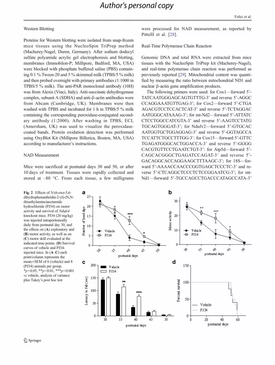
Fig. 2 Effects of N-(6-oxo-5,6-dihydrophenanthridin-2-yl)-(N,N-dimethylamino)acetamidehydrochloride (PJ34) on motoractivity and survival of Ndufs4knockout mice. PJ34 (20 mg/kg)was injected intraperitoneallydaily from postnatal day 30, andthe effects on (A) exploratory and(B) motor activity, as well as on(C) motor skill evaluated at theindicated time points. (D) Survivalcurves of vehicle and PJ34-injected mice. In (A–C) eachpoint/column represents themean±SEM of 6 (vehicle) and 8(PJ34) animals per group.*p<0.05, **p<0.01, ***p<0.001vs vehicle, analysis of varianceplus Tukey’s post hoc test
Felici et al.
Author's personal copy

and reverse 5’-ATTCTCCTTCTGTCAGGTCGAAGGG-3’;for β-actin—forward 5’-GCAGCCACATTCCCGCGGTGTAG-3’ and reverse 5’-CCGGTTTGGACAAAGACCCAGAGG-3’.
Mouse Primary Glial Cultures
Primary cultures of glial cells were prepared from P1mice as previously described [30]. Briefly, cortices wereisolated in cold PBS and then incubated for 30 mins at37 °C in PBS containing 0.25 % trypsin and 0.05 %DNase. After blocking enzymatic digestion with theaddition of 10 % heat-inactivated fetal bovine serum,
cortices were mechanically disrupted by pipetting. Cellsobtained from each cortex were washed, resuspended inDulbecco’s modified Eagle medium plus 10 % fetalbovine serum (GIBCO, Life Technologies, Rockville,MD, USA) and plated separately. Glial cells fromNdufs4 knockout (KO) mice were identified by genotyp-ing and used for mitochondrial membrane potentialevaluation at 7 days in vitro (DIV).
Evaluation of Mitochondrial Membrane Potential
Mitochondrial membrane potential was evaluated bymeans offlow cytometry [29]. Glial cells from Ndufs4 KO mice were
Fig. 3 Protein carbonylation,poly(ADP-ribose) (PAR) andnicotinamide adeninedinucleotide (NAD) content in themotor cortex of heterozygous(HET) and Ndufs4-null mice. (A)Oxyblot analysis of proteincarbonylation in the motor cortexof heterozygous (HET) andknockout (KO) mice at postnataldays 30 (P30) and 50 (P50). (B)Densitometric analysis ofoxyblots. Western blottingevaluation of PAR content in themotor cortex of HET and KOmice at (C) P30 and (D) P50. (E)Densitometric analysis ofWesternblots of PAR. (F) NAD contentsin the motor cortex of HET andKO mice at P30 and P50. BasalNAD content was 0.73±0.12 μmol/g tissue. In (A), (C),and (D), each blot isrepresentative of 6 animals pergroup. In (B), (E), and (F), eachcolumn represents the mean±SEM of 6 animals per group
PARP and Mitochondrial Disorders
Author's personal copy

treated with vehicle or with the 2 PARP inhibitors, PJ34(20 μM) or Olaparib (100 nM), for 72 h. Cells were then
detached, incubated with tetramethylrhodamine ethyl ester(TMRE) 2.5 nM, and analyzed with a Coulter EPICS XL flow
Felici et al.
Author's personal copy

cytometer (Beckman Coulter, Fullerton, CA, USA) equippedwith the EXPO32 Flow Cytometry ADC software (BeckmanCoulter).
Transmission Electron Microscopy
Tissues were fixed in 4 % glutaraldehyde, postfixed in 1 %osmium tetroxide, and embedded in Epon 812. Ultrathinsections were stained with uranyl acetate and alkaline bismuthsubnitrate and examined under a JEM 1010 electron micro-scope (Jeol, Tokyo, Japan) at 80 kV.
Micrographs were taken throughout the whole motor cortex,skeletal muscle, and liver at final magnifications of 12,000× and50,000× using a MegaView III digital camera and interfacingsoftware (SIS-Soft Imaging System, Munster, Germany). Thefirst ones were used for determination of the amount of mito-chondria, and the latter ones for analysis of mitochondria andinternal cristae volumes. Briefly, to analyze the number of mito-chondria, 5 cytoplasmic fields (test area per field 97.8 μm2) foreach section were chosen at random and only mitochondriaunequivocally present within neuronal structures were counted/analyzed. Areas of mitochondria and areas of cristae were mea-sured using iTEM image analysis software (SIS).
Immunohistochemistry
Immunohistochemistry was performed as previously described[31], according to standard procedure. Briefly, snap-frozen brainwas embedded in embedding matrix (CellPath Ltd., UK) (OCT)and cut with a cryostat (Leica, Solms, Germany). Brain section(14μm)were fixedwith 4%paraformaldehyde and incubated in
PBSwith 0.3% Triton X-100 (Sigma, St. Louis, MO, USA) and2 % of bovine albumin. Sections were double-stained with anti-Neuronal Nuclei (NeuN) monoclonal antibody (mouse mono-clonal, 1:100; Chemicon International, Temecula, CA,USA) andanti-glial fibrillary acidic protein (GFAP; monoclonal, clone G-A-5, 1:200; Sigma). To-pro3 (Molecular Probes, Eugene, OR,USA) was used as nuclear counterstain. Quantification of fluo-rescence was performed using Metamorph/Metafluor software.Values correspond to the mean±SEM of 5 different microscopicfields per 3 different mouse brain sections per brain (4 brain pergroup).
Data Analysis
Data were analyzed using WinLTP 1.11 reanalysis programand GraphPad Prism (version 4.0; GraphPad, San Diego, CA,USA). All numerical data are expressed as mean±SEM. Sta-tistical significance of differences between results was evalu-ated by performing analysis of variance followed by Tukey'sw test for multiple comparisons.
Results
Inhibition of PARP Improves Neuroscore and Delays DiseaseDevelopment of Ndufs4 KO Mice
To unravel the pathogenetic role of PARP-1 in the develop-ment of mitochondrial encephalopathy and to understand thetherapeutic potential of its inhibition in patients withOXPHOS defects, we evaluated the effect of pharmacologicalPARP suppression on disease development in KO mice. Wetreated animals with daily intraperitoneal injections of PJ34(20 mg/kg body weight), a water-soluble, potent PARP inhib-itor [24]. We found that the number of pups per litter was low(4–5), even though the KO mice in the offspring were at theexpected Mendelian ratio. To adopt a clinically relevant treat-ment protocol, we start injecting mice at day 30 when hairloss, the first sign of disease development, is almost complete[8]. As shown in Fig. 1A, treatment did not alter mouse weightcompared with vehicle-injected animals, although a tendencyto higher values in the PJ34-treated group was evident. Evo-lution of encephalopathy was assessed by evaluator-blindanalysis of neurological impairment [8]. We found that sig-nificant worsening of clinical score occurred at day 37 andmotor impairment inexorably increased up to postnatal day53–55, when mice died. In mice receiving PJ34, the clinicalscore was significantly delayed from postnatal day 37 topostnatal day 43 (Fig. 1B). At later time points, mice treatedwith the PARP inhibitor had a neuroscore that did not differfrom that of vehicle-injected animals, although, again, a ten-dency to slight reduction was obtained (Fig. 1B).
�Fig. 4 Effect of N-(6-oxo-5,6-dihydrophenanthridin-2-yl)-(N,N-dimethylamino)acetamide hydrochloride (PJ34) on tissue poly(ADP-ribose)(PAR) content, respiratory complex subunits expression and mitochondrialDNA (mtDNA) content in Ndufs4 knockout (KO) mice. (A) The effects of a10-day treatment (postnatal days 30–40) with PJ34 (daily intraperitonealinjections of 20 mg/kg) on tissue PAR content is shown. (B) Densitometricanalysis of the effects of PJ34 on tissue PAR content ofNdufs4KOmice. (C)mRNA levels of several mitochondrial [cyclooxygenase (COX)1, COX2,NADH dehydrogenase 2 (ND2)] and nuclear (NADH dehydrogenase(ubiquinone) flavoprotein 2 (NDUFV2), COX15, and ATP synthase, H+transporting, mitochondrial F1 complex, delta subunit (ATP5D)) respiratorycomplex subunits in different organs ofNdufs4 heterozygous (HET) and KOmice. (D) The effects of PJ34 on transcripts levels of the respiratory complexsubunits in KO mice are also shown. Succinate dehydrogenase complex,subunit A (SDHA) expression levels in different organs of (E) heterozygousand (F) KO mice treated or not with PJ34 is shown by Western blotting and(G) Densitometric analysis. (H) Effects of PJ34 on mitochondrial content(expressed as ND1/beta actin gene ratio) or (I) nicotinamide adeninedinucleotide (NAD) levels in different organs of Ndufs4 KO mice. BasalNAD content was 0.73±0.12 μmol/g tissue, 0.64±17 μmol/g tissue, 35±0.08 μmol/g tissue, 0.1±0.005 μmol/g tissue, 0.67±0.21 μmol/gtissue, 0.59±0.16 μmol/g tissue in the brain, pancreas, liver, spleen,heart, and skeletal muscle (sk. muscle), respectively. (A, E, F) A blotrepresentative of 4 mice per group is shown. (B, C, D, G, H, I), columnsrepresent the mean±SEM of 4 mice per group. *p<0.05, **p<0.01,***p<0.001 vs vehicle, analysis of variance plus Tukey’s post hoc test
PARP and Mitochondrial Disorders
Author's personal copy

Detailed analysis of specific symptoms indicates that treat-ment reduced the severity of ataxia and improved balance,having no effects on hind limb clasping and limb tone(Fig. 1C–F). Of note, analysis of exploratory and motor ac-tivity also revealed that treatment with the PARP inhibitorimproved both parameters during postnatal days 40–45 and35–45, respectively (Fig. 2A, B). When motor skill wasevaluated by means of rota-rod assay, we found that KO micereceiving PJ34 showed significantly prolonged latency to fallat P35-40 compared with vehicle-injected animals (Fig. 2C).However, PJ34 only delayed worsening of motor perfor-mances, given that at later time points (day 50) the therapeuticeffects disappeared. In keeping with this, drug treatment didnot prolong survival of the KO mice (Fig. 2D).
Oxidative Stress, PARPActivity, and NAD Levels in Ndufs4KO Mice
OXPHOS defects are typically characterized by derangementof electron transfer through the respiratory chain, a conditionleading to the formation of reactive oxygen species and oxi-dative stress. The latter is thought to play a key pathogeneticrole in encephalopathy of patients with mitochondrial disor-ders [32]. Given that PARP-1 is hyperactivated in conditionoxidative stress and causes massive energy consumption [33],we reasoned that PARP-1 activation-dependent ATP depletioncould further compromise the precarious energy homeostasisin the brains of KO mice. Therefore, we evaluated whetheroxidative stress occurs within the motor cortex of these ani-mals at different stages of disease development. As a markerof oxidative stress in vivo, we analyzed protein carbonylationby means of Oxyblot in KO and heterozygous mice. The latterare healthy, indistinguishable from wild-type mice, and have
previously been used as controls [8]. Although prior workdemonstrates increased protein carbonylation in the olfactorybulb of KO mice [9], we found that this marker of oxidativestress did not differ between KO and heterozygous mice atpostnatal day 30, whereas it was reduced in KO animals atpostnatal day 50 (Fig. 3A, B).
Western blot analysis of poly(ADP-ribosyl)ated proteins istypically used as an index of PARP activity. Therefore, weevaluated basal poly(ADP-ribosyl)ation in the motor cortex ofheterozygous and KO mice. In keeping with the lack of oxida-tive stress, levels of poly(ADP-ribosyl)ated proteins did notdiffer between the 2 mouse strains at postnatal day 30 andpostnatal day 50 (Fig. 3C–E). A reduction in NAD contenttypically occurs in tissues undergoing PARP-1 hyperactivity[33].Hence, as an additional index of PARP activity, we quan-tified the NAD content in the motor cortex of heterozygous andKO mice. Again, we were unable to find any difference in thecontent of NAD in the cortices of the two mouse strains at bothp30 and p50 (Fig. 3F).
Inhibition of PARP Increases the Expression of RespiratoryComplex Subunits and Promotes Mitochondrial Biogenesisin Ndufs4 KO Mice
To obtain evidence that PJ34 was, indeed, inhibiting PARP inKOmice, we analyzed PAR content in their tissues after10 daysof treatment (i.e., postnatal day 40). In keeping with the phar-macodynamic effect of the drug, we found a reduced PARcontent in brain, pancreas, liver, spleen, and skeletal muscle ofanimals challenged with PJ34 compared with vehicle-injectedmice (Fig. 4A, B).
We next wondered whether the expression of differentrespiratory complex subunits is altered in KO compared with
Fig. 5 Effects of poly(adenosine diphosphate-ribose) polymerase (PARP)inhibitors on mitochondrial membrane potential in Ndufs4 knockout (KO)cultured glial cells. The effect of a 72-h treatment with N-(6-oxo-5,6-dihydrophenanthridin-2-yl)-(N,N-dimethylamino)acetamide hydrochlo-ride (PJ34) (20 μM) or Olaparib (100 nM) on mitochondrial membranepotential [measured by means of potentiometric, fluorescent dye
tetramethylrhodamine ethyl ester (TMRE)] of cultured glial cells fromNdufs4 KO mice is shown as (A) the mean±SEM of 2 experimentsconducted in triplicate and (B) a representative cytofluorimetric plot.*p<0.05, **p<0.01, vs control, analysis of variance plus Tukey’s posthoc test
Felici et al.
Author's personal copy

heterozygous mice. Interestingly, we found a significant re-duction of transcripts for mitochondrial- and nuclear-encoded respiratory subunits, such as cyclooxygenase(COX)1, COX2, NADH dehydrogenase 2 (ND2), COX15,NADH dehydrogenase (ubiquinone) flavoprotein 2 (NDUFV2),and ATP synthase, H+ transporting, mitochondrial F1 complex,
delta subunit (ATP5D), in different mouse organs, with theexception of the heart (Fig. 4C). It has previously been reportedthat PARP-1-dependent NAD consumption limits PGC1α tran-scriptional activity and overall mitochondrial efficiency [21].Therefore we evaluated whether treatment with PJ34 promotestranscription of mitochondrial- and nuclear-encoded respiratory
Fig. 6 Mitochondrial number and morphology of Ndufs4 heterozygousand knockout mice treated or not withN-(6-oxo-5,6-dihydrophenanthridin-2-yl)-(N,N-dimethylamino)acetamide hydrochloride (PJ34).Mitochondrialmorphology and number in shown in representative electron microscopyimages at 2 different magnifications for (A) motor cortex, (B) skeletalmuscle, and (C) liver. Data summarizing the effects of Ndufs4 deletion in
the presence or absence of PJ34 on (D) mitochondrial number, (E) cristaearea, and (F) mitochondrial area in the different tissues is shown. Eachcolumn is themean±SEMof 5microscopic fields per 5 (+/–), 3 (–/–), and 4(–/– treated with PJ34) animals per group. *p<0.05, **p<0.01,***p<0.001 vs Ndufs4+/– mice, analysis of variance plus Tukey’s posthoc test
PARP and Mitochondrial Disorders
Author's personal copy

Felici et al.
Author's personal copy

complex subunits. Notably, we found that the PARP1 inhibitorincreased the transcript levels of the different respiratory subunitsin an organ-specific manner. Specifically, the mRNA levels ofmitochondrial genesCox1,Cox2, andmt-Nd2 increased in all theorgans tested (brain, pancreas, spleen, heart, and skeletal muscle)with the exception of liver. Conversely, transcripts of the nucleargenes Ndufv2, Cox5, and Atp5d were only augmented in liver,spleen, and heart (Fig. 4D). We also evaluated expression of theSDHA subunit of succinate dehydrogenase, and found that it wasnot affected in KO mice compared with heterozygous ones,whereas it increased in the organs of PJ34-treated mice, withthe exception of skeletal muscle (Fig. 4E–G).
The increased mitochondrial content reported in PARP-1 KOmice prompted us to evaluate whether the same phenotype couldbe recapitulated by pharmacological PARP inhibition [21]. As aprototypical index of mitochondrial content we quantitated themitochondrial DNA (mtDNA) gene mt-Nd1 in the differentorgans of KO mice treated or not with PJ34. As shown inFig. 4H, a 10-day treatment with the PARP inhibitor increasedthe content of mtDNA in all the organs tested except the liver.Notably, with the exception of the spleen, theNAD content in themouse organs was not increased by the PARP inhibitor (Fig. 4I).
To corroborate the evidence that PARP inhibitors improvemitochondrial function and that the effects of PJ34 are due toPARP inhibition, we next evaluated the impact of PJ34 and astructurally unrelated, very potent PARP inhibitor such asOlaparib, on mitochondrial membrane potential of cultured glialcells from Ndufs4 KO mice. As shown in Fig. 5, we found thatboth compounds increased the mitochondrial membrane poten-tial by approximately 25 % upon 72 h of treatment, at concen-trations consistent with their relative IC50 on PARP-1 [34].
These findings taken together with knowledge that transcrip-tional networks leading to increased oxidative capacity alsoregulate mitochondrial biogenesis [35], prompted us to evaluatewhether mitochondrial number and morphology of KO micewas affected by PARP inhibition. Electron microscopy revealedthat mitochondrial number and cristae area were reduced inmotor cortex and skeletal muscle but not in liver of KO micecompared with heterozygous animals at postnatal day 40(Fig. 6). We also found that the mitochondrial area increasedin motor cortex and liver but not in skeletal muscle of KO mice
(Fig. 6). Remarkably, a reduction in mitochondrial number, aswell as changes in organelle morphology, were prevented in KOmice treatedwith PJ34 from postnatal day 30 to postnatal day 40(Fig. 6). Also, the area of mitochondrial cristae in the liver wasincreased by drug treatment even if it was not reduced in KOmice (Fig. 6F).
Effects of PARP Inhibition on Astrogliosis and Neuronal Lossin Ndufs4 KO Mice
Improved neurological score by PJ34, along with the notion thatneurodegeneration takes place in the olfactory bulb and cerebel-lum of Ndufs4 mice [9], prompted us to evaluate the impact ofPJ34 on neuronal loss and astrogliosis in these mice. We foundthat a robust increase of GFAP-positive cell number (a prototyp-ical marker of astrogliosis) occurred at the level of the olfactorybulb and motor cortex of Ndufs4 mice at p40, but not in thecerebellum. Of note, treatment with the PARP inhibitor signifi-cantly reduced GFAP expression in these brain regions. Howev-er, neuronal loss occurring at p40 in olfactory bulb, cerebellumand motor cortex was not affected by drug treatment (Fig. 7).
Discussion
We report that a pharmacological inhibitor of PARP delays thedevelopment of encephalomyopathy in a mouse model of mito-chondrial disorder. We also show that PARP inhibition promptsa transcriptional program leading to increased expression ofrespiratory complex subunits and mitochondrial biogenesis. Inlight of the urgent need for drugs able to improve symptoms inpatients with OXPHOS defects [5, 32], along with the apparentsafety profile shown by PARP1 inhibitors in clinical trials [26],the present study might have realistic clinical implications.
Several transgenic mouse models of OXPHOS defectshave recently been developed; among them, those related togenetic mutations of respiratory complex I subunits appear toreproduce closely the symptomatology of patients [6]. TheKO mice used in our study lack exon 2 of Ndufs4 so that thecorresponding 18-kDa protein is absent because of frameshift.This mouse develops a phenotype resembling Leigh syn-drome and dies by fatal encephalomyopathy within approxi-mately 50 days [8]. Notably, mice bearing the same Ndufs4mutation selectively in neural cells display a phenotype almostidentical to those with systemic mutation [9]. This findingindicates that the therapeutic effects exerted by the PARPinhibitor should be ascribed to its ability to reduce neurode-generation during the development of mitochondrial enceph-alopathy. This assumption is in keeping with the large body ofevidence that PARP inhibitors, including PJ34, have remark-able neuroprotective effects in different models of neuronaldeath in vitro and in vivo [24]. Of note, we show that tissue
�Fig. 7 Neuronal loss and astrogliosis in different brain regions ofNdufs4heterozygous (HET) and knockout (KO) mice treated or not with PJ34.Neuronal loss and astrogliosis have been evaluated in (A–H) olfactorybulb, (I–R) cerebellar, and (S–Z) motor cortex. Neuronal loss has beenevaluated according to Chiarugi et al. [9] by staining neurons with NeuN(green) and nuclei with To-pro3 (red). Co-localization of both labels isshown in yellow. Astrocyte activation has been evaluated by means ofglial fibrillary acidic protein (GFAP) staining (blue). Imagesrepresentative of 4 brains per group are shown. (D, H, N, R, V, Z) Eachcolumn is the mean±SEM of 5 different microscopic fields per 3 differentmouse brain sections per brain. *p<0.05, **p<0.01, ***p<0.001 vsNdufs4+/– mice, analysis of variance plus Tukey’s post hoc test. Bar=500 μm. C=Vehicle treated mice
PARP and Mitochondrial Disorders
Author's personal copy
PAR content is reduced in KOmice upon PJ34 administration,which is in keeping with the notion that PARP-1 contributes tothe majority of PAR formations [13, 14]. However, given thatthe drug is not strictly PARP-1 selective [36], we cannot ruleout the possibility that inhibition of additional PARPs,including PARP-2 [37], may have contributed to thepharmacodynamic effects of PJ34.
In principle, PARP inhibition might exert its therapeuticeffect in KO mice by different mechanisms. For instance,necrotic neuronal death occurs in the brain of KO mice [9],and numerous reports demonstrate the ability of PARP inhibi-tors to protect from this form of neuronal demise [33]. How-ever, our findings showing lack of oxidative stress, PARPactivation, and NAD depletion in the motor brain cortex ofKO mice at different stages of encephalopathy suggest thatPARP1 is not causative in necrotic neuronal death in this modelof mitochondrial disorder. Although data are consistent withprior work showing no increase of ROS in fibroblasts from apatient with a nonsense mutation in Ndufs4 [38], recent find-ings in Ndufs4 KO mice show the occurrence of oxidativestress in the olfactory bulb during disease progression [9]. Inthis regard, even though our electron microscopy analysis andimmunohistochemistry reveal mitochondrial morphologicalabnormalities, astrogliosis and neuronal loss in the motor cor-tex, the olfactory bulb is the first and most compromised brainstructure in KO mice [9]. Therefore, we speculate that mecha-nisms underlying neurodegeneration in KO mice are brainregion-specific. The decrease of protein carbonylation in KOmice compared with heterozygous mice at P50 could be as-cribed to themoribund conditions of the animals and the relatedbreathing defect resulting in reduced blood perfusionand oxygenation [39]
PARP-1 is a key player of apoptosis inducing factor-dependent apoptosis during neurodegeneration [40]. However,given that the extrinsic (i.e., mitochondrial independent) apo-ptotic pathway is triggered in the brain of KO mice [9], it isunlikely that prevention of AIF release and apoptosis is a majormechanism responsible for the PJ34 effect. Interestingly, inkeeping with evidence that astrocyte and microglia activationoccurs in the degenerating brain regions of Ndufs4 KO mice[9], we show that GFAP immunoreactivity is increased inolfactory bulb and motor cortex. Although the pathogeneticrelevance of this inflammatory event still needs to be clarified,it is tempting to speculate that the ability of PARP inhibitors tosuppress astroglia activation contributed to reduce the severityof encephalopathy and related symptoms [41].
In addition to the possibility that PARP inhibition counter-acts neurodegeneration by blocking neurotoxic events in theKO mice, pharmacological suppression of PARP could alsoprompt neuroprotective mechanisms. In this regard, a keypathway of relevance to neuroprotection in these animals mightbe that prompted by PGC1α. Indeed, both genetic or pharma-cological suppression of PARP-1 promotes SIRT-1-dependent
PGC1α activation which leads to increased oxidative capacityand mitochondrial content [21]. Accordingly, we found thatPJ34 induced the expression of respiratory complex subunitsand mitochondrial biogenesis. This finding, along with evi-dence that mRNAs for respiratory complex subunits are re-duced in KO compared with heterozygous mice, is of particularimportance because it suggests that the therapeutic effects ofPARP inhibition may be due to a restoration of homeostatictranscript levels. Notably, KO mice receiving the PARP inhib-itor showed increased mRNA abundance of both nuclear- andmitochondrial-encoded respiratory complex subunits. We rea-son that this occurred because, in addition to the activation ofthe PGC1α-dependent transcriptional program, PARP inhibi-tion also alters nuclear transcription directly. Indeed, it is wellappreciated that PARP-1 activity epigenetically regulates tran-scription of numerous genes by direct interaction with bothgene promoters and basal transcriptional machinery [15].PARP1 can also regulate the activity of several transcriptionfactors, including YY1 or NRF-1 [42, 43], which are of rele-vance to mitochondrial functioning. Interestingly, nuclear re-spiratory factor (NRF)-1, a key regulator of nuclear genesinvolved in mitochondrial respiration and mtDNA duplication,is negatively regulated by PARP-1 activity [43]. Therefore,inhibition of PARP-1 by PJ34 might have unleashed NRF-1,thereby potentiating PGC1α-dependent mitochondrial biogen-esis. Evidence that NAD content increased only in the spleen ofKO mice treated with PJ34 is in line with the hypothesis thatmechanisms in addition to SIRT1-dependent PGC1α activa-tion contribute tomitochondrial biogenesis. The selective NADincrease in the spleen is also in keeping with our recent studythat showed a high NAD turnover in this mouse organ [28].
At present we do not know why PJ34 affected mitochon-drial number and morphology in some organs but not inothers. Possibly, this is owing to tissue-specific mechanismsof epigenetic regulation, as well as to different impairment oftissue homeostasis during disease development. Accordingly,we previously reported that PJ34 impairs mitochondrial DNAtranscription in cultured human tumor cells [44].We speculatethat the reason(s) of this apparent inconsistency can be as-cribed to differences in experimental settings, that is in vivoversus in vitro and/or acute versus chronic exposure to PJ34.
Unfortunately, in spite of the ability of PJ34 to reduceneurological impairment after a few days of treatment, neitherneuronal loss nor death of mice was reduced or delayed.Although this KO mouse model is extremely severe, showinga shift from healthy condition to fatal breathing dysfunction inonly 20 days [39], recent work demonstrates that rapamycinincreases median survival of maleNdufs4KOmice from 50 to114 days [45]. In light of this, we speculate that inhibition ofPARP prompts a cascade of events, such as mitochondrialbiogenesis or increased oxidative capacity, that is of symp-tomatic relevance, but eventually unable to counteract specificmechanisms responsible for neurodegeneration and disease
Felici et al.
Author's personal copy

development. Still, symptom improvement obtained withPJ34 is of pathogenetic and therapeutic significance, andmight be potentiated by different means such as use of ultra-potent PARP inhibitors [24] and co-treatment with symptom-atic drugs already used in mitochondrial patients. In keepingwith this hypothesis, very recent studies report improvementof mitochondrial functioning and muscle fitness in micechallenged with PARP inhibitors [46, 47].
Acknowledgments This work was supported by grants from RegioneToscana Health Projects 2009 (recipient A.C.) and 2012 (recipient A. L.),Association of Amyotrophic Lateral Sclerosis (ARISLA), and Ente Cassadi Risparmio di Firenze. The authors gratefully acknowledge R.D.Palmiter for the kind gift of Ndufs4 KO mice and helpful comments.
Required Author Forms Disclosure forms provided by the authors areavailable with the online version of this article.
References
1. Wallace DC. Mitochondrial diseases in man and mouse. Science1999;283:1482-1488.
2. Wallace DC, Fan W, Procaccio V. Mitochondrial energetics andtherapeutics. Annu Rev Pathol 2010;5:297-348.
3. Sofou K. Mitochondrial disease: a challenge for the caregiver, thefamily, and society. J Child Neurol 2013;28:663-667.
4. Pfeffer G, Majamaa K, Turnbull DM, Thorburn D, Chinnery PF.Treatment for mitochondrial disorders. Cochrane Database Syst Rev2012;4:CD004426.
5. Andreux PA, Houtkooper RH, Auwerx J. Pharmacological ap-proaches to restore mitochondrial function. Nat Rev Drug Discov2013;12:465-483.
6. Koene S, Willems PH, Roestenberg P, Koopman WJ, Smeitink JA.Mouse models for nuclear DNA-encoded mitochondrial complex Ideficiency. J Inherit Metab Dis 2011;34:293-307.
7. Valsecchi F, Koopman WJ, Manjeri GR, et al. Complex I disorders:causes, mechanisms, and development of treatment strategies at thecellular level. Dev Disabil Res Rev 2010;16:175-182.
8. Kruse SE, Watt WC, Marcinek DJ, et al. Mice with mitochondrialcomplex I deficiency develop a fatal encephalomyopathy. Cell Metab2008;7:312-320.
9. Quintana A, Kruse SE, Kapur RP, Sanz E, Palmiter RD. Complex Ideficiency due to loss of Ndufs4 in the brain results in progressiveencephalopathy resembling Leigh syndrome. Proc Natl Acad Sci U SA 2010;107:10996-11001.
10. Chiarugi A, Dolle C, Felici R, Ziegler M. The NAD metabolome – akey determinant of cancer cell biology. Nat Rev Cancer 2012;12:741-752.
11. Canto C, Auwerx J. NAD+ as a signaling molecule modulatingmetabolism. Cold Spring Harb Symp Quant Biol 2011;76:291-298.
12. Houtkooper RH, Auwerx J. Exploring the therapeutic space aroundNAD+. J Cell Biol 2012;199:205-209.
13. D'Amours D, Desnoyers S, Poirier GG. Poly(ADP-ribosyl)ationreactions in the regulation of nuclear functions. Biochem J1999;342:249-268.
14. Gibson BA, Kraus WL. New insights into the molecular and cellularfunctions of poly(ADP-ribose) and PARPs. Nat Rev Mol Cell Biol2012;13:411-424.
15. Kraus WL. Transcriptional control by PARP-1: chromatin modula-tion, enhancer-binding, coregulation, and insulation. Curr Opin CellBiol 2008;20:294-302.
16. Kraus WL, Lis JT. PARP goes transcription. Cell 2003;113:677-683.17. Imai S, Guarente L. Ten years of NAD-dependent SIR2 family
deacetylases: implications for metabolic diseases. TrendsPharmacol Sci 2010;31:212-220.
18. Canto C, Auwerx J. PGC-1alpha, SIRT1 and AMPK, an energysensing network that controls energy expenditure. Curr OpinLipidol 2009;20:98-105.
19. Zhang T, Berrocal JG, Frizzell KM, et al. Enzymes in the NAD+salvage pathway regulate SIRT1 activity at target gene promoters. JBiol Chem 2009;284:20408-20417.
20. Pillai JB, Isbatan A, Imai S, Gupta MP. Poly(ADP-ribose)polymerase-1-dependent cardiac myocyte cell death during heartfailure is mediated by NAD+ depletion and reduced Sir2alphadeacetylase activity. J Biol Chem 2005;280:43121-43130.
21. Bai P, Canto C, Oudart H, et al. PARP-1 inhibition increases mito-chondrial metabolism through SIRT1 activation. Cell Metab2011;13:461-468.
22. Pittelli M, Felici R, Pitozzi V, et al. Pharmacological effects ofexogenous NAD on mitochondrial bioenergetics, DNA repair, andapoptosis. Mol Pharmacol 2011;80:1136-1146.
23. Canto C, Houtkooper RH, Pirinen E, et al. The NAD(+) precursornicotinamide riboside enhances oxidative metabolism and protectsagainst high-fat diet-induced obesity. Cell Metab 2012;15:838-847.
24. Jagtap P, Szabo C. Poly(ADP-ribose) polymerase and the therapeuticeffects of its inhibitors. Nat Rev Drug Discov 2005;4:421-440.
25. Rouleau M, Patel A, Hendzel MJ, Kaufmann SH, Poirier GG. PARPinhibition: PARP1 and beyond. Nat Rev Cancer 2010;10:293-301.
26. Papeo G, Forte B, Orsini P, et al. Poly(ADP-ribose) polymeraseinhibition in cancer therapy: are we close to maturity? Expert OpinTher Pat 2009;19:1377-1400.
27. Kuribara H, Higuchi Y, Tadokoro S. Effects of central depressants onrota-rod and traction performances in mice. Jpn J Pharmacol1977;27:117-126.
28. Pittelli M, Cavone L, Lapucci A, et al. Nicotinamidephosphoribosyltransferase (NAMPT) activity is essential for surviv-al of resting lymphocytes. Immunol Cell Biol 2014;92:191-199.
29. Felici R, Lapucci A, Ramazzotti M, Chiarugi A. Insight into molec-ular and functional properties of NMNAT3 reveals new hints of NADhomeostasis within human mitochondria. PLoS One 2013;8:e76938.
30. Faraco G, Pittelli M, Cavone L, et al. Histone deacetylase (HDAC)inhibitors reduce the glial inflammatory response in vitro and in vivo.Neurobiol Dis 2009;36:269-279.
31. Faraco G, Pancani T, Formentini L, et al. Pharmacological inhibitionof histone deacetylases by suberoylanilide hydroxamic Acid specif-ically alters gene expression and reduces ischemic injury in themouse brain. Mol Pharmacol 2006;70:1876-1884.
32. Dimauro S, Rustin P. A critical approach to the therapy of mitochon-drial respiratory chain and oxidative phosphorylation diseases.Biochim Biophys Acta 2009;1792:1159-1167.
33. Chiarugi A. PARP-1: killer or conspirator? The suicide hypothesisrevisited. Trends Pharmacol Sci 2002;23:122-129.
34. Wahlberg E, Karlberg T, Kouznetsova E, et al. Family-wide chemicalprofiling and structural analysis of PARP and tankyrase inhibitors.Nat Biotechnol 2012;30:283-288.
35. Scarpulla RC. Transcriptional paradigms in mammalian mitochon-drial biogenesis and function. Physiol Rev 2008;88:611-638.
36. Pellicciari R, Camaioni E, Costantino G, et al. On the way to selectivePARP-2 inhibitors. Design, synthesis, and preliminary evaluation of aseries of isoquinolinone derivatives. Chem Med Chem 2008;3:914-923.
37. Bai P, Canto C, Brunyanszki A, et al. PARP-2 regulates SIRT1expression and whole-body energy expenditure. Cell Metab2011;13:450-460.
38. Iuso A, Scacco S, Piccoli C, et al. Dysfunctions of cellular oxidativemetabolism in patients with mutations in the NDUFS1 and NDUFS4genes of complex I. J Biol Chem 2006;281:10374-10380.
PARP and Mitochondrial Disorders
Author's personal copy
39. Quintana A, Zanella S, Koch H, et al. Fatal breathing dysfunction in amouse model of Leigh syndrome. J Clin Invest 2012;122:2359-2368.
40. Wang Y, Dawson VL, Dawson TM. Poly(ADP-ribose) signals tomitochondrial AIF: a key event in parthanatos. Exp Neurol2009;218:193-202.
41. Chiarugi A, Moskowitz MA. Poly(ADP-ribose) polymerase-1 activ-ity promotes NF-kappaB-driven transcription and microglial activa-tion: implication for neurodegenerative disorders. J Neurochem2003;85:306-317.
42. Oei SL, Shi Y. Poly(ADP-ribosyl)ation of transcription factor YinYang 1 under conditions of DNA damage. Biochem Biophys ResCommun 2001;285:27-31.
43. Hossain MB, Ji P, Anish R, Jacobson RH, Takada S. Poly(ADP-ribose) polymerase 1 interacts with nuclear respiratory factor 1
(NRF-1) and plays a role in NRF-1 transcriptional regulation. JBiol Chem 2009;284:8621-8632.
44. Lapucci A, Pittelli M, Rapizzi E, et al. Poly(ADP-ribose)polymerase-1 is a nuclear epigenetic regulator of mitochondrialDNA repair and transcription. Mol Pharmacol 2011;79:932-940.
45. Johnson SC, YanosME, Kayser EB, et al. mTOR inhibition alleviatesmitochondrial disease in a mouse model of Leigh syndrome. Science2013;342:1524-1528.
46. Cerutti R, Pirinen E, Lamperti C, et al. NAD-dependent activation ofSirt1 corrects the phenotype in a mouse model of mitochondrialdisease. Cell Metab 2014;19:1042-1049.
47. Pirinen E, Canto C, Jo YS, et al. Pharmacological inhibition ofpoly(ADP-ribose) polymerases improves fitness and mitochondrialfunction in skeletal muscle. Cell Metab 2014;9:1034-1041.
Felici et al.
Author's personal copy


