Embed Size (px)
Citation preview
RESEARCH
ARTIC
LE
Copyright © 2011 American Scientific PublishersAll rights reservedPrinted in the United States of America
Journal ofBiomedical Nanotechnology
Vol. 8, 1–9, 2011
Osteoblast Activity on Anodized Titania Nanotubes:Effect of Simulated Body Fluid Soaking Time
Cem Bayram1�2, Murat Demirbilek1, Nazlı Çalıskan1, Melike Erol Demirbilek3,and Emir Baki Denkbas1�∗
1 Hacettepe University, Nanotechnology and Nanomedicine Division, Beytepe 06800 Ankara, TR2 Aksaray University, Faculty of Science, Department of Chemistry, 68100, Aksaray, TR
3Aksaray University, School of Health Sciences, 68100, Aksaray, TR
Early phase osseointegration is crucial for orthopedic implants. For the improvement of osseointe-grative properties of orthopedic implants several surface modification methods such as acid etching,hydroxyapatite (HA) coating and sandblasting can be applied. In this article titanium implants wereanodized to possess nanotubular titania structures on the surface. Titania nanotube structures witha 45–50 nm of average inner diameter were obtained and to enhance bioactivity, samples weresoaked in 10X simulated body fluid (SBF) for apatite deposition on surface for different time periods(1, 2, 3, 5, 8 hours). Apatitic calcium phosphate deposited surfaces were analyzed with infraredspectrometry and wettability studies. Effect of soaking time on osteoblast cell was investigated bycell viability, alkaline phosphatase activity tests and morphological evaluations. As a result, 3 hoursof soaking time was found as the optimum time period (p < 0�005). This in vitro study indicatedthat soaking in 10X SBF can be a rapid and economical technique to enhance osseointegration ofanodized titanium implants however excess and/or uncontrolled HA coating of titania layer limits thebioactive potential of the implant.
Keywords: Anodization, Titanium, Titania Nanotube, Osteoblast, Hydroxyapatite, SimulatedBody Fluid.
1. INTRODUCTION
Metallic materials, especially titanium and its alloys arewidely used in both orthopedic and dental applications.1–5
Titanium has sufficient load bearing capacity and bondingability with osteoblasts. However, its insufficient osseoin-tegration and weak cell adhesion remains as a majorproblem after implantation. Early osseointegration is cru-cial for orthopedic implants.6�7 The elevated number ofadhered cells in early phase shortens the osseointegra-tion period. Although the bulk structure of titanium ful-fills the mechanical requirements, surface properties play akey role in osteoblast adhesion.8 While untreated titaniummetal lacks certain bioactive properties, a titania layer canenhance bond formation with osteoblasts. Biocompatibilityand bioactivity are the two phenomena commonly relatedwith material surface.9 Modifications which have effectson the surface characteristics can be applied to biomate-rials without altering the bulk properties.10–13 Sandblast-ing and/or acid etching are such processes beneficial for
∗Author to whom correspondence should be addressed.
enhanced osseointegration for titanium based implants inthe production of highly porous implant surfaces.On the other hand, the bone itself has nanometer sized
organic and inorganic entities such as collagen nanofibersand nano sized hydroxyapatite crystals. It can be concludedthat the best integration between bone and biomaterial canbe achieved with nano structured biomaterial surfaces. Tita-nia nanotube arrays arise on metal surfaces during theanodization process. Under specific electrochemical envi-ronments, tube like titania layers grow on Ti metal surfacesspontaneously.14 In a typical procedure, titanium metal isused as an anode against a platinum cathode in an elec-trolytic cell. Dilute hydrogen fluoride solutions are oftenused as electrolyte. Altering the condition parameters suchas electric potential and anodization time, tube-like tita-nia structures of 25–100 nm in diameter and a length of300–2500 nm 14–16 are obtainable. To enhance biointegra-tion various coating processes have been applied.17�18
Hydroxyapatite (HA), which is a naturally occurring andabundant mineral in bone tissue, has been used as a coat-ing material to induce bone growth.19�20 Commercial HAcoatings on orthopedic implants are generally applied by
J. Biomed. Nanotechnol. 2011, Vol. 8, No. 3 1550-7033/2011/8/001/009 doi:10.1166/jbn.2011.1391 1
RESEARCH
ARTIC
LE
Osteoblast Activity on Anodized Titania Nanotubes: Effect of Simulated Body Fluid Soaking Time Bayram et al.
plasma spraying. Coatings obtained using this method maycontain crystal phases that can lead to delamination.21�22
Since plasma spraying requires high processing tempera-tures; it cannot be used in combination with organic com-pounds such as drug or growth factors as it leads to thedeformation of such additives. During postoperative period,patients are required to undergo drug therapies to pre-vent immune response to the implant. Antibiotics, growthfactors and anti-inflammatory agents can be delivered viaintramuscular, intravenous or oral routes. Several well-known limitations of drug therapies such as systemic toxic-ity, ineffective drug concentration or biological barriers aremajor complications. Moreover it has been shown that drugelution from implant site has numerous advantages such asreduced drug amount and toxicity, as well as enhanced localtargeting.23�24 Recently, Peng et al. reported that proteinsand small bioactive molecules can be loaded into titaniananotube layers and elution from the nanostructures canlast for days to weeks.16 Penicillin/streptomycin mixturewas also successfully loaded into porous titania layers.25
These promising results on titania nanotube layers as drugeluting platforms could be a solution for primary prophy-laxis against bacterial infections in postoperative period.In order to create a better implant with high osseointe-gration ability, these drug eluting nanostructures can besoaked in concentrated simulated body fluid (SBF) solu-tions. Several compositions of SBF solutions have beenproposed.26–28 In concentrated SBF solutions, it is possi-ble to obtain a rapid coating on titania surfaces in severalhours. This type of coating formation is less time consum-ing and a desired feature for pre-loaded implants. Althoughsoaking in SBF solution is non-destructive against bioac-tive molecule, lengthy soaking times may cause the releaseof loaded biomolecules from the implant, so the coatingtime must be both sufficient and quick.In this study, we investigated the effect of apatite coating
density on osteoblast cells. Titania nanotubular surfaceswere generated and soaked in 10X SBF solution for rapidcoating of hydroxyapatite. The physicochemical character-istics and in vitro osteoblast cell activities on HA coatedsamples with varying soaking times were investigated indetail.
2. MATERIALS AND METHODS
2.1. Titania Nanotube Layer Formation
Titanium foil (99% purity, Alfa Aesar, USA) was mechan-ically cut and cleaned with liquid soap and 70% ethanolsolutions. Before anodization, titanium samples wereimmersed in an acidic mixture consisting of 5 M HNO3
(Merck, Germany) and a few drops of 48% (w/w) HF(Fluka, USA) in order to remove the naturally occuringoxide layer on foil. After this step, Ti samples were rinsedtwice with deionized (DI) water and dried in the ovenat 70 �C for 30 min. In a two electrode electrochemical
cell, Ti metal was used as anode and platinum mesh (99%purity, Alfa Aesar, USA) was used as cathode electrodes.The electrodes were kept parallel with 4 cm distancebetween each other. Electrolyte was selected as 1% wtaqueous HF solution. During anodization, electric poten-tial was kept constant at 15 V and the procedure lasted for20 minutes. After anodization, Ti samples were immedi-ately rinsed with DI water, dried in the oven at 70 �C for30 min.
2.2. Apatitic Calcium Phosphate Precipitation
The dried samples were heated at 500 �C for two hours forthe complete conversion of rutile phase titania into anatasephase. With this heat treatment procedure, a better latticematch between anatase phase and apatite crystals can beobtained.29 After cooling at ambient temperature, Ti sam-ples were cut into 1 cm2 portions and immersed in 100 mlof 10X simulated body fluid (SBF) in separate containersfor different time intervals. 10X SBF stock solution con-tains aqueous solutions of 1000 mM NaCl, 5 mM KCl,25 mM CaCl2, 5 mM MgCl2, 10 mM Na2HPO4.
26 Afteradding 10 mM NaHCO3 into stock solution, pH raisesaround 6.5 and the apatite like calcium phosphate precip-itation starts. The precipitation procedure was carried outin ambient temperature without any additional heat or agi-tation. Ti samples were soaked in 10X SBF solution for1,2,3,5 and 8 hours. Soaked samples were then rinsed withDI water and dried in vacuum oven for characterizationand cell culture studies.
2.3. Surface Characterization
Untreated titanium (Ti), anodized titanium (An-Ti) andanodized-SBF soaked titanium (An-Ti-SBF) sampleswere characterized morphologically by Scanning Elec-tron Microscopy (SEM) (Quanta 200 FEG, FEI Instru-ments, USA). The wettability of sample surfaces wereevaluated by sessile drop contact angle measurements. Foreach sample group, eight randomized spots were analyzedwith DI water by a goniometer system (DSA100 ContactAngle Analyzer, Krüss, Germany). Also chemical com-position of the surfaces were characterized by Attenuatedtotal reflectance—Fourier transform infrared spectroscopy(ATR-FTIR) instrument (Nicolet iS5, Thermo Scientific,USA).
2.4. Cell Culture
To investigate the effects of apatitic calcium phos-phate deposition density on cell proliferation and alka-line phosphatase (ALP) activity, human bone osteogenicsarcoma cell line (Saos-2/An1) was used. The cellswere obtained from Foot and Mouth Disease Institute,(Ankara, Turkey). For the cell proliferation assay, MTT
2 J. Biomed. Nanotechnol. 8, 1–9, 2011
RESEARCH
ARTIC
LE
Bayram et al. Osteoblast Activity on Anodized Titania Nanotubes: Effect of Simulated Body Fluid Soaking Time
(3-[4,5-Dimethylthiazol-2-yl]-2,5-diphenyltetrazoluim bro-mide; Thiazolyl blue) test was performed. The cells werecultured in Dulbecco’s Modified Eagle Medium (DMEM)supplemented with 10% fetal bovine serum and 1% peni-cillin/streptomycin solution under standard cell cultureconditions, a humidified 5% CO2/95% air environment at37 �C. The medium was replaced every two days.
Titanium samples from all sample groups were cut in1 cm2 portions. Before seeding, the samples were placedin a 12-well culture dish and soaked in 70% ethanol for30 minutes and exposed to ultraviolet light in a lam-inar flow cabinet for 30 minutes of sterilization. Theplaques were then washed twice with phosphate buffersaline (PBS) and the cells were pipetted at a density of1×103 cells/sample. The cell proliferation was measuredafter 3, 5 and 7 days. Cell culture media was changedevery two days during the experiment and removed afterincubation. 300 �l fresh medium and 30 �l MTT solu-tion (5 �g/ml, diluted with RPMI 1640 without phenolred) were added to the each well. Incubation was allowedfor another 4 h in the dark at 37 �C. Living cells canmetabolize the MTT in their mitochondria and form darkblue formazan crystals. To solve formed formazan crys-tals, 100 �l/well isopropanol-HCl (absolute isopropanolcontaining 0.04 M HCl) solution was added to wells. Atotal of 100 �L solution from each well was aspirated andpoured into a 96-well plate. Cell growth was determinedby the MTT assay and was directly proportional to theabsorbance at a wavelength of 570 nm.
2.5. Alkaline Phosphatase Activity
ALP activity is an important parameter to evaluate the nor-mal functionality of osteoblasts on a surface30 and ALPis an early marker for osteoblast differentiation and isthought to play a major role in bone formation.31 In thisstudy, ALP activity was measured on 3, 5 and 7 dayscultured cells on all sample groups. ALP activity wasevaluated on the transformation of p-nitrophenyl phos-phate into p-nitrophenol at pH 10.2. The samples werewashed three times with PBS and 250 �L of cell lysissolution (0.5% (v/v) TritonX-100, 50 mM Tris (pH 7.6)and 1 mM MgCl2 were added to each well and sampleswere frozen and thawed three times to obtain cell lysis.After the final thaw, the cell lysates were centrifuged for10 min. at 5000 rpm. The supernatants were then incu-bated with the ALP reagent (Sigma, USA) and the ALPactivity was spectrophotometrically measured at 410 nm.Alkaline phosphatase synthesized by osteoblasts culturedon the plaques were determined from a standard curveof absorbance versus known concentrations of precinorm(Roche Diagnostics, Germany) run in parallel with exper-imental samples. Data were normalized for total proteinconcentration of samples. Thus, the ALP activity wasexpressed as mU/mgprotein/min.
2.6. Total Protein Measurement
Total protein content in the cell lysates were determinedspectrophotometrically using Lowry’s method, and theabsorbance of the solution was measured using a spec-trophotometer at a wavelength of 750 nm. The absorbancewas then converted to protein content using the BovineSerum Albumine (BSA) standard curve to determine theamount of protein.32
2.7. Cell Morphology
On the 3rd day of culture, samples were washed threetimes with PBS, followed by fixing with 4% paraformalde-hyde in PBS for 30 min at 4 �C and then observed bySEM (Quanta 200 FEG, FEI Instruments, USA).
2.8. Statistical Analysis
Eight specimens for each experimental group were usedand each analysis was triplicated. Numerical data were ana-lyzed using standard analysis of variance (ANOVA) tech-nique; statistical significance was considered at p < 0�005.
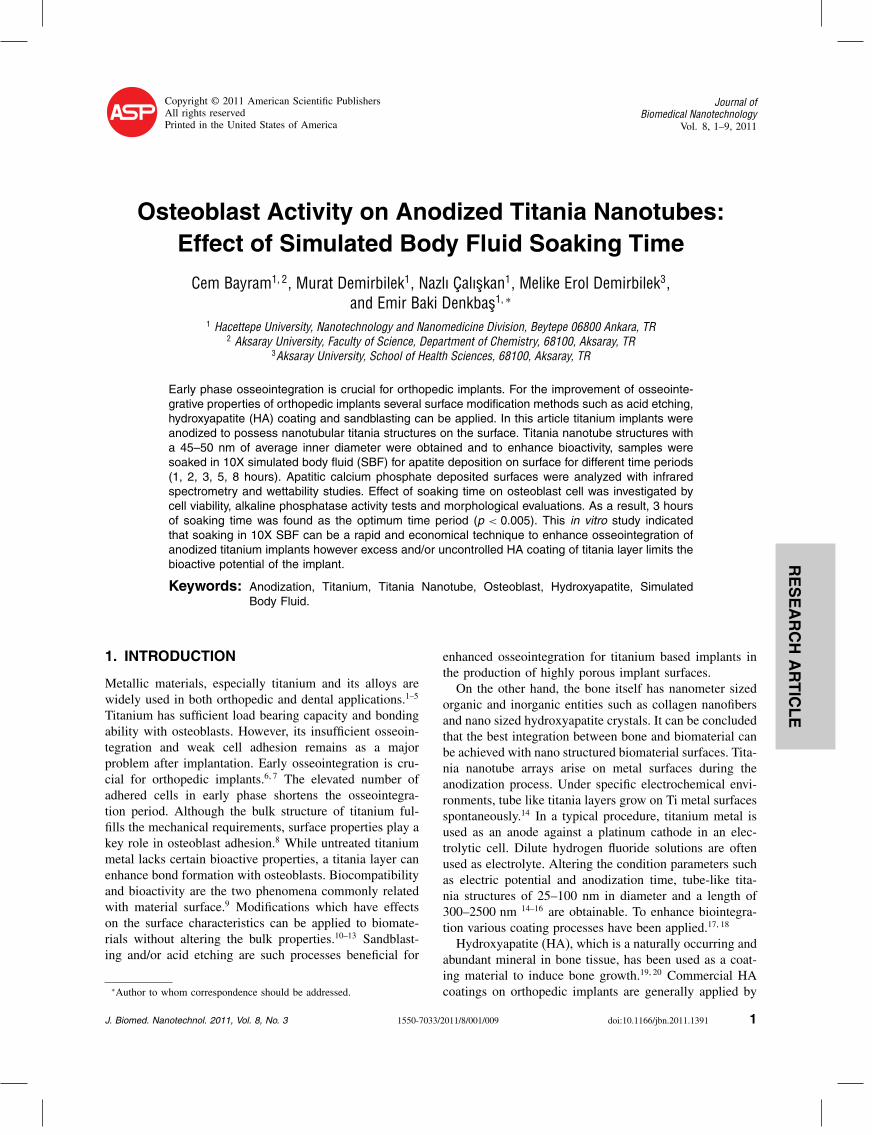
Fig. 1. Representative SEM images of untreated (A) and anodized (B)titanium surfaces.
J. Biomed. Nanotechnol. 8, 1–9, 2011 3
RESEARCH
ARTIC
LE
Osteoblast Activity on Anodized Titania Nanotubes: Effect of Simulated Body Fluid Soaking Time Bayram et al.
3. RESULTS
3.1. Surface Morphology
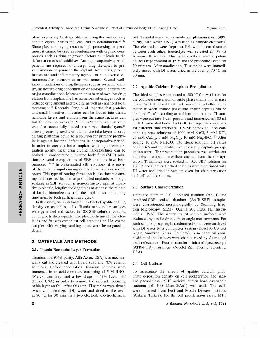
Titania nanotubes with an average inner diameter of45–50 nm were obtained on anodized samples. The sur-face morphology of the anodized titanium was clearly dif-ferent compared to the untreated one (Figs. 1(A and B)).As untreated titanium surface (Fig. 1(A)) has a randomroughness and topography consisting of some broad cavi-ties the surface of anodized titanium has a nanotube layerwith a wall thickness of 10 nm (Fig. 1(B)). SBF soak-ing time determines the amount of HA deposition whichdirectly affects the surface morphology. It was noted thatHA layer was barely deposited on the surface after 1 hourof soaking in SBF. After the second hour of soaking, petallike monosized apatite entities with an average diameterof 1–2 �m began to form on titania nanotubes. Densityand thickness of apatite structures increased with soakingtime and resulted in complete coverage of titania nanotubesurface after the 5th hour of SBF treatment. The surfaceconsisted of both apatite and titania nanotubes after twoand three hours of soaking (Fig. 2).
3.2. Wettability Studies
Surface wettability was dramatically increased after theanodization process. The increase in surface wettabilitycontinued with deposited apatite density with an exceptionat 1 hour of soaking in 10X SBF. After 1 hour of soaking,
Fig. 2. Representative SEM images of anodized titanium plaques soaked in 10X SBF. Soaking times are; 1 h (A), 2 h (B), 3 h(C), 5 h(D), 8 h(E).
Table I. Water Contact Angles of Titanium Samples
Sample � Degree
Ti 78,1±3,9An-Ti 42,1±3,4An-Ti-SBF(1 h) 47,5±7,3An-Ti-SBF(2 h) 41,9±7,8An-Ti-SBF(3 h) 32,2±6,5An-Ti-SBF(5 h) <10An-Ti-SBF(8 h) <10
water contact angle increased up to 47.5� due to surfacemorphology. Here, HA just began to precipitate resultingin a very thin crystal structure on titania nanotube layer.Water contact angle value decreases as the soaking timelengthens and finally the surface becomes completely wet-table (� < 10�) after 5 hours of soaking. Apatite densityon 5th and 8th hours of soaking in 10X SBF solution cre-ated a very rough surface consisting of calcium phosphatespecies only. The results of wettability studies are given inTable I.
3.3. ATR-FTIR Spectroscopy
Figure 3 shows the infrared spectra of four species: Ti,An-Ti, An-Ti-SBF(3 h) and An-Ti-SBF(5 h). Titanium andanodized titanium were found similar with a broad peakof Ti–O–Ti bonding at 670 cm−1. An-Ti-SBF(3 h) sam-ple has both precipitated HA and titania layer characteris-tics. PO3−
4 absorptions at 550, 960, 1035 and 1105 cm−1
represent P–O bending and symmetric vibrations and the
4 J. Biomed. Nanotechnol. 8, 1–9, 2011
RESEARCH
ARTIC
LE
Bayram et al. Osteoblast Activity on Anodized Titania Nanotubes: Effect of Simulated Body Fluid Soaking Time
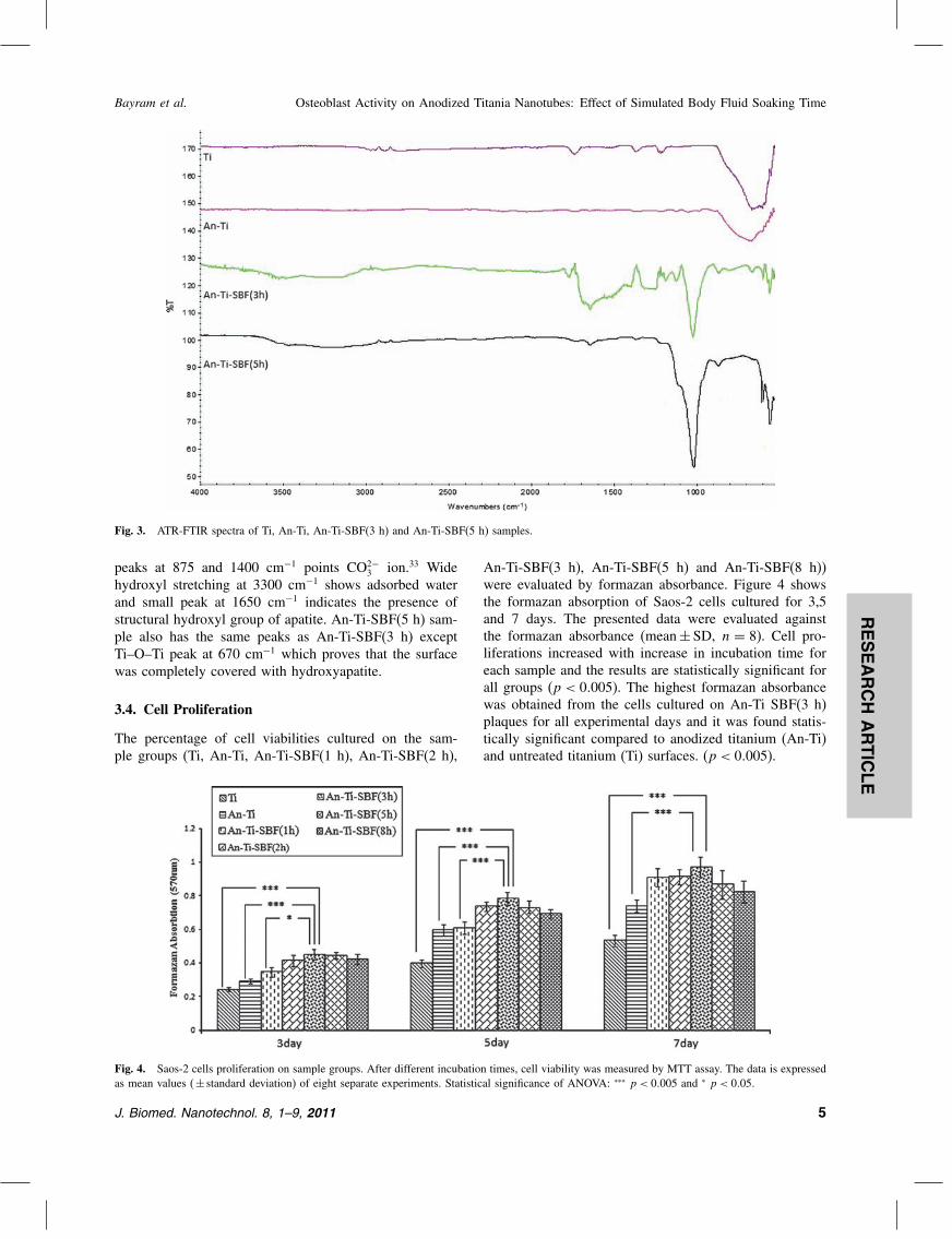
Fig. 3. ATR-FTIR spectra of Ti, An-Ti, An-Ti-SBF(3 h) and An-Ti-SBF(5 h) samples.
peaks at 875 and 1400 cm−1 points CO2−3 ion.33 Wide
hydroxyl stretching at 3300 cm−1 shows adsorbed waterand small peak at 1650 cm−1 indicates the presence ofstructural hydroxyl group of apatite. An-Ti-SBF(5 h) sam-ple also has the same peaks as An-Ti-SBF(3 h) exceptTi–O–Ti peak at 670 cm−1 which proves that the surfacewas completely covered with hydroxyapatite.
3.4. Cell Proliferation
The percentage of cell viabilities cultured on the sam-ple groups (Ti, An-Ti, An-Ti-SBF(1 h), An-Ti-SBF(2 h),
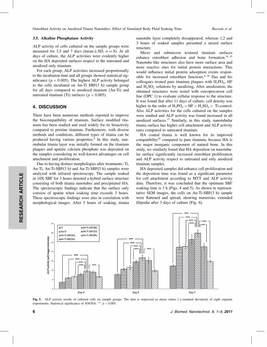
Fig. 4. Saos-2 cells proliferation on sample groups. After different incubation times, cell viability was measured by MTT assay. The data is expressedas mean values (± standard deviation) of eight separate experiments. Statistical significance of ANOVA: ∗∗∗ p < 0�005 and ∗ p < 0�05�
An-Ti-SBF(3 h), An-Ti-SBF(5 h) and An-Ti-SBF(8 h))were evaluated by formazan absorbance. Figure 4 showsthe formazan absorption of Saos-2 cells cultured for 3,5and 7 days. The presented data were evaluated againstthe formazan absorbance (mean± SD, n = 8). Cell pro-liferations increased with increase in incubation time foreach sample and the results are statistically significant forall groups (p < 0�005). The highest formazan absorbancewas obtained from the cells cultured on An-Ti SBF(3 h)plaques for all experimental days and it was found statis-tically significant compared to anodized titanium (An-Ti)and untreated titanium (Ti) surfaces. (p < 0�005).
J. Biomed. Nanotechnol. 8, 1–9, 2011 5
RESEARCH
ARTIC
LE
Osteoblast Activity on Anodized Titania Nanotubes: Effect of Simulated Body Fluid Soaking Time Bayram et al.
3.5. Alkaline Phosphatase Activity
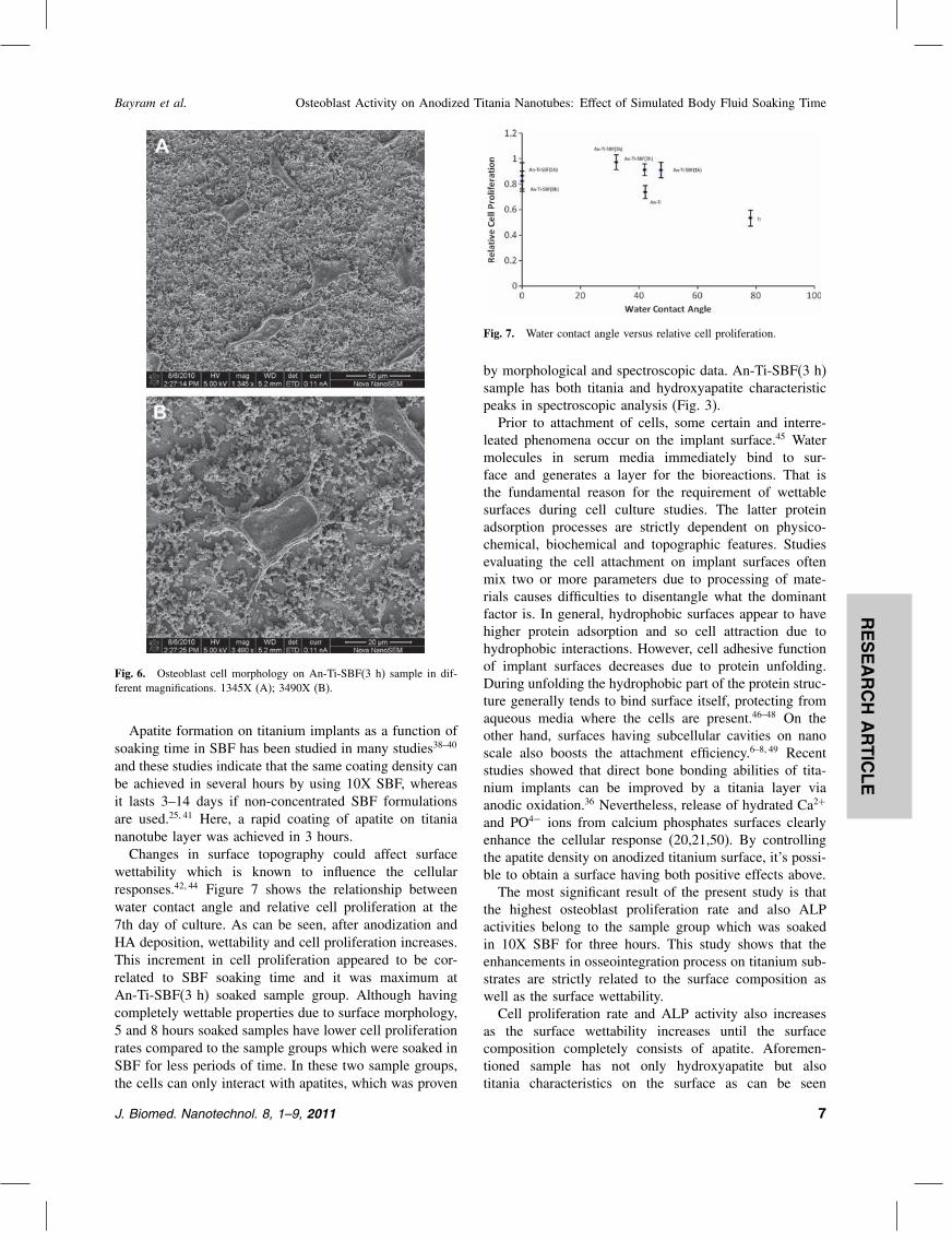
ALP activity of cells cultured on the sample groups weremeasured for 3,5 and 7 days (mean±SD, n = 8). At alldays of culture, the ALP activities were evidently higheron the HA deposited surfaces respect to the untreated andanodized only titanium.For each group, ALP activities increased proportionally
to the incubation time and all groups showed statistical sig-nificance (p < 0�005). The highest ALP activity belongedto the cells incubated on An-Ti SBF(3 h) sample groupfor all days compared to anodized titanium (An-Ti) anduntreated titanium (Ti) surfaces (p < 0�005).
4. DISCUSSION
There have been numerous methods reported to improvethe biocompatibility of titanium. Surface modified tita-nium has been studied and used widely for its bioactivitycompared to pristine titanium. Furthermore, with diversemethods and conditions, different types of titania can beproduced having various properties.8 In this study, nan-otubular titania layer was initially formed on the titaniumplaques and apatitic calcium phosphate was deposited onthe samples considering its well-known advantages on cellattachment and proliferation.Due to having distinct morphologies after treatments; Ti,
An-Ti, An-Ti-SBF(3 h) and An-Ti-SBF(5 h) samples wereanalyzed with infrared spectroscopy. The sample soakedin 10X SBF for 3 hours denoted a hybrid surface structureconsisting of both titania nanotubes and precipitated HA.The spectroscopic findings indicate that the surface onlyconsists of apatite when soaking time exceeds 3 hours.These spectroscopic findings were also in correlation withmorphological images. After 5 hours of soaking, titania
Fig. 5. ALP activity results of cultured cells on sample groups. The data is expressed as mean values (± standard deviation) of eight separateexperiments. Statistical significance of ANOVA: ∗∗∗ p < 0�005.
nanotube layer completely dissappeared, whereas 1,2 and3 hours of soaked samples presented a mixed surfacestructure.Micro and submicron textured titanium surfaces
enhance osteoblast adhesion and bone formation.34�35
Nanotube-like structures also have more surface area andmore reactive sites for initial protein interactions. Thiswould influence initial protein adsorption events respon-sible for increased osteoblast functions.6�36 Das and hiscolleagues treated pure titanium plaques with H3PO4, HFand H2SO4 solutions by anodizing. After anodization, theobtained structures were tested with osteoprecursor cellline (OPC 1) to evaluate cellular response to the structure.It was found that after 11 days of culture, cell density washigher in the order of H3PO4 >HF>H2SO4 > Ti-control.Also ALP activities for the cells cultured on the sampleswere studied and ALP activity was found increased in allanodized surfaces.37 Similarly, in this study, nanotubulartitania surface has higher cell attachment and ALP activityrates compared to untreated titanium.HA coated titania is well known for its improved
compatibility20 compared to pure titanium, because HA isthe major inorganic component of natural bone. In thisstudy, we similarly found that HA deposition on nanotubu-lar surface significantly increased osteoblast proliferationand ALP activity respect to untreated and only anodizedtitanium samples.HA deposited samples did enhance cell proliferation and
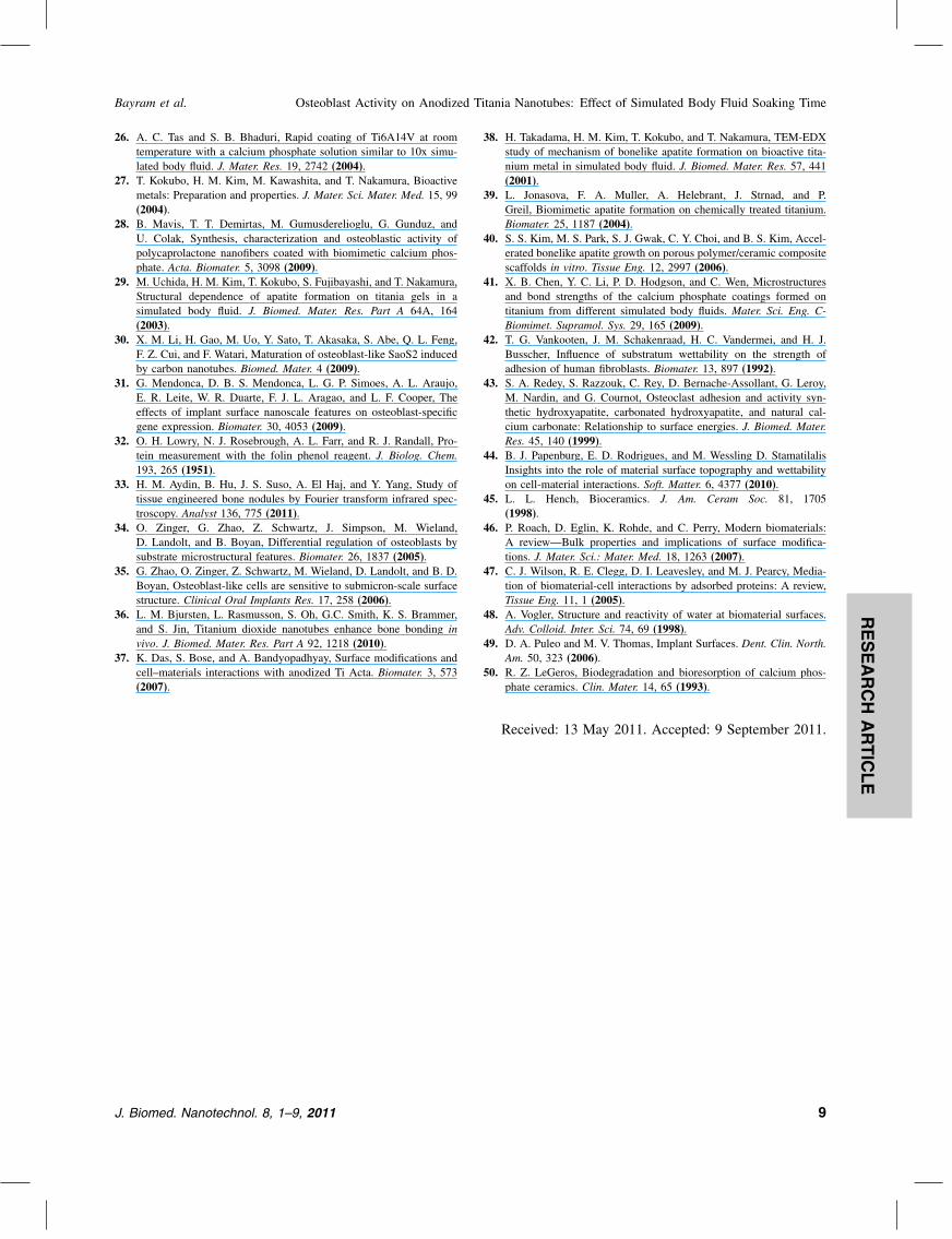
the deposition time was found as a significant parameterfor cell attachment according to MTT and ALP activitydata. Therefore, it was concluded that the optimum SBFsoaking time is 3 h (Figs. 4 and 5). As shown in represen-tative SEM images, the cells on An-Ti-SBF(3 h) samplewere flattened and spread, showing numerous, extendedfilipodia after 3 days of culture (Fig. 6).
6 J. Biomed. Nanotechnol. 8, 1–9, 2011
RESEARCH
ARTIC
LE
Bayram et al. Osteoblast Activity on Anodized Titania Nanotubes: Effect of Simulated Body Fluid Soaking Time
Fig. 6. Osteoblast cell morphology on An-Ti-SBF(3 h) sample in dif-ferent magnifications. 1345X (A); 3490X (B).
Apatite formation on titanium implants as a function ofsoaking time in SBF has been studied in many studies38–40
and these studies indicate that the same coating density canbe achieved in several hours by using 10X SBF, whereasit lasts 3–14 days if non-concentrated SBF formulationsare used.25�41 Here, a rapid coating of apatite on titaniananotube layer was achieved in 3 hours.Changes in surface topography could affect surface
wettability which is known to influence the cellularresponses.42�44 Figure 7 shows the relationship betweenwater contact angle and relative cell proliferation at the7th day of culture. As can be seen, after anodization andHA deposition, wettability and cell proliferation increases.This increment in cell proliferation appeared to be cor-related to SBF soaking time and it was maximum atAn-Ti-SBF(3 h) soaked sample group. Although havingcompletely wettable properties due to surface morphology,5 and 8 hours soaked samples have lower cell proliferationrates compared to the sample groups which were soaked inSBF for less periods of time. In these two sample groups,the cells can only interact with apatites, which was proven
Fig. 7. Water contact angle versus relative cell proliferation.
by morphological and spectroscopic data. An-Ti-SBF(3 h)sample has both titania and hydroxyapatite characteristicpeaks in spectroscopic analysis (Fig. 3).Prior to attachment of cells, some certain and interre-
leated phenomena occur on the implant surface.45 Watermolecules in serum media immediately bind to sur-face and generates a layer for the bioreactions. That isthe fundamental reason for the requirement of wettablesurfaces during cell culture studies. The latter proteinadsorption processes are strictly dependent on physico-chemical, biochemical and topographic features. Studiesevaluating the cell attachment on implant surfaces oftenmix two or more parameters due to processing of mate-rials causes difficulties to disentangle what the dominantfactor is. In general, hydrophobic surfaces appear to havehigher protein adsorption and so cell attraction due tohydrophobic interactions. However, cell adhesive functionof implant surfaces decreases due to protein unfolding.During unfolding the hydrophobic part of the protein struc-ture generally tends to bind surface itself, protecting fromaqueous media where the cells are present.46–48 On theother hand, surfaces having subcellular cavities on nanoscale also boosts the attachment efficiency.6–8�49 Recentstudies showed that direct bone bonding abilities of tita-nium implants can be improved by a titania layer viaanodic oxidation.36 Nevertheless, release of hydrated Ca2+
and PO4− ions from calcium phosphates surfaces clearlyenhance the cellular response (20,21,50). By controllingthe apatite density on anodized titanium surface, it’s possi-ble to obtain a surface having both positive effects above.The most significant result of the present study is that
the highest osteoblast proliferation rate and also ALPactivities belong to the sample group which was soakedin 10X SBF for three hours. This study shows that theenhancements in osseointegration process on titanium sub-strates are strictly related to the surface composition aswell as the surface wettability.Cell proliferation rate and ALP activity also increases
as the surface wettability increases until the surfacecomposition completely consists of apatite. Aforemen-tioned sample has not only hydroxyapatite but alsotitania characteristics on the surface as can be seen
J. Biomed. Nanotechnol. 8, 1–9, 2011 7

RESEARCH
ARTIC
LE
Osteoblast Activity on Anodized Titania Nanotubes: Effect of Simulated Body Fluid Soaking Time Bayram et al.
from vibrational spectra. Therefore, results of this studysuggested that the surface topography and chemistry result-ing from HA deposition and nanotubular structure aremajor factors that influence the ability of osteoblasts attach-ment, proliferation and ALP activity. Excess and/or uncon-trolled HA deposition and complete coverage of titanialayer decreases the potential of the implant.
5. CONCLUSION
In this study, the effect of rapid HA deposition on anodizedtitanium surface against osteoblast cell activity was inves-tigated. The results showed that the hybrid surface havingboth titania nanotube layer and calcium phosphate saltsmostly enhanced the cell proliferation and the ALP activityof cells as well (p < 0�005). Optimized rapid HA coatingsachieved on anodized titanium, which is a hybrid surface,is highly promising for enhancing the bioactive featuresof current titanium implants. In the last decade, bioac-tive implants having properties such as rapid osseointegra-tion or antibacterial properties have been intensely studied.Anodized nanotubular titania surface layers are consideredto be used as potential bioactive agent storages and elutingplatforms. Combining possible antibacterial and osteointe-grative features of titanium implants would make a greatimpact.
Acknowledgments: This study funded by HacettepeUniversity Scientific Research Projects Department.Project number: 010BIYP03601001. The authors alsowould like to thank Alpay Koray Mizrak from UNAM, forthe SEM images of samples and Zeynep Ekemen for thepreparation of this paper.
References and Notes
1. Y. Z. Yang, R. Cavin, and J. L. Ong, Protein adsorption on titaniumsurfaces and their effect on osteoblast attachment. J. Biomed. Mater.Res. 67A, 344 (2003).
2. V. Borsari, M. Fini, G. Giavaresi, L. Rimondini, U. Consolo,L. Chiusoli, A. Salito, A. Volpert, R. Chiesa, and R. Giardino,Osteointegration of titanium and hydroxyapatite rough surfaces inhealthy and compromised cortical and trabecular bone: In vivocomparative study on young, aged, and estrogen-deficient sheep.J. Orthop. Res. 25, 1250 (2007).
3. S. Fujibayashi, M. Neo, H. M. Kim, T. Kokubo, and T. Nakamura,Osteoinduction of porous bioactive titanium metal. Biomaterials25, 443 (2004).
4. P. Thomsen, C. Larsson, L. E. Ericson, L. Sennerby, J. Lausmaa, andB. Kasemo, Structure of the interface between rabbit cortical boneand implants of gold, zirconium and titanium. J. Mater. Sci. Mater.Med. 8, 653 (1997).
5. C. Aparicio, F. J. Gil, J. A. Planell, and E. Engel, Human-osteoblastproliferation and differentiation on grit-blasted and bioactive tita-nium for dental applications. J. Mater. Sci. Mater. Med. 13, 1105(2002).
6. C. Yao, E. B. Slamovich, and T. J. Webster, Enhanced osteoblastfunctions on anodized titanium with nanotube-like structures.J. Biomed. Mater. Res. 85A, 157 (2008).
7. M. D. Rong, L. Zhou, Z. H. Gou, A. D. Zhu, and D. F. Zhou,The early osseointegration of the laser-treated and acid-etched dentalimplants surface: An experimental study in rabbits. J. Mater. Sci.Mater. Med. 20, 1721 (2009).
8. C. Yao, V. Perla, J. L. McKenzie, E. B. Slamovich, and T. J. Webster,Anodized Ti and Ti6Al4V possessing nanometer surface featuresenhances osteoblast adhesion. J. Biomed. Nanotechnol. 1, 68 (2005).
9. A. Nanci, J. D. Wuest, L. Peru, P. Brunet, V. Sharma, S. Zalzal,and M. D. McKee, Chemical modification of titanium surfaces forcovalent attachment of biological molecules. J. Biomed. Mater. Res.40, 324 (1998).
10. L. Zhu, X. Ye, G. Tang, N. Zhao, Y. Gong, Y. Zhao, J. Zhao, andX. Zhang, Biomimetic coating of compound titania and hydroxyap-atite on titanium. J. Biomed. Mater. Res. P. A 83A, 1165 (2007).
11. Y. M. Lee, E. J. Lee, S. T. Yee, B. I. Kim, E. S. Choe, and H. W.Cho, In vivo and in vitro response to electrochemically anodizedTi-6Al-4V alloy. J. Mater. Sci. Mater. Med. 19, 1851 (2008).
12. C. Bayram, A. K. Mizrak, S. Akturk, H. Kursaklioglu, A. Iyisoy,A. Ifran, and E. B. Denkbas, In vitro biocompatibility of plasma-aided surface-modified 316L stainless steel for intracoronary stents.Biomed. Mater. 5 (2010).
13. A. Bagno and C. Di Bello, Surface treatments and roughness prop-erties of Ti-based biomaterials. J. Mater. Sci. Mater. Med. 15, 935(2004).
14. J. M. Macak, H. Tsuchiya, L. Taveira, A. Ghicov, and P. Schmuki,Self-organized nanotubular oxide layers on Ti-6A1-7Nb and Ti-6A1-4V formed by anodization in NH4F solutions. J. Biomed. Mater. Res.Part A 75A, 928 (2005).
15. Y. Li, D. Y. Ding, C. Q. Ning, S. O. Bai, L. Huang, M. Li, andD. L. Mao, Thermal stability and in vitro bioactivity of Ti-Al-V-Onanostructures fabricated on Ti6Al4V alloy. Nanotechnol 20 (2009).
16. L. L. Peng, A. D. Mendelsohn, T. J. LaTempa, S. Yoriya, C. A.Grimes, and T. A. Desai, Long-term small molecule and proteinelution from TiO2 nanotubes. Nano Lett. 9, 1932 (2009).
17. A. Kar, K. S. Raja, and M. Misra, Electrodeposition of hydroxya-patite onto nanotubular TiO2 for implant applications. Surf. Coat.Technol. 201, 3723 (2006).
18. A. Bigi, E. Boanini, B. Bracci, A. Facchini, S. Panzavolta, F. Segatti,and L. Sturba, Nanocrystalline hydroxyapatite coatings on titanium:a new fast biomimetic method. Biomater. 26, 4085 (2005).
19. H. M. Aydin, Y. Yang, T. Kohler, A. El Haj, R. Muller, andE. Piskin, Interaction of osteoblasts with macroporous scaffoldsmade of PLLA/PCL blends modified with collagen and hydroxyap-atite. Adv. Eng. Mater. 11, B83 (2009).
20. L. Meirelles, A. Arvidsson, M. Andersson, P. Kjellin, T. Albrektsson,and A. Wennerberg, Nano hydroxyapatite structures influence earlybone formation. J. Biomed. Mater. Res. Part A 87A, 299 (2008).
21. P. O’Hare, B. J. Meenan, G. A. Burke, G. Byrne, D. Dowling, andJ. A. Hunt, Biological responses to hydroxyapatite surfaces depositedvia a co-incident microblasting technique. Biomater. 31, 515 (2010).
22. L. Saruwatari, H. Aita, F. Butz, H. K. Nakamura, J. Ouyang, Y. Yang,W. A. Chiou, and T. Ogawa, Osteoblasts generate harder, stiffer, andmore delamination-resistant mineralized tissue on titanium than onpolystyrene, associated with distinct tissue micro- and ultrastructure.Journal of Bone and Mineral Research 20, 2002 (2005).
23. C. Bayram, E. B. Denkbas, E. Kilicay, B. Hazer, H. B. Cakmak,and I. Noda, Preparation and characterization of triamcinoloneacetonide—loaded poly(3-hydroxybutyrate-co-3-hydroxyhexanoate)(PHBHx) microspheres. J. Bioact. Compat. Polym. 23, 334 (2008).
24. D. Kavaz, S. Odabas, E. Guven, M. Demirbilek, and E. B. Denkbas,Bleomycin loaded magnetic chitosan nanoparticles as multifunc-tional nanocarriers. J. Bioact. Compat. Polym. 25, 305 (2010).
25. C. Yao and T. J. Webster, Prolonged Antibiotic Delivery FromAnodized Nanotubular Titanium Using a Co-precipitation DrugLoading Method. J. Biomed. Mater. Res. Part B-Appl. Biomater.91B, 587 (2009).
8 J. Biomed. Nanotechnol. 8, 1–9, 2011
RESEARCH
ARTIC
LE
Bayram et al. Osteoblast Activity on Anodized Titania Nanotubes: Effect of Simulated Body Fluid Soaking Time
26. A. C. Tas and S. B. Bhaduri, Rapid coating of Ti6A14V at roomtemperature with a calcium phosphate solution similar to 10x simu-lated body fluid. J. Mater. Res. 19, 2742 (2004).
27. T. Kokubo, H. M. Kim, M. Kawashita, and T. Nakamura, Bioactivemetals: Preparation and properties. J. Mater. Sci. Mater. Med. 15, 99(2004).
28. B. Mavis, T. T. Demirtas, M. Gumusderelioglu, G. Gunduz, andU. Colak, Synthesis, characterization and osteoblastic activity ofpolycaprolactone nanofibers coated with biomimetic calcium phos-phate. Acta. Biomater. 5, 3098 (2009).
29. M. Uchida, H. M. Kim, T. Kokubo, S. Fujibayashi, and T. Nakamura,Structural dependence of apatite formation on titania gels in asimulated body fluid. J. Biomed. Mater. Res. Part A 64A, 164(2003).
30. X. M. Li, H. Gao, M. Uo, Y. Sato, T. Akasaka, S. Abe, Q. L. Feng,F. Z. Cui, and F. Watari, Maturation of osteoblast-like SaoS2 inducedby carbon nanotubes. Biomed. Mater. 4 (2009).
31. G. Mendonca, D. B. S. Mendonca, L. G. P. Simoes, A. L. Araujo,E. R. Leite, W. R. Duarte, F. J. L. Aragao, and L. F. Cooper, Theeffects of implant surface nanoscale features on osteoblast-specificgene expression. Biomater. 30, 4053 (2009).
32. O. H. Lowry, N. J. Rosebrough, A. L. Farr, and R. J. Randall, Pro-tein measurement with the folin phenol reagent. J. Biolog. Chem.193, 265 (1951).
33. H. M. Aydin, B. Hu, J. S. Suso, A. El Haj, and Y. Yang, Study oftissue engineered bone nodules by Fourier transform infrared spec-troscopy. Analyst 136, 775 (2011).
34. O. Zinger, G. Zhao, Z. Schwartz, J. Simpson, M. Wieland,D. Landolt, and B. Boyan, Differential regulation of osteoblasts bysubstrate microstructural features. Biomater. 26, 1837 (2005).
35. G. Zhao, O. Zinger, Z. Schwartz, M. Wieland, D. Landolt, and B. D.Boyan, Osteoblast-like cells are sensitive to submicron-scale surfacestructure. Clinical Oral Implants Res. 17, 258 (2006).
36. L. M. Bjursten, L. Rasmusson, S. Oh, G.C. Smith, K. S. Brammer,and S. Jin, Titanium dioxide nanotubes enhance bone bonding invivo. J. Biomed. Mater. Res. Part A 92, 1218 (2010).
37. K. Das, S. Bose, and A. Bandyopadhyay, Surface modifications andcell–materials interactions with anodized Ti Acta. Biomater. 3, 573(2007).
38. H. Takadama, H. M. Kim, T. Kokubo, and T. Nakamura, TEM-EDXstudy of mechanism of bonelike apatite formation on bioactive tita-nium metal in simulated body fluid. J. Biomed. Mater. Res. 57, 441(2001).
39. L. Jonasova, F. A. Muller, A. Helebrant, J. Strnad, and P.Greil, Biomimetic apatite formation on chemically treated titanium.Biomater. 25, 1187 (2004).
40. S. S. Kim, M. S. Park, S. J. Gwak, C. Y. Choi, and B. S. Kim, Accel-erated bonelike apatite growth on porous polymer/ceramic compositescaffolds in vitro. Tissue Eng. 12, 2997 (2006).
41. X. B. Chen, Y. C. Li, P. D. Hodgson, and C. Wen, Microstructuresand bond strengths of the calcium phosphate coatings formed ontitanium from different simulated body fluids. Mater. Sci. Eng. C-Biomimet. Supramol. Sys. 29, 165 (2009).
42. T. G. Vankooten, J. M. Schakenraad, H. C. Vandermei, and H. J.Busscher, Influence of substratum wettability on the strength ofadhesion of human fibroblasts. Biomater. 13, 897 (1992).
43. S. A. Redey, S. Razzouk, C. Rey, D. Bernache-Assollant, G. Leroy,M. Nardin, and G. Cournot, Osteoclast adhesion and activity syn-thetic hydroxyapatite, carbonated hydroxyapatite, and natural cal-cium carbonate: Relationship to surface energies. J. Biomed. Mater.Res. 45, 140 (1999).
44. B. J. Papenburg, E. D. Rodrigues, and M. Wessling D. StamatilalisInsights into the role of material surface topography and wettabilityon cell-material interactions. Soft. Matter. 6, 4377 (2010).
45. L. L. Hench, Bioceramics. J. Am. Ceram Soc. 81, 1705(1998).
46. P. Roach, D. Eglin, K. Rohde, and C. Perry, Modern biomaterials:A review—Bulk properties and implications of surface modifica-tions. J. Mater. Sci.: Mater. Med. 18, 1263 (2007).
47. C. J. Wilson, R. E. Clegg, D. I. Leavesley, and M. J. Pearcy, Media-tion of biomaterial-cell interactions by adsorbed proteins: A review,Tissue Eng. 11, 1 (2005).
48. A. Vogler, Structure and reactivity of water at biomaterial surfaces.Adv. Colloid. Inter. Sci. 74, 69 (1998).
49. D. A. Puleo and M. V. Thomas, Implant Surfaces. Dent. Clin. North.Am. 50, 323 (2006).
50. R. Z. LeGeros, Biodegradation and bioresorption of calcium phos-phate ceramics. Clin. Mater. 14, 65 (1993).
Received: 13 May 2011. Accepted: 9 September 2011.
J. Biomed. Nanotechnol. 8, 1–9, 2011 9


