Embed Size (px)
Citation preview
Materials Science and Engineering C 49 (2015) 735–745
Contents lists available at ScienceDirect
Materials Science and Engineering C
j ourna l homepage: www.e lsev ie r .com/ locate /msec
Titania nanotube arrays as interfaces for neural prostheses
Jonathan A. Sorkin a, Stephen Hughes b,c, Paulo Soares d, Ketul C. Popat a,c,⁎a Department of Mechanical Engineering, Colorado State University, Fort Collins CO 80523, USAb Department of Chemical and Biological Engineering, Colorado State University, Fort Collins CO 80523, USAc School of Biomedical Engineering, Colorado State University, Fort Collins CO 80523, USAd Department of Mechanical Engineering, Polytechnic School, Pontifícia Universidade Católica do Paraná, Curitiba, PR 80215-901, Brazil
⁎ Corresponding author at: Department of MechanicUniversity, Fort Collins, CO 80523, USA.
E-mail address: [email protected] (K.C. Popat
http://dx.doi.org/10.1016/j.msec.2015.01.0770928-4931/© 2015 Elsevier B.V. All rights reserved.
a b s t r a c t
a r t i c l e i n f oArticle history:Received 11 August 2014Received in revised form 8 December 2014Accepted 23 January 2015Available online 26 January 2015
Keywords:Titania nanotube arraysC17.2 neural stem cell lineNeural prosthesesNeuronsAstrocytes
Neural prostheses have become ever more acceptable treatments for many different types of neurologicaldamage and disease. Here we investigate the use of two different morphologies of titania nanotube arrays as in-terfaces to advance the longevity and effectiveness of these prostheses. The nanotube arrays were characterizedfor their nanotopography, crystallinity, conductivity,wettability, surfacemechanical properties and adsorption ofkey proteins: fibrinogen, albumin and laminin. The loosely packed nanotube arrays fabricated using a diethyleneglycol based electrolyte, contained a higher presence of the anatase crystal phase and were subsequently moreconductive. These arrays yielded surfaces with higher wettability and lower modulus than the densely packednanotube arrays fabricated using water based electrolyte. Further the adhesion, proliferation and differentiationof the C17.2 neural stem cell line was investigated on the nanotube arrays. The proliferation ratio of the cells aswell as the level of neuronal differentiationwas seen to increase on the loosely packed arrays. The results indicatethat loosely packed nanotube arrays similar to the ones produced herewith a DEG based electrolyte, may providea favorable template for growth and maintenance of C17.2 neural stem cell line.
© 2015 Elsevier B.V. All rights reserved.
1. Introduction
Neural prostheses have become evermore acceptable treatments formany different types of neurological damage and disease. There arethree main types of neural prostheses. Auditory and visual prosthesescan act on various neural populations in an attempt to emulate thenatural signaling from the auditory or visual pathways [1–4]. Sensoryand motor control prostheses are fully implantable complex electricalarrays often referred to as brain machine interfaces (BMIs). They areused to translate neuronal activity across damaged neural networks inorder to restore sensation and motor control in limbs [5–7]. Cognitiveprostheses electrically stimulate deep lying brain structures to alleviatesymptoms of neurodegenerative diseases [8–10]. For example,Parkinson' disease is one of the most common neurodegenerative dis-eases which results in the loss of movement control and coordination,it is predicted to affect between 8.7 and 9.3 million people worldwideby 2030 [11,12]. Deep brain stimulation (DBS) is a form of cognitiveprostheses used in the treatment of Parkinson's disease. DBS worksthrough electrical stimulation at 1–5 V 120–180 Hz, and pulse width'sof 60–200 μs in order to interferewith the overactive signaling triggeredby dopamine depletion in the degenerative neural population [13,14].However the longevity and effectiveness of these prostheses are
al Engineering, Colorado State
).
reduced as a result of the immune response. The response occurs inthe form of a compact sheath of glial cells around the prostheses,which electrically segregates it from the target neurons by 50–200 μm[15,16]. This is referred to as glial encapsulation or reactive gliosis. It isthe result of the initial trauma from prosthesis implantation, as well asthe recognition of the prosthesis surface as a foreign body by the cells.Gliosis is recognized by the increased expression of proteins specific toastrocytes and astroglia, which are known to assist in the formation ofthe blood brain barrier, and play a role in the repair and scarring ofneural tissue after trauma [17–19]. The encapsulation of the prosthesesby these cells drastically limits electrical signaling as the neurons areforced further away from the surface of prostheses [20]. In attempt toreduce gliosis, different prosthesis geometries, sizes, and surface rough-nesses have been examined, with device size having the most signifi-cant effect by reducing the volume disturbed during implantation[16]. Nanotopographies such as nanotubes have already been shownto improve cell adhesion and growth of cells [21]. Studies have shownthat nanoporous silicon promotes neuronal adhesion while limitinggliosis [22]. Submicron machining on the order of 10–70 nm has beenshown to direct neurite outgrowth [23,24]. Despite these advance-ments, there is still a need to develop robust interfaces that increaseprosthesis effectiveness and longevity.
Titanium has proven biocompatibility and has been used for a varietyof biomedical applications. Although it is not often used in neurologicalapplications, its ability to be produced as thin films on other materialsmakes it a good candidate for neurological applications. Even in its thin
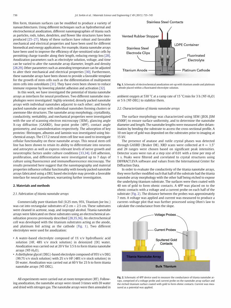
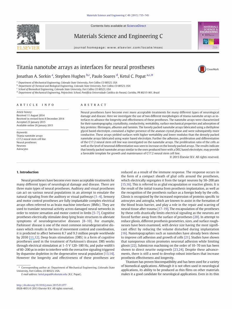
Fig. 1. Schematic of electrochemical anodization set-upwith titanium anode and platinumcathode placed within a fluorinated electrolyte solution.
Fig. 2. Schematic of 4PP device used to measure the conductance of titania nanotube ar-rays, comprised of a voltage probe and current probe on the nanotube array surface andthe etched titanium surface coated with gold to form ohmic contacts. Current was mea-sured as a potential was applied.
736 J.A. Sorkin et al. / Materials Science and Engineering C 49 (2015) 735–745
film form, titanium surfaces can be modified to produce a variety ofnanoarchitectures. Using different techniques such as hydrothermal andelectrochemical anodization, different nanotopographies of titania suchas particles, rods, tubes, dendrites, and flower like structures have beenproduced [25–27]. Many of these surfaces have robust and favorablemechanical and electrical properties and have been used for differentbiomedical and energy applications. For example, titania nanotube arrayshave been used to improve the efficiency of dye-sensitized solar cells bypermitting charge transfer along their length, reducing energy loss [28].Anodization parameters such as electrolyte solution, voltage, and timecan be varied to alter the nanotube array diameter, length and density[28,29]. Other parameters such as annealing temperature can be adjustedto alter their mechanical and electrical properties [30]. Furthermore,these nanotube arrays have been shown to provide a favorable templatefor the growth of stem cells such as the differentiation of multipotentstem cells into osteoblasts [31]. They have even been shown to reduceimmune response by lowering platelet adhesion and activation [32].
In this work, we have investigated the potential of titania nanotubearrays as interfaces for neural prostheses. Two different nanotube mor-phologies were investigated: highly oriented, densely packed nanotubearrays with individual nanotubes adjacent to each other; and looselypacked nanotube arrays with individual nanotubes forming clusters oranemone-like structures. The nanotube arraymorphology, crystallinity,conductivity, wettability, and mechanical properties were investigatedwith the use of scanning electron microscopy (SEM), glancing angleX-ray diffraction (GAXRD), four-point probe (4PP), contact anglegoniometry, and nanoindentation respectively. The adsorption of keyproteins: fibrinogen, albumin and laminin was investigated using bio-chemical assays. The C17.2 neural stem cell line was used to investigatecellular functionality on these nanotube arrays. This neural stem cellline has been shown to retain its ability to differentiate into neuronsand astrocytes as well as express relevant levels of nerve growth andneurotrophic factors under culture conditions [33,34]. Cell adhesion,proliferation, and differentiation were investigated up to 7 days ofculture using fluorescence and immunofluorescence microscopy. Theresults presented here suggest that the nanotopography and materialproperties influence cellular functionalitywith loosely packed nanotubearrays fabricated using a DEG based electrolyte may provide a favorableinterface for neural prostheses, warranting further investigation.
2. Materials and methods
2.1. Fabrication of titania nanotube arrays
Commercially pure titanium foil (0.25 mm, 95%, Titanium Joe Inc.)was cut into rectangular substrates of 2 cm × 2.5 cm. These substrateswere cleaned in acetone, soap, and isopropyl alcohol. Titania nanotubearrays were fabricated on these substrates using an electrochemical an-odization process previously described [28,35,36]. An electrochemicalcell was developed with the titanium substrates acting as the anode,and platinum foil acting as the cathode (Fig. 1). Two differentelectrolytes were used for anodization:
• A water-based electrolyte composed of 1% v/v hydrofluoric acidsolution (HF, 48% v/v stock solution) in deionized (DI) water.Anodization was carried out at 20 V for 3.5 h to form titania nanotubearrays (NT-H2O).
• Adiethylene glycol (DEG)-based electrolyte composed of 95% v/vDEG(99.7% v/v stock solution) with 2% v/v HF (48% v/v stock solution) inDI water. Anodization was carried out at 60 V for 23 h to form titaniananotube arrays (NT-DEG).
All experiments were carried out at room temperature (RT). Follow-ing anodization, the nanotube arrays were rinsed 3 times with DI waterand driedwith nitrogen gas. The nanotube arrayswere then annealed in
ambient oxygen at 530 °C at a ramp rate of 15 °C/min for 3 h (NT-H2O)or 5 h (NT-DEG) to stabilize them.
2.2. Characterization of titania nanotube arrays
The surface morphology was characterized using SEM (JEOL JSM6500F) to ensure surface uniformity, and to determine the nanotubediameter and length. The nanotube lengthsweremeasured after delam-ination by bending the substrate to access the cross-sectional profile. A10 nm layer of gold was deposited on the substrates prior to imaging at15 kV.
The presence of anatase and rutile crystal phases was detectedthrough GAXRD (Bruker D8). XRD scans were collected at θ = 1.5°and 2θ ranges were chosen based on significant peak intensities.Detector scans were run at a step size of 0.01 with a time per step of1 s. Peaks were filtered and correlated to crystal structures usingDIFFRACT.EVA software and values from the International Center forDiffraction Data.
In order to evaluate the conductivity of the titania nanotube arrays,theywere furthermodified such that half of the substrate had the titaniananotube array morphology with the other half being etched to exposethe underlying titanium substrate. The surfaces were then coated with40 nm of gold to form ohmic contacts. A 4PP was placed on to theohmic contacts with a voltage and a current probe on each half of thesubstrate (Fig. 2). The distance between the probes was approximately7 mm. A voltage was applied and current was measured to produce acurrent–voltage plot that was further processed using Ohm's law tocalculate the conductance from the slope.
737J.A. Sorkin et al. / Materials Science and Engineering C 49 (2015) 735–745
Surface wettability or contact angles weremeasured using the staticsessile water-drop method on a contact angle goniometer (Rame-hart250). A predetermined volume of water was dropped onto the surfacesand images of the water droplet were immediately captured. Theimages were analyzed to determine the contact angles. The contactangles were correlated to surface energy using the followingequation [37]:
Es ¼ Elv cosθ
where Es is the surface energy, Elv=72.8mJ/m2 (energy of liquid/vaporinterface) at 20 °C for DIwater, and θ represents the static contact angle.
Nanoindentation was performed using a Nanoindenter (XP, MTS)with a spherical tip of 100-micron radius (for measuring the elasticmodulus), and a Berkovich tip (for measuring hardness). Indentationswere made under two conditions: 1 load-unload cycle reaching amaximum applied load of 1 mN, and a set of 6 loading cycles doublingfrom 1.25 mN to 50 mN. The elastic modulus was calculated based offthe spherical tip indentation using the Oliver and Pharr method [38]:
1Eeff
¼ 1−v2
Eþ 1−v2i
Ei
where Eeff is the effective elastic modulus, E is the elastic modulus of thematerial, Ei is the elastic modulus of indenter material and ν is thePoisson's ratio. The hardness was calculated based off the Berkovichtip indentations using the Oliver and Pharr method [38]:
H ¼ Pmax
A
where, H is the hardness, Pmax is the maximum load and A is the cross-sectional area.
2.3. Protein adsorption on titania nanotube arrays
To understand how proteins adsorb on titania nanotube arrays;fibrinogen (Sigma), albumin (Sigma), and laminin (BD Biosciences)adsorption was investigated. All substrates were cut into squares of1 cm × 1 cm. They were then sterilized by incubation in 70% ethanolfor 30 min followed by 3 rinses in phosphate buffer solution (PBS,10×) and 30 min of UV exposure. Following sterilization the substrateswere incubated in 100 μg/mL of protein solutions in 24 well plates on ahorizontal shaker plate at 100 rpm at RT for 2 h. The protein solutionwas aspirated and the substrates were rinsed 2 times with PBS. Theywere then incubated in 1% v/v sodium dodecyl sulfate (SDS, Sigma)solution in PBS on a shaker plate at 100 rpm for 4 h to desorb theproteins. The protein concentrations in this solution were measuredusing a commercially available micro-BCA assay (Pierce Biotechnology)and a plate reader (BMG Labtech).
2.4. C17.2 cell culture
Murine neural stem cell-like subclone C17.2 generously provided byDr. Evan Y. Snyder's group at the Sanford Burnham Medical ResearchInstitute were used in this study. C17.2 cells were cultured at 37 °C ina 5% CO2 atmosphere in 100 mm × 20 mm polystyrene vented tissue-culture petri dishes. A growth media consisting of DMEM containinghigh glucose (4500 mg/L), L-glutamine and sodium pyruvate, andsupplemented with 10% fetal bovine serum, 5% horse serum, 1% L-glutamine and 1% Penicillin/Streptomycin/Fungizone was used. Cellsof passage 3 were used for subsequent studies. Prior to seeding thecells, the substrates were incubated in 70% ethanol for 30 min followedby 2 rinses in PBS and 30 min of UV exposure. The cells were seededonto substrates at a concentration of 1500 cells/well in 24 well platesand were cultured at 37 °C in a 5% CO2.
2.5. C17.2 adhesion and proliferation on titania nanotube arrays
The C17.2 cell response was investigated after 1, 4 and 7 days of cul-ture in growthmedia. Cell adhesion and proliferationwere evaluated bystaining the cells with 5-chloromethylfluorescein diacetate (CMFDA,Life Technologies), rhodamine phalloidin, and 4′, 6-diamidino-2-phenylindole (DAPI, Invitrogen) to visualize cytoplasm, cytoskeleton,and nucleus respectively using a fluorescence microscope (Zeiss).Prior to staining unadhered cells were aspirated and the substrateswere gently rinsed 2 times with PBS before being transferred to a new24-well plate. The substrates were then incubated at 37 °C in 5% CO2
in a 10 μM solution of CMFDA in PBS for 45 min. Following this incuba-tion, the solution was aspirated and the substrates were incubated ingrowth media at 37 °C in 5% CO2 for 30 min. The media was then aspi-rated and the substrates were rinsed once in PBS before being trans-ferred to a new 24-well plate where the cells were fixed in a 3.7% w/vformaldehyde solution in DI water for 15 min at RT. The fixative wasthen aspirated and the substrates were rinsed 3 times in PBS for 5 minper rinse before being transferred to a new 24-well plate. The cellswere permeabilized in a 1% v/v Triton-X solution in water for 3 min atRT. The permeative was aspirated and the substrates were rinsed andtransferred to a new 24 well plate where they were incubated at 37 °Cin 5% CO2 in a 5 μL/mL rhodamine-phalloidin solution in DI water for25 min at RT before DAPI was added to the solution at a concentrationof 1 μL/mL and were incubated for additional 5 min. The solution wasthen aspirated and the substrates were rinsed 2 times in PBS beforebeing stored in PBS in a light resistant container at 20 °C until imaging.Analysis of the fluorescence images was performed with ImageJsoftware.
2.6. Differentiation marker protein expression and cell morphology on tita-nia nanotube arrays
C17.2 cell differentiation was investigated after 7 days of culture indifferentiation media. The cells were initially seeded in growth media,which was then replaced with differentiation media after 1 day of cul-ture. The differentiation media was formulated to encourage differenti-ation towards astrocytic and neuronal lineages [39]. The mediaconsisted of a 1:1 ratio of DMEM containing high glucose (4500mg/L), L-glutamine, sodiumpyruvate, andHam's F-12NutrientMixture;and supplemented with 1% L-glutamine, 1% Penicillin/Streptomycin/Fungizone, N-2 supplement, glial derived neurotropic factor (GDNF,10 ng/mL) and nerve growth factor (NGF, 10 ng/mL).
After 4 and 7 days of culture, indirect immunofluorescence stainingwas performed to measure the level of cellular differentiation throughmarker protein expression. The C17.2 cells were immunostained forthe presence of neuronal marker: light neurofilament (NF-L), astrocytemarker: aldehyde dehydrogenase 1 family member L1 (ALDH1L1), andneural precursor: Nestin.
Prior to staining, unadhered cells were aspirated and the substrateswere gently rinsed 2 times with PBS before being transferred to a new24-well platewhere theywere fixed using a 3.7%w/v formaldehyde so-lution in DI water for 15 min at RT. The fixative was then aspirated andthe substrates were rinsed 3 times in PBS for 5 min per rinse beforebeing transferred to a new 24-well plate. The cells were permeabilizedin a 1% v/v Triton-X solution in water for 3 min at RT. The permeativewas aspirated and the substrates were rinsed and transferred to a new24 well plate where they were incubated in 10% blocking serum inPBS for 30 min at RT to block any non-specific binding. The substrateswere then rinsed and transferred to a new 24-well plate and incubatedat 20 °C overnight in a primary antibody solution of either NF-L orALDH1L1 (rabbit polyclonal, 1:1000, Neuromics) with Nestin (mousemonoclonal, 1:250, Neuromics) with 2% blocking serum in PBS. Thesubstrates were rinsed 3 times with PBS for 5 min per wash. Theywere then incubated for 1 h at RT with appropriate fluorescentlylabeled secondary-antibody solution (bovine ant-rabbit IgG-TR for
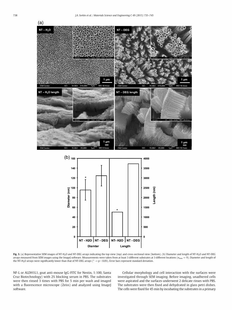
Fig. 3. (a) Representative SEM images of NT-H2O and NT-DEG arrays indicating the top view (top) and cross-sectional view (bottom). (b) Diameter and length of NT-H2O and NT-DEGarrays measured from SEM images using the ImageJ software. Measurements were taken from at least 3 different substrates at 3 different locations (nmin = 9). Diameter and length ofthe NT-H2O arrays were significantly lower than that of NT-DEG arrays (* → p b 0.05). Error bars represent standard deviation.
738 J.A. Sorkin et al. / Materials Science and Engineering C 49 (2015) 735–745
NF-L or ALDH1L1, goat anti-mouse IgG-FITC for Nestin, 1:100, SantaCruz Biotechnology) with 2% blocking serum in PBS. The substrateswere then rinsed 3 times with PBS for 5 min per wash and imagedwith a fluorescence microscope (Zeiss) and analyzed using ImageJsoftware.
Cellular morphology and cell interaction with the surfaces wereinvestigated through SEM imaging. Before imaging, unadhered cellswere aspirated and the surfaces underwent 2 delicate rinses with PBS.The substrates were then fixed and dehydrated in glass petri dishes.The cellswere fixed for 45min by incubating the substrates in a primary
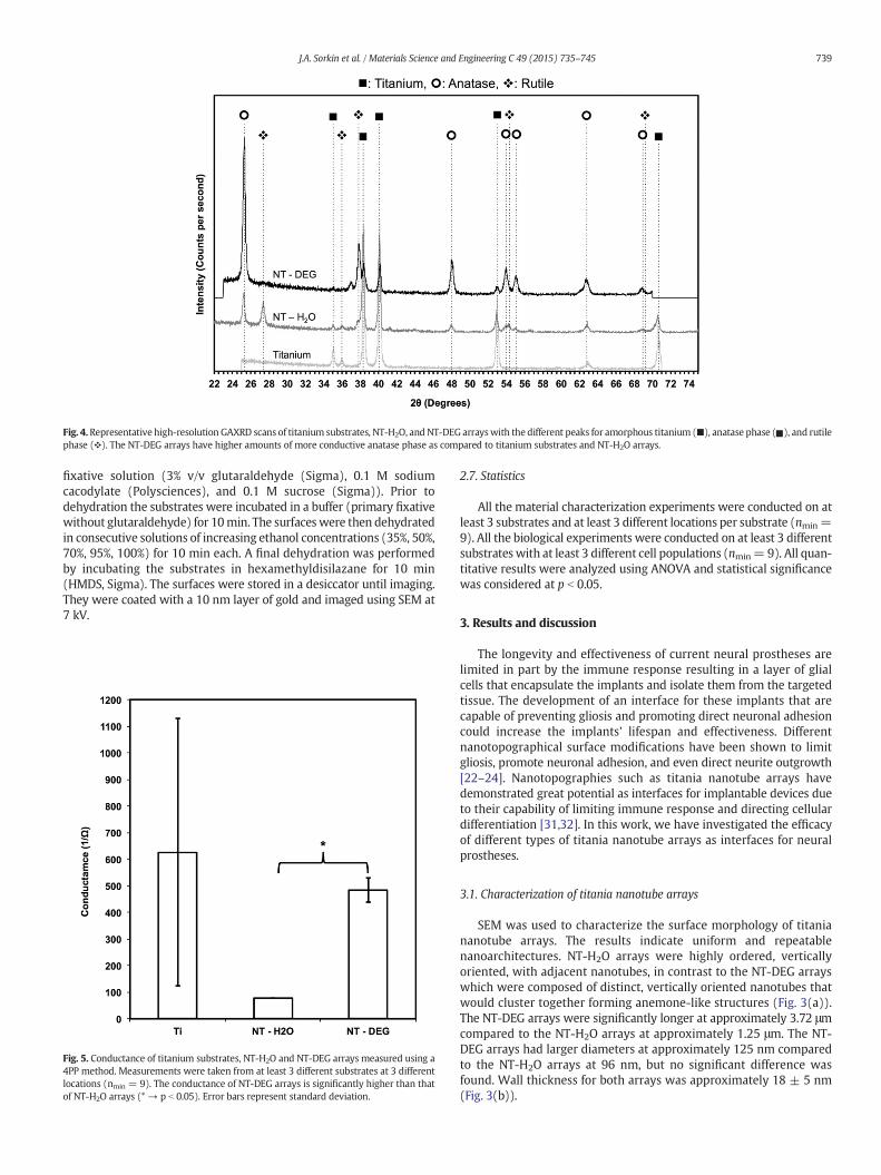
Fig. 4.Representative high-resolution GAXRD scans of titanium substrates, NT-H2O, and NT-DEG arrayswith the different peaks for amorphous titanium (■), anatase phase ( ), and rutilephase (❖). The NT-DEG arrays have higher amounts of more conductive anatase phase as compared to titanium substrates and NT-H2O arrays.
739J.A. Sorkin et al. / Materials Science and Engineering C 49 (2015) 735–745
fixative solution (3% v/v glutaraldehyde (Sigma), 0.1 M sodiumcacodylate (Polysciences), and 0.1 M sucrose (Sigma)). Prior todehydration the substrates were incubated in a buffer (primary fixativewithout glutaraldehyde) for 10min. The surfaceswere then dehydratedin consecutive solutions of increasing ethanol concentrations (35%, 50%,70%, 95%, 100%) for 10 min each. A final dehydration was performedby incubating the substrates in hexamethyldisilazane for 10 min(HMDS, Sigma). The surfaces were stored in a desiccator until imaging.They were coated with a 10 nm layer of gold and imaged using SEM at7 kV.
Fig. 5. Conductance of titanium substrates, NT-H2O and NT-DEG arrays measured using a4PP method. Measurements were taken from at least 3 different substrates at 3 differentlocations (nmin = 9). The conductance of NT-DEG arrays is significantly higher than thatof NT-H2O arrays (* → p b 0.05). Error bars represent standard deviation.
2.7. Statistics
All the material characterization experiments were conducted on atleast 3 substrates and at least 3 different locations per substrate (nmin=9). All the biological experiments were conducted on at least 3 differentsubstrates with at least 3 different cell populations (nmin= 9). All quan-titative results were analyzed using ANOVA and statistical significancewas considered at p b 0.05.
3. Results and discussion
The longevity and effectiveness of current neural prostheses arelimited in part by the immune response resulting in a layer of glialcells that encapsulate the implants and isolate them from the targetedtissue. The development of an interface for these implants that arecapable of preventing gliosis and promoting direct neuronal adhesioncould increase the implants' lifespan and effectiveness. Differentnanotopographical surface modifications have been shown to limitgliosis, promote neuronal adhesion, and even direct neurite outgrowth[22–24]. Nanotopographies such as titania nanotube arrays havedemonstrated great potential as interfaces for implantable devices dueto their capability of limiting immune response and directing cellulardifferentiation [31,32]. In this work, we have investigated the efficacyof different types of titania nanotube arrays as interfaces for neuralprostheses.
3.1. Characterization of titania nanotube arrays
SEM was used to characterize the surface morphology of titaniananotube arrays. The results indicate uniform and repeatablenanoarchitectures. NT-H2O arrays were highly ordered, verticallyoriented, with adjacent nanotubes, in contrast to the NT-DEG arrayswhich were composed of distinct, vertically oriented nanotubes thatwould cluster together forming anemone-like structures (Fig. 3(a)).The NT-DEG arrays were significantly longer at approximately 3.72 μmcompared to the NT-H2O arrays at approximately 1.25 μm. The NT-DEG arrays had larger diameters at approximately 125 nm comparedto the NT-H2O arrays at 96 nm, but no significant difference wasfound. Wall thickness for both arrays was approximately 18 ± 5 nm(Fig. 3(b)).
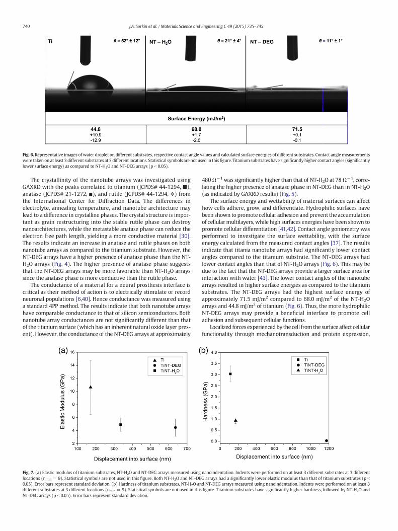
Fig. 6. Representative images of water droplet on different substrates, respective contact angle values and calculated surface energies of different substrates. Contact anglemeasurementswere taken on at least 3 different substrates at 3 different locations. Statistical symbols are not used in this figure. Titanium substrates have significantly higher contact angles (significantlylower surface energy) as compared to NT-H2O and NT-DEG arrays (p b 0.05).
740 J.A. Sorkin et al. / Materials Science and Engineering C 49 (2015) 735–745
The crystallinity of the nanotube arrays was investigated usingGAXRD with the peaks correlated to titanium (JCPDS# 44-1294, ■),anatase (JCPDS# 21-1272, ), and rutile (JCPDS# 44-1294, ❖) fromthe International Center for Diffraction Data. The differences inelectrolyte, annealing temperature, and nanotube architecture maylead to a difference in crystalline phases. The crystal structure is impor-tant as grain restructuring into the stable rutile phase can destroynanoarchitectures, while the metastable anatase phase can reduce theelectron free path length, yielding a more conductive material [30].The results indicate an increase in anatase and rutile phases on bothnanotube arrays as compared to the titanium substrate. However, theNT-DEG arrays have a higher presence of anatase phase than the NT-H2O arrays (Fig. 4). The higher presence of anatase phase suggeststhat the NT-DEG arrays may be more favorable than NT-H2O arrayssince the anatase phase is more conductive than the rutile phase.
The conductance of a material for a neural prosthesis interface iscritical as their method of action is to electrically stimulate or recordneuronal populations [6,40]. Hence conductance was measured usinga standard 4PP method. The results indicate that both nanotube arrayshave comparable conductance to that of silicon semiconductors. Bothnanotube array conductances are not significantly different than thatof the titanium surface (which has an inherent natural oxide layer pres-ent). However, the conductance of the NT-DEG arrays at approximately
Fig. 7. (a) Elastic modulus of titanium substrates, NT-H2O and NT-DEG arrays measured usinglocations (nmin = 9). Statistical symbols are not used in this figure. Both NT-H2O and NT-DE0.05). Error bars represent standard deviation. (b) Hardness of titanium substrates, NT-H2O andifferent substrates at 3 different locations (nmin = 9). Statistical symbols are not used in thisNT-DEG arrays (p b 0.05). Error bars represent standard deviation.
480Ω−1 was significantly higher than that of NT-H2O at 78Ω−1, corre-lating the higher presence of anatase phase in NT-DEG than in NT-H2O(as indicated by GAXRD results) (Fig. 5).
The surface energy and wettability of material surfaces can affecthow cells adhere, grow, and differentiate. Hydrophilic surfaces havebeen shown to promote cellular adhesion and prevent the accumulationof cellularmultilayers, while high surfaces energies have been shown topromote cellular differentiation [41,42]. Contact angle goniometry wasperformed to investigate the surface wettability, with the surfaceenergy calculated from the measured contact angles [37]. The resultsindicate that titania nanotube arrays had significantly lower contactangles compared to the titanium substrate. The NT-DEG arrays hadlower contact angles than that of NT-H2O arrays (Fig. 6). This may bedue to the fact that the NT-DEG arrays provide a larger surface area forinteraction with water [43]. The lower contact angles of the nanotubearrays resulted in higher surface energies as compared to the titaniumsubstrates. The NT-DEG arrays had the highest surface energy ofapproximately 71.5 mJ/m2 compared to 68.0 mJ/m2 of the NT-H2Oarrays and 44.8 mJ/m2 of titanium (Fig. 6). Thus, the more hydrophilicNT-DEG arrays may provide a beneficial interface to promote celladhesion and subsequent cellular functions.
Localized forces experiencedby the cell from the surface affect cellularfunctionality through mechanotransduction and protein expression,
nanoindentation. Indents were performed on at least 3 different substrates at 3 differentG arrays had a significantly lower elastic modulus than that of titanium substrates (p b
d NT-DEG arrays measured using nanoindentation. Indents were performed on at least 3figure. Titanium substrates have significantly higher hardness, followed by NT-H2O and
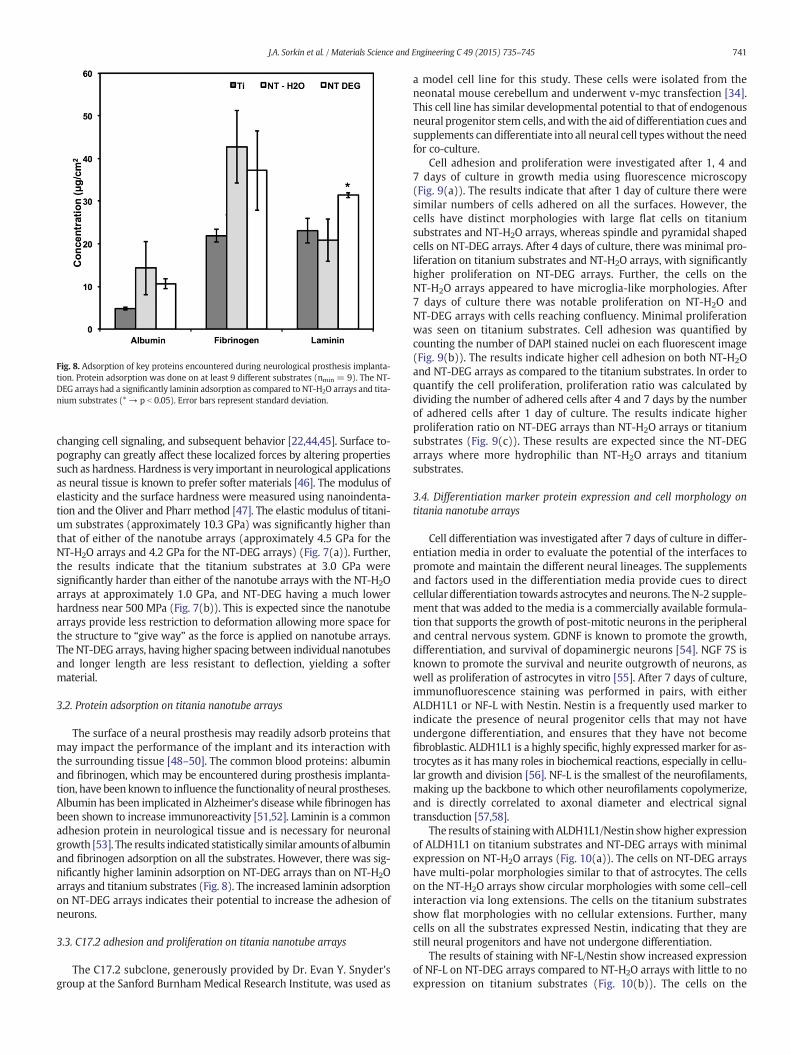
Fig. 8. Adsorption of key proteins encountered during neurological prosthesis implanta-tion. Protein adsorption was done on at least 9 different substrates (nmin = 9). The NT-DEG arrays had a significantly laminin adsorption as compared to NT-H2O arrays and tita-nium substrates (* → p b 0.05). Error bars represent standard deviation.
741J.A. Sorkin et al. / Materials Science and Engineering C 49 (2015) 735–745
changing cell signaling, and subsequent behavior [22,44,45]. Surface to-pography can greatly affect these localized forces by altering propertiessuch as hardness. Hardness is very important in neurological applicationsas neural tissue is known to prefer softer materials [46]. The modulus ofelasticity and the surface hardness were measured using nanoindenta-tion and the Oliver and Pharr method [47]. The elastic modulus of titani-um substrates (approximately 10.3 GPa) was significantly higher thanthat of either of the nanotube arrays (approximately 4.5 GPa for theNT-H2O arrays and 4.2 GPa for the NT-DEG arrays) (Fig. 7(a)). Further,the results indicate that the titanium substrates at 3.0 GPa weresignificantly harder than either of the nanotube arrays with the NT-H2Oarrays at approximately 1.0 GPa, and NT-DEG having a much lowerhardness near 500 MPa (Fig. 7(b)). This is expected since the nanotubearrays provide less restriction to deformation allowing more space forthe structure to “give way” as the force is applied on nanotube arrays.The NT-DEG arrays, having higher spacing between individual nanotubesand longer length are less resistant to deflection, yielding a softermaterial.
3.2. Protein adsorption on titania nanotube arrays
The surface of a neural prosthesis may readily adsorb proteins thatmay impact the performance of the implant and its interaction withthe surrounding tissue [48–50]. The common blood proteins: albuminand fibrinogen, which may be encountered during prosthesis implanta-tion, have been known to influence the functionality of neural prostheses.Albumin has been implicated in Alzheimer's diseasewhile fibrinogen hasbeen shown to increase immunoreactivity [51,52]. Laminin is a commonadhesion protein in neurological tissue and is necessary for neuronalgrowth [53]. The results indicated statistically similar amounts of albuminand fibrinogen adsorption on all the substrates. However, there was sig-nificantly higher laminin adsorption on NT-DEG arrays than on NT-H2Oarrays and titanium substrates (Fig. 8). The increased laminin adsorptionon NT-DEG arrays indicates their potential to increase the adhesion ofneurons.
3.3. C17.2 adhesion and proliferation on titania nanotube arrays
The C17.2 subclone, generously provided by Dr. Evan Y. Snyder'sgroup at the Sanford BurnhamMedical Research Institute, was used as
a model cell line for this study. These cells were isolated from theneonatal mouse cerebellum and underwent v-myc transfection [34].This cell line has similar developmental potential to that of endogenousneural progenitor stem cells, andwith the aid of differentiation cues andsupplements can differentiate into all neural cell typeswithout the needfor co-culture.
Cell adhesion and proliferation were investigated after 1, 4 and7 days of culture in growth media using fluorescence microscopy(Fig. 9(a)). The results indicate that after 1 day of culture there weresimilar numbers of cells adhered on all the surfaces. However, thecells have distinct morphologies with large flat cells on titaniumsubstrates and NT-H2O arrays, whereas spindle and pyramidal shapedcells on NT-DEG arrays. After 4 days of culture, there was minimal pro-liferation on titanium substrates and NT-H2O arrays, with significantlyhigher proliferation on NT-DEG arrays. Further, the cells on theNT-H2O arrays appeared to have microglia-like morphologies. After7 days of culture there was notable proliferation on NT-H2O andNT-DEG arrays with cells reaching confluency. Minimal proliferationwas seen on titanium substrates. Cell adhesion was quantified bycounting the number of DAPI stained nuclei on each fluorescent image(Fig. 9(b)). The results indicate higher cell adhesion on both NT-H2Oand NT-DEG arrays as compared to the titanium substrates. In order toquantify the cell proliferation, proliferation ratio was calculated bydividing the number of adhered cells after 4 and 7 days by the numberof adhered cells after 1 day of culture. The results indicate higherproliferation ratio on NT-DEG arrays than NT-H2O arrays or titaniumsubstrates (Fig. 9(c)). These results are expected since the NT-DEGarrays where more hydrophilic than NT-H2O arrays and titaniumsubstrates.
3.4. Differentiation marker protein expression and cell morphology ontitania nanotube arrays
Cell differentiation was investigated after 7 days of culture in differ-entiation media in order to evaluate the potential of the interfaces topromote and maintain the different neural lineages. The supplementsand factors used in the differentiation media provide cues to directcellular differentiation towards astrocytes andneurons. TheN-2 supple-ment that was added to the media is a commercially available formula-tion that supports the growth of post-mitotic neurons in the peripheraland central nervous system. GDNF is known to promote the growth,differentiation, and survival of dopaminergic neurons [54]. NGF 7S isknown to promote the survival and neurite outgrowth of neurons, aswell as proliferation of astrocytes in vitro [55]. After 7 days of culture,immunofluorescence staining was performed in pairs, with eitherALDH1L1 or NF-L with Nestin. Nestin is a frequently used marker toindicate the presence of neural progenitor cells that may not haveundergone differentiation, and ensures that they have not becomefibroblastic. ALDH1L1 is a highly specific, highly expressedmarker for as-trocytes as it has many roles in biochemical reactions, especially in cellu-lar growth and division [56]. NF-L is the smallest of the neurofilaments,making up the backbone to which other neurofilaments copolymerize,and is directly correlated to axonal diameter and electrical signaltransduction [57,58].
The results of stainingwith ALDH1L1/Nestin showhigher expressionof ALDH1L1 on titanium substrates and NT-DEG arrays with minimalexpression on NT-H2O arrays (Fig. 10(a)). The cells on NT-DEG arrayshave multi-polar morphologies similar to that of astrocytes. The cellson the NT-H2O arrays show circular morphologies with some cell–cellinteraction via long extensions. The cells on the titanium substratesshow flat morphologies with no cellular extensions. Further, manycells on all the substrates expressed Nestin, indicating that they arestill neural progenitors and have not undergone differentiation.
The results of staining with NF-L/Nestin show increased expressionof NF-L on NT-DEG arrays compared to NT-H2O arrays with little to noexpression on titanium substrates (Fig. 10(b)). The cells on the
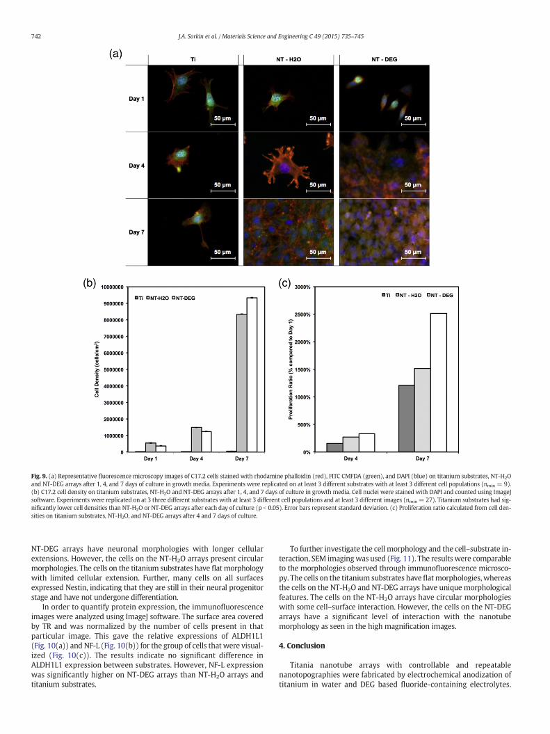
Fig. 9. (a) Representative fluorescence microscopy images of C17.2 cells stained with rhodamine phalloidin (red), FITC CMFDA (green), and DAPI (blue) on titanium substrates, NT-H2Oand NT-DEG arrays after 1, 4, and 7 days of culture in growth media. Experiments were replicated on at least 3 different substrates with at least 3 different cell populations (nmin = 9).(b) C17.2 cell density on titanium substrates, NT-H2O and NT-DEG arrays after 1, 4, and 7 days of culture in growth media. Cell nuclei were stained with DAPI and counted using ImageJsoftware. Experiments were replicated on at 3 three different substrates with at least 3 different cell populations and at least 3 different images (nmin= 27). Titanium substrates had sig-nificantly lower cell densities than NT-H2O or NT-DEG arrays after each day of culture (p b 0.05). Error bars represent standard deviation. (c) Proliferation ratio calculated from cell den-sities on titanium substrates, NT-H2O, and NT-DEG arrays after 4 and 7 days of culture.
742 J.A. Sorkin et al. / Materials Science and Engineering C 49 (2015) 735–745
NT-DEG arrays have neuronal morphologies with longer cellularextensions. However, the cells on the NT-H2O arrays present circularmorphologies. The cells on the titanium substrates have flat morphologywith limited cellular extension. Further, many cells on all surfacesexpressed Nestin, indicating that they are still in their neural progenitorstage and have not undergone differentiation.
In order to quantify protein expression, the immunofluorescenceimages were analyzed using ImageJ software. The surface area coveredby TR and was normalized by the number of cells present in thatparticular image. This gave the relative expressions of ALDH1L1(Fig. 10(a)) and NF-L (Fig. 10(b)) for the group of cells that were visual-ized (Fig. 10(c)). The results indicate no significant difference inALDH1L1 expression between substrates. However, NF-L expressionwas significantly higher on NT-DEG arrays than NT-H2O arrays andtitanium substrates.
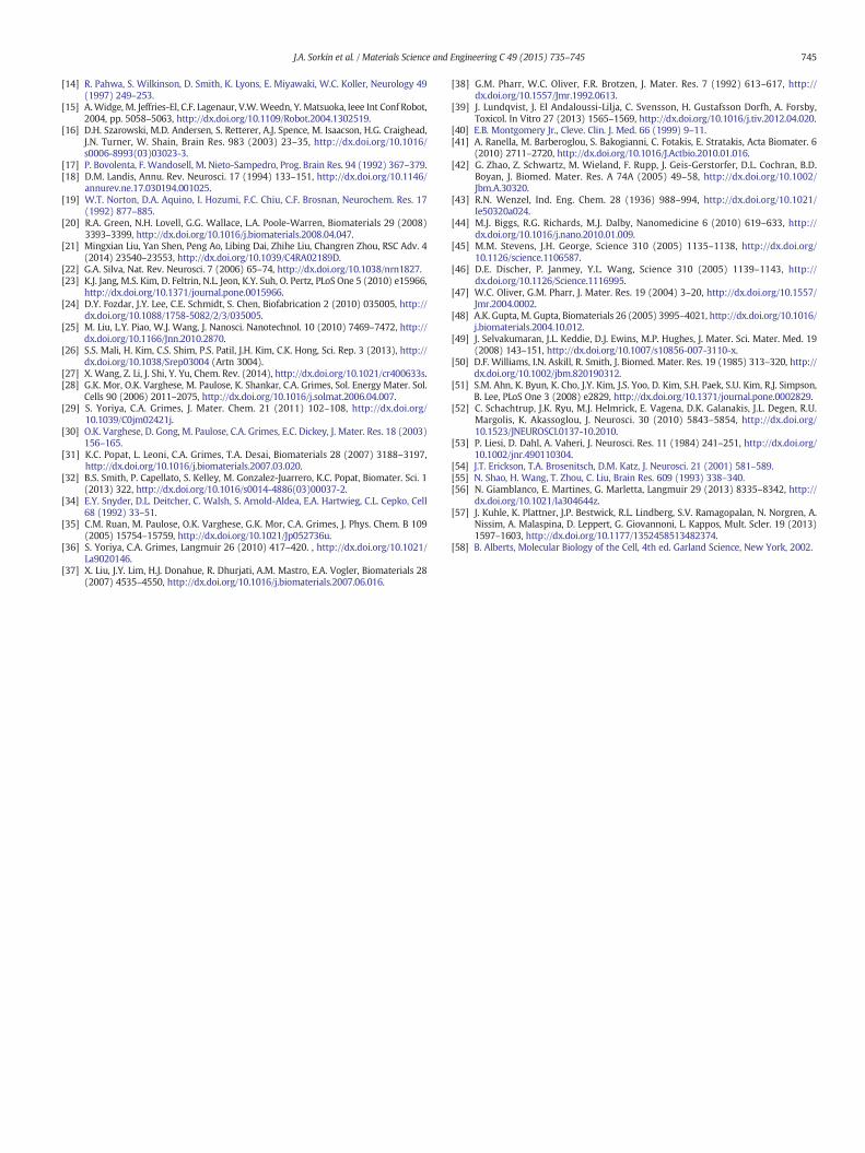
To further investigate the cell morphology and the cell–substrate in-teraction, SEM imagingwas used (Fig. 11). The results were comparableto the morphologies observed through immunofluorescence microsco-py. The cells on the titanium substrates have flatmorphologies,whereasthe cells on the NT-H2O and NT-DEG arrays have unique morphologicalfeatures. The cells on the NT-H2O arrays have circular morphologieswith some cell–surface interaction. However, the cells on the NT-DEGarrays have a significant level of interaction with the nanotubemorphology as seen in the high magnification images.
4. Conclusion
Titania nanotube arrays with controllable and repeatablenanotopographies were fabricated by electrochemical anodization oftitanium in water and DEG based fluoride-containing electrolytes.
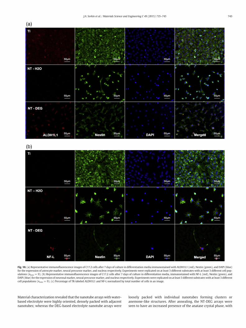
Fig. 10. (a) Representative immunofluorescence images of C17.2 cells after 7 days of culture in differentiationmedia immunostainedwith ALDH1L1 (red), Nestin (green), and DAPI (blue)for the expression of astrocyte marker, neural precursormarker, and nucleus respectively. Experiments were replicated on at least 3 different substrates with at least 3 different cell pop-ulations (nmin = 9). (b) Representative immunofluorescence images of C17.2 cells after 7 days of culture in differentiation media, immunostained with NF-L (red), Nestin (green), andDAPI (blue) for the expressionof neuronalmarker, neural precursormarker, andnucleus respectively. Experimentswere replicated onat least 3 different substrateswith at least 3 differentcell populations (nmin = 9). (c) Percentage of TR-labeled ALDH1L1 and NF-L normalized by total number of cells in an image.
743J.A. Sorkin et al. / Materials Science and Engineering C 49 (2015) 735–745
Material characterization revealed that the nanotube arrayswithwater-based electrolyte were highly oriented, densely packed with adjacentnanotubes; whereas the DEG-based electrolyte nanotube arrays were
loosely packed with individual nanotubes forming clusters oranemone-like structures. After annealing, the NT-DEG arrays wereseen to have an increased presence of the anatase crystal phase, with
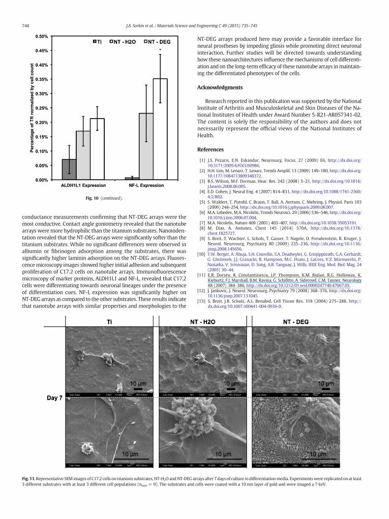
Fig. 10 (continued).
744 J.A. Sorkin et al. / Materials Science and Engineering C 49 (2015) 735–745
conductance measurements confirming that NT-DEG arrays were themost conductive. Contact angle goniometry revealed that the nanotubearrays weremore hydrophilic than the titanium substrates. Nanoinden-tation revealed that the NT-DEG arrays were significantly softer than thetitanium substrates. While no significant differences were observed inalbumin or fibrinogen adsorption among the substrates, there wassignificantly higher laminin adsorption on the NT-DEG arrays. Fluores-cencemicroscopy images showed higher initial adhesion and subsequentproliferation of C17.2 cells on nanotube arrays. Immunofluorescencemicroscopy of marker proteins, ALDH1L1 and NF-L, revealed that C17.2cells were differentiating towards neuronal lineages under the presenceof differentiation cues. NF-L expression was significantly higher onNT-DEGarrays as compared to the other substrates. These results indicatethat nanotube arrays with similar properties and morphologies to the
Fig. 11.Representative SEM images of C17.2 cells on titanium substrates, NT-H2O andNT-DEG ar3 different substrates with at least 3 different cell populations (nmin = 9). The substrates and c
NT-DEG arrays produced here may provide a favorable interface forneural prostheses by impeding gliosis while promoting direct neuronalinteraction. Further studies will be directed towards understandinghow these nanoarchitectures influence themechanisms of cell differenti-ation and on the long-term efficacy of these nanotube arrays inmaintain-ing the differentiated phenotypes of the cells.
Acknowledgments
Research reported in this publication was supported by the NationalInstitute of Arthritis and Musculoskeletal and Skin Diseases of the Na-tional Institutes of Health under Award Number 5-R21-AR057341-02.The content is solely the responsibility of the authors and does notnecessarily represent the official views of the National Institutes ofHealth.
References
[1] J.S. Pezaris, E.N. Eskandar, Neurosurg. Focus. 27 (2009) E6, http://dx.doi.org/10.3171/2009.4.FOCUS0986.
[2] H.H. Lim, M. Lenarz, T. Lenarz, Trends Amplif. 13 (2009) 149–180, http://dx.doi.org/10.1177/1084713809348372.
[3] B.S. Wilson, M.F. Dorman, Hear. Res. 242 (2008) 3–21, http://dx.doi.org/10.1016/j.heares.2008.06.005.
[4] E.D. Cohen, J. Neural Eng. 4 (2007) R14–R31, http://dx.doi.org/10.1088/1741-2560/4/2/R02.
[5] S. Waldert, T. Pistohl, C. Braun, T. Ball, A. Aertsen, C. Mehring, J. Physiol. Paris 103(2009) 244–254, http://dx.doi.org/10.1016/j.jphysparis.2009.08.007.
[6] M.A. Lebedev, M.A. Nicolelis, Trends Neurosci. 29 (2006) 536–546, http://dx.doi.org/10.1016/j.tins.2006.07.004.
[7] M.A. Nicolelis, Nature 409 (2001) 403–407, http://dx.doi.org/10.1038/35053191.[8] M. Dias, A. Antunes, Chest 145 (2014) 570A, http://dx.doi.org/10.1378/
chest.1825727.[9] S. Breit, T. Wachter, L. Schols, T. Gasser, T. Nagele, D. Freudenstein, R. Kruger, J.
Neurol. Neurosurg. Psychiatry 80 (2009) 235–236, http://dx.doi.org/10.1136/jnnp.2008.145656.
[10] T.W. Berger, A. Ahuja, S.H. Courellis, S.A. Deadwyler, G. Erinjippurath, G.A. Gerhardt,G. Gholmieh, J.J. Granacki, R. Hampson, M.C. Hsaio, J. LaCoss, V.Z. Marmarelis, P.Nasiatka, V. Srinivasan, D. Song, A.R. Tanguay, J. Wills, IEEE Eng. Med. Biol. Mag. 24(2005) 30–44.
[11] E.R. Dorsey, R. Constantinescu, J.P. Thompson, K.M. Biglan, R.G. Holloway, K.Kieburtz, F.J. Marshall, B.M. Ravina, G. Schifitto, A. Siderowf, C.M. Tanner, Neurology68 (2007) 384–386, http://dx.doi.org/10.1212/01.wnl.0000247740.47667.03.
[12] J. Jankovic, J. Neurol. Neurosurg. Psychiatry 79 (2008) 368–376, http://dx.doi.org/10.1136/jnnp.2007.131045.
[13] S. Breit, J.B. Schulz, A.L. Benabid, Cell Tissue Res. 318 (2004) 275–288, http://dx.doi.org/10.1007/s00441-004-0936-0.
rays after 7 days of culture indifferentiationmedia. Experimentswere replicatedon at leastells were coated with a 10 nm layer of gold and were imaged a 7 keV.
745J.A. Sorkin et al. / Materials Science and Engineering C 49 (2015) 735–745
[14] R. Pahwa, S. Wilkinson, D. Smith, K. Lyons, E. Miyawaki, W.C. Koller, Neurology 49(1997) 249–253.
[15] A.Widge, M. Jeffries-El, C.F. Lagenaur, V.W.Weedn, Y. Matsuoka, Ieee Int Conf Robot,2004, pp. 5058–5063, http://dx.doi.org/10.1109/Robot.2004.1302519.
[16] D.H. Szarowski, M.D. Andersen, S. Retterer, A.J. Spence, M. Isaacson, H.G. Craighead,J.N. Turner, W. Shain, Brain Res. 983 (2003) 23–35, http://dx.doi.org/10.1016/s0006-8993(03)03023-3.
[17] P. Bovolenta, F. Wandosell, M. Nieto-Sampedro, Prog. Brain Res. 94 (1992) 367–379.[18] D.M. Landis, Annu. Rev. Neurosci. 17 (1994) 133–151, http://dx.doi.org/10.1146/
annurev.ne.17.030194.001025.[19] W.T. Norton, D.A. Aquino, I. Hozumi, F.C. Chiu, C.F. Brosnan, Neurochem. Res. 17
(1992) 877–885.[20] R.A. Green, N.H. Lovell, G.G. Wallace, L.A. Poole-Warren, Biomaterials 29 (2008)
3393–3399, http://dx.doi.org/10.1016/j.biomaterials.2008.04.047.[21] Mingxian Liu, Yan Shen, Peng Ao, Libing Dai, Zhihe Liu, Changren Zhou, RSC Adv. 4
(2014) 23540–23553, http://dx.doi.org/10.1039/C4RA02189D.[22] G.A. Silva, Nat. Rev. Neurosci. 7 (2006) 65–74, http://dx.doi.org/10.1038/nrn1827.[23] K.J. Jang, M.S. Kim, D. Feltrin, N.L. Jeon, K.Y. Suh, O. Pertz, PLoS One 5 (2010) e15966,
http://dx.doi.org/10.1371/journal.pone.0015966.[24] D.Y. Fozdar, J.Y. Lee, C.E. Schmidt, S. Chen, Biofabrication 2 (2010) 035005, http://
dx.doi.org/10.1088/1758-5082/2/3/035005.[25] M. Liu, L.Y. Piao, W.J. Wang, J. Nanosci. Nanotechnol. 10 (2010) 7469–7472, http://
dx.doi.org/10.1166/Jnn.2010.2870.[26] S.S. Mali, H. Kim, C.S. Shim, P.S. Patil, J.H. Kim, C.K. Hong, Sci. Rep. 3 (2013), http://
dx.doi.org/10.1038/Srep03004 (Artn 3004).[27] X. Wang, Z. Li, J. Shi, Y. Yu, Chem. Rev. (2014), http://dx.doi.org/10.1021/cr400633s.[28] G.K. Mor, O.K. Varghese, M. Paulose, K. Shankar, C.A. Grimes, Sol. Energy Mater. Sol.
Cells 90 (2006) 2011–2075, http://dx.doi.org/10.1016/j.solmat.2006.04.007.[29] S. Yoriya, C.A. Grimes, J. Mater. Chem. 21 (2011) 102–108, http://dx.doi.org/
10.1039/C0jm02421j.[30] O.K. Varghese, D. Gong, M. Paulose, C.A. Grimes, E.C. Dickey, J. Mater. Res. 18 (2003)
156–165.[31] K.C. Popat, L. Leoni, C.A. Grimes, T.A. Desai, Biomaterials 28 (2007) 3188–3197,
http://dx.doi.org/10.1016/j.biomaterials.2007.03.020.[32] B.S. Smith, P. Capellato, S. Kelley, M. Gonzalez-Juarrero, K.C. Popat, Biomater. Sci. 1
(2013) 322, http://dx.doi.org/10.1016/s0014-4886(03)00037-2.[34] E.Y. Snyder, D.L. Deitcher, C. Walsh, S. Arnold-Aldea, E.A. Hartwieg, C.L. Cepko, Cell
68 (1992) 33–51.[35] C.M. Ruan, M. Paulose, O.K. Varghese, G.K. Mor, C.A. Grimes, J. Phys. Chem. B 109
(2005) 15754–15759, http://dx.doi.org/10.1021/Jp052736u.[36] S. Yoriya, C.A. Grimes, Langmuir 26 (2010) 417–420. , http://dx.doi.org/10.1021/
La9020146.[37] X. Liu, J.Y. Lim, H.J. Donahue, R. Dhurjati, A.M. Mastro, E.A. Vogler, Biomaterials 28
(2007) 4535–4550, http://dx.doi.org/10.1016/j.biomaterials.2007.06.016.
[38] G.M. Pharr, W.C. Oliver, F.R. Brotzen, J. Mater. Res. 7 (1992) 613–617, http://dx.doi.org/10.1557/Jmr.1992.0613.
[39] J. Lundqvist, J. El Andaloussi-Lilja, C. Svensson, H. Gustafsson Dorfh, A. Forsby,Toxicol. In Vitro 27 (2013) 1565–1569, http://dx.doi.org/10.1016/j.tiv.2012.04.020.
[40] E.B. Montgomery Jr., Cleve. Clin. J. Med. 66 (1999) 9–11.[41] A. Ranella, M. Barberoglou, S. Bakogianni, C. Fotakis, E. Stratakis, Acta Biomater. 6
(2010) 2711–2720, http://dx.doi.org/10.1016/J.Actbio.2010.01.016.[42] G. Zhao, Z. Schwartz, M. Wieland, F. Rupp, J. Geis-Gerstorfer, D.L. Cochran, B.D.
Boyan, J. Biomed. Mater. Res. A 74A (2005) 49–58, http://dx.doi.org/10.1002/Jbm.A.30320.
[43] R.N. Wenzel, Ind. Eng. Chem. 28 (1936) 988–994, http://dx.doi.org/10.1021/Ie50320a024.
[44] M.J. Biggs, R.G. Richards, M.J. Dalby, Nanomedicine 6 (2010) 619–633, http://dx.doi.org/10.1016/j.nano.2010.01.009.
[45] M.M. Stevens, J.H. George, Science 310 (2005) 1135–1138, http://dx.doi.org/10.1126/science.1106587.
[46] D.E. Discher, P. Janmey, Y.L. Wang, Science 310 (2005) 1139–1143, http://dx.doi.org/10.1126/Science.1116995.
[47] W.C. Oliver, G.M. Pharr, J. Mater. Res. 19 (2004) 3–20, http://dx.doi.org/10.1557/Jmr.2004.0002.
[48] A.K. Gupta, M. Gupta, Biomaterials 26 (2005) 3995–4021, http://dx.doi.org/10.1016/j.biomaterials.2004.10.012.
[49] J. Selvakumaran, J.L. Keddie, D.J. Ewins, M.P. Hughes, J. Mater. Sci. Mater. Med. 19(2008) 143–151, http://dx.doi.org/10.1007/s10856-007-3110-x.
[50] D.F. Williams, I.N. Askill, R. Smith, J. Biomed. Mater. Res. 19 (1985) 313–320, http://dx.doi.org/10.1002/jbm.820190312.
[51] S.M. Ahn, K. Byun, K. Cho, J.Y. Kim, J.S. Yoo, D. Kim, S.H. Paek, S.U. Kim, R.J. Simpson,B. Lee, PLoS One 3 (2008) e2829, http://dx.doi.org/10.1371/journal.pone.0002829.
[52] C. Schachtrup, J.K. Ryu, M.J. Helmrick, E. Vagena, D.K. Galanakis, J.L. Degen, R.U.Margolis, K. Akassoglou, J. Neurosci. 30 (2010) 5843–5854, http://dx.doi.org/10.1523/JNEUROSCI.0137-10.2010.
[53] P. Liesi, D. Dahl, A. Vaheri, J. Neurosci. Res. 11 (1984) 241–251, http://dx.doi.org/10.1002/jnr.490110304.
[54] J.T. Erickson, T.A. Brosenitsch, D.M. Katz, J. Neurosci. 21 (2001) 581–589.[55] N. Shao, H. Wang, T. Zhou, C. Liu, Brain Res. 609 (1993) 338–340.[56] N. Giamblanco, E. Martines, G. Marletta, Langmuir 29 (2013) 8335–8342, http://
dx.doi.org/10.1021/la304644z.[57] J. Kuhle, K. Plattner, J.P. Bestwick, R.L. Lindberg, S.V. Ramagopalan, N. Norgren, A.
Nissim, A. Malaspina, D. Leppert, G. Giovannoni, L. Kappos, Mult. Scler. 19 (2013)1597–1603, http://dx.doi.org/10.1177/1352458513482374.
[58] B. Alberts, Molecular Biology of the Cell, 4th ed. Garland Science, New York, 2002.


