Embed Size (px)
Citation preview

Accepted Manuscript
Title: Orexins cause epileptic activity
Authors: Haydar Ali Erken, Gulten Erken, Osman Genc, SelimKortunay, Melike Sahiner, Gunfer Turgut, Sebahat Turgut
PII: S0196-9781(12)00274-4DOI: doi:10.1016/j.peptides.2012.06.012Reference: PEP 68730
To appear in: Peptides
Received date: 1-5-2012Revised date: 5-6-2012Accepted date: 5-6-2012
Please cite this article as: Erken HA, Erken G, Genc O, Kortunay S, SahinerM, Turgut G, Turgut S, Orexins cause epileptic activity, Peptides (2010),doi:10.1016/j.peptides.2012.06.012
This is a PDF file of an unedited manuscript that has been accepted for publication.As a service to our customers we are providing this early version of the manuscript.The manuscript will undergo copyediting, typesetting, and review of the resulting proofbefore it is published in its final form. Please note that during the production processerrors may be discovered which could affect the content, and all legal disclaimers thatapply to the journal pertain.

Page 1 of 14
Accep
ted
Man
uscr
ipt
Highlights
• Orexins are excitatory neuropeptides. • Orexins were intracortically applied to rats. • Orexins cause epileptic activity in rats.

Page 2 of 14
Accep
ted
Man
uscr
ipt
OREXINS CAUSE EPILEPTIC ACTIVITY
Haydar Ali Erkena*, Gülten Erkenb, Osman Gençc, Selim Kortunayd, Melike Şahinere,
Günfer Turgutf, Sebahat Turgutf.
aBalikesir State Hospital, Balikesir, Turkey; bBalikesir University, Faculty of Medicine,
Department of Physiology, Balikesir, Turkey; cDumlupinar University, Faculty of
Medicine, Department of Physiology, Kutahya, Turkey; dPamukkale University,
Faculty of Medicine, Department of Pharmacology, Denizli, Turkey; eAcibadem
University, Faculty of Medicine, Department of Physiology, Istanbul, Turkey;
fPamukkale University, Faculty of Medicine, Department of Physiology, Denizli,
Turkey.
Abstract
Orexins have been implicated in the regulation of sleep-awake cycle, energy
homeostasis, drinking behavior, analgesia, attention, learning and memory but their
effects on epileptic activity are controversial. We investigated whether intracortical
injections of orexin A (100 pmol) and B (100 pmol) cause epileptic activity in rats. We
observed epileptic seizure findings on these two groups rats. Orexin A and B also
significantly increased total EEG power spectrum. Our findings indicate that orexins
cause epileptic activity.
Key words: EEG; epilepsy; hypocretin; orexin; power spectrum; seizure.
1.Introduction
The orexins (OXs), orexin A and B, also known as hypocretins (hypocretin 1 and 2),
are neuropeptides derived from the same precursor molecule, prepro-orexin,
Page 3 of 14
Accep
ted
Man
uscr
ipt
synthesized in the lateral hypothalamic area [5,31]. Orexinergic neurons project
widely to numerous brain regions including cerebral cortex, thalamus, hypothalamus,
nucleus accumbens, brain stem and spinal cord [4,27,29]. OX receptors (OX1 and
OX2) are expressed in these areas especially in the cortical regions, hippocampus,
thalamic, hypothalamic and brain stem nuclei [22,40]. It has been reported that OXs
may play a role in various physiological functions including the energy homeostasis
[12,32,44], sleep–wake cycle [32], drinking behavior [19], analgesia [25], attention
[10], learning [36] and memory [1,15]. Although it was shown in numerous studies
that orexins have neuroexcitatory effect [5,42], there were few studies, which
investigate orexin-epilepsy relationship [8,18,30]. In a previous study, it was shown
that after generalized convulsions, the levels of orexin A decrease in cerebrospinal
fluid [30]. In another study it has been reported that orexin A decreased bicuculline-
induced epileptic activity according to in vitro experiments [8]. On the other hand, in
our previous study, we showed that orexins enhance the cortical epileptic activity
induced by intracortical application of penicillin-G [18]. The findings of previous
studies investigating orexin-epilepsy relationship are controversial [8,18,30] and
orexin-epilepsy relationship is not clear yet. Based on our previous findings we
thought that orexins induce epileptic activity without using any epileptogenic agent.
Therefore the aim of this study was to investigate whether orexins cause epileptic
activity in rats.
2. Materials and methods
2.1. Animals and study design
All of the experiments were approved by the Committee of Animal Care at
Pamukkale University and the experiments were performed according to the
guidelines (NIH, UCSF) on animal use. Twenty adult male Wistar Albino rats

Page 4 of 14
Accep
ted
Man
uscr
ipt
weighing 243±26g (mean±SD) were used. All of the rats were maintained in a 12-h
light/dark cycle environment (lights on 7:00–19:00 h) at a temperature of 23±2 °C and
50% humidity. Rats had access to food and water ad libitum.
The rats were randomly assigned to the following four groups (n=5 for each group).
Intracortical (i.c.) orexin A (OXA, 100 pmol, dissolved in 2 μl saline), orexin B (OXB,
100 pmol, dissolved in 2 μl saline) and saline (2 μl) was administered into the rats in
the groups 1, 2 and 3, respectively. Group 4 received no drug or saline.
2.2. Anesthesia and experimental procedure
The rats were anesthetized with ketamine/xylazine (90 and 10 mg/kg respectively
i.p.) and their heads were shaved. Then, the rats were placed on a stereotaxic
instrument (Stoelting Co., USA) and their head’s were disinfected with batticon
(Batticon, Adeka Co., Turkey) and incised from mid-frontal to mid-occipital. After the
bregma was exposed, a hole was drilled by a dental drill to a point that was
determined to be the rat brain atlas of Paxinos and Watson [28] (from bregma: 0.7
mm anterior, 2.0 mm right laterally, 2.0 mm vertically). 100 pmol orexin A and 100
pmol orexin B were dissolved in 2 µl saline and were administered to the primary
motor cortex by microinjector (Hamilton Co., USA) to group 1 and group 2,
respectively. Similarly, 2 µl saline injection was administered to the same area in
group 3. Aliquots of orexins were prepared and frozen at –200C for each experiment
and thawed and dissolved in 2 µl saline immediately before use. Orexin A and B
were purchased from Sigma-Aldrich Co., Germany.
2.3. EEG record and analyses
Two AgCl flat electrodes were placed on the scalp for bipolar EEG recording; one of
them was placed on the right parietal area, and the other on the mid-occipital area. A
ground electrode was placed on the tail of the rat. EEG was recorded by PowerLab

Page 5 of 14
Accep
ted
Man
uscr
ipt
8/SP data acquisition system and Chart 5.2.2 program (ADInstruments Co.,
Australia). The recording parameters were as follows: 0.3-100 Hz low and high
frequency filter, 50 Hz notch filter, and a recording speed of 25 mm/sec. Whenever
additional anesthesia was needed, it was administered to the rats. The rats were
observed and recorded during the experimental period. Thirty second artifact-free
epochs were chosen from the EEG recordings as the samples of cortical activity, at
before the orexin/saline injection and 30th, 60th, 90th and 120th minutes of
orexin/saline injection. The power spectrum analysis of these EEG samples was
performed by Chart 5.2.2 software program (ADInstruments Co., Australia). Spectral
power values were transferred to the SPSS 10.0 program and analyzed by Repeated
Measures ANOVA and Post Hoc Tukey Test. P value of <0.05 being considered as
significant.
3.Results
We observed epileptic seizure findings on the group 1 and group 2 rats, which were
applied 100 pmol (i.c.) OXA and 100 pmol (i.c.) OXB, respectively. After 25-32
minutes OXs administrations, tonic–clonic contractions on the left anterior extremities
of the rats were observed. The contractions spreaded to the left posterior, right
anterior and posterior extremities, tail and whole body of the rats. The severity of the
contractions increased and continued to the ending of the experiments (Until 120
minutes after OXs administrations). In the group 1 rats (OXA applied) the
contractions were observed more severe than group 2 rats (OXB applied). However
there were no epileptic seizure findings in the saline and control groups. Also, total
EEG power spectrum was increased significantly in the orexin A and orexin B groups,
at 90 and 120 minutes after the orexin injections compared to the values of before
Page 6 of 14
Accep
ted
Man
uscr
ipt
orexin application (Table 1). Whereas, total EEG power spectrum did not significantly
change in the control and saline groups (Table 1).
4.Discussion
In this study, it was shown that orexin applications caused apparent increase on total
EEG power spectrum. In addition to EEG findings, during experiments epileptic
activity findings including tonic–clonic contractions on the whole extremities, tail and
body of the rats were observed by physical observations. Also, similarly to previous
studies [6] the effect of OXA was more potent than that of OXB in this study. On the
other hand in the saline and control groups neither contractions nor change in EEG
findings were observed.
In the previous studies using 100 pmol orexin A or B epileptic contractions or
epileptic seizure findings in EEG were not reported [6,7,24,37,38,43]. There may be
numerous reasons for this situation. One of them can be the localization of the
brain area where orexins were injected. In the present study, orexins were injected
to the primary motor area. On the other hand in the previous studies orexins were
injected at the same dose to different brain areas including the basal forebrain,
nucleus basalis, substantia innominata, magnocellular preoptic nucleus, nucleus
accumbens, lateral cerebral ventricle and rostral lateral hypothalamic area
[6,7,24,37,38,43]. Several factors (e.g. orexin receptors density) in these areas may
change the effects of orexins. The second reason may be the origin of the orexins
which were provided from different companies having possibly different efficiency.
In this study, orexins were bought from Sigma-Aldrich. In the previous studies orexins
were bought from different companies (American Peptides, Sunnyvale, CA, USA and
Peptide Institute, Minoh, Japan) [7,24,37,38,43]. Thorpe et al. (2005) reported that
orexins which have been bought from different companies had no similar effects on

Page 7 of 14
Accep
ted
Man
uscr
ipt
appetite stimulation [38]. Therefore orexins, which were provided from different
companies, may have different effects on epileptic activity. The third reason may be,
in the previous studies, the same doses of orexins might have induced focal epileptic
activity but it could not be observed. In our study, because of orexins’ injection to the
primary motor area we could observe physically apparent epileptic activity. Also, in
the determination of focal epileptic activity, appropriate EEG recording is important.
However in some of the previous studies using 100 pmol orexin, EEG was not
recorded [24,37,38,43]. On the other hand in the two of the studies using 100 pmol
orexin, EEG was recorded and changes on arousal pattern of EEG caused by
orexins were reported. But in these studies, epileptic activity findings in EEG were not
reported [6,7].
According to some other previous studies, orexins have neuroexcitatory effect and
may cause behavioral convulsion activity [5,13,42]. Our results confirm these reports.
It is known that during epileptic activity, balance between glutamate and GABA
release is abolished [2,34]. In the previous studies it was reported that orexins
increased glutamate [16,17,41] and GABA [23,41] release. On the other hand
there are several studies reported that orexins decreased glutamate [11] and
GABA [39] release. In another study, it has been shown that NMDA receptors
are also related with epileptic activity [21]. In rats, the results of previous
studies for explanation of the mechanisms of epileptic activity caused by
orexins are straightforward.
Another possible cause of orexin-induced epileptic activity may be the direct effect of
orexins on neuronal depolarization. According to in vitro experimental studies,
orexins cause depolarization in neurons [33] and increase in firing frequencies of
neurons [3,9]. Orexins may form direct excitatory effects on neurons via increasing

Page 8 of 14
Accep
ted
Man
uscr
ipt
the influx of sodium [20], activate sodium-calcium exchanger pump [9], increase
influx of calcium [26,42] or decrease efflux of potassium [14]. The importance of
intracellular calcium increase in terms of neuronal excitability and epileptic activity is
known [35]. For this reason, this mechanism also mediates the effects of orexins on
the central nervous system. Hence these effects of orexins, which facilitate neuronal
depolarization, support epileptic activity and may explain the increase in total EEG
power spectrum in our study.
In conclusion, in this study, we showed that intracortical applications of orexins
caused epileptic activity, but the mechanisms of this effect are not clear.
Funding
This study was supported by Pamukkale University Research Fund.
Disclosure
The authors declare that there is no conflict of interest.
References
[1] Akbari E, Naghdi N, Motamedi F. The selective orexin 1 receptor antagonist SB-
334867-A impairs acquisition and consolidation but not retrieval of spatial
memory in Morris water maze. Peptides 2007;28(3):650-6.
[2] André V, Marescaux C, Nehlig A, Fritschy JM. Alterations of hippocampal
GABAergic system contribute to development of spontaneous recurrent
seizures in the rat lithium-pilocarpine model of temporal lobe epilepsy.
Hippocampus 2001;11(4):452-68.
[3] Burlet S, Tyler CJ, Leonard CS. Direct and indirect excitation of laterodorsal
tegmental neurons by hypocretin/orexin peptides: implications for wakefulness
and narcolepsy. J Neurosci 2002;22(7):2862-72.
Page 9 of 14
Accep
ted
Man
uscr
ipt
[4] Cutler DJ, Morris R, Sheridhar V, Wattam TA, Holmes S, Patel S, et al.
Differential distribution of orexin-A and orexin-B immunoreactivity in the rat brain
and spinal cord. Peptides 1999;20(12):1455-70.
[5] de Lecea L, Kilduff TS, Peyron C, Gao X, Foye PE, Danielson PE, et al. The
hypocretins: hypothalamus-specific peptides with neuroexcitatory activity. Proc
Natl Acad Sci USA 1998;95(1):322-7.
[6] Dong HL, Fukuda S, Murata E, Zhu Z, Higuchi T. Orexins increase cortical
acetylcholine release and electroencephalographic activation through orexin-1
receptor in the rat basal forebrain during isoflurane anesthesia. Anesthesiology
2006;104(5):1023-32.
[7] Dong H, Niu J, Su B, Zhu Z, Lv Y, Li Y, Xiong L. Activation of orexin signal in
basal forebrain facilitates the emergence from sevoflurane anesthesia in rat.
Neuropeptides 2009;43(3):179-85.
[8] Doreulee N, Alania M, Vashalomidze G, Skhirtladze E, Kapanadze Ts.
Orexinergic system and pathophysiology of epilepsy. Georgian Med News
2010;188:74-9
[9] Eriksson KS, Sergeeva O, Brown RE, Haas HL. Orexin/hypocretin excites the
histaminergic neurons of the tuberomammillary nucleus. J Neurosci
2001;21(23):9273-9.
[10] Fadel J, Burk JA. Orexin/hypocretin modulation of the basal forebrain
cholinergic system: role in attention. Brain Res 2010;1314:112-23.
[11] Haj-Dahmane S, Shen RY. The wake-promoting peptide orexin-B inhibits
glutamatergic transmission to dorsal raphe nucleus serotonin neurons through
retrograde endocannabinoid signaling. J Neurosci 2005;25(4):896-905.
Page 10 of 14
Accep
ted
Man
uscr
ipt
[12] Haynes AC, Jackson B, Overend P, Buckingham RE, Wilson S, Tadayyon M,
et al. Effects of single and chronic intracerebroventricular administration of the
orexins on feeding in the rat. Peptides 1999;20(9):1099-105.
[13] Ida T, Nakahara K, Katayama T, Murakami N, Nakazato M. Effect of lateral
cerebroventricular injection of the appetite-stimulating neuropeptide, orexin
and neuropeptide Y, on the various behavioral activities of rats. Brain Res
1999;821(2):526-9.
[14] Ivanov A, Aston-Jones G. Hypocretin/orexin depolarizes and decreases
potassium conductance in locus coeruleus neurons. Neuroreport
2000;11(8):1755-8.
[15] Jaeger LB, Farr SA, Banks WA, Morley JE. Effects of orexin-A on memory
processing. Peptides 2002;23(9):1683-8.
[16] John J, Wu MF, Kodama T, Siegel JM. Intravenously administered hypocretin-
1 alters brain amino acid release: an in vivo microdialysis study in rats. J
Physiol 2003;548(Pt 2):557-62.
[17] Kodama T, Kimura M. Arousal effects of orexin-A correlate with GLU release
from the locus coeruleus in rats. Peptides 2002;23(9):1673-81.
[18] Kortunay S, Erken HA, Erken G, Genç O, Şahiner M, Turgut S, Turgut G.
Orexins increase penicillin-induced epileptic activity. Peptides 2012;34(2):419-
22.
[19] Kunii K, Yamanaka A, Nambu T, Matsuzaki I, Goto K, Sakurai T.
Orexins/hypocretins regulate drinking behaviour. Brain Res 1999;842(1):256-
61.
Page 11 of 14
Accep
ted
Man
uscr
ipt
[20] Liu RJ, van den Pol AN, Aghajanian GK. Hypocretins (orexins) regulate
serotonin neurons in the dorsal raphe nucleus by excitatory direct and
inhibitory indirect actions. J Neurosci 2002;22(21):9453-64
[21] Loscher W. New visions in the pharmacology of anticonvulsion. Eur J
Pharmacol 1998;342(1):1-13
[22] Marcus JN, Aschkenasi CJ, Lee CE, Chemelli RM, Saper CB, Yanagisawa M,
et al. Differential expression of orexin receptors 1 and 2 in the rat brain. J
Comp Neurol 2001;435(1):6-25.
[23] Martin G, Fabre V, Siggins GR, de Lecea L. Interaction of the hypocretins with
neurotransmitters in the nucleus accumbens. Regul Pept 2002;104(1-3):111-7.
[24] Matsumura K, Tsuchihashi T, Abe I. Central orexin-A augments
sympathoadrenal outflow in conscious rabbits. Hypertension 2001;37(6):1382-
7.
[25] Mobarakeh JI, Takahashi K, Sakurada S, Nishino S, Watanabe H, Kato M,
Yanai K. Enhanced antinociception by intracerebroventricularly and
intrathecally-administered orexin A and B (hypocretin-1 and -2) in mice.
Peptides 2005;26(5):767-77.
[26] Nakamura Y, Miura S, Yoshida T, Kim J, Sasaki K. Cytosolic calcium elevation
induced by orexin/hypocretin in granule cell domain cells of the rat cochlear
nucleus in vitro. Peptides 2010;31(8):1579-88.
[27] Nambu T, Sakurai T, Mizukami K, Hosoya Y, Yanagisawa M, Goto K.
Distribution of orexin neurons in the adult rat brain. Brain Res 1999;827(1-
2):243-60.
[28] Paxinos G, Watson C. The rat brain in stereotaxic coordinates. 4th ed. San
Diego: Academic Pres; 1998.

Page 12 of 14
Accep
ted
Man
uscr
ipt
[29] Peyron C, Tighe DK, van den Pol AN, de Lecea L, Heller HC, Sutcliffe JG, et
al. Neurons containing hypocretin (orexin) project to multiple neuronal
systems. J Neurosci 1998;18(23):9996-10015.
[30] Rejdak K, Papuć E, Grieb P, Stelmasiak Z. Decreased cerebrospinal fluid
hypocretin-1 (orexin A) in patients after repetitive generalized tonic–clonic
seizures. Epilepsia 2009;50(6):1641-4.
[31] Sakurai T, Amemiya A, Ishii M, Matsuzaki I, Chemelli RM, Tanaka H, et al.
Orexins and orexin receptors: a family of hypothalamic neuropeptides and G
protein-coupled receptors that regulate feeding behavior. Cell 1998;92(4):573-
85.
[32] Sakurai T. Roles of orexin/hypocretin in regulation of sleep/wakefulness and
energy homeostasis. Sleep Med Rev 2005;9(4):231-41.
[33] Shirasaka T, Miyahara S, Kunitake T, Jin QH, Kato K, Takasaki M, et al.
Orexin depolarizes rat hypothalamic paraventricular nucleus neurons. Am J
Physiol Regul Integr Comp Physiol 2001;281(4):R1114-8.
[34] Sitges M, Sanchez-Tafolla BM, Chiu LM, Aldana BI, Guarneros A. Vinpocetine
inhibits glutamate release induced by the convulsive agent 4-aminopyridine
more potently than several antiepileptic drugs. Epilepsy Res 2011;96(3):257-
66.
[35] Speckmann EJ, Straub H, Köhling R. Contribution of calcium ions to the
generation of epileptic activity and antiepileptic calcium antagonism.
Neuropsychobiology 1993;27(3):122-6.
[36] Telegdy G, Adamik A. The action of orexin A on passive avoidance learning:
involvement of transmitters. Regul Pept 2002;104(1-3):105-10.

Page 13 of 14
Accep
ted
Man
uscr
ipt
[37] Thorpe AJ, Cleary JP, Levine AS, Kotz CM. Centrally administered orexin A
increases motivation for sweet pellets in rats. Psychopharmacology (Berl)
2005;182(1):75-83.
[38] Thorpe AJ, Kotz CM. Orexin A in the nucleus accumbens stimulates feeding
and locomotor activity. Brain Res 2005;1050(1-2):156-62.
[39] Thorpe AJ, Doane DF, Sweet DC, Beverly JL, Kotz CM. Orexin A in the
rostrolateral hypothalamic area induces feeding by modulating GABAergic
transmission. Brain Res 2006;1125(1):60-6.
[40] Trivedi P, Yu H, MacNeil DJ, Van der Ploeg LH, Guan XM. Distribution of
orexin receptor mRNA in the rat brain. FEBS Lett 1998;438(1-2):71-5.
[41] van den Pol AN, Gao XB, Obrietan K, Kilduff TS, Belousov AB. Presynaptic
and postsynaptic actions and modulation of neuroendocrine neurons by a new
hypothalamic peptide, hypocretin/orexin. J Neurosci 1998;18(19):7962-71.
[42] van den Pol AN, Ghosh PK, Liu RJ, Li Y, Aghajanian GK, Gao XB. Hypocretin
(orexin) enhances neuron activity and cell synchrony in developing mouse
GFP-expressing locus coeruleus. J Physiol 2002;541(Pt 1):169-85.
[43] Wang J, Osaka T, Inoue S. Orexin-A-sensitive site for energy expenditure
localized in the arcuate nucleus of the hypothalamus. Brain Res
2003;971(1):128-34.
[44] Yokobori E, Kojima K, Azuma M, Kang KS, Maejima S, Uchiyama M, et al.
Stimulatory effect of intracerebroventricular administration of orexin A on food
intake in the zebrafish, Danio rerio. Peptides 2011;32(7):1357-62.
Page 14 of 14
Accep
ted
Man
uscr
ipt
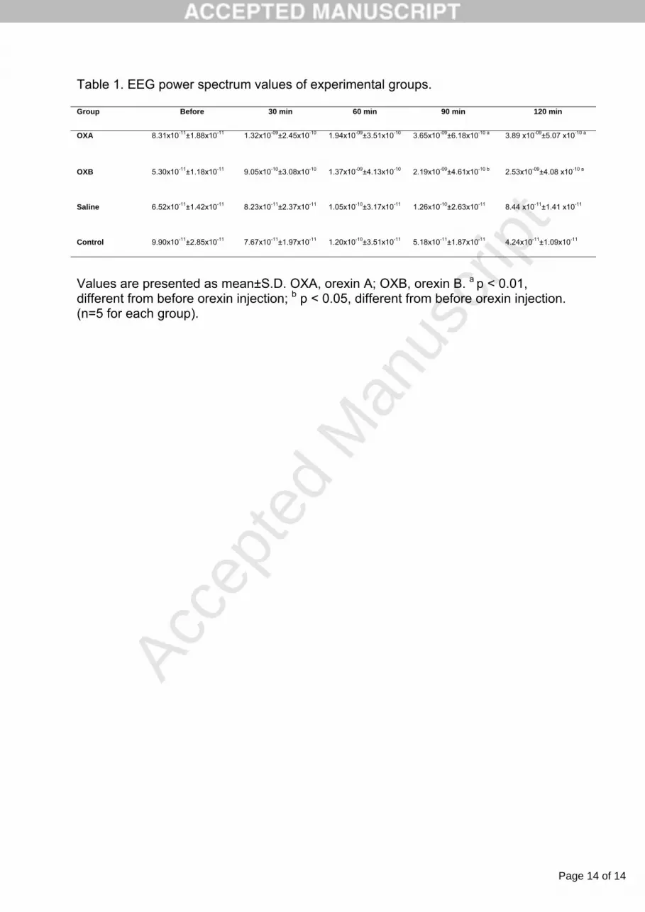
Table 1. EEG power spectrum values of experimental groups.
Group Before 30 min 60 min 90 min 120 min
OXA 8.31x10⁻¹¹±1.88x10⁻¹¹ 1.32x10-09±2.45x10⁻¹⁰ 1.94x10-09±3.51x10⁻¹⁰ 3.65x10-09±6.18x10⁻¹⁰ a 3.89 x10-09±5.07 x10⁻¹⁰ a
OXB 5.30x10⁻¹¹±1.18x10⁻¹¹ 9.05x10⁻¹⁰±3.08x10⁻¹⁰ 1.37x10-09±4.13x10⁻¹⁰ 2.19x10-09±4.61x10⁻¹⁰ b 2.53x10-09±4.08 x10⁻¹⁰ a
Saline 6.52x10⁻¹¹±1.42x10⁻¹¹ 8.23x10⁻¹¹±2.37x10⁻¹¹ 1.05x10⁻¹⁰±3.17x10⁻¹¹ 1.26x10⁻¹⁰±2.63x10⁻¹¹ 8.44 x10⁻¹¹±1.41 x10⁻¹¹
Control 9.90x10⁻¹¹±2.85x10⁻¹¹ 7.67x10⁻¹¹±1.97x10⁻¹¹ 1.20x10⁻¹⁰±3.51x10⁻¹¹ 5.18x10⁻¹¹±1.87x10⁻¹¹ 4.24x10⁻¹¹±1.09x10⁻¹¹
Values are presented as mean±S.D. OXA, orexin A; OXB, orexin B. a p < 0.01, different from before orexin injection; b p < 0.05, different from before orexin injection. (n=5 for each group).


