Embed Size (px)
Citation preview
NSC 215 HUMAN ANATOMY I 2Unit
1
NATIONAL OPEN UNIVERSITY OF NIGERIA
SCHOOL OF HEALTH SCIENCES
DEPARTMENT OF NURSING SCIENCES
NSC 215: Human Anatomy I- General and musculoskeletal anatomy (1–0–4)=2 UNITS This course shall cover anatomical terminologies, general body organization including cell structure, structure of membranes, body tissue and organs, and body defense. Definitions and terminologies in embryology, and developmental biology, cell division, gametogenesis, events leading to fertilization, cleavage, implantation and formation of germ layers shall be covered. Placenta formation and functions shall also be covered. It shall also cover the gross anatomy of the muscles, bones tendons, ligaments and joints of the body. It shall also cover the histology of bones, muscles and joints. Types and classification of muscles and joints as well as developmental processes in bones shall be included COURSE CODE: NSC 215 COURSE TITLE: Human Anatomy I COURSE UNITS: 2 Credit units (24 hours of instruction online; 12 hours of Discussion forum online/tutorial; 24 hours of laboratory practical) PRE-REQUISITE COURSES: NONE; CON-CURRENT COURSES: NSC 213, NSC209, NSC211, NSC217, NSC219 NSC221 COURSE WEBSITE: www.noun.edu.ng/ COURSE WRITERS Dr. Adewole O.S. MBBS, PhD, (Associate Professor). Dr. Abiodun A. O. MBBS, FWCS, M.Sc. (Senior Lecturer) Dr. Ayannuga A. A. MBBS, Ph.D (Senior Lecturer) Dr. Adeyemi D.A. PhD (Senior Lecturer) Dr. Ojo S. K. MBChB, MSc (Lecturer II); Dr. Arayombo B. E. MBChB, MSc (Lecturer II) Department of Anatomy and Cell Biology, College of Health Sciences, Obafemi Awolowo University, Ile-Ife, Nigeria COURSE EDITORS: Dr O.O. Irinoye and Dr E.O Oladogba PROGRAMME LEADER: COURSE COORDINATOR: As provided by the Department. COURSE REVIEWER Dr S S Bello MBBS, PhD
NSC 215 HUMAN ANATOMY I 2Unit
2
Department of Anatomy, FBMS, College of Health Sciences, Usmanu Danfodiyo University,
Sokoto, Nigeria
Table of Contents Page
Course Guide General Introduction 4 Course Aims 4 Course Objectives 4 Working through the Course 4 Course Materials 5 Study Units 5 Reference Textbooks 6 Equipment and Software Needed to Access Course 6 Number and Places of Meeting 6 Discussion Forum 7 Course Evaluation 7 Grading Criteria 7 Grading Scale 7 Schedule of Assignments with Dates 7 Course Overview 8 How to get the most from this Course 8
NSC 215 HUMAN ANATOMY I 2Unit
3
COURSE GUIDE GENERAL INTRODUCTION Hello, welcome to this course. We are happy to have you doing NSC 215 – Human AnatomyI. You would have done some anatomy when you were in the basic school of nursing. You are going to do a little more and have opportunities to have practical sessions to give you more facts on the structure of the human body. Interestingly, we all learn a lot from been able to look at our own bodies too. As nurses you must know what the body is made off and how it functions before you can determine if and when something goes wrong, what goes wrong and what you can do within your professional responsibility to help clients achieve, maintain, sustain, retain and adjust to permanent change in the body. You cannot practice safe without sound knowledge of anatomy. Everything you have to do with the body of a patient requires sound knowledge of anatomy for the patient to be safe with you in practice. Over a period of three semesters, you are going to learn about the different organs that make up the human body. This course along with the others must be learnt with your professional roles and duties in mind at all times for you to also see how you can apply your new learning to improve your practice. COURSE AIM. The aim of this course is to build your foundation in the developmental process and the structure of the human body as such prepares you to apply your knowledge in planning to meet the care needs of your body and that of your clients as such may relate to normal and abnormal changes in the various organs that make up the body. COURSE OBJECTIVES At the completion of this course, you should be able to: i. Use anatomical terminology correctly.
ii. Discuss the levels of organization of the human body.
iii. Discuss the components of the body defense system
iv. Discuss the human embryology from fertilization to birth
v. Discuss the two basic systems that provide support and movement for the human body. COURSE IMPLEMENTATION – WORKING THROUGH THIS COURSE The course will be delivered adopting the blended learning mode, 70% of online but interactive sessions and 30% of face-to-face during laboratory sessions. You are expected to register for this course online before you can have access to all the materials and have access to the class sessions online. You will have the hard and soft copies of course materials, you will also have online interactive sessions, face-to-face sessions with instructors during practical sessions in the laboratory. The interactive online activities will be available to you on the course link on the Website of NOUN. There are activities and assignments online for every unit every week. It is important that you visit the course sites weekly and do all assignments to meet deadlines and to contribute to the topical issues that would be raised for everyone’s contribution. You will be expected to read every module along with all assigned readings to prepare you to have meaningful contributions to all sessions and to complete all activities. It is important that you attempt all the Self-Assessment Questions (SAQ) at the end of every unit to help your understanding of the contents and to help you prepare for the in-course tests and the final examination. You will also be expected to keep a portfolio where you keep all your completed assignments.
NSC 215 HUMAN ANATOMY I 2Unit
4
COURSE MATERIALS Course Guide Course Text in Study Units Textbooks (Hard and electronic) Book of Laboratory Practical Assignment File/Portfolio STUDY UNITS This course has three Modules and 13 units. They are structured as presented Module 1 - Introduction to the Human body Unit 1 - General Body Organizations Unit 2 - Anatomical Terminology Unit 3 - Cells, Tissues, Organs, Systems & Membranes Unit 4 - Body Tissues Unit 5 - The Human Defense System Module 2 - Embryology Unit 1 - Embryology Terminology Unit 2 - Garmetogenesis Unit 3 - Placenta Formation and Functions Module 3 – Support and Movement Unit 1 - Integumentary System Unit 2 - Skeletal System Unit 3 - Muscular system Unit 4 - Tendons and Ligaments Unit 5 - Joints and Bursae REFERENCE TEXTBOOKS 1. Bruce M. Carlson (2019) Human Embryology & Developmental Biology. 6th edition
2. Kathryn A. Booth, Terri. D. Wyman (2008) Anatomy, physiology, and pathophysiology for allied health
3. Katherine M. A. Rogers and William N. Scott (2011) Nurses! Test yourself in anatomy and physiology
4. Kent M. Van De Graff, R.Ward Rhees, Sidney Palmer (2010) Schaum’s Outline of Human Anatomy and Physiology 3rd edition
5. Keith L Moore, Persuade T.V.N (2016), The Developing Human Clinically Oriented Embryology 10th Edition Lippincott Williams & Wilkins
6. Philip Tate (2012) Seeley’s Principles of Anatomy & Physiology 2nd edition.
7. Sadler T.W (2019), Langman’s Medical Embryology 14th edition. Lippincott Williams & Wilkins
COURSE REQUIREMENTS AND EXPECTATIONS OF YOU Attendance of 95% of all interactive sessions, submission of all assignments to meet deadlines; participation in all CMA, attendance of all laboratory sessions with evidence as provided in the log book, submission of reports from all laboratory practical sessions and attendance of the final course examination. You are also expected to: 1. Be versatile in basic computer skills
2. Participate in all laboratory practical up to 90% of the time
NSC 215 HUMAN ANATOMY I 2Unit
5
3. Submit personal reports from laboratory practical sessions on schedule
4. Log in to the class online discussion board at least once a week and contribute to ongoing discussions.
5. Contribute actively to group seminar presentations. EQUIPMENT AND SOFTWARE NEEDED TO ACCESS COURSE You will be expected to have the following tools: 1. A computer (laptop or desktop or a tablet)
2. Internet access, preferably broadband rather than dial-up access
3. MS Office software – Word PROCESSOR, PowerPoint, Spreadsheet
4. Browser – Preferably Internet Explorer, Moxilla Firefox
5. Adobe Acrobat Reader NUMBER AND PLACES OF MEETING (ONLINE, FACE-TO-FACE, LABORATORY PRACTICALS) The details of these will be provided to you at the time of commencement of this course DISCUSSION FORUM There will be an online discussion forum and topics for discussion will be available for your contributions. It is mandatory that you participate in every discussion every week. You participation link you, your face, your ideas and views to that of every member of the class and earns you some mark. COURSE EVALUATION There are two forms of evaluation of the progress you are making in this course. The first are the series of activities, assignments and end of unit, computer or tutor marked assignments, and laboratory practical sessions and report that constitute the continuous assessment that all carry 30% of the total mark. The second is a written examination with multiple choice, short answers and essay questions that take 70% of the total mark that you will do on completion of the course. Students evaluation: The students will be assessed and evaluated based on the following criteria In-Course Examination: In-course examination will come up in the middle of the semester. These would come in form of Computer Marked Assignment. This will be in addition to one compulsory Tutor Marked Assignment (TMA’s) and three Computer marked Assignment that comes after the modules.
Laboratory practical: Attendance, record of participation and other assignments will be graded and added to the other scores from other forms of examinations.
Final Examination: The final written examination will come up at the end of the semester comprising essay and objective questions covering all the contents covered in the course. The final examination will amount to 60% of the total grade for the course. Learner-Facilitator evaluation of the course
NSC 215 HUMAN ANATOMY I 2Unit
6
This will be done through group review, written assessment of learning (theory and laboratory practical) by you and the facilitators. GRADING CRITERIA Grades will be based on the following Percentages Tutor Marked Individual Assignments 10% Computer marked Assignment 10% Group assignment 5% 30% Discussion Topic participation 5% Laboratory practical 10% End of Course examination 70% GRADING SCALE A = 70-100 B = 60 - 69 C= 50 - 59 F = < 49 SCHEDULE OF ASSIGNMENTS WITH DATES Every Unit has activity that must be done by you as spelt out in your course materials. In addition to this, specific assignment will also be provided for each module by the facilitator. SPECIFIC READING ASSIGNMENTS To be provided by each module COURSE OVERVIEW Human Anatomy (I) Human Anatomy is a basic life science that helps us learn about the body structure. This course examines the body organization, anatomical terminology, cells, tissues, organs, systems, membranes, body tissues, the human defense system, embryology terminology, garmetogenesis, placenta formation and functions, intergumentary, skeletal and muscular systems. The course has the theory and laboratory components that spread over 15 weeks. The course is presented in Modules with small units. Each unit is presented to follow the same pattern that guides your learning. Each module and unit have the learning objectives that helps you track what to learn and what you should be able to do after completion. Small units of contents will be presented every week with guidelines of what you should do to enhance knowledge retention as had been laid out in the course materials. Practical sessions will be negotiated online with you as desirable with information about venue, date and title of practical session.
NSC 215 HUMAN ANATOMY I 2Unit
7
HOW TO GET THE MOST FROM THIS COURSE 1. Read and understand the context of this course by reading through this course guide paying attention to details. You must know the requirements before you will do well. 2. Develop a study plan for yourself. 3. Follow instructions about registration and master expectations in terms of reading, participation in discussion forum, end of unit and module assignments, laboratory practical and other directives given by the course coordinator, facilitators and tutors. 4. Read your course texts and other reference textbooks. 5. Listen to audio files, watch the video clips and consult websites when given. 6. Participate actively in online discussion forum and make sure you are in touch with your study group and your course coordinator. 7. Submit your assignments as at when due. 8. Work ahead of the interactive sessions. 9. Work through your assignments when returned to you and do not wait until when examination is approaching before resolving any challenge you have with any unit or any topic. 10. Keep in touch with your study centre, the NOUN, School of Health Sciences websites as information will be provided continuously on these sites. 11. Be optimistic about doing well. COURSE TEXT/MATERIAL Table of Contents Page Human Anatomy I Module 1 - Introduction to the Human body 10 Unit 1 - General Body Organizations 10 Unit 2 - Anatomical Terminology 18 Unit 3 - Cells, Tissues, Organs, Systems & Membranes 26 Unit 4 - Body Tissues 42 Unit 5 - The Human Defense System 60 Module 2 - Embryology 72 Unit 1 - Embryology Terminology 72 Unit 2 - Garmetogenesis 76 Unit 3 - Placenta Formation and Functions Module 3 – Support and Movement 98 Unit 1 - Integumentary System 98 Unit 2 - Skeletal System 109 Unit 3 - Muscular system 129 Unit 4 - Tendons and Ligaments 147 Unit 5 - Joints and Bursae 152
NSC 215 HUMAN ANATOMY I 2Unit
8
Module 1 - Introduction to the Human body Although the primary concern of anatomy is with structure, structure and function should be considered together. Many times, a student first realizes the importance of human anatomy only when brought to the bedside or the operating table of his/her patient, when the first thing he/she is faced with is the least he has considered. Anatomy is the science of the structure of the body. In relation to the size of the parts studied, anatomy is usually divided into (1) macroscopic or gross anatomy, and (2) microscopic anatomy or histology (now used synonymously). In addition, embryology is the study of the embryo and the fetus, that is, the study of prenatal development, whereas the study of congenital malformations is known as teratology. At the end of this module, you should be able to: i. Use anatomical terminology correctly. ii. Discuss the levels of organization of the human body. iii. Discuss the components of the body defense system CONTENTS Unit 1: General Body Organizations Unit 2: Anatomical Terminology Unit 3: Cells, Tissues, & Membranes Unit 4: Body Tissues Unit 5: The Human Defense System UNIT ONE: GENERAL BODY ORGANIZATION CONTENT 1.0 Introduction 2.0 Objectives 3.0 Contents 3.1 Structures of the human body 3.2 Body functions 3.3 Characteristics of life 4.0 Conclusion 5.0 Summary 6.0 Tutor Marked Assignments 6.1 Activity 6.2 Tutor Marked Tests 7.0 Reference and other resources
NSC 215 HUMAN ANATOMY I 2Unit
9
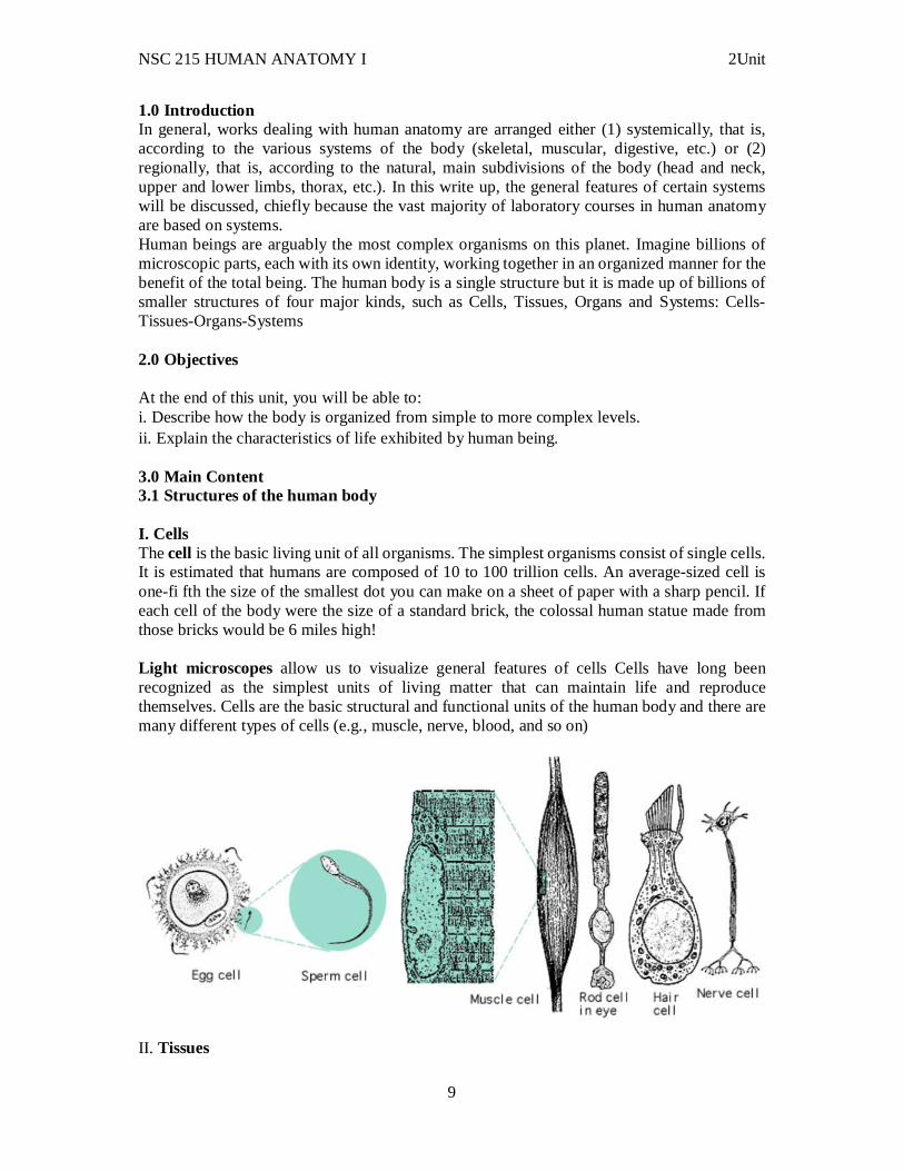
1.0 Introduction In general, works dealing with human anatomy are arranged either (1) systemically, that is, according to the various systems of the body (skeletal, muscular, digestive, etc.) or (2) regionally, that is, according to the natural, main subdivisions of the body (head and neck, upper and lower limbs, thorax, etc.). In this write up, the general features of certain systems will be discussed, chiefly because the vast majority of laboratory courses in human anatomy are based on systems. Human beings are arguably the most complex organisms on this planet. Imagine billions of microscopic parts, each with its own identity, working together in an organized manner for the benefit of the total being. The human body is a single structure but it is made up of billions of smaller structures of four major kinds, such as Cells, Tissues, Organs and Systems: Cells-Tissues-Organs-Systems 2.0 Objectives At the end of this unit, you will be able to: i. Describe how the body is organized from simple to more complex levels. ii. Explain the characteristics of life exhibited by human being. 3.0 Main Content 3.1 Structures of the human body I. Cells The cell is the basic living unit of all organisms. The simplest organisms consist of single cells. It is estimated that humans are composed of 10 to 100 trillion cells. An average-sized cell is one-fi fth the size of the smallest dot you can make on a sheet of paper with a sharp pencil. If each cell of the body were the size of a standard brick, the colossal human statue made from those bricks would be 6 miles high! Light microscopes allow us to visualize general features of cells Cells have long been recognized as the simplest units of living matter that can maintain life and reproduce themselves. Cells are the basic structural and functional units of the human body and there are many different types of cells (e.g., muscle, nerve, blood, and so on)
II. Tissues
NSC 215 HUMAN ANATOMY I 2Unit
10
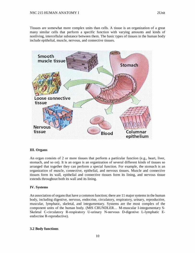
Tissues are somewhat more complex units than cells. A tissue is an organization of a great many similar cells that perform a specific function with varying amounts and kinds of nonliving, intercellular substance between them. The basic types of tissues in the human body include epithelial, muscle, nervous, and connective tissues.
III. Organs An organ consists of 2 or more tissues that perform a particular function (e.g., heart, liver, stomach, and so on). It is an organ is an organization of several different kinds of tissues so arranged that together they can perform a special function. For example, the stomach is an organization of muscle, connective, epithelial, and nervous tissues. Muscle and connective tissues form its wall, epithelial and connective tissues form its lining, and nervous tissue extends throughout both its wall and its lining. IV. Systems An association of organs that have a common function; there are 11 major systems in the human body, including digestive, nervous, endocrine, circulatory, respiratory, urinary, reproductive, muscular, lymphatic, skeletal, and integumentary. Systems are the most complex of the component units of the human body. (MIS CRUNDLER… M-muscular I-integumentary S-Skeletal C-circulatory R-respiratory U-urinary N-nervous D-digestive L-lymphatic E-endocrine R-reproductive). 3.2 Body functions
NSC 215 HUMAN ANATOMY I 2Unit
11
Body functions are the physiological or psychological functions of body systems. The body's functions are ultimately its cells' functions. Survival is the body's most important business. Survival depends on the body's maintaining or restoring homeostasis, a state of relative constancy, of its internal environment. More than a century ago, French physiologist, Claude Bernard (1813-1878), made a remarkable observation. He noted that body cells survived in a healthy condition only when the temperature, pressure, and chemical composition of their environment remained relatively constant. Later, an American physiologist, Walter B. Cannon (1871-1945), suggested the name homeostasis for the relatively constant states maintained by the body. Homeostasis is a key word in modern physiology. It comes from two Greek words - "homeo," meaning the same, and "stasis," meaning standing. "Standing or staying the same" then is the literal meaning of homeostasis. However, as Cannon emphasized, homeostasis does not mean something set and immobile that stays exactly the same all the time. In his words, homeostasis "means a condition that may vary, but which is relatively constant." Homeostasis depends on the body's ceaselessly carrying on many activities. Its major activities or functions are responding to changes in the body's environment, exchanging materials between the environment and cells, metabolizing foods, and integrating all of the body's diverse activities. The body's ability to perform many of its functions changes gradually over the years. In general, the body performs its functions least well at both ends of life - in infancy and in old age. During childhood, body functions gradually become more and more efficient and effective. During late maturity and old age the opposite is true. They gradually become less and less efficient and effective. During young adulthood, they normally operate with maximum efficiency and effectiveness. 3.3 Characteristics of life All living organisms have certain characteristics that distinguish them from non-living forms: (MR NIGER D) The basic processes of life include organization, metabolism, responsiveness, movements, and reproduction. In humans, who represent the most complex form of life, there are additional requirements such as growth, differentiation, respiration, digestion, and excretion. All of these processes are interrelated. No part of the body, from the smallest cell to a complete body system, works in isolation. All function together, in fine-tuned balance, for the well-being of the individual and to maintain life. Disease such as cancer and death represent a disruption of the balance in these processes. The following are a brief description of the life process: Organization At all levels of the organizational scheme, there is a division of labor. Each component has its own job to perform in cooperation with others. Even a single cell, if it loses its integrity or organization, will die. Atoms to Molecules to Macromolecules to Organelles to Cells to Tissues to Organ to Organ Systems to Organism Metabolism
NSC 215 HUMAN ANATOMY I 2Unit
12
Metabolism is a broad term that includes all the chemical reactions that occur in the body. One phase of metabolism is catabolism in which complex substances are broken down into simpler building blocks and energy is released. Needs: Water, food, oxygen, heat, pressure - all must be regulated, the other phase is anabolism; which involves construction of complex substances from simpler ones Responsiveness Responsiveness or irritability is concerned with detecting changes in the internal or external environments and reacting to that change. It is the act of sensing a stimulus and responding to it. Movement There are many types of movement within the body. On the cellular level, molecules move from one place to another. Blood moves from one part of the body to another. The diaphragm moves with every breath. The ability of muscle fibers to shorten and thus to produce movement is called contractility. Reproduction For most people, reproduction refers to the formation of a new person, the birth of a baby. In this way, life is transmitted from one generation to the next through reproduction of the organism. In a broader sense, reproduction also refers to the formation of new cells for the replacement and repair of old cells as well as for growth. This is cellular reproduction. Both are essential to the survival of the human race. Growth Growth refers to an increase in size either through an increase in the number of cells or through an increase in the size of each individual cell. In order for growth to occur, anabolic processes must occur at a faster rate than catabolic processes. Differentiation Differentiation is a developmental process by which unspecialized cells change into specialized cells with distinctive structural and functional characteristics. Through differentiation, cells develop into tissues and organs. Respiration Respiration refers to all the processes involved in the exchange of oxygen and carbon dioxide between the cells and the external environment. It includes ventilation, the diffusion of oxygen and carbon dioxide, and the transport of the gases in the blood. Cellular respiration deals with the cell's utilization of oxygen and release of carbon dioxide in its metabolism. Digestion Digestion is the process of breaking down complex ingested foods into simple molecules that can be absorbed into the blood and utilized by the body. Excretion Excretion is the process that removes the waste products of digestion and metabolism from the body. It gets rid of by-products that the body is unable to use, many of which are toxic and incompatible with life. The ten life processes described above are not enough to ensure the survival of the individual. In addition to these processes, life depends on certain physical factors from the environment. These include water, oxygen, nutrients, heat, and pressure.
NSC 215 HUMAN ANATOMY I 2Unit
13
Self-Assessment Exercise i. Describe how the body is organized from simple to more complex levels. ii. Explain the characteristics of life exhibited by human being 4.0 Conclusion The body is made of structures organized from simple to complex at six levels with 11 organ-systems. 5.0 Summary: In this unit, you have learnt: i. Anatomy is the study of the structure of the human body
ii. Systemic anatomy is the study of the body by organ systems. Regional anatomy is the study of the body by areas.
iii. Surface anatomy uses superficial structures to locate deeper structures, and anatomical imaging is a non-invasive method for examining deep structures.
iv. The human body can be organized into six levels: chemical (atoms and molecules), cell, tissue (groups of similar cells and the materials surrounding them), organ (two or more tissues that perform one or more common functions), organ system (groups of organs with common functions), and organism. v. The 11 organ systems are the integumentary, skeletal, muscular, nervous, endocrine, cardiovascular, lymphatic, respiratory, digestive, urinary, and reproductive systems.
vi. The characteristics of life include organization, metabolism, responsiveness, growth, development, and reproduction. 6.0 Tutor Marked Assignments Look at the differences between the cell and tissue under the microscope at the Histology and report your findings in your log book. Answer the following questions: 1. From smallest to largest, list and define the body’s six levels of organization. 2. What are the four primary tissue types? 3. Which two organ systems are responsible for regulating the other organ systems? 4. Which two are responsible for support and movement? 5. What are the functions of the integumentary, cardiovascular, lymphatic, respiratory, digestive, urinary, and reproductive systems? 6. Describe six characteristics of life. 7. Why is it important to realize that humans share many, but not all, characteristics with other animals? 8. The following are organizational levels for considering the body.
a. cell b. chemical c. organ d. organ system, e. organism f. tissue
Choose the correct order for these organizational levels from smallest to largest. a. 1,2,3,6,4,5
NSC 215 HUMAN ANATOMY I 2Unit
14
b. 2,1,6,3,4,5 c. 3,1,6,4,5,2 d. 2,6,1,3,5,4 e. 1,6,5,3,4,2
7.0 References and other resources 1. Bruce M. Carlson (2019) Human Embryology & Developmental Biology. 6th edition
2. Kathryn A. Booth, Terri. D. Wyman (2008) Anatomy, physiology, and pathophysiology for allied health
3. Katherine M. A. Rogers and William N. Scott (2011) Nurses! Test yourself in anatomy and physiology
4. Kent M. Van De Graff, R.Ward Rhees, Sidney Palmer (2010) Schaum’s Outline of Human Anatomy and Physiology 3rd edition
5. Keith L Moore, Persuade T.V.N (2016), The Developing Human Clinically Oriented Embryology 10th Edition Lippincott Williams & Wilkins
6. Philip Tate (2012) Seeley’s Principles of Anatomy & Physiology 2nd edition.
7. Sadler T.W (2019), Langman’s Medical Embryology 14th edition. Lippincott Williams & Wilkins UNIT TWO: ANATOMICAL TERMINOLOGY CONTENT 1.0 Introduction 2.0 Objectives 3.0 Main Content 3.1 Directional terms 3.2 Planes of the body 3.3 Body cavities 3.4 Clinical correlates 4.0 Conclusion 5.0 Summary 6.0 Tutor Marked Assignments 6.1 Activity 6.2 Teacher Marked Tests 7.0 References and other resources 1.0 Introduction Before we get into the following learning units, which will provide more detailed discussion of topics on different human body systems, it is necessary to learn some useful terms for describing body structure. Knowing these terms will make it much easier for us to understand the content of the following learning units. Three groups of terms are introduced here:
Directional Terms
Planes of the Body
Body Cavities 2.0 Objectives
NSC 215 HUMAN ANATOMY I 2Unit
15
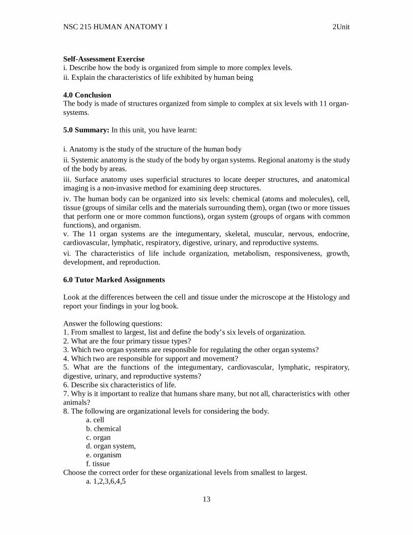
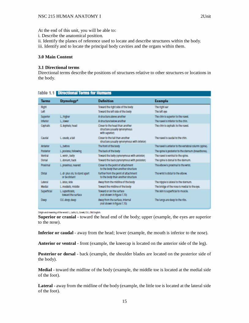
At the end of this unit, you will be able to: i. Describe the anatomical position. ii. Identify the planes of reference used to locate and describe structures within the body. iii. Identify and to locate the principal body cavities and the organs within them. 3.0 Main Content 3.1 Directional terms Directional terms describe the positions of structures relative to other structures or locations in the body.
Superior or cranial - toward the head end of the body; upper (example, the eyes are superior to the nose). Inferior or caudal - away from the head; lower (example, the mouth is inferior to the nose). Anterior or ventral - front (example, the kneecap is located on the anterior side of the leg). Posterior or dorsal - back (example, the shoulder blades are located on the posterior side of the body). Medial - toward the midline of the body (example, the middle toe is located at the medial side of the foot). Lateral - away from the midline of the body (example, the little toe is located at the lateral side of the foot).
NSC 215 HUMAN ANATOMY I 2Unit
16
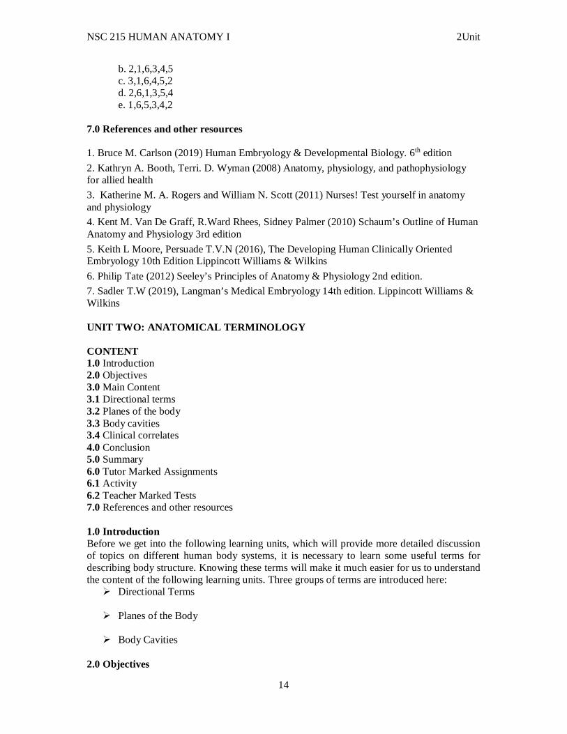
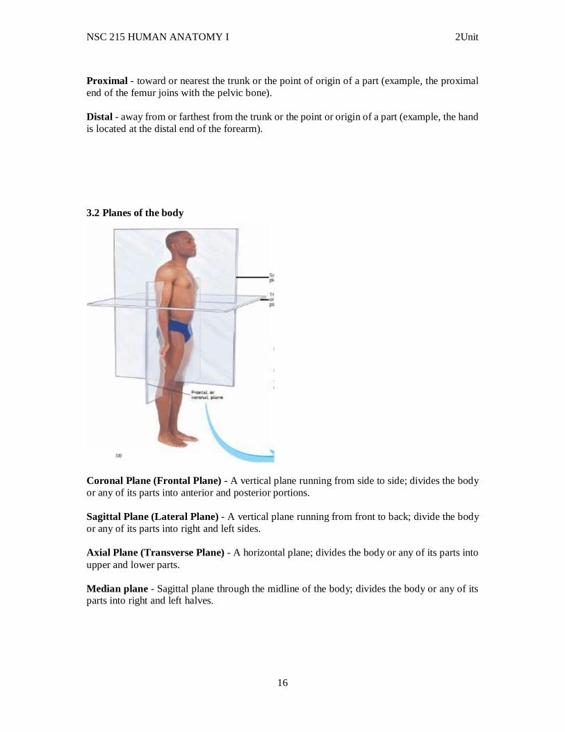
Proximal - toward or nearest the trunk or the point of origin of a part (example, the proximal end of the femur joins with the pelvic bone). Distal - away from or farthest from the trunk or the point or origin of a part (example, the hand is located at the distal end of the forearm). 3.2 Planes of the body
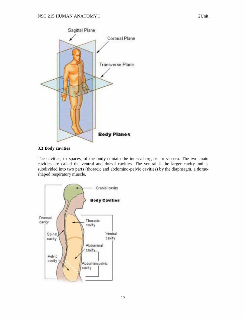
Coronal Plane (Frontal Plane) - A vertical plane running from side to side; divides the body or any of its parts into anterior and posterior portions. Sagittal Plane (Lateral Plane) - A vertical plane running from front to back; divide the body or any of its parts into right and left sides. Axial Plane (Transverse Plane) - A horizontal plane; divides the body or any of its parts into upper and lower parts. Median plane - Sagittal plane through the midline of the body; divides the body or any of its parts into right and left halves.
NSC 215 HUMAN ANATOMY I 2Unit
17
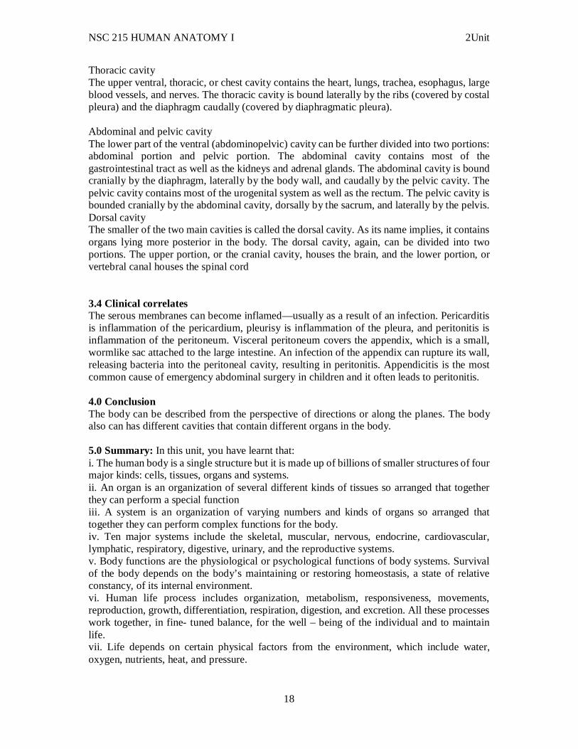
3.3 Body cavities The cavities, or spaces, of the body contain the internal organs, or viscera. The two main cavities are called the ventral and dorsal cavities. The ventral is the larger cavity and is subdivided into two parts (thoracic and abdomino-pelvic cavities) by the diaphragm, a dome-shaped respiratory muscle.
NSC 215 HUMAN ANATOMY I 2Unit
18
Thoracic cavity The upper ventral, thoracic, or chest cavity contains the heart, lungs, trachea, esophagus, large blood vessels, and nerves. The thoracic cavity is bound laterally by the ribs (covered by costal pleura) and the diaphragm caudally (covered by diaphragmatic pleura). Abdominal and pelvic cavity The lower part of the ventral (abdominopelvic) cavity can be further divided into two portions: abdominal portion and pelvic portion. The abdominal cavity contains most of the gastrointestinal tract as well as the kidneys and adrenal glands. The abdominal cavity is bound cranially by the diaphragm, laterally by the body wall, and caudally by the pelvic cavity. The pelvic cavity contains most of the urogenital system as well as the rectum. The pelvic cavity is bounded cranially by the abdominal cavity, dorsally by the sacrum, and laterally by the pelvis. Dorsal cavity The smaller of the two main cavities is called the dorsal cavity. As its name implies, it contains organs lying more posterior in the body. The dorsal cavity, again, can be divided into two portions. The upper portion, or the cranial cavity, houses the brain, and the lower portion, or vertebral canal houses the spinal cord 3.4 Clinical correlates The serous membranes can become inflamed—usually as a result of an infection. Pericarditis is inflammation of the pericardium, pleurisy is inflammation of the pleura, and peritonitis is inflammation of the peritoneum. Visceral peritoneum covers the appendix, which is a small, wormlike sac attached to the large intestine. An infection of the appendix can rupture its wall, releasing bacteria into the peritoneal cavity, resulting in peritonitis. Appendicitis is the most common cause of emergency abdominal surgery in children and it often leads to peritonitis. 4.0 Conclusion The body can be described from the perspective of directions or along the planes. The body also can has different cavities that contain different organs in the body. 5.0 Summary: In this unit, you have learnt that: i. The human body is a single structure but it is made up of billions of smaller structures of four major kinds: cells, tissues, organs and systems. ii. An organ is an organization of several different kinds of tissues so arranged that together they can perform a special function iii. A system is an organization of varying numbers and kinds of organs so arranged that together they can perform complex functions for the body. iv. Ten major systems include the skeletal, muscular, nervous, endocrine, cardiovascular, lymphatic, respiratory, digestive, urinary, and the reproductive systems. v. Body functions are the physiological or psychological functions of body systems. Survival of the body depends on the body’s maintaining or restoring homeostasis, a state of relative constancy, of its internal environment. vi. Human life process includes organization, metabolism, responsiveness, movements, reproduction, growth, differentiation, respiration, digestion, and excretion. All these processes work together, in fine- tuned balance, for the well – being of the individual and to maintain life. vii. Life depends on certain physical factors from the environment, which include water, oxygen, nutrients, heat, and pressure.
NSC 215 HUMAN ANATOMY I 2Unit
19
viii. Terms used in describing body parts and activities include – directional terms, terms used in describing body planes and body cavities. 6.0 Tutor Marked Assignments At the gross anatomy laboratory, identify the different cavities of the body and the contents of each cavity in relation to one another with directional terms and report in your log book. Answer all these questions. 1. The clavicle (collarbone) is to the nipple of the breast.
a. anterior b. distal c. superficial d. superior e. ventral
2. The term that means nearer to the attached end of a limb is a. distal b. lateral.. c. medial. d. proximal
3. Which of these directional terms are paired most appropriately as opposites? a. superficial and deep b. medial and proximal c. distal and lateral d. superior and posterior e. anterior and inferior
8. With reference to the planes of the body, discuss the advantage of computed tomography (CT or CAT) scans and magnetic resonance images (MRIs) over conventional x-rays. 10. What are visceral organs? 7.0 References/further reading: 1. Bruce M. Carlson (2019) Human Embryology & Developmental Biology. 6th edition
2. Kathryn A. Booth, Terri. D. Wyman (2008) Anatomy, physiology, and pathophysiology for allied health
3. Katherine M. A. Rogers and William N. Scott (2011) Nurses! Test yourself in anatomy and physiology
4. Kent M. Van De Graff, R.Ward Rhees, Sidney Palmer (2010) Schaum’s Outline of Human Anatomy and Physiology 3rd edition
5. Keith L Moore, Persuade T.V.N (2016), The Developing Human Clinically Oriented Embryology 10th Edition Lippincott Williams & Wilkins
6. Philip Tate (2012) Seeley’s Principles of Anatomy & Physiology 2nd edition.
7. Sadler T.W (2019), Langman’s Medical Embryology 14th edition. Lippincott Williams & Wilkins UNIT THREE: CELLS, TISSUES, & MEMBRANES CONTENT 1.0 Introduction 2.0 Objectives 3.0 Main Content
NSC 215 HUMAN ANATOMY I 2Unit
20
3.1 Cell theory 3.2 Cell structure and function 3.3 Cell division 3.4 DNA replication and protein synthesis 3.5 Clinical correlates 4.0 Conclusion 5.0 Summary 6.0 Tutor Marked Assignments 6.1 Activity 6.2 Tutor Marked Tests 7.0 References and other resources 1.0 Introduction This section provides detailed information about cell structure and function, four basic types of tissue in the human body, and the different types of membranes found in the body. 2.0 Objectives At the end of this unit, you should be able to: i. Discuss the cell theory ii. Explain the cellular organization of the human body. iii. Discuss the types and functions of the various body membranes iv. Discuss the importance of cell division in the human body. v. Explain the processes of replication, transcription, and translation. 3.0 Main Content 3.1 Cell theory Important Events in the Discovery of Cells
1665 - Robert Hooke looks at cork under a microscope. Calls the chambers he see "cells"
1665 - 75 Anton van Leeuwenhoek, the person incorrectly given credit for the invention of the microscope (actually, he was just damn good at making and using them, and his scopes soon became the standard, and history has just given him credit as the inventor of the microscope), studies organisms living in pond water (like you did in lab). He calls them "Animalcules."
NSC 215 HUMAN ANATOMY I 2Unit
21
1830 - German scientists Schleiden and Schawann summarize the findings of many scientists and conclude that all living organisms are made of cells. This forms the basis of the Cell Theory of Biology
The Cell Theory
All organisms are composed of cells The cell is the structural unit of life - units smaller than cells are not alive Cells arise by division of preexisting cells - spontaneous generation does not exist Cells can be cultured to produce more cells
o in vitro = outside organism or cell o in vivo = inside organism or cell
Properties of Cells Cells are complex and highly organized
They contain numerous internal structures Some are membrane bound (organelles) while others do not
Cells contain a genetic blueprint and machinery to use it
Genes are instructions for cells to create specific proteins All cells use the same types of information
o The genetic code is universal o The machinery used for synthesis is interchangeable
However, for this to function properly, information transfer must be error free o Errors are called mutations
Cells arise from the division of other cells
Daughter cells inherit the genes from the mother cells Binary fission - cell division in bacteria Mitosis - the genetic complement of each daughter cell is identical to the other and to
the mother cell. This is asexual reproduction Meiosis - the genetic complement of each daughter cell is reduced by half and each
daughter cell is genetically unique. This is used in sexual reproduction Daughter cells inherit cytoplasm and organelles from the mother cells
o Asexual - organelles from mother cell o Sexual - organelles predominately from one parent o In eukaryotes, the chloroplasts and mitochondria come from the egg cell o This can be used to trace the evolutionary origin of the organism
Cells acquire and utilize energy
Most cells respire o release energy found in organic compounds o convert organic compounds to CO2 and O2 o make ATP
Cells can perform a variety of chemical reactions
Transform simple organic molecules into complex molecules (anabolism) Breakdown complex molecules to release energy (catabolism) Metabolism = all reactions performed by cells
NSC 215 HUMAN ANATOMY I 2Unit
22
Cells can engage in mechanical activities Cells can move Organelles can move Cells can respond to stimuli
o chemotaxis - movement towards chemicals o phototaxis - movement towards light o hormone responses o touch responses
Cells can regulate activities
Cells control DNA synthesis and cell division Gene regulation - cells make specific proteins only when needed Turn on and off metabolic pathways
Cells contain the following structures:
Plasma membrane - separates the cell from the external environment Cytoplasm - fluid-filled cell interior Nuclear material - genetic information stored as DNA
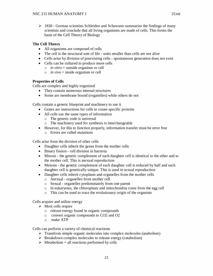
3.2 Cell structure and function Cells, the smallest structures capable of maintaining life and reproducing, compose all living things, from single-celled plants to multibillion-celled animals. The human body, which is made up of numerous cells, begins as a single, newly fertilized cell. Almost all human cells are microscopic in size. To give you an idea how small a cell is, one average-sized adult body, according to one estimate, consists of 100 trillion cells! Cell Structure Ideas about cell structure have changed considerably over the years. Early biologists saw cells as simple membranous sacs containing fluid and a few floating particles. Today's biologists know that cells are infinitely more complex than this.
NSC 215 HUMAN ANATOMY I 2Unit
23
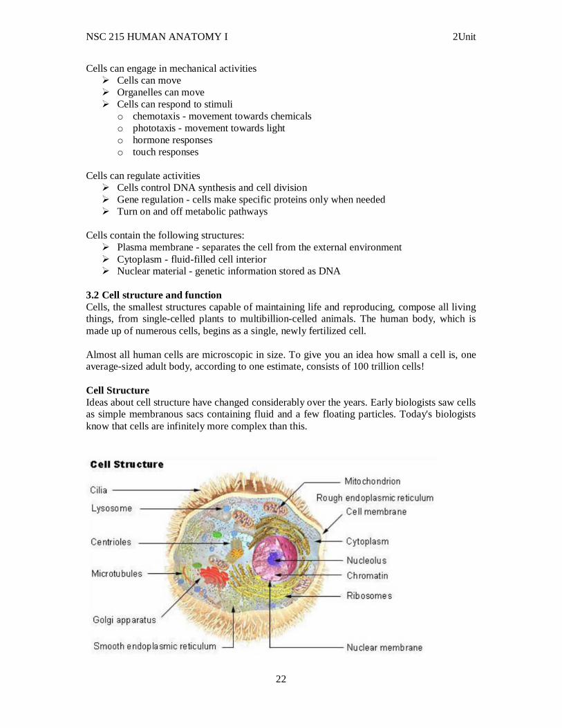
There are many different types, sizes, and shapes of cells in the body. For descriptive purposes, the concept of a "generalized cell" is introduced. It includes features from all cell types. A cell consists of three parts: the cell membrane, the nucleus, and, between the two, the cytoplasm. Within the cytoplasm lie intricate arrangements of fine fibers and hundreds or even thousands of miniscule but distinct structures called organelles. Cell membrane Every cell in the body is enclosed by a cell (Plasma) membrane. The cell membrane separates the material outside the cell, extracellular, from the material inside the cell, intracellular . It maintains the integrity of a cell and controls passage of materials into and out of the cell. All materials within a cell must have access to the cell membrane (the cell's boundary) for the needed exchange. The cell membrane is a double layer of phospholipid molecules. Proteins in the cell membrane provide structural support, form channels for passage of materials, act as receptor sites, function as carrier molecules, and provide identification markers.
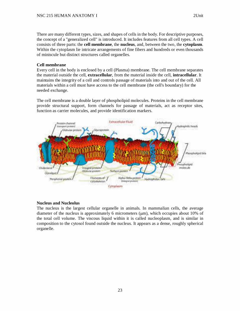
Nucleus and Nucleolus The nucleus is the largest cellular organelle in animals. In mammalian cells, the average diameter of the nucleus is approximately 6 micrometers (μm), which occupies about 10% of the total cell volume. The viscous liquid within it is called nucleoplasm, and is similar in composition to the cytosol found outside the nucleus. It appears as a dense, roughly spherical organelle.
NSC 215 HUMAN ANATOMY I 2Unit
24
The nuclear envelope, otherwise known as nuclear membrane, consists of two cellular membranes, an inner and an outer membrane, arranged parallel to one another and separated by 10 to 50 nanometers (nm). The nuclear envelope completely encloses the nucleus and separates the cell's genetic material from the surrounding cytoplasm, serving as a barrier to prevent macromolecules from diffusing freely between the nucleoplasm and the cytoplasm. The outer nuclear membrane is continuous with the membrane of the rough endoplasmic reticulum (RER), and is similarly studded with ribosomes. The space between the membranes is called the perinuclear space and is continuous with the RER lumen. Nuclear pores, which provide aqueous channels through the envelope, are composed of multiple proteins, collectively referred to as nucleoporins. The pores are about 125 million daltons in molecular weight and consist of around 50 (in yeast) to several hundred proteins (in vertebrates).[5] The pores are 100 nm in total diameter; however, the gap through which molecules freely diffuse is only about 9 nm wide, due to the presence of regulatory systems within the center of the pore. The cell nucleus contains the majority of the cell's genetic material in the form of multiple linear DNA molecules organized into structures called chromosomes. Each human cell contains roughly 2 m of DNA. During most of the cell cycle these are organized in a DNA-protein complex known as chromatin. A small fraction of the cell's genes are located instead in the mitochondria. There are two types of chromatin. Euchromatin is the less compact DNA form, and contains genes that are frequently expressed by the cell. The other type, heterochromatin, is the more compact form, and contains DNA that is infrequently transcribed. During interphase the chromatin organizes itself into discrete individual patches, called chromosome territories. The nucleolus is a discrete densely stained structure found in the nucleus. It is not surrounded by a membrane, and is sometimes called a suborganelle. It forms around tandem repeats of rDNA, DNA coding for ribosomal RNA (rRNA). These regions are called nucleolar organizer
NSC 215 HUMAN ANATOMY I 2Unit
25
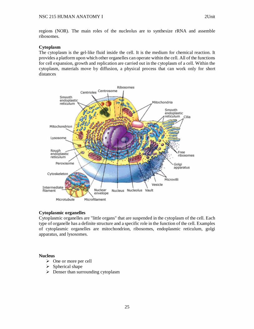
regions (NOR). The main roles of the nucleolus are to synthesize rRNA and assemble ribosomes. Cytoplasm The cytoplasm is the gel-like fluid inside the cell. It is the medium for chemical reaction. It provides a platform upon which other organelles can operate within the cell. All of the functions for cell expansion, growth and replication are carried out in the cytoplasm of a cell. Within the cytoplasm, materials move by diffusion, a physical process that can work only for short distances
Cytoplasmic organelles Cytoplasmic organelles are "little organs" that are suspended in the cytoplasm of the cell. Each type of organelle has a definite structure and a specific role in the function of the cell. Examples of cytoplasmic organelles are mitochondrion, ribosomes, endoplasmic reticulum, golgi apparatus, and lysosomes. Nucleus
One or more per cell Spherical shape Denser than surrounding cytoplasm
NSC 215 HUMAN ANATOMY I 2Unit
26
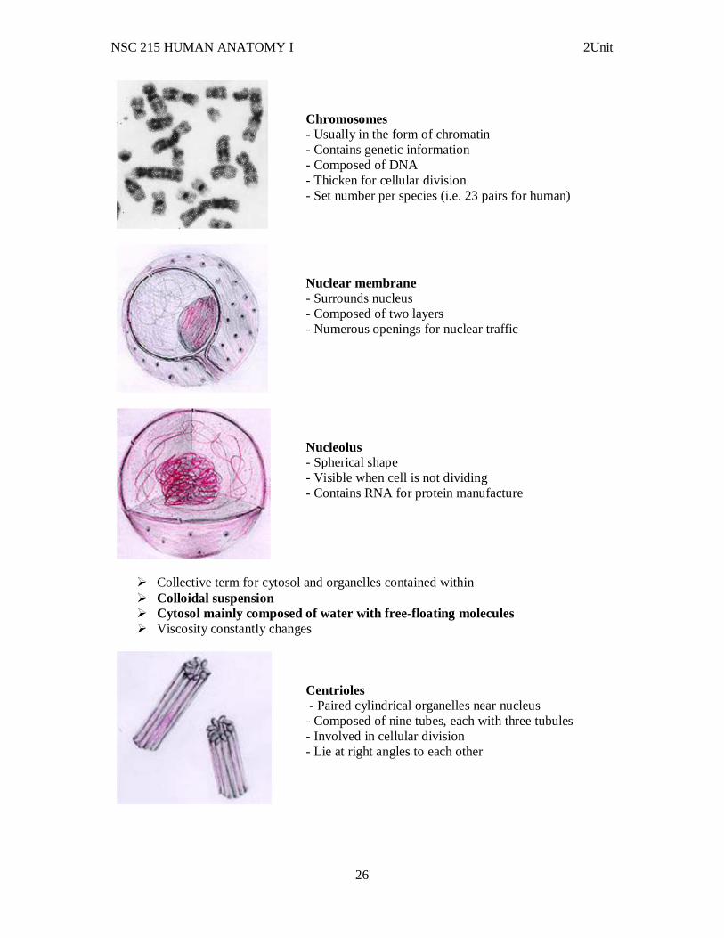
Chromosomes - Usually in the form of chromatin - Contains genetic information - Composed of DNA - Thicken for cellular division - Set number per species (i.e. 23 pairs for human)
Nuclear membrane - Surrounds nucleus - Composed of two layers - Numerous openings for nuclear traffic
Nucleolus - Spherical shape - Visible when cell is not dividing - Contains RNA for protein manufacture
Collective term for cytosol and organelles contained within Colloidal suspension Cytosol mainly composed of water with free-floating molecules Viscosity constantly changes
Centrioles - Paired cylindrical organelles near nucleus - Composed of nine tubes, each with three tubules - Involved in cellular division - Lie at right angles to each other
NSC 215 HUMAN ANATOMY I 2Unit
27
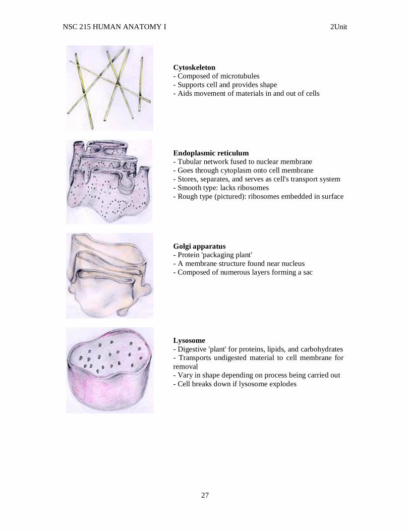
Cytoskeleton - Composed of microtubules - Supports cell and provides shape - Aids movement of materials in and out of cells
Endoplasmic reticulum - Tubular network fused to nuclear membrane - Goes through cytoplasm onto cell membrane - Stores, separates, and serves as cell's transport system - Smooth type: lacks ribosomes - Rough type (pictured): ribosomes embedded in surface
Golgi apparatus - Protein 'packaging plant' - A membrane structure found near nucleus - Composed of numerous layers forming a sac
Lysosome - Digestive 'plant' for proteins, lipids, and carbohydrates - Transports undigested material to cell membrane for removal - Vary in shape depending on process being carried out - Cell breaks down if lysosome explodes
NSC 215 HUMAN ANATOMY I 2Unit
28
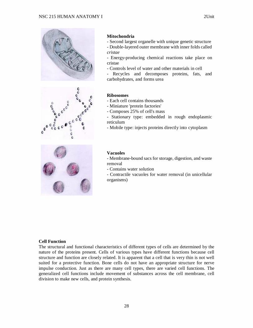
Mitochondria - Second largest organelle with unique genetic structure - Double-layered outer membrane with inner folds called cristae - Energy-producing chemical reactions take place on cristae - Controls level of water and other materials in cell - Recycles and decomposes proteins, fats, and carbohydrates, and forms urea
Ribosomes - Each cell contains thousands - Miniature 'protein factories' - Composes 25% of cell's mass - Stationary type: embedded in rough endoplasmic reticulum - Mobile type: injects proteins directly into cytoplasm
Vacuoles - Membrane-bound sacs for storage, digestion, and waste removal - Contains water solution - Contractile vacuoles for water removal (in unicellular organisms)
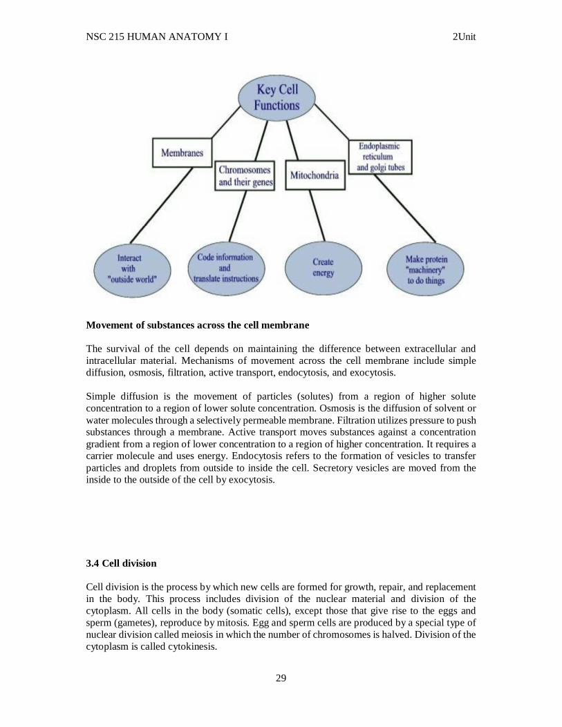
Cell Function The structural and functional characteristics of different types of cells are determined by the nature of the proteins present. Cells of various types have different functions because cell structure and function are closely related. It is apparent that a cell that is very thin is not well suited for a protective function. Bone cells do not have an appropriate structure for nerve impulse conduction. Just as there are many cell types, there are varied cell functions. The generalized cell functions include movement of substances across the cell membrane, cell division to make new cells, and protein synthesis.
NSC 215 HUMAN ANATOMY I 2Unit
29
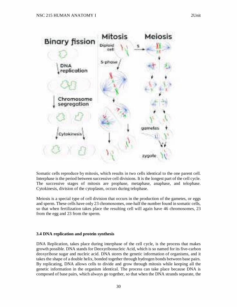
Movement of substances across the cell membrane The survival of the cell depends on maintaining the difference between extracellular and intracellular material. Mechanisms of movement across the cell membrane include simple diffusion, osmosis, filtration, active transport, endocytosis, and exocytosis. Simple diffusion is the movement of particles (solutes) from a region of higher solute concentration to a region of lower solute concentration. Osmosis is the diffusion of solvent or water molecules through a selectively permeable membrane. Filtration utilizes pressure to push substances through a membrane. Active transport moves substances against a concentration gradient from a region of lower concentration to a region of higher concentration. It requires a carrier molecule and uses energy. Endocytosis refers to the formation of vesicles to transfer particles and droplets from outside to inside the cell. Secretory vesicles are moved from the inside to the outside of the cell by exocytosis. 3.4 Cell division Cell division is the process by which new cells are formed for growth, repair, and replacement in the body. This process includes division of the nuclear material and division of the cytoplasm. All cells in the body (somatic cells), except those that give rise to the eggs and sperm (gametes), reproduce by mitosis. Egg and sperm cells are produced by a special type of nuclear division called meiosis in which the number of chromosomes is halved. Division of the cytoplasm is called cytokinesis.
NSC 215 HUMAN ANATOMY I 2Unit
30
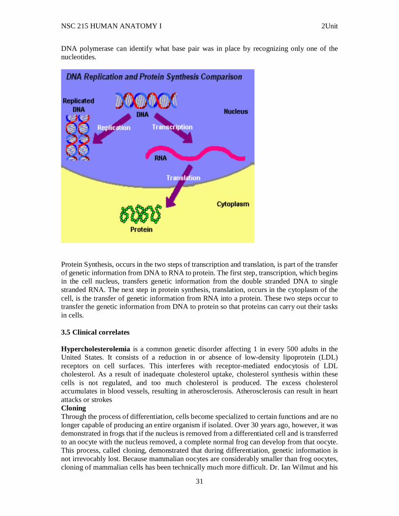
Somatic cells reproduce by mitosis, which results in two cells identical to the one parent cell. Interphase is the period between successive cell divisions. It is the longest part of the cell cycle. The successive stages of mitosis are prophase, metaphase, anaphase, and telophase. Cytokinesis, division of the cytoplasm, occurs during telophase. Meiosis is a special type of cell division that occurs in the production of the gametes, or eggs and sperm. These cells have only 23 chromosomes, one-half the number found in somatic cells, so that when fertilization takes place the resulting cell will again have 46 chromosomes, 23 from the egg and 23 from the sperm. 3.4 DNA replication and protein synthesis DNA Replication, takes place during interphase of the cell cycle, is the process that makes growth possible. DNA stands for Deoxyribonucleic Acid, which is so named for its five-carbon deoxyribose sugar and nucleic acid. DNA stores the genetic information of organisms, and it takes the shape of a double helix, bonded together through hydrogen bonds between base pairs. By replicating, DNA allows cells to divide and grow through mitosis while keeping all the genetic information in the organism identical. The process can take place because DNA is composed of base pairs, which always go together, so that when the DNA strands separate, the
NSC 215 HUMAN ANATOMY I 2Unit
31
DNA polymerase can identify what base pair was in place by recognizing only one of the nucleotides.
Protein Synthesis, occurs in the two steps of transcription and translation, is part of the transfer of genetic information from DNA to RNA to protein. The first step, transcription, which begins in the cell nucleus, transfers genetic information from the double stranded DNA to single stranded RNA. The next step in protein synthesis, translation, occurs in the cytoplasm of the cell, is the transfer of genetic information from RNA into a protein. These two steps occur to transfer the genetic information from DNA to protein so that proteins can carry out their tasks in cells. 3.5 Clinical correlates Hypercholesterolemia is a common genetic disorder affecting 1 in every 500 adults in the United States. It consists of a reduction in or absence of low-density lipoprotein (LDL) receptors on cell surfaces. This interferes with receptor-mediated endocytosis of LDL cholesterol. As a result of inadequate cholesterol uptake, cholesterol synthesis within these cells is not regulated, and too much cholesterol is produced. The excess cholesterol accumulates in blood vessels, resulting in atherosclerosis. Atherosclerosis can result in heart attacks or strokes Cloning Through the process of differentiation, cells become specialized to certain functions and are no longer capable of producing an entire organism if isolated. Over 30 years ago, however, it was demonstrated in frogs that if the nucleus is removed from a differentiated cell and is transferred to an oocyte with the nucleus removed, a complete normal frog can develop from that oocyte. This process, called cloning, demonstrated that during differentiation, genetic information is not irrevocably lost. Because mammalian oocytes are considerably smaller than frog oocytes, cloning of mammalian cells has been technically much more difficult. Dr. Ian Wilmut and his
NSC 215 HUMAN ANATOMY I 2Unit
32
colleagues at the Roslin Institute in Edinburgh, Scotland, overcame those technical difficulties in 1996, when they successfully cloned the first mammal, a sheep. Since that time, many other mammalian species have been cloned. 4.0 Conclusion The cell as the smallest unit of life contain structures that serve different purposes by the functions they perform. Understanding the various structures and how cells multiply helps our understanding of cell growth, repairs and reproduction. 5.0 Summary In this unit, you have learnt that: i. Cells are the basic unit of life, containing organelles, which perform specific functions. ii. The plasma membrane forms the outer boundary of the cell, the nucleus contains genetic material and directs cell activities, and cytoplasm is material between the nucleus and plasma membrane. iii. Cells metabolize and release energy, synthesize molecules, provide a means of communication, reproduce, and provide for inheritance. iv. Intracellular substances are inside cells, whereas extracellular (intercellular) substances are between cells. v. The plasma membrane is composed of a double layer of phospholipid molecules (lipid bilayer) in which proteins float (fluid-mosaic model). vi. Cell division that occurs by mitosis produces new cells for growth and tissue repair. vii. Cell division that occurs by meiosis produces gametes (sex cells). Sperm cells in males and oocytes (egg cells) in females are gametes. viii. Humans have 22 pairs of autosomal chromosomes and one pair of sex chromosomes. Females have the sex chromosomes XX and males have XY. ix. Mitosis is divided into four stages: Prophase, Metaphase, Anaphase, Telophase. x. Cytokinesis is the division of the cytoplasm of the cell. It begins with the formation of the cleavage furrow during anaphase and is complete when the plasma membrane comes together at the equator, producing two new daughter cells. xi. Differentiation, the process by which cells develop specialized structures and functions, results from the selective activation and inactivation of DNA sections. 6.0 Tutor Marked Assignments In the histology laboratory, examine the following and report in your log book: i. Structures found inside the cell and identify the properties of each structure ii. The stages of cell division Answer the following questions. 1. Cells.........
a. produce heat that helps to maintain body temperature. b. are different from each other because of the types of molecules they produce. c. communicate with each other through chemical and electric signals. d. divide to produce new cells containing the same genetic information. e. all of the above.
2. In the plasma membrane, phospholipids
NSC 215 HUMAN ANATOMY I 2Unit
33
a. form most of the bilayer. b. function as enzymes. c. bind cells together. d. allow cells to identify each other. e. all of the above.
3. Concerning diffusion, a. most non-lipid-soluble molecules and ions diff use through the lipid bilayer. b. it stops when random movement of molecules and ions stops. c. it is the movement of molecules or ions from areas of lower concentration to areas of higher concentration. d. the greater the concentration gradient, the greater the rate of diffusion. e. it requires ATP.
4. Which of these statements about osmosis is true? a. Osmosis always involves a membrane that allows water and all solutes to move through it. b. The greater the solute concentration, the smaller the osmotic pressure of a solution. c. Osmosis moves water from a solution with a greater solute concentration to a solution with a lesser solute concentration. d. The greater the osmotic pressure of a solution, the greater the tendency for water to move into the solution. e. Osmosis occurs because of hydrostatic pressure outside the cell.
5. If a cell is placed in a (an) solution, lysis of the cell may occur. a. hypertonic b. hypotonic c. isotonic d. isosmotic
6. Suppose that a man is doing heavy exercise in the hot summer sun. He sweats profusely. He then drinks a large amount of distilled water. After he drinks the water, you would expect his tissue cells to
a. shrink. b. swell. c. remain the same.
7.0 References/further reading: 1. Bruce M. Carlson (2019) Human Embryology & Developmental Biology. 6th edition
2. Kathryn A. Booth, Terri. D. Wyman (2008) Anatomy, physiology, and pathophysiology for allied health
3. Katherine M. A. Rogers and William N. Scott (2011) Nurses! Test yourself in anatomy and physiology
4. Kent M. Van De Graff, R.Ward Rhees, Sidney Palmer (2010) Schaum’s Outline of Human Anatomy and Physiology 3rd edition
5. Keith L Moore, Persuade T.V.N (2016), The Developing Human Clinically Oriented Embryology 10th Edition Lippincott Williams & Wilkins
6. Philip Tate (2012) Seeley’s Principles of Anatomy & Physiology 2nd edition.
7. Sadler T.W (2019), Langman’s Medical Embryology 14th edition. Lippincott Williams & Wilkins UNIT FOUR: BODY TISSUES
NSC 215 HUMAN ANATOMY I 2Unit
34
1.0 Introduction 2.0 Objectives 3.0 Main Content 3.1 Epithelial tissues 3.2 Connective tissues 3.3 Muscle tissues 3.4 Nervous tissues and membranes 3.5 Clinical correlates 4.0 Conclusion 5.0 Summary 6.0 Tutor Marked Assignments 6.1 Activity 6.2 Tutor Marked Tests 7.0 References and other Resources 1.0 Introduction In some ways, the human body is like a complex machine, such as a car. Not all parts of a car can be made from a single type of material. Metal, capable of withstanding the heat of the engine, cannot be used for windows or tires. Similarly, the many parts of the human body are made of collections of specialized cells and the materials surrounding them. Muscle cells that contract to produce body movements have a structure and function different from that of epithelial cells that protect, secrete, or absorb. Knowledge of tissue structure and function is important in understanding how individual cells are organized to form tissues and how tissues are organized to form organs, organ systems, and the complete organism. There is a relationship between the structure of each tissue type and its function and between the tissues in an organ and the organ’s function. The structure and function of tissues are so closely related that you should be able to predict the function of a tissue when given its structure, and vice versa. 2.0 Objectives At the end of this unit, you should be able to: i. Define histology and tissue and to distinguish between the four major tissue types ii. Explain purpose of the specialization of the tissues in the body. iii. Describe epithelial tissue on the cellular level and to differentiate between the various kinds. iv. Describe the characteristics, locations, and functions of connective tissue. v. Describe muscle tissue and to distinguish between the three types. vi. Describe the basic characteristics and functions of nervous tissue. 3.0 Main Content 3.1 Epithelial tissues Epithelial tissue covers the whole surface of the body. It is made up of cells closely packed and ranged in one or more layers. This tissue is specialised to form the covering or lining of all internal and external body surfaces. Epithelial tissue that occurs on surfaces on the interior of the body is known as endothelium. Epithelial cells are packed tightly together, with almost no intercellular spaces and only a small amount of intercellular substance. Epithelial tissue, regardless of the type, is usually separated from the underlying tissue by a thin sheet of connective tissue; basement membrane. The basement membrane provides structural support for the epithelium and also binds it to neighbouring structures.
NSC 215 HUMAN ANATOMY I 2Unit
35
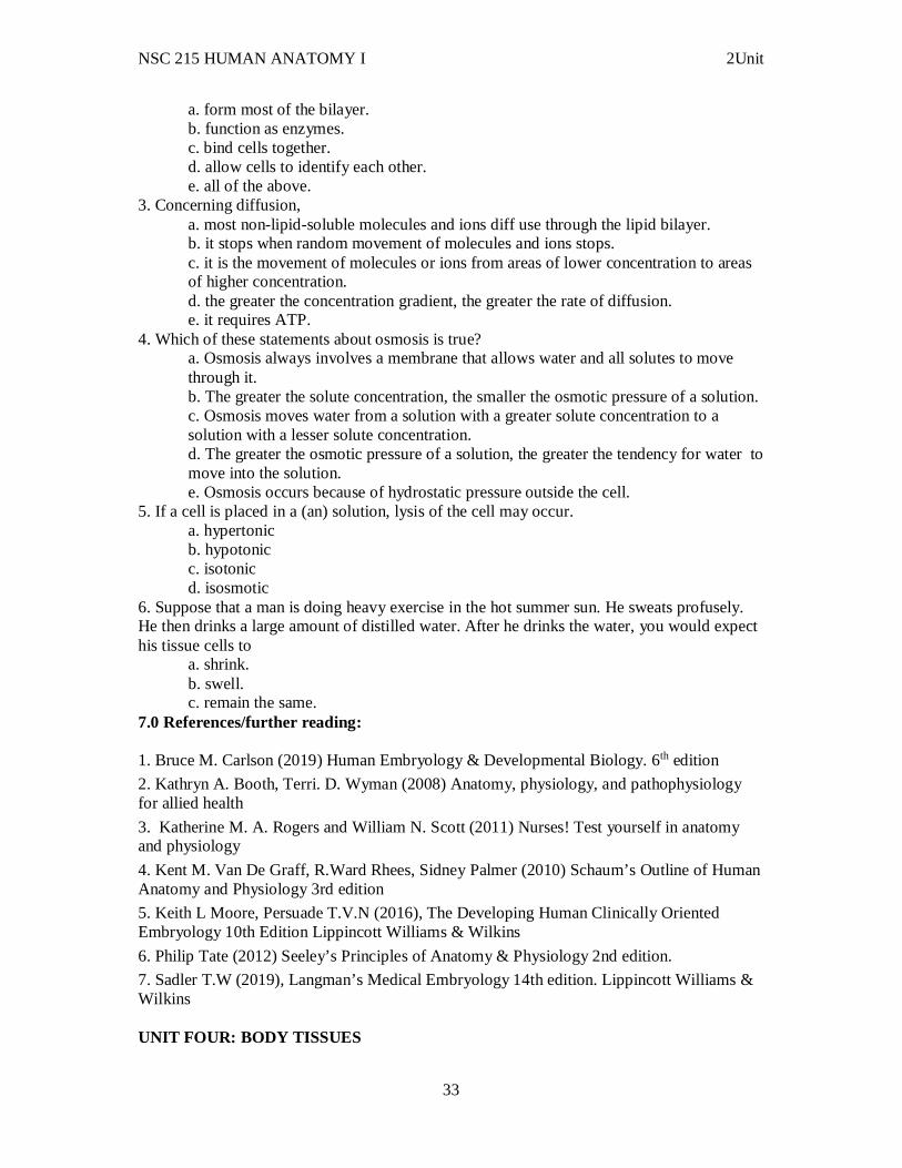
Types of Epithelial Tissue Epithelial tissue can be divided into two groups depending on the number of layers of which it is composes. Epithelial tissue which is only one cell thick is known as simple epithelium. If it is two or more cells thick such as the skin, it is known as stratified epithelium. Simple epithelium Simple epithelium can be subdivided according to the shape and function of its cells. Squamous (pavement) epithelium. Squamous cells have the appearance of thin, flat plates. The shape of the nucleus usually corresponds to the cell form and help to identify the type of epithelium. Squamous cells, for example, tend to have horizontall flattened, elliptical nuclei because of the thin flattened form of the cell. They form the lining of cavities such as the mouth, blood vessels, heart and lungs and make up the outer layers of the skin.
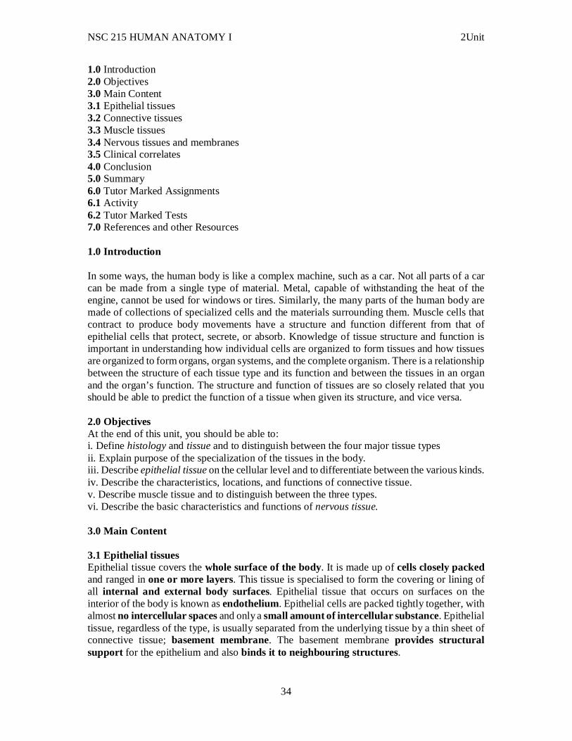
Simple sqaumous epithelium Simple Cuboidal Epithelium. As their name implies, cuboidal cells are roughly square or cuboidal in shape. Each cell has a spherical nucleus in the centre. Cuboidal epithelium is found in glands and in the lining of the kidney tubules as well as in the ducts of the glands. They also constitute the germinal epithelium which produces the egg cells in the female ovary and the sperm cells in the male testes.
NSC 215 HUMAN ANATOMY I 2Unit
36
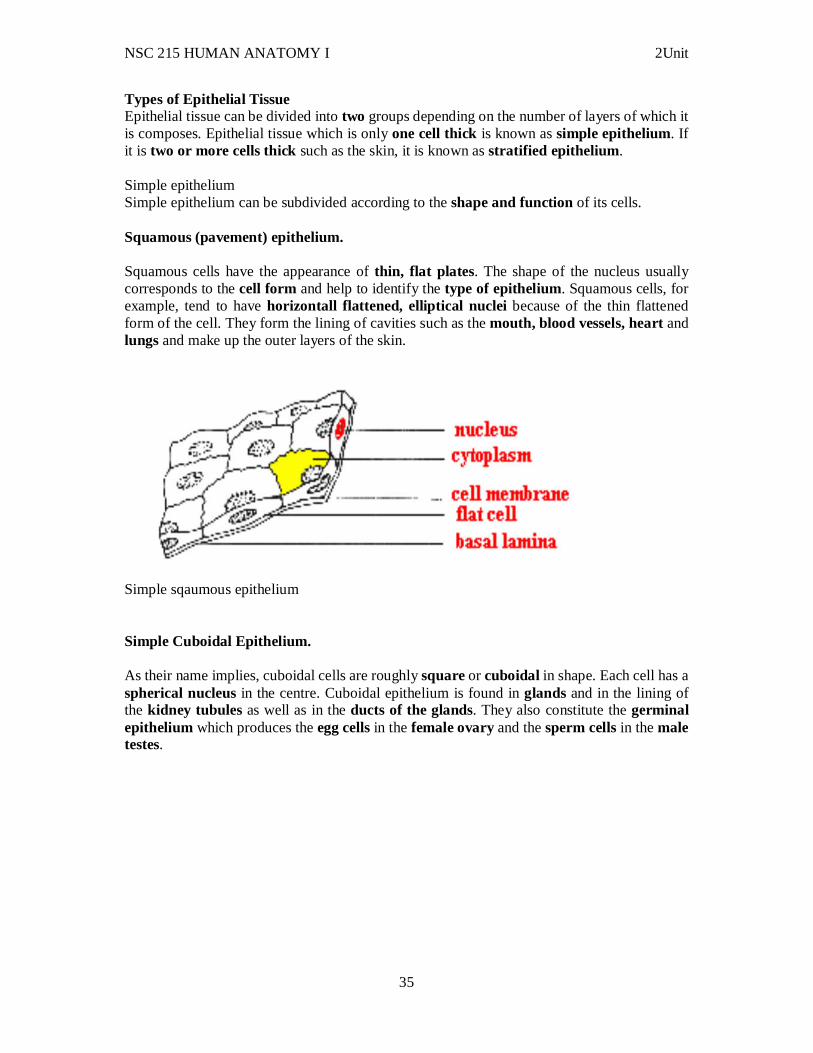
Simple cuboidal epithelium Simple Columnar Epithelium Columnar epithelial cells occur in one or more layers. The cells are elongated and column-shaped. The nuclei are elongated and are usually located near the base of the cells. Columnar epithelium forms the lining of the stomach and intestines. Some columnar cells are specialised for sensory reception such as in the nose, ears and the taste buds of the tongue. Goblet cells (unicellular glands) are found between the columnar epithelial cells of the duodenum. They secrete mucus or slime, a lubricating substance which keeps the surface smooth.
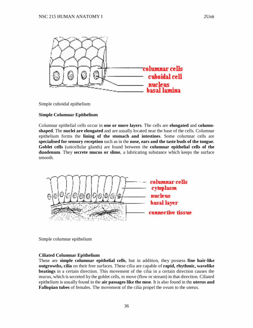
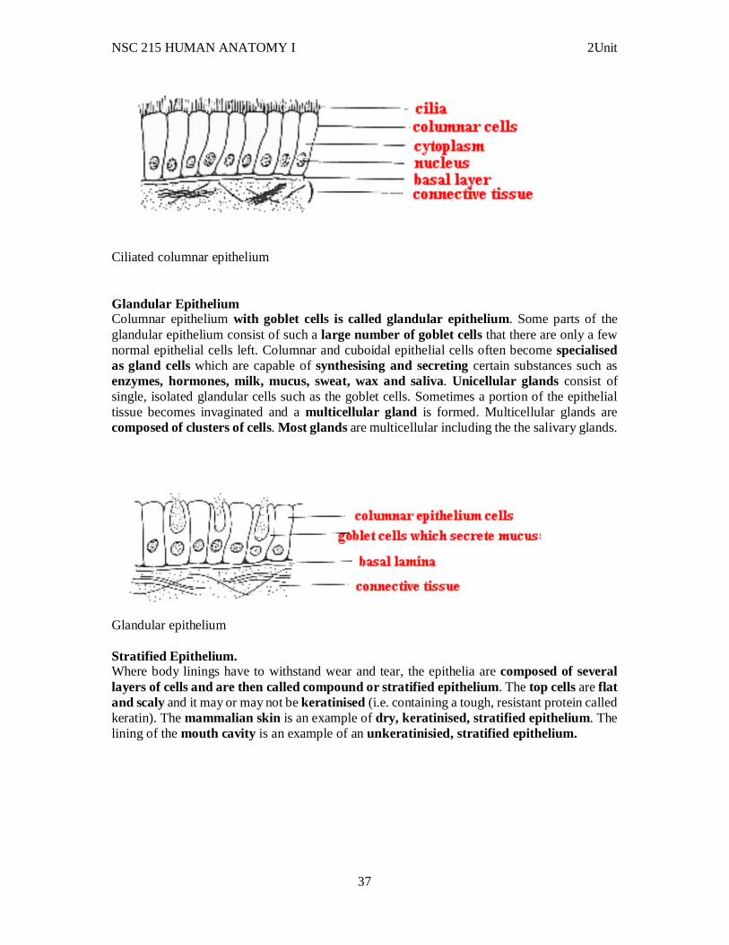
Simple columnar epithelium Ciliated Columnar Epithelium These are simple columnar epithelial cells, but in addition, they possess fine hair-like outgrowths, cilia on their free surfaces. These cilia are capable of rapid, rhythmic, wavelike beatings in a certain direction. This movement of the cilia in a certain direction causes the mucus, which is secreted by the goblet cells, to move (flow or stream) in that direction. Ciliated epithelium is usually found in the air passages like the nose. It is also found in the uterus and Fallopian tubes of females. The movement of the cilia propel the ovum to the uterus.
NSC 215 HUMAN ANATOMY I 2Unit
37
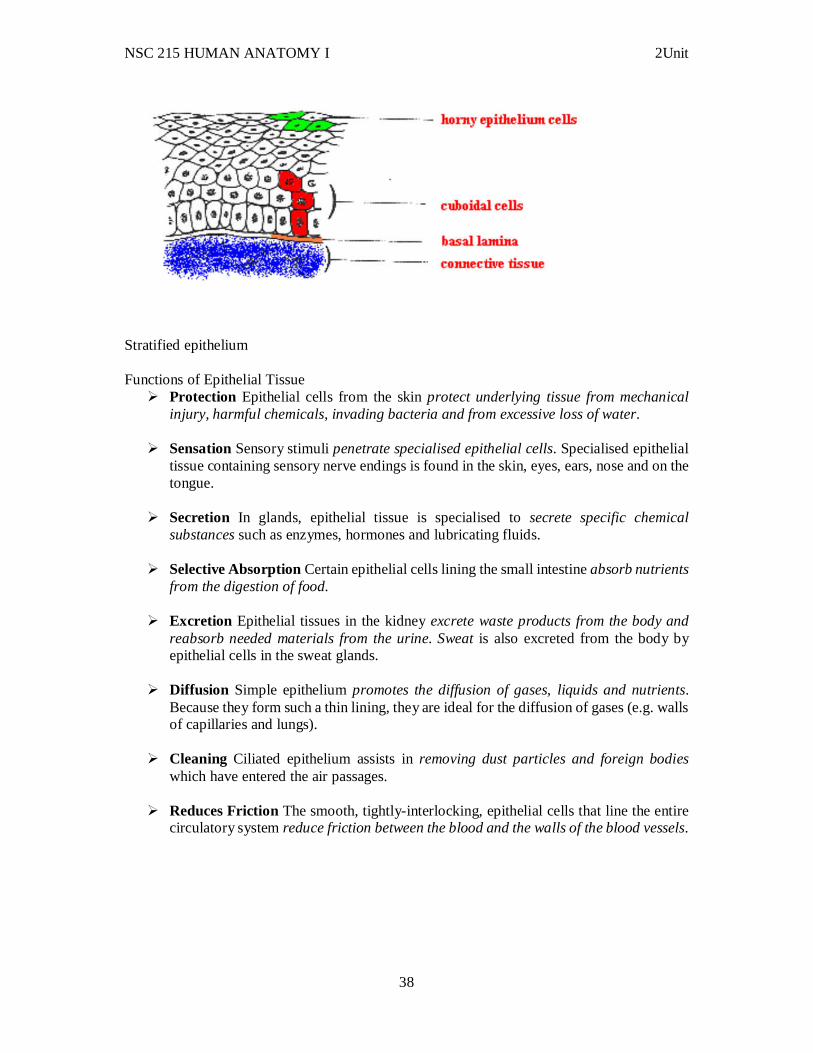
Ciliated columnar epithelium Glandular Epithelium Columnar epithelium with goblet cells is called glandular epithelium. Some parts of the glandular epithelium consist of such a large number of goblet cells that there are only a few normal epithelial cells left. Columnar and cuboidal epithelial cells often become specialised as gland cells which are capable of synthesising and secreting certain substances such as enzymes, hormones, milk, mucus, sweat, wax and saliva. Unicellular glands consist of single, isolated glandular cells such as the goblet cells. Sometimes a portion of the epithelial tissue becomes invaginated and a multicellular gland is formed. Multicellular glands are composed of clusters of cells. Most glands are multicellular including the the salivary glands.
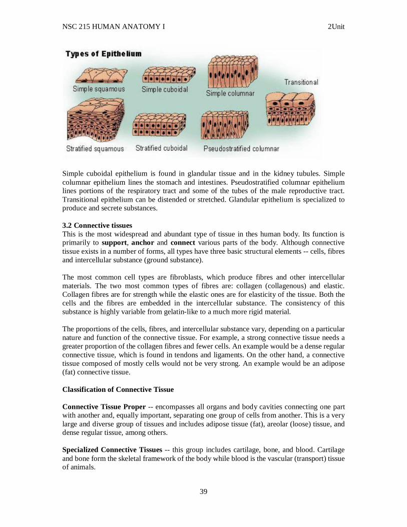
Glandular epithelium Stratified Epithelium. Where body linings have to withstand wear and tear, the epithelia are composed of several layers of cells and are then called compound or stratified epithelium. The top cells are flat and scaly and it may or may not be keratinised (i.e. containing a tough, resistant protein called keratin). The mammalian skin is an example of dry, keratinised, stratified epithelium. The lining of the mouth cavity is an example of an unkeratinisied, stratified epithelium.
NSC 215 HUMAN ANATOMY I 2Unit
38
Stratified epithelium Functions of Epithelial Tissue
Protection Epithelial cells from the skin protect underlying tissue from mechanical injury, harmful chemicals, invading bacteria and from excessive loss of water.
Sensation Sensory stimuli penetrate specialised epithelial cells. Specialised epithelial tissue containing sensory nerve endings is found in the skin, eyes, ears, nose and on the tongue.
Secretion In glands, epithelial tissue is specialised to secrete specific chemical substances such as enzymes, hormones and lubricating fluids.
Selective Absorption Certain epithelial cells lining the small intestine absorb nutrients from the digestion of food.
Excretion Epithelial tissues in the kidney excrete waste products from the body and reabsorb needed materials from the urine. Sweat is also excreted from the body by epithelial cells in the sweat glands.
Diffusion Simple epithelium promotes the diffusion of gases, liquids and nutrients. Because they form such a thin lining, they are ideal for the diffusion of gases (e.g. walls of capillaries and lungs).
Cleaning Ciliated epithelium assists in removing dust particles and foreign bodies which have entered the air passages.
Reduces Friction The smooth, tightly-interlocking, epithelial cells that line the entire circulatory system reduce friction between the blood and the walls of the blood vessels.
NSC 215 HUMAN ANATOMY I 2Unit
39
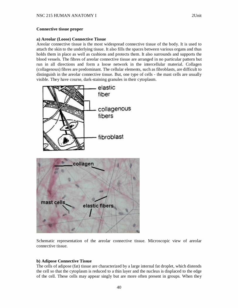
Simple cuboidal epithelium is found in glandular tissue and in the kidney tubules. Simple columnar epithelium lines the stomach and intestines. Pseudostratified columnar epithelium lines portions of the respiratory tract and some of the tubes of the male reproductive tract. Transitional epithelium can be distended or stretched. Glandular epithelium is specialized to produce and secrete substances. 3.2 Connective tissues This is the most widespread and abundant type of tissue in thes human body. Its function is primarily to support, anchor and connect various parts of the body. Although connective tissue exists in a number of forms, all types have three basic structural elements -- cells, fibres and intercellular substance (ground substance). The most common cell types are fibroblasts, which produce fibres and other intercellular materials. The two most common types of fibres are: collagen (collagenous) and elastic. Collagen fibres are for strength while the elastic ones are for elasticity of the tissue. Both the cells and the fibres are embedded in the intercellular substance. The consistency of this substance is highly variable from gelatin-like to a much more rigid material. The proportions of the cells, fibres, and intercellular substance vary, depending on a particular nature and function of the connective tissue. For example, a strong connective tissue needs a greater proportion of the collagen fibres and fewer cells. An example would be a dense regular connective tissue, which is found in tendons and ligaments. On the other hand, a connective tissue composed of mostly cells would not be very strong. An example would be an adipose (fat) connective tissue. Classification of Connective Tissue Connective Tissue Proper -- encompasses all organs and body cavities connecting one part with another and, equally important, separating one group of cells from another. This is a very large and diverse group of tissues and includes adipose tissue (fat), areolar (loose) tissue, and dense regular tissue, among others. Specialized Connective Tissues -- this group includes cartilage, bone, and blood. Cartilage and bone form the skeletal framework of the body while blood is the vascular (transport) tissue of animals.
NSC 215 HUMAN ANATOMY I 2Unit
40
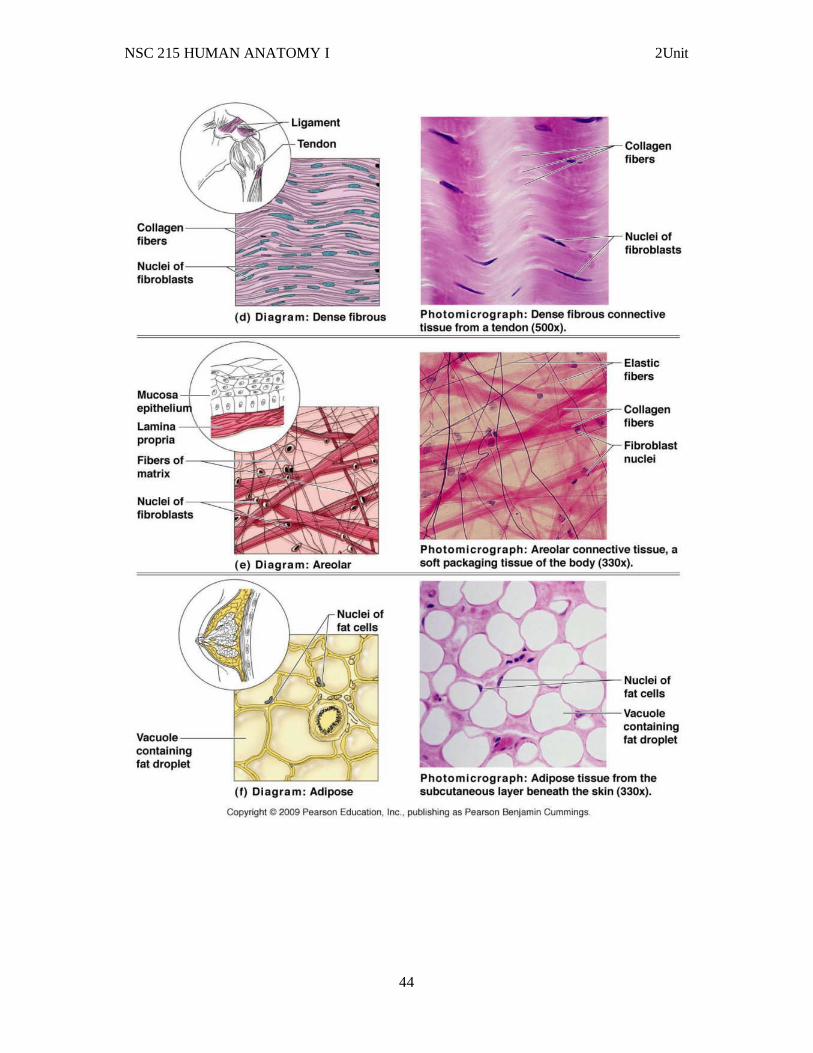
Connective tissue proper a) Areolar (Loose) Connective Tissue Areolar connective tissue is the most widespread connective tissue of the body. It is used to attach the skin to the underlying tissue. It also fills the spaces between various organs and thus holds them in place as well as cushions and protects them. It also surrounds and supports the blood vessels. The fibres of areolar connective tissue are arranged in no particular pattern but run in all directions and form a loose network in the intercellular material. Collagen (collagenous) fibres are predominant. The cellular elements, such as fibroblasts, are difficult to distinguish in the areolar connective tissue. But, one type of cells - the mast cells are usually visible. They have course, dark-staining granules in their cytoplasm.
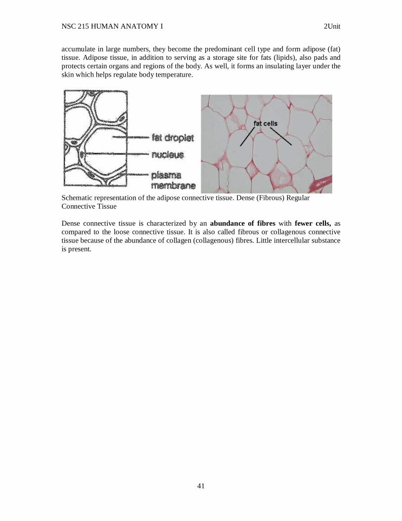
Schematic representation of the areolar connective tissue. Microscopic view of areolar connective tissue. b) Adipose Connective Tissue The cells of adipose (fat) tissue are characterized by a large internal fat droplet, which distends the cell so that the cytoplasm is reduced to a thin layer and the nucleus is displaced to the edge of the cell. These cells may appear singly but are more often present in groups. When they
NSC 215 HUMAN ANATOMY I 2Unit
41
accumulate in large numbers, they become the predominant cell type and form adipose (fat) tissue. Adipose tissue, in addition to serving as a storage site for fats (lipids), also pads and protects certain organs and regions of the body. As well, it forms an insulating layer under the skin which helps regulate body temperature.
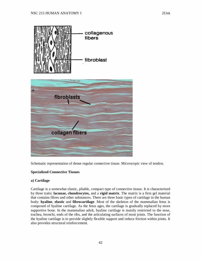
Schematic representation of the adipose connective tissue. Dense (Fibrous) Regular Connective Tissue Dense connective tissue is characterized by an abundance of fibres with fewer cells, as compared to the loose connective tissue. It is also called fibrous or collagenous connective tissue because of the abundance of collagen (collagenous) fibres. Little intercellular substance is present.
NSC 215 HUMAN ANATOMY I 2Unit
42
Schematic representation of dense regular connective tissue. Microscopic view of tendon. Specialized Connective Tissues a) Cartilage Cartilage is a somewhat elastic, pliable, compact type of connective tissue. It is characterized by three traits: lacunae, chondrocytes, and a rigid matrix . The matrix is a firm gel material that contains fibres and other substances. There are three basic types of cartilage in the human body: hyaline, elastic and fibrocartilage . Most of the skeleton of the mammalian fetus is composed of hyaline cartilage. As the fetus ages, the cartilage is gradually replaced by more supportive bone. In the mammalian adult, hyaline cartilage is mainly restricted to the nose, trachea, bronchi, ends of the ribs, and the articulating surfaces of most joints. The function of the hyaline cartilage is to provide slightly flexible support and reduce friction within joints. It also provides structural reinforcement.
NSC 215 HUMAN ANATOMY I 2Unit
43
Cartilage is a non-vascular tissue. As such, the chondrocytes rely on blood vessels in the tissue surrounding the cartilage for nutrient supply and waste removal. Function
Storage of energy Protection of organs Provision of structural framework for the body Connection of body tissues Connection of epithelial tissues to muscle fiber. supply of hormones all over the body
NSC 215 HUMAN ANATOMY I 2Unit
44
NSC 215 HUMAN ANATOMY I 2Unit
45
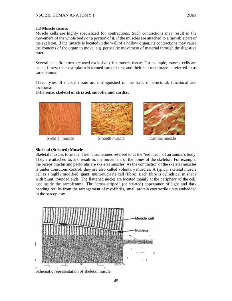
3.3 Muscle tissues Muscle cells are highly specialized for contractions. Such contractions may result in the movement of the whole body or a portion of it, if the muscles are attached to a movable part of the skeleton. If the muscle is located in the wall of a hollow organ, its contractions may cause the contents of the organ to move, e.g. peristaltic movement of material through the digestive tract. Several specific terms are used exclusively for muscle tissue. For example, muscle cells are called fibres; their cytoplasm is termed sarcoplasm; and their cell membrane is referred to as sarcolemma. Three types of muscle tissue are distinguished on the basis of structural, functional and locational Difference: skeletal or striated, smooth, and cardiac
Skeletal (Striated) Muscle Skeletal muscles form the "flesh"; sometimes referred to as the "red meat" of an animal's body. They are attached to, and result in, the movement of the bones of the skeleton. For example, the biceps brachii and pectoralis are skeletal muscles. As the contraction of the skeletal muscles is under conscious control, they are also called voluntary muscles. A typical skeletal muscle cell is a highly modified, giant, multi-nucleate cell (fibre). Each fibre is cylindrical in shape with blunt, rounded ends. The flattened nuclei are located mainly at the periphery of the cell, just inside the sarcolemma. The "cross-striped" (or striated) appearance of light and dark banding results from the arrangement of myofibrils, small protein contractile units embedded in the sarcoplasm.
Schematic representation of skeletal muscle
NSC 215 HUMAN ANATOMY I 2Unit
46
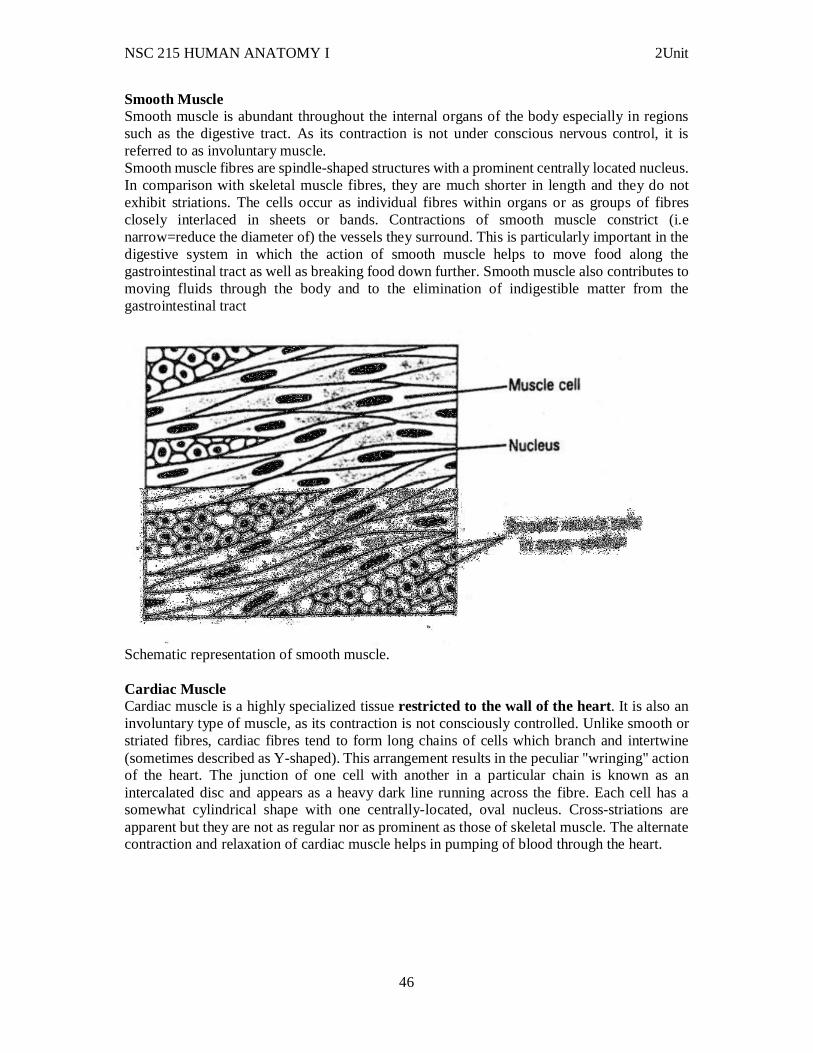
Smooth Muscle Smooth muscle is abundant throughout the internal organs of the body especially in regions such as the digestive tract. As its contraction is not under conscious nervous control, it is referred to as involuntary muscle. Smooth muscle fibres are spindle-shaped structures with a prominent centrally located nucleus. In comparison with skeletal muscle fibres, they are much shorter in length and they do not exhibit striations. The cells occur as individual fibres within organs or as groups of fibres closely interlaced in sheets or bands. Contractions of smooth muscle constrict (i.e narrow=reduce the diameter of) the vessels they surround. This is particularly important in the digestive system in which the action of smooth muscle helps to move food along the gastrointestinal tract as well as breaking food down further. Smooth muscle also contributes to moving fluids through the body and to the elimination of indigestible matter from the gastrointestinal tract
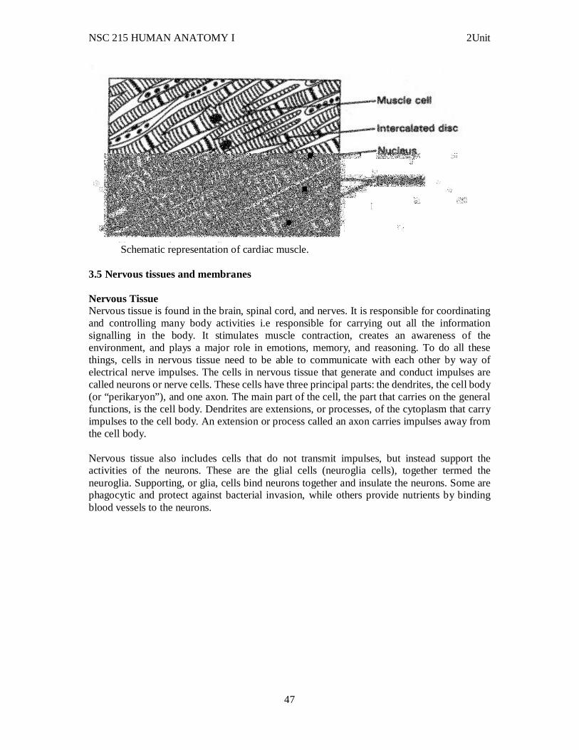
Schematic representation of smooth muscle. Cardiac Muscle Cardiac muscle is a highly specialized tissue restricted to the wall of the heart. It is also an involuntary type of muscle, as its contraction is not consciously controlled. Unlike smooth or striated fibres, cardiac fibres tend to form long chains of cells which branch and intertwine (sometimes described as Y-shaped). This arrangement results in the peculiar "wringing" action of the heart. The junction of one cell with another in a particular chain is known as an intercalated disc and appears as a heavy dark line running across the fibre. Each cell has a somewhat cylindrical shape with one centrally-located, oval nucleus. Cross-striations are apparent but they are not as regular nor as prominent as those of skeletal muscle. The alternate contraction and relaxation of cardiac muscle helps in pumping of blood through the heart.
NSC 215 HUMAN ANATOMY I 2Unit
47
Schematic representation of cardiac muscle.
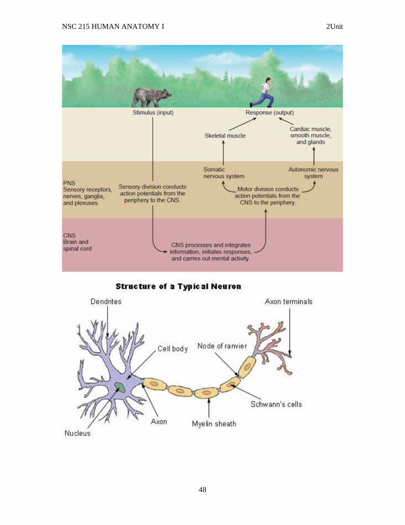
3.5 Nervous tissues and membranes Nervous Tissue Nervous tissue is found in the brain, spinal cord, and nerves. It is responsible for coordinating and controlling many body activities i.e responsible for carrying out all the information signalling in the body. It stimulates muscle contraction, creates an awareness of the environment, and plays a major role in emotions, memory, and reasoning. To do all these things, cells in nervous tissue need to be able to communicate with each other by way of electrical nerve impulses. The cells in nervous tissue that generate and conduct impulses are called neurons or nerve cells. These cells have three principal parts: the dendrites, the cell body (or “perikaryon”), and one axon. The main part of the cell, the part that carries on the general functions, is the cell body. Dendrites are extensions, or processes, of the cytoplasm that carry impulses to the cell body. An extension or process called an axon carries impulses away from the cell body. Nervous tissue also includes cells that do not transmit impulses, but instead support the activities of the neurons. These are the glial cells (neuroglia cells), together termed the neuroglia. Supporting, or glia, cells bind neurons together and insulate the neurons. Some are phagocytic and protect against bacterial invasion, while others provide nutrients by binding blood vessels to the neurons.
NSC 215 HUMAN ANATOMY I 2Unit
48
NSC 215 HUMAN ANATOMY I 2Unit
49
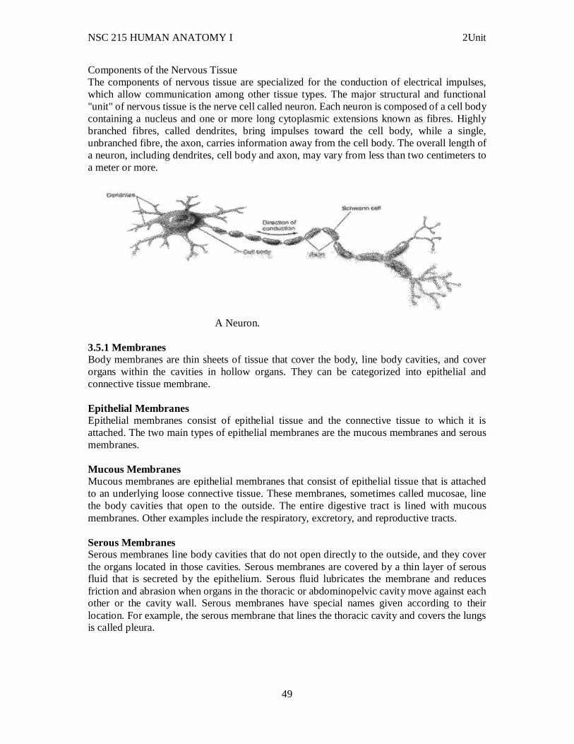
Components of the Nervous Tissue The components of nervous tissue are specialized for the conduction of electrical impulses, which allow communication among other tissue types. The major structural and functional "unit" of nervous tissue is the nerve cell called neuron. Each neuron is composed of a cell body containing a nucleus and one or more long cytoplasmic extensions known as fibres. Highly branched fibres, called dendrites, bring impulses toward the cell body, while a single, unbranched fibre, the axon, carries information away from the cell body. The overall length of a neuron, including dendrites, cell body and axon, may vary from less than two centimeters to a meter or more.
A Neuron.
3.5.1 Membranes Body membranes are thin sheets of tissue that cover the body, line body cavities, and cover organs within the cavities in hollow organs. They can be categorized into epithelial and connective tissue membrane. Epithelial Membranes Epithelial membranes consist of epithelial tissue and the connective tissue to which it is attached. The two main types of epithelial membranes are the mucous membranes and serous membranes. Mucous Membranes Mucous membranes are epithelial membranes that consist of epithelial tissue that is attached to an underlying loose connective tissue. These membranes, sometimes called mucosae, line the body cavities that open to the outside. The entire digestive tract is lined with mucous membranes. Other examples include the respiratory, excretory, and reproductive tracts. Serous Membranes Serous membranes line body cavities that do not open directly to the outside, and they cover the organs located in those cavities. Serous membranes are covered by a thin layer of serous fluid that is secreted by the epithelium. Serous fluid lubricates the membrane and reduces friction and abrasion when organs in the thoracic or abdominopelvic cavity move against each other or the cavity wall. Serous membranes have special names given according to their location. For example, the serous membrane that lines the thoracic cavity and covers the lungs is called pleura.
NSC 215 HUMAN ANATOMY I 2Unit
50
Connective Tissue Membranes Connective tissue membranes contain only connective tissue. Synovial membranes and meninges belong to this category. Synovial Membranes Synovial membranes are connective tissue membranes that line the cavities of the freely movable joints such as the shoulder, elbow, and knee. Like serous membranes, they line cavities that do not open to the outside. Unlike serous membranes, they do not have a layer of epithelium. Synovial membranes secrete synovial fluid into the joint cavity, and this lubricates the cartilage on the ends of the bones so that they can move freely and without friction. Meninges The connective tissue covering on the brain and spinal cord, within the dorsal cavity, are called meninges. They provide protection for these vital structures. 3.6 Clinical correlates i. Inflammation Sometimes the inflammatory response lasts longer or is more intense than is desirable, and drugs are used to suppress the inflammation by inhibiting the synthesis, release, or actions of the mediators of inflammation. For example, the effects of histamine released in people with hay fever are suppressed by antihistamines. Aspirin and related drugs, such as ibuprofen and naproxen, are effective anti-inflammatory agents that relieve pain by preventing the synthesis of prostaglandins and related substances. ii. Meningitis Meningitis can be defined as the inflammation of the meninges which can be caused by various organisms including bacteria and viruses. 4.0 Summary In this unit, you have learnt that: i. The cell nucleus contains genetic material and regulates activities of the cell. It determines how the cell will function, as well as the basic structure of that cell. ii. All of the functions for cell expansion, growth and replication are carried out in the cytoplasm of a cell. iii. Tissue is a group of cells that have similar structure and that function together as a unit. Primary types of body tissues include epithelial, connective, muscular, and nervous tissues. iv. Epithelial tissues form the covering of all body surfaces, line body cavities and hollow organs, and are the major tissue in glands. v. Connective tissues bind structures together, form a framework and support for organs and the body as a whole, store fat, transport substances, protect against disease, and help repair tissue damage. vi. Muscle tissue is composed of cells that have the special ability to shorten or contract in order to produce movement of body parts. vii. Nervous tissue is responsible for coordinating an controlling many body activities. viii. Body membranes are thin sheets of tissue that cover the body, line body cavities, and cover organs within the cavities in hollow organs.
NSC 215 HUMAN ANATOMY I 2Unit
51
ix. Two main categories of body membranes are epithelial and connective tissue membranes. Sub-categories include mucous membranes, serous membranes, synovial membranes, and meninges. 6.0 Tutor Marked Assignments 6.1 In the histology laboratory, study and identify the differences between the various types of epithelial, connective, muscle and nervous tissues slides. Report your findings 6.2 Answer the following questions 1. Epithelial tissue
a. covers free body surfaces. b. lacks blood vessels. c. composes various glands. d. is anchored to connective tissue by a basement membrane. e. all of the above.
2. A tissue that covers a surface, is one cell layer thick, and is composed of flat cells is a. simple squamous epithelium. b. simple cuboidal epithelium. c. simple columnar epithelium. d. stratified squamous epithelium. e. transitional epithelium.
3. Epithelium composed of two or more layers of cells with only the deepest layer in contact with the basement membrane is
a. stratified epithelium. b. simple epithelium.. c. pseudostratified epithelium. d. columnar epithelium. e. cuboidal epithelium
4. Simple squamous epithelium most likely a. performs phagocytosis. b. is involved with active transport. c. secretes many complex lipids and proteins. d. allows certain substances to diff use across it.
5. Pseudostratifi ed ciliated columnar epithelium can be found lining the a. digestive tract. b. trachea. c thyroid gland. d. kidney tubules. e. urinary bladder.
7.0 References/further reading: 1. Bruce M. Carlson (2019) Human Embryology & Developmental Biology. 6th edition
2. Kathryn A. Booth, Terri. D. Wyman (2008) Anatomy, physiology, and pathophysiology for allied health
3. Katherine M. A. Rogers and William N. Scott (2011) Nurses! Test yourself in anatomy and physiology

NSC 215 HUMAN ANATOMY I 2Unit
52
4. Kent M. Van De Graff, R.Ward Rhees, Sidney Palmer (2010) Schaum’s Outline of Human Anatomy and Physiology 3rd edition
5. Keith L Moore, Persuade T.V.N (2016), The Developing Human Clinically Oriented Embryology 10th Edition Lippincott Williams & Wilkins
6. Philip Tate (2012) Seeley’s Principles of Anatomy & Physiology 2nd edition.
7. Sadler T.W (2019), Langman’s Medical Embryology 14th edition. Lippincott Williams & Wilkins
NSC 215 HUMAN ANATOMY I 2Unit
53
UNIT FIVE: THE HUMAN DEFENSE SYSTEM CONTENTS 1.0 Introduction 2.0 Objectives 3.0 Main Content 3.1 General defense system 3.2 Components of general defense system 3.3 Specific defense system 3.4 Components of specific system 3.5 Clinical correlates 4.0 Conclusion 5.0 Summary 6.0 Tutor Marked Assignments 6.1 Activity 6.2 Tutor Marked Tests 7.0 References and other resources 1.0 Introduction We are constantly under attack by bacteria, fungi, viruses and other organisms. These invaders are called pathogens. Our body is a rich source of nutrients and water that these invaders need to survive. Amazingly we remain healthy most of the time. We are obviously very good at protecting ourselves. There are two major aspects to our defense system – general and specific. 2.0 Objectives At the end of this unit, you should be able to: i. Discuss the differences between the two major defense systems of the body ii. List the components of the general defense system iii. List the components of the specific defense system 3.0 Main Content 3.1 General defense system The first parts of the general defense system are really barriers that stop the pathogens from entering the body. These barriers try to stop all pathogens therefore they are considered non-specific defenses. The physical barriers of our general defense system consist of: The Skin- prevents the entry of microorganisms and prevents dehydration by reducing water loss from the body. It is a physical barrier that stops pathogens. Clotting - If the skin is broken the blood clot stops entry of pathogens. Sebaceous and sweat glands- These produce chemicals that kill bacteria. Lysozyme- This is in the saliva and the tear glands. It kills bacteria. Mucous membranes- These secrete mucus which lines many body parts. The mucous traps pathogens and prevents them from entering the body. Nasal hairs- These remove suspended micro-organisms from the air.
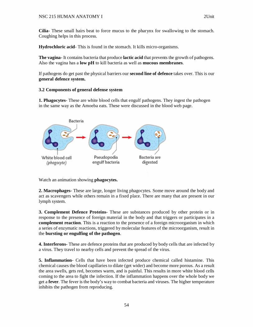
NSC 215 HUMAN ANATOMY I 2Unit
54
Cilia - These small hairs beat to force mucus to the pharynx for swallowing to the stomach. Coughing helps in this process. Hydrochloric acid - This is found in the stomach. It kills micro-organisms. The vagina- It contains bacteria that produce lactic acid that prevents the growth of pathogens. Also the vagina has a low pH to kill bacteria as well as mucous membranes. If pathogens do get past the physical barriers our second line of defence takes over. This is our general defence system. 3.2 Components of general defense system 1. Phagocytes- These are white blood cells that engulf pathogens. They ingest the pathogen in the same way as the Amoeba eats. These were discussed in the blood web page.
Watch an animation showing phagocytes. 2. Macrophages- These are large, longer living phagocytes. Some move around the body and act as scavengers while others remain in a fixed place. There are many that are present in our lymph system. 3. Complement Defence Proteins- These are substances produced by other protein or in response to the presence of foreign material in the body and that triggers or participates in a complement reaction. This is a reaction to the presence of a foreign microorganism in which a series of enzymatic reactions, triggered by molecular features of the microorganism, result in the bursting or engulfing of the pathogen. 4. Interferons- These are defence proteins that are produced by body cells that are infected by a virus. They travel to nearby cells and prevent the spread of the virus. 5. Inflammation- Cells that have been infected produce chemical called histamine. This chemical causes the blood capillaries to dilate (get wider) and become more porous. As a result the area swells, gets red, becomes warm, and is painful. This results in more white blood cells coming to the area to fight the infection. If the inflammation happens over the whole body we get a fever. The fever is the body’s way to combat bacteria and viruses. The higher temperature inhibits the pathogen from reproducing.
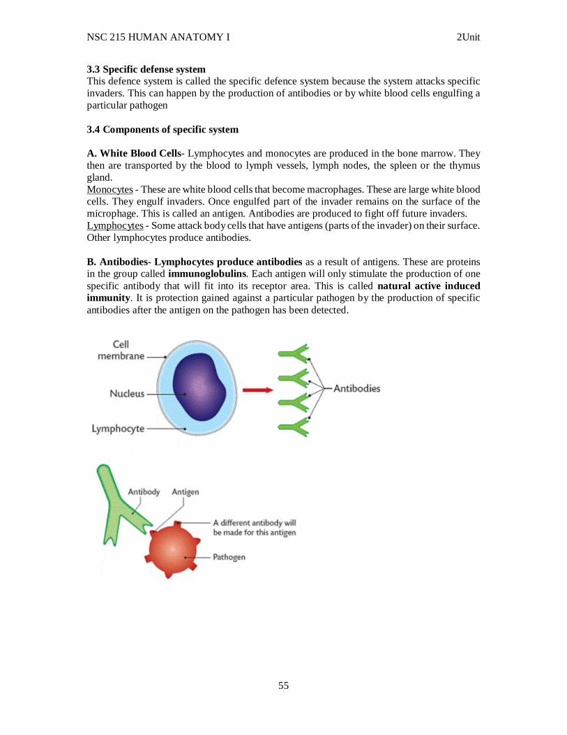
NSC 215 HUMAN ANATOMY I 2Unit
55
3.3 Specific defense system This defence system is called the specific defence system because the system attacks specific invaders. This can happen by the production of antibodies or by white blood cells engulfing a particular pathogen 3.4 Components of specific system A. White Blood Cells- Lymphocytes and monocytes are produced in the bone marrow. They then are transported by the blood to lymph vessels, lymph nodes, the spleen or the thymus gland. Monocytes - These are white blood cells that become macrophages. These are large white blood cells. They engulf invaders. Once engulfed part of the invader remains on the surface of the microphage. This is called an antigen. Antibodies are produced to fight off future invaders. Lymphocytes - Some attack body cells that have antigens (parts of the invader) on their surface. Other lymphocytes produce antibodies. B. Antibodies- Lymphocytes produce antibodies as a result of antigens. These are proteins in the group called immunoglobulins. Each antigen will only stimulate the production of one specific antibody that will fit into its receptor area. This is called natural active induced immunity . It is protection gained against a particular pathogen by the production of specific antibodies after the antigen on the pathogen has been detected.
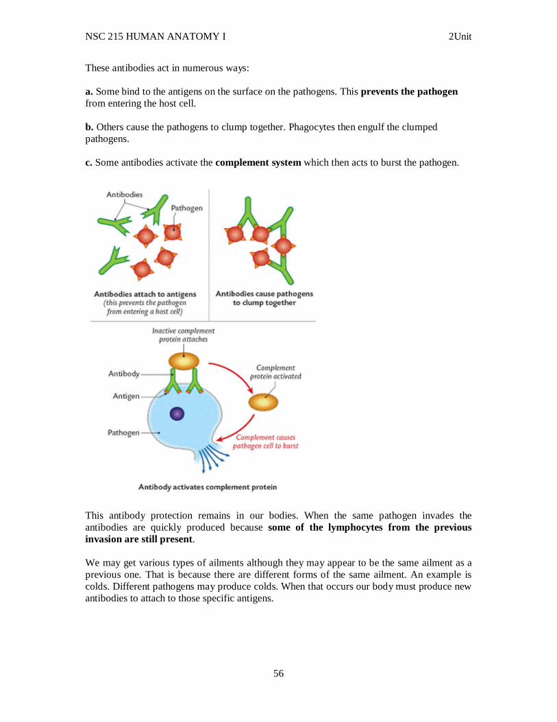
NSC 215 HUMAN ANATOMY I 2Unit
56
These antibodies act in numerous ways: a. Some bind to the antigens on the surface on the pathogens. This prevents the pathogen from entering the host cell. b. Others cause the pathogens to clump together. Phagocytes then engulf the clumped pathogens. c. Some antibodies activate the complement system which then acts to burst the pathogen.
This antibody protection remains in our bodies. When the same pathogen invades the antibodies are quickly produced because some of the lymphocytes from the previous invasion are still present. We may get various types of ailments although they may appear to be the same ailment as a previous one. That is because there are different forms of the same ailment. An example is colds. Different pathogens may produce colds. When that occurs our body must produce new antibodies to attach to those specific antigens.
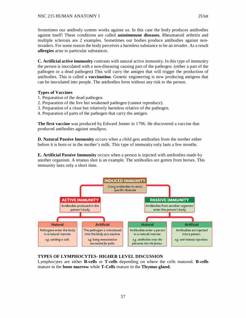
NSC 215 HUMAN ANATOMY I 2Unit
57
Sometimes our antibody system works against us. In this case the body produces antibodies against itself! These conditions are called autoimmune diseases. Rheumatoid arthritis and multiple sclerosis are 2 examples. Sometimes our bodies produce antibodies against non-invaders. For some reason the body perceives a harmless substance to be an invader. As a result allergies arise to particular substances. C. Artificial active immunity contrasts with natural active immunity. In this type of immunity the person is inoculated with a non-diseasing causing part of the pathogen. (either a part of the pathogen or a dead pathogen) This will carry the antigen that will trigger the production of antibodies. This is called a vaccination. Genetic engineering is now producing antigens that can be inoculated into people. The antibodies form without any risk to the person. Types of Vaccines 1. Preparation of the dead pathogen. 2. Preparation of the live but weakened pathogen (cannot reproduce). 3. Preparation of a close but relatively harmless relative of the pathogen. 4. Preparation of parts of the pathogen that carry the antigen. The first vaccine was produced by Edward Jenner in 1796. He discovered a vaccine that produced antibodies against smallpox. D. Natural Passive Immunity occurs when a child gets antibodies from the mother either before it is born or in the mother’s milk. This type of immunity only lasts a few months. E. Artificial Passive Immunity occurs when a person is injected with antibodies made by another organism. A tetanus shot is an example. The antibodies are gotten from horses. This immunity lasts only a short time.
TYPES OF LYMPHOCYTES- HIGHER LEVEL DISCUSSION Lymphocytes are either B-cells or T-cells depending on where the cells matured. B-cells mature in the bone marrow while T-Cells mature in the Thymus gland.
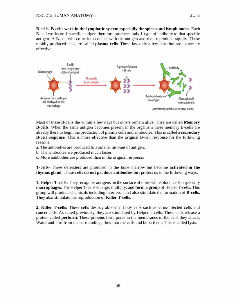
NSC 215 HUMAN ANATOMY I 2Unit
58
B-cells- B-cells work in the lymphatic system especially the spleen and lymph nodes. Each B-cell works on 1 specific antigen therefore produces only 1 type of antibody to that specific antigen. A B-cell will come into contact with the antigen and then reproduce rapidly. These rapidly produced cells are called plasma cells. These last only a few days but are extremely effective.
Most of these B-cells die within a few days but others remain alive. They are called Memory B-cells. When the same antigen becomes present in the organism these memory B-cells are already there to begin the production of plasma cells and antibodies. This is called a secondary B-cell response. This is more effective than the original B-cell response for the following reasons: a. The antibodies are produced to a smaller amount of antigen. b. The antibodies are produced much faster. c. More antibodies are produced than in the original response. T-cells- These defenders are produced in the bone marrow but become activated in the thymus gland. These cells do not produce antibodies but protect us in the following ways: 1. Helper T-cells: They recognise antigens on the surface of other white blood cells, especially macrophages. The Helper T-cells enlarge, multiply, and form a group of Helper T-cells. This group will produce chemicals including interferon and also stimulate the formation of B-cells. They also stimulate the reproduction of Killer T-cells . 2. Killer T-cells: These cells destroy abnormal body cells such as virus-infected cells and cancer cells. As stated previously, they are stimulated by Helper T-cells. These cells release a protein called perforin . These proteins form pores in the membranes of the cells they attack. Water and ions from the surroundings flow into the cells and burst them. This is called lysis.
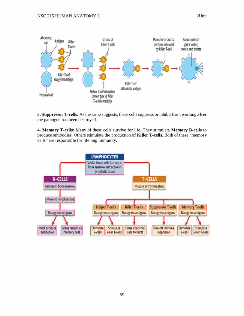
NSC 215 HUMAN ANATOMY I 2Unit
59
3. Suppressor T-cells: As the same suggests, these cells suppress or inhibit from working after the pathogen has been destroyed. 4. Memory T-cells: Many of these cells survive for life. They stimulate Memory B-cells to produce antibodies. Others stimulate the production of Killer T-cells. Both of these “memory cells” are responsible for lifelong immunity.
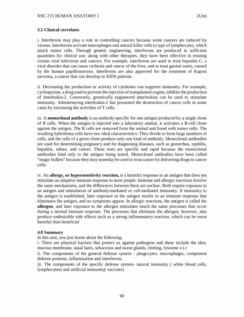
NSC 215 HUMAN ANATOMY I 2Unit
60
3.5 Clinical correlates i. Interferons may play a role in controlling cancers because some cancers are induced by viruses. Interferons activate macrophages and natural killer cells (a type of lymphocyte), which attack tumor cells. Through genetic engineering, interferons are produced in sufficient quantities for clinical use; along with other therapies, they have been effective in treating certain viral infections and cancers. For example, interferons are used to treat hepatitis C, a viral disorder that can cause cirrhosis and cancer of the liver, and to treat genital warts, caused by the human papillomavirus. Interferons are also approved for the treatment of Kaposi sarcoma, a cancer that can develop in AIDS patients. ii. Decreasing the production or activity of cytokines can suppress immunity. For example, cyclosporine, a drug used to prevent the rejection of transplanted organs, inhibits the production of interleukin-2. Conversely, genetically engineered interleukins can be used to stimulate immunity. Administering interleukin-2 has promoted the destruction of cancer cells in some cases by increasing the activities of T cells. iii. A monoclonal antibody is an antibody specific for one antigen produced by a single clone of B cells. When the antigen is injected into a laboratory animal, it activates a B-cell clone against the antigen. The B cells are removed from the animal and fused with tumor cells. The resulting hybridoma cells have two ideal characteristics: They divide to form large numbers of cells, and the cells of a given clone produce only one kind of antibody. Monoclonal antibodies are used for determining pregnancy and for diagnosing diseases, such as gonorrhea, syphilis, hepatitis, rabies, and cancer. These tests are specific and rapid because the monoclonal antibodies bind only to the antigen being tested. Monoclonal antibodies have been called “magic bullets” because they may someday be used to treat cancer by delivering drugs to cancer cells. iv. An allergy, or hypersensitivity reaction, is a harmful response to an antigen that does not stimulate an adaptive immune response in most people. Immune and allergic reactions involve the same mechanisms, and the differences between them are unclear. Both require exposure to an antigen and stimulation of antibody-mediated or cell-mediated immunity. If immunity to the antigen is established, later exposure to the antigen results in an immune response that eliminates the antigen, and no symptoms appear. In allergic reactions, the antigen is called the allergen, and later exposure to the allergen stimulates much the same processes that occur during a normal immune response. The processes that eliminate the allergen, however, also produce undesirable side effects such as a strong inflammatory reaction, which can be more harmful than beneficial 4.0 Summary In this unit, you just learnt about the following: i. There are physical barriers that protect us against pathogens and these include the skin, mucous membrane, nasal hairs, sebaceous and sweat glands, clotting, lysozme e.t.c ii. The components of the general defense system - phagocytes, macrophages, component defense proteins, inflammation and interferons. iii. The components of the specific defense system- natural immunity ( white blood cells, lymphocytes) and artificial immunity( vaccines)
NSC 215 HUMAN ANATOMY I 2Unit
61
6.0 Tutor Marked Assignments 6.1 At the histology laboratory, under the microscope, examine the lymphocytes, the components of the white blood cells and their defining properties. Report your findings in your log book. 6.2 Answer the following question. 1. Which of the following statements is true?
a. The adaptive immune response is effective against bacteria in general. b. Immunological memory is a characteristic of innate immunity. c. In innate immunity, the response to a second exposure to bacteria is faster than the response to the first exposure. d. Innate immunity is required for the initiation and regulation of the adaptive immune response. e. All of the above are correct.
2. Antigens....... a. stimulate an adaptive immunity response. b. are molecules common to groups of pathogens, but are not found in human cells. c. are produced by plasma cells. d. all of the above.
3. Macrophages...... a. are usually the first cells to leave the blood and enter infected tissues. b. develop from mast cells. c. are permanent residents beneath the free surfaces of the body. d. secrete cytokines that inhibit inflammation. e. die and accumulate to form pus.
4. Dendritic cells a. have almost all of the diff erent innate immunity receptors. b. are phagocytic cells. c. secrete interferons. d. activate adaptive immunity. e. all of the above.
5. Which of the following statements is true? a. Natural killer cells are specialized to kill extracellular pathogens. b. Antibodies attach to mast cells, basophils, and eosinophils. c. Mast cells circulate in the blood and enter infected tissues. d. Eosinophils are specialized to kill intracellular pathogens. e. Basophils inhibit inflammation.
7.0 References and other resources Katherine M. A. Rogers and William N. Scott (2011) Nurses! Test yourself in anatomy and physiology Kathryn A. Booth, Terri. D. Wyman (2008) Anatomy, physiology, and pathophysiology for allied health Kent M. Van De Graff, R.Ward Rhees, Sidney Palmer (2010) Schaum’s outline of human anatomy and physiology 3rd ed. Philip Tate (2012) Seeley’s principles of anatomy & physiology 2nd ed.
NSC 215 HUMAN ANATOMY I 2Unit
62
MODULE TWO: EMBRYOLOGY Introduction “The end of thing is better than the beginning” this is a true saying but in certain scenarios, the beginning of a thing might be the major factor to be considered. The life span is usually considered the period between birth and death; however, the 9 months before birth are a critical part of a person’s existence. What happens in these 9 months profoundly affects the rest of a person’s life. Although most people develop normally and are born without defects, approximately 3 out of every 100 people are born with a birth defect so severe that it requires medical attention during the first year of life. Later in life, many more people discover previously unknown problems, such as the tendency to develop asthma, certain brain disorders, or cancer. Well, in this module, we will be discussing “the beginning” as I see it, a very interesting and amazing topic called Embryology. Embryology (from Greek, embryon, the unborn, embryo; and , -logia) is the science of the development of an embryo from the fertilization of the ovum to the fetus stage. It may also refer to the structure and development of the embryo of a particular organism. Module objective: At the end of this module, you should be able to: i. Discuss the human embryology from fertilization to birth CONTENTS: UNIT 1 –Embryology terminology UNIT 2 – Gametogenesis UNIT 3 – Placenta formation and functions UNIT ONE – EMBRYOLOGY TERMINOLOGY CONTENTS 1.0 Introduction 2.0 Objectives 3.0 Main Content 3.1 Terminologies 3.2 Clinical correlates 4.0 Conclusion 5.0 Summary 6.0 Tutor Marked Assignments 6.1 Activity 6.2 Tutor Marked Tests 7.0 References and other resources 1.0 Introduction There are several terms commonly used in discussions of developing humans; several of these terms are used in the Timetable of Human Prenatal Development. Most terms have Latin (L.) or Greek (Gr.) origins. 2.0 0bjectives In this unit, you will be able to define the various terminologies used in embryology.
NSC 215 HUMAN ANATOMY I 2Unit
63
3.0 Main Content 3.1 Terminology Zygote: earliest development stage of embryo; formed when sperm and egg fuse Morula: compact ball of 16 cells formed on Day 3 Embryoblast: inner cell mass (ICM), aka pluriblast, which gives rise to endoderm, ectoderm, and mesoderm Trophoblast: cells forming outer layer of blastocyst (Day 4); forms placenta Blastocoele: blastocyst cavity Blastocyst: forms on Day 4; consists of trophoblast surrounding blastocoele and embryoblast Hypoblast: forms on Day 8 from inner cell mass; gives rise to extraembryonic endoderm (e.g. yolk sac); cuboidal cells Epiblast: forms on Day 8 from ICM; dorsal (above) the hypoblast; columnar cells Amniotic cavity: formed by end of Week 2 from epiblast cells Yolk sac: formed by end of Week 2 from hypoblast cells (ventral); vestigial in humans, though perhaps early nutritional value Gastrulation: embryonic development phase whereby bilaminar germ disc acquires a third germ layer. Sequence: (1) embryo becomes asymmetric, (2) primitive streak forms, (3) cells from epiblast at primitive streak undergo epithelial to mesenchymal transition Trilaminar germ disc: ectoderm, endoderm, mesoderm (arise during gastrulation) Primitive streak: formed in Week 3, initial event of gastrulation; establishes bilateral symmetry in embryo Primitive node: cephalic (rostral) end of primitive streak Ectoderm: forms CNS, PNS, sensory epithelia of eye (retina), nose and ear, and epidermis Endoderm: forms gut epithelial lining, cloaca (future urethra and bladder), respiratory tract, tympanic cavity, parenchyma of thyroid, parathyroids, liver, pancreas, and reticular stroma of tonsils and thymus Mesoderm: MESODERM (mnemonic): Mesothelium (peritoneal, pleural, pericardial)/ Muscle (striated, smooth, cardiac) Embryologic Spleen/ Soft tissue/ Serous linings/ Sarcoma/ Somite Osseous tissue/ Outer layer of suprarenal gland (cortex)/ Ovaries Dura/ Ducts of genitalia Endothelium, Renal, Microglia/ Mesenchyme/ Male gonad
NSC 215 HUMAN ANATOMY I 2Unit
64
Buccopharyngeal membrane: Thin membrane where ectoderm and endoderm come into direct contact; forms septum between primitive mouth and pharynx Cloacal membrane: Thin membrane where ectoderm and endoderm come into direct contact; forms anal-urogentical opening Prochordal plate: condensation of mesoderm cells extending rostrally from the primitive node in the midline Cardiogenic plate: originally exists at most anterior end of trilaminar germ disk, but will fold ventrally and develop into the heart Notochord: derived from mesoderm, notochord becomes vertebral column to provide axial support Allantois: connecting stalk; helps embryo exchange gases and handle liquid waste; vestigial in humans (like yolk sac) Cloaca: posterior opening that serves as opening for intestinal, reproductive, and urinary tracts Lateral folding: transforms embryo from three flat germ layers to cylinder with tube of endoderm (gut) in the center Cephalocaudul folding: cephalic and caudal ends of three-layered germ disc fold ventrally, most notably bringing heart region down to thoracic region Neurulation: Week 4 process in which flat neural ectoderm rolls up to become neural tube; upon completion the brain and spinal cord have been formed Neural tube: embryonic precursor to central nervous system Neural ectoderm: aka neural plate, includes neural crest cells and neural tube Placodes: surface ectodermal thickenings that contribute to formation of epithelium, ear and eye Neural crest cells: specialized cells that form skin pigment cells, ganglia of autonomic nervous system, dorsal root ganglia, facial cartilage, spiral septum of developing heart, ciliary body of the eye, and adrenal medulla Mesenchyme: middle germ layer composed of both mesoderm and ectoderm (e.g. the neural crest cells) Paraxial mesoderm: formed during Week 4; organized into segmented units called somites Somites: segmental units added on a cranial to caudal manner and are a good indicator of precise embryonic age Sclerotome: somite that becomes future axial skeleton
NSC 215 HUMAN ANATOMY I 2Unit
65
Dermomyotome: somite that becomes future skeletal muscle and dermal components Intermediate mesoderm: forms urogenital system; bridges paraxial and lateral plate mesoderm Lateral plate mesoderm: two layers: somatic/parietal mesoderm and splanchnic or visceral mesoderm Visceral mesoderm: layer continuous with mesoderm covering yolk sac; forms wall of gut Parietal mesoderm: layer continuous with mesoderm covering the amnioin; forms parietal pleura and peritoneum Coelemic cavity: potential space between visceral and parietal mesoderm Vitelline duct: long, narrow tube that joins yolk sac to midgut lumen of developing fetus. Appears at end of fourth week and normally closes by week VI (VI telline) Foregut: anterior part of alimentary canal, from mouth to duodenum (esophagus, stomach, duodenum, liver, gallbladder, pancreas, spleen) Midgut: part of alimentary canal (forms intestines) between foregut (at opening of bile duct) and hindgut. Includes latter parts of duodenum, jejunum, ileum, cecum, appendix, ascending colon, hepatic flexure of colon, transverse colon (proximal 2/3) Hindgut: posterior (caudal) part of alimentary canal; includes distal 1/3 of transverse colon, splenic flexure, descending colon, sigmoid colon and rectum Homeobox: highly conserved DNA region that codes for homeodomain proteins that are crucial for embryonic development Hox combinational code: phrase used to describe association between segmentation in developing embryo and unique combinations of overlapping Hox gene expression Teratogen: substance that causes abnormality in physiological development Embryonic period: from conception (zygote formation) to the third month of development. Fetal period: from third month of development to birth (266 days post-fertilization): total of about 6 months. 3.2 Clinical correlates During the first 2 weeks of development, the embryo is quite resistant to outside influences that may cause malformations. Factors that adversely affect the embryo at this age are more likely to kill it. Between 2 weeks and the next 4–7 weeks (depending on the structure considered), the embryo is more sensitive to outside influence that cause malformations than at any other time.
NSC 215 HUMAN ANATOMY I 2Unit
66
4.0 Conclusion 5.0 Summary In this unit, you have learnt about the definitions of the various terminologies used in embryology ranging from Zygote to fetal period. 6.0 Tutor Marked Assignments 6.1 At the embryology/ anatomy museum, identify the zygote, morula and the trilaminar germ discs. Record your findings in your log book. 6.2 Define the following terms: a. Morula b. Embryonic period c. Teratogen d. Fetal period e. Trilaminar germ disc: ectoderm, endoderm, mesoderm (arise during gastrulation) 7.0 References and other resources 7.0 References/further reading: 1. Bruce M. Carlson (2019) Human Embryology & Developmental Biology. 6th edition
2. Kathryn A. Booth, Terri. D. Wyman (2008) Anatomy, physiology, and pathophysiology for allied health
3. Katherine M. A. Rogers and William N. Scott (2011) Nurses! Test yourself in anatomy and physiology
4. Kent M. Van De Graff, R.Ward Rhees, Sidney Palmer (2010) Schaum’s Outline of Human Anatomy and Physiology 3rd edition
5. Keith L Moore, Persuade T.V.N (2016), The Developing Human Clinically Oriented Embryology 10th Edition Lippincott Williams & Wilkins
6. Philip Tate (2012) Seeley’s Principles of Anatomy & Physiology 2nd edition.
7. Sadler T.W (2019), Langman’s Medical Embryology 14th edition. Lippincott Williams & Wilkins UNIT TWO: GAMETOGENESIS CONTENTS 1.0 Introduction 2.0 Objectives 3.0 Main Content 3.1 Spermatogenesis 3.2 Oogenesis 3.3 Embryogenesis 3.4 Organogenesis 3.5 Clinical correlates 4.0 Conclusion 5.0 Summary
NSC 215 HUMAN ANATOMY I 2Unit
67
6.0 Tutor Marked Assignments 6.1 Activity 6.2 Tutor Marked Tests 7.0 References and other resources 1.0 Introduction Gametogenesis is the process of forming gametes (by definition haploid, n) from diploid cells of the germ line. Spermatogenesis is the process of forming sperm cells by meiosis (in animals, by mitosis in plants) in specialized organs known as gonads (in males these are termed testes). After division the cells undergo differentiation to become sperm cells. Oogenesis is the process of forming an ovum (egg) by meiosis (in animals, by mitosis in the gametophyte in plants) in specialized gonads known as ovaries. 2.0 Objectives At the end of this unit, you should be able to: i. Discuss the process of Spermatogenesis ii. Discuss the prevention of polyspermy iii. Discuss the process of oogenesis iv. Discuss the process of organogenesis 3.0 Main Content 3.1 Spermatogenesis Sperm production begins at puberty at continues throughout life, with several hundred million sperm being produced each day. Once sperm form they move into the epididymis, where they mature and are stored.
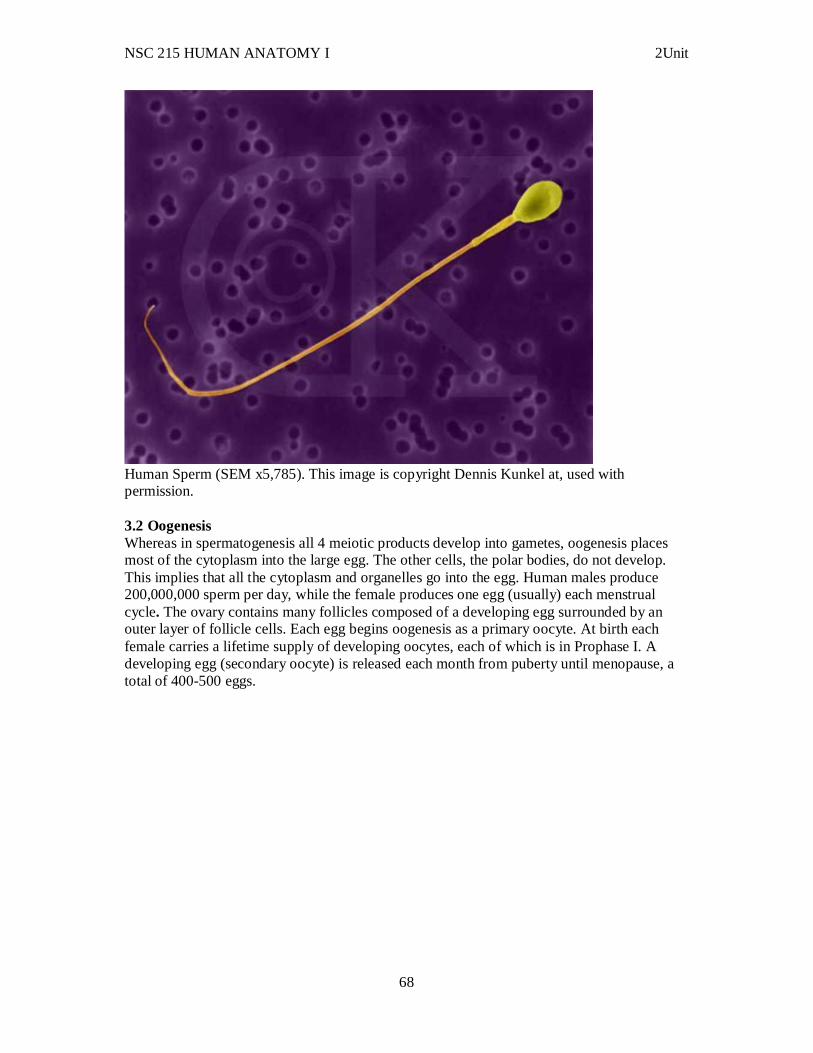
NSC 215 HUMAN ANATOMY I 2Unit
68
Human Sperm (SEM x5,785). This image is copyright Dennis Kunkel at, used with permission. 3.2 Oogenesis Whereas in spermatogenesis all 4 meiotic products develop into gametes, oogenesis places most of the cytoplasm into the large egg. The other cells, the polar bodies, do not develop. This implies that all the cytoplasm and organelles go into the egg. Human males produce 200,000,000 sperm per day, while the female produces one egg (usually) each menstrual cycle. The ovary contains many follicles composed of a developing egg surrounded by an outer layer of follicle cells. Each egg begins oogenesis as a primary oocyte. At birth each female carries a lifetime supply of developing oocytes, each of which is in Prophase I. A developing egg (secondary oocyte) is released each month from puberty until menopause, a total of 400-500 eggs.
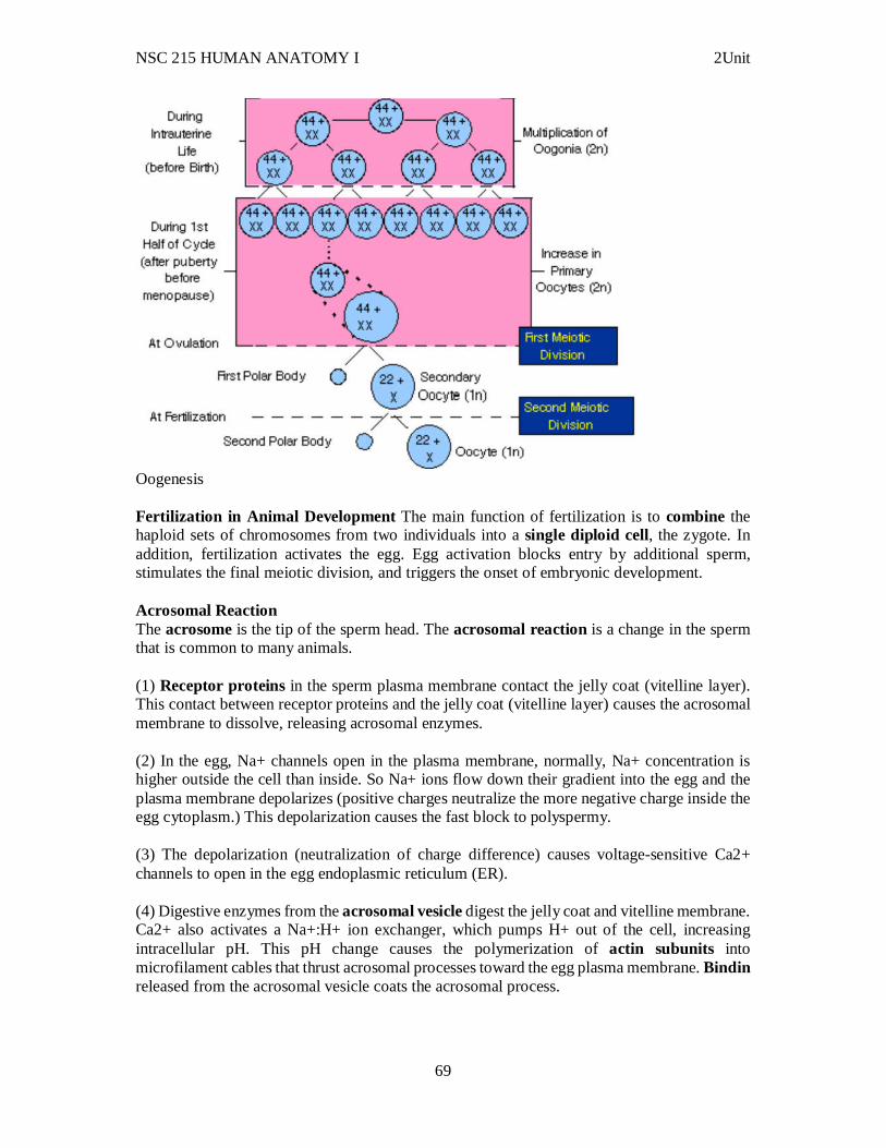
NSC 215 HUMAN ANATOMY I 2Unit
69
Oogenesis Fertilization in Animal Development The main function of fertilization is to combine the haploid sets of chromosomes from two individuals into a single diploid cell, the zygote. In addition, fertilization activates the egg. Egg activation blocks entry by additional sperm, stimulates the final meiotic division, and triggers the onset of embryonic development. Acrosomal Reaction The acrosome is the tip of the sperm head. The acrosomal reaction is a change in the sperm that is common to many animals. (1) Receptor proteins in the sperm plasma membrane contact the jelly coat (vitelline layer). This contact between receptor proteins and the jelly coat (vitelline layer) causes the acrosomal membrane to dissolve, releasing acrosomal enzymes. (2) In the egg, Na+ channels open in the plasma membrane, normally, Na+ concentration is higher outside the cell than inside. So Na+ ions flow down their gradient into the egg and the plasma membrane depolarizes (positive charges neutralize the more negative charge inside the egg cytoplasm.) This depolarization causes the fast block to polyspermy. (3) The depolarization (neutralization of charge difference) causes voltage-sensitive Ca2+ channels to open in the egg endoplasmic reticulum (ER). (4) Digestive enzymes from the acrosomal vesicle digest the jelly coat and vitelline membrane. Ca2+ also activates a Na+:H+ ion exchanger, which pumps H+ out of the cell, increasing intracellular pH. This pH change causes the polymerization of actin subunits into microfilament cables that thrust acrosomal processes toward the egg plasma membrane. Bindin released from the acrosomal vesicle coats the acrosomal process.
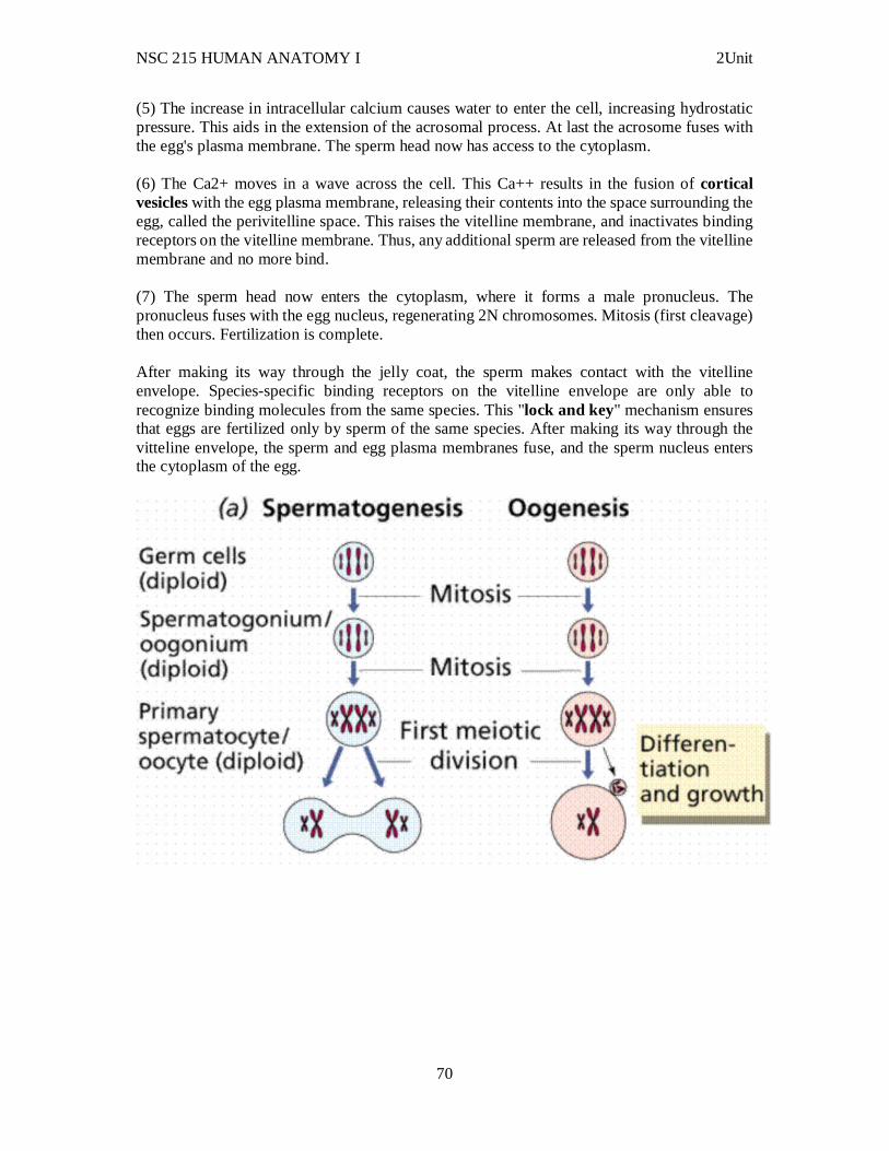
NSC 215 HUMAN ANATOMY I 2Unit
70
(5) The increase in intracellular calcium causes water to enter the cell, increasing hydrostatic pressure. This aids in the extension of the acrosomal process. At last the acrosome fuses with the egg's plasma membrane. The sperm head now has access to the cytoplasm. (6) The Ca2+ moves in a wave across the cell. This Ca++ results in the fusion of cortical vesicles with the egg plasma membrane, releasing their contents into the space surrounding the egg, called the perivitelline space. This raises the vitelline membrane, and inactivates binding receptors on the vitelline membrane. Thus, any additional sperm are released from the vitelline membrane and no more bind. (7) The sperm head now enters the cytoplasm, where it forms a male pronucleus. The pronucleus fuses with the egg nucleus, regenerating 2N chromosomes. Mitosis (first cleavage) then occurs. Fertilization is complete. After making its way through the jelly coat, the sperm makes contact with the vitelline envelope. Species-specific binding receptors on the vitelline envelope are only able to recognize binding molecules from the same species. This "lock and key" mechanism ensures that eggs are fertilized only by sperm of the same species. After making its way through the vitteline envelope, the sperm and egg plasma membranes fuse, and the sperm nucleus enters the cytoplasm of the egg.
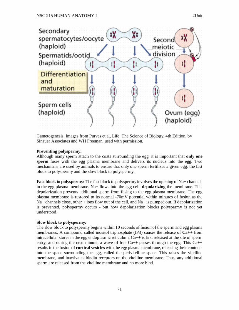
NSC 215 HUMAN ANATOMY I 2Unit
71
Gametogenesis. Images from Purves et al, Life: The Science of Biology, 4th Edition, by Sinauer Associates and WH Freeman, used with permission. Preventing polyspermy: Although many sperm attach to the coats surrounding the egg, it is important that only one sperm fuses with the egg plasma membrane and delivers its nucleus into the egg. Two mechanisms are used by animals to ensure that only one sperm fertilizes a given egg: the fast block to polyspermy and the slow block to polyspermy. Fast block to polyspermy: The fast block to polyspermy involves the opening of Na+ channels in the egg plasma membrane. Na+ flows into the egg cell, depolarizing the membrane. This depolarization prevents additional sperm from fusing to the egg plasma membrane. The egg plasma membrane is restored to its normal -70mV potential within minutes of fusion as the Na+ channels close, other + ions flow out of the cell, and Na+ is pumped out. If depolarization is prevented, polyspermy occurs - but how depolarization blocks polyspermy is not yet understood. Slow block to polyspermy: The slow block to polyspermy begins within 10 seconds of fusion of the sperm and egg plasma membranes. A compound called inositol triphosphate (IP3) causes the release of Ca++ from intracellular stores in the egg endoplasmic reticulum. Ca++ is first released at the site of sperm entry, and during the next minute, a wave of free Ca++ passes through the egg. This Ca++ results in the fusion of cortical vesicles with the egg plasma membrane, releasing their contents into the space surrounding the egg, called the perivitelline space. This raises the vitelline membrane, and inactivates bindin receptors on the vitelline membrane. Thus, any additional sperm are released from the vitelline membrane and no more bind.
NSC 215 HUMAN ANATOMY I 2Unit
72
Egg activation Ca++ release at fertilization results in an increase in metabolic activity within the egg, apparently due to an increase in the intracellular pH of the egg. Diacyl gycerol (DAG) causes protein phosphorylation cascades to be initiated, with one result being the phosphorylation and activation of a plasma membrane Na+:H+ ion exchanger. Na+ is pumped into the cell, H+ is pumped out of the cell, and the pH inside the cell increases. Sperm themselves are NOT required for egg activation - injection of Ca++ can artificially induce egg activation in many species. Cortical rotation Positional information is already contained within many eggs, with the exception of mammals. Egg polarity is due to the asymmetric distribution of cytoplasmic molecules, including mRNAs, proteins, and yolk, and is roughly oriented along the anterior/posterior axis in most animals. Cleavage In embryology, cleavage is the division of cells in the early embryo. The zygotes of many species undergo rapid cell cycles with no significant growth, producing a cluster of cells the same size as the original zygote. The different cells derived from cleavage are called blastomeres and form a compact mass called the morula. Cleavage ends with the formation of the blastula. Depending mostly on the amount of yolk in the egg, the cleavage can be holoblastic (total or entire cleavage) or meroblastic (partial cleavage). The pole of the egg with the highest concentration of yolk is referred to as the vegetal pole while the opposite is referred to as the animal pole. Cleavage differs from other forms of cell division in that it increases the number of cells without increasing the mass. This means that with each successive subdivision, the ratio of nuclear to cytoplasmic material increases. There are several differences between the cleavage in placental mammals and the cleavage in other animals. Mammals have a slow rate of division that is between 12 and 24 hours. These cellular divisions are asynchronous. Zygotic transcription starts at the two-, four-, or eight-cell stage. Cleavage is holoblastic and rotational. At the eight-cell stage, the embryo goes through some changes. Most of the blastomeres in this stage become polarized and develop tight junctions with the other blastomeres. This process leads to the development of two different populations of cells: Polar cells on the outside and apolar cells on the inside. The outer cells, called the trophoblast cells, pump sodium in from the outside, which automatically brings water in with it to the basal (inner) surface to form a blastocoel cavity in a process called compaction. The embryo is now called a blastocyst. The trophoblast cells will eventually give rise to the embryonic contribution to the placenta called the chorion. The inner cells are pushed to one side of the cavity (because the embryo isn't getting any bigger) to form the inner cell mass (ICM) and will give rise to the embryo and some extraembryonic membranes. At this stage, the embryo is called a blastocyst.
NSC 215 HUMAN ANATOMY I 2Unit
73
3.3 Embryogenesis Embryogenesis is the step in the life cycle after fertilisation – the development of the embryo, starting from the zygote (fertilised egg). Organisms can differ drastically in how the embryo develops, especially when they belong to different phyla. For example, embryonal development in placental mammals starts with cleavage of the zygote into eight uncommited cells, which then form a ball (morula). The outer cells become the trophoblast, which will form in combination with maternal uterine endometrial tissue the placenta, needed for fetal nurturing via maternal blood, while inner cells become the inner cell mass that will form all fetal organs (the bridge between these two parts eventually forms the umbilical cord). Patterning is important for determining which cells develop into which organs. This is mediated by signaling between adjacent cells by proteins on their surfaces, and by gradients of signaling secreted molecules. An example is retinoic acid, which forms a gradient in the head to tail direction in animals. Retinoic acid enters cells and activates Hox genes in a concentration-dependent manner – Hox genes differ in how much retinoic acid they require for activation and will thus show differential rostral expression boundaries, in a colinear fashion with their genomic order. As Hox genes code for transcription factors, this causes different activated combinations of both Hox and other genes in discrete anteroposterior transverse segments of the neural tube (neuromeres) and related patterns in surrounding tissues, such as branchial arches, lateral mesoderm, neural crest, skin and endoderm, in the head to tail direction. This is important for e.g. the segmentation of the spine in vertebrates. Growth Growth is the enlargement of a tissue or organism. Growth continues after the embryonal stage, and occurs through cell proliferation, enlargement of cells or accumulation of extracellular material. In plants, growth results in an adult organism that is strikingly different from the embryo. The proliferating cells tend to be distinct from differentiated cells. In some tissues proliferating cells are restricted to specialised areas, such as the growth plates of bones. But some stem cells migrate to where they are needed, such as mesenchymal stem cells which can migrate from the bone marrow to form e.g. muscle, bone or adipose tissue. The size of an organ frequently determines its growth, as in the case of the liver which grows back to its previous size if a part is removed. Growth factors, such as fibroblast growth factors in the animal embryo and growth hormone in juvenile mammals, also control the extent of growth. 3.4 Organogenesis During organogenesis, the three germ layers of the embryo differentiate and further specialize to form the various organs of the body.
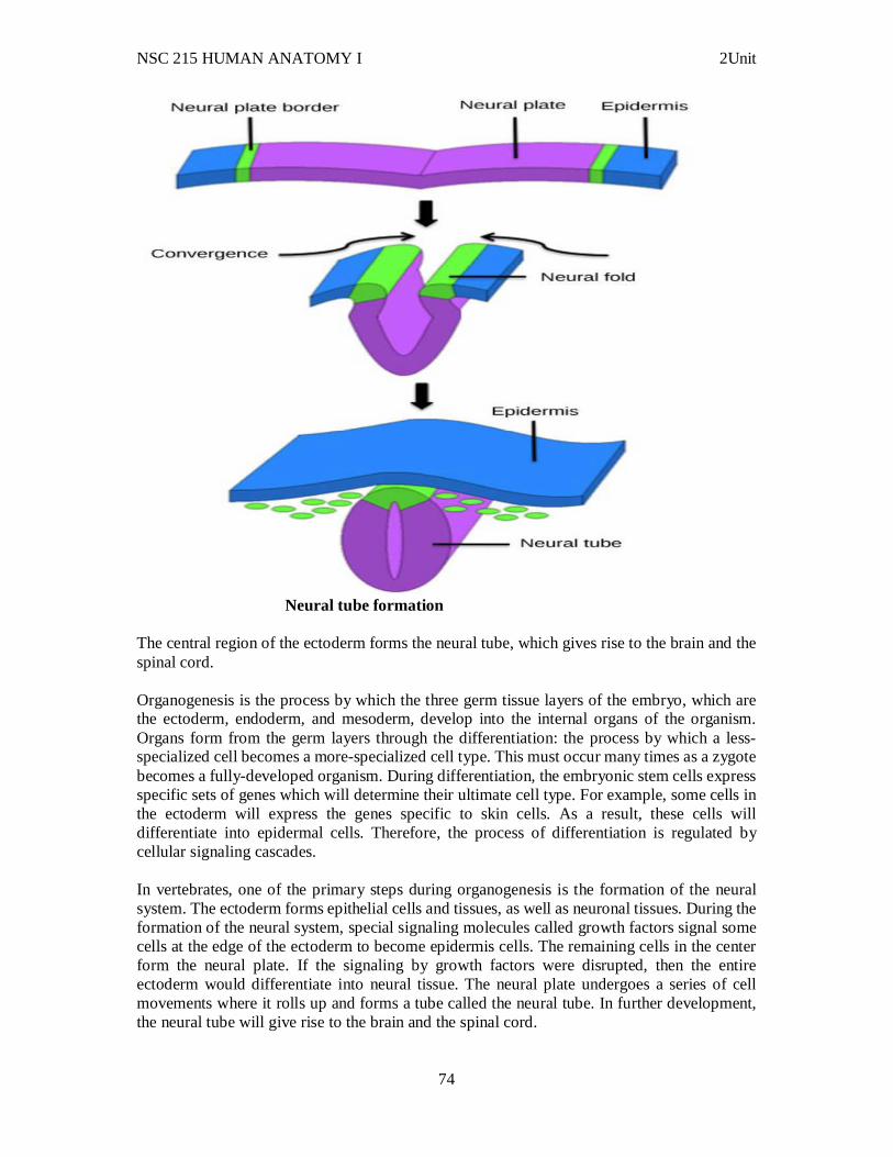
NSC 215 HUMAN ANATOMY I 2Unit
74
Neural tube formation
The central region of the ectoderm forms the neural tube, which gives rise to the brain and the spinal cord. Organogenesis is the process by which the three germ tissue layers of the embryo, which are the ectoderm, endoderm, and mesoderm, develop into the internal organs of the organism. Organs form from the germ layers through the differentiation: the process by which a less-specialized cell becomes a more-specialized cell type. This must occur many times as a zygote becomes a fully-developed organism. During differentiation, the embryonic stem cells express specific sets of genes which will determine their ultimate cell type. For example, some cells in the ectoderm will express the genes specific to skin cells. As a result, these cells will differentiate into epidermal cells. Therefore, the process of differentiation is regulated by cellular signaling cascades. In vertebrates, one of the primary steps during organogenesis is the formation of the neural system. The ectoderm forms epithelial cells and tissues, as well as neuronal tissues. During the formation of the neural system, special signaling molecules called growth factors signal some cells at the edge of the ectoderm to become epidermis cells. The remaining cells in the center form the neural plate. If the signaling by growth factors were disrupted, then the entire ectoderm would differentiate into neural tissue. The neural plate undergoes a series of cell movements where it rolls up and forms a tube called the neural tube. In further development, the neural tube will give rise to the brain and the spinal cord.
NSC 215 HUMAN ANATOMY I 2Unit
75
The mesoderm that lies on either side of the vertebrate neural tube will develop into the various connective tissues of the animal body. A spatial pattern of gene expression reorganizes the mesoderm into groups of cells called somites, with spaces between them. The somites will further develop into the ribs, lungs, and segmental (spine) muscle. The mesoderm also forms a structure called the notochord, which is rod-shaped and forms the central axis of the animal body. The endoderm consists, at first, of flattened cells, which subsequently become columnar. It forms the epithelial lining of the whole of the digestive tube (except part of the mouth and pharynx) and the terminal part of the rectum (which is lined by involutions of the ectoderm). It also forms the lining cells of all the glands which open into the digestive tube, including those of the liver and pancreas; the epithelium of the auditory tube and tympanic cavity; the trachea, bronchi, and air cells of the lungs; the urinary bladder and part of the urethra; and the follicle lining of the thyroid gland and thymus. Additionally, the endoderm forms internal organs including the stomach, the colon, the liver, the pancreas, the urinary bladder, the epithelial parts of trachea, the lungs, the pharynx, the thyroid, the parathyroid, and the intestines. Vertebrate Axis Formation Even as the germ layers form, the ball of cells still retains its spherical shape. However, animal bodies have lateral-medial (toward the side-toward the midline), dorsal-ventral (toward the back-toward the belly), and anterior-posterior (toward the front-toward the back) axes. As the body forms, it must develop in such a way that cells, tissues, and organs are organized correctly along these axes. How are these established? In one of the most seminal experiments ever to be carried out in developmental biology, Spemann and Mangold took dorsal cells from one embryo and transplanted them into the belly region of another embryo. They found that the transplanted embryo now had two notochords: one at the dorsal site from the original cells and another at the transplanted site. This suggested that the dorsal cells were genetically programmed to form the notochord and define the dorsal-ventral axis. Since then, researchers have identified many genes that are responsible for axis formation. Mutations in these genes leads to the loss of symmetry required for organism development. Many of these genes are involved in the Wnt signaling pathway. In early embryonic development, the formation of the primary body axes is a crucial step in establishing the overall body plan of each particular organism. Wnt signaling can be implicated in the formation of the anteroposterior and dorsoventral axes. Wnt signaling activity in anterior-posterior development can be seen in several organisms including mammals, fish, and frogs. Wnt signaling is also involved in the axis formation of specific body parts and organ systems that are a part of later development. In vertebrates, sonic hedgehog (Shh) and Wnt morphogenetic signaling gradients establish the dorsoventral axis of the central nervous system during neural tube axial patterning. High Wnt signaling establishes the dorsal region while high Shh signaling indicates in the ventral region. Wnt is also involved in the dorsal-ventral formation of the central nervous system through its involvement in axon guidance. Wnt proteins guide the axons of the spinal cord in an anterior-posterior direction. Wnt is also involved in the formation of the limb dorsal-ventral axis. Specifically, Wnt7a helps produce the dorsal patterning of the developing limb.
NSC 215 HUMAN ANATOMY I 2Unit
76
Placenta Formation and Functions The placenta is an organ that connects the developing fetus to the uterine wall to allow nutrient uptake, waste elimination, and gas exchange via the mother's blood supply. Placentas are a defining characteristic of eutherian or "placental" mammals, The word placenta comes from the Latin word for cake, from Greek plakóenta/plakoúnta, accusative of "flat, slab-like", in reference to its round, flat appearance in humans. The classical plural is placentae, but the form placentas is common in modern English and probably has the wider currency at present. In pre-Roman languages of tribal cultures, the placenta is often referred to as the "little mother" or "grandmother," reflective of cultural values that revered the life mystery inherent in the childbearing process which bears fruit in the form of a child. The placenta functions as a fetomaternal organ with two components: the fetal placenta (Chorion frondosum), which develops from the same blastocyst that forms the fetus, and the maternal placenta (Decidua basalis), which develops from the maternal uterine tissue. Placental Classification Classification of placenta is on the basis of histological (microscopic) structural organization and layers between fetal and maternal circulation. Three main groups: 1. Haemochorial - placenta where the chorion comes in direct contact with maternal blood (human). 2. Endotheliochorial - maternal endometrial blood vessels are bare to their endothelium and these comes in contact with the chorion (dogs, cats). 3. Epitheliochorial - maternal epithelium of the uterus comes in contact with the chorion, considered as primitive (pigs, cows, horse). Two characteristics are particularly divergent and form bases for classification of placental types: 1. The gross shape of the placenta and the distribution of contact sites between fetal membranes and endometrium. 2. The number of layers of tissue between maternal and fetal vascular systems. Differences in these two properties allow classification of placentas into several fundamental types. Classification Based on Placental Shape and Contact Points Examination of placentae from different species reveals striking differences in their shape and the area of contact between fetal and maternal tissue:
Diffuse: Almost the entire surface of the allantochorion is involved in formation of the placenta. Seen in horses and pigs.
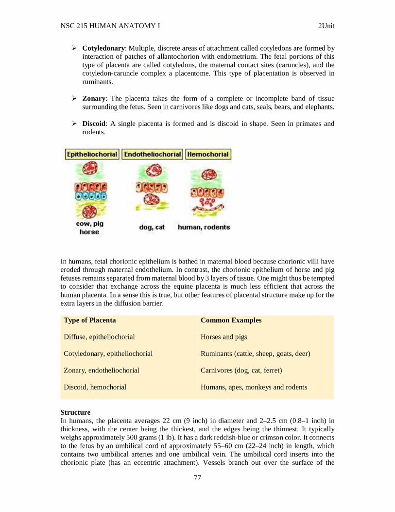
NSC 215 HUMAN ANATOMY I 2Unit
77
Cotyledonary: Multiple, discrete areas of attachment called cotyledons are formed by interaction of patches of allantochorion with endometrium. The fetal portions of this type of placenta are called cotyledons, the maternal contact sites (caruncles), and the cotyledon-caruncle complex a placentome. This type of placentation is observed in ruminants.
Zonary: The placenta takes the form of a complete or incomplete band of tissue surrounding the fetus. Seen in carnivores like dogs and cats, seals, bears, and elephants.
Discoid: A single placenta is formed and is discoid in shape. Seen in primates and rodents.
In humans, fetal chorionic epithelium is bathed in maternal blood because chorionic villi have eroded through maternal endothelium. In contrast, the chorionic epithelium of horse and pig fetuses remains separated from maternal blood by 3 layers of tissue. One might thus be tempted to consider that exchange across the equine placenta is much less efficient that across the human placenta. In a sense this is true, but other features of placental structure make up for the extra layers in the diffusion barrier. Type of Placenta
Common Examples
Diffuse, epitheliochorial
Horses and pigs
Cotyledonary, epitheliochorial
Ruminants (cattle, sheep, goats, deer)
Zonary, endotheliochorial
Carnivores (dog, cat, ferret)
Discoid, hemochorial
Humans, apes, monkeys and rodents
Structure In humans, the placenta averages 22 cm (9 inch) in diameter and 2–2.5 cm (0.8–1 inch) in thickness, with the center being the thickest, and the edges being the thinnest. It typically weighs approximately 500 grams (1 lb). It has a dark reddish-blue or crimson color. It connects to the fetus by an umbilical cord of approximately 55–60 cm (22–24 inch) in length, which contains two umbilical arteries and one umbilical vein. The umbilical cord inserts into the chorionic plate (has an eccentric attachment). Vessels branch out over the surface of the
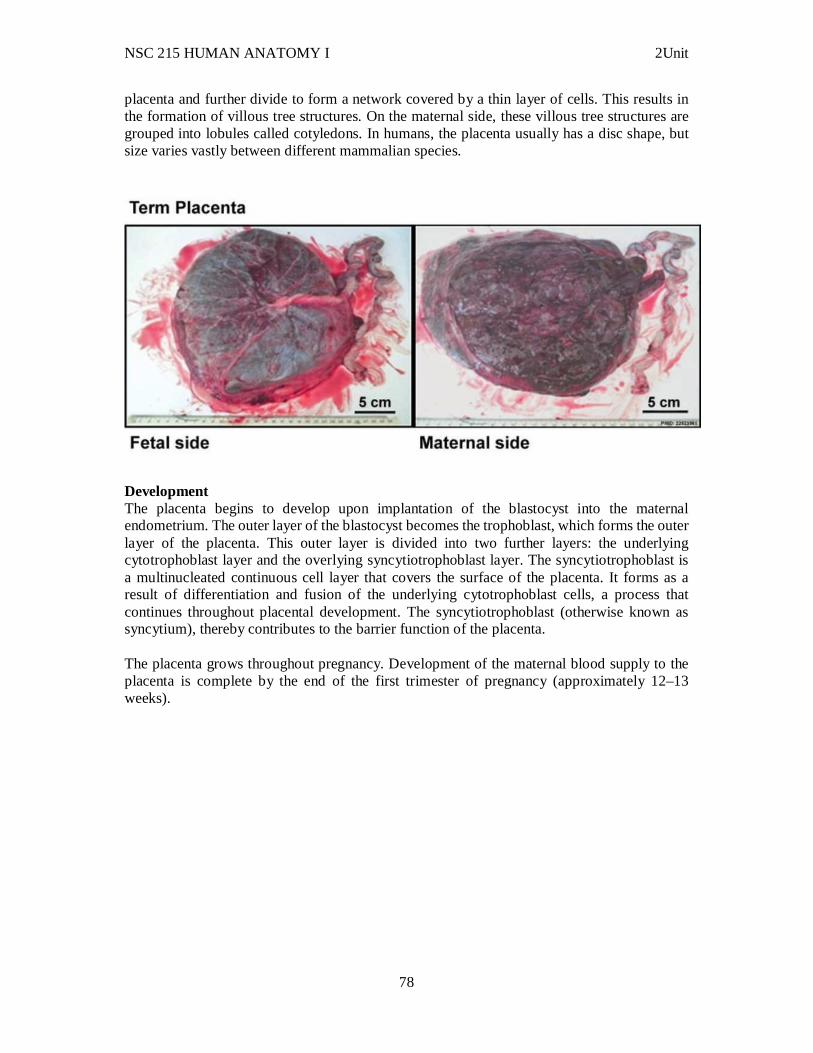
NSC 215 HUMAN ANATOMY I 2Unit
78
placenta and further divide to form a network covered by a thin layer of cells. This results in the formation of villous tree structures. On the maternal side, these villous tree structures are grouped into lobules called cotyledons. In humans, the placenta usually has a disc shape, but size varies vastly between different mammalian species.
Development The placenta begins to develop upon implantation of the blastocyst into the maternal endometrium. The outer layer of the blastocyst becomes the trophoblast, which forms the outer layer of the placenta. This outer layer is divided into two further layers: the underlying cytotrophoblast layer and the overlying syncytiotrophoblast layer. The syncytiotrophoblast is a multinucleated continuous cell layer that covers the surface of the placenta. It forms as a result of differentiation and fusion of the underlying cytotrophoblast cells, a process that continues throughout placental development. The syncytiotrophoblast (otherwise known as syncytium), thereby contributes to the barrier function of the placenta. The placenta grows throughout pregnancy. Development of the maternal blood supply to the placenta is complete by the end of the first trimester of pregnancy (approximately 12–13 weeks).
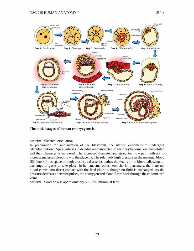
NSC 215 HUMAN ANATOMY I 2Unit
79
The initial stages of human embryogenesis. Maternal placental circulation In preparation for implantation of the blastocyst, the uterine endometrium undergoes "decidualisation". Spiral arteries in decidua are remodeled so that they become less convoluted and their diameter is increased. The increased diameter and straighter flow path both act to increase maternal blood flow to the placenta. The relatively high pressure as the maternal blood fills intervillous space through these spiral arteries bathes the fetal villi in blood, allowing an exchange of gases to take place. In humans and other hemochorial placentals, the maternal blood comes into direct contact with the fetal chorion, though no fluid is exchanged. As the pressure decreases between pulses, the deoxygenated blood flows back through the endometrial veins. Maternal blood flow is approximately 600–700 ml/min at term.
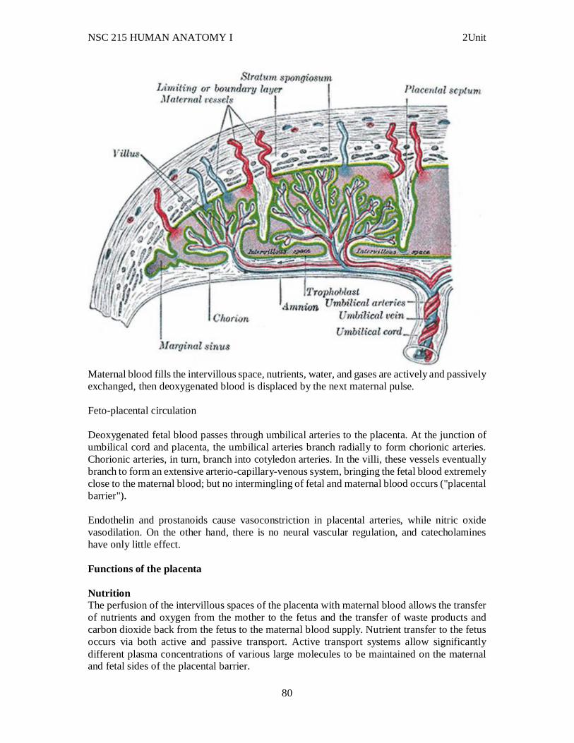
NSC 215 HUMAN ANATOMY I 2Unit
80
Maternal blood fills the intervillous space, nutrients, water, and gases are actively and passively exchanged, then deoxygenated blood is displaced by the next maternal pulse. Feto-placental circulation Deoxygenated fetal blood passes through umbilical arteries to the placenta. At the junction of umbilical cord and placenta, the umbilical arteries branch radially to form chorionic arteries. Chorionic arteries, in turn, branch into cotyledon arteries. In the villi, these vessels eventually branch to form an extensive arterio-capillary-venous system, bringing the fetal blood extremely close to the maternal blood; but no intermingling of fetal and maternal blood occurs ("placental barrier"). Endothelin and prostanoids cause vasoconstriction in placental arteries, while nitric oxide vasodilation. On the other hand, there is no neural vascular regulation, and catecholamines have only little effect. Functions of the placenta Nutrition The perfusion of the intervillous spaces of the placenta with maternal blood allows the transfer of nutrients and oxygen from the mother to the fetus and the transfer of waste products and carbon dioxide back from the fetus to the maternal blood supply. Nutrient transfer to the fetus occurs via both active and passive transport. Active transport systems allow significantly different plasma concentrations of various large molecules to be maintained on the maternal and fetal sides of the placental barrier.
NSC 215 HUMAN ANATOMY I 2Unit
81
Adverse pregnancy situations, such as those involving maternal diabetes or obesity, can increase or decrease levels of nutrient transporters in the placenta resulting in overgrowth or restricted growth of the fetus. Excretion Waste products excreted from the fetus such as urea, uric acid, and creatinine are transferred to the maternal blood by diffusion across the placenta. Immunity IgG antibodies can pass through the human placenta, thereby providing protection to the fetus in utero. This transfer of antibodies begin as early as the 20th week of gestational age, and certainly by the 24th week. This passive immunity lingers for several months after birth, thus providing the newborn with a carbon copy of the mother's long-term humoral immunity to see the infant through the crucial first months of extrauterine life. IgM, however, cannot cross the placenta, which is why some infections acquired during pregnancy can be hazardous for the fetus. Furthermore, the placenta functions as a selective maternal-fetal barrier against transmission of microbes. However, insufficiency in this function may still cause mother-to-child transmission of infectious diseases. Endocrine function In humans, aside from serving as the conduit for oxygen and nutrients for fetus, the placenta secretes, from the syncytial layer of chorionic villi, hormones that are important during pregnancy. Human Chorionic Gonadotropin (hCG): The first placental hormone produced is hCG, which can be found in maternal blood and urine as early as the first missed menstrual period (shortly after implantation has occurred) through the 100th day of pregnancy. This is the hormone analyzed by pregnancy test; a false-negative result from a pregnancy test may be obtained before or after this period. Women's blood serum will be completely negative for hCG by one to two weeks after birth. hCG testing is proof that all placental tissue is delivered. hCG is present only during pregnancy because it is secreted by the placenta. HCG also ensures that the corpus luteum continues to secrete progesterone and estrogen. Progesterone is very important during pregnancy because, when its secretion decreases, the endometrial lining will slough off and pregnancy will be lost. hCG suppresses the maternal immunologic response so that placenta is not rejected. Human Placental Lactogen (hPL [Human Chorionic Somatomammotropin]): This hormone is lactogenic and growth-promoting properties. It promotes mammary gland growth in preparation for lactation in the mother. It also regulates maternal glucose, protein, and fat levels so that this is always available to the fetus. Estrogen: referred to as the "hormone of women" because it stimulates the development of secondary female sex characteristics. It contributes to the woman's mammary gland development in preparation for lactation and stimulates uterine growth to accommodate growing fetus.
NSC 215 HUMAN ANATOMY I 2Unit
82
Progesterone: necessary to maintain endometrial lining of the uterus during pregnancy. This hormone prevents preterm labor by reducing myometrial contraction. Levels of progesterone are high during pregnancy. Cloaking from immune system of mother Further information: Immune tolerance in pregnancy The placenta and fetus may be regarded as a foreign allograft inside the mother, and thus must evade from attack by the mother's immune system. For this purpose, the placenta uses several mechanisms:
It secretes Neurokinin B-containing phosphocholine molecules. This is the same mechanism used by parasitic nematodes to avoid detection by the immune system of their host.
There is presence of small lymphocytic suppressor cells in the fetus that inhibit maternal cytotoxic T cells by inhibiting the response to interleukin 2.
However, the Placental barrier is not the sole means to evade the immune system, as foreign foetal cells also persist in the maternal circulation, on the other side of the placental barrier. Other functions The placenta also provides a reservoir of blood for the fetus, delivering blood to it in case of hypotension and vice versa, comparable to a capacitor. Birth Placental expulsion begins as a physiological separation from the wall of the uterus. The period from just after the child is born until just after the placenta is expelled is called the "third stage of labor". The placenta is usually expelled within 15–30 minutes of birth. Placental expulsion can be managed actively, for example by giving oxytocin via intramuscular injection followed by cord traction to assist in delivering the placenta. Alternatively, it can be managed expectantly, allowing the placenta to be expelled without medical assistance. A Cochrane database study suggests that blood loss and the risk of postpartum bleeding will be reduced in women offered active management of the third stage of labour (needs updating). The habit is to cut the cord immediately after birth, but allegedly there is no medical reason to do that; on the contrary, it is theorized that not cutting the cord helps the baby in its adaptation to extrauterine life, especially in preterm infants. 3.5 Clinical correlates Numerous pathologies can affect the placenta.
Placenta accreta, when the placenta implants too deeply, into actual muscle of uterine wall)
Placenta praevia, when the placement of the placenta is too close to or blocks the cervix Placental abruption/abruptio placentae - a tearing away of a normally positioned
placenta from the uterine wall accompanied by hemorrhaging. Both of these above conditions can result in miscarriage and can be life-threatening to the mother.
NSC 215 HUMAN ANATOMY I 2Unit
83
Infections involving the placenta: Placentitis, such as the TORCH infections. Chorioamnionitis which can lead to preterm labour and delivery
4.0 Conclusion 5.0 Summary In this unit, you have learnt that: i. Sperm production begins at puberty at continues throughout life, with several hundred million sperm being produced each day. ii. In the process of oogenesis, the ovary contains many follicles composed of a developing egg surrounded by an outer layer of follicle cells. Each egg begins oogenesis as a primary oocyte. iii. Fertilization combines the haploid sets of chromosomes from two individuals into a single diploid cell, the zygote. In addition, fertilization activates the egg. Egg activation blocks entry by additional sperm, stimulates the final meiotic division, and triggers the onset of embryonic development. iv. The process of acrosomal reaction which is a change in the sperm that is common to many animals. v. Prevention of polyspermy – fast block and slow block. vi. In embryogenesis, Patterning is important for determining which cells develop into which organs. vii. The placenta functions as a fetomaternal organ with two components: the fetal placenta (Chorion frondosum) and the maternal placenta (Decidua basalis). viii. Classification of placenta is on the basis of histological (microscopic) structural organization and layers between fetal and maternal circulation. ix. The Three main groups of placenta are: Haemochorial, endotheliochorial and epitheliochorial. x. Two characteristics that form bases for classification of placental types: The gross shape of the placenta and the distribution of contact sites between fetal membranes and endometrium. 6.0 Tutor Marked Assignments 6.1 At the anatomy/ embryology museum, identify the different stages in the fertilization process and observe the characteristics of each process. Also, observe the maternal and fetal parts of the placenta and the various conditions associated with the placenta. Record your findings in your log book. 6.2 Answer the following questions. 1. Given these structures: (1) blastocyst (2) morula (3) zygote Choose the arrangement that lists the structures in the order in which they are formed during development.
a. 1,2,3 b. 1,3,2 c. 2,3,1 d. 3,1,2 e. 3,2,1
NSC 215 HUMAN ANATOMY I 2Unit
84
2. The embryonic disk develops from the a. inner cell mass b. trophoblast. c. blastocyst cavity. d. yolk sac.
3. The placenta a. develops from the trophoblast. b. allows maternal blood to mix with embryonic blood. c. invades the lacunae of the embryo. d. all of the above.
4. The embryonic disk a. forms between the amniotic cavity and the yolk sac. b. contains the primitive streak. c. becomes a three-layered structure. d. all of the above.
5. The brain develops from a. ectoderm. b. endoderm. c. mesoderm.
6. Most of the skeletal system develops from a. ectoderm. b. endoderm. c. mesoderm.
7. Given these structures: (1) neural crest (2) neural plate (3) neural tube Choose the arrangement that lists the structure in the order in which they form during development.
a. 1,2,3 b. 1,3,2 c. 2,1,3 d. 2,3,1 e. 3,2,1
7.0 References/further reading: 1. Bruce M. Carlson (2019) Human Embryology & Developmental Biology. 6th edition
2. Kathryn A. Booth, Terri. D. Wyman (2008) Anatomy, physiology, and pathophysiology for allied health
3. Katherine M. A. Rogers and William N. Scott (2011) Nurses! Test yourself in anatomy and physiology
4. Kent M. Van De Graff, R.Ward Rhees, Sidney Palmer (2010) Schaum’s Outline of Human Anatomy and Physiology 3rd edition
5. Keith L Moore, Persuade T.V.N (2016), The Developing Human Clinically Oriented Embryology 10th Edition Lippincott Williams & Wilkins
6. Philip Tate (2012) Seeley’s Principles of Anatomy & Physiology 2nd edition.
7. Sadler T.W (2019), Langman’s Medical Embryology 14th edition. Lippincott Williams & Wilkins
NSC 215 HUMAN ANATOMY I 2Unit
85
MODULE THREE: SUPPORT AND MOVEMENT Introduction The human musculoskeletal system (also known as the locomotor system, and previously the activity system) is an organ system that gives humans (and many animal species) the ability to move using the muscular and skeletal systems. The musculoskeletal system provides form, support, stability, and movement to the body. The skeletal portion of the system serves as the main storage system for calcium and phosphorus and contains critical components of the hematopoietic system. It is made up of the body's bones (the skeleton), muscles, cartilage, tendons, ligaments, joints, and other connective tissue that supports and binds tissues and organs together. Module Objectives: At the end of this module, you will be able to i. discuss the two basic systems that provide support and movement for the human body – integumentary system and musculoskeletal system. CONTENTS UNIT 1: Integumentary System UNIT 2: Skeleton UNIT 3: Muscles UNIT 4: Tendons and ligaments UNIT 5: Joints and bursae UNIT ONE: INTEGUMENTARY SYSTEM CONTENTS 1.0 Introduction 2.0 Objectives 3.0 Main Content 3.1 Functions of the integumentary system 3.2 Skin 3.3 Nails 3.4 Hair 3.5 Clinical correlates 4.0 Conclusion 5.0 Summary 6.0 Tutor Marked Assignments 6.1 Activity 6.2 Tutor Marked Tests 7.0 References and other resources 1.0 Introduction The integumentary system is familiar to most people because it covers the outside the appearance of the system It is composed of a complex set of organs that includes the skin and its derivatives (sweat and oil glands, hairs, and nails).
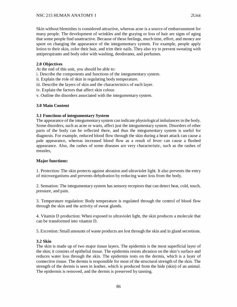
NSC 215 HUMAN ANATOMY I 2Unit
86
Skin without blemishes is considered attractive, whereas acne is a source of embarrassment for many people. The development of wrinkles and the graying or loss of hair are signs of aging that some people find unattractive. Because of these feelings, much time, effort, and money are spent on changing the appearance of the integumentary system. For example, people apply lotion to their skin, color their hair, and trim their nails. They also try to prevent sweating with antiperspirants and body odor with washing, deodorants, and perfumes. 2.0 Objectives At the end of this unit, you should be able to: i. Describe the components and functions of the integumentary system. ii. Explain the role of skin in regulating body temperature. iii. Describe the layers of skin and the characteristics of each layer. iv. Explain the factors that affect skin colour. v. Outline the disorders associated with the integumentary system. 3.0 Main Content 3.1 Functions of integumentary System The appearance of the integumentary system can indicate physiological imbalances in the body. Some disorders, such as acne or warts, affect just the integumentary system. Disorders of other parts of the body can be reflected there, and thus the integumentary system is useful for diagnosis. For example, reduced blood flow through the skin during a heart attack can cause a pale appearance, whereas increased blood flow as a result of fever can cause a flushed appearance. Also, the rashes of some diseases are very characteristic, such as the rashes of measles, Major functions: 1. Protection: The skin protects against abrasion and ultraviolet light. It also prevents the entry of microorganisms and prevents dehydration by reducing water loss from the body. 2. Sensation: The integumentary system has sensory receptors that can detect heat, cold, touch, pressure, and pain. 3. Temperature regulation: Body temperature is regulated through the control of blood flow through the skin and the activity of sweat glands. 4. Vitamin D production: When exposed to ultraviolet light, the skin produces a molecule that can be transformed into vitamin D. 5. Excretion: Small amounts of waste products are lost through the skin and in gland secretions. 3.2 Skin The skin is made up of two major tissue layers. The epidermis is the most superficial layer of the skin; it consists of epithelial tissue. The epidermis resists abrasion on the skin’s surface and reduces water loss through the skin. The epidermis rests on the dermis, which is a layer of connective tissue. The dermis is responsible for most of the structural strength of the skin. The strength of the dermis is seen in leather, which is produced from the hide (skin) of an animal. The epidermis is removed, and the dermis is preserved by tanning.
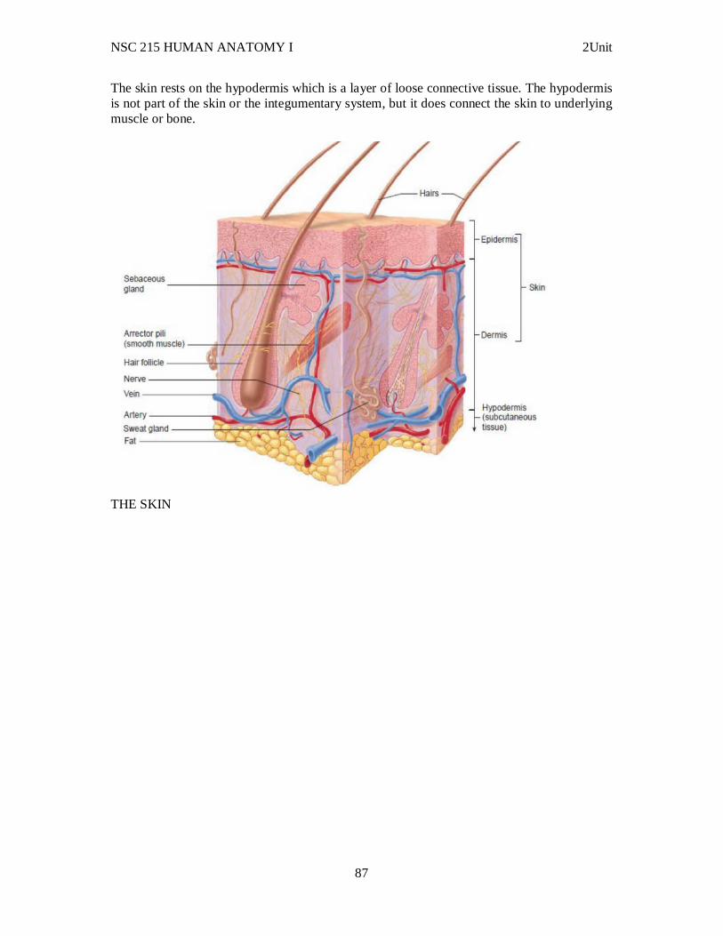
NSC 215 HUMAN ANATOMY I 2Unit
87
The skin rests on the hypodermis which is a layer of loose connective tissue. The hypodermis is not part of the skin or the integumentary system, but it does connect the skin to underlying muscle or bone.
THE SKIN
NSC 215 HUMAN ANATOMY I 2Unit
88
COMPARISON OF THE SKIN (EPIDERMIS &DERMIS) AND HYPODERMIS Dermis It is composed of strong and flexible connective tissue. Its cells are typical of those found in any connective tissue proper:
i. Fibroblasts: cells that form the fibers of connective tissue ii. Macrophages: protective cell capable of phagocytosis iii. Mast cells: immune cell that initiates inflammation iv. White blood cells: protection
Its semifluid matrix is heavily embedded with:
Collagen: strong, fibrous (threadlike) insoluble protein Elastin: extracellular connective tissue protein Reticular fibers: supporting framework tissue
The dermis binds the entire body together like a body stocking. It is your hide and corresponds exactly to animal hides used to make leather products. It is richly supplied with nerve fibers, blood vessels, and lymphatic vessels. Major portions of hair follicles, as well as oil and sweat glands, are derived from epidermal tissue but reside in the dermis
NSC 215 HUMAN ANATOMY I 2Unit
89
The layers of dermis 1. The thin, superficial papillary layer: highly vascularized areolar connective tissue containing a woven mat of collagen and elastin fibers. Its superior surface is thrown into peglike projections called dermal papillae (papill=nipple) that indent the overlaying epidermis. Many contain: Capillary loops, meissner’s corpuscles: touch receptors and pain receptors. On the palms of the hands and soles of the feet, these papillae lie atop larger mounds called dermal ridges, which in turn cause the overlying epidermis to form epidermal ridges that increase friction and enhance the gripping ability of the fingers and feet. Epidermal ridge patterns are genetically determined and unique to each of us. Because sweat pores open along their crest, our fingerprints leave identifying films of sweat called fingerprints on almost anything they touch. There are three types of skin markings: Finger prints, Cleavage lines, Flexure lines 2. The reticular layer: It is deeper and account for 80% of the thickness of the dermis. Extracellular matrix contains thick bundles of interlacing fibers that run in various planes, most run parallel to the skin surface, less dense regions, between these bundles form cleavage, or tension lines. Collagen fibers give skin strength and resiliency, binds water, helping keep the skin hydrated while elastin fibers provide the stretch-recoil properties of skin. Skin Colour: This is determined by three pigments: Melanin, Hemoglobin and carotene Melanin- Only pigment made in the skin, it is a polymer of tyrosine which is an amino acid, ranges in color from yellow to reddish-brown to black. Synthesis depends on an enzyme in melanocytes called tyrosinase. It passes from melanocytes to the basal keratinocytes. Melanocytes are stimulated by sunlight which causes substantial melanin buildup, which helps protect the DNA of viable skin cells from UV radiation by absorbing the light and dissipating the energy as heat. Carotene: Yellow to orange pigment found in certain plant products such as carrots. It tends to accumulate in the stratum corneum and in the tissue of the hypodermis, it is the color most obvious in the palms and soles, where the stratum corneum is thickest (example: the skin of the heel). Epidermis Derivatives of the epidermis include the Sweat glands, Sebaceous glands, Nails, Hair, Hair Follicles Sweat (Sudoriferous) glands: Distributed over the entire skin surface except the nipples and parts of the external genitalia. Two types of sweat glands:
1-Eccrine
2-Apocrine Eccrine: sweat glands, or merocrine sweat glands, produce true sweat, they are the most numerous of the sweat glands, and are particularly abundant on the palms of the hands, soles of the feet, and forehead. Secretory part lies coiled in the dermis: The duct extends to open in a funnel-shaped pore at the skin surface. Secretion commonly called sweat is a hypotonic
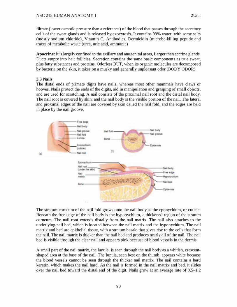
NSC 215 HUMAN ANATOMY I 2Unit
90
filtrate (lower osmotic pressure than a reference) of the blood that passes through the secretory cells of the sweat glands and is released by exocytosis. It contains 99% water, with some salts (mostly sodium chloride), Vitamin C, Antibodies, Dermicidin (microbe-killing peptide and traces of metabolic waste (urea, uric acid, ammonia) Apocrine: It is largely confined to the axillary and anogenital areas, Larger than eccrine glands. Ducts empty into hair follicles. Secretion contains the same basic components as true sweat, plus fatty substances and proteins. Odorless BUT, when its organic molecules are decomposed by bacteria on the skin, it takes on a musky and generally unpleasant odor (BODY ODOR). 3.3 Nails The distal ends of primate digits have nails, whereas most other mammals have claws or hooves. Nails protect the ends of the digits, aid in manipulation and grasping of small objects, and are used for scratching. A nail consists of the proximal nail root and the distal nail body. The nail root is covered by skin, and the nail body is the visible portion of the nail. The lateral and proximal edges of the nail are covered by skin called the nail fold, and the edges are held in place by the nail groove.
The stratum corneum of the nail fold grows onto the nail body as the eponychium, or cuticle. Beneath the free edge of the nail body is the hyponychium, a thickened region of the stratum corneum. The nail root extends distally from the nail matrix. The nail also attaches to the underlying nail bed, which is located between the nail matrix and the hyponychium. The nail matrix and bed are epithelial tissue, with a stratum basale that gives rise to the cells that form the nail. The nail matrix is thicker than the nail bed and produces nearly all of the nail. The nail bed is visible through the clear nail and appears pink because of blood vessels in the dermis. A small part of the nail matrix, the lunula, is seen through the nail body as a whitish, crescent-shaped area at the base of the nail. The lunula, seen best on the thumb, appears white because the blood vessels cannot be seen through the thicker nail matrix. The nail contains a hard keratin, which makes the nail hard. As the nail is formed in the nail matrix and bed, it slides over the nail bed toward the distal end of the digit. Nails grow at an average rate of 0.5–1.2
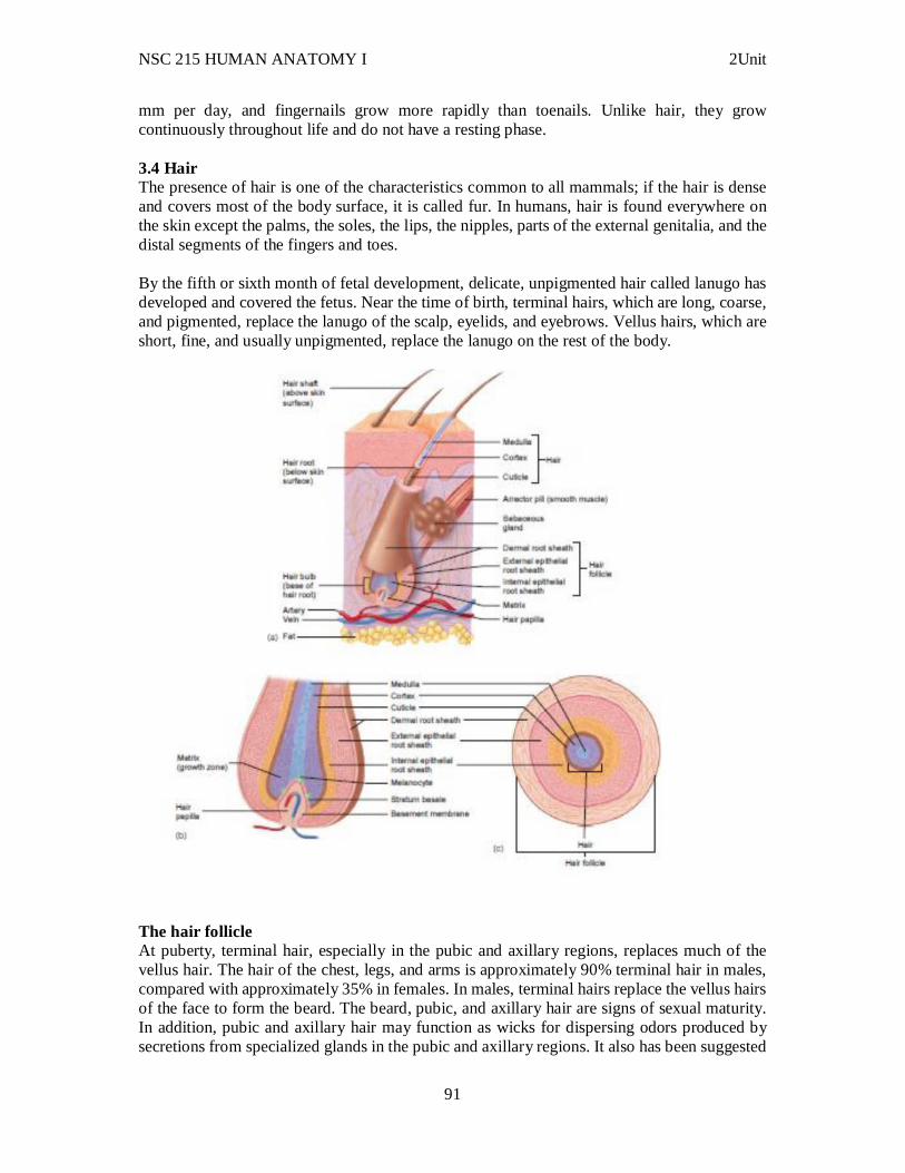
NSC 215 HUMAN ANATOMY I 2Unit
91
mm per day, and fingernails grow more rapidly than toenails. Unlike hair, they grow continuously throughout life and do not have a resting phase. 3.4 Hair The presence of hair is one of the characteristics common to all mammals; if the hair is dense and covers most of the body surface, it is called fur. In humans, hair is found everywhere on the skin except the palms, the soles, the lips, the nipples, parts of the external genitalia, and the distal segments of the fingers and toes. By the fifth or sixth month of fetal development, delicate, unpigmented hair called lanugo has developed and covered the fetus. Near the time of birth, terminal hairs, which are long, coarse, and pigmented, replace the lanugo of the scalp, eyelids, and eyebrows. Vellus hairs, which are short, fine, and usually unpigmented, replace the lanugo on the rest of the body.
The hair follicle At puberty, terminal hair, especially in the pubic and axillary regions, replaces much of the vellus hair. The hair of the chest, legs, and arms is approximately 90% terminal hair in males, compared with approximately 35% in females. In males, terminal hairs replace the vellus hairs of the face to form the beard. The beard, pubic, and axillary hair are signs of sexual maturity. In addition, pubic and axillary hair may function as wicks for dispersing odors produced by secretions from specialized glands in the pubic and axillary regions. It also has been suggested
NSC 215 HUMAN ANATOMY I 2Unit
92
that pubic hair protects against abrasion during intercourse and axillary hair reduces friction when the arms move. 3.5 Clinical correlates i. Cleavage lines in the reticular layer of the dermis are important to a surgeon because incision made parallel to these lines, the skin gapes less and heals more readily than when the incision is made across cleavage lines. ii. Folliculitis, which is a disorder specific to hair, is an inflammation of hair follicles. This disorder usually results from shaving or excess rubbing of skin areas. It may also be caused by bacteria and fungi. Follicles become red and itchy and often look like pimples. Treatments include regular cleansing of skin, topical antibiotics, and use of electric razors instead of razor blades. iii. Acne is an active inflammation of the sebaceous glands accompanied by “pimples” (pustules or cysts) on the skin. Usually caused by bacterial infection, particularly by staphylococcus. iv. Seborrhea: cradle cap in infants, caused by overactive sebaceous glands.Raised lesions that gradually become yellow to brown and begin to slough off oily scales v. A burn is injury to a tissue caused by heat, cold, friction, chemicals, electricity, or radiation. Burns are classified according to the extent of surface area involved and the depth of the burn. For an adult, the surface area that is burned can be conveniently estimated by “the rule of nines,” in which the body is divided into areas that are approximately 9%. vi. Scabies is a very contagious skin condition. Scabies is caused by mites that burrow beneath skin. Sometimes the burrows of the mites, which look like red pencil marks, can be seen. Redness and severe itching are usually the only symptoms of scabies. Most cases are easily treated with prescription medications. Because scabies is contagious, it is wise to treat an entire family if one member is infected. 4.0 Conclusion 5.0 Summary In this unit, you have learnt that: i. The components of the integumentary system includes the skin, the nails, glands and hairs. ii. The integumentary system protects us from the external environment. Other functions include sensation, temperature regulation, vitamin D production, and excretion of small amounts of waste products. iii. The epidermis is stratified squamous epithelium divided into five strata. iv. Melanocytes produce melanin inside melanosomes and then transfer the melanin to keratinocytes. The size and distribution of melanosomes determine skin color. Melanin production is determined genetically but can be influenced by ultraviolet light (tanning) and hormones. v. The nail is stratum corneum containing hard keratin and the nail root is covered by skin, and the nail body is the visible part of the nail. Nearly all of the nail is formed by the nail matrix, but the nail bed contributes.
NSC 215 HUMAN ANATOMY I 2Unit
93
vi. Integumentary system is easily observed and often reflects events occurring in other parts of the body (e.g., cyanosis, jaundice, rashes). 6.0 Tutor Marked Assignments 6.1 At the histology laboratory, examine slides of the hair, skin and nail and identify the parts of each of these components. Record your findings in the log book. 6.2 Tutor Marked Tests For questions 1–6, match the layer of the epidermis with the correct description or function: a. stratum basale b. stratum corneum c. stratum granulosum d. stratum lucidum e. stratum spinosum 1. Production of keratin fi bers; formation of lamellar bodies 2. Desquamation occurs; 25 or more layers of dead squamous cells 3. Production of cells; melanocytes produce and contribute melanin; hemidesmosomes present 4. Production of keratohyalin granules; lamellar bodies release lipids; cells die 5. Dispersion of keratohyalin around keratin fibers; layer appears transparent; cells dead 6. In which of these areas of the body is thick skin found?
a. back of the hand d. bridge of the nose b. abdomen e. sole of the foot c. over the shin
Given these parts of a hair and hair follicle: 1. cortex 2. cuticle 3. dermal root sheath 4. epithelial root sheath 5. medulla 7. Arrange the structures in the correct order from the outside of the hair follicle to the center of the hair.
a. 1,4,3,5,2 b. 2,1,5,3,4 c. 3,4,2,1,5 d. 4,3,1,2,5 e. 5,4,3,2,1
8. Concerning hair growth, a. hair falls out of the hair follicle at the end of the growth stage. b. most of the hair on the body grows continuously. c. genetic factors and the hormone testosterone are involved in “pattern baldness.” d. eyebrows have a longer growth stage and resting stage than scalp hair.
9. Why is the skin considered an organ? 7.0 References/further reading: 1. Bruce M. Carlson (2019) Human Embryology & Developmental Biology. 6th edition
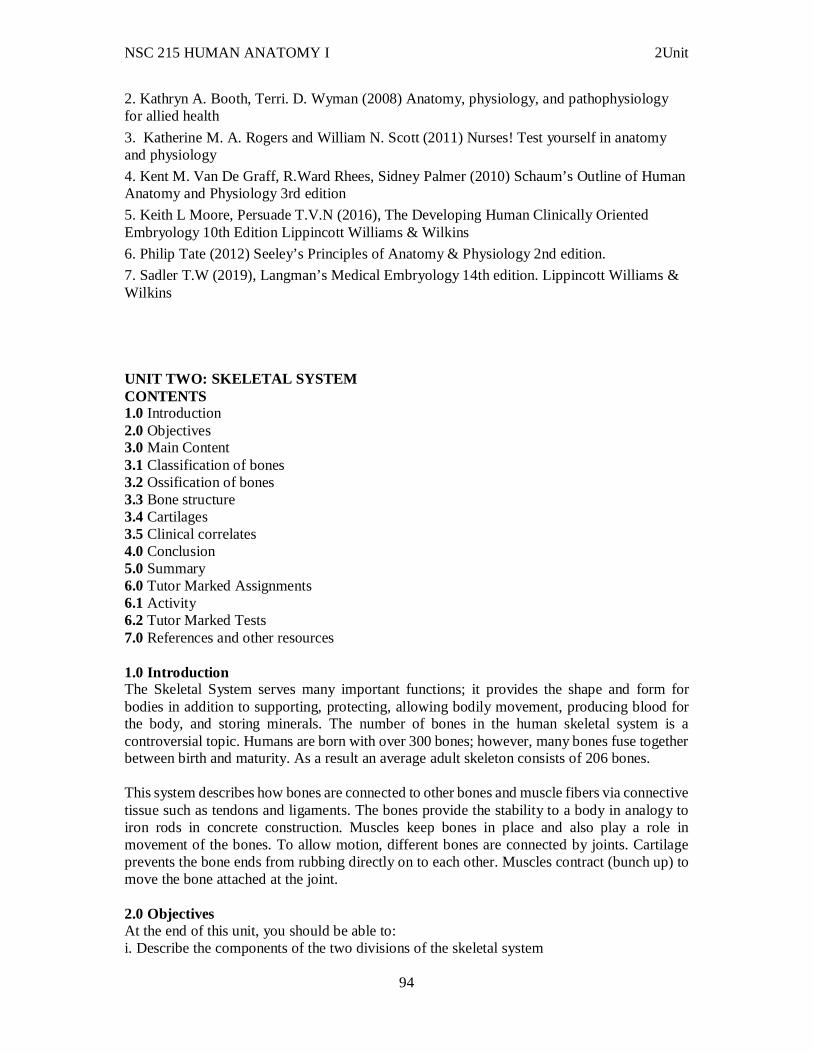
NSC 215 HUMAN ANATOMY I 2Unit
94
2. Kathryn A. Booth, Terri. D. Wyman (2008) Anatomy, physiology, and pathophysiology for allied health
3. Katherine M. A. Rogers and William N. Scott (2011) Nurses! Test yourself in anatomy and physiology
4. Kent M. Van De Graff, R.Ward Rhees, Sidney Palmer (2010) Schaum’s Outline of Human Anatomy and Physiology 3rd edition
5. Keith L Moore, Persuade T.V.N (2016), The Developing Human Clinically Oriented Embryology 10th Edition Lippincott Williams & Wilkins
6. Philip Tate (2012) Seeley’s Principles of Anatomy & Physiology 2nd edition.
7. Sadler T.W (2019), Langman’s Medical Embryology 14th edition. Lippincott Williams & Wilkins UNIT TWO: SKELETAL SYSTEM CONTENTS 1.0 Introduction 2.0 Objectives 3.0 Main Content 3.1 Classification of bones 3.2 Ossification of bones 3.3 Bone structure 3.4 Cartilages 3.5 Clinical correlates 4.0 Conclusion 5.0 Summary 6.0 Tutor Marked Assignments 6.1 Activity 6.2 Tutor Marked Tests 7.0 References and other resources 1.0 Introduction The Skeletal System serves many important functions; it provides the shape and form for bodies in addition to supporting, protecting, allowing bodily movement, producing blood for the body, and storing minerals. The number of bones in the human skeletal system is a controversial topic. Humans are born with over 300 bones; however, many bones fuse together between birth and maturity. As a result an average adult skeleton consists of 206 bones. This system describes how bones are connected to other bones and muscle fibers via connective tissue such as tendons and ligaments. The bones provide the stability to a body in analogy to iron rods in concrete construction. Muscles keep bones in place and also play a role in movement of the bones. To allow motion, different bones are connected by joints. Cartilage prevents the bone ends from rubbing directly on to each other. Muscles contract (bunch up) to move the bone attached at the joint. 2.0 Objectives At the end of this unit, you should be able to: i. Describe the components of the two divisions of the skeletal system
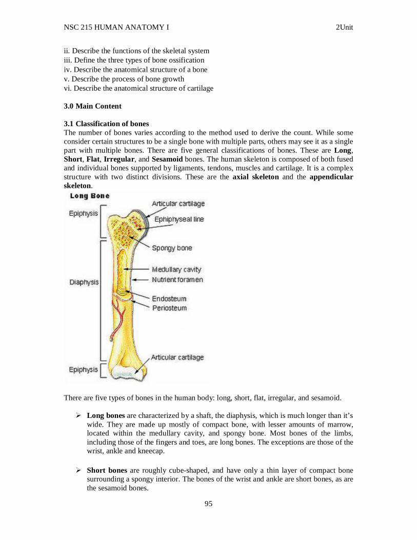
NSC 215 HUMAN ANATOMY I 2Unit
95
ii. Describe the functions of the skeletal system iii. Define the three types of bone ossification iv. Describe the anatomical structure of a bone v. Describe the process of bone growth vi. Describe the anatomical structure of cartilage 3.0 Main Content 3.1 Classification of bones The number of bones varies according to the method used to derive the count. While some consider certain structures to be a single bone with multiple parts, others may see it as a single part with multiple bones. There are five general classifications of bones. These are Long, Short, Flat, Irregular , and Sesamoid bones. The human skeleton is composed of both fused and individual bones supported by ligaments, tendons, muscles and cartilage. It is a complex structure with two distinct divisions. These are the axial skeleton and the appendicular skeleton.
There are five types of bones in the human body: long, short, flat, irregular, and sesamoid.
Long bones are characterized by a shaft, the diaphysis, which is much longer than it’s wide. They are made up mostly of compact bone, with lesser amounts of marrow, located within the medullary cavity, and spongy bone. Most bones of the limbs, including those of the fingers and toes, are long bones. The exceptions are those of the wrist, ankle and kneecap.
Short bones are roughly cube-shaped, and have only a thin layer of compact bone surrounding a spongy interior. The bones of the wrist and ankle are short bones, as are the sesamoid bones.
NSC 215 HUMAN ANATOMY I 2Unit
96
Flat bones are thin and generally curved, with two parallel layers of compact bones
sandwiching a layer of spongy bone. Most of the bones of the skull are flat bones, as is the sternum.
Sesamoid bones are bones embedded in tendons. Since they act to hold the tendon further away from the joint, the angle of the tendon is increased and thus the leverage of the muscle is increased. Examples of sesamoid bones are the patella and the pisiform.
Irregular bones do not fit into the above categories. They consist of thin layers of compact bone surrounding a spongy interior. As implied by the name, their shapes are irregular and complicated. Often this irregular shape is due to their many centers of ossification or because they contain bony sinuses. The bones of the spine, Pelvis, and some bones of the skull are irregular bones. Examples include the ethmoid and sphenoid bones.
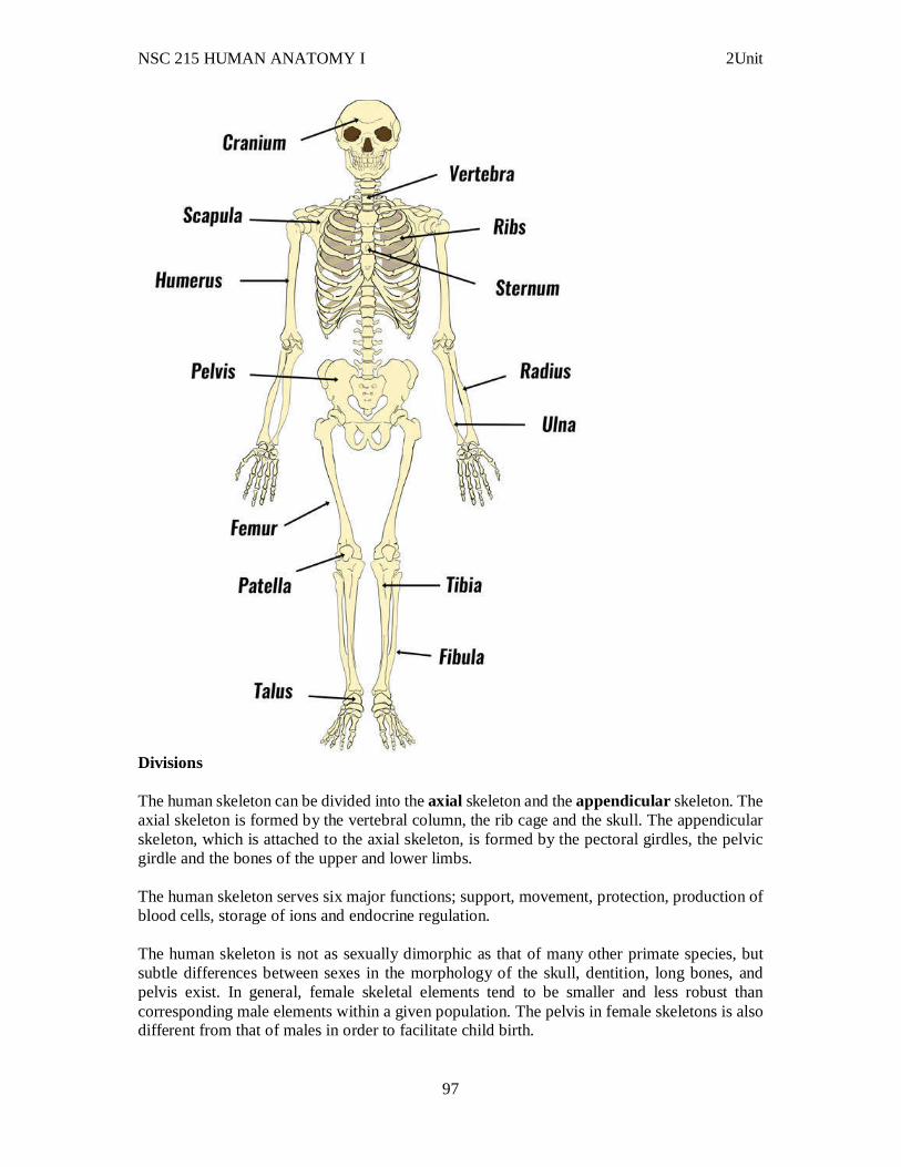
NSC 215 HUMAN ANATOMY I 2Unit
97
Divisions The human skeleton can be divided into the axial skeleton and the appendicular skeleton. The axial skeleton is formed by the vertebral column, the rib cage and the skull. The appendicular skeleton, which is attached to the axial skeleton, is formed by the pectoral girdles, the pelvic girdle and the bones of the upper and lower limbs. The human skeleton serves six major functions; support, movement, protection, production of blood cells, storage of ions and endocrine regulation. The human skeleton is not as sexually dimorphic as that of many other primate species, but subtle differences between sexes in the morphology of the skull, dentition, long bones, and pelvis exist. In general, female skeletal elements tend to be smaller and less robust than corresponding male elements within a given population. The pelvis in female skeletons is also different from that of males in order to facilitate child birth.
NSC 215 HUMAN ANATOMY I 2Unit
98
Axial skeleton The axial skeleton (80 bones) is formed by the vertebral column (32-34; the number of the vertebrae differs from human to human as the lower 2 parts, sacral and coccygeal bone may vary in length), the rib cage (12 pairs of ribs and the sternum), and the skull (22 bones and 7 associated bones). The upright posture of humans is maintained by the axial skeleton, which transmits the weight from the head, the trunk, and the upper extremities down to the lower extremities at the hip joints. The bones of the spine are supported by many ligaments. The erectors spinae muscles are also supporting and are useful for balance. The vertebral column consists of 5 parts. The most cranial (uppermost) part is made up by the cervical vertebras (7), followed by thoracic (12), lumbar (5), sacral (4-5) and coccygeal vertebras (3-4). Cervical vertebras make up the junction between the vertebral column and the cranium. Sacral and coccygeal vertebras are fused and thus often called "sacral bone" or "coccygeal bone" as unit. The sacral bone makes up the junction between the vertebral column and the pelvic bones. Appendicular skeleton The appendicular skeleton (126 bones) is formed by the pectoral girdles, the upper limbs, the pelvic girdle or pelvis, and the lower limbs. Their functions are to make locomotion possible and to protect the major organs of digestion, excretion and reproduction. Functions of the skeleton The skeleton serves six major functions; support, movement, protection, production of blood cells, storage of ions and endocrine regulation. Support: The skeleton provides the framework which supports the body and maintains its shape. The pelvis, associated ligaments and muscles provide a floor for the pelvic structures. Without the rib cages, costal cartilages, and intercostal muscles, the lungs would collapse. Movement: The joints between bones allow movement, some allowing a wider range of movement than others, e.g. the ball and socket joint allows a greater range of movement than the pivot joint at the neck. Movement is powered by skeletal muscles, which are attached to the skeleton at various sites on bones. Muscles, bones, and joints provide the principal mechanics for movement, all coordinated by the nervous system. Protection: The skeleton protects many vital organs:
The skull protects the brain, the eyes, and the middle and inner ears.
The vertebrae protect the spinal cord.
The rib cage, spine, and sternum protect the lungs, heart and major blood vessels.
The clavicle and scapula protect the shoulder.
The ilium and spine protect the digestive and urogenital systems and the hip.
The patella and the ulna protect the knee and the elbow respectively.
The carpals and tarsals protect the wrist and ankle respectively.
NSC 215 HUMAN ANATOMY I 2Unit
99
Blood cell production: The skeleton is the site of haematopoiesis, the development of blood cells that takes place in the bone marrow. Storage: Bone matrix can store calcium and is involved in calcium metabolism, and bone marrow can store iron in ferrotin and is involved in iron metabolism. However, bones are not entirely made of calcium, but a mixture of chondroitin sulfate and hydroxyapatite, the latter making up 70% of a bone. Hydroxyapatite is in turn composed of 39.8% of calcium, 41.4% of oxygen, 18.5% of phosphorus, and 0.2% of hydrogen. Endocrine regulation: Bone cells release a hormone called osteocalcin, which contributes to the regulation of blood sugar (glucose) and fat deposition. Osteocalcin increases both the insulin secretion and sensitivity, in addition to boosting the number of insulin-producing cells and reducing stores of fat. Sexual dimorphism The skull A variety of gross morphological traits of the human skull demonstrate sexual dimorphism, such as the nuchal crest, mastoid processes, supraorbital margin, supraorbital ridge, and mental eminence. Dentition Human inter-sex dental dimorphism centers on the canines, but it is not nearly as pronounced as in the other great apes. Long bones Long bones are generally larger in males than in females within a given population. Muscle attachment sites on long bones are often more robust in males than in females, reflecting a difference in overall muscle mass and development between sexes. Sexual dimorphism in the long bones is commonly characterized by morphometric or gross morphological analyses. The pelvis Human pelvis exhibit greater sexual dimorphism than other bones, specifically in the size and shape of the pelvic cavity, iliac, greater sciatic notches, and the sub-pubic angle. The Phenice method is commonly used to determine the sex of an unidentified human skeleton by anthropologists with 96% to 100% accuracy in some populations. Blood supply of bones Although bone is not an actively growing tissue in adults, it does need a constant blood supply to stay alive. In fact, the supply of blood to long bones is profuse and derived from a number of sources that are explained with detail in the lines below.
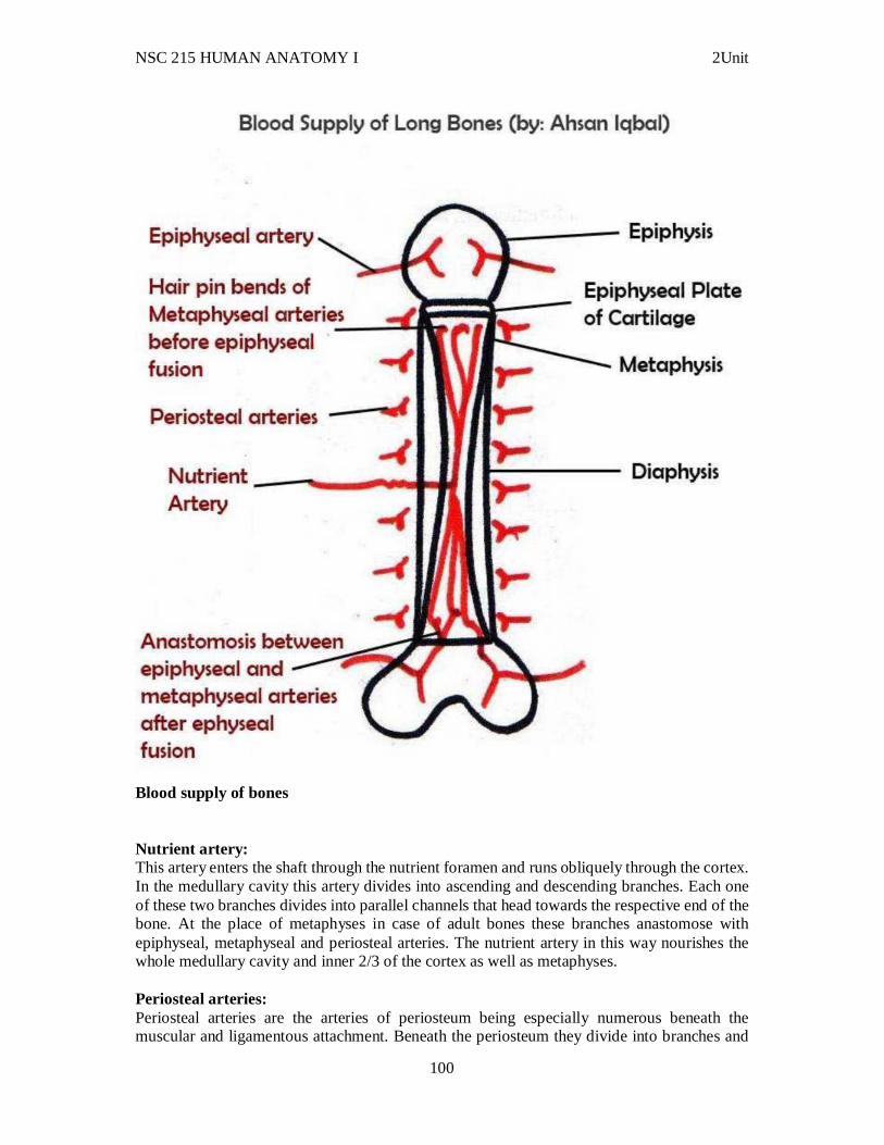
NSC 215 HUMAN ANATOMY I 2Unit
100
Blood supply of bones Nutrient artery: This artery enters the shaft through the nutrient foramen and runs obliquely through the cortex. In the medullary cavity this artery divides into ascending and descending branches. Each one of these two branches divides into parallel channels that head towards the respective end of the bone. At the place of metaphyses in case of adult bones these branches anastomose with epiphyseal, metaphyseal and periosteal arteries. The nutrient artery in this way nourishes the whole medullary cavity and inner 2/3 of the cortex as well as metaphyses. Periosteal arteries: Periosteal arteries are the arteries of periosteum being especially numerous beneath the muscular and ligamentous attachment. Beneath the periosteum they divide into branches and
NSC 215 HUMAN ANATOMY I 2Unit
101
thereby entering the Volkmann’s canals to supply the outer one third (1/3) portion of the cortex. Remember that the inner 2/3 of the cortex was supplied by the nutrient artery discussed above. Epiphyseal arteries: These are the arteries of epiphyses and are derived from the peri-articular vascular arcades found on the non-articular bony surfaces. This area also has numerous foramina out of which only few are the entrance points of these arteries while the remaining are the venous exits. Metaphyseal arteries: These arteries are derived from the neighboring systemic vessels. These arteries directly go into the metaphyses and reinforce the metaphyseal branches of the primary nutrient artery. Nerve supply of bones As with all other living tissues, the bone is innervated by peripheral nerves so that it can coordinate with the central nervous system. The important part of this coordination is formed by the sensory signals coming from the bones. The brain reads these signals and makes necessary changes to avoid any damage to bones and other body tissues. Hilton’s Law: Hilton’s law explains the pattern of innervation of bones by the peripheral nerves. According to this law, the nerve supplying a muscle will also supply the underlying bone. Thus if a group of muscles over a bone receive nerve supply from a specific nerve, the bone, over which the muscles lie will also be innervated by the same nerve. Features of innervation of bones: The innervation of nerves shows the following common features:
Nerves accompany the blood vessels so if you have to find the specific nerve supplying a specific bone, you will have to look for the nerves which accompany the arteries and veins of a bone.
Most of the nerves coming to a bone are sympathetic and vasomotor in function.
Some of the nerves are sensory and such nerves are distributed to the articular ends and periosteum of the bones.
3.2 Ossification of bones Ossification is the process by which bone is formed. The formation of bone is in fact conversion of other types of connective tissues into bone. Based on the type of tissue converted into bone, the process of ossification is of three types. These types with appropriate detail are explained in the lines below. Intra-membranous ossification: This type of ossification is also known as mesenchymal ossification. In this type the bone is ossified from mesenchymal condensations. The bones formed by this process of ossification are known as membranous bones or dermal bones. Intra-cartilaginous ossification: In this type of ossification, the mesenchyme has been converted to cartilaginous models and the process of ossification starts in these cartilaginous models. The process of conversion of mesenchymal condensations in cartilage is known as chondrosification and this process takes
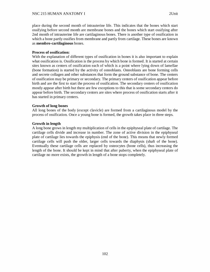
NSC 215 HUMAN ANATOMY I 2Unit
102
place during the second month of intrauterine life. This indicates that the bones which start ossifying before second month are membrane bones and the bones which start ossifying after 2nd month of intrauterine life are cartilaginous bones. There is another type of ossification in which a bone partly ossifies from membrane and partly from cartilage. These bones are known as membro-cartilaginous bones. Process of ossification: With the explanation of different types of ossification in bones it is also important to explain what ossification is. Ossification is the process by which bone is formed. It is started at certain sites known as centers of ossification each of which is a point where lying down of lamellae (bone formation) is started by the activity of osteoblasts. Osteoblasts are bone forming cells and secrete collagen and other substances that form the ground substance of bone. The centers of ossification may be primary or secondary. The primary centers of ossification appear before birth and are the first to start the process of ossification. The secondary centers of ossification mostly appear after birth but there are few exceptions to this that is some secondary centers do appear before birth. The secondary centers are sites where process of ossification starts after it has started in primary centers. Growth of long bones All long bones of the body (except clavicle) are formed from a cartilaginous model by the process of ossification. Once a young bone is formed, the growth takes place in three steps. Growth in length A long bone grows in length my multiplication of cells in the epiphyseal plate of cartilage. The cartilage cells divide and increase in number. The zone of active division in the epiphyseal plate of cartilage lies towards the epiphysis (end of the bone). This means that newly formed cartilage cells will push the older, larger cells towards the diaphysis (shaft of the bone). Eventually these cartilage cells are replaced by osteocytes (bone cells), thus increasing the length of the bone. It should be kept in mind that after puberty, when the epiphyseal plate of cartilage no more exists, the growth in length of a bone stops completely.
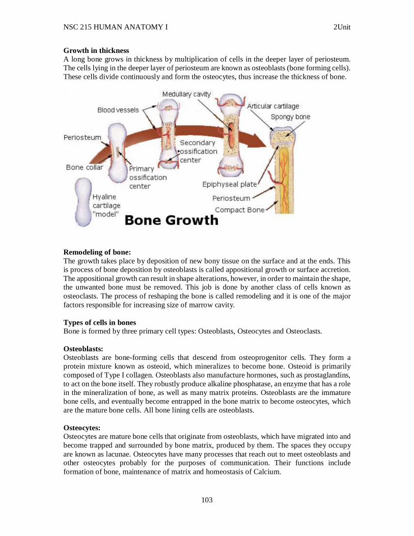
NSC 215 HUMAN ANATOMY I 2Unit
103
Growth in thickness A long bone grows in thickness by multiplication of cells in the deeper layer of periosteum. The cells lying in the deeper layer of periosteum are known as osteoblasts (bone forming cells). These cells divide continuously and form the osteocytes, thus increase the thickness of bone.
Remodeling of bone: The growth takes place by deposition of new bony tissue on the surface and at the ends. This is process of bone deposition by osteoblasts is called appositional growth or surface accretion. The appositional growth can result in shape alterations, however, in order to maintain the shape, the unwanted bone must be removed. This job is done by another class of cells known as osteoclasts. The process of reshaping the bone is called remodeling and it is one of the major factors responsible for increasing size of marrow cavity. Types of cells in bones Bone is formed by three primary cell types: Osteoblasts, Osteocytes and Osteoclasts. Osteoblasts: Osteoblasts are bone-forming cells that descend from osteoprogenitor cells. They form a protein mixture known as osteoid, which mineralizes to become bone. Osteoid is primarily composed of Type I collagen. Osteoblasts also manufacture hormones, such as prostaglandins, to act on the bone itself. They robustly produce alkaline phosphatase, an enzyme that has a role in the mineralization of bone, as well as many matrix proteins. Osteoblasts are the immature bone cells, and eventually become entrapped in the bone matrix to become osteocytes, which are the mature bone cells. All bone lining cells are osteoblasts. Osteocytes: Osteocytes are mature bone cells that originate from osteoblasts, which have migrated into and become trapped and surrounded by bone matrix, produced by them. The spaces they occupy are known as lacunae. Osteocytes have many processes that reach out to meet osteoblasts and other osteocytes probably for the purposes of communication. Their functions include formation of bone, maintenance of matrix and homeostasis of Calcium.
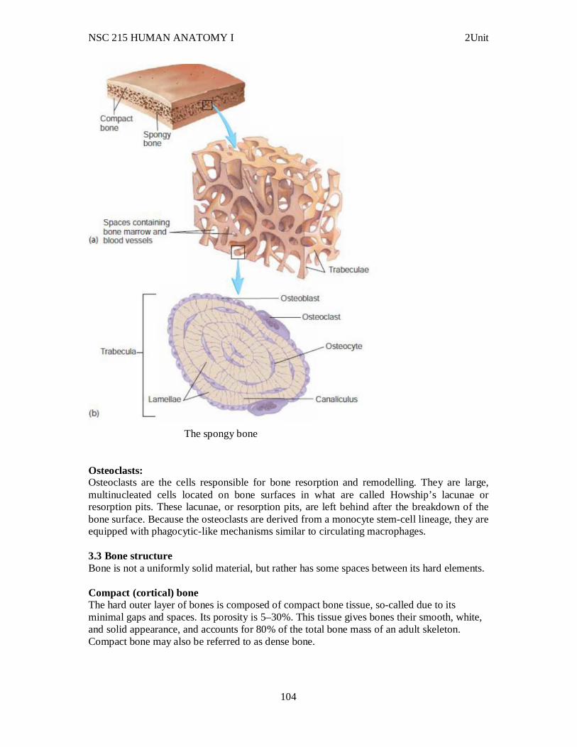
NSC 215 HUMAN ANATOMY I 2Unit
104
The spongy bone
Osteoclasts: Osteoclasts are the cells responsible for bone resorption and remodelling. They are large, multinucleated cells located on bone surfaces in what are called Howship’s lacunae or resorption pits. These lacunae, or resorption pits, are left behind after the breakdown of the bone surface. Because the osteoclasts are derived from a monocyte stem-cell lineage, they are equipped with phagocytic-like mechanisms similar to circulating macrophages. 3.3 Bone structure Bone is not a uniformly solid material, but rather has some spaces between its hard elements. Compact (cortical) bone The hard outer layer of bones is composed of compact bone tissue, so-called due to its minimal gaps and spaces. Its porosity is 5–30%. This tissue gives bones their smooth, white, and solid appearance, and accounts for 80% of the total bone mass of an adult skeleton. Compact bone may also be referred to as dense bone.
NSC 215 HUMAN ANATOMY I 2Unit
105
Compact bone Trabecular (cancellous or spongy) bone Filling the interior of the bone is the trabecular bone tissue (an open cell porous network also called cancellous or spongy bone), which is composed of a network of rod- and plate-like elements that make the overall organ lighter and allow room for blood vessels and marrow. Trabecular bone accounts for the remaining 20% of total bone mass but has nearly ten times the surface area of compact bone. Its porosity is 30–90%. If, for any reason, there is an alteration in the strain the cancellous is subjected to, there is a rearrangement of the trabeculae. The microscopic difference between compact and cancellous bone is that compact bone consists of haversian sites and osteons, while cancellous bones do not. Also, bone surrounds blood in the compact bone, while blood surrounds bone in the cancellous bone. Molecular structure Matrix The majority of bone is made of the bone matrix. It is composed primarily of inorganic hydroxyapatite and organic collagen. Bone is formed by the hardening of this matrix around entrapped cells. When these cells become entrapped from osteoblasts they become osteocytes.
NSC 215 HUMAN ANATOMY I 2Unit
106
Inorganic The inorganic composition of bone (bone mineral) is formed from carbonated hydroxyapatite (Ca10 (PO4)6(OH)2) with lower crystallinity. The matrix is initially laid down as unmineralised osteoid (manufactured by osteoblasts). Mineralisation involves osteoblasts secreting vesicles containing alkaline phosphatase. This cleaves the phosphate groups and acts as the foci for calcium and phosphate deposition. The vesicles then rupture and act as a centre for crystals to grow on. More particularly, bone mineral is formed from globular and plate structures, distributed among the collagen fibrils of bone and forming yet larger structure. Organic The organic part of matrix is mainly composed of Type I collagen. This is synthesised intracellularly as tropocollagen and then exported, forming fibrils. The organic part is also composed of various growth factors, the functions of which are not fully known. Factors present include glycosaminoglycans, osteocalcin, osteonectin, bone sialo protein, osteopontin and Cell Attachment Factor. Two types of bone can be identified microscopically according to the pattern of collagen forming the osteoid (collagenous support tissue of type I collagen embedded in glycosaminoglycan gel):
Woven bone, which is characterized by haphazard organization of collagen fibers and is mechanically weak
Lamellar bone, which has a regular parallel alignment of collagen into sheets (lamellae) and is mechanically strong.
Woven bone is produced when osteoblasts produce osteoid rapidly, which occurs initially in all fetal bones (but is later replaced by more resilient lamellar bone). In adults woven bone is created after fractures or in Paget's disease. Woven bone is weaker, with a smaller number of randomly oriented collagen fibers, but forms quickly; it is for this appearance of the fibrous matrix that the bone is termed woven. It is soon replaced by lamellar bone, which is highly organized in concentric sheets with a much lower proportion of osteocytes to surrounding tissue. Lamellar bone, which makes its first appearance in the fetus during the third trimester, is stronger and filled with many collagen fibers parallel to other fibers in the same layer (these parallel columns are called osteons). In cross-section, the fibers run in opposite directions in alternating layers, much like in plywood, assisting in the bone's ability to resist torsion forces. After a fracture, woven bone forms initially and is gradually replaced by lamellar bone during a process known as "bony substitution." Compared to woven bone, lamellar bone formation takes place more slowly. The orderly deposition of collagen fibers restricts the formation of osteoid to about 1 to 2 μm per day. Lamellar bone also requires a relatively flat surface to lay the collagen fibers in parallel or concentric layers. 3.4 Cartilages Cartilage is a type of connective tissue composed of special cells known as chondrocytes along with collagen or yellow elastic fibers. The fibers and the cells are embedded in a firm gel like matrix rich in mucopolysaccharides. Cartilage is not as hard and rigid as bone. It is much more flexible and elastic.
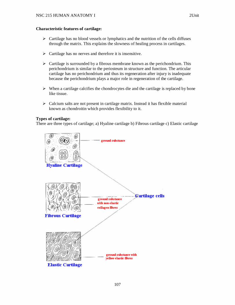
NSC 215 HUMAN ANATOMY I 2Unit
107
Characteristic features of cartilage:
Cartilage has no blood vessels or lymphatics and the nutrition of the cells diffuses through the matrix. This explains the slowness of healing process in cartilages.
Cartilage has no nerves and therefore it is insensitive.
Cartilage is surrounded by a fibrous membrane known as the perichondrium. This perichondrium is similar to the periosteum in structure and function. The articular cartilage has no perichondrium and thus its regeneration after injury is inadequate because the perichondrium plays a major role in regeneration of the cartilage.
When a cartilage calcifies the chondrocytes die and the cartilage is replaced by bone like tissue.
Calcium salts are not present in cartilage matrix. Instead it has flexible material known as chondroitin which provides flexibility to it.
Types of cartilage: There are three types of cartilage; a) Hyaline cartilage b) Fibrous cartilage c) Elastic cartilage
NSC 215 HUMAN ANATOMY I 2Unit
108
Types of Cartilage Hyaline cartilage: This type of cartilage has very thin fibers having same refractive index as the matrix of the cartilage and thus these fibers are not seen. Hyaline cartilage is the articular cartilage of long bones, sternum, ribs etc. Its color is bluish white and it is flexible. Fibrous cartilage: This type of cartilage has numerous white fibers. It is present in the symphysis pubis, and sternoclavicular joint etc. Its color is glistening white and the appearance is opaque. Elastic cartilage: This type of cartilage has numerous yellow elastic fibers. It is present in the ear pinna, external auditory meatus, Eustachian tubes, and epiglottis etc. Its color is yellowish and the appearance is opaque. 3.5 Clinical correlates 1. There are many classified skeletal disorders. One of the most common is osteoporosis. Also common is scoliosis, a side-to-side curve in the back or spine, often creating a pronounced "C" or "S" shape when viewed on an x-ray of the spine. This condition is most apparent during adolescence, and is most common with females. Osteoporosis is a disease of bone, which leads to an increased risk of fracture. In osteoporosis, the bone mineral density (BMD) is reduced, bone microarchitecture is disrupted, and the amount and variety of non-collagenous proteins in bone is altered. Osteoporosis is most common in women after the menopause, when it is called postmenopausal osteoporosis, but may develop in men and premenopausal women in the presence of particular hormonal disorders and other chronic diseases or as a result of smoking and medications, specifically glucocorticoids, when the disease is craned steroid- or glucocorticoid-induced osteoporosis (SIOP or GIOP).Osteoporosis can be prevented with lifestyle advice and medication, and preventing falls in people with known or suspected osteoporosis is an established way to prevent fractures. Osteoporosis can also be prevented with having a good source of calcium and vitamin D. Osteoporosis can be treated with bisphosphonates and various other medical treatments. 2. The most common type of dwarfism is not caused by a hormonal disorder. Achondroplasia or achondroplastic dwarfism results in a person with a nearly normal-sized trunk and head but shorter than normal limbs. Achondroplasia is an autosomal-dominant trait caused by a mutation of a gene regulating bone growth. The normal effect of the gene is to slow bone growth by inhibiting chondrocyte division at the epiphyseal plate. Mutation of the gene results in a “gain of function,” in which the normal inhibitory effect is increased, resulting in severely reduced bone growth in length. Approximately 80% of cases result from a spontaneous mutation of a gene during the formation of sperm cells or oocytes. Thus, the parents of most achondroplastic dwarfs are of normal height and proportions.
NSC 215 HUMAN ANATOMY I 2Unit
109
3. Osteogenesis imperfecta (OI) (imperfect bone formation) is a rare disorder caused by any one of a number of faulty genes that results in either too little collagen formation or a poor quality of collagen. As a result, bone matrix has decreased flexibility and is more easily broken than normal bone. Osteogenesis imperfecta is also known as the “brittle bone” disorder. In mild forms of the disorder, children may appear normal except for a history of broken bones. It is important for children with OI to be properly diagnosed because broken bones can be associated with child abuse. Over a lifetime, the number of fractures can vary from a few to more than 100. In more severe forms of the disorder, fractures heal in poor alignment, resulting in bent limbs, short stature, curved spine, and small thorax. 4.0 Summary In this unit, you have learnt that: i. The skeletal system consists of bones, cartilage, tendons, and ligaments. ii. The skeletal system supports the body, protects the organs it surrounds, allows body movements, stores minerals and fats, and is the site of blood cell and platelet production. iii. Chondroblasts produce cartilage and become chondrocytes and chondrocytes are located in lacunae surrounded by matrix. iv. The matrix of cartilage contains collagen fi bers (for strength) and proteoglycans (trap water). v. Bone cells include: Osteoblasts, which produce bone matrix and become osteocytes; Osteoblasts, which connect to one another through cell processes and surround themselves with bone matrix to become osteocytes; Osteocytes are located in lacunae and are connected to one another through canaliculi. vi. In intramembranous ossification, some skull bones, part of the mandible, and the diaphyses of the clavicles develop from membranes, beneath the periosteum, osteoblasts lay down compact bone to form the outer surface of the bone and Fontanels are areas of membrane that are not ossified at birth. vii. In endochondral ossification, most bones develop from a cartilage model. viii. Osteoblasts form bone on the calcified cartilage matrix, producing spongy bone. vi. Primary ossification centers form in the diaphysis during fetal development. Secondary ossification centers form in the epiphyses. viii. Bones increase in size only by appositional growth, the adding of new bone on the surface of older bone or cartilage. ix. Remodeling converts woven bone to lamellar bone and allows bone to change shape, adjust to stress, repair itself, and regulate body calcium levels. 6.0 Tutor Marked Assignments 6.1 At the Gross anatomy laboratory, identify the various types of bones in the body. Also, at the histology laboratory, examine the structure of a bone under the microscope and identify its parts. 6.2 Answer the following questions. 1. The skeletal system
a. includes bone, cartilage, tendons, and ligaments. b. is a storage site for calcium and phosphorus. c. contains cavities in which blood cells are produced and fat is stored. d. supports weight, protects organs, and allows movements. e. all of the above.
NSC 215 HUMAN ANATOMY I 2Unit
110
2. Which of these statements concerning cartilage is correct? a. Chondrocytes receive nutrients and oxygen from blood vessels in the matrix. b. Articular cartilage has a thick perichondrium layer. c. The perichondrium has both chondrocytes and osteocytes. d. Interstitial cartilage growth occurs when chondrocytes within the tissue add more matrix from the inside.
3. Which of these substances makes up the major portion of bone? a. collagen b. hydroxyapatite c. proteoglycans d. osteocytes e. Osteoblasts
7.0 References/further reading: 1. Bruce M. Carlson (2019) Human Embryology & Developmental Biology. 6th edition
2. Kathryn A. Booth, Terri. D. Wyman (2008) Anatomy, physiology, and pathophysiology for allied health
3. Katherine M. A. Rogers and William N. Scott (2011) Nurses! Test yourself in anatomy and physiology
4. Kent M. Van De Graff, R.Ward Rhees, Sidney Palmer (2010) Schaum’s Outline of Human Anatomy and Physiology 3rd edition
5. Keith L Moore, Persuade T.V.N (2016), The Developing Human Clinically Oriented Embryology 10th Edition Lippincott Williams & Wilkins
6. Philip Tate (2012) Seeley’s Principles of Anatomy & Physiology 2nd edition.
7. Sadler T.W (2019), Langman’s Medical Embryology 14th edition. Lippincott Williams & Wilkins UNIT THREE: MUSCULAR SYSTEM CONTENTS 1.0 Introduction 2.0 Objectives 3.0 Main Content 3.1 Classification of muscles 3.2 Gross anatomy 3.3 Histogenesis 3.4 Neuromuscular junction 3.5 Clinical correlates 4.0 Summary 5.0 Tutor Marked Assignments 6.1 Activity 6.2 Tutor Marked Tests 7.0 Reference and other resources 1.0 Introduction As a runner rounds the last corner of the track and sprints for the finish line, her arms and legs are pumping as she tries to reach her maximum speed. Her heart is beating rapidly and her breathing is rapid, deep, and regular. Blood is shunted away from digestive organs, and a
NSC 215 HUMAN ANATOMY I 2Unit
111
greater volume is delivered to skeletal muscles to maximize the oxygen supply to them. These actions are accomplished by muscle tissue, the most abundant tissue of the body, and one of the most adaptable. Movements of the limbs, the heart, and other parts of the body are made possible by muscle cells that function like tiny motors. Muscle cells use energy extracted from nutrient molecules much as motors use energy provided by electric current. The nervous system regulates and coordinates muscle cells so that smooth, coordinated movements are produced much as a computer regulates and coordinates several motors in robotic machines that perform assembly line functions. 1.0 Objectives At the end of this unit, you should be able to: i. Describe the gross anatomy of muscles ii. Locate and learn the actions of the muscles of the axial skeleton. iii. To locate and learn the actions of the muscles of the appendicular skeleton. iv. Describe the neuromuscular junction v. Explain the term “nervous control” vi. List some disorders associated with the muscular system 3 Main Content 3.1 Types of muscles Muscle is a soft tissue found in most animals. The term muscle is derived from the Latin musculus meaning "little mouse" perhaps because of the shape of certain muscles or because contracting muscles look like mice moving under the skin. Muscle cells contain protein filaments of actin and myosin that slide past one another, producing a contraction that changes both the length and the shape of the cell. Muscles function to produce force and motion. They are primarily responsible for maintaining and changing posture, locomotion, as well as movement of internal organs, such as the contraction of the heart and the movement of food through the digestive system via peristalsis. Muscle tissues are derived from the mesodermal layer of embryonic germ cells in a process known as myogenesis. There are three types of muscle, skeletal or striated, cardiac, and smooth. Muscle action can be classified as being either voluntary or involuntary. Cardiac and smooth muscles contract without conscious thought and are termed involuntary, while the skeletal muscles contract upon command. Skeletal muscles in turn can be divided into fast and slow twitch fibers. Skeletal Muscles:
They are also known as striped, striated, somatic and voluntary muscles They are the most abundant type and are found attached to the skeleton. For this reason
they are called skeletal muscles. They are innervated by somatic nervous system and are therefore under voluntary
control. They obey the will of human beings. They respond quickly to stimuli and are capable of rapid contractions. They get fatigued
easily because of their rapidity Each muscle fiber is multinucleated cylindrical cell containing groups of myofibrils.
The myofibrils are in turn made up of myofilaments of three types namely actin,
NSC 215 HUMAN ANATOMY I 2Unit
112
myosin, and tropomyosin. Thus the skeletal muscles have three structural levels namely muscle fibers, myofibrils and myofilaments.
Examples of skeletal muscles include all muscles of body wall. Smooth muscles:
They are also known as plain, unstriped, visceral and involuntary muscles. Unlike skeletal muscles, they do not exhibit cross striations under the microscope and
thus they got the name “smooth”. They are supplied by autonomic nervous system and therefore they are involuntary in
their action. They do not obey the will of human being. They respond slowly to stimuli but are capable of long time sustained contractions.
They do not get fatigued easily because of their slowness of response. They provide motor power for regulating internal environment related to digestion,
circulation, secretion and excretion. Each smooth muscle fiber is an elongated spindle shaped cell with a single nucleus
placed at the center. They also possess actin and myosin filaments but the structural arrangement of these filaments is very different as compared to the skeletal muscles.
Examples of smooth muscles include muscles of blood vessels, and muscles of the gut etc.
Cardiac muscles:
They form the myocardium of human heart. Cardiac muscle is intermediate in structure lying between the skeletal and smooth
muscles. They are striated like skeletal muscles but at the same time they are involuntary and have uninuclear cells like smooth muscles.
They are meant for automatic rhythmic contractions for long period of time. Each muscle fiber has a single centrally placed nucleus. The fibers branch and
anastomoses with each other to form a syncitium. Neighboring cells are joined by intercalated discs which provide conductive pathways from one cell to another.
Cardiac and skeletal muscles are "striated" in that they contain sarcomeres that are packed into highly regular arrangements of bundles; the myofibrils of smooth muscle cells are not arranged in sarcomeres and so are not striated. While the sarcomeres in skeletal muscles are arranged in regular, parallel bundles, cardiac muscle sarcomeres connect at branching, irregular angles (called intercalated discs). Striated muscle contracts and relaxes in short, intense burst, whereas smooth muscle sustains longer or even near-permanent contractions.
Comparison of muscle types
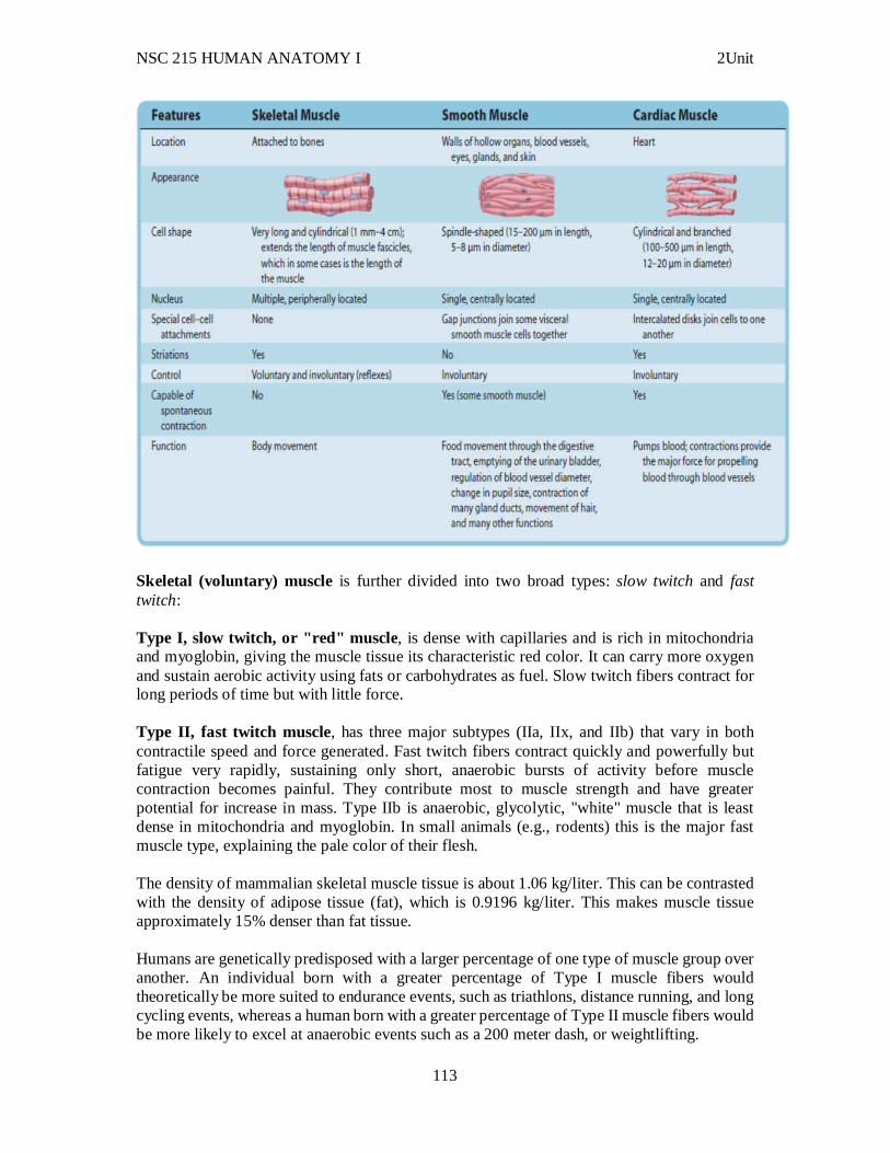
NSC 215 HUMAN ANATOMY I 2Unit
113
Skeletal (voluntary) muscle is further divided into two broad types: slow twitch and fast twitch: Type I, slow twitch, or "red" muscle, is dense with capillaries and is rich in mitochondria and myoglobin, giving the muscle tissue its characteristic red color. It can carry more oxygen and sustain aerobic activity using fats or carbohydrates as fuel. Slow twitch fibers contract for long periods of time but with little force. Type II, fast twitch muscle, has three major subtypes (IIa, IIx, and IIb) that vary in both contractile speed and force generated. Fast twitch fibers contract quickly and powerfully but fatigue very rapidly, sustaining only short, anaerobic bursts of activity before muscle contraction becomes painful. They contribute most to muscle strength and have greater potential for increase in mass. Type IIb is anaerobic, glycolytic, "white" muscle that is least dense in mitochondria and myoglobin. In small animals (e.g., rodents) this is the major fast muscle type, explaining the pale color of their flesh. The density of mammalian skeletal muscle tissue is about 1.06 kg/liter. This can be contrasted with the density of adipose tissue (fat), which is 0.9196 kg/liter. This makes muscle tissue approximately 15% denser than fat tissue. Humans are genetically predisposed with a larger percentage of one type of muscle group over another. An individual born with a greater percentage of Type I muscle fibers would theoretically be more suited to endurance events, such as triathlons, distance running, and long cycling events, whereas a human born with a greater percentage of Type II muscle fibers would be more likely to excel at anaerobic events such as a 200 meter dash, or weightlifting.
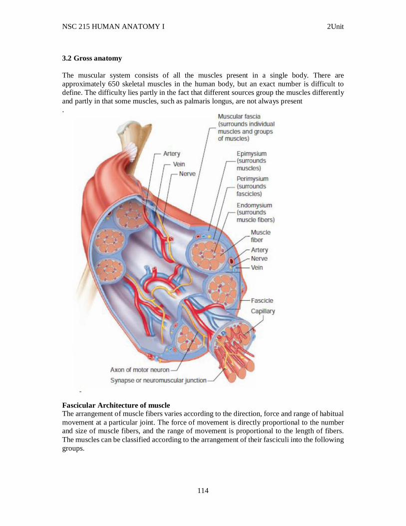
NSC 215 HUMAN ANATOMY I 2Unit
114
3.2 Gross anatomy The muscular system consists of all the muscles present in a single body. There are approximately 650 skeletal muscles in the human body, but an exact number is difficult to define. The difficulty lies partly in the fact that different sources group the muscles differently and partly in that some muscles, such as palmaris longus, are not always present .
Fascicular Architecture of muscle The arrangement of muscle fibers varies according to the direction, force and range of habitual movement at a particular joint. The force of movement is directly proportional to the number and size of muscle fibers, and the range of movement is proportional to the length of fibers. The muscles can be classified according to the arrangement of their fasciculi into the following groups.
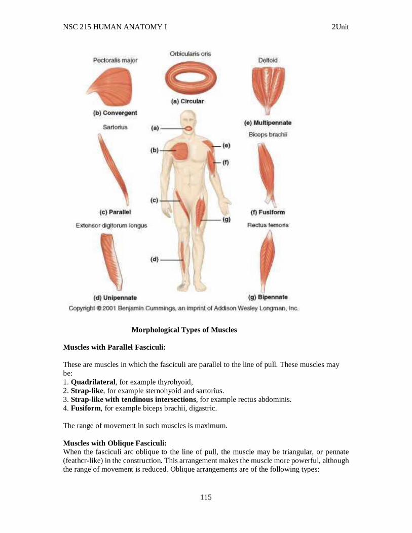
NSC 215 HUMAN ANATOMY I 2Unit
115
Morphological Types of Muscles Muscles with Parallel Fasciculi: These are muscles in which the fasciculi are parallel to the line of pull. These muscles may be: 1. Quadrilateral , for example thyrohyoid, 2. Strap-like, for example sternohyoid and sartorius. 3. Strap-like with tendinous intersections, for example rectus abdominis. 4. Fusiform, for example biceps brachii, digastric. The range of movement in such muscles is maximum. Muscles with Oblique Fasciculi: When the fasciculi arc oblique to the line of pull, the muscle may be triangular, or pennate (feathcr-like) in the construction. This arrangement makes the muscle more powerful, although the range of movement is reduced. Oblique arrangements are of the following types:
NSC 215 HUMAN ANATOMY I 2Unit
116
1. Triangular , e.g. temporalis, adductor longus. 2. Unipennate, e.g. flexor pollicis longus, extensor digitorum longus, pcroncus tcrtius, palmar interossci. 3. Bipennate, e.g. rectus femoris, dorsal interossei, pcroncus longus, flexor hallucis longus. 4. Multipennate, e.g. subscapularis, deltoid (acromial fibers]. 5. Circumpennate, e.g. tibialis anterior. Muscles with Spiral or Twisted Fasciculi: Spiral or twisted fibers are found in trapezius, pectoralis major, latissimus dorsi, supinator, etc. In certain muscles the fasciculi are crossed. These are called cruciate muscles, e.g. sternocleidomastoid, masseter and adductor magnus. Nomenclature of Muscles The muscles have been named in a number of ways. 1. According to their shape, e.g. trapezius, rhomboideus, scrratus anterior, latissimus dorsi, etc. 2. According to the number of heads of origin, e.g. biceps, triceps, quadriceps, digastric, etc. 3. According to their gross structure, e.g. scmilcndinosus, semi-’membranosus, etc. 4. According to their location, e.g. temporalis, supra-spinatus, intercostales. 5. According to their attachments, e.g. stylohyoid, cricothyroid, etc. 6. According to their action, e.g. adductor longus, flexor carpi ulnaris, abductor pollicis longus, etc. 7. According to direction of their fibers, e.g. rectus abdominis, transversus abdominis, orbicularis oculi. 8. A muscle with two bellies with an intervening tendon is called digastric muscle. Muscle with number of intervening tendons or intersections are the rectus abdominis. 9. The muscles that extend over two or more joints are called diarthric or polyarthric muscles, e.g. flexor carpi radialis and flexor digitorum profundus. Nerve supply to the skeletal muscles The nerve supplying a muscle is called motor nerve. In fact it is a mixed nerve and consists of the following types of fibers. Motor fibers: These fibers make up to 60% of a nerve to skeletal muscle. They comprise of:
Large myelinated alpha efferents which supply extramural muscle fibers. Smaller myelin gamma efferents which supply intramural fibers of the muscle spindles
which refine and control muscle contraction. The fine non-myelinated autonomic efferents which supply smooth muscle fibers of the
blood vessels. Sensory fibers: They form up to 40% of a nerve to the muscle and comprise of: Myelinated fibers distributed to muscle spindles for proprioception, also to tendons. The axon of the motor nerve branches many times after entering a target muscle. Each of these branches makes its way to different set of muscle fibers (motor unit, see below). Near the midpoint of the muscle fiber, the each
NSC 215 HUMAN ANATOMY I 2Unit
117
branch splits again, forming a small cluster of terminal branches, which form the neuromuscular junction. 3.3 Histogenesis All muscles are derived from paraxial mesoderm. The paraxial mesoderm is divided along the embryo's length into somites, corresponding to the segmentation of the body (most obviously seen in the vertebral column. Each somite has 3 divisions, sclerotome (which forms vertebrae), dermatome (which forms skin), and myotome (which forms muscle). The myotome is divided into two sections, the epimere and hypomere, which form epaxial and hypaxial muscles, respectively. The only epaxial muscles in humans are the erector spinae and small intervertebral muscles, and are innervated by the dorsal rami of the spinal nerves. All other muscles, including those of the limbs are hypaxial, and innervated by the ventral rami of the spinal nerves. During development, myoblasts (muscle progenitor cells) either remain in the somite to form muscles associated with the vertebral column or migrate out into the body to form all other muscles. Myoblast migration is preceded by the formation of connective tissue frameworks, usually formed from the somatic lateral plate mesoderm. Myoblasts follow chemical signals to the appropriate locations, where they fuse into elongate skeletal muscle cells. Microanatomy Skeletal muscles are sheathed by a tough layer of connective tissue called the epimysium. The epimysium anchors muscle tissue to tendons at each end, where the epimysium becomes thicker and collagenous. It also protects muscles from friction against other muscles and bones. Within the epimysium are multiple bundles called fascicles, each of which contains 10 to 100 or more muscle fibers collectively sheathed by a perimysium. Besides surrounding each fascicle, the perimysium is a pathway for nerves and the flow of blood within the muscle. The threadlike muscle fibers are the individual muscle cells (myocytes), and each cell is encased within its own endomysium of collagen fibers. Thus, the overall muscle consists of fibers (cells) that are bundled into fascicles, which are themselves grouped together to form muscles. At each level of bundling, a collagenous membrane surrounds the bundle, and these membranes support muscle function both by resisting passive stretching of the tissue and by distributing forces applied to the muscle. Scattered throughout the muscles are muscle spindles that provide sensory feedback information to the central nervous system. (This grouping structure is analogous to the organization of nerves which uses epineurium, perineurium, and endoneurium).
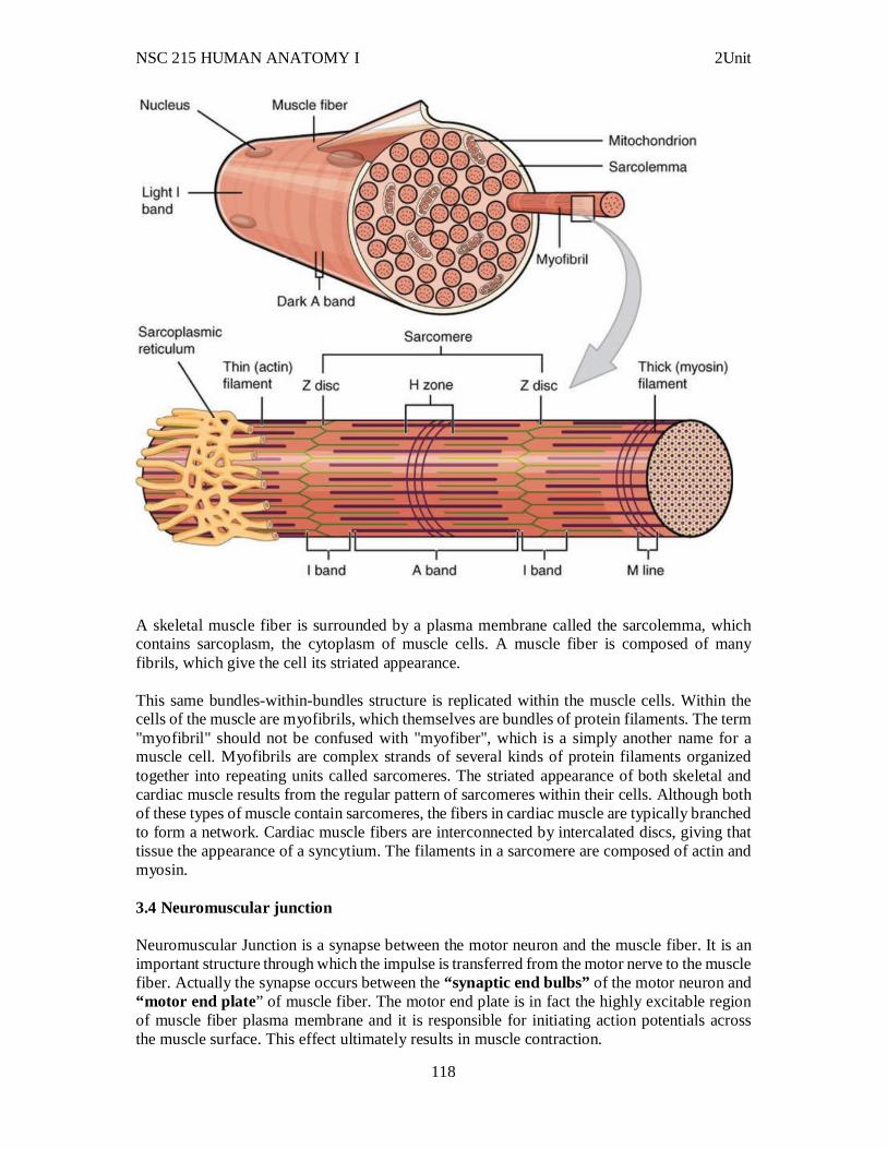
NSC 215 HUMAN ANATOMY I 2Unit
118
A skeletal muscle fiber is surrounded by a plasma membrane called the sarcolemma, which contains sarcoplasm, the cytoplasm of muscle cells. A muscle fiber is composed of many fibrils, which give the cell its striated appearance. This same bundles-within-bundles structure is replicated within the muscle cells. Within the cells of the muscle are myofibrils, which themselves are bundles of protein filaments. The term "myofibril" should not be confused with "myofiber", which is a simply another name for a muscle cell. Myofibrils are complex strands of several kinds of protein filaments organized together into repeating units called sarcomeres. The striated appearance of both skeletal and cardiac muscle results from the regular pattern of sarcomeres within their cells. Although both of these types of muscle contain sarcomeres, the fibers in cardiac muscle are typically branched to form a network. Cardiac muscle fibers are interconnected by intercalated discs, giving that tissue the appearance of a syncytium. The filaments in a sarcomere are composed of actin and myosin. 3.4 Neuromuscular junction Neuromuscular Junction is a synapse between the motor neuron and the muscle fiber. It is an important structure through which the impulse is transferred from the motor nerve to the muscle fiber. Actually the synapse occurs between the “synaptic end bulbs” of the motor neuron and “motor end plate” of muscle fiber. The motor end plate is in fact the highly excitable region of muscle fiber plasma membrane and it is responsible for initiating action potentials across the muscle surface. This effect ultimately results in muscle contraction.
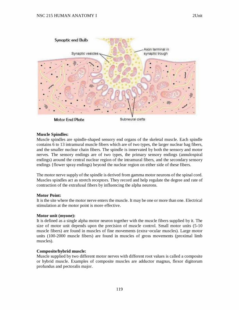
NSC 215 HUMAN ANATOMY I 2Unit
119
Muscle Spindles: Muscle spindles are spindle-shaped sensory end organs of the skeletal muscle. Each spindle contains 6 to 13 intramural muscle fibers which are of two types, the larger nuclear bag fibers, and the smaller nuclear chain fibers. The spindle is innervated by both the sensory and motor nerves. The sensory endings are of two types, the primary sensory endings (annulospiral endings) around the central nuclear region of the intramural fibers, and the secondary sensory endings {flower spray endings) beyond the nuclear region on either side of these fibers. The motor nerve supply of the spindle is derived from gamma motor neurons of the spinal cord. Muscles spindles act as stretch receptors. They record and help regulate the degree and rate of contraction of the extrafusal fibers by influencing the alpha neurons. Motor Point: It is the site where the motor nerve enters the muscle. It may be one or more than one. Electrical stimulation at the motor point is more effective. Motor unit (myone): It is defined as a single alpha motor neuron together with the muscle fibers supplied by it. The size of motor unit depends upon the precision of muscle control. Small motor units (5-10 muscle fibers) are found in muscles of fine movements (extra¬ocular muscles). Large motor units (100-2000 muscle fibers) are found in muscles of gross movements (proximal limb muscles). Composite/hybrid muscle: Muscle supplied by two different motor nerves with different root values is called a composite or hybrid muscle. Examples of composite muscles are adductor magnus, flexor digitorum profundus and pectoralis major.
NSC 215 HUMAN ANATOMY I 2Unit
120
Anatomy of neuromuscular junction: The neuromuscular junction is composed of three parts: Synaptic end bulbs: As the axon of the motor neuron enters the skeletal muscle, it forms many branches called axon terminals. At the end of each axon terminal, there is a bulbous swelling called “synaptic end bulb”. Each synaptic end bulb contains many synaptic vesicles. These vesicles contain the all-important neurotransmitter substances such as acetylcholine. These neurotransmitter substances are responsible for transmission of impulse from axon to muscle fiber through the synapse. Motor end plate: It is the part of the sarcolemma of muscle cell, which is in closest proximity to the synaptic end bulb. It shows certain specific features different than those of other regions of muscle cell sarcolemma, including: Synaptic Gutter: It is the invaginated membrane, which forms space for the synaptic end bulbs to reach close to the muscle fiber sarcolemma. Subneural Clefts: These are small folds of the muscle membrane present at the bottom of the synaptic gutter. They greatly increase the surface area at which the neurotransmitter can act. Increased number of mitochondria: The area of the muscle fiber surrounding the motor end plate shows a considerable increase in the number of mitochondria. The obvious reason for this is the energy demand of the neuromuscular junction. The Synaptic cleft: Synaptic cleft is the space between the motor end plate (muscle fiber part) and synaptic end bulb (motor neuron part) of the neuromuscular junction. It is 20 to 30 nanometers wide. Because of this cleft, the connection between the motor neuron and the muscle fiber is not continuous and there is a break. This break is traversed by the neurotransmitters. This shows the importance of these substances in the activity of muscles (and all other nervous control mechanisms). Difference between synapse and neuromuscular junction: Synapses and neuromuscular junctions are physiologically the same; however, the neuromuscular junction is a specific type of synapse that occurs between motor neuron and muscle fiber. At other places in the body, such as the Central Nervous System, the structure with similar role is known as synapse. 3.5 Clinical correlates Weight Lifters and Muscle Length Weight lifters and others who lift heavy objects usually assume positions so that their muscles are stretched close to their optimum length before lifting. For example, the position a weight lifter assumes before power lifting stretches the upper limb and lower limb muscles to a near-optimum length for muscle contraction, and the stance a lineman assumes in a football game stretches most muscle groups in the lower limbs so they are near their optimum length for suddenly moving the body forward.
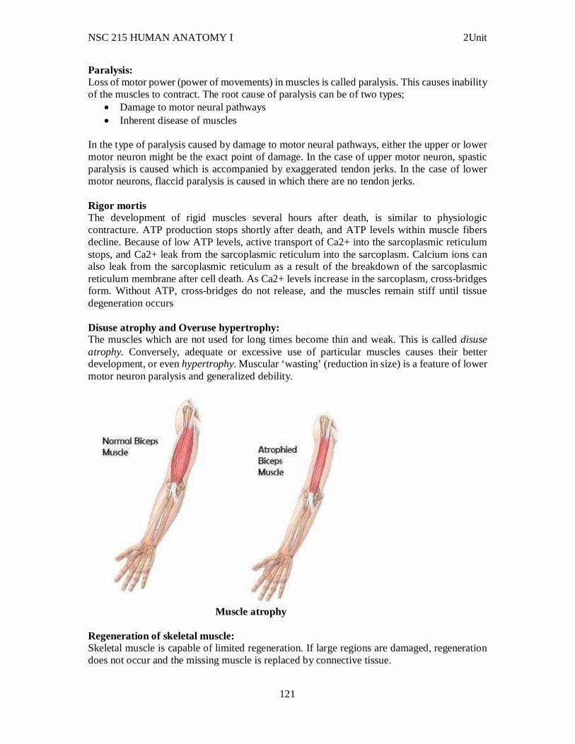
NSC 215 HUMAN ANATOMY I 2Unit
121
Paralysis: Loss of motor power (power of movements) in muscles is called paralysis. This causes inability of the muscles to contract. The root cause of paralysis can be of two types;
Damage to motor neural pathways Inherent disease of muscles
In the type of paralysis caused by damage to motor neural pathways, either the upper or lower motor neuron might be the exact point of damage. In the case of upper motor neuron, spastic paralysis is caused which is accompanied by exaggerated tendon jerks. In the case of lower motor neurons, flaccid paralysis is caused in which there are no tendon jerks. Rigor mortis The development of rigid muscles several hours after death, is similar to physiologic contracture. ATP production stops shortly after death, and ATP levels within muscle fibers decline. Because of low ATP levels, active transport of Ca2+ into the sarcoplasmic reticulum stops, and Ca2+ leak from the sarcoplasmic reticulum into the sarcoplasm. Calcium ions can also leak from the sarcoplasmic reticulum as a result of the breakdown of the sarcoplasmic reticulum membrane after cell death. As Ca2+ levels increase in the sarcoplasm, cross-bridges form. Without ATP, cross-bridges do not release, and the muscles remain stiff until tissue degeneration occurs Disuse atrophy and Overuse hypertrophy: The muscles which are not used for long times become thin and weak. This is called disuse atrophy. Conversely, adequate or excessive use of particular muscles causes their better development, or even hypertrophy. Muscular ‘wasting’ (reduction in size) is a feature of lower motor neuron paralysis and generalized debility.
Muscle atrophy
Regeneration of skeletal muscle: Skeletal muscle is capable of limited regeneration. If large regions are damaged, regeneration does not occur and the missing muscle is replaced by connective tissue.
NSC 215 HUMAN ANATOMY I 2Unit
122
Hyperplasia: Hyperplasia means increase in muscle mass. The most common cause is the increase in number of smooth muscle fibers. Usually occurs in uterus during pregnancy. Myasthenia Gravis: Myasthenia gravis is an autoimmune disease of muscle of unknown origin. Antibodies are produced that bind to acetylcholine receptor and block it. The nerve impulse transmission to muscle fibers is therefore blocked. This leads to extensive and progressive muscle weakness although the muscles arc normal. Extraocular and eyelid muscles are affected first, followed by those of the neck and limbs. It affects more in women than men and usually those between age of 20 and 40 years. Polymyositis: Polymyositis is a disease of muscle characterized by inflammation of the muscle fibers. It starts when white blood cells (immune cells of inflammation) spontaneously invade the muscle. Muscles close to trunk or torso are mostly affected by polymyositis that results in severe weakness. Polymyositis associated with skin rash is referred to as “dermamyositis”. Anabolic steroids are synthetic hormones related to testosterone, a reproductive hormone secreted by the testes. Anabolic steroids have been altered so that their reproductive effects are minimized but their effect on skeletal muscles is maintained. They increase muscle size and strength, but not muscle endurance. Some athletes have taken large doses of anabolic steroids to improve their athletic performance. Harmful side effects are associated with taking anabolic steroids, however, such as periods of irritability, acne, testicular atrophy and sterility, heart attack, stroke, and abnormal liver function. Most athletic organizations prohibit the use of anabolic steroids. Angina Pectoris: Angina pectoris is episode of chest pain due to temporary ischemia of cardiac muscle. It is usually relieved by rest and nitrites. Myocardial ischemia: Persistent ischemia due to blockage of more than one artery results in necrosis (death) of the cardiac muscle. Pain is not relieved by rest and gets referred to the left arm, chest and neighboring areas. 4.0 Summary In this unit, you have learnt that: i. Muscle is responsible for movement of the arms, legs, heart, and other parts of the body; maintenance of posture; respiration; production of body heat; communication; constriction of organs and vessels; and heartbeat. ii. Muscle exhibits contractility (shortens forcefully), excitability (responds to stimuli), extensibility (can be stretched and still contract), and elasticity (recoils to resting length). iii. The three types of muscle are skeletal, smooth, and cardiac. Skeletal muscle is responsible for most body movements, smooth muscle is found in the walls of hollow organs and tubes and moves substances through them, and cardiac muscle is found in the heart and pumps blood. iv. Muscles are composed of muscle fascicles, which are composed of muscle fibers. Epimysium surrounds muscles, perimysium surrounds muscle fascicles (bundles), and endomysium surrounds muscle fibers.
NSC 215 HUMAN ANATOMY I 2Unit
123
v. Actin and myosin myofilaments do not change in length during contraction. Actin and myosin myofilaments slide past one another in a way that causes sarcomeres to shorten. vi. In the neuromuscular junction, the presynaptic terminal of the axon is separated from the postsynaptic membrane of the muscle fiber by the synaptic cleft. Action potentials cause Ca2+ channels in the presynaptic terminal to open. Calcium ions diff use into the presynaptic terminal, stimulating the release of acetylcholine from the presynaptic terminal. Acetylcholine binds to receptors of the postsynaptic membrane, thereby changing membrane permeability and producing an action potential, which stimulates muscle contraction. vii. Muscles contract in a graded fashion because of multiple-fiber summation and frequency summation. Multiple-fiber summation increases the number of motor units activated, whereas frequency summation increases the force of contraction of motor units. 6.0 Tutor Marked Assignments 6.1 In the histology laboratory, identify the differences in the various types of muscles and report your findings in the log book. 6.2 Answer the following questions. 1. The major functions of muscle include
a. movements, such as walking. b. maintaining body posture when standing still. c. maintaining body temperature. d. moving materials through hollow organs, such as the stomach and heart. e. all of the above
2. Which of these is true of skeletal muscle? a. spindle-shaped cells b. under involuntary control c. many peripherally located nuclei per muscle cell d. forms the walls of hollow internal organs e. may be autorhythmic
3. Which of these events occurs during the lag (latent) phase of muscle contraction? a. cross-bridge movement b. active transport of Ca2+ into the sarcoplasmic reticulum c. Ca2+ binding to troponin d. sarcomere shortening e. ATP binding to myosin
4. With stimuli of increasing strength, which of these is capable of a graded response? a. nerve axon b. muscle fiber c. motor unit d. whole muscle
5. A patient is thought to be suffering from either muscular dystrophy or myasthenia gravis. How would you distinguish between the two conditions? 7.0 References/further reading: 1. Bruce M. Carlson (2019) Human Embryology & Developmental Biology. 6th edition
2. Kathryn A. Booth, Terri. D. Wyman (2008) Anatomy, physiology, and pathophysiology for allied health
NSC 215 HUMAN ANATOMY I 2Unit
124
3. Katherine M. A. Rogers and William N. Scott (2011) Nurses! Test yourself in anatomy and physiology
4. Kent M. Van De Graff, R.Ward Rhees, Sidney Palmer (2010) Schaum’s Outline of Human Anatomy and Physiology 3rd edition
5. Keith L Moore, Persuade T.V.N (2016), The Developing Human Clinically Oriented Embryology 10th Edition Lippincott Williams & Wilkins
6. Philip Tate (2012) Seeley’s Principles of Anatomy & Physiology 2nd edition.
7. Sadler T.W (2019), Langman’s Medical Embryology 14th edition. Lippincott Williams & Wilkins
NSC 215 HUMAN ANATOMY I 2Unit
125
UNIT FOUR TENDONS AND LIGAMENTS CONTENTS 1.0 Introduction 2.0 Objectives 3.0 Main Content 3.1 Ligaments 3.2 Tendons 3.3 Clinical correlates 4.0 Conclusion 5.0 Summary 6.0 Tutor Marked Assignments 6.1 Activity 6.2 Tutor Marked Tests 7.0 References and other resources 1.0 Introduction Mannequins are rigid, expressionless, immobile recreations of the human form. They cannot walk or talk. One of the major characteristics of living human beings is our ability to move about. Without muscles, humans would be little more than mannequins. We would not be able to hold this book or turn its pages. We would not be able to blink, so our eyes would dry out. None of these inconveniences would bother us for long because we would not be able to breathe, either. We use our skeletal muscles all the time. Skeletal muscles are attached to bones by tendons while Ligaments are fibrous tissues that connect bones to other bones. 2.0 Objectives At the end of this unit, you should be able to: i. Describe the anatomy of the ligaments and tendons ii. Name the types of ligaments found in the parts of the body. 3.0 Main Content 3.1 Ligaments In anatomy, a ligament is the fibrous tissue that connects bones to other bones and is also known as articular ligament, articular larua, fibrous ligament, or true ligament. Ligament can also refer to:
Peritoneal ligament: a fold of peritoneum or other membranes. Fetal remnant ligament: the remnants of a fetal tubular structure. Periodontal ligament: a group of fibers that attach the cementum of teeth to the
surrounding alveolar bone. Ligaments are similar to tendons and fasciae as they are all made of connective tissue. The differences in them are in the connections that they make; ligaments connect one bone to another bone, tendons connect muscle to bone and fasciae connect muscles to other muscles. These are all found in the skeletal system of the human body. Ligaments cannot usually be regenerated naturally, however there are periodontal ligament stem cells located near the periodontal ligament which are involved in the adult regeneration of periodontal ligament.
NSC 215 HUMAN ANATOMY I 2Unit
126
Articular ligaments "Ligament" most commonly refers to a band of tough, fibrous dense regular connective tissue bundles, made of attenuated collagenous fibers; with said bundles protected by dense irregular connective tissue sheaths. Ligaments connect bones to other bones to form a joint. They do not connect muscles to bones; that is the job of tendons. Some ligaments limit the mobility of articulations, or prevent certain movements altogether. Capsular ligaments are part of the articular capsule that surrounds synovial joints. They act as mechanical reinforcements. Extra-capsular ligaments join together in harmony with the other ligaments and provide joint stability. Intra-capsular ligaments, which are much less common, also provide stability but permit a far larger range of motion. Cruciate ligaments occur in pairs of three. Ligaments are viscoelastic. They gradually shrink when under tension, and return to their original shape when the tension is removed. However, they cannot retain their original shape when compassed past a certain point or for a prolonged period of time. This is one reason why dislocated joints must be set as quickly as possible: if the ligaments lengthen too much, then the joint will be weakened, becoming prone to future dislocations. Athletes, gymnasts, dancers, and martial artists perform stretching exercises to lengthen their ligaments, making their joints more subtle. The term hypermobility refers to people with more-elastic ligaments, allowing their joints to stretch and contort further; this is sometimes still called double-jointedness. The consequence of a broken ligament can be instability of the joint. Not all broken ligaments need surgery, but, if surgery is needed to stabilise the joint, the broken ligament can be repaired. Scar tissue may prevent this. If it is not possible to fix the broken ligament, other procedures such as the Brunelli procedure can correct the instability. Instability of a joint can over time lead to wear of the cartilage and eventually to osteoarthritis. Peritoneal ligaments Certain folds of peritoneum are referred to as ligaments. Examples include:
The hepatoduodenal ligament that surrounds the hepatic portal vein and other vessels as they travel from the duodenum to the liver.
The broad ligament of the uterus, also a fold of peritoneum. Fetal remnant ligaments Certain tubular structures from the fetal period are referred to as ligaments after they close up and turn into cord-like structures: Fetal
Adult
ductus arteriosus
ligamentum arteriosum
extra-hepatic portion of the fetal left umbilical vein
ligamentum teres hepatis (the "round ligament of the liver").
NSC 215 HUMAN ANATOMY I 2Unit
127
intra-hepatic portion of the fetal left umbilical vein (the ductus venosus)
ligamentum venosum
distal portions of the fetal left and right umbilical arteries
medial umbilical ligaments
3.2 Tendons A tendon is a tough, flexible band of fibrous connective tissue that connects muscles to bones. The extra-cellular connective tissue between muscle fibers binds to tendons at the distal and proximal ends, and the tendon binds to the periosteum of individual bones at the muscle's origin and insertion. As muscles contract, tendons transmit the forces to the relatively rigid bones, pulling on them and causing movement. Tendons can stretch substantially, allowing them to function as springs during locomotion, thereby saving energy. Tendons are similar to ligaments and fasciae. All three are made of collagen but histologically, tendons consist of dense regular connective tissue fascicles encased in dense irregular connective tissue. Tendon lengths varies in all major groups and from person to person. The tendon length is, in practice the deciding factor regarding actual and potential muscle size. For example, all other relevant biological factors being equal, a man with shorter tendons and longer biceps muscle will have a greater potential for muscle mass than a man with a longer tendon and a shorter muscle. Tendon length is determined by genetic predisposition, and has not been shown to either increase or decrease in response to environment, unlike muscles, which can be shortened by trauma, use imbalances and a lack of recovery and stretching. The function of tendons have been traditionally considered to simply be a mechanism by which muscle connect to bone, functioning simply to transmit forces. Tendons are viscoelastic structures which implies that they exhibit both elastic and viscous behaviour. HEALING The healing process for a broken tendon is long and painful. Most people who do not receive medical attention within the first 48 hours of the injury will suffer from severe swelling, pain, and a burning sensation where the injury occurred. There three main stages of tendon healing are inflammation, repair or proliferation and remodeling. These stages can overlap with each other.
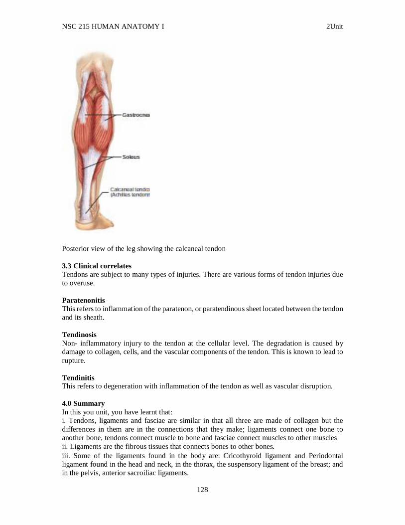
NSC 215 HUMAN ANATOMY I 2Unit
128
Posterior view of the leg showing the calcaneal tendon 3.3 Clinical correlates Tendons are subject to many types of injuries. There are various forms of tendon injuries due to overuse. Paratenonitis This refers to inflammation of the paratenon, or paratendinous sheet located between the tendon and its sheath. Tendinosis Non- inflammatory injury to the tendon at the cellular level. The degradation is caused by damage to collagen, cells, and the vascular components of the tendon. This is known to lead to rupture. Tendinitis This refers to degeneration with inflammation of the tendon as well as vascular disruption. 4.0 Summary In this you unit, you have learnt that: i. Tendons, ligaments and fasciae are similar in that all three are made of collagen but the differences in them are in the connections that they make; ligaments connect one bone to another bone, tendons connect muscle to bone and fasciae connect muscles to other muscles ii. Ligaments are the fibrous tissues that connects bones to other bones. iii. Some of the ligaments found in the body are: Cricothyroid ligament and Periodontal ligament found in the head and neck, in the thorax, the suspensory ligament of the breast; and in the pelvis, anterior sacroiliac ligaments.
NSC 215 HUMAN ANATOMY I 2Unit
129
iv. Some clinical disorders associated with tendons include tendonitis and tendinosis. 6.0 Tutor Marked Assignments 6.1 At the gross anatomy laboratory, identify the ligaments and muscle tendons that are found in the knee joint. 6.2 Self-assessment tests i. Describe the ligaments found at the knee joint ii. Discuss the four fetal remnant ligaments 7.0 References and other resources UNIT FIVE: JOINTS AND BURSAE CONTENTS 1.0 Introduction 2.0 Learning objectives 3.0 Main Content 3.1 Joints 3.2 Classification of joints 3.3 Bursae 3.4 Classification of bursae 3.5 Clinical correlates 4.0 Conclusion 5.0 Summary 6.0 Tutor Marked Assignments 6.1 Activity 6.2 Tutor Marked Tests 7.0 References and other resources 1.0 Introduction Muscles pull on bones to make them move, but movement would not be possible without joints between the bones. Humans would resemble statues, were it not for the joints between bones that allow bones to move once the muscles have provided the pull. Machine parts most likely to wear out are those that rub together, and they require the most maintenance. Movable joints are places in the body where the bones rub together, yet we tend to pay little attention to them. Fortunately, our joints are self-maintaining, but damage to or disease of a joint can make movement very difficult. 2.0 Objectives At the end of this unit, you should be able to: i. Classify the types of joints in the body and identify their location. ii. Describe the bursae in the body. 3.0 Main content 3.1 Joints
NSC 215 HUMAN ANATOMY I 2Unit
130
A joint , articulation (or articulate surface) is the location at which bones connect. They are constructed to allow movement (except for skull bones) and provide mechanical support, and are classified structurally, functionally and regional. An articulate facet is generally seen as a small joint, especially used when speaking of the joints of the ribs. Joint is a junction between two or more bones or cartilages. It is a device to permit movements in a hard and rigid skeleton. Joints are formed in such a way that they keep a balance between the movement, stability and strength of human skeleton. However there are some types of joints where movements are sacrificed for stability and in other cases the stability is sacrificed for movements. For example the shoulder joint and hip joint are both ball and socket joints but shoulder joint is more mobile and less stable while hip joint is more stable and less mobile. Also there are certain types of joints where motion does not occur. These joints are called immovable joints and are primarily meant for growth and they permit moulding during child birth. There are more joints in a child then in an adult because as growth proceeds some of the bones fuse together e.g. the ischium, ilium and pubis fuse together to form the pelvic bone (hip bone). Similarly the two halves of the infant frontal bone and of the infant mandible fuse together, five sacral vertebrae form one sacrum and four coccygeal vertebrae form one coccyx. Some basic terms related to joints: Male surface: An articulating surface which is larger in surface area and always convex in all directions. Female surface: An articulating surface which is smaller and concave in all directions. Simple joints: Joints with only two articulating surfaces; male and female. Compound joints: Joints possessing more than two articulating surfaces. Degrees of freedom: Number of axes at which the bone in a joint can move. Uni-axial movement: Movement of bone at a joint limited to one axis only that is with one degree of freedom Bi-axial movement: Movement of a bone at a joint with two degrees of freedom Multi-axial movement: Movement of a bone at a joint limited to three axes with intermediate positions as well. Terms used for movements of joints:
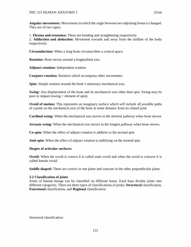
NSC 215 HUMAN ANATOMY I 2Unit
131
Angular movements: Movements in which the angle between two adjoining bones is changed. They are of two types; 1. Flexion and extension: These are bending and straightening respectively. 2. Adduction and abduction: Movement towards and away from the midline of the body respectively: Circumduction: When a long bone circumscribes a conical space. Rotation: Bone moves around a longitudinal axis. Adjunct rotation: Independent rotation Conjunct rotation: Rotation which accompany other movements. Spin: Simple rotation around the bone’s stationary mechanical axis. Swing: Any displacement of the bone and its mechanical axis other than spin. Swing may be pure or impure (swing + element of spin). Ovoid of motion: This represents an imaginary surface which will include all possible paths of a point on the mechanical axis of the bone at some distance from its related joint. Cardinal swing: When the mechanical axis moves in the shortest pathway when bone moves Arcuate swing: When the mechanical axis moves in the longest pathway when bone moves. Co-spin: When the effect of adjunct rotation is additive to the normal spin Anti-spin: When the effect of adjunct rotation is nullifying on the normal spin. Shapes of articular surfaces: Ovoid: When the ovoid is convex it is called male ovoid and when the ovoid is concave it is called female ovoid. Saddle shaped: These are convex in one plane and concave in the other perpendicular plane. 3.2 Classification of joints Joints of human beings can be classified on different bases. Each base divides joints into different categories. There are three types of classifications of joints; Structural classification, Functional classification, and Regional classification. Structural classification:
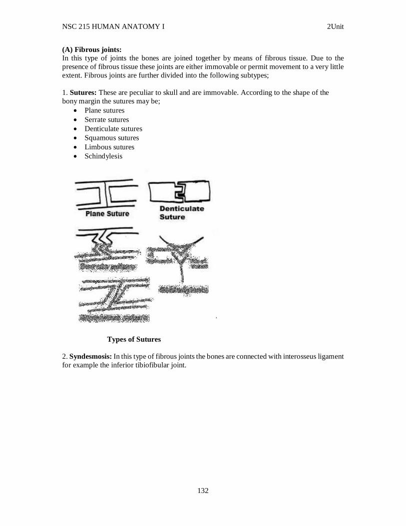
NSC 215 HUMAN ANATOMY I 2Unit
132
(A) Fibrous joints: In this type of joints the bones are joined together by means of fibrous tissue. Due to the presence of fibrous tissue these joints are either immovable or permit movement to a very little extent. Fibrous joints are further divided into the following subtypes; 1. Sutures: These are peculiar to skull and are immovable. According to the shape of the bony margin the sutures may be;
Plane sutures Serrate sutures Denticulate sutures Squamous sutures Limbous sutures Schindylesis
Types of Sutures 2. Syndesmosis: In this type of fibrous joints the bones are connected with interosseus ligament for example the inferior tibiofibular joint.
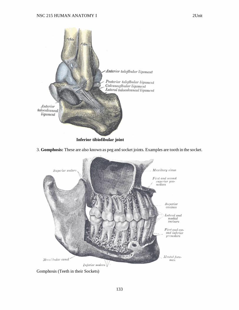
NSC 215 HUMAN ANATOMY I 2Unit
133
Inferior tibiofibular joint
3. Gomphosis: These are also known as peg and socket joints. Examples are tooth in the socket.
Gomphosis (Teeth in their Sockets)
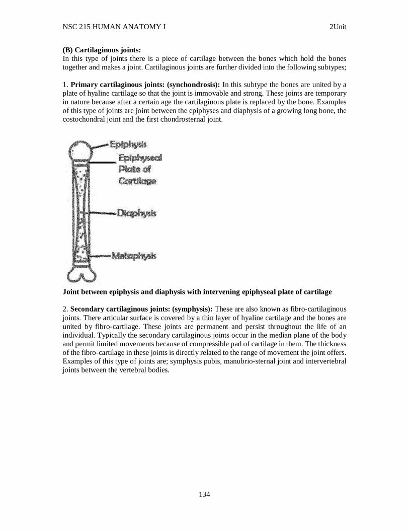
NSC 215 HUMAN ANATOMY I 2Unit
134
(B) Cartilaginous joints: In this type of joints there is a piece of cartilage between the bones which hold the bones together and makes a joint. Cartilaginous joints are further divided into the following subtypes; 1. Primary cartilaginous joints: (synchondrosis): In this subtype the bones are united by a plate of hyaline cartilage so that the joint is immovable and strong. These joints are temporary in nature because after a certain age the cartilaginous plate is replaced by the bone. Examples of this type of joints are joint between the epiphyses and diaphysis of a growing long bone, the costochondral joint and the first chondrosternal joint.
Joint between epiphysis and diaphysis with intervening epiphyseal plate of cartilage 2. Secondary cartilaginous joints: (symphysis): These are also known as fibro-cartilaginous joints. There articular surface is covered by a thin layer of hyaline cartilage and the bones are united by fibro-cartilage. These joints are permanent and persist throughout the life of an individual. Typically the secondary cartilaginous joints occur in the median plane of the body and permit limited movements because of compressible pad of cartilage in them. The thickness of the fibro-cartilage in these joints is directly related to the range of movement the joint offers. Examples of this type of joints are; symphysis pubis, manubrio-sternal joint and intervertebral joints between the vertebral bodies.
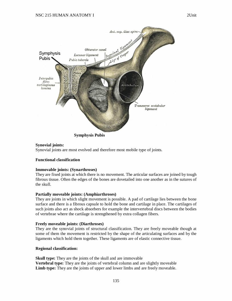
NSC 215 HUMAN ANATOMY I 2Unit
135
Symphysis Pubis
Synovial joints: Synovial joints are most evolved and therefore most mobile type of joints. Functional classification Immovable joints: (Synarthroses) They are fixed joints at which there is no movement. The articular surfaces are joined by tough fibrous tissue. Often the edges of the bones are dovetailed into one another as in the sutures of the skull. Partially moveable joints: (Amphiarthroses) They are joints in which slight movement is possible. A pad of cartilage lies between the bone surface and there is a fibrous capsule to hold the bone and cartilage in place. The cartilages of such joints also act as shock absorbers for example the intervertebral discs between the bodies of vertebrae where the cartilage is strengthened by extra collagen fibers. Freely moveable joints: (Diarthroses) They are the synovial joints of structural classification. They are freely moveable though at some of them the movement is restricted by the shape of the articulating surfaces and by the ligaments which hold them together. These ligaments are of elastic connective tissue. Regional classification: Skull type: They are the joints of the skull and are immovable Vertebral type: They are the joints of vertebral column and are slightly moveable Limb type: They are the joints of upper and lower limbs and are freely moveable.
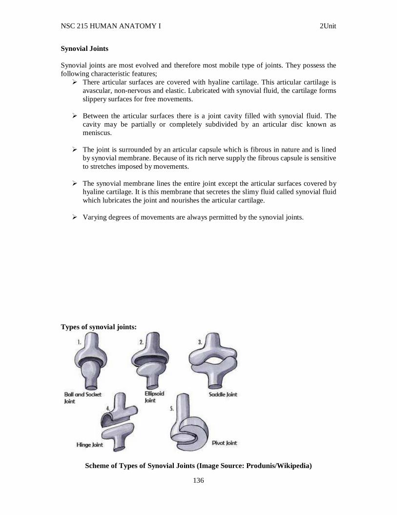
NSC 215 HUMAN ANATOMY I 2Unit
136
Synovial Joints Synovial joints are most evolved and therefore most mobile type of joints. They possess the following characteristic features;
There articular surfaces are covered with hyaline cartilage. This articular cartilage is avascular, non-nervous and elastic. Lubricated with synovial fluid, the cartilage forms slippery surfaces for free movements.
Between the articular surfaces there is a joint cavity filled with synovial fluid. The cavity may be partially or completely subdivided by an articular disc known as meniscus.
The joint is surrounded by an articular capsule which is fibrous in nature and is lined by synovial membrane. Because of its rich nerve supply the fibrous capsule is sensitive to stretches imposed by movements.
The synovial membrane lines the entire joint except the articular surfaces covered by hyaline cartilage. It is this membrane that secretes the slimy fluid called synovial fluid which lubricates the joint and nourishes the articular cartilage.
Varying degrees of movements are always permitted by the synovial joints. Types of synovial joints:
Scheme of Types of Synovial Joints (Image Source: Produnis/Wikipedia)
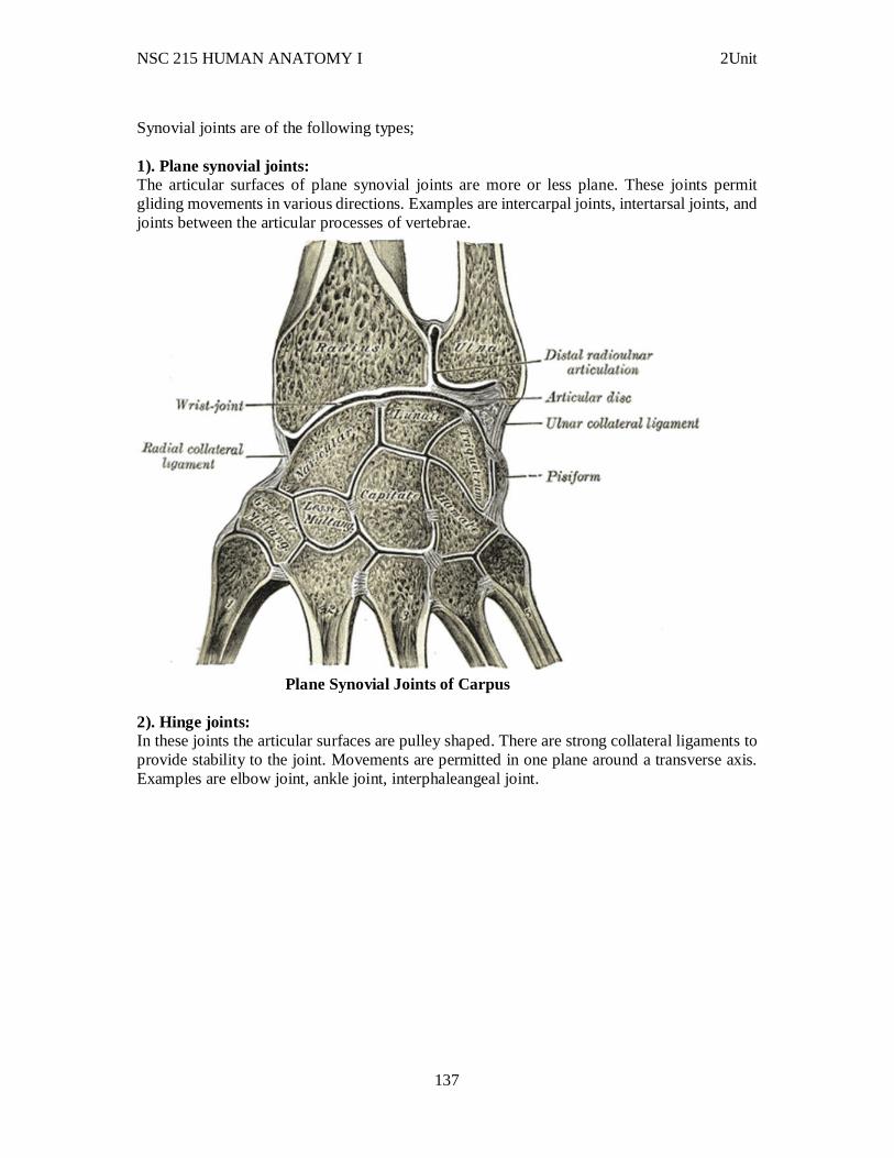
NSC 215 HUMAN ANATOMY I 2Unit
137
Synovial joints are of the following types; 1). Plane synovial joints: The articular surfaces of plane synovial joints are more or less plane. These joints permit gliding movements in various directions. Examples are intercarpal joints, intertarsal joints, and joints between the articular processes of vertebrae.
Plane Synovial Joints of Carpus
2). Hinge joints: In these joints the articular surfaces are pulley shaped. There are strong collateral ligaments to provide stability to the joint. Movements are permitted in one plane around a transverse axis. Examples are elbow joint, ankle joint, interphaleangeal joint.
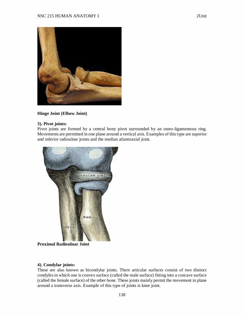
NSC 215 HUMAN ANATOMY I 2Unit
138
Hinge Joint (Elbow Joint) 3). Pivot joints: Pivot joints are formed by a central bony pivot surrounded by an osteo-ligamentous ring. Movements are permitted in one plane around a vertical axis. Examples of this type are superior and inferior radioulnar joints and the median atlantoaxial joint.
Proximal Radioulnar Joint 4). Condylar joints: These are also known as bicondylar joints. There articular surfaces consist of two distinct condyles in which one is convex surface (called the male surface) fitting into a concave surface (called the female surface) of the other bone. These joints mainly permit the movement in plane around a transverse axis. Example of this type of joints is knee joint.
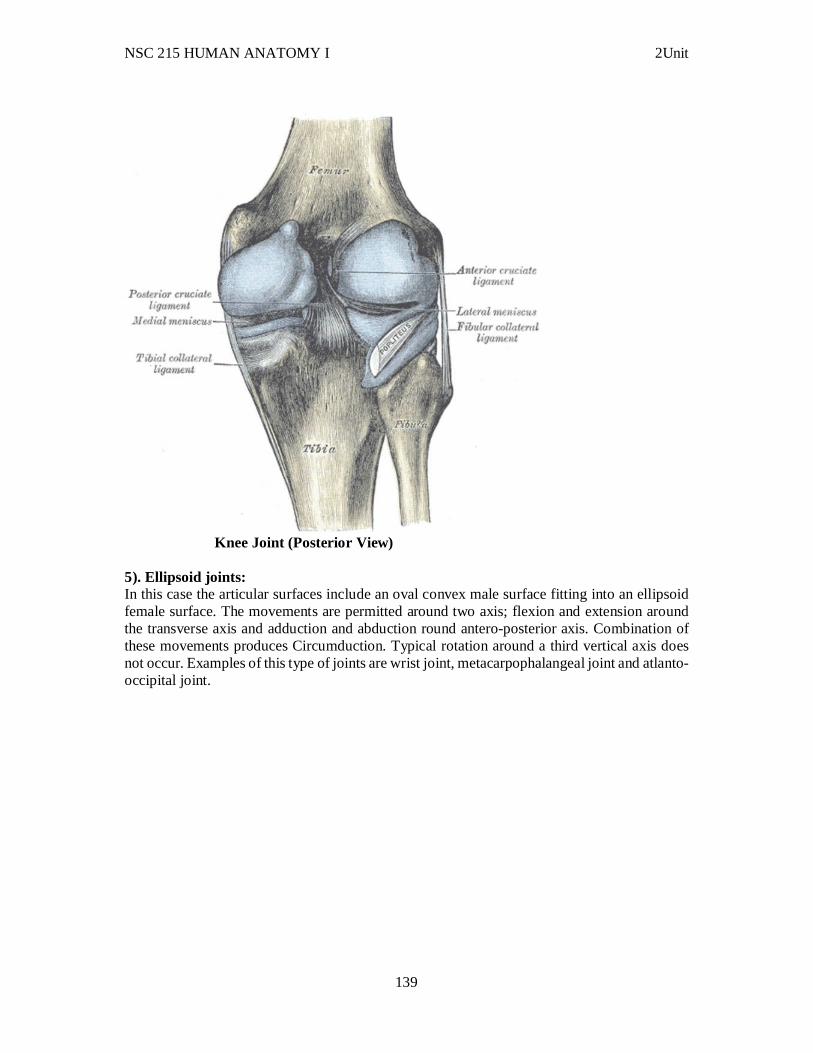
NSC 215 HUMAN ANATOMY I 2Unit
139
Knee Joint (Posterior View)
5). Ellipsoid joints: In this case the articular surfaces include an oval convex male surface fitting into an ellipsoid female surface. The movements are permitted around two axis; flexion and extension around the transverse axis and adduction and abduction round antero-posterior axis. Combination of these movements produces Circumduction. Typical rotation around a third vertical axis does not occur. Examples of this type of joints are wrist joint, metacarpophalangeal joint and atlanto-occipital joint.
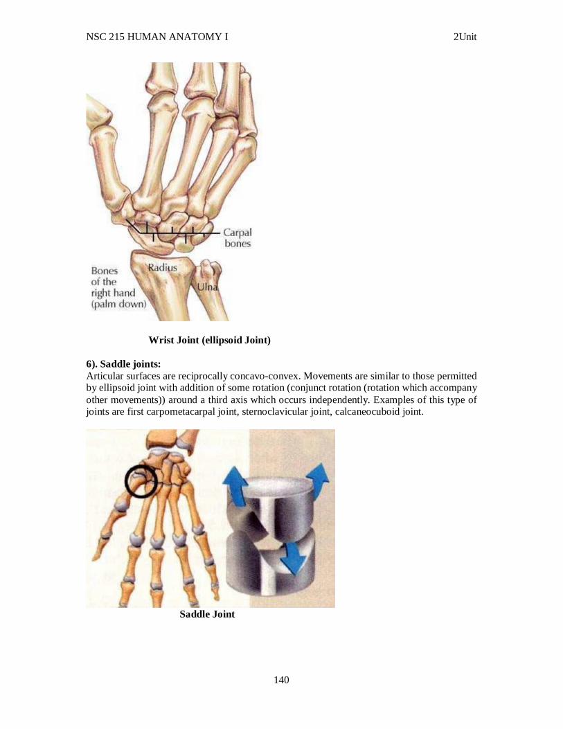
NSC 215 HUMAN ANATOMY I 2Unit
140
Wrist Joint (ellipsoid Joint) 6). Saddle joints: Articular surfaces are reciprocally concavo-convex. Movements are similar to those permitted by ellipsoid joint with addition of some rotation (conjunct rotation (rotation which accompany other movements)) around a third axis which occurs independently. Examples of this type of joints are first carpometacarpal joint, sternoclavicular joint, calcaneocuboid joint.
Saddle Joint
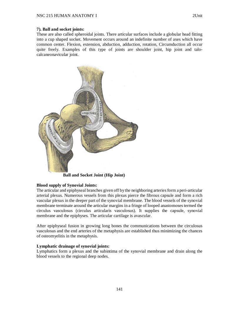
NSC 215 HUMAN ANATOMY I 2Unit
141
7). Ball and socket joints: These are also called spheroidal joints. There articular surfaces include a globular head fitting into a cup shaped socket. Movement occurs around an indefinite number of axes which have common center. Flexion, extension, abduction, adduction, rotation, Circumduction all occur quite freely. Examples of this type of joints are shoulder joint, hip joint and talo-calcaneonavicular joint.
Ball and Socket Joint (Hip Joint)
Blood supply of Synovial Joints: The articular and epiphyseal branches given off by the neighboring arteries form a peri-articular arterial plexus. Numerous vessels from this plexus pierce the fibrous capsule and form a rich vascular plexus in the deeper part of the synovial membrane. The blood vessels of the synovial membrane terminate around the articular margins in a fringe of looped anastomoses termed the circulus vasculosus (circulus articularis vasculosus). It supplies the capsule, synovial membrane and the epiphyses. The articular cartilage is avascular. After epiphyseal fusion in growing long bones the communications between the circulosus vasculosus and the end arteries of the metaphysis are established thus minimizing the chances of osteomyelitis in the metaphysis. Lymphatic drainage of synovial joints: Lymphatics form a plexus and the subintima of the synovial membrane and drain along the blood vessels to the regional deep nodes.
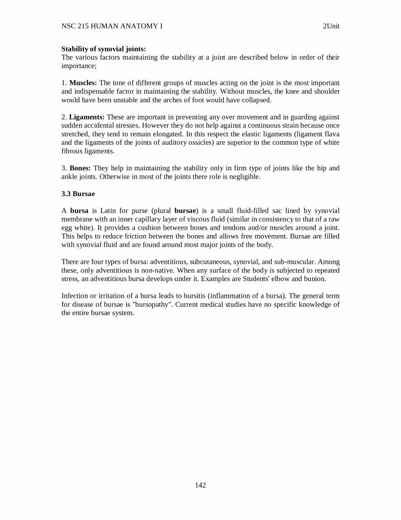
NSC 215 HUMAN ANATOMY I 2Unit
142
Stability of synovial joints: The various factors maintaining the stability at a joint are described below in order of their importance; 1. Muscles: The tone of different groups of muscles acting on the joint is the most important and indispensable factor in maintaining the stability. Without muscles, the knee and shoulder would have been unstable and the arches of foot would have collapsed. 2. Ligaments: These are important in preventing any over movement and in guarding against sudden accidental stresses. However they do not help against a continuous strain because once stretched, they tend to remain elongated. In this respect the elastic ligaments (ligament flava and the ligaments of the joints of auditory ossicles) are superior to the common type of white fibrous ligaments. 3. Bones: They help in maintaining the stability only in firm type of joints like the hip and ankle joints. Otherwise in most of the joints there role is negligible. 3.3 Bursae A bursa is Latin for purse (plural bursae) is a small fluid-filled sac lined by synovial membrane with an inner capillary layer of viscous fluid (similar in consistency to that of a raw egg white). It provides a cushion between bones and tendons and/or muscles around a joint. This helps to reduce friction between the bones and allows free movement. Bursae are filled with synovial fluid and are found around most major joints of the body. There are four types of bursa: adventitious, subcutaneous, synovial, and sub-muscular. Among these, only adventitious is non-native. When any surface of the body is subjected to repeated stress, an adventitious bursa develops under it. Examples are Students' elbow and bunion. Infection or irritation of a bursa leads to bursitis (inflammation of a bursa). The general term for disease of bursae is "bursopathy". Current medical studies have no specific knowledge of the entire bursae system.
NSC 215 HUMAN ANATOMY I 2Unit
143
A bursa is a small fluid-filled sac made of white fibrous tissue and lined with synovial membrane. Bursa may also be formed by a synovial membrane that extends outside of the joint capsule. 1 It provides a cushion between bones and tendons and/or muscles around a joint; bursa is filled with synovial fluid and is found around almost every major joint of the body. 3.4 Classification of bursae There are four types of bursa: adventitious, subcutaneous, synovial, and sub-muscular. Among these, only adventitious is non-native. When any surface of the body is subjected to repeated stress, an adventitious bursa develops under it. Examples are Students' elbow and bunion. Infection or irritation of a bursa leads to bursitis (inflammation of a bursa). The general term for disease of bursae is "bursopathy". Current medical studies have no specific knowledge of the entire bursae system. 4.5 Clinical correlates Dislocation of joint: This is a condition in which the articular surfaces of the joint are abnormally displaced so that one surface loses its contact completely with the other. If a partial contact is still retained, it is better called subluxation rather than dislocation. Dislocation is commonly caused by trauma and is characterized by pain, deformity and loss of function. X-ray is used for confirmation. Sprain: It is the severe pain in the joint caused by ligamentous tear, but without any associated dislocation or fracture. The tear leads to effusion into the ligament and joint causing great pain. Arthritis: It is the inflammation of one or more joints. It can be caused by a variety of diseases but the common types of arthritis are rheumatic, rheumatoid, osteoarthritis and tuberculoses. The involved joint is commonly swollen and its movements are restricted and painful.
NSC 215 HUMAN ANATOMY I 2Unit
144
Osteoarthritis: It represents the ageing process. In old age the articular cartilage shows degenerative changes in the center (fibrillation of cartilage) and proliferative changes around the edges. Due to this lips are formed around the edges of joints. Stiffness of joints related to weather: The viscosity of synovial fluid increases with fall in temperature. This accounts for stiffness of the joints in cold weather. Mobility of the joint itself is an important factor in promoting lubrication. Thus the stiffness of the joints experienced in the morning gradually passes off as the movements are resumed. Neuropathic joint: It is the result of complete denervation of the joint so that all reflexes are eliminated and the joint is left unprotected and liable to mechanical damage. A neuropathic joint shows painless swelling, excessive mobility and bony destruction. It is commonly caused by leprosy, tabes dorsalis and syringomyelia. Arthropathy: A joint disorder is termed an arthropathy, and when involving inflammation of one or more joints the disorder is called an arthritis. Most joint disorders involve arthritis, but joint damage by external physical trauma is typically not termed arthritis. Arthropathies are called polyarticular when involving many joints and monoarticular when involving only one single joint. Arthritis is the leading cause of disability in people over the age of 55. There are many different forms of arthritis, each of which has a different cause. The most common form of arthritis, osteoarthritis (also known as degenerative joint disease) occurs following trauma to the joint, following an infection of the joint or simply as a result of aging. Furthermore, there is emerging evidence that abnormal anatomy may contribute to early development of osteoarthritis. Other forms of arthritis are rheumatoid arthritis and psoriatic arthritis, which are autoimmune diseases in which the body is attacking itself. Septic arthritis is caused by joint infection. Gouty arthritis is caused by deposition of uric acid crystals in the joint that results in subsequent inflammation. Additionally, there is a less common form of gout that is caused by the formation of rhomboidal shaped crystals of calcium pyrophosphate. This form of gout is known as pseudo-gout. Diseases and disorders Because many other body systems, including the vascular, nervous, and integumentary systems, are interrelated, disorders of one of these systems may also affect the musculoskeletal system and complicate the diagnosis of the disorder's origin. Diseases of the musculoskeletal system mostly encompass functional disorders or motion discrepancies; the level of impairment depends specifically on the problem and its severity. Articular (of or pertaining to the joints) disorders are the most common. However, also among the diagnoses are: primary muscular diseases, neurologic (related to the medical science that deals with the nervous system and disorders affecting it) deficits, toxins, endocrine abnormalities, metabolic disorders, infectious diseases, blood and vascular disorders, and nutritional imbalances. Disorders of muscles from another body system can bring about irregularities such as: impairment of ocular motion and control, respiratory dysfunction, and bladder malfunction. Complete paralysis,
NSC 215 HUMAN ANATOMY I 2Unit
145
paresis, or ataxia may be caused by primary muscular dysfunctions of infectious or toxic origin; however, the primary disorder is usually related to the nervous system, with the muscular system acting as the effector organ, an organ capable of responding to a stimulus, especially a nerve impulse. One understated disorder that begins during pregnancy is Pelvic girdle pain, it is complex and multi-factorial and likely to be also represented by a series of sub-groups driven by pain varying from peripheral or central nervous system, altered laxity/stiffness of muscles, laxity to injury of tendinous/ligamentous structures to ‘mal-adaptive’ body mechanics. 4.0 Summary In this unit, we have learnt that: i. A joint, or articulation, is a place where two bones come together. ii. Joints are named according to the bones or parts of bones involved iii. Bones are classified according to function or type of connective tissue that binds them together and whether fluid is present between the bones. iv. Fibrous joints are those in which bones are connected by fibrous tissue with no joint cavity. They are capable of little or no movement. Sutures have interlocking finger-like processes held together by dense fibrous connective tissue. They occur between most skull bones. v. Syndesmoses are joints consisting of fibrous ligaments. vi. Synovial joints are capable of considerable movement. vii. Bursae are extensions of synovial joints that protect skin, tendons, or bone from structures that could rub against them. 6.0 Tutor Marked Assignments 6.1 At the gross anatomy laboratory, identify the bones and joints of the body and report your findings 6.2 Self-assessment questions 1. Given these types of joints:
1. gomphosis 2. suture 3. symphysis 4. synchondrosis 5. syndesmosis
Which types are classified as fibrous joints? a. 1,2,3 b. 1,2,5 c. 2,3,5 d. 3,4,5 e. 1,2,3,4,5
2. In which of these joints are periodontal ligaments found? a. sutures b. syndesmoses c. symphyses d. synovial e. gomphoses
4. The intervertebral disks are an example of
NSC 215 HUMAN ANATOMY I 2Unit
146
a. sutures. b. syndesmoses. c. symphyses.. d. synovial joints. e. gomphoses
REFERENCES/ FURTHER READING Hall, John E. (2011) Guyton and Hall textbook of medical physiology 12th ed. Katherine M. A. Rogers and William N. Scott (2011) Nurses! Test yourself in anatomy and physiology Kathryn A. Booth, Terri. D. Wyman (2008) Anatomy, physiology, and pathophysiology for allied health Kent M. Van De Graff, R.Ward Rhees, Sidney Palmer (2010) Schaum’s outline of human anatomy and physiology 3rd edition Philip Tate (2012) Seeley’s principles of anatomy & physiology 2nd edition.


