Embed Size (px)
Citation preview

Novel Genotypes of H9N2 Influenza A Viruses Isolatedfrom Poultry in Pakistan Containing NS Genes Similar toHighly Pathogenic H7N3 and H5N1 VirusesMunir Iqbal1*, Tahir Yaqub2, Kolli Reddy1, John W. McCauley3
1 Division of Microbiology, Institute for Animal Health, Compton Laboratory, Compton, Newbury, Berkshire, United Kingdom, 2 University Diagnostic Laboratory (UDL),
University of Veterinary and Animal Sciences, Lahore, Pakistan, 3 Division of Virology, MRC National Institute for Medical Research, Mill Hill, London, United Kingdom
Abstract
The impact of avian influenza caused by H9N2 viruses in Pakistan is now significantly more severe than in previous years.Since all gene segments contribute towards the virulence of avian influenza virus, it was imperative to investigate themolecular features and genetic relationships of H9N2 viruses prevalent in this region. Analysis of the gene sequences of alleight RNA segments from 12 viruses isolated between 2005 and 2008 was undertaken. The hemagglutinin (HA) sequencesof all isolates were closely related to H9N2 viruses isolated from Iran between 2004 and 2007 and contained leucine insteadof glutamine at position 226 in the receptor binding pocket, a recognised marker for the recognition of sialic acids linkeda2–6 to galactose. The neuraminidase (NA) of two isolates contained a unique five residue deletion in the stalk (fromresidues 80 to 84), a possible indication of greater adaptation of these viruses to the chicken host. The HA, NA,nucleoprotein (NP), and matrix (M) genes showed close identity with H9N2 viruses isolated during 1999 in Pakistan andclustered in the A/Quail/Hong Kong/G1/97 virus lineage. In contrast, the polymerase genes clustered with H9N2 virusesfrom India, Iran and Dubai. The NS gene segment showed greater genetic diversity and shared a high level of similarity withNS genes from either H5 or H7 subtypes rather than with established H9N2 Eurasian lineages. These results indicate thatduring recent years the H9N2 viruses have undergone extensive genetic reassortment which has led to the generation ofH9N2 viruses of novel genotypes in the Indian sub-continent. The novel genotypes of H9N2 viruses may play a role in theincreased problems observed by H9N2 to poultry and reinforce the continued need to monitor H9N2 infections for theirzoonotic potential.
Citation: Iqbal M, Yaqub T, Reddy K, McCauley JW (2009) Novel Genotypes of H9N2 Influenza A Viruses Isolated from Poultry in Pakistan Containing NS GenesSimilar to Highly Pathogenic H7N3 and H5N1 Viruses. PLoS ONE 4(6): e5788. doi:10.1371/journal.pone.0005788
Editor: Adolfo Garcia-Sastre, Mount Sinai School of Medicine, United States of America
Received March 2, 2009; Accepted April 28, 2009; Published June 11, 2009
Copyright: � 2009 Iqbal et al. This is an open-access article distributed under the terms of the Creative Commons Attribution License, which permitsunrestricted use, distribution, and reproduction in any medium, provided the original author and source are credited.
Funding: Partial funding for this work was provided by the Higher Education Commission of Pakistan to Dr. Tahir Yaqub as a visiting postdoctoral fellow at theInstitute for Animal Health, Compton laboratory UK. The funders had no role in study design, data collection and analysis, decision to publish, or preparation ofthe manuscript.
Competing Interests: The authors have declared that no competing interests exist.
* E-mail: [email protected]
Introduction
Avian influenza viruses naturally circulate in wild aquatic birds
as a diverse population having 16 HA and 9 NA antigenic
subtypes [1,2]. When these wild bird-origin viruses are transmitted
to new hosts, including domestic gallinaceous poultry, horses,
swine and humans, they can undergo adaptive changes leading to
the establishment of infection with increased transmissibility and
pathogenicity; adaptation to domestic poultry species is seen most
frequently [3,4]. To date, mainly viruses belonging to H5, H7 and
H9 subtypes have gained sufficient adaptive molecular changes to
become established in domestic poultry and to cause mild to severe
disease [5,6]. These viruses also pose a threat of zoonotic infection
[7,8,9,10]. In contrast to H5 and H7 viruses, avian H9 subtypes
exist only as low pathogenicity avian influenza (LPAI) viruses. The
H9N2 subtype has become prevalent in domestic poultry in many
countries in Asia and the Middle East since the late 1990’s
[5,11,12,13,14]. Despite being LPAI viruses, these viruses have
gained the ability to cause severe respiratory distress accompanied
by high morbidity and mortality and a marked reduction in egg
production [15,16]. The frequent heavy losses incurred with
H9N2 infection have raised serious concerns for the poultry
industry in many countries.
In Pakistan the first H9N2 outbreak in poultry was reported in
1998 [13]. These viruses showed a close relationship with H9N2
viruses circulating in Hong Kong in 1997, and were phylogenet-
ically grouped together within the G1-lineage (Qa/HK/G1/97,
HK/1073/99) of H9N2 viruses [17]. Since 1999 the poultry
industry in Pakistan has also experienced sporadic infection with
H7N3 and H5N1 highly pathogenic avian influenza (HPAI) viruses
[18] including the cross-species transmission of H5N1 viruses to
humans in late 2007, thought to have led to a family cluster of
human cases [19]. In response to the circulation of these viruses in
poultry an extensive vaccination programme against H5, H7 and
H9 subtype viruses is practised to reduce their impact [20]. The
extensive co-circulation of H9N2 viruses with other avian influenza
viruses, including highly pathogenic H5N1 and H7N3 subtypes,
coupled with extensive vaccination, is likely to generate appropriate
conditions for the generation of novel variant and reassortant
viruses, possibly with increased epizootic and zoonotic potential.
To analyse the genetic nature of H9N2 viruses in the enzootic
region of Pakistan, we sequenced the complete coding regions of
PLoS ONE | www.plosone.org 1 June 2009 | Volume 4 | Issue 6 | e5788

all eight segments of twelve H9N2 viruses isolated between May
2005 and March 2008 from poultry flocks in the Punjab and the
North West Frontier Province (NWFP) of Pakistan. The results
showed that these H9N2 viruses had changed considerably
compared with previous H9N2 viruses. The eight gene segments
of these viruses no longer clustered in a single established Eurasian
H9N2 virus sublineage but there was evidence for complex
reassortment of genes from viruses belonging to H9N2 (G1-
lineage) HPAI H5N1 (Z-genotype), and HPAI H7N3 viruses.
Therefore, these viruses represent novel genotypes of H9N2
viruses, the potential consequences of which should not be
overlooked.
Materials and Methods
Virus isolation and propagationThe twelve viruses under study (UDL viruses) were isolated at
the University Diagnostic Laboratory (UDL) Lahore, from farm
outbreaks in the Punjab and the NWFP of Pakistan (Table S1).
Viruses were propagated in 10-day old embryonated hens’ eggs
and initial subtype identification was performed using standard
hemagglutination-inhibition (HI) assays and neuraminidase inhi-
bition assays [21] using a panel of reference antisera and reference
antigens obtained from the OIE Avian Influenza Reference
Laboratory (VLA Weybridge-UK).
Intravenous pathogenicity testingIntravenous pathogenicity testing was carried out using
standard methods [22] with groups of 10, 6-week old chicks
infected with virus diluted in physiological saline. Infected birds
were examined for disease signs daily and the intravenous
pathogenicity index (IVPI) was recorded.
Nucleotide sequence analysisViral RNA was extracted from allantoic fluid using the High
Pure RNA Extraction Kit (Roche Diagnostics) and cDNA was
prepared using an influenza universal oligonucleotide primer,
59AGCAAAAGCAGG-39 with the VersoTM cDNA Kit (Thermo
Scientific). PCR amplification was performed using Pfu Ultra II
fusion HS DNA polymerase (Stratagene) and specific primers for
the eight gene segments. For HA, both specific and universal
primers [23] were used, with slight modification, to amplify HA
sequences of any other subtype (H1–H16) that may be present in
the sample (primer sequences are available on request). In most
cases, PCR products were gel purified using a gel extraction kit
(QIA quick, Qiagen) and full-length gene segments were cloned
into pCR-Blunt (Zero Blunt PCR Cloning Kit, Invitrogen). The
exceptions were that the PB2, PB1 and PA genes were amplified in
two overlapping amplicons. A minimum of five independent
clones from each gene segment for each virus were sequenced
using a commercial sequencing service (GATC Biotech, Constance,
Germany). Five virus samples that contained the NS gene
homologous to H5N1 viruses and the two samples (Ck/Pak/
UDL-01/06 and Ck/Pak/UDL-03/08) that contained the NS
gene homologous to H7N3 were re-examined in greater detail.
The HA genes of these seven virus samples were subjected to in-
depth sequence analysis to exclude the possibility that the samples
were from mixed infections with H9N2 viruses and H5N1 or
H7N3 viruses. Sequence analyses of the HA genes were performed
by sequencing a minimum of 60 clones per sample that was
calculated to detect with 95% probability any variant sequences at
a level of 5% within the virus sample.
Sequence data were analysed using the Staden package
(pregap4 v1.5 and gap4 v1.0). Blast homology searches (http://
www.ebi.ac.uk/Tools/fasta33/nucleotide.html) were used to re-
trieve the top fifty homologous sequences to the sequenced gene
from public sequence databases. Multiple nucleotide and amino
acid sequence alignments for all eight gene segments were
performed using ClustalX (version 1.83). Unrooted phylogenetic
trees were generated from nucleotide sequences based on the
complete open reading frame of all eight gene segments using
minimum evolution analysis with maximum composite likelihood
and the Tamura-Nei model [24] with 1000 bootstrapping
replications in Molecular Evolutionary Genetics Analysis (MEGA,
version 4. 1 beta). The nucleotide sequences obtained in this study
have been submitted to the GenBank database and are available
under accession numbers (CY038391 to CY038486).
Results and Discussion
Phylogenetic analysisTo determine the genetic relationship of H9N2 avian influenza
viruses currently prevalent in poultry in Pakistan, we selected
twelve H9N2 viruses isolated between May 2005 and March 2008
from different districts of the Punjab and the NWFP of Pakistan,
Supplementary Table 1. These districts have a large concentration
of commercial poultry farms and a number of H5N1 and H7N3
outbreaks have also been reported in this region over the last few
years (18). The H9N2 isolates were all defined as LPAI viruses by
an intravenous pathogenicity test of diluted infectious allantoic
fluid (Table 1). Complete full length sequencing of all 8 segments
was performed on at least 5 cDNA clones for each segment and a
consensus sequence for each virus was produced; there was only
very limited polymorphism in the analysed sequences. Phyloge-
netic relationships were examined between these viruses and
representative H9N2 viruses from Asia and the Middle East, along
with the established Eurasian H9N2 lineages: namely the G1-
lineage, the Y280-lineage and the Korean-lineage represented by
prototype viruses A/Quail/Hong Kong/G1/97, A/Duck/Hong
Kong/Y280/97 and A/Chicken/Korea/38349-p96323/96 re-
spectively, and with viruses identified in the Far East as emerging
H9N2 lineages [6,25]. Phylogenetic analysis of the HA revealed
that all twelve isolates cluster together with G1-lineage viruses and
show a very close relationship (93.0 to 96.4% nucleotide identity)
with recent poultry-isolated H9N2 viruses from Iran (Figure 1 and
Table 2), which shares a border with Pakistan. A slightly higher
divergence (6.3 to 9.7%) was seen between HA genes of H9N2
viruses isolated from Pakistan in 1999 (Ck/Pakistan/2/99, Ck/
Pakistan/4/99, and Ck/Pakistan/5/99); another set of viruses
from Middle East isolated between 2000 and 2003 [14] also cluster
with this group (Figure 1). The phylogenetic relationship of NA
and M genes of the Pakistan viruses also fell within the G1-lineage
and showed close identity (93.0–97.0% in the NA gene) with Ck/
Pakistan/2/99, Ck/Pakistan/4/99, and Ck/Pakistan/5/99
(Figure 2, 3 and Table 2) and for the M gene 96.5 to 97.6%
nucleotide identity with A/Ck/Pakistan/2/99. Like the HA, NA
and M genes, the NP genes also grouped together with G1-lineage
viruses (Figure 4): the closest nucleotide sequence identity (95.8–
97.3%, Table 2) was found with viruses isolated from Pakistan
during 1999 and A/Parakeet/Chiba/1/97, an introduction
thought to be imported from Pakistan [26]. There was relatively
little nucleotide divergence (2.4–7.0%) of NA, M and NP gene
compared with H9N2 viruses isolated from Pakistan in 1999 (Ck/
Pakistan/2/99, Ck/Pakistan/4/99 and Ck/Pakistan/5/99),
which were of the G1-lineage but within this lineage some sub-
clustering is evident (Figure 1, 2, 3 and 4). The phylogenetic
analyses of each of the three polymerase complex genes (PB2, PB1
and PA), in contrast to the HA, NA, M and the NP genes, did not
Novel Genotypes of H9N2 Virus
PLoS ONE | www.plosone.org 2 June 2009 | Volume 4 | Issue 6 | e5788
Ta
ble
1.
Co
mp
aris
on
of
IVP
Ian
dam
ino
acid
resi
du
es
inse
vera
lke
ysi
tes
of
HA
,N
Aan
dN
S1p
rote
ins.
Vir
us
stra
inIV
PI
HA
NA
NS
1
Re
cep
tor
bin
din
gsi
teC
lea
va
ge
Sit
eG
lyco
syla
tio
nsi
tea
tp
osi
tio
nP
rese
nce
of
NA
sta
lkd
ele
tio
nH
Bsi
teD
ele
tio
no
fa
aT
ota
ln
oo
fa
aP
Lm
oti
f
19
1(1
83
)a1
98
(19
0)a
23
4(2
26
)a2
35
(22
7)a
33
5–
33
82
18
(20
0)a
38
–3
94
6–
50
62
–6
43
72
40
24
03
80
–8
4
A/C
hic
ken
/Pak
ista
n/2
/99
ND
HA
RSS
RY
es
No
No
No
TN
WN
o2
30
EPEV
A/Q
uai
l/H
on
gK
on
g/G
1/9
7N
DH
EL
QR
SSR
Ye
sY
es
No
No
SI
RN
o2
30
EPEV
A/H
on
gK
on
g/1
07
3/9
9N
DH
EL
QR
SSR
Ye
sY
es
No
No
SN
WN
oEP
EV
A/D
uck
/Ho
ng
Ko
ng
/Y2
80
/97
ND
NT
LQ
RSS
RY
es
No
No
Ye
sS
NW
No
23
0EP
EV
A/C
hic
ken
/Ko
rea/
99
02
9/9
9N
DH
EQ
QA
SGR
Ye
sN
oN
oN
oS
NW
No
23
0ES
EV
A/C
hic
ken
/Pak
ista
n/U
DL-
01
/05
0.0
0H
AL
IR
SSR
No
No
No
No
AN
WY
es
22
5ES
KV
A/C
hic
ken
/Pak
ista
n/U
DL-
02
/05
0.0
0H
AL
IR
SSR
No
No
Ye
sN
oA
NR
Ye
s2
25
ESK
V
A/C
hic
ken
/Pak
ista
n/U
DL-
03
/05
0.0
0H
AL
IR
SSR
No
No
Ye
sN
oA
NR
Ye
s2
25
ESK
V
A/C
hic
ken
/Pak
ista
n/U
DL-
01
/06
0.0
2H
AL
IR
SSR
No
No
No
No
AN
RN
o2
17
LPP
K
A/C
hic
ken
/Pak
ista
n/U
DL-
02
/06
0.0
0H
AL
IR
SSR
No
No
No
No
AN
RN
o2
30
KSE
I
A/C
hic
ken
/Pak
ista
n/U
DL-
04
/06
0.0
0H
AL
IR
SSR
No
No
No
No
AS
RY
es
22
5ES
KV
A/C
hic
ken
/Pak
ista
n/U
DL-
01
/07
0.0
0H
AL
IR
SSR
No
No
No
No
AN
RN
o2
30
KSE
I
A/C
hic
ken
/Pak
ista
n/U
DL-
03
/07
0.0
1H
AL
IK
SSR
No
No
No
No
AN
RY
es
22
5ES
KV
A/C
hic
ken
/Pak
ista
n/U
DL-
04
/07
0.0
3H
AL
IR
SSR
No
No
No
No
TN
RN
o2
30
KSE
I
A/C
hic
ken
/Pak
ista
n/U
DL-
01
/08
0.0
0H
AL
IK
SSR
No
No
No
No
AN
RN
o2
30
KSE
I
A/C
hic
ken
/Pak
ista
n/U
DL-
02
/08
0.0
1H
AL
IK
SSR
No
No
No
No
AN
RN
o2
30
KSE
I
A/C
hic
ken
/Pak
ista
n/U
DL-
03
/08
0.0
1H
AL
IK
SSR
No
No
No
No
AN
RN
o2
30
KSE
I
aN
um
be
rin
gac
cord
ing
toH
3in
par
en
the
ses,
IVP
I(i
ntr
ave
no
us
pat
ho
ge
nic
ity
ind
ex)
,H
B(h
em
adso
rbin
gsi
te),
aa(a
min
oac
ids)
PL
(PL
mo
tif
isre
ferr
ed
asP
DZ
-bin
din
gm
oti
fat
the
C-t
erm
inal
en
do
fth
eN
S1p
rote
in).
ND
no
td
ete
rmin
ed
inth
isst
ud
y.d
oi:1
0.1
37
1/j
ou
rnal
.po
ne
.00
05
78
8.t
00
1
Novel Genotypes of H9N2 Virus
PLoS ONE | www.plosone.org 3 June 2009 | Volume 4 | Issue 6 | e5788
Novel Genotypes of H9N2 Virus
PLoS ONE | www.plosone.org 4 June 2009 | Volume 4 | Issue 6 | e5788

branch with exemplars of the G1-lineage or any of the other
established Eurasian lineages (Figure 5, 6 and 7). All three
polymerase complex genes were most closely related to those of
H9N2 viruses isolated from the Persian Gulf and India between
2000 and 2004 [14,27]. Nucleotide identities were from 94.1 to
96.4% for the PA gene, 91.2–94.4% for the PB1 gene and 94.5–
96.6% identity in PB2; these similarities may indicate a separate
Indian sub-continental lineage of H9N2 virus [6,25]. This notion
is supported by conclusions based on analyses of H9N2 viruses
from the Middle East [14] where it was observed that reassortment
of the PB2 gene had taken place and it was postulated that a UAE
lineage of H9N2 viruses may have emerged.
In contrast to the other segments phylogenetic analysis of the
NS gene segment of UDL viruses split the 12 viruses into three
distinct groups (Figure 8). Six isolates of one group cluster together
with 97.8% to 100% identity to each other and showed closest
nucleotide identity (95.9–96.6%) with Ck/Pak/NARC-100/04, an
H7N3 virus (Figure 8 and Table 3). The second group
(represented by a single virus, A/Ck/Pak/UDL-01/06) showed
a strikingly close relationship with 99.8 to 100% nucleotide
identity within the NS1 coding region to H7N3 viruses isolated
from Pakistan between 1995 and 2005. Members of this group
contained a 13 amino acid deletion at the C-terminus of NS1 and
this group was quite distinct from the Ck/Pak/NARC-100/04,
H7N3 - group of viruses. The third group of five H9N2 viruses
showed highest nucleotide similarity (99.3–99.7%) with Ck/
Rawalakot/NARC2441A/06 and Ck/Sihala/NARC3033.4/
2006, Ck/Afghanistan/1207/06 and Ck/Afghanistan/1573-47/
06 (H5N1) viruses (Table 3) and clustered together with Clade 2.2
highly pathogenic H5N1 viruses of the Z-genotype (Figure 8), the
closest related H5N1 viruses being isolated in Pakistan, India, Iran
and Russia during 2006 [28,29]. These five viruses were able to be
differentiated into two groups, but both groups were very similar
to other viruses isolated in the region.
The pattern of nucleotide similarity indicates that the 12 H9N2
viruses retained 4 genes (HA, M, NA and NP) similar to the H9N2
G1-lineage, 3 genes (PB1, PB2, PA) from a lineage of viruses from
the Indian sub-continent and the Middle-East, and NS genes from
one of two H7N3 lineages or from H5N1 genotype Z viruses that
were previously present in Pakistan and neighbouring countries
(Figure 9). A set of viruses from the Middle East studied by Aamir
et al. [14] also showed evidence of reassortment of the NS gene
segment in the proposed UAE lineage and it is possible that the
UAE viruses and the six viruses from Pakistan that are closest to
the Ck/Pak/NARC-100/04 H7N3 virus may share a common
ancestor. Although the HA and NA gene sequences have drifted
over the years (Figure S1), the H9N2 lineage-defining residues
[17,25] have been conserved: eight amino acids in HA (L17, T96,
V179, L209, G262, S290, T304 in HA1 and V91 in HA2); seven amino
acids ( L10, T43, S77, S153, T212, V307, G346) in NA. Similarly, M
and NP genes also retained conserved G1-lineage defining residues
(A157) in M1, one (L10) in M2, and four amino acids (Q52, V317,
A374, K430) in NP.
Molecular characteristicsIn order to identify molecular markers that may correlate with
the pathogenic properties of these H9N2 UDL isolates analysis
was performed to compare deduced amino acid sequences of the
envelope glycoproteins and the internal proteins of the Pakistan
virus isolates with representative strains of established H9N2 virus
Eurasian lineages.
HemagglutininChanges in the HA glycoprotein are central to the switch from
LPAI to HPAI, but additional amino acid substitutions in the HA
molecule have been associated with inter-species adaptation [30].
The key molecular determinants of pathogenicity and virulence in
the HA molecule are the HA1/HA2 connecting peptide sequence,
specific amino acids residues at the receptor binding site, and the
presence or absence of glycosylation sites near the receptor
binding site [31]. As expected from the pathogenicity tests,
nucleotide sequence analysis corresponding to the HA1/HA2
junction of these viruses revealed no poly-basic cleavage motif.
Eight out of twelve isolates retained an R-S-S-R motif at the
cleavage site which is the signature of H9N2 LPAI viruses adapted
to the chicken host, as seen in recent years in H9N2 virus isolates
from Asia and the Middle East [14,25,26,27,32,33]. It was noted
that four UDL isolates possess lysine instead of arginine at the 24
position of the cleavage site (Table 1). Lysine at this position has
been observed only infrequently in H9N2 viruses [34] and the
significance, if any, of this alteration from one basic amino acid to
another is not known.
The receptor binding site motif of HA is critical for cellular
receptor specificity and determining virus host range [35,36].
Residues at positions 183, 190, 226, 227 and 228 (H3 numbering)
are major components of the receptor-binding site of the HA
molecule. All UDL viruses showed conservation of residues H183,
A190, L 226, I227 and G228 in the receptor binding cleft. Of these,
Figure 1. Phylogenetic relationships of HA genes of H9N2 avian influenza viruses isolated from Pakistan between 2005 and 2008.The Phylogenetic tree was generated using minimum evolution analysis with maximum composite likelihood using the Tamura-Nei model [24] withMEGA (version 4.1 beta). Numbers below branches indicate bootstrap value percentages from 1000 replicates, bootstrap values .70% are shown.Analysis was based on complete open reading frames of all gene segments. The scale bar represents the distance unit between sequence pairs. Bhgs,bar headed goose, GF, guinea fowl; Ph, pheasant, Ck, chicken; Dk, duck; Qa, quail; Ty, turkey; Par, parakeet, Sw, swine; Pak, Pakistan; RP, Rawalpindi;Afg, Afghanistan; HK, Hong Kong; ST, Shantou; VN, Vietnam; Wis, Wisconsin; Cal, California; Kor, Korea; GZ, Guangzhou; Rus, Russia; UP, Uttar Pradesh.The viruses characterised in this report are indicated as red. The sequences of H9N2 and H7N3 viruses previously isolated from Pakistan are indicatedblue and pink respectively.doi:10.1371/journal.pone.0005788.g001
Table 2. Nucleotide similarities (%*) between H9N2 virusesisolated in Pakistan during 2005–2008 and other influenzaviruses.
Gene Viruses showing highest similarity Identity (%)
PB2 A/Quail/Dubai/303/00 (H9N2) 95.3–96.6
PB1 A/Chicken/Dubai/463/03 (H9N2) 93.1–94.2
PA A/Quail/Dubai/303/00 (H9N2) 95.2–96.4
HA A/Chicken/Iran/B102/05 (H9N2) 93.9–96.4
NP A/Parakeet/Chiba/1/97 (H9N2) 96.0–97.3
NA A/Chicken/Pakistan/2/99 (H9N2) 93.0–97.0
M A/Chicken/Pakistan/2/99 (H9N2) 96.5–97.6
*Identities were calculated based on complete open reading frames of eachRNA segment for all twelve virus isolates sequenced in this study.
doi:10.1371/journal.pone.0005788.t002
Novel Genotypes of H9N2 Virus
PLoS ONE | www.plosone.org 5 June 2009 | Volume 4 | Issue 6 | e5788
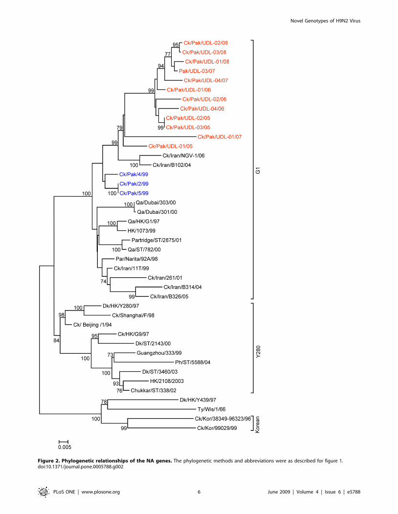
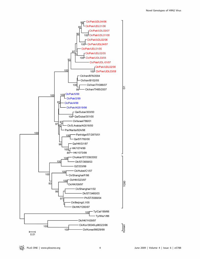
Figure 2. Phylogenetic relationships of the NA genes. The phylogenetic methods and abbreviations were as described for figure 1.doi:10.1371/journal.pone.0005788.g002
Novel Genotypes of H9N2 Virus
PLoS ONE | www.plosone.org 6 June 2009 | Volume 4 | Issue 6 | e5788
Figure 3. Phylogenetic relationships of the M genes. The phylogenetic methods and abbreviations were as described for figure 1.doi:10.1371/journal.pone.0005788.g003
Novel Genotypes of H9N2 Virus
PLoS ONE | www.plosone.org 7 June 2009 | Volume 4 | Issue 6 | e5788

Figure 4. Phylogenetic relationships of the NP genes. The phylogenetic methods and abbreviations were as described for figure 1.doi:10.1371/journal.pone.0005788.g004
Novel Genotypes of H9N2 Virus
PLoS ONE | www.plosone.org 8 June 2009 | Volume 4 | Issue 6 | e5788

Figure 5. Phylogenetic relationships of the PB2 genes. The phylogenetic methods and abbreviations were as described for figure 1.doi:10.1371/journal.pone.0005788.g005
Novel Genotypes of H9N2 Virus
PLoS ONE | www.plosone.org 9 June 2009 | Volume 4 | Issue 6 | e5788
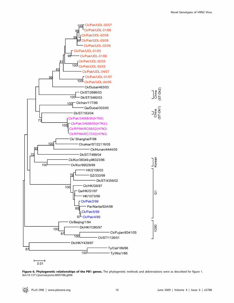
Figure 6. Phylogenetic relationships of the PB1 genes. The phylogenetic methods and abbreviations were as described for figure 1.doi:10.1371/journal.pone.0005788.g006
Novel Genotypes of H9N2 Virus
PLoS ONE | www.plosone.org 10 June 2009 | Volume 4 | Issue 6 | e5788
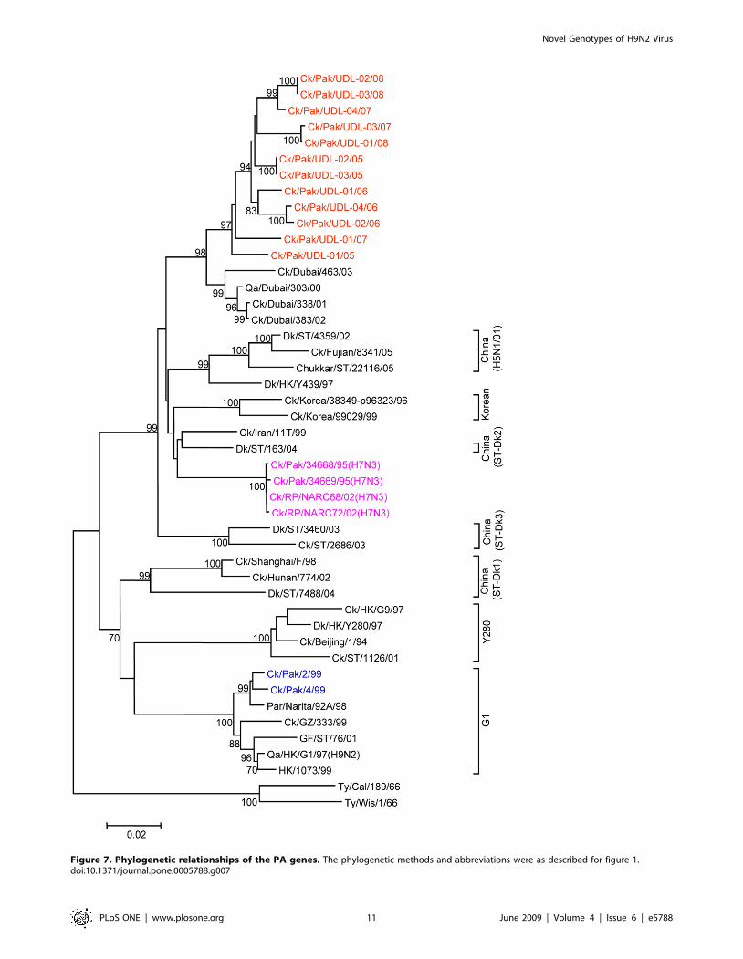
Figure 7. Phylogenetic relationships of the PA genes. The phylogenetic methods and abbreviations were as described for figure 1.doi:10.1371/journal.pone.0005788.g007
Novel Genotypes of H9N2 Virus
PLoS ONE | www.plosone.org 11 June 2009 | Volume 4 | Issue 6 | e5788

Novel Genotypes of H9N2 Virus
PLoS ONE | www.plosone.org 12 June 2009 | Volume 4 | Issue 6 | e5788

three represent substitutions (E/T190A, Q226L and Q227I) when
compared with the reference strains of the H9 lineage (Table 1).
Glutamine at position 226 dictates a preferential binding to SA
a2,3-linked to galactose found in avian hosts and is a major host
range determinant for influenza A viruses of the H2 and the H3
subtypes associated with pandemic human infections [37,38]. The
substitution, as seen in the viruses under study here, of Q226L at
the receptor binding site in the HA allows H9N2 viruses to
replicate more efficiently (with 100-fold higher peak titres) in
human cells in culture [39,40] and is associated with a preferential
receptor binding specificity for sialic acid a2,6-linked galactose
[37] and was also observed in the UAE isolates analysed by Aamir
[14]. The significance of ERA and QRI substitutions at positions
190 and 227 respectively observed in all UDL isolates on the
receptor binding specificity within this group of viruses is not
known and requires detailed structural analysis to resolve their
importance. In addition, the alteration at position 150 (138 in H3)
from Ala to Ser, which may affect the conformation of residues
lining the receptor binding site, has also been linked to changes in
receptor specificity in H1 viruses [38]. Analysis of potential
glycosylation site motifs N-X-S/T in the HA molecule of the UDL
viruses revealed seven sites at positions 29, 105, 141, 298, 305, 492
and at position 551, C-terminal to the membrane anchor (21, 94,
132, 289, and 296 in HA1 and 154 and 213 in HA2 in H3
numbering). One potential glycosylation site at position 218 (210
in H3) was lost compared with representative reference strains of
the G1-lineage, the Y280-lineage, the Korean-lineage and the
prototypic Pakistani isolate Ck/Pakistan/2/99 [41] (Table 1). Loss
of an additional glycosylation site at position 206, compared with
the prototype G1 virus A/Qa/HK/G1/97 was observed; this was
also seen in H9N2 viruses from the Middle East in 2000–2003
[14]. The loss of potential glycosylation sites may represent a
selected adaptation of H9N2 within poultry since alteration in
glycosylation pattern has been suggested to influence adaptation of
avian influenza viruses to poultry [30,31].
NeuraminidaseThe major molecular determinants that are known to influence
the functional activities of the neuraminidase (NA) glycoprotein
are the enzyme active site, the stalk length, the sialic acid binding
site also referred to as the hemadsorbing site (HB), and potential
glycosylation sites. The neuraminidase sequences of all 12 isolates
were compared with N2 NA sequences of H9N2 viruses prevalent
in Asia and the Middle East during the last fifteen years. For each
virus amino acids in the enzyme active site were conserved and
showed no evidence of substitutions associated with resistance to
the sialidase inhibitors oseltamivir and zanamivir. The data
revealed that, of the 12 viruses analysed, 2 isolates (Ck/Pak/
Table 3. Nucleotide similarities (%) of the NS genes between H9N2 viruses isolated in Pakistan during 2005–2008 and NS genes ofother influenza viruses.
H9N2 viruses isolated from Pakistan Viruses in the database showing highest similarity Identity (%)
A/Chicken/Pakistan/UDL-02/06 A/Chicken/Pakistan/NARC-100/04 (H7N3)* 95.9 to 96.7
A/Chicken/Pakistan/UDL-01/07
A/Chicken/Pakistan/UDL-04/07
A/Chicken/Pakistan/UDL-01/08
A/Chicken/Pakistan/UDL-02/08
A/Chicken/Pakistan/UDL-03/08
A/Chicken/Pakistan/UDL-01/06 A/Chicken/Pakistan/34668/95 (H7N3)* 99.5 to 100
A/Chicken/Pakistan/NARC-1/95 (H7N3)*
A/Chicken/Pakistan/NARC-143/04 (H7N3)*
A/Chicken/Pakistan/NARC-2402/05 (H7N3)*
A/Chicken/Pakistan/NARC-74/04 (H7N3)*
A/Chicken/Pakistan/NARC-BM/00 (H7N3)*
A/Chicken/Pakistan/NARC-108/04 (H7N3)*
A/Chicken/Pakistan/NARC-35/01 (H7N3)*
A/Chicken/Pakistan/UDL-01/05 A/Chicken/Afghanistan/1207/06 (H5N1) 99.5 to 99.7
A/Chicken/Pakistan/UDL-02/05 A/Chicken/Afghanistan/1573-47/06 (H5N1)
A/Chicken/Pakistan/UDL-03/05 A/Chicken/India/NIV33487/06 (H5N1)
A/Chicken/Pakistan/UDL-04/06 A/Cygnus cygnus/Iran/754/06 (H5N1)
A/Chicken/Pakistan/UDL-03/07 A/Chicken/Rawalakot/NARC2441A/06(H5N1)
A/Chicken/Sihala/NARC3303.4/2006(H5N1)
*NS1 sequences only available in public databases, therefore percentage identities were calculated based on the NS1 coding region nucleotide sequences.doi:10.1371/journal.pone.0005788.t003
Figure 8. Phylogenetic relationships of the NS genes of H9N2 avian influenza viruses isolated from poultry in Pakistan. Analysis wasbased on nucleotides 27–870 of segment eight of all viruses included in this analysis. NS gene sequences were compared with closely related H5, H7and H9 viruses (the viruses highlighted in red were sequenced in this study and H9N2, H7N3 and H5N1 viruses isolated in Pakistan previously areindicated in blue, pink and brown respectively). The phylogenetic methods and abbreviations were as described for figure 1.doi:10.1371/journal.pone.0005788.g008
Novel Genotypes of H9N2 Virus
PLoS ONE | www.plosone.org 13 June 2009 | Volume 4 | Issue 6 | e5788

UDL-02/05 and Ck/Pak/UDL-03/05) contained a unique 5
amino acid residue deletion (aa 46–50) in the stalk region, the
others having no deletion in the NA stalk, as was also observed in
H9N2 viruses from the Middle East [14]. Deletions in the stalk of
avian N2 viruses have been observed previously in H9N2 viruses:
the two prototype viruses, Qa/HK/G1/97 and HK/1073/97, of
the G1-lineage contained a two amino acid deletion in the NA
stalk region at positions 38 and 39, and Dk/HK/Y280/97 and
Ck/Shanghai/F/98, another virus belonging to the Y-280-lineage,
lacked three amino acid at positions 63–65 in the NA stalk region
(Table 1). The precise 5 amino acid deletion at position 46–50 in
the stalk region represents a unique event not observed previously
in H9N2 viruses. It is recognised that deletion within the stalk
region of the neuraminidase may be an important feature
balancing the complementary activities of the HA and NA on
adaptation of virus to poultry [30,31].
Analysis of the HB site, which is located on the surface of the
NA molecule away from the neuraminidase enzyme active site at
positions 366–373, Asn-400, Trp-403 and Lys-432 [42,43],
revealed that although these sites have been well conserved in
H9N2 viruses isolated from wild aquatic birds [44], poultry-
adapted H9N2 viruses, including isolates sequenced in this study,
contained substitutions similar to those detected in both A/Qa/
HK/G1/97 and A/Dk/HK/Y280/97. These substitutions were
Figure 9. Representation of the newly identified genotypes of H9N2 viruses in poultry in Pakistan. The possible genetic progenitors ofthese new genotypes are (A) H9N2 (LPAI) viruses represented by A/Ck/Pak/2/99, belonging to G1-lineage, (B) H9N2 (LPAI) viruses represented asIndian/Middle East lineage containing distinct polymerase complex genes, (C) H7N3 (HPAI) viruses represented by the prototype A/Ck/Pak/NARC-100/2004 virus, (D) H7N3 (HPAI) viruses represented by the prototype A/Ck/Pak/34668/95 virus containing a 13 amino acid deletion in their NS1polypeptide and (E) H5N1 (HPAI) viruses represented by A/Ck/Afghanistan/1207/2006 belonging to genotype Z. The three newly identified H9N2genotypes (F, G and H) contained PB2, PB1, and PA genes similar to an Indian/Middle East lineage of H9N2 virus, along with the NS genes derivedfrom two distinct H7N3 parental viruses and an H5N1 parental virus and the HA, NA, NP and M genes from H9N2 viruses of the G1 lineage.doi:10.1371/journal.pone.0005788.g009
Novel Genotypes of H9N2 Virus
PLoS ONE | www.plosone.org 14 June 2009 | Volume 4 | Issue 6 | e5788
seen previously in H9N2 human isolates from Hong Kong in 1999
and are typical of human pandemic H2N2 and H3N2 viruses [45].
The substitutions at residue 372 from serine to alanine or, for one
virus, threonine (Table 1) were similar to those detected in
Parakeet/Narita/92A/98, Ck/Pak/2/99 and H9N2 viruses
isolated from Iran and the United Arab Emirates [14,26]. In
addition, a substitution from tryptophan to arginine at position
403 was observed in 11 of the 12 viruses recently isolated from
Pakistan; this substitution was present in Qa/HK/G1/97 but not
in other H9N2 viruses, including those from the UAE from 2000
to 2003 [14] and, importantly, not in the human (e.g. A/Hong
Kong/1073/99) H9N2 isolates (Table 1). Within H9N2 viruses,
polymorphism is seen at this site with W, L, S or R present. The
Qa/Hong Kong/G1/97 isolate is unusual in carrying a N402I
substitution and one of the viruses characterised here (A/Ck/Pak/
UDL-04/06) has serine at this position. The biological significance
of any of these substitutions in the HB site is not known.
The comparison of conserved potential glycosylation sites in NA
glycoprotein showed that the majority of H9N2 viruses belonging
to established Eurasian lineages contain seven conserved potential
glycosylation sites at positions 61, 69, 86, 146, 200, 234 and 402
(N2 numbering). The viruses sequenced in this study contained an
additional glycosylation site at Asn 44 due to the substitution of
Pro45Ser. This additional glycosylation site had also been reported
in a number of other H9N2 viruses including Ck/Ind/2048/03,
Ck/HK/G9/97 [27]. One UDL isolate Ck/Pak/UDL-02/06
lacked one potential glycosylation site at Asn 234 due to T236I
substitution. Again, the significance of glycosylation changes at
sites 44 and 234 is unknown; however, addition or loss of potential
glycosylation may contribute towards increased virulence [46] due
to altered antigenicity or sialidase activity.
A comparison of the amino acid sequences of the H9N2
neuraminidases shows that certain amino acid substitutions have
become fixed as the viruses have evolved but many are unique
substitutions represented in individual viruses (Supplementary
Figure S1B). It appears that fewer amino acids were unique to
individual viruses in the HA than in the NA; this may reflect
different evolutionary pressures applied to the two glycoproteins.
The necessity for compensatory changes in the NA to ensure its
compatibility with the HA as the HA evolves may be one such
force.
NS1 and NS2 proteinsThe NS gene showed a high level of sequence diversity among
the twelve UDL isolates analysed. Of particular note were five
isolates (UDL-01/05, UDL-02/05, UDL-03/05, UDL-04/06,
UDL-03/07) that contained an NS gene closely related to the
highly pathogenic H5N1 viruses belonging to clade 2.2 of
genotype Z with over 99% nucleotide identity (Figure 8 and
Table 3). The NS1 portions of these viruses showed between 0 and
3 amino acid changes from H5N1 viruses isolated during 2006 to
2007 from Pakistan, Afghanistan, Iran, India, and Russia [28] and
their NS1 proteins, being from of the Z-lineage H5N1 viruses, had
a five-amino acid deletion (deleting residues 80–84) resulting in an
NS1 protein of 225 amino acids in length; the NS1 proteins also
contained the ‘‘ESKV’’ PDZ ligand (PL) C-terminal motif typical
of H5N1 viruses of the Z-genotype (Table 1). The role of the NS1
protein in infection is complex and includes countering interferon
and cytokine induction (reviewed recently in Hale et al. [47]) and
its effects on pathogenesis have become clearer but the biological
significance of the 5 amino acid deletion is not well understood. It
has been reported that viruses containing NS1 with deleted
(80TIASV84) residues show increased virulence in both mouse and
poultry infections [48]. The specific functional role of this deletion
in H9N2 viruses needs to be determined but there was no evidence
of any increased pathogenicity associated with the deletion in our
IVPI tests.
RNA segment 8 of one virus (UDL-01/06) was very closely
related to that of H7N3 viruses from Pakistan isolated over an
eleven year period. This single virus isolate shared a 13 amino acid
C-terminal truncation; similar truncations have been reported
previously in H7 and H9 subtype viruses isolated from poultry
[49,50], suggesting a natural virus adaptation, but the significance
of this truncation and the resulting C-terminal LPPK motif to the
virus life cycle is also not understood.
The other 6 viruses (UDL-02/06, UDL-01/07, UDL-04/07,
UDL-01/08, UDL-02/08, and UDL-03/08) formed a distinct sub-
clade and showed no truncation or deletion. They contained a PL
motif ‘‘KSEI’’. This KSEI sequence as a PL motif is uncommon.
The large scale sequence analysis of avian influenza viruses reported
by Obenauer et al. [29] revealed that a C-terminal isoleucine
residue in NS1 was rare: out of 1196 PL motif sequences only 48
sequences from avian-origin viruses and one from a swine-origin
virus contained I at the C-terminus. In addition, K at the 24
position in the PL motif was also rare, with the H1N1 1918
pandemic virus, two H5N1 viruses isolated during 2005 in
Indonesia and a further two H5N1 viruses isolated in 2007 from
Saudi Arabia which contained a ‘‘KSEV’’ C-terminal sequence in
the NS1 protein, being the only examples reported [29,51]. We can
summarise, the H9N2 viruses circulating recently in Pakistan have
three distinct C-terminal motifs in NS1: KSEI, ESKV and LPPK.
The importance of the C-terminal residues of NS1 has been
demonstrated recently in mouse studies, which showed that the
insertion of four C-terminal amino acids, either ESEV, EPEV, or
KSEV, into otherwise avirulent viruses resulted in an increase in
virus virulence and caused severe disease signs [52]. However, it is
not known whether deletion or variation in the PL motif of NS1
proteins influence the virulence of H9N2 viruses in the poultry host.
Nucleoprotein and polymerase proteinsA number of residues in the polymerase proteins (PB1, PB2 and
PA) and in the nucleoprotein (NP) are known to play a key role in
the host range of AI viruses to increase virulence or replication in
the mammalian host. Some may be considered as hall-marks of
avian or mammalian viruses, whilst others can influence
replication efficiency in mammalian or avian hosts. Two analyses
of molecular changes associated with the transmission of avian-
origin H5N1 and H9N2 viruses to humans [53,54] showed that
the PB2, PA and NP proteins contained a number of distinct host-
specific residues (Table S2). Of the 44 host-associated genetic
signatures of avian- and human-host origin viruses, 42 residues
were identical in all 12 UDL isolates and show the avian host
signature. The exceptions were that a single isolate (Ck/Pak/
UDL-04/06) contained a S44A substitution in PB2 and all isolates
had aspartic acid at position 372 in NP which has been found in
avian influenza viruses isolated from humans [54].
Additional residues in the ribonucleoproteins implicated in
enhanced replication in mammalian cells have been defined
(Table S2) and these residues have been examined in the UDL
H9N2 viruses under study here. These viruses all possessed the
typical avian residue at each of these locations excepting positions
13 in PB1 (Proline not Leucine was present) and residue 34 in the
NP protein, which was Glycine and not Asparagine, the mouse
preferred counterpart, or the more typical avian residue Aspartic
acid. Proline at position 13 of PB1 is common within H9N2
viruses.
RNA segment 2 encodes a second polypeptide in addition to
PB1, termed PB1-F2 [55]. The understanding of the importance
Novel Genotypes of H9N2 Virus
PLoS ONE | www.plosone.org 15 June 2009 | Volume 4 | Issue 6 | e5788

of PB1-F2 in virus pathogenicity in mouse models has increased
recently [56,57,58] and the observation that over 95% of avian
influenza viruses encode a full length PB1-F2 [59] highlights the
potential importance of the PB1-F2 in the avian host. It is striking
that N66S in PB1-F2 is associated with increased virulence in a
mouse model [57] but viruses having S at residue 66 of PB1-F2 are
rare and were not present in the H9N2 viruses in this study. Table
S2 lists the amino acid substitutions associated with host range for
the genes encoding polypeptides of the replication complex and
the PB1-F2 polypeptide.
It is notable that the three polymerase polypeptides have
reassorted as a group and the maintenance of the avian specificity
at residue 627 of PB2 is striking. The reason for the maintenance
of the three polymerase polypeptides may be chance or it is quite
likely that the three PB1, PB2 and PA polypeptides act most
efficiently as a set and so there may be pressure to maintain a gene
constellation for optimal replication proficiency. This may be
analogous to the situation observed in human influenza A viruses
in which H1N1 and H3N2 co-circulate but with only very limited
gene reassortment [60,61,62]. In contrast though, the results of an
analysis of gene reassortment in a wide variety of avian influenza
isolates showed that apparently free reassortment among the genes
encoding the virus replication complex was on-going [63].
M1 and M2 proteinsSeveral amino acids in the virus matrix, M1, protein are linked
with increased replication in mammals or increased pathogenicity
in small animal models (Table S2). At residue 15, all the H9N2
UDL viruses encode isoleucine and the amino acid substitution of
V15I is common within H9N2 lineages. The UDL viruses showed
heterogeneity at residue 37 with 5 viruses encoding valine and 7
viruses alanine but the substitution T37A/V clusters exclusively
with the recent UDL H9N2 viruses from Pakistan. The
significance of any of these changes to the potential of the
currently evolving H9N2 viruses in Pakistan to increase their host
range is not known.
None of the UDL viruses contained substitutions at amino acid
positions 26, 27, 30, 31 or 34 in the M2 proteins suggesting that
these viruses have no resistance to amantadine [64,65] which
contrasts with viruses from the Middle East isolated between 2000
and 2003 [14]. Residues associated with host range have been
adduced for M2, again summarised in Table S2. The recent
H9N2 viruses show the typical avian sequence at several of the
sites but at residue 11, two virus isolates encoded isoleucine; at
residue 16 nine of the twelve isolates encoded glycine whilst three
encoded aspartic acid; at residue 20 lysine was present; at position
28 isolates contained isoleucine or valine – the majority valine; and
residue 55 was phenylalanine. Whether these changes result in
increased replication efficiency in mammalian cells has to be
determined.
ConclusionThe emergence of these novel genotypes of H9N2 viruses and
the sustained prevalence of these viruses in poultry warrant further
surveillance of H9N2 viruses by complete genomic analysis. These
viruses are endemic in poultry in many countries in Asia and the
surrounding regions and the generation of new genotypes within
H9N2 viruses by reassortment should not be ignored since it
allows for an increased ability of the virus to adapt rapidly to
different challenges. The observation shown here that further gene
reassortment has occurred subsequent to the emergence of viruses
in the Middle East highlights the potential for viruses to evolve
rapidly. It is widely recognised that genetic reassortment in avian
influenza viruses is common in wild bird and poultry isolates
[63,66,67] but the species in which reassortment most readily
occurs is not known for certain. The continuous circulation of
H9N2 in chickens might suggest that reassortment may occur in
domesticated poultry. Recent evidence links passage of H9N2
viruses in domestic poultry with increased virus replication in
mouse models [68]. This observation combined with evidence of
human and animal infection by H9N2 viruses [8,9,69,70,71],
combined with the frequent exposure of people to infected poultry,
argues that a greater effort should be put in place to manage and
control not only H5 and H7 HPAI infections but also H9N2 virus
infections in poultry to reduce the risk of a human influenza
pandemic.
Supporting Information
Figure S1 Amino acid changes in the surface glycoproteins of
H9N2 viruses isolated from poultry in Pakistan between 2005 and
2008. The substitutions in the HA (A) and NA (B) are shown in
coloured boxes and arrows represent the common changes at a
particular node of the tree and the unique changes within
individual isolates are represented by black boxes/arrows. Amino
acid numbers are based on the H9 amino acid sequence starting at
the initiator methionine residue.
Found at: doi:10.1371/journal.pone.0005788.s001 (2.41 MB TIF)
Table S1 H9N2 viruses isolated from chicken flocks in Pakistan.
Found at: doi:10.1371/journal.pone.0005788.s002 (0.06 MB
DOC)
Table S2 Analysis of host range and pathogenicity determinants
in the PB2, PB1, PA, NP, M1 and M2 proteins in H9N2 viruses
isolated from poultry in Pakistan.
Found at: doi:10.1371/journal.pone.0005788.s003 (0.38 MB
DOC)
Acknowledgments
We wish to thank Andrew Worry (BITS) for providing assistance in
phylogenetic analysis; the Higher Education Commission of Pakistan for
providing a visiting fellowship to Dr. Tahir Yaqub; Dr. Rod Daniels, Dr
Haixia Xiao, Dr Dave Cavanagh and Dr Susan Baigent for critical review
of the manuscript; Dr Junfeng Liu and Dr Patrick Collins for discussion
and Michelle Hill for providing day to day assistance in the laboratory.
Author Contributions
Conceived and designed the experiments: MI. Performed the experiments:
MI TY KR. Analyzed the data: MI. Contributed reagents/materials/
analysis tools: MI TY. Wrote the paper: MI JWM.
References
1. Webster RG, Bean WJ, Gorman OT, Chambers TM, Kawaoka Y (1992)
Evolution and ecology of influenza A viruses. Microbiol Rev 56: 152–179.
2. Fouchier RA, Munster V, Wallensten A, Bestebroer TM, Herfst S, et al. (2005)
Characterization of a novel influenza A virus hemagglutinin subtype (H16)
obtained from black-headed gulls. J Virol 79: 2814–2822.
3. Webster RG, Shortridge KF, Kawaoka Y (1997) Influenza: interspecies
transmission and emergence of new pandemics. FEMS Immunol Med Microbiol
18: 275–279.
4. Alexander DJ (2000) A review of avian influenza in different bird species. Vet
Microbiol 74: 3–13.
5. Alexander DJ (2007) Summary of avian influenza activity in Europe, Asia,
Africa, and Australasia, 2002–2006. Avian Dis 51: 161–166.
6. Xu KM, Li KS, Smith GJ, Li JW, Tai H, et al. (2007) Evolution and molecular
epidemiology of H9N2 influenza A viruses from quail in southern China, 2000
to 2005. J Virol 81: 2635–2645.
Novel Genotypes of H9N2 Virus
PLoS ONE | www.plosone.org 16 June 2009 | Volume 4 | Issue 6 | e5788

7. Guo Y, Li J, Cheng X (1999) [Discovery of men infected by avian influenza A(H9N2) virus]. Zhonghua Shi Yan He Lin Chuang Bing Du Xue Za Zhi 13:
105–108.
8. Butt KM, Smith GJ, Chen H, Zhang LJ, Leung YH, et al. (2005) Human
infection with an avian H9N2 influenza A virus in Hong Kong in 2003. J ClinMicrobiol 43: 5760–5767.
9. Peiris M, Yuen KY, Leung CW, Chan KH, Ip PL, et al. (1999) Human infection
with influenza H9N2. Lancet 354: 916–917.
10. Lin YP, Shaw M, Gregory V, Cameron K, Lim W, et al. (2000) Avian-to-human
transmission of H9N2 subtype influenza A viruses: relationship between H9N2and H5N1 human isolates. Proc Natl Acad Sci U S A 97: 9654–9658.
11. Guan Y, Shortridge KF, Krauss S, Chin PS, Dyrting KC, et al. (2000) H9N2influenza viruses possessing H5N1-like internal genomes continue to circulate in
poultry in southeastern China. J Virol 74: 9372–9380.
12. Alexander DJ (2007) An overview of the epidemiology of avian influenza.
Vaccine 25: 5637–5644.
13. Naeem K, Ullah A, Manvell RJ, Alexander DJ (1999) Avian influenza A subtype
H9N2 in poultry in Pakistan. Vet Rec 145: 560.
14. Aamir UB, Wernery U, Ilyushina N, Webster RG (2007) Characterization of
avian H9N2 influenza viruses from United Arab Emirates 2000 to 2003.Virology 361: 45–55.
15. Bano S, Naeem K, Malik SA (2003) Evaluation of pathogenic potential of avian
influenza virus serotype H9N2 in chickens. Avian Dis 47: 817–822.
16. Kishida N, Sakoda Y, Eto M, Sunaga Y, Kida H (2004) Co-infection of
Staphylococcus aureus or Haemophilus paragallinarum exacerbates H9N2influenza A virus infection in chickens. Arch Virol 149: 2095–2104.
17. Cameron KR, Gregory V, Banks J, Brown IH, Alexander DJ, et al. (2000)H9N2 subtype influenza A viruses in poultry in pakistan are closely related to the
H9N2 viruses responsible for human infection in Hong Kong. Virology 278:36–41.
18. Naeem K, Siddique N, Ayaz M, Jalalee MA (2007) Avian influenza in Pakistan:outbreaks of low- and high-pathogenicity avian influenza in Pakistan during
2003–2006. Avian Dis 51: 189–193.
19. WHO (2008) Avian influenza – situation in Pakistan - update 2: World Health
organization, http://www.who.int/csr/don/2008_04_03/en.
20. Naeem K, Siddique N (2006) Use of strategic vaccination for the control of avian
influenza in Pakistan. Dev Biol (Basel) 124: 145–150.
21. Alexander DJ, Spackman D (1981) Characterisation of influenza A virusesisolated from turkeys in England during March–May 1979. Avian Pathol 10:
281–293.
22. Alexander DJ (2004) Highly pathogenic avian influenza. In: Manual of
diagnostic tests and vaccines for terrestrial animals, Office International desEpizooties, Paris, France 5th Ed. pp 258–269.
23. Hoffmann E, Stech J, Guan Y, Webster RG, Perez DR (2001) Universal primerset for the full-length amplification of all influenza A viruses. Arch Virol 146:
2275–2289.
24. Tamura K, Dudley J, Nei M, Kumar S (2007) MEGA4: Molecular Evolutionary
Genetics Analysis (MEGA) software version 4.0. Mol Biol Evol 24: 1596–1599.
25. Xu KM, Smith GJ, Bahl J, Duan L, Tai H, et al. (2007) The genesis and
evolution of H9N2 influenza viruses in poultry from southern China, 2000 to
2005. J Virol 81: 10389–10401.
26. Mase M, Imada T, Sanada Y, Etoh M, Sanada N, et al. (2001) Imported
parakeets harbor H9N2 influenza A viruses that are genetically closely related tothose transmitted to humans in Hong Kong. J Virol 75: 3490–3494.
27. Tosh C, Nagarajan S, Behera P, Rajukumar K, Purohit K, et al. (2008) Geneticanalysis of H9N2 avian influenza viruses isolated from India. Arch Virol 153:
1433–1439.
28. Ray K, Potdar VA, Cherian SS, Pawar SD, Jadhav SM, et al. (2008)
Characterization of the complete genome of influenza A (H5N1) virus isolatedduring the 2006 outbreak in poultry in India. Virus Genes 36: 345–353.
29. Obenauer JC, Denson J, Mehta PK, Su X, Mukatira S, et al. (2006) Large-scale
sequence analysis of avian influenza isolates. Science 311: 1576–1580.
30. Matrosovich M, Zhou N, Kawaoka Y, Webster R (1999) The surface
glycoproteins of H5 influenza viruses isolated from humans, chickens, and wildaquatic birds have distinguishable properties. J Virol 73: 1146–1155.
31. Baigent SJ, McCauley JW (2003) Influenza type A in humans, mammals andbirds: determinants of virus virulence, host-range and interspecies transmission.
Bioessays 25: 657–671.
32. Golender N, Panshin A, Banet-Noach C, Nagar S, Pokamunski S, et al. (2008)
Genetic characterization of avian influenza viruses isolated in Israel during2000–2006. Virus Genes 37: 289–297.
33. Monne I, Cattoli G, Mazzacan E, Amarin NM, Al Maaitah HM, et al. (2007)Genetic comparison of H9N2 AI viruses isolated in Jordan in 2003. Avian Dis
51: 451–454.
34. Liu H, Liu X, Cheng J, Peng D, Jia L, et al. (2003) Phylogenetic analysis of the
hemagglutinin genes of twenty-six avian influenza viruses of subtype H9N2
isolated from chickens in China during 1996–2001. Avian Dis 47: 116–127.
35. Gambaryan A, Webster R, Matrosovich M (2002) Differences between influenza
virus receptors on target cells of duck and chicken. Arch Virol 147: 1197–1208.
36. Ha Y, Stevens DJ, Skehel JJ, Wiley DC (2001) X-ray structures of H5 avian and
H9 swine influenza virus hemagglutinins bound to avian and human receptoranalogs. Proc Natl Acad Sci U S A 98: 11181–11186.
37. Matrosovich M, Gambaryan A, Klenk H (2008) Receptor Specificity ofInfluenza Viruses and Its Alteration during Interspecies Transmission. In Avian
Influenza Klenk H-D, Matrosovich MN, Stech J, eds. Monogr Virol. Basel,
Karger, 2008, vol 27. pp 134–155.
38. Gamblin SJ, Haire LF, Russell RJ, Stevens DJ, Xiao B, et al. (2004) The
structure and receptor binding properties of the 1918 influenza hemagglutinin.
Science 303: 1838–1842.
39. Matrosovich MN, Matrosovich TY, Gray T, Roberts NA, Klenk HD (2004)
Human and avian influenza viruses target different cell types in cultures of
human airway epithelium. Proc Natl Acad Sci U S A 101: 4620–4624.
40. Wan H, Perez DR (2007) Amino acid 226 in the hemagglutinin of H9N2
influenza viruses determines cell tropism and replication in human airway
epithelial cells. J Virol 81: 5181–5191.
41. Guo YJ, Krauss S, Senne DA, Mo IP, Lo KS, et al. (2000) Characterization of
the pathogenicity of members of the newly established H9N2 influenza virus
lineages in Asia. Virology 267: 279–288.
42. Kobasa D, Kodihalli S, Luo M, Castrucci MR, Donatelli I, et al. (1999) Amino
acid residues contributing to the substrate specificity of the influenza A virus
neuraminidase. J Virol 73: 6743–6751.
43. Kobasa D, Wells K, Kawaoka Y (2001) Amino acids responsible for the absolute
sialidase activity of the influenza A virus neuraminidase: relationship to growth
in the duck intestine. J Virol 75: 11773–11780.
44. Kobasa D, Rodgers ME, Wells K, Kawaoka Y (1997) Neuraminidase
hemadsorption activity, conserved in avian influenza A viruses, does not
influence viral replication in ducks. J Virol 71: 6706–6713.
45. Matrosovich MN, Krauss S, Webster RG (2001) H9N2 influenza A viruses from
poultry in Asia have human virus-like receptor specificity. Virology 281:
156–162.
46. Hulse DJ, Webster RG, Russell RJ, Perez DR (2004) Molecular determinants
within the surface proteins involved in the pathogenicity of H5N1 influenza
viruses in chickens. J Virol 78: 9954–9964.
47. Hale BG, Randall RE, Ortin J, Jackson D (2008) The multifunctional NS1
protein of influenza A viruses. J Gen Virol 89: 2359–2376.
48. Long JX, Peng DX, Liu YL, Wu YT, Liu XF (2008) Virulence of H5N1 avian
influenza virus enhanced by a 15-nucleotide deletion in the viral nonstructural
gene. Virus Genes 36: 471–478.
49. Dundon WG, Milani A, Cattoli G, Capua I (2006) Progressive truncation of the
Non-Structural 1 gene of H7N1 avian influenza viruses following extensive
circulation in poultry. Virus Res 119: 171–176.
50. Choi YK, Ozaki H, Webby RJ, Webster RG, Peiris JS, et al. (2004) Continuing
evolution of H9N2 influenza viruses in Southeastern China. J Virol 78:
8609–8614.
51. Monne I, Fusaro A, Al-Blowi MH, Ismail MM, Khan OA, et al. (2008) Co-
circulation of two sublineages of HPAI H5N1 virus in the Kingdom of Saudi
Arabia with unique molecular signatures suggesting separate introductions into
the commercial poultry and falconry sectors. J Gen Virol 89: 2691–2697.
52. Jackson D, Hossain MJ, Hickman D, Perez DR, Lamb RA (2008) A new
influenza virus virulence determinant: the NS1 protein four C-terminal residues
modulate pathogenicity. Proc Natl Acad Sci U S A 105: 4381–4386.
53. Shaw M, Cooper L, Xu X, Thompson W, Krauss S, et al. (2002) Molecular
changes associated with the transmission of avian influenza a H5N1 and H9N2
viruses to humans. J Med Virol 66: 107–114.
54. Chen GW, Chang SC, Mok CK, Lo YL, Kung YN, et al. (2006) Genomic
signatures of human versus avian influenza A viruses. Emerg Infect Dis 12:
1353–1360.
55. Chen W, Calvo PA, Malide D, Gibbs J, Schubert U, et al. (2001) A novel
influenza A virus mitochondrial protein that induces cell death. Nat Med 7:
1306–1312.
56. McAuley JL, Hornung F, Boyd KL, Smith AM, McKeon R, et al. (2007)
Expression of the 1918 influenza A virus PB1-F2 enhances the pathogenesis of
viral and secondary bacterial pneumonia. Cell Host Microbe 2: 240–249.
57. Conenello GM, Zamarin D, Perrone LA, Tumpey T, Palese P (2007) A single
mutation in the PB1-F2 of H5N1 (HK/97) and 1918 influenza A viruses
contributes to increased virulence. PLoS Pathog 3: 1414–1421.
58. Zamarin D, Ortigoza MB, Palese P (2006) Influenza A virus PB1-F2 protein
contributes to viral pathogenesis in mice. J Virol 80: 7976–7983.
59. Zell R, Krumbholz A, Eitner A, Krieg R, Halbhuber KJ, et al. (2007) Prevalence
of PB1-F2 of influenza A viruses. J Gen Virol 88: 536–546.
60. Young JF, Palese P (1979) Evolution of human influenza A viruses in nature:
recombination contributes to genetic variation of H1N1 strains. Proc Natl Acad
Sci U S A 76: 6547–6551.
61. Guo YJ, Xu XY, Cox NJ (1992) Human influenza A (H1N2) viruses isolated
from China. J Gen Virol 73: 383–387.
62. Li XS, Zhao CY, Gao HM, Zhang YQ, Ishida M, et al. (1992) Origin and
evolutionary characteristics of antigenic reassortant influenza A (H1N2) viruses
isolated from man in China. J Gen Virol 73: 1329–1337.
63. Macken CA, Webby RJ, Bruno WJ (2006) Genotype turnover by reassortment
of replication complex genes from avian influenza A virus. J Gen Virol 87:
2803–2815.
64. Grambas S, Bennett MS, Hay AJ (1992) Influence of amantadine resistance
mutations on the pH regulatory function of the M2 protein of influenza A
viruses. Virology 191: 541–549.
65. Holsinger LJ, Nichani D, Pinto LH, Lamb RA (1994) Influenza A virus M2 ion
channel protein: a structure-function analysis. J Virol 68: 1551–1563.
Novel Genotypes of H9N2 Virus
PLoS ONE | www.plosone.org 17 June 2009 | Volume 4 | Issue 6 | e5788

66. Dugan VG, Chen R, Spiro DJ, Sengamalay N, Zaborsky J, et al. (2008) The
evolutionary genetics and emergence of avian influenza viruses in wild birds.PLoS Pathog 4: e1000076.
67. Campitelli L, Di Martino A, Spagnolo D, Smith GJ, Di Trani L, et al. (2008)
Molecular analysis of avian H7 influenza viruses circulating in Eurasia in 1999–2005: detection of multiple reassortant virus genotypes. J Gen Virol 89: 48–59.
68. Hossain MJ, Hickman D, Perez DR (2008) Evidence of expanded host rangeand mammalian-associated genetic changes in a duck H9N2 influenza virus
following adaptation in quail and chickens. PLoS ONE 3: e3170.
69. Cong YL, Pu J, Liu QF, Wang S, Zhang GZ, et al. (2007) Antigenic and geneticcharacterization of H9N2 swine influenza viruses in China. J Gen Virol 88:
2035–2041.70. Cong YL, Wang CF, Yan CM, Peng JS, Jiang ZL, et al. (2008) Swine infection
with H9N2 influenza viruses in China in 2004. Virus Genes 36: 461–469.71. Shi WF, Gibbs MJ, Zhang YZ, Zhang Z, Zhao XM, et al. (2008) Genetic
analysis of four porcine avian influenza viruses isolated from Shandong, China.
Arch Virol 153: 211–217.
72. Manzoor R, Sakoda Y, Nomura N, Tsuda Y, Ozaki H, et al. (2009) PB2 protein
of a highly pathogenic avian influenza virus strain A/chicken/Yamaguchi/7/
2004 (H5N1) determines its replication potential in pigs. J Virol 83: 1572–1578.
73. Naffakh N, Tomoiu A, Rameix-Welti MA, van der Werf S (2008) Host
restriction of avian influenza viruses at the level of the ribonucleoproteins. Annu
Rev Microbiol 62: 403–424.
74. Katz JM, Lu X, Tumpey TM, Smith CB, Shaw MW, et al. (2000) Molecular
correlates of influenza A H5N1 virus pathogenesis in mice. J Virol 74:
10807–10810.
75. Yao Y, Mingay LJ, McCauley JW, Barclay WS (2001) Sequences in influenza A
virus PB2 protein that determine productive infection for an avian influenza
virus in mouse and human cell lines. J Virol 75: 5410–5415.
76. Gabriel G, Dauber B, Wolff T, Planz O, Klenk HD, et al. (2005) The viral
polymerase mediates adaptation of an avian influenza virus to a mammalian
host. Proc Natl Acad Sci U S A 102: 18590–18595.
Novel Genotypes of H9N2 Virus
PLoS ONE | www.plosone.org 18 June 2009 | Volume 4 | Issue 6 | e5788


