Embed Size (px)
Citation preview

Antiviral Research 92 (2011) 389–407
Contents lists available at SciVerse ScienceDirect
Antiviral Research
journal homepage: www.elsevier .com/locate /ant iv i ra l
Review
Nanobodies�: New ammunition to battle viruses q
Peter Vanlandschoot a,⇑, Catelijne Stortelers a, Els Beirnaert a,1, Lorena Itatí Ibañez b,c, Bert Schepens b,c,Erik Depla a, Xavier Saelens b,c
a Ablynx NV, Technologiepark 21, B-9052 Ghent, Belgiumb Department of Molecular Biomedical Research, VIB, Technologiepark 927, B-9052 Ghent, Belgiumc Department of Biomedical Molecular Biology, Ghent University, Technologiepark 927, B-9052 Ghent, Belgium
a r t i c l e i n f o
Article history:Received 8 July 2011Revised 30 August 2011Accepted 6 September 2011Available online 10 September 2011
Keywords:NanobodyCamelidaeHeavy chain-only antibodyVHHAntiviral
0166-3542/$ - see front matter � 2011 Published bydoi:10.1016/j.antiviral.2011.09.002
q Nanobody� is a registered trademark of Ablynx N⇑ Corresponding author. Tel.: +32 9 262 01 13; fax:
E-mail address: [email protected] Present address: VIB headquarters, Rijvisschestraat
a b s t r a c t
In 1989, a new type of antibody was identified, first in the sera of dromedaries and later also in all otherspecies of the Camelidae family. These antibodies do not contain a light chain and also lack the first con-stant heavy domain. Today it is still unclear what the evolutionary advantage of such heavy chain-onlyantibodies could be. In sharp contrast, the broad applicability of the isolated variable antigen-bindingdomains (VHH) was rapidly recognized, especially for the development of therapeutic proteins, calledNanobodies�. Here we summarize first some of the unique characteristics and features of VHHs. Thesewill next be described in the context of different experimental therapeutic applications of Nanobodiesagainst different viruses: HIV, Hepatitis B virus, influenza virus, Respiratory Syncytial virus, Rabies virus,FMDV, Poliovirus, Rotavirus, and PERVs. Next, the diagnostic application of VHHs (Vaccinia virus, Mar-burg virus and plant Tulip virus X), as well as an industrial application (lytic lactococcal 936 phage) willbe described. In addition, the described data show that monovalent Nanobodies can possess unique char-acteristics not observed with conventional antibodies. The straightforward formatting into bivalent, mul-tivalent, and/or multispecific Nanobodies allowed tailoring molecules for potency and cross-reactivityagainst viral targets with high sequence diversity.
� 2011 Published by Elsevier B.V.
Contents
1. Introduction . . . . . . . . . . . . . . . . . . . . . . . . . . . . . . . . . . . . . . . . . . . . . . . . . . . . . . . . . . . . . . . . . . . . . . . . . . . . . . . . . . . . . . . . . . . . . . . . . . . . . . . . . 3902. Camelid antibodies and Nanobodies . . . . . . . . . . . . . . . . . . . . . . . . . . . . . . . . . . . . . . . . . . . . . . . . . . . . . . . . . . . . . . . . . . . . . . . . . . . . . . . . . . . . . . 393
2.1. VHHs display high affinity . . . . . . . . . . . . . . . . . . . . . . . . . . . . . . . . . . . . . . . . . . . . . . . . . . . . . . . . . . . . . . . . . . . . . . . . . . . . . . . . . . . . . . . . 3942.2. VHHs can recognize structures not recognized by or inaccessible for conventional antibodies . . . . . . . . . . . . . . . . . . . . . . . . . . . . . . . . . 3942.3. VHH are remarkably stable under different extreme conditions . . . . . . . . . . . . . . . . . . . . . . . . . . . . . . . . . . . . . . . . . . . . . . . . . . . . . . . . . . 3942.4. Enhanced functionality by easy multimerization of VHHs . . . . . . . . . . . . . . . . . . . . . . . . . . . . . . . . . . . . . . . . . . . . . . . . . . . . . . . . . . . . . . . 3942.5. Alternative expression of Nanobodies . . . . . . . . . . . . . . . . . . . . . . . . . . . . . . . . . . . . . . . . . . . . . . . . . . . . . . . . . . . . . . . . . . . . . . . . . . . . . . . 394
3. Therapeutic applications . . . . . . . . . . . . . . . . . . . . . . . . . . . . . . . . . . . . . . . . . . . . . . . . . . . . . . . . . . . . . . . . . . . . . . . . . . . . . . . . . . . . . . . . . . . . . . . 394
3.1. Influenza virus Nanobodies . . . . . . . . . . . . . . . . . . . . . . . . . . . . . . . . . . . . . . . . . . . . . . . . . . . . . . . . . . . . . . . . . . . . . . . . . . . . . . . . . . . . . . . 3943.2. Respiratory Syncytial virus (RSV) Nanobodies. . . . . . . . . . . . . . . . . . . . . . . . . . . . . . . . . . . . . . . . . . . . . . . . . . . . . . . . . . . . . . . . . . . . . . . . . 3953.3. Rabies virus Nanobodies . . . . . . . . . . . . . . . . . . . . . . . . . . . . . . . . . . . . . . . . . . . . . . . . . . . . . . . . . . . . . . . . . . . . . . . . . . . . . . . . . . . . . . . . . . 3963.4. Poliovirus Nanobodies. . . . . . . . . . . . . . . . . . . . . . . . . . . . . . . . . . . . . . . . . . . . . . . . . . . . . . . . . . . . . . . . . . . . . . . . . . . . . . . . . . . . . . . . . . . . 3963.5. Foot-and-mouth disease virus (FMDV) Nanobodies . . . . . . . . . . . . . . . . . . . . . . . . . . . . . . . . . . . . . . . . . . . . . . . . . . . . . . . . . . . . . . . . . . . . 3963.6. Rotavirus Nanobodies . . . . . . . . . . . . . . . . . . . . . . . . . . . . . . . . . . . . . . . . . . . . . . . . . . . . . . . . . . . . . . . . . . . . . . . . . . . . . . . . . . . . . . . . . . . . 3973.7. Human Immunodeficiency virus (HIV) Nanobodies . . . . . . . . . . . . . . . . . . . . . . . . . . . . . . . . . . . . . . . . . . . . . . . . . . . . . . . . . . . . . . . . . . . . 3983.7.1. HIV gp120 Nanobodies. . . . . . . . . . . . . . . . . . . . . . . . . . . . . . . . . . . . . . . . . . . . . . . . . . . . . . . . . . . . . . . . . . . . . . . . . . . . . . . . . . . . 3993.7.2. HIV Rev Nanobodies . . . . . . . . . . . . . . . . . . . . . . . . . . . . . . . . . . . . . . . . . . . . . . . . . . . . . . . . . . . . . . . . . . . . . . . . . . . . . . . . . . . . . . 3993.7.3. HIV Nef Nanobodies . . . . . . . . . . . . . . . . . . . . . . . . . . . . . . . . . . . . . . . . . . . . . . . . . . . . . . . . . . . . . . . . . . . . . . . . . . . . . . . . . . . . . . 4003.7.4. CXCR4 Nanobodies . . . . . . . . . . . . . . . . . . . . . . . . . . . . . . . . . . . . . . . . . . . . . . . . . . . . . . . . . . . . . . . . . . . . . . . . . . . . . . . . . . . . . . . 400
Elsevier B.V.
V.+32 9 262 00 02.(P. Vanlandschoot).120, B-9052 Gent, Belgium.

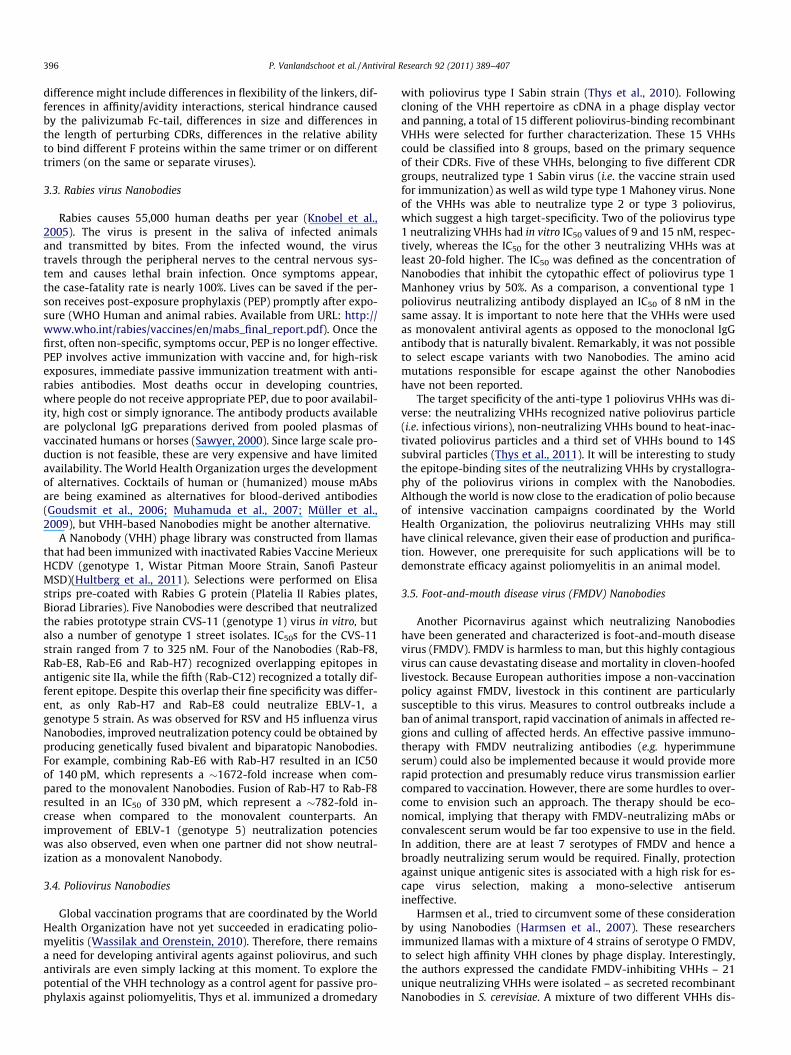
Fig. 1.two ligantibodchain vhallmathird co
390 P. Vanlandschoot et al. / Antiviral Research 92 (2011) 389–407
3.8. Hepatitis B virus (HBV) Nanobodies. . . . . . . . . . . . . . . . . . . . . . . . . . . . . . . . . . . . . . . . . . . . . . . . . . . . . . . . . . . . . . . . . . . . . . . . . . . . . . . . . 401
3.8.1. HBV S domain Nanobodies . . . . . . . . . . . . . . . . . . . . . . . . . . . . . . . . . . . . . . . . . . . . . . . . . . . . . . . . . . . . . . . . . . . . . . . . . . . . . . . . 4013.8.2. HBV nucleocapsid Nanobodies. . . . . . . . . . . . . . . . . . . . . . . . . . . . . . . . . . . . . . . . . . . . . . . . . . . . . . . . . . . . . . . . . . . . . . . . . . . . . . 4013.8.3. Porcine endogenous retrovirus (PERV) Nanobodies . . . . . . . . . . . . . . . . . . . . . . . . . . . . . . . . . . . . . . . . . . . . . . . . . . . . . . . . . . . . . 4024. Diagnostic applications . . . . . . . . . . . . . . . . . . . . . . . . . . . . . . . . . . . . . . . . . . . . . . . . . . . . . . . . . . . . . . . . . . . . . . . . . . . . . . . . . . . . . . . . . . . . . . . . 402
4.1. Vaccinia virus and Marburg virus VHHs . . . . . . . . . . . . . . . . . . . . . . . . . . . . . . . . . . . . . . . . . . . . . . . . . . . . . . . . . . . . . . . . . . . . . . . . . . . . . 4024.2. Plant virus VHHs . . . . . . . . . . . . . . . . . . . . . . . . . . . . . . . . . . . . . . . . . . . . . . . . . . . . . . . . . . . . . . . . . . . . . . . . . . . . . . . . . . . . . . . . . . . . . . . . 4025. Industrial applications . . . . . . . . . . . . . . . . . . . . . . . . . . . . . . . . . . . . . . . . . . . . . . . . . . . . . . . . . . . . . . . . . . . . . . . . . . . . . . . . . . . . . . . . . . . . . . . . . 403
5.1. Lactococcus bacteriophage VHH . . . . . . . . . . . . . . . . . . . . . . . . . . . . . . . . . . . . . . . . . . . . . . . . . . . . . . . . . . . . . . . . . . . . . . . . . . . . . . . . . . . . 4036. Conclusion . . . . . . . . . . . . . . . . . . . . . . . . . . . . . . . . . . . . . . . . . . . . . . . . . . . . . . . . . . . . . . . . . . . . . . . . . . . . . . . . . . . . . . . . . . . . . . . . . . . . . . . . . . 403Disclosure statement . . . . . . . . . . . . . . . . . . . . . . . . . . . . . . . . . . . . . . . . . . . . . . . . . . . . . . . . . . . . . . . . . . . . . . . . . . . . . . . . . . . . . . . . . . . . . . . . . . 404Acknowledgements . . . . . . . . . . . . . . . . . . . . . . . . . . . . . . . . . . . . . . . . . . . . . . . . . . . . . . . . . . . . . . . . . . . . . . . . . . . . . . . . . . . . . . . . . . . . . . . . . . . 404References . . . . . . . . . . . . . . . . . . . . . . . . . . . . . . . . . . . . . . . . . . . . . . . . . . . . . . . . . . . . . . . . . . . . . . . . . . . . . . . . . . . . . . . . . . . . . . . . . . . . . . . . . . 404
1. Introduction
Billions of different antibody molecules are generated by thevertebrate immune system. A specific antibody binding everyexisting compound is thought to be present in this antibody reper-toire. It is this unparalleled high diversity and selectivity that makeantibodies attractive and efficient research tools but also therapeu-tic molecules. Up to 1989, all antibodies were thought to be com-posed of two heavy chains and two light chains. The two heavychains are covalently linked by disulfide bonds. The heavy chainsof IgGs consist of 1 variable domain (VH) and 3 constant domainscalled CH1, CH2 and CH3. The light chains consist of a variable (VL)and constant domain (CL) that interact non-covalently with the VHand CH1 domains, respectively (Fig. 1). In 1989, a new type of anti-body was identified, first in the sera of dromedaries and later alsoin all other species of the Camelidae family (Hamers-Castermanet al., 1993). These antibodies do not contain a light chain and also
Distinguishing structural features of conventional antibodies and camelid heavy-cht (L) chains, and carry two antigen-binding sites determined by the combinationies lack both constant and variable light chains (CL and VL) and the first heavy chaariable domain (VHH or Nanobody). VHHs are characterized by the presence of hrk residues (indicated according to Kabat numbering). In many VHHs an additionamplementary determining regions (CDRs).
lack the first constant heavy domain. Today it is still unclear whatthe evolutionary advantage of such heavy chain-only antibodies(HcAbs) could be. In sharp contrast, the broad applicability of theisolated variable antigen-binding domains (VHH) was rapidly rec-ognized, especially for the development of therapeutic proteins(for recent reviews see Harmsen and De Haard (2007), VanBockstaele et al. (2009), Muyldermans et al. (2009), Wesolowskiet al. (2009), and Kolkman and Law (2010)). Such therapeutic pro-teins based on the smallest functional fragments of heavy chainantibodies, naturally occurring in Camelidae have been calledNanobodies�. Another type of heavy chain-only antibodies wasfound in sharks, the socalled immunoglobulin new antigen recep-tors (IgNARs) (Greenberg et al., 1996; Nuttall et al., 2001). Theantigen-binding variable domains of these antibodies (vNARs), aswells as single-domain antibodies (dAbs) derived from humanvariable heavy domains (VH) and variable light domains (VL) havesimilar applications (Holliger and Hudson, 2005).
hain antibodies. Conventional IgG antibodies comprise of two heavy (H) chains andof the variable domains of heavy and light chains (VH and VL). Camelid heavy-chainin constant (CH1) domain, and the antigen-binding site is formed only by the heavyydrophilic amino acid residues in the second framework region (FR2), the socalledl disulfide bond is present connecting the first (camels) or second (llamas) with the

Table 1Overview of published camelid-derived Nanobodies directed against viruses.
Virus References Immunogen Camelid Producing host Mechanism In vitro data In vivo data Specificity Observations
Influenza A Hultberg et al.(2011)
HA (H5N1) Llama E. coli Neutralization Microneutralization IC50 0003–7 nM. Neutralizing pseudotypedMLV(H5) IC50: 1–150 nM
H5N1 (clade 1>clade2.2>clade 2.5)
Influenza A Ibañez et al.2011)
HA (H5N1) Llama E. coli Neutralization ProphylacticTherapy: 0.5 lg/mouse: lung virustiters below thedetection limit.Therapeutic: VHHb4, 24 or 48 h afterchallenge, higherbody weights andlower lung virusloads
H5N1
RSV Hultberg et al.(2011)
Fusion protein Llama E. coli IC50 bivalent: 0.1 nM; monovalent250 nM
RSV Long (A) RSV B1 (B).Antigenic site II
Rabies Hultberg et al.(2011)
G protein Llama E. coli IC50 CVS-11 (genotype 1): 7,5–325 nM; EBLV-1 (genotype 5):012–586 nM
CVS-11 strain (genotype1), street isolates, 5 EBLV-1 strain; antigenic site IIa
Poliovirus Thys et al.(2010)
Type 1 Sabinstrain
Drome-dary
E. coli WK6 Neutralization IC50 7–692 nM Type 1 viruses
FMDV Harmsenet al. (2007)
Crude extract ofFMDV-infectedBHK cells
Llama Yeast strainVWK 18gal�
Neutralization VHHs neutralize FMDV O1Manisa at concentrations below0.34 mg/ml
Passive transfer ofVHH did not protectguinea pigs againstFMDV challengeinfection.
GH-loop, FMDV type O1Manisa
FMDV Harmsenet al. (2008)
O1 Manisa/Turkey/69 FMDV
Llama Yeast strainVWK 18gal�
Neutralization FMDV neutralization titer 1–4 mg/l
3 mg/kg of VHHs(i.m.) reduceviremia and viralshedding but do notprevent thedevelopment ofFMDV clinical signsor transmission.
GH-loop, FMDV type O1Manisa
Porcine immunoglobulin(pIg) binding VHHsgenetically fused to VHHsagainst FMDV (100-foldincreased serum half-life)
FMDV Harmsenet al. (2009b)
O1 Manisa/Turkey/69FMDV
Llama Yeast strainVWK 18gal�
Neutralization FMDV neutralization titer1–5 mg/l
VHHs reduce anddelay thedevelopment ofclinical disease,viraemia and viralshedding; delayFMD transmission.
FMDV O1 Manisa > FMDVA Turkey > FMDV Asia 1Shamir
FMDV Harmsenet al. (2009a)
O1 Manisa/Turkey/69 FMDV
Llama Yeast strainVWK 18gal�
Neutralization FMDV neutralization titer 0.008–0016 mg/ml
GH-loop, FMDV type O1Manisa
Increase the FMDV-neutralizing capacity oftwo non-glycosylatedVHHs by genetic fusion toanother VHH that isglycosylated.
Rotavirus van der Vaartet al. (2006)
G3 strains Llama YeaststrainVWK 18gal1
Neutralization IC50 �50 ng/ml 50–100 lgsignificantlyreduced the numberof days withdiarrhea per pup
Rotavirus G3 strains. A VHH has lost it’sneutralizing capacity afterproduction in yeast.
(continued on next page)
P.Vanlandschoot
etal./A
ntiviralR
esearch92
(2011)389–
407391

Table 1 (continued)
Virus References Immunogen Camelid Producing host Mechanism In vitro data In vivo data Specificity Observations
Rotavirus Garaicoecheaet al. (2008)
VP6 proteinderived from thebovine rotavirusc486 strain
Llama E. coli TG1 Neutralization 0,2–15,6 lg/ml (concentrationthat reduces > 80% of focusforming units)
Partial protectionagainst rotavirusdiarrhea
Bovine rotavirus C486,IND and B223, Humanrotavirus Wa; Equinerotavirus H2.
Broad neutralizationactivity in vitro
Rotavirus Pant et al.(2006)
G3 RRV strain Llama Lactobacilliparacasei
Neutralization 60 ng/ml reduce by 60% thenumber of RRV-infected cells
Lower prevalence,duration andseverity of diarrhea
G3 RRV strain Reconstituted freeze-dried VHH1-anchoredlactobacilli are equally asprotective as their freshcounterparts. VHH1-secreting lactobacilli donot offer better protectionthan contransformedlactobacilli
Rotavirus Martín et al.(2011)
G3 RRV strain Llama L. paracasei(codingsequenceintegrated inbacterialgenome)
Neutralization VHHs were showntoreduce the durationand severity ofdiarrhea
Rotavirus G3 strains
HIV-1 Forsman et al.(2008)
Envelopeprotein gp120from HIV-1CN54(subtype B/C)
Llama E. coli TG1 Neutralization IC50 0.003 to 38 lg/ml HIV-1 primary isolates ofsubtypesB, C, andCRF07_BC
First description ofbroadly neutralizingMAbs to HIV-1 envelopewhich were derived froman immunized animal
HIV-1 Koh et al.(2010)
Envelopeprotein gp120from HIV-1CN54(subtype B/C)
Llama E. coli TG1 Neutralization IC50: Subtype B: 0,07–0,57 lg/ml;Subtype C: 0,04–0,96 lg/ml
Subtypes B and C Construction of a Family-specific Phage DisplayLibrary
HIV-1 Vercruysseet al. (2010)
RecombinantHIV-Rev protein
Llama E. coli;eucariotic cells
intrabody bindsthemultimerizationdomain of Revand inhibits itsoligomerization.
�0,2 lg of plasmid inhibit 50% ofp24 Gag amount in thesupernatants oftransfected cells
Intrabody-Rev interactionis completely abolishedby the K20A and Y23Amutations
First known molecule thatdestabilizes and preventsthe formation of a largeorganized homoproteincomplex required forefficientHIV-1 mRNAexport from the nucleus
HIV-1 Bouchet et al.(2011)
RecombinantNef protein(fragment 57–205)
Llama E. coli K12strainTG1
VHH binds toHIV-1 Nef andinhibit itscritical biologicactivities
VHH inhibits virus infectivity in aNef-dependent mannerandcounteracts the positive effect ofNef on virus replication
VHH rescues Nef-mediated thymicCD4 T-cellmaturation defectand peripheral CD4T-cell activationphenotypes of theCD4C/HIV-1Nef Tgmouse model
VHH counteracts most ofthe HIV-1 nef allelesincluding Nef proteinsfrom groups M, N, O, and P
HIV-1 Jähnichenet al. (2010)
CXCR4-expressingHEK293T cells
Llama E. coli TG1 MonovalentVHHs: neutralantagonists,biparatopicVHHs: inverseagonists
IC50 Monovalent: 13,6–82 mM;Bivalent: 0,2–0,5 nM
The biparatopicnanobodyeffectivelymobilized CD34-positive stem cellsin cynomolgusmonkeys
238D4: binds D187,F189,E179, and S178 in ECL2;238D2: binds F189, N192,W195,P191,V196 andE277 in ECL3. F189,positioned in ECL2, iscritical for binding of bothVHHs
More than 1000-foldselectivity of 238D2and238D4 for CXCR4 versusall other GPCRs tested
HBV Serruys et al.(2009)
E. coli-derivednucleocapsids(HBcAg) andplasma-purifiedHBsAg
Llama E. coli WK6;HepG2 cells
Suppress hbsag particle secretion(80–90%) and increase hbsagaccumulation/retention insidethe cell.
The concentration ofhbv dna in plasmawas reduced 10–100-fold. The levelsof secreted hbeagwere not affected
Envelope protein s First report of intrabody-mediated inhibition ofviral secretion inmammals
392P.V
anlandschootet
al./Antiviral
Research
92(2011)
389–407

HB
VSe
rru
yset
al.
(201
0)R
ecom
bin
ant
HB
cAg
Llam
aE.
coli
WK
6;H
epG
2ce
lls
Com
peti
tion
assa
y:po
siti
vere
sult
at±
70n
m.V
hh
sta
rget
edto
the
nu
cleu
s:el
evat
edin
trac
ellu
lar
amou
nt
ofh
beag
and
abse
nce
ofh
bcag
inly
sate
s
Cro
ss-r
eati
vity
HB
cAg
and
HB
eAg
(su
btyp
essu
btyp
eay
wan
dad
w)
Porc
ine
Ret
rovi
rus
Dek
ker
etal
.(2
003)
Puri
fied
60-k
Da
Gag
prot
ein
Llam
aE.
coli
TG1
Intr
abod
yre
duce
sR
Tac
tivi
tyto
appr
oxim
atel
y7%
ofth
eac
tivi
tyin
un
indu
ced
stat
e.
PER
V-A
and
PER
V-B
Vac
cin
iaG
oldm
anet
al.(
2006
)N
aiev
ell
ama
E.co
liTu
ner
+pR
AR
EC
lon
este
sted
inEL
ISA
are
vacc
inia
spec
ific
Vac
cin
ia
Mar
burg
Vir
us
Sher
woo
det
al.(
2007
)N
aiev
ell
ama
E.co
liTu
ner
+pR
AR
ETh
eli
mit
ofde
tect
ion
0.1–
100
pfu
/wel
l.N
ucl
eopr
otei
n(M
AR
Vva
rian
tsM
uso
ke,R
avn
,an
dA
ngo
la)
Dia
gnos
tic
assa
ys.F
irst
reco
mbi
nan
tan
tibo
dysp
ecifi
call
yis
olat
edfo
ran
yM
AR
Vpr
otei
nTu
lip
viru
sX
Bee
kwil
-der
etal
.(20
08)
TuV
Xpa
rtic
les
Alp
aca
E.co
liTG
1Po
siti
vede
tect
ion
ofvi
rus
part
icle
sor
dilu
ted
tuli
ple
afex
trac
t
Tuli
pvi
rus
XD
iagn
osti
cas
say
for
dete
ctio
nof
the
plan
tpa
thog
enic
Tuli
pvi
rus
X(T
uV
X)
Bac
teri
o-ph
age
De
Haa
rdet
al.(
2005
)L.
lact
isba
cter
io-
phag
ep2
Llam
aE.
coli
and
yeas
tN
eutr
aliz
atio
nV
HH
prev
ent
phag
ein
fect
ion
,ev
enat
con
cen
trat
ion
sas
low
as2.
25n
M
Rec
epto
r-bi
ndi
ng
prot
ein
(RB
P/O
RF1
8)of
Phag
esk
1,ph
age
p2.
Bac
teri
o-ph
age
Hu
ltbe
rget
al.
(200
7)L.
lact
isba
cter
io-
phag
ep2
Llam
aL.
para
case
iN
eutr
aliz
atio
nV
HH
-sec
rete
dn
eutr
alis
eph
age
p2by
bin
din
gto
its
RB
Pan
din
hib
itin
g(8
6%)
its
adso
rpti
onto
the
hos
tst
rain
.Su
rfac
eex
pres
sed
VH
Hin
hib
itph
age
infe
ctio
n(3
1%)
P. Vanlandschoot et al. / Antiviral Research 92 (2011) 389–407 393
Here we summarize first some of the unique characteristicsand features of VHHs. These will next be described in the contextof different experimental therapeutic applications of Nanobodiesagainst different viruses: HIV, Hepatitis B virus, influenza virus,Respiratory Syncytial virus, Rabies virus, FMDV, Poliovirus,Rotavirus, and PERVs. Next, the diagnostic application of VHHs(Vaccinia virus, Marburg virus and plant Tulip virus X), as wellas an industrial application (lytic lactococcal 936 phage) will bedescribed. All these different applications are summarized inTable 1.
2. Camelid antibodies and Nanobodies
Camelids produce conventional antibodies but they also pro-duce heavy chain-only antibodies (HcAbs). In llama species�45% of serum antibodies are HcAbs, while in camelus speciesthis is �75% (Hamers-Casterman et al., 1993). In camelids, con-ventional antibodies are IgG1 isotypes, while IgG2 and IgG3 areHcAbs (Fig. 1). Despite this abundance, which points to a signifi-cant role, there is little information on the functions and specific-ities of these HcAbs in immunity (Daley et al., 2010; Daley-Baueret al., 2010).
Because of a splice site mutation, heavy chain-only antibodieslack the CH1 domain and also lack the complete light chain thatis partially anchored to the CH1 domain (Nguyen et al., 1999;Woolven et al., 1999). The variable heavy-chain domains of HcAbs(VHH) are generated form a V–D–JH gene rearrangement using aseparate set of �40 V gene segments, all related to the humanVH3 gene family (Harmsen et al., 2000; Nguyen et al., 2000). Differ-ent VHH subfamilies have been defined, but all share a few crucialsubstitutions of germline-encoded amino acids Val37 ? Phe/Tyr,Gly44 ? Glu/Gln, Leu45 ? Arg, and Trp47 ? Gly/Phe/Leu (Kabatnumbering), that increase the hydrophilicity of frame work 2(FR2), the putative VH–VL interface (Fig. 1). These VHH hall markresidues abrogate a possible interaction with VL domains and con-tribute to the stability, increased solubility and reduced aggrega-tion tendency of HcAbs and VHHs compared to other singledomain antibodies (Hamers-Casterman et al., 1993; Muyldermanset al., 1994; Vu et al., 1997). Remarkably the third complementarydetermining region CDR3 of VHHs has been shown in many crystalstructures to fold back and cover the former VL interface, furthercontributing to the stability and solubility of VHHs (Desmyteret al., 1996; Muyldermans et al., 2001).
Compared to human VH domains, VHH often display a long-er CDR3 loop (Muyldermans et al., 1994; Vu et al., 1997). Thisleads to an increased surface area and repertoire that can inter-act with antigens. Such extended CDR3 loops are often stabi-lized by a disulfide bond between CDR1 and CDR3 orbetween FR2 and CDR3. Nevertheless, a significant proportionof VHH has a short CDR3 and lack the additional disulfide bond(Vu et al., 1997; Harmsen et al., 2000). Increased binding diver-sity also results from non-canonical CDR1 and CDR2 loop struc-tures and additional hotspots for somatic hyper mutation in theCDR1 (Nguyen et al., 2000). Besides these VHH domains, VHH-like domains lacking the hall mark residues, the long CDR3loops and the interloop disulfide bonds are also used in HcAbs(Harmsen and De Haard, 2007). More recently it was suggestedthat variable genes displaying a high degree of homology to thehuman VH4 family add to the HcAb Ag-binding diversity(Deschacht et al., 2010).
Because of these unique biophysical and biochemical features ofthe antigen binding domains of HcAbs, VHH domains have beenproduced recombinantly as separate entities (VHHs). This creatednew possibilities and as such several (new) features and applica-tions for VHH have been explored and described.

394 P. Vanlandschoot et al. / Antiviral Research 92 (2011) 389–407
2.1. VHHs display high affinity
VHHs against many different targets that include haptens, pep-tides, soluble and transmembrane proteins have been reported. Ac-tive immunization of dromedaries or llamas is most often used forproteins and as a result VHHs with affinities in the lower nanomo-lar or even picomolar range have been reported (reviewed inHarmsen and De Haard (2007), Van Bockstaele et al. (2009), Weso-lowski et al. (2009), and Kolkman and Law (2010)). Considering themonomeric nature of VHHs this is remarkable as these affinitiesare in the same range of what is readily observed for conventionalbivalent antibodies. VHHs with nanomolar affinities have also beenobtained using naïve or synthetic libraries (Goldman et al., 2006;Groot et al., 2006; Verheesen et al., 2006).
2.2. VHHs can recognize structures not recognized by or inaccessiblefor conventional antibodies
Structural analysis of conventional antibodies and VHHs, incomplex with their antigen has revealed a major difference inthe structure of the CDRs. While conventional antibodies typicallyhave a concave or flat antigen binding site, VHHs have a convexconformation with large solvent exposed CDR loops (Desmyteret al., 1996; Muyldermans et al., 2001). The compact shape of VHHscombined with the convex paratope allows binding into clefts orpockets. This was demonstrated for several VHHs that inhibit en-zymes like lysozyme and carbonic anhydrase (Desmyter et al.,1996, 2001; Lauwereys et al., 1998; Transue et al., 1998; De Genstet al., 2006; Conrath et al., 2001a, 2009). Besides this unique cav-ity-penetrating properties, it has also been shown that VHH canbe isolated that bind cryptic epitopes on the variant surface glyco-proteins of African trypanosome, not accessible for conventionalantibodies (Stijlemans et al., 2004, 2011).
2.3. VHH are remarkably stable under different extreme conditions
VHHs display high thermal stability. Tm values between 60 and80 �C are the rule, not the exception with thermal unfolding oftenshown to be fully reversible and functional activity sometimes re-tained at temperatures up to 90 �C (Lauwereys et al., 1998; van derLinden et al., 1999; Pérez et al., 2001; Ewert et al., 2002). VHHs arealso exceptionally resistant to high pressure, chemical unfoldingwith guanidinium chloride and urea, detergents or alkaline andacid pH (Dumoulin et al., 2002; Dolk et al., 2005). Compared toconventional antibodies and antibody-derived fragments, resis-tance of VHHs to proteases can be improved by in vitro selectionto generate VHHs that can resist the harsh conditions of the gas-tro-intestinal tract (Harmsen et al., 2006).
2.4. Enhanced functionality by easy multimerization of VHHs
The high solubility, single domain and single gene nature ofVHHs allows rapid and successful generation of multimeric VHHsusing genetically encoded amino acid linkers or carrier proteins(Fig. 1). Bivalent, trivalent, pentavalent and even decavalent mole-cules have been described (Conrath et al., 2001b; Zhang et al.,2004; Groot et al., 2006; Mai et al., 2006; Stewart et al., 2007; Stoneet al., 2007a, 2007b; Garaicoechea et al., 2008; Hmila et al., 2010).Multimerization is an easy way to rapidly improve functional po-tency due to an avidity effect. Fusion of two identical anti-TNFaNanobodies resulted in a 500-fold increase in TNFa neutralizingactivity. The in vitro potency of this bivalent Nanobody even ex-ceeded those of clinically used conventional antibodies (Coppieterset al., 2006). Increased potency due to avidity has also been dem-onstrated for membrane bound receptors using bivalent monospe-cific and biparatopic VHHs (Roovers et al., 2007). The latter consists
of two different VHHs recognizing overlapping or non-overlappingepitopes on the same antigen. Besides improvements in potency,formatting also allows the facile generation of one single moleculecapable of binding different molecules (Conrath et al., 2001b;Harmsen et al., 2008; Hmila et al., 2010). An important multispec-ific application is extension of in vivo half-life of therapeutic VHHs.Indeed the molecular weight of monovalent VHHs (�15 kDa) is be-low the threshold of renal filtration. As a result mono, bi and tri-meric VHHs are all rapidly cleared form the blood. By coupling toan VHH that binds an abundant serum protein like albumin orIgG, the half-life of the therapeutic Nanobody becomes similar tothat of such proteins (Harmsen et al., 2005; Roovers et al., 2007;Tijink et al., 2008).
2.5. Alternative expression of Nanobodies
Classical antibodies are successfully expressed and secreted inmammalian cell lines mainly. Several smaller formats derived fromconventional antibodies have been generated: monovalent anti-body fragment (Fab), Fab dimer, variable fragment (Fv), single-chain Fv (scFv) and heavy or light chain single domain antibodies(Fig. 1). Although these derivatives can be produced in other hostcells, the hydrophobic nature of the VL and VH FR2 interface re-mains, and is partially responsible for problems in expression yield,solubility, stability and aggregation. In contrast, the single domainnature of VHHs and their increased hydrophilicity enables highproduction levels in microbial hosts like Escherichia coli, Pichia pas-toris and Saccharomyces cerevisiae. In addition VHHs can also beproduced and function in different cellular compartments wherethe formation of disulfide bonds cannot occur. VHHS have been ex-pressed in the ER and in the cytoplasm of cells (Klooster et al.,2009). VHHs have been successfully targeted to the nucleus ormitochondria (Serruys et al., 2010; Van den Abbeele et al., 2010).
3. Therapeutic applications
3.1. Influenza virus Nanobodies
Influenza is an important respiratory disease caused by influ-enza A and B viruses. In moderate climate zones, influenza typi-cally occurs in epidemics that peak during wintertime. InfluenzaA viruses can also cause unpredictable pandemic outbreaks, asso-ciated with antigenic shift of the viral hemagglutinin. Thanks to in-tense global monitoring of influenza viruses, currently usedvaccines to prevent seasonal influenza have a fairly accurate anti-genic composition and protect well in most target groups (Russellet al., 2008). However, pandemic outbreaks remain unpredictable,as illustrated by the 2009 H1N1 virus (also named Mexican flu),which took the world and the vaccine manufactures by surprise.In addition, outbreaks of highly pathogenic avian influenza suchas H5N1 have occurred without cessation since 2003, which has re-sulted in culling of more than a billion birds in the poultry indus-try, leading to major losses of food and economical income, mainlyin south East Asia. Occasional zoonotic infections with these H5N1viruses and their high propensity to reassort with swine influenzaviruses, have earmarked these viruses as a major pandemic threat.The case fatality of zoonotic infections with H5N1 viruses is closeto 60% despite intensive care interventions and the use of antiviraldrugs such as oseltamivir. In summary, there is a need for noveltreatment options against influenza. Therefore, we decided to iso-late and evaluate the prophylactic and therapeutic activity ofNanobodies directed against highly pathogenic H5N1 virus. To thisend, a llama was immunized with recombinant H5 hemagglutininand, following phage display and panning against the recombinantantigen, two Nanobodies that neutralized H5N1-pseudotyped

P. Vanlandschoot et al. / Antiviral Research 92 (2011) 389–407 395
lentiviruses with an IC50 of 10 and 30 nM, respectively, were se-lected (Hultberg et al., 2011). These two Nanobodies blocked thebinding of hemagglutinin to sialic acid residues on fetuin, whichserves as an in vitro surrogate for the natural receptor. Interest-ingly, by producing bivalent or trivalent Nanobodies by genetic fu-sion of the coding information of two or three of the neutralizingNanobodies separated by a glycine–serine (GS) linker of variablelength the in vitro neutralizing activity against H5N1 pseudotypedlentiviruses or against H5N1 influenza virus increased dramati-cally. One of the monovalent Nanobodies had an IC50 of 7 nM asmeasured in a microneutralization assay against H5N1 virus,whereas its bivalent and trivalent counterpart displayed an IC50
of 3–9 pM in the same assay (Hultberg et al., 2011). This dramaticdifference in neutralizing activity between a mono- and a bi- or tri-valent VHH can partially most probably be explained by the in-creased avidity and by a more potent mode of interactioninvolving intermolecular binding. Ultimately, however, co-crystalstructure analysis will be required to help explain the 1000-fold in-crease in in vitro efficacy of these Nanobodies. Remarkably, a num-ber of H5N1 variants which were not or poorly neutralized(IC50 > 120–150 nM) by the monovalent Nanobodies, were effi-ciently neutralized by the bivalent or trivalent Nanobodies(IC50 < 10 nM).
We next assessed the in vivo efficacy of these Nanobodies in amouse model for H5N1 influenza (Ibañez et al., 2011). Importantly,we decided to administer the Nanobodies intranasally. The rationalfor this route of administration was in part to avoid multiple orcontinuous dosing as systemic administration of Nanobodies leadsto rapid clearance from the body. Both prophylactic (up to 48 h be-fore challenge; supporting a long local half life of Nanobodies) andtherapeutic (up to 72 h after challenge) significantly reduced virusreplication in H5-specific Nanobody treated animals. Interestingly,also in vivo, the bivalent neutralizing Nanobody outperformed itsmonovalent counterpart, by a factor 60. In addition, intranasaladministration of bivalent Nanobodies 24 h before a potentiallylethal challenge with H5N1 virus, fully protected the animals fromdeath and morbidity. Also in a therapeutic setting, the bivalentNanobodies were protective and significantly delayed time todeath. Finally, we identified the likely site of binding of the Nano-body in the HA, by selecting escape viruses in vitro. Both mono-and bi-valent Nanobody selection pressure resulted in escapeviruses in which residue Lysine 189 located in antigenic site ofhemagglutinin near the receptor-binding site, was changed to glu-tamic acid. These findings provide proof-of-concept that Nanobod-ies can protect against H5N1 influenza virus challenge whenadministered by the intranasal route. In addition, our results favorthe design of bi- or trivalent Nanobodies to increase their potency.Additional experiments in another animal model such as the ferretand the isolation of broadly neutralizing Nanobodies will beneeded to provide additional proof that the use of Nanobodies isa highly interesting treatment option to prevent and treat influ-enza virus infection.
3.2. Respiratory Syncytial virus (RSV) Nanobodies
Infections by Respiratory Syncytial virus (RSV) are the leadingcause of viral acute lower respiratory tract disease in childrenworldwide (Hall et al., 2009). In developed countries 1–2% of theRSV infected infants require hospitalization. In this way RSV infec-tions are the most important cause of infant hospitalization. Oncehospitalized, there is no effective anti-viral or anti-inflammatorytherapy available and treatment is mainly based on supplying oxy-gen (by mechanically assisted ventilation if required) and rehydra-tion. As RSV infections themselves do not evoke long-livingimmune protection, RSV infections repeatedly occur throughoutlife, causing also significant morbidity and mortality in elderly
and immune compromised adults (Falsey et al., 1995; Hall,2001). It has been estimated that annually RSV infects about 64million people resulting in 160,000 deaths. Next to the acute con-sequences of infection, severe RSV infections at young age arepotentially associated with the development of long-term pulmon-ary distress. Despite the major clinical importance of RSV, there isneither a vaccine nor any antiviral therapy available.
Although immune protection by natural RSV infections is par-tial, it correlates with high serum titers of neutralizing antibodiesand high serum titers of RSV F specific IgG antibodies (Hendersonet al., 1979). Administration of RSV neutralizing serum or antibod-ies was shown to reduce pulmonary RSV replication in differentanimal models (Henderson et al., 1979; Taylor et al., 1984; Walshet al., 1984; Hemming et al., 1985; Prince et al., 1985). These find-ings suggested that passive immunoprophylaxis with IgG mightprotect infants from RSV disease. Indeed intravenous administra-tion of human IgG preparations enriched for RSV neutralizing anti-bodies could partially prevent RSV lower respiratory tract diseasein infants (Groothuis et al., 1993; Simoes et al., 1996). Hence in1996 this therapy was approved by the FDA for the prevention ofRSV disease in high-risk infants. Subsequent palivizumab (Syna-gis), a humanized RSV monoclonal antibody (mAb) directed againstthe conserved RSV F protein was developed. This mAb could effi-ciently neutralize a broad range of RSV strains in vitro (Johnsonet al., 1997). Intramuscular administration of 2.5 mg/kg pali-vizumab effectively reduced RSV replication in cotton rats. The im-pact RSV Study revealed that five monthly intramuscular injectionsof 15 mg/kg could reduce RSV-related hospitalization of infants by55%. In 1998 the FDA approved monthly intramuscular administra-tion of palivizumab for immunoprophylaxis of RSV induced diseasein high-risk infants and children (Wu et al., 2008).
Hultberg et al. aimed at developing RSV F specific Nanobodieswith enhanced neutralizing activity (Hultberg et al., 2011). Llamaswere immunized 6 times weekly with recombinant membraneanchorless F protein (F-TM�) derived from the RSV Long strain (sub-type A) (Calder et al., 2000). Biopanning using F-TM� and competi-tive elution using excess of palivizumab was used to enrich for RSVneutralizing F-specific Nanobody-phages. The majority of the ob-tained clones were able to bind to F-TM�. Twelve clones were se-lected for Nanobody production and purification. From theseclones two Nanobodies (RSV-D3 and RSV-C4) could neutralizeRSV A subtype Long strain virus in vitro and one clone could neu-tralize RSV B subtype B1 strain virus (RSV-E4). Based on competi-tion experiments with mAbs that specifically recognize welldescribed antigenic sites within the RSV F protein, it was shownthat RSV-D3 and RSV-C4, bind to the antigenic site II to which alsopalivizumab is binding. In contrast, the RSV-E4 Nanobody binds tothe antigenic sites IV–VI. Binding of Nanobodies to these specificepitopes was confirmed by the use of specific RSV escape mutants.In order to boost their neutralizing activity, monovalent Nanobod-ies were fused by GS linkers. Remarkably, linking two identicalRSV-D3 Nanobodies improved in vitro neutralization by about4000-fold. As a result bivalent RSV-D3 Nanobodies (IC50:0.11 nM) could neutralize RSV Long in vitro considerably more effi-cient than palivizumab (IC50: 6.5 nM). In addition bivalent RSV-D3Nanobodies could also neutralize RSV B1 (subtype B) more effi-ciently than their monovalent counterparts. Linking two Nanobod-ies with different epitopes (RSV-D3/RSV-E4) also significantlyincreased neutralization of RSV Long (subtype A, 50 to100-fold)and RSV B1 (subtype B, 500-fold).
The neutralizing efficiency of palivizumab is about 180-fold en-hanced compared to its monovalent Fab fragment. In contrast, link-ing of two identical monovalent RSV Nanobodies enhancedneutralization efficiency by a much larger extend up to 4000-fold.It is difficult to speculate on the reasons for this difference in in-crease of neutralization activity. Factors that could attribute to this
396 P. Vanlandschoot et al. / Antiviral Research 92 (2011) 389–407
difference might include differences in flexibility of the linkers, dif-ferences in affinity/avidity interactions, sterical hindrance causedby the palivizumab Fc-tail, differences in size and differences inthe length of perturbing CDRs, differences in the relative abilityto bind different F proteins within the same trimer or on differenttrimers (on the same or separate viruses).
3.3. Rabies virus Nanobodies
Rabies causes 55,000 human deaths per year (Knobel et al.,2005). The virus is present in the saliva of infected animalsand transmitted by bites. From the infected wound, the virustravels through the peripheral nerves to the central nervous sys-tem and causes lethal brain infection. Once symptoms appear,the case-fatality rate is nearly 100%. Lives can be saved if the per-son receives post-exposure prophylaxis (PEP) promptly after expo-sure (WHO Human and animal rabies. Available from URL: http://www.who.int/rabies/vaccines/en/mabs_final_report.pdf). Once thefirst, often non-specific, symptoms occur, PEP is no longer effective.PEP involves active immunization with vaccine and, for high-riskexposures, immediate passive immunization treatment with anti-rabies antibodies. Most deaths occur in developing countries,where people do not receive appropriate PEP, due to poor availabil-ity, high cost or simply ignorance. The antibody products availableare polyclonal IgG preparations derived from pooled plasmas ofvaccinated humans or horses (Sawyer, 2000). Since large scale pro-duction is not feasible, these are very expensive and have limitedavailability. The World Health Organization urges the developmentof alternatives. Cocktails of human or (humanized) mouse mAbsare being examined as alternatives for blood-derived antibodies(Goudsmit et al., 2006; Muhamuda et al., 2007; Müller et al.,2009), but VHH-based Nanobodies might be another alternative.
A Nanobody (VHH) phage library was constructed from llamasthat had been immunized with inactivated Rabies Vaccine MerieuxHCDV (genotype 1, Wistar Pitman Moore Strain, Sanofi PasteurMSD)(Hultberg et al., 2011). Selections were performed on Elisastrips pre-coated with Rabies G protein (Platelia II Rabies plates,Biorad Libraries). Five Nanobodies were described that neutralizedthe rabies prototype strain CVS-11 (genotype 1) virus in vitro, butalso a number of genotype 1 street isolates. IC50s for the CVS-11strain ranged from 7 to 325 nM. Four of the Nanobodies (Rab-F8,Rab-E8, Rab-E6 and Rab-H7) recognized overlapping epitopes inantigenic site IIa, while the fifth (Rab-C12) recognized a totally dif-ferent epitope. Despite this overlap their fine specificity was differ-ent, as only Rab-H7 and Rab-E8 could neutralize EBLV-1, agenotype 5 strain. As was observed for RSV and H5 influenza virusNanobodies, improved neutralization potency could be obtained byproducing genetically fused bivalent and biparatopic Nanobodies.For example, combining Rab-E6 with Rab-H7 resulted in an IC50of 140 pM, which represents a �1672-fold increase when com-pared to the monovalent Nanobodies. Fusion of Rab-H7 to Rab-F8resulted in an IC50 of 330 pM, which represent a �782-fold in-crease when compared to the monovalent counterparts. Animprovement of EBLV-1 (genotype 5) neutralization potencieswas also observed, even when one partner did not show neutral-ization as a monovalent Nanobody.
3.4. Poliovirus Nanobodies
Global vaccination programs that are coordinated by the WorldHealth Organization have not yet succeeded in eradicating polio-myelitis (Wassilak and Orenstein, 2010). Therefore, there remainsa need for developing antiviral agents against poliovirus, and suchantivirals are even simply lacking at this moment. To explore thepotential of the VHH technology as a control agent for passive pro-phylaxis against poliomyelitis, Thys et al. immunized a dromedary
with poliovirus type I Sabin strain (Thys et al., 2010). Followingcloning of the VHH repertoire as cDNA in a phage display vectorand panning, a total of 15 different poliovirus-binding recombinantVHHs were selected for further characterization. These 15 VHHscould be classified into 8 groups, based on the primary sequenceof their CDRs. Five of these VHHs, belonging to five different CDRgroups, neutralized type 1 Sabin virus (i.e. the vaccine strain usedfor immunization) as well as wild type type 1 Mahoney virus. Noneof the VHHs was able to neutralize type 2 or type 3 poliovirus,which suggest a high target-specificity. Two of the poliovirus type1 neutralizing VHHs had in vitro IC50 values of 9 and 15 nM, respec-tively, whereas the IC50 for the other 3 neutralizing VHHs was atleast 20-fold higher. The IC50 was defined as the concentration ofNanobodies that inhibit the cytopathic effect of poliovirus type 1Manhoney vrius by 50%. As a comparison, a conventional type 1poliovirus neutralizing antibody displayed an IC50 of 8 nM in thesame assay. It is important to note here that the VHHs were usedas monovalent antiviral agents as opposed to the monoclonal IgGantibody that is naturally bivalent. Remarkably, it was not possibleto select escape variants with two Nanobodies. The amino acidmutations responsible for escape against the other Nanobodieshave not been reported.
The target specificity of the anti-type 1 poliovirus VHHs was di-verse: the neutralizing VHHs recognized native poliovirus particle(i.e. infectious virions), non-neutralizing VHHs bound to heat-inac-tivated poliovirus particles and a third set of VHHs bound to 14Ssubviral particles (Thys et al., 2011). It will be interesting to studythe epitope-binding sites of the neutralizing VHHs by crystallogra-phy of the poliovirus virions in complex with the Nanobodies.Although the world is now close to the eradication of polio becauseof intensive vaccination campaigns coordinated by the WorldHealth Organization, the poliovirus neutralizing VHHs may stillhave clinical relevance, given their ease of production and purifica-tion. However, one prerequisite for such applications will be todemonstrate efficacy against poliomyelitis in an animal model.
3.5. Foot-and-mouth disease virus (FMDV) Nanobodies
Another Picornavirus against which neutralizing Nanobodieshave been generated and characterized is foot-and-mouth diseasevirus (FMDV). FMDV is harmless to man, but this highly contagiousvirus can cause devastating disease and mortality in cloven-hoofedlivestock. Because European authorities impose a non-vaccinationpolicy against FMDV, livestock in this continent are particularlysusceptible to this virus. Measures to control outbreaks include aban of animal transport, rapid vaccination of animals in affected re-gions and culling of affected herds. An effective passive immuno-therapy with FMDV neutralizing antibodies (e.g. hyperimmuneserum) could also be implemented because it would provide morerapid protection and presumably reduce virus transmission earliercompared to vaccination. However, there are some hurdles to over-come to envision such an approach. The therapy should be eco-nomical, implying that therapy with FMDV-neutralizing mAbs orconvalescent serum would be far too expensive to use in the field.In addition, there are at least 7 serotypes of FMDV and hence abroadly neutralizing serum would be required. Finally, protectionagainst unique antigenic sites is associated with a high risk for es-cape virus selection, making a mono-selective antiserumineffective.
Harmsen et al., tried to circumvent some of these considerationby using Nanobodies (Harmsen et al., 2007). These researchersimmunized llamas with a mixture of 4 strains of serotype O FMDV,to select high affinity VHH clones by phage display. Interestingly,the authors expressed the candidate FMDV-inhibiting VHHs – 21unique neutralizing VHHs were isolated – as secreted recombinantNanobodies in S. cerevisiae. A mixture of two different VHHs dis-
P. Vanlandschoot et al. / Antiviral Research 92 (2011) 389–407 397
playing synergistic in vitro FMDV-neutralizing activity turned outto be most effective in a passive prophylaxis setting. However, pro-tection was only partial whereas a convalescent guinea pig serumwith a comparable in vitro neutralizing titer fully protected the ani-mals against the development of FMDV lesions. Since the Nano-bodies had been PEGylated to increase their serum half life, thelimited in vivo protection was presumably due to the lack of Fc-dependent antiviral effector mechanisms, such as opsonophagocy-tosis, i.e. binding of antibodies to virions followed by phagocytosisof the opsonized virions by macrophages.
In a follow-up study using a swine model for immuno-prophy-lactic treatment against FMDV challenge, bispecific Nanobodieswere engineered (Harmsen et al., 2005, 2008). These bispecificNanobodies combined one of three VHHs that neutralized FMDVin vitro with a second VHH that binds with high affinity to porcineImmunoglobulin G light chain (Harmsen et al., 2005). The rationalfor this approach was to increase the serum half-life of the Nano-bodies, as an alternative for PEGylation. Addionally it was assumedthat this VHH would not interfere with Fc-encoded effector func-tions. Both VHHs, i.e. the FMDV neutralizing and the Ig-bindingVHHs were separated by a short GGS linker and produced inS. cerevisiae in a 100 L fermentor before affinity purification basedon a C-terminal poly-histidine tag. The affinity and in vitro FMDVneutralizing activity of the bispecific VHHs was comparable withtheir respective monovalent counterparts with KD values as lowas 0.3–0.5 nM. Likewise the affinities of the bispecific VHHs forswine Ig was comparable to that of the monovalent swine Ig-binding Nanobody (KD approximately 1 nM). However, when thebispecific VHHs were complexed with pig Ig, the in vitro neutraliz-ing activity of two out of tree VHHs increased 4- to 30-fold, presum-ably as a result of steric hindrance and/or avidity effects.Intramuscular administration of a dose of 3 mg/kg in pigs 24 h priorto challenge with 1000 plaque-forming units FMDV resulted in re-duced viremia and virus shedding but did not prevent transmission.
To further increase the neutralizing activity of the anti-FMDVNanobodies, bispecific VHHs were constructed with two VHH do-mains directed against the virus and a third VHH directed againstswine Ig (Harmsen et al., 2009a,b). This resulted in VHHs with a 5-fold higher neutralizing activity. These molecules, again producedin S. cerevisae, were able to reduce clinical disease, viraemia, virusshedding and now also transmission in a pig model when admin-istered (i.v. in the ear) at a dose of 50 mg/kg, 24 h before intrader-mal inoculation with 10,000 TCID50 of FMDV. Finally, the authorsalso demonstrated that the presence of FMDV-neutralizing VHHsdid not interfere with the immune response upon vaccination withconventional FMDV vaccine. This is an important finding becauseFMDV vaccination of animals that are at risk for being infected isused to try to control outbreaks. In summary, this developmentof FMDV-neutralizing Nanobodies has provided proof-of-conceptthat passive immuno-prophylaxis can protect animals againstFMDV-induced disease. In particular engineering steps to producetrivalent Nanobodies in which two of the three paratopes havein vitro FMDV neutralizing activity and the third paratope allowshigh affinity binding to circulating immunoglobulin, proved to beeffective in preventing disease and transmission. However, it re-mains to be determined if FMDV-escape viruses would be rapidlyselected upon use of such a Nanobody-based intervention, and ifa similar approach would also be effective against the other FMDVserotypes.
3.6. Rotavirus Nanobodies
Group A Rotavirus (RV) strains are the most frequent cause ofacute gastroenteritis in infants and children under the age of 5.Although fatal outcome of RV infections in developed countriesare rare, RV infections cause annually more than 500,000 deaths
worldwide (Parashar et al., 2006). RV is a non-enveloped doublestranded RNA virus with an outer and inner capsid. The outer cap-sid is composed of VP4 and VP7 proteins, which are highly variable.Based on the variability of VP7 and VP4, Rotaviruses diverge into23 G (based on the VP7 glycoprotein) and 31 P (based on the pro-tease-sensitive VP4 protein) serotypes. RV infections induce neu-tralizing antibodies specific for VP4 and VP7 (Ward, 2009). Uponprimary infection these neutralizing antibodies are mainly sero-type specific and can hence protect against homosubtypic infec-tions. The inner capsid is composed of VP6 proteins which areimmunodominant and highly conserved. Although VP6 antibodiescan protect mice from RV infection VP6 antibodies do not neutral-ize RV in vitro (Burns et al., 1996). Their protective capacity canhowever be explained by polymeric VP6 IgA antibodies that neu-tralize RV via transcytosis (Corthésy et al., 2006).
Rotarix and Rotateq are two licensed vaccines that have beenshown to protect against the main circulating RV strains (G1, G2,G3, G4 and P1A) and are hence applied in childhood vaccinationprograms. Next to vaccines, oral administration of antibodiesagainst VP7 and VP4 has also been shown to prevent or treat RVinfections in children (Sarker et al., 2001). As a more feasible alter-native to preparations of conventional antibodies, van der Vaartet al. developed RV neutralizing Nanobodies that are produced inyeast (van der Vaart et al., 2006). A Nanobody (VHH) phage librarywas constructed from circulating plasma cells derived from a llamathat had been immunized five times with whole Rhesus rotavirus(RRV, G3 serotypes). RV binding phages were selected by biopan-ning, using the RRV strain that was used for immunization. As or-ally administered anti-RV Nanobodies should be functional in thegut they must resist the acidic conditions of the stomach. There-fore, as part of the selection strategy, the phages were pre-treatedat low pH (pH 2.3). The selected Nanobodies were screened andfurther selected for binding to RRV and a second G3 serotype RV(CK5). Neutralizing Nanobodies and control Nanobodies were re-cloned for production in S. cerevisiae. The yeast produced Nanobod-ies were tested for CK5 RV in vitro neutralization. The most potentNanobody (2B10) could neutralize CK5 RV in an in vitro plaque as-say with an IC50 of approximately 3 nM. Neutralizing Nanobodiesthat could be efficiently produced in yeast were also tested in amouse pup model for rotavirus infection. Daily oral administrationof 50 or 100 lg 2B10 Nanobody could either prevent diarrhea orreduce the number of days with diarrhea per pup. These findingsillustrate that oral administration of yeast produced Nanobodiescould be a feasible strategy for reducing rotavirus induced acutegastroenteritis in infants (van der Linden et al., 1999; van der Vaartet al., 2006). Immunization, selection and in vivo testing of the de-scribed Nanobodies were all performed with whole G3 serotyperotaviruses. Therefore it is unclear whether the described anti-rotavirus Nanobodies are also effective against other circulatinghuman rotavirus serotypes. As for conventional antibodies, onlyVP7 or VP4 specific antibodies can neutralize rotavirus in vitro,one could conclude that also the neutralizing rotavirus Nanobodiesare directed against either VP7 or VP4 and would therefore be spe-cific for G3 serotype rotaviruses. However, as Nanobodies differconsiderably from conventional antibodies, the neutralizing Nano-bodies might access more conserved neutralizing epitopes withinthe VP7 of VP4 proteins or within other more conserved rotavirusproteins such as the immune dominant VP6 protein (see later) (DeGenst et al., 2006). In the absence of a defined epitope of theseNanobodies it is difficult to speculate on the mechanism by whichthey neutralize rotavirus. As the described Nanobodies are mono-valent it is unlikely that these Nanobodies neutralize rotavirus bycross linking multiple infective viral particles. These findings illus-trate that Nanobodies have the potential to be investigated as anoral prophylactic treatment against rotavirus induced gastroenter-itis in infants.

398 P. Vanlandschoot et al. / Antiviral Research 92 (2011) 389–407
As mentioned before the rotavirus inner capsid protein VP6 ishighly conserved and highly immunogenic but is not or rarely atarget for neutralizing conventional antibodies (Burns et al.,1996; Corthésy et al., 2006). Garaicoechea et al. investigated if thisVP6 protein might be a target for neutralizing Nanobodies (Garai-coechea et al., 2008). A llama was immunized 5 times with Sf9 cellextract containing recombinant VP6 protein, derived from the Bo-vine rotavirus C486 strain. After the final immunization a highamount of rotavirus specific antibody-secreting cells were detectedin the blood. In contrast, as expected there was no increase in virusneutralizing antibody titer. From the circulating mononuclear cellsa Nanobody phage display library was constructed. RV bindingphages were enriched by successive biopanning, using a bovinerotavirus strain (BRV IND). Phage clones with strong specific bind-ing to rotavirus and recombinant VP6 were recloned into a Nano-body expression vector for production in E. coli. After purificationfour selected Nanobodies were all shown to recognize a series ofrotaviruses from human and animal origin with different VP6 spec-ificities and different G and P serotypes. Three of the four testedNanobodies could also neutralize these rotaviruses in vitro (IC80
ranging from 13 to 1000 nM). Remarkably homo-bivalent formatsof these Nanobodies could neutralize these rotaviruses less effi-ciently. These findings suggest that in contrast to large conven-tional antibodies of Fab fragments, small monovalent Nanobodiescan efficiently access conserved neutralizing epitopes within theinner capsid VP6 protein. As VP6 has been shown to be involvedin viral entry via interactions with hsp70 cellular protein, Nano-bodies might prevent infection by interfering with the binding be-tween VP6 and hsp70 (Gualtero et al., 2007). The protectivepotential of these Nanobodies was investigated in a mouse pupmodel for rotavirus induced diarrhea. Daily, a single dose of100 lg of monovalent Nanobody was administered intragastrical-ly. On the second day the pups were challenged with either bovineor mouse rotaviruses. Treatment with Nanobody 3B2 could signif-icantly reduce the prevalence of bovine and murine rotavirus in-duced diarrhea. These findings suggest that VP6 specificNanobodies could potentially protect against most circulating rota-virus strains. This study has demonstrated that Nanobodies, likelydue to their small size and long CDR3, can reach neutralizing epi-topes that are inaccessible for conventional antibodies or Fabfragments.
As especially infants from developing countries would benefitfrom Nanobody based anti-rotavirus therapy, such a therapyshould be inexpensive and very easy to distribute, store and apply.To overcome these hurdles Pant et al. investigated the possibility ofan anti-rotavirus therapy based on Nanobody expressing lactoba-cilli, which are normal commensals of the gut (Pant et al., 2006).Recombinant Lactobacilli paracasei expressing either surface mem-brane-anchored (Nanobody fused to the long anchor sequence ofthe L. casei proteinase P gene) or secreted anti-rotavirus 2B10Nanobody were constructed. When mixed with recombinant lacto-bacilli, multiple rotaviruses bound to lactobacilli that expressmembrane anchored Nanobody. Both secreted Nanobodies and lac-tobacilli (starting from 1000 CFU) that express membrane an-chored Nanobody could neutralize G3 serotype rotavirus (RRV)in vitro. After oral treatment of mice with Nanobody-anchored lac-tobacilli, Nanobody expressing lactobacilli could be detected in themurine intestine. It is not clear whether the detected Nanobodieson the surface of lactobacilli in the gut, represent Nanobodies thatresisted the environment of the stomach and gut or represent new-ly synthesized Nanobodies. By surviving these conditions andallowing de novo Nanobody expression, lactobacillus might actas stealth for Nanobody delivery to hard to reach sites. Dailyadministration of Nanobody-anchored lactobacilli (1.108 cfu),starting from 1 day before rotavirus challenge (20 diarrhea doses50
RRV) could reduce the rotavirus titer in the small intestine, the
prevalence, duration and severity of diarrhea and inflammationof the small intestine in mouse pups. Comparable protection wasalso observed for reconstituted lyophilized Nanobody-anchoredlactobacilli. Importantly, recombinant lactobacilli in which thecoding sequence of the membrane-anchored Nanobody was inte-grated in its chromosome were able to reduce the duration andseverity of rotavirus induced diarrhea to comparable extend as lac-tobacilli that express this Nanobody from plasmids (Martín et al.,2011). In contrast, lactobacilli expressing secreted Nanobodiesdid not protect against rotavirus induced diarrhea (Pant et al.,2006). This is in line with the observation that monovalent Nano-bodies that neutralize rotavirus infection in vitro do only reducerotavirus induced diarrhea in mice when administered in highdoses (>10 lg). The authors suggest that the protective activity ofNanobodies anchored to the lactobacillus surface membrane isdue to the multivalency of these anchored Nanobodies that allowhigh avidity interactions. In addition lactobacilli might contributeto protection by killing bound viruses via the production of anti-viral molecules such as lactate. Although in this study it was indi-cated that the described Nanobodies could react with a variety ofhuman strains is was not reported whether they could also neu-tralize and protect against other human rotavirus serotypes. Ide-ally, to be protective against most circulating rotavirus strains,lactobacilli should express either a mix of Nanobodies with differ-ent specificities at their surface or a single Nanobody that is spe-cific for a strongly conserved neutralizing epitope. This study hasillustrated that polypeptide Nanobodies are suitable to be deliv-ered by commensal micro-organisms like lactobacilli.
3.7. Human Immunodeficiency virus (HIV) Nanobodies
Since the start of the HIV pandemic in 1981 over 25 million peo-ple have died from acquired immunodeficiency syndrome (AIDS).Current therapy effectively suppresses viral replication, but cannoteradicate the virus and as such does not cure the disease. The ther-apy has considerable side-effects, is very expensive, lifelong treat-ment is needed and drug resistance can develop. Approvedantiretroviral drugs can be broadly classified by the phase of theretrovirus life-cycle that the drug inhibits and have focused on 5viral and 1 cellular proteins: reverse-transcriptase, integrase, pro-tease, gp41, GAG and CCR5. Effective preventive methods are an-other way to control the pandemic. However, the developmentsof topological microbicides and vaccines, that prevent viral entryand thus could prevent transmission, have proven difficult. HIV en-try into target cells is mediated by the trimeric viral envelope pro-tein which consists of gp120 non-covalently bound to themembrane bound gp41 unit (Dalgleish et al., 1984; Klatzmannet al., 1984; Wyatt and Sodroski, 1998). The gp120 binds firstCD4 on the target cell and following a conformational changegp120 binds either CCR5 or CXCR4 (Moore et al., 1997). This inter-action is normally followed by a gp41-induced fusion of viral andplasma membrane. A very small number of mAbs have been iso-lated that display broad-neutralizing activity (Binley et al., 2004).Two mAbs, called b12 and 2G12 are directed against gp120. Mabb12 binds an epitope that overlaps a subset of the CD4-bindingsite, while mAb 2G12 recognizes a carbohydrate motif (Burtonet al., 1991, 1994; Barbas et al., 1992; Roben et al., 1994; Zhouet al., 2007). Mabs 4E10 and 2F5 bind gp41 (Buchacher et al.,1994; Trkola et al., 1996; Sanders et al., 2002), while mAb X5 bindsto a gp120 epitope exposed after binding to CD4 (Moulard et al.,2002). These antibodies are from individuals infected with HIV-1subtype B, the dominant subtype in North-America and Europe.So far, immunizations of animals and humans with recombinantgp120 or gp140 have not resulted in successful induction ofbroadly-neutralizing antibodies and isolation of broadly neutraliz-ing mAbs. Because of the small size of VHH, combined with their

P. Vanlandschoot et al. / Antiviral Research 92 (2011) 389–407 399
protruding CDR3 loops and their cleft-recognition properties, itwas hypothesized that VHH might be able to recognize conservedepitopes.
3.7.1. HIV gp120 NanobodiesForsman et al. immunized llamas with recombinant gp120 de-
rived from a subtype B/C virus (CN54) (Forsman et al., 2008). To in-crease the chance of isolating broad-neutralizing Nanobodies,panning was followed by competitive elution with soluble CD4.Panning on the CN54 gp120 yielded one Nanobody out of 96tested, that did bind recombinant CN54 gp120 protein. Althoughthis Nanobody neutralized the CN54 virus, it did not neutralizeother HIV strains. In a second attempt, recombinant gp120 fromanother clade B strain (IIIB) was used in the selection effort. Thistime, 30 clones out of 48 tested were shown to bind IIIB gp120and 24 of these did neutralize the IIIB virus. From this effort, threeNanobodies (A12, D7 and C8) that were able to neutralize a limitedpanel of subtype B and C isolates were selected. In a third and finalattempt, alternating selections against recombinant gp120 from asubtype A, a subtype C virus and IIIB were performed. Out of 700clones tested, only 43 did bind gp120 and did neutralize HIV. How-ever, these 43 clones were shown to be identical to the A12 cloneisolated already in the second attempt. Further characterizationdemonstrated that A12 neutralized 42% of the strains tested withIC50s in the range of <0.2–2533 nM. Nanobody D7 neutralized31% of the virus panel and Nanobody C8 neutralized 35% of thestrains. A12 and D7 seemed more potent against subtype B viruses.In comparison, the well known mAb b12 neutralized 54% of theviruses. The three VHH, like mAb b12 did not neutralize strainsthat belong to clade A, A/G or D. The Nanobodies did bind withaffinities between 0.1 and 1 nM to gp120 and blocked binding ofCD4 to gp120. The Nanobodies also competed with binding ofmAbs known to bind to the CD4 binding site of gp120. Finally itwas demonstrated that CD4 inhibited binding of the Nanobodiesto gp120 and that the Nanobodies competed with each other forbinding to gp120 (Forsman et al., 2008).
The structure of D7 resembles known llama VHH structures,contains two canonical CDR1 and CDR2 conformations and a long18 residue CDR3 with a non-canonical conformation (Hinz et al.,2010). The structure revealed that the tip of the long CDR3 is highlymobile and suggest that this conformational flexibility might beimportant for gp120 recognition. A comparison with the CDR3loops of antibodies that bind to CD4 site on gp120 did not revealany significant structural homology, indicating differences in bind-ing mode. Mutational analysis identified 3 CDR3 residues thatmake crucial contributions to the interaction with IIIB gp120.One of these key residues is part of the flexible tip, further empha-sizing the importance of the CDR3 flexibility in binding IIIB gp120.The same mutations that lead to this decreased interactions withIIIB gp120, resulted in weaker neutralization potencies. Compari-son of the D7 and A12 sequence demonstrated differences inCDR1, CDR2 and CDR3, which could account for the higher neutral-ization potency of A12. Indeed, introduction of the A12 CDR3residues YYD into D7, resulted in a 10-fold improved affinity and5-fold improved neutralization.
In a follow up study, Koh et al. reported a new approach to iso-late Nanobodies closely related to A12 and D7 (Koh et al., 2010).They constructed an A12/D7 family specific phage display library,using a degenerate primer that recognizes the C-terminal stretchof nucleotides in the CDR3 loops and the first 4 conserved aminoacids of framework 4 of A12/D7. Together with a primer to a highlyconserved frame work 1 region RNA was amplified by PCR and aphage display library constructed. From this library 49 uniqueVHH amino acid sequences were isolated with high homology toA12 and D7. Variations in the frame work regions as well as inthe CDRs were observed. Of these, 15 were tested and shown to in-
hibit binding of sCD4 to gp120. Thirty-one clones, including the 15tested for CD4 inhibition, were evaluated in HIV neutralization as-says against 3 subtype B and 3 subtype C viruses. While all Nano-bodies showed identical neutralization profiles against the Bstrains, three different neutralization profiles (Broad A12-like,Intermediate and Narrow D7-like potency) could be distinguishedfor the C type strains. To understand the underlying molecular ba-sis of these differences, the amino acid sequences of the CDRs werestudied. Interestingly, a triple amino acid motif YYD at the C-termi-nal end of the CDR3 was suggested to be crucial for the broad neu-tralizing potency of A12/D7 family members. Mutations in thistriple motif changed the neutralization phenotype from Broad toNarrow and vice versa, demonstrating that this YYD motif is indeedresponsible for the broad potency against subtype C viruses. Final-ly, it was demonstrated that Nanobodies with affinities <1 nM forIIIB gp120 all carried the YYD motif. All Nanobodies with affinities>1 nM were without this motif.
Overall this work on the anti-gp120 Nanobodies has demon-strated for the first time that broadly neutralizing antibodies canbe obtained upon immunization. It also suggests that such Nano-bodies can be considered for applications as microbicide develop-ment. In addition this work demonstrates that Nanobodies mightbe very useful tools to define broadly-neutralizing epitopes in or-der to rationally design HIV-1 vaccines.
3.7.2. HIV Rev NanobodiesHIV RNAs are exported from the nucleus to the cytoplasm (Pol-
lard and Malim, 1998). Cellular mechanisms export fully splicedviral mRNA, but to transport unspliced viral RNAs, the Rev proteinis essential and exploits the CRM1-mediated cellular machinery(Fornerod et al., 1997; Fukuda et al., 1997; Neville et al., 1997).The Rev protein recognizes the Rev responsive element (RRE, a sec-ondary structured RNA element) present in the (partially) unsp-liced viral mRNAs. Rev consists of 116 amino acids. A stretch of10 arginine residues serves both as a nuclear localization signal(NLS) and an RNA binding domain. This basic stretch is flankedon both sides by sequences that contribute to Rev oligomerizationon the RRE (Malim et al., 1989). A leucine-rich nuclear export sig-nal (NES) binds CRM1 and mediates nuclear export (Fischer et al.,1995) (Daelemans et al., 2005). The essential role of Rev in HIV rep-lication makes this protein an important therapeutic target. Candi-date Rev inhibitors all target the Rev-RRE or the Rev-CRM1interaction. Rev multimerization, which is crucial for efficient viralreplication, has not been targeted. Rev specific Nanobodies wereisolated from llamas immunized with recombinant Rev (Ver-cruysse et al., 2010, 2011). After 3 rounds of selection on immobi-lized Rev protein, 12 different Nanobodies that interacted with Revprotein were selected. An in vitro multimerization assay based onfluorescence resonance energy transfer (FRET) was designed toidentify Nanobodies that inhibit Rev multimerization. Only oneNanobody, Nb190 was shown to inhibit the Rev protein–proteininteraction. This Nanobody not only inhibited multimerization ofRev but it could also disassemble existing multimers of Rev, con-firming the dynamic nature of the Rev-Rev interaction. Nb190complexed with Rev still interacted with the RRE, but preventedfurther Rev assembly on the RRE, causing an accumulation of Revdimers on the RNA. Rev residues critical for the interactions withNb190 were shown to be Lys20 and Tyr23 in the N-terminal al-pha-helix. To study whether inhibition of Rev multimerizationcould also interfere with Rev-mediated functions, Nb190 was ex-pressed as an intrabody in mammalian cells. Expression of Rev inHeLa cells localized primarily to the nucleoli while Nb190 wasfound in cytoplasm and nucleus. Upon co-expression of Rev andNb190, both proteins co-localized in the cytoplasm. Using inhibi-tors of nuclear export and disruption of the NES, it was demon-strated that Nb190 does not prohibit shuffling of Rev between

400 P. Vanlandschoot et al. / Antiviral Research 92 (2011) 389–407
nucleus and cytoplasm. It was further demonstrated that Nb190inhibits Rev protein–protein interactions and inhibited the Revdependent expression of a RRE reporter system. Finally, it wasdemonstrated that cytoplasmic expression of Nb190 dose-depen-dent inhibited HIV production. Moreover, it was demonstrated thatin the presence Nb190 the late viral unspliced and partially RNAspecies were no longer detectable. Overall this data demonstratedthat Nb190 is the first molecule that prevents the formation of alarge protein complex required for HIV mRNA export to the cyto-plasm. It also demonstrates that interfering with Rev multimeriza-tion is a valid approach to inhibit HIV replication.
3.7.3. HIV Nef NanobodiesThe Nef protein is a multifunctional non-structural HIV protein.
Nef is necessary for full HIV-1 virulence and has been defined as apathogenic factor because disease progression is lacking in patientsinfected with Nef deleted viruses. Whether Nef is directly patho-genic remains to be dissolved (Foster and Garcia, 2008). Nef is asmall myristoylated protein of 200–215 amino acids. It is foundin the cytoplasm, mainly localized in the paranuclear region. Fourin vitro Nef activities have been documented and each of thesecould contribute to the Nef-induced pathologies: down regulationof CD4, down regulation of MHC-I molecules, cellular signaling andactivation and infectivity enhancement of viral particles byCD4-independent mechanism. Different motifs located in differentlocations of Nef are involved in the different Nef-mediated actions.Given its central role, targeting Nef might prevent or delay patho-genesis, yet only a few Nef-inhibitors have been described(reviewed in Foster and Garcia, 2008). A Nef Nanobody, sdAb19,was isolated from a llama immunized with a recombinant Nef frag-ment (aa 57–205) (Bouchet et al., 2011). It was identified in anELISA on immobilized Nef and has a calculated KD of 2 nM as deter-mined by surface plasmon resonance. A cytoplasmic and nucleardistribution was observed upon expression of sdAb19 in cells.Upon expression of sdAb19 in Nef-GFP producing cells, both pro-teins co-localized in cytoplasmic dotted structures, concentratedin the perinuclear region. Co-localized association was confirmedby immune precipitation experiments. sdAb19 did not bind a N-terminal deletion mutant (aa1–61), core deletion mutant (aa 58–189) and C-terminal deletion mutant (aa 160–206). This suggeststhat this Nanobody recognizes a conformational core domainstructure. sdAb19 was able to cross-react with a broad panel ofNef proteins derived from different HIV-1 groups. Upon co-expres-sion of Nef and sdAb19, the Nef-induced down regulation of CD4was dose-dependently inhibited. This probably resulted from inhi-bition of the Nef-mediated CD4 internalization likely throughinterference with AP complex machinery. The Nanobody failed toinhibit Nef-induced down regulation of MHC-I cell surface expres-sion. Expression of sdAb19 reversed the inhibitory effect of Nef onanti-CD3 induced actin remodeling in T cells, probably by inhibi-tion of Pak2 and subsequently cofilin phosphorylation. WhensdAb19 was expressed during production of GFP reporter viruses,the Nanobody was incorporated into the viral particles. The incor-poration was Nef dependent. This presence of the Nanobody re-duced viral infectivity of GFP reporter viruses capable of a singleround infection only. A similar reduction in viral infectivity was ob-served when replication competent viruses were produced first inthe presence of sdAb19. Finally, it was demonstrated that sdAb19inhibited Nef-mediated activities in vivo, using transgenic mice inwhich expression of the Nef gene is driven by the CD4 regulatorysequences. In these mice Nef is expressed in CD4 T cells and cellsof the monocyte/macrophage lineage (Hanna et al., 1998). In suchanimals, CD4 cell surface down regulation, altered thymic CD4 Tcell development and peripheral CD4 T cell depletion has beendemonstrated. Transplantation of such mice with Nef transgenicfetal liver cells, first infected with a retrovirus encoding the sdAb19
Nanobody, demonstrated reversal of the thymic maturation defect.Moreover there was also a reversal of the CD4 down regulation inthese thymic CD4 T cells. This reversal was not due to absence ofNef. sdAb19 also reduced the number of activated (effector/mem-ory) peripheral CD4 T cells, but did not prevent their depletion.Overall this data demonstrate that Nanobodies against Nef areexcellent tools to study the role of Nef in viral replication and dis-ease progression. The work also demonstrates that it is possible tointerfere with important functions of Nef with a single agent.
3.7.4. CXCR4 NanobodiesUntil recently, approved antiviral drugs were always directed
against viral proteins. Maravoric, which was approved in 2007, isprobably the first anti-viral drug that targets a cellular protein.Maravoric binds CCR5, preventing the interaction of HIV gp120with this co-receptor for viral entry. Besides CCR5, HIV can alsouse CXCR4 as a co-receptor for viral entry (Moore et al., 1997).Viruses using CXCR4 are typically associated with developmentof AIDS. AMD3100, a CXCR4 antagonist, which is now approvedfor stem cell mobilization, was originally developed for blockingHIV entry. The chemokine receptors CCR5 and CXCR4 are membersof the large family of G protein coupled receptors (GPCRs). Theserepresent the largest family of drug target proteins to date andthey are mostly targeted by small molecules. GPCRs appear to bedifficult targets for antibody-based therapeutics. To investigatethe potential of Nanobodies to target GPCRs, Jähnichen et al., se-lected CXCR4. Llamas were immunized with HEK293T cells tran-siently transfected with human CXCR4 (Jähnichen et al., 2010). Inthe first round of phage selections cell membranes of CHO cells,over-expressing CXCR4 were used. Counter selection with non-transfected CHO cell membranes was performed to deplete non-CXCR4 specific phages. In a second round of selection, membranesfrom CXCR4-expressing COS-7 cells were used. A total of only 180Nanobodies were selected and periplasmatic fractions werescreened for competition of the natural CXCR4 ligand SDF-1(CXCL12). Two Nanobodies, 238D2 and 238D4, were shown to in-hibit bind of radiolabeled SDF-1 to CXCR4. Following sequencingand purification of these Nanobodies, the binding characteristicswere determined. Both Nanobodies fully displaced binding ofSDF-1 showing potencies in the low nM range. Both Nanobodiesspecifically competed each other for binding to CXCR4. AMD3100displaced binding of 238D2 and 238D4. A well known mAb 12G5also inhibited binding of the Nanobodies to CXCR4. Using a shot-gun mutagenesis approach the epitope of 238D2 and 238D4 wasmapped (Jähnichen et al., 2010). Both Nanobodies focus on the sec-ond extracellular loop, but different amino acids are involved. Crit-ical residues for binding of 238D4 are D187, F189, E179 and S178.Critical residues for binding of 238D2 are F189, N192, W195, P191,V196 and also E277, a residue located in the third extracellularloop of CXCR4. Importantly, F189 appears critical for binding ofboth Nanobodies. 238D2 and 238D4 are highly selective for humanCXCR4, as no binding to mouse CXCR4 was detected. In addition,the Nanobodies did not bind or alter the agonist-induced activityof 11 other GPCRs tested. 238D2 and 238D4 were shown to inhibitSDF-1 induced signaling and SDF-1 induced cellular chemotaxis ofJurkat leukemia T cells. Astonishingly, two biparatopic Nanobodies,obtained by short peptide linkage of 238D2 to 238D4, resulted in asignificantly increased affinity for CXCR4. SDF-1 displacement andinhibition of chemotaxis now reached picomolar potencies. Inter-estingly, only these biparatopic Nanobodies were capable of reduc-ing the high basal signaling activity of a constitutive active CXCR4mutant. The monovalent 238D2 and 238D4 Nanobodies were final-ly shown to display anti-HIV-1 activity. Nanomolar (�10–100 nMIC50) inhibition was observed against the CXCR4-using lab strain(NL4.3) but also for the HE strain which can use both CXCR4 andCCR5. Viral infection mediated by CCR5 using the BAL strain was

P. Vanlandschoot et al. / Antiviral Research 92 (2011) 389–407 401
not inhibited, again demonstrating the high selectivity of 238D2and 238D4 for CXCR4. The anti-HIV activity of the Nanobodieswas independent of the types of cells (cell lines or PBMC). Whenthe biparatopic 238D2–238D4 Nanobodies were tested again a sig-nificantly increased inhibitory potency was obtained. The IC50 forthe CXCR4-specifc NL4.3 virus reached �100–250 pM. A more po-tent inhibition (�2 nM IC50) was also observed for the HE strain,while infection of the BAL strain was still not affected. In conclu-sion, in this study the isolation and rapid generation of the mostpotent CXCR4 antagonist and HIV entry blockers were reported.
3.8. Hepatitis B virus (HBV) Nanobodies
3.8.1. HBV S domain NanobodiesHepatitis B virus infections represent a global health problem.
With �400 million people infected and �500,000–700,000 deathsper year, the virus can be considered a destructive major healthburden (Lavanchy, 2008). Effective prophylactic vaccines are avail-able, but therapy with interferons and synthetic nucleos(t)ide re-verse transcriptase inhibitors are the only approved treatments.Interferons are only effective in 20–40% of patients and causenumerous side effects. Appearance of drug resistant mutants is arecurrent problem with the reverse transcriptase blockers(Hilleman, 2003; Lavanchy, 2005). A number of novel strategiesto combat chronic HBV infections are being explored. One ap-proach is the use of intrabodies. Using Nanobodies, Serruys et al.demonstrated for the first time intrabody-mediated inhibition ofviral replication in vivo (Serruys et al., 2009). A late step in theHBV replication cycles, namely secretion of viral particles (virionsand non-infectious viral like particles), was targeted. Nanobodiesused recognized the HBV S domain (HBsAg) present in the threeviral membrane proteins (S, M and L) (Serruys et al., 2009). Thesethree proteins all share 226 C-terminal amino acids S domain.The M and L proteins have N-terminal amino acid extensions.The S protein is the most abundant viral membrane protein. Llamaswere immunized with serum derived non-infectious viral-like par-ticles. These contain S, M and L proteins. To isolate S domain spe-cific Nanobodies, panning was performed on recombinant VLPsthat only contained S protein. Five Nanobodies were selected thatwere shown to bind S protein with different affinities. To expressthese Nanobodies in eukaryotic cells, their coding sequence wascloned into an expression vector in frame with an ER-targeting se-quence and the SEKDEL ER-retention signal. Co-transfection of theHepG2 hepatoma cell line with these plasmids together with anHBV-expressing plasmid was performed to study the effect onsecretion of viral particles. Confocal microscopy revealed the pres-ence of Nanobodies in the ER, co-localizing with the S domains.Interestingly, more positive cells with a more intense S domainstaining were observed, only when S-specific Nanobodies wereco-expressed. This suggested a Nanobody-mediated intracellularaccumulation of the S domains. Indeed, the levels in cell superna-tant of secreted S domains dropped by more than 80–90%, whilethere was an accumulation detected inside the cells. There was aselective retention of the S domains, because the secretion of an-other HBV protein (HBeAg) was not affected. Whether the S do-main specific Nanobodies can reduce secretion of HBV viralparticles in vivo was tested in the hydrodynamics-based HBVmouse model (Yang et al., 2002). The Nanobody and HBV express-ing plasmids were co-injected intravenously in immune deficientScid mice (Serruys et al., 2009). Immunohistochemistry demon-strated expression of the five different Nanobodies in hepatocytesat day 1 after injection of the plasmids. By day 7 very little Nano-body was detected. The presence of the HBV nucleocapsid proteinwas clearly detected from day 1 to day 7. However, only at day 4after plasmid injections, the intracellular presence of the S proteinsin hepatocytes was observed. Importantly, this was only observed
when an S domain specific Nanobody was expressed. This sug-gested that the S domain specific Nanobodies prevented secretionof S molecules and were capable of retaining S molecules inside thehepatocytes. Indeed, the concentration of the VLPs and moreimportantly HBV virions in the blood were reduced significantly(10- to >100-fold). In addition an increased amount of intracellularS proteins was demonstrated further confirming the intracellularretention of viral envelope proteins. The secretion of anotherHBV protein (HBeAg) was not affected, demonstrating that the Sdomain specific Nanobodies had no inhibitory effect on proteinsecretion in general. No evidence for Nanobody induced liver dam-age (hepatocyte apoptosis and kuppfer cell activation) was ob-served. Overall, these reports provided proof of principle for theuse of Nanobody-based intrabodies to inhibit viral replication byinterference with viral secretion in vivo.
3.8.2. HBV nucleocapsid NanobodiesHBV nucleocapsids are formed in the cytoplasm by 180–240
monomeric core proteins. During this multimerization processone copy of viral pre-genomic RNA (pgRNA) is encapsidated (Bruss,2007). Nucleocapsids can either move to the ER where they inter-act with the cytoplasmic loops of the viral membrane proteins.This leads to budding of virions into the secretion pathway. Thecytoplasmic nucleocapsids can also be transported to the nucleus,where they disintegrate and release the viral genome (Rabe et al.,2003; Kann et al., 2007). This causes persistence of viral infectionof the cells. The biological role of the nucleocapsids in the nucleusis unclear, although it has been suggested they function as inter-feron antagonists. The HBV nucleocapsid is an attractive new ther-apeutic candidate, and several small molecule antivirals haveshown to inhibit viral replication (Feld et al., 2003; Xu et al.,2003; Zoulim, 2011). Nanobodies binding the nucleocapsid wereobtained by immunization of llamas with recombinant protein(Serruys et al., 2010). Following two rounds of panning 6 Nanobod-ies were selected and their binding properties characterized. Thebinding affinity of three Nanobodies (C2, C4 and C6) to HBcAgwas comparable with that of a reference mAb. Two Nanobodies(C4 and C6) also recognized the HBeAg antigen. This secreted pro-tein shares a large part of its amino acid sequence with HBcAg. De-spite difference in conformation and sequence, HBeAg and HBcAgshare indeed some antigenic epitopes. Nanobodies C2, C4 and C6were also shown to recognize two HBcAg variants. Because thenucleocapsids have different functions in different cellular com-partments, the coding sequences of C2, C4 and C6 were cloned ex-pressed either in the cytoplasm or targeted to the nucleus. Thelatter was obtained by adding a triple nuclear localization signal(NLS) form the SV40 large T antigen. Confocal microscopy demon-strated indeed that the Nanobodies without this NLS were found inthe cytoplasm. Nanobodies with an NLS were detected in the nu-cleus. Co-expression of cytoplasmic Nanobodies with HBV had novisible effect on the cellular distribution of nucleocapsids. A diffusecytoplasmic staining was observed. Unexpectedly, co-expression ofHBV with the nuclear Nanobodies caused a more intense specklednucleocapsid staining in the cytoplasm. No nucleocapsid stainingwas observed in the nucleus. The cytoplasmic or nuclear presenceof Nanobodies did not reduce or enhance secretion of S proteinsand HBeAg. However an increase in intracellular HBeAg was ob-served when Nanobodies C2 and C6 were present in the nucleus.Even more surprisingly was the observation that only in lysatesof cells expressing the cytoplasmic Nanobodies, nucleocapsidswere detected by ELISA. Nucleocapsids were no longer detectedwhen the nuclear Nanobodies were present. While in this studyit was not demonstrate that nucleocapsid Nanobodies inhibitedviral replication, these did reveal that targeting these Nanobodiesto the nucleus have an effect on the expression and intracellulartrafficking of nucleocapsids and HBeAg.

402 P. Vanlandschoot et al. / Antiviral Research 92 (2011) 389–407
3.8.3. Porcine endogenous retrovirus (PERV) NanobodiesTo solve the shortage of human organs for transplantation, the
use of organs from other species is considered. The preferred donorspecies is the pig (Sus scrofa) due to the anatomical and physiolog-ical similarities. Also the lower risk of transmissible infectious dis-eases when compared to primates is a widely used argument.While specific pathogen-free breeding might solve this problemfor known pathogens, this is less obvious for the in the pig genomeencoded endogenous retroviruses (PERV). There are �50 proviralintegration sites in the genome, but elimination by knock out orbreeding is impossible. Three classes of PERVs (A, B and C) havebeen identified (Le Tissier et al., 1997; Takeuchi et al., 1998; Pa-tience et al., 2001). PERV-A and PERV-B are able to infect humancells in vitro. PERVs can infect mouse cells in vivo, but long terminfection of humans after transplantation has not been demon-strated (Patience et al., 1997; Takeuchi et al., 1998; Paradis et al.,1999; Czauderna et al., 2000; Deng et al., 2000; van der Laanet al., 2000; Specke et al., 2001). Nevertheless, the potential riskfor infection and spread of infectious PERVs beyond the xenotrans-plant recipient cannot be excluded (reviewed in (Denner, 2011).The GAG and POL genes, but not the envelope genes, from the A,B and C PERVs show high homology (Takeuchi et al., 1998). For thisreason llamas were immunized with a recombinant 60 kDa GAGprotein produced in E. coli (Dekker et al., 2003). This GAG proteinwas derived from a B type PERV from the porcine PK15 cell line.Sera recognized the 60 kDa precursor but also all intermediateand mature forms of the GAG proteins: major capsid (p27), matrix(p15), inner coat and nucleocapsid protein. Following selection andscreening on purified GAG protein, eight Nanobodies were ob-tained. These Nanobodies not only recognized the recombinantGAG protein, but also GAG protein form porcine cell lysates andvirus. Two Nanobodies were shown to bind p27, while the other6 recognized the p15 protein. To test the capacity of the Nanobod-ies to reduce viral replication, three Nanobodies were stably ex-pressed in the cytoplasm of PK15 cell line using the TET-onexpression system. In these cells, the GAG protein is detected ina punctuate staining at the plasma membrane when no Nanobod-ies are present. In the presence of two Nanobodies A5 and E11, this(punctuate) staining was not detected. Western blotting analysisdemonstrated the rapid disappearance of the precursor GAG andp27 proteins. The activity of the Reverse transcriptase was also re-duced strongly only when A5 and E11 were expressed. Expressionof the third Nanobody D2, which binds p27 did not reduce GAGand RT activity. Finally it was demonstrated that production of cellfree PERV-A and PERV-B viral particles was reduced in the presenceof the A5 Nanobody.
To avoid hyper acute rejection of the transplanted organ, pigshave been obtained in which the a-1,3 galactosyltransferase genehas been knocked out (Dai et al., 2002; Lai et al., 2002; Phelpset al., 2003). In addition pigs transgenic for human complementregulatory proteins have been generated (Platt, 2002). Whilereducing hyper acute rejection, these modifications also poten-tially increase the risk for xenozoonosis caused by PERVs. Thetransgenic intracellular expression of the inhibitory GAG-specificNanobodies in pigs might represent an elegant solution of the PERVsafety problem.
4. Diagnostic applications
4.1. Vaccinia virus and Marburg virus VHHs
The remarkable heat stability and refolding capacity of VHHshas spurred the idea to use VHHs in rugged diagnostic assays tobe used in resource-poor and remote settings where reliable coldchains are lacking. This was considered to be of importance to
detect bio-threat targets and/or emerging viruses like smallpoxvirus and filoviruses. To isolate VHHs binding vaccinia virus, asmallpox virus surrogate, a highly diverse library was generatedstarting from a 106 member non-immune library (Goldman et al.,2006). Error prone PCR was followed by PCR segmentation andfragment reshuffling. This resulted in a library of 109 individualVHH members. Selections were performed on coated purified vac-cinia virus (strain Western Reserve). After panning and screening 7unique VHHs binding vaccinia virus were identified. Two VHHS,called G and D showed significant binding to vaccinia virus in ELISAand did not cross-react with SARS and an influenza virus. Thisdemonstrated the selectivity of the two VHHs for vaccinia virus.After a 5 min incubation of the VHHs at a temperature between95 and 100 �C, the two VHHS retained their binding capacity. Incomparison, the binding ability of conventional antibodies andscFv was rapidly lost even after incubation at 60 or 75 �C. Evenafter incubation at 95 �C for 40 min, clone G retained 40% of itsactivity. Only 3% of the activity was retained after incubation for80 min.
Marburg virus and Ebola virus belong to the filovirus familyand are causative agents of hemorrhagic fever often resulting ina fatal outcome in humans. These viruses cause sporadic out-breaks in Sub-Saharan Africa (Peterson et al., 2004). Highly sensi-tive, specific and rapid diagnostics are crucial for an adequateresponse to viral outbreaks of this nature. Using phage display,Sherwood and colleagues were able to select Marburg specificNanobodies, not cross-reactive to Ebola virus, from a semi-syn-thetic library (Sherwood et al., 2007). In addition, they developeda Marburg virus specific antigen capture assay and this all in lessthan 3 weeks in a biosafety level 4 (BSL4) environment. The rapidassay development was made possible by the use of a syntheticlibrary, phage display and purification of polyhistidine taggedNanobodies from prokaryotic expression systems which bypassedthe need for time consuming immunization and hybridoma pro-duction typically associated with mAb generation. The most sen-sitive assay using chemiluminescence and phage displayedNanobodies could detect virus as low as 0.1–1 pfu/ml whichwas superior over RT-PCR. As the initial library contained heatstable Nanobodies the authors developed a second assay using la-beled purified Nanobodies for virus detection. Such reagents areconsidered essential for diagnosis in remote settings for which acold chain may not be feasible.
In conclusion, Nanobodies are promising reagents for viraldiagnosis and have clear advantages in difficult environmentssuch as BSL4 and field testing without the need for reagentrefrigeration.
4.2. Plant virus VHHs
Tulip virus X (TuVX), a flexuous filamentous particles, is a posi-tive-stranded RNA virus belonging to the family of flexiviridae. It isa mechanically transmissible virus that causes chlorotic and necro-tic lesions in leaves and streaks of intensified pigmentation intepals of tulip plants. Several governments consider this virus asa pathogen of potential quarantine concern on flower bulb im-ports; therefore it is necessary to have tools that allow its reliableand fast identification to test the phytosanitary status of largevolumes of plant material for trade. Traditionally, immunologicaltests for detection of plant viruses have involved the use of antise-rum raised in rabbit, chicken or mouse. However, Beekwilder andcolleagues developed a novel immunological detection tool, baseon VHHs, to identify TuVX-infected tulip leaf (Beekwilder et al.,2008). They immunized alpacas with TuVX particles and selected28 positive clones by phage display, which were subsequentlygrouped in five sequence groups by variations in the CDR regions.The specificity of these clones to TuVX was investigated using an

P. Vanlandschoot et al. / Antiviral Research 92 (2011) 389–407 403
ELISA test, they compared the binding properties of VHHs todifferent members of the flexiviridae family such as Potato virusS, Potato virus X, Kalanchoe mosaic virus, Pepino mosaic virus,and the carlavirus Chrysanthemum virus B. They were able to con-firm that VHHs were specific for TuVX. They also tested one of theVHHs in a double antibody sandwich ELISA tests (DAS-ELISA), aroutinely used technique. Virus particles and leaf extract weretested in different dilutions, significant signals were observed withboth kind of samples and the detection limit was comparable tothat of other serological virus tests.
5. Industrial applications
5.1. Lactococcus bacteriophage VHH
Lactic acid bacteria such as Lactococcus lactis have been used forcenturies for the industrial production of fermented dairy prod-ucts. A major and critical factor in this process is the risk of lysisof the milk-fermenting lactic acid bacteria by bacteriophages, anevent that leads to delays in the milk fermentation process or evenfailure. Lactococcal phages have a head structure, containing thedouble-stranded DNA genome, and a tail, which is involved in hostrecognition. Based on DNA sequence homology, 3 groups of lacto-coccal phages are discerned: 936-, c2- and P335-like phages (Moi-neau, 1999). One theoretical way to reduce infection of the milk-fermenting lactococci by lytic bacteriophages would be the use ofneutralizing agents that can prevent binding of phages to theirreceptors expressed on the bacterial surface. However, a practicalconcern is that these neutralizing agents, apart from being highlyeffective, should be applicable at the vast scale of industrial milkfermentors and be cost-effective. For such an application heavy-chain antibodies appeared to be very well suited since they caneasily be produced in gram amounts in gram-positive and -nega-tive bacteria or in lower eukaryotes such as S. cerevisiae (a GRASorganism), ensuring a low production cost.
The lactococcal bacteriophage p2 (group 936 phage) was se-lected to isolate and functionally characterize such VHHs (Ledeb-oer et al., 2002; De Haard et al., 2005). A llama was immunizedwith purified p2 bacteriophages for the generation of a phageM13 library to display the VHH-repertoire of the animal. By com-bining conventional biopanning against immobilized p2 phagewith a p2 phage neutralization assay, three neutralizing and fournon-neutralizing VHH antibody producing clones were isolated(De Haard et al., 2005). The neutralizing VHH fragments had a sim-ilar sequence and therefore presumably recognized the same epi-tope in p2, whereas the non-neutralizing VHH fragments weremore divergent. Both types of VHHs were subsequently producedin S. cerevisiae and purified for further characterization. One ofthe VHHs (VHH5) neutralized the infectivity of p2 phage appliedat a load of 103–105 plaque-forming units/ml, at a concentrationas low as 2.25 nM. This value agreed well with the affinity of1.4 nM (KD, as measured by surface plasmon resonance). Using im-muno-electron microscopy analysis, De Haard and colleagues dem-onstrated that VHH5 recognized the tip to the p2 phage tail,whereas a non-neutralizing VHH2 bound to ORF11, the majorstructural protein of the phage head. The tip of head-and-tail bac-teriophages is typically involved in bacterial host cell recognitionand hence this recognition agrees well with the neutralizing activ-ity of VHH5. The target epitope of this neutralizing VHH was fur-ther characterized by Western blot analysis and characterizationof purified recombinant p2 phage ORF18, a protein located at thetip of the tail. In fact the neutralizing Nanobodies were instrumen-tal in elucidating the role of ORF18 in 936-type bacteriophage biol-ogy (Tremblay et al., 2006). The structure of the p2 receptorbinding protein was finally solved and to show similarity proteins
form other bacterial and mammalian viruses. The structure of thereceptor binding site on p2 was identified by solving the structureof p2, complexed with VVH5 (Spinelli et al., 2006).
In a follow up study, VHH2 and VHH5 were produced in L. para-casei, a Gram-positive bacterium that is also used in the milk fer-mentation industry, e.g. for the production of cheese (Hultberget al., 2007). VHH5 secreted by the recombinant L. paracasei re-tained its capacity to neutralize bacteriophage p2. Interestingly,also the non-neutralizing VHH2 could inhibit the infectivity ofp2, by expressing this VHH on the surface of L. paracasei. Mostlikely, phage p2 particles are trapped by the surface-anchoredVHH2 on L. paracasei cells, as was demonstrated by scanning elec-tron microscopy (Hultberg et al., 2007). This result demonstratedthat also VHHs, not recognizing the receptor binding protein, couldbe applied to prevent infection milk-fermenting lactic withbacteriophages.
Taken together, bacteriophage neutralizing VHH can be used inindustrial milk-fermentation processes such as cheese production,to protect the microbial starter culture against lysis by bacterio-phages. Such VHHs can be produced in GRAS organisms, includingmilk-fermenting bacteria, either as secreted antibody-fragments oras surface anchored baits.
6. Conclusion
While VHH share the high selectivity, specificity and affinities ofconventional antibodies, their smaller size, heightened stability,solubility and modularity give them unparalleled advantages fordrug development and other applications. Although heavy chain-only antibodies in Camelidae were discovered more than 20 yearsago, it was not until 2002 before the first VHHs directed againsta virus (bacteriophage) were reported. One year later the firstNanobody directed against a mammalian virus (PERV) was de-scribed. Since then, the list of viruses and viral proteins for whichNanobodies have been raised is growing steadily. However, formthe work published one might indeed think of Nanobodies as‘‘new ammunition to battle viruses’’. Antiviral Nanobodies withnanomolar neutralization potencies are isolated after immuniza-tion. However, picomolar neutralization potencies are easily ob-tained by simple genetic multmerization of Nanobodies.Multimerization can also be used to generate broadly neutralizingNanobodies which is important when dealing with highly variableviruses like HIV and influenza. The fusion of Nanobodies that rec-ognize different neutralizing epitopes might even reduce thechange of viral escape during treatment. The Nanobodies can bedelivered into the pulmonary and gastro-intestinal tract insteadof being injected peripherally. This allows very high dosing in therelevant infected organ or tissue. The advantage of Nanobodies asantiviral intrabodies has been recognized very early on. In fact,the first successful intrabody-mediated inhibition of viral replica-tion in vivo was obtained with a Nanobody. However the successfulapplication of intra-Nanobodies in patients requires more efficientand safe delivery tools. Viral delivery seems to be the most ad-vanced methodology.
Several Nanobodies have entered clinical trials the last fewyears. An RSV neutralizing Nanobody is expected to enter phase Iclinical trial during the course of 2011. Of note, this Nanobody isplanned to be delivered via the lungs (see www.Ablynx.com). Acause for concern for the use of Nanobodies is of course their llamaorigin. However, to reduce the risk of immunogenicity, Nanobodiescan be humanized (Vincke et al., 2009). This is a straightforwardprocedure because Nanobodies already display relatively high se-quence homology to human heavy chain variable domains. How-ever, similarly to conventional antibodies, immunogenicity needsto be assessed on a case by case basis in the clinical setting.

404 P. Vanlandschoot et al. / Antiviral Research 92 (2011) 389–407
Disclosure statement
P.V., C.S., E.D. are employees of ABlynx NV. E.B. is a former em-ployee of Ablynx. P.V., C.S., E.D., E.B. own Ablynx stocks or war-rants. P.V., C.S., E.D., E.B., X.S. and B.S. are inventors on patentapplications owned by Ablynx NV.
Acknowledgements
The authors would like to thank all the colleagues at Ablynx andour academic collaborators who have contributed to the antiviralNanobody work mentioned in this paper. Part of the work per-formed by Ablynx and our collaborators was supported by theInstitute for the Promotion of Innovation by Science and technol-ogy in Flanders Ghent (IWT70050) from the Flemish government.
References
Van den Abbeele, A., De Clercq, S., De Ganck, A., De Corte, V., Van Loo, B., Soror, S.H.,Srinivasan, V., Steyaert, J., Vandekerckhove, J., Gettemans, J., 2010. A llama-derived gelsolin single-domain antibody blocks gelsolin-G-actin interaction.Cell. Mol. Life Sci. 67, 1519–1535.
Barbas 3rd, C.F., Björling, E., Chiodi, F., Dunlop, N., Cababa, D., Jones, T.M., Zebedee,S.L., Persson, M.A., Nara, P.L., Norrby, E., 1992. Recombinant human Fabfragments neutralize human type 1 immunodeficiency virus in vitro. Proc. Natl.Acad. Sci. USA 89, 9339–9343.
Beekwilder, J., Houwelingen, A., Beckhoven, J., Speksnijder, A., 2008. Stablerecombinant alpaca antibodies for detection of Tulip virus X. Eur. J. PlantPathol. 121, 477–485.
Binley, J.M., Wrin, T., Korber, B., Zwick, M.B., Wang, M., Chappey, C., Stiegler, G.,Kunert, R., Zolla-Pazner, S., Katinger, H., Petropoulos, C.J., Burton, D.R., 2004.Comprehensive cross-clade neutralization analysis of a panel of anti-humanimmunodeficiency virus type 1 monoclonal antibodies. J. Virol. 78,13232–13252.
Van Bockstaele, F., Holz, J.-B., Revets, H., 2009. The development of nanobodies fortherapeutic applications. Curr. Opin. Investig. Drugs 10, 1212–1224.
Bouchet, J., Basmaciogullari, S.E., Chrobak, P., Stolp, B., Bouchard, N., Fackler, O.T.,Chames, P., Jolicoeur, P., Benichou, S., Baty, D., 2011. Inhibition of the Nefregulatory protein of HIV-1 by a single-domain antibody. Blood 117,3559–3568.
Bruss, V., 2007. Hepatitis B virus morphogenesis. World J. Gastroenterol. 13, 65–73.Buchacher, A., Predl, R., Strutzenberger, K., Steinfellner, W., Trkola, A., Purtscher, M.,
Gruber, G., Tauer, C., Steindl, F., Jungbauer, A., 1994. Generation of humanmonoclonal antibodies against HIV-1 proteins; electrofusion and Epstein-Barrvirus transformation for peripheral blood lymphocyte immortalization. AIDSRes. Hum. Retroviruses 10, 359–369.
Burns, J.W., Siadat-Pajouh, M., Krishnaney, A.A., Greenberg, H.B., 1996. Protectiveeffect of rotavirus VP6-specific IgA monoclonal antibodies that lack neutralizingactivity. Science 272, 104–107.
Burton, D.R., Barbas 3rd, C.F., Persson, M.A., Koenig, S., Chanock, R.M., Lerner, R.A.,1991. A large array of human monoclonal antibodies to type 1 humanimmunodeficiency virus from combinatorial libraries of asymptomaticseropositive individuals. Proc. Natl. Acad. Sci. USA 88, 10134–10137.
Burton, D.R., Pyati, J., Koduri, R., Sharp, S.J., Thornton, G.B., Parren, P.W., Sawyer, L.S.,Hendry, R.M., Dunlop, N., Nara, P.L., 1994. Efficient neutralization of primaryisolates of HIV-1 by a recombinant human monoclonal antibody. Science 266,1024–1027.
Calder, L.J., González-Reyes, L., García-Barreno, B., Wharton, S.A., Skehel, J.J., Wiley,D.C., Melero, J.A., 2000. Electron microscopy of the human respiratory syncytialvirus fusion protein and complexes that it forms with monoclonal antibodies.Virology 271, 122–131.
Conrath, K.E., Lauwereys, M., Galleni, M., Matagne, A., Frère, J.M., Kinne, J., Wyns, L.,Muyldermans, S., 2001a. Beta-lactamase inhibitors derived from single-domainantibody fragments elicited in the Camelidae. Antimicrob. Agents Chemother.45, 2807–2812.
Conrath, K., Lauwereys, M., Wyns, L., Muyldermans, S., 2001b. Camel single-domainantibodies as modular building units in bispecific and bivalent antibodyconstructs. J. Biol. Chem. 276, 7346–7350.
Conrath, K., Pereira, A.S., Martins, C.E., Timóteo, C.G., Tavares, P., Spinelli, S., Kinne, J.,Flaudrops, C., Cambillau, C., Muyldermans, S., Moura, I., Moura, J.J.G., Tegoni, M.,Desmyter, A., 2009. Camelid nanobodies raised against an integral membraneenzyme, nitric oxide reductase. Protein Sci. 18, 619–628.
Coppieters, K., Dreier, T., Silence, K., de Haard, H., Lauwereys, M., Casteels, P.,Beirnaert, E., Jonckheere, H., Van de Wiele, C., Staelens, L., Hostens, J., Revets, H.,Remaut, E., Elewaut, D., Rottiers, P., 2006. Formatted anti-tumor necrosis factoralpha VHH proteins derived from camelids show superior potency and targetingto inflamed joints in a murine model of collagen-induced arthritis. ArthritisRheum. 54, 1856–1866.
Corthésy, B., Benureau, Y., Perrier, C., Fourgeux, C., Parez, N., Greenberg, H.,Schwartz-Cornil, I., 2006. Rotavirus anti-VP6 secretory immunoglobulin A
contributes to protection via intracellular neutralization but not via immuneexclusion. J. Virol. 80, 10692–10699.
Czauderna, F., Fischer, N., Boller, K., Kurth, R., Tönjes, R.R., 2000. Establishment andcharacterization of molecular clones of porcine endogenous retrovirusesreplicating on human cells. J. Virol. 74, 4028–4038.
Daelemans, D., Costes, S.V., Lockett, S., Pavlakis, G.N., 2005. Kinetic and molecularanalysis of nuclear export factor CRM1 association with its cargo in vivo. Mol.Cell. Biol. 25, 728–739.
Dai, Y., Vaught, T.D., Boone, J., Chen, S.-H., Phelps, C.J., Ball, S., Monahan, J.A., Jobst,P.M., McCreath, K.J., Lamborn, A.E., Cowell-Lucero, J.L., Wells, K.D., Colman, A.,Polejaeva, I.A., Ayares, D.L., 2002. Targeted disruption of the alpha1, 3-galactosyltransferase gene in cloned pigs. Nat. Biotechnol. 20, 251–255.
Daley, L.P., Kutzler, M.A., Bennett, B.W., Smith, M.C., Glaser, A.L., Appleton, J.A., 2010.Effector functions of camelid heavy-chain antibodies in immunity to West Nilevirus. Clin. Vaccine Immunol. 17, 239–246.
Daley-Bauer, L.P., Purdy, S.R., Smith, M.C., Gagliardo, L.F., Davis, W.C., Appleton, J.A.,2010. Contributions of conventional and heavy-chain IgG to immunity in fetal,neonatal, and adult alpacas. Clin. Vaccine Immunol. 17, 2007–2015.
Dalgleish, A.G., Beverley, P.C., Clapham, P.R., Crawford, D.H., Greaves, M.F., Weiss,R.A., 1984. The CD4 (T4) antigen is an essential component of the receptor forthe AIDS retrovirus. Nature 312, 763–767.
Dekker, S., Toussaint, W., Panayotou, G., de Wit, T., Visser, P., Grosveld, F., Drabek, D.,2003. Intracellularly expressed single-domain antibody against p15 matrixprotein prevents the production of porcine retroviruses. J. Virol. 77,12132–12139.
Deng, Y.M., Tuch, B.E., Rawlinson, W.D., 2000. Transmission of porcine endogenousretroviruses in severe combined immunodeficient mice xenotransplanted withfetal porcine pancreatic cells. Transplantation 70, 1010–1016.
Denner, J., 2011. Infectious risk in xenotransplantation – what post-transplantscreening for the human recipient? Xenotransplantation 18, 151–157.
Deschacht, N., De Groeve, K., Vincke, C., Raes, G., De Baetselier, P., Muyldermans, S.,2010. A novel promiscuous class of camelid single-domain antibody contributesto the antigen-binding repertoire. J. Immunol. 184, 5696–5704.
Desmyter, A., Decanniere, K., Muyldermans, S., Wyns, L., 2001. Antigen specificityand high affinity binding provided by one single loop of a camel single-domainantibody. J. Biol. Chem. 276, 26285–26290.
Desmyter, A., Transue, T.R., Ghahroudi, M.A., Thi, M.H., Poortmans, F., Hamers, R.,Muyldermans, S., Wyns, L., 1996. Crystal structure of a camel single-domain VHantibody fragment in complex with lysozyme. Nat. Struct. Biol. 3, 803–811.
Dolk, E., van der Vaart, M., Lutje Hulsik, D., Vriend, G., de Haard, H., Spinelli, S.,Cambillau, C., Frenken, L., Verrips, T., 2005. Isolation of llama antibodyfragments for prevention of dandruff by phage display in shampoo. Appl.Environ. Microbiol. 71, 442–450.
Dumoulin, M., Conrath, K., Van Meirhaeghe, A., Meersman, F., Heremans, K.,Frenken, L.G.J., Muyldermans, S., Wyns, L., Matagne, A., 2002. Single-domainantibody fragments with high conformational stability. Protein Sci. 11,500–515.
Ewert, S., Cambillau, C., Conrath, K., Plückthun, A., 2002. Biophysical properties ofcamelid V(HH) domains compared to those of human V(H)3 domains.Biochemistry 41, 3628–3636.
Falsey, A.R., Cunningham, C.K., Barker, W.H., Kouides, R.W., Yuen, J.B., Menegus,M., Weiner, L.B., Bonville, C.A., Betts, R.F., 1995. Respiratory syncytial virusand influenza A infections in the hospitalized elderly. J. Infect. Dis. 172,389–394.
Feld, J., Lee, J.-yeeLocarnini, S., 2003. New targets and possible new therapeuticapproaches in the chemotherapy of chronic hepatitis B. Hepatology 38,545–553.
Fischer, U., Huber, J., Boelens, W.C., Mattaj, I.W., Lührmann, R., 1995. The HIV-1 Revactivation domain is a nuclear export signal that accesses an export pathwayused by specific cellular RNAs. Cell 82, 475–483.
Fornerod, M., Ohno, M., Yoshida, M., Mattaj, I.W., 1997. CRM1 is an export receptorfor leucine-rich nuclear export signals. Cell 90, 1051–1060.
Forsman, A., Beirnaert, E., Aasa-Chapman, M.M.I., Hoorelbeke, B., Hijazi, K., Koh, W.,Tack, V., Szynol, A., Kelly, C., McKnight, A., Verrips, T., de Haard, H., Weiss, R.A.,2008. Llama antibody fragments with cross-subtype human immunodeficiencyvirus type 1 (HIV-1)-neutralizing properties and high affinity for HIV-1 gp120. J.Virol. 82, 12069–12081.
Foster, J.L., Garcia, J.V., 2008. HIV-1 Nef: at the crossroads. Retrovirology 5, 84.Fukuda, M., Asano, S., Nakamura, T., Adachi, M., Yoshida, M., Yanagida, M., Nishida,
E., 1997. CRM1 is responsible for intracellular transport mediated by thenuclear export signal. Nature 390, 308–311.
Garaicoechea, L., Olichon, A., Marcoppido, G., Wigdorovitz, A., Mozgovoj, M., Saif, L.,Surrey, T., Parreño, V., 2008. Llama-derived single-chain antibody fragmentsdirected to rotavirus VP6 protein possess broad neutralizing activity in vitroand confer protection against diarrhea in mice. J. Virol. 82, 9753–9764.
De Genst, E., Silence, K., Decanniere, K., Conrath, K., Loris, R., Kinne, J., Muyldermans,S., Wyns, L., 2006. Molecular basis for the preferential cleft recognition bydromedary heavy-chain antibodies. Proc. Natl. Acad. Sci. USA 103, 4586–4591.
Goldman, E.R., Anderson, G.P., Liu, J.L., Delehanty, J.B., Sherwood, L.J., Osborn, L.E.,Cummins, L.B., Hayhurst, A., 2006. Facile generation of heat-stable antiviral andantitoxin single domain antibodies from a semisynthetic llama library. Anal.Chem. 78, 8245–8255.
Goudsmit, J., Marissen, W.E., Weldon, W.C., Niezgoda, M., Hanlon, C.A., Rice, A.B.,Kruif, J., de Dietzschold, B., Bakker, A.B.H., Rupprecht, C.E., 2006. Comparison ofan anti-rabies human monoclonal antibody combination with humanpolyclonal anti-rabies immune globulin. J. Infect. Dis 193, 796–801.

P. Vanlandschoot et al. / Antiviral Research 92 (2011) 389–407 405
Greenberg, A.S., Avila, D., Hughes, M., McKinney, E.C., Flajnik, M.F., 1996. A newantigen receptor gene family that undergoes rearrangement and extensivesomatic diversification in sharks. Nature 374, 168–173.
Groot, A.J., Verheesen, P., Westerlaken, E.J., Gort, E.H., van der Groep, P., Bovenschen,N., van der Wall, E., van Diest, P.J., Shvarts, A., 2006. Identification by phagedisplay of single-domain antibody fragments specific for the ODD domain inhypoxia-inducible factor 1alpha. Lab. Invest. 86, 345–356.
Groothuis, J.R., Simoes, E.A., Levin, M.J., Hall, C.B., Long, C.E., Rodriguez, W.J., Arrobio,J., Meissner, H.C., Fulton, D.R., Welliver, R.C., 1993. Prophylactic administrationof respiratory syncytial virus immune globulin to high-risk infants and youngchildren. The respiratory syncytial virus immune globulin study group. N. Engl.J. Med. 329, 1524–1530.
Gualtero, D.F., Guzmán, F., Acosta, O., Guerrero, C.A., 2007. Amino acid domains280–297 of VP6 and 531–554 of VP4 are implicated in heat shock cognateprotein hsc70-mediated rotavirus infection. Arch. Virol. 152, 2183–2196.
De Haard, H.J.W., Bezemer, S., Ledeboer, A.M., Müller, W.H., Boender, P.J., Moineau,S., Coppelmans, M.-C., Verkleij, A.J., Frenken, L.G.J., Verrips, C.T., 2005. Llamaantibodies against a lactococcal protein located at the tip of the phage tailprevent phage infection. J. Bacteriol. 187, 4531–4541.
Hall, C.B., 2001. Respiratory syncytial virus and parainfluenza virus. N. Engl. J. Med.344, 1917–1928.
Hall, C.B., Weinberg, G.A., Iwane, M.K., Blumkin, A.K., Edwards, K.M., Staat, M.A.,Auinger, P., Griffin, M.R., Poehling, K.A., Erdman, D., Grijalva, C.G., Zhu, Y.,Szilagyi, P., 2009. The burden of respiratory syncytial virus infection in youngchildren. N. Engl. J. Med. 360, 588–598.
Hamers-Casterman, C., Atarhouch, T., Muyldermans, S., Robinson, G., Hamers, C.,Songa, E.B., Bendahman, N., Hamers, R., 1993. Naturally occurring antibodiesdevoid of light chains. Nature 363, 446–448.
Hanna, Z., Kay, D.G., Rebai, N., Guimond, A., Jothy, S., Jolicoeur, P., 1998. Nef harborsa major determinant of pathogenicity for an AIDS-like disease induced by HIV-1in transgenic mice. Cell 95, 163–175.
Harmsen, M.M., De Haard, H.J., 2007. Properties, production, and applications ofcamelid single-domain antibody fragments. Appl. Microbiol. Biotechnol. 77,13–22.
Harmsen, M.M., Ruuls, R.C., Nijman, I.J., Niewold, T.A., Frenken, L.G., de Geus, B.,2000. Llama heavy-chain V regions consist of at least four distinct subfamiliesrevealing novel sequence features. Mol. Immunol. 37, 579–590.
Harmsen, M.M., Van Solt, C.B., Fijten, H.P.D., Van Setten, M.C., 2005. Prolongedin vivo residence times of llama single-domain antibody fragments in pigs bybinding to porcine immunoglobulins. Vaccine 23, 4926–4934.
Harmsen, M.M., van Solt, C.B., van Zijderveld-van Bemmel, A.M., Niewold, T.A., vanZijderveld, F.G., 2006. Selection and optimization of proteolytically stable llamasingle-domain antibody fragments for oral immunotherapy. Appl. Microbiol.Biotechnol. 72, 544–551.
Harmsen, M.M., van Solt, C.B., Fijten, H.P.D., van Keulen, L., Rosalia, R.A.,Weerdmeester, K., Cornelissen, A.H.M., De Bruin, M.G.M., Eblé, P.L., Dekker,A., 2007. Passive immunization of guinea pigs with llama single-domainantibody fragments against foot-and-mouth disease. Vet. Microbiol. 120,193–206.
Harmsen, M.M., Fijten, H.P.D., Dekker, A., Eblé, P.L., 2008. Passive immunization ofpigs with bispecific llama single-domain antibody fragments against foot-and-mouth disease and porcine immunoglobulin. Vet. Microbiol. 132, 56–64.
Harmsen, M.M., van Solt, C.B., Fijten, H.P.D., 2009a. Enhancement of toxin- andvirus-neutralizing capacity of single-domain antibody fragments by N-glycosylation. Appl. Microbiol. Biotechnol. 84, 1087–1094.
Harmsen, M.M., Fijten, H.P.D., Engel, B., Dekker, A., Eblé, P.L., 2009b. Passiveimmunization with llama single-domain antibody fragments reduces foot-and-mouth disease transmission between pigs. Vaccine 27, 1904–1911.
Hemming, V.G., Prince, G.A., Horswood, R.L., London, W.J., Murphy, B.R., Walsh, E.E.,Fischer, G.W., Weisman, L.E., Baron, P.A., Chanock, R.M., 1985. Studies of passiveimmunotherapy for infections of respiratory syncytial virus in the respiratorytract of a primate model. J. Infect. Dis. 152, 1083–1087.
Henderson, F.W., Collier, A.M., Clyde Jr., W.A., Denny, F.W., 1979. Respiratory-syncytial-virus infections, reinfections and immunity. A prospective,longitudinal study in young children. N. Engl. J. Med 300, 530–534.
Hilleman, M.R., 2003. Critical overview and outlook: pathogenesis, prevention, andtreatment of hepatitis and hepatocarcinoma caused by hepatitis B virus.Vaccine 21, 4626–4649.
Hinz, A., Lutje Hulsik, D., Forsman, A., Koh, W.W.-L., Belrhali, H., Gorlani, A., deHaard, H., Weiss, R.A., Verrips, T., Weissenhorn, W., 2010. Crystal structure ofthe neutralizing Llama V(HH) D7 and its mode of HIV-1 gp120 interaction. PLoSONE 5, e10482.
Hmila, I., Saerens, D., Ben Abderrazek, R., Vincke, C., Abidi, N., Benlasfar, Z., Govaert,J., El Ayeb, M., Bouhaouala-Zahar, B., Muyldermans, S., 2010. A bispecificNanobody to provide full protection against lethal scorpion envenoming. FASEBJ. 24, 3479–3489.
Holliger, P., Hudson, P.J., 2005. Engineered antibody fragments and the rise of singledomains. Nat. Biotechnol. 23, 1126–1136.
Hultberg, A., Temperton, N.J., Rosseels, V., Koenders, M., Gonzalez-Pajuelo, M.,Schepens, B., Ibañez, L.I., Vanlandschoot, P., Schillemans, J., Saunders, M., Weiss,R.A., Saelens, X., Melero, J.A., Verrips, C.T., Van Gucht, S., de Haard, H.J., 2011.Llama-derived single domain antibodies to build multivalent, superpotent andbroadened neutralizing anti-viral molecules. PLoS ONE 6, e17665.
Hultberg, A., Tremblay, D.M., de Haard, H., Verrips, T., Moineau, S., Hammarström, L.,Marcotte, H., 2007. Lactobacillli expressing llama VHH fragments neutraliseLactococcus phages. BMC Biotechnol. 7, 58.
Ibañez, L.I., De Filette, M., Hultberg, A., Verrips, T., Temperton, N., Weiss, R.A.,Vandevelde, W., Schepens, B., Vanlandschoot, P., Saelens, X., 2011. Nanobodieswith in vitro neutralizing activity protect mice against H5N1 influenza virusinfection. J. Infect. Dis. 203, 1063–1072.
Jähnichen, S., Blanchetot, C., Maussang, D., Gonzalez-Pajuelo, M., Chow, K.Y., Bosch,L., De Vrieze, S., Serruys, B., Ulrichts, H., Vandevelde, W., Saunders, M., De Haard,H.J., Schols, D., Leurs, R., Vanlandschoot, P., Verrips, T., Smit, M.J., 2010. CXCR4nanobodies (VHH-based single variable domains) potently inhibit chemotaxisand HIV-1 replication and mobilize stem cells. Proc. Natl. Acad. Sci. USA 107,20565–20570.
Johnson, S., Oliver, C., Prince, G.A., Hemming, V.G., Pfarr, D.S., Wang, S.C., Dormitzer,M., O’Grady, J., Koenig, S., Tamura, J.K., Woods, R., Bansal, G., Couchenour, D.,Tsao, E., Hall, W.C., Young, J.F., 1997. Development of a humanized monoclonalantibody (MEDI-493) with potent in vitro and in vivo activity againstrespiratory syncytial virus. J. Infect. Dis. 176, 1215–1224.
Kann, M., Schmitz, A., Rabe, B., 2007. Intracellular transport of hepatitis B virus.World J. Gastroenterol. 13, 39–47.
Klatzmann, D., Champagne, E., Chamaret, S., Gruest, J., Guetard, D., Hercend, T.,Gluckman, J.C., Montagnier, L., 1984. T-lymphocyte T4 molecule behaves as thereceptor for human retrovirus LAV. Nature 312, 767–768.
Klooster, R., Eman, M.R., le Duc, Q., Verheesen, P., Verrips, C.T., Roovers, R.C., Post,J.A., 2009. Selection and characterization of KDEL-specific VHH antibodyfragments and their application in the study of ER resident proteinexpression. J. Immunol. Methods 342, 1–12.
Knobel, D.L., Cleaveland, S., Coleman, P.G., Fèvre, E.M., Meltzer, M.I., Miranda, M.E.G.,Shaw, A., Zinsstag, J., Meslin, F.-X., 2005. Re-evaluating the burden of rabies inAfrica and Asia. Bull. World Health Organ. 83, 360–368.
Koh, W.W.L., Steffensen, S., Gonzalez-Pajuelo, M., Hoorelbeke, B., Gorlani, A.,Szynol, A., Forsman, A., Aasa-Chapman, M.M.I., de Haard, H., Verrips, T.,Weiss, R.A., 2010. Generation of a family-specific phage library of llamasingle chain antibody fragments that neutralize HIV-1. J. Biol. Chem. 285,19116–19124.
Kolkman, J.A., Law, D.A., 2010. Nanobodies-from llamas to therapeutic proteins.Drug Discov. Today: Technol. 7, e139–e146.
van der Laan, L.J., Lockey, C., Griffeth, B.C., Frasier, F.S., Wilson, C.A., Onions, D.E.,Hering, B.J., Long, Z., Otto, E., Torbett, B.E., Salomon, D.R., 2000. Infection byporcine endogenous retrovirus after islet xenotransplantation in SCID mice.Nature 407, 90–94.
Lai, L., Kolber-Simonds, D., Park, K.-W., Cheong, H.-T., Greenstein, J.L., Im, G.-S.,Samuel, M., Bonk, A., Rieke, A., Day, B.N., Murphy, C.N., Carter, D.B., Hawley, R.J.,Prather, R.S., 2002. Production of alpha-1,3-galactosyltransferase knockout pigsby nuclear transfer cloning. Science 295, 1089–1092.
Lauwereys, M., Arbabi Ghahroudi, M., Desmyter, A., Kinne, J., Hölzer, W., De Genst,E., Wyns, L., Muyldermans, S., 1998. Potent enzyme inhibitors derived fromdromedary heavy-chain antibodies. EMBO J. 17, 3512–3520.
Lavanchy, D., 2005. Worldwide epidemiology of HBV infection, disease burden, andvaccine prevention. J. Clin. Virol 34 (Suppl. 1), S1–S3.
Lavanchy, D., 2008. Chronic viral hepatitis as a public health issue in the world. Best.Pract. Res. Clin. Gastroenterol. 22, 991–1008.
Ledeboer, A.M., Bezemer, S., de Hiaard, J.J.W., Schaffers, I.M., Verrips, C.T., van Vliet,C., Düsterhöft, E.M., Zoon, P., Moineau, S., Frenken, L.G.J., 2002. Preventing phagelysis of Lactococcus lactis in cheese production using a neutralizing heavy-chainantibody fragment from llama. J. Dairy Sci. 85, 1376–1382.
van der Linden, R.H., Frenken, L.G., de Geus, B., Harmsen, M.M., Ruuls, R.C., Stok, W.,de Ron, L., Wilson, S., Davis, P., Verrips, C.T., 1999. Comparison of physicalchemical properties of llama VHH antibody fragments and mouse monoclonalantibodies. Biochim. Biophys. Acta 1431, 37–46.
Mai, K.T., Perkins, D.G., Zhang, J., Mackenzie, C.R., 2006. ES1, a new lung carcinomaantibody – an immunohistochemical study. Histopathology 49, 515–522.
Malim, M.H., Hauber, J., Le, S.Y., Maizel, J.V., Cullen, B.R., 1989. The HIV-1 rev trans-activator acts through a structured target sequence to activate nuclear export ofunspliced viral mRNA. Nature 338, 254–257.
Martín, M.C., Pant, N., Ladero, V., Günaydin, G., Andersen, K.K., Alvarez, B., Martínez,N., Alvarez, M.A., Hammarström, L., Marcotte, H., 2011. Integrative expressionsystem for delivery of antibody fragments by lactobacilli. Appl. Environ.Microbiol. 77, 2174–2179.
Moineau, S., 1999. Applications of phage resistance in lactic acid bacteria. AntonieVan Leeuwenhoek 76, 377–382.
Moore, J.P., Trkola, A., Dragic, T., 1997. Co-receptors for HIV-1 entry. Curr. Opin.Immunol. 9, 551–562.
Moulard, M., Phogat, S.K., Shu, Y., Labrijn, A.F., Xiao, X., Binley, J.M., Zhang, M.-Y.,Sidorov, I.A., Broder, C.C., Robinson, J., Parren, P.W.H.I., Burton, D.R., Dimitrov,D.S., 2002. Broadly cross-reactive HIV-1-neutralizing human monoclonal Fabselected for binding to gp120–CD4–CCR5 complexes. Proc. Natl. Acad. Sci. USA99, 6913–6918.
Muhamuda, K., Madhusudana, S.N., Ravi, V., 2007. Use of neutralizing murinemonoclonal antibodies to rabies glycoprotein in passive immunotherapyagainst rabies. Hum. Vaccine 3, 192–195.
Müller, T., Dietzschold, B., Ertl, H., Fooks, A.R., Freuling, C., Fehlner-Gardiner, C.,Kliemt, J., Meslin, F.X., Franka, R., Rupprecht, C.E., Tordo, N., Wanderler, A.I.,Kieny, M.P., 2009. Development of a mouse monoclonal antibody cocktailfor post-exposure rabies prophylaxis in humans. PLoS Negl. Trop. Dis. 3,e542.
Muyldermans, S., Atarhouch, T., Saldanha, J., Barbosa, J.A., Hamers, R., 1994.Sequence and structure of VH domain from naturally occurring camel heavychain immunoglobulins lacking light chains. Protein Eng. 7, 1129–1135.
406 P. Vanlandschoot et al. / Antiviral Research 92 (2011) 389–407
Muyldermans, S., Cambillau, C., Wyns, L., 2001. Recognition of antigens by single-domain antibody fragments: the superfluous luxury of paired domains. TrendsBiochem. Sci. 26, 230–235.
Muyldermans, S., Baral, T.N., Retamozzo, V.C., De Baetselier, P., De Genst, E., Kinne, J.,Leonhardt, H., Magez, S., Nguyen, V.K., Revets, H., Rothbauer, U., Stijlemans, B.,Tillib, S., Wernery, U., Wyns, L., Hassanzadeh-Ghassabeh, G., Saerens, D., 2009.Camelid immunoglobulins and nanobody technology. Vet. Immunol.Immunopathol. 128, 178–183.
Neville, M., Stutz, F., Lee, L., Davis, L.I., Rosbash, M., 1997. The importin-beta familymember Crm1p bridges the interaction between Rev and the nuclear porecomplex during nuclear export. Curr. Biol. 7, 767–775.
Nguyen, V.K., Hamers, R., Wyns, L., Muyldermans, S., 1999. Loss of splice consensussignal is responsible for the removal of the entire C(H)1 domain of thefunctional camel IGG2A heavy-chain antibodies. Mol. Immunol. 36, 515–524.
Nguyen, V.K., Hamers, R., Wyns, L., Muyldermans, S., 2000. Camel heavy-chainantibodies: diverse germline V(H)H and specific mechanisms enlarge theantigen-binding repertoire. EMBO J. 19, 921–930.
Nuttall, S.D., Krishnan, U.V., Hattarki, M., De Gori, R., Irving, R.A., Hudson, P.J.,2001. Isolation of the new antigen receptor from wobbegong sharks, and useas a scaffold for the display of protein loop libraries. Mol. Immunol. 38,313–326.
Pant, N., Hultberg, A., Zhao, Y., Svensson, L., Pan-Hammarstrom, Q., Johansen, K.,Pouwels, P.H., Ruggeri, F.M., Hermans, P., Frenken, L., Boren, T., Marcotte, H.,Hammarstrom, L., 2006. Lactobacilli expressing variable domain of llamaheavy-chain antibody fragments (lactobodies) confer protection againstrotavirus-induced diarrhea. J. Infect. Dis. 194, 1580–1588.
Paradis, K., Langford, G., Long, Z., Heneine, W., Sandstrom, P., Switzer, W.M.,Chapman, L.E., Lockey, C., Onions, D., Otto, E., 1999. Search for cross-speciestransmission of porcine endogenous retrovirus in patients treated with livingpig tissue. The XEN 111 Study Group. Science 285, 1236–1241.
Parashar, U.D., Gibson, C.J., Bresse, J.S., Glass, R.I., 2006. Rotavirus and severechildhood diarrhea. Emerging Infect. Dis. 12, 304–306.
Patience, C., Takeuchi, Y., Weiss, R.A., 1997. Infection of human cells by anendogenous retrovirus of pigs. Nat. Med. 3, 282–286.
Patience, C., Switzer, W.M., Takeuchi, Y., Griffiths, D.J., Goward, M.E., Heneine, W.,Stoye, J.P., Weiss, R.A., 2001. Multiple groups of novel retroviral genomes in pigsand related species. J. Virol. 75, 2771–2775.
Pérez, J.M., Renisio, J.G., Prompers, J.J., van Platerink, C.J., Cambillau, C., Darbon, H.,Frenken, L.G., 2001. Thermal unfolding of a llama antibody fragment: a two-state reversible process. Biochemistry 40, 74–83.
Peterson, A.T., Bauer, J.T., Mills, J.N., 2004. Ecologic and geographic distribution offilovirus disease. Emerging Infect. Dis. 10, 40–47.
Phelps, C.J., Koike, C., Vaught, T.D., Boone, J., Wells, K.D., Chen, S.-H., Ball, S., Specht,S.M., Polejaeva, I.A., Monahan, J.A., Jobst, P.M., Sharma, S.B., Lamborn, A.E., Garst,A.S., Moore, M., Demetris, A.J., Rudert, W.A., Bottino, R., Bertera, S., Trucco, M.,Starzl, T.E., Dai, Y., Ayares, D.L., 2003. Production of alpha 1,3-galactosyltransferase-deficient pigs. Science 299, 411–414.
Platt, J.L., 2002. Knocking out xenograft rejection. Nat. Biotechnol. 20, 231–232.Pollard, V.W., Malim, M.H., 1998. The HIV-1 Rev protein. Annu. Rev. Microbiol. 52,
491–532.Prince, G.A., Hemming, V.G., Horswood, R.L., Chanock, R.M., 1985.
Immunoprophylaxis and immunotherapy of respiratory syncytial virusinfection in the cotton rat. Virus Res. 3, 193–206.
Rabe, B., Vlachou, A., Panté, N., Helenius, A., Kann, M., 2003. Nuclear import ofhepatitis B virus capsids and release of the viral genome. Proc. Natl. Acad. Sci.USA 100, 9849–9854.
Roben, P., Moore, J.P., Thali, M., Sodroski, J., Barbas 3rd, C.F., Burton, D.R., 1994.Recognition properties of a panel of human recombinant Fab fragments to theCD4 binding site of gp120 that show differing abilities to neutralize humanimmunodeficiency virus type 1. J. Virol. 68, 4821–4828.
Roovers, R.C., Laeremans, T., Huang, L., De Taeye, S., Verkleij, A.J., Revets, H., deHaard, H.J., en Henegouwen, P.M.P., 2007. Efficient inhibition of EGFR signalingand of tumour growth by antagonistic anti-EFGR Nanobodies. Cancer Immunol.Immunother. 56, 303–317.
Russell, C.A., Jones, T.C., Barr, I.G., Cox, N.J., Garten, R.J., Gregory, V., Gust, I.D.,Hampson, A.W., Hay, A.J., Hurt, A.C., de Jong, J.C., Kelso, A., Klimov, A.I.,Kageyama, T., Komadina, N., Lapedes, A.S., Lin, Y.P., Mosterin, A., Obuchi, M.,Odagiri, T., Osterhaus, A.D.M.E., Rimmelzwaan, G.F., Shaw, M.W., Skepner, E.,Stohr, K., Tashiro, M., Fouchier, R.A.M., Smith, D.J., 2008. The global circulationof seasonal influenza A (H3N2) viruses. Science 320, 340–346.
Sanders, R.W., Venturi, M., Schiffner, L., Kalyanaraman, R., Katinger, H., Lloyd, K.O.,Kwong, P.D., Moore, J.P., 2002. The mannose-dependent epitope for neutralizingantibody 2G12 on human immunodeficiency virus type 1 glycoprotein gp120. J.Virol. 76, 7293–7305.
Sarker, S.A., Casswall, T.H., Juneja, L.R., Hoq, E., Hossain, I., Fuchs, G.J., Hammarström,L., 2001. Randomized, placebo-controlled, clinical trial of hyperimmunizedchicken egg yolk immunoglobulin in children with rotavirus diarrhea. J. Pediatr.Gastroenterol. Nutr. 32, 19–25.
Sawyer, L.A., 2000. Antibodies for the prevention and treatment of viral diseases.Antiviral Res. 47, 57–77.
Serruys, B., Van Houtte, F., Farhoudi-Moghadam, A., Leroux-Roels, G.,Vanlandschoot, P., 2010. Production, characterization and in vitro testing ofHBcAg-specific VHH intrabodies. J. Gen. Virol. 91, 643–652.
Serruys, B., Van Houtte, F., Verbrugghe, P., Leroux-Roels, G., Vanlandschoot, P., 2009.Llama-derived single-domain intrabodies inhibit secretion of hepatitis B virionsin mice. Hepatology 49, 39–49.
Sherwood, L.J., Osborn, L.E., Carrion Jr., R., Patterson, J.L., Hayhurst, A., 2007. Rapidassembly of sensitive antigen-capture assays for Marburg virus, using in vitroselection of llama single-domain antibodies, at biosafety level 4. J. Infect. Dis.196 (Suppl. 2), S213–S219.
Simoes, E.A., Groothuis, J.R., Tristram, D.A., Allessi, K., Lehr, M.V., Siber, G.R.,Welliver, R.C., 1996. Respiratory syncytial virus-enriched globulin for theprevention of acute otitis media in high risk children. J. Pediatr. 129,214–219.
Specke, V., Rubant, S., Denner, J., 2001. Productive infection of human primarycells and cell lines with porcine endogenous retroviruses. Virology 285,177–180.
Spinelli, S., Desmyter, A., Verrips, C.T., de Haard, H.J.W., Moineau, S., Cambillau, C.,2006. Lactococcal bacteriophage p2 receptor-binding protein structure suggestsa common ancestor gene with bacterial and mammalian viruses. Nat. Struct.Mol. Biol. 13, 85–89.
Stewart, C.S., MacKenzie, C.R., Hall, J.C., 2007. Isolation, characterization andpentamerization of alpha-cobrotoxin specific single-domain antibodies from anaïve phage display library: preliminary findings for antivenom development.Toxicon 49, 699–709.
Stijlemans, B., Caljon, G., Natesan, S.K.A., Saerens, D., Conrath, K., Pérez-Morga, D.,Skepper, J.N., Nikolaou, A., Brys, L., Pays, E., Magez, S., Field, M.C., De Baetselier,P., Muyldermans, S., 2011. High affinity nanobodies against the Trypanosomebrucei VSG are potent trypanolytic agents that block endocytosis. PLoS Pathog.7, e1002072.
Stijlemans, B., Conrath, K., Cortez-Retamozo, V., Van Xong, H., Wyns, L., Senter, P.,Revets, H., De Baetselier, P., Muyldermans, S., Magez, S., 2004. Efficient targetingof conserved cryptic epitopes of infectious agents by single domain antibodies.African trypanosomes as paradigm. J. Biol. Chem. 279, 1256–1261.
Stone, E., Hirama, T., Chen, W., Soltyk, A.L., Brunton, J., MacKenzie, C.R., Zhang, J.,2007a. A novel pentamer versus pentamer approach to generating neutralizersof verotoxin 1. Mol. Immunol. 44, 2487–2491.
Stone, E., Hirama, T., Tanha, J., Tong-Sevinc, H., Li, S., MacKenzie, C.R., Zhang, J.,2007b. The assembly of single domain antibodies into bispecific decavalentmolecules. J. Immunol. Methods 318, 88–94.
Takeuchi, Y., Patience, C., Magre, S., Weiss, R.A., Banerjee, P.T., Le Tissier, P., Stoye,J.P., 1998. Host range and interference studies of three classes of pigendogenous retrovirus. J. Virol. 72, 9986–9991.
Taylor, G., Stott, E.J., Bew, M., Fernie, B.F., Cote, P.J., Collins, A.P., Hughes, M., Jebbett,J., 1984. Monoclonal antibodies protect against respiratory syncytial virusinfection in mice. Immunology 52, 137–142.
Thys, B., Saerens, D., Schotte, L., De Bleeser, G., Muyldermans, S., Hassanzadeh-Ghassabeh, G., Rombaut, B., 2011. A simple quantitative affinity capturing assayof poliovirus antigens and subviral particles by single-domain antibodies usingmagnetic beads. J. Virol. Methods 173, 300–305.
Thys, B., Schotte, L., Muyldermans, S., Wernery, U., Hassanzadeh-Ghassabeh, G.,Rombaut, B., 2010. In vitro antiviral activity of single domain antibodyfragments against poliovirus. Antiviral Res. 87, 257–264.
Tijink, B.M., Laeremans, T., Budde, M., Stigter-van Walsum, M., Dreier, T., de Haard,H.J., Leemans, C.R., van Dongen, G.A.M.S., 2008. Improved tumor targeting ofanti-epidermal growth factor receptor Nanobodies through albumin binding:taking advantage of modular Nanobody technology. Mol. Cancer Ther. 7,2288–2297.
Le Tissier, P., Stoye, J.P., Takeuchi, Y., Patience, C., Weiss, R.A., 1997. Two sets ofhuman-tropic pig retrovirus. Nature 389, 681–682.
Transue, T.R., De Genst, E., Ghahroudi, M.A., Wyns, L., Muyldermans, S., 1998. Camelsingle-domain antibody inhibits enzyme by mimicking carbohydrate substrate.Proteins 32, 515–522.
Tremblay, D.M., Tegoni, M., Spinelli, S., Campanacci, V., Blangy, S., Huyghe, C.,Desmyter, A., Labrie, S., Moineau, S., Cambillau, C., 2006. Receptor-bindingprotein of Lactococcus lactis phages: identification and characterization of thesaccharide receptor-binding site. J. Bacteriol. 188, 2400–2410.
Trkola, A., Purtscher, M., Muster, T., Ballaun, C., Buchacher, A., Sullivan, N.,Srinivasan, K., Sodroski, J., Moore, J.P., Katinger, H., 1996. Human monoclonalantibody 2G12 defines a distinctive neutralization epitope on the gp120glycoprotein of human immunodeficiency virus type 1. J. Virol. 70,1100–1108.
van der Vaart, J.M., Pant, N., Wolvers, D., Bezemer, S., Hermans, P.W., Bellamy, K.,Sarker, S.A., van der Logt, C.P.E., Svensson, L., Verrips, C.T., Hammarstrom, L., vanKlinken, B.J.W., 2006. Reduction in morbidity of rotavirus induced diarrhoea inmice by yeast produced monovalent llama-derived antibody fragments. Vaccine24, 4130–4137.
Vercruysse, T., Pardon, E., Vanstreels, E., Steyaert, J., Daelemans, D., 2010. Anintrabody based on a llama single-domain antibody targeting the N-terminalalpha-helical multimerization domain of HIV-1 rev prevents viral production. J.Biol. Chem. 285, 21768–21780.
Vercruysse, T., Pawar, S., De Borggraeve, W., Pardon, E., Pavlakis, G.N., Pannecouque,C., Steyaert, J., Balzarini, J., Daelemans, D., 2011. Measuring cooperative Revprotein-protein interactions on Rev responsive RNA by fluorescence resonanceenergy transfer. RNA Biol. 8, 316–324.
Verheesen, P., Roussis, A., de Haard, H.J., Groot, A.J., Stam, J.C., den Dunnen, J.T.,Frants, R.R., Verkleij, A.J., Theo Verrips, C., van der Maarel, S.M., 2006.Reliable and controllable antibody fragment selections from Camelidnon-immune libraries for target validation. Biochim. Biophys. Acta 1764,1307–1319.
Vincke, C., Loris, R., Saerens, D., Martinez-Rodriguez, S., Muyldermans, S., Conrath,K., 2009. General strategy to humanize a camelid single-domain antibody and

P. Vanlandschoot et al. / Antiviral Research 92 (2011) 389–407 407
identification of a universal humanized nanobody scaffold. J. Biol. Chem. 284,3273–3284.
Vu, K.B., Ghahroudi, M.A., Wyns, L., Muyldermans, S., 1997. Comparison of llama VHsequences from conventional and heavy chain antibodies. Mol. Immunol. 34,1121–1131.
Walsh, E.E., Schlesinger, J.J., Brandriss, M.W., 1984. Protection from respiratorysyncytial virus infection in cotton rats by passive transfer of monoclonalantibodies. Infect. Immun. 43, 756–758.
Ward, R., 2009. Mechanisms of protection against rotavirus infection and disease.Pediatr. Infect. Dis. J. 28, S57–59.
Wassilak, S., Orenstein, W., 2010. Challenges faced by the global polio eradicationinitiative. Expert. Rev. Vaccines 9, 447–449.
Wesolowski, J., Alzogaray, V., Reyelt, J., Unger, M., Juarez, K., Urrutia, M., Cauerhff, A.,Danquah, W., Rissiek, B., Scheuplein, F., Schwarz, N., Adriouch, S., Boyer, O.,Seman, M., Licea, A., Serreze, D.V., Goldbaum, F.A., Haag, F., Koch-Nolte, F., 2009.Single domain antibodies: promising experimental and therapeutic tools ininfection and immunity. Med. Microbiol. Immunol. 198, 157–174.
Woolven, B.P., Frenken, L.G., van der Logt, P., Nicholls, P.J., 1999. The structure of thellama heavy chain constant genes reveals a mechanism for heavy-chainantibody formation. Immunogenetics 50, 98–101.
Wu, H., Pfarr, D.S., Losonsky, G.A., Kiener, P.A., 2008. Immunoprophylaxis of RSVinfection: advancing from RSV-IGIV to palivizumab and motavizumab. Curr.Top. Microbiol. Immunol. 317, 103–123.
Wyatt, R., Sodroski, J., 1998. The HIV-1 envelope glycoproteins: fusogens, antigens,and immunogens. Science 280, 1884–1888.
Xu, R., Cai, K., Zheng, D., Ma, H., Xu, S., Fan, S.-T., 2003. Molecular therapeutics ofHBV. Curr. Gene Ther. 3, 341–355.
Yang, P.L., Althage, A., Chung, J., Chisari, F.V., 2002. Hydrodynamic injection of viralDNA: a mouse model of acute hepatitis B virus infection. Proc. Natl. Acad. Sci.USA 99, 13825–13830.
Zhang, J., Tanha, J., Hirama, T., Khieu, N.H., To, R., Tong-Sevinc, H., Stone, E., Brisson,J.R., MacKenzie, C.R., 2004. Pentamerization of single-domain antibodies fromphage libraries: a novel strategy for the rapid generation of high-avidityantibody reagents. J. Mol. Biol. 335, 49–56.
Zhou, T., Xu, L., Dey, B., Hessell, A.J., Van Ryk, D., Xiang, S.-H., Yang, X., Zhang, M.-Y.,Zwick, M.B., Arthos, J., Burton, D.R., Dimitrov, D.S., Sodroski, J., Wyatt, R., Nabel,G.J., Kwong, P.D., 2007. Structural definition of a conserved neutralizationepitope on HIV-1 gp120. Nature 445, 732–737.
Zoulim, F., 2011. Hepatitis B virus resistance to antiviral drugs: where are we going?Liver. Int. 31 (Suppl. 1), 111–116.