Embed Size (px)
Citation preview

NIX is required for programmed mitochondrialclearance during reticulocyte maturationRachel L. Schweers*, Ji Zhang*†, Mindy S. Randall*, Melanie R. Loyd*, Weimin Li*‡, Frank C. Dorsey*‡, Mondira Kundu§,Joseph T. Opferman*, John L. Cleveland*‡, Jeffery L. Miller¶, and Paul A. Ney*�
*Department of Biochemistry, St. Jude Children’s Research Hospital, Memphis, TN 38105; †Integrated Program in Biomedical Sciences, University ofTennessee Health Science Center, Memphis, TN 38126; ‡Department of Cancer Biology, The Scripps Research Institute-Florida, Jupiter, FL 33458;§Department of Pathology and Laboratory Medicine, University of Pennsylvania, Philadelphia, PA 19104; and ¶Molecular Medicine Branch,National Institute of Diabetes and Digestive and Kidney Diseases, National Institutes of Health, Bethesda, MD 20892
Edited by Stuart H. Orkin, Harvard Medical School, Boston, MA, and approved October 17, 2007 (received for review September 17, 2007)
The regulated clearance of mitochondria is a well recognized butpoorly understood aspect of cellular homeostasis, and defects in thisprocess have been linked to aging, degenerative diseases, and cancer.Mitochondria are recycled through an autophagy-related process,and reticulocytes, which completely eliminate their mitochondriaduring maturation, provide a physiological model to study this phe-nomenon. Here, we show that mitochondrial clearance in reticulo-cytes requires the BCL2-related protein NIX (BNIP3L). Mitochondrialclearance does not require BAX, BAK, BCL-XL, BIM, or PUMA, indicat-ing that NIX does not function through established proapoptoticpathways. Similarly, NIX is not required for the induction of autoph-agy during terminal erythroid differentiation. NIX is required for theselective elimination of mitochondria, however, because mitochon-drial clearance, in the absence of NIX, is arrested at the stage ofmitochondrial incorporation into autophagosomes and autophago-some maturation. These results yield insight into the mechanism ofmitochondrial clearance in higher eukaryotes. Furthermore, theyshow a BAX- and BAK-independent role for a BCL2-related protein indevelopment.
autophagy � mitochondria � BCL2 family
BCL2-related proteins play essential roles in the regulation ofprogrammed cell death. Members of the BCL2 family are
divided into subgroups based on the presence of one or moreBCL2 homology domains (BH1–BH4) (1). BCL2-related pro-teins possessing a single BH3 domain (BH3-only proteins) areactivated by diverse death-inducing stimuli including DNA dam-age, glucocorticoids, and growth factor deprivation, and theirsignals are integrated at the mitochondria by the multidomainproapoptotic proteins BAX and BAK (2). BH3-only proteinsactivate BAX and BAK either directly or indirectly, throughbinding to and inhibiting the function of antiapoptotic BCL2-related proteins (3–5). BAX or BAK activation in turn causescytochrome c release, caspase activation, and apoptosis (6, 7).
BNIP3 and NIX (also known as BNIP3L) are related proteinswith limited homology to BH3-only proteins in a BH3-likedomain (8–10). BNIP3 and NIX have uncertain biologicalfunction. BNIP3 and NIX localize to the mitochondria whenoverexpressed, induce cytochrome c release, and cause apoptosis(11–13), however, BNIP3 also causes necrosis-like cell death(14). Hypoxia induces and retinoblastoma protein repressesBNIP3 expression through HIF-1� and E2F binding sites in theBNIP3 promoter, respectively (15–17). In contrast, NIX isinduced by Gq-coupled hypertrophic agonists in neonatal ratcardiomyocytes, by p53 in U2OS osteosarcoma cells, and bydifferentiation of human erythroid cells (18–20). Accordingly,NIX functions as an effector of Gq-dependent cardiomyopathyand negatively regulates tumor growth in nude mice injectedwith U2OS osteosarcoma cells (19, 21). NIX has a role inerythroid development, because Nix�/� mice exhibit anemia anderythroid hyperplasia (22).
Results and DiscussionDefective Erythropoiesis in Nix�/� Mice. NIX, BCL-XL, and PUMAare up-regulated during terminal erythroid differentiation(Fig. 1 and ref. 20). To gain insight into the function of NIX,we generated mice with a targeted mutation of the Nix gene[supporting information (SI) Fig. 6]. Friend virus-infectederythroid cells (FVA cells) from Nix�/� mice did not expressNIX protein, confirming that the targeted mutation created anull allele (Fig. 1). As previously reported (22), Nix�/� mice
Author contributions: R.L.S., J.Z., M.S.R., and M.R.L. contributed equally to this work; R.L.S.,J.Z., F.C.D., M.K., J.T.O., J.L.M., and P.A.N. designed research; R.L.S., J.Z., M.S.R., M.R.L., W.L.,and F.C.D. performed research; and J.L.C. and P.A.N. wrote the paper.
The authors declare no conflict of interest.
This article is a PNAS Direct Submission.
�To whom correspondence should be addressed at: Department of Biochemistry, St. JudeChildren’s Research Hospital, 332 North Lauderdale Street, Memphis, TN 38105-2794.E-mail: [email protected].
This article contains supporting information online at www.pnas.org/cgi/content/full/0708818104/DC1.
© 2007 by The National Academy of Sciences of the USA
Nix-/-
BCL-XL
45 kD
20 kD
31 kD
NIX
β-actin
0 012 1224 2436 3648 48Time (hr):
*
Nix+/+
Time (hr):
PUMA BAK
BID
BAX
0 12 24 36 48
MCL1
BIMEL
β-actin
0 12 24 36 48
Nix+/+
Fig. 1. Expression of BCL2-related proteins during terminal erythroid dif-ferentiation. Expression of NIX, BCL-XL, and other BCL2-related proteins dur-ing FVA cell differentiation. Wild-type and Nix�/� FVA cells were cultured for48 h in the presence of erythropoietin. NIX monomer and a lower molecularmass form of NIX (asterisk) are shown. Molecular mass marks are shown on theright.
19500–19505 � PNAS � December 4, 2007 � vol. 104 � no. 49 www.pnas.org�cgi�doi�10.1073�pnas.0708818104
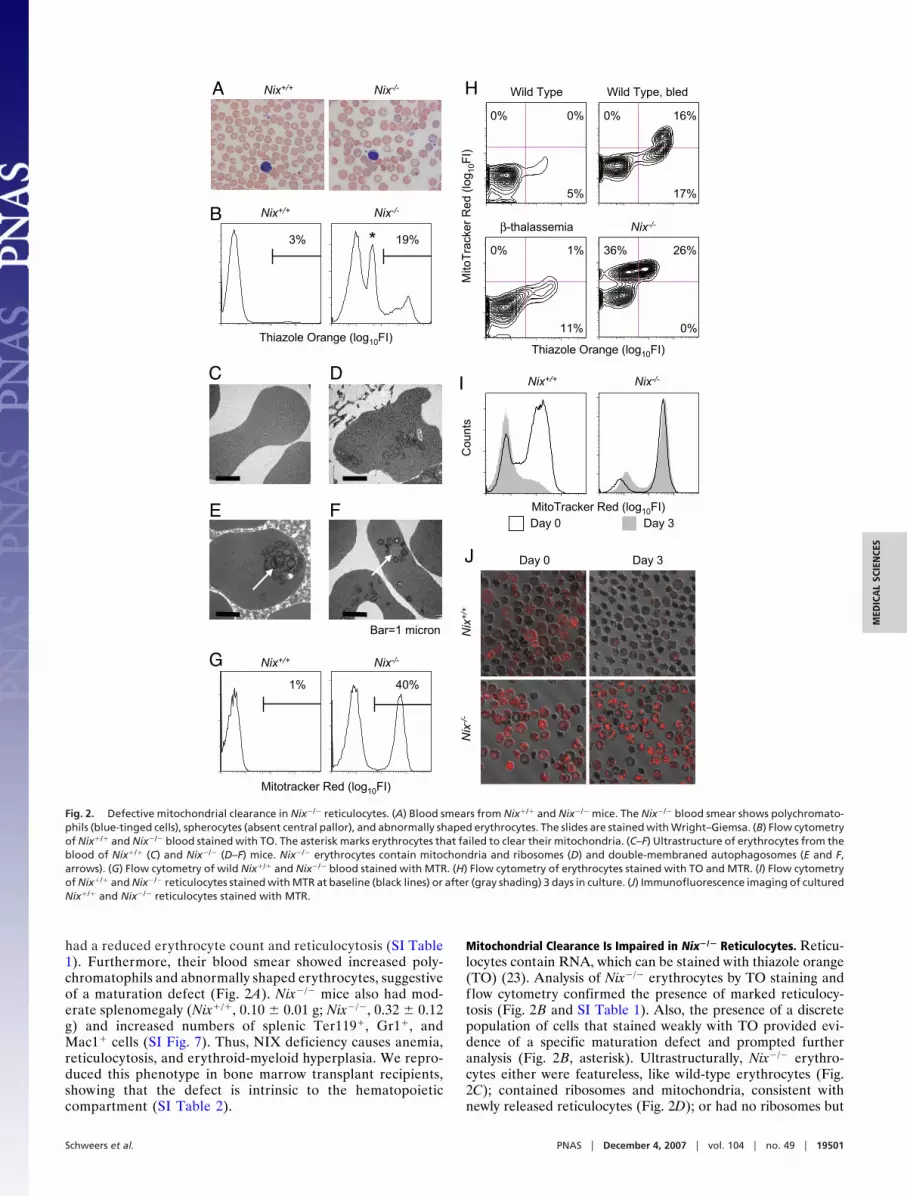
had a reduced erythrocyte count and reticulocytosis (SI Table1). Furthermore, their blood smear showed increased poly-chromatophils and abnormally shaped erythrocytes, suggestiveof a maturation defect (Fig. 2A). Nix�/� mice also had mod-erate splenomegaly (Nix�/�, 0.10 � 0.01 g; Nix�/�, 0.32 � 0.12g) and increased numbers of splenic Ter119�, Gr1�, andMac1� cells (SI Fig. 7). Thus, NIX deficiency causes anemia,reticulocytosis, and erythroid-myeloid hyperplasia. We repro-duced this phenotype in bone marrow transplant recipients,showing that the defect is intrinsic to the hematopoieticcompartment (SI Table 2).
Mitochondrial Clearance Is Impaired in Nix�/� Reticulocytes. Reticu-locytes contain RNA, which can be stained with thiazole orange(TO) (23). Analysis of Nix�/� erythrocytes by TO staining andflow cytometry confirmed the presence of marked reticulocy-tosis (Fig. 2B and SI Table 1). Also, the presence of a discretepopulation of cells that stained weakly with TO provided evi-dence of a specific maturation defect and prompted furtheranalysis (Fig. 2B, asterisk). Ultrastructurally, Nix�/� erythro-cytes either were featureless, like wild-type erythrocytes (Fig.2C); contained ribosomes and mitochondria, consistent withnewly released reticulocytes (Fig. 2D); or had no ribosomes but
A
B
Thiazole Orange (log10FI)
3% 19%
Nix-/-Nix+/+
*
G
Mitotracker Red (log10FI)
1% 40%
Nix+/+ Nix-/-
DC
FE
Bar=1 micron
Nix-/-Nix+/+
I
H Wild Type, bledWild Type
β-thalassemia Nix-/-
0%
gol( deR rek car
T otiM
01) I
F
Thiazole Orange (log10FI)
0%
5%
0% 1%
11%
0% 16%
17%
36% 26%
0%
xiN
+/+
x iN
-/-
Day 0 Day 3J
Nix-/-Nix+/+
MitoTracker Red (log10FI)
stnuoC
Day 0 Day 3
Fig. 2. Defective mitochondrial clearance in Nix�/� reticulocytes. (A) Blood smears from Nix�/� and Nix�/� mice. The Nix�/� blood smear shows polychromato-phils (blue-tinged cells), spherocytes (absent central pallor), and abnormally shaped erythrocytes. The slides are stained with Wright–Giemsa. (B) Flow cytometryof Nix�/� and Nix�/� blood stained with TO. The asterisk marks erythrocytes that failed to clear their mitochondria. (C–F) Ultrastructure of erythrocytes from theblood of Nix�/� (C) and Nix�/� (D–F) mice. Nix�/� erythrocytes contain mitochondria and ribosomes (D) and double-membraned autophagosomes (E and F,arrows). (G) Flow cytometry of wild Nix�/� and Nix�/� blood stained with MTR. (H) Flow cytometry of erythrocytes stained with TO and MTR. (I) Flow cytometryof Nix�/� and Nix�/� reticulocytes stained with MTR at baseline (black lines) or after (gray shading) 3 days in culture. (J) Immunofluorescence imaging of culturedNix�/� and Nix�/� reticulocytes stained with MTR.
Schweers et al. PNAS � December 4, 2007 � vol. 104 � no. 49 � 19501
MED
ICA
LSC
IEN
CES

contained mitochondria and undegraded autophagosomes (Fig.2 E and F, arrows). By Mitotracker Red (MTR) staining and flowcytometry, approximately half of the circulating Nix�/� eryth-rocytes contained mitochondria (Fig. 2G and SI Table 1).Furthermore, consistent with the ultrastructural studies, Nix�/�
erythrocytes contained three populations of cells (MTR�TO�,MTR�TO�, MTR�TOweak) (Fig. 2H). The MTR�TOweak pop-ulation was not seen in wild-type cells, even under conditions oferythropoietic stress, or in hemolytic diseases like �-thalassemia.Thus, Nix�/� erythrocytes contain an abnormal subpopulation ofcells that have cleared their ribosomes but retained their mito-chondria. Freshly isolated Nix�/� reticulocytes retained theirmitochondria after 3 days in culture, clearly showing thatmitochondrial clearance is defective in the absence of NIX (Fig.2 I and J). Mitochondrial clearance is regulated by 15-lipooxygenase (24); however, the effect of the lipooxygenaseinhibitor eicosatetraynoic acid on mitochondrial clearance wasmodest compared with loss of NIX (data not shown),consequently, we explored other potential mechanisms of NIXactivity.
Mitochondrial Clearance Does Not Require Established ProapoptoticPathways. The proapoptotic activity of BH3-only proteins re-quires the downstream functions of BAX or BAK (2). Therefore,we tested whether NIX-dependent mitochondrial clearance wassimilarly dependent on these proteins. Bax�/�;Bak�/� mice were
bred from compound heterozygotes and identified by genotypingand the presence of interdigital webbing (25, 26). Bax�/�;Bak�/�
reticulocytes cleared their mitochondria normally (Fig. 3A).Additionally, mitochondrial clearance from wild-type reticulo-cytes was unaffected by chemical inhibitors of the mitochondrialpermeability transition pore (MPTP), cyclosporin A, andbongkrekic acid (data not shown). Thus, NIX-dependent mito-chondrial clearance is independent of BAX, BAK, and theMPTP.
BH3-only proteins can be categorized as activators or sensi-tizers depending on whether they activate BAX and BAKdirectly or indirectly (27). BIM, BID, and possibly PUMA areactivators, whereas other BH3-only proteins are sensitizers.BIM, BID, and PUMA are expressed in late-stage erythroblasts,although BID is not present in its cleaved active form t-BID (Fig.1); therefore, we examined the roles of BIM and PUMA.Reticulocytes from Bim�/� and Puma�/� mice cleared theirmitochondria normally (Fig. 3A). Thus, mitochondrial clearancedepends on NIX but not the activator BIM or the putativeactivator PUMA. Finally, the mitochondrial clearance defect inNix�/� and Nix�/�;Puma�/� mice was equivalent, indicating alack of redundancy between these proteins.
BCL-XL is up-regulated during late erythroid maturation inparallel with NIX (Fig. 1). We therefore considered whether theeffect of NIX on mitochondrial clearance depends on BCL-XL.BCL-XL is essential for erythroid cell survival; consequently, weused a conditional model of BCL-XL deficiency (28). BclXfl/fl;Tg(MMTV-Cre) mice develop severe anemia and massive spleno-megaly because of ineffective erythropoiesis (28, 29). To rescue thesurvival defect, we bred these mice with Bax and Bak mutant mice.BclXfl/fl;Bax�/fl;Bak�/�;Tg(MMTV-Cre) mice showed dramatic cor-rection of the anemia and splenomegaly (SI Table 3). Furthermore,mitochondrial clearance was intact before or after erythropoieticstress induced by phlebotomy in BCL-XL/BAX/BAK triply defi-cient reticulocytes (Fig. 3B). Reticulocytes isolated from these micehad no detectable BCL-XL, BAX, or BAK protein but had equallevels of the mitochondrial marker protein succinate dehydroge-nase subunit B used as an internal control (Fig. 3C). Therefore,BCL-XL does not have a BAX- or BAK-independent role inmitochondrial clearance from reticulocytes.
A
B BclXfl/fl;Bax-/fl;Bak-/-;Tg(MMTV-Cre)
0% 1%
3%
BclXfl/fl;Bax-/fl;Bak-/-;Tg(MMTV-Cre), bled
0% 11%
5%
Thiazole Orange (log10FI)
gol( deR rekcar
TotiM
01)I
F
C
ep
yT dli
W
tnat
uM
elpir
T
BCL-XL
SDHB
BAX
BAK
Reticulocytes(CD71+ cells)
Wild type Bax-/-;Bak-/-
Nix-/- Puma-/-
0% 0%
2%
26% 10%
0%
0%0% 0%
4%
1%
5%
gol( deR rekcar
TotiM
0 1)I
F
Bim-/-
0%
Nix-/-;Puma-/-
0%
1%
11%
0%
23%
Thiazole Orange (log10FI)
Fig. 3. Mitochondrial clearance is BAX-, BAK-, and BCL-XL-independent. (Aand B) Flow cytometry of erythrocytes stained with TO and MTR. Genotypesare indicated. (C) Western blots of BCL-XL, BAX, BAK, and succinate dehydro-genase subunit B (SDHB). Reticulocytes were obtained from wild-type andBclXfl/fl;Bax�/fl;Bak�/�;Tg(MMTV-Cre) (triple mutant) phlebotomized mice bypositive selection for CD71 expression.
Nix-/-
Time (hr):
Beclin-1
Beclin-1
0 12 24 36 48 60
LC3-ILC3-II
LC3-ILC3-II
wild type
UN CQ
LC3-ILC3-II
Controls
Nix+/+
Fig. 4. Ubiquitin-like conjugation activity is induced independently of NIX.Nix�/� and Nix�/� FVA cells were cultured for 60 h in the presence of EPO. LC3-Iis the unconjugated form of LC3, and LC3-II is the lipid-conjugated form of LC3.Beclin-1 levels are shown for comparison. Wild-type LC3-overexpressing mu-rine embryonic fibroblasts untreated (UN) or treated with chloroquine (CQ),which causes accumulation of the lipid-modified LC3 (F.C.D. and J.L.C., un-published results), were used to confirm the identity of LC3-I and LC3-II.
19502 � www.pnas.org�cgi�doi�10.1073�pnas.0708818104 Schweers et al.

Ubiquitin-Like Conjugation Activity Is Induced Independently of NIX.Mitochondrial clearance is thought to be regulated through anautophagy-related process, and autophagy is regulated by BCL2-related proteins (30, 31); therefore, we examined the inductionof autophagy. Autophagy depends on two ubiquitin-like conju-gation pathways (32). The final products of these pathways area covalently linked form of the proteins Atg5 and Atg12 (Atg5-Atg12) and a phosphatidylethanolamine-linked form of theprotein Atg8 (Atg8-PE). The mammalian homologues of Atg8are �2-GABA(A) receptor, golgi-associated enhancer of 16 kDa,and microtubule-associated protein-1 light chain-3 (LC3) (33,34). Activation of autophagy converts LC3 from a slower mi-grating unconjugated form (LC3-I) to a faster migrating lipid-conjugated form (LC3-II). To assess the induction of autophagyduring erythroid differentiation, and its regulation by NIX, wemonitored LC3 conjugation by Western blotting of FVA cells.LC3-I was fully converted to LC3-II in Nix�/� and Nix�/� FVA
cells during terminal differentiation (Fig. 4). Therefore, auto-phagy is induced during terminal erythroid differentiation, andits induction is independent of NIX.
Mitochondrial Autophagy Is Defective in Nix�/� Reticulocytes. Todetermine the cellular processes affected by deficiency of NIX,we examined the ultrastructure of Nix�/� and Nix�/� reticulo-cytes cultured in vitro for 3 days. In Nix�/� reticulocytes,mitochondria showed an increase in electron density and loss ofinternal structure but did not show significant swelling orblebbing of the outer mitochondrial membrane (Fig. 5 Upper).Mitochondria were found individually in small vacuoles (regularwhite arrows) and in multiples in mature autophagic vacuoles(closed white arrows). These structures were eliminated throughexocytosis, and the number of mitochondria correspondinglydecreased with time. The number of ribosomes also decreasedwith time, although their mode of elimination was not obvious.The ultrastructural appearance of Nix�/� reticulocytes was strik-ingly different (Fig. 5 Lower). Mitochondria in Nix�/� reticulo-cytes were intimately associated with autophagosomes and in-completely formed isolation membranes (barbed white arrows).Notably, although some mitochondria were located inside au-tophagosomes, the majority were associated with the cytoplas-mic face of the outer autophagosomal membranes, and thecontents of the autophagosomes were frequently undegraded.Thus, mitochondrial clearance depends on the incorporation ofmitochondria into autophagosomes, and autophagosome matu-ration, and these processes depend on NIX.
Collectively, these results show a critical role for NIX inprogrammed mitochondrial clearance during reticulocyte mat-uration. Recent studies show that the autophagy-inducing activ-ity of beclin-1 is negatively regulated through interactions withmultidomain antiapoptotic BCL2-related proteins and positivelyregulated through interactions with BH3-only proteins or thecoiled-coil protein UVRAG (30, 31, 35). In the present context,however, NIX does not appear to function through beclin-1,because, as we have shown, NIX is dispensable for the inductionof autophagy in erythroid cells. Indeed, the role of macroauto-phagy in mitochondrial clearance is not well established; Atg7�/�
hepatocytes contain defective mitochondria, but Atg5�/� reticu-locytes clear their mitochondria normally (36, 37). Instead, asrecently described for two unrelated genes in yeast (38, 39), ourresults indicate that NIX is required for a selective form ofautophagy that targets mitochondria for elimination.
NIX may function through the controlled release of cyto-chrome c or other mitochondrial effector molecules. In thatregard, there are other examples of compartmentalized, caspase-dependent degradation in development (40, 41). However, to beconsistent with our results, any pore-forming activity of NIX thatmediates mitochondrial clearance must be independent of BAXand BAK, and the MPTP. Other mechanistic roles of NIX canbe envisioned but remain to be tested. Mitochondrial clearanceis important for the health of long-lived postmitotic cells, anddefects in this process have been linked to aging, degenerativediseases, and cancer; therefore, the identification of NIX as aprotein to regulate this process in higher eukaryotes, representsa significant advance.
Materials and MethodsTargeted Disruption of Nix. The Nix targeting construct was pre-pared by subcloning fragments from bacterial artificial chromo-some 199e23 (Children’s Hospital, Oakland Research Institute).Embryonic stem cell clones with homologous recombinationwere identified by Southern blot of genomic DNA digested withEcoRV. PCR primers used to generate the probe for hybridiza-tion were gcactgagtagtttgccattggcgt and ccaataaagcgtatcacaaagg.Chimeric mice were produced by the Transgenic Core Unit of St.Jude Children’s Research Hospital (SJCRH).
d1 d1
d2 d2 d2
d3 d3 d3
Nix+/+
d0
Bar=1 micron
d0 d1 d1
d1 d2 d2
d3 d3d3
Nix-/-
Fig. 5. Mitochondrial autophagy is defective in Nix�/� reticulocytes. Ultra-structure of Nix�/� (Upper) and Nix�/� (Lower) reticulocytes. (Upper) Regularwhite arrows point to individual mitochondria in vacuoles, and closed whitearrows point to mitochondria in mature autophagic vacuoles. Both wereobserved undergoing exocytosis. (Lower) Barbed white arrows point to iso-lation membranes and double-membraned autophagosomes. The length oftime in culture (days) is indicated in the lower right corner of each panel as d0,d1, d2, or d3.
Schweers et al. PNAS � December 4, 2007 � vol. 104 � no. 49 � 19503
MED
ICA
LSC
IEN
CES
Animal Studies. Reticulocytosis was induced by phlebotomy of 0.4ml of blood daily for 4 days with saline volume replacementthrough i.p. injection. On the fifth day, mice were anesthetizedand terminally bled by cardiac puncture with a heparinizedsyringe and a 23-gauge needle. For bone marrow reconstitutionstudies, 5 � 106 Nix�/� or Nix�/� bone marrow cells were injectedinto the tail vein of lethally irradiated (1,100 rads) C57BL6/Jrecipient mice. Bax and Bak mice were a gift of the StanleyKorsmeyer laboratory (Dana Farber Cancer Institute, Boston).BclXfl/fl;Tg(MMTV-Cre) mice were a gift of Lothar Hen-nighausen (National Institutes of Health, Bethesda) (28, 42).Puma mice were a gift of Gerard Zambetti (SJCRH) (43). Bimmice were purchased from The Jackson Laboratory (stocknumber 004525). All studies were performed under an animalprotocol approved by the institutional animal care and usecommittee of SJCRH.
Flow Cytometry and Immunofluorescence. Reticulocytes stainedwith MTR CMXRos (Molecular Probes) were analyzed on a BDFACSVantage SE DiVa cell sorter. Optimal separation wasobtained with an argon/krypton laser excited at 568 nm, and a610-nm-long pass filter. Reticulocytes stained with TO wereanalyzed with a 488-nm argon laser and a 525-nm band-passfilter. For some studies, reticulocytes were enriched by positiveselection with magnetic beads for CD71 expression. For theimaging of mitochondria by immunofluorescence, reticulocyteswere stained with MTR, transferred to a glass-bottom dish, andviewed with a Nikon TE2000-E inverted microscope equippedwith a C1Si confocal system and a �60 Plan Apo oil-immersionobjective (N.A. 1.45).
Electron Microscopy. Reticulocytes were fixed in 2.5% glutaral-dehyde (Tousimis). The cell pellet was postfixed with osmiumtetroxide, progressively dehydrated in ethanol, and embedded inlow-viscosity resin. We cut 80-nm-thin sections, mounted thesections on copper grids, and stained them with uranyl acetateand lead citrate. Sections were examined with a Jeol JEM-
1200EX II electron microscope and archived with a GatanES500W digital camera.
Protein Assays and Antibodies. FVA cell culture was performed asdescribed in ref. 44. Whole-cell extracts of FVA cells were madewith RIPA buffer (150 mM NaCl, 1.0% Nonidet P-40, 0.5%sodium deoxycholate, 0.1% SDS, 1 mM EDTA, and 50 mMTris�HCl, pH 8.0) with protease and phosphatase inhibitors. Forthe NIX Western blot, we resolved 50 �g of protein on an 11%SDS/PAGE gel with Laemmli running buffer and transferred tonitrocellulose. For the LC3 Western blot, we resolved 100 �g ofprotein on a 12% NuPAGE Bis-Tris gel (Invitrogen) withNuPAGE Mes SDS running buffer and transferred to PVDFmembrane. We used the following primary antibodies: NIX(X1120P; Exalpha Biologicals), BCL-X (610209; BD Bio-sciences), PUMA (JM-3339–100; Medical and Biological Lab-oratories), BIM (AAP-330; Stressgen Bioreagents), MCL1 (600-401-394; Rockland Immunochemicals), BAK (06-536; UpstateCell Signaling Solutions), �-actin monoclonal (A5441; Sigma–Aldrich), SDHB (sc-34150; Santa Cruz Biotechnology), LC3monoclonal (M115–3; Medical and Biological Laboratories),and beclin-1 (sc-11427, Santa Cruz Biotechnology). BAX andBID antibodies were a gift of the Stanley Korsmeyer laboratory.Secondary antibodies were donkey anti-rabbit IgG-HRP conju-gate, and sheep anti-mouse IgG-HRP conjugate (GE HealthcareBio-Sciences).
We thank Lothar Hennighausen (National Institutes of Health) for theBclX-f loxed and the Tg(MMTV-Cre) mice, Stanley Korsmeyer’s labora-tory for the Bax and Bak mice, Gerard Zambetti for the Puma mice,Derek Persons (SJCRH) for the �-thalassemic mice, Maurice Bondurantand Mark Koury (Vanderbilt University, Nashville, TN) for help andreagents, and Janet F. Partridge and Paul K. Brindle for comments. Theauthors acknowledge the support of the Animal Resource Center, FlowCytometry, Scientific Imaging, and Cell Microinjection facilities ofSJCRH. This work was supported by the National Institutes of Health(P.A.N., J.L.C., and M.K.), the Burroughs Wellcome Fund (M.K.), theAmerican Society of Hematology (M.K.), the Pew Charitable Trusts(J.T.O.), and the American, Lebanese, and Syrian Associated Charities(ALSAC) of St. Jude Children’s Research Hospital.
1. Danial NN, Korsmeyer SJ (2004) Cell 116:205–219.2. Wei MC, Zong WX, Cheng EH, Lindsten T, Panoutsakopoulou V, Ross AJ,
Roth KA, MacGregor GR, Thompson CB, Korsmeyer SJ (2001) Science292:727–730.
3. Kim H, Rafiuddin-Shah M, Tu HC, Jeffers JR, Zambetti GP, Hsieh JJ, ChengEH (2006) Nat Cell Biol 8:1348–1358.
4. Certo M, Del Gaizo Moore V, Nishino M, Wei G, Korsmeyer S, Armstrong SA,Letai A (2006) Cancer Cell 9:351–365.
5. Willis SN, Fletcher JI, Kaufmann T, van Delft MF, Chen L, Czabotar PE, IerinoH, Lee EF, Fairlie WD, Bouillet P, et al. (2007) Science 315:856–859.
6. Jurgensmeier JM, Xie Z, Deveraux Q, Ellerby L, Bredesen D, Reed JC (1998)Proc Natl Acad Sci USA 95:4997–5002.
7. Rosse T, Olivier R, Monney L, Rager M, Conus S, Fellay I, Jansen B, BornerC (1998) Nature 391:496–499.
8. Boyd JM, Malstrom S, Subramanian T, Venkatesh LK, Schaeper U, ElangovanB, Sa-Eipper C, Chinnadurai G (1994) Cell 79:341–351.
9. Matsushima M, Fujiwara T, Takahashi E, Minaguchi T, Eguchi Y, TsujimotoY, Suzumori K, Nakamura Y (1998) Genes Chromosomes Cancer 21:230–235.
10. Chen G, Ray R, Dubik D, Shi L, Cizeau J, Bleackley RC, Saxena S, Gietz RD,Greenberg AH (1997) J Exp Med 186:1975–1983.
11. Chen G, Cizeau J, Vande VC, Park JH, Bozek G, Bolton J, Shi L, Dubik D,Greenberg A (1999) J Biol Chem 274:7–10.
12. Yasuda M, Theodorakis P, Subramanian T, Chinnadurai G (1998) J Biol Chem273:12415–12421.
13. Imazu T, Shimizu S, Tagami S, Matsushima M, Nakamura Y, Miki T, OkuyamaA, Tsujimoto Y (1999) Oncogene 18:4523–4529.
14. Vande Velde C, Cizeau J, Dubik D, Alimonti J, Brown T, Israels S, Hakem R,Greenberg AH (2000) Mol Cell Biol 20:5454–5468.
15. Bruick RK (2000) Proc Natl Acad Sci USA 97:9082–9087.16. Sowter HM, Ratcliffe PJ, Watson P, Greenberg AH, Harris AL (2001) Cancer
Res 61:6669–6673.17. Tracy K, Dibling BC, Spike BT, Knabb JR, Schumacker P, Macleod KF (2007)
Mol Cell Biol 27:6229–6242.
18. Galvez AS, Brunskill EW, Marreez Y, Benner BJ, Regula KM, KirschenbaumLA, Dorn GW (2006) J Biol Chem 281:1442–1448.
19. Fei P, Wang W, Kim SH, Wang S, Burns TF, Sax JK, Buzzai M, Dicker DT,McKenna WG, Bernhard EJ, et al. (2004) Cancer Cell 6:597–609.
20. Aerbajinai W, Giattina M, Lee YT, Raffeld M, Miller JL (2003) Blood102:712–717.
21. Yussman MG, Toyokawa T, Odley A, Lynch RA, Wu G, Colbert MC, AronowBJ, Lorenz JN, Dorn GW (2002) Nat Med 8:725–730.
22. Diwan A, Koesters AG, Odley AM, Pushkaran S, Baines CP, Spike BT, DariaD, Jegga AG, Geiger H, Aronow BJ, et al. (2007) Proc Natl Acad Sci USA104:6794–6799.
23. Nobes PR, Carter AB (1990) J Clin Pathol 43:675–678.24. van Leyen K, Duvoisin RM, Engelhardt H, Wiedmann M (1998) Nature
395:392–395.25. Lindsten T, Ross AJ, King A, Zong WX, Rathmell JC, Shiels HA, Ulrich
E, Waymire KG, Mahar P, Frauwirth K, et al. (2000) Mol Cell 6:1389–1399.
26. Takeuchi O, Fisher J, Suh H, Harada H, Malynn BA, Korsmeyer SJ (2005) ProcNatl Acad Sci USA 102:11272–11277.
27. Letai A, Bassik MC, Walensky LD, Sorcinelli MD, Weiler S, Korsmeyer SJ(2002) Cancer Cell 2:183–192.
28. Wagner KU, Claudio E, Rucker EB, III, Riedlinger G, Broussard C,Schwartzberg PL, Siebenlist U, Hennighausen L (2000) Development (Cam-bridge, UK) 127:4949–4958.
29. Rhodes MM, Kopsombut P, Bondurant MC, Price JO, Koury MJ (2005) Blood106:1857–1863.
30. Pattingre S, Tassa A, Qu X, Garuti R, Liang XH, Mizushima N, Packer M,Schneider MD, Levine B (2005) Cell 122:927–939.
31. Maiuri MC, Le TG, Criollo A, Rain JC, Gautier F, Juin P, Tasdemir E, PierronG, Troulinaki K, Tavernarakis N, et al. (2007) EMBO J 26:2527–2539.
32. Yorimitsu T, Klionsky DJ (2005) Cell Death Differ 12(Suppl 2):1542–1552.33. Kabeya Y, Mizushima N, Ueno T, Yamamoto A, Kirisako T, Noda T,
Kominami E, Ohsumi Y, Yoshimori T (2000) EMBO J 19:5720–5728.
19504 � www.pnas.org�cgi�doi�10.1073�pnas.0708818104 Schweers et al.

34. Kabeya Y, Mizushima N, Yamamoto A, Oshitani-Okamoto S, Ohsumi Y,Yoshimori T (2004) J Cell Sci 117:2805–2812.
35. Liang C, Feng P, Ku B, Dotan I, Canaani D, Oh BH, Jung JU (2006) Nat CellBiol 8:688–699.
36. Komatsu M, Waguri S, Ueno T, Iwata J, Murata S, Tanida I, Ezaki J,Mizushima N, Ohsumi Y, Uchiyama Y, et al. (2005) J Cell Biol 169:425–434.
37. Matsui M, Yamamoto A, Kuma A, Ohsumi Y, Mizushima N (2006) BiochemBiophys Res Commun 339:485–489.
38. Kissova I, Salin B, Schaeffer J, Bhatia S, Manon S, Camougrand N (2007)Autophagy 3:329–336.
39. Tal R, Winter G, Ecker N, Klionsky DJ, Abeliovich H (2007) J Biol Chem282:5617–5624.
40. de Botton S, Sabri S, Daugas E, Zermati Y, Guidotti JE, Hermine O, KroemerG, Vainchenker W, Debili N (2002) Blood 100:1310–1317.
41. Arama E, Agapite J, Steller H (2003) Dev Cell 4:687–697.42. Wagner KU, Wall RJ, St-Onge L, Gruss P, Wynshaw-Boris A, Garrett L, Li M,
Furth PA, Hennighausen L (1997) Nucleic Acids Res 25:4323–4330.43. Jeffers JR, Parganas E, Lee Y, Yang C, Wang J, Brennan J, MacLean KH, Han
J, Chittenden T, Ihle JN, et al. (2003) Cancer Cell 4:321–328.44. Koury MJ, Sawyer ST, Bondurant MC (1984) J Cell Physiol 121:526–532.
Schweers et al. PNAS � December 4, 2007 � vol. 104 � no. 49 � 19505
MED
ICA
LSC
IEN
CES


