Embed Size (px)
Citation preview
ORIGINAL PAPER
Morphological and genetical description of Loma psittaca sp. n.isolated from the Amazonian fish speciesColomesus psittacus
Graça Casal & Edilson Matos & M. Leonor Teles-Grilo &
Carlos Azevedo
Received: 2 April 2009 /Accepted: 19 June 2009 /Published online: 11 July 2009# Springer-Verlag 2009
Abstract A previously unrecognised fish-infecting micro-sporidia (Loma psittaca n. sp.), found adherent to theintestinal mucosa of the freshwater puffer fish Colomesuspsittacus (Teleostei, Tetraodontidae) from lower AmazonRiver, was described based on light and transmissionelectron microscope and phylogenetic analysis. The whitishxenoma was completely filled by numerous spores, includ-ing several developmental stages of the parasite. In all ofthese stages, the nuclei were monokaryotic. The merogonialplasmodium divided by binary fission and the sporont gaverise to disporoblastic ovoid spores measuring 4.2 ± 0.4 ×2.8 ± 0.4 μm. In mature spores, the polar filament was
arranged in 10–11 (rarely 12) coils in one row in turn ofposterior vacuole. The polaroplast had two distinct regionsaround the manubrium. The polyribosomes were organisedin coiled tapes. The small subunit rRNA gene wassequenced and maximum parsimony analysis placed themicrosporidian described here in the clade that includes thegenera Ichthyosporidium, Loma and Pseudoloma. Based ondifferences from previously described microsporidians,such as ultrastructural characteristics of the xenoma,developmental stages including the spore and phylogeneticanalysis supported the recognition of a new species, hereinnamed L. psittaca n. sp.
Introduction
The members of the phylum Microsporidia Balbiani, 1882are widespread, minute, obligatory intracellular parasitesfound in most invertebrate phyla and in vertebrates, withthe majority of species in insects and fish (Lom andDyková 1992; Sprague et al. 1992; Larsson 1999; Lom2002). Presently, there are at least 144 available genera(Larsson 1999), 18 of them occurring in teleost fishes fromthe different geographic areas and habitat (Azevedo andMatos 2003; Lom and Nilsen 2003; Baquero et al. 2005;Casal et al. 2008), and some of them are recognised asserious pathogens for their hosts. Fishes are hosts to 156recorded species of microsporidia, 11 species belonging tothe genus Loma Morrison and Sprague, 1981 and the othereight parasitoses were classified as Loma spp. (Lom 2002).One of them, Loma myrophis, was found in the subepithe-lial tissues of the fish gut Myrophis platyrhynchus fromAmazonian fauna (Azevedo and Matos 2002; Matos et al.2003). About those from South America, particularly fromthe Amazon River where lives a diverse assemblage of
G. Casal : C. Azevedo (*)Department of Cell Biology, Institute of Biomedical Sciences,University of Porto (ICBAS/UP),Lg. A. Salazar no. 2,4099-003 Porto, Portugale-mail: [email protected]
G. Casal : C. AzevedoLaboratory of Pathology,Centre for Marine Environmental Research (CIIMAR/UP),4050-123 Porto, Portugal
G. CasalDepartamento de Ciências,Instituto Superior de Ciências da Saúde–Norte,Gandra, Portugal
E. MatosCarlos Azevedo Research Laboratory,Federal Rural University of Amazonia,Belém (Pará), Brazil
M. L. Teles-GriloLaboratory of Molecular Genetics,Institute of Biomedical Sciences,University of Porto (ICBAS/UP),Porto, Portugal
Parasitol Res (2009) 105:1261–1271DOI 10.1007/s00436-009-1547-1
several hundred species of fishes, little is known. Recently,some other microsporidiosis were described in Amazonianfishes: Amazonspora hassar was found in the gill of theteleost Hassar orestis (Azevedo and Matos 2003), Micro-sporidium brevirostris in the skeletal muscle of theabdominal cavity of the fish Brachyhypopomus brevirostris(Matos and Azevedo 2004), and Potaspora morhaphisadherent to the wall of coelomic cavity of the freshwaterfish, Potamorhaphis guianensis (Casal et al. 2008).
Ultrastructurally, the genus Loma is characterised toform xenoma, the nuclei to be unpaired during all stages ofdevelopment and the sporogony to be polysporoblasticwithin parasitophorous vacuole bound with host cell-derived membrane (Morrison and Sprague 1981; Lom andPekkarinen 1999; Lom 2002). Presently, there is littleinformation about the origin of vacuole formed during thesporogony of Loma species (Matthews et al. 2001). Smallsubunit (SSU) ribosomal DNA (rDNA) sequence compar-ison is a well-recognised technique for providing valuableinformation about phylogenetic relationships (Hillis andDixon 1991). Only for three Loma species was the SSUrDNA gene sequenced: Loma embiotocia in shiner perchCymatogaster aggregata (Shaw et al. 1997), Loma salmo-nae found in Oncorhynchus mykiss (Docker et al. 1997)and Loma acerinae (Cheney et al. 2000). Phylogeneticanalysis using SSU rDNA gene show evidences that Lomaspp. do not comprise a monophyletic group, being placed inthe same clade with the genera Ichthyosporidium andPseudoloma (Lom and Nilsen 2003). Sometimes, thephylogenetic trees do not support traditional taxonomicschemes (Sprague et al. 1992). Important morphologicalcharacters presented by those genera, such as the number ofnuclei per spores and the presence of a parasitophorousvacuole or sporophorous vesicle, are not in concordancewith molecular data.
In this paper, we describe a new species of a micro-sporidian based on morphological and ultrastructuralobservations. Phylogenetic relationships comparing theLoma psittaca SSU rRNA gene with those of other fish-infecting microsporidian species were also done. Themorphological characteristics and taxonomic position arediscussed.
Materials and methods
Fish, location of infection and prevalence
Thirty specimens of freshwater teleost puffer fish Colome-sus psittacus Bloch and Schneider, 1801 (Teleostei,Tetraodontidae) (Brazilian common name “baiacú”) werecollected from the estuarine region of the Amazon River(02°14′ S, 48°57′ W) near the city of Cametá (Pará State),
Brazil. The specimens were anaesthetised by MS 222(Sandoz Lab.) and later measured (8–12 cm in length).Infection was determined by the presence of xenomaslocated in the intestinal mucosa. The prevalence ofinfection was 30% (nine fishes in 30 examined) in bothsexes.
Light and transmission electron microscopy
For light microscopy, smears of xenoma and free sporeswere observed directly without fixation or stain by a lightmicroscope equipped with Nomarski interference contrast[differential interference contrast (DIC)] optics.
For ultrastructural studies, the xenomas were excised andfixed in 3% glutaraldehyde in 0.2 M sodium cacodylatebuffer (pH 7.2) at 4°C for 24 h. After washing overnight inthe same buffer at 4°C and post-fixed in 2% osmiumtetroxide in the same buffer and temperature for 3 h, thefragments were dehydrated through a graded ethanolascending series, followed by propylene oxide (threechanges of 2 h each) and embedded in Epon (12 h in eachchange). Semi-thin sections were stained with methyleneblue-Azur II and observed by DIC optics. Ultrathin sectionswere contrasted with aqueous uranyl acetate and lead citrateand observed with a JEOL 100CXII TEM, operated at60 kV.
DNA isolation and PCR amplification
Several xenomas were dissected from fishes followinghomogenisation to isolate the spores and then were storedin 80% ethanol at 4°C. The genomic DNA of about 5×10⁶
spores was extracted using a GenEluteTM Mammalian
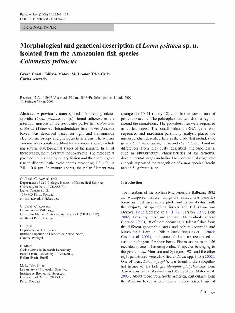
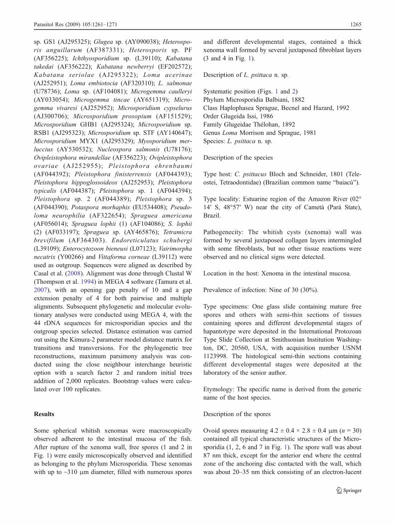
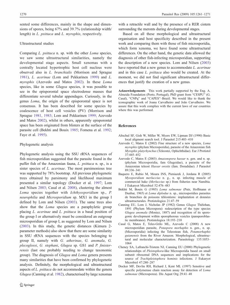
Fig. 1 Light and transmission electron micrographs of the micro-sporidian L. psittaca n. sp. parasite of Colomessus psittacus. 1 Agroup of fresh spores observed in DIC. Scale bar, 10 μm. 2 Anisolated fresh mature spore observed in DIC. Scale bar, 10 μm.3 Semi-thin section of the xenoma showing the wall (W) and thematrix of the xenoma containing numerous spores. Scale bar, 50 μm.4 Semi-thin section of the xenoma periphery showing the wall (W) andthe matrix containing developmental stages (asterisk) and numerousspores. Scale bar, 10 μm. 5 Ultrathin section of a xenoma showing thewall formed by several fibroblast layers (Fb). The matrix shows somespores (Sp). Scale bar, 5 μm. 6 Ultrathin section of some spores (Sp)sectioned at different levels showing the internal organisation. Scalebar, 1 μm. 7 Ultrastructural details of the spore apical zone showingthe spore wall (Wa), the anchoring disc (AD) and the polar filamentsections (PF) of which the anterior part was surrounded by two typesof polaroplast lamellae (Pp). Several polyribosomes organised in longtapes (arrows) are observed. Scale bar, 0.5 μm. 8 Ultrastructuraldetails of the polyribosomes arranged in long coiled tapes (arrows).The wall (Wa) and some transverse section of the polar filament (PF)are also observed. Scale bar, 0.2 μm. 9 Ultrastructural details of sometransverse sections of the polar filaments (PF) containing someinternal concentric layers. The spore wall (Wa) was composed bytwo layers of different densities (arrowheads). Scale bar, 0.2 μm
b
1262 Parasitol Res (2009) 105:1261–1271
Parasitol Res (2009) 105:1261–1271 1263
Genomic DNA Miniprep kit (Sigma) according to themanufacturer’s instructions for animal tissue protocol,except for the incubation time. The DNA was stored in50 µl of TE buffer at −20°C until use. The DNAconcentration was estimated with the QubitTM Fluorometer(Invitrogen). The majority of the region coding for the SSUrRNA gene was amplified by polymerase chain reaction(PCR) using the primers V1f (5′ CACCAGGTTGATTCTGCC 3′) and 1492r (5′ GGTTACCTTGTTACGACTT 3′) (Vossbrinck et al. 1993; Nilsen 2000). PCR wascarried out in 50 µl reactions using 10 pmol of each primer,10 nmol of each dNTP, 2 mM of MgCl2, 5 µl 10X Taqpolymerase buffer, 1.25 U Taq DNA polymerase (Invitro-gen products) and 3 µl of the genomic DNA. The reactionswere run on Hybaid PxE Thermocycler (Thermo ElectronCorporation, Milford, MA, USA). The amplificationprogramme consisted of 94°C denaturation for 5 min,followed by 35 cycles of 94°C for 1 min, 50°C for 1 minand 72°C for 2 min. A final elongation step was performedat 72°C for 10 min. Aliquots (5 µl) of PCR products werevisualised with ethidium bromide staining after running ona 1% agarose gel.
DNA sequencing
PCR product for the SSU rRNA gene has an approximatesize of 1,400 bp. It was cleaned using the MinElute PCRpurification kit (Qiagen) and then three purified PCRproducts were sequenced in both directions. Sequencingwas done using BigDye terminator v1.1 of AppliedBiosytems kit, and the sequence reactions were run on anABI3700 DNA analyser Perkin-Elmer, Applied Biosys-tems, Stabvida, Co., Oeiras, Portugal).
Distance and phylogenetic analysis
To evaluate the relationship of L. psittaca to other micro-sporidians, a homology search was performed usingBLAST programme (Altschul et al. 1990). We used 44rDNA sequences belonging to the microsporidians para-sitising fish species. The sequence and NCBI accessionnumber data obtained from GenBank are the following:Aspalatospora milevae (EF990668); Glugea anomala(AF044391); Glugea atherinae (U15987); Glugea pleco-glossi (AJ295326); Glugea stephani (AF056015); Glugea
Table 1 Comparative measurements (in μm) from Loma spp.
Loma sp. Host and local infection Habitatcountries
Spore shape Spore Polar filament References
Length Width Coils Row
L. branchialis(=L. morhua)
Melanogrammusaeglefinus Gill filaments
MarineBoreo-Artic
Ellispoidal /ovoid
4.2 2.0 16–17 isofilar (Morrison andSprague 1981)6 4 16–19
L. salmonae Oncorhynchus mykiss Freshwater Pyriform/ellipsoidal
3.7 2.2 12–14 (Putz et al. 1965)Gill filaments Several countries 4.4 2.3 14–17
L. fontinalis Salvelinus fontinalis Freshwater – 12–14 (Morrison andSprague 1983)Gill lamellae Canada
L. dimorpha Gobius niger (andothers species)
Marine Ovoid/ellipsoidal
4.5 1.8–2.0 13–15 Isofilar (Loubès et al. 1984)
Connective tissue ofdigestive tract
France and Spain
L. diplodae Diplodus sargus Marine Ovoid 4.17 2.22 17–18 Bekhti and Bouix 1985)
Vessels of the gill filaments France
L. trichiuri Trichurus savala Marine Pyriform 3.0 2.0 – (Sandeep andKalavati 1985)Gill filaments India
L. camerounensis Oerochromis niloticus Freshwater Ovoid 3.96 2.16 11–12 (Fomena et al. 1992)Oesophagus to intestine Cameroon
L. boopsi Boops boops Marine Ovoid 3.7 2.4 12–14 Isofilar (Faye et al. 1995)Liver and digestive tract Senegal 16–18
L. embiotocia Cymatogaster aggregate Marine Ovoid 4.8 2.6 14–18 (Shaw et al. 1997)Gills Canada
L. acerinae Gymnocaphalus cernuus Freshwater Ellipsoidal 4.64 2.19 11–23 Isofilar (Lom andPekkarinen 1999)Intestine wall Czech Republic
L. myrophis Myrophis platyrhynchus Freshwater Ellipsoidal 4.06 1.61 13–14 Isofilar (Azevedo andMatos 2002)Subepithelial gut tissue Brazil
L. psittaca n. sp. Colomesus psittacus Freshwater Ovoid 4.2 2.8 11–12 Isofilar This studyIntestinal wall Brazil
1264 Parasitol Res (2009) 105:1261–1271
sp. GS1 (AJ295325); Glugea sp. (AY090038); Heterospo-ris anguillarum (AF387331); Heterosporis sp. PF(AF356225); Ichthyosporidium sp. (L39110); Kabatanatakedai (AF356222); Kabatana newberryi (EF202572);Kabatana seriolae (AJ295322); Loma acerinae(AJ252951); Loma embiotocia (AF320310); L. salmonae(U78736); Loma sp. (AF104081); Microgemma caulleryi(AY033054); Microgemma tincae (AY651319); Micro-gemma vivaresi (AJ252952); Microsporidium cypselurus(AJ300706); Microsporidium prosopium (AF151529);Microsporidium GHB1 (AJ295324); Microsporidium sp.RSB1 (AJ295323); Microsporidium sp. STF (AY140647);Microsporidium MYX1 (AJ295329); Myosporidium mer-luccius (AY530532); Nucleospora salmonis (U78176);Ovipleistophora mirandellae (AF356223); Ovipleistophoraovariae (AJ252955); Pleis tophora ehrenbaumi(AF044392); Pleistophora finisterrensis (AF044393);Pleistophora hippoglossoideos (AJ252953); Pleistophoratypicalis (AF044387); Pleistophora sp. 1 (AF044394);Pleistophora sp. 2 (AF044389); Pleistophora sp. 3(AF044390); Potaspora morhaphis (EU534408); Pseudo-loma neurophilia (AF322654); Spraguea americana(AF056014); Spraguea lophii (1) (AF104086); S. lophii(2) (AF033197); Spraguea sp. (AY465876); Tetramicrabrevifilum (AF364303). Endoreticulatus schubergi(L39109); Enterocytozoon bieneusi (L07123); Vairimorphanecatrix (Y00266) and Vittaforma corneae (L39112) wereused as outgroup. Sequences were aligned as described byCasal et al. (2008). Alignment was done through Clustal W(Thompson et al. 1994) in MEGA 4 software (Tamura et al.2007), with an opening gap penalty of 10 and a gapextension penalty of 4 for both pairwise and multiplealignments. Subsequent phylogenetic and molecular evolu-tionary analyses were conducted using MEGA 4, with the44 rDNA sequences for microsporidian species and theoutgroup species selected. Distance estimation was carriedout using the Kimura-2 parameter model distance matrix fortransitions and transversions. For the phylogenetic treereconstructions, maximum parsimony analysis was con-ducted using the close neighbour interchange heuristicoption with a search factor 2 and random initial treesaddition of 2,000 replicates. Bootstrap values were calcu-lated over 100 replicates.
Results
Some spherical whitish xenomas were macroscopicallyobserved adherent to the intestinal mucosa of the fish.After rupture of the xenoma wall, free spores (1 and 2 inFig. 1) were easily microscopically observed and identifiedas belonging to the phylum Microsporidia. These xenomaswith up to ~310 μm diameter, filled with numerous spores
and different developmental stages, contained a thickxenoma wall formed by several juxtaposed fibroblast layers(3 and 4 in Fig. 1).
Description of L. psittaca n. sp.
Systematic position (Figs. 1 and 2)Phylum Microsporidia Balbiani, 1882Class Haplophasea Sprague, Becnel and Hazard, 1992Order Glugeida Issi, 1986Family Glugeidae Thélohan, 1892Genus Loma Morrison and Sprague, 1981Species: L. psittaca n. sp.
Description of the species
Type host: C. psittacus Bloch and Schneider, 1801 (Tele-ostei, Tetraodontidae) (Brazilian common name “baiacú”).
Type locality: Estuarine region of the Amazon River (02°14′ S, 48°57′ W) near the city of Cametá (Pará State),Brazil.
Pathogenecity: The whitish cysts (xenoma) wall wasformed by several juxtaposed collagen layers intermingledwith some fibroblasts, but no other tissue reactions wereobserved and no clinical signs were detected.
Location in the host: Xenoma in the intestinal mucosa.
Prevalence of infection: Nine of 30 (30%).
Type specimens: One glass slide containing mature freespores and others with semi-thin sections of tissuescontaining spores and different developmental stages ofhapantotype were deposited in the International ProtozoanType Slide Collection at Smithsonian Institution Washing-ton, DC, 20560, USA, with acquisition number USNM1123998. The histological semi-thin sections containingdifferent developmental stages were deposited at thelaboratory of the senior author.
Etymology: The specific name is derived from the genericname of the host species.
Description of the spores
Ovoid spores measuring 4.2 ± 0.4 × 2.8 ± 0.4 μm (n = 30)contained all typical characteristic structures of the Micro-sporidia (1, 2, 6 and 7 in Fig. 1). The spore wall was about87 nm thick, except for the anterior end where the centralzone of the anchoring disc contacted with the wall, whichwas about 20–35 nm thick consisting of an electron-lucent
Parasitol Res (2009) 105:1261–1271 1265
endospore and a thin electron-dense exospore (7–9 inFig. 1). The anchoring disc is located in the apical regionof the spore in an eccentric position in relation to the sporeaxis, giving a bilateral asymmetry (6 and 7 in Fig. 1). Theanterior part of the polar filament (PF) (manubrium)measured about 125 (118–131) nm in diameter and theangle of tilt anterior PF to the spore axis was ~48° (7 inFig. 1). The PF was isofilar arranged into 10–11 (rarely12) coils in one row, and when sectioned transversally, ithad 80–90 nm in diameter and exhibited three concentriclayers (9 in Fig. 1). The last coil measured ~60 nm indiameter (9 in Fig. 1). The polaroplast had two distinctlamellar structures folded around the PF. In the anteriorzone, the compacted lamellae was without lumen, whilstin the posterior lamellae, the lumen was filled withelectron-dense material (7 in Fig. 1). The nucleus, contain-ing a moderately uniform nucleoplasm, was surrounded bynumerous polyribosomes forming coiled tapes (7 and 8 inFig. 1). The posterior vacuole, situated at the basal part of thespore between the PF coils, was irregular and contained lightmaterial (7 in Fig. 1).
Developmental stages
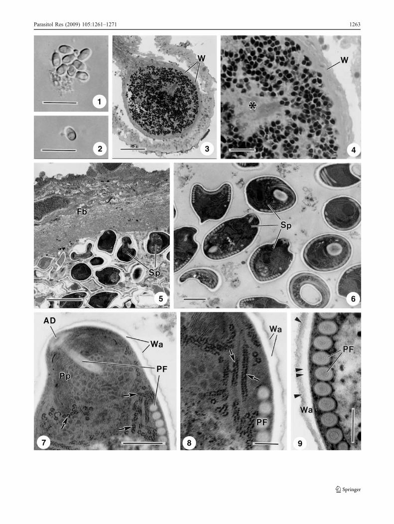
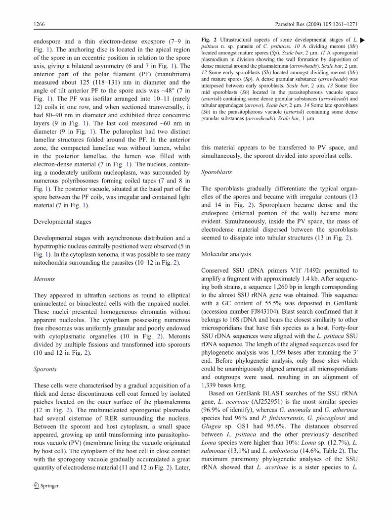
Developmental stages with asynchronous distribution and ahypertrophic nucleus centrally positioned were observed (5 inFig. 1). In the cytoplasm xenoma, it was possible to see manymitochondria surrounding the parasites (10–12 in Fig. 2).
Meronts
They appeared in ultrathin sections as round to ellipticaluninucleated or binucleated cells with the unpaired nuclei.These nuclei presented homogeneous chromatin withoutapparent nucleolus. The cytoplasm possessing numerousfree ribosomes was uniformly granular and poorly endowedwith cytoplasmatic organelles (10 in Fig. 2). Merontsdivided by multiple fissions and transformed into sporonts(10 and 12 in Fig. 2).
Sporonts
These cells were characterised by a gradual acquisition of athick and dense discontinuous cell coat formed by isolatedpatches located on the outer surface of the plasmalemma(12 in Fig. 2). The multinucleated sporogonial plasmodiahad several cisternae of RER surrounding the nucleus.Between the sporont and host cytoplasm, a small spaceappeared, growing up until transforming into parasitopho-rous vacuole (PV) (membrane lining the vacuole originatedby host cell). The cytoplasm of the host cell in close contactwith the sporogony vacuole gradually accumulated a greatquantity of electrodense material (11 and 12 in Fig. 2). Later,
this material appears to be transferred to PV space, andsimultaneously, the sporont divided into sporoblast cells.
Sporoblasts
The sporoblasts gradually differentiate the typical organ-elles of the spores and became with irregular contours (13and 14 in Fig. 2). Sporoplasm became dense and theendospore (internal portion of the wall) became moreevident. Simultaneously, inside the PV space, the mass ofelectrodense material dispersed between the sporoblastsseemed to dissipate into tubular structures (13 in Fig. 2).
Molecular analysis
Conserved SSU rDNA primers V1f /1492r permitted toamplify a fragment with approximately 1.4 kb. After sequenc-ing both strains, a sequence 1,260 bp in length correspondingto the almost SSU rRNA gene was obtained. This sequencewith a GC content of 55.5% was deposited in GenBank(accession number FJ843104). Blast search confirmed that itbelongs to 16S rDNA and bears the closest similarity to othermicrosporidians that have fish species as a host. Forty-fourSSU rDNA sequences were aligned with the L. psittaca SSUrDNA sequence. The length of the aligned sequences used forphylogenetic analysis was 1,459 bases after trimming the 3′end. Before phylogenetic analysis, only those sites whichcould be unambiguously aligned amongst all microsporidiansand outgroups were used, resulting in an alignment of1,339 bases long.
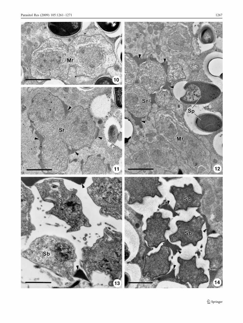
Based on GenBank BLAST searches of the SSU rRNAgene, L. acerinae (AJ252951) is the most similar species(96.9% of identify), whereas G. anomala and G. atherinaespecies had 96% and P. finisterrensis, G. plecoglossi andGlugea sp. GS1 had 95.6%. The distances observedbetween L. psittaca and the other previously describedLoma species were higher than 10%: Loma sp. (12.7%), L.salmonae (13.1%) and L. embiotocia (14.6%; Table 2). Themaximum parsimony phylogenetic analyses of the SSUrRNA showed that L. acerinae is a sister species to L.
Fig. 2 Ultrastructural aspects of some developmental stages of L.psittaca n. sp. parasite of C. psittacus. 10 A dividing meront (Mr)located amongst mature spores (Sp). Scale bar, 2 μm. 11 A sporogonialplasmodium in division showing the wall formation by deposition ofdense material around the plasmalemma (arrowheads). Scale bar, 2 μm.12 Some early sporoblasts (Sb) located amongst dividing meront (Mr)and mature spores (Sp). A dense granular substance (arrowheads) wasinterposed between early sporoblasts. Scale bar, 2 μm. 13 Some freemid sporoblasts (Sb) located in the parasitophorous vacuole space(asterisk) containing some dense granular substances (arrowheads) andtubular appendages (arrows). Scale bar, 2 μm. 14 Some late sporoblasts(Sb) in the parasitophorous vacuole (asterisk) containing some densegranular substances (arrowheads). Scale bar, 1 μm
b
1266 Parasitol Res (2009) 105:1261–1271
Parasitol Res (2009) 105:1261–1271 1267
psittaca, supported by 78% bootstrap. Both are clustered ina group together with Ichthyosporidium, Loma, Pseudo-loma genera and the Microsporidium sp. MX1. However,this clade is poorly supported with a bootstrap lower than50%. The most parsimonious trees suggested paraphyly forLoma species (Fig. 3).
Discussion
The light and ultrastructural observation of the xenoma,developmental stages as well as spore morphology de-scribed in the present study, showed all structures typical ofthe parasites belonging to the phylum Microsporidia (Lomand Dyková 1992; Larsson 1999; Lom and Nilsen 2003).
The fishes represented at least 156 species is one of thelargest group parasitised by microsporidians. They werefound in different geographic area, habitat and local ofinfection (Lom 2002). The parasite described in the presentwork is the second occurrence in teleost fish belonging tothe family Tetraodontidae. Ogawa and Yokoyama (1998)found in the intestine of the tiger puffer fish, Takifugurubripes, from a mariculture in Japan, another micro-sporidian, but it was not classified. Comparing themorphology and ultrastructural aspects of the developmen-tal stages of the parasite here described with microsporidianfish previously characterised, it seemed similar to Lomaspp. (Lom and Dyková 1992; Lom and Nilsen 2003).
Presently, there are 11 Loma species and they werereported in the gills and digestive tract of the fresh andmarine fishes (Lom 2002; Table 1). The species type Lomabranchialis was found in the gills of Atlantic cod (Morrisonand Sprague 1981), likely as L. salmonae in severalsalmonids species and from different regions (Putz et al.1965), Loma fontinalis (Morrison and Sprague 1983), Lomadimorpha found in different hosts (Loubès 1984), Lomatrichiuri (Sandeep and Kalavati 1985) and L. embiotocia(Shaw et al. 1997). Parasitising the intestine, oesophagus orliver, five species were reported: one in Europe, Lomadiplodae found in Diplodus sargus (Bekhti and Bouix 1985);Loma boopsi and Loma camerounensis identified in Africanfishes, Boops boops from Senegal (Faye et al. 1995) and inthe tilapia species Oreochromis niloticus from the Cameroon(Fomena et al. 1992), respectively; L. acerinae (Lom andPekkarinen 1999) in the freshwater Gymnocaphaluscernuus from Czech Republic. Finally, L. myrophis foundin the Amazonian fish M. platyrhynchus was described byAzevedo and Matos (2002). Concerning the habitat, shapeand size of the mature spores and the number of polarfilament coils, L. psittaca did not seem similar with otherpreviously described species. Comparing with the speciesfrom the same geographic area, L. myrophis found also inintestinal tissue of an Amazonian freshwater fish pre-T
able
2Com
parisonof
someSSU
rDNA
sequ
ences:percentage
ofidentity(top
diagon
al)andpairwisedistance
(bottom
diagon
al)ob
tained
byKim
ura-2parameter
analysis
Species
12
34
56
78
910
1112
1314
15
Lom
apsittacan.
sp.
–96
.996
.096
.095
.695
.695
.695
.294
.489
.689
.689
.287
.386
.985
.4
Lom
aacerinae
0.03
1–
97.3
97.3
96.5
96.5
96.5
96.5
96.5
90.6
89.7
91.0
88.8
88.3
86.9
Glugeaatherina
e0.04
00.02
7–
100
98.8
98.8
98.8
99.2
97.3
92.7
90.1
91.5
89.6
89.2
87.8
Glugeaan
omala
0.04
00.02
70.00
0–
98.8
98.8
98.8
99.2
97.3
92.7
90.1
91.5
89.6
89.2
87.8
Pleistoph
orafin
isterrensis
0.04
40.03
50.01
20.01
2–
100
100
98.1
96.5
93.6
89.6
91.0
89.6
90.5
89.2
Glugeaplecog
lossi
0.04
40.03
50.01
20.01
20.00
0–
100
98.1
96.5
93.6
89.6
91.0
89.6
90.5
89.2
Glugeasp.GS1
0.04
40.03
50.01
20.01
20.00
00.00
0–
98.1
96.5
93.6
89.6
91.0
89.6
90.5
89.2
Glugeastepha
ni0.04
80.03
50.00
80.00
80.01
90.01
90.01
9–
96.5
91.9
89.6
91.0
88.8
88.3
86.9
Glugeasp.
0.05
60.03
50.02
70.02
70.03
50.03
50.03
50.03
5–
90.6
90.1
90.0
87.8
87.4
86.0
Microsporidium
sp.MX1
0.10
40.09
40.07
30.07
30.06
40.06
40.06
40.08
10.09
4–
91.9
92.8
92.7
93.1
91.8
Pseud
olom
aneurop
hilia
0.10
40.10
30.09
90.09
90.10
40.10
40.10
40.10
40.09
90.08
1–
93.6
87.8
88.3
86.9
Ichthyospo
ridium
sp.
0.10
80.09
00.08
50.08
50.09
00.09
00.09
00.09
00.09
00.07
20.06
4–
90.6
90.2
88.8
Lom
asp.
0.12
70.112
0.10
40.10
40.10
40.10
40.10
40.112
0.12
20.07
30.12
20.09
4–
98.4
97.3
Lom
asalmon
ae0.13
10.117
0.10
80.10
80.09
50.09
50.09
50.117
0.12
60.06
90.117
0.09
80.01
6–
98.8
Lom
aem
biotocia
0.14
60.13
10.12
20.12
20.10
80.10
80.10
80.13
10.14
00.08
20.13
10.112
0.02
70.01
2–
1268 Parasitol Res (2009) 105:1261–1271
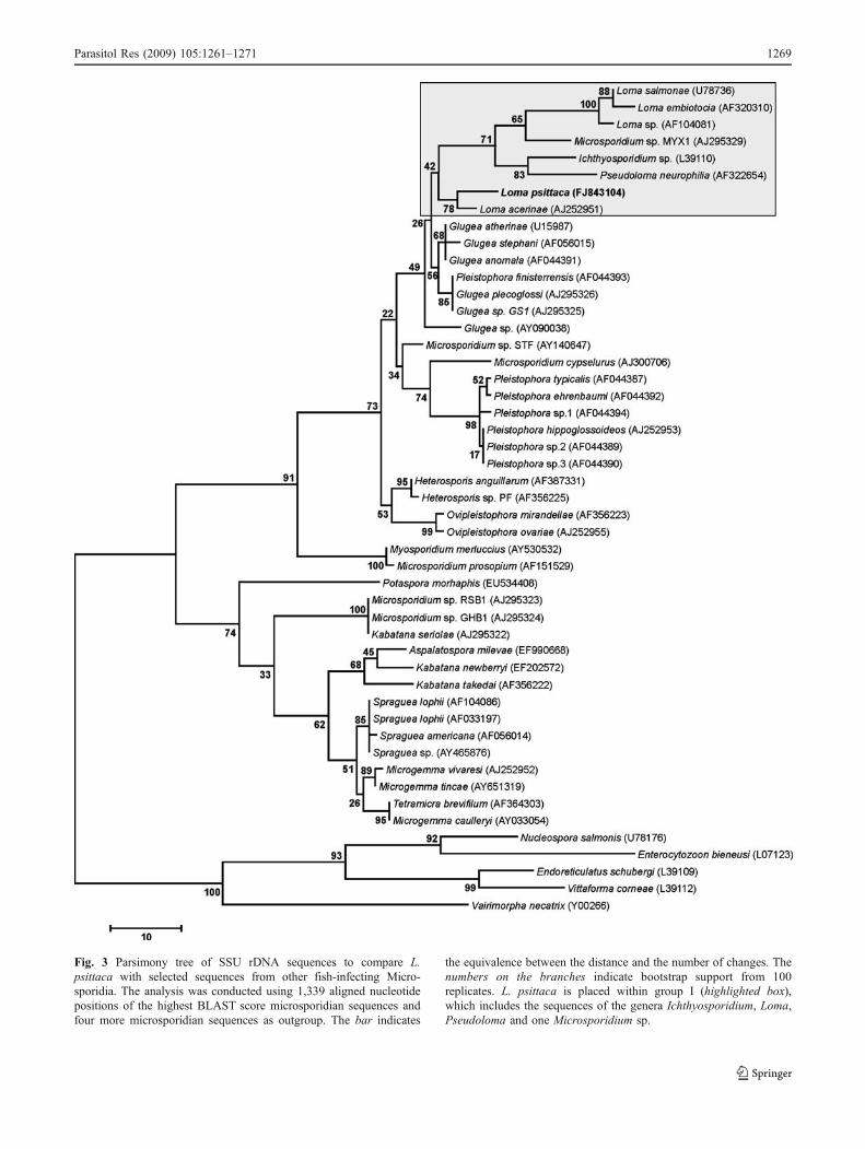
Fig. 3 Parsimony tree of SSU rDNA sequences to compare L.psittaca with selected sequences from other fish-infecting Micro-sporidia. The analysis was conducted using 1,339 aligned nucleotidepositions of the highest BLAST score microsporidian sequences andfour more microsporidian sequences as outgroup. The bar indicates
the equivalence between the distance and the number of changes. Thenumbers on the branches indicate bootstrap support from 100replicates. L. psittaca is placed within group I (highlighted box),which includes the sequences of the genera Ichthyosporidium, Loma,Pseudoloma and one Microsporidium sp.
Parasitol Res (2009) 105:1261–1271 1269
sented some differences, mainly in the shape and dimen-sions of spores, being 67% and 39.7% (relationship width/length) in L. psittaca and L. myrophis, respectively.
Ultrastructural studies
Comparing L. psittaca n. sp. with the other Loma species,we saw some ultrastructural similarities, namely thedevelopmental stage aspects. Small xenomas with acentrally located hypertrophic host cell nucleus wereobserved also in L. branchialis (Morrison and Sprague1981), L. acerinae (Lom and Pekkarinen 1999) and L.myrophis (Azevedo and Matos 2002). In these Lomaspecies, like in some Glugea species, it was possible tosee in the episporontal space electrodense masses thatdifferentiate several tubular appendages. Curiously, in thegenus Loma, the origin of the episporontal space is notconsensus. It has been described for some species bycoalescence of host cell vesicles (PV) (Morrison andSprague 1981, 1983; Lom and Pekkarinen 1999; Azevedoand Matos 2002), whilst in others, apparently episporontalspace has been originated from blisters at the surface of theparasite cell (Bekhti and Bouix 1985; Fomena et al. 1992;Faye et al. 1995).
Phylogenetic analysis
Phylogenetic analysis using the SSU rRNA sequences offish microsporidian suggested that the parasite found in thepuffer fish of the Amazonian fauna, L. psittaca n. sp., is asister species of L. acerinae. The most parsimonious treewas supported by 78% bootstrap. All previous phylogenetictrees obtained by parsimony and likelihood maximumpresented a similar topology (Docker et al. 1997; Lomand Nilsen 2003; Casal et al. 2008), clustering the almostLoma species together with Ichthyosporidium sp., P.neurophilia and Microsporidium sp. MX1 in the group Idefined by Lom and Nilsen (2003). The same trees alsoshow that the Loma species are a paraphyletic groupplacing L. acerinae and L. psittaca in a basal position ofthe group I or alternatively must be considered an outgroupmicrosporidian of group I, as suggested by Lom and Nilsen(2003). In this study, the genetic distances (Kimura 2-parameter methods) also show that there are some similarityin SSU rRNA sequences with the species belonging togroup II, namely with G. atherinae, G. anomala, G.plecoglossi, G. stephani, Glugea sp. GS1 and P. finister-rensis (last one probably needing to change taxonomicgroup). The diagnosis of Glugea and Loma genera presentsmany similarities that have been confirmed by phylogeneticanalysis. Definitely, the morphological and ultrastructuralaspects of L. psittaca do not accommodate within the generaGlugea (Canning et al. 1982), characterised by large xenomas
with a retractile wall and by the presence of a RER cisternsurrounding the meronts during developmental stages.
Based on all these morphological and ultrastructuralorganisation and host specificity described in the presentwork and comparing them with those of fish microsporidia,which form xenoma, we have found some ultrastructuraldifferences. On the other hand, the genetic data allowed thediagnosis of other fish-infecting microsporidian, supportingthe description of a new species. Lom and Nilsen (2003)have reported that a new genus to accommodate L. acerinaeand in this case L. psittaca also would be created. At themoment, we did not find significant ultrastructural differ-ences that justify the creation of a new genus.
Acknowledgements This work partially supported by the Eng. A.Almeida Foundation (Porto, Portugal), PhD grant from “CESPU” (G.Casal), “CNPq” and “CAPES”–Brazil. We would like to thank theiconographic work of Joana Carvalheiro and João Carvalheiro. Weassure that this work complies with the current laws of our countrieswhere this was performed.
References
Altschul SF, Gish W, Miller W, Myers EW, Lipman DJ (1990) Basiclocal aligment search tool. J Parasitol 215:403–410
Azevedo C, Matos E (2002) Fine structure of a new species, Lomamyrophis (phylum Microsporidia), parasite of the Amazonian fishMyrophis platyrhynchus (Teleostei, Ophichthidae). Eur J Protistol37:445–452
Azevedo C, Matos E (2003) Amazonspora hassar n. gen. and n. sp.(phylum Microsporidia, fam Glugeidae), a parasite of theAmazonian teleost Hassar orestis (fam. Doradidae). J Parasitol89:336–341
Baquero E, Rubio M, Moura INS, Pieniazek J, Jordana R (2005)Myosporidium merluccius n. g., n. sp. infecting muscle ofcommercial hake (Merluccius sp.) from fisheries near Namibia.J Eukaryot Microbiol 52:476–483
Bekhti M, Bouix G (1985) Loma salmonae (Putz, Hoffmann etDunbar, 1965) et Loma diplodae n. sp., microsporidies parasitesde branchies de poissons téléostéens: implantation et donnéesultrastructurales. Protistologica 21:47–59
Canning EU, Lom J, Nicholas JP (1982) Genus Glugea Thélohan,1891 (Phylum Microspora): redescription of the type speciesGlugea anomala (Moniez, 1887) and recognition of its sporo-gonic development within sporophorous vesicles (pansporoblas-tic membranes). Protistologica 18:193–210
Casal G, Matos E, Teles-Grilo ML, Azevedo C (2008) A newmicrosporidian parasite, Potaspora morhaphis n. gen., n. sp.(Microsporidia) infecting the Teleostean fish, Potamorhaphisguianensis from the River Amazon. Morphological, ultrastruc-tural and molecular characterization. Parasitology 135:1053–1064
Cheney SA, Lafranchi-Tristem NJ, Canning EU (2000) Phylogeneticrelationships of Pleistophora-like Microsporidia based on smallsubunit ribosomal DNA sequences and implications for thesource of Trachipleistophora hominis infections. J EukaryotMicrobiol 47:280–287
Docker MF, Devlin RH, Richard J, Kent ML (1997) Sensitive andspecific polymerase chain reaction assay for detection of Lomasalmonae (Microsporea). Dis Aquat Org 29:41–48
1270 Parasitol Res (2009) 105:1261–1271
Faye N, Toguebaye BS, Bouix G (1995) On the cytology anddevelopment of Loma boopsi n. sp. (Microspora, Glugeidae),parasite of Boops boops (Pisces, Teleostei, Sparidae) from thecoasts of Senegal. Arch Protistenkd 146:85–93
Fomena A, Coste F, Bouix G (1992) Loma camerounensis sp. nov.(Protozoa: Microsporida) a parasite of Oreochromis niloticusLinnaeus, 1757 (Teleost: Cichlidae) in fish-rearing ponds inMelen, Yaoundé, Cameroon. Parasitol Res 78:201–208
Hillis DM, Dixon MT (1991) Ribosomal DNA: molecular evolutionand phylogenetic inference. Q Rev Biol 66:411–453
Larsson JIR (1999) Identification of Microsporidia. Acta Protozool38:161–197
Lom J (2002) A catalogue of described genera and species ofmicrosporidians parasitic in fish. Syst Parasitol 53:81–99
Lom J, Dyková I (1992) Protozoan parasites of fishes. Elsevier,Amsterdam, p 315
Lom J, Nilsen F (2003) Fish microsporidia: fine structural diversityand phylogeny. Int J Parasitol 33:107–127
Lom J, Pekkarinen M (1999) Ultrastructural observations on Lomaacerinae (Jírovec, 1930) comb. nov. (phylum Microsporidia).Acta Protozool 38:61–74
Loubès C, Maurand J, Gasc C, Buron I, Barral J (1984) Étudeultrastructurale de Loma dimorpha n. sp., microsporidie parasitede poissons Gobiidae languedociens. Protistologica 20:579–589
Matos E, Azevedo C (2004) Ultrastructural description of Micro-sporidium brevirostris sp. n., parasite of the teleostean Brachy-hypopomus brevirostris (Hypopomidae) from the Amazon River.Acta Protozool 43:261–267
Matos E, Corral L, Azevedo C (2003) Ultrastructural details of thexenoma of Loma myrophis (phylum Microsporidia) and extrusionof the polar tube during autoinfection. Dis Aquat Org 54:203–207
Matthews JL, Brown AMV, Larison K, Bishop-Stewart JK, Rogers P,Kent ML (2001) Pseudoloma neurophilia n. g., n. sp., a newmicrosporidium from the central nervous system of the zebrafish(Danio rerio). J Eukaryot Microbiol 48:227–233
Morrison CM, Sprague V (1981) Electron microscopical study of anew genus and new species of microsporida in the gills ofAtlantic cod Gadus morhua L. J Fish Dis 4:15–32
Morrison CM, Sprague V (1983) Loma salmonae (Putz, Hoffman andDunbar, 1965) in the rainbow trout, Salmo gairdneri Richarson,and L. fontinalis sp. nov. (Microsporida) in the brook trout,Salvelinus fontinalis (Mitchill). J Fish Dis 6:345–353
Nilsen F (2000) Small subunit ribosomal DNA phylogeny of micro-sporidia with particular reference to genera that infect fish. JParasitol 86:128–133
Ogawa K, Yokoyama H (1998) Parasitic diseases of cultured marinefish in Japan. Fish Pathol 33:303–309
Putz RE, Hoffman GL, Dunbar CE (1965) Two new species ofPleistophora (Microsporidea) from North America fish with asynopsis of Microsporidea of freshwater and euryhaline fishes. JProtozool 12:228–236
Sandeep BV, Kalavati C (1985) A new microsporidian, Loma trichiurin. sp., from the gill of a marine fish, Trichiurus salva Cuv.(Trichiuridae). Indian J Parasitol 9:257–259
Shaw RW, Kent ML, Docker MF, Brown AMV, Devlin RH, AdamsonML (1997) A new species of Loma (Microsporea) in shiner perch(Cymatogaster aggregata). J Parasitol 83:296–301
Sprague V, Becnel JJ, Hazard EI (1992) Taxonomy of phylumMicrospora. Crit Rev Microbiol 18:285–395
Tamura K, Dudley J, Nei M, Kumar S (2007) MEGA4: molecularevolutionary genetics analysis (MEGA) software version 4.0.Mol Biol Evol 24:1596–1599
Thompson JD, Higgins DG, Gilson TJ (1994) Clustal W: improvingthe sensitivity of progressive multiple sequence alignmentthrough sequence weighting, position-specific gap penalties andweight matrix choice. Nucleic Acids Res 22:4673–4680
Vossbrinck CR, Baker MD, Didier ES, Debrunner-Vossbrinck BA,Shadduck JA (1993) Ribosomal DNA sequences of Encephali-tozoon hellem and Encephalitozoon cuniculi: species identifica-tion and phylogenetic construction. J Eukaryot Microbiol40:354–362
Parasitol Res (2009) 105:1261–1271 1271


