Embed Size (px)
Citation preview

Local and genetic determinants of vascular endothelial growthfactor expression in advanced proliferative diabetic retinopathy
Mojca Globočnik Petrovič,1 Peter Korošec,2 Mitja Košnik,2 Joško Osredkar,3 Marko Hawlina,1 Borut Peterlin,4 Daniel Petrovič5
1Eye Clinic, University Medical Centre Ljubljana, Ljubljana, Slovenia; 2University Clinic of Respiratory and Allergic Diseases,Golnik, Slovenia; 3University Institute for Clinical Chemistry and Biochemistry, University Medical Centre Ljubljana, Ljubljana,Slovenia; 4Division of Medical Genetics, Department of Obstetrics and Gynecology, University Medical Centre Ljubljana,Ljubljana, Slovenia; 5Institute of Histology and Embryology, Medical Faculty, University of Ljubljana, Ljubljana, Slovenia
Purpose: In proliferative diabetic retinopathy (PDR) and other angiogenesis-associated diseases, increased levels ofcytokines, inflammatory cells, growth factors, and angiogenic factors are present. Vascular endothelial growth factor(VEGF) appears to play a central role in mediating microvascular pathology in PDR. The purpose of the present studywas to search for the association between the –634 C/G polymorphism of the VEGF gene and PDR. Moreover, it washoped to determine whether serum and vitreous levels of VEGF are affected by genetic factors.Methods: This cross-sectional case-control study enrolled 349 unrelated Slovene subjects (Caucasians) with type 2diabetes mellitus. The case group consisted of 206 patients with an advanced form of PDR and for whom vitrectomy wasperformed, and the control group had 143 patients who had no clinical signs of diabetic retinopathy but did have type 2diabetes of more than 10 years duration. To analyze the genotype distribution we had to compare the genotype frequenciesin diabetics with PDR (cases, n=206) and diabetics without diabetic retinopathy (control group, n=143). Additionally, toevaluate the effect of diabetes on the VEGF serum levels 2 groups, diabetics and non diabetics, were compared. Firstgroup were diabetics (diabetics with PDR, n=104), and second group were 29 subjects without diabetes.Results: The –634 C/G VEGF polymorphism was not associated with PDR. Mean serum and vitreous levels of VEGFwere statistically significantly higher in PDR in comparison to the control group. Moreover, significantly higher serumand vitreous levels of VEGF were demonstrated in diabetics with the CC genotype compared to those with the other (CG+ GG) genotypes.Conclusions: VEGF is an important cytokine in PDR. Despite the effect of the –634 C/G VEGF polymorphism on serumand vitreous levels of VEGF in PDR, it failed to contribute to the genetic susceptibility to PDR.
In proliferative diabetic retinopathy (PDR) and otherangiogenesis-associated diseases, increased levels ofcytokines, inflammatory cells, growth factors, and angiogenicfactors are present [1-5]. Vascular endothelial growth factor(VEGF) appears to play a central role in mediatingmicrovascular pathology in PDR. VEGF is capable ofinducing the earliest changes in diabetic retinopathy such asleukostasis and blood-retinal barrier breakdown [6,7] as wellas macular edema and neovascularization in progression ofdiabetic retinopathy [1]. In the vitreous of patients with PDR,VEGF levels have been found to be increased [1,2,4,5].Although diabetes duration and inadequate glycemic controlare important risk factors in the development of PDR, geneticfactors may play a significant role in the pathogenesis of PDR[8,9]. There is considerable variation in VEGF expressionamong individuals, with several different polymorphismsbeing reported [10]. The VEGF 634 C/G (rs2010963)polymorphism in the 5′-untranslated region has been reported
Correspondence to: Daniel Petrovič, Institute of Histology andEmbryology, Medical Faculty, University Ljubljana, Korytkova 2,1000 Ljubljana, Slovenia; Phone: 386 1 5437367; FAX: 386 15437361; email: [email protected]
to be associated with variations in VEGF serumconcentrations and with a susceptibility to disorders, such asdiabetic retinopathy, diabetic nephropathy, andcardiovascular diseases [8,10-13].
To investigate the impact of genetic polymorphisms ofVEGF on PDR in a Slovenian population (Caucasians) withtype 2 diabetes, we searched for the association between the-634 C/G VEGF polymorphism and PDR in subjects with type2 diabetes. Moreover, the aim of the study was to determinethe serum and vitreous levels of VEGF of patients with PDR,and whether serum and vitreous levels of VEGF are affectedby genetic factors.
METHODSPatients: This cross-sectional case-control study enrolled 349(age range 35 to 87 years; 152 men, 197 women) unrelatedSlovene subjects (Caucasians) with type 2 diabetes mellituswho had a defined ophthalmologic status. Patients wereclassified as having type 2 diabetes according to the currentAmerican Diabetes Association criteria for the diagnosis andclassification of diabetes [14]. Patients were recruited fromthe Eye Clinic of the University Medical Centre Ljubljana
Molecular Vision 2008; 14:1382-1387 <http://www.molvis.org/molvis/v14/a166>Received 5 January 2008 | Accepted 29 June 2008 | Published 30 July 2008
© 2008 Molecular Vision
1382
between January 2002 and April 2007. Fundus examinationwas performed by a senior ophthalmologist (M.G.P.) afterpupil dilatation (tropicamide and phenylephrine 2.5%) usingslit-lamp biomicroscopy with non-contact lens, and waselectronically documented with a 50°-angle fundus camera(Topcon-TRC 40-IX; Topcon, Tokyo, Japan). Staging ofdiabetic retinopathy was determined according to the EarlyTreatment Diabetic Retinopathy Study Research Groupretinopathy severity scale [15].
The study group consisted of 206 patients with anadvanced form of PDR (new vessel formation as well asfibrous proliferation with or without vitreous hemorrhage) inwhom vitrectomy was indicated and performed due tovitreous hemorrhage, macular detachment, or macularthreatening detachment. The control group consisted of 143patients who had type 2 diabetes of more than 10 yearsduration but had no clinical signs of diabetic retinopathy.
In 68 out of 206 patients with PDR (71 eyes) 0.3 mlvitreous fluid samples were obtained by vitreoretinal surgery.The study excluded patients who had previous vitrectomy,neovascularization of no diabetic etiology, recent vitreoushemorrhage (less than two months), or a history of ocularinflammation and photocoagulation in the preceding threemonths.
Macular edema was defined as being clinicallysignificant based on observations rendered by the EarlyTreatment Diabetic Retinopathy Study [16]. PDR was foundto be active in 54 eyes and inactive in 17 eyes, according tothe method reported by Aiello and associates [1].Neovascularization was considered to be active if new vesselswere perfused, multibranching iridic, or preretinal capillaries;it was considered to be inactive if previously documentedactive proliferation had regressed fully or if only nonperfusedgliotic vessels or fibrosis were present [1]. The extent ofretinal laser photocoagulation was classified into three grades:grade 1) no photocoagulation; grade 2) focalphotocoagulation; and grade 3) panretinal photocoagulation(defined as extensive photocoagulation in all four quadrantsof the retina).
Sample collection: Fasting serum VEGF levels were analyzedin 104 out 206 of patients with PDR and in 29 patients withoutdiabetes. Vitreous fluid samples (0.3 ml) were obtained byvitreoretinal surgery (from the midvitreous at the initial stageof vitrectomy by aspiration into a 2 ml syringe attached to thevitreous counter before starting intravitreal infusion ofbalanced salt solution) from 71 eyes of 68 consecutive patientswith PDR, and from 17 eyes of 17 consecutive nondiabeticpatients in whom vitrectomy was performed because ofidiopatic macular hole [17]. Vitreous was cut and aspiratedinto a 2 ml syringe attached to the vitreous cutter beforestarting intravitreal infusion of balanced salt solution.
Single nucleotide polymorphism –634 C/G genepolymorphisms and diabetic retinopathy: The –634 C/GVEGF polymorphism was evaluated as described previously[8]. Genotype determination: Genomic DNA was isolatedfrom peripheral blood lymphocytes by standard methods andstored at -20 °C. Genotyping was carried out by polymerasechain reaction-restriction fragment length polymorphism(PCR-RFLP) analysis. In brief, each PCR reaction (20 μl)contained 0.4 μl 10 mM dNTP (Gibco), 2 μl 10-X PCR buffer(Gibco), 1.2 μl 25 mM MgCl2 (final concentration 1,5 mM),1 μl 10 μM of each primer (5′-TTG CTT GCC ATT CCC CACTTG A-3′, 5′-CCG AAG CGA GAA CAG CCC AGA A-3′),0.7 μl (500 ng) DNA, 12.5 μl H20, 1 μl 5% DMSO, 0,5 unitof AmliTaq Gold DNA-polymerase (Applied Biosystems).Cycling parameters were as follows: 3 min 94 °C for primarydenaturation, followed by 30 cycles of 1 minute 94 °C and 1minute 59 °C, 1 minute 72 °C. After digestion for 16 hours at65 °C by 1 unit of the restriction enzyme BsmFI, the restrictionproducts were electrophoresed on a 2.0% agarose gel and thenvisualized using ethidium bromide staining. The -634G alleleresult in the gain of a BsmFI site. Two investigators (B.P.,D.P.), blinded for case or control status of the DNA sample,performed the genotype classification.VEGF serum concentration assay: For the determination offasting serum VEGF concentration (isoform VEGF 165), weused a solid phase sandwich ELISA, which involved twokinds of highly specific antibodies (hVEGF Assay Kit; IBLCo., Ltd., Takasaki-shi, Gunma, Japan). In our study therespective coefficient of variation (CV; %) were between 3and 5.5 for interassay measurements, and between 2.6 and 5.3for intraassay measurements.
The vitreous VEGF concentration was determined withthe cytometric bead array method (CBA; BD Biosciences, SanDiego, CA). The samples were collected in sterile tubes,rapidly frozen and then stored at –80 C until analyzed. VEGFconcentrations were measured using CBA (BD Biosciences).The VEGF capture beads were mixed with phycoerythrin –conjugated detection antibodies. These capture beads wereincubated with recombinant standards or test samples(vitreous) to form sandwich complexes. Two-color flowcytometric analysis was performed using a FACSCalibur flowcytometer (BD Biosciences). Data were acquired andanalyzed using Becton Dickinson Cytometric Bead ArrayCBA software, and concentrations were determined from thestandard curves, plotting recombinant calibratorconcentration versus FL-2 mean fluorescence intensity.Theuse of this method made it possible to detect two of the fourVEGF isoforms (VEGF 121 and VEGF 165).Statistical analysis: We used a nonparametric Mann–Whitneytest and Kruskal–Wallis test for multiple group comparison orthe Student t-test as appropriate. The χ2 test was used tocompare discrete variables and to compare genotypedistributions. In addition, all variables that showed significant
Molecular Vision 2008; 14:1382-1387 <http://www.molvis.org/molvis/v14/a166> © 2008 Molecular Vision
1383
differences by univariate methods (χ2 test, unpaired Studentt test) were analyzed together in a logistic regression analysis.Statistical analysis was performed using the SPSS programfor Windows version 14 (SPSS Inc. Chicago, IL).
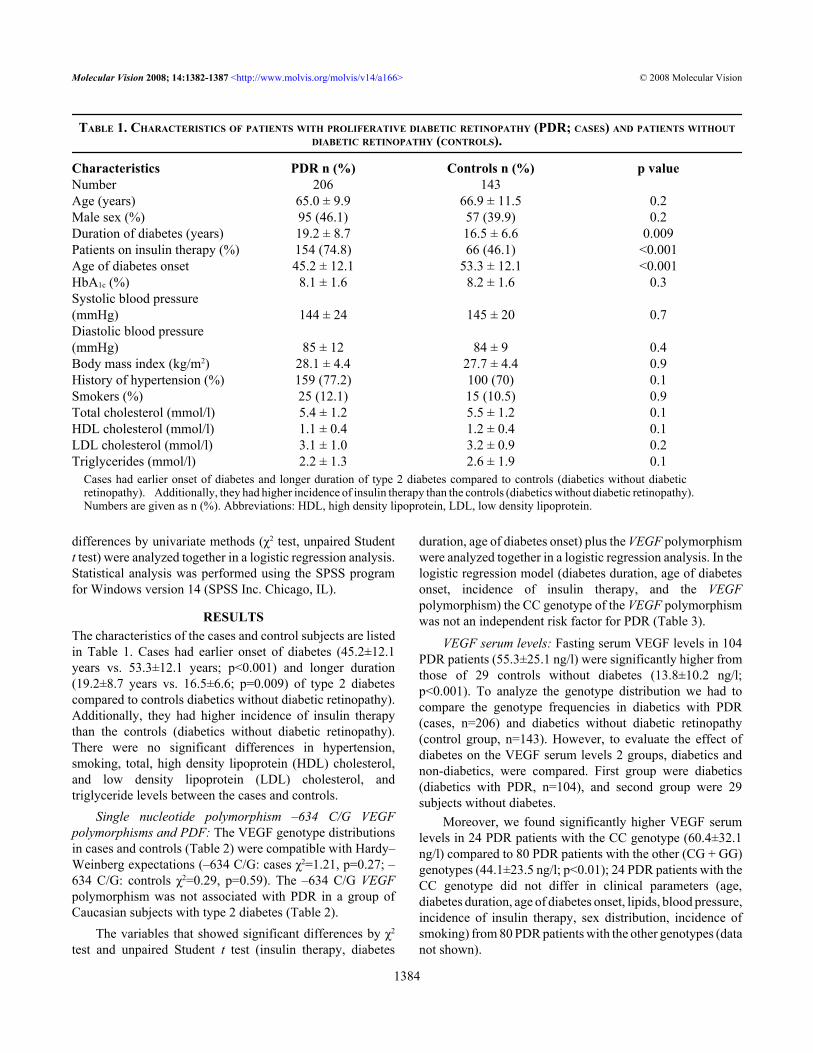
RESULTSThe characteristics of the cases and control subjects are listedin Table 1. Cases had earlier onset of diabetes (45.2±12.1years vs. 53.3±12.1 years; p<0.001) and longer duration(19.2±8.7 years vs. 16.5±6.6; p=0.009) of type 2 diabetescompared to controls diabetics without diabetic retinopathy).Additionally, they had higher incidence of insulin therapythan the controls (diabetics without diabetic retinopathy).There were no significant differences in hypertension,smoking, total, high density lipoprotein (HDL) cholesterol,and low density lipoprotein (LDL) cholesterol, andtriglyceride levels between the cases and controls.
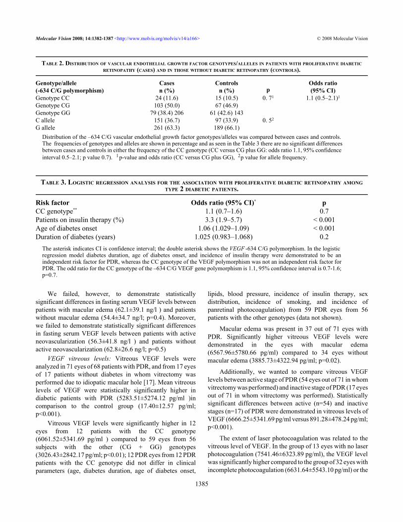
Single nucleotide polymorphism –634 C/G VEGFpolymorphisms and PDF: The VEGF genotype distributionsin cases and controls (Table 2) were compatible with Hardy–Weinberg expectations (–634 C/G: cases χ2=1.21, p=0.27; –634 C/G: controls χ2=0.29, p=0.59). The –634 C/G VEGFpolymorphism was not associated with PDR in a group ofCaucasian subjects with type 2 diabetes (Table 2).
The variables that showed significant differences by χ2
test and unpaired Student t test (insulin therapy, diabetes
duration, age of diabetes onset) plus the VEGF polymorphismwere analyzed together in a logistic regression analysis. In thelogistic regression model (diabetes duration, age of diabetesonset, incidence of insulin therapy, and the VEGFpolymorphism) the CC genotype of the VEGF polymorphismwas not an independent risk factor for PDR (Table 3).
VEGF serum levels: Fasting serum VEGF levels in 104PDR patients (55.3±25.1 ng/l) were significantly higher fromthose of 29 controls without diabetes (13.8±10.2 ng/l;p<0.001). To analyze the genotype distribution we had tocompare the genotype frequencies in diabetics with PDR(cases, n=206) and diabetics without diabetic retinopathy(control group, n=143). However, to evaluate the effect ofdiabetes on the VEGF serum levels 2 groups, diabetics andnon-diabetics, were compared. First group were diabetics(diabetics with PDR, n=104), and second group were 29subjects without diabetes.
Moreover, we found significantly higher VEGF serumlevels in 24 PDR patients with the CC genotype (60.4±32.1ng/l) compared to 80 PDR patients with the other (CG + GG)genotypes (44.1±23.5 ng/l; p<0.01); 24 PDR patients with theCC genotype did not differ in clinical parameters (age,diabetes duration, age of diabetes onset, lipids, blood pressure,incidence of insulin therapy, sex distribution, incidence ofsmoking) from 80 PDR patients with the other genotypes (datanot shown).
TABLE 1. CHARACTERISTICS OF PATIENTS WITH PROLIFERATIVE DIABETIC RETINOPATHY (PDR; CASES) AND PATIENTS WITHOUTDIABETIC RETINOPATHY (CONTROLS).
Characteristics PDR n (%) Controls n (%) p valueNumber 206 143Age (years) 65.0 ± 9.9 66.9 ± 11.5 0.2Male sex (%) 95 (46.1) 57 (39.9) 0.2Duration of diabetes (years) 19.2 ± 8.7 16.5 ± 6.6 0.009Patients on insulin therapy (%) 154 (74.8) 66 (46.1) <0.001Age of diabetes onset 45.2 ± 12.1 53.3 ± 12.1 <0.001HbA1c (%) 8.1 ± 1.6 8.2 ± 1.6 0.3Systolic blood pressure(mmHg) 144 ± 24 145 ± 20 0.7Diastolic blood pressure(mmHg) 85 ± 12 84 ± 9 0.4Body mass index (kg/m2) 28.1 ± 4.4 27.7 ± 4.4 0.9History of hypertension (%) 159 (77.2) 100 (70) 0.1Smokers (%) 25 (12.1) 15 (10.5) 0.9Total cholesterol (mmol/l) 5.4 ± 1.2 5.5 ± 1.2 0.1HDL cholesterol (mmol/l) 1.1 ± 0.4 1.2 ± 0.4 0.1LDL cholesterol (mmol/l) 3.1 ± 1.0 3.2 ± 0.9 0.2Triglycerides (mmol/l) 2.2 ± 1.3 2.6 ± 1.9 0.1
Cases had earlier onset of diabetes and longer duration of type 2 diabetes compared to controls (diabetics without diabeticretinopathy). Additionally, they had higher incidence of insulin therapy than the controls (diabetics without diabetic retinopathy).Numbers are given as n (%). Abbreviations: HDL, high density lipoprotein, LDL, low density lipoprotein.
Molecular Vision 2008; 14:1382-1387 <http://www.molvis.org/molvis/v14/a166> © 2008 Molecular Vision
1384
We failed, however, to demonstrate statisticallysignificant differences in fasting serum VEGF levels betweenpatients with macular edema (62.1±39.1 ng/l ) and patientswithout macular edema (54.4±34.7 ng/l; p=0.4). Moreover,we failed to demonstrate statistically significant differencesin fasting serum VEGF levels between patients with activeneovascularization (56.3±41.8 ng/l ) and patients withoutactive neovascularization (62.8±26.6 ng/l; p=0.5)
VEGF vitreous levels: Vitreous VEGF levels wereanalyzed in 71 eyes of 68 patients with PDR, and from 17 eyesof 17 patients without diabetes in whom vitrectomy wasperformed due to idiopatic macular hole [17]. Mean vitreouslevels of VEGF were statistically significantly higher indiabetic patients with PDR (5283.51±5274.12 pg/ml )incomparison to the control group (17.40±12.57 pg/ml;p<0.001).
Vitreous VEGF levels were significantly higher in 12eyes from 12 patients with the CC genotype(6061.52±5341.69 pg/ml ) compared to 59 eyes from 56subjects with the other (CG + GG) genotypes(3026.43±2842.17 pg/ml; p<0.01); 12 PDR eyes from 12 PDRpatients with the CC genotype did not differ in clinicalparameters (age, diabetes duration, age of diabetes onset,
lipids, blood pressure, incidence of insulin therapy, sexdistribution, incidence of smoking, and incidence ofpanretinal photocoagulation) from 59 PDR eyes from 56patients with the other genotypes (data not shown).
Macular edema was present in 37 out of 71 eyes withPDR. Significantly higher vitreous VEGF levels weredemonstrated in the eyes with macular edema(6567.96±5780.66 pg/ml) compared to 34 eyes withoutmacular edema (3885.73±4322.94 pg/ml; p=0.02).
Additionally, we wanted to compare vitreous VEGFlevels between active stage of PDR (54 eyes out of 71 in whomvitrectomy was performed) and inactive stage of PDR (17 eyesout of 71 in whom vitrectomy was performed). Statisticallysignificant differences between active (n=54) and inactivestages (n=17) of PDR were demonstrated in vitreous levels ofVEGF (6666.25±5341.69 pg/ml versus 891.28±478.24 pg/ml;p<0.001).
The extent of laser photocoagulation was related to thevitreous level of VEGF. In the group of 13 eyes with no laserphotocoagulation (7541.46±6323.89 pg/ml), the VEGF levelwas significantly higher compared to the group of 32 eyes withincomplete photocoagulation (6631.64±5543.10 pg/ml) or the
TABLE 2. DISTRIBUTION OF VASCULAR ENDOTHELIAL GROWTH FACTOR GENOTYPES/ALLELES IN PATIENTS WITH PROLIFERATIVE DIABETICRETINOPATHY (CASES) AND IN THOSE WITHOUT DIABETIC RETINOPATHY (CONTROLS).
Genotype/allele(-634 C/G polymorphism)
Casesn (%)
Controlsn (%) p
Odds ratio(95% CI)
Genotype CC 24 (11.6) 15 (10.5) 0. 71 1.1 (0.5–2.1)1
Genotype CG 103 (50.0) 67 (46.9)Genotype GG 79 (38.4) 206 61 (42.6) 143C allele 151 (36.7) 97 (33.9) 0. 52
G allele 261 (63.3) 189 (66.1)Distribution of the –634 C/G vascular endothelial growth factor genotypes/alleles was compared between cases and controls.The frequencies of genotypes and alleles are shown in percentage and as seen in the Table 3 there are no significant differencesbetween cases and controls in either the frequency of the CC genotype (CC versus CG plus GG: odds ratio 1.1, 95% confidenceinterval 0.5–2.1; p value 0.7). 1p-value and odds ratio (CC versus CG plus GG), 2p value for allele frequency.
TABLE 3. LOGISTIC REGRESSION ANALYSIS FOR THE ASSOCIATION WITH PROLIFERATIVE DIABETIC RETINOPATHY AMONGTYPE 2 DIABETIC PATIENTS.
Risk factor Odds ratio (95% CI)* pCC genotype** 1.1 (0.7–1.6) 0.7Patients on insulin therapy (%) 3.3 (1.9–5.7) < 0.001Age of diabetes onset 1.06 (1.029–1.09) < 0.001Duration of diabetes (years) 1.025 (0.983–1.068) 0.2
The asterisk indicates CI is confidence interval; the double asterisk shows the VEGF -634 C/G polymorphism. In the logisticregression model diabetes duration, age of diabetes onset, and incidence of insulin therapy were demonstrated to be anindependent risk factor for PDR, whereas the CC genotype of the VEGF polymorphism was not an independent risk factor forPDR. The odd ratio for the CC genotype of the –634 C/G VEGF gene polymorphism is 1.1, 95% confidence interval is 0.7-1.6;p=0.7.
Molecular Vision 2008; 14:1382-1387 <http://www.molvis.org/molvis/v14/a166> © 2008 Molecular Vision
1385
group of 26 eyes with photocoagulation in all four quadrants(2495.3±2743.56 pg/ml; p=0.001).
DISCUSSIONIn our study we analyzed the VEGF –634 C/G polymorphismas a potential genetic marker of PDR. The VEGF –634 C/Gpolymorphism was not associated with PDR in a group ofCaucasians with type 2 diabetes. In accordance with our study,Awata and coworkers [8] failed to demonstrate an associationbetween the CC genotype of the VEGF –634 C/Gpolymorphism and PDR, but they reported an association withdiabetic retinopathy. They compared 70 Japanese patientswith PDR (22.9% ) to 118 Japanese diabetics without diabeticretinopathy (10.3%), and they failed to demonstrate astatistically significant difference (p=0.081) in the frequencyof the CC genotype [8]. Suganthalakshmi and coworkers[12] compared 120 Indian patients with diabetic retinopathy(17.5%) to 90 diabetics without diabetic retinopathy (30%),and they failed to demonstrate a statistically significantdifference in the frequency of the CC genotype (p=n.s.).Moreover, Awata and coworkers [13] have recentlydemonstrated that the VEGF C-634G polymorphism is agenetic risk factor for diabetic macular edema as well asdiabetic retinopathy.
Serum VEGF levels were found to be affected by geneticfactors (the VEGF –634 C/G polymorphism). We analyzedfasting serum VEGF levels in 104 PDR patients and foundsignificantly higher VEGF serum levels in 24 patietns withthe CC genotype compared to 80 patients with the other (CG+ GG) genotypes. The VEGF –634 C/G polymorphism is mostprobably a functional polymorphism, since significantlyhigher VEGF serum levels have also been reported in healthysubjects with the CC genotype of the C(–634)Gpolymorphism compared to those with the other genotypes[8]. It was reported that a −634G>C substitution enhancedVEGF expression at both transcriptional and translationallevels [18]. Additionally, fasting serum VEGF levels in 104diabetics with PDR were significantly higher from those of 29controls without diabetes. Several environmental andgenetical factors (hypoxia, hyperglycemia, oxydative stress,ischemia, VEGF gene polymorphism) influence serum VEGFlevels [8,19].
In our study, vitreous VEGF levels were found to beaffected by genetic factors (VEGF –634 C/G polymorphism)and by local factors in the eyes (extent of laserphotocoagulation). Vitreous VEGF levels were affected bythe VEGF –634 C/G polymorphism. Vitreous VEGF levelswere significantly higher in 12 PDR patients with the CCgenotype compared to 56 PDR patients with the other (CG +GG) genotypes. In our study as well as in other studies it wasdemonstrated that in PDR the extent of laser photocoagulationsignificantly affects VEGF level [1,4].
Vitreous VEGF levels were statistically higher in PDReyes in comparison to the control group without diabetes,
which is in agreement with other studies [1,3-5]. In our study,however, the levels of VEGF were higher than in other studies.The reason for this discrepancy is probably due to differentmethods of VEGF detection [20]. Moreover, vitreous VEGFlevels were significantly higher in 12 PDR patients with theCC genotype compared to 56 PDR patients with the other (CG+ GG) genotypes. We speculate that vitreous VEGF levelswere also found to be affected by genetic factors.
Significantly higher vitreous VEGF levels weredemonstrated in the eyes with macular edema compared to theeyes without macular edema. Our findings are in accordancewith the findings of Funatsu and coworkers [4]. Theydemonstrated elevated vitreous VEGF in nonproliferativediabetic macular edema [1,3-5]. Moreover, vitreous levels ofVEGF were elevated in active neovascularization in PDR thatis in agreement with other reports [1,21].
VEGF is an important mediator in pathogenesis ofdiabetic retinopathy [8,13,18,22]. In the present study wedemonstrated that the VEGF –634 C/G polymorphism affectsthe vitreous and serum levels of VEGF in patients with PDR,whereas the association, on the other hand, has not beendemonstrated between the VEGF –634 C/G polymorphismand PDR. We speculate that other factors are more importantthan the VEGF –634 C/G polymorphism in the pathogenesisof advanced form of PDR (our group of patients with PDR inwhom vitrectomy was performed).
In conclusion, VEGF is an important cytokine in PDR.The serum and vitreous VEGF levels were statistically higherin PDR eyes in comparison to the control group. Despite theeffect of the VEGF –634 C/G polymorphism on serum andvitreous levels of VEGF in PDR, we found the VEGF –634C/G polymorphism failed to contribute to the geneticsusceptibility to PDR.
ACKNOWLEDGMENTSThe authors thank Mojca Pirc, B.A., for revising the English.
REFERENCES1. Aiello LP, Avery RL, Arrigg PG, Keyt BA, Jampel HD, Shah
ST, Pasquale LR, Thieme H, Iwamoto MA, Park JE, NguyenHV, Aiello LM, Ferrara N, King GL. Vascular endothelialgrowth factor in ocular fluid of patients with diabeticretinopathy and other retinal disorders. Comment in: Engl JMed. 1994 Dec 1;331(22):1519-20. N Engl J Med 1994;331:1480-7. [PMID: 7526212]
2. Aiello LP, Northrup JM, Keyt BA, Takagi H, Iwamoto MA.Hypoxic regulation of vascular endothelial growth factor inretinal cells. Arch Ophthalmol 1995; 113:1538-44. [PMID:7487623]
3. Adamis AP, Miller JW, Bernal MT, D'Amico DJ, Folkman J,Yeo TK, Yeo KT. Increased vascular endothelial growthfactor levels in the vitreous of eyes with proliferative diabeticretinopathy. Am J Ophthalmol 1994; 118:445-50. [PMID:7943121]
4. Funatsu H, Yamashita H, Nakanishi Y, Hori S. Angiotensin IIand vascular endothelial growth factor in the vitreous fluid of
Molecular Vision 2008; 14:1382-1387 <http://www.molvis.org/molvis/v14/a166> © 2008 Molecular Vision
1386
patients with proliferative diabetic retinopathy. Br JOphthalmol 2002; 86:311-5. [PMID: 11864890]
5. Watanabe D, Suzuma K, Suzuma I, Ohashi H, Ojima T,Kurimoto M, Murakami T, Kimura T, Takagi H. Vitreouslevels of angiopoietin 2 and vascular endothelial growthfactor in patients with proliferative diabetic retinopathy. AmJ Ophthalmol 2005; 139:476-81. [PMID: 15767056]
6. Qaum T, Xu Q, Joussen AM, Clemens MW, Qin W, MiyamotoK, Hassessian H, Wiegand SJ, Rudge J, Yancopoulos GD,Adamis AP. VEGF-initiated blood-retinal barrier breakdownin early diabetes. Invest Ophthalmol Vis Sci 2001;42:2408-13. [PMID: 11527957]
7. Joussen AM, Poulaki V, Qin W, Kirchhof B, Mitsiades N,Wiegand SJ, Rudge J, Yancopoulos GD, Adamis AP. Retinalvascular endothelial growth factor induces intercellularadhesion molecule-1 and endothelial nitric oxide synthaseexpression and initiates early diabetic retinal leukocyteadhesion in vivo. Am J Pathol 2002; 160:501-9. [PMID:11839570]
8. Awata T, Inoue K, Kurihara S, Ohkubo T, Watanabe M, InukaiK, Inoue I, Katayama S. A common polymorphism in the 5'-untranslated region of the VEGF gene is associated withdiabetic retinopathy in type 2 diabetes. Diabetes 2002;51:1635-9. [PMID: 11978667]
9. Peterlin B, Globocnik Petrovic M, Makuc J, Hawlina M,Petrovic D. A hemochromatosis-causing mutation C282Y isa risk factor for proliferative diabetic retinopathy inCaucasians with type 2 diabetes. J Hum Genet 2003;48:646-9. [PMID: 14618419]
10. Watson CJ, Webb NJ, Bottomley MJ, Brenchley PE.Identification of polymorphisms within the vascularendothelial growth factor (VEGF) gene: correlation withvariation in VEGF protein production. Cytokine 2000;12:1232-5. [PMID: 10930302]
11. Brogan IJ, Khan N, Isaac K, Hutchinson JA, Pravica V,Hutchinson IV. Novel polymorphisms in the promoter and 5′UTR regions of the human vascular endothelial growth factorgene. Hum Immunol 1999; 60:1245-9. [PMID: 10626738]
12. Suganthalakshmi B, Anand R, Kim R, Mahalakshmi R,Karthikprakash S, Namperumalsamy P, Sundaresan P.Association of VEGF and eNOS gene polymorphisms in type2 diabetic retinopathy. Mol Vis 2006; 12:336-41. [PMID:16636650]
13. Awata T, Kurihara S, Takata N, Neda T, Iizuka H, Ohkubo T,Osaki M, Watanabe M, Nakashima Y, Inukai K, Inoue I,Kawasaki I, Mori K, Yoneya S, Katayama S. FunctionalVEGF C-634G polymorphism is associated withdevelopment of diabetic macular edema and correlated withmacular retinal thickness in type 2 diabetes. Biochem BiophysRes Commun 2005; 333:679-85. [PMID: 15963467]
14. Expert Committee on the Diagnosis and Classification ofDiabetes Mellitus. Report of the expert committee on thediagnosis and classification of diabetes mellitus. DiabetesCare 2003; 26:S5-20. [PMID: 12502614]
15. Early Treatment Diabetic Retinopathy Study Research Group.Grading diabetic retinopathy from stereoscopic color fundusphotographs-an extension of the modified Airlie Houseclassification ETDRS report number 10. Ophthalmology1991; 98:786-806. [PMID: 2062513]
16. ETDRS Research Group. Photocoagulation for diabeticmacular edema: ETDRS Report 1. Arch Ophthalmol 1985;103:1796-806. [PMID: 2866759]
17. Petrovic MG, Korosec P, Kosnik M, Hawlina M. Vitreouslevels of interleukin-8 in patients with proliferative diabeticretinopathy. Am J Ophthalmol 2007; 143:175-6. [PMID:17188064]
18. Huez I, Bornes S, Bresson D, Creancier L, Prats H. Newvascular endothelial growth factor isoform generated byinternal ribosome entry site-driven CUG translation initiation.Mol Endocrinol 2001; 15:2197-210. [PMID: 11731620]
19. Duh E, Aiello LP. Vascular endothelial growth factor anddiabetes: the agonist versus antagonist paradox. Diabetes1999; 48:1899-906. [PMID: 10512352]
20. Maier R, Weger M, Haller-Schober EM, El-Shabrawi Y, TheislA, Barth A, Aigner R, Haas A. Application of multiplexcytometric bead array technology for the measurement ofangiogenic factors in the vitreous. Mol Vis 2006;12:1143-7. [PMID: 17093399]
21. Yoshida A, Yoshida S, Khalil AK, Ishibashi T, Inomata H. Roleof NF-kappaB-mediated interleukin-8 expression inintraocular neovascularization. Invest Ophthalmol Vis Sci1998; 39:1097-106. [PMID: 9620068]
22. Ray D, Mishra M, Ralph S, Read I, Davies R, Brenchley P.Association of the VEGF gene with proliferative diabeticretinopathy but not proteinuria in diabetes. Diabetes 2004;53:861-4. [PMID: 14988276]
Molecular Vision 2008; 14:1382-1387 <http://www.molvis.org/molvis/v14/a166> © 2008 Molecular Vision
The print version of this article was created on 28 July 2008. This reflects all typographical corrections and errata to the articlethrough that date. Details of any changes may be found in the online version of the article.
1387


