Embed Size (px)
Citation preview

Review
Molecular biological approaches to unravel adenylyl cyclasesignaling and function
Tarun B. Patel*, Ziyun Du, Sandra Pierre, Laura Cartin, Klaus Scholich
Department of Pharmacology and the Vascular Biology Center of Excellence, University of Tennessee, Memphis, 874 Union Avenue,
Memphis, TN 38163, USA
Received 29 December 2000; received in revised form 9 March 2001; accepted 19 March 2001
Received by A.J. van Wijnen
Abstract
Signal transduction through the cell membrane requires the participation of one or more plasma membrane proteins. For many transmem-
brane signaling events adenylyl cyclases (ACs) are the ®nal effector enzymes which integrate and interpret divergent signals from different
pathways. The enzymatic activity of adenylyl cyclases is stimulated or inhibited in response to the activation of a large number of receptors in
virtually all cells of the human body. To date, ten different mammalian isoforms of adenylyl cyclase (AC) have been cloned and character-
ized. Each isoform has its own distinct tissue distribution and regulatory properties, providing possibilities for different cells to respond
diversely to similar stimuli. The product of the enzymatic reaction catalyzed by ACs, cyclic AMP (cAMP) has been shown to play a crucial
role for a variety of fundamental physiological cell functions ranging from cell growth and differentiation, to transcriptional regulation and
apoptosis. In the past, investigations into the regulatory mechanisms of ACs were limited by dif®culties associated with their puri®cation and
the availability of the proteins in any signi®cant amount. Moreover, nearly every cell expresses several AC isoforms. Therefore, it was
dif®cult to perform biochemical characterization of the different AC isoforms and nearly impossible to assess the physiological roles of the
individual isoforms in intact cells, tissue or organisms. Recently, however, different molecular biological approaches have permitted several
breakthroughs in the study of ACs. Recombinant technologies have allowed biochemical analysis of adenylyl cyclases in-vitro and the
development of transgenic animals as well as knock-out mice have yielded new insights in the physiological role of some AC isoforms. In
this review, we will focus mainly on the most novel approaches and concepts, which have delineated the mechanisms regulating AC and
unravelled novel functions for this enzyme. q 2001 Elsevier Science B.V. All rights reserved.
1. Adenylyl cyclase family: topology, homology andchromosomal location
To date, nine membrane-bound isoforms and one soluble
form of mammalian AC have been cloned and characterized
(Table 1 (Krupinski et al., 1989; Bakalyar and Reed, 1990;
Feinstein et al., 1991; Gao and Gilman, 1991; Tang et al.,
1991; Ishikawa et al., 1992; Katsushika et al., 1992;
Krupinski et al., 1992; Premont et al., 1992; Yoshimura
and Cooper, 1992; Watson et al., 1994; Paterson et al.,
1995; Cali et al., 1996)). Examination of the amino acid
sequences of the membrane-bound ACs reveals 12 stretches
of hydrophobic residues in conserved positions which are
arranged in two sets of six, separated by a large hydrophilic
domain. Each of these hydrophobic stretches is presumed to
be a transmembrane region. Thus, the predicted topology of
the membrane-bound ACs is depicted in Fig. 1. The proposed
structure includes a short variable amino terminus, followed
by six transmembrane spans (M1), a large cytoplasmic
domain (C1), a second set of six transmembrane regions
(M2), and another large cytoplasmic domain (C2). The over-
all similarity among the different ACs is roughly 60%: the
most conserved sequences are located in the cytoplasmic
domains (C1 and C2) and range from 50±90%. Additionally,
there is considerable homology between the ACs and guany-
lyl cyclases. Indeed, by introducing three point mutations in
an engineered soluble form of AC it is possible to convert an
Gene 269 (2001) 13±25
0378-1119/01/$ - see front matter q 2001 Elsevier Science B.V. All rights reserved.
PII: S0378-1119(01)00448-6
www.elsevier.com/locate/gene
Abbreviations: AC, adenylyl cyclase; ACs, adenylyl cyclases; Roman
Numeral after AC designates AC type, e.g. ACV, type V AC; Gs, hetero-
trimeric stimulatory GTP binding protein of AC; Gi, heterotrimeric inhibi-
tory GTP binding protein of AC; Gsa, a subunit of Gs; Gia, subunit of Gi;
Gia1, type 1 isoform of Gia; Gbg, bg subunits of heterotrimeric G
proteins; PKA, cAMP dependent protein kinase; PKC, protein kinase C;
CaM, calmodulin; Fsk, forskolin; RGS, regulators of G protein signaling;
GAP, GTPase activating protein; GEF, guanine nucleotide exchange factor;
GTP[g-S], guanosine 5 0-[g-thio]triphosphate; CRE, cAMP response
element; CREB, CRE binding protein; For de®nitions of the C1, C1b,
C2, C2I subdomains of ACV please refer to Fig. 1
* Corresponding author. Tel.: 11-901-448-6006; fax: 11-901-448-4828.
E-mail address: [email protected] (T.B. Patel).

AC into a guanylyl cyclase (Sunahara et al., 1998; Beuve,
1999).
Although the various isoforms of AC share a considerable
degree of homology (Fig. 2), the genes for the different AC
isoforms are distributed independently from each other
throughout the genome (Table 2). While the chromosomal
locations of the genes for only some of the different
isoforms is known, so far no cluster of genes on one chro-
mosome has been found (Table 2). The tissue distribution of
adenylyl cyclases is not discussed here because this is very
well described in previous reviews (Smit and Iyengar, 1998;
Defer et al., 2000).
Presently, the amount of genome sequence data available
for the different isoforms of AC is limited. The mouse
ACVIII gene structure has been characterized (Muglia et
al., 1999) and this enzyme is encoded by 18 exons which
T.B. Patel et al. / Gene 269 (2001) 13±2514
Fig. 1. Schematic of the proposed structure for membrane-bound adenylyl
cyclases. Panel (A) represents the putative topology of AC isoforms. The
location of the major cytosolic regions C1 and C2 are shown in reference to
the whole molecule. M1 and M2 denotes the regions in the AC molecule
which span the membrane 6 times each. Panel (B) shows the boundaries of
the C1 and C2 domains as well as the C1b subdomain and two highly
homologous regions within the C2 designated C2I and C2II in ACV. The
amino acid numbers referred to are those for canine ACV. Note: The C2b
domain on the C terminus of the C2 region is not shown. This domain is
present only in ACI, ACII, ACIII and ACVIII isoforms.
Table 1
List of AC Isoforms cloned from different species. The cDNAs cloned for different isoforms of AC are shown. The Genbank accession numbers for the cDNAs
that have been cloned are provideda
Human Mouse Rat Bovine Dog Rabbit Chicken
ACI L05500 M25579
ACII X74210 M80550 U25635
AC III NM_004036.2 AF253540 M55075
AC IV M80633
AC V U65473 M96159 M88649 Z29371 AJ293817.1
AC VI NM_020983 M93422 L01115 M94968
AC VII D25538 U12919 AF184150 Z49806
AC VIII NM_001115.1 U85021 L26986
AC IX NM_001116.1 U30602 AJ401469
Soluble NM_018673 AF081941.1
a Human, Homo sapiens; Dog, Canis familiaris; Mouse, Mus musculus; Rabbit, Oryctolagus cuniculus; Rat, Rattus norvegicus; Chicken, Gallus gallus;
Bovine, Bos taurus.
Fig. 2. Phylogenetic tree of membrane bound adenylyl cyclase isoforms.
Amino acid sequence homologies among the various membrane bound
isoforms of mammalian ACs is shown. For clarity, the isoforms from
different species are not shown. As discussed under `Regulation of AC
Isoforms' in the text, the sequence homologies also permit the classi®cation
of the isoforms in four major groups. The ®fth group consisting of soluble
AC is not shown.

are distributed over approximately 200 kb of genomic DNA.
Comparison of the ACVIII gene with the information avail-
able about the ACIII gene suggests that the structures of the
genes encoding various membrane bound AC isoforms are
different. Thus, the ®rst intron of the ACIII gene is not
translated (Wang et al., 1993). Additionally, less than 1 kb
region of the promoter preceding the ®rst exon is suf®cient
for expression of the enzyme (Abdel-Halim et al., 1998).
However, in the case of ACVIII, the 10 kb region of the
DNA preceding the ®rst exon is necessary to obtain tissue
speci®c expression of this isoform (Muglia et al., 1999).
Interestingly, none of the two genes contain a canonical
TATA box and the promoters of both these genes contain
binding sites for transcriptional factors, which may be
physiologically relevant in their transcription. In this
respect, the ACIII promoter contains a binding site for the
transcriptional factor Olf-1 which is speci®c for olfactory
neurons (Wang and Reed, 1993; Wang et al., 1993). Since
ACIII was originally identi®ed as the predominant isoform
in olfactory cilia (Bakalyar and Reed, 1990; Menco et al.,
1992) the expression of this isoform in this tissue could be
regulated by Olf-1. Similarly, the ACVIII promoter contains
a cAMP response element (CRE) which would bind the
CRE binding protein (CREB). This could be important in
understanding the reasons underlying decreased ACVIII
expression in certain brain regions in response to drugs
such as morphine (Matsuoka et al., 1994; Lane-Ladd et
al., 1997) after injection of anti-sense oligonucleotides
directed toward CREB (Lane-Ladd et al., 1997).
2. Regulation of adenylyl cyclases
The different AC isoforms demonstrate signi®cant diver-
sity in their regulation. Therefore, the various family
members can be broadly divided into groups according to
the similarities in their sequences (Fig. 2) and regulatory
properties (Table 3). Group 1 consists of AC type I (ACI),
III (ACIII) and VIII (ACVIII), which are regulated by Ca21
and calmodulin (reviewed in (Bakalyar and Reed, 1990;
Tang et al., 1991; Choi et al., 1992; Krupinski et al., 1992;
Cali et al., 1996). While ACI and ACVIII are stimulated by
Ca21 and calmodulin (Tang et al., 1991; Krupinski et al.,
1992; Cali et al., 1996) ACIII is only activated by Ca21/
T.B. Patel et al. / Gene 269 (2001) 13±25 15
Table 2
Chromosomal localization of genes for adenylyl cyclases in human and mice are shown. References describing the chromosomal localization are provided
AC isoform Human chromosomes References Mouse chromosomes References
Soluble 1q24 (Buck et al., 1999;
Chen et al., 2000)
I 7p13-p12 (Villacres et al., 1993)
II 5p15.3 (Stengel et al., 1992)
III 2p24-p22 (Haber et al., 1994) 12 in the A-B region (Edelhoff et al., 1995)
IV 14q11.2 (Edelhoff et al., 1995) 14 in the D3 region (Edelhoff et al., 1995)
V 3q13.2-q21 (Haber et al., 1994) 16 in the B5 region (Edelhoff et al., 1995)
VI 12q12-q13 (Haber et al., 1994) 15 in the F region (Edelhoff et al., 1995)
VII 16q12-q13 (Hellevuo et al., 1995)
VIII 8q24.2 (Stengel et al., 1992)
IX 16p13.3 (Hacker et al., 1998) 16 band B1 (Hacker et al., 1998)
Table 3
Classi®cation of mammalian ACs according to their regulation by various modulators. The table summarizes the regulation of each isoform (see text for
details). The abbreviations used are: Fsk, forskolin; CaM, calmodulin; PKA, cAMP dependent protein kinase; PKC, protein kinase C
Group Isoforms Activators Inhibitors
Group1 ACI Gsa, Fsk, Ca21/CaM Giaa, Gbg, CaM Kinase IV, P-site analogs
ACIII Gsa, Fsk, Ca21/CaMb CaM Kinase II, P-site analogs
ACVIII Gsa, Fsk, Ca21/CaM P-site analogs
Group 2 ACII Gsa, Fsk, Gbg, PKC P-site analogs
ACIV Gsa, Fsk, Gbg P-site analogs
ACVII Gsa, Fsk, Gbg, PKC P-site analogs
Group 3 ACV Gsa, Fsk, PKC & z Gia, Ca21, PKA, P-site analogs, Gbgc
ACVI Gsa, Fsk Gia, Ca21, PKA, PKC, P-site analogs
Group 4 ACIX Gsa Calcineurin, P-site analogs
Group 5 Soluble AC HCO32 ?
a Gia inhibits Ca21/CaM stimulated activity of ACI; Gsa- and forskolin stimulated activity of ACI is either not or very weakly (,10±20%) inhibited by Gia.b Ca21/CaM stimulate ACIII activity in-vitro in the presence of Gpp(NH)p or forskolin. In intact cells Ca21/CaM inhibit ACIII activity (see text).c Direct inhibition of ACV by Gbg has not been observed. However, in cells over-expressing AC isoforms and Gbg, inhibition of ACV has been reported
(see text).

calmodulin if Gpp(NH)p or forskolin are present (Choi et al.,
1992). However, in intact cells the ACIII is inhibited by Ca21
via the actions of Ca21/calmodulin dependent protein kinase
II (Wayman et al., 1995; Wei et al., 1996; Wei et al., 1998).
ACI can also be inhibited by Gbg subunits of heterotrimeric
G proteins and by the a subunit (Gia) of the inhibitory GTP
binding protein of adenylyl cyclase Gi. This latter inhibition
of ACI is observed if the enzyme activity is stimulated by
calcium and calmodulin (Wittpoth et al., 1999; Taussig et al.,
1994). The second group comprises type II (ACII), IV
(ACIV) and VII (ACVII) isoforms. These isoforms are
stimulated by Gbg subunits of the heterotrimeric G proteins
provided that the active a subunit (Gsa) of the stimulatory
GTP binding protein Gs is also present (Feinstein et al., 1991;
Gao and Gilman, 1991; Tang and Gilman, 1991; Yoshimura
et al., 1996). Type V (ACV) and VI (ACVI) isoforms which
are the predominant ACs in the heart (Ishikawa et al., 1992;
Katsushika et al., 1992; Premont et al., 1992; Yoshimura and
Cooper, 1992) form the third group. Both these enzymes are
inhibited by Gia subunit and directly by calcium (Yoshi-
mura and Cooper, 1992; Ishikawa et al., 1992; Katsushika et
al., 1992; Premont et al., 1992; Yoshimura and Cooper,
1992). Although ACV and ACVI are not inhibited by Gbgsubunits in-vitro (Premont et al., 1992; Wittpoth et al., 1999),
in cells transfected to over-express these isoforms, it has been
reported that ACV and ACVI activity can be decreased by
Gbg subunits, especially b1g2 (Bayewitch et al., 1998).
Whether this is a direct or indirect effect of Gbg subunits,
remains to be determined. The fourth group consists of a
recently characterized AC isoform (type IX) which is regu-
lated by calcineurin (Paterson et al., 1995). The last group
comprises the only soluble mammalian AC described so far
(Chen et al., 2000). This enzyme is not stimulated by Gsa or
forskolin (Chen et al., 2000).
The membrane associated AC isoforms are also regulated
by phosphorylation events. Thus, ACII and ACVII are
stimulated by protein kinase C (Jacobowitz and Iyengar,
1994; Watson et al., 1994) but the Gsa-stimulated activity
of ACIV and ACVI is decreased by PKC (Zimmermann and
Taussig, 1996; Lai et al., 1999). Using puri®ed ACV as well
as PKC-a and z isoforms, Kawabe et al., (Kawabe et al.,
1994) demonstrated that the ACV activity could be
enhanced by 50-fold. However, whether or not ACV activ-
ity is altered by PKC in intact cells remains to be determined
since treatment of cells with phorbol esters did not alter
ACV activity to any signi®cant extent (Jacobowitz et al.,
1993). Likewise, although PKA has been shown to phos-
phorylate and inhibit the activities of ACV and ACVI
(Iwami et al., 1995; Chen et al., 1997), whether or not
PKA inhibits ACV in intact cells remains to be determined.
Interestingly, although calcium and calmodulin activate
ACI and ACIII these isoforms are phosphorylated and
inhibited by calmodulin kinase IV and II, respectively
(Wayman et al., 1996; Wei et al., 1996). The activity of
calcium and calmodulin stimulated ACVIII is not altered
by either calmodulin kinase II or IV (Wayman et al., 1996).
Despite the differences in the regulation of the different
ACs, the one common feature shared by all membrane-
bound isoforms of the enzyme is that they are all stimulated
by the GTP bound form of the a subunit of the stimulatory
GTP-binding protein Gs (reviewed in Sunahara et al., 1996;
Smit and Iyengar, 1998). Additionally, all membrane-bound
ACs, except for type IX isoform (Premont et al., 1996; Yan
et al., 1998) are stimulated by the diterpene, forskolin.
Moreover, all members of the membrane bound ACs are
inhibited by P-site analogs ((Desaubry et al., 1996) and
references therein). P-site inhibitors are essentially adenine
nucleoside 3 0 polyphosphates which inhibit AC activity by a
dead-end non-competitive mechanism (Johnson and
Shoshani, 1990; Dessauer and Gilman, 1997). A summary
of the regulation of various AC isoforms is provided in
Table 3.
3. Structure±function studies on adenylyl cyclases
Several studies have been performed to delineate the
regions of adenylyl cyclase which are essential for activity
and regulation by various regulators of the enzymes. In this
section we have described the regions of the enzymes which
are essential for activity and also discussed the interactions
of adenylyl cyclase with its regulators.
As mentioned before, all membrane-bound AC isoforms
share the characteristic structure depicted in Fig. 1. They all
consist of a short and variable amino-terminus, followed by
two repeats of a module predicted to be composed of six
trans-membrane spans (M1 and M2) and two cytoplasmic
domains (C1 and C2) of approximately 40 kDa each. The
C1 and C2 cytosolic domains can be subdivided into `a' and
`b' subdomains (Fig. 1). The most highly conserved regions
among the ACs are within the amino-terminal halves of
each cytosolic domain (C1a and C2a). Notably, the C2b
region is only present in Type I, II, III and VIII isoforms
(Hurley, 1999). None of the two halves of the AC molecule
when expressed alone (i.e. M1C1 or M2C2) exhibit AC
activity (Katsushika et al., 1993; Tang et al., 1995).
However, the co-expression of the two halves of the AC
molecule (M1C1 and M2C2) (Katsushika et al., 1993;
Tang et al., 1995) or the expression of the C1a and C2a
domains joined by a linker (Tang and Gilman, 1995; Scho-
lich et al., 1997a), reconstitutes AC activity which can be
stimulated by forskolin and GTP bound Gsa. Individually
expressed C1a and C2 domains also reconstitute enzyme
activity when mixed together (Whisnant et al., 1996; Yan
et al., 1996; Wittpoth et al., 1999). These data suggested that
the two cytoplasmic domains interact with each other to
form a catalytic site for the enzyme. Indeed, this prediction
was borne out by the crystal structure of the C1a region of
ACV with the C2a domain of ACII (Tesmer et al., 1997).
Essentially, this structure demonstrated that the two cytoso-
lic domains of AC form an anti-parallel pseudosymmetrical
structure (Tesmer et al., 1997) and resembled the structure
T.B. Patel et al. / Gene 269 (2001) 13±2516
of the dimeric C2 domain of ACII (Zhang et al., 1997).
However, whereas the C2 dimer demonstrated two forskolin
binding sites (Zhang et al., 1997), the C1a and C2a complex
showed only one forskolin binding site. This single site is
made up of residues F394, Y443, W507, and V511 in the
C1a region of ACV and K896, I940, G941, and S942 in the
C2a region of ACII (Tesmer et al., 1997). By interacting
with residues in both domains, forskolin stabilizes the inter-
actions between the two cytoplasmic domains and increases
the af®nity of the two domains for each other by $10-fold
(Sunahara et al., 1997). This increased interaction between
the two domains and their stabilization in the presence of
forskolin would then augment the activity of the enzyme.
Since both cytosolic domains are required for catalytic
activity, it would be predicted that the binding site for
ATP would comprise of both these domains. Indeed, the
crystal structure of C1a and C2a regions of ACV and
ACII, respectively, in the presence of forskolin and Gsapermitted the modelling of a hypothetical ATP binding
site within the molecule (Tesmer et al., 1997).
4. Interactions with the stimulatory and inhibitory GTPbinding proteins, Gsa and Gia
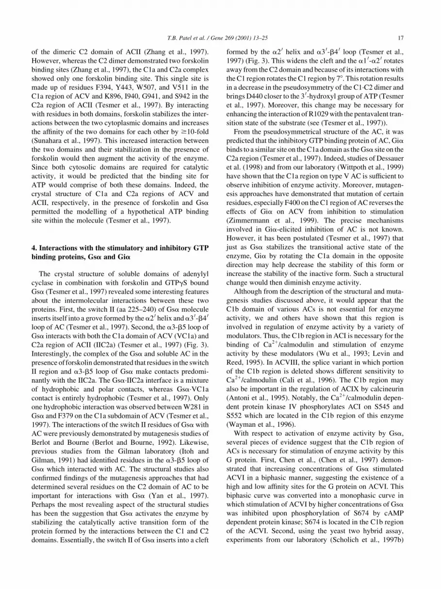
The crystal structure of soluble domains of adenylyl
cyclase in combination with forskolin and GTPgS bound
Gsa (Tesmer et al., 1997) revealed some interesting features
about the intermolecular interactions between these two
proteins. First, the switch II (aa 225±240) of Gsa molecule
inserts itself into a grove formed by thea2 0 helix anda3 0-b4 0
loop of AC (Tesmer et al., 1997). Second, the a3-b5 loop of
Gsa interacts with both the C1a domain of ACV (VC1a) and
C2a region of ACII (IIC2a) (Tesmer et al., 1997) (Fig. 3).
Interestingly, the complex of the Gsa and soluble AC in the
presence offorskolin demonstrated that residues in the switch
II region and a3-b5 loop of Gsa make contacts predomi-
nantly with the IIC2a. The Gsa´IIC2a interface is a mixture
of hydrophobic and polar contacts, whereas Gsa´VC1a
contact is entirely hydrophobic (Tesmer et al., 1997). Only
one hydrophobic interaction was observed between W281 in
Gsa and F379 on the C1a subdomain of ACV (Tesmer et al.,
1997). The interactions of the switch II residues of Gsa with
AC were previously demonstrated by mutagenesis studies of
Berlot and Bourne (Berlot and Bourne, 1992). Likewise,
previous studies from the Gilman laboratory (Itoh and
Gilman, 1991) had identi®ed residues in the a3-b5 loop of
Gsa which interacted with AC. The structural studies also
con®rmed ®ndings of the mutagenesis approaches that had
determined several residues on the C2 domain of AC to be
important for interactions with Gsa (Yan et al., 1997).
Perhaps the most revealing aspect of the structural studies
has been the suggestion that Gsa activates the enzyme by
stabilizing the catalytically active transition form of the
protein formed by the interactions between the C1 and C2
domains. Essentially, the switch II of Gsa inserts into a cleft
formed by the a2 0 helix and a3 0-b4 0 loop (Tesmer et al.,
1997) (Fig. 3). This widens the cleft and the a1 0-a2 0 rotates
away from the C2 domain and because of its interactions with
the C1 region rotates the C1 region by 78. This rotation results
in a decrease in the pseudosymmetry of the C1´C2 dimer and
brings D440 closer to the 3 0-hydroxyl group of ATP (Tesmer
et al., 1997). Moreover, this change may be necessary for
enhancing the interaction of R1029 with the pentavalent tran-
sition state of the substrate (see (Tesmer et al., 1997)).
From the pseudosymmetrical structure of the AC, it was
predicted that the inhibitory GTP binding protein of AC, Giabinds to a similar site on the C1a domain as the Gsa site on the
C2a region (Tesmer et al., 1997). Indeed, studies of Dessauer
et al. (1998) and from our laboratory (Wittpoth et al., 1999)
have shown that the C1a region on type V AC is suf®cient to
observe inhibition of enzyme activity. Moreover, mutagen-
esis approaches have demonstrated that mutation of certain
residues, especially F400 on the C1 region of AC reverses the
effects of Gia on ACV from inhibition to stimulation
(Zimmermann et al., 1999). The precise mechanisms
involved in Gia-elicited inhibition of AC is not known.
However, it has been postulated (Tesmer et al., 1997) that
just as Gsa stabilizes the transitional active state of the
enzyme, Gia by rotating the C1a domain in the opposite
direction may help decrease the stability of this form or
increase the stability of the inactive form. Such a structural
change would then diminish enzyme activity.
Although from the description of the structural and muta-
genesis studies discussed above, it would appear that the
C1b domain of various ACs is not essential for enzyme
activity, we and others have shown that this region is
involved in regulation of enzyme activity by a variety of
modulators. Thus, the C1b region in ACI is necessary for the
binding of Ca21/calmodulin and stimulation of enzyme
activity by these modulators (Wu et al., 1993; Levin and
Reed, 1995). In ACVIII, the splice variant in which portion
of the C1b region is deleted shows different sensitivity to
Ca21/calmodulin (Cali et al., 1996). The C1b region may
also be important in the regulation of ACIX by calcineurin
(Antoni et al., 1995). Notably, the Ca21/calmodulin depen-
dent protein kinase IV phosphorylates ACI on S545 and
S552 which are located in the C1b region of this enzyme
(Wayman et al., 1996).
With respect to activation of enzyme activity by Gsa,
several pieces of evidence suggest that the C1b region of
ACs is necessary for stimulation of enzyme activity by this
G protein. First, Chen et al., (Chen et al., 1997) demon-
strated that increasing concentrations of Gsa stimulated
ACVI in a biphasic manner, suggesting the existence of a
high and low af®nity sites for the G protein on ACVI. This
biphasic curve was converted into a monophasic curve in
which stimulation of ACVI by higher concentrations of Gsawas inhibited upon phosphorylation of S674 by cAMP
dependent protein kinase; S674 is located in the C1b region
of the ACVI. Second, using the yeast two hybrid assay,
experiments from our laboratory (Scholich et al., 1997b)
T.B. Patel et al. / Gene 269 (2001) 13±25 17

demonstrated that the C1b region of ACV interacts with a 64
amino acid long region C2I (Fig. 1) within the C2 domain of
ACV. Moreover, in the bacterially expressed soluble forms
of ACV, deletion of the C1b region altered the pro®le of
stimulation of enzyme activity by varying concentrations of
Gsa (Scholich et al., 1997b). We identi®ed two peptides,
corresponding to small regions (aa 1042±1058 and aa 1042±
1051) in the C-terminus of the C2I domain (Fig. 1) of ACV
which disrupt the C1b-C2 interaction (Scholich et al.,
1997b). Using these peptides, the pro®le of stimulation of
the full-length enzyme by different concentrations of Gsacould be converted to that observed with the soluble form in
which the C1b region was missing (see (Scholich et al.,
1997b) for details). Thus the C1b region of ACV is impor-
tant in stabilizing the interactions of AC with Gsa for stimu-
lation of activity by high concentrations of the G protein.
Further evidence that C1b region of ACV is important in
activation of enzyme activity by Gsa is provided by the
recent studies from Tang's group (Yan et al., 2000). These
authors demonstrated that the C1b region of ACVII interacts
with the C1a and C2a regions of this enzyme. Additionally,
the C1b region of ACVII was found to inhibit enzyme activ-
ity and interfere with the interactions of Gsa with C1a and
C2a regions of the enzyme (Yan et al., 2000).
The C1b region is also important in the regulation of
ACV activity by the inhibitory GTP binding protein Gia.
Thus, although the C1a region of ACV is suf®cient to
observe inhibition of activity by Gia, the presence of the
C1b region increases the sensitivity of inhibition by the G
protein (Dessauer et al., 1998; Wittpoth et al., 1999). Like-
wise, with respect to ACI, we have demonstrated that
although neither the C1b nor the C2 regions of ACI by
themselves are suf®cient to observe inhibition by G protein
bg (Gbg) subunits (Wittpoth et al., 1999). However, when
the C1b and C2 regions of ACI are present together, Gbgsubunits can inhibit enzyme activity (Wittpoth et al., 1999).
Interestingly, the C1a region of ACI is also suf®cient to
observe inhibition of enzyme activity by Gbg subunits
(Wittpoth et al., 1999). These ®ndings (Wittpoth et al.,
1999) suggest that on ACI there are two sites of Gbg inter-
action, one site is formed by the C1a region and the other by
the C1b and C2 regions in combination.
The signi®cance of the C1b region of AC in regulation of
enzyme activity discussed above was not made evident by the
structural studies of the C1a and C2a regions of AC (Tesmer
et al., 1997). Likewise, this limited structure of the enzyme
also leaves unexplained the signi®cance of the ®ndings that
mutations in the a4-b6 region of Gsa decreases or abolishes
the ability of the G protein to stimulate AC (Berlot and
Bourne, 1992). It is therefore, possible that the a4-b6 region
of Gsa is important in the modulation of AC activity invol-
ving the C1b region. Unfortunately, interactions of the C1b
region with the C2 region of AC and/or with Gsa (or Gbg)
cannot be resolved at the structural level because the expres-
sion of longer C1 domains in large amounts have proven to be
extremely dif®cult (Tesmer et al., 1997; Scholich and Patel,
unpublished observations).
5. Novel functions of adenylyl cyclases
Recently, we have discovered two novel functions for
membrane bound adenylyl cyclases. To facilitate their
discussion, ®rst the normal activation and inactivation
cycles of heterotrimeric G proteins is brie¯y described.
Essentially, following the binding of hormones to their
T.B. Patel et al. / Gene 269 (2001) 13±2518
Fig. 3. Interactions between Gsa and the C2a domain of ACII in complex with the C1a region of ACV. This ®gure is derived from the structure of the complex
of GTPgS-Gsa and soluble AC formed by the C1a region of ACV and C2a region of ACII (Tesmer et al., 1997). Panel (A) The a3-b5 loop and switch II
regions of Gsa are shown in magenta and red, respectively. The C1a region of ACV is shown in dark gray. The a2 0 helix on the C2a region of ACII is depicted
in blue and the a3 0-b4 0 region including the loop are colored green. Panel (B) Enlargement of the regions between AC and Gsa that are involved in
intermolecular interactions.

respective receptors which couple with G proteins, the
receptors are activated and act as guanine nucleotide
exchange factors (GEF) for their respective G proteins. In
Fig. 4 the active receptor is designated as R*. The hormone
binding elicits a change on the receptor structure and
permits the receptor to augment the exchange of GTP for
GDP on the a subunit of the trimeric G protein. The GTP-
bound Ga subunit has a lower af®nity for the Gbg subunits
and dissociates from them (Fig. 4A). The GTP-bound, acti-
vated, a subunit and the Gbg subunits then interact with
their respective effectors to generate the appropriate signals.
In the case of ACs, Gsa and Gia activate and inhibit activity
of the enzyme, respectively. The Ga subunits of heterotri-
meric G proteins express an intrinsic GTPase activity,
which hydrolyzes the GTP and converts the GTP-bound
form to the GDP bound, inactive form (Fig. 4A). The
GDP-bound, inactive Ga then associates with the Gbgsubunits and returns to the resting state terminating the
transmission of signal (Fig. 4A).
In recent years, a large family of proteins (Regulators of
G-protein Signaling or RGS proteins) has been cloned and
characterized (see Siderovski et al., 1999; Burchett, 2000)
for reviews). These RGS proteins enhance the intrinsic
GTPase activity of a variety of Ga subunits and expedite
the termination of signals from these subunits (reviewed in
(Siderovski et al., 1999; Burchett, 2000). Although a large
number of RGS proteins which act as GTPase Activating
Proteins (GAPs) against a variety of Ga subunits have been
cloned and characterized, no RGS proteins or GAPs for Gsahave been found. Therefore, we proposed the hypothesis
that AC acts as a GAP for Gsa. Indeed, the C1±C2 form
of ACV and its C2 domain were found to act as GAPs for
T.B. Patel et al. / Gene 269 (2001) 13±25 19
Fig. 4. The activation and inactivation cycle of G proteins regulating adenylyl cyclase activity. Panel A depicts the classical activation and inactivation cycle of
G proteins by their receptors. Panel B has been revised to incorporate the two novel functions of AC. The hatched arrow denotes the GAP activity of AC against
Gsa only. AC does not act as a GAP against Gia (see text). However, AC enhances signal onset via both Gs and Gi coupled receptors (see text) and, therefore,
this arrow is common to both types of receptors and heterotrimeric G proteins.

Gsa (see (Scholich et al., 1999) for elaboration). Notably,
the soluble form of ACV does not act as a GAP for Gia(Scholich et al., 1999). Thus, AC is selective as a GAP for
the active form of Gsa. This action of AC is similar to that
of other effector enzymes such as phospholipase Cb(Berstein et al., 1992), and the g subunit of cGMP phospho-
diesterase (Arshavsky and Bownds, 1992) which can also
act as GAPs for Gqa and Gta, respectively.
Because the GAP activity of AC can expedite the termi-
nation of signaling via Gsa, we investigated the effects of
AC on signal onset. For these experiments, the Gs hetero-
trimer was reconstituted from puri®ed recombinant Gsa and
puri®ed bovine brain Gbg subunits ((Scholich et al., 1999),
also see (Patel et al., 2001) for experimental details). A
peptide bIII-2 corresponding to amino acids 259±272 of
the b2-adrenergic receptors was used as a constitutively
active receptor. This peptide has previously been shown to
mimic the actions of the b2-adrenergic receptor (Okamoto
et al., 1991; Sun et al., 1995). By monitoring activation of
Gs using three parameters, i.e. steady state GTPase activity,
GTPgS binding, and activation of AC in membranes of S49
cyc2 cells, we demonstrated that in the presence of the
soluble form of ACV (C1±C2 ACV), the concentration of
the bIII-2 required to activate the G protein was decreased
by 100-fold (Scholich et al., 1999). Overall, these experi-
ments showed that AC facilitates the onset of signaling via
receptors so that the amount of active receptor required to
activate the enzyme is decreased by 100-fold (Scholich et
al., 1999). In the case of Gs, AC and its C2 domain as well as
smaller regions within the C2 domain, C2I and C2II (Fig. 1)
were suf®cient to enhance the actions of the peptide bIII-2
(Scholich et al., 1999). This novel function of AC would
serve to amplify signaling in the presence of low concentra-
tions of ligand or active receptors, which signal via Gs. In
Fig. 4B, we have revised the classical activation/inactiva-
tion cycle to include the GAP and GEF enhancing actions of
AC.
Since the C2 domain of AC which interacts with Gsacould augment the GEF activity of Gs, it may be postulated
that the C1 domain of the enzyme in ACV which interacts
with the Gia would also enhance the GEF activity of Gi
coupled receptors. Indeed, using a number of different
approaches, including the inhibition of AC activity in
membranes of S49 cyc2 cells by somatostatin, we demon-
strated that indeed the C1 domain of ACV could increase
the onset of signaling via Gi coupled receptors (Wittpoth et
al., 2000). Since AC does not act as a GAP for Gi, in the new
activation/inactivation cycle for Gi, in Fig. 4B, the hatched
arrow from AC to the `signal off' portion of the cycle should
be omitted.
An interesting aspect of the novel functions of AC with
respect to Gsa and Gs, is that the enzyme both inactivates
Gsa (GAP activity of AC) and facilitates activation of Gs
(GEF enhancing activity of AC). This apparently paradox is
also applicable to other proteins with GAP activity against
Ga subunits. For example, besides acting as GAPs some
RGS proteins may also enhance the onset of signaling.
Thus, it has been demonstrated that RGS4 and RGS8 can
increase the activation kinetic of G protein coupled inward
rectifying K1 current (Doupnik et al., 1997; Saitoh et al.,
1997). More recently, Chen and Lambert (Chen and
Lambert, 2000) have shown that endogenous RGS proteins
in hippocampal neurons can increase the onset of presynap-
tic inhibition in response to adenosine and baclofen. There-
fore, the concept that GAPs, which expedite signal
termination, can also augment or facilitate signal onset is
more generally applicable than previously thought and AC
and RGS proteins could be interchangeably used in Fig. 4B.
While the membrane bound ACs may act as GAPs or
GEF enhancers, the soluble AC may act as a bicarbonate
sensor. The soluble enzyme is expressed in tissues such as
the testes, kidneys and the choroid plexus (Buck et al., 1999)
where changes in bicarbonate levels alter cAMP levels. It is
well recognized that spermatozoa undergo a number of
bicarbonate induced changes which increase motility, capa-
citation, and the acrosome reaction (Garty and Salomon,
1987; Okamura et al., 1991; Visconti et al., 1998). These
processes are also cAMP dependent. Interestingly, Chen et
al. (2000) found that the soluble AC is not modulated by the
regulators which alter the activity of membrane bound ACs,
but is activated by bicarbonate ions independently of
changes in pH. These ®ndings have provided the link
between changes in bicarbonate levels and alterations in
cAMP content and explain why increases in bicarbonate
and cAMP levels are associated with certain alterations in
spermatozoa function.
6. Knockout and transgenic models for adenylyl cyclases
Although the different isoforms of membrane bound ACs
are differentially regulated by a variety of modulators,
because some of these isoforms which are co-expressed in
some organs and cell types demonstrate similar modes of
regulation one wonders what physiological role each isoform
plays. To address this and other issues concerning the physio-
logical relevance of each isoform, several studies have
utilized knock-out and transgenic mice. Some of the clues
concerning the physiological role of AC isoforms were
derived from the drosophila mutants that were de®cient in
learning and memory. Four of these mutants, involved
defects in genes encoding one of the following: AC activat-
ing peptide (amnesiac) (Feany and Quinn, 1995), AC (ruta-
baga) (Livingstone et al., 1984), PKA (DCO) (Foster et al.,
1984), or cAMP phosphodiesterase (dunce) (Chen et al.,
1986). The drosophila AC is stimulated by Ca21 and calmo-
dulin and is similar to the mammalian ACI and ACVIII
isoforms. Furthermore, earlier studies had demonstrated
that in mammals PKA is important for synaptic plasticity
and some forms of long-term potentiation (LTP) (Frey et
al., 1993; Abrams et al., 1991). Therefore, to study the role
of ACI in synaptic plasticity and LTP, Storm and co-workers
T.B. Patel et al. / Gene 269 (2001) 13±2520

knocked out this isoform in mice. Essentially, their ®ndings
demonstrated that the ACI is important in behaviour and LTP
in mice (Wu et al., 1995). In additional studies the Storm
laboratory demonstrated that in ACI knockout mice, AC
activity in cerebellar cortex was reduced by 65% and this
was accompanied by an almost complete blockade of cere-
bellar LTP (Storm et al., 1998). These ®ndings demonstrate
that ACI plays a critical role in regulating synaptic plasticity
and LTP. Interestingly, in mice lacking ACI or ACVIII late
phase LTP (L-LTP) and long-term memory are unaffected
(Storm et al., 1998). However, the double knock-out mice in
which both ACI and ACVIII genes have been disrupted do
not exhibit L-LTP or LTM (Storm et al., 1998). Injection of
the diterpene forskolin, which activates all isoforms of AC
except ACIX, in the Cornu Ammonis 1 (CA1) region of the
brain in double knockout mice, restored LTM (Storm et al.,
1998). These ®ndings clearly demonstrate that the Ca21 and
calmodulin stimulated ACs (I and VIII) are required for L-
LTP and LTM.
The ACVIII knockout mice have also unveiled at least
two other functions for this isoform. First, it has been
shown that capacitative Ca21 entry stimulates cAMP synth-
esis in the parotid acini. The parotid acini which contain a
variety of AC isoforms including ACI, ACIII, ACV, ACVI
and ACVIII, (Watson et al., 2000). Therefore, Watson et al
(Watson et al., 2000) used the ACI and ACVIII knockout
mice to determine the isoform which was activated by
agonists such as carbachol which increase Ca21 entry.
The disruption of ACVIII, but not ACI, gene obliterated
increase in cAMP levels in response to calcium (Watson et
al., 2000). In fact, in parotid acini from ACVIII de®cient
mice, increase in calcium entry decreased cAMP accumu-
lation (Watson et al., 2000). This latter effect can be attrib-
uted to the presence of the Ca21 inhibited ACV and ACVI
in parotid acini (Watson et al., 2000). Thus, the same
modulator may alter the activities of more than one isoform
in cells and the net of the positive and negative input from
different AC isoforms determines the ®nal level of cAMP
accumulation. A second physiological function of ACVIII
was reported by Schaefer et al., (Schaefer et al., 2000).
Essentially, mice de®cient in ACVIII do not show
increased anxiety in repeated stress tests. The ACVIII
knock-out mice also do not show CA1 region long-term
depression after low frequency stimulation and do not acti-
vate the CREB in the CA1 region after repeated stress tests
(Schaefer et al., 2000).
More recently, by disrupting the gene encoding ACIII,
the Storm laboratory has investigated the role of ACIII in
olfactory responses (Wong et al., 2000). Olfactory cilia
express ACII, III, and IV (Wong et al., 2000). However,
disruption of the ACIII gene abrogated electro-olfactogram
responses to a number of cAMP or inositol trisphosphate
elevating agents (Wong et al., 2000). Moreover, in olfac-
tion-based avoidance tests, the ACIII knockout mice
performed signi®cantly poorly as compared with their
wild type controls (Wong et al., 2000). These ®ndings
demonstrate that the ACIII is critical and of paramount
importance in olfaction and olfaction related responses.
Besides the knockout approach, several investigators
have used transgenic mice in which a given isoform of
AC is over-expressed in a tissue speci®c manner. These
studies have provided insights into the physiological role
of some of the AC isoforms. For instance, since the Ca21
inhibited ACV and ACVI are the predominant isoforms
expressed in the heart (Ishikawa et al., 1992; Katsushika
et al., 1992; Premont et al., 1992; Yoshimura and Cooper,
1992), it has been postulated that elevations in intracellular
Ca21 concentrations during the contractile cycle may inhibit
the activity of these isoforms in a feed-back regulatory
manner. Indeed, cyclical alterations in cAMP levels in
ventricular strips have been observed with each contractile
cycle (Brooker, 1973). Therefore, if the inhibition of ACV
or ACVI activity at the peak of a contraction is physiologi-
cally relevant, then it would be expected that the over-
expression of an AC isoform such as ACI or ACVIII
whose activity is augmented by Ca21 (and calmodulin),
would grossly alter cardiac function. Surprisingly, however,
cardiac speci®c over-expression of ACVIII did not alter
basal heart rate and contractility (Lipskaia et al., 2000).
However, when the parasympathetic tone was released,
cardiac contractility and heart rate of the ACVIII transgenic
mice increased markedly and was unresponsive to modula-
tion by b-adrenergic receptor agonists (Lipskaia et al.,
2000). These ®ndings differ from the study of Gao et al.,
(Gao et al., 1999) which demonstrated that over expression
of ACVI in the heart increased the responsiveness to b-
adrenergic receptor stimulation. One reason for this differ-
ence could be related to the cAMP levels in the hearts of
transgenic mice over-expressing ACVI and ACVIII. Since
Ca21 regulates these two enzymes in opposite ways, it
would be expected that in hearts over-expressing ACVIII,
the cAMP levels would be higher than in those over-expres-
sing ACVI. Such an increase in cAMP levels may desensi-
tize the b-adrenergic receptors in hearts of ACVIII
expressing animals but not in ACVI over-expressing
animals. Unfortunately, Lipskaia (Lipskaia et al., 2000)
did not measure cAMP levels in ACVIII transgenic animals.
However, the basal cAMP levels in myocytes derived from
ACVI transgenic animals were similar to those in controls
(Gao et al., 1999). Moreover, as expected from the func-
tional responsiveness to b-adrenergic receptor activation,
isoproterenol increased cAMP levels to a greater extent in
ACVI transgenic mice as compared with controls (Gao et
al., 1999).
One of the implications of the studies with cardiac speci-
®c expression of ACVI is that in certain pathological states
such as ischemia or hypertrophy in which responsiveness
of AC system to b-adrenoreceptor agonists is decreased
(Vatner et al., 1988; Strasser et al., 1990; D'Angelo et
al., 1997), the over-expression of ACVI or ACV, would
restore the responsiveness of the heart to b-receptor stimu-
lation. Indeed, the studies of Roth et al. (1999) demon-
T.B. Patel et al. / Gene 269 (2001) 13±25 21

strated that over-expression of ACVI in hearts of mice
over-expressing Gqa improved cardiac function and
restored the cAMP generating capacity in response to cate-
cholamines. Similarly, because over-expression of Gqaresults in approximately 45% decrease in ACV expression,
decreased b-adrenoreceptor mediated activation of AC,
and hypertrophy (D'Angelo et al., 1997; Roth et al.,
1999), Tepe and Liggett (1999) investigated whether or
not over-expression of ACV in Gqa over-expressing
animals would ameliorate the cardiac myopathy and restore
function. Essentially, these studies (Tepe and Liggett,
1999) demonstrated that cardiac speci®c over-expression
of ACV in the Gqa over-expressing mice improved cardiac
function and responsiveness to b-adrenoreceptor activa-
tion. However, cardiac hypertrophy and expression of
hypertrophy related genes was not altered (Tepe and
Liggett, 1999). These ®ndings demonstrate that neither
the decrease in ACV nor the loss of responsiveness to b-
receptor activation was the underlying cause of cardiac
hypertrophy. Nevertheless, the studies with ACV and
ACVI over-expression in the heart provide important infor-
mation concerning how the cardiac responsiveness to b-
adrenergic receptors can be restored. In this respect, it
should be noted that over-expression of ACV or ACVI
may improve cardiac function since it is generally accepted
that in the receptor ± Gs ± AC complex, AC is in limiting
amounts (Post et al., 1995). Thus, over-expression of the
limiting signaling element (AC) may amplify the actions of
activated receptors. On the other hand, it should be noted
that we have demonstrated that AC and its domains which
interact with G protein a subunits, facilitate the onset of
signaling via receptors which couple to Gs and Gi (Scho-
lich et al., 1999; Wittpoth et al., 2000). Therefore, by this
mechanism, it is equally possible that the over-expression
of adenylyl cyclase augments the onset of signaling from
the receptor to the effector.
7. Concluding remarks and future directions
Since the discovery of cAMP as a second messenger
some forty years ago by Dr. Earl Sutherland, the amount
of research that has been performed in understanding of the
regulation and role of the diverse family of ACs is beyond
the scope of any single review. Therefore, we have focused
on the aspects of ACs not previously represented in other
reviews. However, the readers are strongly encouraged to
read other comprehensive reviews on the subject which
address the ®ndings and inferences derived from studies
in different tissues (Defer et al., 2000), the distribution of
Ca21 modulated AC isoforms in the brain (Mons et al.,
1998) and the role of ACs as signal integrators (Ishikawa
and Homcy, 1997). The underlying message from the
numerous reviews and research articles published in the
area is that the family of diverse ACs offers a fertile ground
for future investigations. Thus, while the knock-out and
transgenic mice experiments have provided some informa-
tion on the physiological roles that some of the AC
isoforms may play in the context of the nervous system
and the heart, the roles of other members of the family
remain to be clearly de®ned. This task is made even
more daunting since most tissues and organs express a
variety of AC isoforms. Moreover the structures of the
genes encoding the majority of AC isoforms remain to
be determined. Additionally, the role of ACs in disease
conditions involving mutant Gsa forms also remains to
be determined. For instance, certain pituitary tumors are
associated with the expression of constitutively active
mutant forms of Gsa (Lyons et al., 1990). Recently, Gu
et al. (2000) have shown that Gsa also activates the tyro-
sine kinase Lck. Thus, it could be argued that the pituitary
tumors associated with mutant, constitutively active, Gsaare due to activation of Lck rather than AC. To investigate
this and other possibilities it is essential that selective inhi-
bitors of AC be developed. Presently, the only inhibitors
which selectively inhibit ACs are the `P site' inhibitors
developed by Johnson and colleagues (see e.g. Doronin
et al., 1999; Shoshani et al., 1999a). Most of these,
however, do not permeate the cell membrane. The few
inhibitors of AC, which are taken up by cells, exert other
non-speci®c effects (Shoshani et al., 1999b; Kudlacek et
al., 2000). Moreover, no inhibitors such as dominant nega-
tive forms of AC are presently available. Therefore, efforts
in this area would probably present another breakthrough
in the ®eld. Likewise, our recent studies (Scholich et al.,
1999; Wittpoth et al., 2000) have shown that AC clearly
has a function in regulation Gsa activity and signaling via
receptors coupled to Gs and Gi. Therefore, with the intent
to increase or decrease the signaling via receptors it is
essential to determine the molecular mechanisms underly-
ing this mode of regulation of G proteins by AC.
Acknowledgements
We are greatly indebted to numerous investigators in the
®eld who provided their most recent publications and
preprints of their publications to ensure that our review
would be as up to date as possible. Due to the numerous
publications in this ®eld and because of space limitations,
we had to refer to some previous reviews in our citations,
and, therefore, we apologize to those authors whose original
®ndings were not cited in this review. This work was
supported by grants from the NIH (HL59679 and HL
07641).
References
Abdel-Halim, S.M., Gueni®, A., He, B., Yang, B., Mustafa, M., Hojeberg,
B., Hillert, J., Bakhiet, M., Efendic, S., 1998. Mutations in the promoter
of adenylyl cyclase (AC)-III gene, over-expression of AC-III mRNA,
T.B. Patel et al. / Gene 269 (2001) 13±2522

and enhanced cAMP generation in islets from the spontaneously
diabetic GK rat model of type 2 diabetes. Diabetes 47, 498±504.
Abrams, T.W., Karl, K.A., Kandel, E.R., 1991. Biochemical studies of
stimulus convergence during classical conditioning in Aplysia: dual
regulation of adenylate cyclase by Ca21/calmodulin and transmitter.
J. Neurosci. 11, 2655±2665.
Antoni, F.A., Barnard, R.J., Shipston, M.J., Smith, S.M., Simpson, J., Pater-
son, J.M., 1995. Calcineurin feedback inhibition of agonist-evoked
cAMP formation. J. Biol. Chem. 270, 28055±28061.
Arshavsky, V., Bownds, M.D., 1992. Regulation of deactivation of photo-
receptor G protein by its target enzyme and cGMP. Nature 357, 416±417.
Bakalyar, H.A., Reed, R.R., 1990. Identi®cation of a specialized adenylyl
cyclase that may mediate odorant detection. Science 250, 1403±1406.
Bayewitch, M.L., Avidor-Reiss, T., Levy, R., Pfeuffer, T., Nevo, I.,
Simonds, W.F., Vogel, Z., 1998. Inhibition of adenylyl cyclase isoforms
V and VI by various Gbetagamma subunits. FASEB J. 12, 1019±1025.
Berlot, C.H., Bourne, H.R., 1992. Identi®cation of effector-activating resi-
dues of Gs a. Cell 68, 911±922.
Berstein, G., Blank, J.L., Jhon, D.Y., Exton, J.H., Rhee, S.G., Ross, E.M.,
1992. Phospholipase C-beta 1 is a GTPase-activating protein for Gq/11,
its physiologic regulator. Cell 70, 411±418.
Beuve, A., 1999. Conversion of a guanylyl cyclase to an adenylyl cyclase.
Methods 19, 545±550.
Brooker, G., 1973. Oscillation of cyclic adenosine monophosphate concen-
tration during the myocardial contraction cycle. Science 182, 933±934.
Buck, J., Sinclair, M.L., Schapal, L., Cann, M.J., Levin, L.R., 1999. Cyto-
solic adenylyl cyclase de®nes a unique signaling molecule in mammals.
Proc. Natl. Acad. Sci. USA 96, 79±84.
Burchett, S.A., 2000. Regulators of G protein signaling: a bestiary of modu-
lar protein binding domains. J. Neurochem. 75, 1335±1351.
Cali, J.J., Parekh, R.S., Krupinski, J., 1996. Splice variants of type VIII
adenylyl cyclase. Differences in glycosylation and regulation by
Ca2 1 /calmodulin. J. Biol. Chem. 271, 1089±1095.
Chen, H., Lambert, N.A., 2000. Endogenous regulators of G protein signal-
ing proteins regulate presynaptic inhibition at rat hippocampal
synapses. Proc. Natl. Acad. Sci. USA 97, 12810±12815.
Chen, C.N., Denome, S., Davis, R.L., 1986. Molecular analysis of cDNA
clones and the corresponding genomic coding sequences of the Droso-
phila dunce 1 gene, the structural gene for cAMP phosphodiesterase.
Proc Natl. Acad. Sci. USA 83, 9313±9317.
Chen, Y., Harry, A., Li, J., Smit, M.J., Bai, X., Magnusson, R., Pieroni, J.P.,
Weng, G., Iyengar, R., 1997. Adenylyl cyclase 6 is selectively regulated
by protein kinase A phosphorylation in a region involved in Galphas
stimulation. Proc. Natl. Acad. Sci. USA 94, 14100±14104.
Chen, Y., Cann, M.J., Litvin, T.N., Iourgenko, V., Sinclair, M.L., Levin,
L.R., Buck, J., 2000. Soluble adenylyl cyclase as an evolutionarily
conserved bicarbonate sensor. Science 289, 625±628.
Choi, E.J., Xia, Z., Storm, D.R., 1992. Stimulation of the type III olfactory
adenylyl cyclase by calcium and calmodulin. Biochemistry 31, 6492±
6498.
D'Angelo, D.D., Sakata, Y., Lorenz, J.N., Boivin, G.P., Walsh, R.A.,
Liggett, S.B., Dorn, G.W., 1997. Transgenic Galphaq over-expression
induces cardiac contractile failure in mice. Proc. Natl. Acad. Sci. USA
94, 8121±8126.
Defer, N., Best-Belpomme, M., Hanoune, J., 2000. Tissue speci®city and
physiological relevance of various isoforms of adenylyl cyclase. Am. J.
Physiol. Renal Physiol. 279, F400±F416.
Desaubry, L., Shoshani, I., Johnson, R.A., 1996. 2 0,5 0-Dideoxyadenosine
3 0-polyphosphates are potent inhibitors of adenylyl cyclases. J. Biol.
Chem. 271, 2380±2382.
Dessauer, C.W., Gilman, A.G., 1997. The catalytic mechanism of mamma-
lian adenylyl cyclase. Equilibrium binding and kinetic analysis of P-site
inhibition. J. Biol. Chem. 272, 27787±27795.
Dessauer, C.W., Tesmer, J.J., Sprang, S.R., Gilman, A.G., 1998. Identi®ca-
tion of a Gialpha binding site on type V adenylyl cyclase. J. Biol. Chem.
273, 25831±25839.
Doronin, S., Murray, L., Dessauer, C.W., Johnson, R.A., 1999. Covalent
labeling of adenylyl cyclase cytosolic domains with gamma-methyli-
midazole-2 0,5 0-dideoxy-[gamma-(32)P]3 0-ATP and the mechanism for
P-site-mediated inhibition. J. Biol. Chem. 274, 34745±34750.
Doupnik, C.A., Davidson, N., Lester, H.A., Kofuji, P., 1997. RGS proteins
reconstitute the rapid gating kinetics of gbetagamma-activated inwardly
rectifying K 1 channels. Proc. Natl. Acad. Sci. USA 94, 10461±10466.
Edelhoff, S., Villacres, E.C., Storm, D.R., Disteche, C.M., 1995. Mapping
of adenylyl cyclase genes type I, II, III, IV, V, and VI in mouse. Mamm.
Genome 6, 111±113.
Feany, M.B., Quinn, W.G., 1995. A neuropeptide gene de®ned by the
Drosophila memory mutant amnesiac. Science 268, 869±873.
Feinstein, P.G., Schrader, K.A., Bakalyar, H.A., Tang, W.J., Krupinski, J.,
Gilman, A.G., Reed, R.R., 1991. Molecular cloning and characteriza-
tion of a Ca2 1 /calmodulin-insensitive adenylyl cyclase from rat brain.
Proc. Natl. Acad. Sci. USA 88, 10173±10177.
Foster, J.L., Guttman, J.J., Hall, L.M., Rosen, O.M., 1984. Drosophila
cAMP-dependent protein kinase. J. Biol. Chem. 259, 13049±13055.
Frey, U., Huang, Y.Y., Kandel, E.R., 1993. Effects of cAMP simulate a late
stage of LTP in hippocampal CA1 neurons. Science 260, 1661±1664.
Gao, B.N., Gilman, A.G., 1991. Cloning and expression of a widely distrib-
uted (type IV) adenylyl cyclase. Proc. Natl. Acad. Sci. USA 88, 10178±
10182.
Gao, M.H., Lai, N.C., Roth, D.M., Zhou, J., Zhu, J., Anzai, T., Dalton, N.,
Hammond, H.K., 1999. Adenylylcyclase increases responsiveness to
catecholamine stimulation in transgenic mice. Circulation 99, 1618±
1622.
Garty, N.B., Salomon, Y., 1987. Stimulation of partially puri®ed adenylate
cyclase from bull sperm by bicarbonate. FEBS Lett. 218, 148±152.
Gu, C., Ma, Y.C., Benjamin, J., Littman, D., Chao, M.V., Huang, X.Y.,
2000. Apoptotic signaling through the beta -adrenergic receptor. A new
Gs effector pathway. J. Biol. Chem. 275, 20726±20733.
Haber, N., Stengel, D., Defer, N., Roeckel, N., Mattei, M.G., Hanoune, J.,
1994. Chromosomal mapping of human adenylyl cyclase genes type III,
type V and type VI. Hum. Genet. 94, 69±73.
Hacker, B.M., Tomlinson, J.E., Wayman, G.A., Sultana, R., Chan, G.,
Villacres, E., Disteche, C., Storm, D.R., 1998. Cloning, chromosomal
mapping, and regulatory properties of the human type 9 adenylyl
cyclase (ADCY9). Genomics 50, 97±104.
Hellevuo, K., Berry, R., Sikela, J.M., Tabakoff, B., 1995. Localization of
the gene for a novel human adenylyl cyclase (ADCY7) to chromosome
16. Hum. Genet. 95, 197±200.
Hurley, J.H., 1999. Structure, mechanism, and regulation of mammalian
adenylyl cyclase. J. Biol. Chem. 274, 7599±7602.
Ishikawa, Y., Homcy, C.J., 1997. The adenylyl cyclases as integrators of
transmembrane signal transduction. Circ. Res. 80, 297±304.
Ishikawa, Y., Katsushika, S., Chen, L., Halnon, N.J., Kawabe, J., Homcy,
C.J., 1992. Isolation and characterization of a novel cardiac adenylyl-
cyclase cDNA. J. Biol. Chem. 267, 13553±13557.
Itoh, H., Gilman, A.G., 1991. Expression and analysis of Gs alpha mutants
with decreased ability to activate adenylylcyclase. J. Biol. Chem. 266,
16226±16231.
Iwami, G., Kawabe, J., Ebina, T., Cannon, P.J., Homcy, C.J., Ishikawa, Y.,
1995. Regulation of adenylyl cyclase by protein kinase A. J. Biol.
Chem. 270, 12481±12484.
Jacobowitz, O., Iyengar, R., 1994. Phorbol ester-induced stimulation and
phosphorylation of adenylyl cyclase 2. Proc. Natl. Acad. Sci. USA 91,
10630±10634.
Jacobowitz, O., Chen, J., Premont, R.T., Iyengar, R., 1993. Stimulation of
speci®c types of Gs-stimulated adenylyl cyclases by phorbol ester treat-
ment. J. Biol. Chem. 268, 3829±3832.
Johnson, R.A., Shoshani, I., 1990. Kinetics of `P'-site-mediated inhibition
of adenylyl cyclase and the requirements for substrate. J. Biol. Chem.
265, 11595±11600.
Katsushika, S., Chen, L., Kawabe, J., Nilakantan, R., Halnon, N.J., Homcy,
C.J., Ishikawa, Y., 1992. Cloning and characterization of a sixth adeny-
lyl cyclase isoform: types V and VI constitute a subgroup within the
T.B. Patel et al. / Gene 269 (2001) 13±25 23

mammalian adenylyl cyclase family. Proc. Natl. Acad. Sci. USA 89,
8774±8778.
Katsushika, S., Kawabe, J., Homcy, C.J., Ishikawa, Y., 1993. In vivo
generation of an adenylylcyclase isoform with a half-molecule motif.
J. Biol. Chem. 268, 2273±2276.
Kawabe, J., Iwami, G., Ebina, T., Ohno, S., Katada, T., Ueda, Y., Homcy,
C.J., Ishikawa, Y., 1994. Differential activation of adenylyl cyclase by
protein kinase C isoenzymes. J. Biol. Chem. 269, 16554±16558; err.
269, 22912.
Krupinski, J., Coussen, F., Bakalyar, H.A., Tang, W.J., Feinstein, P.G.,
Orth, K., Slaughter, C., Reed, R.R., Gilman, A.G., 1989. Adenylyl
cyclase amino acid sequence: possible channel- or transporter-like
structure. Science 244, 1558±1564.
Krupinski, J., Lehman, T.C., Franken®eld, C.D., Zwaagstra, J.C., Watson,
P.A., 1992. Molecular diversity in the adenylylcyclase family. Evidence
for eight forms of the enzyme and cloning of type VI. J. Biol. Chem.
267, 24858±24862.
Kudlacek, O., Mitterauer, T., Nanoff, C., Hohenegger, M., Tang, W.J.,
Freissmuth, M., Kleuss, C., 2000. Inhibition of adenylyl and guanylyl
cycalse isoforms by the antiviral drug foscarnet. J. Biol. Chem..
Lai, H.L., Lin, T.H., Kao, Y.Y., Lin, W.J., Hwang, M.J., Chern, Y., 1999.
The N terminus domain of type VI adenylyl cyclase mediates its inhibi-
tion by protein kinase C. Mol. Pharmacol. 56, 644±650.
Lane-Ladd, S.B., Pineda, J., Boundy, V.A., Pfeuffer, T., Krupinski, J.,
Aghajanian, G.K., Nestler, E.J., 1997. CREB (cAMP response
element-binding protein) in the locus coeruleus: biochemical, physio-
logical, and behavioral evidence for a role in opiate dependence. J.
Neurosci. 17, 7890±7901.
Levin, L.R., Reed, R.R., 1995. Identi®cation of functional domains of
adenylyl cyclase using in vivo chimeras. J. Biol. Chem. 270, 7573±
7579.
Lipskaia, L., Defer, N., Esposito, G., Hajar, I., Garel, M.C., Rockman, H.A.,
Hanoune, J., 2000. Enhanced cardiac function in transgenic mice
expressing a Ca(21)-stimulated adenylyl cyclase. Circ. Res. 86, 795±
801.
Livingstone, M.S., Sziber, P.P., Quinn, W.G., 1984. Loss of calcium/
calmodulin responsiveness in adenylate cyclase of rutabaga, a Droso-
phila learning mutant. Cell 37, 205±215.
Lyons, J., Landis, C.A., Harsh, G., Vallar, L., Grunewald, K., Feichtinger,
H., Duh, Q.Y., Clark, O.H., Kawasaki, E., Bourne, H.R., et al., 1990.
Two G protein oncogenes in human endocrine tumors. Science 249,
655±659.
Matsuoka, I., Maldonado, R., Defer, N., Noel, F., Hanoune, J., Roques,
B.P., 1994. Chronic morphine administration causes region-speci®c
increase of brain type VIII adenylyl cyclase mRNA. Eur. J. Pharmacol.
268, 215±221.
Menco, B.P., Bruch, R.C., Dau, B., Danho, W., 1992. Ultrastructural loca-
lization of olfactory transduction components: the G protein subunit
Golf alpha and type III adenylyl cyclase. Neuron 8, 441±453.
Mons, N., Decorte, L., Jaffard, R., Cooper, D.M., 1998. Ca2 1 -sensitive
adenylyl cyclases, key integrators of cellular signalling. Life Sci. 62,
1647±1652.
Muglia, L.M., Schaefer, M.L., Vogt, S.K., Gurtner, G., Imamura, A.,
Muglia, L.J., 1999. The 5 0-¯anking region of the mouse adenylyl
cyclase type VIII gene imparts tissue-speci®c expression in transgenic
mice. J. Neurosci. 19, 2051±2058.
Okamoto, T., Murayama, Y., Hayashi, Y., Inagaki, M., Ogata, E., Nishi-
moto, I., 1991. Identi®cation of a Gs activator region of the beta 2-
adrenergic receptor that is autoregulated via protein kinase A-depen-
dent phosphorylation. Cell 67, 723±730.
Okamura, N., Tajima, Y., Onoe, S., Sugita, Y., 1991. Puri®cation of bicar-
bonate-sensitive sperm adenylylcyclase by 4-acetamido-4 0-isothiocya-
nostilbene-2,2 0-disulfonic acid-af®nity chromatography. J. Biol. Chem.
266, 17754±17759.
Patel, T.B., Wittpoth, C., Barbier, A.J., Yigzaw, Y., Scholich, K., 2001.
Functional analyses of Type V adenylyl cyclase. Methods Enzymol, in
press.
Paterson, J.M., Smith, S.M., Harmar, A.J., Antoni, F.A., 1995. Control of a
novel adenylyl cyclase by calcineurin. Biochem. Biophys. Res.
Commun. 214, 1000±1008.
Post, S.R., Hilal-Dandan, R., Urasawa, K., Brunton, L.L., Insel, P.A., 1995.
Quanti®cation of signalling components and ampli®cation in the beta-
adrenergic-receptor-adenylate cyclase pathway in isolated adult rat
ventricular myocytes. Biochem. J. 311, 75±80.
Premont, R.T., Chen, J., Ma, H.W., Ponnapalli, M., Iyengar, R., 1992. Two
members of a widely expressed subfamily of hormone-stimulated
adenylyl cyclases. Proc. Natl. Acad. Sci. USA 89, 9809±9813.
Premont, R.T., Matsuoka, I., Mattei, M.G., Pouille, Y., Defer, N., Hanoune,
J., 1996. Identi®cation and characterization of a widely expressed form
of adenylyl cyclase. J. Biol. Chem. 271, 13900±13907.
Roth, D.M., Gao, M.H., Lai, N.C., Drumm, J., Dalton, N., Zhou, J.Y., Zhu,
J., Entrikin, D., Hammond, H.K., 1999. Cardiac-directed adenylyl
cyclase expression improves heart function in murine cardiomyopathy.
Circulation 99, 3099±3102.
Saitoh, O., Kubo, Y., Miyatani, Y., Asano, T., Nakata, H., 1997. RGS8
accelerates G-protein-mediated modulation of K 1 currents. Nature
390, 525±529.
Schaefer, M.L., Wong, S.T., Wozniak, D.F., Muglia, L.M., Liauw, J.A.,
Zhuo, M., Nardi, A., Hartman, R.E., Vogt, S.K., Luedke, C.E., Storm,
D.R., Muglia, L.J., 2000. Altered stress-induced anxiety in adenylyl
cyclase type VIII-de®cient mice. J. Neurosci. 20, 4809±4820.
Scholich, K, Barbier, A.J, Mullenix, J.B, Patel, T.B., 1997a. Characteriza-
tion of soluble forms of non-chimeric type V adenylyl cyclases . Proc.
Natl. Acad. Sci. USA 94, 2915±2920; err. 94, 10485.
Scholich, K., Wittpoth, C., Barbier, A.J., Mullenix, J.B., Patel, T.B., 1997b.
Identi®cation of an intramolecular interaction between small regions in
type V adenylyl cyclase that in¯uences stimulation of enzyme activity
by Gsalpha. Proc. Natl. Acad. Sci. USA 94, 9602±9607.
Scholich, K., Mullenix, J.B., Wittpoth, C., Poppleton, H.M., Pierre, S.C.,
Lindorfer, M.A., Garrison, J.C., Patel, T.B., 1999. Facilitation of signal
onset and termination by adenylyl cyclase. Science 283, 1328±1331.
Shoshani, I., Boudou, V., Pierra, C., Gosselin, G., Johnson, R.A., 1999a.
Enzymatic synthesis of unlabeled and beta-(32)P-labeled -L-2 0, 3 0-dideoxyadenosine-5 0-triphosphate as a potent inhibitor of adenylyl
cyclases and its use as reversible binding ligand. J. Biol. Chem. 274,
34735±34741.
Shoshani, I., Laux, W.H., Perigaud, C., Gosselin, G., Johnson, R.A., 1999b.
Inhibition of adenylyl cyclase by acyclic nucleoside phosphonate anti-
viral agents. J. Biol. Chem. 274, 34742±34744.
Siderovski, D.P., Strockbine, B., Behe, C.I., 1999. Whither goest the RGS
proteins? Crit. Rev. Biochem. Mol. Biol. 34, 215±251.
Smit, M.J., Iyengar, R., 1998. Mammalian adenylyl cyclases. Adv. Second
Messenger Phosphoprotein Res. 32, 1±21.
Stengel, D., Parma, J., Gannage, M.H., Roeckel, N., Mattei, M.G., Barouki,
R., Hanoune, J., 1992. Different chromosomal localization of two
adenylyl cyclase genes expressed in human brain. Hum. Genet. 90,
126±130.
Storm, D.R., Hansel, C., Hacker, B., Parent, A., Linden, D.J., 1998.
Impaired cerebellar long-term potentiation in type I adenylyl cyclase
mutant mice. Neuron 20, 1199±1210.
Strasser, R.H., Krimmer, J., Braun-Dullaeus, R., Marquetant, R., Kubler,
W., 1990. Dual sensitization of the adrenergic system in early myocar-
dial ischemia: independent regulation of the beta-adrenergic receptors
and the adenylyl cyclase. J. Mol. Cell Cardiol. 22, 1405±1423.
Sun, H., Seyer, J.M., Patel, T.B., 1995. A region in the cytosolic domain of
the epidermal growth factor receptor antithetically regulates the stimu-
latory and inhibitory guanine nucleotide-binding regulatory proteins of
adenylyl cyclase. Proc. Natl. Acad. Sci. USA 92, 2229±2233.
Sunahara, R.K., Dessauer, C.W., Gilman, A.G., 1996. Complexity and
diversity of mammalian adenylyl cyclases. Annu. Rev. Pharmacol.
Toxicol. 36, 461±480.
Sunahara, R.K., Dessauer, C.W., Whisnant, R.E., Kleuss, C., Gilman, A.G.,
1997. Interaction of Gsalpha with the cytosolic domains of mammalian
adenylyl cyclase. J. Biol. Chem. 272, 22265±22271.
T.B. Patel et al. / Gene 269 (2001) 13±2524

Sunahara, R.K., Beuve, A., Tesmer, J.J.G., Sprang, S.R., Garbers, D.L.,
Gilman, A.G., 1998. Exchange of substrate and inhibitor speci®cities
between adenylyl and guanylyl cyclases. J. Biol. Chem. 273, 16332±
16338.
Tang, W.J., Gilman, A.G., 1991. Type-speci®c regulation of adenylyl
cyclase by G protein beta gamma subunits. Science 254, 1500±1503.
Tang, W.J., Gilman, A.G., 1995. Construction of a soluble adenylyl cyclase
activated by Gs alpha and forskolin. Science 268, 1769±1772.
Tang, W.J., Krupinski, J., Gilman, A.G., 1991. Expression and character-
ization of calmodulin-activated (type I) adenylylcyclase. J. Biol. Chem.
266, 8595±8603.
Tang, W.J., Stanzel, M., Gilman, A.G., 1995. Truncation and alanine-scan-
ning mutants of type I adenylyl cyclase. Biochemistry 34, 14563±
14572.
Taussig, R., Tang, W.J., Hepler, J.R., Gilman, A.G., 1994. Distinct patterns
of bidirectional regulation of mammalian adenylyl cyclases. J. Biol.
Chem. 269, 6093±6100.
Tepe, N.M., Liggett, S.B., 1999. Transgenic replacement of type V adeny-
lyl cyclase identi®es a critical mechanism of beta-adrenergic receptor
dysfunction in the G alpha q over-expressing mouse. FEBS Lett. 458,
236±240.
Tesmer, J.J., Sunahara, R.K., Gilman, A.G., Sprang, S.R., 1997. Crystal
structure of the catalytic domains of adenylyl cyclase in a complex with
Gsalpha.GTPgammaS. Science 278, 1907±1916.
Vatner, D.E., Knight, D.R., Shen, Y.T., Thomas Jr, J.X., Homcy, C.J.,
Vatner, S.F., 1988. One hour of myocardial ischemia in conscious
dogs increases beta-adrenergic receptors, but decreases adenylate
cyclase activity. J. Mol. Cell Cardiol. 20, 75±82.
Villacres, E.C., Xia, Z., Bookbinder, L.H., Edelhoff, S., Disteche, C.M.,
Storm, D.R., 1993. Cloning, chromosomal mapping, and expression of
human fetal brain type I adenylyl cyclase. Genomics 16, 473±478.
Visconti, P.E., Galantino-Homer, H., Moore, G.D., Bailey, J.L., Ning, X.,
Fornes, M., Kopf, G.S., 1998. The molecular basis of sperm capacita-
tion. J. Androl. 19, 242±248.
Wang, M.M., Reed, R.R., 1993. Molecular cloning of the olfactory neuro-
nal transcription factor Olf-1 by genetic selection in yeast. Nature 364,
121±126.
Wang, M.M., Tsai, R.Y., Schrader, K.A., Reed, R.R., 1993. Genes encod-
ing components of the olfactory signal transduction cascade contain a
DNA binding site that may direct neuronal expression. Mol. Cell Biol.
13, 5805±5813.
Watson, P.A., Krupinski, J., Kempinski, A.M., Franken®eld, C.D., 1994.
Molecular cloning and characterization of the type VII isoform of
mammalian adenylyl cyclase expressed widely in mouse tissues and
in S49 mouse lymphoma cells. J. Biol. Chem. 269, 28893±28898.
Watson, E.L., Jacobson, K.L., Singh, J.C., Idzerda, R., Ott, S.M., DiJulio,
D.H., Wong, S.T., Storm, D.R., 2000. The Type 8 adenylyl cyclase is
critical for Ca21 stimulation of cAMP accumulation in mouse Parotid
Acini. J. Biol. Chem. 275, 14691±14699.
Wayman, G.A., Impey, S., Storm, D.R., 1995. Ca21 inhibition of type III
adenylyl cyclase in vivo. J. Biol. Chem. 270, 21480±21486.
Wayman, G.A., Wei, J., Wong, S., Storm, D.R., 1996. Regulation of type I
adenylyl cyclase by calmodulin kinase IV in vivo. Mol. Cell Biol. 16,
6075±6082.
Wei, J., Wayman, G., Storm, D.R., 1996. Phosphorylation and inhibition of
type III adenylyl cyclase by calmodulin-dependent protein kinase II in
vivo. J. Biol. Chem. 271, 24231±24235.
Wei, J., Zhao, A.Z., Chan, G.C., Baker, L.P., Impey, S., Beavo, J.A., Storm,
D.R., 1998. Phosphorylation and inhibition of olfactory adenylyl
cyclase by CaM kinase II in neurons: a mechanism for attenuation of
olfactory signals. Neuron 21, 495±504.
Whisnant, R.E., Gilman, A.G., Dessauer, C.W., 1996. Interaction of the two
cytosolic domains of mammalian adenylyl cyclase. Proc. Natl. Acad.
Sci. USA 93, 6621±6625.
Wittpoth, C., Scholich, K., Yigzaw, Y., String®eld, T.M., Patel, T.B., 1999.
Regions on adenylyl cyclase that are necessary for inhibition of activity
by beta gamma and G(ialpha) subunits of heterotrimeric G proteins.
Proc. Natl. Acad. Sci. USA 96, 9551±9556.
Wittpoth, C., Scholich, K., Bilyeu, J.D., Patel, T.B., 2000. Adenylyl
Cyclase Regulates Signal Onset via the Inhibitory GTP-binding Protein,
Gi. J. Biol. Chem. 275, 25915±25919.
Wong, S.T., Trinh, K., Hacker, B., Chan, G.C., Lowe, G., Gaggar, A., Xia,
Z., Gold, G.H., Storm, D.R., 2000. Disruption of the type III adenylyl
cyclase gene leads to peripheral and behavioral anosmia in transgenic
mice. Neuron 27, 487±497.
Wu, Z., Wong, S.T., Storms, D.R., 1993. Modi®cation of the calcium and
calmodulin sensitivity of the type I adenylyl cyclase by mutagenesis of
its calmodulin binding domain. J. Biol. Chem. 268, 23766±23768.
Wu, Z.L., Thomas, S.A., Villacres, E.C., Xia, Z., Simmons, M.L., Chavkin,
C., Palmiter, R.D., Storm, D.R., 1995. Altered behavior and long-term
potentiation in type I adenylyl cyclase mutant mice. Proc. Natl. Acad.
Sci. USA 92, 220±224.
Yan, S.Z., Hahn, D., Huang, Z.H., Tang, W.J., 1996. Two cytoplasmic
domains of mammalian adenylyl cyclase form a Gs alpha- and forsko-
lin-activated enzyme in vitro. J. Biol. Chem. 271, 10941±10945.
Yan, S.Z., Huang, Z.H., Rao, V.D., Hurley, J.H., Tang, W.J., 1997. Three
discrete regions of mammalian adenylyl cyclase form a site for Gsalpha
activation. J. Biol. Chem. 272, 18849±18854.
Yan, S.Z., Huang, Z.H., Andrews, R.K., Tang, W.J., 1998. Conversion of
forskolin-insensitive to forskolin-sensitive (mouse-type IX) adenylyl
cyclase. Mol. Pharmacol. 53, 182±187.
Yan, S.-Z., Beeler, J.A., Chen, Y., Shelton, R.K., Tang, W.-J., 2000. The
regulation of type 7 adenylyl cyclase by its C1b region and E. coli
peptidyl proplyl isomerase, SlyD. J. Biol. Chem, M010361200.
Yoshimura, M., Cooper, D.M., 1992. Cloning and expression of a Ca(21)-
inhibitable adenylyl cyclase from NCB-20 cells. Proc. Natl. Acad. Sci.
USA 89, 6716±6720.
Yoshimura, M., Ikeda, H., Tabakoff, B., 1996. mu-Opioid receptors inhibit
dopamine-stimulated activity of type V adenylyl cyclase but enhance
dopamine-stimulated activity of type VII adenylyl cyclase. Mol. Phar-
macol. 50, 43±51.
Zhang, G., Liu, Y., Ruoho,, A.E., Hurley, J.H., 1997. Structure of the
adenylyl cyclase catalytic core. Nature 386, 247±253; err. 388, 204.
Zimmermann, G., Taussig, R., 1996. Protein kinase C alters the responsive-
ness of adenylyl cyclases to G protein alpha and betagamma subunits. J.
Biol. Chem. 271, 27161±27166.
Zimmermann, G., Zhou, D., Taussig, R., 1999. Activating mutation of
adenylyl cyclase reverses its inhibition by G proteins. Mol. Pharmacol.
56, 895±901.
T.B. Patel et al. / Gene 269 (2001) 13±25 25


![Modulation of NaCl absorption by [HCOFormula] in the marine teleost intestine is mediated by soluble adenylyl cyclase](https://img.dokumen.tips/doc/110x75/634f5711eb6c0295f803c6c4/modulation-of-nacl-absorption-by-hcoformula-in-the-marine-teleost-intestine-is.jpg)


