Embed Size (px)
Citation preview
Mineralogy and petrography of C asteroid regolith: The Sutter’s Mill CM meteorite
Michael ZOLENSKY1*, Takashi MIKOUCHI2, Marc FRIES1, Robert BODNAR3,Peter JENNISKENS4, Qing-zhu YIN5, Kenji HAGIYA6, Kazumasa OHSUMI7, Mutsumi KOMATSU8,
Matthew COLBERT9, Romy HANNA9, Jessie MAISANO9, Richard KETCHAM9,Yoko KEBUKAWA10, Tomoki NAKAMURA11, Moe MATSUOKA11, Sho SASAKI12,Akira TSUCHIYAMA13, Matthieu GOUNELLE14, Loan LE15, James MARTINEZ15,
Kent ROSS15, and Zia RAHMAN15
1ARES, NASA Johnson Space Center, Houston, Texas 77058, USA2Department of Earth and Planetary Science, University of Tokyo, Bunkyo-ku, Tokyo 113-0033, Japan
3Department of Geosciences, Virginia Tech, Blacksburg, Virginia 24061, USA4SETI Institute, Mountain View, California 94043, USA
5Department of Earth and Planetary Sciences, University of California Davis, Davis, California 95616, USA6Graduate School of Life Sciences, University of Hyogo, Kamigori-cho, Hyogo 678-1297, Japan
7JASRI, Sayo-cho, Hyogo 679-5198, Japan8Waseda University, Shinjuku, Tokyo 169-8050, Japan
9High-Resolution X-ray CT Facility, University of Texas, Austin, Texas 78712, USA10Hokkaido University, N10 W8, Sapporo 060-0810, Japan
11Graduate School of Science, Tohoku University, Sendai 980-8578, Japan12Graduate School of Science, Osaka University, Toyonaka 560-0043, Japan
13Division of Earth and Planetary Sciences, Kyoto University, Kitashirakawa Oiwake-cho, Sakyo-ku,Kyoto 606-8502, Japan
14Mus�eum National d’Histoire Naturelle, 61 rue Buffon, Paris 70005, France15ESCG Jacobs, Houston, Texas 77058, USA
*Corresponding author. E-mail: [email protected]
(Received 12 January 2014; revision accepted 02 October 2014)
Abstract–Based upon our characterization of three separate stones by electron and X-raybeam analyses, computed X-ray microtomography, Raman microspectrometry, and visible-IR spectrometry, Sutter’s Mill is a unique regolith breccia consisting mainly of various CMlithologies. Most samples resemble existing available CM2 chondrites, consisting ofchondrules and calcium-aluminum-rich inclusion (CAI) set within phyllosilicate-dominatedmatrix (mainly serpentine), pyrrhotite, pentlandite, tochilinite, and variable amounts of Ca-Mg-Fe carbonates. Some lithologies have witnessed sufficient thermal metamorphism totransform phyllosilicates into fine-grained olivine, tochilinite into troilite, and destroycarbonates. One finely comminuted lithology contains xenolithic materials (enstatite, Fe-Crphosphides) suggesting impact of a reduced asteroid (E or M class) onto the main Sutter’sMill parent asteroid, which was probably a C class asteroid. One can use Sutter’s Mill tohelp predict what will be found on the surfaces of C class asteroids such as Ceres and thetarget asteroids of the OSIRIS-REx and Hayabusa 2 sample return missions (which willvisit predominantly primitive asteroids). C class asteroid regolith may well contain a mixtureof hydrated and thermally dehydrated indigenous materials as well as a significantadmixture of exogenous material would be essential to the successful interpretation ofmineralogical and bulk compositional data.
Meteoritics & Planetary Science 49, Nr 11, 1997–2016 (2014)
doi: 10.1111/maps.12386
1997 © The Meteoritical Society, 2014.
INTRODUCTION
The Sutter’s Mill meteorite fell to Earth at 7:51 A.M.Pacific time on April 22, 2012, in a dramatic shower ofstones. A large recovery effort resulted in the harvest ofapproximately 1 kg of material, which mainly proved tobe CM carbonaceous chondrite (Jenniskens et al. 2012).This meteorite is an exceptionally varied mix of CMmaterial, including a wide range of degree of aqueousalteration and thermal metamorphism. In addition,some stones are clearly finely comminuted regolithsamples containing non-CM materials which witness theinteraction of the CM parent body with other asteroids.Fortunately for this study, numerous thermallymetamorphosed carbonaceous chondrites have beenrecognized and their mineralogy and petrography arenow fairly well understood (Nakamura 2005; Tonuiet al. 2014).
This paper is the result of a mineralogic andpetrologic consortium study of a fuller range of samplesthan could be accommodated in the initial report of themeteorite in the Jenniskens et al. (2012) Science paper.We present evidence that Sutter’s Mill is a uniquesample of C class asteroid regolith, and use thecharacterization of Sutter’s Mill to help predict whatwill be found by spacecraft on the surfaces of C classasteroids. Regolith from C (and related) class asteroidbodies are a focus of the current missions Dawn atCeres, Hayabusa 2, and OSIRIS-REx, and thus the fallof this meteorite at this time was extremely fortuitous.The reader should bear in mind that Sutter’s Mill is sopetrographically variable that other reports of itsmineralogy are quite varied, with some investigatorsseeming to have characterized different meteorites. Fora fuller picture of this meteorite the results from othergroups should be considered (e.g., Ebel et al. 2012;Nagashima et al. 2012; Sears 2012; Yin et al. 2012).
SAMPLES AND EXPERIMENTAL PROCEDURES
We examined chips and thin sections of threeseparately recovered Sutter’s Mill stones. These wereSM2 (4 g, recovered on April 24, 2012, by PeterJenniskens on a parking lot surface), SM44 (fusion-crusted 5.5 g, recovered on May 9, 2012), and SM51(12.3 g, recovered on May 2, 2012). Sutter’s Millsamples were named in the order in which they werereported to author PJ. Subsamples were numbered asthey were created sequentially after adding a comma tothe main sample number, as described below whereused. Of the analyzed stones, only sample SM2 wasrecovered before any rain fell and because this stonewas also well cared for it exhibits the least terrestrialweathering of any Sutter’s Mill sample. Readers should
bear in mind that practically all Sutter’s Mill sampleswere exposed to rainfall to varying degrees beforecollection and then were not always carefully collectedor maintained, and so water-soluble minerals have beenredistributed or leached away, and secondary mineralsof terrestrial origin have been deposited in their place,as has been the case for previous carbonaceouschondrite falls (Abreu and Brearley 2005). Thisalteration is probably pervasive in some samples, basedon our experience with the Tagish Lake meteorite(Zolensky et al. 2002a, 2002b).
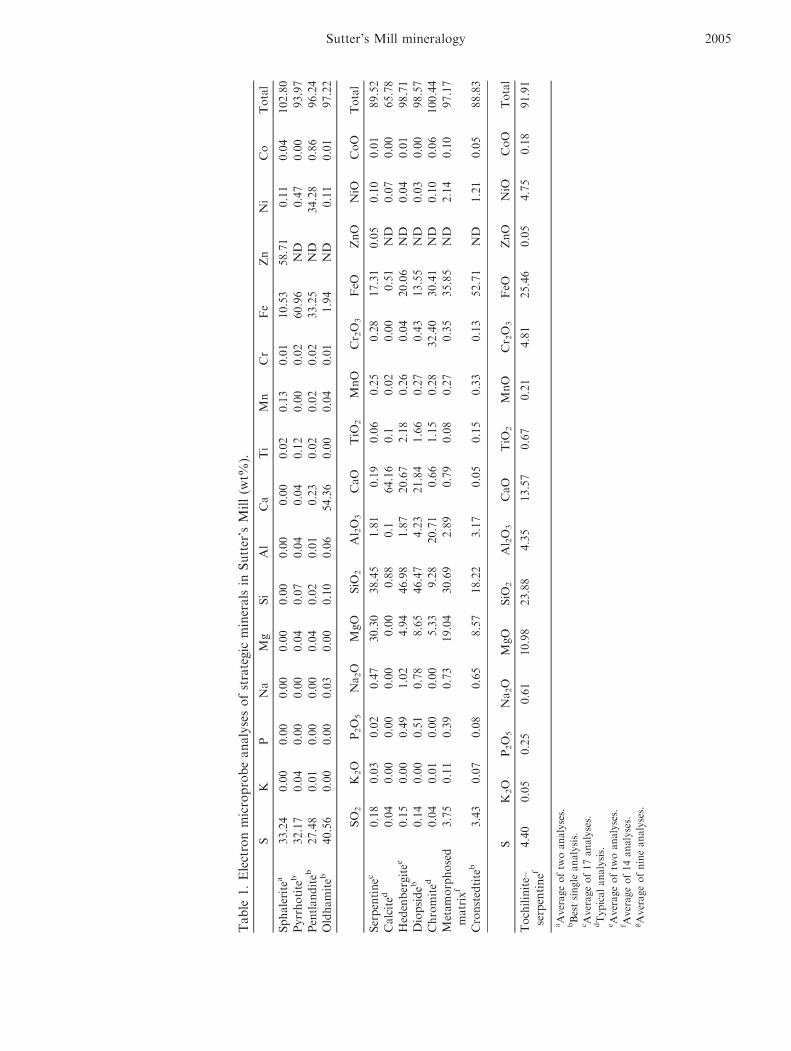
Some Sutter’s Mill mineral compositions weredetermined from all three main samples using theCameca SX100 microprobe at the E-beam laboratory ofthe Astromaterials and Exploration Science Directorate,Johnson Space Center (JSC), and others using theJEOL JXA-8900L microprobe at the University ofTokyo. For these analyses, natural mineral standardswere employed, and analytical errors are at the 0.1 wt%level for most elements. We used a 1 lm focused beamin all analyses, at 15 kV and 20 nA, which resulted inlow analytical totals for carbonates. However, therelative cation ratios for carbonates should not havebeen affected by the beam damage.
To assess mineral distributions, WDS maps ofelemental concentrations were acquired using the JEOLJXA 8900L electron microprobe (accelerating voltage,15 kV; beam current, 120 nA) at the Department ofEarth and Planetary Sciences, University of Tokyo, andalso using the Cameca SX100 microprobe at the JSC(described above), and JEOL 4600 field emission SEMat JSC.
Electron back-scatter diffraction (EBSD) pattern(Kikuchi diffraction pattern) analysis providescrystallographic and phase information of micrometer-sized crystalline materials prepared for observation inan SEM (Goehner and Michael 1996) in all three mainsamples. Backscattered electron (BSE) images weretaken using several SEMs. We used a cold fieldemission Hitachi S-4800 SEM at JSC equipped with anOxford CHANNEL5 EBSD system with associatedreduction software. Imaging and data reductionprocedures were as above.
We collected single crystal and powder Lauepatterns of individual crystals from samples SM2 andSM44 by the Laue synchrotron X-ray diffraction(SXRD) method using polychromatic synchrotronradiation at beamline BL-4B1 of the Photon Factory,National Laboratory for High Energy AcceleratorResearch Organization (KEK), Tsukuba, Japan. TheLaue diffraction experiment was performed with thefollowing conditions. The ring operated at 2.5 GeV.The exposure times were generally 30–60 min. We usedpolychromatic radiation at 0.3–3.0 �A. The beam size at
1998 M. Zolensky et al.

the sample position was 1.6 lm in diameter, with abeam divergence of 40 lrad. These procedures aredescribed in detail by Ivanov et al. (2000).
Sample SM44 (5.5 g) was scanned at the Universityof Texas High-Resolution X-ray CT Facility (XCT)using the ACTIS scanner, which in the ultrahigh-resolution mode yields maps of objects that can bepenetrated by relatively low-energy X-rays—in this casea 225 kV microfocal X-ray source (Ketcham andCarlson 2001). Pixel diameter was approximately 5 lm.Streak- and ring-removal processing of the images wasbased on correction of raw sinogram data using IDLroutines “RK_SinoDeStreak” and “RK_SinoRingProcSimul,” both with default parameters (Ketcham 2005).The tomographic imaging was useful in identifyinginternal lithologic and mineralogic differences, andoverall sample fabrics.
In an effort to locate aqueous fluid-filled fluidinclusions, we removed one FIB section from each ofthree preselected calcite crystals from SM44 at KyotoUniversity. The FIB slices of calcite were then imagedby synchrotron X-ray computer microtomography(SXCT) at SPring-8 Beam Line 47XU (Oct, 2013).These samples were also examined by micro-Ramanspectrosccopy at Kyoto University.
Raman analyses of SM44 and SM51 were alsoconducted in the Vibrational Spectroscopy Laboratoryin the Department of Geosciences at Virginia Techusing a JY Horiba LabRam HR (800 mm)spectrometer, with 600 grooves mm�1 gratings and aslit width of 150 lm. The confocal aperture was set at400 lm. Excitation was provided by a 514.53 nmModu-Laser Stellar Pro-L 100 mW laser which wasfocused onto the sample through a 1009 objective. Thedetector was an electronically cooled open electrodeCCD. Owing to the high background signal from thesample, collection times for individual spectra were 1 s;longer collection times resulting in the detectorbecoming saturated in the fluorescence signalbackground.
We measured visible to infrared reflectance spectraof rough surfaces of two chips of SM2–5 (subsample 5from sample SM2) over the wavelength range of 0.25–14 lm. Bidirectional visible and near-infrared (BD-VNIR) reflectance spectra were recorded at every 5 nmover the wavelength range of 0.25–2.50 lm with a spotsize of 2 9 3 mm2 at the Mizusawa VLBI Observatoryin Japan. Spectralon (SRS99, Labsphere) was used asthe reflectance standard. The incidence and emissionangles were set to 30° and 0°, respectively, and thephase angle was 30�. Wavelength scans were performedby combining data collected using different diffractiongratings and order-sorting filters. The light sources werea deuterium lamp (0.25–0.39 lm) and a tungsten-
halogen lamp (0.39–2.50 lm), and the detectors were aphotomultiplier tube (0.25–0.90 lm) and two differentInGaAs detectors (0.90–1.64 lm and 1.64–2.50 lm).
Fourier transform infrared (FTIR) reflectancespectra of the same SM2–5 samples were measured overthe wavelength range of 2.50–14.00 lm using a ThermoScientific Nicolet iN10 instrument with OMNIC Pictasoftware at Tohoku University, Japan. Thus, twoportions (SM21L1 001 and SM21L3 003 in fig. A) ofchip 1 (3 9 5 mm) were measured by both BD-VNIRand FTIR, while one portion of chip 2 (5 9 5 mm) wasanalyzed by BD-VNIR and two portions, close to theposition (SM22L2) analyzed by BD-VNIR, wereanalyzed by FTIR. The spectra were recorded in stepsof 1.9 cm�1. The measured surface area was400 9 400 lm2 in size, and samples were purged in N2
gas during measurements. A specular gold disk wasused as the reflectance standard. Both the incident andemission angles were set to 0°. We performedbackground subtraction during sample measurements.Measured FTIR spectra were combined with BD-VNIRspectra collected at 2.50 lm through adjustment ofreflectance to the BD-VNIR side by multiplying theFTIR spectra by a correlation factor.
We examined several samples of Sutter’s Millsample SM44 using the JEOL 2500SE field emissiontransmission electron microscope (FEGTEM) at JSC,equipped with thin window EDX, Gatan imaging filter,high-angle annular dark field detector and 2Kx2K slowscan CCD camera. The samples were prepared for TEMobservation using JSC’s Quanta 3-D focused ion beam(FIB) instrument to mill the samples.
RESULTS
Mineralogy and Petrography of CM1–2 Lithologies
The Sutter’s Mill meteorite contains numerousdistinct CM lithologies of considerably varyingpetrographies (Figs. 1 and 2). We found CM1–CM2lithologies present as angular to rounded clasts in bothSM44 and SM51 (which are both mainly CM2). Thereare also thermally metamorphosed CM lithologies inSM2. Although these lithologies are describedseparately in this paper, we have only examined a tinypercentage of the available SM samples. Futureinvestigations may well reveal that these lithologiesentirely grade into one another, as is so oftenthe case for CMs (e.g., Zolensky et al. 1997). Wenote that Sutter’s Mill is currently officially classified asa “C chondrite” (see http://www.lpi.usra.edu/meteor/metbull.php?code=55529), which happened because themeteorite required an official name very soon after itsfall and well before detailed mineralogic information
Sutter’s Mill mineralogy 1999
had been available. We strongly recommend that theMeteorite Nomenclature Committee amend the officialclassification to “CM.”
Note that we do not use here the more recentlyproposed reclassification of CMs proposed by Rubinet al. (2007), because it is not readily compatible withthe descriptions of the metamorphosed CM material(Tonui et al. 2014). Readers are reminded that underthe previous CM classification, which we are using here,CM2 indicates partial aqueous alteration, and CM1denotes complete aqueous alteration.
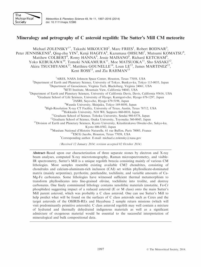
Tomographic imaging was useful in identifyinginternal lithologic and mineralogic differences, andoverall sample fabrics, which we used to decide whereto make the initial slice into the sample and preparethin sections. Sample SM44, a 5.5 g fusion-crustedstone, was scanned by XRCT. As shown in the two
computed images in Fig. 1, some lithologies were verywell delineated by the XRCT imaging, as long aschondrule and sulfide content significantly varied.However, variations in degree of development ofcarbonate and tochilinite rims, matrix phyllosilicatetextures, and degree of brecciation of tochilinite-serpentine pellets in matrix are not well discriminatedbased only on these particular XRCT images. However,as the XRCT scanning procedure appears to becompletely nondestructive, there is every reason toemploy it regularly with new stones, especially formeteorites as lithologically variable and brecciated asSutter’s Mill.
From the actual cut slices, we prepared thin sectionsfrom each of SM44 and SM51, which were bothrecovered after they had been rained on. There wereinevitable terrestrial modifications, including networks of
a b
3mm
Fig. 1. (a) and (b) Two computer “slices” through Sutter’s Mill sample SM44 produced from the CXRT scanning. Several of themore easily discriminated clasts are indicated by arrows.
a b
3mm 2mm
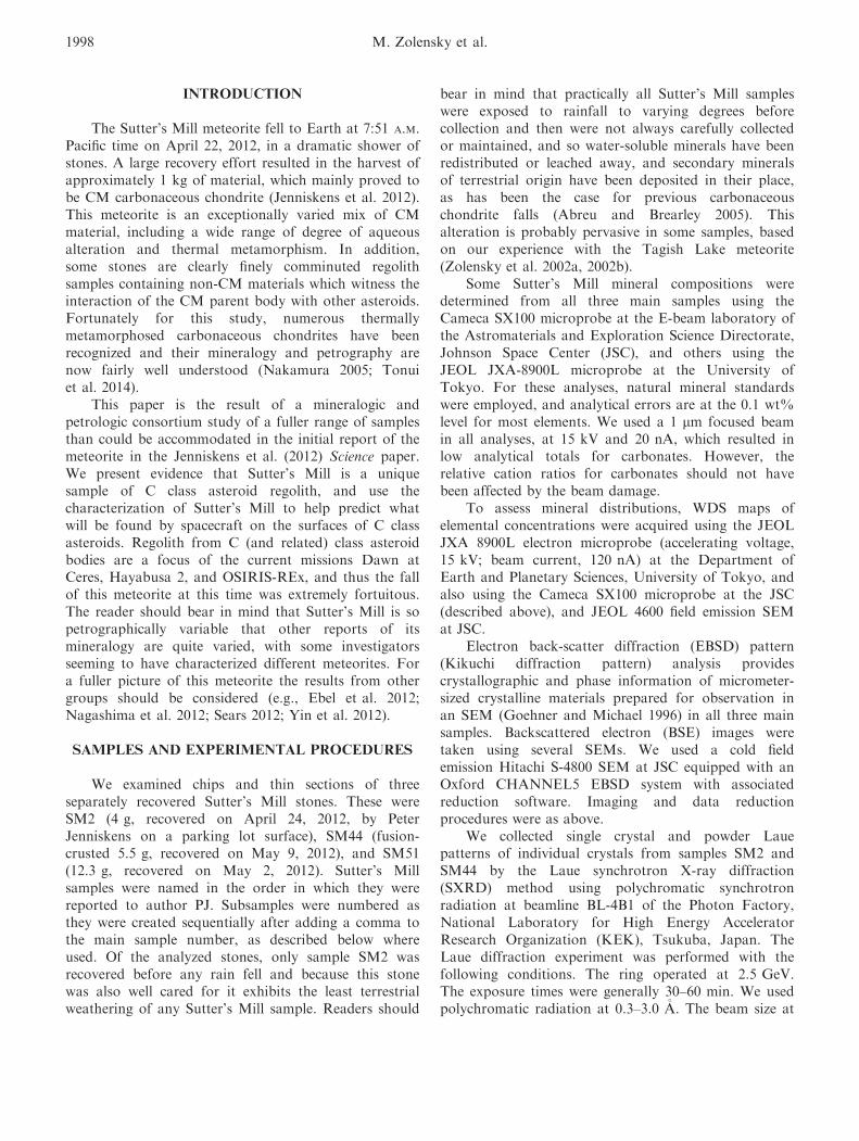
Fig. 2. BSE images of thin section of Sutter’s Mill samples (a) SM51 and (b) SM44. Outlines of some of the different lithologiesare indicated by dotted lines.
2000 M. Zolensky et al.
cracks apparent in both sections in both optical and BSEimages, and some rusting of sulfides. Rusting of metalwas not a significant issue since, as in most CMs, metalin Sutter’s Mill CM lithologies is largely confined tosmall beads in olivine grains. BSE imaging clearly showsthat SM44 and SM51 are both breccias of diverselithologies, which vary in chondrule abundance and size,thickness of fine-grained rims, quantity of tochilinite–serpentine aggregates, quantity of fine-grained sulfideswithin serpentine, quantity and mineralogy ofcarbonates, and degree of brecciation (Fig. 2).
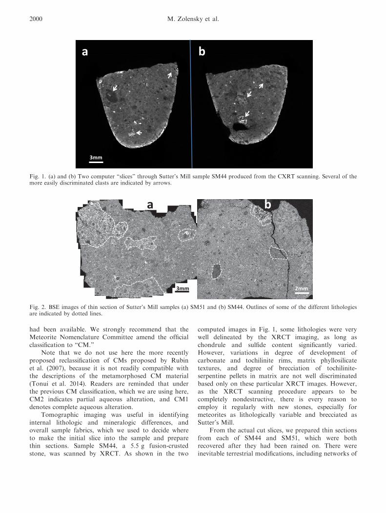
Clasts in the sections vary up to approximately1 cm across, with irregular to rounded outlines (Figs. 2and 3). Some lithologies consist entirely of rounded,serpentine and tochilinite-rich pellets and chondrules(left side of Fig. 3a), with minimal fracturing. This
texture was previously described from Y791198 byMetzler et al. (1992), who called it the “primaryaccretionary rock.” Lithic clasts, matrix, and materialsother than the pellets are rare in these particular clasts.
At the other end of the scale, some CM lithologiesconsist entirely of fine-grained phyllosilicate, sulfides,and magnetite with no apparent larger components(Fig. 3f, center). In some lithologies, components havewell-developed, layered rims (Figs. 3a, 3c, 3d, and 3e),or fine-scale, delicate rims of pyrrhotite, tochilinite, Cacarbonate, or cronstedtite (Figs. 3, 4a, 5, and 6b). Inother lithologies, chondrules and other largercomponents are entirely fractured and rims are muchless well developed (Figs. 3a–d).
As is typical of CM2 chondrites which are oftenbrecciated and contain clasts of CM1 lithologies, olivine
ed
a b
200µm
500µm
200µm 600µm
f
cb
500µm
Fig. 3. BSE images of different CM1 to CM2 lithologies in Sutter’s Mill samples SM44 and 51. (a–e) are from SM51, (f) is fromSM44. Outlines of some of the different lithologies are indicated by dotted lines. Dramatic differences can be observed inpresence and degree of development of fine-grained rims, quantity of sulfides (white in these images), differences in the quantityand sizes of chondrules, and degree of brecciation. a) Left side is CM2 dominated by tochilinite–serpentine pellets, right sidelithology is a CM2 fragmental breccia. b) Matrix-supported CM2 where coarse components have poorly developed or ablatedfine-grained rims. c) Matrix-supported CM2 where coarse components have well-developed fine-grained rims. d) Two CM2lithologies with a stark difference in the quantity of fragmented sulfides (white phases) in the matrix. e) Dashed line surrounds alarge lithic fragment with a layered, well-developed, fine-grained rim. f) Dashed line surrounds a CM1 lithology.
Sutter’s Mill mineralogy 2001
in the Sutter’s Mill CM1-2 lithologies has a widecompositional range (mainly Fa0-50, with a distributionpeak at Fa2), and normally zoned crystals areabundant. There are amoeboid olivine aggregates(AOAs), usually partially replaced by secondary phasesincluding serpentine, carbonates, sulfides, andmagnetite. Broken olivine crystals in matrix measure upto approximately 250 lm in size. Table 1 presentsrepresentative analyses of serpentine, cronstedtite,tochilinite–serpentine, calcite, pyrrhotite, andpentlandite from several of the Sutter’s Mill CM2lithologies. We note here that we have not performedthe detailed TEM imaging necessary to uniquelyidentify the exact serpentine group phases present inSutter’s Mill, so when we call something “serpentine”please bear in mind that additional work is necessarybefore one can state just exactly what these phases are(M€uller et al. 1979). Similarly, tochilinite andtochilinite–serpentine were identified mainly on thebasis of optical and physical properties (including color,luster, hardness, form, and mineral associations) inaddition to the microprobe analyses, and the exactcrystal structure of these phases still awaits verification.
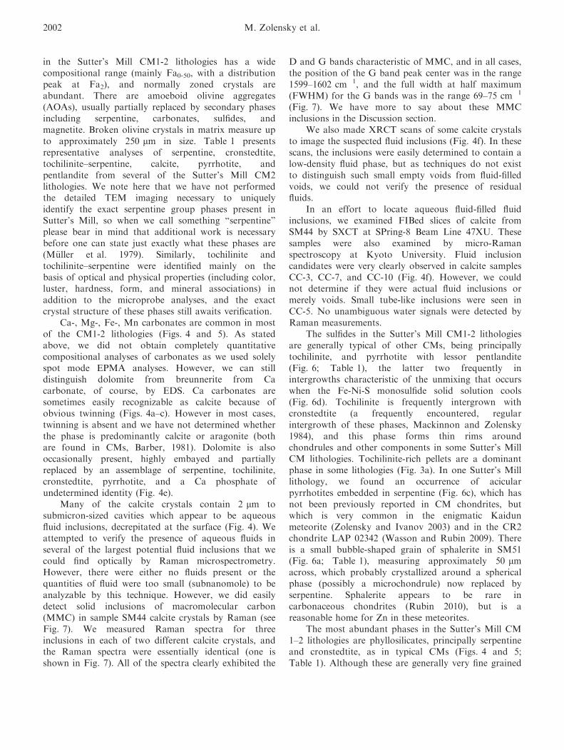
Ca-, Mg-, Fe-, Mn carbonates are common in mostof the CM1-2 lithologies (Figs. 4 and 5). As statedabove, we did not obtain completely quantitativecompositional analyses of carbonates as we used solelyspot mode EPMA analyses. However, we can stilldistinguish dolomite from breunnerite from Cacarbonate, of course, by EDS. Ca carbonates aresometimes easily recognizable as calcite because ofobvious twinning (Figs. 4a–c). However in most cases,twinning is absent and we have not determined whetherthe phase is predominantly calcite or aragonite (bothare found in CMs, Barber, 1981). Dolomite is alsooccasionally present, highly embayed and partiallyreplaced by an assemblage of serpentine, tochilinite,cronstedtite, pyrrhotite, and a Ca phosphate ofundetermined identity (Fig. 4e).
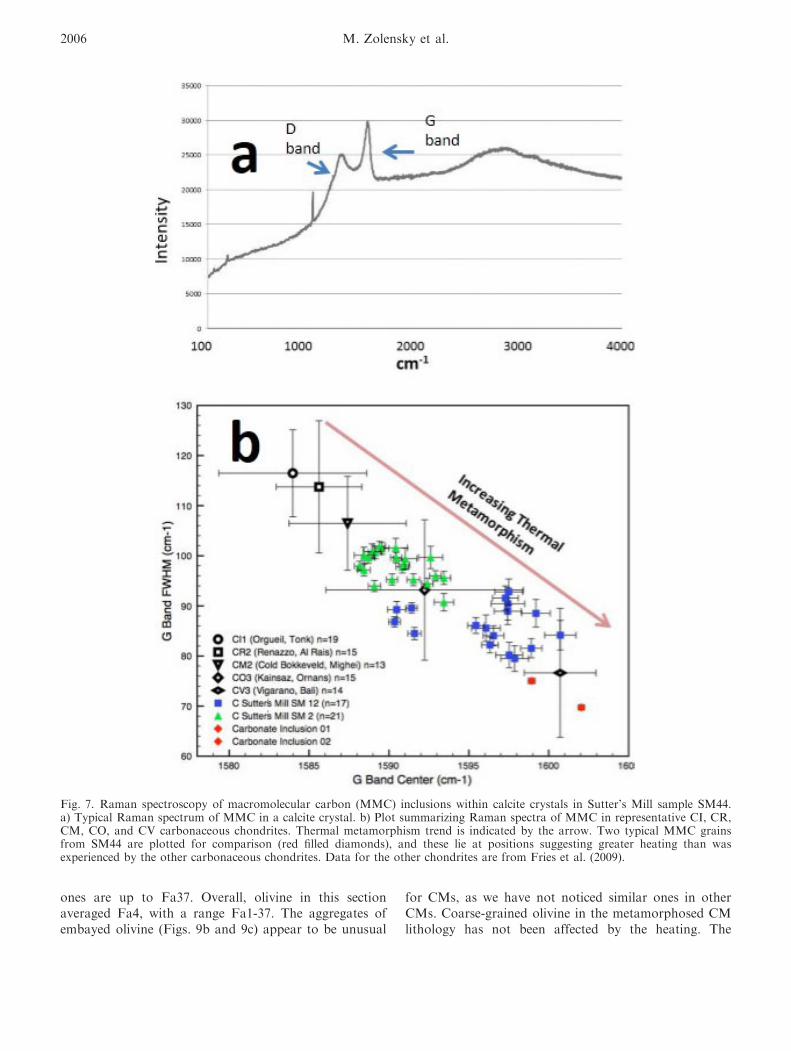
Many of the calcite crystals contain 2 lm tosubmicron-sized cavities which appear to be aqueousfluid inclusions, decrepitated at the surface (Fig. 4). Weattempted to verify the presence of aqueous fluids inseveral of the largest potential fluid inclusions that wecould find optically by Raman microspectrometry.However, there were either no fluids present or thequantities of fluid were too small (subnanomole) to beanalyzable by this technique. However, we did easilydetect solid inclusions of macromolecular carbon(MMC) in sample SM44 calcite crystals by Raman (seeFig. 7). We measured Raman spectra for threeinclusions in each of two different calcite crystals, andthe Raman spectra were essentially identical (one isshown in Fig. 7). All of the spectra clearly exhibited the
D and G bands characteristic of MMC, and in all cases,the position of the G band peak center was in the range1599–1602 cm�1, and the full width at half maximum(FWHM) for the G bands was in the range 69–75 cm�1
(Fig. 7). We have more to say about these MMCinclusions in the Discussion section.
We also made XRCT scans of some calcite crystalsto image the suspected fluid inclusions (Fig. 4f). In thesescans, the inclusions were easily determined to contain alow-density fluid phase, but as techniques do not existto distinguish such small empty voids from fluid-filledvoids, we could not verify the presence of residualfluids.
In an effort to locate aqueous fluid-filled fluidinclusions, we examined FIBed slices of calcite fromSM44 by SXCT at SPring-8 Beam Line 47XU. Thesesamples were also examined by micro-Ramanspectroscopy at Kyoto University. Fluid inclusioncandidates were very clearly observed in calcite samplesCC-3, CC-7, and CC-10 (Fig. 4f). However, we couldnot determine if they were actual fluid inclusions ormerely voids. Small tube-like inclusions were seen inCC-5. No unambiguous water signals were detected byRaman measurements.
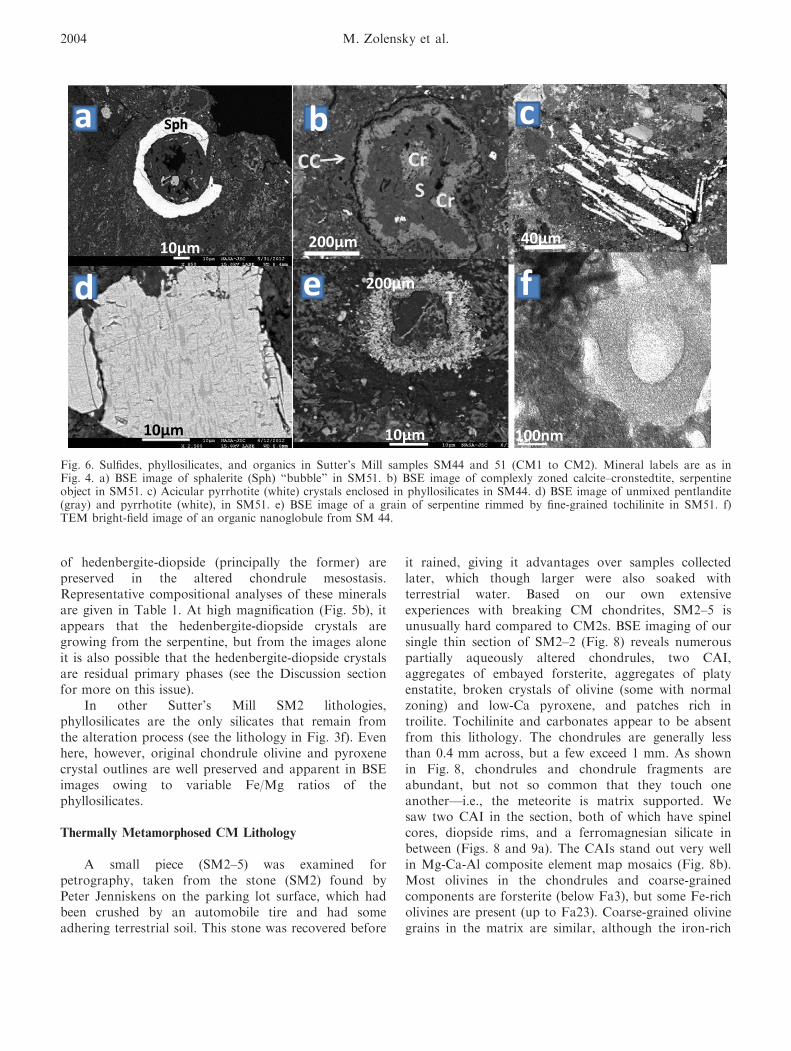
The sulfides in the Sutter’s Mill CM1-2 lithologiesare generally typical of other CMs, being principallytochilinite, and pyrrhotite with lessor pentlandite(Fig. 6; Table 1), the latter two frequently inintergrowths characteristic of the unmixing that occurswhen the Fe-Ni-S monosulfide solid solution cools(Fig. 6d). Tochilinite is frequently intergrown withcronstedtite (a frequently encountered, regularintergrowth of these phases, Mackinnon and Zolensky1984), and this phase forms thin rims aroundchondrules and other components in some Sutter’s MillCM lithologies. Tochilinite-rich pellets are a dominantphase in some lithologies (Fig. 3a). In one Sutter’s Milllithology, we found an occurrence of acicularpyrrhotites embedded in serpentine (Fig. 6c), which hasnot been previously reported in CM chondrites, butwhich is very common in the enigmatic Kaidunmeteorite (Zolensky and Ivanov 2003) and in the CR2chondrite LAP 02342 (Wasson and Rubin 2009). Thereis a small bubble-shaped grain of sphalerite in SM51(Fig. 6a; Table 1), measuring approximately 50 lmacross, which probably crystallized around a sphericalphase (possibly a microchondrule) now replaced byserpentine. Sphalerite appears to be rare incarbonaceous chondrites (Rubin 2010), but is areasonable home for Zn in these meteorites.
The most abundant phases in the Sutter’s Mill CM1–2 lithologies are phyllosilicates, principally serpentineand cronstedtite, as in typical CMs (Figs. 4 and 5;Table 1). Although these are generally very fine grained
2002 M. Zolensky et al.
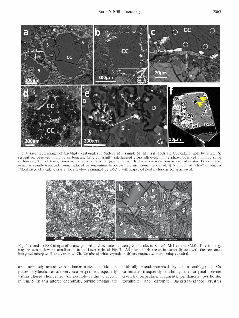
and intimately mixed with submicron-sized sulfides, inplaces phyllosilicates are very coarse grained, especiallywithin altered chondrules. An example of this is shownin Fig. 5. In this altered chondrule, olivine crystals are
faithfully pseudomorphed by an assemblage of Cacarbonate (frequently outlining the original olivinecrystals), serpentine, magnetite, pentlandite, pyrrhotite,tochilinite, and chromite. Jackstraw-shaped crystals
a b c
d e f
200µm 10µm
20µm200µm200µm
200µm
Fig. 4. (a–e) BSE images of Ca-Mg-Fe carbonates in Sutter’s Mill sample 51. Mineral labels are CC: calcite (note twinning); S:serpentine, observed rimming carbonates; CrT: coherently interlayered cronstedtite–tochilinite phase, observed rimming somecarbonates; T: tochilinite, rimming some carbonates; P: pyrrhotite, which discontinuously rims some carbonates; D: dolomite,which is usually embayed, being replaced by serpentine. Probable fluid inclusions are circled. f) A computed “slice” through aFIBed piece of a calcite crystal from SM44, as imaged by SXCT, with suspected fluid inclusions being arrowed.
a b
20µm 50µm
S
S
S H
H
S
HCC
Ch
CC
CC
P
HH
S CC
T
Fig. 5. a and b) BSE images of coarse-grained phyllosilicates replacing chondrules in Sutter’s Mill sample SM51. This lithologymay be seen at lower magnification in the lower right of Fig. 3e. All phase labels are as in earlier figures, with the new onesbeing hedenbergite: H and chromite: Ch. Unlabeled white crystals in (b) are magnetite, many being euhedral.
Sutter’s Mill mineralogy 2003
of hedenbergite-diopside (principally the former) arepreserved in the altered chondrule mesostasis.Representative compositional analyses of these mineralsare given in Table 1. At high magnification (Fig. 5b), itappears that the hedenbergite-diopside crystals aregrowing from the serpentine, but from the images aloneit is also possible that the hedenbergite-diopside crystalsare residual primary phases (see the Discussion sectionfor more on this issue).
In other Sutter’s Mill SM2 lithologies,phyllosilicates are the only silicates that remain fromthe alteration process (see the lithology in Fig. 3f). Evenhere, however, original chondrule olivine and pyroxenecrystal outlines are well preserved and apparent in BSEimages owing to variable Fe/Mg ratios of thephyllosilicates.
Thermally Metamorphosed CM Lithology
A small piece (SM2–5) was examined forpetrography, taken from the stone (SM2) found byPeter Jenniskens on the parking lot surface, which hadbeen crushed by an automobile tire and had someadhering terrestrial soil. This stone was recovered before
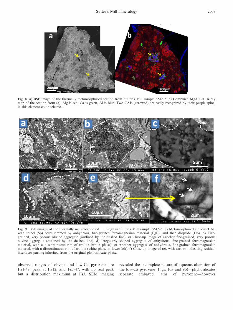
it rained, giving it advantages over samples collectedlater, which though larger were also soaked withterrestrial water. Based on our own extensiveexperiences with breaking CM chondrites, SM2–5 isunusually hard compared to CM2s. BSE imaging of oursingle thin section of SM2–2 (Fig. 8) reveals numerouspartially aqueously altered chondrules, two CAI,aggregates of embayed forsterite, aggregates of platyenstatite, broken crystals of olivine (some with normalzoning) and low-Ca pyroxene, and patches rich introilite. Tochilinite and carbonates appear to be absentfrom this lithology. The chondrules are generally lessthan 0.4 mm across, but a few exceed 1 mm. As shownin Fig. 8, chondrules and chondrule fragments areabundant, but not so common that they touch oneanother—i.e., the meteorite is matrix supported. Wesaw two CAI in the section, both of which have spinelcores, diopside rims, and a ferromagnesian silicate inbetween (Figs. 8 and 9a). The CAIs stand out very wellin Mg-Ca-Al composite element map mosaics (Fig. 8b).Most olivines in the chondrules and coarse-grainedcomponents are forsterite (below Fa3), but some Fe-richolivines are present (up to Fa23). Coarse-grained olivinegrains in the matrix are similar, although the iron-rich
a b c
d e f200µm10µm 40µm
200µm
100nm10µm10µm
Fig. 6. Sulfides, phyllosilicates, and organics in Sutter’s Mill samples SM44 and 51 (CM1 to CM2). Mineral labels are as inFig. 4. a) BSE image of sphalerite (Sph) “bubble” in SM51. b) BSE image of complexly zoned calcite–cronstedtite, serpentineobject in SM51. c) Acicular pyrrhotite (white) crystals enclosed in phyllosilicates in SM44. d) BSE image of unmixed pentlandite(gray) and pyrrhotite (white), in SM51. e) BSE image of a grain of serpentine rimmed by fine-grained tochilinite in SM51. f)TEM bright-field image of an organic nanoglobule from SM 44.
2004 M. Zolensky et al.
Table
1.Electronmicroprobeanalysesofstrategic
minerals
inSutter’sMill(w
t%).
SK
PNa
Mg
Si
Al
Ca
Ti
Mn
Cr
Fe
Zn
Ni
Co
Total
Sphalerite
a33.24
0.00
0.00
0.00
0.00
0.00
0.00
0.00
0.02
0.13
0.01
10.53
58.71
0.11
0.04
102.80
Pyrrhotite
b32.17
0.04
0.00
0.00
0.04
0.07
0.04
0.04
0.12
0.00
0.02
60.96
ND
0.47
0.00
93.97
Pentlanditeb
27.48
0.01
0.00
0.00
0.04
0.02
0.01
0.23
0.02
0.02
0.02
33.25
ND
34.28
0.86
96.24
Oldhamiteb
40.56
0.00
0.00
0.03
0.00
0.10
0.06
54.36
0.00
0.04
0.01
1.94
ND
0.11
0.01
97.22
SO
2K
2O
P2O
5Na2O
MgO
SiO
2Al 2O
3CaO
TiO
2MnO
Cr 2O
3FeO
ZnO
NiO
CoO
Total
Serpentinec
0.18
0.03
0.02
0.47
30.30
38.45
1.81
0.19
0.06
0.25
0.28
17.31
0.05
0.10
0.01
89.52
Calcited
0.04
0.00
0.00
0.00
0.00
0.88
0.1
64.16
0.1
0.02
0.00
0.51
ND
0.07
0.00
65.78
Hedenbergitee
0.15
0.00
0.49
1.02
4.94
46.98
1.87
20.67
2.18
0.26
0.04
20.06
ND
0.04
0.01
98.71
Diopsideb
0.14
0.00
0.51
0.78
8.65
46.47
4.23
21.84
1.66
0.27
0.43
13.55
ND
0.03
0.00
98.57
Chromited
0.04
0.01
0.00
0.00
5.33
9.28
20.71
0.66
1.15
0.28
32.40
30.41
ND
0.10
0.06
100.44
Metamorphosed
matrix
f3.75
0.11
0.39
0.73
19.04
30.69
2.89
0.79
0.08
0.27
0.35
35.85
ND
2.14
0.10
97.17
Cronstedtite
b3.43
0.07
0.08
0.65
8.57
18.22
3.17
0.05
0.15
0.33
0.13
52.71
ND
1.21
0.05
88.83
SK
2O
P2O
5Na2O
MgO
SiO
2Al 2O
3CaO
TiO
2MnO
Cr 2O
3FeO
ZnO
NiO
CoO
Total
Tochilinite–
serpentinef
4.40
0.05
0.25
0.61
10.98
23.88
4.35
13.57
0.67
0.21
4.81
25.46
0.05
4.75
0.18
91.91
aAverageoftw
oanalyses.
bBestsingle
analysis.
cAverageof17analyses.
dTypicalanalysis.
eAverageoftw
oanalyses.
f Averageof14analyses.
gAverageofnineanalyses.
Sutter’s Mill mineralogy 2005
ones are up to Fa37. Overall, olivine in this sectionaveraged Fa4, with a range Fa1-37. The aggregates ofembayed olivine (Figs. 9b and 9c) appear to be unusual
for CMs, as we have not noticed similar ones in otherCMs. Coarse-grained olivine in the metamorphosed CMlithology has not been affected by the heating. The
Fig. 7. Raman spectroscopy of macromolecular carbon (MMC) inclusions within calcite crystals in Sutter’s Mill sample SM44.a) Typical Raman spectrum of MMC in a calcite crystal. b) Plot summarizing Raman spectra of MMC in representative CI, CR,CM, CO, and CV carbonaceous chondrites. Thermal metamorphism trend is indicated by the arrow. Two typical MMC grainsfrom SM44 are plotted for comparison (red filled diamonds), and these lie at positions suggesting greater heating than wasexperienced by the other carbonaceous chondrites. Data for the other chondrites are from Fries et al. (2009).
2006 M. Zolensky et al.
observed ranges of olivine and low-Ca pyroxene areFa1-49, peak at Fa12, and Fs3-47, with no real peakbut a distribution maximum at Fs3. SEM imaging
revealed the incomplete nature of aqueous alteration ofthe low-Ca pyroxene (Figs. 10a and 9b)—phyllosilicatesseparate embayed laths of pyroxene—however
a b
200µm200µm
Fig. 8. a) BSE image of the thermally metamorphosed section from Sutter’s Mill sample SM2–5. b) Combined Mg-Ca-Al X-raymap of the section from (a). Mg is red, Ca is green, Al is blue. Two CAIs (arrowed) are easily recognized by their purple spinelin this element color scheme.
f
a b c
d e
10µm
10µm5µm20µm
2µm10µm
p
p
Fig. 9. BSE images of the thermally metamorphosed lithology in Sutter’s Mill sample SM2–5. a) Metamorphosed sinuous CAI,with spinel (Sp) cores rimmed by anhydrous, fine-grained ferromagnesian material (FgF), and then diopside (Dp). b) Fine-grained, very porous olivine aggregate (outlined by the dashed line). c) Close-up image of another fine-grained, very porousolivine aggregate (outlined by the dashed line). d) Irregularly shaped aggregate of anhydrous, fine-grained ferromagnesianmaterial, with a discontinuous rim of troilite (white phase). e) Another aggregate of anhydrous, fine-grained ferromagnesianmaterial, with a discontinuous rim of troilite (white phase at lower left). f) Close-up image of (e), with arrows indicating residualinterlayer parting inherited from the original phyllosilicate phase.
Sutter’s Mill mineralogy 2007
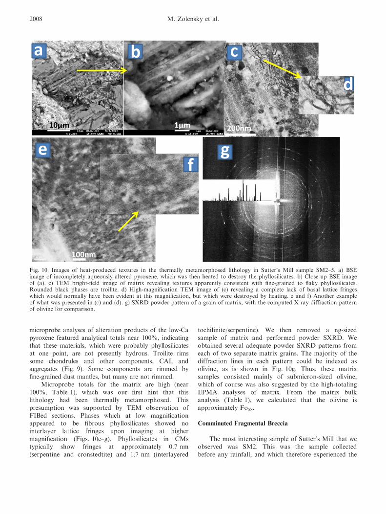
microprobe analyses of alteration products of the low-Capyroxene featured analytical totals near 100%, indicatingthat these materials, which were probably phyllosilicatesat one point, are not presently hydrous. Troilite rimssome chondrules and other components, CAI, andaggregates (Fig. 9). Some components are rimmed byfine-grained dust mantles, but many are not rimmed.
Microprobe totals for the matrix are high (near100%, Table 1), which was our first hint that thislithology had been thermally metamorphosed. Thispresumption was supported by TEM observation ofFIBed sections. Phases which at low magnificationappeared to be fibrous phyllosilicates showed nointerlayer lattice fringes upon imaging at highermagnification (Figs. 10c–g). Phyllosilicates in CMstypically show fringes at approximately 0.7 nm(serpentine and cronstedtite) and 1.7 nm (interlayered
tochilinite/serpentine). We then removed a ng-sizedsample of matrix and performed powder SXRD. Weobtained several adequate powder SXRD patterns fromeach of two separate matrix grains. The majority of thediffraction lines in each pattern could be indexed asolivine, as is shown in Fig. 10g. Thus, these matrixsamples consisted mainly of submicron-sized olivine,which of course was also suggested by the high-totalingEPMA analyses of matrix. From the matrix bulkanalysis (Table 1), we calculated that the olivine isapproximately Fo58.
Comminuted Fragmental Breccia
The most interesting sample of Sutter’s Mill that weobserved was SM2. This was the sample collectedbefore any rainfall, and which therefore experienced the
ca b
e
d
fg
100nm
10µm 1µm 200nm
Fig. 10. Images of heat-produced textures in the thermally metamorphosed lithology in Sutter’s Mill sample SM2–5. a) BSEimage of incompletely aqueously altered pyroxene, which was then heated to destroy the phyllosilicates. b) Close-up BSE imageof (a). c) TEM bright-field image of matrix revealing textures apparently consistent with fine-grained to flaky phyllosilicates.Rounded black phases are troilite. d) High-magnification TEM image of (c) revealing a complete lack of basal lattice fringeswhich would normally have been evident at this magnification, but which were destroyed by heating. e and f) Another exampleof what was presented in (c) and (d). g) SXRD powder pattern of a grain of matrix, with the computed X-ray diffraction patternof olivine for comparison.
2008 M. Zolensky et al.

least terrestrial alteration. This was indeed fortunate,because BSE imaging of subsample SM2-5 immediatelyrevealed it to be unique. Subsample SM2-5 is a matrix-supported breccia of olivine and low-Ca pyroxeneaggregates, some with fine-grained dust mantles, CAI,diverse unusual lithic fragments, fragments of olivineand low-Ca pyroxene (up to 1 mm), and very abundantsulfide grains. We observed no chondrules, but some ofthe lithic fragments could be pieces of chondrules.
The most abundant sulfide in our sample of SM2-5 isCaS—oldhamite, which ranges up to approximately300 lm in size. We initially identified this unexpectedphase from a surface we had exposed (by splitting asample) in the lab, and which had been carbon coatedbut not polished. EDX analysis in the SEM suggestedthat it was oldhamite (Fig. 11). Because thisidentification seemed rather improbable, we spentconsiderable effort verifying it. Attempts to make goodEPMA analyses were frustrated by the extremeinstability of the phase, even though we used only fresh
methanol for grinding and polishing, and stored thesample in a dry environment. We were only successful inobtaining useful EPMA analyses (Table 1) when wecarbon coated and probed the sample immediately uponpolishing it. We also removed a 20 lm sized oldhamitegrain from the polished section and collected a SXRDpowder pattern (at SPring-8). As shown in Fig. 11c, theobserved powder pattern is an excellent match to thecalculated pattern. X-ray mapping of the polished mountof SM2–4 indicates that oldhamite grains are abundantthroughout. As the groundmass of this sample isextremely fragmented and mixed, it is probablyimpossible to securely identify other phases that aredirectly associated with the oldhamite. Fe-Ni sulfides inthe groundmass are fine-grained, usually associated withphosphides, and less common than oldhamite.
SXRD of the fine-grained silicate matrix revealedthat it consists mainly of submicron olivine. We couldlocate no carbonates or sulfates in SM2 (and wesearched pretty hard), or if sulfates appear later on they
Fig. 11. Oldhamite in the fine-grained lithology of Sutter’s Mill sample SM2–5. a) BSE image of the fine-grained section. Asmall CM clast and CAI are indicated. The box indicates the position of the largest oldhamite grain we found. b) Ca X-ray mapof the section from (a). With the exception of the CAIs, essentially all of the white grains are oldhamite. c) SXRD powderpattern of a grain of oldhamite, with the computed X-ray diffraction pattern of oldhamite for comparison.
Sutter’s Mill mineralogy 2009
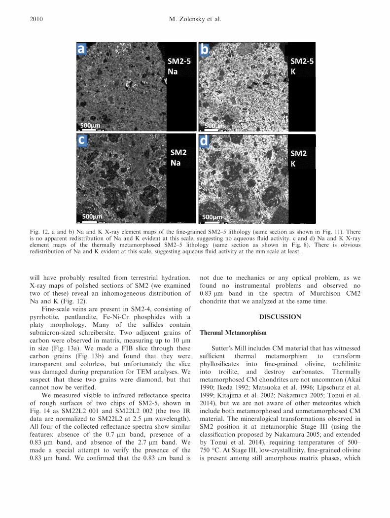
will have probably resulted from terrestrial hydration.X-ray maps of polished sections of SM2 (we examinedtwo of these) reveal an inhomogeneous distribution ofNa and K (Fig. 12).
Fine-scale veins are present in SM2-4, consisting ofpyrrhotite, pentlandite, Fe-Ni-Cr phosphides with aplaty morphology. Many of the sulfides containsubmicron-sized schreibersite. Two adjacent grains ofcarbon were observed in matrix, measuring up to 10 lmin size (Fig. 13a). We made a FIB slice through thesecarbon grains (Fig. 13b) and found that they weretransparent and colorless, but unfortunately the slicewas damaged during preparation for TEM analyses. Wesuspect that these two grains were diamond, but thatcannot now be verified.
We measured visible to infrared reflectance spectraof rough surfaces of two chips of SM2-5, shown inFig. 14 as SM22L2 001 and SM22L2 002 (the two IRdata are normalized to SM22L2 at 2.5 lm wavelength).All four of the collected reflectance spectra show similarfeatures: absence of the 0.7 lm band, presence of a0.83 lm band, and absence of the 2.7 lm band. Wemade a special attempt to verify the presence of the0.83 lm band. We confirmed that the 0.83 lm band is
not due to mechanics or any optical problem, as wefound no instrumental problems and observed no0.83 lm band in the spectra of Murchison CM2chondrite that we analyzed at the same time.
DISCUSSION
Thermal Metamorphism
Sutter’s Mill includes CM material that has witnessedsufficient thermal metamorphism to transformphyllosilicates into fine-grained olivine, tochiliniteinto troilite, and destroy carbonates. Thermallymetamorphosed CM chondrites are not uncommon (Akai1990; Ikeda 1992; Matsuoka et al. 1996; Lipschutz et al.1999; Kitajima et al. 2002; Nakamura 2005; Tonui et al.2014), but we are not aware of other meteorites whichinclude both metamorphosed and unmetamorphosed CMmaterial. The mineralogical transformations observed inSM2 position it at metamorphic Stage III (using theclassification proposed by Nakamura 2005; and extendedby Tonui et al. 2014), requiring temperatures of 500–750 °C. At Stage III, low-crystallinity, fine-grained olivineis present among still amorphous matrix phases, which
a b
c d500µm
500µm500µm
500µm
Fig. 12. a and b) Na and K X-ray element maps of the fine-grained SM2–5 lithology (same section as shown in Fig. 11). Thereis no apparent redistribution of Na and K evident at this scale, suggesting no aqueous fluid activity. c and d) Na and K X-rayelement maps of the thermally metamorphosed SM2–5 lithology (same section as shown in Fig. 8). There is obviousredistribution of Na and K evident at this scale, suggesting aqueous fluid activity at the mm scale at least.
2010 M. Zolensky et al.

also include low-crystallinity neoformed troilite. Thestage temperatures are approximate because they dependon many complex factors, including heating duration,sample porosity and grain size, presence of organics, etc.Using this classification, this material would be termed
“CM2TIII”: originally CM2 material which has beenthermally metamorphosed to stage. This would make themetamorphosed Sutter’s Mill materials similar to the CMchondrites Y86029, A-881655, and PCA 91008 (Tonuiet al. 2014).
a b c
d e f5µm10µm
1µm1µm
10µm
50µm
Fig. 13. Unusual components of the fine-grained lithology in Sutter’s Mill sample SM2–5. a) BSE image of an unpolished chip.The largest dark gray phase is enstatite, white veins consist of sulfides and phosphides. Two apparent diamond grains are in theupper right, with a white line indicating where a FIB slice was removed. b) BSE image of the FIB slice removed from (a). Thedark gray phases are probable diamonds. c) BSE image of a porous aggregate of platy enstatite crystals. d and e) BSE image ofa vein containing Fe-Ni sulfides and platy Fe-Ni-Ti phosphide phases (arrowed). f) BSE image of a porous olivine aggregate.White phases are Fe sulfides.
Fig. 14. Vis-infrared spectra of the fine-grained SM5 lithology. a) The spectrum from 0 to 2.5 lm. b) The spectrum from 0 to15 lm. Arrow indicates the mysterious 0.83 lm feature.
Sutter’s Mill mineralogy 2011
As shown in Fig. 5, one CM lithology features whatat first appeared to be hedenbergite-diopside crystals aregrowing from serpentine. However, while relatively low-temperature, authigenic hedenbergite is known (Enlowsand Oles 1966), it is much more likely that thehedenbergite-diopside crystals are primary phases whichare breaking down to form the secondary assemblageserpentine–magnetite–carbonate (Chao 1974), which ismuch more reasonable at low temperatures (andtemperatures must have been below approximately350 °C to preserve the tochilinite; Fuchs et al. 1973).
Macromolecular in Calcite
We measured Raman spectra for three inclusions ineach of two different calcite crystals in sample SM44(CM1–2), with the original intention of detectingaqueous fluids. While this search was unsuccessful, wewere surprised to instead easily detect macromolecular(MMC) inclusions in the calcites. The MMC Ramanspectra were very similar (Fig. 7), and in all cases theposition of the G band peak center was in the range1598–1602 cm�1, and the G band FWHM were in therange 68–75 cm�1 (Fig. 7). The G band position andwidth has been developed as an indicator or thermalmetamorphism in chondrites (Fries et al. 2009).Inspection of Fig. 7 shows the positions found in earlierinvestigations for MMC in chondrites and organicinclusions in meteoritic halite. Metamorphism drives theMMC G band peak to higher wave numbers andsharper peaks (smaller values of FWHM). According tothe G band characteristics, MMC in the Sutter’s MillSM44 calcite crystals have been thermallymetamorphosed beyond what has been observed in theKainsaz CO3.2 and Vigarano CV3.3 chondrites.Although rather uncertain, these meteorites are thoughtto have seen temperatures as high as 300 °C (Huss et al.2006). The interesting aspect of this is that thesepresumably metamorphosed MMC grains are residingin calcite crystals in CM1–2 stones, which should neverhave been heated beyond 100 °C (Zolensky et al. 1997),well below the peak temperatures of Kainsaz orVigarano. Presumably, the calcites sampled MMCwhich had experienced heating or shock at an earlierstage. It would be interesting to make Ramanmeasurements of MCC-bearing carbonates in othercarbonaceous chondrites to better illuminate the earlyhistory of organic materials in the solar system.
Reflectance Spectroscopy
All four of the reflectance spectra we collected ofSutter’s Mill sample SM2–5 show similar features,including the complete absence of the 0.7 lm band,
presence of a 0.83 lm band, and absence of the2.7 lm band. The absence of the 0.7 lm bandindicates either that no Fe3+ is in serpentine or thatthere is no serpentine. The absence of the 2.7 lmstructural water band verifies that there are nophyllosilicates, including serpentine, in the analyzedsample. On the other hand, the 0.83 lm absorptionband is detected from all of the three analyzedportions. This absorption band appears to be uniqueand appears to have never been reported from anycarbonaceous chondrite. We have not been able toidentify the cause of this absorption band. Weconfirmed that the 0.83 lm band is not due tomechanics or any optical problem, as we found noproblems and observed no 0.83 lm band in a newlycollected spectrum of Murchison CM2 chondrite. The3.4 lm band is ascribed to organics in the meteorite.The spectra we collected are notably differentfrom those reported previously in other samplesof Sutter’s Mill (Jenniskens et al. 2012) which didexhibit a 2.7 lm band. This difference underscores thelithological heterogeneity of the Sutter’s Millmeteorite.
Sample heterogeneity is to be expected in theregolith of an otherwise hydrated asteroid or comet.Impacts will have caused heating, dehydratingphyllosilicates, destroying carbonates, cooking organics,and adding foreign material. We infer all of theseprocesses in the Sutter’s Mill samples. It is interesting tocompare the reflectance spectra of Sutter’s Mill withthose of the C class asteroid target of the Hayabusa 2Mission, 1999 JU3. As described by different observerteams, 1999 JU3 apparently has a considerablyheterogeneous surface mineralogy. It sometimes featuresthe 0.7 lm Fe charge transfer feature ascribed tophyllosilicates and/or hydroxylated or oxidized minerals(Vilas 2008), and at other times the spectrum iscompletely featureless (Binzel et al. 2002; Hasegawaet al. 2008; Mueller et al. 2010; Pinilla–Alonso et al.2013), due either to lack of secondary alterationminerals or to the presence of significant organics ormagnetite, which mask spectral peaks. This suggeststhat the surface material of JU3 consists of dehydratedand hydrated C chondrite material. The mineralogicalheterogeneity of the surface of 1999 JU3 is mirrored bythat of Sutter’s Mill, which in some samples containsabundant phyllosilicates, and in other samples nophyllosilicates whatsoever. All Sutter’s Mill samples weexamined contain significant amounts of organics andother phases like sulfides and magnetite, which canmask spectral peaks. The apparent reason some samplessuccessfully display the 0.7 and 2.7 lm bands is thatthey are dominated by phyllosilicates. In fact, thepreviously published reflectance spectrum of Sutter’s
2012 M. Zolensky et al.
Mill showed 0.7 lm band (Jenniskens et al. 2012), asthis particular sample was indeed dominated byphyllosilicates.
C Asteroid Regolith
The olivine-rich matrix we described in sampleSM2-5 could be primitive, fine-grained material, or aproduct of thermal metamorphism (see Tonui et al.2014). However, the heterogeneous distribution of Naand K in the matrix suggests that it has not experiencedaqueous alteration. This lithology is probably a highlycomminuted regolith breccia. Clasts in the sections varyup to approximately 1 cm across, with irregular torounded outlines (Figs. 2 and 3) indicating varyingdegrees of abrasion and mechanical weathering in theCM asteroid regolith. Some lithologies consist entirelyof rounded, serpentine and tochilinite-rich pellets andchondrules (left side of Fig. 3a), with minimalfracturing. In other lithologies, chondrules and otherlarger components are entirely fractured and rims aremuch less well developed (Fig. 3). It is not clearwhether rims have been abraded away, or whether theywere never present, but the former explanation isconsistent with the degree of mechanical processingexperienced by this lithology.
The striking abundance of oldhamite, phosphides,and enstatite suggests a physical mixing of C and Easteroid materials. The abundant oldhamite in SM2,collected before it rained, suggests that terrestrialweathering including cracking and sulfates will beextreme on specimens collected later as progressiveterrestrial weathering (even in clean lab air) dissolvesoldhamite and other sulfides and remobilizes sulfur.
Regolith from C (and related) asteroid bodies are afocus of the current missions Dawn at Ceres, Hayabusa2 and OSIRIS-REx, and the proposed mission MarcoPolo. An asteroid as large as Ceres, for example, isexpected to be covered by a mature regolith, and asHayabusa demonstrated, flat and thereforeengineeringly safe ponded deposits will probably be thesampling site of choice for sampling missions. Fromimaging of Itokawa from Hayabusa and Eros from theNEAR Mission we know that even sub-km sizedasteroids have fine-grained (cm-sized or small grain size)ponds (Robinson et al. 2001), and as the HayabusaMission illustrated, the relative safety of these pondsmake them probable sampling targets of all foreseeablemissions. As the processes that form ponds includeelectrostatic grain levitation (Lee 1996), it is likely thatponds provide a global sampling of lithologies on anasteroid, and also include a representative sampling ofxenolithic objects. However, seismic shaking causes theponds to differentiate vertically, such that smaller and
denser grains settle to the pond bottoms, and larger andlower density grains remain near the surface, whichintroduces a bias in spectroscopic and XRD analyses ofpond surfaces from spacecraft, and in collected samplesfrom the tops of ponds. There is some insight into themineralogy and composition of the Eros ponds’ surfacesfrom NEAR spacecraft spectroscopy and XRF (Lee1996; Nittler et al. 2001; Robinson et al. 2001;Trombka et al. 2001; Veverka et al. 2001a, 2001b;Cheng et al. 2002). Compared to the bulk asteroid,ponds are distinctly bluer (high 550/760 nm ratio). Thisblueness is consistent with loss of metal from the pondsurfaces compared to bulk Eros regolith, as seismicshaking causes the heavy metal grains to percolatedownwards. The samples returned from Itokawa (an LLchondrite object) by the Hayabusa spacecraft revealedthe same phenomenon—metal was much less abundantin the returned samples than in the LL chondrites(Nakamura et al. 2011). Similarly, we have interpretedclasts in Vigarano and Allende to be indurated ponddeposit fragments, where metal is absent from toplayers, and concentrated in bottom layers (Zolenskyet al. 2002a, 2002b). This observation is germane toSutter’s Mill because it plausibly explains the lack ofmetal grains in the regolith breccia lithology, where wehave proposed admixture of foreign enstatite chondritematerial. In our scenario, metal grains would haveoriginally been present in the breccia, but would havedrained away downward through the regolith uponrepeated nearby impacts which set proposed shakingand consequent seismic settling.
Admixture of Foreign Materials
It is a curious fact that although C chondrite andrelated clasts are rather common as xenoliths in othermeteorites, xenolithic clasts are relatively uncommon inC chondrites themselves. We have examined xenolithicclasts in 60 meteorites, and only six of these meteoritesare C chondrites. The vast majority of what appear tobe foreign clasts in C chondrites turn out to be hostmaterials that have been more extensively aqueouslyaltered (most clasts in CRs and CMs [Zolensky et al.2009], for example Al Rais, Renazzo, Cold Bokkeveld,LON 94101), or are thermally metamorphosed (clasts inCVs, for example NWA 2086, NWA 2900, NWA 3118,NWA 1232, Allende [Krot et al. 1995], and CamelDonga 040 [Zolensky et al. 2004]). Exceptions are asingle R chondrite clast in Murchison, and the clasts inCH/CB chondrites such as QUE 94411, HaH 237, PAT91546, ALH 85085 (Greshake et al. 2002) and Isheyevo(Briani et al. 2009; Bonal et al. 2010), Bencubbin,and the unique C chondrite Ningqiang (Zolensky et al.2003).
Sutter’s Mill mineralogy 2013
Sutter’s Mill is regolith breccia, consistingpredominantly of CM clasts of differing degrees ofaqueous alteration and thermal metamorphism. Thefine-grained matrix consists mainly of fine-grainedolivine, but also contains comminuted xenolithicmaterials including very abundant enstatite, Fe-Ni-Crphosphides, and oldhamite—phases characteristic ofenstatite chondrites and/or aubrites. The strikingabundance of materials from these very-reducedmeteorites strongly suggests a physical mixing of C andE asteroid materials. The scarcity of metal could be dueto seismically driven gravitational settling, and perhapscarbonates were destroyed by shock. The possiblepresence of the two diamonds hints at an component,but until additional diamonds can be located foranalyses this remains only a tantalizing possibility.
It is very interesting that there is another meteoritethat consists mainly of a mixture of carbonaceous andenstatite chondrite (or aubrite) materials—Kaidun(Zolensky and Ivanov 2003). There are additionalinteresting parallels between these two meteorites. Bothcontain a very wide range of CM lithologies, includingmaterials not found as individual meteorites, includingvery “wet” meteorites, i.e., showing a high degree ofaqueous alteration. Kaidun contains the only knownsamples of aqueously altered enstatite chondritematerials. Both meteorites contain phyllosilicate clastswith acicular pyrrhotite, which is otherwise known onlyfrom a single other CM chondrite (Boroskino, YvesMarrocchi, and Matthieu Gounelle, personalcommunication, 2013) and a single CR2 chondrite (LAP02342, Wasson and Rubin 2009). Both meteoritescontain areas of highly comminuted, well-mixedmaterial derived from several meteorite types, indicatingderivation from regolith that received foreign material.These meteorites probably require overlapping orbits ofC and E or M class asteroids.
CONCLUSIONS AND PREDICTIONS
Based upon our characterization of three separatestones by electron and X-ray beam analyses, computedX-ray microtomography, Raman microspectrometry,and visible-IR spectrometry, Sutter’s Mill is a uniqueregolith breccia consisting mainly of various CMlithologies, including thermally metamorphosed ones,but with an admixture of endogenous material includingE chondrite or aubrite. The striking abundance ofmaterials from these very-reduced meteorites stronglysuggests a physical mixing of C and E class asteroidmaterials. Sutter’s Mill provides a unique window intoregolith processes on C class asteroids at the same timethat three spacecraft are targeted to visit and/or gatherC asteroid regolith samples. When the Dawn spacecraft
arrives at Ceres, and when Hayabusa II and OSIRIS-REx scoop up and return their samples to Earth, theywill find that the materials in the fine-grained pools ofdust at the surface (the safest and therefore mostprobable sampling sites) will be global samples, but willnot be completely representative of the host asteroid.The topmost surface samples will likely lack much ofthe most Fe-rich olivine and pyroxene. Metal andsulfides will be lacking. Brittle minerals such ascarbonates may be lacking. It is likely that thermallymetamorphosed materials will lie adjacent tounmetamorphosed clasts. Xenolithic materials will bepresent, probably including reduced (enstatite chondriteor aubrite) materials. Although brittle materials may belacking, late arriving halides may be present, containingaqueous fluid inclusions and trapped solid inclusionsfrom earlier formed worlds.
Acknowledgments—We thank Mr. Charles Farley forsetting up and calibrating the Raman microprobe andfor assisting with Raman analyses at Virginia Tech.Reviews of the first version of this paper by NeydaAbreu and an anonymous person greatly improved thetext. MZ and QZY acknowledge support from theNASA Cosmochemistry Program. PJ is supported bythe NASA NEO Observation Program.
Editorial Handling—Dr. Alex Ruzicka
REFERENCES
Abreu N. M. and Brearley A. J. 2005. Carbonates inVigarano: Terrestrial, preterrestrial, or both? Meteoritics &Planetary Science 40:609–625.
Akai J. 1990. Mineralogical evidence of heating events inAntarctic carbonaceous chondrites, Y-86720 and Y-82162.Proceedings of the NIPR Symposium on AntarcticMeteorites 3:55–68.
Binzel R. P., Lupishko D. F., Di Martino M., Whiteley R. J.,and Hahn G. J. 2002. Physical properties of NEOs. InAsteroids III, edited by Bottke W., Cellino A., PaolicchiP., and Binzel R. Tucson, Arizona: University of ArizonaPress. 255 p.
Bonal L., Huss G., Krot A., and Nagashima K. 2010.Chondritic lithic clasts in the CB/CH-like meteoriteIsheyevo: Fragments of previously unsampled parentbodies. Geochimica et Cosmochimica Acta 74:2500–2522.
Briani G., Gounelle M., Marrocchi Y., Mostefaoui S., LerouxH., Quirico E., and Meibom A. 2009. Pristineextraterrestrial material with unprecedented nitrogenisotopic variation. Proceedings of the National Academy ofSciences 106:10,395–10,397.
Chao P. 1974. Experimental studies of hedenbergite alteration.Geochimica (Beijing) 3:196–202 (in Chinese with Englishabstract).
Cheng A. F., Izenberg N., Chapman C. R., and Zuber M.2002. Ponded deposits on asteroid 433 Eros. Meteoritics &Planetary Science 37:1095–1105.
2014 M. Zolensky et al.
Ebel D. S., Yin Q.-Z., Friedrich J. M., Jenniskens P., FriesM., and Hill M. G. 2012. X-ray tomographic study of theSutter’s Mill CM chondrite breccia (abstract #5380). 2012Meteoritical Society Meeting.
Enlows H. and Oles K. 1966. Authigenic silicates in marineSpencer Formation at Corvallis, Oregon. AAPG Bulletin50:1918–1926.
Fries M., Burchell M., Kearsley A., and Steele A. 2009.Capture effects in carbonaceous material: A Stardustanalogue study. Meteoritics & Planetary Science 44:1465–1474.
Fuchs L. H., Olsen E., and Jensen K. J. 1973. Mineralogy,mineral chemistry, and composition of the Murchison (C2)meteorite. Smithsonian Contributions to the Earth Sciences10:39.
Goehner R. P. and Michael J. R. 1996. Phase identification ina scanning electron microscope using backscatteredelectron Kikuchi patterns. Journal of Research of NationalInstitute of Standards and Technology 101:301–308.
Greshake A., Krot A. N., Meibom A., Weisberg M. K.,Zolensky M., and Keil K. 2002. Heavily-hydrated matrixlumps in the CH and metal-rich chondrites QUE 94411and Hammadah Al Hamra 237. Meteoritics & PlanetaryScience 37:281–293.
Hasegawa S., M€uller T. G., Kawakami K., Kasuga T., WadaT., Ita Y., Takato N., Terada H., Fujiyoshi T., and AbeM. 2008. Albedo, size, and surface characteristics ofHayabusa-2 sample-return target 162173 1999 JU3 fromAKARI. Publications of the Astronomical Society of Japan60:S399–S405.
Huss G., Rubin A., and Grossman J. 2006. Thermalmetamorphism in chondrites. In Meteorites and the earlysolar system II, edited by Lauretta D. and McSween H.Tucson, Arizona: University of Arizona Press. pp. 567–586.
Ikeda Y. 1992. An overview of the research consortium,“Antarctic carbonaceous chondrites with CI affinities, Y-86720, Y-82162, and B-7904.” Proceedings of the NIPRSymposium on Antarctic Meteorites 5:49–73.
Ivanov A. V., Zolensky M. E., Saito A., Ohsumi K.,MacPherson G. J., Yang S. V., Kononkova N. N., andMikouchi T. 2000. Florenskyite, FeTiP, a new phosphidefrom the Kaidun meteorite. American Mineralogist85:1082–1086.
Jenniskens P., Fries M., Yin Q.-Z., Zolensky M., Krot A.,Sandford S., Sears D., Beauford R., Ebel D., Friedrich J.,Nagashima K., Wimpenny J., Yamakawa A., KunihikoNishiizumi K., Hamajima Y., Caffee M., Welten K.,Laubenstein M., Davis A., Simon S., Heck P., Young E.,Kohl I., Thiemens M., Nunn M., Mikouchi T., Hagiya K.,Ohsumi K., Cahill T., Lawton J., Barnes D., Steele A.,Rochette P., Verosub K., Gattacceca J., Cooper G.,Glavin D., Burton A., Dworkin J., Elsila J., Pizzarello S.,Ogliore R., Schmitt-Kopplin P., Harir M., Hertkorn N.,Verchovsky A., Grady M., Nagao K., Okazaki R.,Takechi H., Hiroi T., Smith K., Silber E., Brown P.,Albers J., Klotz D., Hankey M., Matson R., Fries J.,Walker R., Puchtel I., Lee C.-T., Erdman M., Eppich G.,Roeske S., Gabelica Z., Lerche M., Nuevo M., Girten B.,and Worden S. 2012. Radar enabled recovery of Sutter’sMill, a unique carbonaceous chondrite regolith breccia.Science 338:1521–1524.
Ketcham R. A. 2005. Computational methods for quantitativeanalysis of three dimensional features in geologicalspecimens. Geosphere 1:32–41.
Ketcham R. A. and Carlson W. D. 2001. Acquisition,optimization and interpretation of X-ray computedtomographic imagery: Applications to the geosciences.Computers & Geosciences 27:381–400.
Kitajima F., Nakamura T., Takaoka N., and Murae T. 2002.Evaluating the thermal metamorphism of CM chondritesby using the pyrolytic behavior of carbonaceousmacromolecular matter. Geochimica et Cosmochimica Acta66:163–172.
Krot A. N., Scott E. R. D., and Zolensky M. E. 1995.Mineralogical and chemical modification of components inCV3 chondrites.Meteoritics & Planetary Science 30:748–775.
Lee P. 1996. Dust levitation on asteroids. Icarus 124:181–194.Lipschutz M. E., Zolensky M. E., and Bell M. S. 1999. New
petrographic and trace element data on thermallymetamorphosed carbonaceous chondrites. Proceedings ofthe NIPR Symposium on Antarctic Meteorites 12:57–80.
Mackinnon I. D. R. and Zolensky M. E. 1984. Proposedstructures for poorly characterized phases in C2Mcarbonaceous chondrite meteorites. Nature 309:240–242.
Matsuoka K., Nakamura T., Nakamuta Y., and Takaoka N.1996. Yamato-86789: A heated CM-like carbonaceouschondrite. Proceedings of the NIPR Symposium onAntarctic Meteorites 9:20–36.
Metzler K., Bischoff A., and St€offler D. 1992. Accretionarydust mantles in CM chondrites; Evidence for solar nebulaprocesses. Geochimica et Cosmochimica Acta 56:2873–2897.
Mueller T., Durech J., Hasegawa S., Abe M., Kawakami K.,Kasuga T., Kinoshita D., Kuroda D., Urakawa S.,Okumura S., Sarugaku Y., Miyasaka S., Takagi Y.,Weissman P., Choi Y.-J., Larson S., Yanagisawa K., andNagayama S. 2010. Thermo-physical properties of 162173(1999 JU3), a potential flyby and rendezvous target forinterplanetary missions. Astronomy & Astrophysics 525:1–6.
M€uller W. F., Kurat G., and Kracher A. 1979. Chemical andcrystallographic study of cronstedtite in the matrix of theCochabamba (CM2) carbonaceous chondrite. TschermaksMineralogische und Petrographische Mitteilungen 26:293–304.
Nagashima K., Yin Q.-Z., Krot A. N., and Ogliore R. C.2012. Mineralogy, petrography, and oxygen-isotopecompositions of carbonates and olivines in Sutter’s Mill,CM chondrite breccia (abstract #5160). 2012 MeteoriticalSociety Meeting.
Nakamura T. 2005. Post-hydration thermal metamorphism ofcarbonaceous chondrites. Journal of Mineralogical andPetrological Sciences 100:260–272.
Nakamura T., Noguchi T., Tanaka M., Zolensky M., KimuraM., Tsuchiyama A., Nakato A., Ogami T., Ishida H.,Uesugi M., Yada T., Shirai K., Fujimura A., Okazaki R.,Ishibashi Y., Abe M., Okada T., Ueno M., Mukai T.,Yoshikawa M., and Kawaguchi J. 2011. Itokawa dustparticles: A direct link between S-type asteroids andprimitive meteorites. Science 333:1113–1116.
Nittler L. R., Starr R. D., Lim L., McCoy T. J., Burbine T.H., Reedy R. C., Trombka J. I., Gorenstein P., Squyres S.W., Boynton W. V., McClanahan T. P., Bhangoo J. S.,Clark P. E., Murphy M. E., and Killen R. 2001. X-rayfluorescence measurements of the surface elementalcomposition of asteroid 433 Eros. Meteoritics & PlanetaryScience 36:1673–1695.
Pinilla-Alonso N., Lorenzi V., Campins H., de Leon J., andLicandro J. 2013. Near-infrared spectroscopy of 1999 JU3,the target of the Hayabusa 2 mission (Research Note).Astronomy & Astrophysics 552:A79.
Sutter’s Mill mineralogy 2015
Robinson M. S., Thomas P. C., Veverka J., Murchie S., andCarcich B. 2001. The nature of ponded deposits on Eros.Nature 413:396–400.
Rubin A. E. 2010. Mineralogy of meteorite groups.Meteoritics & Planetary Science 32:231–247.
Rubin A. E., Trigo-Rodr�ıguez J. M., Huber H., and WassonJ. T. 2007. Progressive aqueous alteration of CMcarbonaceous chondrites. Geochimica et CosmochimicaActa 71:2361–2382.
Sears D. W. G. 2012. Thermoluminescence characterization ofthe Sutter’s Mill meteorite (abstract #5098). 2012Meteoritical Society Meeting.
Tonui E., Zolensky M., Hiroi T., Nakamura T., LipschutzM., Wang M.-S., and Okudaira K. 2014. Petrographic,chemical and spectroscopic evidence for thermalmetamorphism in carbonaceous chondrites I: CI and CMchondrites. Geochimica et Cosmochimica Acta 126:284–306.
Trombka J. I., Nittler L. R., Starr R. D., Evans L. G.,McCoy T. J., Boynton W. V., Burbine T. H., Bruckner J.,Gorenstein P., Squyres S. W., Reedy R. C., Goldsten J.O., Lim L., Hurley K., Clark P. E., Floyd S. R.,McClanahan T. P., McCartney E., Branscomb J.,Bhangoo J. S., Mikheeva I., and Murphy M. E. 2001. TheNEAR-Shoemaker X-ray/gamma-ray spectrometerexperiment: Overview and lessons learned. Meteoritics &Planetary Science 36:1605–1616.
Veverka J., Farquar R., Robinson M., Thomas P. C., MurchieS., Harch A., Antreasian P. G., Chesley S. R., Miller J.K., Owen W. M. Jr., Williams B. G., Yeomans D.,Dunham D., Heyler G., Holdridge M., Nelson R. L.,Whittenburg K. E., Ray J. C., Carcich B., Cheng A.,Chapman C., Bell J. F. III, Bell M., Busey B., Clark B.,Domingue D., Gaffey M., Hawkins E., Izenberg N.,Joseph J., Kirk R., Lucey P., Malin M., McFadden L.,Merline W. J., Peterson C., Procter L., Warren J., andWellnitz D. 2001a. The landing of the NEAR-Shoemakerspacecraft on asteroid 433 Eros. Nature 413:390–393.
Veverka J., Thomas P. C., Robinson M., Murchie S.,Chapman C., Bell M., Harch A., Merline W. J., Bell J. F.III, Busey B., Carcich B., Cheng A., Clark B., DomingueD., Dunham D., Farquar R., Gaffey M., Hawkins E.,Izenberg N., Joseph J., Kirk R., Li H., Lucey P., MalinM., McFadden L., Miller J. K., Owen W. M. Jr., PetersonC., Procter L., Warren J., Wellnitz D., Williams B. G.,and Yeomans D. 2001b. Imaging of small-scale features
on 433 Eros from NEAR: Evidence for a complexregolith. Science 292:484–488.
Vilas F. 2008. Spectral characteristics of Hayabusa 2 near-earth asteroid targets 162173 1999 JU3 and 2001 QC34.The Astronomical Journal 135:1101–1105.
Wasson J. and Rubin A. 2009. Composition of matrix in theCR chondrite LAP 02342. Geochimica et CosmochimicaActa 73:1436–1460.
Yin Q.-Z., Wimpenny J., Yamakawa A., Roeske S., andVerosub K. 2012. Sutter’s Mill CM chondrite: Mineralogy,petrography, bulk chemistry, isotopes and magneticsusceptibility (abstract #5276). Meteoritics & PlanetaryScience 47.
Zolensky M. E. and Ivanov A. 2003. The Kaidunmicrobreccia meteorite: A harvest from the inner andouter asteroid belt. Chemie de Erde 63:185–246.
Zolensky M. E., Mittlefehldt D. W., Lipschutz M. E., WangM.-S., Clayton R. N., Mayeda T., Grady M. M., PillingerC., and Barber D. 1997. CM chondrites exhibit thecomplete petrologic range from type 2 to 1. Geochimica etCosmochimica Acta 61:5099–5115.
Zolensky M. E., Nakamura K., Cheng A. F., Cintala M. J.,Horz F., Morris R. V., and Criswell D. 2002a. Meteoriticevidence for the mechanism of pond formation on asteroidEros (abstract 1593). 33rd Lunar and Planetary ScienceConference. CD-ROM.
Zolensky M., Nakamura K., Gounelle M., Mikouchi T.,Kasama T., Tachikawa O., and Tonui E. 2002b. Mineralogyof Tagish Lake: An ungrouped type 2 carbonaceouschondrite. Meteoritics & Planetary Science 37:737–761.
Zolensky M., Nakamura K., Weisberg M. K., Prinz M.,Nakamura T., Ohsumi K., Saitow A., Mukai M., andGounelle M. 2003. A primitive dark inclusion withradiation-damaged silicates in the Ningqiangcarbonaceous chondrite. Meteoritics & Planetary Science38:305–322.
Zolensky M., Tonui E., and Bevan A. 2004. Camel Donga040: A CV chondrite genomict breccia with unequilibratedand metamorphosed material. Antarctic MeteoritesXXVIII: Twenty Eighth Symposium on AntarcticMeteorites. pp. 95–96.
Zolensky M. E., Briani G., Gounelle M., Mikouchi T.,Ohsumi K., Weisberg M., Le L., Satake W., and KuriharaT. 2009. Searching for chips of Kuiper Belt objects inmeteorites (abstract 2162). 39th Lunar and PlanetaryScience Conference. CD-ROM.
2016 M. Zolensky et al.