Embed Size (px)
Citation preview
Melatonin limits the expression of profibrogenicgenes and ameliorates the progression of hepaticfibrosis in mice
IRENE CRESPO, BEATRIZ SAN-MIGUEL, ANA FERN�ANDEZ, JUAN ORTIZ DE URBINA,JAVIER GONZ�ALEZ-GALLEGO, and MAR�IA J. TU~N �ON
LE �ON, SPAIN
From the Institute of Biomedicine
Le�on, Spain; Centro de Invest
Enfermedades Hep�aticas y Digest
of Pharmacy, Hospital of Le�on, Le
Submitted for publication Augus
October 2, 2014; accepted for publ
346
We investigated whether melatonin ameliorates fibrosis and limits the expression offibrogenic genes in mice treated with carbon tetrachloride (CCl4). Mice in treatmentgroups received CCl4 5 mL/g body weight intraperitoneally twice a week for 4 or6 weeks. Melatonin was given at 5 or 10 mg/kg/d intraperitoneally, beginning2 weeks after the start of CCl4 administration. Treatment with CCl4 resulted in fibrosisevidenced by the staining of Van Gieson and a-smooth muscle actin (a-SMA) pos-itive cells in the liver. At both 4 and 6 weeks, CCl4 induced an increase in themessenger RNA levels of collagens I and III, transforming growth factor (TGF)-b,platelet-derived growth factor (PDGF), connective tissue growth factor (CTGF), am-phiregulin, matrix metalloproteinase (MMP)-9, and tissue inhibitor of metalloprotei-nase (TIMP)-1. Protein concentrations of CTGF, amphiregulin, MMP-9, TIMP-1, andphospho-Smad3 were also significantly augmented in fibrotic mice. Melatonin suc-cessfully attenuated liver injury, as shown by histopathology and decreased levels ofserum transaminases. Immunohistochemical staining of a-SMA indicated an abro-gation of hepatic stellate cell activation by the indol. Furthermore, melatonin treat-ment resulted in significant inhibition of the expression of collagens I and III, TGF-b,PDGF, CTGF, amphiregulin, and phospho-Smad3. The MMP-9 activity decreasedand the expression of nuclear factor erythroid–2–related factor 2 (Nrf2) increasedin mice receivingmelatonin. Data obtained suggest that attenuation of multiple pro-fibrogenic gene pathways contributes to the beneficial effects of melatonin in micewith CCl4-induced liver fibrosis. (Translational Research 2015;165:346–357)
Abbreviations: a-SMA ¼ a-smooth muscle actin; CCl4 ¼ carbon tetrachloride; CTGF ¼ connec-tive tissuegrowth factor; HSC¼ hepatic stellate cell; MMP-9¼matrixmetalloproteinase 9; Nrf2¼nuclear factor erythroid 2–related factor 2; PDGF ¼ platelet-derived growth factor; TGF-b ¼transforming growth factor b; TIMP-1 ¼ tissue inhibitor of metalloproteinase 1
H epatic fibrosis is a reversible wound-healingresponse to either acute or chronic cellularinjury from a wide variety of etiologies, char-
acterized by an excessive deposition of extracellularmatrix (ECM) resulting in liver dysfunction and
(IBIOMED), University of Le�on,igaci�on Biom�edica en Red de
ivas (CIBERehd), Spain; Service�on, Spain.
t 24, 2014; revision submitted
ication October 2, 2014.
irreversible cirrhosis. During liver fibrogenesis, hepa-tic stellate cells (HSCs) undergo activation to aa-smooth muscle actin (SMA)-positive myofibroblasticphenotype and synthesize excess ECM compo-nents, particularly collagen.1 Among the numerous
Reprint requests: Mar�ıa J. Tu~n�on, Institute of Biomedicine, University
of Le�on, 24071 Le�on, Spain; e-mail: [email protected].
1931-5244/$ - see front matter
� 2015 Elsevier Inc. All rights reserved.
http://dx.doi.org/10.1016/j.trsl.2014.10.003
AT A GLANCE COMMENTARY
Crespo I, et al.
Background
Melatonin reduces liver damage in animal models
of experimentally induced liver fibrosis such as
carbon tetrachloride administration. However,
changes in the expression of fibrogenic factors
have not been tested, and only a preventive effect
before the onset of liver toxicity has been demon-
strated.
Translational Significance
Melatonin given 2 weeks after the start of chronic
carbon tetrachloride treatment delays the develop-
ment of fibrosis in mice through effects involving
the inhibition of hepatic stellate cell activation,
the suppression of various profibrogenic media-
tors, and the promotion of extracellular matrix
degradation. Results suggest that melatonin might
be an effective antifibrotic drug in the prevention
of liver disease progression.
Translational ResearchVolume 165, Number 2 Crespo et al 347
profibrogenic factors, transforming growth factor(TGF)-b is a key mediator that activates Smad2/3 toinduce fibrosis. Other cytokines, such as platelet-derived growth factor (PDGF) or connective tissuegrowth factor (CTGF), and the epidermal growth factorreceptor amphiregulin, play an important fibrogenicrole.2 Moreover, fibrogenesis is a dynamic processinvolving not only net accumulation of ECM but alsoits ongoing remodeling by proteases, including the bal-ance between matrix metalloproteinases (MMPs) andtissue inhibitors of metalloproteinase (TIMPs).3
Recent clinical and experimental evidence indicatesthat hepatic fibrosis may be reversed on removal ofthe underlying etiologic agent.4 The prospect thatfibrosis is reversible has generated great interest forresearchers to develop antifibrotic therapies, althoughan effective therapeutic approach is still required andthere is a need for searching antifibrotic strategiesthat can prevent, halt, or reverse hepatic fibrosis.Oxidative stress aggravates liver fibrosis.5 Thus, inhib-iting oxidative stress has been considered a potentialuseful strategy to prevent the development of hepaticfibrogenesis, and it has been reported that antioxidantssuch as epigallocatechin-3-gallate,6 polyprenols,7
quercetin,8 or proanthocyanidin,9 among others, mayprevent liver injury in different animal models offibrosis.
Melatonin is a versatile molecule endowed with anabrogated activation of HSCs induced by reactive oxy-gen species in vitro,10 and different studies have shownthat the pineal hormone prevents liver damage in ratswith fibrosis induced by bile duct ligation,11 dimethylni-trosamine,12 or thioacetamide.13 The most commonlyused approach to cause experimental liver fibrosis isthe periodic administration of carbon tetrachloride(CCl4) in mice or rats.14 CCl4-induced liver fibrosis inrodents can be completely resolved within severalweeks after withdrawal of the toxic treatment, and it re-sembles all important properties of human liver fibrosis,including inflammation, regeneration, fiber formation,and potentially fibrosis regression.15 Using this toxin-mediated model, it has been found that melatoninadministration, at doses ranging from 2.5 to 20 mg/kgbody weight, prevents liver histopathologic changes,reduces hepatic hydroxyproline content, inhibits oxida-tive stress and apoptosis, increases antioxidant enzymelevels, or reduces proinflammatory cytokine production,when administered intraperitoneally to rats or mice.16-22
However, in these in vivo studies, effects of melatoninon the activation of HSCs and changes in the expressionof fibrogenic factors or molecules involved in ECMdegradation have not been tested. Moreover, becausemelatonin was always given before or in parallel toCCl4 administration, only a preventive effect beforethe onset of liver toxicity was demonstrated. Thus, inthe present research, it was decided to assess if mela-tonin treatment, beginning 2 weeks after the start ofthe toxic injection to allow initial activation of HSCs,could attenuate the development of liver fibrosis in theprogression of chronic CCl4-induced liver injury inmice. HSCs’ turnover, ECM components, profibrogeniccytokines, and molecules involved in ECM degradationwere evaluated. We showed that melatonin treatmentimpaired HSC activation, reduced the MMP-9 activity,and resulted in a significant inhibition of the expressionof profibrogenic factors in a dose dependent-manner,leading to the improvement in liver function andamelioration of fibrosis.
MATERIAL AND METHODS
Animal experiments and drug treatment. Male C57BL/6J mice (Harlan Laboratories, Barcelona, Spain) weigh-ing 20–25 g were used in this study. The animals wereacclimated to the temperature (22 6 2�C) and humidity(55 6 5%) of controlled rooms with a 12–12 hour light-dark cycle for at least a week before experiments. Theywere allowed access to mice chow and water adlibitum. Mice in treatment groups received CCl4 at adose of 5 mL/g body weight (10% CCl4 in corn oil)via intraperitoneal injection twice a week for 4 or
Translational Research348 Crespo et al February 2015
6 weeks. Melatonin (Sigma, St. Louis, Missouri) wasadministered via intraperitoneal injection (5 or 10 mg/kg/d), beginning 2 weeks after the start of CCl4administration. Melatonin was dissolved into absoluteethanol and further dilutions were made in saline; thefinal concentration of ethanol was 5%. Mice thatreceived corn oil injection or melatonin injection onlyserved as sham controls. Each group consisted of 8mice. The study protocol was carried out in strictaccordance with the recommendations in the Guide forthe Care and Use of Laboratory Animals of theNational Institutes of Health, and was specificallyapproved by the Ethics Committee of the Universityof Le�on. At the end of the experiment, mice wereanesthetized with ketamine-xylazine cocktail andsacrificed. Serum samples were collected from eachmouse and stored at 280�C to determine the serumbiochemical parameters. Livers were harvested24 hours after the last injection of CCl4 for 3 uses:(1) fixed with 10% buffered formalin for histologicexaminations; (2) preserved at 280�C for Westernblot; and (3) homogenized in Trizol for RNA isolation.
Biochemical determinations. The levels of alanineaminotransferase (ALT) and aspartate aminotransferase(AST) in serum of mice were determined in the Instru-mental Techniques Laboratory of the University ofLe�on using standard techniques.
Histologic analysis. Tissue samples were recovered,fixed in 10% buffered formalin, and embedded inparaffin. For the microscopic analysis, the liver frag-ment slides were stained with Van Gieson. Fibrosiswas quantified with WinRoof version 6.3 software(Visual System Division, Mitani Corporation) byanalyzing 10 nonconsecutive and randomly histologicfields. Results were expressed as the percentage offibrotic area in each field.
Immunohistochemical staining. Immunohistochemistryusing polyclonal antibody a-SMA was performed as amarker of activated HSCs. Tissue samples were recov-ered, fixed in 10% buffered formalin, and embedded inparaffin. Sections (4 mm) were dewaxed and hydratedthrough graded ethanol, cooked in 25 mM citrate buffer,pH 6.0, in a pressure cooker for 10 minutes, transferredinto boiling deionized water, and let to cool for 20 mi-nutes. Tissue sections were then treated with 3%hydrogen peroxide to inactivate endogenous peroxidaseactivity. The slides were incubated with antibody a-SMA (Abcam, Cambridge, UK) at its working dilutionof 1:200 overnight at 4�C.23 The specificity of thetechnique was evaluated by negative controls (omittingthe incubation with the primary antibody andincubating it with nonimmune sera). Areas stainingpositive for a-SMAwere analyzed by WinRoof version6.3 software with 10 nonconsecutive randomly chosen
histologic fields. Results were expressed as thepercentage of stained area in each field.
Real-time Reverse Transcription-Polymerase ChainReaction (RT-PCR). Total RNAwas obtained from frozenmouse liver using a Trizol reagent (Life Technologies,Madrid, Spain) and quantified using a Nano Drop1000spectrophotometer (Thermo Scientific, Wilmington,Delaware). Residual genomic DNA was removedby incubating RNA with RNA Quantified (RQ1)RNase-free DNase (Promega, Madison, Wisconsin).RNA integrity was confirmed by formaldehyde gelelectrophoresis. Total RNA (1 mg) was reversetranscribed as described24 and messenger RNA(mRNA) was determined by real-time reversetranscription-polymerase chain reaction (RT-PCR)analysis using Taqman Universal PCR MasterMix(Roche Diagnostics GmbH, Mannheim, Germany).Taqman primers and probes for collagen I (GenBankaccession no. NM_007742.3 and Mm00801666_g1),collagen III (GenBank accession no. NM_009930.2and Mm01254476_m1), TGF-b (GenBank accessionno. NM_009367.3 and Mm00436955_m1), PDGF(GenBank accession no. NM_011057.3 and Mm00440677_m1), CTGF (GenBank accession no. NM_010217.2 and Mm01192933_g1), MMP-9 (GenBankaccession no. NM_013599.2 and Mm00442991_m1),TIMP-1 (GenBank accession no. NM_0001044384.1and Mm00441818_m1), and glyceraldehide-3-phosphate dehydrogenase (GenBank accession no.NM_008084.2 and Mm99999915_g1) genes werederived from the commercially available TaqMan GeneExpression Assay (Applied Bio-systems). Relativechanges in gene expression levels were determinedusing the 22DDCt method.25,26 The cycle number atwhich the transcripts were detec-table (Ct) wasnormalized to the cycle number of glyceraldehide-3-phosphate dehydrogenase gene detection, referred toas DCt.
Western blot analysis. For Western blot analysis, livertissue (25 mg) was homogenized in 1 mL radioimmuno-precipitation assay buffer (RIPA) buffer containing pro-tease and phosphatase inhibitor cocktails (RocheDiagnostics GmbH), maintaining temperature at 4�Cthroughout all procedures. Then the homogenate wasincubated on ice for 30 minutes and finally the sampleswere centrifuged at 13,000 3 g for 30 minutes at 4�C.The supernatant fraction was stored at 280�C inaliquots until use. Protein concentration was measuredby Bradford assay. Equal amounts of protein extracts(30 mg) were separated by 7%–12% sodium dodecylsulfate (SDS)-polyacrylamide gel electrophoresis andtransferred electrically to polyvinylidene difluoridemembranes (Millipore, Bedford, Massachusetts). Themembranes were then blocked with 5% nonfat dry
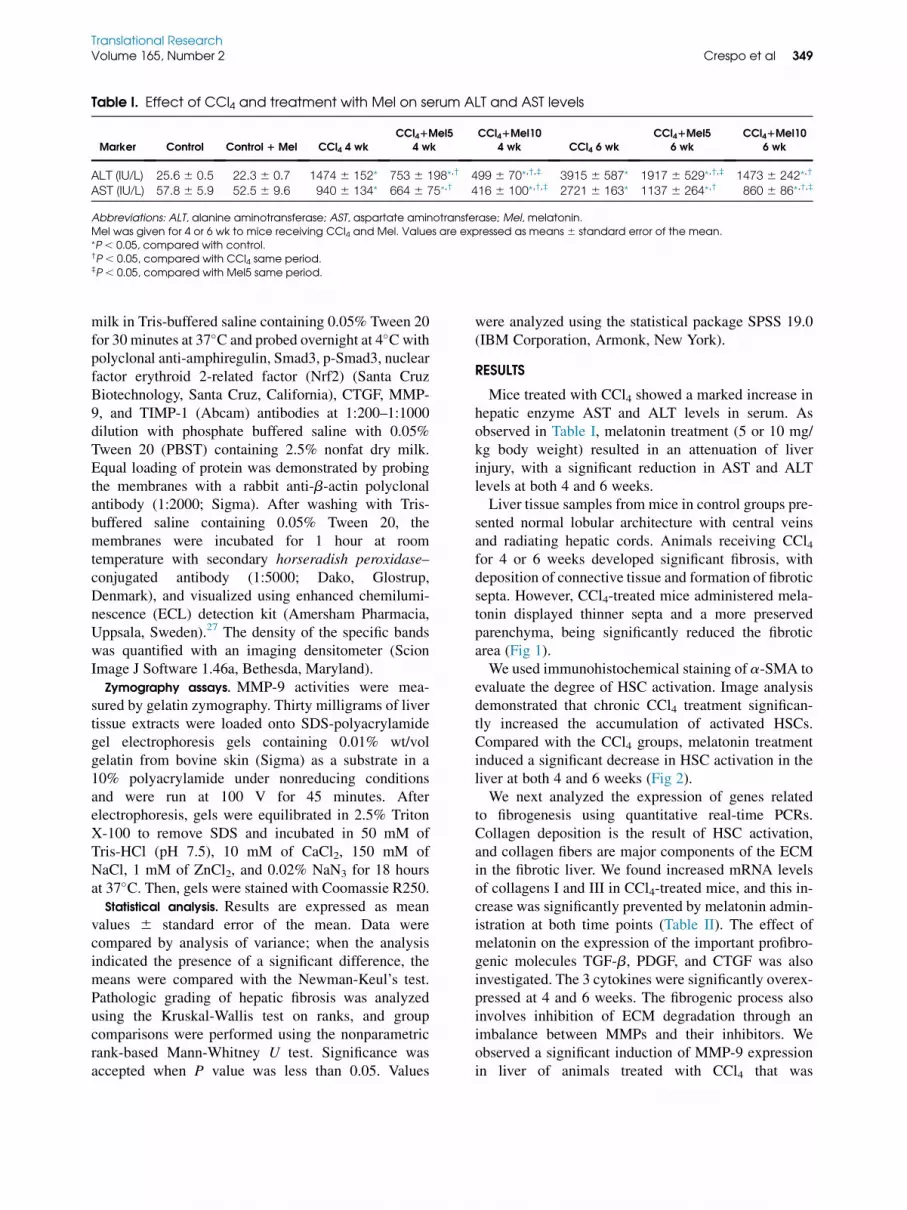
Table I. Effect of CCl4 and treatment with Mel on serum ALT and AST levels
Marker Control Control 1 Mel CCl4 4 wkCCl41Mel5
4 wkCCl41Mel10
4 wk CCl4 6 wkCCl41Mel5
6 wkCCl41Mel10
6 wk
ALT (IU/L) 25.6 6 0.5 22.3 6 0.7 1474 6 152* 753 6 198*,† 499 6 70*,†,‡ 3915 6 587* 1917 6 529*,†,‡ 1473 6 242*,†
AST (IU/L) 57.8 6 5.9 52.5 6 9.6 940 6 134* 664 6 75*,† 416 6 100*,†,‡ 2721 6 163* 1137 6 264*,† 860 6 86*,†,‡
Abbreviations: ALT, alanine aminotransferase; AST, aspartate aminotransferase; Mel, melatonin.Mel was given for 4 or 6 wk to mice receiving CCl4 and Mel. Values are expressed as means 6 standard error of the mean.*P , 0.05, compared with control.†P , 0.05, compared with CCl4 same period.‡P , 0.05, compared with Mel5 same period.
Translational ResearchVolume 165, Number 2 Crespo et al 349
milk in Tris-buffered saline containing 0.05% Tween 20for 30 minutes at 37�C and probed overnight at 4�C withpolyclonal anti-amphiregulin, Smad3, p-Smad3, nuclearfactor erythroid 2-related factor (Nrf2) (Santa CruzBiotechnology, Santa Cruz, California), CTGF, MMP-9, and TIMP-1 (Abcam) antibodies at 1:200–1:1000dilution with phosphate buffered saline with 0.05%Tween 20 (PBST) containing 2.5% nonfat dry milk.Equal loading of protein was demonstrated by probingthe membranes with a rabbit anti-b-actin polyclonalantibody (1:2000; Sigma). After washing with Tris-buffered saline containing 0.05% Tween 20, themembranes were incubated for 1 hour at roomtemperature with secondary horseradish peroxidase–conjugated antibody (1:5000; Dako, Glostrup,Denmark), and visualized using enhanced chemilumi-nescence (ECL) detection kit (Amersham Pharmacia,Uppsala, Sweden).27 The density of the specific bandswas quantified with an imaging densitometer (ScionImage J Software 1.46a, Bethesda, Maryland).
Zymography assays. MMP-9 activities were mea-sured by gelatin zymography. Thirty milligrams of livertissue extracts were loaded onto SDS-polyacrylamidegel electrophoresis gels containing 0.01% wt/volgelatin from bovine skin (Sigma) as a substrate in a10% polyacrylamide under nonreducing conditionsand were run at 100 V for 45 minutes. Afterelectrophoresis, gels were equilibrated in 2.5% TritonX-100 to remove SDS and incubated in 50 mM ofTris-HCl (pH 7.5), 10 mM of CaCl2, 150 mM ofNaCl, 1 mM of ZnCl2, and 0.02% NaN3 for 18 hoursat 37�C. Then, gels were stained with Coomassie R250.
Statistical analysis. Results are expressed as meanvalues 6 standard error of the mean. Data werecompared by analysis of variance; when the analysisindicated the presence of a significant difference, themeans were compared with the Newman-Keul’s test.Pathologic grading of hepatic fibrosis was analyzedusing the Kruskal-Wallis test on ranks, and groupcomparisons were performed using the nonparametricrank-based Mann-Whitney U test. Significance wasaccepted when P value was less than 0.05. Values
were analyzed using the statistical package SPSS 19.0(IBM Corporation, Armonk, New York).
RESULTS
Mice treated with CCl4 showed a marked increase inhepatic enzyme AST and ALT levels in serum. Asobserved in Table I, melatonin treatment (5 or 10 mg/kg body weight) resulted in an attenuation of liverinjury, with a significant reduction in AST and ALTlevels at both 4 and 6 weeks.Liver tissue samples from mice in control groups pre-
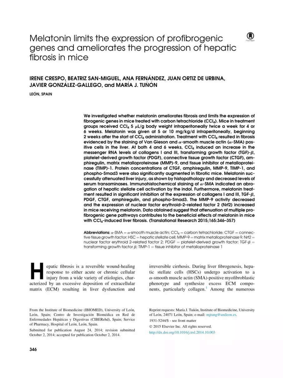
sented normal lobular architecture with central veinsand radiating hepatic cords. Animals receiving CCl4for 4 or 6 weeks developed significant fibrosis, withdeposition of connective tissue and formation of fibroticsepta. However, CCl4-treated mice administered mela-tonin displayed thinner septa and a more preservedparenchyma, being significantly reduced the fibroticarea (Fig 1).We used immunohistochemical staining of a-SMA to
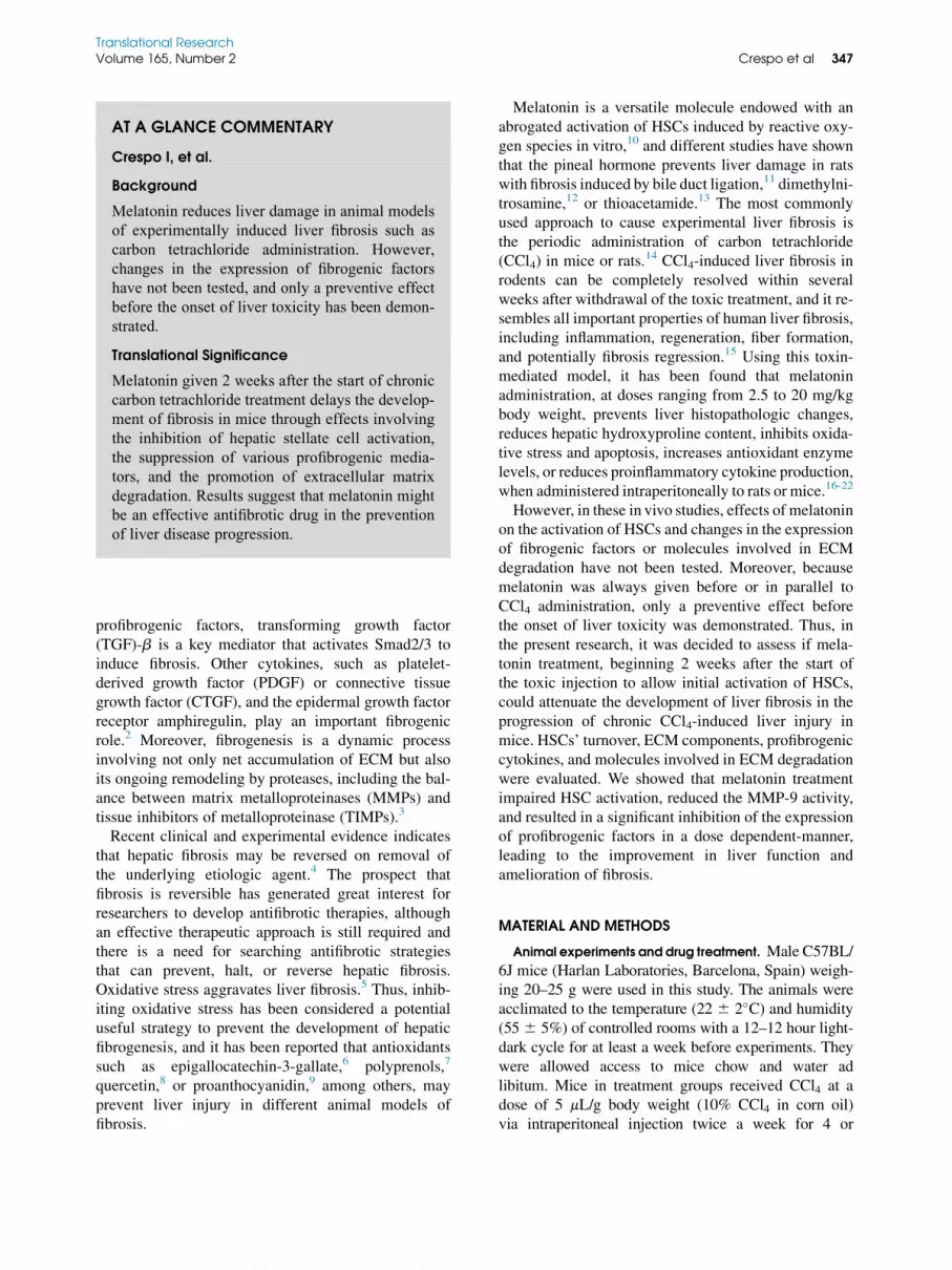
evaluate the degree of HSC activation. Image analysisdemonstrated that chronic CCl4 treatment significan-tly increased the accumulation of activated HSCs.Compared with the CCl4 groups, melatonin treatmentinduced a significant decrease in HSC activation in theliver at both 4 and 6 weeks (Fig 2).We next analyzed the expression of genes related
to fibrogenesis using quantitative real-time PCRs.Collagen deposition is the result of HSC activation,and collagen fibers are major components of the ECMin the fibrotic liver. We found increased mRNA levelsof collagens I and III in CCl4-treated mice, and this in-crease was significantly prevented by melatonin admin-istration at both time points (Table II). The effect ofmelatonin on the expression of the important profibro-genic molecules TGF-b, PDGF, and CTGF was alsoinvestigated. The 3 cytokines were significantly overex-pressed at 4 and 6 weeks. The fibrogenic process alsoinvolves inhibition of ECM degradation through animbalance between MMPs and their inhibitors. Weobserved a significant induction of MMP-9 expressionin liver of animals treated with CCl4 that was
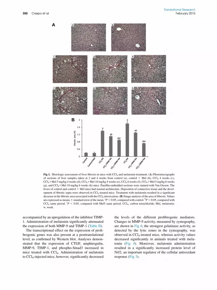
Fig 1. Histologic assessment of liver fibrosis in mice with CCl4 and melatonin treatment. (A) Photomicrographs
of sections of liver samples taken at 2 and 4 weeks from control (a), control 1 Mel (b), CCl4 4 weeks (c),
CCl41Mel 5 mg/kg 4 weeks (d), CCl41Mel 10 mg/kg 4 weeks (e), CCl4 6 weeks (f), CCl41Mel 5 mg/kg 6 weeks
(g), and CCl41Mel 10 mg/kg 6 weeks (h) mice. Paraffin-embedded sections were stained with Van Gieson. The
livers of control and control1 Mel mice had normal architecture. Deposition of connective tissue and the devel-
opment of fibrotic septa were observed in CCl4-treated mice. Treatment with melatonin resulted in a significant
decrease in the fibrotic area associated with the CCl4 intoxication. (B) Image analysis of the area of fibrosis. Values
are expressed as means6 standard error of themean. aP, 0.05, comparedwith control. bP, 0.05, comparedwith
CCl4 same period. cP , 0.05, compared with Mel5 same period. CCl4, carbon tetrachloride; Mel, melatonin;
w, week.
Translational Research350 Crespo et al February 2015
accompanied by an upregulation of the inhibitor TIMP-1. Administration of melatonin significantly attenuatedthe expression of both MMP-9 and TIMP-1 (Table II).The transcriptional effect on the expression of profi-
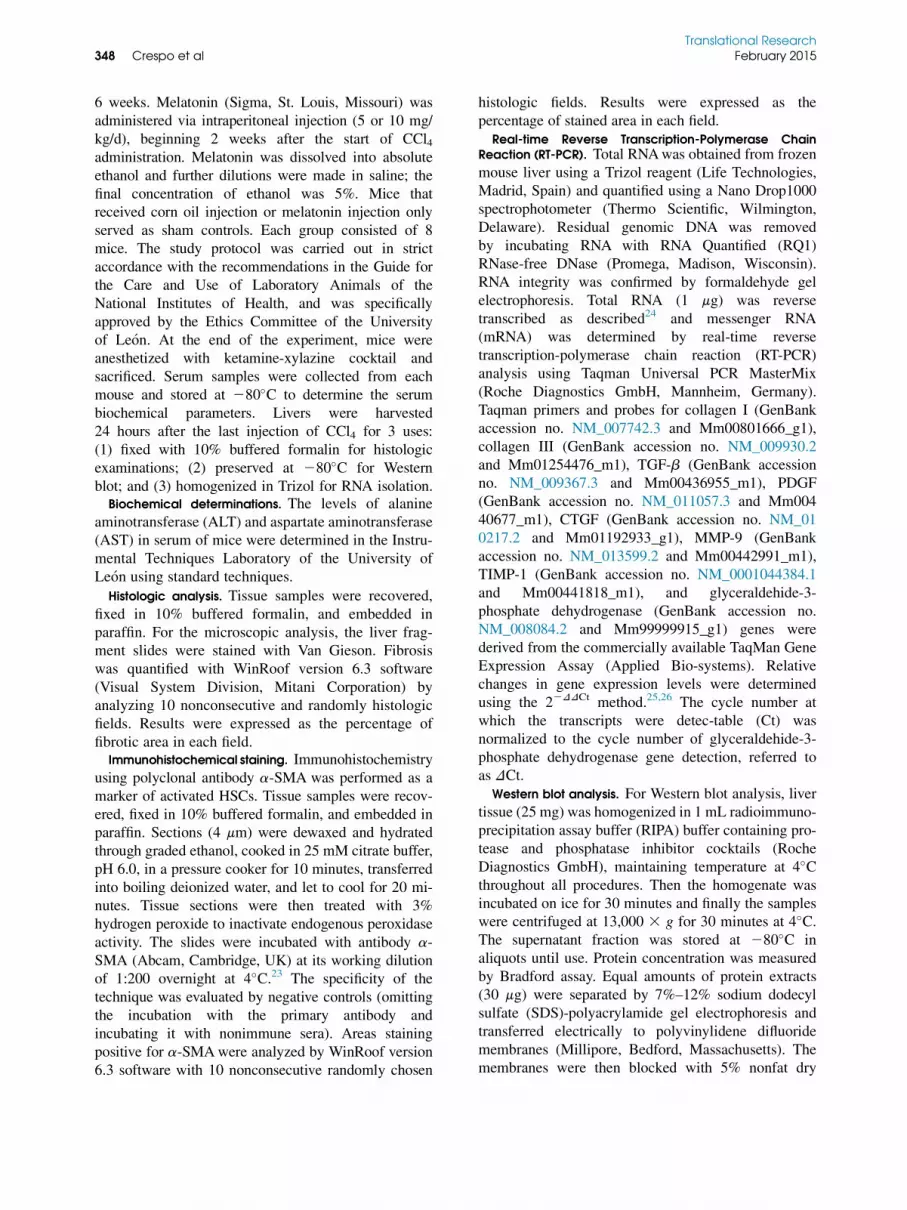
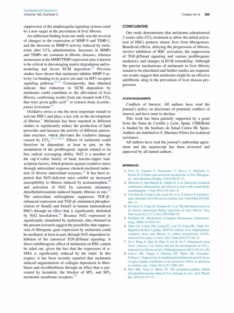
brogenic genes was also present at a posttranslationallevel, as confirmed by Western blot. Analyses demon-strated that the expression of CTGF, amphiregulin,MMP-9, TIMP-1, and phospho-Smad3 increased inmice treated with CCl4. Administration of melatoninto CCl4-injected mice, however, significantly decreased
the levels of the different profibrogenic mediators.Changes in MMP-9 activity, measured by zymography,are shown in Fig 4; the strongest gelatinase activity, asdetected by the lytic zones in the zymography, wasobserved in CCl4-treated mice, whereas activity valuesdecreased significantly in animals treated with mela-tonin (Fig 4). Moreover, melatonin administrationresulted in a significantly increased protein level ofNrf2, an important regulator of the cellular antioxidantresponse (Fig 3).

Fig 2. Effect of CCl4 and treatment with melatonin on liver a-SMA immunohistochemistry. (A) Photomicro-
graphs of sections of liver samples taken at 2 and 4 weeks from control (a), control 1 Mel (b), CCl4 4 weeks
(c), CCl41Mel 5 mg/kg 4 weeks (d), CCl41Mel 10 mg/kg 4 weeks (e), CCl4 6 weeks (f), CCl41Mel 5 mg/kg
6 weeks (g), and CCl41Mel 10 mg/kg 6 weeks (h) mice. Paraffin-embedded sections were stained with a
a-SMA antibody. (B) Image analysis of the area of a-SMA staining. Treatment with melatonin resulted in a sig-
nificant decrease in the amount of a-SMA immunostaining associated with the CCl4 intoxication. Values are ex-
pressed as means6 standard error of the mean. aP, 0.05, compared with control. bP, 0.05, compared with CCl4same period. cP , 0.05, compared with Mel5 same period. CCl4, carbon tetrachloride; Mel, melatonin, SMA,
smooth muscle actin; w, week.
Translational ResearchVolume 165, Number 2 Crespo et al 351
Effects induced by melatonin on the activation ofHSCs and the expression of factors involved in thefibrogenic process were dose dependent, reachingvalues that were in most cases significantly lower inmice treated with 5 mg/kg body weight melatoninwhen compared with those receiving the pinealhormone at 10 mg/kg body weight (Table II, Figs 2and 3).
DISCUSSION
Although beneficial effects of melatonin on hepaticfibrogenesis induced by CCl4 and other toxins havebeen reported by different authors, this is the first inves-tigation demonstrating that in mice with liver fibrosisinduced by CCl4 injection, melatonin significantly sup-presses the activation of HSCs and reduces the expres-sion of collagen proteins, effects that are accompanied
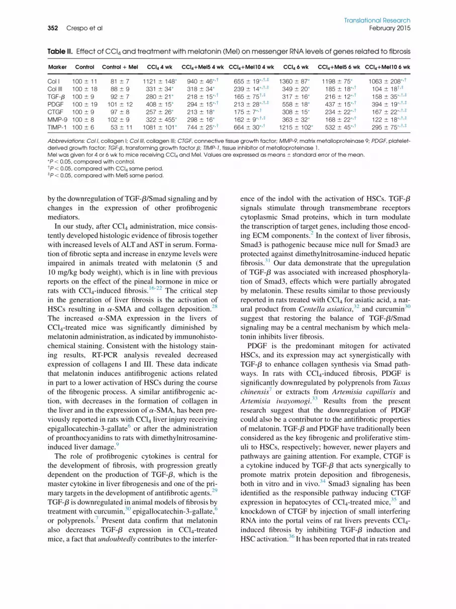
Table II. Effect of CCl4 and treatment with melatonin (Mel) onmessenger RNA levels of genes related to fibrosis
Marker Control Control 1 Mel CCl4 4 wk CCl41Mel5 4 wk CCl41Mel10 4 wk CCl4 6 wk CCl41Mel5 6 wk CCl41Mel10 6 wk
Col I 100 6 11 81 6 7 1121 6 148* 940 6 46*,† 655 6 19*,†,‡ 1360 6 87* 1198 6 75* 1063 6 208*,†
Col III 100 6 18 88 6 9 331 6 34* 318 6 34* 239 6 14*,†,‡ 349 6 20* 185 6 18*,† 104 6 18†,‡
TGF-b 100 6 9 92 6 7 280 6 21* 218 6 15*,† 165 6 75†,‡ 317 6 16* 216 6 12*,† 158 6 35*,†,‡
PDGF 100 6 19 101 6 12 408 6 15* 294 6 15*,† 213 6 28*,†,‡ 558 6 18* 437 6 15*,† 394 6 19*,†,‡
CTGF 100 6 9 97 6 8 257 6 26* 213 6 18* 175 6 7*,† 308 6 15* 234 6 22*,† 167 6 22*,†,‡
MMP-9 100 6 8 102 6 9 322 6 455* 298 6 16* 162 6 9*,†,‡ 363 6 32* 168 6 22*,† 122 6 18*,†,‡
TIMP-1 100 6 6 53 6 11 1081 6 101* 744 6 25*,† 664 6 30*,† 1215 6 102* 532 6 45*,† 295 6 75*,†,‡
Abbreviations: Col I, collagen I; Col III, collagen III; CTGF, connective tissue growth factor; MMP-9, matrix metalloproteinase 9; PDGF, platelet-
derived growth factor; TGF-b, transforming growth factor b; TIMP-1, tissue inhibitor of metalloproteinase 1.Mel was given for 4 or 6 wk to mice receiving CCl4 and Mel. Values are expressed as means 6 standard error of the mean.*P , 0.05, compared with control.†P , 0.05, compared with CCl4 same period.‡P , 0.05, compared with Mel5 same period.
Translational Research352 Crespo et al February 2015
by the downregulation of TGF-b/Smad signaling and bychanges in the expression of other profibrogenicmediators.In our study, after CCl4 administration, mice consis-
tently developed histologic evidence of fibrosis togetherwith increased levels of ALTand AST in serum. Forma-tion of fibrotic septa and increase in enzyme levels wereimpaired in animals treated with melatonin (5 and10 mg/kg body weight), which is in line with previousreports on the effect of the pineal hormone in mice orrats with CCl4-induced fibrosis.16-22 The critical stepin the generation of liver fibrosis is the activation ofHSCs resulting in a-SMA and collagen deposition.28
The increased a-SMA expression in the livers ofCCl4-treated mice was significantly diminished bymelatonin administration, as indicated by immunohisto-chemical staining. Consistent with the histology stain-ing results, RT-PCR analysis revealed decreasedexpression of collagens I and III. These data indicatethat melatonin induces antifibrogenic actions relatedin part to a lower activation of HSCs during the courseof the fibrogenic process. A similar antifibrogenic ac-tion, with decreases in the formation of collagen inthe liver and in the expression of a-SMA, has been pre-viously reported in rats with CCl4 liver injury receivingepigallocatechin-3-gallate6 or after the administrationof proanthocyanidins to rats with dimethylnitrosamine-induced liver damage.9
The role of profibrogenic cytokines is central forthe development of fibrosis, with progression greatlydependent on the production of TGF-b, which is themaster cytokine in liver fibrogenesis and one of the pri-mary targets in the development of antifibrotic agents.29
TGF-b is downregulated in animal models of fibrosis bytreatment with curcumin,30 epigallocatechin-3-gallate,6
or polyprenols.7 Present data confirm that melatoninalso decreases TGF-b expression in CCl4-treatedmice, a fact that undoubtedly contributes to the interfer-
ence of the indol with the activation of HSCs. TGF-bsignals stimulate through transmembrane receptorscytoplasmic Smad proteins, which in turn modulatethe transcription of target genes, including those encod-ing ECM components.2 In the context of liver fibrosis,Smad3 is pathogenic because mice null for Smad3 areprotected against dimethylnitrosamine-induced hepaticfibrosis.31 Our data demonstrate that the upregulationof TGF-b was associated with increased phosphoryla-tion of Smad3, effects which were partially abrogatedby melatonin. These results similar to those previouslyreported in rats treated with CCl4 for asiatic acid, a nat-ural product from Centella asiatica,32 and curcumin30
suggest that restoring the balance of TGF-b/Smadsignaling may be a central mechanism by which mela-tonin inhibits liver fibrosis.PDGF is the predominant mitogen for activated
HSCs, and its expression may act synergistically withTGF-b to enhance collagen synthesis via Smad path-ways. In rats with CCl4-induced fibrosis, PDGF issignificantly downregulated by polyprenols from Taxuschinensis7 or extracts from Artemisia capillaris andArtemisia iwayomogi.33 Results from the presentresearch suggest that the downregulation of PDGFcould also be a contributor to the antifibrotic propertiesof melatonin. TGF-b and PDGF have traditionally beenconsidered as the key fibrogenic and proliferative stim-uli to HSCs, respectively; however, newer players andpathways are gaining attention. For example, CTGF isa cytokine induced by TGF-b that acts synergically topromote matrix protein deposition and fibrogenesis,both in vitro and in vivo.34 Smad3 signaling has beenidentified as the responsible pathway inducing CTGFexpression in hepatocytes of CCl4-treated mice,35 andknockdown of CTGF by injection of small interferingRNA into the portal veins of rat livers prevents CCl4-induced fibrosis by inhibiting TGF-b induction andHSC activation.36 It has been reported that in rats treated

Fig 3. Effect of CCl4 and treatment with melatonin on protein concentration of CTGF, amphiregulin, MMP-9,
TIMP-1, phospho-Smad3, and Nrf2. Protein from liver extracts taken at 4 and 6 weeks was separated by sodium
dodecyl sulfate polyacrylamide gel electrophoresis, followed by immunoblotting. Equal loading of proteins is
Translational ResearchVolume 165, Number 2 Crespo et al 353

=
Fig 4. Effect of CCl4 and treatment with melatonin on MMP-9 activity. The MMP-9 gelatinase activities in liver
tissue extracts taken at 4 and 6 weeks were measured by gelatin zymography assays as described. Melatonin (Mel)
was given for 4 or 6 weeks. (A) Representative zymography photographs. (B) Densitometric quantification. Values
are expressed as means6 standard error of themean. aP, 0.05, comparedwith control. bP, 0.05, comparedwith
CCl4 same period. cP, 0.05, compared with Mel5 same period. CCl4, carbon tetrachloride; MMP-9, matrix met-
alloproteinase 9; w, week.
Translational Research354 Crespo et al February 2015
with CCl4 silymarin decreases CTGF to improve liverfibrosis37 and curcumin significantly attenuates theseverity of liver damage through inhibition of CTGFexpression.30 Our findings confirmed that chronicCCl4 administration markedly enhanced the intrahe-patic expression of CTGF mRNA and protein, andthat melatonin significantly reduced these CCl4-inducedincreases.The ligand of the epidermal growth factor amphiregu-
lin is another molecule, which plays a specific role inliver fibrosis, contributing to the expression of fibro-genic mediators and to the proliferation of fibrogenic
illustrated by b-actin bands. Melatonin (Mel) was given for 4
tographs. (B) Densitometric quantification. Values are expaP , 0.05, compared with control. bP , 0.05, compared w
Mel5 same period. CCl4, carbon tetrachloride; CTGF, conne
proteinase 9; Nrf2, nuclear factor erythroid 2; TIMP-1, tissu
cells.38 The expression of amphiregulin increases mark-edly in liver injury induced by CCl4,
39 and amphiregulindeficient-mice develop significantly less collagen accu-mulation.40 We have previously reported that sup-pression of amphiregulin signals contributes to theprotective effects of quercetin in cirrhotic rats withcommon bile duct ligation,7 and in mice fed a methio-nine-choline–deficient diet.2 It is also known thatamphiregulin amplifies the HSC response throughincreased production of CTGF and TIMP-1.39 Thus,the inhibition of amphiregulin expression by melatoninin CCl4-treated mice supports the suggestion that
or 6 weeks. (A) Representative Western blot pho-
ressed as means 6 standard error of the mean.
ith CCl4 same period. cP , 0.05, compared with
ctive tissue growth factor; MMP-9, matrix metallo-
e inhibitor of metalloproteinase 1; w, week.
Translational ResearchVolume 165, Number 2 Crespo et al 355
suppression of the amphiregulin signaling system couldbe a new target in the prevention of liver fibrosis.An additional finding from our study was the reversal
of changes in the expression of MMP-9 and TIMP-1,and the decrease in MMP-9 activity induced by mela-tonin after CCl4 administration. Increases in MMPsand TIMPs are common in fibrotic diseases, whereasan increase in theMMP/TIMP expression ratio is knownto be critical in discouraging matrix degradation and re-modeling and favors ECM deposition.28 Previousstudies have shown that melatonin inhibits MMP-9 ac-tivity via binding to its active site and via MT1 receptorsignaling pathway.41,42 Consequently, data obtainedindicate that reduction in ECM deposition bymelatonin could contribute to the alleviation of liverfibrosis, confirming results from rats treated with CCl4that were given gallic acid43 or extracts from Acantho-panax koreanum.44
Oxidative stress is one the most important stimuli toactivate HSCs and plays a key role in the developmentof fibrosis.5 Melatonin has been reported in differentstudies to significantly reduce the production of lipidperoxides and increase the activity of different antioxi-dant enzymes, which alleviates the oxidative damagecaused by CCl4.
16,17,20,21 Effects of melatonin couldtherefore be dependent, at least in part, on themodulation of the profibrogenic signals related to itsfree radical scavenging ability. Nrf2 is a member ofthe cap‘n’collar family of basic leucine-zipper tran-scription factors, which protects against oxidative stressthrough antioxidant response element-mediated induc-tion of diverse antioxidant enzymes.45 It has been re-ported that Nrf2-deficient mice exhibit an increasedsusceptibility to fibrosis induced by acetaminophen,46
and activation of Nrf2 by curcumin attenuatesdimethylnitrosamine-induced hepatic fibrosis in rats.47
The antioxidant sulforaphane suppresses TGF-b–enhanced expression and TGF-b–stimulated phosphor-ylation of Smad2 and Smad3 in human immortalizedHSCs through an effect that is significantly abolishedby Nrf2 knockdown.48 Because Nrf2 expression insignificantly stimulated by melatonin, data obtained inthe present research suggest the possibility that suppres-sion of fibrogenic gene expression by melatonin couldbe mediated, at least in part, through Nrf2-dependent in-hibition of the canonical TGF-b/Smad signaling. Adirect antifibrogenic effect of melatonin on HSC cannotbe ruled out, given the fact that the expression of a-SMA is significantly reduced by the indol. In thisrespect, it has been recently reported that melatonininduced augmentation of collagen deposition in fibro-blasts and myofibroblasts through an effect that is pre-vented by luzindole, the blocker of MT1 and MT2
melatonin membrane receptors.49
CONCLUSIONS
Our study demonstrates that melatonin administered2 weeks after CCl4 treatment to allow the initial activa-tion of HSCs protects mouse liver from fibrogenesis.Beneficial effects, delaying the progression of fibrosis,involve inhibition of HSC activation, the suppressionof TGF-b/Smad signaling and various profibrogenicmediators, and changes in ECM remodeling. Althoughthe precise mechanisms of melatonin in liver fibrosisremain to be elucidated and further studies are required,our results suggest that melatonin might be an effectiveantifibrotic drug in the prevention of liver disease pro-gression.
ACKNOWLEDGMENTS
Conflicts of Interest: All authors have read thejournal’s policy on disclosure of potential conflicts ofinterest and have none to declare.This work has been partially supported by a grant
from the Junta de Castilla y Le�on, Spain. CIBERehdis funded by the Instituto de Salud Carlos III, Spain.Authors are indebted to S. Mart�ınez Fl�ores for technicalassistance.All authors have read the journal’s authorship agree-
ment and the manuscript has been reviewed andapproved by all named authors.
REFERENCES
1. Novo E, Cannito S, Paternostro C, Bocca C, Miglietta A,
Parola M. Cellular and molecular mechanisms in liver fibrogene-
sis. Arch Biochem Biophys 2014;548:20–37.
2. Marcolin E, San Miguel B, Vallejo D, et al. Quercetin treatment
ameliorates inflammation and fibrosis in mice with nonalcoholic
steatohepatitis. J Nutr 2012;142:1821–8.
3. Vercelino R, Crespo I, De souza GF, et al. S-nitroso-N-acetylcys-
teine attenuates liver fibrosis in cirrhotic rats. J Mol Med 2010;88:
401–11.
4. Kisseleva T, Cong M, Scholten D, et al. Myofibroblasts revert to
an inactive phenotype during regression of liver fibrosis. Proc
Natl Acad Sci U S A 2012;109:9448–53.
5. Friedman SL. Mechanisms of hepatic fibrogenesis. Gastroenter-
ology 2008;134:1655–61.
6. Tipoe GL, Leung TM, Liong EC, Lau TY, Fung ML, Nanji AA.
Epigallocatechin-3-gallate (EGCG) reduces liver inflammation,
oxidative stress and fibrosis in carbon tetrachloride (CCl4)-
induced liver injury in mice. Eur J Nutr 2010;273:45–51.
7. Yu J, Wang Y, Qian H, Zhao Y, Liu B, Fu C. Polyprenols from
Tuxus chinensis var. mairei prevent the development of CCL4-
induced liver fibrosis in rats. J Ethnopharmacol 2012;142:151–60.
8. Cuevas MJ, Tieppo J, Marroni NP, Tu~n�on MJ, Gonz�alez-
Gallego J. Suppression of amphiregulin/epidermal growth factor
receptor signals contributes to the protective effects of quercetin
in cirrhotic rats. J Nutr 2011;141:1299–305.
9. Shin MO, Yoon S, Moon JO. The proanthocyanidins inhibit
dimethylnitrosamine-induced liver damage in rats. Arch Pharm
Res 2010;33:167–73.
Translational Research356 Crespo et al February 2015
10. Gu J, Zhuang L, Huang GC. Melatonin prevents H2O2-indiced
activation of rat hepatic stellate cells. J Pineal Res 2006;41:275–8.
11. Tahan G, Akin H, Aydogan F, et al. Melatonin ameliorates liver
fibrosis induced by bile-duct ligation in rats. Can J Surg 2010;
53:313–8.
12. Tahan V, Ozaras R, Canbakan B, et al. Melatonin reduces
dimethylnitrosamine-induced liver fibrosis in rats. J Pineal Res
2004;37:78–84.
13. Cruz A, Padillo FJ, Torres E, et al. Melatonin prevents experi-
mental liver cirrhosis induced by thioacetamide in rats. J Pineal
Res 2005;39:143–50.
14. Liedke C, Luedde T, Sauerbruch T, et al. Experimental liver
fibrosis research: update on animal models, legal issues and trans-
lational aspects. Fibrogenesis Tissue Repair 2013;6:19.
15. Starkel P, Leclerq IA. Animal models for the study of hepatic
fibrosis. Best Pract Res Clin Gastroenterol 2011;25:319–33.
16. Ohta Y, Kongo-Nishimuras M, Matsura T, Yamada K,
Kitagawa A, Kishikawa T. Melatonin prevents disruption of he-
patic reactive oxygen species metabolism in rats treated with car-
bon tetrachloride. J Pineal Res 2004;36:10–7.
17. Kus I, Ogeturk M, Oner H, Sahin S, Yekeler H, Sarsilmaz M. Pro-
tective effects of melatonin against carbon tetrachloride-induced
hepatotoxicity in rats: a light microscopic and biochemical study.
Cell Biochem Funct 2005;23:169–74.
18. Wang H, Wei W, Wang NP, et al. Melatonin ameliorates carbon
tetrachloride-induced hepatic fibrogenesis in rats via inhibition
of oxidative stress. Life Sci 2005;72:1907–15.
19. Noyan T, Komuroqlu U, Bayram I, Sekero�glu MR. Comparison
of the effects of melatonin and pentoxifylline on carbon
tetrachloride-induced liver toxicity in mice. Cell Biol Toxicol
2006;22:381–91.
20. Ogeturk M, Kus I, Pekmez H, Yekeler H, Sahin S, Sarsilmaz M.
Inhibition of carbon tetrachloride-mediated apoptosis and oxida-
tive stress by melatonin in experimental liver fibrosis. Toxicol
Ind Health 2008;24:20108.
21. Hong RH, Xu JM,Mei Q.Melatonin ameliorates experimental he-
patic fibrosis induced by carbon tetrachloride in rats. World J Gas-
troenterol 2009;15:1452–8.
22. Ebaid H, Bashandi SAE, Alhazza IM, Rady A, El-Shehry S. Folic
acid and melatonin ameliorate carbon tetrachloride-induced he-
patic injury, oxidative stress and inflammation in rats. Nutr Metab
2013;10:20.
23. San Miguel B, Crespo I, Kretzmann N, et al. Glutamine prevents
fibrosis development in rats with colitis induced by 2,4,6-
trinitrobenzene sulfonic acid. J Nutr 2010;140:1065–71.
24. Tu~n�on MJ, San Miguel B, Crespo I, et al. Melatonin treatment re-
duces endoplasmic reticulum stress and modulates the unfolded
protein response in rabbits with lethal fulminant hepatitis of viral
origin. J Pineal Res 2013;55:221–8.
25. Garc�ıa-Mediavilla MV, S�anchez-Campos S, Gonz�alez-Perez P,
et al. Differential contribution of hepatitis C virus NS5A and
core proteins to the induction of oxidative and nitrosative stress
in human hepatocyte-derived cells. J Hepatol 2005;43:606–13.
26. Lima-Cabello E, Garc�ıa-Mediavilla MV, Miquelina-Colina M,
et al. Enhanced expression of pro-inflammatory mediators and
liver X-receptor-regulated lipogenic genes in non-alcoholic fatty
liver disease and hepatitis C. Clin Sci 2011;120:239–50.
27. Kretzmann N, Fillmann H, Mauriz JL, et al. Effects of glutamine
on proinflammatory gene expression and activation of nuclear fac-
tor kappa B and signal transducers and activators of transcription
in TNBS-induced colitis. Inflamm Bowel Dis 2008;14:1504–13.
28. Yoshida K, Matsuzaki K. Differential regulation of TGF-b/Smad
signaling in hepatic stellate cells between acute and chronic liver
injuries. Front Physiol 2012;3:53.
29. Friedman SL. Evolving challenges in hepatic fibrosis. Nat Rev
Gastroenterol Hepatol 2010;7:425–36.
30. Yao QY, Xu BL, Wang JY, Liu HC, Zhang SC, Tu CT. Inhibition
by curcumin of multiple sites of the transforming growth factor-
beta1 signaling pathway ameliorates the progression of liver
fibrosis induced by carbon tetrachloride in rats. BMC Comple-
ment Altern Med 2012;12:156.
31. Latella G, Vetuschi A, Sferra R, et al. Targeted disruption of Smad3
confers resistance to the development of dimethylnitrosamine-
induced hepatic fibrosis in mice. Liver Int 2009;29:997–1009.
32. Tang LX, He RH, Yang G, et al. Asiatic acid inhibits liver fibrosis
by blocking TGF-beta/Smad signaling in vivo and in vitro. PLos
One 2012;7:e31350.
33. Wang JY, ChoiMK, Shin JW, Hwang SY, Son CG. Antifibrotic ef-
fects of Artemisia capillaris and Artemisia iwayomogi in a carbon
tetrachloride-induced chronic hepatic fibrosis animal model.
J Ethnopharmacol 2012;140:179–85.
34. Jun JI, Lau LF. Taking aim at the extracellular matrix: CCN pro-
teins as emerging therapeutic targets. Nat Rev Drug Discov 2011;
10:945–63.
35. Weng HL, Ciclan L, Liu Y, et al. Profibrogenic transforming
growth factor-beta/activin receptor-like kinase 5 signaling via
connective tissue growth factor expression in hepatocytes. Hepa-
tology 2007;46:1257–70.
36. Li G, Xie Q, Shi Y, et al. Inhibition of connective tissue growth
factor by siRNA prevents liver fibrosis in rats. J Gene Med
2006;8:889–900.
37. Tzeng JI, Chen FM, Chung HH, Cheng JT. Silymarin decreases
connective tissue growth factor to improve liver fibrosis in rats
treated with carbon tetrachloride. Phytother Res 2013;27:
1023–8.
38. Berasain C, Garcia-Trevijano ER, Castillo J, et al. Novel role for
amphiregulin in protection from liver injury. J Biol Chem 2005;
280:1912–20.
39. Perugorria MJ, Latasa MU, Nicou A, et al. The epidermal
growth factor receptor ligand amphiregulin participates in the
development of mouse liver fibrosis. Hepatology 2008;48:
1251–61.
40. Fujii T, Fuchs BC, Yamada S, et al. Mouse model of carbon tetra-
chloride induced liver fibrosis: histopathological changes and
expression of CD133 and epidermal growth factor. BMC Gastro-
enterol 2010;10:79.
41. Rudra DS, Paul U, Maiti NC, Reiter RJ, Swarnakar S. Melatonin
inhibits matrix metalloproteinase-9 activity by binding to its
active site. J Pineal Res 2013;54:398–405.
42. Mao L, Yuan L, Stakey L, Jones FE, Burow ME, Hill SM.
Inhibition of breast cancer cell invasion by melatonin is
mediated through regulations of the p38 mitogen-activated
protein kinase signaling pathway. Breast Cancer Res 2010;
12:R107.
43. Wang J, Tang L,White J, Fang J. Inhibitory effect of gallic acid on
CCl4-mediated liver fibrosis in mice. Cell Biochem Biophys
2014;69:21–6.
44. Bai T, Yao YL, Jin XJ, et al. Acanthoic acid, a diterpene in Acan-
thopanax koreanum, ameliorates the development of liver fibrosis
via LXRs signals. Chem Biol Interact 2014;218:63–70.
45. Crespo I, San Miguel B, Laliena A, et al. Melatonin prevents
the decreased activity of antioxidant enzymes and activates nu-
clear erythroid 2-related factor 2 signaling in an animal model
of fulminant hepatic failure of viral origin. J Pineal Res 2010;
49:193–200.
46. Xu W, Hellerbrand C, Kohler UA, et al. The Nrf2 transcription
factor protects from toxin-induced liver injury and fibrosis. Lab
Invest 2008;88:1068–78.
Translational ResearchVolume 165, Number 2 Crespo et al 357
47. Oh CJ, Farombi EO, Shrotriya S, Kim SH, Surh YJ. Curcumin at-
tenuates dimethylnitrosamine-induced liver injury in rats through
Nrf2-mediated induction of heme oxygenase-1. Food Chem Tox-
icol 2008;46:1279–87.
48. Oh CJ, Kim MJY, Min AK, et al. Sulforaphane attenuates hepatic
fibrosis via NF-E2-related factor 2-mediated inhibition of trans-
forming growth factor-b/Smad signaling. Free Radic Biol Med
2012;52:671–82.
49. Drobinik J, Owczarek K, Piera L, et al. Melatonin-induced
augmentation of collagen deposition in cultures of fibroblasts
and myofibroblasts is blocked by luzindole—a melatonin mem-
brane receptor inhibitor. Pharmacol Rev 2013;65:642–9.


