Embed Size (px)
Citation preview

MINI REVIEW
Melatonin and the hair follicle
Introduction
Observations suggesting that the pineal gland and its chiefsecretory product, melatonin [1, 2], are involved in theregulation of hair growth and pigmentation date backseveral decades, and have long-intrigued chronobiologists,
animal scientists, veterinarians, endocrinologists, dermatol-ogists and researchers in the wool-industry among others[3–9]. Thus, it is well-recognized that melatonin can alter
wool and cashmere production, the development and cyclefrequency of pelage, seasonal molting and coat color inseveral species, presumably as a major neuroendocrine
regulator that couples coat phenotype and function tophotoperiod-dependent environmental and reproductivechanges [10, 11].
Yet, the understanding of the role of melatonin in hair
follicle (HF) biology is still very limited. Because of thecomplexity of melatonin interactions and metabolism [11–18] and the substantial, often seemingly contradictory
species-, gender-, and dose-dependency of melatonin-related hair effects [7, 10, 11, 19–26], the picture of theexact functions of melatonin in hair biology seems to be still
a blurred and confusing one.
However, the recent discovery that mammalian skin notonly is a target of melatonin bioactivity, but also animportant extrapineal site of its synthesis, regulation andmetabolism [13, 27–29], and that additionally even in HFs
of normal mouse skin and human scalp melatonin wasdetected [11, 18, 30], has re-vitalized general interest inmelatonin as a modulator of hair growth and/or pigmen-
tation.On this background, after summarizing some salient
features of melatonin biology that are most pertinent in the
current context, this review summarizes the availableevidence indicating a significant role of melatonin in hairbiology. We will interpret this evidence in view of emerging
concepts on the role of melatonin in general skin biology.Major open questions and unresolved controversies aredefined and particularly promising avenues for futureresearch into the �melatonin-hair connection� and its
potential clinical implications are delineated.
Melatonin biology �in a nutshell�
Melatonin is a phylogenetically ancient, highly conservedindole with astoundingly pleiotropic biologic effects on
Abstract: Melatonin, the chief secretory product of the pineal gland, has long
been known to modulate hair growth, pigmentation and/or molting in many
species, presumably as a key neuroendocrine regulator that couples coat
phenotype and function to photoperiod-dependent environmental and
reproductive changes. However, the detailed effects and mechanisms of this
surprisingly pleiotropic indole on the hair follicle (HF) regarding growth
control and pigmentation have not yet been completely understood. While
unspecific melatonin binding sites have long been identified (e.g., in goat and
mouse HFs), specific melatonin membraneMT2 receptor transcripts and both
protein and mRNA expression for a specific nuclear melatonin binding site
[retinoid-related orphan receptor a (RORa)] have only recently been
identified in murine HFs. MT1, known to be expressed in human skin cells, is
not transcribed in mouse skin. After initial enzymologic data from hamster
skin related to potential intracutaneous melatonin synthesis, it has recently
been demonstrated that murine and human skin, namely human scalp HFs in
anagen, are important sites of extrapineal melatonin synthesis. Moreover, HF
melatonin production is enhanced by catecholamines (as it classically occurs
in the pineal gland). Melatonin may also functionally play a role in hair-cycle
control, as it down-regulates both apoptosis and estrogen receptor-aexpression, and modulates MT2 and RORa expression in murine skin in a
hair-cycle-dependent manner. Because of melatonin¢s additional potency as a
free radical scavenger and DNA repair inducer, the metabolically and
proliferatively highly active anagen hair bulb may also exploit melatonin
synthesis in loco as a self-cytoprotective strategy.
Tobias W. Fischer1, AndrzejSlominski2, Desmond J. Tobin3
and Ralf Paus1
1Department of Dermatology, University
Hospital Schleswig-Holstein, University of
Lubeck, Lubeck, Germany; 2Department of
Pathology and Laboratory Medicine, University
of Tennessee Health Science Center,
Memphis, TN, USA; 3Medical Biosciences
Research, School of Life Sciences, University
of Bradford, Bradford, West Yorkshire,
England
Key words: anagen, apoptosis, estrogen
receptor, hair cycle, hair follicle, hair
pigmentation, melatonin, melatonin receptor
Address reprint requests to Ralf Paus,
Department of Dermatology, University
Hospital Schleswig-Holstein, University of
Lubeck, Ratzeburger Allee 160, 23538
Lubeck, Germany.
E-mail: [email protected]
Received August 19, 2007;
accepted September 17, 2007.
J. Pineal Res. 2008; 44:1–15Doi:10.1111/j.1600-079X.2007.00512.x
� 2007 The AuthorsJournal compilation � 2007 Blackwell Munksgaard
Journal of Pineal Research
1
multiple cells, tissues and organisms. Because of its highlylipophilic chemical structure, it easily penetrates cellmembranes and organelles where it, as well as its
metabolites, protects intra- and extracellular componentsfrom oxidative damage [31–36]. Melatonin is generatedenzymatically in a cascade of reactions beginning withuptake of the essential amino-acid l-tryptophan and the
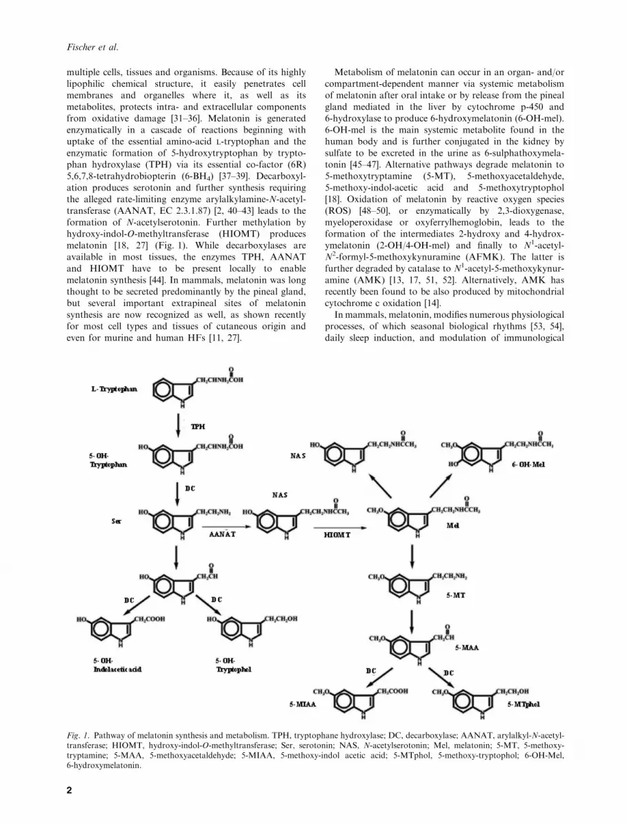
enzymatic formation of 5-hydroxytryptophan by trypto-phan hydroxylase (TPH) via its essential co-factor (6R)5,6,7,8-tetrahydrobiopterin (6-BH4) [37–39]. Decarboxyl-
ation produces serotonin and further synthesis requiringthe alleged rate-limiting enzyme arylalkylamine-N-acetyl-transferase (AANAT, EC 2.3.1.87) [2, 40–43] leads to the
formation of N-acetylserotonin. Further methylation byhydroxy-indol-O-methyltransferase (HIOMT) producesmelatonin [18, 27] (Fig. 1). While decarboxylases areavailable in most tissues, the enzymes TPH, AANAT
and HIOMT have to be present locally to enablemelatonin synthesis [44]. In mammals, melatonin was longthought to be secreted predominantly by the pineal gland,
but several important extrapineal sites of melatoninsynthesis are now recognized as well, as shown recentlyfor most cell types and tissues of cutaneous origin and
even for murine and human HFs [11, 27].
Metabolism of melatonin can occur in an organ- and/orcompartment-dependent manner via systemic metabolismof melatonin after oral intake or by release from the pineal
gland mediated in the liver by cytochrome p-450 and6-hydroxylase to produce 6-hydroxymelatonin (6-OH-mel).6-OH-mel is the main systemic metabolite found in thehuman body and is further conjugated in the kidney by
sulfate to be excreted in the urine as 6-sulphathoxymela-tonin [45–47]. Alternative pathways degrade melatonin to5-methoxytryptamine (5-MT), 5-methoxyacetaldehyde,
5-methoxy-indol-acetic acid and 5-methoxytryptophol[18]. Oxidation of melatonin by reactive oxygen species(ROS) [48–50], or enzymatically by 2,3-dioxygenase,
myeloperoxidase or oxyferrylhemoglobin, leads to theformation of the intermediates 2-hydroxy and 4-hydrox-ymelatonin (2-OH/4-OH-mel) and finally to N1-acetyl-N2-formyl-5-methoxykynuramine (AFMK). The latter is
further degraded by catalase to N1-acetyl-5-methoxykynur-amine (AMK) [13, 17, 51, 52]. Alternatively, AMK hasrecently been found to be also produced by mitochondrial
cytochrome c oxidation [14].In mammals, melatonin, modifies numerous physiological
processes, of which seasonal biological rhythms [53, 54],
daily sleep induction, and modulation of immunological
Fig. 1. Pathway of melatonin synthesis and metabolism. TPH, tryptophane hydroxylase; DC, decarboxylase; AANAT, arylalkyl-N-acetyl-transferase; HIOMT, hydroxy-indol-O-methyltransferase; Ser, serotonin; NAS, N-acetylserotonin; Mel, melatonin; 5-MT, 5-methoxy-tryptamine; 5-MAA, 5-methoxyacetaldehyde; 5-MIAA, 5-methoxy-indol acetic acid; 5-MTphol, 5-methoxy-tryptophol; 6-OH-Mel,6-hydroxymelatonin.
Fischer et al.
2

defense reactions [55] represent but a few prominentexamples. Furthermore, melatonin exerts anti-carcinogenicactivities both in vitro and in vivo, that can be enhanced by
expression of MT1, MT2 or retinoid-related orphan recep-tor a (RORa) receptors depending on the cell line [56–61].
The exceptionally wide range of documented biologicalactivities of melatonin in different systems, cells, and species
is further complicated by many (biologically active) deriv-atives that are generated in vivo from melatonin [16]. Inaddition to its mainly receptor-mediated functions, mela-
tonin also exerts direct receptor-independent chemicaleffects, which render it a potent radical scavenger [12,62–64] as well as a chemocytotoxicity-preventive substance
[62, 65].Melatonin binding protein have been first described in
murine skin in the epidermis and the epithelial bulb of theHF [66]. At present, melatonin receptors can be specifically
identified as membrane-bound, cytosolic and nuclearreceptors [67–69] (Table 1). MT1 and MT2 receptors(formerly Mel1a and Mel1b) are membrane-bound, G pro-
tein-coupled receptors that were initially thought to beexpressed primarily in the central nervous system (firstidentified as MT1 in the retina and MT2 in the brain of
chicken and hamster. As then, melatonin membranereceptors have been associated with many different sitesand functions, e.g., MT1 transcripts have now also been
found in murine heart, kidney, liver, and lung tissue,while MT2 mRNA was also detected in mouse lung [56, 68,70, 71].
A third specific melatonin binding site, initially named
MT3, was later identified as the cytosolic enzyme, NRH:quinone oxidoreductase 2 (NQO2, EC 1.6.99.2), a flavo-protein that catalyzes the reduction of quinones and
therefore is related to the redox status of the cell [67, 72,73]. To date, the biological role of NQO2 is poorlyunderstood, but there is some evidence for association with
anti-carcinogenic effects, as NQO2 knockout mice aresignificantly more sensitive to skin tumor induction bycarcinogens compared with normal mice [74]. In functional
cell growth assays of malignant cells (e.g., melanoma),NQO2 correlated with tumor suppressive effects of mela-tonin [60] and NQO2 is also involved in the protection ofcells by melatonin from oxidative damage [75]. Thus, it
might be hypothesized that NQO2 may play a role in theprevention of (oxidative?) stress-induced HF catagenregression, and this is supported by the wide expression
of the NQO2 gene in human skin [30] (Table 1). However,next to nothing is yet known about the NQO2 hair-connection.
The nuclear receptors for melatonin belong to the RORathat is a member of the RZR/ROR subfamily. Thissubfamily consists of at least four splicing variants:RORa1, RORa2, RORa3 and RZRa (RORa4) [69, 76,
77]. We recently suggested to change the nomenclature ofthe last isoform (RZRa) to RORa4 for consistent termi-nology, as RZRa and RORa4 differ only by a single
nucleotide substitution [30]. RORa appears to be widelyexpressed, with the highest levels found in leukocytes andskin [78]. While classical chronobiology considers melato-
nin exclusively a hormone occurring in the plasma atdaytime levels of 20–50 pg/mL in mammals including
humans, recent data have revealed a variety of compart-ments including bile, bone marrow, cerebrospinal fluid, andgastric mucosa [79–82] that not only represent important
sites of extrapineal melatonin synthesis in situ, but evenmore surprisingly reveal melatonin concentrations at ordersof magnitudes higher than those in the plasma. These datatherefore support the view that melatonin might occur at
tissue-specific concentrations in different compartmentswhere it exerts biologically-relevant effects at both physi-ological and pharmacological concentrations [83, 84].
While the relevance of melatonin has been systematicallyinvestigated in different organ systems, including ovary [85],eye [86], gut [82, 87, 88], bone marrow [79] as well as in
lymphocytes [89], and skin (reviewed in [12, 18, 30, 90]),detailed, systematic knowledge of melatonin in hair biologyremains rather limited.
Melatonin receptor expression in the hair follicle
Some of the reported hair growth- and/or pigmentation-
modulatory effects of melatonin might result from receptorindependent, direct effects of melatonin, while others arelikely to result from signaling via functional melatonin
receptors expressed by HFs.Genes encoding the MT1 receptor have been identified in
HF keratinocytes and dermal papilla fibroblasts, but not in
HF melanocytes [28] (Table 1). Moreover, an aberrantform of MT2 has been identified in dermal papillafibroblasts, but was not expressed by HF keratinocytes ormelanocytes. Hair-cycle-dependent MT2 and RORamRNA transcription [as assessed by reverse transcriptasepolymerase chain reaction (RT-PCR)] has been reported inC57BL/6 mouse skin, although not in single murine HFs,
where MT2 expression was up-regulated in late-anagen andcatagen, and down-regulated in telogen (Table 1). Alterna-tively, RORa was down-regulated in late anagen and
up-regulated in late catagen and decreased in telogen [11].In contrast to human cell lines, MT1 expression was notfound in mouse skin and no high affinity melatonin bindingsite was found in cashmere goat skin [11, 91] (Table 1).
Prominent RORa-like immunoreactivity (IR) wasdetected in the mesenchymal dermal papilla and theepithelial inner and outer root sheaths of C57BL/6 mouse
pelage HFs in situ [11]. While MT1-like IR in human skin
has but yet been detected in HFs, this receptor has beendetected in keratinocytes of the differentiating layers of the
epidermis and in eccrine sweat glands. MT2 receptor IR hasonly been shown in eccrine sweat glands (Fig. 2). Howeverwhile melatonin receptors are quite likely to exhibit
functional effects on human HF cycling and growthregulation, their precise expression pattern and proof oftheir functional activity is still lacking.
Interaction of melatonin with androgenreceptor- and estrogen receptor-mediated signaling
Melatonin not only interacts with its cognate receptors butsurprisingly can interact also with androgen- and estrogenreceptor-mediated signaling pathways. This may be highly
relevant, given the central importance of androgens andestrogens in hair growth control [92–94]. Melatonin is
Melatonin and hair
3

Table
1.Expressionofgenes
encodingmelatonin
receptors
insingle
cellsofskin
andhairfollicle
origin,mouse
andhumanskin
Cell/tissuetype
Species
Detection
Melatonin
bindingsite
MT1
MT2
NQO2
(MT3)
ROR
aROR
a1ROR
a2ROR
a3ROR
a4(R
ZR1)
Ref.
Keratinocytes
Epidermal
keratinocytes
Human
RT-PCR
+)
++
))
)+
[28,30]
Immortalized
keratinocytes
(HaCaT)
Human
RT-PCR
)Aberrant
++
))
)+
[28,30]
HFkeratinocytes
Human
RT-PCR
+)
n.d.
n.d.
n.d.
n.d.
n.d.
n.d.
[28]
Melanocytes
Epidermal
melanocytes
Human
RT-PCR
+)
n.d.
n.d.
n.d.
n.d.
n.d.
n.d.
[28]
Immortalized
melanocytes
(PIG
-1)
Human
RT-PCR
)Aberrant
++
)+
))
[28,30]
Immortalized
norm
al
melanocytes
Mouse
(C57BL/6)
RT-PCR
)+
n.d.
n.d.
n.d.
n.d.
n.d.
n.d.
[112]
HFmelanocytes
Human
RT-PCR
))
n.d.
n.d.
n.d.
n.d.
n.d.
n.d.
[28,30]
Fibroblasts
Adultdermal
fibroblasts
Human
RT-PCR
+)
++
+)
)+
[28,30]
HFfibroblasts
Human
RT-PCR
+Aberrant
n.d.
n.d.
n.d.
n.d.
n.d.
n.d.
[28,30]
Skin
Epidermis
Mouse
(C57BL/6)
Insitu
autoradiography
+n.d.
n.d.
n.d.
n.d.
n.d.
n.d.
n.d.
n.d.
[66]
HF(epithelial
bulb)
Mouse
(C57BL/6)
Insitu
autoradiography
+n.d.
n.d.
n.d.
n.d.
n.d.
n.d.
n.d.
n.d.
[66]
Skin
)Goat
(Cashmere/Angora)
Insitu
autoradiography
)n.d.
n.d.
n.d.
n.d.
n.d.
n.d.
n.d.
n.d.
[91]
Skin
)Mouse
RT-PCR
+n.d.
n.d.
n.d.
n.d.
n.d.
n.d.
n.d.
n.d.
[111]
Skin
)Mouse
(C57BL/6)
RT-PCR
b.d.
+n.d.
n.d.
n.d.
n.d.
n.d.
n.d.
[112]
Skin
)Mouse
(C57BL/6)
Insitu
immunoreactivity
)+
h.c.d.
n.d.
+ h.c.d.
n.d.
n.d.
n.d.
n.d.
[11]
HF
)Mouse
(C57BL/6)
Insitu
immunoreactivity
n.d.
n.d.
n.d.
+ h.c.d.
n.d.
n.d.
n.d.
n.d.
[11]
Norm
alskin
)Human
RT-PCR
+)
n.d.
n.d.
n.d.
n.d.
n.d.
n.d.
[28]
Scalp
skin
Epidermis
Human
Insitu
immunoreactivity
+)
n.d.
n.d.
n.d.
n.d.
n.d.
n.d.
[30]
HF
Human
Insitu
immunoreactivity
+(upper
ORS,IR
S)
+(IRS)
n.d.
n.d.
n.d.
n.d.
n.d.
n.d.
[30]
+,present;
),absent;n.d.:notdone;
b.d.:below
detectability;h.c.d.:hair-cycledependent;ORS:outerrootsheath,IR
S:inner
rootsheath;HF,hair
follicle;RT,PCR,reverse
transcriptase
polymerase
chain
reaction.
Fischer et al.
4
reported to exert anti-androgenic effects on prostate cells inrodents, which are exerted via androgen receptors at theperipheral level [95, 96]. Specifically, melatonin interactswith the nuclear androgen receptor and counteracts its
growth stimulatory effects by facilitating translocation ofthe receptor from the nucleus to the cytoplasm [97]. Thistranslocation is mediated by a melatonin-induced increase
in calcium and protein kinase c (PKC) activation [98].Furthermore, 17-b-estradiol-mediated inactivation of mel-atonin binding to the androgen receptor is ablated by
preincubation of prostate cells with a specific inhibitor ofPKC [99].
On the other side, human prostate cells express func-
tional melatonin receptors (MT1), and sex steroids report-edly interfere with the melatonin receptor in benignprostatic cells [99, 100], e.g., 17-b-estradiol reduces theaffinity of the melatonin receptor to [125I]-melatonin, and
dihydrotestoterone attenuates the melatonin-mediatedinhibitory effects on cell growth [99, 101]. Also, themelatonin-related increase in 3¢,5¢-cyclic adenosine mono-
phosphate and decrease in 3¢,5¢-cyclic guanosine mono-phosphate is attenuated by 17-b-estradiol [99].
In other sex-steroid sensitive tissues such as ovarian
granulosa cell membranes, MT1 expression and bindingmay be down-regulated by estradiol and up-regulated byFSH and testosterone [101]. On the other hand, melatoninhas direct and indirect effects on the estrogen/estrogen-
receptor pathway as shown in human breast cancer cells[102–105]. Growth of human breast cancer cells is inhibitedvia inactivation of estrogen receptor a (but not estrogen
receptor b) through activation of melatonin membranereceptor MT1 and nuclear receptor RZRa [104–106]. Thisanti-estrogenic effects are mediated by inhibiting the
calmodulin-mediated pathway of estrogen receptor activa-tion and gene transcription [104]. Additionally, melatonininteracts on a estrogen presynthesis step by modulating
aromatase (the enzyme responsible for local androgen toestrogen transformation) activity and gene expression [107,108]. Recently, it has been clearly demonstrated in MT1receptor-transfected breast cancer cells that the MT1
melatonin receptor is a key to reduce aromatase activityand expression, leading to a melatonin-induced inhibitionof breast carcinoma cell proliferation [102]. In murine HFs,
melatonin has already been shown to inhibit estrogenreceptor a expression in a hair-cycle-dependent manner,with maximum mRNA reduction in late anagen and
telogen, whereas estrogen receptor a protein is reduced bymelatonin in all hair-cycle phases [11].From the above observations, several conclusions may be
drawn to help explain the effects of melatonin in hair
growth regulation: as the high affinity melatonin receptorMT1 expressed in human prostate epithelial cells and breastcancer cells is the same as the MT1 receptor expressed in
human skin [28], the anti-androgenic effects of melatoninmight be also expressed in the skin. Similar conclusions forthe HF might be drawn carefully, as the expression of MT1
has been only shown for single cells of human HF origin(HF keratinocytes, dermal papilla fibroblasts), and inhuman epidermis [28]. It is hypothesized, although, that
melatonin¢s anti-androgenetic effects could be mediated viathe same mechanisms as described for prostate epithelialand breast cancer cells. Such mechanism could very wellexplain the clinically observed anti-hair loss effects of
melatonin in androgenetic alopecia (AGA) [25].
Melatonin and the skin
Over the last decade, increasing evidence has accumulatedthat melatonin plays a significant role in skin biology –
either as an endogenous factor within the melatoninergicfunctionally active system of the skin or when exogenouslyadministered (reviewed in [12, 13, 18, 27, 28, 30, 90,109, 110]).
Mammalian skin expresses melatonin binding sites,membrane receptors, cytosolic and nuclear receptors [28,30, 66, 111, 112]. Whereas mouse skin expresses MT2, but
not MT1 receptor [11, 112], human skin shows variableexpression of both receptors. Skin-derived cells in vitromainly express MT1 and an aberrant form of MT2,
whereas MT1 is expressed in situ in epidermis, HF, eccrineglands, blood vessel endothelium while and MT2 is onlyweakly expressed in HF inner-root sheath (IRS), eccrine
glands, and blood vessel endothelium (Table 1) [28, 30].The RORa and its isoforms are heterogeneously
expressed in different cell lines of cutaneous origin asassessed by RT-PCR (Table 1). While RORa1 and RORa4are expressed in adult dermal fibroblasts, the isoformRORa2 was detected only in an immortalized melanocyteline (PIG-1). RORa3 has not been detected in any cell line
investigated so far, though RORa4 was detected in malig-nant melanoma cells [30, 60].The skin – the largest organ of the mammalian body –
has been identified as yet another, important site ofperipheral, extra-pineal melatonin synthesis. This work
(A)
(B)
(C)
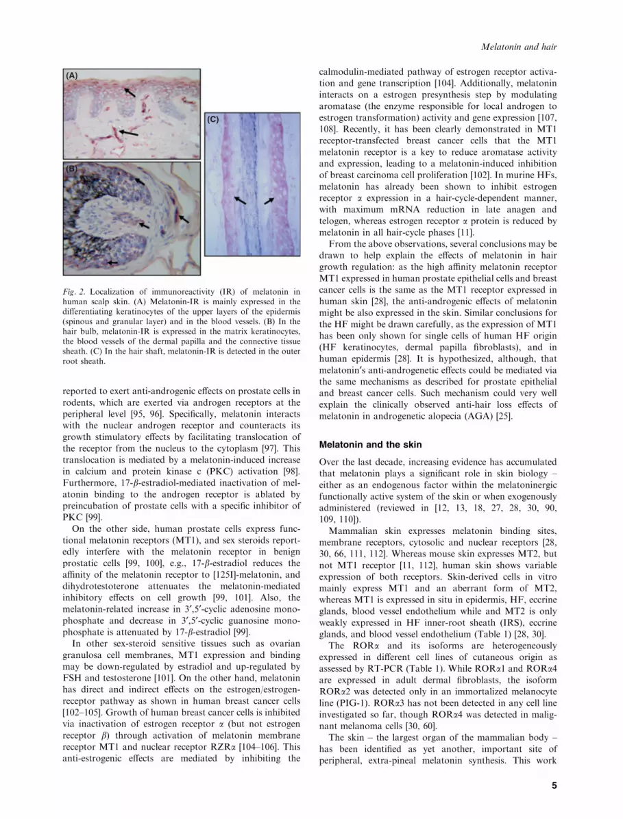
Fig. 2. Localization of immunoreactivity (IR) of melatonin inhuman scalp skin. (A) Melatonin-IR is mainly expressed in thedifferentiating keratinocytes of the upper layers of the epidermis(spinous and granular layer) and in the blood vessels. (B) In thehair bulb, melatonin-IR is expressed in the matrix keratinocytes,the blood vessels of the dermal papilla and the connective tissuesheath. (C) In the hair shaft, melatonin-IR is detected in the outerroot sheath.
Melatonin and hair
5

was stimulated by the discovery that hamster skin containsactivity for AANAT, the key enzyme of melatonin synthesis[113]. This finding prompted a series of further studies that
reported expression of a full melatoninergic system inhuman and rodent skin in situ as well as several of theirconstituent cell populations in vitro [27, 29, 37, 114, 115](Fig. 1). Specifically, transcripts of the key relevant
enzymes for melatonin synthesis, and the actual proteinsynthesis and/or activity of these enzymes have beenidentified both in the intact tissue and in primary cutaneous
cell populations prepared from hamster, mouse [11, 115]and human skin [11, 27, 114].An important exception to the classical pathway of
intrapineal melatonin synthesis can be found in the skin ofC57BL/6 mouse. These mice have a mutation in theAANAT gene, which results in the production of aninactive enzyme. Here serotonin is acetylated to NAS, the
obligate precursor to melatonin by alternative enzyme(s)[115]. Therefore, the C57BL/6 mouse should not any longerbe considered a �natural melatonin knockdown� species, asit is still often claimed [116], because NAS of cutaneousorigin may be methylated to melatonin at local or distanttissue sites expressing HIOMT activity [18, 115].
While the skin is richly endowed with the requiredprecursors for melatonin synthesis (e.g., via massive storesof serotonin within murine skin mast cell granules [117]),
the essential enzymes for melatonin synthesis have all beenidentified in mammalian skin, and in a great variety ofisolated, cultured human skin cells [27–29, 114]. Redundantto the above mentioned evidence that human or mouse skin
actually engages in extrapineal melatonin synthesis, mela-tonin detection in situ has been missing until recently.However, melatonin-IR has been found in epidermis and
blood vessels of human scalp skin as well as in the outer-root sheath (ORS) and the hair-follicle bulb (Fig. 2).Constitutive melatonin production [18] and UV-induced
melatonin metabolism, with additional formation of anti-oxidant degradation products, has been identified in humankeratinocytes [13], thereby defining a melatoninergic anti-
oxidative system in the skin to protect against sun damage[13]. While melatonin ameliorates UV-induced oxidativestress, it also inhibits melanogenesis and melanocyte growth[61, 118–120].
Melatonin exerts growth regulatory (stimulatory/inhibi-tory) effects in benign cells (human keratinocytes andfibroblasts) depending on the experimental conditions
(serum-free/serum-supplemented, UV-exposed) [28, 34,121], but shows clearly growth suppressive, anti-tumori-genic effects in malignant melanoma cells [30, 58, 60, 122,
123]. Melatonin has entered clinical use for metastaticmalignant melanoma [124–126], but this anti-tumor effect isnot limited to pigment cells, but has been found also inbreast cancer [56, 127], colon carcinoma [57, 128], and
squamous cell carcinoma [30]. In UV-induced damage,melatonin can reduce ROS more effectively than evenvitamin C and trolox [33, 129, 130], and promotes cell
survival and colony growth by influencing several check-points of apoptosis [34, 36]. Indeed, pretreatment ofUV-exposed skin with melatonin, either alone or in
combination with vitamin C and E, can significantly reduceUV-induced erythema [131–133].
The �melatonin-hair connection�
Hair follicles and their associated sebaceous glands (�pilo-sebaceous unit�) are the skin�s most prominent appendagesand, together with the mammary gland, represent one ofthe defining features of mammals. This mini-organ, which isconstructed as the result of complex neuroectodermal-
mesodermal interactions [94, 134–136], not only is a targetorgan for numerous (neuro-)hormones, neuropeptides,neurotrophins, and neurotransmitters, but also produces
many of these [137–141]. For example, the HF is both atarget and source of prolactin [142, 143], estrogen [144],cortisol [145], CRH [146], thyroid hormones [147], and
erythropoietin [148], and exhibits a functional hypotha-lamic–pituitary–adrenal axis [145] that has been alsodescribed for the skin [149]. Thus, the pilosebaceous unitis best viewed as a major neuroendocrine organ. On this
emerging background, it is particularly intriguing to eval-uate the existing evidence that yet another neuroendocrinekey mediator – melatonin – also enjoys intimate connec-
tions with the biology of the HF.
Extrapineal melatonin synthesis by human andmouse hair follicles
It is increasingly appreciated that multiple extrapineal sites
of melatonin synthesis exist in mammals [79, 80, 86, 88, 89],and mammalian skin has been shown to express the fullenzymatic apparatus (and all the substrates and co-factors)necessary for melatonin synthesis [27, 29]. Therefore, it was
rather confirmative, when final evidence for actual melato-nin synthesis in mammalian skin in situ was generated byshowing that mouse and human HFs actively generate this
indoleamine under organ-culture conditions [11].To begin with, prominent melatonin-like IR in human
scalp HFs in situ has been independently reported by two
laboratories, using different primary antibodies and immu-nohistological detection techniques [11, 18]. In normalhuman scalp skin sections, melatonin-like IR is seen in theHF ORS, at lower levels in the keratinocytes of the hair
bulb matrix, blood vessels of the connective tissue sheathand in the basal lamina separating the hair bulb matrixfrom the follicular papilla. Distinct melatonin-like IR was
also detected in the ORS of organ-cultured human scalpHFs, and also in the lower IRS and follicular papillafibroblasts [11]. Interestingly, the latter study also revealed
melatonin IR in keratinocytes of the ORS and the lowerpart of the IRS in murine back skin, as well as in thesebaceous gland and showed discrete, hair-cycle-dependent
changes in expression [11]. IR for serotonin-N-acetyltrans-ferase in human scalp epidermis and HF epithelium hasalso been reported [18].However, specific follicular melatonin-like IR in skin and
its appendages may represent serum-derived melatoninbound to intrafollicular melatonin receptors/binding sites,and thus does not, by itself, prove intrafollicular melatonin
synthesis. Although already much less likely, a similarargument may still be evoked for explaining the intriguingradioimmuno assay (RIA) finding that tissue extracts
showed 100–500-fold higher melatonin concentrations inmurine vibrissae follicles and human scalp HFs than in
Fischer et al.
6

corresponding serum [11]. The most convincing evidenceavailable so far that mouse skin fragments, mouse vibrissaefollicles, and human scalp HFs do indeed synthesize
melatonin was provided in HF organ culture wheremelatonin levels were significantly increased after stimula-tion with norepinephrin [11] – the physiological keystimulus in the b-adrenergic control of intrapineal melato-
nin synthesis [150].
Hair growth-modulatory effects of melatonin innonhuman mammals
An indication that melatonin may modulate hair growth in
several nonhuman mammalian species was proposed sev-eral decades ago. In the late sixties, the first influence of thepineal gland on hair cycle in mice was reported [5], followedby several studies reporting an induction or stimulation of
the autumn molt in weasel, mink, red deer, and soay rams[7, 151–153] (Table 2). Thus, mammals exhibit a circadianand seasonal rhythm, which is most evident in those species
that modulate their hair/fur growth according to seasonalalteration of the photoperiod (molting). This influence onfur was later described in limousine ram as an melatonin-
induced increase of HF activity [154], an increase of growthinitializing activity of secondary HFs in situ and hair shaftelongation in cultured HFs from cashmere goat [21, 155]
(Table 2). Furthermore, melatonin was reported to inducethe pro-anagen phase in the New Zealand goat [22] and toincrease pelage development and cycle frequency in pigs [10](Table 2). Indeed, the list of animal species showing effects of
melatonin on hair growth is very extensive, and includescashmere goat andother goat species [21, 22, 155, 156], ferrets[157], merino sheep [158, 159], mink [19], dogs [24, 160, 161],
red deer [20], and others [162]. In many of these species theovercoat and undercoat fur are populated by primary andsecondary HFs, and these are altered with change of the
seasons and their cyclical activity is further disturbed whenthe pineal gland is experimentally removed [154].
Dietary supplementation with melatonin can increase themitosis rate of secondary HF in cashmere goats during
spring [21]. Moreover, the administration of melatonin(70 mg/day) over 14 days to New Zealand goats resulted inincreased melatonin blood levels (914 pg/mL versus
19.9 pg/mL in controls), and this was associated with thetransition of HFs from telogen (resting phase) into thegrowing pro-anagen phase; HFs of the untreated goats
remained in the telogen stage [22]. The hair growth-promoting effect of melatonin is further supported by thefinding that it can, dose dependently, stimulate both DNA-
synthesis and hair shaft elongation in cashmere goat HFs ina 6-day ex vivo organ culture assay [155] (Table 2).
Melatonin at concentrations of 0.1–10 nm significantlystimulated epidermal keratinocyte DNA synthesis when
added to organ-cultured mouse skin with the HFs in theresting phase (telogen), although it did not affect keratino-cytes of the HF [66]. However, recent murine skin organ
culture data suggest that melatonin can reduce spontaneousapoptosis in HF keratinocytes (as assessed by TUNEL) inun-manipulated organ culture of telogen mouse skin,
confirming also the lack of any proliferative effect on HFkeratinocytes (as assessed by Ki-67). Interestingly, in this
study melatonin also significantly down-regulated theexpression of estrogen receptor ERa in the HF matrixand IRS keratinocytes in organ-cultured C57BL/6 mouse
skin [11].Possible mechanisms of melatonin growth stimulatory
effects might be deduced from assays using keratinocytes,the cell population that mainly builds the HF, in which
melatonin at the concentration of 10 lm to 1 nm increasedDNA synthesis, while 1 mm inhibited DNA synthesis.Using the ATP bioluminescence viability assay, melatonin
increased cell proliferation at concentrations of 0.032–20lm
[121]. However, while melatonin increased DNA synthesisin serum-free media (synchronized cell cycle), melatonin
had the opposite effect in growth factor-containing media[28].
Melatonin effects on human hair growth
Reports on the direct effects of melatonin on human hairgrowth in vitro (using microdissected, organ-cultured
anagen VI human scalp HFs) have been conflicting. Oneorgan culture study using female and male HFs from scalpskin reported a stimulation of hair shaft elongation with
30 lm melatonin, while concentrations in the mm rangewere inhibitory [26]. In the former concentration thestimulatory effect was seen only during the early culture
period from day 1–5, and this apparent hair �growth�stimulation may instead reflect an enhanced protection ofmelatonin-treated organ-culture HFs from the conse-quences of general tissue damage after microdissection/
wounding. This interpretation concurs with a subsequentindependent study that reported no effects of melatonin onhuman scalp hair growth or hair matrix proliferation in
vitro over a wide range of melatonin concentrations [11].However, at present it has to be stated that melatonin at10)12– 10)6
m does not influence hair growth in vitro,
whereas melatonin at 3.0 · 10)5m does [11, 26] (Table 2).
Data on the clinical effects of melatonin on human scalphair growth are limited. So far, there has been only a singledouble-blind, randomized, placebo-controlled trial in 40
women aged 20–70 years diagnosed with diffuse alopecia(AD) or AGA [25]. In this study, 1 mL of a 0.1%melatonin-containing alcohol solution was topically
applied each evening for 6 months. To evaluate the effectof melatonin treatment on hair growth, trichograms weretaken in defined areas on the frontal and occipital region of
scalp hair before treatment and after 3 and 6 months oftreatment. After 6 months of treatment, the occipitaltrichograms from women with AGA treated with melatonin
showed an increase in the anagen rate from 76.3% to 85%(+8.7%) while the placebo showed only an increase from78.22% to 82.11% (+3,89) (odds ratio 1.90; P = 0.012). Inwomen with AD, however, the increase of anagen rate was
from 82.2% to 83.8% (+1.6%) while there was a reductionof the anagen rate from 83.16% to 81.13% ()2.03%) inwomen treated with placebo (odds ratio of 1.41;
P = 0.046). Thus, growth modulation induded bymelatonin was slightly relevant in AGA, while in AD onlymarginal, however statistical significant in both cases [25].
In this pilot study, melatonin did not influence the rate ofanagen hair growth in HF located in the frontal scalp area
Melatonin and hair
7

Table
2.Effects
ofmelatonin
onhairgrowth
andpigmentation
Species
Effect
Ref.
Growth
Mouse
Influence
onthehaircyclebythepinealgland
[5]
Weasel
Inductionofmolt
[152]
Mink
Inductionofautumnmolt
[7]
Red
deer
Premature
moultingofsummer
pelageandreducedserum
prolactin
concentrations
[20]
Soayrams
Stimulationofmoulting
[151]
Lim
ousineram
IncreasedHFactivityandreducedprolactin
plasm
alevels
[154]
Mink
Inductionofwinterfurgrowth
(supposedly
byinhibitionofprolactin)
[19]
Cashmeregoat
Increase
ofgrowth
initializingactivityofsecondary
HFsin
springtime
[21]
New
Zealandgoat
Inductionofpro-anagen
phase
[22]
Cashmeregoat(culturedHFs)
Increase
ofhairshaftelongationandDNA-synthesis
[155]
Domesticpig
Increase
ofpelagedevelopmentandcyclefrequency
[10]
Ferret
Earlierchangeofwinterandconsecutivespringcoat
[157]
Raccoondogs
More
rapid
sheddingofmature
underfurhairsandgrowth
ofnew
underfurhairs;suppressionofprolactin
levels
[160]
Merinosheep
Noinfluence
ofpinealectomyonwoolgrowth
andhairdensity
[159]
SiberianHuskydogs
Nochangein
hairgrowth
oranagen
rate
(topicaladministration)
[23]
Human(culturedHFs)
Increase
ofhairshaftelongation(30
lm);Decrease
ofhairshaft
elongation(1–5m
m)
[26]
Human(culturedHFs)
Noinfluence
onhairshaftelongation,matrix
keratinocyte
proliferation/apoptosisandhaircycling(10)12–10)6
m)
[11]
Human(trichograms)
Slightincrease
ofanagen
hairrate
inwomen
withandrogenetic
anddiffuse
alopecia
[25]
Pigmentation
Weasel
Inductionofhaircolorchange
[152]
Mammalians
Effects
onhaircolor
[178]
Djungarianhamster
Patternofmelatonin
release
inducedbyexperim
entallyinducedphotoperiodsmodifies
moltinto
summer
pelage
[6]
Siberianhamster
(culturedHFs)
Post-tyrosinase
inhibitionofmelanogensis(10
)10–10)6
m)
[118]
Yellow
mice(C
3H/H
e-A*vy)
Slightreductionofcoatdarkening
[176]
Mountain
hares
Season-dependenteff
ects
ofmelatonin
onfurcolor
[9]
Djungarianhamster
Inductionofthewintermoltandpelagecolorchange
[8]
Djungarianhamster
Changeoffurcolor
[54]
Mouse
Inhibitionofmelanogenesis
[66]
Human(culturedHFs)
Noeff
ectonpigmentation(10)12–10)6
m)
[11]
HF,hairfollicle.
Fischer et al.
8

of women with AGA – the area mostly affected by hairthinning in this disorder, while the less androgen-sensitiveoccipital scalp skin area appeared to be positively influ-
enced by melatonin. This effect might be interpreted asinduction of hair growth by prolongation of the anagenphase, in part via retardation of the transition to catagenand/or by promotion of the transition from telogen to
anagen, as has been observed in animals [22]. However, asthe effects of melatonin in this study were only tested in sixpatients with AGA and 14 patients with AD (against equal
number of patients treated with placebo), this study wouldrequire to be repeated with a larger number of patients forone diagnosis, and for a longer period. Moreover, it may
also benefit from being complemented with additional hairgrowth parameters (e.g., phototrichogram, global hairphotography, effluvium count, hair number, and shaftdiameter), before sound conclusions can be drawn on the
clinical efficacy of topical melatonin as an agent in themanagement of defined hair loss disorders. Also, whilecutaneous penetration of topically applied melatonin has
been reported [163, 164], the depth of melatonin penetra-tion and the exact concentrations that are reached in theHF, especially the matrix keratinocytes remain open
questions. However, topically applied melatonin may trig-ger complex secondary signaling cascades (from epidermis)that may then affect the pilosebaceous unit also indirectly.
The impact of melatonin on hair pigmentation
Melatonin effects on pigmentation have been reviewed in
detail, focusing on skin [3] and the HF [4]. Hair shaftpigmentation is generated by specialized melanocytes of theHF pigmentary unit, whose melanogenic activity is strictly
coupled to HF cycling (i.e., anagen III–VI) [165–170].Growth, survival, and melanogeneic activity of these spe-cializedmelanocytes underlies complex, species- site- andHF
type-dependent controls, which are only partially under-stood, and can not simply be equated with those recognizedfor epidermal melanocytes [3, 171–173] (Table 2). Whilemelanocortins like alpha-melanocyte-stimulating hormone
(a-MSH) and adrenocorticotrophic hormone (ACTH) havebeen the main focus of endocrinologists interested in hairpigmentation, many additional (neuro-)hormones, neuro-
trophins, neuropeptides and neurotransmitters are involvedin the control of hair pigmentation in various mammalianspecies (e.g., beta-endorphine, histamine, estrogen, POMC,
and NGF, to name but a few prominent examples) [4, 174–177]. Melatonin has been described to increase number ofmelanocytes in culture [120].
Early observations in farm and laboratory animals havereported that pinealectomy and/or melatonin administra-tion altered hair shaft color in addition to hair growth,cycling or molting [6, 8, 10, 54, 152, 178] (Table 2). These
observations have long suggested that melatonin may beone such neuroendocrine regulator of HF pigmentation.However, the literature continues to paint a rather confus-
ing picture, and so, evidence that melatonin is indeed animportant regulator of follicular melanogenesis underphysiological conditions remains inconclusive.
While the classical �skin lightening� effects of melatonin,which reflect primarily the induction of melanosome
aggregation e.g., in frog melanophores, are well-knownfrom work in amphibian skin [2, 179], much less is knownon the effect of melatonin on mammalian melanocytes [3,
120, 165, 180, 181]. Given the numerous biological differ-ences between epidermal and HF melanocytes [3, 172],however, it is quite unclear whether these findings are at allrelevant to hair pigmentation. Evidently, this is even more
the case for the reported inhibitory effects of melatonin onmelanoma cell melanogenesis and/or growth, which may beantagonized in part by a-MSH [119, 182]. Therefore, the
best currently available evidence for pigmentary effectscomes from organ culture studies using hamster, mouse andhuman HFs – all of which are hampered by the shortcom-
ings and limitations that are inherent to such complexassays [6, 8, 118, 176, 183].Melatonin (0.1 nm–1 lm) reportedly inhibits the post-
tyrosinase steps of melanogenesis in hamster HFs [118], and
we have found that high dose-melatonin (0.01–100 lm) caninhibit follicular tyrosinase activity in organ-cultured mouseskin with all HFs in anagen growth phase [66] (Table 2).
Thody and co-workers reported that melatonin administra-tion slightly reduced coat darkening in young mice in vivo,when hair re-growth after shaft plucking was examined
[176]. However, when we checked the effect of 0.001–1000 nm melatonin on organ-cultured human scalp HFs inanagen, no consistent and significant effects on the histo-
chemcially detectable melanin content of human anagen VIhair bulbs in situ could be identified (as assessed by quan-titative Masson-Fontana histochemistry) [11] (Table 2).While this study certainly does not rule-out effects of
melatonin on human HF pigmentation under physiologicalconditions, it makes it likely that this indole is not a majormodulator of human hair pigmentation. This conclusion is
further supported by the lack of case reports of pigmentaryeffects induced by melatonin dietary supplementation,despite the copious, almost �epidemic� consumption of
sometimes massive oral doses of melatonin worldwide.
Conclusions and perspectives
In summary, murine HFs express transcripts and proteinfor the melatonin membrane receptor (MT2) and mRNAfor the putative nuclear melatonin receptors (RORa) [11].These intra-follicular melatonin receptors may be function-ally active, as their stimulation by melatonin can down-regulate both HF keratinocyte apoptosis and estrogen
receptor-a expression in situ [11]. Together with the factthat MT2 and RORa expression in murine skin arestrikingly hair-cycle dependent, this raises the possibility
that melatonin is somehow involved in hair-cycle control.Even more importantly, murine and human HFs areimportant sites of extrapineal melatonin synthesis anddisplay a genuine melatoninergic system, which can be
stimulated by catecholamines [11].The two most significant remaining questions are:
(i) What is the principal requirement for melatonin by
HFs under physiological and pathological conditions and(ii) can melatonin administration be therapeuticallyexploited for the clinical management of hair growth
disorders? Despite much suggestive in vivo evidence fromthe older literature of melatonin being an important
Melatonin and hair
9

modulator of hair growth, cycling, molting and pigmenta-tion in selected species (Table 2), the available evidence thatmelatonin substantially and reproducibly alters hair
growth, pigmentation and/or cycling in mouse or humanHFs under physiological conditions remains unsatisfactory.Because of the potency of melatonin as a free radical
scavenger [12, 63, 64], its anti-apoptotic properties in some
systems [28, 34, 184, 185] and its proposed capacity tostimulate DNA repair [62, 186], the metabolically activeand proliferatively active (but exceptionally damage-sensi-
tive) anagen hair bulb may exploit melatonin synthesisin loco as a cytoprotective and apoptosis-suppressivestrategy [11]. This concept deserves systematic exploration.
If confirmed, it may become exploitable in the context ofchemotherapy-induced alopecia [187–189]. Given that ana-gen termination by premature entry into apoptosis-drivenHF regression (catagen) lies at the heart of essentially all of
the clinically most relevant hair loss disorders [94, 190], ittherefore certainly is a key challenge for future, clinicallyrelevant research into the �melatonin-hair connection� to
clarify whether and under which circumstances defineddoses of melatonin effectively inhibit human HF keratino-cyte apoptosis in situ.
Also, the documented down-regulatory effect of melato-nin on ER-a expression may render the HF less sensitive tostimulation by estrogens [144]. In addition to the intriguing
endocrine link between estrogens and melatonin, anotherone exists between prolactin and melatonin. Melatoninserum levels have long been recognized to modulatepituitary prolactin secretion [22, 154]. In view of our recent
finding that both murine pelage HFs and human scalp HFsexpress prolactin and prolactin receptors and employprolactin receptor stimulation to induce catagen [142,
143], it will be interesting to study whether exogenousmelatonin and/or melatonin generated by the HF itself hasany impact on follicular prolactin synthesis.
This begs the question: does melatonin exert its mostimportant hair growth-modulatory properties in vivo andin physiological concentrations indirectly, e.g., via the
estrogen/prolactin axes sketched here? Perhaps, this ex-plains, at least in part, why it has been so difficult toactually prove hair growth- and/or pigmentation-modula-tory effects of melatonin? Moreover, given the well-recog-
nized regulation of clock gene expression and activity bymelatonin (e.g., in birds, fish, mice nonhuman primates[191–194], and the potential importance of clock genes in
hair-cycle control [195], species-dependent hair-cycle-regu-latory effects of intrafollicularly generated melatonin mayalso result from targeting the expression/ activity of clock
genes, some of which may actually be expressed in the HF.Apart from its evident relevance for the – as yet unknown
– auto-regulation of intrafollicular melatonin synthesis thestimulation of HF melatonin synthesis by catecholamines
raises the question whether this melatoninergic systemprimarily has inducible, hair growth-regulatory functions,or serves to protect the HF against systemic stressors
(sensed and activated by high noradrenaline levels [138]. Ifthe latter speculation holds true, stress-induced hair lossmight result from an imbalance between increased systemic
noradrenalin levels and the HF�s inability to protect itselfvia the production of sufficient melatonin.
Exploration of the melatonin-hair connection likelyholds lessons to better understand the role of melatoninin other skin appendages as well – especially the largest
one of all: the mammary gland! It deserves mentioninghere that melatonin has long been recognized as aninhibitor of mammary gland development and growth[196, 197].
In short, with the recent recognition of melatoninreceptor expression and melatonin synthesis in the HFs ofmouse and human, and the tremendous recent progress in
understanding the molecular mechanisms which underliemelatonin�s vexingly pleiotropic functions (amply docu-mented on the pages of this journal throughout the past
decade), it has now become fascinating, clinically impor-tant, and scientifically productive to systematically follow-up, at long last, the existing ancient leads to an importantrole for melatonin in hair biology.
Acknowledgments
The authors gratefully acknowledge the funding agencieswhich have supported some of their original studies cited inthis review: German Academy of Natural Scientists Leo-
poldina, Halle, and Federal Ministry of Education andResearch� BMBF-LPD 9901/8-113 (TWF), Deutsche Fors-chungsgemeinschaft (Pa 345/11-2) (RP) and University of
Tennessee Cancer Center Pilot Grant (AS, TWF).
References
1. Arendt J. Melatonin. Clin Endocrinol (Oxf) 1988; 29:205–
229.
2. Lerner AB, Case JD, Takahashi Y. Isolation of melatonin,
a pineal factor that lightens melanocytes. J Am Chem Soc
1958; 80:2587.
3. Slominski A, Tobin DJ, Shibahara S et al. Melanin pig-
mentation in mammalian skin and its hormonal regulation.
Physiol Rev 2004; 84:1155–1228.
4. Slominski A, Wortsman J, Plonka PM et al. Hair follicle
pigmentation. J Invest Dermatol 2005; 124:13–21.
5. Houssay AB, Pazo JH, Epper CE. Effects of the pineal gland
upon the hair cycles in mice. J Invest Dermatol 1966; 47:230–
234.
6. Hoffmann K. Photoperiodic effects in the Djungarian
hamster: one minute of light during darktime mimics influ-
ence of long photoperiods on testicular recrudescence, body
weight and pelage colour. Experientia 1979; 35:1529–1530.
7. Allain D, Rougeot J. Induction of autumn moult in mink
(Mustela vison Peale and Beauvois) with melatonin. Reprod
Nutr Dev 1980; 20:197–201.
8. Duncan MJ, Goldman BD, Di Pinto MN et al. Testicular
function and pelage color have different critical daylengths in
the Djungarian hamster, Phodopus sungorus. Endocrinology
1985; 116:424–430.
9. Kuderling I, Cedrini MC, Fraschini F et al. Season-
dependent effects of melatonin on testes and fur color in
mountain hares (Lepus timidus L.). Experientia 1984; 40:501–
502.
10. Paterson AM, Foldes A. Melatonin and farm animals:
endogenous rhythms and exogenous applications. J Pineal
Res 1994; 16:167–177.
Fischer et al.
10
11. Kobayashi H, Kromminga A, Dunlop TW et al. A role of
melatonin in neuroectodermal-mesodermal interactions: the
hair follicle synthesizes melatonin and expresses functional
melatonin receptors. FASEB J 2005; 19:1710–1712.
12. Fischer TW, Elsner P. The antioxidative potential of mel-
atonin in the skin. Curr Probl Dermatol 2001; 29:165–174.
13. Fischer TW, Sweatman TW, Semak I et al. Constitutive
and UV-induced metabolism of melatonin in keratinocytes
and cell-free systems. FASEB J 2006; 20:1564–1566.
14. Semak I, Naumova M, Korik E et al. A novel metabolic
pathway of melatonin: oxidation by cytochrome C. Bio-
chemistry 2005; 44:9300–9307.
15. Tan DX, Manchester LC, Hardeland R et al. Melatonin:
a hormone, a tissue factor, an autocoid, a paracoid, and an
antioxidant vitamin. J Pineal Res 2003; 34:75–78.
16. Tan DX, Manchester LC, Terron MP et al. One molecule,
many derivatives: a never-ending interaction of melatonin
with reactive oxygen and nitrogen species? J Pineal Res 2007;
42:28–42.
17. Tesoriere L, Avellone G, Ceraulo L et al. Oxidation of
melatonin by oxoferryl hemoglobin: a mechanistic study. Free
Radic Res 2001; 35:633–642.
18. Slominski A, Wortsman J, Tobin DJ. The cutaneous sero-
toninergic/melatoninergic system: securing a place under the
sun. FASEB J 2005; 19:176–194.
19. Rose J, Oldfield J, Stormshak F. Apparent role of mela-
tonin and prolactin in initiating winter fur growth in mink.
Gen Comp Endocrinol 1987; 65:212–215.
20. Webster JR, Suttie JM, Corson ID. Effects of melatonin
implants on reproductive seasonality of male red deer (Cervus
elaphus). J Reprod Fertil 1991; 92:1–11.
21. Welch RAS, Gurnsey MP, Betteridge K et al. Goat fibre
response to melatonin given in spring in two consecutive
years. Proc N Z Soc Anim Prod 1990; 50:335–338.
22. Nixon AJ, Choy VJ, Parry AL et al. Fiber growth initiation
in hair follicles of goats treated with melatonin. J Exp Zool
1993; 267:47–56.
23. Diaz SF, Torres SM, Nogueira SA et al. The impact of
body site, topical melatonin and brushing on hair regrowth
after clipping normal Siberian Husky dogs. Vet Dermatol
2006; 17:45–50.
24. Xiao Y, Forsberg M, Laitinen JT et al. Effects of melato-
nin implants on winter fur growth and testicular recrudes-
cence in adult male raccoon dogs (Nyctereutes procyonoides).
J Pineal Res 1996; 20:148–156.
25. Fischer TW, Burmeister G, Schmidt HW et al. Melatonin
increases anagen hair rate in women with androgenetic alo-
pecia or diffuse alopecia: results of a pilot randomized con-
trolled trial. Br J Dermatol 2004; 150:341–345.
26. Fischer TW, Fischer A, Knoll B et al. Melatonin in low
doses enhances in vitro human hair follicle proliferation and
inhibits hair growth in high doses. Arch Derm Res 2000;
292:147.
27. Slominski A, Pisarchik A, Semak I et al. Serotoninergic
and melatoninergic systems are fully expressed in human skin.
FASEB J 2002; 16:896–898.
28. Slominski A, Pisarchik A, Zbytek B et al. Functional
activity of serotoninergic and melatoninergic systems
expressed in the skin. J Cell Physiol 2003; 196:144–153.
29. Slominski A, Baker J, Rosano TG et al. Metabolism of
serotonin to N-acetylserotonin, melatonin, and 5-methoxy-
tryptamine in hamster skin culture. J Biol Chem 1996;
271:12281–12286.
30. Slominski A, Fischer TW, Zmijewski MA et al. On the role
of melatonin in skin physiology and pathology. Endocrine
2005; 27:137–148.
31. Hardeland R. Antioxidative protection by melatonin:
multiplicity of mechanisms from radical detoxification to
radical avoidance. Endocrine 2005; 27:119–130.
32. Leon J, Acuna-Castroviejo D, Sainz RM et al. Melatonin
and mitochondrial function. Life Sci 2004; 75:765–790.
33. Fischer TW, Scholz G, Knoll B et al. Melatonin
suppresses reactive oxygen species induced by UV irradiation
in leukocytes. J Pineal Res 2004; 37:107–112.
34. Fischer TW, Zbytek B, Sayre RM et al. Melatonin
increases survival of HaCaT keratinocytes by suppressing
UV-induced apoptosis. J Pineal Res 2006; 40:18–26.
35. Leon J, Acuna-Castroviejo D, Escames G et al. Melatonin
mitigates mitochondrial malfunction. J Pineal Res 2005;
38:1–9.
36. Fischer TW, Zmijewski MA, Wortsman J et al. Melatonin
maintains mitochondrial membrane potential and attenuates
activation of initiator (casp-9) and effector caspases (casp-3/
casp-7) and PARP in UVR-exposed HaCaT keratinocytes.
J Pineal Res 2007; In press.
37. Slominski A, Pisarchik A, Johansson O et al. Tryptophan
hydroxylase expression in human skin cells. Biochim Biophys
Acta 2003; 1639:80–86.
38. Schallreuter KU, Wood JM, Pittelkow MR et al. Reg-
ulation of melanin biosynthesis in the human epidermis by
tetrahydrobiopterin. Science 1994; 263:1444–1446.
39. Hasse S, Gibbons NC, Rokos H et al. Perturbed 6-tetrahy-
drobiopterin recycling via decreased dihydropteridine reduc-
tase in vitiligo: more evidence for H2O2 stress. J Invest
Dermatol 2004; 122:307–313.
40. Hardeland RFB. Ubiquitous melatonin – presence and
effects in unicells, plants and animals. Trends Comp Biochem
Physiol 1996; 2:25–45.
41. Coon SL, Roseboom PH, Baler R et al. Pineal serotonin
N-acetyltransferase: expression cloning and molecular anal-
ysis. Science 1995; 270:1681–1683.
42. Ganguly S, Coon SL, Klein DC. Control of melatonin
synthesis in the mammalian pineal gland: the critical
role of serotonin acetylation. Cell Tissue Res 2002;
309:127–137.
43. Klein DC. Arylalkylamine N-acetyltransferase: �the Time-
zyme�. J Biol Chem 2007; 282:4233–4237.
44. Kema IP, De Vries EG, Muskiet FA. Clinical chemistry of
serotonin and metabolites. J Chromatogr B Biomed Sci Appl
2000; 747:33–48.
45. Lerner AB, Nordlund JJ. Melatonin: clinical pharmacol-
ogy. J Neural Transm Suppl 1978; 13:339–347.
46. Ma X, Idle JR, Krausz KW et al. Metabolism of melatonin
by human cytochromes p450. Drug Metab Dispos 2005;
33:489–494.
47. Kopin IJ, Pare CM, Axelrod J et al. 6-Hydroxylation, the
major metabolic pathway for melatonin. Biochim Biophys
Acta 1960; 40:377–378.
48. de Almeida EA, Martinez GR, Klitzke CF et al.
Oxidation of melatonin by singlet molecular oxygen
(O2(1deltag)) produces N1-acetyl-N2-formyl-5-methoxyky-
nurenine. J Pineal Res 2003; 35:131–137.
49. Almeida EA, Klitzke CF, Martinez GR et al. Synthesis of
internal labeled standards of melatonin and its metabolite
N1-acetyl-N2-formyl-5-methoxykynuramine for their quanti-
fication using an on-line liquid chromatography-electrospray
Melatonin and hair
11
tandem mass spectrometry system. J Pineal Res 2004; 36:64–
71.
50. Hardeland R, Reiter RJ, Poeggeler B et al. The signifi-
cance of the metabolism of the neurohormone melatonin:
antioxidative protection and formation of bioactive sub-
stances. Neurosci Biobehav Rev 1993; 17:347–357.
51. Tan DX, Manchester LC, Burkhardt S et al. N1-acetyl-
N2-formyl-5-methoxykynuramine, a biogenic amine and
melatonin metabolite, functions as a potent antioxidant.
FASEB J 2001; 15:2294–2296.
52. Ximenes VF, de OSS, Rodrigues MR et al. Superoxide-
dependent oxidation of melatonin by myeloperoxidase. J Biol
Chem 2005; 280:38160–38169.
53. Bubenik GA, Smith PS. Circadian and circannual rhythms of
melatonin in plasma of male white- tailed deer and the effect of
oral administration of melatonin. J Exp Zool 1987; 241:81–89.
54. Lerchl A, Schlatt S. Influence of photoperiod on pineal
melatonin synthesis, fur color, body weight, and reproductive
function in the female Djungarian hamster, Phodopus sung-
orus. Neuroendocrinology 1993; 57:359–364.
55. Maestroni GJ. The immunotherapeutic potential of mela-
tonin. Expert Opin Investig Drugs 2001; 10:467–476.
56. Dillon DC, Easley SE, Asch BB et al. Differential expres-
sion of high-affinity melatonin receptors (MT1) in normal and
malignant human breast tissue. Am J Clin Pathol 2002;
118:451–458.
57. Karasek M, Carrillo-Vico A, Guerrero JM et al.
Expression of melatonin MT(1) and MT(2) receptors, and
ROR alpha(1) receptor in transplantable murine colon 38
cancer. Neuroendocrinol Lett 2002; 23(Suppl. 1):55–60.
58. Kadekaro AL, Andrade LNS, Floeter-Winter LM et al.
MT-1 melatonin receptor expression increases the antiprolif-
erative effect of melatonin on S-91 murine melanoma cells.
J Pineal Res 2004; 36:204–211.
59. Winczyk K, Pawlikowski M, Guerrero JM et al. Possible
involvement of the nuclear RZR/ROR-alpha receptor in the
antitumor action of melatonin on murine colon 38 cancer.
Tumour Biol 2002; 23:298–302.
60. Fischer TW, Zmijewski MA, Zbytek B et al. Oncostatic
effects of the indole melatonin and expression of its cytosolic
and nuclear receptors in cultured human melanoma cell lines.
Int J Oncol 2006; 29:665–672.
61. Bartsch C, Bartsch H, Karasek M. Melatonin in clinical
oncology. Neuroendocrinol Lett 2002; 23(Suppl. 1):30–38.
62. Tan DX, Reiter RJ, Manchester LC et al. Chemical and
physical properties and potential mechanisms: melatonin as a
broad spectrum antioxidant and free radical scavenger. Curr
Top Med Chem 2002; 2:181–197.
63. Tan DX, Chen LD, Poeggeler B et al. Melatonin: a potent,
endogenous hydroxyl radical scavenger. Endocr J 1993; 1:57–
60.
64. Reiter RJ, Tan DX, Poeggeler B et al. Melatonin as a free
radical scavenger: implications for aging and age-related
diseases. Ann N Y Acad Sci 1994; 719:1–12.
65. Reiter RJ, Tan DX, Sainz RM et al. Melatonin: reducing
the toxicity and increasing the efficacy of drugs. J Pharm
Pharmacol 2002; 54:1299–1321.
66. Slominski A, Chassalerris N, Mazurkiewicz J et al.
Murine skin as a target for melatonin bioregulation. Exp
Dermatol 1994; 3:45–50.
67. Nosjean O, Ferro M, Coge F et al. Identification of the
melatonin-binding site MT3 as the quinone reductase 2.
J Biol Chem 2000; 275:31311–31317.
68. Dubocovich ML, Masana MI, Iacob S et al. Melatonin
receptor antagonists that differentiate between the human
Mel1a and Mel1b recombinant subtypes are used to assess the
pharmacological profile of the rabbit retina ML1 presynaptic
heteroreceptor. Naunyn Schmiedebergs Arch Pharmacol
1997; 355:365–375.
69. Becker-Andre M, Andre E, DeLamarter JF. Identifica-
tion of nuclear receptor mRNAs by RT-PCR amplification of
conserved zinc-finger motif sequences. Biochem Biophys Res
Commun 1993; 194:1371–1379.
70. Dubocovich ML, Yun K, Al-Ghoul WM et al. Selective
MT2 melatonin receptor antagonists block melatonin-medi-
ated phase advances of circadian rhythms. FASEB J 1998;
12:1211–1220.
71. Nosjean O, Nicolas JP, Klupsch F et al. Comparative
pharmacological studies of melatonin receptors: MT1, MT2
and MT3/QR2. Tissue distribution of MT3/QR2. Biochem
Pharmacol 2001; 61:1369–1379.
72. Mailliet F, Ferry G, Vella F et al. Characterization of the
melatoninergic MT(3) binding site on the NRH:quinone oxi-
doreductase 2 enzyme. Biochem Pharmacol 2005; 71:74–88.
73. Mailliet F, Ferry G, Vella F et al. Organs from mice
deleted for NRH:quinone oxidoreductase 2 are deprived of
the melatonin binding site MT3. FEBS Lett 2004; 578:116–
120.
74. Iskander K, Paquet M, Brayton C et al. Deficiency of
NRH:quinone oxidoreductase 2 increases susceptibility to
7,12-dimethylbenz(a)anthracene and benzo(a)pyrene-induced
skin carcinogenesis. Cancer Res 2004; 64:5925–5928.
75. Vella F, Ferry G, Delagrange P et al. NRH:quinone
reductase 2: an enzyme of surprises and mysteries. Biochem
Pharmacol 2005; 71:1–12.
76. Carlberg C, Hooft van Huijsduijnen R, Staple JK et al.
RZRs, a new family of retinoid-related orphan receptors that
function as both monomers and homodimers. Mol Endocri-
nol 1994; 8:757–770.
77. Pozo D, Garcia-Maurino S, Guerrero JM et al. mRNA
expression of nuclear receptor RZR/RORalpha, melatonin
membrane receptor MT, and hydroxindole-O-methyl-
transferase in different populations of human immune cells.
J Pineal Res 2004; 37:48–54.
78. Steinmayr M, Andre E, Conquet F et al. Staggerer phe-
notype in retinoid-related orphan receptor alpha-deficient
mice. Proc Natl Acad Sci USA 1998; 95:3960–3965.
79. Tan DX, Manchester LC, Reiter RJ et al. Identification of
highly elevated levels of melatonin in bone marrow: its origin
and significance. Biochim Biophys Acta 1999; 1472:206–214.
80. Tan D, Manchester LC, Reiter RJ et al. High physiolog-
ical levels of melatonin in the bile of mammals. Life Sci 1999;
65:2523–2529.
81. Skinner DC, Malpaux B. High melatonin concentrations in
third ventricular cerebrospinal fluid are not due to Galen vein
blood recirculating through the choroid plexus. Endocrinol-
ogy 1999; 140:4399–4405.
82. Bubenik GA, Hacker RR, Brown GM et al. Melatonin
concentrations in the luminal fluid, mucosa, and muscularis
of the bovine and porcine gastrointestinal tract. J Pineal Res
1999; 26:56–63.
83. Reiter RJ, Tan DX. What constitutes a physiological con-
centration of melatonin? J Pineal Res 2003; 34:79–80.
84. Reiter RJ, Tan DX, Maldonado MD. Melatonin as an
antioxidant: physiology versus pharmacology. J Pineal Res
2005; 39:215–216.
Fischer et al.
12

85. Itoh MT, Ishizuka B, Kuribayashi Y et al. Melatonin, its
precursors, and synthesizing enzyme activities in the human
ovary. Mol Hum Reprod 1999; 5:402–408.
86. Cahill GM, Besharse JC. Light-sensitive melatonin syn-
thesis by Xenopus photoreceptors after destruction of the
inner retina. Vis Neurosci 1992; 8:487–490.
87. Bubenik GA. Localization, physiological significance and
possible clinical implication of gastrointestinal melatonin.
Biol Signals Recept 2001; 10:350–366.
88. Bubenik GA. Gastrointestinal melatonin: localization, func-
tion, and clinical relevance. Dig Dis Sci 2002; 47:2336–2348.
89. Carrillo-Vico A, Calvo JR, Abreu P et al. Evidence of
melatonin synthesis by human lymphocytes and its physio-
logical significance: possible role as intracrine, autocrine, and/
or paracrine substance. FASEB J 2004; 18:537–539.
90. Fischer T, Wigger-Alberti W, Elsner P. Melatonin in
dermatology. Experimental and clinical aspects. Hautarzt
1999; 50:5–11.
91. Dicks P, Morgan CJ, Morgan PJ et al. The localisation
and characterisation of insulin-like growth factor-I receptors
and the investigation of melatonin receptors on the hair fol-
licles of seasonal and non-seasonal fibre-producing goats.
J Endocrinol 1996; 151:55–63.
92. Hoffmann R, Niiyama S, Huth A et al. 17alpha-estradiol
induces aromatase activity in intact human anagen hair fol-
licles ex vivo. Exp Dermatol 2002; 11:376–380.
93. Kaufman KD. Androgen metabolism as it affects hair
growth in androgenetic alopecia. Dermatol Clin 1996;
14:697–711.
94. Paus R, Cotsarelis G. The biology of hair follicles. N Engl J
Med 1999; 341:491–497.
95. Moeller H, Koz A, Rodl W et al. Role of the pineal gland
in the regulation of prostatic androgen receptors in pubertal
and mature rats. Res Exp Med (Berl) 1983; 183:157–165.
96. Alonso R, Prieto L, Hernandez C et al. Antiandrogenic
effects of the pineal gland and melatonin in castrated and
intact prepubertal male rats. J Endocrinol 1978; 79:77–83.
97. Rimler A, Culig Z, Lupowitz Z et al. Nuclear exclusion of
the androgen receptor by melatonin. J Steroid Biochem Mol
Biol 2002; 81:77–84.
98. Rimler A, Jockers R, Lupowitz Z et al. Gi and RGS
proteins provide biochemical control of androgen receptor
nuclear exclusion. J Mol Neurosci 2007; 31:1–12.
99. Gilad E, Matzkin H, Zisapel N. Interplay between sex
steroids and melatonin in regulation of human benign pros-
tate epithelial cell growth. J Clin Endocrinol Metab 1997;
82:2535–2541.
100. Siu SW, Lau KW, Tam PC et al. Melatonin and prostate
cancer cell proliferation: interplay with castration, epidermal
growth factor, and androgen sensitivity. Prostate 2002;
52:106–122.
101. Clemens JW, Jarzynka MJ, Witt-Enderby PA. Down-
regulation of mt1 melatonin receptors in rat ovary following
estrogen exposure. Life Sci 2001; 69:27–35.
102. Gonzalez A, Martinez-Campa C, Mediavilla MD et al.
Effects of MT1 melatonin receptor overexpression on the
aromatase-suppressive effect of melatonin in MCF-7 human
breast cancer cells. Oncol Rep 2007; 17:947–953.
103. Kiefer TL, Lai L, Yuan L et al. Differential regulation of
estrogen receptor alpha, glucocorticoid receptor and retinoic
acid receptor alpha transcriptional activity by melatonin is
mediated via different G proteins. J Pineal Res 2005; 38:231–
239.
104. Del Rio B, Garcia Pedrero JM, Martinez-Campa C et al.
Melatonin, an endogenous-specific inhibitor of estrogen
receptor alpha via calmodulin. J Biol Chem 2004; 279:38294–
38302.
105. Girgert R, Bartsch C, Hill SM et al. Tracking the elusive
antiestrogenic effect of melatonin: a new methodological
approach. Neuro Endocrinol Lett 2003; 24:440–444.
106. Martinez-Campa C, Alonso-Gonzalez C, Mediavilla
MD et al. Melatonin inhibits both ER alpha activation and
breast cancer cell proliferation induced by a metalloestrogen,
cadmium. J Pineal Res 2006; 40:291–296.
107. Cos S, Martinez-Campa C, Mediavilla MD et al. Mela-
tonin modulates aromatase activity in MCF-7 human breast
cancer cells. J Pineal Res 2005; 38:136–142.
108. Cos S, Gonzalez A, Martinez-Campa C et al. Estrogen-
signaling pathway: a link between breast cancer and melato-
nin oncostatic actions. Cancer Detect Prev 2006; 30:118–128.
109. Fischer TW, Elsner P. Melatonin: a hormone, drug or
cosmeceutical. In: Cosmeceuticals and Active Cosmetics, Vol.
1. Elsner P, Maibach HI, eds. Taylor and Francis Group,
Boca Raton, 2005; pp. 413–419.
110. Slominski A, Wortsman J. Neuroendocrinology of the skin.
Endocrine Rev 2000; 21:457–487.
111. Drew JE, Barrett P, Mercer JG et al. Localization of the
melatonin-related receptor in the rodent brain and peripheral
tissues. J Neuroendocrinol 2001; 13:453–458.
112. Slominski A, Pisarchik A, Wortsman J. Expression of
genes coding melatonin and serotonin receptors in rodent
skin. Biochim Biophys Acta 2004; 1680:67–70.
113. Gaudet SJ, Slominski A, Etminan M et al. Identification
and characterization of two isozymic forms of arylamine
N-acetyltransferase in Syrian hamster skin. J Invest Dermatol
1993; 101:660–665.
114. Slominski A, Semak I, Pisarchik A et al. Conversion of
L-tryptophan to serotonin and melatonin in human mela-
noma cells. FEBS Lett 2002; 511:102–106.
115. Slominski A, Pisarchik A, Semak I et al. Characterization
of the serotoninergic system in the C57BL/6 mouse skin. Eur
J Biochem 2003; 270:3335–3344.
116. Roseboom PH, Namboodiri MA, Zimonjic DB et al. Nat-
ural melatonin �knockdown� in C57BL/6J mice: rare mecha-
nism truncates serotonin N-acetyltransferase. Brain Res Mol
Brain Res 1998; 63:189–197.
117. Maurer M, Metz M. The status quo and quo vadis of mast
cells. Exp Dermatol 2005; 14:923–929.
118. Logan A, Weatherhead B. Post-tyrosinase inhibition of
melanogenesis by melatonin in hair follicles in vitro. J Invest
Dermatol 1980; 74:47–50.
119. Slominski A, Pruski D. Melatonin inhibits proliferation and
melanogenesis in rodent melanoma cells. Exp Cell Res 1993;
206:189–194.
120. Iyengar B. Melatonin and melanocyte functions. Biol Sig-
nals Recept 2000; 9:260–266.
121. Hipler UC, Fischer TW, Elsner P. HaCaT cell prolifera-
tion influenced by melatonin. Skin Pharmacol Appl Skin
Physiol 2003; 16:379–385.
122. Hu DN, Roberts JE. Melatonin inhibits growth of cultured
human uveal melanoma cells. Melanoma Res 1997; 7:27–31.
123. Helton RA, Harrison WA, Kelley K et al. Melatonin
interactions with cultured murine B16 melanoma cells. Mel-
anoma Res 1993; 3:403–413.
124. Lissoni P, Vaghi M, Ardizzoia A et al. A phase II study of
chemoneuroimmunotherapy with platinum, subcutaneous
Melatonin and hair
13

low-dose interleukin-2 and the pineal neurohormone
melatonin (P.I.M.) as a second-line therapy in metastatic
melanoma patients progressing on dacarbazine plus inter-
feron-alpha. In Vivo 2002; 16:93–96.
125. Gonzalez R, Sanchez A, Ferguson JA et al. Melatonin
therapy of advanced human malignant melanoma. Melanoma
Res 1990; 1:237–243.
126. Lissoni P, Malugani F, Malysheva O et al. Neuroimmu-
notherapy of untreatable metastatic solid tumors with sub-
cutaneous low-dose interleukin-2, melatonin and naltrexone:
modulation of interleukin-2-induced antitumor immunity by
blocking the opioid system. Neuroendocrinol Lett 2002;
23:341–344.
127. Yuan L, Collins AR, Dai J et al. MT(1) melatonin receptor
overexpression enhances the growth suppressive effect of
melatonin in human breast cancer cells. Mol Cell Endocrinol
2002; 192:147–156.
128. Farriol M, Venereo Y, Orta X et al. In vitro effects of
melatonin on cell proliferation in a colon adenocarcinoma
line. J Appl Toxicol 2000; 20:21–24.
129. Fischer TW, Scholz G, Knoll B et al. Melatonin reduces
UV-induced reactive oxygen species in a dose-dependent
manner in IL-3-stimulated leukocytes. J Pineal Res 2001;
31:39–45.
130. Fischer TW, Scholz G, Knoll B et al. Melatonin sup-
presses reactive oxygen species in UV-irradiated leukocytes
more than vitamin C and trolox. Skin Pharmacol Appl Skin
Physiol 2002; 15:367–373.
131. Bangha E, Elsner P, Kistler GS. Suppression of UV-
induced erythema by topical treatment with melatonin
(N-acetyl-5-methoxytryptamine). A dose response study.
Arch Dermatol Res 1996; 288:522–526.
132. Dreher F, Gabard B, Schwindt DA et al. Topical mela-
tonin in combination with vitamins E and C protects skin
from ultraviolet-induced erythema: a human study in vivo. Br
J Dermatol 1998; 139:332–339.
133. Bangha E, Elsner P, Kistler GS. Suppression of UV-
induced erythema by topical treatment with melatonin
(N-acetyl-5-methoxytryptamine). Influence of the application
time point. Dermatology 1997; 195:248–252.
134. Botchkarev VA, Botchkareva NV, Peters EM et al.
Epithelial growth control by neurotrophins: leads and lessons
from the hair follicle. Prog Brain Res 2004; 146:493–513.
135. Paus R, Peters EM, Eichmuller S et al. Neural mecha-
nisms of hair growth control. J Investig Dermatol Symp Proc
1997; 2:61–68.
136. Stenn KS, Paus R. Controls of hair follicle cycling. Physiol
Rev 2001; 81:449–494.
137. Arck PC, Slominski A, Theoharides TC et al. Neuroim-
munology of stress: skin takes center stage. J Invest Dermatol
2006; 126:1697–1704.
138. Peters EM, Arck PC, Paus R. Hair growth inhibition by
psychoemotional stress: a mouse model for neural mecha-
nisms in hair growth control. Exp Dermatol 2006; 15:1–13.
139. Peters EM, Stieglitz MG, Liezman C et al. p75 neuro-
trophin receptor-mediated signaling promotes human hair
follicle regression (catagen). Am J Pathol 2006; 168:221–234.
140. Paus R, Theoharides TC, Arck PC. Neuroimmunoendo-
crine circuitry of the �brain-skin connection�. Trends Immunol
2006; 27:32–39.
141. Slominski A, Zbytek B, Zmijewski M et al. Corticotropin
releasing hormone and the skin. Front Biosci 2006; 11:2230–
2248.
142. Foitzik K, Krause K, Nixon AJ et al. Prolactin and its
receptor are expressed in murine hair follicle epithelium, show
hair cycle-dependent expression, and induce catagen. Am J
Pathol 2003; 162:1611–1621.
143. Foitzik K, Krause K, Conrad F et al. Human scalp hair
follicles are both a target and a source of prolactin, which
serves as an autocrine and/or paracrine promoter of apop-
tosis-driven hair follicle regression. Am J Pathol 2006;
168:748–756.
144. OhnemusU, Uenalan M, Inzunza J et al. The hair follicle as
an estrogen target and source. Endocr Rev 2006; 27:677–706.
145. Ito N, Ito T, Kromminga A et al. Human hair follicles
display a functional equivalent of the hypothalamic-pituitary-
adrenal axis and synthesize cortisol. FASEB J 2005; 19:1332–
1334.
146. Ito N, Ito T, Betterman A et al. The human hair bulb is a
source and target of CRH. J Invest Dermatol 2004; 122:235–
237.
147. Paus R, Bodo E, Duske U et al. Indications that normal
human hair follicles have established a hypothalamic-pitui-
tary-thyroid (HPT) axis. J Invest Dermatol 2006; 126:104.
148. Bodo E, Kromminga A, Funk W et al. Human hair follicles
are an extrarenal source and a nonhematopoietic target of
erythropoietin. FASEB J 2007; 21:3346–3354.
149. Slominski A, Wortsman J, Tuckey RC et al. Differential
expression of HPA axis homolog in the skin. Mol Cell
Endocrinol 2007; 266:143–149.
150. Reiter RJ. Pineal melatonin: cell biology of its synthesis and
of its physiological interactions. Endocr Rev 1991; 12:151–
180.
151. Lincoln GA, Ebling FJ. Effect of constant-release implants
of melatonin on seasonal cycles in reproduction, prolactin
secretion and moulting in rams. J Reprod Fertil 1985; 73:241–
253.
152. Rust CC, Meyer RK. Hair color, molt, and testis size in
male, short-tailed weasels treated with melatonin. Science
1969; 165:921–922.
153. Webster JR, Barrell GK. Advancement of reproductive
activity, seasonal reduction in prolactin secretion and seasonal
pelage changes in pubertal red deer hinds (Cervus elaphus)
subjected to artificially shortened daily photoperiod or daily
melatonin treatments. J Reprod Fertil 1985; 73:255–260.
154. Allain D, Ravault JP, Panaretto BA et al. Effects of
pinealectomy on photoperiodic control of hair follicle activity
in the Limousine ram: possible relationship with plasma
prolactin levels. J Pineal Res 1986; 3:25–32.
155. Ibraheem M, Galbraith H, Scaife J et al. Growth of sec-
ondary hair follicles of the Cashmere goat in vitro and their
response to prolactin and melatonin. J Anat 1994; 185(Pt
1):135–142.
156. Ryder ML. Cashmere, Mohair and Other Luxury Animal
Fibres for the Breeder and Spinner. Itchen, Southhampton,
1987.
157. Nixon AJ, Ashby MG, Saywell DP et al. Seasonal fiber
growth cycles of ferrets (Mustela putorius furo) and long-
term effects of melatonin treatment. J Exp Zool 1995;
272:435–445.
158. Foldes A, Maxwell CA. Effect of pinealectomy and plane
of nutrition on wool growth in Merino sheep. J Pineal Res
1993; 15:27–34.
159. McCloghry E, Foldes A, Hollis D et al. Effects of pine-
alectomy on wool growth and wool follicle density in merino
sheep. J Pineal Res 1992; 13:139–144.
Fischer et al.
14

160. Xiao Y, Forsberg M, Laitinen JT et al. Effects of melato-
nin implants in spring on testicular regression and moulting in
adult male raccoon dogs (Nyctereutes procynoides). J Reprod
Fertil 1995; 105:9–15.
161. Ashley PF, Frank LA, Schmeitzel LP et al. Effect of oral
melatonin administration on sex hormone, prolactin, and
thyroid hormone concentrations in adult dogs. J Am Vet
Med Assoc 1999; 215:1111–1115.
162. Webster JR, Barrell GK. Advancement of reproductive
activity, seasonal reduction in prolactin secretion and sea-
sonal pelage changes in pubertal red deer hinds (Cervus ela-
phus) subjected to artificially shortened daily photoperiod or
daily melatonin treatments. J Reprod Fertil 1985; 73:255–
260.
163. Fischer TW, Greif C, Fluhr JW et al. Percutaneous pen-
etration of topically applied melatonin in a cream and an
alcoholic solution. Skin Pharmacol Physiol 2004; 17:190–194.
164. Bangha E, Lauth D, Kistler GS et al. Daytime serum
levels of melatonin after topical application onto the human
skin. Skin Pharmacol 1997; 10:298–302.
165. Tobin DJ, Slominski A, Botchkarev V et al. The fate of
hair follicle melanocytes during the hair growth cycle.
J Investig Dermatol Symp Proc 1999; 4:323–332.
166. Paus R, Ito N, Takigawa M et al. The hair follicle and
immune privilege. J Investig Dermatol Symp Proc 2003;
8:188–194.
167. Muller-Rover S, Handjiski B, van der Veen C et al. A
comprehensive guide for the accurate classification of murine
hair follicles in distinct hair cycle stages. J Invest Dermatol
2001; 117:3–15.
168. Plonka P, Plonka B, Paus R. Biophysical monitoring of
melanogenesis as a tool for pigment and hair research. Arch
Dermatol Res 1995; 287:687–690.
169. Slominski A, Paus R, Plonka P et al. Melanogenesis during
the anagen-catagen-telogen transformation of the murine hair
cycle. J Invest Dermatol 1994; 102:862–869.
170. Slominski A, Paus R. Melanogenesis is coupled to murine
anagen: toward new concepts for the role of melanocytes and
the regulation of melanogenesis in hair growth. J Invest
Dermatol 1993; 101:90S–97S.
171. Lin JY, Fisher DE. Melanocyte biology and skin pigmen-
tation. Nature 2007; 445:843–850.
172. Tobin DJ, Hordinsky M, Bernard BA. Hair pigmentation:
a research update. J Investig Dermatol Symp Proc 2005;
10:275–279.
173. Tobin DJ, Paus R. Graying: gerontobiology of the hair fol-
licle pigmentary unit. Exp Gerontol 2001; 36:29–54.
174. Rousseau K, Kauser S, Pritchard LE et al. Proopio-
melanocortin (POMC), the ACTH/melanocortin precursor,
is secreted by human epidermal keratinocytes and melano-
cytes and stimulates melanogenesis. FASEB J 2007; 21:
1844–1856.
175. Tobin DJ, Kauser S. Beta-endorphin: the forgotten hair
follicle melanotropin. J Investig Dermatol Symp Proc 2005;
10:212–216.
176. Thody AJ, Ridley K, Carter RJ et al. Alpha-MSH and
coat color changes in the mouse. Peptides 1984; 5:1031–1036.
177. Tobin DJ, Kauser S. Hair melanocytes as neuro-endocrine
sensors–pigments for our imagination. Mol Cell Endocrinol
2005; 243:1–11.
178. Clive D, Snell RS. Effect of melatonin on mammalian hair
color. J Invest Dermatol 1969; 53:159–162.
179. Axelrod J, Quay WB, Baker PC. Enzymatic synthesis of
the skin-lightening agent, melatonin, in amphibians. Nature
1965; 208:386.
180. Snell RS. Effect of melatonin on mammalian epidermal
melanocytes. J Invest Dermatol 1965; 44:273–275.
181. Klaus SN, Snell RS. The response of mammalian epidermal
melanocytes in culture to hormones. J Invest Dermatol 1967;
48:352–358.
182. Logan A, Weatherhead B. Effects of alpha-melanocyte-
stimulating hormone and [8-arginine]-vasotocin upon mela-
nogenesis in hair follicle melanocytes in vitro. J Endocrinol
1981; 91:501–507.
183. Logan A, Weatherhead B. Pelage color cycles and hair
follicle tyrosinase activity in the Siberian hamster. J Invest
Dermatol 1978; 71:295–298.
184. Baydas G, Reiter RJ, Akbulut M et al. Melatonin inhibits
neural apoptosis induced by homocysteine in hippocampus of
rats via inhibition of cytochrome c translocation and caspase-
3 activation and by regulating pro- and anti-apoptotic protein
levels. Neuroscience 2005; 135:879–886.
185. Han YX, Zhang SH, Wang XM et al. Inhibition of mito-
chondria responsible for the anti-apoptotic effects of mela-
tonin during ischemia-reperfusion. J Zhejiang Univ Sci B
2006; 7:142–147.
186. Sun FY, Lin X, Mao LZ et al. Neuroprotection by melato-
nin against ischemic neuronal injury associated with modu-
lation of DNA damage and repair in the rat following a
transient cerebral ischemia. J Pineal Res 2002; 33:48–56.
187. Bodo E, Tobin DJ, Kamenisch et al. Dissecting the impact
of chemotherapy on the human hair follicle. Am J Pathol
2007; 171:1153–1167.
188. Paus R, Handjiski B, Eichmuller S et al. Chemotherapy-
induced alopecia in mice. Induction by cyclophosphamide,
inhibition by cyclosporine A, and modulation by dexameth-
asone. Am J Pathol 1994; 144:719–734.
189. Ohnemus U, Unalan M, Handjiski B et al. Topical estro-
gen accelerates hair regrowth in mice after chemotherapy-
induced alopecia by favoring the dystrophic catagen response
pathway to damage. J Invest Dermatol 2004; 122:7–13.
190. Paus R. Therapeutic strategies for treating hair loss. Drug
Disc Ther Strateg 2006; 3:101–110.
191. Korf HW, Von Gall C. Mice, melatonin and the circadian
system. Mol Cell Endocrinol 2006; 252:57–68.
192. Park YJ, Park JG, Kim SJ et al. Melatonin receptor of a
reef fish with lunar-related rhythmicity: cloning and daily
variations. J Pineal Res 2006; 41:166–174.
193. Torres-Farfan C, Rocco V, Monso C et al. Maternal
melatonin effects on clock gene expression in a nonhuman
primate fetus. Endocrinology 2006; 147:4618–4626.
194. Bailey MJ, Beremand PD, Hammer R et al. Transcrip-
tional profiling of the chick pineal gland, a photoreceptive
circadian oscillator and pacemaker. Mol Endocrinol 2003;
17:2084–2095.
195. Paus R, Foitzik K. In search of the �hair cycle clock�: a
guided tour. Differentiation 2004; 72:489–511.
196. Mediavilla MD, San Martin M, Sanchez-Barcelo EJ.
Melatonin inhibits mammary gland development in female
mice. J Pineal Res 1992; 13:13–19.
197. Sanchez-Barcelo EJ, Mediavilla MD, Tucker HA.
Influence of melatonin on mammary gland growth: in vivo
and in vitro studies. Proc Soc Exp Biol Med 1990; 194:103–
107.
Melatonin and hair
15


