Embed Size (px)
Citation preview

HF
D
A
1d
air Loss in Womenrancisco M. Camacho-Martínez
Female pattern hair loss (FPHL) is a clinical problem that is becoming more common inwomen. Female alopecia with androgen increase is called female androgenetic alopecia(FAGA) and without androgen increase is called female pattern hair loss. The clinicalpicture of typical FAGA begins with a specific “diffuse loss of hair from the parietal orfrontovertical areas with an intact frontal hairline.” Ludwig called this process “rarefac-tion.” In Ludwig’s classification of hair loss in women, progressive type of FAGA, 3 patternswere described: grade I or minimal, grade II or moderate, and grade III or severe. Ludwigalso described female androgenetic alopecia with male pattern (FAGA.M) that should besubclassified according to Ebling’s or Hamilton-Norwood’s classification. FAGA.M may bepresent in 4 conditions: persistent adrenarche syndrome, alopecia caused by an adrenal oran ovarian tumor, posthysterectomy, and as an involutive alopecia. A more recent classi-fication (Olsen’s classification of FPHL) proposes 2 types: early- and late-onset with orwithout excess of androgens in each. The diagnosis of FPHL is made by clinical history,clinical examination, wash test, dermoscopy, trichoscan, trichograms and laboratory test,especially androgenic determinations. Topical treatment of FPHL is with minoxidil, 2-5%twice daily. When FPHL is associated with high levels of androgens, systemic antiandro-genic therapy is needed. Persistent adrenarche syndrome (adrenal SAHA) and alopecia ofadrenal hyperandrogenism is treated with adrenal suppression and antiandrogens. Adrenalsuppression is achieved with glucocorticosteroids. Antiandrogens therapy includes cypro-terone acetate, drospirenone, spironolactone, flutamide, and finasteride. Excess release ofovarian androgens (ovarian SAHA) and alopecia of ovarian hyperandrogenism is treatedwith ovarian suppression and antiandrogens. Ovarian suppression includes the use ofcontraceptives containing an estrogen, ethinylestradiol, and a progestogen. Antiandrogenssuch as cyproterone acetate, always accompanied by tricyclic contraceptives, are the bestchoice of antiandrogens to use in patients with FPHL. Gonadotropin-releasing hormoneagonists such as leuprolide acetate suppress pituitary and gonadal function through areduction in luteinizing hormone and follicle-stimulating hormone levels. Subsequently,ovarian steroid levels also will be reduced, especially in patients with polycystic ovarysyndrome. When polycystic ovary syndrome is associated with insulin resistance, met-formin must be considered as treatment. Hyperprolactinemic SAHA and alopecia of pitu-itary hyperandrogenism should be treated with bromocriptine or cabergoline. Postmeno-pausal alopecia, with previous high levels of androgens or with prostatic-specific antigengreater than 0.04 ng/mL, improves with finasteride or dutasteride. Although we do notknow the reason, postmenopausal alopecia in normoandrogenic women also improves withfinasteride or dutasteride at a dose of 2.5 mg per day. Dermatocosmetic concealment witha hairpiece, hair prosthesis as extensions, or partial hairpieces can be useful. Lastly, weightloss undoubtedly improves hair loss in hyperandrogenic women.
Semin Cutan Med Surg 28:19-32 © 2009 Elsevier Inc. All rights reserved.I
Htcf
epartment of Dermatology, School of Medicine, Hospital UniversitarioVirgen Macarena, Seville, Spain.
ddress reprint requests to: Francisco M. Camacho-Martinez, Department ofDermatology, School of Medicine, Hospital Universitario Virgen Maca-rena, Avda. Dr. Fedriani 3, 41009, Seville, Spain. E-mail: camachodp@
lmedynet.com.
085-5629/09/$-see front matter © 2009 Elsevier Inc. All rights reserved.oi:10.1016/j.sder.2009.01.001
ntroduction
air loss in women is an increasingly frequent problem. Theclinical aspects of female pattern hair loss differ according to
he origin. When the problem is typical female androgenetic alope-ia (FAGA), it starts by a specific diffuse loss of hair of the parietal orrontovertical regions (“in the crown”) maintaining the frontal hair-
ine. The woman needs to be reassured that the hair loss never19
rpd“a“baicttda
Co
LIsl
FItwholwppb
sc
FSbmamSos
FF“ntaaotg
pgac(t
FATtclaifbrraw
F
Fl
20 F.M. Camacho-Martinez
eaches total alopecia; however, the diameter of her hair will berogressively smaller, with the hair becoming more fine, short, andepigmented, permitting the scalp to be seen.1 This process, namedrarefaction” by Erick Ludwig,2 means that female alopecia starts byuniform miniaturization of the hairs from centroparietal regions orcrown” and reaches diffuse alopecia of oval form that is surroundedy a circular band of hair with normal density. This band has vari-ble dimensions in accordance with the area; in the frontal region its 1 cm-3 cm, in temporoparietal areas a little wider, from 4 cm to 5m, and in the occipital region the area of alopecia is separated fromhe normal hairy occipital zone by a wide line that is located be-ween vertex and occipital area. FAGA differs from a completelyeveloped MAGA (male androgenetic alopecia), or “Hippocraticlopecia,” because it always maintains the frontal hairline.
lassificationsf Female Alopecia
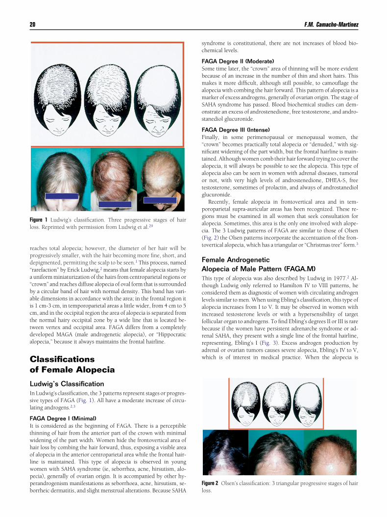
udwig’s Classificationn Ludwig’s classification, the 3 patterns represent stages or progres-ive types of FAGA (Fig. 1). All have a moderate increase of circu-ating androgens.2,3
AGA Degree I (Minimal)t is considered as the beginning of FAGA. There is a perceptiblehinning of hair from the anterior part of the crown with minimalidening of the part width. Women hide the frontovertical area ofair loss by combing the hair forward, thus, exposing a visible areaf alopecia in the anterior centroparietal area while the frontal hair-ine is maintained. This type of alopecia is observed in youngomen with SAHA syndrome (ie, seborrhea, acne, hirsutism, alo-ecia), generally of ovarian origin. It is accompanied by other hy-erandrogenism manifestations as seborrhoea, acne, hirsutism, se-
igure 1 Ludwig’s classification. Three progressive stages of haiross. Reprinted with permission from Ludwig et al.29
orrheic dermatitis, and slight menstrual alterations. Because SAHA l
yndrome is constitutional, there are not increases of blood bio-hemical levels.
AGA Degree II (Moderate)ome time later, the “crown” area of thinning will be more evidentecause of an increase in the number of thin and short hairs. Thisakes it more difficult, although still possible, to camouflage the
lopecia with combing the hair forward. This pattern of alopecia is aarker of excess androgens, generally of ovarian origin. The stage of
AHA syndrome has passed. Blood biochemical studies can dem-nstrate an excess of androstenedione, free testosterone, and andro-tanediol glucuronide.
AGA Degree III (Intense)inally, in some perimenopausal or menopausal women, thecrown” becomes practically total alopecia or “denuded,” with sig-ificant widening of the part width, but the frontal hairline is main-ained. Although women comb their hair forward trying to cover thelopecia, it will always be possible to see the alopecia. This type oflopecia also can be seen in women with adrenal diseases, tumoralr not, with very high levels of androstenedione, DHEA-S, freeestosterone, sometimes of prolactin, and always of androstanediollucuronide.
Recently, female alopecia in frontovertical area and in tem-oroparietal supra-auricular areas has been recognized. These re-ions must be examined in all women that seek consultation forlopecia. Sometimes, this area is the only one involved with alope-ia. The 3 Ludwig patterns of FAGA are similar to those of OlsenFig. 2) the Olsen patterns incorporate the accentuation of the fron-overtical alopecia, which has a triangular or “Christmas tree” form.3
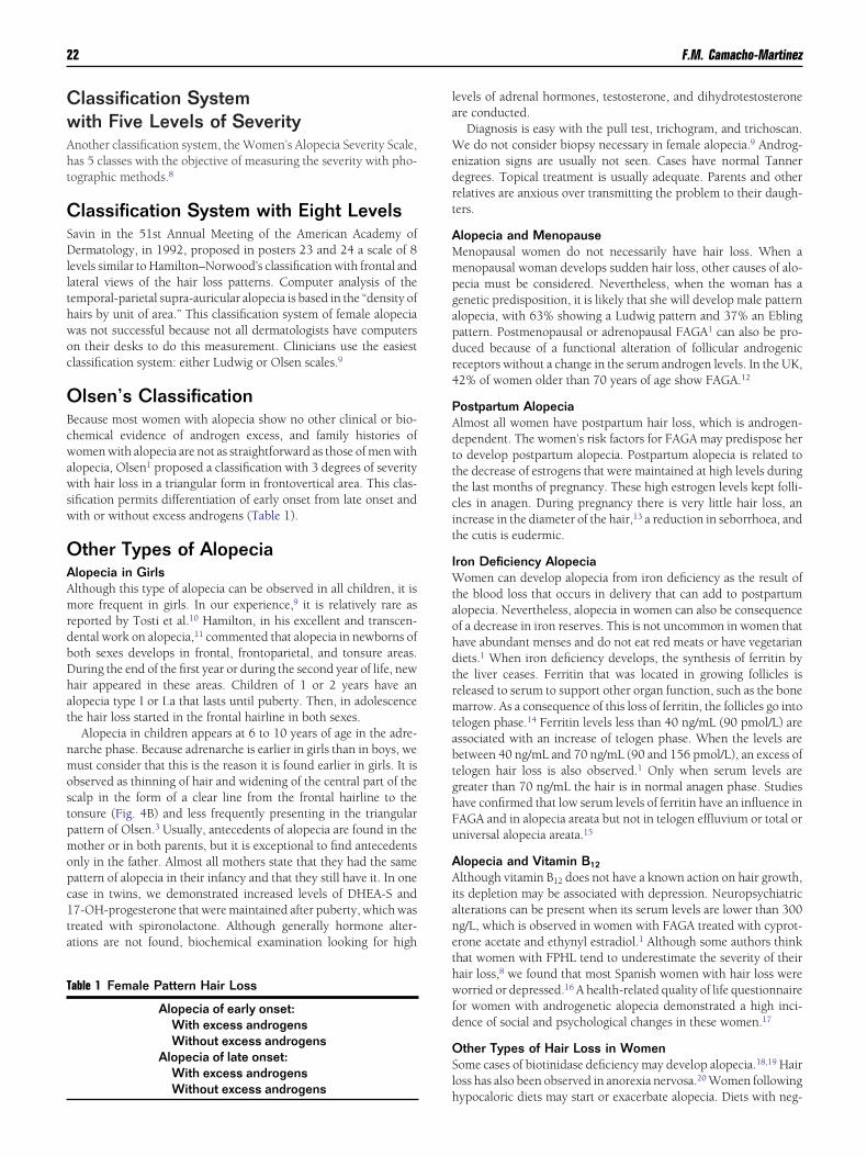
emale Androgeneticlopecia of Male Pattern (FAGA.M)his type of alopecia was also described by Ludwig in 1977.2 Al-
hough Ludwig only referred to Hamilton IV to VIII patterns, heonsidered them as diagnostic of women with circulating androgenevels similar to men. When using Ebling’s classification, this type oflopecia increases from I to V. It may be observed in women withncreased testosterone levels or with a hypersensibility of targetollicular organ to androgens. To find Ebling’s degrees II or III is rareecause if the women have persistent adrenarche syndrome or ad-enal SAHA, they present with a single line of the frontal hairline,epresenting, Ebling’s I (Fig. 3). Excess androgen production bydrenal or ovarian tumors causes severe alopecia, Ebling’s IV to V,hich is of interest in medical practice. When the alopecia is
igure 2 Olsen’s classification: 3 triangular progressive stages of hair
oss.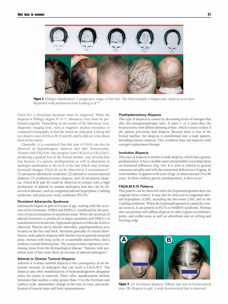
Fafdcnt
oWplah(acpss
PAteatolbatts
AApawhGfi
PTahtfie
ITpocsy
FTonCiapb
F
Hair loss in women 21
AGA.M.I, a functional alteration must be suspected. When thelopecia is Ebling’s degree IV or V, laboratory tests must be per-ormed urgently. Depending on the results of the laboratory tests,iagnostic imaging tests, such as magnetic nuclear resonance oromputed tomography to find the tumor are indicated. Ludwig didot observe cases of FAGA.M. II and III, and he did not write abouthem in his report.
Classically, it is considered that this type of FAGA can also bebserved in hypoestrogenic alopecia and after hysterectomy.omen with FAGA.M, who progress from FAGA.II to FAGA.M.V,
roducing a gradual loss of the frontal hairline, may develop haiross because of a genetic predisposition as well as alterations inndrogen metabolism at the level of the hair follicle and systemicormonal changes. FAGA.M can be observed in 4 circumstances4:1) persistent adrenarche syndrome; (2) adrenal or ovarian tumorallopecia; (3) posthysterectomy alopecia; and (4) involution alope-ia. FAGA.M.II and III could be observed in women with a highroduction of adrenal or ovarian androgens and also can be ob-erved in diseases, such as congenital adrenal hyperplasia, Cushingyndrome, and polycystic ovary syndrome (PCOS).
ersistent Adrenarche Syndromedrenarche begins in girls at 8 years of age, starting with the secre-
ion of the hormones, DHEA and DHEA-S, manifested by the pres-nce of microcomedones in acneiform areas. When the secretion ofdrenal hormones is produced in major quantities and DHEA-S isransformed in testosterone, hyperandrogenism at follicular levels isbserved. Patients show intense seborrhea, papulopustulous acneocated on the face and back, hirsutism generally of central distri-ution, male pattern alopecia with hairline loss in parietal-temporalreas, menses with long cycles or occasionally amenorrhea, and aendency toward thinning hair. The young woman experiences con-inuing stress from the dermatological disease.5 Patients with per-istent acne of late onset show an increase of adrenal androgens.6
drenal or Ovarian Tumoral Alopeciadrenal or ovarian tumoral alopecia is the consequence of an im-ortant increase of androgens that can reach a FAGA.M.V. Thelopecia and other manifestations of hyperandrogenism disappearhen the tumor is removed. These other manifestations includeirsutism that reaches a value greater than 15 in the Ferriman andallwey scale, amenorrhea, change in the tone of voice, and modi-
Figure 3 Ebling’s classification: 5 progressive stages of hReprinted with permission from Ludwig et al.29
cation of muscle mass and body measurements. a
osthysterectomy Alopeciahis type of alopecia is caused by decreasing levels of estrogen thatlter the estrogen/androgen ratio. It starts 1 or 2 years after theysterectomy with diffuse thinning of hair, which is more evident ifhe patient previously had alopecia. Because there is loss of therontal hairline, the alopecia is transformed into a male pattern,ncluding tonsure alopecia. This condition does not improve withstrogen replacement therapy.
nvolution Alopeciahis type of alopecia is similar to male alopecia, which has a geneticredisposition. It has a variable onset and probably is not dependentn hormonal influences (Fig. 4A). It is seen in relation to generalutaneous atrophy and with the nutritional deficiencies of aging. Inome families, it appears at 60 years of age, in others around 70 to 80ears. In those without genetic predisposition, it does occur.7
AGA.M.II-IV Patternshis pattern can be observed when the hyperandrogenism does notriginate from a tumor. It may also be observed in congenital adre-al hyperplasia (CAH), including the late-onset CAH, and in theushing syndrome. When the hyperandrogenism is caused by ovar-
an sources, it can present as PCOS or HAIRAN syndrome. Womanlso can present with diffuse alopecia in other regions as eyebrows,ubis, and axillar areas as well as seborrhoea and an itching andurning scalp.
igure 4 (A) Involution alopecia. Diffuse hair loss in frontovertical
. The final example is Hippocratic alopecia as in men.
air lossrea. (B) Alopecia in girl. A wide frontovertical line is observed.
CwAht
CSDllthwoc
OBcwawsw
OAAmrdbDhat
nmostpmopc1ta
la
Wedrt
AMmpgapdr4
PAdtttcit
IWtaohdtrmtabtghFu
AAianethwfd
OSl
T
22 F.M. Camacho-Martinez
lassification Systemith Five Levels of Severity
nother classification system, the Women’s Alopecia Severity Scale,as 5 classes with the objective of measuring the severity with pho-ographic methods.8
lassification System with Eight Levelsavin in the 51st Annual Meeting of the American Academy ofermatology, in 1992, proposed in posters 23 and 24 a scale of 8
evels similar to Hamilton–Norwood’s classification with frontal andateral views of the hair loss patterns. Computer analysis of theemporal-parietal supra-auricular alopecia is based in the “density ofairs by unit of area.” This classification system of female alopeciaas not successful because not all dermatologists have computersn their desks to do this measurement. Clinicians use the easiestlassification system: either Ludwig or Olsen scales.9
lsen’s Classificationecause most women with alopecia show no other clinical or bio-hemical evidence of androgen excess, and family histories ofomen with alopecia are not as straightforward as those of men with
lopecia, Olsen1 proposed a classification with 3 degrees of severityith hair loss in a triangular form in frontovertical area. This clas-
ification permits differentiation of early onset from late onset andith or without excess androgens (Table 1).
ther Types of Alopecialopecia in Girlslthough this type of alopecia can be observed in all children, it isore frequent in girls. In our experience,9 it is relatively rare as
eported by Tosti et al.10 Hamilton, in his excellent and transcen-ental work on alopecia,11 commented that alopecia in newborns ofoth sexes develops in frontal, frontoparietal, and tonsure areas.uring the end of the first year or during the second year of life, newair appeared in these areas. Children of 1 or 2 years have anlopecia type I or I.a that lasts until puberty. Then, in adolescencehe hair loss started in the frontal hairline in both sexes.
Alopecia in children appears at 6 to 10 years of age in the adre-arche phase. Because adrenarche is earlier in girls than in boys, weust consider that this is the reason it is found earlier in girls. It is
bserved as thinning of hair and widening of the central part of thecalp in the form of a clear line from the frontal hairline to theonsure (Fig. 4B) and less frequently presenting in the triangularattern of Olsen.3 Usually, antecedents of alopecia are found in theother or in both parents, but it is exceptional to find antecedents
nly in the father. Almost all mothers state that they had the sameattern of alopecia in their infancy and that they still have it. In onease in twins, we demonstrated increased levels of DHEA-S and7-OH-progesterone that were maintained after puberty, which wasreated with spironolactone. Although generally hormone alter-tions are not found, biochemical examination looking for high
able 1 Female Pattern Hair Loss
Alopecia of early onset:With excess androgensWithout excess androgens
Alopecia of late onset:With excess androgens
hWithout excess androgens
evels of adrenal hormones, testosterone, and dihydrotestosteronere conducted.
Diagnosis is easy with the pull test, trichogram, and trichoscan.e do not consider biopsy necessary in female alopecia.9 Androg-
nization signs are usually not seen. Cases have normal Tanneregrees. Topical treatment is usually adequate. Parents and otherelatives are anxious over transmitting the problem to their daugh-ers.
lopecia and Menopauseenopausal women do not necessarily have hair loss. When aenopausal woman develops sudden hair loss, other causes of alo-ecia must be considered. Nevertheless, when the woman has aenetic predisposition, it is likely that she will develop male patternlopecia, with 63% showing a Ludwig pattern and 37% an Eblingattern. Postmenopausal or adrenopausal FAGA1 can also be pro-uced because of a functional alteration of follicular androgeniceceptors without a change in the serum androgen levels. In the UK,2% of women older than 70 years of age show FAGA.12
ostpartum Alopecialmost all women have postpartum hair loss, which is androgen-ependent. The women’s risk factors for FAGA may predispose hero develop postpartum alopecia. Postpartum alopecia is related tohe decrease of estrogens that were maintained at high levels duringhe last months of pregnancy. These high estrogen levels kept folli-les in anagen. During pregnancy there is very little hair loss, anncrease in the diameter of the hair,13 a reduction in seborrhoea, andhe cutis is eudermic.
ron Deficiency Alopeciaomen can develop alopecia from iron deficiency as the result of
he blood loss that occurs in delivery that can add to postpartumlopecia. Nevertheless, alopecia in women can also be consequencef a decrease in iron reserves. This is not uncommon in women thatave abundant menses and do not eat red meats or have vegetarianiets.1 When iron deficiency develops, the synthesis of ferritin byhe liver ceases. Ferritin that was located in growing follicles iseleased to serum to support other organ function, such as the bonearrow. As a consequence of this loss of ferritin, the follicles go into
elogen phase.14 Ferritin levels less than 40 ng/mL (90 pmol/L) aressociated with an increase of telogen phase. When the levels areetween 40 ng/mL and 70 ng/mL (90 and 156 pmol/L), an excess ofelogen hair loss is also observed.1 Only when serum levels arereater than 70 ng/mL the hair is in normal anagen phase. Studiesave confirmed that low serum levels of ferritin have an influence inAGA and in alopecia areata but not in telogen effluvium or total orniversal alopecia areata.15
lopecia and Vitamin B12
lthough vitamin B12 does not have a known action on hair growth,ts depletion may be associated with depression. Neuropsychiatriclterations can be present when its serum levels are lower than 300g/L, which is observed in women with FAGA treated with cyprot-rone acetate and ethynyl estradiol.1 Although some authors thinkhat women with FPHL tend to underestimate the severity of theirair loss,8 we found that most Spanish women with hair loss wereorried or depressed.16 A health-related quality of life questionnaire
or women with androgenetic alopecia demonstrated a high inci-ence of social and psychological changes in these women.17
ther Types of Hair Loss in Womenome cases of biotinidase deficiency may develop alopecia.18,19 Haiross has also been observed in anorexia nervosa.20 Women following
ypocaloric diets may start or exacerbate alopecia. Diets with neg-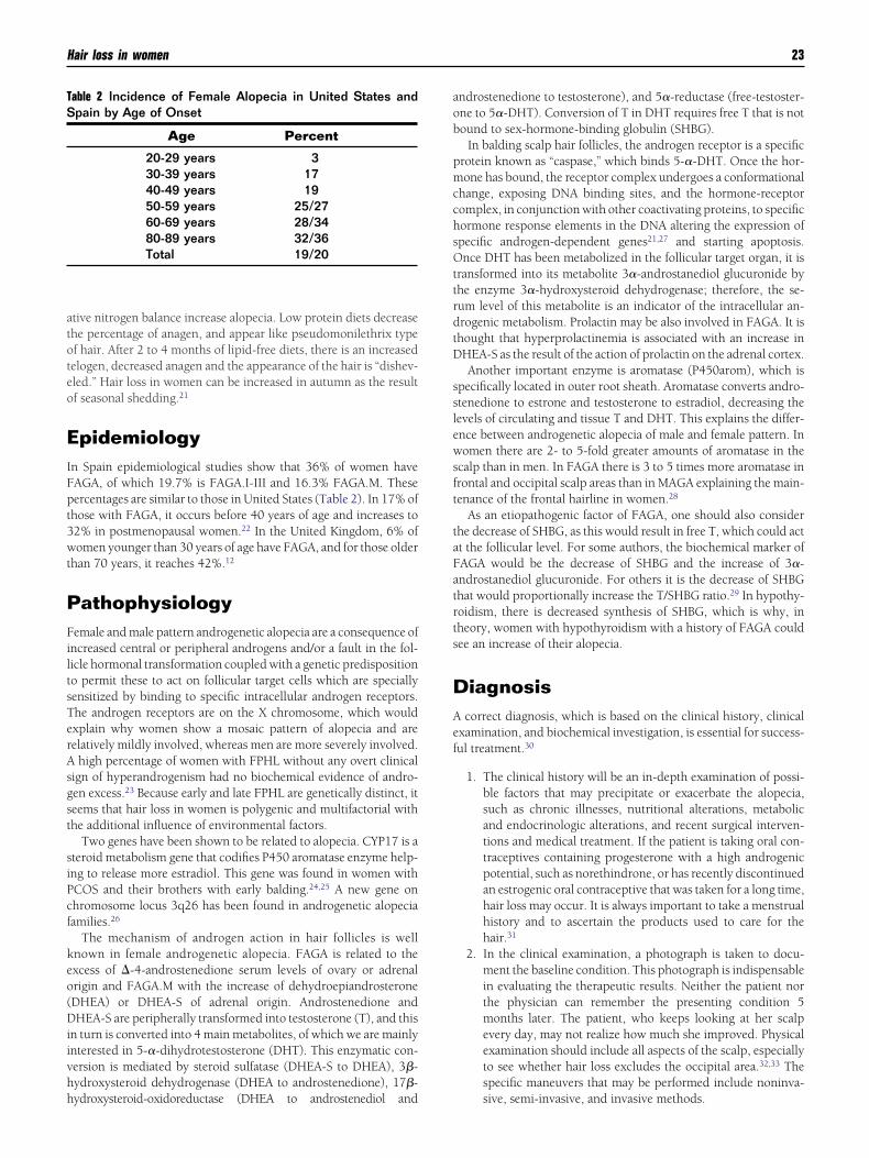
atoteo
EIFpt3wt
PFiltsTerAsgst
siPcf
keo(Diivhh
aob
pmcchsOttrdtD
sslewsft
taFatrts
DAef
TS
Hair loss in women 23
tive nitrogen balance increase alopecia. Low protein diets decreasehe percentage of anagen, and appear like pseudomonilethrix typef hair. After 2 to 4 months of lipid-free diets, there is an increasedelogen, decreased anagen and the appearance of the hair is “dishev-led.” Hair loss in women can be increased in autumn as the resultf seasonal shedding.21
pidemiologyn Spain epidemiological studies show that 36% of women haveAGA, of which 19.7% is FAGA.I-III and 16.3% FAGA.M. Theseercentages are similar to those in United States (Table 2). In 17% ofhose with FAGA, it occurs before 40 years of age and increases to2% in postmenopausal women.22 In the United Kingdom, 6% ofomen younger than 30 years of age have FAGA, and for those older
han 70 years, it reaches 42%.12
athophysiologyemale and male pattern androgenetic alopecia are a consequence of
ncreased central or peripheral androgens and/or a fault in the fol-icle hormonal transformation coupled with a genetic predispositiono permit these to act on follicular target cells which are speciallyensitized by binding to specific intracellular androgen receptors.he androgen receptors are on the X chromosome, which wouldxplain why women show a mosaic pattern of alopecia and areelatively mildly involved, whereas men are more severely involved.
high percentage of women with FPHL without any overt clinicalign of hyperandrogenism had no biochemical evidence of andro-en excess.23 Because early and late FPHL are genetically distinct, iteems that hair loss in women is polygenic and multifactorial withhe additional influence of environmental factors.
Two genes have been shown to be related to alopecia. CYP17 is ateroid metabolism gene that codifies P450 aromatase enzyme help-ng to release more estradiol. This gene was found in women withCOS and their brothers with early balding.24,25 A new gene onhromosome locus 3q26 has been found in androgenetic alopeciaamilies.26
The mechanism of androgen action in hair follicles is wellnown in female androgenetic alopecia. FAGA is related to thexcess of �-4-androstenedione serum levels of ovary or adrenalrigin and FAGA.M with the increase of dehydroepiandrosteroneDHEA) or DHEA-S of adrenal origin. Androstenedione andHEA-S are peripherally transformed into testosterone (T), and this
n turn is converted into 4 main metabolites, of which we are mainlynterested in 5-�-dihydrotestosterone (DHT). This enzymatic con-ersion is mediated by steroid sulfatase (DHEA-S to DHEA), 3�-ydroxysteroid dehydrogenase (DHEA to androstenedione), 17�-
able 2 Incidence of Female Alopecia in United States andpain by Age of Onset
Age Percent
20-29 years 330-39 years 1740-49 years 1950-59 years 25/2760-69 years 28/3480-89 years 32/36Total 19/20
ydroxysteroid-oxidoreductase (DHEA to androstenediol and
ndrostenedione to testosterone), and 5�-reductase (free-testoster-ne to 5�-DHT). Conversion of T in DHT requires free T that is notound to sex-hormone-binding globulin (SHBG).
In balding scalp hair follicles, the androgen receptor is a specificrotein known as “caspase,” which binds 5-�-DHT. Once the hor-one has bound, the receptor complex undergoes a conformational
hange, exposing DNA binding sites, and the hormone-receptoromplex, in conjunction with other coactivating proteins, to specificormone response elements in the DNA altering the expression ofpecific androgen-dependent genes21,27 and starting apoptosis.nce DHT has been metabolized in the follicular target organ, it is
ransformed into its metabolite 3�-androstanediol glucuronide byhe enzyme 3�-hydroxysteroid dehydrogenase; therefore, the se-um level of this metabolite is an indicator of the intracellular an-rogenic metabolism. Prolactin may be also involved in FAGA. It ishought that hyperprolactinemia is associated with an increase inHEA-S as the result of the action of prolactin on the adrenal cortex.Another important enzyme is aromatase (P450arom), which is
pecifically located in outer root sheath. Aromatase converts andro-tenedione to estrone and testosterone to estradiol, decreasing theevels of circulating and tissue T and DHT. This explains the differ-nce between androgenetic alopecia of male and female pattern. Inomen there are 2- to 5-fold greater amounts of aromatase in the
calp than in men. In FAGA there is 3 to 5 times more aromatase inrontal and occipital scalp areas than in MAGA explaining the main-enance of the frontal hairline in women.28
As an etiopathogenic factor of FAGA, one should also considerhe decrease of SHBG, as this would result in free T, which could actt the follicular level. For some authors, the biochemical marker ofAGA would be the decrease of SHBG and the increase of 3�-ndrostanediol glucuronide. For others it is the decrease of SHBGhat would proportionally increase the T/SHBG ratio.29 In hypothy-oidism, there is decreased synthesis of SHBG, which is why, inheory, women with hypothyroidism with a history of FAGA couldee an increase of their alopecia.
iagnosiscorrect diagnosis, which is based on the clinical history, clinical
xamination, and biochemical investigation, is essential for success-ul treatment.30
1. The clinical history will be an in-depth examination of possi-ble factors that may precipitate or exacerbate the alopecia,such as chronic illnesses, nutritional alterations, metabolicand endocrinologic alterations, and recent surgical interven-tions and medical treatment. If the patient is taking oral con-traceptives containing progesterone with a high androgenicpotential, such as norethindrone, or has recently discontinuedan estrogenic oral contraceptive that was taken for a long time,hair loss may occur. It is always important to take a menstrualhistory and to ascertain the products used to care for thehair.31
2. In the clinical examination, a photograph is taken to docu-ment the baseline condition. This photograph is indispensablein evaluating the therapeutic results. Neither the patient northe physician can remember the presenting condition 5months later. The patient, who keeps looking at her scalpevery day, may not realize how much she improved. Physicalexamination should include all aspects of the scalp, especiallyto see whether hair loss excludes the occipital area.32,33 Thespecific maneuvers that may be performed include noninva-
sive, semi-invasive, and invasive methods.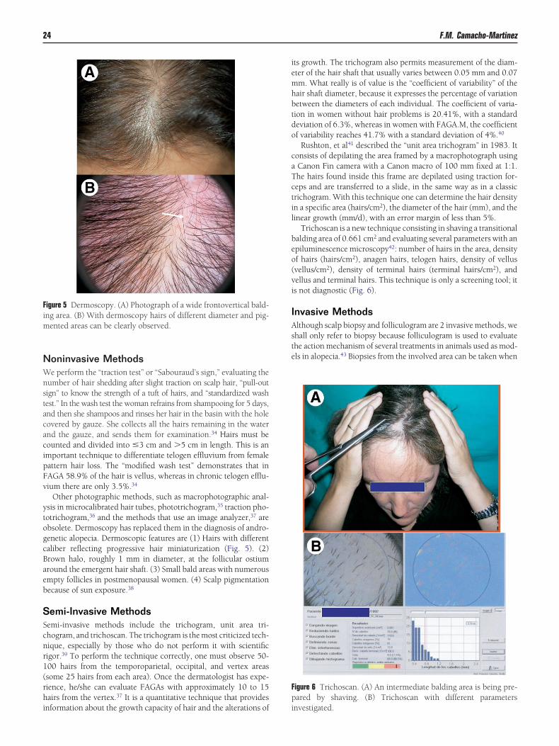
NWnstacacipFv
ytogcBaeb
SScnr1(rhi
iemhbtdo
caTctil
beo(vi
IAste
Fp
Fim
24 F.M. Camacho-Martinez
oninvasive Methodse perform the “traction test” or “Sabouraud’s sign,” evaluating the
umber of hair shedding after slight traction on scalp hair, “pull-outign” to know the strength of a tuft of hairs, and “standardized washest.” In the wash test the woman refrains from shampooing for 5 days,nd then she shampoos and rinses her hair in the basin with the holeovered by gauze. She collects all the hairs remaining in the waternd the gauze, and sends them for examination.34 Hairs must beounted and divided into �3 cm and �5 cm in length. This is anmportant technique to differentiate telogen effluvium from femaleattern hair loss. The “modified wash test” demonstrates that inAGA 58.9% of the hair is vellus, whereas in chronic telogen efflu-ium there are only 3.5%.34
Other photographic methods, such as macrophotographic anal-sis in microcalibrated hair tubes, phototrichogram,35 traction pho-otrichogram,36 and the methods that use an image analyzer,37 arebsolete. Dermoscopy has replaced them in the diagnosis of andro-enetic alopecia. Dermoscopic features are (1) Hairs with differentaliber reflecting progressive hair miniaturization (Fig. 5). (2)rown halo, roughly 1 mm in diameter, at the follicular ostiumround the emergent hair shaft. (3) Small bald areas with numerousmpty follicles in postmenopausal women. (4) Scalp pigmentationecause of sun exposure.38
emi-Invasive Methodsemi-invasive methods include the trichogram, unit area tri-hogram, and trichoscan. The trichogram is the most criticized tech-ique, especially by those who do not perform it with scientificigor.39 To perform the technique correctly, one must observe 50-00 hairs from the temporoparietal, occipital, and vertex areassome 25 hairs from each area). Once the dermatologist has expe-ience, he/she can evaluate FAGAs with approximately 10 to 15airs from the vertex.37 It is a quantitative technique that provides
igure 5 Dermoscopy. (A) Photograph of a wide frontovertical bald-ng area. (B) With dermoscopy hairs of different diameter and pig-
ented areas can be clearly observed.
nformation about the growth capacity of hair and the alterations of i
ts growth. The trichogram also permits measurement of the diam-ter of the hair shaft that usually varies between 0.05 mm and 0.07m. What really is of value is the “coefficient of variability” of theair shaft diameter, because it expresses the percentage of variationetween the diameters of each individual. The coefficient of varia-ion in women without hair problems is 20.41%, with a standardeviation of 6.3%, whereas in women with FAGA.M, the coefficientf variability reaches 41.7% with a standard deviation of 4%.40
Rushton, et al41 described the “unit area trichogram” in 1983. Itonsists of depilating the area framed by a macrophotograph usingCanon Fin camera with a Canon macro of 100 mm fixed at 1:1.he hairs found inside this frame are depilated using traction for-eps and are transferred to a slide, in the same way as in a classicrichogram. With this technique one can determine the hair densityn a specific area (hairs/cm2), the diameter of the hair (mm), and theinear growth (mm/d), with an error margin of less than 5%.
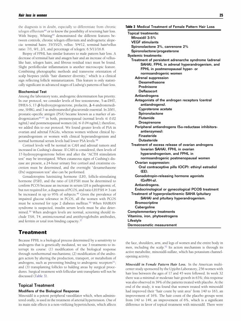
Trichoscan is a new technique consisting in shaving a transitionalalding area of 0.661 cm2 and evaluating several parameters with anpiluminescence microscopy42: number of hairs in the area, densityf hairs (hairs/cm2), anagen hairs, telogen hairs, density of vellusvellus/cm2), density of terminal hairs (terminal hairs/cm2), andellus and terminal hairs. This technique is only a screening tool; its not diagnostic (Fig. 6).
nvasive Methodslthough scalp biopsy and folliculogram are 2 invasive methods, wehall only refer to biopsy because folliculogram is used to evaluatehe action mechanism of several treatments in animals used as mod-ls in alopecia.43 Biopsies from the involved area can be taken when
igure 6 Trichoscan. (A) An intermediate balding area is being pre-ared by shaving. (B) Trichoscan with different parameters
nvestigated.
ttWtcr
dlSCssc
BAIDopdnwopn
i1tec(
hcbbimsmca
TBattgaadd
TMMti
tmao
Mchtwehif
T
Hair loss in women 25
he diagnosis is in doubt, especially to differentiate from chronicelogen effluvium44 or to know the possibility of reversing hair loss.
ith biopsy, Whiting44 demonstrated the different features be-ween controls, chronic telogen effluvium and androgenetic alope-ia: terminal hairs: 35/35/25, vellus: 5/4/12, terminal hair/vellusatio: 7/1, 9/1, 2/1, and percentage of telogen: 6.5/11/16.8.
Biopsy of FPHL has similar features to male pattern hair loss. Aecrease of terminal hair and anagen hair and an increase of vellus-
ike hair, telogen hairs, and fibrous residual tract must be found.light perifollicular inflammation is another microscopic feature.ombining photographic methods and transverse orientation of
calp biopsies yields “hair diameter diversity,” which is a clinicalign reflecting follicle miniaturization. This feature is only statisti-ally significant in advanced stages of Ludwig’s patterns of hair loss.
iochemical Testmong the laboratory tests, androgenic determination has priority.
n our protocol, we consider levels of free testosterone, 5-�-DHT,HEA-S, 17-�-hydroxyprogesterone, prolactin, �-4-androstenedi-ne, SHBG, and 3-�-androstanediol glucuronide essential. In 2001,rostatic-specific antigen (PSA) became known as a marker of an-rogenization45-47 in both, premenopausal (normal levels � 0.02g/mL) and postmenopausal women (nL � 0.04 ng/mL); therefore,e added this to our protocol. We found greater levels of PSA invarian and adrenal FAGAs, whereas women without clinical hy-erandrogenism or women with clinical hyperandrogenism andormal hormonal serum levels had lower PSA levels.48
Cortisol levels will be normal in CAH and adrenal tumors andncreased in Cushing’s disease. If CAH is considered, then levels of7-hydroxyprogesterone before and after the “ACTH stimulationest” may be investigated. When cutaneous signs of Cushing’s dis-ase are present, a 24-hour urinary free cortisol and creatinine ex-retion must be determined, and the overnight “dexamethasoneDx) suppression test” also can be performed.
Gonadotropins luteinizing hormone (LH), follicle-stimulatingormone (FSH), and the ratio of LH:FSH must be determined toonfirm PCOS because an increase in serum LH is pathognomic of,ut not required for, a diagnosis of PCOS, and ratio LH:FSH � 3 cane increased in up to 95% of subjects.49 Given the prevalence of
mpaired glucose tolerance in PCOS, all the women with PCOSust be screened for type 2 diabetes mellitus.50 When HAIRAN
yndrome is suspected, insulin serum levels must be also deter-ined.50 When androgen levels are normal, screening should in-
lude TSH, T4, antimicrosomal and antithyroglobulin antibodies,nd ferritin or total iron binding capacity.37
reatmentecause FPHL is a biological process determined by a sensitivity tondrogens that is genetically mediated, we use 3 treatments to in-errupt its course: (1) modification of the biological responsehrough nonhormonal mechanisms; (2) modification of the andro-en action by altering the production, transport, or metabolism ofndrogens, such as preventing binding to androgenic receptors51;nd (3) transplanting follicles to balding areas by surgical proce-ures. Surgical treatment with follicular unit transplants will not beiscussed (Table 3).
opical Treatmentodifiers of the Biological Responseinoxidil is a potent peripheral vasodilator which, when adminis-
ered orally, is used in the treatment of arterial hypertension. One of
ts main side effects is a non-virilizing hypertrichosis, which affects dhe face, shoulders, arm, and legs of women and the entire body inen, including the scalp.52 Its action mechanism is through its
ctive metabolite, minoxidil-sulfate, which has potassium channel-pening activity.
inoxidil in Female Pattern Hair Loss. In the American multi-enter study sponsored by the Upjohn Laboratory, 256 women withair loss between the ages of 17 and 45 were followed. In week 32here was a minimal or moderate hair growth in 63%; this responseas also observed in 39% of the patients treated with placebo. At the
nd of the study, it was found that women treated with minoxidilad improved their “hair count by unit area” from 140 to 163, an
mprovement of 16%. The hair count of the placebo groups wentrom 140 to 149, an improvement of 6%, which is a significant
able 3 Medical Treatment of Female Pattern Hair Loss
Topical treatments:Minoxidil 3-5%VEGF stimulantsSpironolactone 3%, canrenone 2%Spironolactone/progesterone
Systemic treatments:Treatment of persistent adrenarche syndrome (adrenal
SAHA), FPHL in adrenal hyperandrogenism, andFPHL in postmenopausal hyper- ornormoandrogenic women
Adrenal suppressionDexamethasonePrednisoneDeflazacort
AntiandrogensAntagonists of the androgen receptors (central
antiandrogens).Cyproterone acetateSpironolactoneFlutamideDrospirenone
Peripheral antiandrogens (5�-reductase inhibitors-antienzymes):
FinasterideDutasteride
Treatment of excess release of ovarian androgens(ovarian SAHA), FPHL in ovarianhyperandrogenism, and FPHL innormoandrogenic postmenopausal women
Ovarian suppression:Oral contraceptive pills (OCP): ethinyl estradiol(EE).
Gonadotropin-releasing hormone agonists(GnRH-a).
Antiandrogens.Endocrinological or gynecological PCOS treatment
Treatment of hyperprolactinemic SAHA (pituitarySAHA) and pituitary hyperandrogenism.
BromocriptineCabergoline
Complemmentary treatmentsVitamins, iron, phytoestrogensLifestyleDermocosmetic measurement
ifference in favor of topical treatment with minoxidil. There were
niwttmf
vbmtns6da7
OBMmwcomrmf2tmie
Vwtnpsmhahr
Otatbftmevi1ag
STsFg
TSAPTppu
Acda0cfdpmDsdTwtda
Attbac
Awiofp
Fs1
26 F.M. Camacho-Martinez
o side effects. The Food and Drug Administration approved its usen women in 1991.52 Jacobs, et al53 confirmed that FPHL treatedith 2% minoxidil improved their initial count by 44%, whereas
hose treated with a placebo only did so in 29%. After 32 weeks ofreatment, DeVillez et al54 concluded that topical minoxidil treat-ent in women is superior to the treatment with placebo when they
ound moderate growth in 13% and discrete growth in 50% of cases.Although there is a formulation of 5% topical minoxidil in a foam
ehicle, we prefer using 2% or 3% concentrations twice daily55
ecause at these concentrations it presents fewer side effects. Theost frequent side effects are facial hypertrichosis, irritation derma-
itis, contact eczema, pruritus, scaling, dryness, and headaches. Mi-oxidil is not recommended during pregnancy or lactation. Sometart the treatment of female pattern hair loss with 5% minoxidil formonths and then continue with 2% or 3% concentrations. It is therug of choice in normoandrogenic premenopausal women. It islso useful in postmenopausal women with spectacular results (Fig.A and 7B).
ther Modifiers of theiological Response Used Locallyany attempts have been made to use different biological responseodifiers, but none of these have exceeded the effects of minoxidil,hich is why they have not been commercialized. Minoxidil asso-
iated with tretinoin stimulated the growth of terminal hairs in 66%f the subjects. These results are similar or superior to those ofinoxidil alone. Tretinoin increases the amount of minoxidil that
eaches the follicle.56 Another study demonstrated that when 2%inoxidil is associated with 0.05% tretinoin, the absorption of the
ormer is 3 times greater.57 The combination of 0.01% tretinoin with% or 3% minoxidil achieves an acceptable response in 53.2% ofhose treated.58 The chance of side effects, especially irritation der-atitis, is greater with this combination. Other modifiers of biolog-
cal response, such as diazoxide, viprostol, or cyclosporin, haveffects lower than those of minoxidil.
ascular Endothelial Growth Factor Stimulants. Our experienceith eighty normoandrogenic women first treated with 1% alpha-
ocopherol nicotinate for 3 months, followed with 5% minoxidil atight and 3% in the morning versus 80 men treated with the samerocedure demonstrated that 25% of women and 10% of menhowed an increased vascular supply.52 Although it is clear thatinoxidil up-regulates the expression of VEGF 6-fold in human
air dermal papilla cells, the effect is dose related. VEGF increasesngiogenesis which produces hair growth, and increases the size ofair and follicles.59 We have no explanation for the difference in theesponse of women and men.
igure 7 Normoandrogenic postmenopausal woman treated exclu-ively with 3% minoxidil twice a day. (A) Before treatment. (B) After
Tyear of treatment.
ther Local Therapeutics. Twice-daily applications of 1% to 5%incture of progesterone are useful in women. It should not be usedt concentrations greater than 2% or in amounts greater than 1 mLwice a day because it may cause menstrual alterations. Althoughoth 3% spironolactone and its metabolite 2% canrenone are usefulor the topical treatment of FPHL, we prefer to use 0.025% proges-erone with 0.05% spironolactone because they appear to comple-ent each other synergistically with a greater effect on FPHL than
ither alone.60 In menopausal women, a solution of 0.03% estradiolalerate used during 12 weeks and 24 weeks has demonstratedmprovement of anagen/telogen ratio and a decrease in hair loss at2 weeks and 24 weeks.61 Finally, 0.05% topical finasteride showed40% decrease of DHT serum levels, but does not increase the hairrowth.62
ystemic Treatmenthe treatment of hyperandrogenic women with their different pos-ibilities of androgen origin and normoandrogenic women withPHL is the same that we used in other dermatological hyperandro-enic diseases, such as hirsutism.50,63,64
reatment of Persistent Adrenarcheyndrome (Adrenal SAHA) and FPHL indrenal Hyperandrogenism and FPHL inostmenopausal Hyper- or Normoandrogenic Womenwo types of drugs must be used, corticosteroids for adrenal sup-ression and antiandrogens, central or peripherals, to avoid theroduction of adrenal androgens or their effects on the target follic-lar organ. In postmenopausal women, hair loss has a male pattern.
drenal Suppression. Adrenal suppression is achieved with glu-ocorticosteroids. In the past, we used dexamethasone at an initialose of 0.5 mg every night for 3 months and then alternate nights fornother 3 months. If the dexamethasone doses were greater than.75 mg daily and continued for 6 months or longer, Cushingoidhanges could be observed.50 Prednisone at a dose of 7.5 mg dailyor 2 months, reduced to 5 mg daily for 2 months, and then 2.5 mgaily for 2 months or 6 months of treatment is an alternative. Atresent we use deflazacort at an initial dose of 30 mg daily for 1onth with a maintenance dose of 6 mg daily for up to 2 years.eflazacort has the advantage that at this dose it does not produce
ide effects. These doses of glucocorticosteroids are enough to re-uce the level of DHEA-S, �-4-androstenedione and testosterone.63
he only secondary effect is that obese women tend to gain moreeight. Adrenal hyperplasia is treated with substitute corticosteroid
herapy, regardless of the enzymatic deficiency. Cushing’s syn-rome benefits from substitute therapy with corticosteroids associ-ted with surgery and/or irradiation.64
ntiandrogenic Therapy. Antiandrogenic therapy includes cypro-erone acetate (CA), spironolactone, drospirenone, flutamide, finas-eride, and dutasteride. Central antiandrogens competitively inhibitinding of 5-�-DHT to the androgen receptor,49 and peripheralntiandrogens acts by inhibiting the 5-�-reductase, blocking theonversion of testosterone to 5-�-DHT.65
ntagonists of the Androgen Receptors. CA acts by interferingith the binding of 5-�-DHT to the androgen receptor and by
nhibiting the secretion of FSH and LH as the result of its progester-ne action. The “Hammerstein schedule” is 50 to 100 mg/d of CArom the fifth to the 15th day of the menstrual cycle for a 6-montheriod, which is the time of the glucocorticosteroid suppression.
he 2 mg/d is given from the first day of the cycle to the 21st, with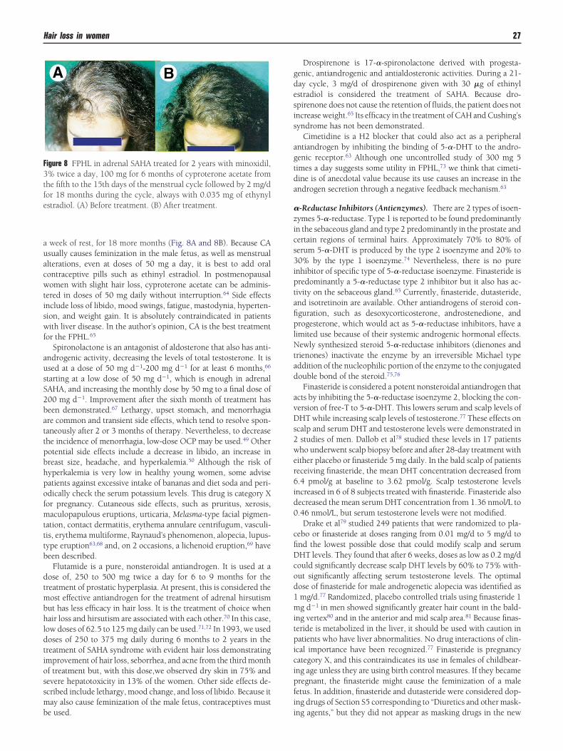
auacwtiswf
ausS2battpbhpofmtttb
dtmbhldtiossmb
gdesis
agtda
�zics3iptafiplNtad
avDs2wer6id0
cfiDcod1mitpicipfi
F3tfe
Hair loss in women 27
week of rest, for 18 more months (Fig. 8A and 8B). Because CAsually causes feminization in the male fetus, as well as menstruallterations, even at doses of 50 mg a day, it is best to add oralontraceptive pills such as ethinyl estradiol. In postmenopausalomen with slight hair loss, cyproterone acetate can be adminis-
ered in doses of 50 mg daily without interruption.64 Side effectsnclude loss of libido, mood swings, fatigue, mastodynia, hyperten-ion, and weight gain. It is absolutely contraindicated in patientsith liver disease. In the author’s opinion, CA is the best treatment
or the FPHL.65
Spironolactone is an antagonist of aldosterone that also has anti-ndrogenic activity, decreasing the levels of total testosterone. It issed at a dose of 50 mg d�1-200 mg d�1 for at least 6 months,66
tarting at a low dose of 50 mg d�1, which is enough in adrenalAHA, and increasing the monthly dose by 50 mg to a final dose of00 mg d�1. Improvement after the sixth month of treatment haseen demonstrated.67 Lethargy, upset stomach, and menorrhagiare common and transient side effects, which tend to resolve spon-aneously after 2 or 3 months of therapy. Nevertheless, to decreasehe incidence of menorrhagia, low-dose OCP may be used.49 Otherotential side effects include a decrease in libido, an increase inreast size, headache, and hyperkalemia.50 Although the risk ofyperkalemia is very low in healthy young women, some adviseatients against excessive intake of bananas and diet soda and peri-dically check the serum potassium levels. This drug is category Xor pregnancy. Cutaneous side effects, such as pruritus, xerosis,
aculopapulous eruptions, urticaria, Melasma-type facial pigmen-ation, contact dermatitis, erythema annulare centrifugum, vasculi-is, erythema multiforme, Raynaud’s phenomenon, alopecia, lupus-ype eruption63,68 and, on 2 occasions, a lichenoid eruption,69 haveeen described.
Flutamide is a pure, nonsteroidal antiandrogen. It is used at aose of, 250 to 500 mg twice a day for 6 to 9 months for thereatment of prostatic hyperplasia. At present, this is considered theost effective antiandrogen for the treatment of adrenal hirsutism
ut has less efficacy in hair loss. It is the treatment of choice whenair loss and hirsutism are associated with each other.70 In this case,
ow doses of 62.5 to 125 mg daily can be used.71,72 In 1993, we usedoses of 250 to 375 mg daily during 6 months to 2 years in thereatment of SAHA syndrome with evident hair loss demonstratingmprovement of hair loss, seborrhea, and acne from the third monthf treatment but, with this dose,we observed dry skin in 75% andevere hepatotoxicity in 13% of the women. Other side effects de-cribed include lethargy, mood change, and loss of libido. Because itay also cause feminization of the male fetus, contraceptives must
igure 8 FPHL in adrenal SAHA treated for 2 years with minoxidil,% twice a day, 100 mg for 6 months of cyproterone acetate fromhe fifth to the 15th days of the menstrual cycle followed by 2 mg/dor 18 months during the cycle, always with 0.035 mg of ethynylstradiol. (A) Before treatment. (B) After treatment.
e used. i
Drospirenone is 17-�-spironolactone derived with progesta-enic, antiandrogenic and antialdosteronic activities. During a 21-ay cycle, 3 mg/d of drospirenone given with 30 �g of ethinylstradiol is considered the treatment of SAHA. Because dro-pirenone does not cause the retention of fluids, the patient does notncrease weight.65 Its efficacy in the treatment of CAH and Cushing’syndrome has not been demonstrated.
Cimetidine is a H2 blocker that could also act as a peripheralntiandrogen by inhibiting the binding of 5-�-DHT to the andro-enic receptor.63 Although one uncontrolled study of 300 mg 5imes a day suggests some utility in FPHL,73 we think that cimeti-ine is of anecdotal value because its use causes an increase in thendrogen secretion through a negative feedback mechanism.63
-Reductase Inhibitors (Antienzymes). There are 2 types of isoen-ymes 5-�-reductase. Type 1 is reported to be found predominantlyn the sebaceous gland and type 2 predominantly in the prostate andertain regions of terminal hairs. Approximately 70% to 80% oferum 5-�-DHT is produced by the type 2 isoenzyme and 20% to0% by the type 1 isoenzyme.74 Nevertheless, there is no pure
nhibitor of specific type of 5-�-reductase isoenzyme. Finasteride isredominantly a 5-�-reductase type 2 inhibitor but it also has ac-ivity on the sebaceous gland.65 Currently, finasteride, dutasteride,nd isotretinoin are available. Other antiandrogens of steroid con-guration, such as desoxycorticosterone, androstenedione, androgesterone, which would act as 5-�-reductase inhibitors, have a
imited use because of their systemic androgenic hormonal effects.ewly synthesized steroid 5-�-reductase inhibitors (dienones and
rienones) inactivate the enzyme by an irreversible Michael typeddition of the nucleophilic portion of the enzyme to the conjugatedouble bond of the steroid.75,76
Finasteride is considered a potent nonsteroidal antiandrogen thatcts by inhibiting the 5-�-reductase isoenzyme 2, blocking the con-ersion of free-T to 5-�-DHT. This lowers serum and scalp levels ofHT while increasing scalp levels of testosterone.77 These effects on
calp and serum DHT and testosterone levels were demonstrated instudies of men. Dallob et al78 studied these levels in 17 patientsho underwent scalp biopsy before and after 28-day treatment with
ither placebo or finasteride 5 mg daily. In the bald scalp of patientseceiving finasteride, the mean DHT concentration decreased from.4 pmol/g at baseline to 3.62 pmol/g. Scalp testosterone levels
ncreased in 6 of 8 subjects treated with finasteride. Finasteride alsoecreased the mean serum DHT concentration from 1.36 nmol/L to.46 nmol/L, but serum testosterone levels were not modified.
Drake et al79 studied 249 patients that were randomized to pla-ebo or finasteride at doses ranging from 0.01 mg/d to 5 mg/d tond the lowest possible dose that could modify scalp and serumHT levels. They found that after 6 weeks, doses as low as 0.2 mg/dould significantly decrease scalp DHT levels by 60% to 75% with-ut significantly affecting serum testosterone levels. The optimalose of finasteride for male androgenetic alopecia was identified asmg/d.77 Randomized, placebo controlled trials using finasteride 1g d�1 in men showed significantly greater hair count in the bald-
ng vertex80 and in the anterior and mid scalp area.81 Because finas-eride is metabolized in the liver, it should be used with caution inatients who have liver abnormalities. No drug interactions of clin-
cal importance have been recognized.77 Finasteride is pregnancyategory X, and this contraindicates its use in females of childbear-ng age unless they are using birth control measures. If they becameregnant, the finasteride might cause the feminization of a maleetus. In addition, finasteride and dutasteride were considered dop-ng drugs of Section S5 corresponding to “Diuretics and other mask-
ng agents,” but they did not appear as masking drugs in the new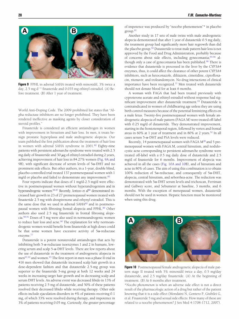
Wprs
wntipma9ppm
thcfitpactdbe
ietm4dswsprem3
og
attaoteeicis
cncbadwsac
mctmaa1adamsw
Fdf
Ftdt*rke
28 F.M. Camacho-Martinez
orld Anti-Doping Code. The 2009 prohibited list states that “Al-ha reductase inhibitors are no longer prohibited. They have beenendered ineffective as masking agents by closer consideration ofteroid profiles.”
Finasteride is considered an efficient antiandrogen in womenith improvement in hirsutism and hair loss. In men, it treats be-ign prostatic hyperplasia and male androgenetic alopecia. Ouream published the first publication about the treatment of hair lossn women with adrenal SAHA syndrome in 2001.82 Eighty-nineatients with persistent adrenarche syndrome were treated with 2.5g daily of finasteride and 30 �g of ethinyl estradiol during 2 years,
chieving improvement of hair loss in 84.27% women (Fig. 9A andB), with significant decrease of serum levels of 5�-DHT and norominent side effects. But other authors, in a 1-year, double blind,lacebo-controlled trial treated 137 postmenopausal women with 1g/d or placebo and failed to demonstrate any improvement.83
Four reports indicate that doses of 1 mg/d-2.5 mg/d were effec-ive in postmenopausal women without hyperandrogenism and inyperandrogenic women.84-87 Recently, Iorizzo et al88 demonstrated in-reased hair growth in 23 of 27 premenopausal women treated withnasteride 2.5 mg with drospirenone and ethynyl estradiol. This ishe same dose that we used in adrenal SAHA82 and in postmeno-ausal women with fibrosing frontal alopecia and FPHL.89 Otheruthors also used 2.5 mg finasteride in frontal fibrosing alope-ia.90,91 Doses of 5 mg were also used in normoandrogenic womeno reduce hair loss and acne.90 The explanation for why normoan-rogenic women would benefit from finasteride at high doses coulde that some women have excessive activity of 5�-reductasenzyme.77
Dutasteride is a potent nonsteroidal antiandrogen that acts bynhibiting both 5-�-reductase isoenzymes 1 and 2 in humans, low-ring serum and scalp 5-�-DHT levels. There are few reports abouthe use of dutasteride in the treatment of androgenetic alopecia inen91,92 and women.93 The first report in men was a phase II trial in
16 men showed that dutasteride increased scalp hair growth in aose-dependent fashion and that dutasteride 2.5-mg group wasuperior to the finasteride 5-mg group at both 12 weeks and 24eeks in increasing target hair growth and in decreasing scalp and
erum DHT levels. An adverse event was decreased libido in 13% ofatients receiving 2.5 mg of dutasteride, and 50% of these patientsesolved their decreased libido while receiving therapy. Other sideffects include ejaculation disorders in 4% of patients receiving 0.1g, of which 33% were resolved during therapy, and impotence in
igure 9 FPHL in adrenal SAHA treated with minoxidil, 3% twice aay, 2.5 mg d�1 finasteride and 0.035 mg ethinyl estradiol. (A) Be-ore treatment. (B) After 1 year of treatment.
% of patients receiving 0.05 mg. Curiously, the greater percentage r
f impotence was produced by “nocebo phenomenon”* in placeboroup.91
Another study in 17 sets of male twins with male androgeneticlopecia demonstrated that after 1 year of dutasteride 0.5 mg daily,he treatment group had significantly more hair regrowth than didhe placebo group.92 Dutasteride to treat male pattern hair loss is notpproved by the Food and Drug Administration, probably becausef concerns about side effects, including gynecomastia;94-96 al-hough only a case of gynecomastia has been published.96 There isvidence that dutasteride is processed in the liver by the CYP3A4nzymes, thus, it could affect the clearance of other potent CYP3A4nhibitors, such as ketoconazole, diltiazem, cimetidine, ciprofloxa-in, ritonavir, and troleandomycin. No drug interactions of clinicalmportance have been recognized.77 Men treated with dutasteridehould not donate blood for at least 6 months.
A woman with FAGA that had been treated previously withyproterone acetate and ethinyl estradiol without response had sig-ificant improvement after dutasteride treatment.93 Dutasteride isontraindicated in women of childbearing age unless they are usingirth control measures because of the potential feminizing effects onmale fetus. Twenty-five postmenopausal women with female an-rogenetic alopecia of male pattern (FAGA.M) were treated off-labelith 0.25 mg/d of dutasteride. They demonstrated improvement,
tarting in the frontotemporal region, followed by vertex and frontalreas in 60% at 1 year of treatment and in 80% at 2 years.52 In allases serum 5-�-DHT and PSA levels were reduced.
Recently, 14 postmenopausal women with FAGA.M28 and 5 pre-enopausal women with FAGA.M, central hirsutism, and nodulo-
ystic acne corresponding to persistent adrenarche syndrome werereated off-label with a 0.5 mg daily dose of dutasteride and 2.5g/d of finasteride for 6 months. Improvement of alopecia was
chieved in all the cases (Fig. 10A and 10B), and of hirsutism andcne in 80% of cases. The aim of using this combination is to obtain00% reduction of 5�-reductase, and consequently of 5�-DHT,lopecia, central hirsutism, and seborrhea-acne. The reduction wasemonstrated with 5�-DHT serum levels and trichoscan, Ferrimannd Gallwey score, and Sebumeter at baseline, 3 months, and 6onths. With the exception of menopausal women, dutasteride
hould not be used in women. Hepatic function must be monitoredhen using this drug.
igure 10 Postmenopausal female androgenetic alopecia of male pat-ern stage II treated with 5% minoxidil twice a day, 0.5 mg/dayutasteride, and 2.5 mg/day finasteride. (A) At the beginning ofreatment. (B) At 6 months after treatment.Nocebo phenomenon is when an adverse side effect is not a directesult of the pharmacologic action of a drug but rather of the patientnowing that it is a side effect (Mondaini N, Gontero P, Giubilei G,t al. Finasteride 5 mg and sexual side effects: How many of these are
elated to a nocebo phenomenon? J Sex Med 4:1708-1712, 2007).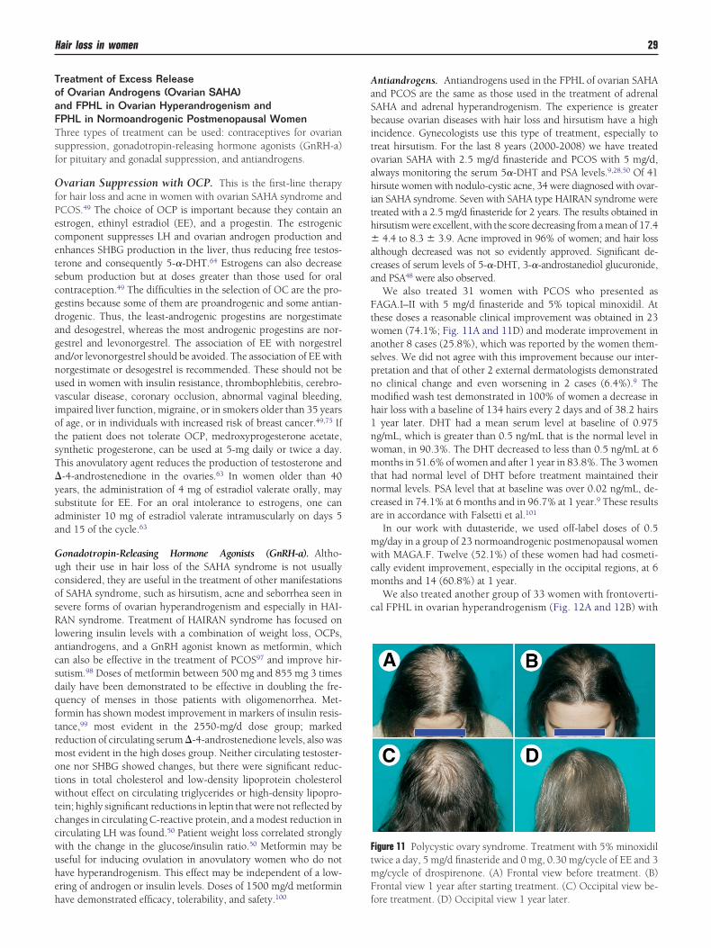
ToaFTsf
OfPecetscgdaganuviotsT�ysaa
GucosRlacsdqftrmotwtccwuheh
AaSbitoahith�aca
Ftwaspnmh1nwmtnca
mwcm
c
FtmF
Hair loss in women 29
reatment of Excess Releasef Ovarian Androgens (Ovarian SAHA)nd FPHL in Ovarian Hyperandrogenism andPHL in Normoandrogenic Postmenopausal Womenhree types of treatment can be used: contraceptives for ovarianuppression, gonadotropin-releasing hormone agonists (GnRH-a)or pituitary and gonadal suppression, and antiandrogens.
varian Suppression with OCP. This is the first-line therapyor hair loss and acne in women with ovarian SAHA syndrome andCOS.49 The choice of OCP is important because they contain anstrogen, ethinyl estradiol (EE), and a progestin. The estrogenicomponent suppresses LH and ovarian androgen production andnhances SHBG production in the liver, thus reducing free testos-erone and consequently 5-�-DHT.64 Estrogens can also decreaseebum production but at doses greater than those used for oralontraception.49 The difficulties in the selection of OC are the pro-estins because some of them are proandrogenic and some antian-rogenic. Thus, the least-androgenic progestins are norgestimatend desogestrel, whereas the most androgenic progestins are nor-estrel and levonorgestrel. The association of EE with norgestrelnd/or levonorgestrel should be avoided. The association of EE withorgestimate or desogestrel is recommended. These should not besed in women with insulin resistance, thrombophlebitis, cerebro-ascular disease, coronary occlusion, abnormal vaginal bleeding,mpaired liver function, migraine, or in smokers older than 35 yearsf age, or in individuals with increased risk of breast cancer.49,75 Ifhe patient does not tolerate OCP, medroxyprogesterone acetate,ynthetic progesterone, can be used at 5-mg daily or twice a day.his anovulatory agent reduces the production of testosterone and-4-androstenedione in the ovaries.63 In women older than 40ears, the administration of 4 mg of estradiol valerate orally, mayubstitute for EE. For an oral intolerance to estrogens, one candminister 10 mg of estradiol valerate intramuscularly on days 5nd 15 of the cycle.63
onadotropin-Releasing Hormone Agonists (GnRH-a). Altho-gh their use in hair loss of the SAHA syndrome is not usuallyonsidered, they are useful in the treatment of other manifestationsf SAHA syndrome, such as hirsutism, acne and seborrhea seen inevere forms of ovarian hyperandrogenism and especially in HAI-AN syndrome. Treatment of HAIRAN syndrome has focused on
owering insulin levels with a combination of weight loss, OCPs,ntiandrogens, and a GnRH agonist known as metformin, whichan also be effective in the treatment of PCOS97 and improve hir-utism.98 Doses of metformin between 500 mg and 855 mg 3 timesaily have been demonstrated to be effective in doubling the fre-uency of menses in those patients with oligomenorrhea. Met-ormin has shown modest improvement in markers of insulin resis-ance,99 most evident in the 2550-mg/d dose group; markededuction of circulating serum �-4-androstenedione levels, also wasost evident in the high doses group. Neither circulating testoster-
ne nor SHBG showed changes, but there were significant reduc-ions in total cholesterol and low-density lipoprotein cholesterolithout effect on circulating triglycerides or high-density lipopro-
ein; highly significant reductions in leptin that were not reflected byhanges in circulating C-reactive protein, and a modest reduction inirculating LH was found.50 Patient weight loss correlated stronglyith the change in the glucose/insulin ratio.50 Metformin may beseful for inducing ovulation in anovulatory women who do notave hyperandrogenism. This effect may be independent of a low-ring of androgen or insulin levels. Doses of 1500 mg/d metformin
ave demonstrated efficacy, tolerability, and safety.100 fntiandrogens. Antiandrogens used in the FPHL of ovarian SAHAnd PCOS are the same as those used in the treatment of adrenalAHA and adrenal hyperandrogenism. The experience is greaterecause ovarian diseases with hair loss and hirsutism have a high
ncidence. Gynecologists use this type of treatment, especially toreat hirsutism. For the last 8 years (2000-2008) we have treatedvarian SAHA with 2.5 mg/d finasteride and PCOS with 5 mg/d,lways monitoring the serum 5�-DHT and PSA levels.9,28,50 Of 41irsute women with nodulo-cystic acne, 34 were diagnosed with ovar-
an SAHA syndrome. Seven with SAHA type HAIRAN syndrome werereated with a 2.5 mg/d finasteride for 2 years. The results obtained inirsutism were excellent, with the score decreasing from a mean of 17.4
4.4 to 8.3 � 3.9. Acne improved in 96% of women; and hair losslthough decreased was not so evidently approved. Significant de-reases of serum levels of 5-�-DHT, 3-�-androstanediol glucuronide,nd PSA48 were also observed.
We also treated 31 women with PCOS who presented asAGA.I–II with 5 mg/d finasteride and 5% topical minoxidil. Athese doses a reasonable clinical improvement was obtained in 23omen (74.1%; Fig. 11A and 11D) and moderate improvement in
nother 8 cases (25.8%), which was reported by the women them-elves. We did not agree with this improvement because our inter-retation and that of other 2 external dermatologists demonstratedo clinical change and even worsening in 2 cases (6.4%).9 Theodified wash test demonstrated in 100% of women a decrease inair loss with a baseline of 134 hairs every 2 days and of 38.2 hairsyear later. DHT had a mean serum level at baseline of 0.975
g/mL, which is greater than 0.5 ng/mL that is the normal level inoman, in 90.3%. The DHT decreased to less than 0.5 ng/mL at 6onths in 51.6% of women and after 1 year in 83.8%. The 3 women
hat had normal level of DHT before treatment maintained theirormal levels. PSA level that at baseline was over 0.02 ng/mL, de-reased in 74.1% at 6 months and in 96.7% at 1 year.9 These resultsre in accordance with Falsetti et al.101
In our work with dutasteride, we used off-label doses of 0.5g/day in a group of 23 normoandrogenic postmenopausal womenith MAGA.F. Twelve (52.1%) of these women had had cosmeti-
ally evident improvement, especially in the occipital regions, at 6onths and 14 (60.8%) at 1 year.We also treated another group of 33 women with frontoverti-
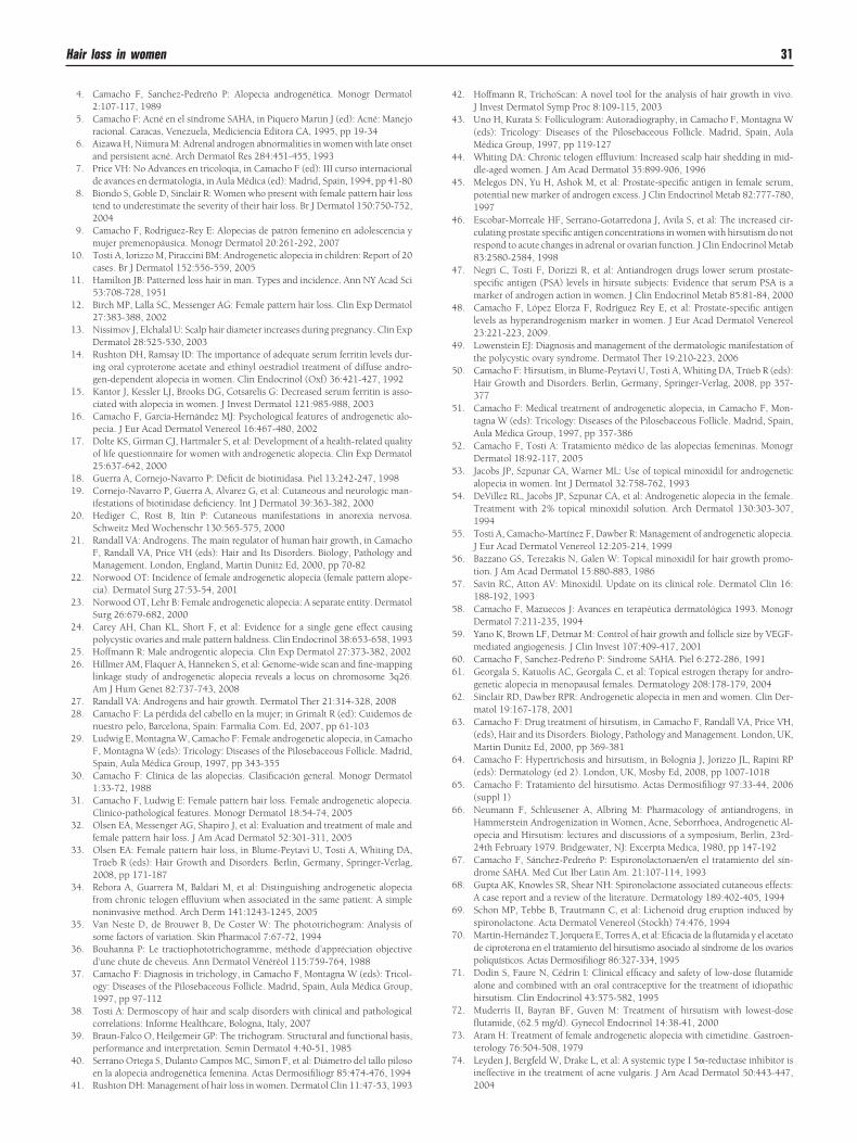
al FPHL in ovarian hyperandrogenism (Fig. 12A and 12B) with
igure 11 Polycystic ovary syndrome. Treatment with 5% minoxidilwice a day, 5 mg/d finasteride and 0 mg, 0.30 mg/cycle of EE and 3g/cycle of drospirenone. (A) Frontal view before treatment. (B)rontal view 1 year after starting treatment. (C) Occipital view be-
ore treatment. (D) Occipital view 1 year later.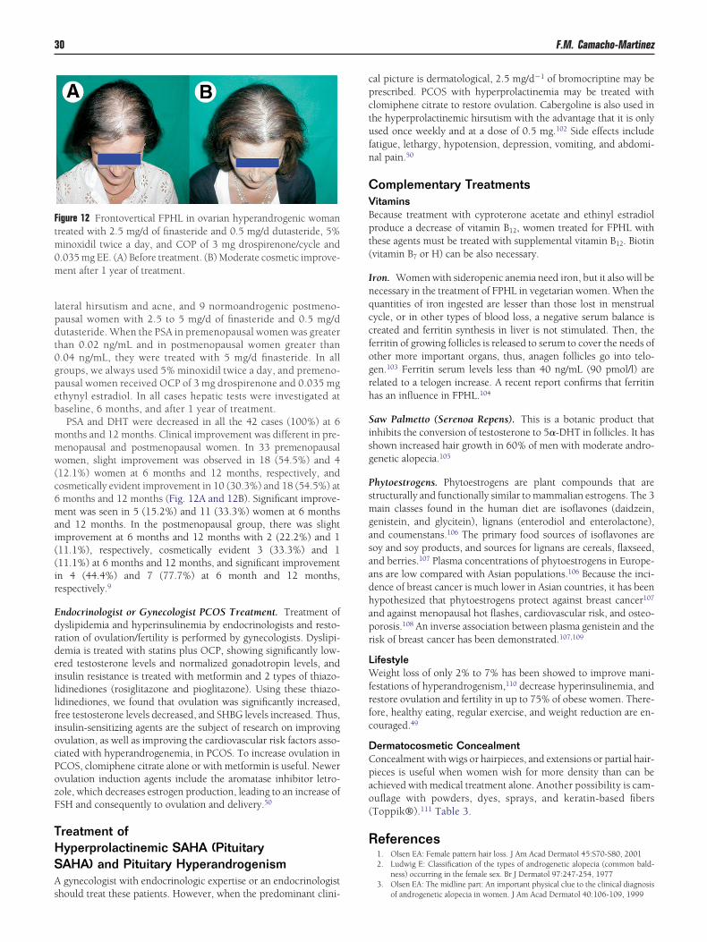
lpdt0gpeb
mmw(c6mai((ir
EdrdeillfiocPozF
THSAs
cpctufn
CVBpt(
Inqccfogrh
Sisg
Psmgasaadhapr
LWfrfc
DCpao(
R
Ftm0m
30 F.M. Camacho-Martinez
ateral hirsutism and acne, and 9 normoandrogenic postmeno-ausal women with 2.5 to 5 mg/d of finasteride and 0.5 mg/dutasteride. When the PSA in premenopausal women was greaterhan 0.02 ng/mL and in postmenopausal women greater than.04 ng/mL, they were treated with 5 mg/d finasteride. In allroups, we always used 5% minoxidil twice a day, and premeno-ausal women received OCP of 3 mg drospirenone and 0.035 mgthynyl estradiol. In all cases hepatic tests were investigated ataseline, 6 months, and after 1 year of treatment.
PSA and DHT were decreased in all the 42 cases (100%) at 6onths and 12 months. Clinical improvement was different in pre-enopausal and postmenopausal women. In 33 premenopausalomen, slight improvement was observed in 18 (54.5%) and 4
12.1%) women at 6 months and 12 months, respectively, andosmetically evident improvement in 10 (30.3%) and 18 (54.5%) atmonths and 12 months (Fig. 12A and 12B). Significant improve-ent was seen in 5 (15.2%) and 11 (33.3%) women at 6 months
nd 12 months. In the postmenopausal group, there was slightmprovement at 6 months and 12 months with 2 (22.2%) and 111.1%), respectively, cosmetically evident 3 (33.3%) and 111.1%) at 6 months and 12 months, and significant improvementn 4 (44.4%) and 7 (77.7%) at 6 month and 12 months,espectively.9
ndocrinologist or Gynecologist PCOS Treatment. Treatment ofyslipidemia and hyperinsulinemia by endocrinologists and resto-ation of ovulation/fertility is performed by gynecologists. Dyslipi-emia is treated with statins plus OCP, showing significantly low-red testosterone levels and normalized gonadotropin levels, andnsulin resistance is treated with metformin and 2 types of thiazo-idinediones (rosiglitazone and pioglitazone). Using these thiazo-idinediones, we found that ovulation was significantly increased,ree testosterone levels decreased, and SHBG levels increased. Thus,nsulin-sensitizing agents are the subject of research on improvingvulation, as well as improving the cardiovascular risk factors asso-iated with hyperandrogenemia, in PCOS. To increase ovulation inCOS, clomiphene citrate alone or with metformin is useful. Newervulation induction agents include the aromatase inhibitor letro-ole, which decreases estrogen production, leading to an increase ofSH and consequently to ovulation and delivery.50
reatment ofyperprolactinemic SAHA (PituitaryAHA) and Pituitary Hyperandrogenismgynecologist with endocrinologic expertise or an endocrinologist
igure 12 Frontovertical FPHL in ovarian hyperandrogenic womanreated with 2.5 mg/d of finasteride and 0.5 mg/d dutasteride, 5%inoxidil twice a day, and COP of 3 mg drospirenone/cycle and
.035 mg EE. (A) Before treatment. (B) Moderate cosmetic improve-ent after 1 year of treatment.
hould treat these patients. However, when the predominant clini-
al picture is dermatological, 2.5 mg/d�1 of bromocriptine may berescribed. PCOS with hyperprolactinemia may be treated withlomiphene citrate to restore ovulation. Cabergoline is also used inhe hyperprolactinemic hirsutism with the advantage that it is onlysed once weekly and at a dose of 0.5 mg.102 Side effects include
atigue, lethargy, hypotension, depression, vomiting, and abdomi-al pain.50
omplementary Treatmentsitaminsecause treatment with cyproterone acetate and ethinyl estradiolroduce a decrease of vitamin B12, women treated for FPHL withhese agents must be treated with supplemental vitamin B12. Biotinvitamin B7 or H) can be also necessary.
ron. Women with sideropenic anemia need iron, but it also will beecessary in the treatment of FPHL in vegetarian women. When theuantities of iron ingested are lesser than those lost in menstrualycle, or in other types of blood loss, a negative serum balance isreated and ferritin synthesis in liver is not stimulated. Then, theerritin of growing follicles is released to serum to cover the needs ofther more important organs, thus, anagen follicles go into telo-en.103 Ferritin serum levels less than 40 ng/mL (90 pmol/l) areelated to a telogen increase. A recent report confirms that ferritinas an influence in FPHL.104
aw Palmetto (Serenoa Repens). This is a botanic product thatnhibits the conversion of testosterone to 5�-DHT in follicles. It hashown increased hair growth in 60% of men with moderate andro-enetic alopecia.105
hytoestrogens. Phytoestrogens are plant compounds that aretructurally and functionally similar to mammalian estrogens. The 3ain classes found in the human diet are isoflavones (daidzein,
enistein, and glycitein), lignans (enterodiol and enterolactone),nd coumenstans.106 The primary food sources of isoflavones areoy and soy products, and sources for lignans are cereals, flaxseed,nd berries.107 Plasma concentrations of phytoestrogens in Europe-ns are low compared with Asian populations.106 Because the inci-ence of breast cancer is much lower in Asian countries, it has beenypothesized that phytoestrogens protect against breast cancer107
nd against menopausal hot flashes, cardiovascular risk, and osteo-orosis.108 An inverse association between plasma genistein and theisk of breast cancer has been demonstrated.107,109
ifestyleeight loss of only 2% to 7% has been showed to improve mani-
estations of hyperandrogenism,110 decrease hyperinsulinemia, andestore ovulation and fertility in up to 75% of obese women. There-ore, healthy eating, regular exercise, and weight reduction are en-ouraged.49
ermatocosmetic Concealmentoncealment with wigs or hairpieces, and extensions or partial hair-ieces is useful when women wish for more density than can bechieved with medical treatment alone. Another possibility is cam-uflage with powders, dyes, sprays, and keratin-based fibersToppik®).111 Table 3.
eferences1. Olsen EA: Female pattern hair loss. J Am Acad Dermatol 45:S70-S80, 20012. Ludwig E: Classification of the types of androgenetic alopecia (common bald-
ness) occurring in the female sex. Br J Dermatol 97:247-254, 1977
3. Olsen EA: The midline part: An important physical clue to the clinical diagnosisof androgenetic alopecia in women. J Am Acad Dermatol 40:106-109, 1999
Hair loss in women 31
4. Camacho F, Sanchez-Pedreño P: Alopecia androgenética. Monogr Dermatol2:107-117, 1989
5. Camacho F: Acné en el síndrome SAHA, in Piquero Martin J (ed): Acné: Manejoracional. Caracas, Venezuela, Mediciencia Editora CA, 1995, pp 19-34
6. Aizawa H, Niimura M: Adrenal androgen abnormalities in women with late onsetand persistent acné. Arch Dermatol Res 284:451-455, 1993
7. Price VH: No Advances en tricoloqia, in Camacho F (ed): III curso internacionalde avances en dermatología, in Aula Médica (ed): Madrid, Spain, 1994, pp 41-80
8. Biondo S, Goble D, Sinclair R: Women who present with female pattern hair losstend to underestimate the severity of their hair loss. Br J Dermatol 150:750-752,2004
9. Camacho F, Rodriguez-Rey E: Alopecias de patrón femenino en adolescencia ymujer premenopáusica. Monogr Dermatol 20:261-292, 2007
10. Tosti A, Iorizzo M, Piraccini BM: Androgenetic alopecia in children: Report of 20cases. Br J Dermatol 152:556-559, 2005
11. Hamilton JB: Patterned loss hair in man. Types and incidence. Ann NY Acad Sci53:708-728, 1951
12. Birch MP, Lalla SC, Messenger AG: Female pattern hair loss. Clin Exp Dermatol27:383-388, 2002
13. Nissimov J, Elchalal U: Scalp hair diameter increases during pregnancy. Clin ExpDermatol 28:525-530, 2003
14. Rushton DH, Ramsay ID: The importance of adequate serum ferritin levels dur-ing oral cyproterone acetate and ethinyl oestradiol treatment of diffuse andro-gen-dependent alopecia in women. Clin Endocrinol (Oxf) 36:421-427, 1992
15. Kantor J, Kessler LJ, Brooks DG, Cotsarelis G: Decreased serum ferritin is asso-ciated with alopecia in women. J Invest Dermatol 121:985-988, 2003
16. Camacho F, García-Hernández MJ: Psychological features of androgenetic alo-pecia. J Eur Acad Dermatol Venereol 16:467-480, 2002
17. Dolte KS, Girman CJ, Hartmaler S, et al: Development of a health-related qualityof life questionnaire for women with androgenetic alopecia. Clin Exp Dermatol25:637-642, 2000
18. Guerra A, Cornejo-Navarro P: Déficit de biotinidasa. Piel 13:242-247, 199819. Cornejo-Navarro P, Guerra A, Alvarez G, et al: Cutaneous and neurologic man-
ifestations of biotinidase deficiency. Int J Dermatol 39:363-382, 200020. Hediger C, Rost B, Itin P: Cutaneous manifestations in anorexia nervosa.
Schweitz Med Wochenschr 130:565-575, 200021. Randall VA: Androgens. The main regulator of human hair growth, in Camacho
F, Randall VA, Price VH (eds): Hair and Its Disorders. Biology, Pathology andManagement. London, England, Martin Dunitz Ed, 2000, pp 70-82
22. Norwood OT: Incidence of female androgenetic alopecia (female pattern alope-cia). Dermatol Surg 27:53-54, 2001
23. Norwood OT, Lehr B: Female androgenetic alopecia: A separate entity. DermatolSurg 26:679-682, 2000
24. Carey AH, Chan KL, Short F, et al: Evidence for a single gene effect causingpolycystic ovaries and male pattern baldness. Clin Endocrinol 38:653-658, 1993
25. Hoffmann R: Male androgentic alopecia. Clin Exp Dermatol 27:373-382, 200226. Hillmer AM, Flaquer A, Hanneken S, et al: Genome-wide scan and fine-mapping
linkage study of androgenetic alopecia reveals a locus on chromosome 3q26.Am J Hum Genet 82:737-743, 2008
27. Randall VA: Androgens and hair growth. Dermatol Ther 21:314-328, 200828. Camacho F: La pérdida del cabello en la mujer; in Grimalt R (ed): Cuidemos de
nuestro pelo, Barcelona, Spain: Farmalia Com. Ed, 2007, pp 61-10329. Ludwig E, Montagna W, Camacho F: Female androgenetic alopecia, in Camacho
F, Montagna W (eds): Tricology: Diseases of the Pilosebaceous Follicle. Madrid,Spain, Aula Médica Group, 1997, pp 343-355
30. Camacho F: Clínica de las alopecias. Clasificación general. Monogr Dermatol1:33-72, 1988
31. Camacho F, Ludwig E: Female pattern hair loss. Female androgenetic alopecia.Clinico-pathological features. Monogr Dermatol 18:54-74, 2005
32. Olsen EA, Messenger AG, Shapiro J, et al: Evaluation and treatment of male andfemale pattern hair loss. J Am Acad Dermatol 52:301-311, 2005
33. Olsen EA: Female pattern hair loss, in Blume-Peytavi U, Tosti A, Whiting DA,Trüeb R (eds): Hair Growth and Disorders. Berlin, Germany, Springer-Verlag,2008, pp 171-187
34. Rebora A, Guarrera M, Baldari M, et al: Distinguishing androgenetic alopeciafrom chronic telogen effluvium when associated in the same patient: A simplenoninvasive method. Arch Derm 141:1243-1245, 2005
35. Van Neste D, de Brouwer B, De Coster W: The phototrichogram: Analysis ofsome factors of variation. Skin Pharmacol 7:67-72, 1994
36. Bouhanna P: Le tractiophototrichogramme, méthode d’appréciation objectived’une chute de cheveus. Ann Dermatol Vénéréol 115:759-764, 1988
37. Camacho F: Diagnosis in trichology, in Camacho F, Montagna W (eds): Tricol-ogy: Diseases of the Pilosebaceous Follicle. Madrid, Spain, Aula Médica Group,1997, pp 97-112
38. Tosti A: Dermoscopy of hair and scalp disorders with clinical and pathologicalcorrelations: Informe Healthcare, Bologna, Italy, 2007
39. Braun-Falco O, Heilgemeir GP: The trichogram. Structural and functional basis,performance and interpretation. Semin Dermatol 4:40-51, 1985
40. Serrano Ortega S, Dulanto Campos MC, Simon F, et al: Diámetro del tallo piloso
en la alopecia androgenética femenina. Actas Dermosifiliogr 85:474-476, 199441. Rushton DH: Management of hair loss in women. Dermatol Clin 11:47-53, 1993
42. Hoffmann R, TrichoScan: A novel tool for the analysis of hair growth in vivo.J Invest Dermatol Symp Proc 8:109-115, 2003
43. Uno H, Kurata S: Folliculogram: Autoradiography, in Camacho F, Montagna W(eds): Tricology: Diseases of the Pilosebaceous Follicle. Madrid, Spain, AulaMédica Group, 1997, pp 119-127
44. Whiting DA: Chronic telogen effluvium: Increased scalp hair shedding in mid-dle-aged women. J Am Acad Dermatol 35:899-906, 1996
45. Melegos DN, Yu H, Ashok M, et al: Prostate-specific antigen in female serum,potential new marker of androgen excess. J Clin Endocrinol Metab 82:777-780,1997
46. Escobar-Morreale HF, Serrano-Gotarredona J, Avila S, et al: The increased cir-culating prostate specific antigen concentrations in women with hirsutism do notrespond to acute changes in adrenal or ovarian function. J Clin Endocrinol Metab83:2580-2584, 1998
47. Negri C, Tosti F, Dorizzi R, et al: Antiandrogen drugs lower serum prostate-specific antigen (PSA) levels in hirsute subjects: Evidence that serum PSA is amarker of androgen action in women. J Clin Endocrinol Metab 85:81-84, 2000
48. Camacho F, López Elorza F, Rodríguez Rey E, et al: Prostate-specific antigenlevels as hyperandrogenism marker in women. J Eur Acad Dermatol Venereol23:221-223, 2009.
49. Lowenstein EJ: Diagnosis and management of the dermatologic manifestation ofthe polycystic ovary syndrome. Dermatol Ther 19:210-223, 2006
50. Camacho F: Hirsutism, in Blume-Peytavi U, Tosti A, Whiting DA, Trüeb R (eds):Hair Growth and Disorders. Berlin, Germany, Springer-Verlag, 2008, pp 357-377
51. Camacho F: Medical treatment of androgenetic alopecia, in Camacho F, Mon-tagna W (eds): Tricology: Diseases of the Pilosebaceous Follicle. Madrid, Spain,Aula Médica Group, 1997, pp 357-386
52. Camacho F, Tosti A: Tratamiento médico de las alopecias femeninas. MonogrDermatol 18:92-117, 2005
53. Jacobs JP, Szpunar CA, Warner ML: Use of topical minoxidil for androgeneticalopecia in women. Int J Dermatol 32:758-762, 1993
54. DeVillez RL, Jacobs JP, Szpunar CA, et al: Androgenetic alopecia in the female.Treatment with 2% topical minoxidil solution. Arch Dermatol 130:303-307,1994
55. Tosti A, Camacho-Martínez F, Dawber R: Management of androgenetic alopecia.J Eur Acad Dermatol Venereol 12:205-214, 1999
56. Bazzano GS, Terezakis N, Galen W: Topical minoxidil for hair growth promo-tion. J Am Acad Dermatol 15:880-883, 1986
57. Savin RC, Atton AV: Minoxidil. Update on its clinical role. Dermatol Clin 16:188-192, 1993
58. Camacho F, Mazuecos J: Avances en terapéutica dermatológica 1993. MonogrDermatol 7:211-235, 1994
59. Yano K, Brown LF, Detmar M: Control of hair growth and follicle size by VEGF-mediated angiogenesis. J Clin Invest 107:409-417, 2001
60. Camacho F, Sanchez-Pedreño P: Sindrome SAHA. Piel 6:272-286, 199161. Georgala S, Katuolis AC, Georgala C, et al: Topical estrogen therapy for andro-
genetic alopecia in menopausal females. Dermatology 208:178-179, 200462. Sinclair RD, Dawber RPR: Androgenetic alopecia in men and women. Clin Der-
matol 19:167-178, 200163. Camacho F: Drug treatment of hirsutism, in Camacho F, Randall VA, Price VH,
(eds), Hair and its Disorders. Biology, Pathology and Management. London, UK,Martin Dunitz Ed, 2000, pp 369-381
64. Camacho F: Hypertrichosis and hirsutism, in Bolognia J, Jorizzo JL, Rapini RP(eds): Dermatology (ed 2). London, UK, Mosby Ed, 2008, pp 1007-1018
65. Camacho F: Tratamiento del hirsutismo. Actas Dermosifiliogr 97:33-44, 2006(suppl 1)
66. Neumann F, Schleusener A, Albring M: Pharmacology of antiandrogens, inHammerstein Androgenization in Women, Acne, Seborrhoea, Androgenetic Al-opecia and Hirsutism: lectures and discussions of a symposium, Berlin, 23rd-24th February 1979. Bridgewater, NJ: Excerpta Medica, 1980, pp 147-192
67. Camacho F, Sánchez-Pedreño P: Espironolactonaen/en el tratamiento del sín-drome SAHA. Med Cut Iber Latin Am. 21:107-114, 1993
68. Gupta AK, Knowles SR, Shear NH: Spironolactone associated cutaneous effects:A case report and a review of the literature. Dermatology 189:402-405, 1994
69. Schon MP, Tebbe B, Trautmann C, et al: Lichenoid drug eruption induced byspironolactone. Acta Dermatol Venereol (Stockh) 74:476, 1994
70. Martín-Hernández T, Jorquera E, Torres A, et al: Eficacia de la flutamida y el acetatode ciproterona en el tratamiento del hirsutismo asociado al síndrome de los ovariospoliquísticos. Actas Dermosifiliogr 86:327-334, 1995
71. Dodin S, Faure N, Cédrin I: Clinical efficacy and safety of low-dose flutamidealone and combined with an oral contraceptive for the treatment of idiopathichirsutism. Clin Endocrinol 43:575-582, 1995
72. Muderris II, Bayran BF, Guven M: Treatment of hirsutism with lowest-doseflutamide, (62.5 mg/d). Gynecol Endocrinol 14:38-41, 2000
73. Aram H: Treatment of female androgenetic alopecia with cimetidine. Gastroen-terology 76:504-508, 1979
74. Leyden J, Bergfeld W, Drake L, et al: A systemic type I 5�-reductase inhibitor is
ineffective in the treatment of acne vulgaris. J Am Acad Dermatol 50:443-447,2004
1
1
1
1
1
1
1
1
1
1
1
1
32 F.M. Camacho-Martinez
75. Flores E, Bratoeff E, Cabeza M, et al: Steroid 5�-reductase inhibitors. Mini RevMed Chem 3:225-237, 2003
76. Bratoeff E, Cabeza M, Ramirez E, et al: Recent advances in the chemistry andpharmacological activity of new steroidal antiandrogen and 5�-reductase inhib-itors. Curr Med Chem 12:927-943, 2005
77. Rogers NE, Avram MR: Medical treatment for male and female pattern hair loss.J Am Acad Dermatol 59:547-566, 2008
78. Dallob A, Sadik NS, Unger W, et al: The effect of finasteride, a 5-alpha-reductaseinhibitor, on scalp skin testosterone and dihydrotestosterone concentration inpatients with male pattern baldness. J Clin Endocrinol Metab 79:703-706, 1994
79. Drake L, Hordinsky M, Fiedler V, et al: The effect of finasteride, a 5-alpha-reductase inhibitor, on scalp skin and serum androgen levels in men with an-drogenetic alopecia. J Am Acad Dermatol 41:550-554, 1999
80. Kaufman KD, Olsen EA, Whiting D, et al: Finasteride in the treatment of menwith androgenetic alopecia. J Am Acad Dermatol 39:578-589, 1998
81. Leyden J, Dunlap F, Miller B, et al: Finasteride in the treatment of men withfrontal male pattern hair loss. J Am Acad Dermatol 40:930-937, 1999
82. Camacho F: Hirsutismo. Enfoque clínico-terapéutico II. Act Terap Dermatol(Arg) 24:190-206, 2001
83. Price VH, Roberts JL, Hordinsky M, et al: Lack of efficacy of finasteride inpostmenopausal women with androgenetic alopecia. J Am Acad Dermatol 43:768-776, 2000
84. Shum KW, Cullen DR, Messenger AG: Hair loss in women with hyperandro-genism: Four cases responding to finasteride. J Am Acad Dermatol 47:733-739,2002
85. Thai K-E, Sinclair RD: Finasteride for female androgenética alopecia. Br J Der-matol 147:812-813, 2002
86. Trüeb RM: Finasteride treatment of patterned hair loss in normoandrogenicpostmenopausal women. Dermatology 209:202-207, 2004
87. Valsecchi R, Leghissa P, Riva M: Female androgenetic alopecia treated by finas-teride: A case forward. Acta Derm Venereol 84:488-489, 2004
88. Iorizzo M, Vinzenci C, Voudouris S, et al: Finasteride treatment of female patternhair loss. Arch Dermatol 142:298-302, 2006
89. Moreno-Ramírez D, Camacho-Martínez F: Frontal fibrosing alopecia: A survey in16 patients. J Eur Acad Dermatol Venereol 19:700-705, 2005
90. Tosti A, Piraccini BM, Iorizzo M, Misciali C: Frontal fibrosing alopecia in post-menopausal women. J Am Acad Dermatol 52:55-60, 2005
91. Iorizzo M, Tosti A: Trastornos del cabello en mujeres postmenopausicas. MonogrDermatol 20:189-203, 2007
92. Kohler C, Tschumi K, Bodmer C, et al: Effect of finasteride, 5 mg (Proscar) onacne and alopecia in female patients with normal serum levels of free testoster-one. Gynecol Endocrinol 23:142-145, 2007
93. Olsen EA, Hordinsky M, Whiting D, et al: The importance of dual 5�-reductaseinhibition in the treatment of male pattern hair loss: Results of a randomizedplacebo-controlled study of dutasteride versus finasteride. J Am Acad Dermatol55:1014-1023, 2006
94. Olszewska M, Rudnicke L: Effective treatment of female androgenic alopeciawith dutasteride. J Drugs Dermatol 4:637-640, 2005
95. Debruyme F, Barkin J, van Erps P, et al: Efficacy and safety of long-term treat-ment with the dual 5�-reductase inhibitor dutasteride in men with symptomaticbenign prostatic hyperplasia. Eur Urol 46:488-495, 2004
96. Fernández Olarte D, Morán García A, Iglesias Bonilla P, et al: Ginecomastia pordutasterida. Med Clin (Barc) 128:250, 2007
97. Barbieri RL, Gargiulo AR: Metformin for the treatment of the polycystic ovarysyndrome. Minerva Ginecol 56:63-79, 2004
98. Harborne L, Fleming R, Lyall H, et al: Metformin or antiandrogen in the treat-ment of hirsutism in polycystic ovary syndrome. J Clin Endocrinol Metab 88:4116-4123, 2003
99. Kazerooni T, Dehghan-Kooshkghazi M: Effects of metformin therapy on hy-perandrogenism in women with polycystic ovarian syndrome. Gynecol Endocri-nol 17:51-56, 2003
00. Schwartz S, Fonseca V, Berner B, et al: Efficacy, tolerability, and safety of a novelonce-daily extended-release metformin in patients with type 2 diabetes. DiabCare 29:759-764, 2006
01. Falsetti L, De Fusco D, Eleftheriou G: Treatment of hirsutism by finasteride andflutamide in women with polycystic ovary syndrome. Gynecol Endocrinol 11:251-257, 1997
02. Colao A, Lombardi G, Annunziato L: Cabergoline. Expert Opin Pharmacother1:555-574, 2000
03. Rushton DH, Ramsay ID: The importance of adequate serum ferritin levelsduring oral cyproterone acetate and ethinyl oestradiol treatment of diffuse an-drogen-dependent alopecia in women. Clin Endocrinol (Oxf) 36:421-427, 1992
04. Kantor J, Kessler LJ, Brooks DG, Cotsarelis G: Decreased serum ferritin is asso-ciated with alopecia in women. J Invest Dermatol 121:985-988, 2003
05. Prager N, Bickett K, French N, et al: A randomized, double-blind, placebo-controlled trial to determine the effectiveness of botanically derived inhibitor of5-alpha-reductase in the treatment of androgenetic alopecia. J Altern Comple-ment Med 8:142-152, 2002
06. Peeters PHM, Slimani N, van der Schouw YT, et al: Variations in plasma phy-toestrogen concentrations in European adults. J Nutr 137:1294-1300, 2007
07. Verheus M, van Gils CH, Keinan-Boker L, et al: Plasma phytoestrogens andsubsequent breast cancer risk. J Clin Oncol 25:648-655, 2007
08. Muti P, Awad AB, Schünemann H, et al: A plant food-based diet modifies theserum �-sitosterol concentration in hyperandrogenic postmenopausal women. JNutr 133:4252-4255, 2003
09. Iwasaki M, Inoue M, Otani T, et al: Plasma isoflavone level and subsequent riskof breast cancer among Japanese women: A nested case-control study from theJapan Public Health Center-based prospective study group. J Clin Oncol 26:1677-1683, 2008
10. Pasquali R, Fabbri R, Venturoli S, et al: Effect of weight loss and anti-androgenictherapy on sex-hormone blood levels and insulin resistance in obese patientswith polycystic ovary syndrome. Am J Obstet Gynecol 154:139-144, 1986
11. Camacho F, Cleasing: Maintenance, decorative and therapeutic cosmetics:Shampoos. Conditioners. Dyes. Permanent waves. Hair setting lotions andsprays. Fixing agents and others. Adverse reactions. Legislation, in: Camacho F,
Montagna W, eds. Tricology: Diseases of the Pilosebaceous Follicle. Madrid,Spain, Aula Mediea Group, 1997, pp 641-661


