Embed Size (px)
Citation preview
Mechanisms of antibioticresistance in enterococciExpert Rev. Anti Infect. Ther. 12(10), 1221–1236 (2014)
William R Miller1,Jose M Munita1,2 andCesar A Arias*1,3
1Department of Internal Medicine,
Division of Infectious Diseases,
University of Texas Medical School,
6431 Fannin St. Rm. MSB 2.112,
Houston, TX 77030, USA2Clinica Alemana de Santiago and
Universidad del Desarrollo, Santiago,
Chile3Molecular Genetics and Antimicrobial
Resistance Unit, Universidad El Bosque,
Bogota, Colombia
*Author for correspondence:
Tel.: +1 713 500 6767
Fax: +1 713 500 5495
Multidrug-resistant (MDR) enterococci are important nosocomial pathogens and a growingclinical challenge. These organisms have developed resistance to virtually all antimicrobialscurrently used in clinical practice using a diverse number of genetic strategies. Due to thisability to recruit antibiotic resistance determinants, MDR enterococci display a wide repertoireof antibiotic resistance mechanisms including modification of drug targets, inactivation oftherapeutic agents, overexpression of efflux pumps and a sophisticated cell envelope adaptiveresponse that promotes survival in the human host and the nosocomial environment. MDRenterococci are well adapted to survive in the gastrointestinal tract and can become thedominant flora under antibiotic pressure, predisposing the severely ill and immunocompromisedpatient to invasive infections. A thorough understanding of the mechanisms underlyingantibiotic resistance in enterococci is the first step for devising strategies to control the spreadof these organisms and potentially establish novel therapeutic approaches.
KEYWORDS: antimicrobial resistance • enterococcus • mechanisms of resistance • vancomycin-resistant enterococci
Enterococci are gram-positive facultative anae-robes that live as commensals in the GI tractof a variety of organisms including humans [1].Shaped by the selective pressures of their com-petitive environment, these bacteria haveevolved a diverse array of responses andgenetic plasticity allowing them to thrive inthe modern healthcare environment. Advancesin modern medicine have increased the abilityto sustain life in critically ill patients increasingthe susceptibility to infection and resulting inrapid patient turnover and widespread use ofantibiotics. Armed with multiple antibioticresistance determinants, enterococci ‘takeadvantage’ of this opportunity and expandwithin their ecologic niche (GI tract of hospi-talized patients) to gain the upper hand anddominate the intestinal microbiota. From theGI tract, multidrug-resistant (MDR) entero-cocci disseminate rapidly in the hospital envi-ronment. Indeed, enterococci are leadingcauses of nosocomial infections and are secondonly to staphylococci as a cause of gram-positive nosocomial infections [2]. Here, wewill discuss the historical context behind therise of enterococci as MDR pathogens fol-lowed by a review of the current understand-ing of the molecular basis of enterococcalantimicrobial resistance. Finally, we will brieflyaddress the need for the development of novel
drug targets and combination therapies againstthese sturdy and recalcitrant organisms.
Evolution of antibiotic resistance in MDRenterococciThe advent of the antibiotic era spurred a newrevolution in modern medicine. With thediscovery of antimicrobial agents and theunderstanding of the microbiological basis ofdisease, infection became a treatable diseasewith remarkable results. However, cliniciansrapidly realized that certain microorganismsappear to respond less well to specific antimicro-bial agents. For example, with the introductionof penicillin, streptococcal infection was success-fully treated with this agent. However, a subsetof ‘streptococcal’ organisms (later known asenterococci) appeared to respond less well topenicillin due to an inherent tolerance to thekilling action of these compounds [3]. It waslater found that the addition of aminoglycosides(streptomycin was discovered in 1944 [4]) topenicillin produced synergistic activity improv-ing the cure rates for enterococcal infectiveendocarditis from 40 to 88% [5]. This synergis-tic effect was seen despite the fact that entero-cocci are also inherently less susceptible toaminoglycosides compared to other gram-positive bacteria. Thus, the combination of acell-wall active agent (i.e., ampicillin/penicillin)
informahealthcare.com 10.1586/14787210.2014.956092 � 2014 Informa UK Ltd ISSN 1478-7210 1221
Review
Exp
ert R
evie
w o
f A
nti-
infe
ctiv
e T
hera
py D
ownl
oade
d fr
om in
form
ahea
lthca
re.c
om b
y 99
.25.
205.
113
on 0
9/11
/14
For
pers
onal
use
onl
y.
plus an aminoglycoside became the standard of care for deep-seated enterococcal infections and this combination is still usedto the present day [6].
Though it was not known at that time, the seeds of the mod-ern MDR enterococci were already being sown. Using compara-tive genomics, Lebreton et al. demonstrated that the modern-dayMDR Enterococcus faecium belongs to a genetic clade that appearsto have diverged from animal-adapted E. faecium isolates about75 years ago, coinciding with the introduction of antibiotics inclinical practice [7]. Features of this clade (designated A1) includean increase in mobile genetic elements, alterations in metabolismand hypermutability, a skill set that provides E. faecium amalleable genome in the face of multiple selective pressures. Theremarkable increase in the use of antimicrobials in clinical medi-cine in the latter half of the 20th century provided the selectiveenvironment for these microorganisms to evolve by recruiting avariety of antibiotic resistance determinants.
Among the most distinct examples of this adaptability is theacquisition of the genes encoding vancomycin resistance. Van-comycin use was associated with the emergence and spread ofmethicillin-resistant Staphylococcus aureus (MRSA) in the1960s. However, unlike MRSA, enterococci have been able torecruit and maintain a variety of gene clusters encoding thebiochemical machinery for vancomycin resistance. From thefirst recognition of vancomycin-resistant enterococci (VRE) in1988 to late in the first decade of this century, the rates of van-comycin resistance in E. faecium in the USA have surpassed80% [8]. Of concern, E. faecium is also increasingly identifiedin nosocomial infections, now occurring as frequently as iso-lates of Enterococcus faecalis (whose rates of vancomycin resis-tance are about 5%) [2]. More recently, enterococci have alsoserved as donors of vancomycin resistance gene clusters to morepathogenic microorganisms such as MRSA, an event consideredto be a serious public health threat [9,10].
Despite the availability of anti-gram-positive agents (e.g., line-zolid, quinupristin/dalfopristin [Q/D], daptomycin [DAP], tige-cycline), enterococci have rapidly adapted and emergence ofresistance to all these newer agents have been well documented.This phenomenon makes the treatment of MDR enterococcalinfections a daunting clinical challenge. Thus, in the followingsections, we will review the molecular mechanisms of antibioticresistance in enterococci with the objective to place such mecha-nisms in context within a clinical perspective and explore inno-vative strategies to combat these recalcitrant microorganisms.
Mechanisms of resistance to cell-wall active agentsAmpicillin/penicillin
Ampicillin and penicillin are the most active b-lactams againstenterococci inhibiting the synthesis of peptidoglycan, the basicstructure of the bacterial cell wall and a critical componentneeded for bacterial viability. As such, the peptidoglycan syn-thesis and maintenance machinery has long been a target forantimicrobial therapy. Furthermore, the lack of similar struc-tures in eukaryotic cells decreases the toxicity profiles of manyof these agents, making them an ideal target against bacteria.
Penicillin-binding proteins (PBPs) are the workhorses of thecell wall synthesis machinery and they can be roughly dividedinto two groups: class A, which are bifunctional enzymes thatpossess both D,D-transpeptidase and transglycosylase activity,and class B, which possess only the transpeptidase domain andrely on the transglycosylase activity of other enzymes.
All enterococci produce at least five PBPs, which can be dif-ferentiated by migration pattern on gel electrophoresis and wereoriginally named by convention on the order of migration [11].Subsequent genomic analysis of both E. faecalis and E. faeciumrevealed six putative PBP genes, three of which are class A(ponA, pbpF, pbpZ) and three class B (pbp5, pbpA, pbpB) [12].Intrinsic tolerance to the action of b-lactams is associated withthe presence of a species-specific chromosomal gene, pbp5,which encodes a class B PBP with low binding affinity for ampi-cillin and the cephalosporins [13,14]. In E. faecium, pbp5 exists inan operon with two other genes implicated in cell wall synthesis;psr, for PBP synthesis repressor, as it was originally thought torepress transcription of pbp5 (this effect was later attributed to adeletion in the promoter region of the gene) and ftsW, a proteinknown to interact with Escherichia coli PBP3 during septum for-mation [15]. High-level resistance to ampicillin (MIC 128 mg/mlor more) was first associated with increased production of theenzyme, requiring a higher concentration of antibiotic to satu-rate the active site [16]. However, many resistant clinical isolateswere found to have no difference in protein expression fromtheir sensitive counterparts. Thus, resistance in these strains wasattributed to mutations altering the protein sequence, specificallya Met485 ! Ala substitution near the active serine residue andthe insertion of a serine residue at position 466 in a loop struc-ture predicted to interact with the active site complex [17].Although the authors of this study observed a decrease in bind-ing of b-lactams and a subsequent increase in the MIC, the rela-tionship was not directly proportional. Additionally, copies ofthe pbp5 gene from highly resistant clinical isolates were unableto fully restore the resistant phenotype of a hypersensitive strainlacking a copy of pbp5 [14].
Sequence variation of PBP5 is sufficient to distinguish twogroups of E. faecium, one possessing high-level ampicillin resis-tance (termed Pbp5-R) associated with the hospital environmentand a community-associated variant (Pbp5-S) that results in lowerMICs to ampicillin (usually <64 mg/ml), but is unable tocompletely explain the differing susceptibility profiles observed inclinical practice [18]. Thus, as yet unidentified factors play animportant role in expressing resistance via Pbp5 in E. faecium. InE. faecalis, pbp5 appears to exist independent from the operonstructure described in E. faecium, and although it also encodes aclass B PBP, it has a lower intrinsic tolerance to ampicillin (typicalMICs of E. faecalis isolates are 1–4 mg/ml). Both overexpressionof the enzyme and mutations in amino acid sequence have beenimplicated in higher levels of resistance, though neither methodproduced changes in MIC as dramatic as seen in E. faecium [19,20].
Another form of ampicillin resistance, mediated by ab-lactamase that inactivates the antibiotic through the cleavageof the b-lactam ring, has been described in both E. faecalis and
Review Miller, Munita & Arias
1222 Expert Rev. Anti Infect. Ther. 12(10), (2014)
Exp
ert R
evie
w o
f A
nti-
infe
ctiv
e T
hera
py D
ownl
oade
d fr
om in
form
ahea
lthca
re.c
om b
y 99
.25.
205.
113
on 0
9/11
/14
For
pers
onal
use
onl
y.

E. faecium [21,22]. Originally described in staphylococci, thegene blaZ encodes a b-lactamase as part of an operon withblaR1, a transmembrane sensor and signal transducer, and blaI,a repressor gene [23]. Sequence analysis of enterococci possessingthe entire operon showed amino acid sequence identity of 97,95 and 96% to the blaZ, blaI and blaR1 genes, respectively ofS. aureus, reinforcing previous evidence that these genes likelyoriginated in staphylococci [24]. In contrast to staphylococci,blaZ in enterococci is expressed constitutively and at a muchlower level, resulting in a clinically significant inoculum effect.Thus, when inoculating bacteria at concentrations for routinesusceptibility testing (generally 1 � 105 organisms per ml), theenterococci produce so little enzyme that they test susceptible,while at high inoculum, such as during infection, the presenceof more enzyme leads to resistance. Once identified, however,the presence of a b-lactamase is of trivial clinical concern as theaddition of the b-lactamase inhibitor sulbactam (i.e., ampicil-lin-sulbactam) is sufficient to restore the antibiotic efficacy.
Finally, the development of ampicillin resistance via anL,D-transpeptidase known as Ldtfm has also been describedin vitro [25]. Constitutively expressed in E. faecium, Ldtfm uti-lizes a tetrapeptide substrate (unlike the D,D-transpeptidaseswhich act on pentapeptides) and is thought to be involved inthe maintenance of peptidoglycan in the stationary phase.Mutations in a cryptic locus (ddc), coding for a putative two-component signal transduction system (TCS) induced expres-sion of a D,D-carboxypeptidase (DdcY), which shares similaritieswith the van resistance cluster (discussed below) and producedthe necessary tetrapeptide substrate [26]. Of note, this pathwaywould allow the synthesis of a functional cell wall in the pres-ence of glycopeptides [27].
Cephalosporin resistance
Although the natural resistance of enterococci to cephalosporinsis a well-known feature, the molecular basis of this phenotype isnot completely understood. A common observation is thatintrinsic resistance is associated with a decrease in binding affin-ity of cephalosporins for the enterococcal PBPs, specificallyPbp5 [28,29]. As a class B PBP possessing only transpeptidaseenzymatic activity, Pbp5 must partner with a glycosyltransferaseto synthesize peptidoglycan. In two studies performed byArbeloa et al. [29], and Rice et al. [28], sequential deletion of theknown class A PBPs (PbpF, PonA or PbpZ) in both E. faecalisand E. faecium was undertaken alone and in combination toinfer their effect on the resistance phenotype. In both species, itwas found that either PonA or PbpF was needed to expresscephalosporin resistance; PbpZ alone was unable to provide theneeded transglycosylase activity. Likewise, deletion of all threegenes led to a susceptible phenotype in both species, however,the triple mutant was still viable, albeit at an impaired rate ofgrowth. The enzyme providing transglycosylase activity in theabsence of the class A PBPs was not identified, and genomicanalysis looking for conserved sequences similar to known glyco-syltransferases did not suggest a suitable candidate. Interestingly,in E. faecium, it was noted that deletion of the class A PBPs
showed a dissociation of cephalosporin and penicillin resistance,as MICs to ampicillin did not change. Further, an inducibleresistance to cephalosporins in triple mutants (that is deletion ofall class A PBPs) was unmasked by disk diffusion with penicil-lin, suggesting differential activation of regulatory pathways as aresponse to b-lactam antibiotics.
Several regulatory pathways controlled by bacterial two-component regulatory systems (TCS) have also been associatedwith the intrinsic resistance to cephalosporins. However, we arejust beginning to scratch the surface of the action of theirdownstream effectors [30]. Among them, CroRS was shown tobe important for cephalosporin resistance. CroS, a sensor withhistidine kinase activity, phosphorylates the cognate responseregulator CroR, which is postulated to alter transcription via aDNA binding domain [31]. Several genes under direct CroRregulation have been identified; however, none of them appearsto alter pathways currently known to induce cephalosporinresistance [32,33]. Another TCS implicated in resistance consistsof a serine/threonine kinase designated IreK and a phosphataseIreP [34]. E. faecalis with deletions of IreP were shown toexhibit increased resistance to ceftriaxone, but had a substantialreduction of fitness in the absence of antibiotics [35]. A thirdprotein, IreB, was demonstrated to be the target of both IreKand IreP and to negatively regulate the expression of cephalo-sporin resistance [36]. Furthermore, the concomitant deletion ofIreK and IreB, or the elimination of the threonine residuewhere phosphorylation occurs, was able to restore resistance viaa release of inhibition of the pathway. This system seems to bespecific to cephalosporin resistance, as MICs to ampicillin andother cell wall active agents were unchanged. One protein thatmay be involved in the downstream effects of the IreK signal-ing pathway is MurAA, one of a pair of homologs that catalyzethe first committed step in peptidoglycan synthesis [37]. Dele-tion of the gene coding for this enzyme resulted in a loss ofresistance to cephalosporins despite the ability of the homologMurAB to carry out enzymatic activity as a uridine diphos-phate-N-acetylglucosamine 1-carboxyvinyl transferase. Whenexpressed from a plasmid vector in ireK-deficient E. faecalis,but not in croR or pbp5 deletion mutants, MurAA was able torestore ceftriaxone resistance [37].
Glycopeptide resistance
Glycopeptides (vancomycin and teicoplanin) bind to the termi-nal D-alanine-D-alanine (D-Ala-D-Ala) moiety of peptidoglycanprecursors, thus preventing cross-linking of peptidoglycan chainsand inhibiting synthesis of the cell wall. Resistance to theseagents, typified by vancomycin, can be described as high-level(MIC >64 mg/ml) or low-level (MIC between 4 and 32 mg/ml),both due to a change in the terminal amino acids of the peptido-glycan precursor from D-Ala-D-Ala to D-alanine-D-lactate (D-Ala-D-Lac) or, less frequently, D-alanine-D-serine (D-Ala-D-Ser) [38].The type of amino acid change is relevant, as it determines thelevel of resistance. Thus, low-level resistance is conferred by theD-Ala-D-Ser-ending precursors, which decreases the bindingaffinity of the antibiotic about sevenfold. On the other hand,
Mechanisms of antibiotic resistance in enterococci Review
informahealthcare.com 1223
Exp
ert R
evie
w o
f A
nti-
infe
ctiv
e T
hera
py D
ownl
oade
d fr
om in
form
ahea
lthca
re.c
om b
y 99
.25.
205.
113
on 0
9/11
/14
For
pers
onal
use
onl
y.
high-level resistance relies on the change of the terminal penta-peptide to D-Ala-D-Lac, a substitution that eliminates one of thefive hydrogen bonds required for the binding of vancomycin tothe peptidoglycan chain, reducing its affinity about 1000-fold.
The origin of vancomycin resistance gene clusters (designatedvan) likely lies in the day-to-day struggle for survival in themicrobial world. Genes nearly identical to the vanA clusterfound in enterococci have been described in the soil organismsPaenibacillus thiaminoluticus and P. apiaries [39]. To date, ninedistinct vancomycin resistance clusters have been described inenterococci (vanA, vanB, vanC, vanD, vanE, vanG, vanL,vanM and vanN) [38,40–42]. In general, these clusters consist ofthree groups of genes encoding: TCS; enzymes necessary forthe synthesis of new peptidoglycan precursors and enzymes thatdestroy the normal D-Ala-D-Ala-ending precursors. The differ-ent clusters can be differentiated by the identity at the aminoacid level of the ligase enzyme (e.g., VanA vs VanB), by thedegree of vancomycin resistance conferred, the ability of thesystem to be induced upon exposure to teicoplanin and thestructural arrangement of the gene cluster.
The vanA cluster is the most common mediator of vancomy-cin resistance in enterococci and its expression is under the regu-lation of two promoters. The first is responsible for thetranscription of VanRS, the TCS that regulates VanA expressionand function. The sensor of the system is VanS, which is atransmembrane protein with a histidine kinase domain thatresponds to the presence of glycopeptides by phosphorylatingthe response regulator VanR [43]. Once activated, VanR binds tothe second promoter region, located upstream of the resistancegenes, activating their transcription. The first step in expressingvancomycin resistance is the transcription of vanH that encodesa dehydrogenase, which allows for the production of D-lactatefrom pyruvate. The next gene, vanA, produces a ligase that ena-bles the addition of D-Lac to D-Ala before adding it to a tripep-tide precursor. The resulting pentadepsipeptide is incorporatedinto the growing cell wall and allows for cross-linking of thepeptidoglycan structure. VanX, a D,D-dipeptidase, and VanY, aDdcY, work to clear the usual D-Ala-D-Ala dipeptides (whichwill bind vancomycin if incorporated in the cell wall) and thenormal D-Ala-ending pentapeptide chains from the pool of cellwall precursors, respectively. Thus, destruction of D-Ala-endingpentapeptide precursors is crucial for the mechanism of glyco-peptide resistance. A gene, designated vanZ, encodes for a puta-tive protein whose function has not been completely elucidated,but that was shown to confer teicoplanin resistance whenexpressed independently in an E. faecium strain [44].
VanB is also commonly found in clinical enterococcal iso-lates and functions in a similar manner to the VanA cluster,with several important differences. The sensor kinase (VanSB)and response regulator (VanRB) share only a distant relation-ship (34 and 23% sequence identity, respectively) to theirVanA homologs [43]. As such, VanSB is not activated in thepresence of teicoplanin, allowing strains with the VanB pheno-type to remain susceptible to this antibiotic. Importantly, resis-tance to teicoplanin may arise from mutations in the sensor
kinase, allowing the activation of the system in the presence ofthe antibiotic or impairment of the phosphatase regulatory abil-ity of VanSB resulting in constitutive expression of the resis-tance genes [45]. VanB also lacks the vanZ gene and insteadpossesses vanW, whose function is also unknown.
An intrinsic feature of two species of enterococci, Enterococcusgallinarum and Enterococcus casseliflavus, is that they carry vanC,a chromosomally encoded gene cluster that confers (with othergenes) low-level resistance to vancomycin through the change ofthe terminal dipeptide from D-Ala-D-Ala to D-Ala-D-Ser [46].These species produce the altered D-Ala-D-Ser dipeptide viaVanT, a serine racemase that catalyzes the change from L-Ser toD-Ser [47]. In addition, they also harbor a bifunctional D,D-pepti-dase/DdcY designated VanXYC that hydrolyzes the typicalD-Ala-D-Ala precursors in the cytoplasm. Similar to VanC, VanEis also chromosomally encoded, expresses low-level resistanceand is under the control of a single promoter [48]. Interestingly,Abadıa Patino et al. found that a functional VanE-resistant phe-notype was fully expressed despite the inactivation of VanSE by apremature stop codon [49]. Activation and transcription of theoperon by an undetermined endogenous TCS was proposed andunderscores the robust nature of the enterococcal stress response.
The genes encoding the TCS of the van gene clusters are anelegant system that allows bacteria to balance the need of anti-microbial resistance and the cost in fitness they carry for thecell. Defects of this system, as evidenced in the VanD pheno-type, can lead to an interesting series of compensatory muta-tions [50]. Enterococci expressing VanD lack an active signalTCS due to an inactivating mutation of VanSD or VanRD. Assuch, the resistant phenotype is constitutively expressed by theconstant activation of the operon producing the D-Ala-D-Lacdepsipeptide. This would be expected to saddle the bacteriawith a substantial metabolic burden, as it is necessary to main-tain a futile cycle of synthesis and destruction of D-Ala-D-Ala,despite the absence of the glycopeptide threat. However, thesestrains also harbor a second mutation in the gene ddl, encodingthe ligase responsible for the production of normal peptidogly-can precursors. As a result, they express the resistant phenotypewithout a substantial reduction in fitness. In fact, loss of theddl gene in other van cluster-containing enterococci (mostcommonly VanB) leads to vancomycin dependence as thesebacteria are unable to synthesize normal peptidoglycan precur-sors and require the presence of vancomycin to activate the vanoperon in order to build a cell wall [51]. In this case, restorationof normal growth occurs with either a ‘gain-of-function’ muta-tion of ddl or constitutive expression of the van operon [52].
Mechanisms of DAP resistance
The bacterial cell membrane (CM) is a crucial structural andfunctional component that regulates a vast majority of cell pro-cesses by connecting the external milieu with the cytoplasm.This structure also serves to keep out toxic compounds andprovides an anchor for vital membrane-based proteins. DAP isa lipopeptide antibiotic that targets the CM and is related tomany cationic antimicrobial peptides (CAMPs) that are
Review Miller, Munita & Arias
1224 Expert Rev. Anti Infect. Ther. 12(10), (2014)
Exp
ert R
evie
w o
f A
nti-
infe
ctiv
e T
hera
py D
ownl
oade
d fr
om in
form
ahea
lthca
re.c
om b
y 99
.25.
205.
113
on 0
9/11
/14
For
pers
onal
use
onl
y.
produced by the innate immune system of eukaryotic organ-isms. Insertion of DAP into the CM requires the presence ofcalcium ions and appears to bind preferentially at the divisionseptum plane. Once inside the membrane, DAP oligomerizesin the outer leaflet and the DAP complexes reach the innerleaflet of the CM forming a ‘pore’ structure that disrupts theintegrity and functionality of the CM resulting in a variety ofprocesses (including leakage of ions) that lead to cell death [53].Oligomerization of DAP in the outer leaflet appears to dependon the presence of the phospholipid phosphatidylglycerol [54],whereas another phospholipid, cardiolipin (CL), influences theability of DAP oligomers to reach the inner leaflet of theCM [55]. In Bacillus subtilis, DAP has been shown to causerecruitment of a cell division protein (DivIVA) at the sites ofCM damage, resulting in abnormal cell wall synthesis anddefective cell division [56]. Of interest, enterococci are less sus-ceptible to DAP than staphylococci, a fact that is reflected bythe susceptibility breakpoints, which are four-times higher forenterococcal isolates than for staphylococci [57].
Recent work has shed light on the genetic changes responsiblefor the DAP non-susceptible phenotype (hereafter referred to asDAP resistance [DAP-R] for clarity) in enterococci. Sequencingof a clinical strain pair of E. faecalis that developed DAP-R overthe course of therapy revealed three genes responsible for theresistance phenotype [58]. The first is liaF that encodes a mem-ber of a three-component regulatory system (LiaFSR) that hasbeen found to orchestrate the cell-envelope response to stress ingram-positive bacteria. In B. subtilis, LiaF negatively regulatesLiaS, which is a histidine kinase that, upon cell-envelope stress,phosphorylates the response regulator LiaR [59]. In E. faecalis, asingle deletion of an isoleucine at position 177 of LiaF increasedthe DAP MIC from 1 to 4 mg/ml (established breakpoint is4 mg/ml) and, more importantly, abolished the bactericidalactivity of DAP [60]. The other two genes implicated in DAP-Rare involved in phospholipid metabolism: gpdD, which encodesa glycerol-phosphodiester phosphodiesterase, and cls, which enc-odes for a cardiolipinsynthase (Cls) [58,61].
In S. aureus, the actual mechanism of DAP resistance is relatedto the increase in cell envelope surface charge due to changes inMprF (a transmembrane protein that increases the content ofthe positive charged phospholipid lysyl-phosphatidylglycerol inthe outer leaflet of the CM). Under this scenario, positivelycharged DAP is ‘repelled’ from the cell surface (also with posi-tive charge due to high content of lysyl-phosphatidylglycerol)preventing its interaction with the CM [62]. In contrast, recentevidence in E. faecalis suggested that this microorganism uses adifferent strategy to withstand the DAP attack. Indeed, using afluorescent derivative of DAP, Tran et al. showed that bindingof DAP to the CM was similar between a DAP-susceptibleE. faecalis and its DAP-resistant derivative [63], suggesting thatrepulsion of DAP from the cell surface was not relevant for themechanism of DAP resistance. Instead, the resistance phenotypewas associated with redistribution of CL microdomains inthe CM which were relocated from the septum (the principaltarget of DAP) to non-septal areas. These CL microdomain
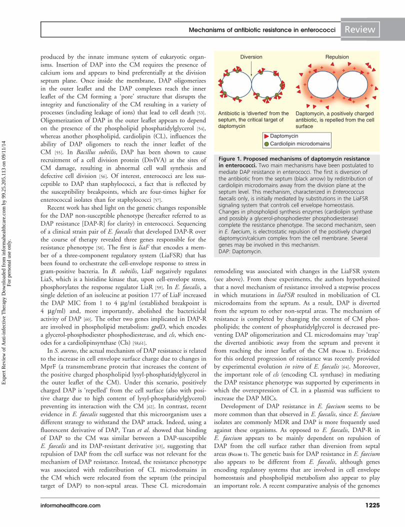
remodeling was associated with changes in the LiaFSR system(see above). From these experiments, the authors hypothesizedthat a novel mechanism of resistance involved a stepwise processin which mutations in liaFSR resulted in mobilization of CLmicrodomains from the septum. As a result, DAP is divertedfrom the septum to other non-septal areas. The mechanism ofresistance is completed by changing the content of CM phos-pholipids; the content of phosphatidylglycerol is decreased pre-venting DAP oligomerization and CL microdomains may ‘trap’the diverted antibiotic away from the septum and prevent itfrom reaching the inner leaflet of the CM (FIGURE 1). Evidencefor this ordered progression of resistance was recently providedby experimental evolution in vitro of E. faecalis [64]. Moreover,the important role of cls (encoding CL synthase) in mediatingthe DAP resistance phenotype was supported by experiments inwhich the overexpression of CL in a plasmid was sufficient toincrease the DAP MICs.
Development of DAP resistance in E. faecium seems to bemore common than that observed in E. faecalis, since E. faeciumisolates are commonly MDR and DAP is more frequently usedagainst these organisms. As opposed to E. faecalis, DAP-R inE. faecium appears to be mainly dependent on repulsion ofDAP from the cell surface rather than diversion from septalareas (FIGURE 1). The genetic basis for DAP resistance in E. faeciumalso appears to be different from E. faecalis, although genesencoding regulatory systems that are involved in cell envelopehomeostasis and phospholipid metabolism also appear to playan important role. A recent comparative analysis of the genomes
Daptomycin
Cardiolipin microdomains
Diversion Repulsion
Antibiotic is ‘diverted’ from theseptum, the critical target ofdaptomycin
Daptomycin, a positively chargedantibiotic, is repelled from the cellsurface
+
+
+
++
+
++
+
+
++
++
Figure 1. Proposed mechanisms of daptomycin resistancein enterococci. Two main mechanisms have been postulated tomediate DAP resistance in enterococci. The first is diversion ofthe antibiotic from the septum (black arrow) by redistribution ofcardiolipin microdomains away from the division plane at theseptum level. This mechanism, characterized in Enterococcusfaecalis only, is initially mediated by substitutions in the LiaFSRsignaling system that controls cell envelope homeostasis.Changes in phospholipid synthesis enzymes (cardiolipin synthaseand possibly a glycerol-phosphodiester phosphodiesterase)complete the resistance phenotype. The second mechanism, seenin E. faecium, is electrostatic repulsion of the positively chargeddaptomycin/calcium complex from the cell membrane. Severalgenes may be involved in this mechanism.DAP: Daptomycin.
Mechanisms of antibiotic resistance in enterococci Review
informahealthcare.com 1225
Exp
ert R
evie
w o
f A
nti-
infe
ctiv
e T
hera
py D
ownl
oade
d fr
om in
form
ahea
lthca
re.c
om b
y 99
.25.
205.
113
on 0
9/11
/14
For
pers
onal
use
onl
y.
from a clinical strain-pair of E. faecium that developed resistanceduring therapy revealed changes in eight different genes, includ-ing a TCS capable of modulating the cell-wall stress response toDAP and designated YycFG [65]. Specific to the low G + Cgram-positive bacteria, this system plays an important role inthe homeostasis of the cell wall with high activity during celldivision [66]. The mechanism by which changes in this systemfacilitate DAP-R in enterococci is not entirely clear and is cur-rently part of active investigations. Another gene that was foundto have changes and is hypothesized to be important is cfa,which encodes for a cyclopropane synthase involved in phospho-lipid metabolism. This enzyme adds a methylene group to thedouble bond of unsaturated fatty acids, which act to stabilizethe CM to respond to a variety of cell envelope stressors [67]. Ofnote, changes in cls have also been commonly identified inDAP-R E. faecium isolates.
The LiaFSR system has also been implicated in DAP-R andtolerance in E. faecium. Recently, Diaz et al. analyzed thegenomes of 19 E. faecium isolates with a wide range of DAPMICs (from 3 to 48 mg/ml) and compared them with all pub-licly available genomes, specifically searching for geneticchanges previously associated with DAP-R (both in enterococciand staphylococci). Interestingly, the most frequent mutationsidentified were in liaFSR, followed by changes in the above-mentioned YycFG systems, supporting the hypothesis thatchanges in TCS are a pivotal step toward DAP-R in entero-cocci [68]. Furthermore, the majority (75%) of DAP-susceptibleE. faecium bacteremia isolates whose MIC was in the higherrange of susceptibility (i.e., between 3 and 4 mg/ml) harboredmutations in LiaFSR [69]. Conversely, none of the isolates ofthe same collection with DAP MIC £2 mg/ml exhibitedchanges in this system. More importantly, these changes weresufficient to abolish the in vitro bactericidal activity of DAP [60]
and were associated with a clinical failure in a neutropenicpatient with VRE bacteremia [70].
Interestingly, development of DAP resistance is inverselyrelated to increased susceptibility to b-lactams. Indeed, combi-nations of DAP with ampicillin, ceftriaxone and ceftarolinehave been noted to be synergistic in vitro and to improve clini-cal outcomes of patients failing DAP therapy [71–73]. Recentdata showed that synergy with ampicillin was dependent onchanges in LiaFSR but not in YycFG, suggesting the possibilitythat the synergism with b-lactams is influenced by the particu-lar signaling pathway through which DAP resistance (or toler-ance) is mediated [68]. Elucidation of the mechanism behindthis interaction requires further study, as genetic information ofDAP-resistance-associated mutations may eventually becomerelevant for clinicians as a useful tool to predict DAP failureand/or the need (and usefulness) of combination therapy.
Mechanisms of resistance to antibiotics that interferewith protein synthesisAminoglycosides
Enterococci display intrinsic tolerance (manifested by the lack ofbactericidal activity) to the aminoglycosides. This phenomenon
seems to be mediated by two main factors: poor uptake of theantibiotic requiring higher concentrations to promote entranceinto the intracellular space and inactivation by covalent modifi-cation of the hydroxyl or amino groups of the aminoglycosidemolecule carried out by naturally occurring enterococcalenzymes, creating a steric hindrance and decreasing the bindingto the ribosomal target. Indeed, E. faecium possess a chro-mosomally encoded 6´-acetyltransferase enzyme (AAC(6´)-Ii)capable of modifying tobramycin, sisomicin, kanamycin andnetilmicin [74]. In addition, many clinical isolates also possess theenzyme APH(3´)-IIIa, which results in resistance to kanamycinand amikacin through its phosphotransferase ability. Addition-ally, enterococci are capable of modifying the ribosomal targetvia a ribosomal RNA (rRNA) methyltransferase known asEfmM [75]. This enzyme recognizes a specific cytidine at position1404 of the 16S rRNA in E. faecium, and methylation of this res-idue confers resistance to kanamycin and tobramycin.
Due to the above issues, only two aminoglycosides (gentami-cin and streptomycin) are reliably used in clinical practice (forsynergism with b-lactams) due to the fact that these compoundsare not readily affected by intrinsic enzymes produced by entero-cocci. However, high-level resistance to aminoglycosides, definedas an MIC >2000 mg/ml for streptomycin and 500 mg/ml forgentamicin (agar dilution method), abolishes the synergisticeffect of these compounds. Resistance to streptomycin occurs byone of two mechanisms. ‘Absolute’ inhibition at the level of theribosome was demonstrated in clinical isolates that possessedMICs to streptomycin >128,000 mg/ml by precipitating theribosomal complex and showing that they were able to translatepolyU RNA (through the quantification of radiolabeled phenyl-alanine) in the presence of the drug [76]. Enzymatic inactivationdue to acquisition of a streptomycin adenyltransferase confershigh-level resistance and abolishes synergy [77]. Similarly, high-level resistance to gentamicin is primarily due to a bifunctionalmodifying enzyme AAC(6´)-Ie/APH(2´)-Ia that possesses both6´-acetyltransferase and 2´-phosphotransferase activities andconfers resistance to all aminoglycosides except streptomycin [78].Three other acquired genes encoding phosphotransferases thatmay affect the activity of gentamicin have been identified: APH(2´)-Ic, which was originally isolated from E. gallinarum and hassince been found in E. faecium and E. faecalis [79], has activityagainst gentamicin and tobramycin, but not amikacin or netil-micin; Aph(2´)-Id, which confers resistance to gentamicin butnot amikacin and has been identified in E. casseliflavus andE. faecium [80] and aph(2´)-Ib is a gene that has been describedin E. faecium and its presence results in resistance to all amino-glycosides except for streptomycin and amikacin [81].
Oxazolidinones
Linezolid is a bacteriostatic agent with broad activity againstgram-positive bacteria. It binds to the 23S rRNA and disrupts thedocking of the aminoacyl-tRNA in the A site of the ribosome,thus inhibiting the delivery of peptides and the subsequentelongation of the polypeptide chain [82,83]. Mutations in genesencoding the 23S rRNA, which is an important part of the
Review Miller, Munita & Arias
1226 Expert Rev. Anti Infect. Ther. 12(10), (2014)
Exp
ert R
evie
w o
f A
nti-
infe
ctiv
e T
hera
py D
ownl
oade
d fr
om in
form
ahea
lthca
re.c
om b
y 99
.25.
205.
113
on 0
9/11
/14
For
pers
onal
use
onl
y.
drug-binding site at the ribosome, are the most common mech-anisms of linezolid resistance. Of note, enterococci, as manyother bacteria, carry multiple copies of the 23S rRNA gene andthe number of mutated alleles correlates with the resistancephenotype [84]. Recombination between these alleles has beendemonstrated to accelerate the increase in MIC in theE. faecalis JH2-2 compared to a recombination-deficientJH2-2 mutant [85]. Among these changes in the domain V ofthe 23S rRNA, substitution G2576T is the most common(see TABLE 1 for complete list of known mutations; E. coli num-bering) and selection for mutations in rRNA is associated withlonger duration of therapy [86]. Additionally, mutations in theribosomal proteins L3 and L4, which border the peptidyl-transferase center where linezolid binds, are associated with anincrease in the linezolid MIC. These mutations were originallydescribed in linezolid-resistant staphylococci and have subse-quently been identified in resistant enterococci as well [87,88].Enzymatic modification of the 23S rRNA by methylation of anadenine in position 2503 has also been described in entero-cocci [89]. The responsible gene, cfr which encodes for a methyl-ase (Cfr), is a plasmid-borne determinant of resistance that hasbeen found in clinical isolates of E. faecalis as well as in otherclinically relevant gram-positive organisms such as staphylo-cocci [90,91]. The cfr gene has been associated with the mobiletransposable element IS256, whose sequence is common inMDR staphylococci and enterococci, and this sequence hasbeen found to mediate the transfer of antibiotic resistancegenes, as well as altering the promoter sequence of regulatoryproteins or activate the expression of existing resistance deter-minants [92]. This phenomenon could explain the ability of cfrto spread across species and portends the possibility of wide-spread dissemination in the clinical setting. Data from anin vivo mouse peritonitis model suggested that cfr resistance instaphylococci may be overcome by linezolid doses that mimichuman pharmacokinetics; however, mutations in the 23SrRNA resulted in therapeutic failure [93].
Streptogramins/macrolides/lincosamides
Q/D is a mixture of pristinamycin derivatives, streptogramin A(dalfopristin) and B (quinupristin), which is effective againstE. faecium, but not E. faecalis. Indeed, E. faecalis possess a chro-mosomal gene named lsa (for lincosamide and streptogramin Aresistance), which encodes for a putative protein with an ATP-binding cassette motif of transporter proteins but not the trans-membrane region that would be expected for an effluxpump [94]. The exact molecular function and how it mediatesresistance remains unknown, but its presence provides allE. faecalis with intrinsic resistance to streptogramin A and linco-samides, which explains the lack of action of Q/D against thesemicroorganisms. Moreover, resistance to macrolides, lincosa-mides and streptogramin B (known as the MLSB phenotype) isprevalent in enterococci [95]. Cross-resistance with all macrolidesarises from modification of the 23S rRNA target (A2508, asopposed to modification of A2503 by cfr in linezolid resistance)by a variety of methylase genes, most commonly ermB [96,97].
The mechanism of bactericidal action of Q/D results from asynergistic effect of both pristinamycin compounds. The bind-ing of dalfopristin induces a conformational change in the ribo-some that unmasks a high-affinity binding site for quinupristin,leading to irreversible inhibition of the ribosome complex [98].Resistance to Q/D in E. faecium is mediated by several mecha-nisms. First, modification of dalfopristin via the acetyltransfer-ases VatD and VatE renders it ineffective, abolishing the synergyobserved with quinupristin [99]. A second mechanism of resis-tance, originally described in staphylococci, involves the enzy-matic cleavage of the ring structure of streptogramin B bythe lactonases VgbA and VgbB [100]. Interestingly, the MLSBphenotype conferred by the erm genes modifies the targetfor quinupristin (streptogramin B); however, dalfopristin, as astreptogramin A, remains active. However, in vivo the presenceof ermB may affect the efficacy of Q/D. Indeed, this phenome-non was demonstrated in a rat endocarditis model whereFantin et al. found that activity of Q/D was decreased in entero-cocci possessing MLSB due to the incomplete penetration of dal-fopristin into the valvular vegetation, resulting in five treatmentfailures in the group with Q/D monotherapy, as compared withnone in the amoxicillin group [101]. Finally, efflux pumps suchas msrC [96] have also been implicated in playing a role inremoving Q/D from the cell and, more recently, a mutation inthe eatA gene (for Enterococcus ABC transporter) was shown toconfer resistance to susceptible E. faecium strains [102].
Tetracyclines & glycylcyclines
Tetracyclines exert their antibacterial effect by binding to theribosome and interfering with the docking of aminoacyl-tRNA.This occurs via association with several loops of the 16S rRNAand the ribosomal protein S7, however, this is a reversible pro-cess and these agents are bacteriostatic [103]. Resistance is medi-ated by multiple genes, but follows two general strategies,efflux of the antibiotic and ribosomal protection. Efflux pumpsencoded by tetK and tetL are plasmid-borne determinants thatencode proteins with 14 a-helices that make up the transmem-brane domains and confer resistance to tetracycline but notminocycline [104]. Expression of resistance is regulated by trans-lational attenuation in the absence of the antibiotic due to theformation of a stem loop structure in the mRNA, which masksthe second of two ribosomal binding sites [105]. In the presenceof tetracycline, the ribosome complex is unable to synthesizethe normal leader peptide, an alternate loop structure forms inthe mRNA and the second binding site becomes accessible,allowing for synthesis of the efflux pump. The genes tetM, tetOand tetS are chromosomal resistance determinants, which conferresistance to doxycycline and minocycline as well as tetracyclineand can be transferred via the Tn916 transposon [106,107]. Thesegenes code for a protein with a significant homology to bacte-rial elongation factors (EFs), and like EFs they are able tohydrolyze GTP in the presence of the ribosome, which altersribosomal conformation and displaces bound tetracycline [104].
Tigecycline, a glycylcycline, is a synthetic derivative of minocy-cline with a broad spectrum of activity against gram-negative and
Mechanisms of antibiotic resistance in enterococci Review
informahealthcare.com 1227
Exp
ert R
evie
w o
f A
nti-
infe
ctiv
e T
hera
py D
ownl
oade
d fr
om in
form
ahea
lthca
re.c
om b
y 99
.25.
205.
113
on 0
9/11
/14
For
pers
onal
use
onl
y.
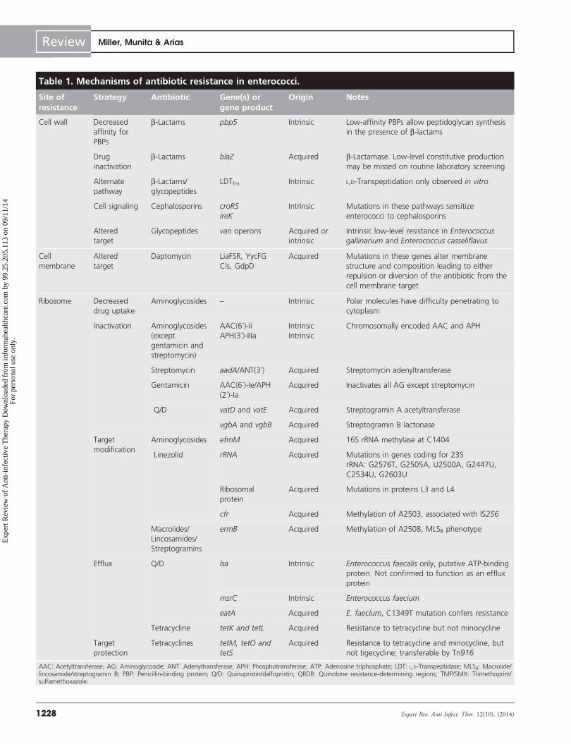
Table 1. Mechanisms of antibiotic resistance in enterococci.
Site ofresistance
Strategy Antibiotic Gene(s) orgene product
Origin Notes
Cell wall Decreased
affinity for
PBPs
b-Lactams pbp5 Intrinsic Low-affinity PBPs allow peptidoglycan synthesis
in the presence of b-lactams
Drug
inactivation
b-Lactams blaZ Acquired b-Lactamase. Low-level constitutive production
may be missed on routine laboratory screening
Alternate
pathway
b-Lactams/
glycopeptides
LDTfm Intrinsic L,D-Transpeptidation only observed in vitro
Cell signaling Cephalosporins croRS
ireK
Intrinsic Mutations in these pathways sensitize
enterococci to cephalosporins
Altered
target
Glycopeptides van operons Acquired or
intrinsic
Intrinsic low-level resistance in Enterococcusgallinarium and Enterococcus casseliflavus
Cell
membrane
Altered
target
Daptomycin LiaFSR, YycFG
Cls, GdpD
Acquired Mutations in these genes alter membrane
structure and composition leading to either
repulsion or diversion of the antibiotic from the
cell membrane target
Ribosome Decreased
drug uptake
Aminoglycosides – Intrinsic Polar molecules have difficulty penetrating to
cytoplasm
Inactivation Aminoglycosides
(except
gentamicin and
streptomycin)
AAC(6’)-IiAPH(3´)-IIIa
Intrinsic
Intrinsic
Chromosomally encoded AAC and APH
Streptomycin aadA/ANT(3’) Acquired Streptomycin adenyltransferase
Gentamicin AAC(6´)-Ie/APH
(2´)-Ia
Acquired Inactivates all AG except streptomycin
Q/D vatD and vatE Acquired Streptogramin A acetyltransferase
vgbA and vgbB Acquired Streptogramin B lactonase
Target
modification
Aminoglycosides efmM Acquired 16S rRNA methylase at C1404
Linezolid rRNA Acquired Mutations in genes coding for 23S
rRNA: G2576T, G2505A, U2500A, G2447U,
C2534U, G2603U
Ribosomal
protein
Acquired Mutations in proteins L3 and L4
cfr Acquired Methylation of A2503, associated with IS256
Macrolides/
Lincosamides/
Streptogramins
ermB Acquired Methylation of A2508, MLSB phenotype
Efflux Q/D lsa Intrinsic Enterococcus faecalis only, putative ATP-binding
protein. Not confirmed to function as an efflux
protein
msrC Intrinsic Enterococcus faecium
eatA Acquired E. faecium, C1349T mutation confers resistance
Tetracycline tetK and tetL Acquired Resistance to tetracycline but not minocycline
Target
protection
Tetracyclines tetM, tetO and
tetS
Acquired Resistance to tetracycline and minocycline, but
not tigecycline; transferable by Tn916
AAC: Acetyltransferase; AG: Aminoglycoside; ANT: Adenyltransferase; APH: Phosphotransferase; ATP: Adenosine triphosphate; LDT: L,D-Transpeptidase; MLSB: Macrolide/lincosamide/streptogramin B; PBP: Penicillin-binding protein; Q/D: Quinupristin/dalfopristin; QRDR: Quinolone resistance-determining regions; TMP/SMX: Trimethoprim/sulfamethoxazole.
Review Miller, Munita & Arias
1228 Expert Rev. Anti Infect. Ther. 12(10), (2014)
Exp
ert R
evie
w o
f A
nti-
infe
ctiv
e T
hera
py D
ownl
oade
d fr
om in
form
ahea
lthca
re.c
om b
y 99
.25.
205.
113
on 0
9/11
/14
For
pers
onal
use
onl
y.
gram-positive bacteria, including MRSA and VRE. This com-pound is US FDA-approved for the treatment of complicatedskin and soft tissue infections and abdominal infections. Giventhe low achievable serum concentrations of the antibiotic, mono-therapy in serious infections is discouraged. Similar to all tetracy-clines, tigecycline binds to the 16S rRNA of the 30S subunit ofthe ribosome and inhibits the association of the aminoacyl-tRNA[108]. Unlike other tetracyclines, however, MICs are not affectedby typical tetracycline resistance determinants [109]. To date, therehave been two published reports of tigecycline resistance inenterococci, both related to intra-abdominal procedures [110,111].The mechanism of resistance remains unknown.
Mechanisms of resistance to agents interfering withnucleic acid replication, transcription & synthesisQuinolones
The introduction and relaxation of supercoils in DNA isimportant for transcription and the replication of the genomeprior to cell division. The quinolones target two of the enzymesresponsible for this process, DNA gyrase and topoisomerase IV.Both enzymes are tetramers composed of two different subu-nits: GyrA and GyrB form the DNA gyrase complex, while thetopoisomerase IV is composed of ParC and ParE. The DNAgyrase introduces negative supercoils into the DNA strand,priming it for the initiation of replication and relaxing thestrand in front of the advancing polymerase. On the otherhand, topoisomerase IV separates the newly replicated inter-locking DNA double helix allowing for segregation to occurbefore cell division. Both processes require double-strandedbreaks in the DNA, and stabilization of the enzyme/DNAcomplex by quinolones results in a disruption of strand conti-nuity and arrest of replication [112]. There is evidence that theremay be differential inhibition of these two enzymes betweengram-positive and gram-negative bacteria and varying grades ofinhibition between the different types of quinolones [112,113].
Enterococci demonstrate low levels of intrinsic resistance tothe quinolones, but can acquire high-level resistance throughseveral mechanisms. Mutations in the target genes, specificallygyrA and parC, have been described in E. faecium and E. faecalis,
but are absent from E. gallinarum and E. casseliflavus [114–116].These changes affect the so-called ‘quinolone resistance-determining regions’, which presumably alter the binding affin-ity of the antibiotic. Externalization of the antibiotic throughefflux pumps is another well-described mechanism of quinoloneresistance. Among them, NorA and PmrA have been implicatedin quinolone resistance in S. aureus and Streptococcus pneumoniae[117], and the former has also been described in E. faecium [112].A third mechanism of resistance, found in E. faecalis [118], ismediated by qnr and encodes for a protein with a series of pen-tapeptide repeats similar to the plasmid-borne quinolone resis-tance genes described in Enterobacteriaceae. The presence of thisprotein is likely to protect DNA gyrase by decreasing DNAbinding of the quinolone and the subsequent formation of thequinolone–gyrase complex [119].
Rifampicin
Rifampicin inhibits transcription of mRNA by binding to theb-subunit of the enterococcal DNA-dependent RNA polymer-ase. Resistance to these agents is widespread, occurring in65.9% of E. faecium isolates from the USA and 67.5% of thosefrom Europe [120]. Rifampicin resistance arises from a variety ofmutations in the rpoB gene that encodes for the b-subunit ofthe RNA polymerase. Interestingly, a specific mutation in rpoB(H486Y) in both E. faecalis and E. faecium was shown toincrease resistance to broad-spectrum cephalosporins, but didnot affect other classes of cell-wall acting antibiotics (includingampicillin and vancomycin) [121]. The authors postulated thatthis was due to differential transcription of genes related tocephalosporin resistance by the mutated polymerase. In anindependent study, assessing the fitness cost of rpoB mutationsin E. faecium, it was noted that the same H486Y substitutionshowed minimal deleterious effect on growth both in vitro andin vivo [122]. Finally, Rand et al. described an isolate of E. fae-cium that developed rifampicin resistance without any evidenceof mutations in the rpoB gene, enzymatic inactivation or effluxpumps. The exact mechanism of resistance is unclear, but inter-estingly, the resistant phenotype was reversible after incubationwith subinhibitory concentrations of DAP [123].
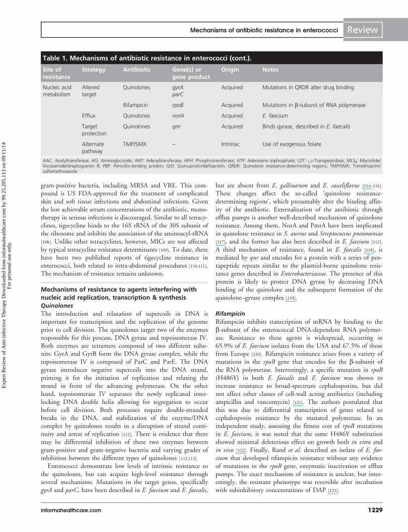
Table 1. Mechanisms of antibiotic resistance in enterococci (cont.).
Site ofresistance
Strategy Antibiotic Gene(s) orgene product
Origin Notes
Nucleic acid
metabolism
Altered
target
Quinolones gyrAparC
Acquired Mutations in QRDR alter drug binding
Rifampicin rpoB Acquired Mutations in b-subunit of RNA polymerase
Efflux Quinolones norA Acquired E. faecium
Target
protection
Quinolones qnr Acquired Binds gyrase, described in E. faecalis
Alternate
pathway
TMP/SMX – Intrinsic Use of exogenous folate
AAC: Acetyltransferase; AG: Aminoglycoside; ANT: Adenyltransferase; APH: Phosphotransferase; ATP: Adenosine triphosphate; LDT: L,D-Transpeptidase; MLSB: Macrolide/lincosamide/streptogramin B; PBP: Penicillin-binding protein; Q/D: Quinupristin/dalfopristin; QRDR: Quinolone resistance-determining regions; TMP/SMX: Trimethoprim/sulfamethoxazole.
Mechanisms of antibiotic resistance in enterococci Review
informahealthcare.com 1229
Exp
ert R
evie
w o
f A
nti-
infe
ctiv
e T
hera
py D
ownl
oade
d fr
om in
form
ahea
lthca
re.c
om b
y 99
.25.
205.
113
on 0
9/11
/14
For
pers
onal
use
onl
y.
Trimethoprim & sulfamethoxazole
Trimethoprim and sulfamethoxazole are inhibitors of bacterialenzymes involved in the folate synthesis pathway. Folic acid isnecessary to carry out a variety of important cellular functions,including synthesis of nucleic acids, particularly thymidine.Most bacteria are unable to take up exogenous folate from theenvironment and instead must synthesize it from the p-aminobenzoic acid precursor. Trimethoprim and sulfamethoxazoleinhibit successive enzymes in this pathway, limiting the produc-tion of dihydrofolate and its subsequent conversion to tetrahy-drofolate. Enterococci show susceptibility to these compoundswhen tested in vitro; however, these compounds are ineffectivein vivo due to the ability of enterococci to utilize exogenoussources of folate [124,125].
Alternative targets
In addition to the metabolic pathways targeted by traditionalantimicrobial compounds, the rise of resistance has generatedinterest in adjunctive treatments to enhance host response anddecrease the pathogenic potential of the enterococci. The presenceof virulence factors, while not directly responsible for resistance,influence the ability of bacteria to persist in a hostile environmentand resist host defenses. The widespread use of indwelling medi-cal devices (e.g., central venous catheters, Foley catheters andendotracheal tubes) provide both a breach of host barriers and asurface amenable to supporting biofilm formation. A host of mac-romolecules associated with the enterococcal cell surface andsecreted into the extracellular matrix act as passive barriers to pre-vent antimicrobial compounds from reaching their intended tar-gets [126]. Moreover, at the interface of the host mucosa withexternal milieu, CAMPs of the innate immune system keep anti-microbial populations in check. These CAMPs possess a widearray of antimicrobial properties (including the ability to disruptpeptidoglycan synthesis, CM structure and activate autolysins)and individual proteins often display multiple modes of action,which may in part explain their effectiveness in the face of ever-evolving bacterial resistance [127]. In E. faecalis, Kandaswamy et al.demonstrated that human b-defensins are targeted to the divisionseptum and specifically disrupt the translocation machineryresponsible for export of virulence-associated macromolecules,even at subinhibitory concentrations [128]. Furthermore, it hasbeen postulated that the long history of co-evolution betweenCAMPs and their bacterial targets could be fruitful grounds fordeveloping therapies that temper the acquisition and dissemina-tion of resistance determinants [129]. Host adaptive responses, spe-cifically humoral immunity, have been exploited through vaccinesto produce antibodies against pathogens. The lipoteichoic acidsand diheteroglycans present in enterococcal cell walls are anti-genic motifs capable of inducing an antibody response that is pro-tective against E. faecalis in a mouse bacteremia model [130].Antibodies directed against these epitopes could be importantadjuncts to antimicrobial therapy in the future; however, at pres-ent, they are limited by a lack of universal cross-reactivity and thepresence of polysaccharides that protect the lipoteichoic acidmotif from antibody binding to its corresponding target [131].
Expert commentaryEnterococci are prime examples of organisms with an impres-sive array of genetic versatility and unparalleled ability torecruit and express antimicrobial resistance determinants. Theseorganisms have adapted through time to outcompete other bac-teria in a specific biological niche such as the GI tract ofeukaryotic organisms. From a simple commensal and tamedmember of the intestinal microbiota, enterococci now haverisen in importance and have become one of the leading causesof intra-hospital infections. This untaming of MDR enterococcihas occurred with the massive increase in the use of antimicro-bials in clinical medicine, which has played a significant role inthe evolution and adaptation of these microorganisms. Indeed,selected by the use of broad-spectrum antimicrobials, their rug-ged durability enables them to persist and disseminate in thenosocomial environment. Furthermore, rising rates of obesity,diabetes and resulting comorbidities, advances in oncology andcritical care and a demographic shift as the population ages areimportant events in modern medicine that increase the rates ofhospital admissions and the prevalence of sicker patients.
Though enterococci lack the virulence armamentarium ofS. aureus or pneumococci, they often pose a challenging problemto clinicians since antibiotic choices to treat these microorgan-isms are now extremely limited. Often, clinicians are faced withthe dilemma of attempting to clear a deep-seated infection whilebalancing treatment-related toxicities. Such scenarios are not newto the treatment of enterococci. This is exemplified by a paperpublished in 1959 entitled ‘Deaf or Dead’ [132], in which theauthors had to deal with extreme aminoglycoside toxicity to treata patient with subacute endocarditis due to resistant E. faecalis.
In addition, enterococci is likely to function as a reservoir ofdrug resistance determinants and can serve as the springboard forthe spread of these genes to other gram-positive pathogens.Indeed, it is now well documented that VRE are the source ofvancomycin resistance genes that have been identified inMRSA [9,10]. Acquisition of vancomycin resistance in staphylococciwith subsequent dissemination of such strains is deemed as one ofthe most pressing public health issues worldwide. Further explora-tion of the mechanisms of enterococcal resistance may thereforepay dividends in treating other infections and in preventing thewidespread dissemination of resistance determinants in the future.
Five-year viewThe dawn of the 21st century is seeing the advent of what manyare calling the ‘post-antibiotic era’. As older therapies gave way tonewer drugs, bacteria rapidly responded with a diverse array ofdefense tactics. We are fighting today’s wars with yesterday’s strat-egies, much like the massed troop assaults against fixed fortifica-tions at the Somme and Verdun in World War I. Out of thesebattles, however, came new innovations, the rise of the airplaneand the tank, and with them new strategies. Just two decades later,the idea of combined arms, manifested as Blitzkrieg, or ‘lightningwar’ would change the battlefield of the 20th century.
Perhaps the idea of combined arms can be used to the clin-ician’s advantage in the conflicts of the post-antibiotic era.
Review Miller, Munita & Arias
1230 Expert Rev. Anti Infect. Ther. 12(10), (2014)
Exp
ert R
evie
w o
f A
nti-
infe
ctiv
e T
hera
py D
ownl
oade
d fr
om in
form
ahea
lthca
re.c
om b
y 99
.25.
205.
113
on 0
9/11
/14
For
pers
onal
use
onl
y.
Seemingly redundant therapy, such as the combination of theb-lactams, ampicillin and ceftriaxone, has been shown clinically tobe as efficacious as more traditional combinations with aminogly-cosides, with the benefit of less toxicity and the ability to bypasshigh-level resistance to aminoglycosides [133,134]. As more is knownabout the mechanisms by which enterococci subvert the assault ofmodern medicine, it will be possible to develop strategies that canbe used to turn enterococcal biology against itself. The inverserelationship between DAP susceptibility and sensitivity tob-lactams [71], combinations of cephalosporins and fosfomycin [135]
and the synergy between DAP and rifampicin [123] could provide away to ‘hold the line’ against the rising tide of resistance. Oncenew agents are deployed, combination therapy could also assist inthe prevention of resistance using the same rationale as combinedantiretroviral therapy in human immunodeficiency virus infection.
Greater understanding of mechanisms of antibiotic action andresistance also offers the hope of new therapeutic targets to reloadan empty antibiotic pipeline. Advances in the understanding ofmembrane physiology provided by research into DAP could pro-vide new ways of attacking enterococcal phospholipid synthesis.Indeed, new agents need not kill on their own. By targeting thesensor and effector proteins of a stress response pathway, it maybe possible to disable the defense network of a resistant microbe,
leaving it vulnerable to decades old therapy. Moreover, monoclo-nal antibodies, which have been successfully used in cancer andauto-immune diseases, may be designed to specifically targetenterococcal signal transduction pathways, leaving resistance geneclusters silent while the antibiotic pours in. Genomic medicineoffers the promise of individually targeted treatments for everypatient. There is no good reason to think antimicrobial therapyshould be any different.
Financial & competing interests disclosure
JM Munita is supported in part by a grant from the Chilean Ministry of
Education and by Clinical Alemana de Santiago and Universidad
del Desarrollo School of Medicine, Chile. CA Arias is supported by
NIH-NIAD grant R01 AI093749. CA Arias has received lecture fees,
research support and consulting fees from Pfizer Inc. Lectures and consulting
honoraria from Novartis, Cubist, Forest Pharmaceuticals, Astra-Zeneca and
Bayer Pharmaceuticals. Research support to CA Arias has been provided by
Forest Pharmaceuticals and Theravance Inc. The authors have no other
relevant affiliations or financial involvement with any organization or entity
with a financial interest in or financial conflict with the subject matter or
materials discussed in the manuscript apart from those disclosed.
No writing assistance was utilized in the production of this
manuscript.
Key issues
• Enterococci are increasingly common nosocomial pathogens. The changing epidemiology of enterococcal infections with the rise of
multidrug-resistant Enterococcus faecium in hospitals worldwide has important therapeutic implications.
• Ampicillin plus an aminoglycoside (gentamicin or streptomycin), the traditional combination for severe enterococcal infections, is
increasingly ineffective due to emergence of resistance.
• Resistance to ampicillin in E. faecium is mediated by PBP5, a transpeptidase that functions in the presence of high concentrations of b-lactams.
• Resistance to cephalosporins is an intrinsic feature of enterococci and is mediated in part by CroRS, a two-component signaling pathway, and a
system with competing kinase and phosphatase activity (IreK and IreP) that function to control expression of resistance while preserving fitness.
• Glycopeptide resistance is mediated by the van operons, of which nine have currently been described in enterococci. In general, they
consist of genes that encode two-component signal transduction systems, which activate the genes responsible for the synthesis of
modified peptidoglycan precursors and destruction of ‘normal’ (D-alanine ending) precursors.
• The vanA gene cluster, conferring resistance to vancomycin and teicoplanin, is the most commonly encountered cause of resistance to
glycopeptides in the clinical setting and can be transferred between enterococci.
• Enterococci are often intrinsically resistant to most aminoglycosides due to the presence of the 6´-acetyltransferase enzyme AAC(6´)-Ii.
As such, only gentamicin or streptomycin should be used to achieve synergy with cell-wall agents.
• Ampicillin plus ceftriaxone is a b-lactam combination against Enterococcus faecalis that appears to be as good as the ‘standard of care’
(ampicillin plus gentamicin) but with much less toxicity.
• Daptomycin (DAP) resistance is associated with multiple mutations but usually involves genes encoding regulatory systems that control
cell envelope homeostasis and enzymes that synthesize cell membrane phospholipids and/or are involved in phospholipid metabolism.
• The combination of DAP with b-lactams may offer promise in the future to restore DAP susceptibility and prevent emergence of resistance.
• Linezolid resistance in enterococci continues to be low, but increasing reports in enterococci have been associated with longer duration
of therapy.
• Quinupristin/dalfopristin retains bactericidal activity in vitro against in E. faecium (not E. faecalis), but the presence of Erm methylases (which
are frequently found in clinical isolates) may decrease the bactericidal effect in vivo and reduce therapeutic efficacy as monotherapy.
• Enterococci are often resistant to quinolones with several mechanisms of resistance including mutation of the quinolone targets, efflux
pumps and a transferable plasmid containing qnr, a gene similar to plasmid-borne quinolone resistance in the Enterobacteriaceae.
• Continued research focused on understanding the mechanisms of resistance in enterococci is important to develop novel combination
therapies or new antimicrobial compounds.
Mechanisms of antibiotic resistance in enterococci Review
informahealthcare.com 1231
Exp
ert R
evie
w o
f A
nti-
infe
ctiv
e T
hera
py D
ownl
oade
d fr
om in
form
ahea
lthca
re.c
om b
y 99
.25.
205.
113
on 0
9/11
/14
For
pers
onal
use
onl
y.
References
Papers of special note have been highlighted as:
• of interest
•• of considerable interest
1. Murray BE. The life and times of the
Enterococcus. Clin Microbiol Rev 1990;3:
46-65
2. Hidron AI, Edwards JR, Patel J, et al.
NHSN annual update:
antimicrobial-resistant pathogens associated
with healthcare-associated infections: annual
summary of data reported to the National
Healthcare Safety Network at the Centers
for Disease Control and Prevention,
2006-2007. Infect Control Hosp Epidemiol
2008;29:996-1011
3. Williamson R, Calderwood SB,
Moellering RC, et al. Studies on the
mechanism of intrinsic resistance to
beta-lactam antibiotics in group D
streptococci. J Gen Microbiol 1983;129:
813-22
4. Schatz A and Waksman S. Effect of
streptomycin and other antibiotic substances
upon Mycobacterium tuberculosis and
related organisms. Proc Soc Exp Biol Med
1944;57:244-8
5. Robbins WC and Tompsett R. Treatment
of enterococcal endocarditis and bacteremia;
results of combined therapy with penicillin
and streptomycin. Am J Med 1951;10:
278-99
6. Baddour LM, Wilson WR, Bayer AS, et al.
Infective endocarditis: diagnosis,
antimicrobial therapy, and management of
complications: a statement for healthcare
professionals from the Committee on
Rheumatic Fever, Endocarditis, and
Kawasaki Disease, Council on
Cardiovascular Disease in the Young, and
the Councils on Clinical Cardiology, Stroke,
and Cardiovascular Surgery and Anesthesia,
American Heart Association: endorsed by
the Infectious Diseases Society of America.
Circulation 2005;111:e394-434
7. Lebreton F, van Schaik W, McGuire AM,
et al. Emergence of Epidemic
Multidrug-Resistant Enterococcus faecium
from Animal and Commensal Strains. MBio
2013;4:e00534-13
8. Arias CA, Murray BE. The rise of the
Enterococcus: beyond vancomycin
resistance. Nat Rev Microbiol 2012;10:
266-78
9. Chang S, Sievert D, Hageman J, et al.
Infection with vancomycin-resistant
Staphylococcus aureus containing the
vanA resistance gene. N Engl J Med
2003;348:1342-7
10. Ray A, Pultz N, Bhalla A, et al. Coexistence
of vancomycin-resistant enterococci and
Staphylococcus aureus in the intestinal tracts
of hospitalized patients. Clin Infect Dis
2003;37:875-81
11. Williamson R, Gutmann L, Horaud T,
et al. Use of Penicillin-binding Proteins for
the identification of enterococci. J Gen
Microbiol 1986;132:1929-37
12. Duez C, Hallut S, Rhazi N, et al. The
ponA gene of Enterococcus faecalis
JH2-2 codes for a low-affinity class
A penicillin-binding protein. J Bacteriol
2004;186:4412-16
13. Signoretto C, Boaretti M and Canepari P.
Cloning, sequencing and expression in
Escherichia coli of the low-affinity penicillin
binding protein of Enterococcus faecalis.
FEMS Microbiol Lett 1994;123:99-106
14. Sifaoui F, Arthur M, Rice L, et al. Role of
Penicillin-Binding Protein 5 in Expression
of Ampicillin Resistance and Peptidoglycan
Structure in Enterococcus faecium.
Antimicrob Agents Chemother 2001;45:
2594-7
15. Rice LB, Carias LL, Hutton-Thomas R,
et al. Penicillin-Binding Protein 5 and
Expression of Ampicillin Resistance in
Enterococcus faecium. Antimicrob Agents
Chemother 2001;45:1480-6
16. Fontana R, Aldegheri M, Ligozzi M, et al.
Overproduction of a low-affinity penicillin-
binding protein and high-level ampicillin
resistance in Enterococcus faecium.
Antimicrob Agents Chemother 1994;38:
1980-3
17. Rice LB, Bellais S, Carias LL, et al. Impact
of Specific pbp5 Mutations on Expression
of Beta-Lactam Resistance in Enterococcus
faecium. Antimicrob Agents Chemother
2004;48:3028-32
18. Galloway-Pena JR, Rice LB, Murray BE.
Analysis of PBP5 of early U.S. isolates of
Enterococcus faecium: sequence variation
alone does not explain increasing ampicillin
resistance over time. Antimicrob Agents
Chemother 2011;55:3272-7
19. Duez C, Zorzi W, Sapunaric F, et al. The
penicillin resistance of Enterococcus faecalis
JH2-2r results from an overproduction of
the low-affinity penicillin-binding protein
PBP4 and does not involve a psr-like gene.
Microbiology 2001;147:2561-9
20. Ono S, Muratani T, Matsumoto T.
Mechanisms of Resistance to Imipenem and
Ampicillin in Enterococcus faecalis.
Antimicrob Agents Chemother 2005;49:
2954-8
21. Murray BE. Beta-lactamase-producing
enterococci. Antimicrob Agents Chemother
1992;36:2355-9
22. Coudron PE, Markowitz SM, Wong ES.
Isolation of a betalactamase-producing,
aminoglycoside-resistant strain of
Enterococcus faecium. Antimicrob Agents
Chemother 1992;36:1125-6
23. Hackbarth CJ, Chambers HF. blaI and
blaR1 regulate beta-lactamase and PBP 2a
production in methicillin-resistant
Staphylococcus aureus. Antimicrob Agents
Chemother 1993;37:1144-9
24. Sarti M, Campanile F, Sabia C, et al.
Polyclonal diffusion of
beta-lactamase-producing Enterococcus
faecium. J Clin Microbiol 2012;50:169-72
25. Mainardi JL, Legrand R, Arthur M, et al.
Novel mechanism of beta-lactam resistance
due to bypass of DD-transpeptidation in
Enterococcus faecium. J Biol Chem
2000;275:16490-6
26. Sacco E, Hugonnet JE, Josseaume N, et al.
Activation of the L,D-transpeptidation
peptidoglycan cross-linking pathway by a
metallo-D,D-carboxypeptidase in
Enterococcus faecium. Mol Microbiol
2010;75:874-85
• Though only seen in vitro, this is a novel
pathway of resistance to many cell-wall
active agents. It demonstrates the
resiliency of the enterococci.
27. Cremniter J, Mainardi JL, Josseaume N,
et al. Novel mechanism of resistance to
glycopeptide antibiotics in Enterococcus
faecium. J Biol Chem 2006;281:32254-62
28. Rice LB, Carias LL, Rudin S, et al. Role of
class A penicillin-binding proteins in the
expression of beta-lactam resistance in
Enterococcus faecium. J Bacteriol 2009;191:
3649-56
• Detailed investigations in both
Enterococcus faecalis and Enterococcusfaecium that outlines the importance of
the partner glycosyltransferase to PBP5 in
resistance to b-lactam antibiotics.
29. Arbeloa A, Segal H, Hugonnet JE, et al.
Role of class A penicillin-binding proteins
in PBP5-mediated beta-lactam resistance in
Enterococcus faecalis. J Bacteriol 2004;186:
1221-8
• Detailed investigations in both E. faecalisand E. faecium that outlines the
importance of the partner
glycosyltransferase to PBP5 in resistance
to b-lactam antibiotics.
30. Hancock LE, Perego M. Systematic
inactivation and phenotypic characterization
Review Miller, Munita & Arias
1232 Expert Rev. Anti Infect. Ther. 12(10), (2014)
Exp
ert R
evie
w o
f A
nti-
infe
ctiv
e T
hera
py D
ownl
oade
d fr
om in
form
ahea
lthca
re.c
om b
y 99
.25.
205.
113
on 0
9/11
/14
For
pers
onal
use
onl
y.
of two-component signal transduction
systems of Enterococcus faecalis V583. J
Bacteriol 2004;186:7951-8
31. Comenge Y, Quintiliani R Jr, Li L, et al.
The CroRS two-component regulatory
system is required for intrinsic beta-lactam
resistance in Enterococcus faecalis. J
Bacteriol 2003;185:7184-92
32. Le Breton Y, Muller C, Auffray Y, et al.
New insights into the Enterococcus faecalis
CroRS two-component system obtained
using a differential-display random
arbitrarily primed PCR approach. Appl
Environ Microbiol 2007;73:3738-41
33. Muller C, Le Breton Y, Morin T, et al. The
response regulator CroR modulates
expression of the secreted stress-induced
SalB protein in Enterococcus faecalis. J
Bacteriol 2006;188:2636-45
34. Kristich CJ, Wells CL, Dunny GM.
A eukaryotic-type Ser/Thr kinase in
Enterococcus faecalis mediates antimicrobial
resistance and intestinal persistence. Proc
Natl Acad Sci USA 2007;104:3508-13
35. Kristich CJ, Little JL, Hall CL, et al.
Reciprocal regulation of cephalosporin
resistance in Enterococcus faecalis. MBio
2011;2:e00199-11
36. Hall CL, Tschannen M, Worthey EA, et al.
IreB, a Ser/Thr kinase substrate, influences
antimicrobial resistance in Enterococcus
faecalis. Antimicrob Agents Chemother
2013;57:6179-86
37. Vesic D, Kristich CJ. MurAA is required
for intrinsic cephalosporin resistance of
Enterococcus faecalis. Antimicrob Agents
Chemother 2012;56:2443-51
38. Courvalin P. Vancomycin resistance in
gram-positive cocci. Clin Infect Dis
2006;42:S25-34
• Informative review of vancomycin
resistance in enterococci.
39. Guardabassi L, Agersø Y. Genes
homologous to glycopeptide resistance
vanA are widespread in soil microbial
communities. FEMS Microbiol Lett
2006;259:221-5
40. Boyd DA, Willey BM, Fawcett D, et al.
Molecular characterization of Enterococcus
faecalis N06-0364 with low-level
vancomycin resistance harboring a novel D-
Ala-D-Ser gene cluster, vanL. Antimicrob
Agents Chemother 2008;52:2667-72
41. Xu X, Lin D, Yan G, et al. vanM, a new
glycopeptide resistance gene cluster found in
Enterococcus faecium. Antimicrob Agents
Chemother 2010;54:4643-7
42. Lebreton F, Depardieu F, Bourdon N, et al.
D-Ala-d-Ser VanN-type transferable
vancomycin resistance in Enterococcus
faecium. Antimicrob Agents Chemother
2011;55:4606-12
43. Depardieu F, Podglajen I, Leclercq R, et al.
Modes and modulations of antibiotic
resistance gene expression. Clin Microbiol
Rev 2007;20:79-114
•• Comprehensive review of the regulation
of antibiotic resistance including
two-component signaling, the role of
insertion sequences and
post-transcriptional regulation.
44. Arthur M, Depardieu F, Molinas C, et al.
The vanZ gene of Tn1546 from
Enterococcus faecium BM4147 confers
resistance to teicoplanin. Gene 1995;154:
87-92
45. Baptista M, Depardieu F, Reynolds P, et al.
Mutations leading to increased levels of
resistance to glycopeptide antibiotics in
VanB-type enterococci. Mol Microbiol
1997;25:93-105
46. Arias CA, Courvalin P, Reynolds PE. vanC
cluster of vancomycin-resistant Enterococcus
gallinarum BM4174. Antimicrob Agents
Chemother 2000;44:1660-6
47. Arias CA, Martın-Martinez M, Blundell TL,
et al. Characterization and modelling of
VanT: a novel, membrane-bound, serine
racemase from vancomycin-resistant
Enterococcus gallinarum BM4174. Mol
Microbiol 1999;31:1653-64
48. Fines M, Perichon B, Reynolds P, et al.
VanE, a new type of acquired glycopeptide
resistance in Enterococcus faecalis BM4405.
Antimicrob Agents Chemother 1999;43:
2161-4
49. Abadıa Patino L, Courvalin P, Perichon B.
vanE gene cluster of vancomycin-resistant
Enterococcus faecalis BM4405. J Bacteriol
2002;184:6457-64
50. Depardieu F, Kolbert M, Pruul H, et al.
VanD-type vancomycin-resistant
Enterococcus faecium and Enterococcus
faecalis. Antimicrob Agents Chemother
2004;48:3892-904
51. Yowler CJ, Blinkhorn RJ, Fratianne RB.
Vancomycin-dependent enterococcal strains:
case report and review. J Trauma 2000;48:
783-5
52. San Millan A, Depardieu F, Godreuil S,
et al. VanB-type Enterococcus faecium
clinical isolate successively inducibly resistant
to, dependent on, and constitutively
resistant to vancomycin. Antimicrob Agents
Chemother 2009;53:1974-82
53. Steenbergen JN, Alder J, Thorne GM, et al.
Daptomycin: a lipopeptide antibiotic for the
treatment of serious Gram-positive
infections. J Antimicrob Chemother
2005;55:283-8
54. Muraih JK, Harris J, Taylor SD, et al.
Characterization of daptomycin
oligomerization with perylene excimer
fluorescence: stoichiometric binding of
phosphatidylglycerol triggers oligomer
formation. Biochim Biophys Acta
2012;1818:673-8
55. Zhang T, Muraih JK, Tishbi N, et al.
Cardiolipin prevents membrane
translocation and permeabilization by
daptomycin. J Biol Chem 2014;289:
11584-91
56. Pogliano J, Pogliano N, Silverman JA.
Daptomycin-mediated reorganization of
membrane architecture causes
mislocalization of essential cell division
proteins. J Bacteriol 2012;194:4494-504
57. Canton R, Ruiz-Garbajosa P, Chaves R,
et al. A potential role for daptomycin in
enterococcal infections: what is the
evidence? J Antimicrob Chemother
2010;65:1126-36
58. Arias CA, Panesso D, McGrath DM, et al.
Genetic basis for in vivo daptomycin
resistance in enterococci. N Engl J Med
2011;365:892-900
•• Identification of multiple genes associated
with daptomycin (DAP) resistance from a
clinical strain pair of E. faecalis thatdeveloped resistance during therapy.
59. Jordan S, Junker A, Helmann JD, et al.
Regulation of LiaRS-dependent gene
expression in bacillus subtilis: identification
of inhibitor proteins, regulator binding sites,
and target genes of a conserved cell envelope
stress-sensing two-component system.
J Bacteriol 2006;188:5153-66
60. Munita JM, Tran TT, Diaz L, et al. A liaF
codon deletion abolishes daptomycin
bactericidal activity against
vancomycin-resistant Enterococcus faecalis.
Antimicrob Agents Chemother 2013;57:
2831-3
61. Palmer KL, Daniel A, Hardy C, et al.
Genetic basis for daptomycin resistance in
enterococci. Antimicrob Agents Chemother
2011;55:3345-56
62. Jones T, Yeaman MR, Sakoulas G, et al.
Failures in clinical treatment of
Staphylococcus aureus Infection with
daptomycin are associated with alterations
in surface charge, membrane phospholipid
asymmetry, and drug binding. Antimicrob
Agents Chemother 2008;52:269-78
Mechanisms of antibiotic resistance in enterococci Review
informahealthcare.com 1233
Exp
ert R
evie
w o
f A
nti-
infe
ctiv
e T
hera
py D
ownl
oade
d fr
om in
form
ahea
lthca
re.c
om b
y 99
.25.
205.
113
on 0
9/11
/14
For
pers
onal
use
onl
y.
63. Tran TT, Panesso D, Mishra NN, et al.
Daptomycin-resistant Enterococcus faecalis
diverts the antibiotic molecule from the
division septum and remodels cell
membrane phospholipids. MBio 2013;4:13
• Investigation of the response to DAP in
E. faecalis that demonstrated changes in
membrane phospholipid content and
subsequent relocalization of DAP from
the dividing septum, suggesting a
mechanism for DAP resistance.
64. Miller C, Kong J, Tran TT, et al.
Adaptation of enterococcus faecalis to
daptomycin reveals an ordered progression
to resistance. Antimicrob Agents Chemother
2013;57:5373-83
65. Tran TT, Panesso D, Gao H, et al.
Whole-genome analysis of a
daptomycin-susceptible Enterococcus
faecium strain and its daptomycin-resistant
variant arising during therapy. Antimicrob
Agents Chemother 2013;57(1):261-8
•• Genomic analysis of the changes in
E. faecium arising as a consequence of
DAP therapy, including the newly
implicated YycFG regulon.
66. Dubrac S, Bisicchia P, Devine KM, et al.
A matter of life and death: cell wall
homeostasis and the WalKR (YycGF)
essential signal transduction pathway. Mol
Microbiol 2008;70:1307-22
67. Mishra NN, Bayer AS, Tran TT, et al.
Daptomycin resistance in enterococci is
associated with distinct alterations of cell
membrane phospholipid content. PLoS One
2012;7:e43958
68. Diaz L, Tran TT, Munita JM, et al.
Whole-Genome Analyses of Enterococcus
faecium Isolates with Diverse Daptomycin
MICs. Antimicrob Agents Chemother
2014;58:4527-34
69. Munita J, Panesso D, Diaz L, et al.
Correlation between mutations in liaFSR of
Enterococcus faecium and MIC of
daptomycin: revisiting daptomycin
breakpoints. Antimicrob Agents Chemother
2012;56:4354-9
70. Munita JM, Mishra NN, Alvarez D, et al.
Failure of High-Dose Daptomycin for
Bacteremia Caused by Daptomycin
Susceptible Enterococcus faecium Harboring
LiaSR Substitutions. Clin Infect Dis 2014.
[Epub ahead of print]
71. Sakoulas G, Bayer AS, Pogliano J, et al.
Ampicillin enhances daptomycin- and
cationic host defense peptide-mediated
killing of ampicillin- and
vancomycin-resistant Enterococcus faecium.
Antimicrob Agents Chemother 2012;56:
838-44
72. Hall Snyder A, Werth BJ, Barber KE, et al.
Evaluation of the novel combination of
daptomycin plus ceftriaxone against
vancomycin-resistant enterococci in an
in vitro pharmacokinetic/pharmacodynamic
simulated endocardial vegetation model. J
Antimicrob Chemother 2014;69(8):2148-54
73. Sakoulas G, Rose W, Nonejuie P, et al.
Ceftaroline restores daptomycin activity
against daptomycin-nonsusceptible
vancomycin-resistant Enterococcus faecium.
Antimicrob Agents Chemother 2014;58:
1494-500
74. Costa Y, Galimand M, Leclercq R, et al.
Characterization of the chromosomal aac
(6’)-Ii gene specific for Enterococcusfaecium. Antimicrob Agents Chemother
1993;37:1896-903
75. Galimand M, Schmitt E, Panvert M, et al.
Intrinsic resistance to aminoglycosides in
Enterococcus faecium is conferred by the
16S rRNA m5C1404-specific
methyltransferase EfmM. RNA 2011;17:
251-62
76. Eliopoulos GM, Farber BF, Murray BE,
et al. Ribosomal resistance of clinical
enterococcal to streptomycin isolates.
Antimicrob Agents Chemother 1984;25:
398-9
77. Krogstad D, Korfhagen T, Moellering R,
et al. Aminoglycoside-inactivating enzymes
in clinical isolates of Streptococcus faecalis.
An explanation for resistance to antibiotic
synergism. J Clin Invest 1978;62:480-6
78. Courvalin P, Carlier C, Collatz E.
Plasmid-mediated resistance to aminocyclitol
antibiotics in group D streptococci. J
Bacteriol 1980;143:541-51
79. Chow J, Zervos M, Lerner S, et al. A novel
gentamicin resistance gene in Enterococcus.
Antimicrob Agents Chemother 1997;41:
511-14
80. Tsai S, Zervos M, Clewell D, et al. A new
high-level gentamicin resistance gene, aph
(2’’)-Id, in Enterococcus spp. Antimicrob
Agents Chemother 1998;42:1229-32
81. Chow J. Aminoglycoside resistance in
enterococci. Clin Infect Dis 2000;31:586-9
82. Shinabarger D, Marotti K, Murray R, et al.
Mechanism of action of oxazolidinones:
effects of linezolid and eperezolid on
translation reactions. Antimicrob Agents
Chemother 1997;41:2132-6
83. Leach K, Swaney S, Colca J, et al. The site
of action of oxazolidinone antibiotics in
living bacteria and in human mitochondria.
Mol Cell 2007;26:393-402
84. Marshall S, Donskey C, Hutton-Thomas R,
et al. Gene dosage and linezolid resistance
in Enterococcus faecium and Enterococcus
faecalis. Antimicrob Agents Chemother
2002;46:3334-6
85. Boumghar-Bourtchaı L, Dhalluin A,
Malbruny B, et al. Influence of
recombination on development of
mutational resistance to linezolid in
Enterococcus faecalis JH2-2. Antimicrob
Agents Chemother 2009;53:4007-9
86. Bourgeois-Nicolaos N, Massias L, Couson B,
et al. Dose dependence of emergence of
resistance to linezolid in Enterococcus faecalis
in vivo. J Infect Dis 2007;195:1480-8
87. Locke JB, Hilgers M, Shaw KJ. Mutations
in ribosomal protein L3 are associated with
oxazolidinone resistance in staphylococci of
clinical origin. Antimicrob Agents
Chemother 2009;53:5275-8
88. Chen H, Wu W, Ni M, et al.
Linezolid-resistant clinical isolates of
enterococci and Staphylococcus cohnii from
a multicentre study in China: molecular
epidemiology and resistance mechanisms.
Int J Antimicrob Agents 2013;42:317-21
89. Toh S, Xiong L, Arias C, et al. Acquisition
of a natural resistance gene renders a clinical
strain of methicillin-resistant Staphylococcus
aureus resistant to the synthetic antibiotic
linezolid. Mol Microbiol 2007;64:1506-14
90. Mendes R, Deshpande L, Castanheira M,
et al. First report of cfr-mediated resistance
to linezolid in human staphylococcal clinical
isolates recovered in the United States.
Antimicrob Agents Chemother 2008;52:
2244-6
91. Diaz L, Kiratisin P, Mendes R, et al.
Transferable plasmid-mediated resistance to
linezolid due to cfr in a human clinical
isolate of Enterococcus faecalis. Antimicrob
Agents Chemother 2012;56:3917-22
92. Hennig S and Ziebuhr W. Characterization
of the transposase encoded by IS256, the
prototype of a major family of bacterial
insertion sequence elements. J Bacteriol
2010;192:4153-63
93. Diaz L, Kontoyiannis DP, Panesso D, et al.
Dissecting the mechanisms of linezolid
resistance in a Drosophila melanogaster
infection model of Staphylococcus aureus.
J Infect Dis 2013;208:83-91
94. Singh K, Weinstock G, Murray B. An
Enterococcus faecalis ABC homologue (Lsa)
is required for the resistance of this species
to clindamycin and
quinupristin-dalfopristin. Antimicrob Agents
Chemother 2002;46:1845-50
Review Miller, Munita & Arias
1234 Expert Rev. Anti Infect. Ther. 12(10), (2014)
Exp
ert R
evie
w o
f A
nti-
infe
ctiv
e T
hera
py D
ownl
oade
d fr
om in
form
ahea
lthca
re.c
om b
y 99
.25.
205.
113
on 0
9/11
/14
For
pers
onal
use
onl
y.
95. Hershberger E, Donabedian S,
Konstantinou K, et al.
Quinupristin-dalfopristin resistance in
gram-positive bacteria: mechanism of
resistance and epidemiology. Clin Infect Dis
2004;38:92-8
96. Portillo A, Ruiz-Larrea F, Zarazaga M, et al.
Macrolide resistance genes in Enterococcus
spp. Antimicrob Agents Chemother
2000;44:967-71
97. Weisblum B. Erythromycin resistance by
ribosome modification. Antimicrob Agents
Chemother 1995;39:577-85
98. Canu A, Leclercq R. Overcoming bacterial
resistance by dual target inhibition: the case
of streptogramins. Curr Drug Targets Infect
Disord 2001;1:215-25
99. Werner G, Klare I, Witte W. Molecular
analysis of streptogramin resistance in
enterococci. Int J Med Microbiol 2002;292:
81-94
100. Korczynska M, Mukhtar T, Wright G,
et al. Structural basis for streptogramin B
resistance in Staphylococcus aureus by
virginiamycin B lyase. Proc Natl Acad Sci
USA 2007;104:10388-93
101. Fantin B, Leclercq R, Garry L, et al.
Influence of inducible cross-resistance to
macrolides, lincosamides, and streptogramin
B-type antibiotics in Enterococcus faecium
on activity of quinupristin-dalfopristin
in vitro and in rabbits with experimental
endocarditis. Antimicrob Agents Chemother
1997;41:931-5
102. Isnard C, Malbruny B, Leclercq R, et al.
Genetic basis for in vitro and in vivo
resistance to lincosamides, streptogramins A,
and pleuromutilins (LSAP phenotype) in
Enterococcus faecium. Antimicrob Agents
Chemother 2013;57:4463-9
103. Schnappinger D, Hillen W. Tetracyclines:
antibiotic action, uptake, and resistance
mechanisms. Arch Microbiol 1996;165:
359-69
104. Chopra I and Roberts M. Tetracycline
antibiotics: mode of action, applications,
molecular biology, and epidemiology of
bacterial resistance. Microbiol Mol Biol Rev
2001;65:232-60
105. Schwarz S, Cardoso M, Wegener HC.
Nucleotide sequence and phylogeny of the
tet(L) tetracycline resistance determinant
encoded by plasmid pSTE1 from
Staphylococcus hyicus. Antimicrob Agents
Chemother 1992;36:580-8
106. Pepper K, Horaud T, Le Bouguenec C,
et al. Location of antibiotic resistance
markers in clinical isolates of Enterococcus
faecalis with similar antibiotypes.
Antimicrob Agents Chemother 1987;31:
1394-402
107. Bentorcha F, De Cespedes G, Horaud T.
Tetracycline resistance heterogeneity in
Enterococcus faecium. Antimicrob Agents
Chemother 1991;35:808-12
108. Bauer G, Berens C, Projan S, et al.
Comparison of tetracycline and tigecycline
binding to ribosomes mapped by
dimethylsulphate and drug-directed Fe2+
cleavage of 16S rRNA. J Antimicrob
Chemother 2004;53:592-9
109. Fluit A, Florijn A, Verhoef J, et al. Presence
of tetracycline resistance determinants and
susceptibility to tigecycline and minocycline.
Antimicrob Agents Chemother 2005;49:
1636-8
110. Werner G, Gfrorer S, Fleige C, et al.
Tigecycline-resistant Enterococcus faecalis
strain isolated from a German intensive care
unit patient. J Antimicrob Chemother
2008;61:1182-3
111. Cordina C, Hill R, Deshpande A, et al.
Tigecycline-resistant Enterococcus faecalis
associated with omeprazole use in a surgical
patient. J Antimicrob Chemother 2012;67:
1806-7
112. Hawkey P. Mechanisms of quinolone action
and microbial response. J Antimicrob
Chemother 2003;51 Suppl 1:29-35
113. Oyamada Y, Ito H, Fujimoto K, et al.
Combination of known and unknown
mechanisms confers high-level resistance to
fluoroquinolones in Enterococcus faecium. J
Med Microbiol 2006;55:729-36
114. Lopez M, Tenorio C, Del Campo R, et al.
Characterization of the mechanisms of
fluoroquinolone resistance in
vancomycin-resistant enterococci of different
origins. J Chemother 2011;23:87-91
115. Werner G, Fleige C, Ewert B, et al.
High-level ciprofloxacin resistance among
hospital-adapted Enterococcus faecium
(CC17). Int J Antimicrob Agents 2010;35:
119-25
116. Yasufuku T, Shigemura K, Shirakawa T,
et al. Mechanisms of and risk factors for
fluoroquinolone resistance in clinical
Enterococcus faecalis isolates from patients
with urinary tract infections. J Clin
Microbiol 2011;49:3912-16
117. Hooper D. Mechanisms of action and
resistance of older and newer
fluoroquinolones. Clin Infect Dis 2000;
31(Suppl 2):S24-8
118. Arsene S, Leclercq R. Role of a qnr-like
gene in the intrinsic resistance of
Enterococcus faecalis to fluoroquinolones.
Antimicrob Agents Chemother 2007;51:
3254-8
119. Tran J, Jacoby G, Hooper D. Interaction of
the plasmid-encoded quinolone resistance
protein Qnr with Escherichia coli
DNA gyrase. Antimicrob Agents Chemother
2005;49:118-25
120. Deshpande L, Fritsche T, Moet G, et al.
Antimicrobial resistance and molecular
epidemiology of vancomycin-resistant
enterococci from North America and
Europe: a report from the SENTRY
antimicrobial surveillance program. Diagn
Microbiol Infect Dis 2007;58:163-70
121. Kristich C, Little J. Mutations in the bsubunit of RNA polymerase alter intrinsic
cephalosporin resistance in Enterococci.
Antimicrob Agents Chemother 2012;56:
2022-7
122. Enne V, Delsol A, Roe J, et al. Rifampicin
resistance and its fitness cost in
Enterococcus faecium. J Antimicrob
Chemother 2004;53:203-7
123. Rand K, Houck H, Silverman J, et al.
Daptomycin-reversible rifampicin resistance
in vancomycin-resistant Enterococcus
faecium. J Antimicrob Chemother 2007;59:
1017-20
124. Chenoweth C, Robinson K, Schaberg D.
Efficacy of ampicillin versus
trimethoprim-sulfamethoxazole in a mouse
model of lethal enterococcal peritonitis.
Antimicrob Agents Chemother 1990;34:
1800-2
125. Grayson M, Thauvin-Eliopoulos C,
Eliopoulos G, et al. Failure of
trimethoprim-sulfamethoxazole therapy in
experimental enterococcal endocarditis.
Antimicrob Agents Chemother 1990;34:
1792-4
126. Otto M. Bacterial evasion of antimicrobial
peptides by biofilm formation. Curr Top
Microbiol Immunol 2006;306:251-8
127. Peschel A, Sahl H. The co-evolution of host
cationic antimicrobial peptides and
microbial resistance. Nat Rev Microbiol
2006;4:529-36
128. Kandaswamy K, Liew T, Wang C, et al.
Focal targeting by human b-defensin2 disrupts localized virulence factor assembly
sites in Enterococcus faecalis. Proc Natl
Acad Sci USA 2013;110:20230-5
129. Gilmore M, Lebreton F, Van Tyne D. Dual
defensin strategy for targeting Enterococcus
faecalis. Proc Natl Acad Sci USA 2013;110:
19980-1
130. Theilacker C, Kaczynski Z, Kropec A, et al.
Serodiversity of opsonic antibodies against
Mechanisms of antibiotic resistance in enterococci Review
informahealthcare.com 1235
Exp
ert R
evie
w o
f A
nti-
infe
ctiv
e T
hera
py D
ownl
oade
d fr
om in
form
ahea
lthca
re.c
om b
y 99
.25.
205.
113
on 0
9/11
/14
For
pers
onal
use
onl
y.
Enterococcus faecalis – glycans of the cell
wall revisited. PLoS One 2011;6:e17839
131. Theilacker C, Kaczynski Z, Kropec A, et al.
Opsonic antibodies to Enterococcus faecalis
strain 12030 are directed against
lipoteichoic acid. Infect Immun 2006;74:
5703-12
132. Havard CW, Garrod LP, Waterworth PM.
Deaf or dead? Br Med J 1959;1:688-9
133. Fernandez-Hidalgo N, Almirante B,
Gavalda J, et al. Ampicillin plus ceftriaxone
is as effective as ampicillin plus gentamicin
for treating Enterococcus faecalis infective
endocarditis. Clin Infect Dis 2013;56:
1261-8
134. Munita JM, Arias CA, Murray BE.
Editorial commentary: Enterococcus faecalis
infective endocarditis: is it time to abandon
aminoglycosides? Clin Infect Dis 2013;56:
1269-72
135. Farina C, Russello G, Chinello P, et al. In
vitro activity effects of twelve antibiotics
alone and in association against
twenty-seven Enterococcus faecalis strains
isolated from Italian patients with infective
endocarditis: high in vitro synergistic effect
of the association ceftriaxone-fosfomycin.
Chemotherapy 2011;57:426-33
Review Miller, Munita & Arias
1236 Expert Rev. Anti Infect. Ther. 12(10), (2014)
Exp
ert R
evie
w o
f A
nti-
infe
ctiv
e T
hera
py D
ownl
oade
d fr
om in
form
ahea
lthca
re.c
om b
y 99
.25.
205.
113
on 0
9/11
/14
For
pers
onal
use
onl
y.


