Embed Size (px)
Citation preview
MCF-7 Breast Cancer Cells Transfected with Protein Kinase C-a ExhibitAltered Expression of Other Protein Kinase C Isoforms and Display a MoreAggressive Neoplastic PhenotypeD. Kirk Ways,* Cynthia A. Kukoly,* James deVente,* Jerry L. Hooker,* Winifred 0. Bryant,* Karla J. Posekany,tDonald J. Fletcher,* Paul P. Cook,* and Peter J. Parker1lDepartments of *Medicine, WMicrobiology/Immunology, and §Anatomy/Cell Biology, East Carolina University School of Medicine,Greenville, North Carolina 27858; and 11 Imperial Cancer Research Fund, Protein Phosphorylation Laboratory, London WC2A 3PX,United Kingdom
AbstractIncreased protein kinase C (PKC) activity in malignantbreast tissue and positive correlations between PKC activityand expression of a more aggressive phenotype in breastcancer cell lines suggest a role for this signal transductionpathway in the pathogenesis and/or progression of breastcancer. To examine the role of PKC in the progression ofbreast cancer, human MCF-7 breast cancer cells were
transfected with PKC-a, and a group of heterogenous cellsstably overexpressing PKC-a were isolated (MCF-7-PKC-a). MCF-7-PKC-a cells expressed fivefold higher levels ofPKC-a as compared to parental or vector-transfected MCF-7 cells. MCF-7-PKC-a cells also displayed a substantialincrease in endogenous expression of PKC-8 and decreasesin expression of the novel 6- and q-PKC isoforms.
MCF-7-PKC-a cells displayed an enhanced prolifera-tive rate, anchorage-independent growth, dramatic morpho-logic alterations including loss of an epithelioid appearance,
and increased tumorigenicity in nude mice. MCF-7-PKC-a cells exhibited a significant reduction in estrogen receptorexpression and decreases in estrogen-dependent gene ex-
pression. These findings suggest that the PKC pathway maymodulate progression of breast cancer to a more aggressiveneoplastic process. (J. Clin. Invest. 1995. 95:1906-1915.)Key words: PKC isoforms * vimentin * estrogen receptor.anchorage independent * tumorigenicity
IntroductionProtein kinase C (PKC)' is a gene family consisting of severalsubfamilies (conventional: a, /3k, 31, y, novel: 6, E, 9, 77; and
This work was presented in part at the American Federation for ClinicalResearch in Baltimore, MD, 29 April-2 May 1994 and published inabstract form (1994. Clin. Res. 42:206a).
Address correspondence to Dr. Kirk Ways, Department of Medicine,Section of Endocrinology & Metabolism, East Carolina UniversitySchool of Medicine, Greenville, NC 27858. Phone: 919-816-2567; FAX:919-816-3096.
Received for publication 26 September 1994 and in revised form29 November 1994.
1. Abbreviations used in this paper: CAT, chloramphenicol acetyltrans-ferase; MDR, multidrug resistant; PKC, protein kinase C; TPA, tetrade-canoyl-13-phorbol acetate; vit-tk-CAT, estrogen-responsive vitellogeninpromoter-CAT reporter construct.
atypical: X, 4, t) that lie on signal transduction pathways regu-lating growth and differentiation in a large and diverse groupof cell types ( 1, 2).
A role for members of the PKC gene family in regulatinggrowth and differentiation of breast cancer has been suggestedby several separate observations. In extracts prepared from sam-ples derived from patients undergoing breast biopsy, PKC activ-ity was elevated in malignant breast tumors relative to normalbreast tissue (3). In cultured breast cancer cell lines, a positivecorrelation also existed between PKC activity and the morebiologically aggressive estrogen receptor negative phenotype(4, 5). In the MCF-7 and other breast cancer cell lines, phorbolester treatment that induced downregulation of PKC activityinhibited growth and enhanced expression of parameters associ-ated with a more differentiated phenotype (6-9). Studies exam-ining the effects of bryostatin-1 and tetradecanoyl-13-phorbolacetate (TPA) on growth of breast cancer cell lines have impli-cated a role for the conventional PKC-a isoform in mediatingTPA-induced growth inhibition (8). Desensitization to phorbolester-induced growth inhibition has been associated with in-creases in PKC content and activity (6). Additionally, inhibitorsof PKC-associated kinase activity, such as the staurosporineanalogue UCN-01, inhibit growth of the MCF-7 cell (10).These findings suggest that aberrations in the PKC signal trans-duction pathway may be involved in the pathogenesis and/orprogression of breast cancer, and that PKC may be involved inthe regulation of estrogen receptor expression, a parameter ofprognostic and therapeutic importance.
Given the possible role for the PKC signal transductionpathway, specifically PKC-a, in the pathogenesis of breast can-cer, we directly examined the effects of stably overexpressingPKC-a on the MCF-7 human breast cancer cell line. Stableoverexpression of PKC-a led to an enhanced rate of prolifera-tion, more efficient anchorage-independent growth, dramaticalterations in cellular morphology manifested by loss of anepithelioid appearance with a marked increase in vimentin ex-pression, a marked decrease in estrogen receptor mRNA tran-scripts and estrogen-dependent gene expression, enhanced tu-morigenicity with metastatic capacity when injected into nudemice, and significant increases in the endogenous expressionof PKC-/3 accompanied by decreases in the content of PKC-77and -6.
These findings demonstrate that overexpression of PKC-a, either directly or indirectly, possibly via increases in theendogenous expression of PKC-,3 or decreases in the 77 or 6isoforms, leads to the acquisition of a more fully transformedphenotype in the MCF-7 breast cancer cell, and provide furtherevidence for a possible role of the PKC signaling pathwayin the pathogenesis and/or progression of breast cancer andregulation of estrogen receptor status.
1906 Ways et al.
J. Clin. Invest.© The American Society for Clinical Investigation, Inc.0021-9738195104/1906/10 $2.00Volume 95, April 1995, 1906-1915
Methods
Cell culture. MCF-7 cells were purchased from the American TypeCulture Collection (Rockville, MD) and grown in DME supplementedwith 2 mM glutamine, 10 mM Hepes, 10% FCS, 50 U/ml penicillin,and 50 jig/ml streptomycin. For cell growth experiments, 7.5 X 10³cells/ml were incubated in 24-well plates. Cell viability was assessedusing trypan blue exclusion. Counting of cells was performed using aCoulter counter (Coulter Corp., Hialeah, FL).
Generation ofMCF-7 cells overexpressing PKC-a. Bovine PKC-a( 1 ) was subcloned into the pSV2M(2)6 plasmid (20 pig) and cotrans-fected with 2 jtg pMAM-neo plasmid (Clonetech, Palo Alto, CA) bycalcium phosphate precipitation. The pSV2M(2)6 vector was gener-ously provided by Dr. Marilyn Sleigh (Commonwealth Scientific andIndustrial Research Organization, Sydney, Australia [12]). 72 h aftertransfection the cells were incubated in 0.75 mg/ml G418. After a 6-wk selection period, multiple clones of cells overexpressing PKC-a(MCF-7-PKC-a) and vector without the PKC insert were detected.The results presented herein are derived from pooling these separateclones into heterogeneous groups of vector- or PKC-a-transfected cells.Although transfected PKC-a is under control of a metallothionein pro-moter, cells stably transfected with this construct displayed markedphenotypic changes and overexpressed the a isoform approximatelyfivefold in the absence of supplemental heavy metals beyond the quanti-ties contained in FCS (see Results). Because of the basal induction ofPKC-a, experiments were done in the absence of further heavy metalsupplementation. The MCF-7-PKC-a and vector-transfected cells havemaintained stable phenotypic characteristics over a 9-mo period.
PKC activity and Western blot analysis. Cytosolic, solubilized par-ticulate, or whole-cell solubilized extracts, were prepared as previouslydescribed (13) and used for quantitation of PKC activity and Westernblot analysis. Protein content was determined by the Bradford technique(14). Using equal protein concentrations (10 jtg), PKC activity was
assessed as previously described ( 15 ) with histone III-s, a peptide corre-
sponding to residues 4-14 of myelin basic protein (16), and a serine-substituted PKC-6 pseudosubstrate site peptide ( 15) as substrates. PKCactivity was defined as the difference in cpm incorporated into thesubstrates in the absence and presence of TPA and phosphatidylserine.
Western blot analysis was done as previously described using anti-sera specific for the a, /3, c, and 4 isoforms (13, 15). Antipeptideantiserum against the carboxy terminus of PKC-77 (INQDEFRNFSYV-SPELQL) was prepared and used in this study at a dilution of 1:1,000.This antiserum recognized a protein doublet at approximately the pre-dicted molecular weight for PKC-77 and a slightly lower molecularweight species. The specificity of these proteins was confirmed by theability of the immunizing peptide to abolish detection of these bands.A separate PKC-77 antiserum, purchased from Santa Cruz Biotechnol-ogy, Inc., (Santa Cruz, CA) gave identical results to those obtainedusing our 7 antipeptide antibody. Antiserum to PKC-6 was purchasedfrom Transduction Labs (Lexington, KY) and was used as per theirrecommendations. Detection was performed using goat anti-mouse an-
tiserum and enhanced chemiluminescence (15). Densitometric analysisof the autoradiograms was performed using a laser densitometer (Ul-trascan XL; Pharmacia LKB Biotechnology, Inc., Piscataway, NJ).
Northern blot analysis. RNA was extracted using a single-step acidguanidinium thiocyanate-phenol extraction technique (17). RNA (20jig) was electrophoresed, transferred to nylon filters, and hybridized torandom primer labeled cDNA probes (18). Ethidium bromide stainingwas used to confirm the integrity of the RNA and assess the equivalencyof loading. The probes used for PKC-fl, -77, and -6 were isolated by P.Parker and represent the full length cDNAs. The vimentin cDNA was
generously provided by Dr. Susan Rittling (Rutgers University). Probesfor the estrogen receptor, cathepsin D, and pS2 genes were purchasedfrom the American Type Culture Collection.
Microscopy. Cells maintained in culture were photographed at a
magnification of 400 with an inverted microscope (Diaphot; Nikon,Inc., Melville, NY). For structural analysis, cells were fixed for 30 minin 2.5% (vol/vol) glutaraldehyde and 0.2 M phosphate buffer (pH
7.4). After postfixation in 1% (wt/vol) osmium tetroxide, cells weredehydrated through a graded series of alcohol and propylene oxide andembedded in Epon (Electron Microscopy Sciences, Fort Washington,PA). Thin sections (< 90 nm) were stained with uranyl acetate andlead citrate and examined with an electron microscope ( 1200 EX; JEOLUSA Inc., Peabody, MA).
Cell cycle analysis. 30 min before harvesting, cells were incubatedwith 10 ,M bromodeoxyuridine. Cells were then trypsinized, washed,resuspended in PBS, and fixed in 70% ethanol at 40C for 30 min. Thefixed cells were treated at room temperature for 6 min with 6 N HCLcontaining 0.5% Triton X-100. After washing with PBS, the cells wereresuspended in serum-free RPMI 1640 media and incubated for 15min at room temperature with FITC-conjugated antibromodeoxyuridineantiserum (Becton Dickinson Immunocytometry Systems, MountainView, CA). The samples were washed, resuspended in 50 jug/ml propid-ium iodide contained in a citrate buffer, and immediately analyzed ona flow cytometer (Facscan®; Becton Dickinson Immunocytometry Sys-tems). Linear red and green fluorescence and logarithmic forward andside scatter signals were obtained on 104 cells. For statistical analysis,polygonal regions were drawn on a propidium iodide versus bromodeox-yuridine 2-parameter histogram display defining cells in GO/GI, S, andG2M phases. The percentage of cells falling within these regions wascalculated using the Facscan® statistical analysis package (Becton Dick-inson Immunocytometry Systems) and normalized to 100%.
Transfection and reporter construct analysis. Cells were transientlytransfected using calcium phosphate precipitation with 3 Isg p-cytomeg-alovirus /3-galactosidase plasmid (Clonetech Laboratories, Palo Alto,CA), 4 ,g estrogen-responsive vitellogenin promoter-chloramphenicolacetyltransferase reporter construct (vit-tk-CAT), and 13 ig of carrierDNA. After an overnight incubation, the cells were washed and grownin media with 10% FCS. 40 h later, fi-galactosidase and CAT activitywere analyzed by standard methodologies and CAT measurements werenormalized to ,6-galactosidase activity. The estrogen-responsive, vit-tk-CAT construct was kindly provided by Dr. Gerhardt Ryffel (Kern-forschungszentrum, Karlsruhe Institut fur Genetik und Toxikolo-gie [19]).
Growth in soft agar. In a 35-mm dish, 1 ml of 0.6% agar in DMEwith 10% FCS was plated and allowed to harden. Top agar, 0.36%,containing 103 cells/ml was layered over the hardened agar. Cultureswere incubated at 37°C in 5% CO2 for 10-12 d. Colonies > 50 cellswere visually enumerated.
Tumor and metastatic evaluation in nude mice. BALB/c nu/nufemale mice, 6-8 wk old, were inoculated with 7 x 10⁶ cells in 0.1ml of PBS into the mammary fat pad. Because of the rapid primarytumor growth in mice inoculated with the MCF-7-PKC-a cells, animalswere killed only 4 wk after inoculation. Tumor volume was assessedby measuring tumor size in three dimensions. Histologic examinationof the primary tumor and other organs was done using hematoxylin andeosin staining.
Results
Overexpressing of PKC-a increased PKC activity and alteredexpression of endogenous PKC isofonns. MCF-7 cells werecotransfected with the neomycin resistance plasmid and eitherthe pSV2M(2)6 vector without the insert or containing a full-length cDNA encoding PKC-a. Cells harboring the neomycinresistance gene were identified by selection in G418. Ratherthan single cell cloning, a mixed group of neomycin-resistantcells was isolated. A heterogenous population of transfectedcells was used to minimize the possible artifacts that can beassociated with single cell cloning. Analysis of PKC-a expres-sion is shown in Fig. 1. As compared to either parental MCF-7 cells or cells stably transfected with the neomycin gene andvector without the PKC insert, PKC-a was overexpressed ap-proximately three- to fivefold, as assessed by densitometric
MCF-7 Cells Transfected with Protein Kinase C- a 1907
120 r-
100 1-
ACell: Con PKCox
Cell Fraction: C P C Pa -
80 k
60k
maCP CP CP CP CP CP CP
Con ax Con a Con a
MBP 4-14 Histone 111- s Protamine
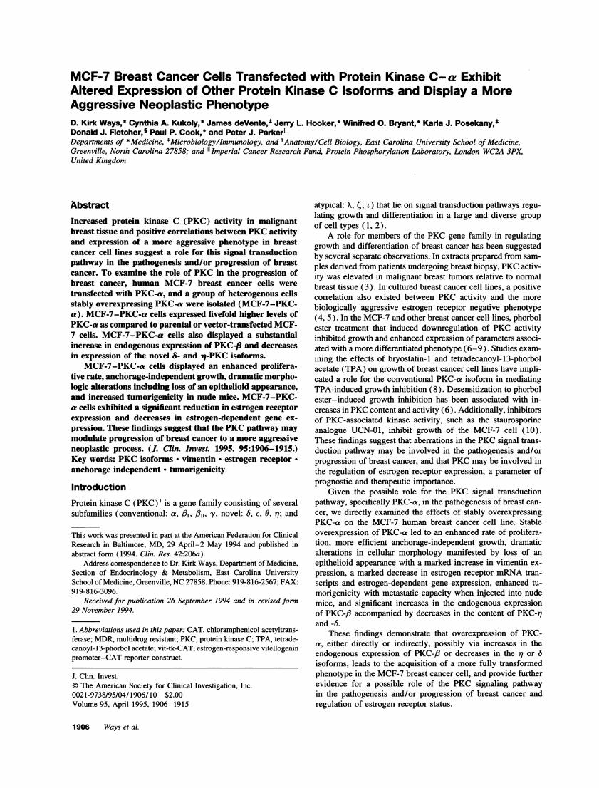
Figure 1. PKC-a content, and PKC activity in MCF-7-PKC-a. Equalamounts of protein (40 [tg) derived from cytosolic (C) and solubilizedparticulate (P) fractions from parental (con) and MCF-7-PKC-a(PKC-a) cells were subjected to Western blot analysis using PKC-aantiserum (inset). PKC activity of the same cells is shown below. Thesubstrates used are indicated beneath the figure. PKC activity is definedas the difference in cpm incorporated into substrate in the absence andpresence of phosphatidylserine and TPA in parental (con) and MCF-7-PKC-a (a) cells. These experiments were repeated on three separateoccasions with similar results.
analysis, in the cells stably transfected with this isoform (MCF-7-PKC-a cells). Corroborating the increased content of thisisoform in PKC-a cells was a 5-16-fold increase, dependingupon the substrate used, in PKC activity in the MCF-7-PKC-a cells as compared to the parental MCF-7 cell (Fig. 1).
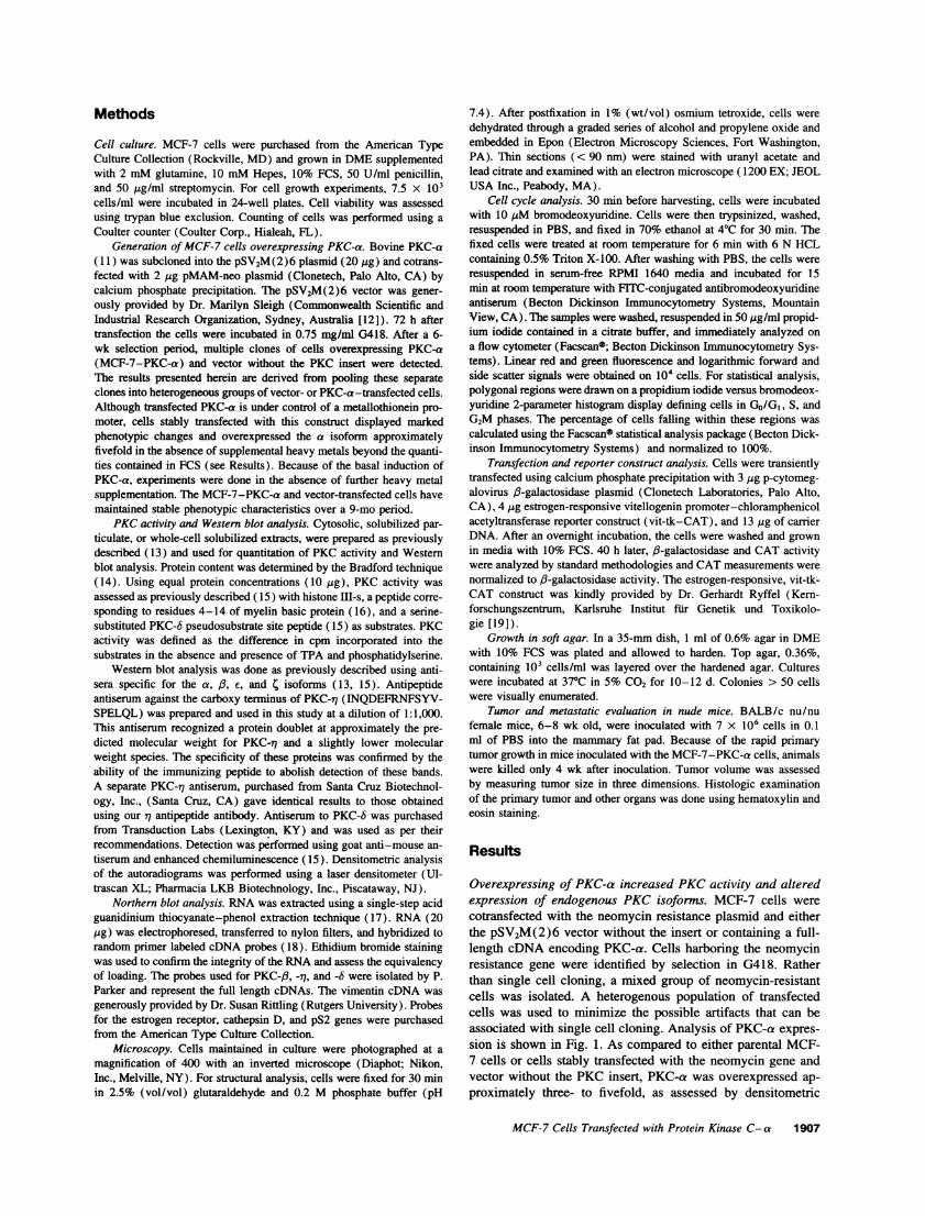
Endogenous expression of other PKC isoforms in MCF-7-PKC-a cells was examined by Western blot analysis (Fig. 2A). Parental MCF-7 cells contained readily apparent amountsof a, E, 6, and 4 isoforms (Figs. 1 and 2). Using a antiserumdirected against the V3 region of the molecule, we were unableto detect significant PKC-,/ expression in the parental MCF-7cell. The PKC-r7 antiserum specifically detected three bandswith molecular weights of 86, 82, and 60 kD. Relative to theparental MCF-7 cells, MCF-7-PKC-a cells displayed a largeincrease in protein content of 6 and similar quantities of c and4. Levels of 6 were reduced modestly in MCF-7-PKC-a cells.Levels of the 86- and 82-kD proteins detected by the PKC-i7antiserum were negligible in MCF-7-PKC-a cells. Content ofthe lower 60-kD protein detected by the 7 antiserum was alsosubstantially reduced in MCF-7-PKC-a cells. The identity ofthe 60-kD protein and its relation to the predicted molecularweight form of remains unresolved. Densitometric scanningof cytosolic and solubilized particulate fractions subjected toWestern blot analysis indicated the following subcellular distri-bution of PKC isoforms in MCF-7 cells: a, 65/35 (percentageof total isoform content in cytosolic/solubilized particulate frac-tions); c, 60/40; 4, 52/48; r 86-kD species, 39/61; i1 82-kDspecies, 53/47; and rq 60-kD species, 80/20. In MCF-7-PKC-acells, the cytosolic/solubilized particulate fraction distributions
.:,\ CG
.4 A wr4r.Ut
a mu
r_
84 m
58 -
B
8.7 ?a;
3.42.6
f 3.3
3.1
EtBr
Figure 2. Altered endog-enous PKC isoform con-
tent in MCF-7-PKC-acells. (A) Western blotanalysis was performedon equal protein concen-trations of whole-cellsolubilized fractions de-rived from parental and
UC V C MCF-7-PKC-a cells.The antisera used are in-dicated (left). Molecularweight standards areshown to the left of the
;... PKC-r1 autoradiogram.Vector-transfected cells
V;k'M displayed identical PKCisoform expression as didparental MCF-7 cells.(B) Northern blot analy-
: * ,s using total RNA (20MIF-g) prepared from pa-
rental (C), vector (V),and MCF-7-PKC-a (a)cells was performed withPKC-,8, -r7, and -6 DNAprobes. The approximatemolecular weight of thedetected mRNA tran-scripts is indicated to theside of the autoradio-gram. Ethidium bromide(EtBr) staining is shownbelow the autoradio-grams. These experi-ments were performed ona separate occasions withsimilar results.
were as follows: a, 46/54; /3, 81/19; E, 56/44; ;, 58/42; andr1 60-kD species, 60/40. Because of their negligible amounts,PKC-,/ and the 86/82-kD species of PKC-r1 could not be quanti-tated in MCF-7 and MCF-7-PKC-a cells, respectively. Be-cause of these changes in the endogenous expression of the /,
6, and isoforms in MCF-7-PKC-a cells, Northern blot analy-sis was performed to examine the content of their mRNA tran-scripts. A concomitant increase in mRNA transcripts for theisoform and decreases in the 6 and rq isoform mRNA transcriptswere noted in the MCF-7-PKC-a cells (Fig. 2 B). Thus, eitherin a direct fashion or secondary to the phenotypic changes dem-onstrated in MCF-7-PKC-a cells (see below), PKC-a overex-
1908 Ways et al.
D
to.0
0
r_
'O
0'a
CI0
0
5:rU
009
40 -
20 k
Fraction: C P
Cell: Con a
Substrate: 8 Peptide
1~ ~ ~ ~~i1
A
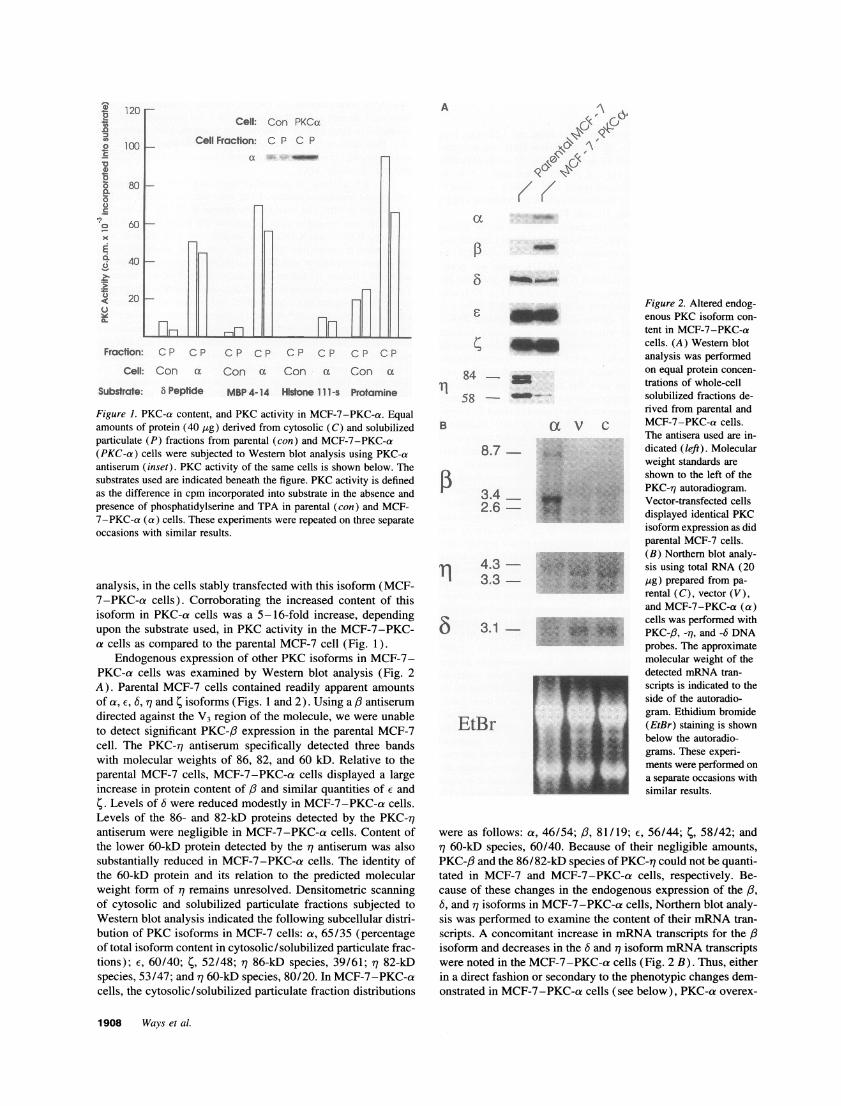
Figure 3. Dissimilar morphology of MCF-7-PKC-a and parental MCF-7 cells. Exponentially growing parental (A), vector-transfected (B), andMCF-7-PKC-a (C) cells were photographed at a magnification of 400 using a Diaphot inverted microscope.
pression modulated expression of other endogenous PKC familymembers.
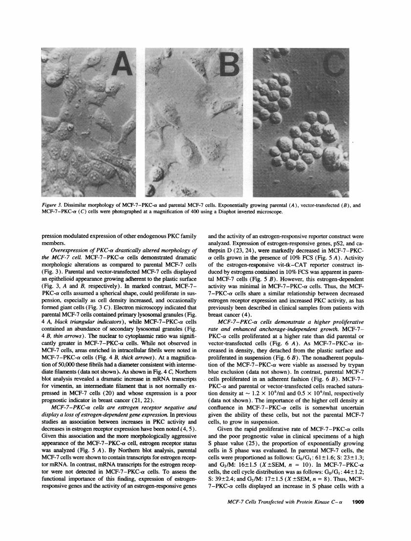
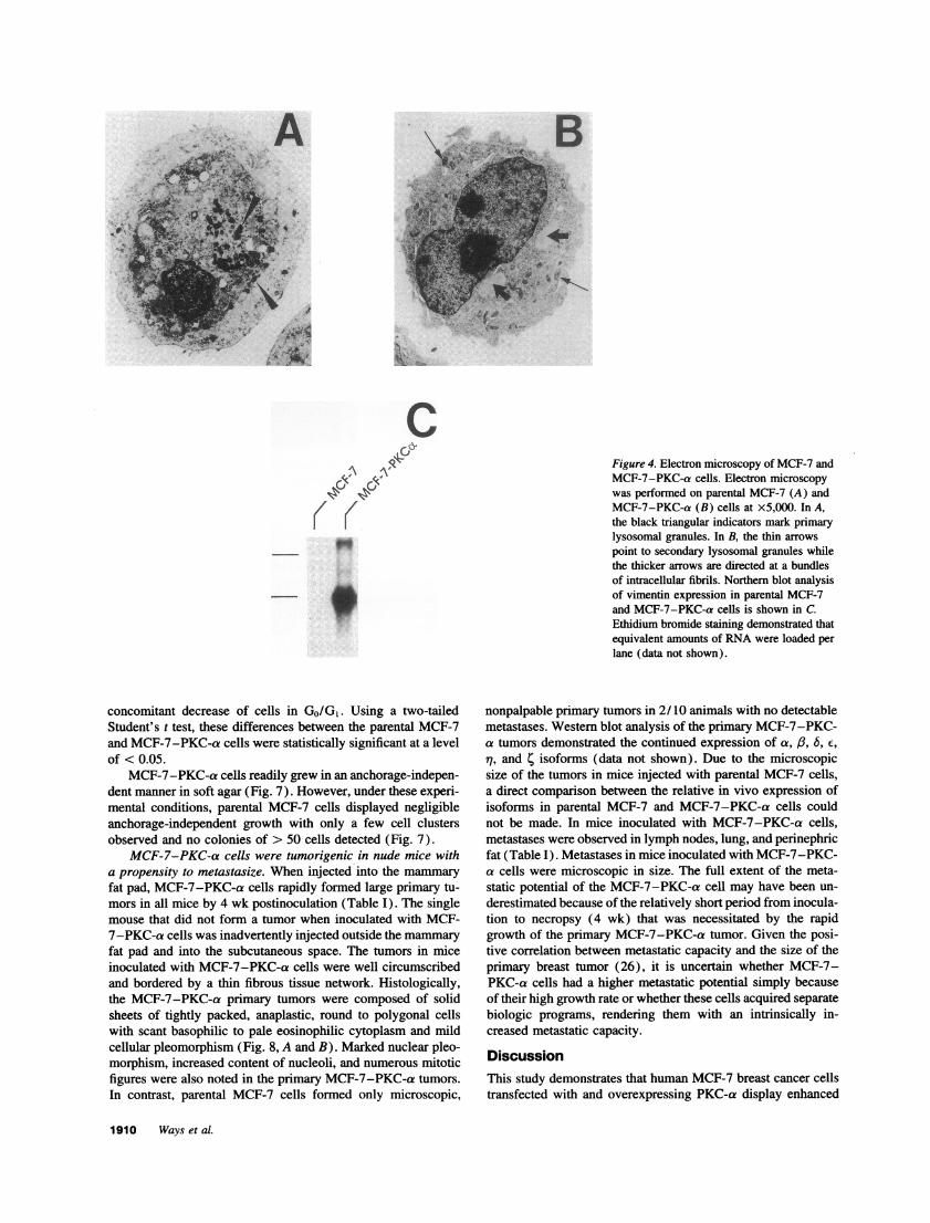
Overexpression ofPKC-a drastically altered morphology ofthe MCF-7 cell. MCF-7-PKC-a cells demonstrated dramaticmorphologic alterations as compared to parental MCF-7 cells(Fig. 3). Parental and vector-transfected MCF-7 cells displayedan epithelioid appearance growing adherent to the plastic surface(Fig. 3, A and B, respectively). In marked contrast, MCF-7-PKC-a cells assumed a spherical shape, could proliferate in sus-pension, especially as cell density increased, and occasionallyformed giant cells (Fig. 3 C). Electron microscopy indicated thatparental MCF-7 cells contained primary lysosomal granules (Fig.4 A, black triangular indicators), while MCF-7-PKC-a cellscontained an abundance of secondary lysosomal granules (Fig.4 B, thin arrows). The nuclear to cytoplasmic ratio was signifi-cantly greater in MCF-7-PKC-a cells. While not observed inMCF-7 cells, areas enriched in intracellular fibrils were noted inMCF-7-PKC-a cells (Fig. 4 X, thick arrows). At a magnifica-tion of 50,000 these fibrils had a diameter consistent with interme-diate filaments (data not shown). As shown in Fig. 4 C, Northernblot analysis revealed a dramatic increase in mRNA transcriptsfor vimentin, an intermediate filament that is not normally ex-pressed in MCF-7 cells (20) and whose expression is a poorprognostic indicator in breast cancer (21, 22).
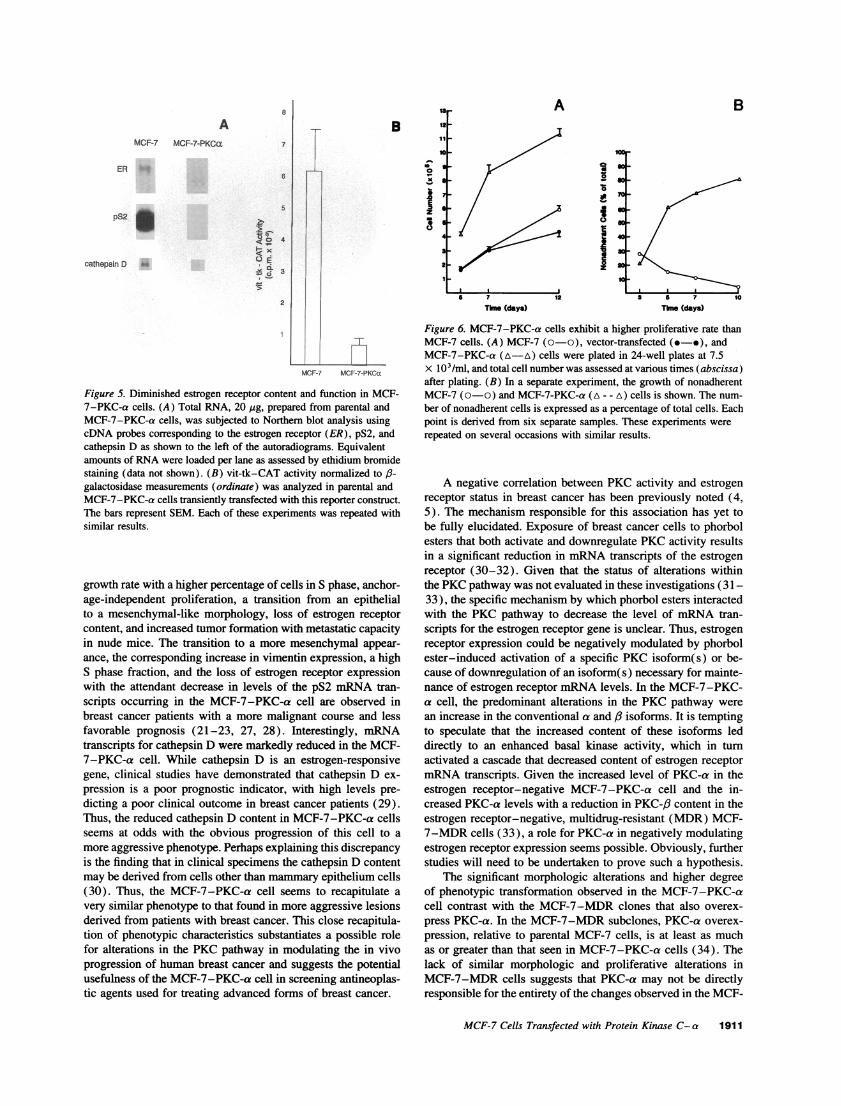
MCF-7-PKC-a cells are estrogen receptor negative anddisplay a loss ofestrogen-dependent gene expression. In previousstudies an association between increases in PKC activity anddecreases in estrogen receptor expression have been noted (4, 5).Given this association and the more morphologically aggressiveappearance of the MCF-7-PKC-a cell, estrogen receptor statuswas analyzed (Fig. 5 A). By Northern blot analysis, parentalMCF-7 cells were shown to contain transcripts for estrogen recep-tor mRNA. In contrast, mRNA transcripts for the estrogen recep-tor were not detected in MCF-7-PKC-a cells. To assess thefunctional importance of this finding, expression of estrogen-responsive genes and the activity of an estrogen-responsive genes
and the activity of an estrogen-responsive reporter construct wereanalyzed. Expression of estrogen-responsive genes, pS2, and ca-thepsin D (23, 24), were markedly decreased in MCF-7-PKC-a cells grown in the presence of 10% FCS (Fig. 5 A). Activityof the estrogen-responsive vit-tk-CAT reporter construct in-duced by estrogens contained in 10% FCS was apparent in paren-tal MCF-7 cells (Fig. 5 B). However, this estrogen-dependentactivity was minimal in MCF-7-PKC-a cells. Thus, the MCF-7-PKC-a cells share a similar relationship between decreasedestrogen receptor expression and increased PKC activity, as haspreviously been described in clinical samples from patients withbreast cancer (4).
MCF-7-PKC-a cells demonstrate a higher proliferativerate and enhanced anchorage-independent growth. MCF-7-PKC-a cells proliferated at a higher rate than did parental orvector-transfected cells (Fig. 6 A). As MCF-7-PKC-a in-creased in density, they detached from the plastic surface andproliferated in suspension (Fig. 6 B). The nonadherent popula-tion of the MCF-7-PKC-a were viable as assessed by trypanblue exclusion (data not shown). In contrast, parental MCF-7cells proliferated in an adherent fashion (Fig. 6 B). MCF-7-PKC-a and parental or vector-transfected cells reached satura-tion density at 1.2 x 10⁶/ml and 0.5 X 10⁶/ml, respectively(data not shown). The importance of the higher cell density atconfluence in MCF-7-PKC-a cells is somewhat uncertaingiven the ability of these cells, but not the parental MCF-7cells, to grow in suspension.
Given the rapid proliferative rate of MCF-7-PKC-a cellsand the poor prognostic value in clinical specimens of a highS phase value (25), the proportion of exponentially growingcells in S phase was evaluated. In parental MCF-7 cells, thecells were proportioned as follows: Go/GI: 61± 1.6; S: 23±1.3;and G2/M: 16±1.5 (X±SEM, n = 10). In MCF-7-PKC-acells, the cell cycle distribution was as follows: GO/GI: 44±1.2;S: 39+2.4; and G2/M: 17±1.5 (X±SEM, n = 8). Thus, MCF-7-PKC-a cells displayed an increase in S phase cells with a
MCF-7 Cells Transfected with Protein Kinase C- a 1909
''" 1.
At
A B
I
,VPOF 4. i .
.f, i>t
C;C,
-p
concomitant decrease of cells in Go/G1. Using a two-tailedStudent's t test, these differences between the parental MCF-7and MCF-7-PKC-a cells were statistically significant at a levelof <0.05.
MCF-7-PKC-a cells readily grew in an anchorage-indepen-dent manner in soft agar (Fig. 7). However, under these experi-mental conditions, parental MCF-7 cells displayed negligibleanchorage-independent growth with only a few cell clustersobserved and no colonies of > 50 cells detected (Fig. 7).
MCF-7-PKC-a cells were tumorigenic in nude mice witha propensity to metastasize. When injected into the mammaryfat pad, MCF-7-PKC-a cells rapidly formed large primary tu-mors in all mice by 4 wk postinoculation (Table I). The singlemouse that did not form a tumor when inoculated with MCF-7-PKC-a cells was inadvertently injected outside the mammaryfat pad and into the subcutaneous space. The tumors in miceinoculated with MCF-7-PKC-a cells were well circumscribedand bordered by a thin fibrous tissue network. Histologically,the MCF-7-PKC-a primary tumors were composed of solidsheets of tightly packed, anaplastic, round to polygonal cellswith scant basophilic to pale eosinophilic cytoplasm and mildcellular pleomorphism (Fig. 8, A and B). Marked nuclear pleo-morphism, increased content of nucleoli, and numerous mitoticfigures were also noted in the primary MCF-7-PKC-a tumors.In contrast, parental MCF-7 cells formed only microscopic,
Figure 4. Electron microscopy of MCF-7 andMCF-7-PKC-a cells. Electron microscopywas performed on parental MCF-7 (A) andMCF-7-PKC-a (B) cells at x5,000. In A,the black triangular indicators mark primarylysosomal granules. In B, the thin arrowspoint to secondary lysosomal granules whilethe thicker arrows are directed at a bundlesof intracellular fibrils. Northern blot analysisof vimentin expression in parental MCF-7and MCF-7-PKC-a cells is shown in C.Ethidium bromide staining demonstrated thatequivalent amounts of RNA were loaded perlane (data not shown).
nonpalpable primary tumors in 2/10 animals with no detectablemetastases. Western blot analysis of the primary MCF-7-PKC-a tumors demonstrated the continued expression of a, A, 6, E,77, and 4 isoforms (data not shown). Due to the microscopicsize of the tumors in mice injected with parental MCF-7 cells,a direct comparison between the relative in vivo expression ofisoforms in parental MCF-7 and MCF-7-PKC-a cells couldnot be made. In mice inoculated with MCF-7-PKC-a cells,metastases were observed in lymph nodes, lung, and perinephricfat (Table I). Metastases in mice inoculated with MCF-7-PKC-a cells were microscopic in size. The full extent of the meta-static potential of the MCF-7-PKC-a cell may have been un-derestimated because of the relatively short period from inocula-tion to necropsy (4 wk) that was necessitated by the rapidgrowth of the primary MCF-7-PKC-a tumor. Given the posi-tive correlation between metastatic capacity and the size of theprimary breast tumor (26), it is uncertain whether MCF-7-PKC-a cells had a higher metastatic potential simply becauseof their high growth rate or whether these cells acquired separatebiologic programs, rendering them with an intrinsically in-creased metastatic capacity.
DiscussionThis study demonstrates that human MCF-7 breast cancer cellstransfected with and overexpressing PKC-a display enhanced
1910 Ways et al.
I ..' ,- -,"
-Al. I t" 1.V A6 `.-nice 711%` -It-
8
AMCF-7 MCF-7-PKCa 7
ER6
5
pS2
cathepsin D
2
B is11
00o 9
aD
014
3
2
IMUCF-7 MCF-7-P~
Figure 5. Diminished estrogen receptor content and function in MCF-7-PKC-a cells. (A) Total RNA, 20 .g, prepared from parental andMCF-7-PKC-a cells, was subjected to Northern blot analysis usingcDNA probes corresponding to the estrogen receptor (ER), pS2, andcathepsin D as shown to the left of the autoradiograms. Equivalentamounts of RNA were loaded per lane as assessed by ethidium bromidestaining (data not shown). (B) vit-tk-CAT activity normalized to f3-galactosidase measurements (ordinate) was analyzed in parental andMCF-7-PKC-a cells transiently transfected with this reporter construct.The bars represent SEM. Each of these experiments was repeated withsimilar results.
growth rate with a higher percentage of cells in S phase, anchor-age-independent proliferation, a transition from an epithelialto a mesenchymal-like morphology, loss of estrogen receptorcontent, and increased tumor formation with metastatic capacityin nude mice. The transition to a more mesenchymal appear-
ance, the corresponding increase in vimentin expression, a highS phase fraction, and the loss of estrogen receptor expressionwith the attendant decrease in levels of the pS2 mRNA tran-scripts occurring in the MCF-7-PKC-a cell are observed inbreast cancer patients with a more malignant course and lessfavorable prognosis (21-23, 27, 28). Interestingly, mRNAtranscripts for cathepsin D were markedly reduced in the MCF-7-PKC-a cell. While cathepsin D is an estrogen-responsivegene, clinical studies have demonstrated that cathepsin D ex-
pression is a poor prognostic indicator, with high levels pre-
dicting a poor clinical outcome in breast cancer patients (29).Thus, the reduced cathepsin D content in MCF-7-PKC-a cellsseems at odds with the obvious progression of this cell to a
more aggressive phenotype. Perhaps explaining this discrepancyis the finding that in clinical specimens the cathepsin D contentmay be derived from cells other than mammary epithelium cells(30). Thus, the MCF-7-PKC-a cell seems to recapitulate a
very similar phenotype to that found in more aggressive lesionsderived from patients with breast cancer. This close recapitula-tion of phenotypic characteristics substantiates a possible rolefor alterations in the PKC pathway in modulating the in vivoprogression of human breast cancer and suggests the potentialusefulness of the MCF-7-PKC-a cell in screening antineoplas-tic agents used for treating advanced forms of breast cancer.
A
aIs
i
10
I1z
7
The (days) Tkme (days)
Figure 6. MCF-7-PKC-a cells exhibit a higher proliferative rate thanMCF-7 cells. (A) MCF-7 (o0o), vector-transfected (.-*), andMCF-7-PKC-a (A-A) cells were plated in 24-well plates at 7.5x 10³/ml, and total cell number was assessed at various times (abscissa)after plating. (B) In a separate experiment, the growth of nonadherentMCF-7 (o0o) and MCF-7-PKC-a (A - - A) cells is shown. The num-
ber of nonadherent cells is expressed as a percentage of total cells. Eachpoint is derived from six separate samples. These experiments were
repeated on several occasions with similar results.
A negative correlation between PKC activity and estrogenreceptor status in breast cancer has been previously noted (4,5). The mechanism responsible for this association has yet tobe fully elucidated. Exposure of breast cancer cells to phorbolesters that both activate and downregulate PKC activity resultsin a significant reduction in mRNA transcripts of the estrogenreceptor (30-32). Given that the status of alterations withinthe PKC pathway was not evaluated in these investigations (31-33), the specific mechanism by which phorbol esters interactedwith the PKC pathway to decrease the level of mRNA tran-scripts for the estrogen receptor gene is unclear. Thus, estrogenreceptor expression could be negatively modulated by phorbolester-induced activation of a specific PKC isoform(s) or be-cause of downregulation of an isoform( s) necessary for mainte-nance of estrogen receptor mRNA levels. In the MCF-7-PKC-a cell, the predominant alterations in the PKC pathway were
an increase in the conventional a and isoforms. It is temptingto speculate that the increased content of these isoforms leddirectly to an enhanced basal kinase activity, which in turnactivated a cascade that decreased content of estrogen receptormRNA transcripts. Given the increased level of PKC-a in theestrogen receptor-negative MCF-7-PKC-a cell and the in-creased PKC-a levels with a reduction in PKC-,B content in theestrogen receptor-negative, multidrug-resistant (MDR) MCF-7-MDR cells (33), a role for PKC-a in negatively modulatingestrogen receptor expression seems possible. Obviously, furtherstudies will need to be undertaken to prove such a hypothesis.
The significant morphologic alterations and higher degreeof phenotypic transformation observed in the MCF-7-PKC-acell contrast with the MCF-7-MDR clones that also overex-
press PKC-a. In the MCF-7-MDR subclones, PKC-a overex-
pression, relative to parental MCF-7 cells, is at least as muchas or greater than that seen in MCF-7-PKC-a cells (34). Thelack of similar morphologic and proliferative alterations inMCF-7-MDR cells suggests that PKC-a may not be directlyresponsible for the entirety of the changes observed in the MCF-
MCF-7 Cells Transfected with Protein Kinase C-a 1911
B
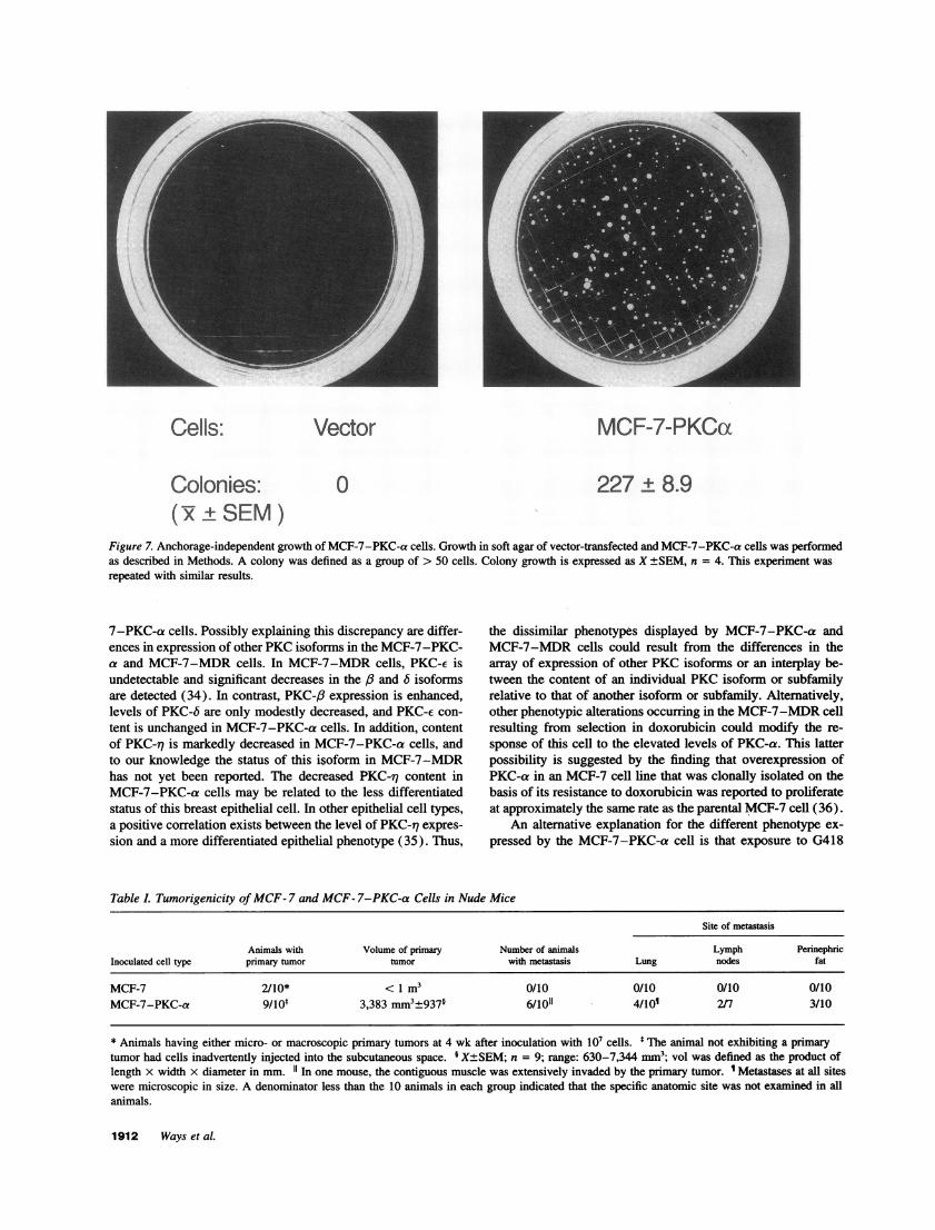
Cells: Vector MCF-7-PKCca
Colonies: 0 227 ± 8.9( +±SEM)
Figure 7. Anchorage-independent growth of MCF-7-PKC-a cells. Growth in soft agar of vector-transfected and MCF-7-PKC-a cells was performedas described in Methods. A colony was defined as a group of > 50 cells. Colony growth is expressed as X +SEM, n = 4. This experiment was
repeated with similar results.
7-PKC-a cells. Possibly explaining this discrepancy are differ-ences in expression of other PKC isoforms in the MCF-7-PKC-a and MCF-7-MDR cells. In MCF-7-MDR cells, PKC-e isundetectable and significant decreases in the /3 and 6 isoformsare detected (34). In contrast, PKC-/3 expression is enhanced,levels of PKC-6 are only modestly decreased, and PKC-e con-tent is unchanged in MCF-7-PKC-a cells. In addition, contentof PKC-iq is markedly decreased in MCF-7-PKC-a cells, andto our knowledge the status of this isoform in MCF-7-MDRhas not yet been reported. The decreased PKC-27 content inMCF-7-PKC-a cells. may be related to the less differentiatedstatus of this breast epithelial cell. In other epithelial cell types,a positive correlation exists between the level of PKC-77 expres-sion and a more differentiated epithelial phenotype (35). Thus,
the dissimilar phenotypes displayed by MCF-7-PKC-a andMCF-7-MDR cells could result from the differences in thearray of expression of other PKC isoforms or an interplay be-tween the content of an individual PKC isoform or subfamilyrelative to that of another isoform or subfamily. Alternatively,other phenotypic alterations occurring in the MCF-7-MDR cellresulting from selection in doxorubicin could modify the re-sponse of this cell to the elevated levels of PKC-a. This latterpossibility is suggested by the finding that overexpression ofPKC-a in an MCF-7 cell line that was clonally isolated on thebasis of its resistance to doxorubicin was reported to proliferateat approximately the same rate as the parental MCF-7 cell (36).
An alternative explanation for the different phenotype ex-pressed by the MCF-7-PKC-a cell is that exposure to G418
Table L Tumorigenicity ofMCF- 7 and MCF- 7-PKC-a Cells in Nude Mice
Site of metastasis
Animals with Volume of primary Number of animals Lymph PerinephricInoculated cell type primary tumor tumor with metastasis Lung nodes fat
MCF-7 2/10* < 1 m3 0/10 0/10 0/10 0/10MCF-7-PKC-a 9/10t 3,383 mm3±937' 6/1011 4/101 2/7 3/10
* Animals having either micro- or macroscopic primary tumors at 4 wk after inoculation with 107 cells. * The animal not exhibiting a primarytumor had cells inadvertently injected into the subcutaneous space. I X±SEM; n = 9; range: 630-7,344 mm3; vol was defined as the product oflength x width x diameter in mm. 11 In one mouse, the contiguous muscle was extensively invaded by the primary tumor. 1 Metastases at all siteswere microscopic in size. A denominator less than the 10 animals in each group indicated that the specific anatomic site was not examined in allanimals.
1912 Ways et al.
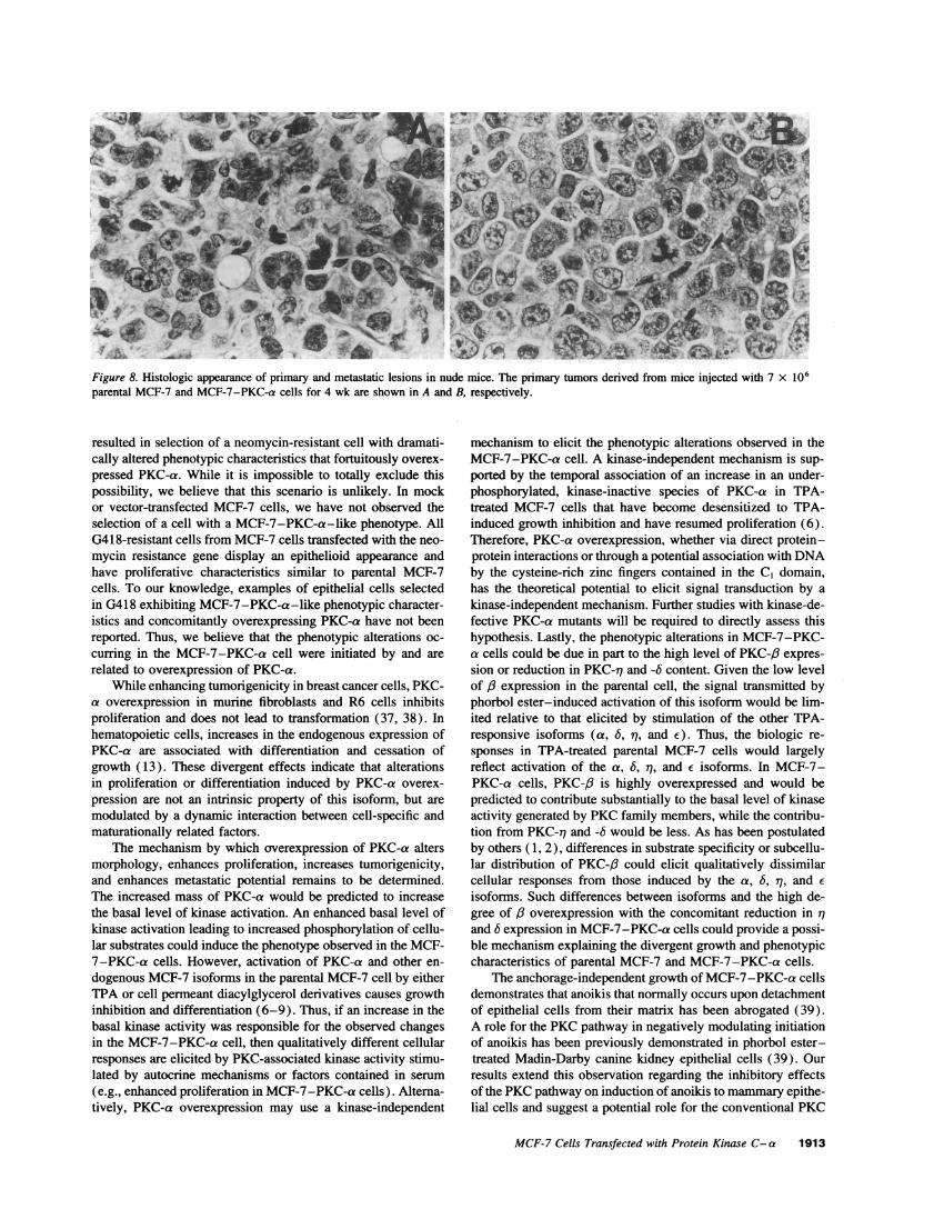
Figure 8. Histologic appearance of primary and metastatic lesions in nude mice. The primary tumors derived from mice injected with 7 x 10⁶parental MCF-7 and MCF-7-PKC-a cells for 4 wk are shown in A and B, respectively.
resulted in selection of a neomycin-resistant cell with dramati-cally altered phenotypic characteristics that fortuitously overex-pressed PKC-a. While it is impossible to totally exclude thispossibility, we believe that this scenario is unlikely. In mockor vector-transfected MCF-7 cells, we have not observed theselection of a cell with a MCF-7-PKC-a-like phenotype. AllG418-resistant cells from MCF-7 cells transfected with the neo-mycin resistance gene display an epithelioid appearance andhave proliferative characteristics similar to parental MCF-7cells. To our knowledge, examples of epithelial cells selectedin G418 exhibiting MCF-7-PKC-a-like phenotypic character-istics and concomitantly overexpressing PKC-a have not beenreported. Thus, we believe that the phenotypic alterations oc-curring in the MCF-7-PKC-a cell were initiated by and arerelated to overexpression of PKC-a.
While enhancing tumorigenicity in breast cancer cells, PKC-a overexpression in murine fibroblasts and R6 cells inhibitsproliferation and does not lead to transformation (37, 38). Inhematopoietic cells, increases in the endogenous expression ofPKC-a are associated with differentiation and cessation ofgrowth (13). These divergent effects indicate that alterationsin proliferation or differentiation induced by PKC-a overex-pression are not an intrinsic property of this isoform, but aremodulated by a dynamic interaction between cell-specific andmaturationally related factors.
The mechanism by which overexpression of PKC-a altersmorphology, enhances proliferation, increases tumorigenicity,and enhances metastatic potential remains to be determined.The increased mass of PKC-a would be predicted to increasethe basal level of kinase activation. An enhanced basal level ofkinase activation leading to increased phosphorylation of cellu-lar substrates could induce the phenotype observed in the MCF-7-PKC-a cells. However, activation of PKC-a and other en-dogenous MCF-7 isoforms in the parental MCF-7 cell by eitherTPA or cell permeant diacylglycerol derivatives causes growthinhibition and differentiation (6-9). Thus, if an increase in thebasal kinase activity was responsible for the observed changesin the MCF-7-PKC-a cell, then qualitatively different cellularresponses are elicited by PKC-associated kinase activity stimu-lated by autocrine mechanisms or factors contained in serum(e.g., enhanced proliferation in MCF-7-PKC-a cells). Alterna-tively, PKC-a overexpression may use a kinase-independent
mechanism to elicit the phenotypic alterations observed in theMCF-7-PKC-a cell. A kinase-independent mechanism is sup-ported by the temporal association of an increase in an under-phosphorylated, kinase-inactive species of PKC-a in TPA-treated MCF-7 cells that have become desensitized to TPA-induced growth inhibition and have resumed proliferation (6).Therefore, PKC-a overexpression, whether via direct protein-protein interactions or through a potential association with DNAby the cysteine-rich zinc fingers contained in the C1 domain,has the theoretical potential to elicit signal transduction by akinase-independent mechanism. Further studies with kinase-de-fective PKC-a mutants will be required to directly assess thishypothesis. Lastly, the phenotypic alterations in MCF-7-PKC-a cells could be due in part to the high level of PKC-#B expres-sion or reduction in PKC-77 and -6 content. Given the low levelof #3 expression in the parental cell, the signal transmitted byphorbol ester-induced activation of this isoform would be lim-ited relative to that elicited by stimulation of the other TPA-responsive isoforms (a, 6, 71, and e). Thus, the biologic re-sponses in TPA-treated parental MCF-7 cells would largelyreflect activation of the a, 6, rq, and e isoforms. In MCF-7-PKC-a cells, PKC-,/ is highly overexpressed and would bepredicted to contribute substantially to the basal level of kinaseactivity generated by PKC family members, while the contribu-tion from PKC-r1 and -6 would be less. As has been postulatedby others ( 1, 2), differences in substrate specificity or subcellu-lar distribution of PKC-,/ could elicit qualitatively dissimilarcellular responses from those induced by the a, 6, i7, and Eisoforms. Such differences between isoforms and the high de-gree of, overexpression with the concomitant reduction in r,and 6 expression in MCF-7-PKC-a cells could provide a possi-ble mechanism explaining the divergent growth and phenotypiccharacteristics of parental MCF-7 and MCF-7-PKC-a cells.
The anchorage-independent growth of MCF-7-PKC-a cellsdemonstrates that anoikis that normally occurs upon detachmentof epithelial cells from their matrix has been abrogated (39).A role for the PKC pathway in negatively modulating initiationof anoikis has been previously demonstrated in phorbol ester-treated Madin-Darby canine kidney epithelial cells (39). Ourresults extend this observation regarding the inhibitory effectsof the PKC pathway on induction of anoikis to mammary epithe-lial cells and suggest a potential role for the conventional PKC
MCF-7 Cells Transfected with Protein Kinase C-a 1913
isoforms in regulating this process and its clinical counterpart,metastasis.
Expression of PKC-/3 was enhanced in the MCF-7-PKC-a cell. The human PKC-f promoter contains multiple enhancerelements, including AP-1, AP-2, SpI, E box, and octamer mo-tifs, and also contains a sequence involved in transcriptionalsilencing (40). Interestingly, in K562 erythroleukemia cells,both basal and TPA-inducible expression of PKC-,6 only requirea sequence containing two SpI sites, an E box, and an octamermotif (40). Given that the AP-1 and AP-2 sites are not neces-sary for induction of this gene, it has been postulated that tran-scriptional activation occurs via a non-AP-1-dependent mecha-nism (40). Consistent with these results, our findings show thatTPA treatment for a 24-h period does not increase expressionof PKC-/3 mRNA transcripts in parental MCF-7 cells (data notshown). These findings suggest that, in MCF-7-PKC-a cells,induction of PKC-,6 expression occurs either as a direct effectof PKC-a overexpression by a kinase-independent mechanismor secondary to phenotypic changes associated with the MCF-7-PKC-a cell. The factors, and the level at which they act tomodulate the endogenous expression of the ,f isoform, are underinvestigation in our laboratories.
The clinical significance of these findings in the prognosticevaluation of malignant breast lesions and in developing noveltherapeutic modalities seems promising. Assessing the level andarray of specific PKC isoform expression in primary breastmasses could be useful in gauging the metastatic potential and/or biological aggressiveness of the lesion. Such informationcould be of use in determining the appropriate degree of radia-tion or chemotherapeutic intervention, especially in circum-stances such as axillary node-negative disease. The ability ofPKC-a overexpression to enhance proliferation and to inducea more biologically aggressive phenotype, either directly orindirectly via alterations in expression of other endogenous PKCisoforms, also indicates the potential promise of therapeuticmodalities directed at inhibiting isoform(s) expression or func-tion. Currently, such methodology is available using antisenseor ribozyme therapy directed against individual PKC isoformsor by downregulating PKCs with bryostatin-1 treatment (41,42). Thus, these findings open exciting new possibilities thatcould improve the evaluation and treatment of patients withcertain forms of breast cancer.
Acknowledgments
We wish to thank Nancy Hamm and June Long for expert assistancein preparation of this manuscript, Dr. Jack Brinn and Dr. Alvin Volkman(both from East Carolina University School of Medicine) for theiradvice on morphologic interpretations, Dr. John Bradfield and the De-partment of Comparative Medicine (East Carolina University School ofMedicine) for assistance in performing the nude mice experiments, andDr. Susan Rittling and Dr. Gerhardt Ryffel for providing the plasmidsused in this study.
This work was partially supported by a National Institutes of Healthgrant (CA43023).
References
1. Nishizuka, Y. 1988. The molecular heterogeneity of protein kinase C andits implications for cellular regulation. Nature (Lond.). 334:661-665.
2. Parker, P., G. Kour, R. Marais, F. Mitchell, C. Pears, S. Stabel, and C.Webster. 1989. Protein kinase C: a family affair. Mol. Cell. Endocrinol. 65:1-11.
3. O'Brian, C., V. Vogel, S. Singletary, and E. Ward. 1989. Elevated proteinkinase C expression in human breast tumor biopsies relative to normal breasttissue. Cancer Res. 49:3215-3217.
4. Borner, C., R. Wyss, R. Regazzi, U. Eppenberger, and D. Fabbro. 1987.Immunological quantitation of phospholipid/Ca2+-dependent protein kinase ofhuman mammary carcinoma cells: inverse relationships to estrogen receptors. Int.J. Cancer. 40:344-348.
5. Lee, S., J. Karaszkiewicz, and W. Anderson. 1992. Elevated level of nuclearprotein kinase C in multidrug-resistant MCF-7 human breast carcinoma cells.Cancer Res. 52:3750-3759.
6. Fabbro, D., R. Regazzi, S. Costa, C. Borner, and U. Eppenberger. 1986.Protein kinase C desensitization by phorbol esters and its impact on growth ofhuman breast cancer cells. Biochem. Biophys. Res. Commun. 135:65-73.
7. Issandou, M., F. Bayard, and J. Darbon. 1988. Inhibition of MCF-7 cellgrowth by 12-0-tetradecanoylphorbol-13-acetate and 1,2 dioctanoyl-SN-glycerol:distinct effects on protein kinase C activity. Cancer Res. 48:6943-6950.
8. Kennedy, M., L. Presligiacoma, G. Tyler, W. May, and N. Davidson. 1992.Differentiation effects of bryostatin 1 and phorbol ester on human breast cancercell lines. Cancer Res. 52:1278-1283.
9. Valette, A., N. Gas, F. Roubinet, M. Dupont, and F. Bayard. 1987. Influenceof 12-0-tetradecanoylphorbol- 13-acetate on proliferation and maturation of humanbreast carcinoma cells (MCF-7): relationship to cell cycle events. Cancer Res.47:1615-1620.
10. Seynaeve, C., M. Stetler-Stevenson, S. Sebers, G. Kaur, E. Sausville, andP. Worland. 1993. Cell cycle arrest and growth inhibition by the protein kinaseantagonist UNC-01 in human breast carcinoma cells. Cancer Res. 53:2081-2086.
11. Pears, C., G. Kour, C. House, B. Kemp, and P. Parker. 1990. Mutagenesisof the pseudosubstrate site of protein kinase C leads to activation. Eur. J. Biochem.194:89-94.
12. McNeall, J., A. Sanchez, P. Gray, C. Chesterman, and M. Sleigh. 1989.Hyperinducible gene expression from a metallothionein promoter containing addi-tional metal-responsive elements. Gene (Amst.). 78:81-88.
13. Ways, D., B. Messer, T. Garris, W. Qin, P. Cook, and P. Parker. 1992.Modulation of protein kinase C e by phorbol esters in the monoblastoid U937cell. Cancer Res. 52:5604-5609.
14. Bradford, M. 1976. A rapid and sensitive method for the quantitation ofmicrogram quantities of protein utilizing the principles of protein-dye binding.Anal. Biochem. 72:248-254.
15. Ways, D., P. Cook, C. Webster, and P. Parker. 1992. Effect of phorbolesters on PKC-4. J. Biol. Chem. 267:4799-4805.
16. Yasuda, I., A. Kishimoto, S. Tanaka, M. Tominaga, A. Sakurai, and Y.Nishizuka. 1990. A synthetic peptide substrate for selective assay of protein kinaseC. Biochem. Biophys. Res. Commun. 166:1220-1227.
17. Chomczynski, P., and N. Sacchi. 1987. Single-step method of RNA isola-tion by acid guanidinium thiocyanate-phenol-chloroform extraction. Anal. Bio-chem. 162:156-159.
18. McCubrey, J., L. Steelman, G. Sandlin, R. Riddle, and D. Ways. 1990.Effects of phorbol esters on an interleukin-3-dependent cell line. Blood. 76:63-72.
19. Klein-Hitpass, L., M. Schorpp, V. Wagner, and G. Ryffel. 1986. Anestrogen-responsive element derived from the 5' flanking region of Xenopusvitellogenin A2 gene functions in transfected human cells. Cell. 46:1053-1061.
20. Heuijerjans, J., F. Pieper, F. Ramaekers, L. Timmermans, H. Kuijpers, H.Bloemendal, and W. Venrooij. 1989. Association of mRNA and elF-2a with thecytoskeleton in cells lacking vimentin. Exp. Cell Res. 181:317-330.
21. Raymond, W., and A. Leong. 1989. Co-expression of cytokeratin andvimentin intermediate filament proteins in benign and neoplastic breast epithelium.J. Pathol. 157:299-306.
22. Raymond, W., and A. Leong. 1989. Vimentin: a new prognostic parameterin breast carcinoma? J. Pathol. 158:107-114.
23. Rio, M., J. Bellocq, B. Gairard, U. Rasmussen, A. Krust, C. Keohl, H.Calderoli, V. Schiff, R. Renaud, and P. Chambon. 1987. Specific expression ofthe pS2 gene in subclasses of breast cancers in comparison with expression ofthe estrogen and progesterone receptors and the oncogene ERB Z. Proc. Natl.Acad. Sci. USA 84:9243-9247.
24. Westley, B., and H. Rochefort. 1980. A secreted glycoprotein induced byestrogen in human breast cancer cell lines. Cell. 20:353-362.
25. Muss, H., and T. Kute. 1982. Flow cytometry in the management of breastcancer. In High-Risk Breast Cancer: Diagnosis. J. Ragaz and I. Ariel, editors.Springer-Verlag, Berlin. 103-119.
26. Kurebayashi, J., S. McLeskey, M. Johnson, M. Lippman, R. Dickson, andF. Kern. 1993. Quantitative demonstration of spontaneous metastasis by MCF-7human breast cancer cells cotransfected with fibroblast growth factor-4 and LacZCancer Res. 53:2178-2187.
27. Bae, S., G. Arand, H. Azzam, P. Pavasant, J. Torri, T. Frandsen, and E.Thompson. 1993. Molecular and cellular analysis of basement membrane invasionby human breast cancer cells in Matrigel-based in vitro assays. Breast CancerRes. Treat. 24:241-255.
28. Knight, W., R. Livingston, E. Gregory, and W. McGuire. 1977. Estrogen
1914 Ways et al.
receptor as an independent prognostic factor for early recurrence in breast cancer.Cancer Res. 37:4669-4671.
29. Rochefort, H. 1990. Cathepsin D in breast cancer. Breast Cancer Res.Treat. 16:3-13.
30. Johnson, M., J. Torri, M. Lippman, and R. Dickson. 1993. The role ofcathepsin D in the invasiveness of human breast cancer cells. Cancer Res. 53:873-877.
31. Ree, A., B. Landmark, S. Walaas, H. Lahooti, L. Eikvar, W. Eskild, andV. Hansson. 1991. Down regulation of messenger ribonucleic acid and proteinlevels for estrogen receptors by phorbol ester and calcium in MCF-7 cells. Endo-crinology. 129:339-344.
32. Saceda, M., C. Knabbe, R. Dickson, M. Lippman, D. Bronzert, R. Lindsey,M. Gottardis, and M. Martin. 1991. Post-transcriptional destabilization of estrogenreceptor mRNA in MCF-7 cells by 12-0-tetradecanoylphorbol-13-acetate. J. Biol.Chem 266:17809-17814.
33. Tzukerman, M., X. Zhang, and M. Pfahl. 1991. Inhibition of estrogenreceptor activity by the tumor promoter 12-0-tetradecanoylphorbol-13-acetate: amolecular analysis. Mol. Endocrinol. 5:1983-1992.
34. Blobe, G., C. Sachs, W. Khan, D. Fabbro, S. Stabel, W. Wetsel, L. Obeid,R. Fine, and Y. Hannun. 1993. Selective regulation of expression of protein kinaseC (PKC) isoenzymes in multidrug-resistant MCF-7 cells. J. Biol. Chem 268:658-664.
35. Osada, S., Y. Hashimoto, S. Nomura, Y. Kohno, K. Chida, 0. Tajima, K.Kubo, K. Askimoto, H. Koizumi, Y. Kitamura, K. Suzuki, S. Ohno, and T. Kuroki.1993. Predominant expression of nPKC-77, a Ca2" independent isoform of protein
kinase C in epithelial tissues, in association with epithelial differentiation. CellGrowth & Differ. 4:167-175.
36. Yu, G., S. Ahmad, A. Aquino, C. Fairchild, J. Trepel, S. Ohno, K. Suzuki,T. Tsuruo, K. Cowan, and R. Glazer. 1991. Transection with protein kinase C-aconfers increased multidrug resistance to MCF-7 cells expressing P-glycoprotein.Cancer Commun. 3:181-189.
37. Borner, C., I. Fillipuzzii, I. Weinstein, and R. Imber. 1991. Failure ofwild-type or a mutant form of protein kinase C-a to transform fibroblasts. Nature(Lond.). 353:78-80.
38. Cacace, A., S. Guadango, R. Krauss, D. Fabbro, and I. Weinstein. 1993.The epsilon isoform of protein kinase C is an oncogene when overexpressed inrat fibroblasts. Oncogene. 8:2095-2014.
39. Frisch, S., and H. Francis. 1994. Disruption of epithelial cell-matrix inter-actions induces apoptosis. J. Cell Biol. 124:619-625.
40. Obeid, L., G. Blobe, L. Karolak, and Y. Hannun. 1992. Cloning andcharacterization of the major promoter of the human protein kinase C P gene. J.BioL Chem. 267:20804-20810.
41. Prendiville, J., D. Crowther, N. Thatcher, P. Woll, B. Fox, A. McGowan,N. Testa, P. Stem, R. McDermott, M. Potter, and G. Pettit. 1993. A phase onestudy of intravenous bryostatin 1 in patients with advanced cancer. Br. J. Cancer.68:418-425.
42. Philip, P., D. Rea, P. Thavasu, J. Carmichael, N. Stuart, H. Rockett, D.Talbot, T. Ganesan, G. Pettit, F. Balkwill, and A. Harris. 1993. Phase I study ofbryostatin 1: assessment of interleukin 6 and tumor necrosis factor a inductionin vitro. J. Nati. Cancer Inst. 85:1812-1818.
MCF-7 Cells Transfected with Protein Kinase C-a 1915


