Embed Size (px)
Citation preview
Mt
AAC
a
ARRAA
KBCdMNOZ
1
oim2(ppiGbos2oaw2
(
0d
Carbohydrate Polymers 85 (2011) 37–43
Contents lists available at ScienceDirect
Carbohydrate Polymers
journa l homepage: www.e lsev ier .com/ locate /carbpol
annosylated chitosan-zinc sulphide nanocrystals as fluorescent bioprobes forargeted cancer imaging
swathy Jayasree1, Sajith Sasidharan1, Manzoor Koyakutty, Shantikumar Nair, Deepthy Menon ∗
mrita Centre for Nanosciences and Molecular Medicine, Amrita Institute of Medical Sciences and Research Centre, Amrita Vishwavidyapeetham University,ochin-682 041, Kerala, India
r t i c l e i n f o
rticle history:eceived 3 November 2010eceived in revised form 18 January 2011ccepted 20 January 2011vailable online 28 January 2011
a b s t r a c t
A novel nanomaterial based on chitosan-zinc sulphide:Mn (ZnS:Mn) conjugated with mannose ligand hasbeen developed for targeted cancer imaging. The nanobioconjugates, prepared through simple aqueouschemistry, possessed high colloidal stability and strong fluorescence emission at ∼600 nm. Characteri-zation using XRD, DLS, SEM, AFM and FTIR revealed that the bioconjugated particles are appropriatelyfunctionalized and stable, with average size ∼150 nm. The presence of polysaccharide chitosan bestowedenhanced biocompatibility to the nanocrystals and provided suitable functionality for mannosylation.
eywords:ioimaginghitosan-Mannoseannose receptoranocrystals
In vitro cytotoxicity studies on mouse fibroblast (L929) and oral epithelial carcinoma (KB) cells confirmedtheir cytocompatibility. Bioconjugation with mannose provided specificity and targeted cellular labellingcharacteristics as demonstrated using KB cells which over-express mannose receptors on their surface.Our investigations highlight the applicability of polysaccharide protected and mannosylated fluorescentZnS nanoprobes for active targeting of cancer cells.
ral cancerinc sulphide
. Introduction
Nanosized particles with embedded luminescent features aref immense importance in analytical chemistry and bioengineer-ng due to their potential applications in medical diagnostics,
ultiplexed bioassays, drug screening, etc. (Doty, Fernig, & Levy,004). Semiconductor nanocrystals, also known as quantum dotsQDs), have unique size-tunable spectral properties, excellenthotostability, etc. (Gao, Cui, Levenson, Chung, & Nie, 2004), whichrovide substantial improvements in the sensitivity of molecular
maging and quantitative cellular analysis (Bruchez, Moronne,in, Weiss, & Alivisatos, 1998; Chan & Nie, 1998). By appropriateio-conjugation, such nanocrystals can replace the conventionalrganic fluorescent dyes in immunostaining and bioimaging of tis-ues and cancerous cells (Medintz, Uyeda, Goldman, & Mattoussi,005; Smith, Duan, Mohs, & Nie, 2008). Surface modification
f these QDs using biocompatible polysaccharides offer severaldvantages such as (i) passivation of the surface defects of QDs,ith enhanced fluorescence (Hezinger, Tessmar, & Goepferich,008; Nie, Tan, & Zhang, 2006); (ii) biocompatibility to the oth-
∗ Corresponding author. Tel.: +91 484 4008750; fax: +91 484 2802030.E-mail addresses: [email protected], [email protected]
D. Menon).1 These authors have contributed equally to the manuscript.
144-8617/$ – see front matter © 2011 Elsevier Ltd. All rights reserved.oi:10.1016/j.carbpol.2011.01.034
© 2011 Elsevier Ltd. All rights reserved.
erwise toxic core of the QDs (Xie et al., 2005; Yu, Chang, Drezek,& Colvin, 2006); (iii) provide functional groups for further bio-conjugation with suitable ligands (Jain et al., 2008; Mathew et al.,2010; Warad, Thanachayanont, Tumcharern, & Dutta, 2007). Theversatility of chitosan, a natural biopolymer, facilitates chemicalmodifications, graft reactions, ionic interactions, etc. and hencewould be an appropriate candidate for this purpose (Jayakumar,Menon, Manzoor, Nair, & Tamura, 2010; Kumar, Muzzarelli,Muzzarelli, Sashiwa, & Domb, 2004). In the present work, wehave utilized this natural polysaccharide to surface passivateas well as enable bioconjugation of ZnS:Mn nanocrystals witha carbohydrate, viz, mannose, to target the mannose receptorsover-expressed on cancer cells.
The cytotoxicity arising from heavy-metal based QDs of CdS,CdSe, CdTe, CdSe/ZnS, etc. (Lovric, Sung, & Francoise, 2005;Mattoussi et al., 2001) necessitates the development of heavy-metal free materials that are predominantly biocompatible andaqueous based (Ashokan, Menon, Nair, & Manzoor, 2010; Pradhan,David, Liu, & Peng, 2007; Setua, Menon, Asok, Nair, & Manzoor,2010). Recently, we have developed a heavy-metal free zinc sul-phide based fluorescent nanocrystal for targeted cancer imagingusing folic acid as the targeting ligand (Manzoor et al., 2009). We
found that compared to many other known QDs, ZnS is a bettersystem owing to its stable physical and chemical characteristics.Doped ZnS nanocrystals exhibit varying fluorescence dependingupon the dopant selected, viz., Mn2+ yields an orange red emission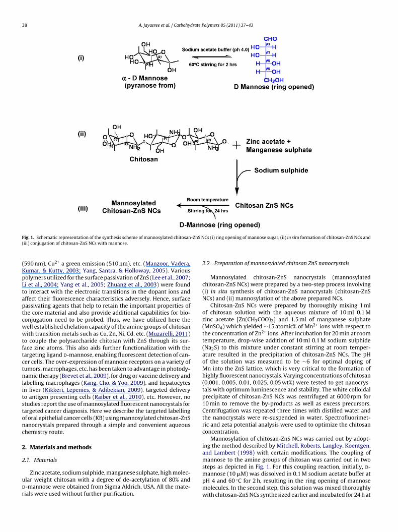
38 A. Jayasree et al. / Carbohydrate Polymers 85 (2011) 37–43
F -ZnS N(
(KpLtaptcwwtftctnlitstonc
2
2
udr
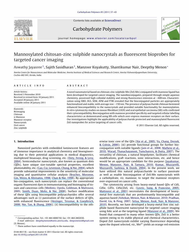
ig. 1. Schematic representation of the synthesis scheme of mannosylated chitosaniii) conjugation of chitosan-ZnS NCs with mannose.
590 nm), Cu2+ a green emission (510 nm), etc. (Manzoor, Vadera,umar, & Kutty, 2003; Yang, Santra, & Holloway, 2005). Variousolymers utilized for the surface passivation of ZnS (Lee et al., 2007;i et al., 2004; Yang et al., 2005; Zhuang et al., 2003) were foundo interact with the electronic transitions in the dopant ions andffect their fluorescence characteristics adversely. Hence, surfaceassivating agents that help to retain the important properties ofhe core material and also provide additional capabilities for bio-onjugation need to be probed. Thus, we have utilized here theell established chelation capacity of the amine groups of chitosanith transition metals such as Cu, Zn, Ni, Cd, etc. (Muzarelli, 2011)
o couple the polysaccharide chitosan with ZnS through its sur-ace zinc atoms. This also aids further functionalization with theargeting ligand d-mannose, enabling fluorescent detection of can-er cells. The over-expression of mannose receptors on a variety ofumors, macrophages, etc. has been taken to advantage in photody-amic therapy (Brevet et al., 2009), for drug or vaccine delivery and
abelling macrophages (Kang, Cho, & Yoo, 2009), and hepatocytesn liver (Kikkeri, Lepenies, & Adibekian, 2009), targeted deliveryo antigen presenting cells (Raiber et al., 2010), etc. However, notudies report the use of mannosylated fluorescent nanocrystals forargeted cancer diagnosis. Here we describe the targeted labellingf oral epithelial cancer cells (KB) using mannosylated chitosan-ZnSanocrystals prepared through a simple and convenient aqueoushemistry route.
. Materials and methods
.1. Materials
Zinc acetate, sodium sulphide, manganese sulphate, high molec-lar weight chitosan with a degree of de-acetylation of 80% and-mannose were obtained from Sigma Aldrich, USA. All the mate-ials were used without further purification.
Cs (i) ring opening of mannose sugar, (ii) in situ formation of chitosan-ZnS NCs and
2.2. Preparation of mannosylated chitosan ZnS nanocrystals
Mannosylated chitosan-ZnS nanocrystals (mannosylatedchitosan-ZnS NCs) were prepared by a two-step process involving(i) in situ synthesis of chitosan-ZnS nanocrystals (chitosan-ZnSNCs) and (ii) mannosylation of the above prepared NCs.
Chitosan-ZnS NCs were prepared by thoroughly mixing 1 mlof chitosan solution with the aqueous mixture of 10 ml 0.1 Mzinc acetate [Zn(CH3COO)2] and 1.5 ml of manganese sulphate(MnSO4) which yielded ∼15 atomic% of Mn2+ ions with respect tothe concentration of Zn2+ ions. After incubation for 20 min at roomtemperature, drop-wise addition of 10 ml 0.1 M sodium sulphide(Na2S) to this mixture under constant stirring at room temper-ature resulted in the precipitation of chitosan-ZnS NCs. The pHof the solution was measured to be ∼6 for optimal doping ofMn into the ZnS lattice, which is very critical to the formation ofhighly fluorescent nanocrystals. Varying concentrations of chitosan(0.001, 0.005, 0.01, 0.025, 0.05 wt%) were tested to get nanocrys-tals with optimum luminescence and stability. The white colloidalprecipitate of chitosan-ZnS NCs was centrifuged at 6000 rpm for10 min to remove the by-products as well as excess precursors.Centrifugation was repeated three times with distilled water andthe nanocrystals were re-suspended in water. Spectrofluorimet-ric and zeta potential analysis were used to optimize the chitosanconcentration.
Mannosylation of chitosan-ZnS NCs was carried out by adopt-ing the method described by Mitchell, Roberts, Langley, Koentgen,and Lambert (1998) with certain modifications. The coupling ofmannose to the amine groups of chitosan was carried out in two
steps as depicted in Fig. 1. For this coupling reaction, initially, d-mannose (10 �M) was dissolved in 0.1 M sodium acetate buffer atpH 4 and 60 ◦C for 2 h, resulting in the ring opening of mannosemolecules. In the second step, this solution was mixed thoroughlywith chitosan-ZnS NCs synthesized earlier and incubated for 24 h at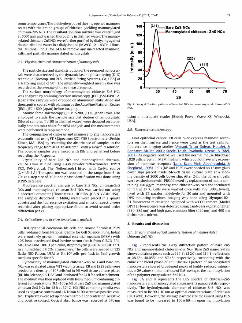
drate Polymers 85 (2011) 37–43 39
rrcasdds
2
ttar
wJt(
eDiw
wEfTr
ZP[7J
NaTcrd
2
cw1Miflm
Ns[Tfcuta
A. Jayasree et al. / Carbohy
oom temperature. The aldehyde group of the ring opened mannoseeacts with the amino groups of chitosan, yielding mannosylatedhitosan-ZnS NCs. The resultant solution mixture was centrifugedt 5000 rpm and washed thoroughly in distilled water. The manno-ylated chitosan-ZnS NCs were further purified by dialyzing againstouble-distilled water in a dialysis tube (MWCO 12–14 kDa; Hime-ia, Mumbai, India) for 24 h to remove any un-reacted mannose,alts, and partially mannosylated nanocrystals.
.3. Physico-chemical characterization of nanocrystals
The particle size and size distribution of the prepared nanocrys-als were characterized by the dynamic laser light scattering (DLS)echnique [Nicomp 380 ZLS, Particle Sizing Systems, CA, USA] atscattering angle of 90◦. The intensity-weighted mean value was
ecorded as the average of three measurements.The surface morphology of mannosylated chitosan-ZnS NCs
as analyzed by scanning electron microscopy (JEOL JSM-649OLA,apan). The samples were dropped on aluminium stubs, dried andhen sputter coated with platinum by the Auto Fine Platinum CoaterJEOL, JFC-1600, Japan) before imaging.
Atomic force microscopy (JSPM 5200, JEOL, Japan) was alsomployed to study the particle size distribution of nanocrystals.iluted samples (1:100 in distilled water) were dropped on atom-
cally smooth mica sheet for AFM analysis and the measurementsere performed in tapping mode.
The conjugation of chitosan and mannose to ZnS nanocrystalsas confirmed using FTIR [Spectrum RX1 FTIR Spectrometer, Perkin
lmer, MA, USA] by recording the absorbance of samples in therequency range from 4000 to 400 cm−1 with a 4 cm−1 resolution.he powder samples were mixed with KBr and pelletized beforeecording the IR spectra.
Crystallinity of bare ZnS NCs and mannosylated chitosan-nS NCs was studied using X-ray powder diffractometer [X‘PertRO, PANalytical, The Netherlands] fitted with Cu-K� source� = 1.541 A]. The spectrum was recorded in the range from 5◦ to0◦ at a step size of 0.02◦ and phase identification was done usingCPDS database.
Fluorescence spectral analysis of bare ZnS NCs, chitosan-ZnSCs and mannosylated chitosan-ZnS NCs was carried out usingspectrofluorometer [FluoroMax-4, HORIBA, JOBIN YVON, USA].
he samples dispersed in MilliQ water were placed in a quartzuvette and the fluorescence excitation and emission spectra wereecorded after placing appropriate filters to avoid second orderiffraction peaks.
.4. Cell culture and in vitro toxicological analysis
Oral epithelial carcinoma KB cells and mouse fibroblast L929ells (obtained from National Centre for Cell Science, Pune, India)ere routinely grown in minimal essential medium (MEM) with
0% heat-inactivated fetal bovine serum (both from GIBCO-BRL,D, USA) and 100 IU penicillin/streptomycin (GIBCO-BRL) at 37 ◦C
n a humidified 5% CO2 atmosphere. The cells were seeded in T25asks (BD Falcon, USA) at 5 × 10⁵ cells per flask in 5 ml growthedium specific for KB.Cytotoxicity of mannosylated chitosan-ZnS NCs and bare ZnS
Cs was evaluated using MTT viability assay. KB and L929 cells wereeeded at a density of 104 cells/ml in 96-well tissue culture platesBD Bio Science, CA, USA] and incubated for 24 h for cell attachment.he medium was then replaced with fresh medium containing dif-
erent concentrations (0.1–100 �M) of bare ZnS and mannosylatedhitosan-ZnS NCs for 48 h at 37 ◦C. 10% FBS containing media wassed as negative control and 1% Triton X100 served as positive con-rol. Triplicates were set up for each sample concentration, negativend positive control. Optical absorbance was recorded at 570 nmFig. 2. X-ray diffraction patterns of bare ZnS NCs and mannosylated chitosan-ZnSNCs.
using a microplate reader [Biotek Power Wave XS, Winooski,USA].
2.5. Fluorescence microscopy
Oral epithelial cancer, KB cells over express mannose recep-tors on their surface and hence were used as the test cells forfluorescence imaging studies (Agnani, Tricot-Doleux, Houalet, &Bonnaure-Mallet, 2003; Steele, Leigh, Swoboda, Ozenci, & Fidel,2001). As negative control, we used the normal mouse fibroblastL929 cells grown in MEM medium, which do not have any expres-sion of mannose receptors (Lane, Egan, Vick, Abdolrasulnia, &Shepherd, 1998). Cells (KB and L929) were seeded on 13 mm glasscover slips placed inside 24-well tissue culture plate at a seed-ing density of 3000 cells/cover slip. After 24 h, the adherent cellswere washed once with PBS followed by replacement of media con-taining 150 �g/ml mannosylated chitosan-ZnS NCs and incubatedfor 1 h at 37 ◦C. Cells were washed once with PBS (300 �l/well),fixed with 2% paraformaldehyde for 20 min and mounted withDPX mounting medium. Imaging was done using Olympus BX-51 fluorescent microscope equipped with a CCD camera (ModelDP71). Fluorescence was detected using band-pass excitation filter(330–385 nm) and high pass emission filter (420 nm) and 400 nmdichromatic mirror.
3. Results and discussion
3.1. Structural and optical characterization of mannosylatedchitosan-ZnS NCs
Fig. 2 represents the X-ray diffraction pattern of bare ZnSNCs and mannosylated chitosan-ZnS NCs. Bare ZnS nanocrystalsshowed distinct peaks such as (1 1 1), (2 2 0) and (3 1 1) reflectionsat 28.63◦, 48.055◦ and 57.45◦ respectively, correlating with thecubic zinc blend phase of ZnS. The XRD pattern of mannosylatednanocrystals showed broadened peaks of highly reduced intensi-ties at 2� values similar to those of ZnS, owing to the mannosylationof the polymer encapsulated ZnS NCs.
Fig. 3A and B represents the DLS spectra of chitosan-ZnS
nanocrystals and mannosylated chitosan-ZnS nanocrystals respec-tively. The hydrodynamic diameter of chitosan-ZnS NCs wasmeasured to be 90 ± 10 nm at optimal concentrations of chitosan(0.01 wt%). However, the average particle size measured using DLSwas found to be increased to 150 ± 60 nm upon mannosylation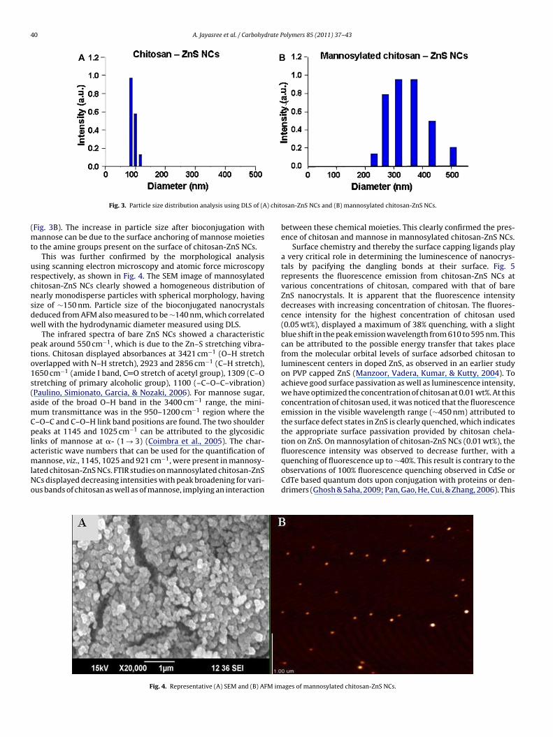
40 A. Jayasree et al. / Carbohydrate Polymers 85 (2011) 37–43
) chit
(mt
urcnsdw
pto1s(amCplamlNo
Fig. 3. Particle size distribution analysis using DLS of (A
Fig. 3B). The increase in particle size after bioconjugation withannose can be due to the surface anchoring of mannose moieties
o the amine groups present on the surface of chitosan-ZnS NCs.This was further confirmed by the morphological analysis
sing scanning electron microscopy and atomic force microscopyespectively, as shown in Fig. 4. The SEM image of mannosylatedhitosan-ZnS NCs clearly showed a homogeneous distribution ofearly monodisperse particles with spherical morphology, havingize of ∼150 nm. Particle size of the bioconjugated nanocrystalseduced from AFM also measured to be ∼140 nm, which correlatedell with the hydrodynamic diameter measured using DLS.
The infrared spectra of bare ZnS NCs showed a characteristiceak around 550 cm−1, which is due to the Zn–S stretching vibra-ions. Chitosan displayed absorbances at 3421 cm−1 (O–H stretchverlapped with N–H stretch), 2923 and 2856 cm−1 (C–H stretch),650 cm−1 (amide I band, C O stretch of acetyl group), 1309 (C–Otretching of primary alcoholic group), 1100 (–C–O–C–vibration)Paulino, Simionato, Garcia, & Nozaki, 2006). For mannose sugar,side of the broad O–H band in the 3400 cm−1 range, the mini-um transmittance was in the 950–1200 cm−1 region where the
–O–C and C–O–H link band positions are found. The two shouldereaks at 1145 and 1025 cm−1 can be attributed to the glycosidic
inks of mannose at �- (1 → 3) (Coimbra et al., 2005). The char-cteristic wave numbers that can be used for the quantification of
annose, viz., 1145, 1025 and 921 cm−1, were present in mannosy-ated chitosan-ZnS NCs. FTIR studies on mannosylated chitosan-ZnSCs displayed decreasing intensities with peak broadening for vari-us bands of chitosan as well as of mannose, implying an interaction
Fig. 4. Representative (A) SEM and (B) AFM im
osan-ZnS NCs and (B) mannosylated chitosan-ZnS NCs.
between these chemical moieties. This clearly confirmed the pres-ence of chitosan and mannose in mannosylated chitosan-ZnS NCs.
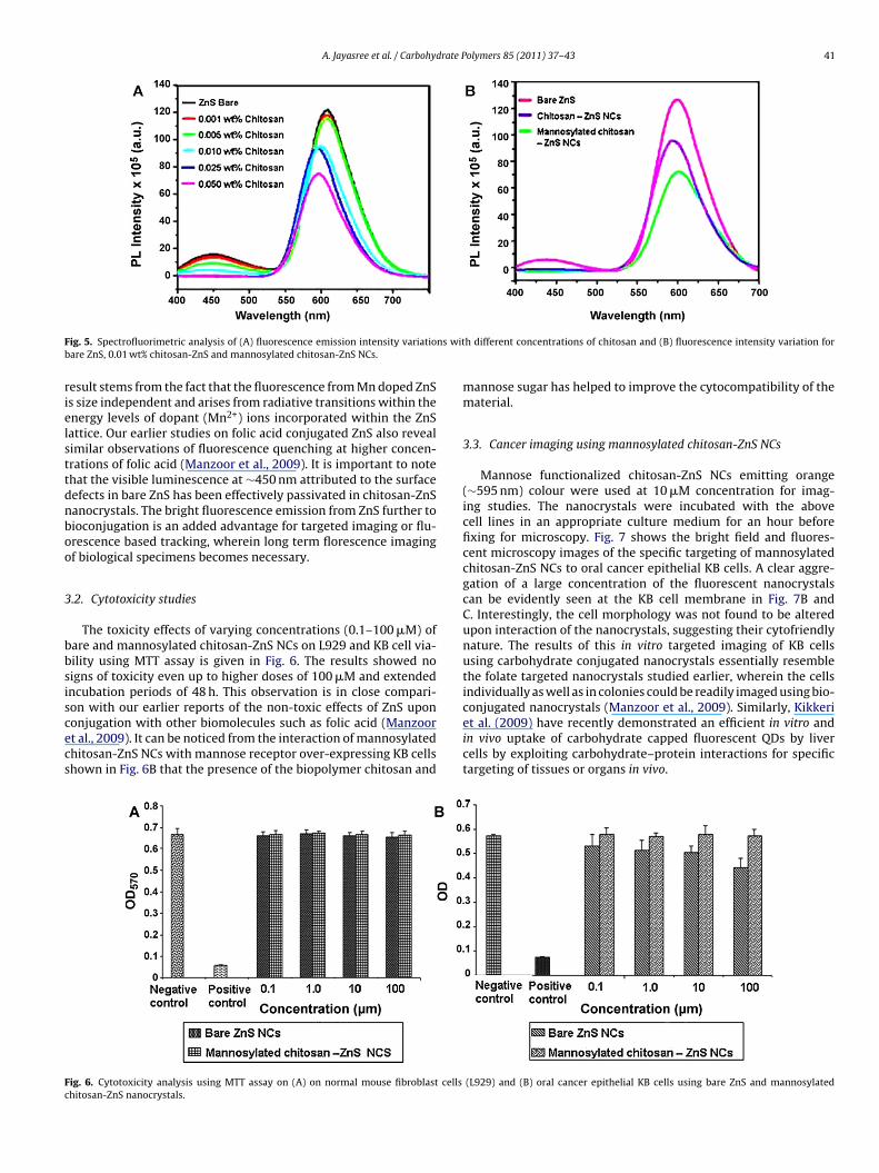
Surface chemistry and thereby the surface capping ligands playa very critical role in determining the luminescence of nanocrys-tals by pacifying the dangling bonds at their surface. Fig. 5represents the fluorescence emission from chitosan-ZnS NCs atvarious concentrations of chitosan, compared with that of bareZnS nanocrystals. It is apparent that the fluorescence intensitydecreases with increasing concentration of chitosan. The fluores-cence intensity for the highest concentration of chitosan used(0.05 wt%), displayed a maximum of 38% quenching, with a slightblue shift in the peak emission wavelength from 610 to 595 nm. Thiscan be attributed to the possible energy transfer that takes placefrom the molecular orbital levels of surface adsorbed chitosan toluminescent centers in doped ZnS, as observed in an earlier studyon PVP capped ZnS (Manzoor, Vadera, Kumar, & Kutty, 2004). Toachieve good surface passivation as well as luminescence intensity,we have optimized the concentration of chitosan at 0.01 wt%. At thisconcentration of chitosan used, it was noticed that the fluorescenceemission in the visible wavelength range (∼450 nm) attributed tothe surface defect states in ZnS is clearly quenched, which indicatesthe appropriate surface passivation provided by chitosan chela-tion on ZnS. On mannosylation of chitosan-ZnS NCs (0.01 wt%), thefluorescence intensity was observed to decrease further, with a
quenching of fluorescence up to ∼40%. This result is contrary to theobservations of 100% fluorescence quenching observed in CdSe orCdTe based quantum dots upon conjugation with proteins or den-drimers (Ghosh & Saha, 2009; Pan, Gao, He, Cui, & Zhang, 2006). Thisages of mannosylated chitosan-ZnS NCs.
A. Jayasree et al. / Carbohydrate Polymers 85 (2011) 37–43 41
F ns witb
rielsttdnboo
3
bbsiscecs
Fc
ig. 5. Spectrofluorimetric analysis of (A) fluorescence emission intensity variatioare ZnS, 0.01 wt% chitosan-ZnS and mannosylated chitosan-ZnS NCs.
esult stems from the fact that the fluorescence from Mn doped ZnSs size independent and arises from radiative transitions within thenergy levels of dopant (Mn2+) ions incorporated within the ZnSattice. Our earlier studies on folic acid conjugated ZnS also revealimilar observations of fluorescence quenching at higher concen-rations of folic acid (Manzoor et al., 2009). It is important to notehat the visible luminescence at ∼450 nm attributed to the surfaceefects in bare ZnS has been effectively passivated in chitosan-ZnSanocrystals. The bright fluorescence emission from ZnS further toioconjugation is an added advantage for targeted imaging or flu-rescence based tracking, wherein long term florescence imagingf biological specimens becomes necessary.
.2. Cytotoxicity studies
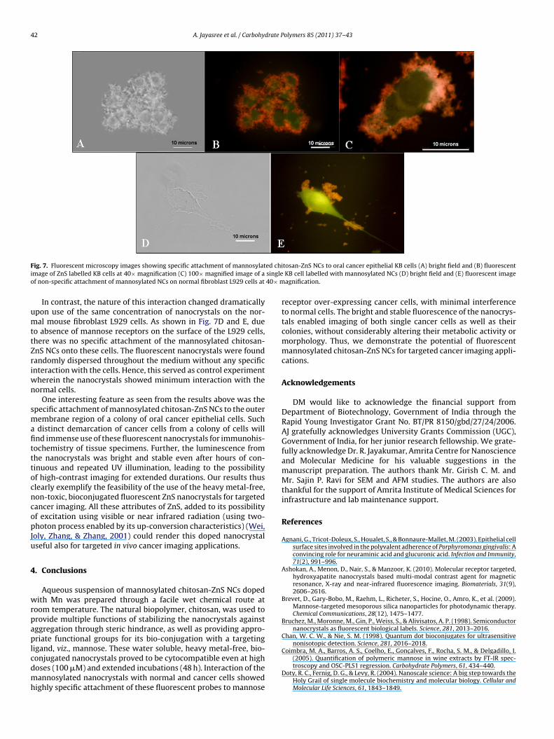
The toxicity effects of varying concentrations (0.1–100 �M) ofare and mannosylated chitosan-ZnS NCs on L929 and KB cell via-ility using MTT assay is given in Fig. 6. The results showed noigns of toxicity even up to higher doses of 100 �M and extendedncubation periods of 48 h. This observation is in close compari-
on with our earlier reports of the non-toxic effects of ZnS upononjugation with other biomolecules such as folic acid (Manzoort al., 2009). It can be noticed from the interaction of mannosylatedhitosan-ZnS NCs with mannose receptor over-expressing KB cellshown in Fig. 6B that the presence of the biopolymer chitosan andig. 6. Cytotoxicity analysis using MTT assay on (A) on normal mouse fibroblast cellshitosan-ZnS nanocrystals.
h different concentrations of chitosan and (B) fluorescence intensity variation for
mannose sugar has helped to improve the cytocompatibility of thematerial.
3.3. Cancer imaging using mannosylated chitosan-ZnS NCs
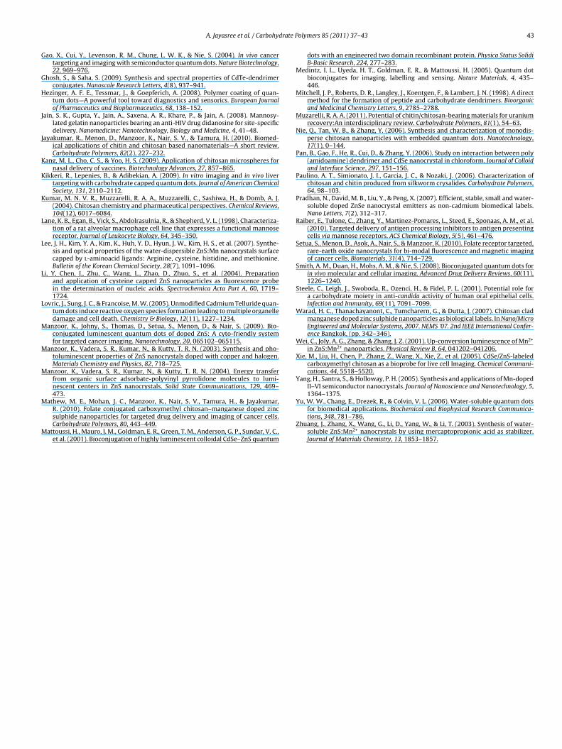
Mannose functionalized chitosan-ZnS NCs emitting orange(∼595 nm) colour were used at 10 �M concentration for imag-ing studies. The nanocrystals were incubated with the abovecell lines in an appropriate culture medium for an hour beforefixing for microscopy. Fig. 7 shows the bright field and fluores-cent microscopy images of the specific targeting of mannosylatedchitosan-ZnS NCs to oral cancer epithelial KB cells. A clear aggre-gation of a large concentration of the fluorescent nanocrystalscan be evidently seen at the KB cell membrane in Fig. 7B andC. Interestingly, the cell morphology was not found to be alteredupon interaction of the nanocrystals, suggesting their cytofriendlynature. The results of this in vitro targeted imaging of KB cellsusing carbohydrate conjugated nanocrystals essentially resemblethe folate targeted nanocrystals studied earlier, wherein the cellsindividually as well as in colonies could be readily imaged using bio-
conjugated nanocrystals (Manzoor et al., 2009). Similarly, Kikkeriet al. (2009) have recently demonstrated an efficient in vitro andin vivo uptake of carbohydrate capped fluorescent QDs by livercells by exploiting carbohydrate–protein interactions for specifictargeting of tissues or organs in vivo.(L929) and (B) oral cancer epithelial KB cells using bare ZnS and mannosylated
42 A. Jayasree et al. / Carbohydrate Polymers 85 (2011) 37–43
F ed chii singleo 40× m
umttZriwn
smafitttocncopJu
4
wrpaplcdmh
ig. 7. Fluorescent microscopy images showing specific attachment of mannosylatmage of ZnS labelled KB cells at 40× magnification (C) 100× magnified image of af non-specific attachment of mannosylated NCs on normal fibroblast L929 cells at
In contrast, the nature of this interaction changed dramaticallypon use of the same concentration of nanocrystals on the nor-al mouse fibroblast L929 cells. As shown in Fig. 7D and E, due
o absence of mannose receptors on the surface of the L929 cells,here was no specific attachment of the mannosylated chitosan-nS NCs onto these cells. The fluorescent nanocrystals were foundandomly dispersed throughout the medium without any specificnteraction with the cells. Hence, this served as control experiment
herein the nanocrystals showed minimum interaction with theormal cells.
One interesting feature as seen from the results above was thepecific attachment of mannosylated chitosan-ZnS NCs to the outerembrane region of a colony of oral cancer epithelial cells. Suchdistinct demarcation of cancer cells from a colony of cells willnd immense use of these fluorescent nanocrystals for immunohis-ochemistry of tissue specimens. Further, the luminescence fromhe nanocrystals was bright and stable even after hours of con-inuous and repeated UV illumination, leading to the possibilityf high-contrast imaging for extended durations. Our results thuslearly exemplify the feasibility of the use of the heavy metal-free,on-toxic, bioconjugated fluorescent ZnS nanocrystals for targetedancer imaging. All these attributes of ZnS, added to its possibilityf excitation using visible or near infrared radiation (using two-hoton process enabled by its up-conversion characteristics) (Wei,
oly, Zhang, & Zhang, 2001) could render this doped nanocrystalseful also for targeted in vivo cancer imaging applications.
. Conclusions
Aqueous suspension of mannosylated chitosan-ZnS NCs dopedith Mn was prepared through a facile wet chemical route at
oom temperature. The natural biopolymer, chitosan, was used torovide multiple functions of stabilizing the nanocrystals againstggregation through steric hindrance, as well as providing appro-riate functional groups for its bio-conjugation with a targeting
igand, viz., mannose. These water soluble, heavy metal-free, bio-onjugated nanocrystals proved to be cytocompatible even at highoses (100 �M) and extended incubations (48 h). Interaction of theannosylated nanocrystals with normal and cancer cells showed
ighly specific attachment of these fluorescent probes to mannose
tosan-ZnS NCs to oral cancer epithelial KB cells (A) bright field and (B) fluorescentKB cell labelled with mannosylated NCs (D) bright field and (E) fluorescent imageagnification.
receptor over-expressing cancer cells, with minimal interferenceto normal cells. The bright and stable fluorescence of the nanocrys-tals enabled imaging of both single cancer cells as well as theircolonies, without considerably altering their metabolic activity ormorphology. Thus, we demonstrate the potential of fluorescentmannosylated chitosan-ZnS NCs for targeted cancer imaging appli-cations.
Acknowledgements
DM would like to acknowledge the financial support fromDepartment of Biotechnology, Government of India through theRapid Young Investigator Grant No. BT/PR 8150/gbd/27/24/2006.AJ gratefully acknowledges University Grants Commission (UGC),Government of India, for her junior research fellowship. We grate-fully acknowledge Dr. R. Jayakumar, Amrita Centre for Nanoscienceand Molecular Medicine for his valuable suggestions in themanuscript preparation. The authors thank Mr. Girish C. M. andMr. Sajin P. Ravi for SEM and AFM studies. The authors are alsothankful for the support of Amrita Institute of Medical Sciences forinfrastructure and lab maintenance support.
References
Agnani, G., Tricot-Doleux, S., Houalet, S., & Bonnaure-Mallet, M. (2003). Epithelial cellsurface sites involved in the polyvalent adherence of Porphyromonas gingivalis: Aconvincing role for neuraminic acid and glucuronic acid. Infection and Immunity,71(2), 991–996.
Ashokan, A., Menon, D., Nair, S., & Manzoor, K. (2010). Molecular receptor targeted,hydroxyapatite nanocrystals based multi-modal contrast agent for magneticresonance, X-ray and near-infrared fluorescence imaging. Biomaterials, 31(9),2606–2616.
Brevet, D., Gary-Bobo, M., Raehm, L., Richeter, S., Hocine, O., Amro, K., et al. (2009).Mannose-targeted mesoporous silica nanoparticles for photodynamic therapy.Chemical Communications, 28(12), 1475–1477.
Bruchez, M., Moronne, M., Gin, P., Weiss, S., & Alivisatos, A. P. (1998). Semiconductornanocrystals as fluorescent biological labels. Science, 281, 2013–2016.
Chan, W. C. W., & Nie, S. M. (1998). Quantum dot bioconjugates for ultrasensitivenonisotopic detection. Science, 281, 2016–2018.
Coimbra, M. A., Barros, A. S., Coelho, E., Goncalves, F., Rocha, S. M., & Delgadillo, I.(2005). Quantification of polymeric mannose in wine extracts by FT-IR spec-troscopy and OSC-PLS1 regression. Carbohydrate Polymers, 61, 434–440.
Doty, R. C., Fernig, D. G., & Levy, R. (2004). Nanoscale science: A big step towards theHoly Grail of single molecule biochemistry and molecular biology. Cellular andMolecular Life Sciences, 61, 1843–1849.
drate P
G
G
H
J
J
K
K
K
L
L
L
L
M
M
M
M
M
A. Jayasree et al. / Carbohy
ao, X., Cui, Y., Levenson, R. M., Chung, L. W. K., & Nie, S. (2004). In vivo cancertargeting and imaging with semiconductor quantum dots. Nature Biotechnology,22, 969–976.
hosh, S., & Saha, S. (2009). Synthesis and spectral properties of CdTe-dendrimerconjugates. Nanoscale Research Letters, 4(8), 937–941.
ezinger, A. F. E., Tessmar, J., & Goepferich, A. (2008). Polymer coating of quan-tum dots—A powerful tool toward diagnostics and sensorics. European Journalof Pharmaceutics and Biopharmaceutics, 68, 138–152.
ain, S. K., Gupta, Y., Jain, A., Saxena, A. R., Khare, P., & Jain, A. (2008). Mannosy-lated gelatin nanoparticles bearing an anti-HIV drug didanosine for site-specificdelivery. Nanomedicine: Nanotechnology, Biology and Medicine, 4, 41–48.
ayakumar, R., Menon, D., Manzoor, K., Nair, S. V., & Tamura, H. (2010). Biomed-ical applications of chitin and chitosan based nanomaterials—A short review.Carbohydrate Polymers, 82(2), 227–232.
ang, M. L., Cho, C. S., & Yoo, H. S. (2009). Application of chitosan microspheres fornasal delivery of vaccines. Biotechnology Advances, 27, 857–865.
ikkeri, R., Lepenies, B., & Adibekian, A. (2009). In vitro imaging and in vivo livertargeting with carbohydrate capped quantum dots. Journal of American ChemicalSociety, 131, 2110–2112.
umar, M. N. V. R., Muzzarelli, R. A. A., Muzzarelli, C., Sashiwa, H., & Domb, A. J.(2004). Chitosan chemistry and pharmaceutical perspectives. Chemical Reviews,104(12), 6017–6084.
ane, K. B., Egan, B., Vick, S., Abdolrasulnia, R., & Shepherd, V. L. (1998). Characteriza-tion of a rat alveolar macrophage cell line that expresses a functional mannosereceptor. Journal of Leukocyte Biology, 64, 345–350.
ee, J. H., Kim, Y. A., Kim, K., Huh, Y. D., Hyun, J. W., Kim, H. S., et al. (2007). Synthe-sis and optical properties of the water-dispersible ZnS:Mn nanocrystals surfacecapped by l-aminoacid ligands: Arginine, cysteine, histidine, and methionine.Bulletin of the Korean Chemical Society, 28(7), 1091–1096.
i, Y, Chen, J., Zhu, C., Wang, L., Zhao, D., Zhuo, S., et al. (2004). Preparationand application of cysteine capped ZnS nanoparticles as fluorescence probein the determination of nucleic acids. Spectrochemica Acta Part A, 60, 1719–1724.
ovric, J., Sung, J. C., & Francoise, M. W. (2005). Unmodified Cadmium Telluride quan-tum dots induce reactive oxygen species formation leading to multiple organelledamage and cell death. Chemistry & Biology, 12(11), 1227–1234.
anzoor, K., Johny, S., Thomas, D., Setua, S., Menon, D., & Nair, S. (2009). Bio-conjugated luminescent quantum dots of doped ZnS: A cyto-friendly systemfor targeted cancer imaging. Nanotechnology, 20, 065102–065115.
anzoor, K., Vadera, S. R., Kumar, N., & Kutty, T. R. N. (2003). Synthesis and pho-toluminescent properties of ZnS nanocrystals doped with copper and halogen.Materials Chemistry and Physics, 82, 718–725.
anzoor, K., Vadera, S. R., Kumar, N., & Kutty, T. R. N. (2004). Energy transferfrom organic surface adsorbate-polyvinyl pyrrolidone molecules to lumi-nescent centers in ZnS nanocrystals. Solid State Communications, 129, 469–473.
athew, M. E., Mohan, J. C., Manzoor, K., Nair, S. V., Tamura, H., & Jayakumar,R. (2010). Folate conjugated carboxymethyl chitosan–manganese doped zincsulphide nanoparticles for targeted drug delivery and imaging of cancer cells.Carbohydrate Polymers, 80, 443–449.
attoussi, H., Mauro, J. M., Goldman, E. R., Green, T. M., Anderson, G. P., Sundar, V. C.,et al. (2001). Bioconjugation of highly luminescent colloidal CdSe–ZnS quantum
olymers 85 (2011) 37–43 43
dots with an engineered two domain recombinant protein. Physica Status SolidiB-Basic Research, 224, 277–283.
Medintz, I. L., Uyeda, H. T., Goldman, E. R., & Mattoussi, H. (2005). Quantum dotbioconjugates for imaging, labelling and sensing. Nature Materials, 4, 435–446.
Mitchell, J. P., Roberts, D. R., Langley, J., Koentgen, F., & Lambert, J. N. (1998). A directmethod for the formation of peptide and carbohydrate dendrimers. Bioorganicand Medicinal Chemistry Letters, 9, 2785–2788.
Muzarelli, R. A. A. (2011). Potential of chitin/chitosan-bearing materials for uraniumrecovery:An interdisciplinary review. Carbohydrate Polymers, 81(1), 54–63.
Nie, Q., Tan, W. B., & Zhang, Y. (2006). Synthesis and characterization of monodis-perse chitosan nanoparticles with embedded quantum dots. Nanotechnology,17(1), 0–144.
Pan, B., Gao, F., He, R., Cui, D., & Zhang, Y. (2006). Study on interaction between poly(amidoamine) dendrimer and CdSe nanocrystal in chloroform. Journal of Colloidand Interface Science, 297, 151–156.
Paulino, A. T., Simionato, J. I., Garcia, J. C., & Nozaki, J. (2006). Characterization ofchitosan and chitin produced from silkworm crysalides. Carbohydrate Polymers,64, 98–103.
Pradhan, N., David, M. B., Liu, Y., & Peng, X. (2007). Efficient, stable, small and water-soluble doped ZnSe nanocrystal emitters as non-cadmium biomedical labels.Nano Letters, 7(2), 312–317.
Raiber, E., Tulone, C., Zhang, Y., Martinez-Pomares, L., Steed, E., Sponaas, A. M., et al.(2010). Targeted delivery of antigen processing inhibitors to antigen presentingcells via mannose receptors. ACS Chemical Biology, 5(5), 461–476.
Setua, S., Menon, D., Asok, A., Nair, S., & Manzoor, K. (2010). Folate receptor targeted,rare-earth oxide nanocrystals for bi-modal fluorescence and magnetic imagingof cancer cells. Biomaterials, 31(4), 714–729.
Smith, A. M., Duan, H., Mohs, A. M., & Nie, S. (2008). Bioconjugated quantum dots forin vivo molecular and cellular imaging. Advanced Drug Delivery Reviews, 60(11),1226–1240.
Steele, C., Leigh, J., Swoboda, R., Ozenci, H., & Fidel, P. L. (2001). Potential role fora carbohydrate moiety in anti-candida activity of human oral epithelial cells.Infection and Immunity, 69(11), 7091–7099.
Warad, H. C., Thanachayanont, C., Tumcharern, G., & Dutta, J. (2007). Chitosan cladmanganese doped zinc sulphide nanoparticles as biological labels. In Nano/MicroEngineered and Molecular Systems, 2007. NEMS ‘07. 2nd IEEE International Confer-ence Bangkok, (pp. 342–346).
Wei, C., Joly, A. G., Zhang, & Zhang, J. Z. (2001). Up-conversion luminescence of Mn2+
in ZnS:Mn2+ nanoparticles. Physical Review B, 64, 041202–041206.Xie, M., Liu, H., Chen, P., Zhang, Z., Wang, X., Xie, Z., et al. (2005). CdSe/ZnS-labeled
carboxymethyl chitosan as a bioprobe for live cell Imaging. Chemical Communi-cations, 44, 5518–5520.
Yang, H., Santra, S., & Holloway, P. H. (2005). Synthesis and applications of Mn-dopedII–VI semiconductor nanocrystals. Journal of Nanoscience and Nanotechnology, 5,1364–1375.
Yu, W. W., Chang, E., Drezek, R., & Colvin, V. L. (2006). Water-soluble quantum dotsfor biomedical applications. Biochemical and Biophysical Research Communica-tions, 348, 781–786.
Zhuang, J., Zhang, X., Wang, G., Li, D., Yang, W., & Li, T. (2003). Synthesis of water-soluble ZnS:Mn2+ nanocrystals by using mercaptopropionic acid as stabilizer.Journal of Materials Chemistry, 13, 1853–1857.


