Embed Size (px)
Citation preview
BioMed CentralBMC Biology
ss
Open AcceResearch articleLow dose pramipexole is neuroprotective in the MPTP mouse model of Parkinson's disease, and downregulates the dopamine transporter via the D3 receptorJeffrey N Joyce*, Cheryl Woolsey, Han Ryoo, Sabine Borwege and Diane HagnerAddress: Thomas H. Christopher Center for Parkinson's Disease Research, Sun Health Research Institute, 10515 West Santa Fe Dr., Sun City, AZ, 85352, USA
Email: Jeffrey N Joyce* - [email protected]; Cheryl Woolsey - [email protected]; Han Ryoo - [email protected]; Sabine Borwege - [email protected]; Diane Hagner - [email protected]
* Corresponding author
AbstractBackground: Our aim was to determine if pramipexole, a D3 preferring agonist, effectively reduceddopamine neuron and fiber loss in the 1-methyl-4-phenyl-1,2,3,6-tetrahydropyridine (MPTP) mouse modelwhen given at intraperitoneal doses corresponding to clinical doses. We also determined whethersubchronic treatment with pramipexole regulates dopamine transporter function, thereby reducingintracellular transport of the active metabolite of MPTP, 1-methyl-4-phenylpyridinium (MPP+).
Methods: Ten 12-month old C57BL/6 mice were treated with MPTP (or saline) twice per day at 20 mg/kg s.c. (4 injections over 48 h). Mice were pretreated for 3 days and during the 2-day MPTP regimen withpramipexole (0.1 mg/kg/day) or saline. Stereological quantification of dopamine neuron number and opticaldensity measurement of dopamine fiber loss were carried out at 1 week after treatment, usingimmunostaining for dopamine transporter (DAT) and tyrosine hydroxylase (TH). Additional wild-type(WT) and D3 receptor knockout (KO) mice were treated for 5 days with pramipexole (0.1 mg/kg/day) orvehicle. The kinetics of [3H]MPP+ and [3H]DA uptake (Vmax and Km) were determined 24 h later; and at24 h and 14 days dopamine transporter density was measured by quantitative autoradiography.
Results: Pramipexole treatment completely antagonized the neurotoxic effects of MPTP, as measured bysubstantia nigra and ventral tegmental area TH-immunoreactive cell counts. MPTP- induced loss of striatalinnervation, as measured by DAT-immunoreactivity, was partially prevented by pramipexole, but not withregard to TH-IR. Pramipexole also reduced DAT- immunoreactivity in non-MPTP treated mice.Subchronic treatment with pramipexole lowered the Vmax for [3H]DA and [3H]MPP+ uptake into striatalsynaptosomes of WT mice. Pramipexole treatment lowered Vmax in WT but not D3 KO mice; however,D3 KO mice had lower Vmax for [3H]DA uptake. There was no change in DAT number in WT withpramipexole treatment or D3 KO mice at 24 h post-treatment, but there was a reduction in WT-pramipexole treated and not in D3 KO mice at 14 days post-treatment.
Conclusion: These results suggest that protection occurs at clinically suitable doses of pramipexole.Protection could be due to a reduced amount of MPP+ taken up into DA terminals via DAT. D3 receptorplays an important role in this regulation of transporter uptake and availability.
Published: 11 October 2004
BMC Biology 2004, 2:22 doi:10.1186/1741-7007-2-22
Received: 17 June 2004Accepted: 11 October 2004
This article is available from: http://www.biomedcentral.com/1741-7007/2/22
© 2004 Joyce et al; licensee BioMed Central Ltd. This is an open-access article distributed under the terms of the Creative Commons Attribution License (http://creativecommons.org/licenses/by/2.0), which permits unrestricted use, distribution, and reproduction in any medium, provided the original work is properly cited.
Page 1 of 12(page number not for citation purposes)

BMC Biology 2004, 2:22 http://www.biomedcentral.com/1741-7007/2/22
BackgroundAn interesting development in the use of dopamine (DA)agonists for treatment of Parkinson's disease (PD) is thatsome of them have proven to be neuroprotective in ani-mal models of PD. Antiparkinsonian agents that are directDA agonists, such as apomorphine [1], bromocriptine [2],and pramipexole [3], are neuroprotective against 1-methyl-4-phenyl-1,2,3,6-tetrahydropyridine (MPTP)-induced damage to the DA system in mice. Administra-tion of MPTP, which is converted to 1-methyl-4-phe-nylpyridinium (MPP+) and intracellularly transportedinto DAergic neurons [4], provides a good model for stud-ying neuroprotection in PD. MPTP produces Parkinson-ism in humans and in subhuman species throughselective loss of DAergic neurons of the substantia nigra(SN) [5,6], and a number of related compounds to MPTPalso produce nigral cell loss in primates [7]. MPTP causesapoptosis associated with PD [8-10] ;MPTP produces pro-gressive cell death in humans for decades after the initialinsult [11]. Hence, drugs that reduce the neurotoxicity ofcompounds like MPTP may be neuroprotective in PD. Infact, it is now hypothesized that direct DA agonists mayslow the loss of DAergic terminal function upon long-term administration to PD patients [12-15].
Dopaminergic neurons are tonically inhibited by den-dritic and terminal autoreceptors, operating in interactionwith DA transporters (DAT) and pharmacologically of theD2 receptor subtype [16-19]. However, Zapata et al [20]have reported that the D3 preferring agonist (+)-PD128907 regulates extracellular DA levels via interactionswith D3 autoreceptors. If D3 preferring agonists are potentautoreceptor agonists, then hypothetically long-termchanges in expression of DAT or the functional propertiesof DAT might occur following subchronic treatment.Since intracellular accumulation of MPP+ following sys-temic injection of MPTP requires DAT [4], then when DATis downregulated by D3 preferring agonists, this couldresult in lower intracellular accumulation of MPP+ andreduced neurotoxicity to MPTP.
The D3 receptor preferring agonists, pramipexole and rop-inirole, are the most potent of the DA agonists affordingneuroprotection at 1 mg/kg for pramipexole againstMPTP-induced neurodegeneration [3,21] and at 2 mg/kgfor ropinirole against 6-OHDA lesions in rats [22]. Doses10–30 times higher of DA agonists with low D3 receptoraffinity such as apomorphine [1] and bromocriptine[2,23] are needed against MPTP-induced neurodegenera-tion. Because neuroprotection by pramipexole is most evi-dent with concurrent treatment with MPTP and not withpost-MPTP treatment [24], i.e. when autoreceptor contri-butions should be most pronounced, regulation of DATmay be important. In addition, while the lowest effectivedose reported is 1.0 mg/kg for mice, this is significantly
greater than a clinically relevant dose in humans (1.5 mgt.i.d., p.o.[25]). Based on information from PharmaciaCorporation, equivalent plasma levels obtained with 1.5mg t.i.d., p.o. in humans could be produced with 0.1 to0.5 mg/kg in the mice. We tested whether 0.1 mg/kgpramipexole would be neuroprotective in aging miceagainst MPTP-induced neurodegeneration to the DA sys-tem, and if this effect could be due to regulation of DATfunction.
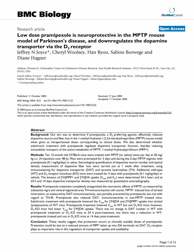
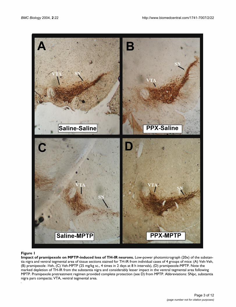
ResultsNeurohistopathologyMale C57BL/6 mice of 8–10 months of age were pre-treated with saline or pramipexole (0.1 mg/kg/day) fol-lowed by MPTP. At the end of the 7-day recovery periodfollowing the last injection of MPTP or vehicle wereassessed for the degree of toxicity to the dopamine systemby MPTP. MPTP produced a marked loss of tyrosinehydroxylase-immunoreactive (TH-IR) neurons in the sub-stantia nigra (SN), but had less impact in the ventral teg-mental area (VTA) (Figs 1 and 2), Unbiased stereologicalquantification of the number of Nissl-stained and TH-IRneurons in the SNpc and VTA was made in the midbrainsof the treated groups. MPTP produced a 31% loss of TH-IR neurons in the SN and 17% loss in the VTA. Pramipex-ole administered once a day for 5 days (i.e. 3 days prior toand during the 2-day vehicle treatment) did not alter thetotal number of TH-IR neurons in the SN or VTA. Prami-pexole administered for 3 days prior to and during the 2-day administration of MPTP completely prevented TH-IRneuron loss in the SN and VTA of MPTP treated mice. Toconfirm that TH-IR neurons were dead and not simplyexhibiting reduced TH-IR, neurons in Nissl stained sec-tions were counted. The results confirmed the data thatD3-preferring agonists can protect against MPTP in vivo aswell as against MPP+ in vitro [26,27].
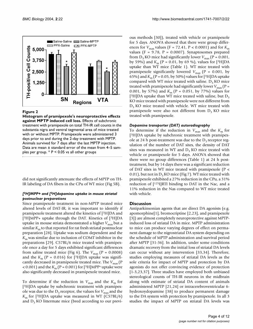
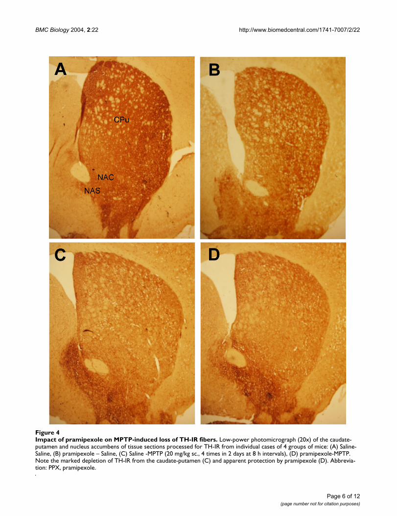
Visualization of DA fibers with dopamine transporterimmunoreactivity, DAT-IR (Fig 3), and TH-IR (Fig 4)demonstrated uniform staining of the caudate-putamen(CPu) and nucleus accumbens (Nac) in the saline/salinecases. In saline pretreated mice, MPTP reduced DAT-IR by52% in the CPu (Figs 3 and 5A) and had a smaller but sig-nificant impact on DAT-IR labeling of DA fibers in theNac. DAT-IR in the CPu in the pramipexole plus vehicle(PPX-SAL)-treated mice was reduced by 17%. Further-more, in pramipexole and MPTP treated mice, a signifi-cant attenuation of the impact of MPTP in the CPu (-27%vs -52% loss) and Nac of MPTP treated mice was seen.
In contrast to DAT-IR, levels of TH-IR (Figs 4 and 5B) wasnot significantly reduced (~11%) by pramipexole in PPX-SAL group. MPTP produced a significant (34%) loss ofTH-IR labeling of DA fibers in the CPu of WT mice and toa lesser degree in the Nac SAL-MPTP group. Pramipexole
Page 2 of 12(page number not for citation purposes)
BMC Biology 2004, 2:22 http://www.biomedcentral.com/1741-7007/2/22
Impact of pramipexole on MPTP-induced loss of TH-IR neuronsFigure 1Impact of pramipexole on MPTP-induced loss of TH-IR neurons. Low-power photomicrograph (20x) of the substan-tia nigra and ventral tegmental area of tissue sections stained for TH-IR from individual cases of 4 groups of mice: (A) Veh-Veh, (B) pramipexole -Veh, (C) Veh-MPTP (25 mg/kg sc., 4 times in 2 days at 8 h intervals), (D) pramipexole-MPTP. Note the marked depletion of TH-IR from the substantia nigra and considerably lesser impact in the ventral tegmental area following MPTP. Pramipexole pretreatment regimen provided complete protection (see D) from MPTP. Abbreviations: SNpc, substantia nigra pars compacta; VTA, ventral tegmental area.
Page 3 of 12(page number not for citation purposes)
BMC Biology 2004, 2:22 http://www.biomedcentral.com/1741-7007/2/22
did not significantly attenuate the effects of MPTP on TH-IR labeling of DA fibers in the CPu of WT mice (Fig 5B).
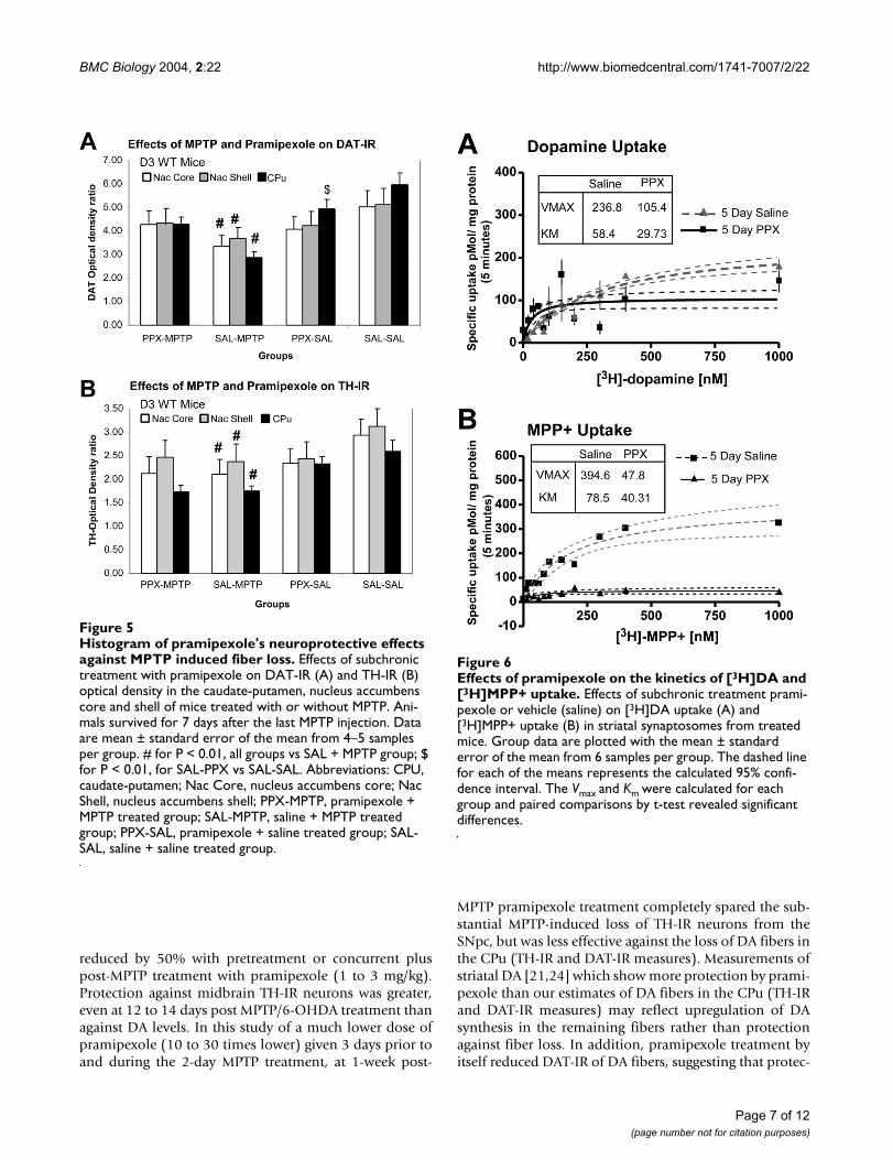
[3H]MPP+ and [3H]dopamine uptake in mouse striatal postnuclear preparationsSince pramipexole treatment in non-MPTP treated micealtered levels of DAT-IR, it was important to identify ifpramipexole treatment altered the kinetics of [3H]DA and[3H]MPP+ uptake through the DAT. Kinetics of [3H]DAuptake in mouse striata demonstrated a higher Vmax but asimilar Km to that reported for rat fresh striatal postnuclearpreparation [28]. Uptake was sodium dependent and theKm was similar due to inclusion of COMT inhibitor in thepreparations [29]. C57BL/6 mice treated with pramipex-ole once a day for 5 days exhibited significant differencesfrom saline treated mice (Fig 6). The Vmax (P = 0.0008)and the Km (P = 0.016) for [3H]DA uptake was signifi-cantly decreased in pramipexole treated mice. The Vmax (P< 0.001) and the Km (P = 0.001) for [3H]MPP+ uptake werealso significantly decreased in pramipexole treated mice.
To determine if the reduction in Vmax and the Km for[3H]DA uptake by subchronic treatment with pramipex-ole was due to the D3 receptor, the values for Vmax and theKm for [3H]DA uptake was measured in WT (C57BL/6)and D3 KO littermate mice (bred according to our previ-
ous methods [30]), treated with vehicle or pramipexolefor 5 days. ANOVA showed that there were group differ-ences for Vmax values (F = 72.41, P < 0.0001) and for Kmvalues (F = 9.78, P = 0.0007). Synaptosomes preparedfrom D3 KO mice had significantly lower Vmax (P = 0.001,by 59%) and Km (P = 0.01, by 69 %), values for [3H]DAuptake than WT mice (Table 1). WT mice treated withpramipexole significantly lowered Vmax (P = 0.001, by65%) and Km (P = 0.05, by 50%) values for [3H]DA uptakecompared with WT mice treated with saline. D3 KO micetreated with pramipexole had significantly lower Vmax (P =0.001, by 57%) and Km (P = 0.051, by 77%) values for[3H]DA uptake than WT mice treated with saline, but D3KO mice treated with pramipexole were not different fromD3 KO mice treated with vehicle. WT mice treated withpramipexole were also not different from D3 KO micetreated with pramipexole.
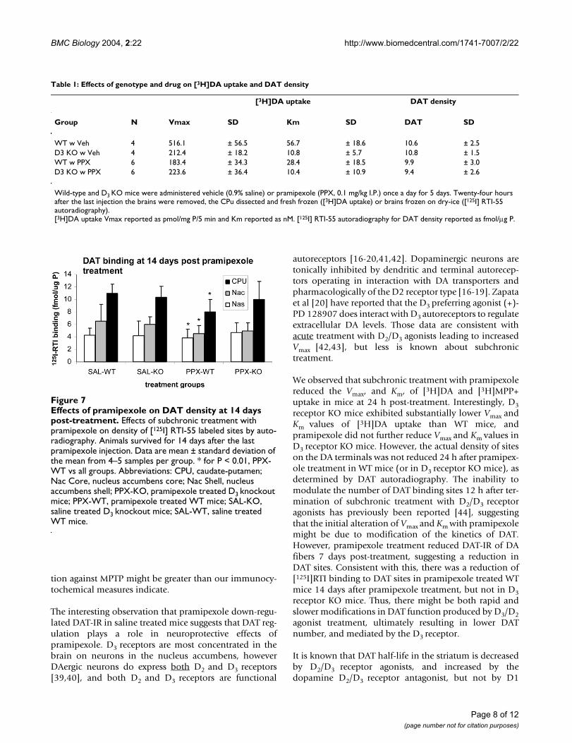
Dopamine transporter (DAT) autoradiographyTo determine if the reduction in Vmax and the Km for[3H]DA uptake by subchronic treatment with pramipex-ole at 24 h post-treatment was due to the D3 receptor reg-ulation of the number of DAT sites, the density of DATsites was measured in WT and D3 KO mice treated withvehicle or pramipexole for 5 days. ANOVA showed thatthere were no group differences (Table 1) at 24 h post-treatment, but by 14 days there was a significant reductionof DAT sites in WT mice treated with pramipexole (P <0.01), but not in D3 KO mice (Fig 7). WT mice treated withpramipexole exhibited a 27% reduction in the CPu, a 31%reduction of [125I]RTI binding to DAT in the Nac, and a11% reduction in the Nas compared to WT mice treatedwith vehicle.
DiscussionAntiparkinsonian agents that are direct DA agonists (e.g.apomorphine[1], bromocriptine [2,23], and pramipexole[3]) are almost completely neuroprotective against MPTP-induced loss of striatal DA in mice. MPTP administrationto mice can produce varying degrees of effect on perma-nent damage to the nigrostriatal DA system depending onthe schedule of MPTP administration and survival periodafter MPTP [31-36]. In addition, under some conditionsdramatic recovery from the initial loss of striatal DA levelscan occur without any intervention [33,34]. Therefore,studies employing measures of striatal DA levels as thesole criteria for impact of MPTP and protection by DAagonists do not offer convincing evidence of protection[1-3,23,37]. Three studies have employed both unbiasedstereological counts of TH-IR neurons in the midbrainalong with estimate of striatal DA content of animalsadministered MPTP [21,24] or intracerebroventricular 6-hydroxoydopamine [38] to produce permanent damageto the DA system with protection by pramipexole. In all 3studies the impact of MPTP on striatal DA levels was
Histogram of pramipexole's neuroprotective effects against MPTP induced cell lossFigure 2Histogram of pramipexole's neuroprotective effects against MPTP induced cell loss. Effects of subchronic treatment with pramipexole on total TH-IR cell counts in the substantia nigra and ventral tegmental area of mice treated with or without MPTP. Pramipexole were administered 3 days prior to and during the 2-day treatment with MPTP. Animals survived for 7 days after the last MPTP injection. Data are mean ± standard error of the mean from 4–5 sam-ples per group. * P < 0.05 vs all other groups
Page 4 of 12(page number not for citation purposes)
BMC Biology 2004, 2:22 http://www.biomedcentral.com/1741-7007/2/22
Impact of pramipexole on MPTP-induced loss of DT-IR fibersFigure 3Impact of pramipexole on MPTP-induced loss of DAT-IR fibers. Low-power photomicrograph (20x) of the caudate-putamen and nucleus accumbens of tissue sections processed for DAT-IR from individual cases of 4 groups of mice: (A) Saline-Saline, (B) pramipexole – Saline, (C) Saline -MPTP (20 mg/kg sc., 4 times in 2 days at 8 h intervals), (D) pramipexole-MPTP. Note the marked depletion of DAT-IR from the caudate-putamen (C) and apparent protection by pramipexole (D). Abbrevia-tion: CPu, caudate-putamen; NAS, nucleus accumbens shell; NAC, nucleus accumbens core.
Page 5 of 12(page number not for citation purposes)
BMC Biology 2004, 2:22 http://www.biomedcentral.com/1741-7007/2/22
Impact of pramipexole on MPTP-induced loss of TH-IR fibersFigure 4Impact of pramipexole on MPTP-induced loss of TH-IR fibers. Low-power photomicrograph (20x) of the caudate-putamen and nucleus accumbens of tissue sections processed for TH-IR from individual cases of 4 groups of mice: (A) Saline-Saline, (B) pramipexole – Saline, (C) Saline -MPTP (20 mg/kg sc., 4 times in 2 days at 8 h intervals), (D) pramipexole-MPTP. Note the marked depletion of TH-IR from the caudate-putamen (C) and apparent protection by pramipexole (D). Abbrevia-tion: PPX, pramipexole.
Page 6 of 12(page number not for citation purposes)
BMC Biology 2004, 2:22 http://www.biomedcentral.com/1741-7007/2/22
reduced by 50% with pretreatment or concurrent pluspost-MPTP treatment with pramipexole (1 to 3 mg/kg).Protection against midbrain TH-IR neurons was greater,even at 12 to 14 days post MPTP/6-OHDA treatment thanagainst DA levels. In this study of a much lower dose ofpramipexole (10 to 30 times lower) given 3 days prior toand during the 2-day MPTP treatment, at 1-week post-
MPTP pramipexole treatment completely spared the sub-stantial MPTP-induced loss of TH-IR neurons from theSNpc, but was less effective against the loss of DA fibers inthe CPu (TH-IR and DAT-IR measures). Measurements ofstriatal DA [21,24] which show more protection by prami-pexole than our estimates of DA fibers in the CPu (TH-IRand DAT-IR measures) may reflect upregulation of DAsynthesis in the remaining fibers rather than protectionagainst fiber loss. In addition, pramipexole treatment byitself reduced DAT-IR of DA fibers, suggesting that protec-
Histogram of pramipexole's neuroprotective effects against MPTP induced fiber lossFigure 5Histogram of pramipexole's neuroprotective effects against MPTP induced fiber loss. Effects of subchronic treatment with pramipexole on DAT-IR (A) and TH-IR (B) optical density in the caudate-putamen, nucleus accumbens core and shell of mice treated with or without MPTP. Ani-mals survived for 7 days after the last MPTP injection. Data are mean ± standard error of the mean from 4–5 samples per group. # for P < 0.01, all groups vs SAL + MPTP group; $ for P < 0.01, for SAL-PPX vs SAL-SAL. Abbreviations: CPU, caudate-putamen; Nac Core, nucleus accumbens core; Nac Shell, nucleus accumbens shell; PPX-MPTP, pramipexole + MPTP treated group; SAL-MPTP, saline + MPTP treated group; PPX-SAL, pramipexole + saline treated group; SAL-SAL, saline + saline treated group.
Effects of pramipexole on the kinetics of [3H]DA and [3H]MPP+ uptakeFigure 6Effects of pramipexole on the kinetics of [3H]DA and [3H]MPP+ uptake. Effects of subchronic treatment prami-pexole or vehicle (saline) on [3H]DA uptake (A) and [3H]MPP+ uptake (B) in striatal synaptosomes from treated mice. Group data are plotted with the mean ± standard error of the mean from 6 samples per group. The dashed line for each of the means represents the calculated 95% confi-dence interval. The Vmax and Km were calculated for each group and paired comparisons by t-test revealed significant differences.
Page 7 of 12(page number not for citation purposes)
BMC Biology 2004, 2:22 http://www.biomedcentral.com/1741-7007/2/22
tion against MPTP might be greater than our immunocy-tochemical measures indicate.
The interesting observation that pramipexole down-regu-lated DAT-IR in saline treated mice suggests that DAT reg-ulation plays a role in neuroprotective effects ofpramipexole. D3 receptors are most concentrated in thebrain on neurons in the nucleus accumbens, howeverDAergic neurons do express both D2 and D3 receptors[39,40], and both D2 and D3 receptors are functional
autoreceptors [16-20,41,42]. Dopaminergic neurons aretonically inhibited by dendritic and terminal autorecep-tors operating in interaction with DA transporters andpharmacologically of the D2 receptor type [16-19]. Zapataet al [20] have reported that the D3 preferring agonist (+)-PD 128907 does interact with D3 autoreceptors to regulateextracellular DA levels. Those data are consistent withacute treatment with D2/D3 agonists leading to increasedVmax [42,43], but less is known about subchronictreatment.
We observed that subchronic treatment with pramipexolereduced the Vmax, and Km, of [3H]DA and [3H]MPP+uptake in mice at 24 h post-treatment. Interestingly, D3receptor KO mice exhibited substantially lower Vmax andKm values of [3H]DA uptake than WT mice, andpramipexole did not further reduce Vmax and Km values inD3 receptor KO mice. However, the actual density of siteson the DA terminals was not reduced 24 h after pramipex-ole treatment in WT mice (or in D3 receptor KO mice), asdetermined by DAT autoradiography. The inability tomodulate the number of DAT binding sites 12 h after ter-mination of subchronic treatment with D2/D3 receptoragonists has previously been reported [44], suggestingthat the initial alteration of Vmax and Km with pramipexolemight be due to modification of the kinetics of DAT.However, pramipexole treatment reduced DAT-IR of DAfibers 7 days post-treatment, suggesting a reduction inDAT sites. Consistent with this, there was a reduction of[125I]RTI binding to DAT sites in pramipexole treated WTmice 14 days after pramipexole treatment, but not in D3receptor KO mice. Thus, there might be both rapid andslower modifications in DAT function produced by D3/D2agonist treatment, ultimately resulting in lower DATnumber, and mediated by the D3 receptor.
It is known that DAT half-life in the striatum is decreasedby D2/D3 receptor agonists, and increased by thedopamine D2/D3 receptor antagonist, but not by D1
Table 1: Effects of genotype and drug on [3H]DA uptake and DAT density
[3H]DA uptake DAT density
Group N Vmax SD Km SD DAT SD
WT w Veh 4 516.1 ± 56.5 56.7 ± 18.6 10.6 ± 2.5D3 KO w Veh 4 212.4 ± 18.2 10.8 ± 5.7 10.8 ± 1.5WT w PPX 6 183.4 ± 34.3 28.4 ± 18.5 9.9 ± 3.0D3 KO w PPX 6 223.6 ± 36.4 10.4 ± 10.9 9.4 ± 2.6
Wild-type and D3 KO mice were administered vehicle (0.9% saline) or pramipexole (PPX, 0.1 mg/kg I.P.) once a day for 5 days. Twenty-four hours after the last injection the brains were removed, the CPu dissected and fresh frozen ([3H]DA uptake) or brains frozen on dry-ice ([125I] RTI-55 autoradiography).[3H]DA uptake Vmax reported as pmol/mg P/5 min and Km reported as nM. [125I] RTI-55 autoradiography for DAT density reported as fmol/µg P.
Effects of pramipexole on DAT density at 14 days post-treat-mentFigure 7Effects of pramipexole on DAT density at 14 days post-treatment. Effects of subchronic treatment with pramipexole on density of [125I] RTI-55 labeled sites by auto-radiography. Animals survived for 14 days after the last pramipexole injection. Data are mean ± standard deviation of the mean from 4–5 samples per group. * for P < 0.01, PPX-WT vs all groups. Abbreviations: CPU, caudate-putamen; Nac Core, nucleus accumbens core; Nac Shell, nucleus accumbens shell; PPX-KO, pramipexole treated D3 knockout mice; PPX-WT, pramipexole treated WT mice; SAL-KO, saline treated D3 knockout mice; SAL-WT, saline treated WT mice.
Page 8 of 12(page number not for citation purposes)
BMC Biology 2004, 2:22 http://www.biomedcentral.com/1741-7007/2/22
agonists and antagonists [45]. Furthermore, the D2 ago-nist-induced change in DAT kinetics iss inhibited by theco-administration of an antagonist. The absence of DAreceptors can also influence DAT function, as shown bydopamine D2 receptor-deficient mice, which exhibitdecreased striatal DA uptake [19]. The present results canbe compared with those reported by Saunders et al. [46]using hDAT-FLAG expressed in human embryonic kidney293-EM4 cells, who showed by confocal microscopy andwhole-cell current recordings that 2 µM d-amphetamineincreased internalization of surface DAT within 1 h. Treat-ment with DA in HEK-hDAT cells also reduced Vmax, dueto a diminished presence of DAT at the surface of synap-tosomes [47]. Subchronic pramipexole might lead toredistribution DAT from the plasma membrane toendosomal compartments, and regulated, in part, by theD3 receptor. The initial change in Vmax, and Km, could berelated to a more rapid turnover of DAT, and the longer-term reduction in Bmax to greater internalization and/orreduced synthesis. Thus, D3 preferring agonists might bepotent autoreceptor agonists, and long-term changes inexpression of the DAT or the functional properties of DATcould occur following subchronic treatment. This, in turn,could lead to reduced MPP+ (and other neurotoxins)uptake into DA neurons, and reduced toxicity in animalmodels of PD. Ramirez and associates [37] reported thatthe neuroprotective effects of pramipexole against MPTP-induced DA loss in mice was attenuated by the selectiveD3 antagonist A-437203. Furthermore, the neuroprotec-tive effect of a low dose of pramipexole was attenuated inD3 transgenic knockout mice and protection by pramipex-ole was not further attenuated by treatment with a D3antagonist. These in vivo data support an important rolefor the D3 receptor in the neuroprotective effects of DAagonists, and our data suggest that this, in part, is due toreduced MPP+ uptake into DA neurons.
ConclusionsWe have identified that subchronic treatment with a clin-ically relevant dose of pramipexole beginning before ini-tiation of MPTP treatment affords neuroprotectionagainst DA neuron loss and, to a lesser extent, DA fiberloss. This might involve down-regulation of DAT andreduced MPP+ uptake into DA fibers. Since intracellularaccumulation of MPP+ following systemic injection ofMPTP requires DAT [4], then if DAT function and/ornumber are reduced by D3 preferring agonists this couldresult in lower intracellular accumulation of MPP+ andreduced neurotoxicity to MPTP. The importance ofknowing the targets of pramipexole, and other D3 prefer-ring agonists, in neuroprotection in animal models of PDcannot be understated, given the possibility that this pro-tection could be extended to humans [12]. However, invivo imaging of DAT as a tool for analyzing the neuropro-tective effects of DA agonists could be difficult to inter-
pret, since DAT might be regulated by DA agonists[25,48]. Our data are consistent with this hypothesis andsuggest that multiple measures of DA fiber integrity arerequired to assess neuroprotection by agents [49,50].
MethodsAll animals were treated in accordance with a protocolapproved by the Sun Health Research Institute AnimalCare and Use Committee.
MPTP TreatmentWe bred C57BL/6 mice from breeding pairs obtainedfrom Jackson Laboratories. Eighteen male C57BL/6 mice8–10 months of age were used for the experiments andwere handled for 1 week prior to treatment. They had freeaccess to food and water, and were maintained in a 12 hlight/dark cycle prior to treatment. Mice were divided into4 groups: the first received only vehicle (0.9% saline, 0.1ml/10 mg body wt) (SAL-SAL), the second receivedpramipexole plus vehicle (PPX-SAL), the third receivedvehicle plus MPTP (RBI, MA) (SAL-MPTP), and the fourthreceived pramipexole plus MPTP (PPX-MPT). For thosereceiving pramipexole (Pharmacia Corporation, Kalama-zoo, MI), the drug was dissolved in 0.9% sterile saline andadministered by i.p. injection (0.1 ml/10 mg body wt). Asingle daily dose of 0.1 mg/kg body weight of pramipex-ole was given for 5 days. Those not receiving pramipexolewere given identical injections of saline., Followingpramipexole or vehicle injections on days 4 and 5, micewere given injections of either MPTP (20 mg/kg; s.c.) orvehicle (saline) twice daily at 8 h intervals.
After 7-day recovery period following the last injection ofMPTP or vehicle, animals were euthanized by intracardiacperfusion with 4% paraformaldehyde in 0.15 M phos-phate buffer (pH 7.2) following overdose with pentobar-bital 120 mg/kg body weight i.p. Brains were removed,postfixed in the perfusion fixative for 24 h at 4°C andtransferred to 30% sucrose solution for additional 24 hincubation at 4°C. The tissue was frozen on dry-ice andsectioned in cryostat at 20 µm thickness. Sections wereplaced in cryoprotectant solution for long-term storage at-80°C.
NeurohistopathologyEvery 10th section at the level of striatum was processedfor visualization of DAT and tyrosine hydroxylase (TH),and every 5th that of the midbrain processed for the visu-alization of TH-positive cell bodies using the avidin-biotin procedure [26,51]. Immediately adjacent sectionsfrom the midbrain were stained for cresyl violet for detec-tion of cells. The sections were washed in phosphatebuffer to remove cryoprotectant, incubated with 5% goatserum for 30 min to block background staining andincubated with anti-DAT (Chemicon, CA) or anti-TH
Page 9 of 12(page number not for citation purposes)
BMC Biology 2004, 2:22 http://www.biomedcentral.com/1741-7007/2/22
(Chemicon, CA) at 1:1000 dilution overnight at roomtemperature. Control sections were treated with identicalsolutions but with no primary antibody. Sections wererinsed and incubated with biotinylated secondary anti-rabbit antiserum at a 1:500 dilution (Vector, CA) for 90min at room temperature. Sections were again rinsed,incubated in streptavidin-peroxidase complex (Vector,CA) at a 1:250 dilution for 2 h at room temperature. Aftermore thorough rinsing, sections were processed for DABwith nickel enhancement. Sections were then rinsed inphosphate buffer, mounted on gelatin-coated slides, air-dried, dehydrated, and cleared in xylene and mountedwith Permount.
Unbiased stereological quantification of Nissl-stained andTH-IR neurons in the SNpc and VTA was used to estimatecell number in the midbrain. The general routine at lowmagnification involved use of a sampling grid for theSNpc and VTA. At high magnification the computer-basedimaging system randomly selected a region of the grid andclearly definable neurons was counted within the 3-dimensional block. This was repeated for every 10th regionof each section. Estimation of total neuron number wasbased upon actual cell counts, tissue thickness, total areaof designated region, and the total number of sectionsanalyzed per animal. Group means and variances werecalculated.
Our routine quantitative measurement of the optical den-sity of regions of the striatum stained for TH-IR and DAT-IR [26,51] was employed. Using a Macintosh-based imageanalysis system with CCD camera and imaging software(BRAIN version 3.0, Drexel University) optical densitymeasurements calibrated to an external standard (Kodakdensity step tablet) of the region of interest (ROI) and acontrol region (corpus callosum) of each section weremade, the ratio of the ROI to control region was calcu-lated, and the average for each animal determined. Groupmeans and variances were estimated. Statistical analysis ofgroup differences were assessed by ANOVA with pairwisecomparisons performed using post-hoc t-tests and theBonferroni correction.
Testing of whether pramipexole administration alters dopamine transporter functionFourteen C57/Bl6 mice (25–30 g, 6 months) were dividedinto 2 groups: one group received pramipexole (0.1 mg/kg) once a day for 5 days and the other group received thevehicle (saline). An additional 10 WT and 10 D3 receptorknockout mice (25–30 g, 6 months), bred according toour previous methods [30], were treated similarly.Twenty-four hours after the last injection of pramipexoleor vehicle the mice were euthanized using CO2 narcosisand the brains rapidly removed and snap-frozen in liquidnitrogen. Km and Vmax values for [3H]1-methyl-4-phe-
nylpyridinium (MPP+) and [3H]DA uptake in synapto-somes derived from the striatum were deterimined by themethod of Eshleman et al [28], with minor modifications.Comparison of mean values for the Km and Vmax of[3H]MPP+ and DA uptake were made by t-test (0.05 levelof significance).
[3H]MPP+ and [3H]dopamine uptake in mouse striatal postnuclear preparationsMouse striata were dissected and homogenized with aglass-Teflon homogenizer in ice-cold modified HEPES (1ml). The sample was centrifuged at 1000 g, for 10 min at4°C. The supernatant was collected and centrifuged at14,000 g for 10 min at 4°C. The pellet was resuspended in8 ml of HEPES buffer (HEPES 25 mM, NaCl 122 mM,CaCl 2.5 mM, MgSO4 1.2 mM, pargyline 10 uM, glucose0.2%, ascorbic acid 0.02%, pH7.4). To the 8 ml sample,butaclamol was added at a final concentration of 100 nM,4 ml of sample was than removed and placed in a separate15 ml centrifuge tube (VWR, Pennsylvania) and Mazindol(Sigma, Missouri) was added at a final concentration of40 uM. 50µl of the samples were added to borosilicatetubes (Fisher, Texas) and placed in a 25°C water bath withthe drugs for a 10 min preincubation. The assay wasinitiated by adding 50µl concentrations of unlabeledMPP+ or DA ranging from 0–300 nM with [3H]MPP+ or[3H]DA at a final concentration of 20 nM. The sampleswere incubated at 25°C for 10 min. Specific uptake wasdefined as the difference in uptake observed in theabsence and presence of mazindol (40µM). Uptake wasterminated after 5 min by filtration through WhatmanGF/C filters presoaked in HEPES buffer. Scintillation fluidwas added to each filtered spot and radioactivity remain-ing on the filters was determined using a Wallac β-scintil-lation spectrometer. Each experiment involved triplicatedeterminations, and 6 independent experiments for eachdrug competition curve were performed.
Dopamine transporter (DAT) autoradiography10 WT and 10 D3 receptor knockout mice (25–30 g, 6months) were divided into 2 groups: one group receivedpramipexole (0.1 mg/kg) once a day for 5 days and theother group received the vehicle (0.9% saline). Twenty-four h after the last injection of pramipexole or vehicle,the mice were overdosed as above and the brains rapidlyremoved and frozen on dry- ice. An additional group of 10WT and 10 D3 receptor knockout mice (25–30 g, 6months) were similarly treated but their brains were proc-essed 14 days after the last treatment. Autoradiography ofDAT sites were quantified following labeling with[125I]RTI-55 (3ß-(4-iodophenyl)tropan-2 ß-carboxylicacid methyl ester) (Dupont, New England Nuclear, Bos-ton, MA) in the presence of 100 nM paroxetine (SmithKlein Beecham BRL 29060A) to block the serotonintransporter, according to published methodology [30].
Page 10 of 12(page number not for citation purposes)
BMC Biology 2004, 2:22 http://www.biomedcentral.com/1741-7007/2/22
Specific binding was defined with 40µM benztropine(Sigma, St. Louis MO), and amounted to 95% of totalbinding. Sections were apposed to 3H-Hyperfilm for 18 hfor DAT. Autoradiographs were analyzed using a compu-ter-based image analysis system (AIS, Imaging ResearchInc., Ontario Canada) that converts transmitted opticaldensity to the amount of radioligand bound in pmol permicrogram of protein.
List of abbreviationsD3 KO = D3 receptor knockout
DA = dopamine
DAT = dopamine transporter
MPP+ = 1-methyl-4-phenylpyridinium
MPTP = 1-methyl-4-phenyl-1,2,3,6-tetrahydropyridine
PD = Parkinson's disease
SN = substantia nigra
TH = tyrosine hydroxylase
VTA = ventral tegmental area
WT = wild type
Competing interestsThe author of this manuscript received a grant from Phar-macia Corporation to partially support the costs of theresearch. This company is no longer in existence.
Authors' contributionsCW and HR carried out the MPTP treatment, performedthe immunocytochemistry and participated in the statisti-cal analysis. SB and DH carried out the uptake assays, per-formed the DAT autoradiography and participated in thestatistical analysis. JNJ conceived of the study, and partic-ipated in its design and coordination. All authors read andapproved the final manuscript.
AcknowledgementsThis work was funded by Federal Grant NS40669, Arizona Alzheimer's Disease Research Center contract 4001(Arizona Parkinson's Disease Center), and Pharmacia Corporation (Kalamzaoo, MI).
References1. Grünblatt E, Mandel S, Berkuzki T, Youdim MB: Apomorphine pro-
tects against MPTP-induced neurotoxicity in mice. Mov Disord1999, 14:612-618.
2. Muralikrishnan D, Mohanakumar KP: Neuroprotection by bro-mocriptine against 1-methyl-4-phenyl-1,2,3,6- tetrahydropy-ridine-induced neurotoxicity in mice. FASEB J 1998, 12:905-912.
3. Kitamura Y, Kohno Y, Nakazawa M, Nomura Y: Inhibitory effectsof talipexole and pramipexole on MPTP-induced dopamine
reduction in the striatum of C57BL/6N mice. Jpn J Pharmacol1997, 74:51-57.
4. Gainetdinov RR, Fumagalli F, Jones SR, Caron MG: Dopaminetransporter is required for in vivo MPTP neurotoxicity: evi-dence from mice lacking the transporter. J Neurochem 1997,69:1322-1325.
5. Burns RS, Chiueh CC, Markey SP, Ebert MH, Jacobowitz DM, KopinIJ: A primate model of parkinsonism: selective destruction ofdopaminergic neurons in the pars compacta of the substan-tia nigra by N-methyl-4- phenyl-1,2,3,6-tetrahydropyridine.Proc Natl Acad Sci U S A 1983, 80:4546-4550.
6. Langston JW, Ballard P, Tetrud JW, Irwin I: Chronic Parkinsonismin humans due to a product of meperidine-analog synthesis.Science 1983, 219:979-980.
7. McNaught KS, Thull U, Carrupt PA, Altomare C, Cellamare S, CarottiA, Testa B, Jenner P, Marsden CD: Nigral cell loss produced byinfusion of isoquinoline derivatives structurally related to 1-methyl-4-phenyl-1,2,3,6-tetrahydropyridine. Neurodegeneration1996, 5:265-274.
8. Cassarino DS, Bennett JP Jr: An evaluation of the role of mito-chondria in neurodegenerative diseases: mitochondrialmutations and oxidative pathology, protective nuclearresponses, and cell death in neurodegeneration. Brain Res BrainRes Rev 1999, 29:1-25.
9. Hirsch EC, Hunot S, Hartmann A: Mechanism of cell death inexperimental models of Parkinson's disease. Funct Neurol 2000,15:229-237.
10. Cohen G, Werner P: Free radicals, oxidative stress and neuro-degeneration. In Neurodegenerative Disorders Edited by: Calne CB.Philadelphia, PA: WB Saunders; 1994:139-162.
11. Snow BJ, Vingerhoets FJ, Langston JW, Tetrud JW, Sossi V, Calne DB:Pattern of dopaminergic loss in the striatum of humans withMPTP induced parkinsonism. J Neurol Neurosurg Psychiatry 2000,68:313-316.
12. Marek K, Seibyl J, Shoulson I, Holloway R, Kieburtz K, McDermott M,Kamp C, Shinaman A, Fahn S, Lang A, et al.: Dopamine transporterbrain imaging to assess the effects of pramipexole vs levo-dopa on Parkinson disease progression. JAMA 2002,287:1653-1661.
13. Whone AL, Watts RL, Stoessl AJ, Davis M, Reske S, Nahmias C, LangAE, Rascol O, Ribeiro MJ, Remy P, et al.: Slower progression ofParkinson's disease with ropinirole versus levodopa: TheREAL-PET study. Ann Neurol 2003, 54:93-101.
14. Clarke CE, Guttman M: Dopamine agonist monotherapy in Par-kinson's disease. Lancet 2002, 360:1767-1769.
15. Stocchi F, Olanow CW: Neuroprotection in Parkinson's dis-ease: clinical trials. Ann Neurol 2003, 53(Suppl 3):S87-S97.
16. Gobert A, Rivet JM, Audinot V, Cistarelli L, Spedding M, Vian J, PeglionJL, Millan MJ: Functional correlates of dopamine D3 receptoractivation in the rat in vivo and their modulation by theselective antagonist, (+)-S 14297: II. Both D2 and "silent" D3autoreceptors control synthesis and release in mesolimbic,mesocortical and nigrostriatal pathways. J Pharmacol Exp Ther1995, 275:899-913.
17. Koeltzow TE, Xu M, Cooper DC, Hu XT, Tonegawa S, Wolf ME,White FJ: Alterations in dopamine release but not dopamineautoreceptor function in dopamine D3 receptor mutantmice. J Neurosci 1998, 18:2231-2238.
18. L'hirondel M, Cheramy A, Godeheu G, Artaud F, Saiardi A, Borrelli E,Glowinski J: Lack of autoreceptor-mediated inhibitory controlof dopamine release in striatal synaptosomes of D2receptor-deficient mice. Brain Res 1998, 792:253-262.
19. Dickinson SD, Sabeti J, Larson GA, Giardina K, Rubinstein M, KellyMA, Grandy DK, Low MJ, Gerhardt GA, Zahniser NR: DopamineD2 receptor-deficient mice exhibit decreased dopaminetransporter function but no changes in dopamine release indorsal striatum. J Neurochem 1999, 72:148-156.
20. Zapata A, Witkin JM, Shippenberg TS: Selective D3 receptor ago-nist effects of (+)-PD 128907 on dialysate dopamine at lowdoses. Neuropharmacology 2001, 41:351-359.
21. Zou L, Xu J, Jankovic J, He Y, Appel SH, Le W: Pramipexole inhib-its lipid peroxidation and reduces injury in the substantianigra induced by the dopaminergic neurotoxin 1-methyl-4-phenyl-1,2,3,6-tetrahydropyridine in C57BL/6 mice. NeurosciLett 2000, 281:167-170.
Page 11 of 12(page number not for citation purposes)
BMC Biology 2004, 2:22 http://www.biomedcentral.com/1741-7007/2/22
Publish with BioMed Central and every scientist can read your work free of charge
"BioMed Central will be the most significant development for disseminating the results of biomedical research in our lifetime."
Sir Paul Nurse, Cancer Research UK
Your research papers will be:
available free of charge to the entire biomedical community
peer reviewed and published immediately upon acceptance
cited in PubMed and archived on PubMed Central
yours — you keep the copyright
Submit your manuscript here:http://www.biomedcentral.com/info/publishing_adv.asp
BioMedcentral
22. Iida M, Miyazaki I, Tanaka K, Kabuto H, Iwata-Ichikawa E, Ogawa N:Dopamine D2 receptor-mediated antioxidant and neuropro-tective effects of ropinirole, a dopamine agonist. Brain Res1999, 838:51-59.
23. Lange KW, Rausch WD, Gsell W, Naumann M, Oestreicher E, Ried-erer P: Neuroprotection by dopamine agonists. J Neural TransmSuppl 1994, 43:183-201.
24. Anderson DW, Neavin T, Smith JA, Schneider JS: Neuroprotectiveeffects of pramipexole in young and aged MPTP-treatedmice. Brain Res 2001, 905:44-53.
25. Guttman M, Stewart D, Hussey D, Wilson A, Houle S, Kish S: Influ-ence of L-dopa and pramipexole on striatal dopamine trans-porter in early PD. Neurology 2001, 56:1559-1564.
26. Joyce JN, Presgraves S, Renish L, Borwege S, Hagner D, Osredkar T,Replogle M, Paz Soldán MM, Millan MJ: Neuroprotective effects ofthe novel D3/D2 receptor agonist and anitparkinson agentS32504, in vitro against 1-methyl-4-phenylpyridinium(MPP+)and in vivo against 1-methyl- 4-phenyl-1,2,2,6-tet-rahydropyridine (MPTP):a comparison to ropinirole. ExpNeurol 2003, 184:393-407.
27. Presgraves S, Ahmed T, Borwege S, Joyce JN: Terminally differen-tiated SH-SY5Y cells provide a model system for studyingneuroprotective effects of dopamine agonists. Neurotox Res2004, 5:579-598.
28. Eshleman AJ, Wolfrum K, Mash DC, Christensen K, Janowsky A:Drug interactions with the dopamine transporter in cryop-reserved human caudate. J Pharmacol Exp Ther 2001,296:442-449.
29. Eshleman AJ, Stewart E, Evenson AK, Mason JN, Blakely RD, JanowskyA, Neve KA: Metabolism of catecholamines by catechol-O-methyltransferase in cells expressing recombinant catecho-lamine transporters. Journal of Neurochemistry 1997,69:1459-1466.
30. Yarkov AV, Hanger D, Reploge M, Joyce JN: Behavioral effects ofdopamine agonists and antagonists in MPTP-lesioned D3receptor knockout mice. Pharmacol Biochem Behav 2003,76:551-562.
31. Sonsalla PK, Heikkila RE: The influence of dose and dosing inter-val on MPTP-induced dopaminergic neurotoxicity in mice.Eur J Pharmacol 1986, 129:339-345.
32. Hallman H, Lange J, Olson L, Stromberg I, Jonsson G: Neurochem-ical and histochemical characterization of neurotoxic effectsof 1-methyl-4-phenyl-1,2,3,6-tetrahydropyridine on braincatecholamine neurones in the mouse. J Neurochem 1985,44:117-127.
33. Ricaurte GA, Langston JW, DeLanney LE, Irwin I, Peroutka SJ, FornoLS: Fate of nigrostriatal neurons in young mature mice given1-methyl-4- phenyl-1,2,3,6-tetrahydropyridine: a neuro-chemical and morphological reassessment. Brain Res 1986,376:117-124.
34. Mori S, Fujitake J, Kuno S, Sano Y: Immunohistochemical evalua-tion of the neurotoxic effects of 1-methyl-4- phenyl-1,2,3,6-tetrahydropyridine (MPTP) on dopaminergic nigrostriatalneurons of young adult mice using dopamine and tyrosinehydroxylase antibodies. Neurosci Lett 1988, 90:57-62.
35. Tatton WG, Greenwood CE: Rescue of dying neurons: a newaction for deprenyl in MPTP parkinsonism. J Neurosci Res 1991,30:666-672.
36. Seniuk NA, Tatton WG, Greenwood CE: Dose-dependentdestruction of the coeruleus-cortical and nigral- striatal pro-jections by MPTP. Brain Res 1990, 527:7-20.
37. Ramirez AD, Wong SK, Menniti FS: Pramipexole inhibits MPTPtoxicity in mice by dopamine D3 receptor dependent andindependent mechanisms. European Journal of Pharmacology 2003,475:29-35.
38. Vu TQ, Ling ZD, Ma SY, Robie HC, Tong CW, Chen EY, Lipton JW,Carvey PM: Pramipexole attenuates the dopaminergic cellloss induced by intraventricular 6-hydroxydopamine. J NeuralTransm 2000, 107:159-176.
39. Diaz J, Pilon C, Le Foll B, Gros C, Triller A, Schwartz JC, Sokoloff P:Dopamine D3 receptors expressed by all mesencephalicdopamine neurons. J Neurosci 2000, 20:8677-8684.
40. Le Moine C, Bloch B: Rat striatal and mesencephalic neuronscontain the long isoform of the D2 dopamine receptormRNA. Brain Res Mol Brain Res 1991, 10:283-289.
41. Jung M-Y, Skryabin BV, Arai M, Abbondanzo S, Fu D, Brosius J,Robakis NK, Polites HG, Pintar JE, Schmauss C: Potentiation of theD2 mutant motor phenotype in mice lacking dopamine D2and D3 receptors. Neuroscience 1999, 91:911-924.
42. Zapata A, Shippenberg TS: D3 receptor ligands modulate extra-cellular dopamine clearance in the nucleus accumbens. JNeurochem 2002, 81:1035-1042.
43. Thompson TL, Bridges S, Miller C: Modulation of dopamineuptake in rat nucleus accumbens: effect of specific dopaminereceptor antagonists and sigma ligands. Neurosci Lett 2001,312:169-172.
44. Little KY, Gorebig J, Carroll FI, Mapili J, Meador-Woodruff JH: Lackof dopamine receptor agonists effect on striatal dopaminetransporter binding sites. Brain Res 1996, 742:313-316.
45. Kimmel HL, Joyce AR, Carroll FI, Kuhar MJ: Dopamine D1 and D2receptors influence dopamine transporter synthesis anddegradation in the rat. J Pharmacol Exp Ther 2001, 298:129-140.
46. Saunders C, Ferrer JV, Shi L, Chen J, Merrill G, Lamb ME, Leeb-Lund-berg LM, Carvelli L, Javitch JA, Galli A: Amphetamine-inducedloss of human dopamine transporter activity: an internaliza-tion-dependent and cocaine-sensitive mechanism. Proc NatlAcad Sci U S A 2000, 97:6850-6855.
47. Chi L, Reith ME: Substrate-induced trafficking of the dopaminetransporter in heterologously expressing cells and in rat stri-atal synaptosomal preparations. J Pharmacol Exp Ther 2003,307:729-736.
48. Ahlskog JE, Maraganore DM, Uitti RJ, Uhl GR: Brain imaging toassess the effects of dopamine agonists on progression ofParkinson disease. JAMA 2002, 288:311-313.
49. Kemmerer ES, Desmond TJ, Albin RL, Kilbourn MR, Frey KA: Treat-ment effects on nigrostriatal projection integrity in partial 6-OHDA lesions: comparison of L-DOPA and pramipexole.Exp Neurol 2003, 183:81-86.
50. Barc S, Page G, Barrier L, Garreau L, Guilloteau D, Fauconneau B,Chalon S: Relevance of different striatal markers in assess-ment of the MPP+- induced dopaminergic nigrostriatalinjury in rat. J Neurochem 2002, 80:365-374.
51. Ryoo HL, Pierrotti D, Joyce JN: Dopamine D3 receptor isdecreased and D2 receptor is elevated in the striatum of Par-kinson's disease. Mov Disord 1998, 13:788-797.
Page 12 of 12(page number not for citation purposes)


