Embed Size (px)
Citation preview
Choriodecidual Infection Downregulates Angiogenesisand Morphogenesis Pathways in Fetal Lungs fromMacaca NemestrinaRyan M. McAdams1*, Jeroen Vanderhoeven2, Richard P. Beyer3, Theo K. Bammler3, Federico M. Farin3, H.
Denny Liggitt4, Raj P. Kapur5, Michael G. Gravett2,6, Craig E. Rubens1,6,7, Kristina M. Adams Waldorf2
1 Department of Pediatrics, University of Washington, Seattle, Washington, United States of America, 2 Department of Obstetrics & Gynecology, University of Washington,
Seattle, Washington, United States of America, 3 Department of Environmental and Occupational Health Sciences, University of Washington, Seattle, Washington, United
States of America, 4 Department of Comparative Medicine, University of Washington, Seattle, Washington, United States of America, 5 Department of Laboratories, Seattle
Children’s, Seattle, Washington, United States of America, 6 Global Alliance to Prevent Prematurity & Stillbirth, Seattle, Washington, United States of America, 7 Division of
Infectious Disease, Seattle Children’s, Seattle, Washington, United States of America
Abstract
Background: Intrauterine exposure to amniotic fluid (AF) cytokines is thought to predispose to bronchopulmonarydysplasia (BPD). We evaluated the effects of GBS exposure on RNA expression in fetal lung tissue to determine earlymolecular pathways associated with fetal lung injury that may progress to BPD.
Methods: Ten chronically catheterized pregnant monkeys (Macaca nemestrina) at 118–125 days gestation (term = 172 days)received choriodecidual inoculation of either: 1) Group B Streptococcus (n = 5) or 2) saline (n = 5). Cesarean section and fetalnecropsy was performed in the first week after GBS or saline inoculation regardless of labor. RNA was extracted from fetallungs and profiled by microarray. Results were analyzed using single gene, Gene Set, and Ingenuity Pathway Analysis.Validation was by RT-PCR and immunohistochemistry.
Results: Despite uterine quiescence in most cases, fetal lung injury occurred in four GBS cases (intra-alveolar neutrophils,interstitial thickening) and one control (peri-mortem hemorrhage). Significant elevations of AF cytokines (TNF-a, IL-8, IL-1b,IL-6) were detected in GBS versus controls (p,0.05). Lung injury was not directly caused by GBS, because GBS wasundetectable by culture and PCR in the AF and fetal lungs. A total of 335 genes were differentially expressed greater than1.5 fold (p,0.05) with GBS exposure associated with a striking upregulation of genes in innate and adaptive immunity anddownregulation of pathways for angiogenesis, morphogenesis, and cellular growth and development.
Conclusions: A transient choriodecidual infection may induce fetal lung injury with profound alterations in the geneticprogram of the fetal lung before signs of preterm labor. Our results provide a window for the first time into early molecularpathways disrupting fetal lung angiogenesis and morphogenesis before preterm labor occurs, which may set the stage forBPD. A strategy to prevent BPD should target the fetus in utero to attenuate alterations in the fetal lung genetic program.
Citation: McAdams RM, Vanderhoeven J, Beyer RP, Bammler TK, Farin FM, et al. (2012) Choriodecidual Infection Downregulates Angiogenesis andMorphogenesis Pathways in Fetal Lungs from Macaca Nemestrina. PLoS ONE 7(10): e46863. doi:10.1371/journal.pone.0046863
Editor: Adam J. Ratner, Columbia University, United States of America
Received July 16, 2012; Accepted September 6, 2012; Published October 9, 2012
Copyright: � 2012 McAdams et al. This is an open-access article distributed under the terms of the Creative Commons Attribution License, which permitsunrestricted use, distribution, and reproduction in any medium, provided the original author and source are credited.
Funding: The authors are grateful for support past and present from the March of Dimes (21-FY06-77, 21-FY08-562,) National Institutes of Health (AI100989,AI067910, HD-001264, HD-002274), University of Washington Institute for Translational Health Research, and Washington State Obstetrical Association. Thefunders had no role in the study design, data collection and analysis, decision to publish, or preparation of the manuscript.
Competing Interests: The authors have declared that no competing interests exist.
* E-mail: [email protected]
Introduction
Intra-amniotic inflammation is thought to play a major role in
the pathogenesis of fetal lung injury, aberrant lung development
and the resulting neonatal and adult chronic lung disease. [1,2]
Bronchopulmonary dysplasia (BPD) accounts for the vast
majority of chronic lung disease in infancy affecting 35% of
infants weighing less than 1,500 grams. [3] Studies have linked
elevated cytokines in the amniotic fluid with an increase in BPD
and neonatal morbidity/mortality. [1,2,4,5] In surfactant-treated
patients, BPD is characterized histologically by some degree of
alveolar septal fibrosis, arrest in acinar development, and
impaired vascular development. [6,7] Current therapies in the
postnatal period are only minimally effective for BPD prevention
[8,9] and the mechanisms initiating and propagating lung injury
in utero remain ill-defined and difficult to study in humans
because of confounding clinical variables in the care of preterm
infants.
The underlying pathogenesis of BPD is thought to be due to
disruption of normal growth and vasculogenesis in the saccular
stage of lung development (24–38 weeks gestation), resulting in
alveolar simplification from a lack of secondary alveolar
septation. [10] Despite surfactant therapy and newer modes of
mechanical ventilation, the prevalence of BPD has increased,
PLOS ONE | www.plosone.org 1 October 2012 | Volume 7 | Issue 10 | e46863

particularly in very immature infants who may have little or no
evidence of respiratory distress syndrome after birth. [11,12]
Neonatal lung samples to study the early pathologic changes
associated with BPD are extremely limited and tend to exist
mainly in end-stage BPD leaving a primary role for animal
models to explain the mechanisms for alveolar simplification.
Possible factors include disrupted signaling between lung
mesenchyme derived growth factors and distal airspace epithe-
lium as well as disrupted endothelial–epithelial cross-talk that
interferes with normal alveolar and vascular morphogenesis.
[7,10] A critical precursor to BPD may be fetal exposure to
cytokines in the amniotic fluid inducing lung injury in utero,
which evolves into chronic lung injury following preterm
delivery and exposure to mechanical ventilation and hyperoxia.
[1,2,4,5,13,14].
Prior studies in animal models have used ventilation after
preterm delivery (sheep, baboon) or inoculation of lipopolysac-
charide during pregnancy to mimic chorioamnionitis and
intrauterine infection (sheep), which have produced histologic
features consistent with BPD. [15,16] A comprehensive genomic
analysis of the lung injury has not been done in these models or
is not yet possible with commercial microarray platforms (e.g.
sheep). These models also differ slightly from humans in terms
of lung developmental stage at the time of insult, which is a
possible limitation in their application to the human neonate.
Most neonates who develop BPD are born during the saccular
period of lung development, which spans 24 to 38 weeks
gestation and reflects a critical period of morphogenesis and
angiogenesis. Clusters of thin-walled saccules begin to form
giving rise to the alveolar ducts. Epithelial type 2 cells and the
number of small vessels increase and capillaries begin to
reorganize to form an air-blood interface. [17] In contrast,
the preterm ventilated baboon model was created in the late
canalicular stage of lung development (67% of term gestation),
which precedes the saccular stage and involves formation of the
terminal bronchioles. [15] The saccular stage of lung develop-
ment is not recognized in the sheep with development
progressing directly to the alveolar stage much earlier in utero.
[18] Although the histopathologic features of BPD have been
described in animal models and humans, there is limited
understanding of the molecular basis of impaired lung
alveolarization and vascular development in the saccular stage
of lung development and the relative contribution of intrauter-
ine inflammation to the process.
To investigate early factors involved in the initiation of
intrauterine inflammation and fetal lung injury, we used a
chronically catheterized pregnant nonhuman primate model
(pigtail macaque; Macaca nemestrina) that shares many important
features with human pregnancy. [19] We infused Group B
Streptococcus, an organism known to cause preterm birth and
neonatal invasive disease, [20,21] into the choriodecidual space via
a catheter placed between the uterine muscle and membranes
(external to amniotic fluid) overlying the lower uterine segment.
We performed Cesarean section four days after choriodecidual
inoculation to capture early biological events associated with
intrauterine infection. Bacteria did not translocate into the
amniotic fluid, but did cause a cytokine-mediated pro-inflamma-
tory response associated with fetal lung injury and in some cases,
preterm labor. In this article, using microarray gene expression
profiling, we describe for the first time molecular pathways that
are activated and disrupted in the fetal lung during the saccular
stage development associated with inflammation induced by a
limited GBS choriodecidual infection.
Results
Cytokines, Placental and Fetal Lung Pathology andImmunohistochemistry
As we previously reported, significant elevations of AF cytokines
(TNF-a, IL-8, IL-1b, IL-6) were detected in GBS versus controls
(p,0.05). [22] Lung injury was not directly caused by GBS,
because GBS was undetectable by culture and PCR in the AF and
fetal lungs (Table 1). Fetal plasma IL-8 was significantly higher in
GBS animals versus controls and of the fetal cytokines measured it
correlated best with fetal lung injury (p = 0.03).
Histopathological examination of placenta, cord and fetal
membranes revealed chorioamnionitis (neutrophils in amnion
and chorion) in 2/5 GBS cases and 0/5 controls (Table 1). In one
of the two affected GBS placentas, active inflammation was
restricted to the inoculation site. In the other, inflammation was
more widely disseminated in the membranes and fetal surface of
the placental disc, and was accompanied by funisitis. Neither
active nor chronic (lymphohistiocytic) inflammation was observed
in any of the other samples, including some GBS cases with
elevated cytokine levels.
While the primary focus of this study was a genomics analysis
we performed a limited correlative histologic evaluation of lung to
aid in confirming these findings. Representative fetal lung sections
from a GBS and saline control animal are shown in Figure 1. Lung
injury was defined as an aggregate of histologic changes involving
all sections from each animal including accumulation of inflam-
matory cells, evidence of necrosis, inflammatory related tissue
thickening, collapse or other injury such as fibrin exudation or
hemorrhage. There was evidence of fetal lung injury in four of the
five GBS animals (lung scores = 2, 3, 3, and 4), as well as one
control (lung score = 2). Inflammatory cells observed in the fetal
lungs from GBS group included high numbers of neutrophils and
macrophages, which is consistent with the pattern described in
preterm infants at different stages of developing BPD. [23] In
addition, there was increased staining density and thickened septa
that was absent in saline controls. A lung score of 0 to 1 was
considered within normal limits because mild alveolar thickening
can be normal in preterm animals and a few scattered neutrophils
are also expected following saline inoculation or lavage.
[24,25,26].
The distribution of injury varied with severity and involved
vascular, perivascular, airway and alveolar compartments. The
most severe cases of fetal lung injury (fetal lung scores = 3, 3, and
4) correlated with the highest levels of amniotic fluid and fetal
interleukin-8 (IL-8) levels, but not with other cytokines or
prostaglandins tested (Table 1). The GBS animal with the greatest
degree of fetal lung injury (lung score = 4) also developed preterm
labor and had a fetal interleukin-6 (IL-6) level of 11.3 pg/ml,
which is diagnostic of the fetal systemic inflammatory response
syndrome (FIRS) in humans. [27] In the other three GBS animals
with fetal lung injury, the fetal IL-6 level (2.6, 3.1, 7.5 pg/ml) was
below the threshold for FIRS. Fetal plasma IL-1b was undetect-
able in all but one animal and fetal plasma tumor necrosis factor-
alpha (TNF-a) was undetectable in all but two animals. In one
control animal with an elevated fetal lung injury score (lung
score = 2), there was an infarction of a lung tip that appeared
histologically very different from the controls and GBS lungs. In
this case, hemorrhage was the predominant finding and thought to
have occurred peri-mortem.
Single Gene AnalysisAfter choriodecidual GBS exposure, there was differential
expression of 707 out of 52,865 probesets (428 up- and 279
Choriodecidual Infection Induces Fetal Lung Injury
PLOS ONE | www.plosone.org 2 October 2012 | Volume 7 | Issue 10 | e46863
downregulated) in the fetal lung at least 1.5 fold (p,0.05). When
probesets were matched to genes and duplicates removed, there
was differential expression of 335 out of 19,571 genes (232 up- and
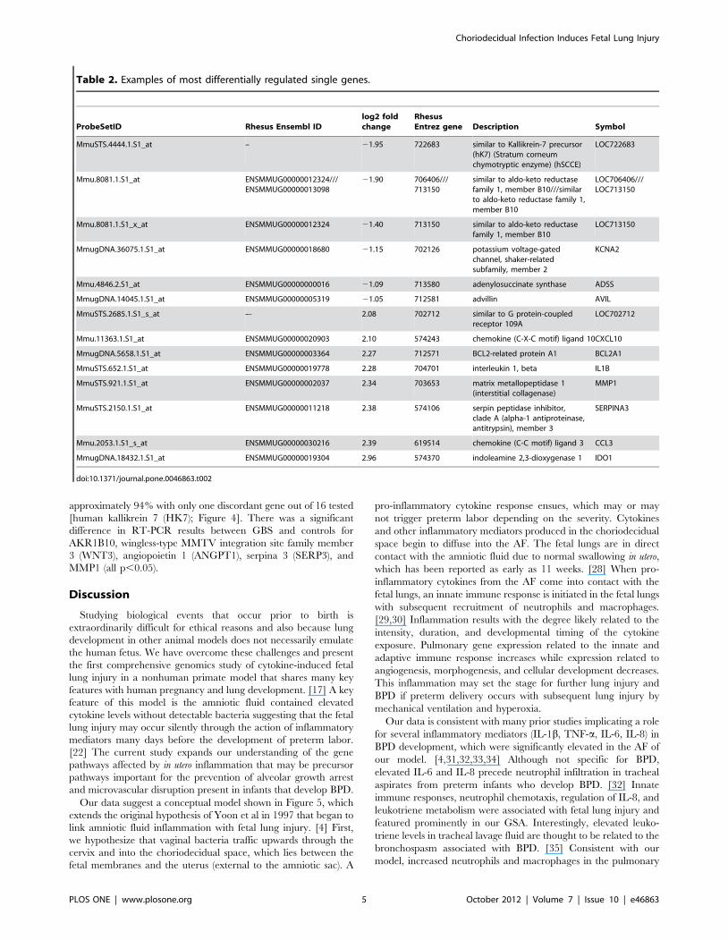
103 downregulated). A subset of these genes is shown in Table 2
and the entire set (707 probesets) and a heatmap (335 genes) is
available in supplementary material (Table S1, Figure S1).
Examples of genes significantly upregulated included indoleamine
2,3-dioxygenase 1 (IDO1), serpin peptidase inhibitor clade A
member 3 (SERP3; also called alpha-1 antiproteinase, antitrypsin),
chemokine (C-C motif) ligand 3 (CCL3), matrix metalloproteinase
1 (MMP1), IL-1b, and IL-8. Genes significantly downregulated by
choriodecidual GBS exposure included similar to aldo-keto
reductase family 1 member B10 (AKR1B10), which is implicated
in the process of lung septation (see discussion) and adenylosucci-
nate synthase (ADSS). The magnitude of change ranged from 3.0
log2 fold for upregulated and 3.5 log2 fold for downregulated
genes.
There are eight surfactant protein probesets on the Affymetrix
Rhesus Macaque Array, but only 5 of the probesets were
annotated in the single gene analysis by the Affymetrix software.
When the IPA software was used to annotate the genes using their
proprietary database, more surfactant proteins (SFTP) were
identified. None of the eight probesets demonstrated significant
differential regulation in the single gene analysis, but SFTPA1 and
SFTPA2 were the most differentially expressed of the group (log2
fold 0.71 and 0.73, respectively).
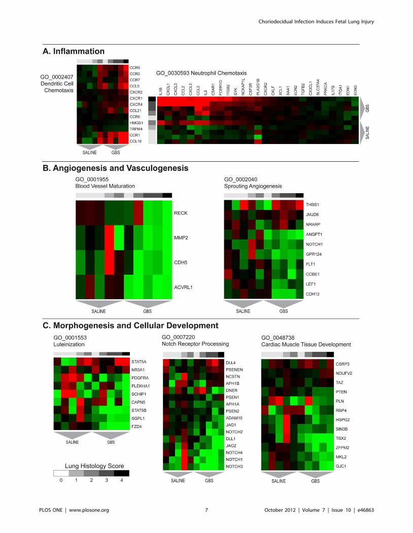
Gene Set Analysis (GSA)Gene sets and pathways with concordant changes in expression
were identified using GSA. Gene sets enriched after GBS exposure
are shown in Table 3 with a complete listing provided in
supplementary material (Table S2). Heat maps of select gene sets
associated with inflammation, angiogenesis, and cellular growth
are shown in Figure 2. Upregulated gene sets in the GBS group
were frequently related to activation of an innate and adaptive
immune response including positive regulation of immune
response, neutrophil chemotaxis, positive regulation of IL-8,
regulation of T cell activation, positive regulation of adaptive
immune response, dendritic cell chemotaxis, and antigen process-
ing and presentation of exogenous peptide antigen via Major
Histocompatibility Complex (MHC) class II. Other pathways
upregulated in the GBS group included pyrimidine base metabolic
process, leukotriene metabolic process, cell-cell signaling, pyrim-
idine nucleoside salvage, and negative regulation of nitric oxide
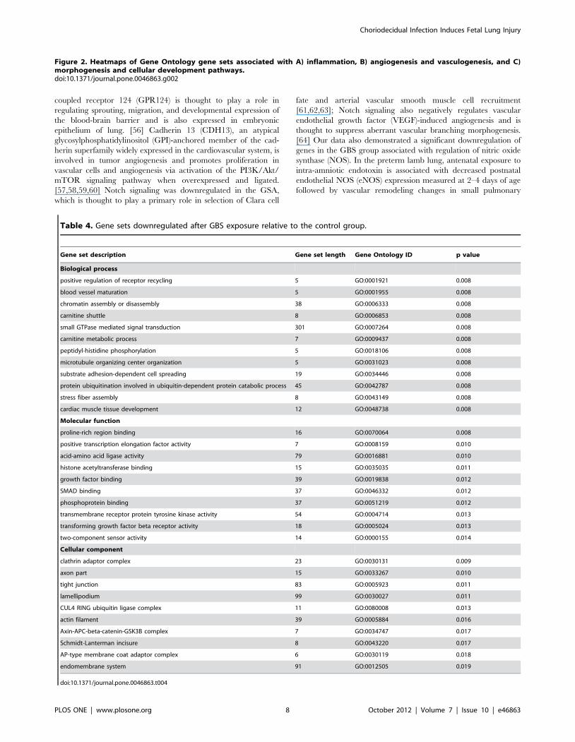
synthase activity. Gene sets downregulated following GBS
exposure were frequently related to morphogenesis, cellular
growth and structure (e.g. cardiac muscle tissue development,
luteinization, Notch receptor processing) and angiogenesis (e.g.
sprouting angiogenesis, blood vessel maturation, vasculogenesis).
These gene sets are shown in Table 4 with a full listing in
supplementary material (Table S3).
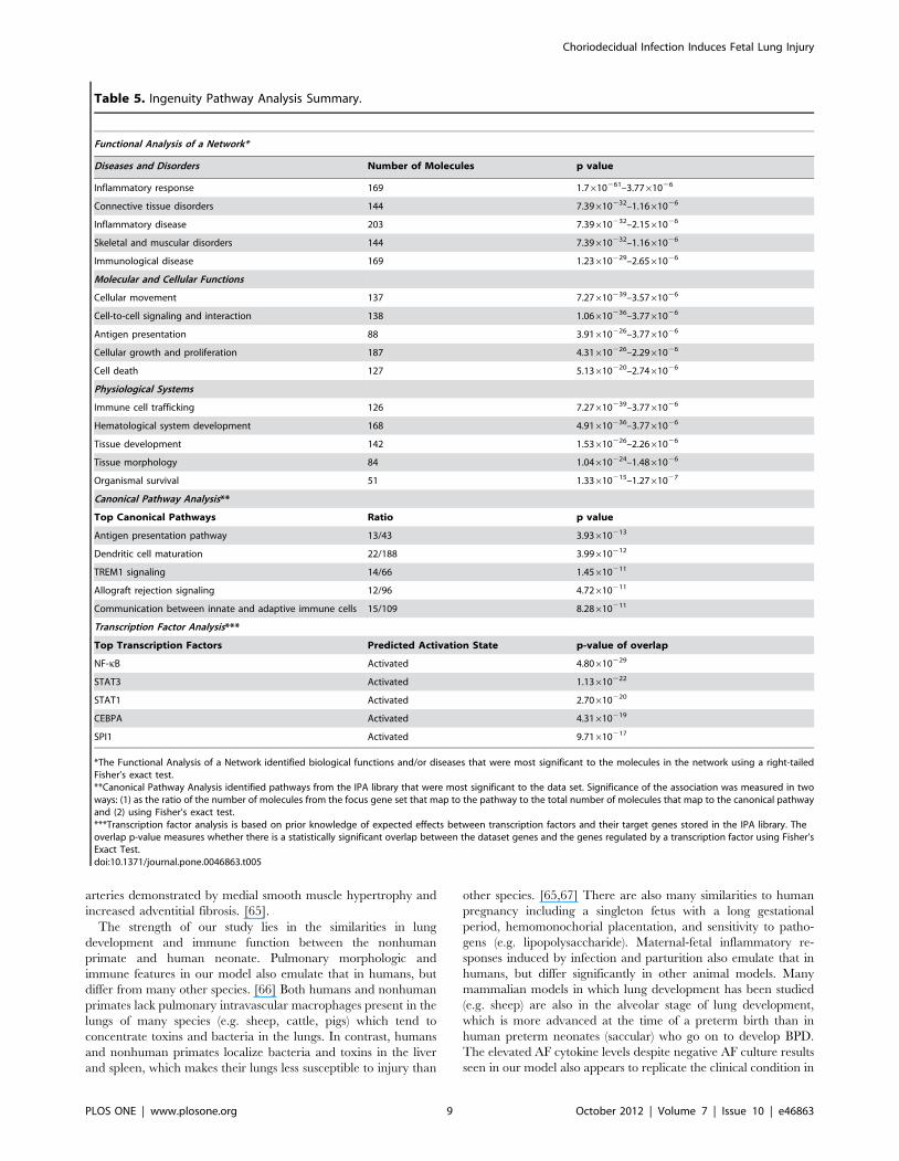
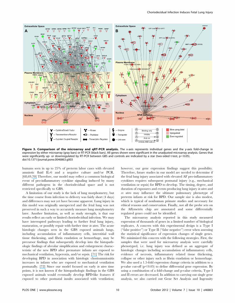
Ingenuity Pathway Analysis (IPA)IPA mapped 36,617 probesets out of the 52,779 probesets on
the Affymetrix Macaca mulatta microarray. The top five canonical
pathways identified by IPA analysis were the antigen presentation
pathway, dendritic cell maturation, triggering receptor expressed
on myeloid cells 1 (TREM1) signaling, allograft rejection signaling,
and communication between innate and adaptive immune cells
(Table 5). IPA analysis also has the capability to predict activation
states of transcriptional regulators based on the activation or
suppression of downstream genes. The top five transcription
factors predicted to be associated with the changes in genes
expression were NF-kappa B (NF-kB), signal transducer and
activator of transcription 3 (STAT3), STAT1, CCAAT/enhancer-
binding protein alpha (CEBPA), and spleen focus forming virus
(SFFV) proviral integration oncogene (SPI1); all were predicted to
be in the activated state. IPA diagrams of the Gene Ontology gene
set neutrophil chemotaxis and NF-kB are shown in Figure 3.
Validation of cDNA Microarray by Quantitative RT-PCRWe identified 16 genes of interest from the microarray dataset,
which we analyzed by quantitative RT-PCR. We directly
compared levels of gene expression obtained with amplified
RNA samples using GAPDH expression as a control for input
cDNA. Overall agreement between the mRNA generated
microarray data and the quantitative RT-PCR data was
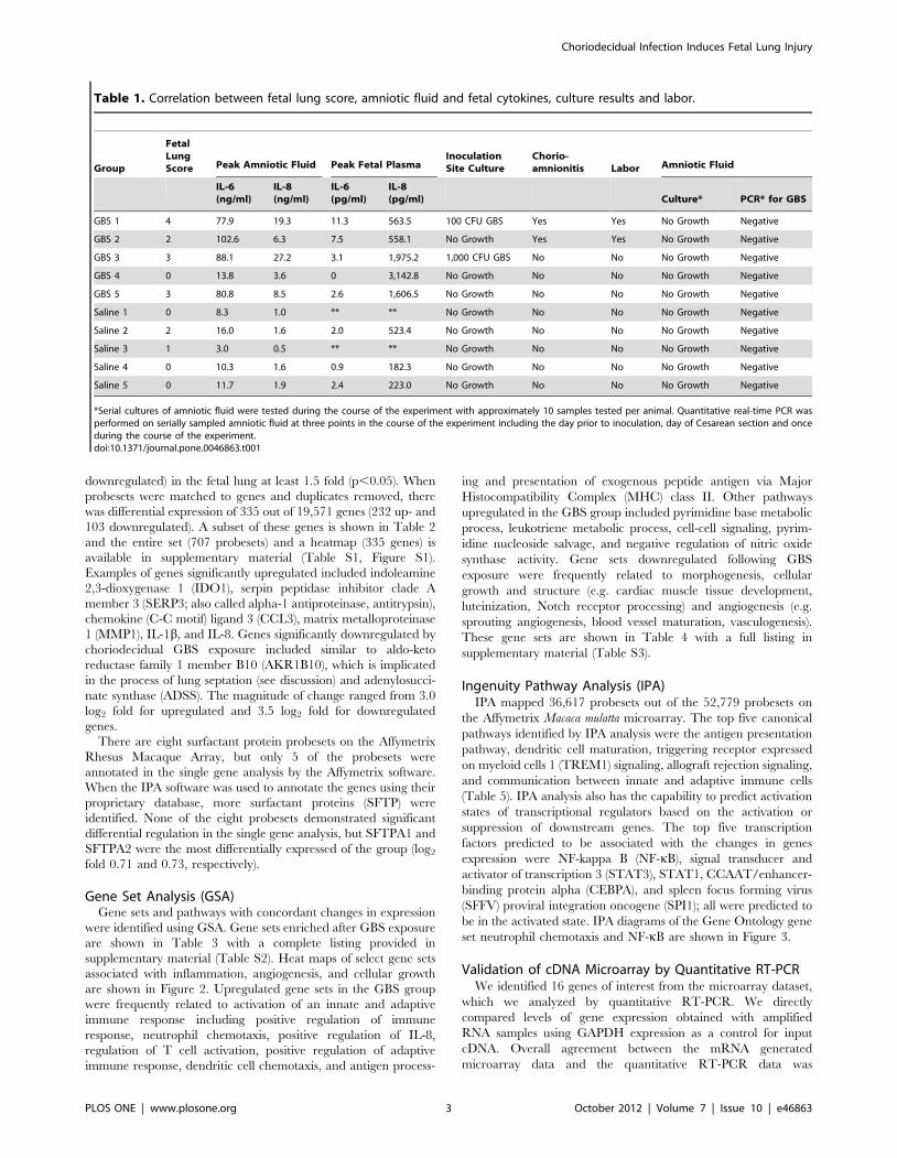
Table 1. Correlation between fetal lung score, amniotic fluid and fetal cytokines, culture results and labor.
Group
FetalLungScore Peak Amniotic Fluid Peak Fetal Plasma
InoculationSite Culture
Chorio-amnionitis Labor Amniotic Fluid
IL-6(ng/ml)
IL-8(ng/ml)
IL-6(pg/ml)
IL-8(pg/ml) Culture* PCR* for GBS
GBS 1 4 77.9 19.3 11.3 563.5 100 CFU GBS Yes Yes No Growth Negative
GBS 2 2 102.6 6.3 7.5 558.1 No Growth Yes Yes No Growth Negative
GBS 3 3 88.1 27.2 3.1 1,975.2 1,000 CFU GBS No No No Growth Negative
GBS 4 0 13.8 3.6 0 3,142.8 No Growth No No No Growth Negative
GBS 5 3 80.8 8.5 2.6 1,606.5 No Growth No No No Growth Negative
Saline 1 0 8.3 1.0 ** ** No Growth No No No Growth Negative
Saline 2 2 16.0 1.6 2.0 523.4 No Growth No No No Growth Negative
Saline 3 1 3.0 0.5 ** ** No Growth No No No Growth Negative
Saline 4 0 10.3 1.6 0.9 182.3 No Growth No No No Growth Negative
Saline 5 0 11.7 1.9 2.4 223.0 No Growth No No No Growth Negative
*Serial cultures of amniotic fluid were tested during the course of the experiment with approximately 10 samples tested per animal. Quantitative real-time PCR wasperformed on serially sampled amniotic fluid at three points in the course of the experiment including the day prior to inoculation, day of Cesarean section and onceduring the course of the experiment.doi:10.1371/journal.pone.0046863.t001
Choriodecidual Infection Induces Fetal Lung Injury
PLOS ONE | www.plosone.org 3 October 2012 | Volume 7 | Issue 10 | e46863
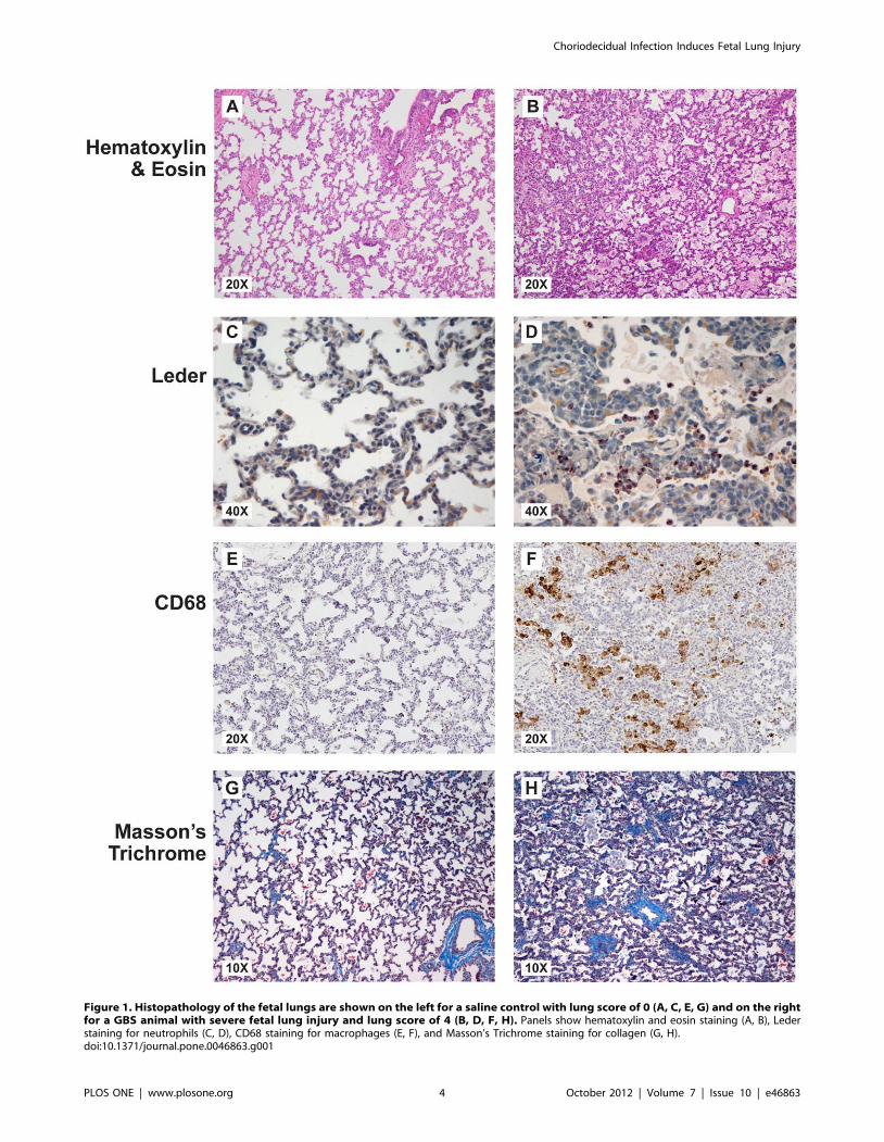
Figure 1. Histopathology of the fetal lungs are shown on the left for a saline control with lung score of 0 (A, C, E, G) and on the rightfor a GBS animal with severe fetal lung injury and lung score of 4 (B, D, F, H). Panels show hematoxylin and eosin staining (A, B), Lederstaining for neutrophils (C, D), CD68 staining for macrophages (E, F), and Masson’s Trichrome staining for collagen (G, H).doi:10.1371/journal.pone.0046863.g001
Choriodecidual Infection Induces Fetal Lung Injury
PLOS ONE | www.plosone.org 4 October 2012 | Volume 7 | Issue 10 | e46863
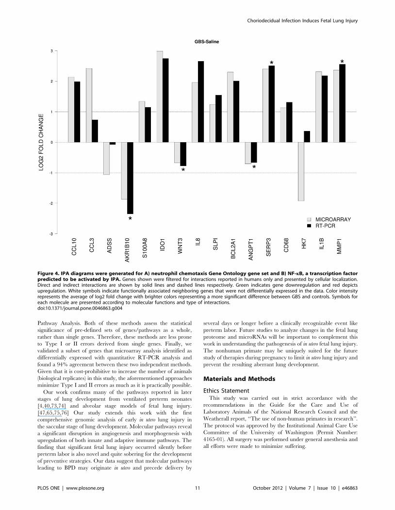
approximately 94% with only one discordant gene out of 16 tested
[human kallikrein 7 (HK7); Figure 4]. There was a significant
difference in RT-PCR results between GBS and controls for
AKR1B10, wingless-type MMTV integration site family member
3 (WNT3), angiopoietin 1 (ANGPT1), serpina 3 (SERP3), and
MMP1 (all p,0.05).
Discussion
Studying biological events that occur prior to birth is
extraordinarily difficult for ethical reasons and also because lung
development in other animal models does not necessarily emulate
the human fetus. We have overcome these challenges and present
the first comprehensive genomics study of cytokine-induced fetal
lung injury in a nonhuman primate model that shares many key
features with human pregnancy and lung development. [17] A key
feature of this model is the amniotic fluid contained elevated
cytokine levels without detectable bacteria suggesting that the fetal
lung injury may occur silently through the action of inflammatory
mediators many days before the development of preterm labor.
[22] The current study expands our understanding of the gene
pathways affected by in utero inflammation that may be precursor
pathways important for the prevention of alveolar growth arrest
and microvascular disruption present in infants that develop BPD.
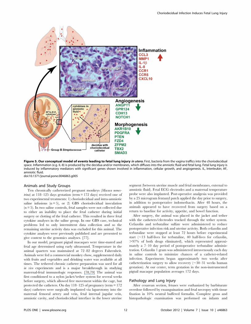
Our data suggest a conceptual model shown in Figure 5, which
extends the original hypothesis of Yoon et al in 1997 that began to
link amniotic fluid inflammation with fetal lung injury. [4] First,
we hypothesize that vaginal bacteria traffic upwards through the
cervix and into the choriodecidual space, which lies between the
fetal membranes and the uterus (external to the amniotic sac). A
pro-inflammatory cytokine response ensues, which may or may
not trigger preterm labor depending on the severity. Cytokines
and other inflammatory mediators produced in the choriodecidual
space begin to diffuse into the AF. The fetal lungs are in direct
contact with the amniotic fluid due to normal swallowing in utero,
which has been reported as early as 11 weeks. [28] When pro-
inflammatory cytokines from the AF come into contact with the
fetal lungs, an innate immune response is initiated in the fetal lungs
with subsequent recruitment of neutrophils and macrophages.
[29,30] Inflammation results with the degree likely related to the
intensity, duration, and developmental timing of the cytokine
exposure. Pulmonary gene expression related to the innate and
adaptive immune response increases while expression related to
angiogenesis, morphogenesis, and cellular development decreases.
This inflammation may set the stage for further lung injury and
BPD if preterm delivery occurs with subsequent lung injury by
mechanical ventilation and hyperoxia.
Our data is consistent with many prior studies implicating a role
for several inflammatory mediators (IL-1b, TNF-a, IL-6, IL-8) in
BPD development, which were significantly elevated in the AF of
our model. [4,31,32,33,34] Although not specific for BPD,
elevated IL-6 and IL-8 precede neutrophil infiltration in tracheal
aspirates from preterm infants who develop BPD. [32] Innate
immune responses, neutrophil chemotaxis, regulation of IL-8, and
leukotriene metabolism were associated with fetal lung injury and
featured prominently in our GSA. Interestingly, elevated leuko-
triene levels in tracheal lavage fluid are thought to be related to the
bronchospasm associated with BPD. [35] Consistent with our
model, increased neutrophils and macrophages in the pulmonary
Table 2. Examples of most differentially regulated single genes.
ProbeSetID Rhesus Ensembl IDlog2 foldchange
RhesusEntrez gene Description Symbol
MmuSTS.4444.1.S1_at – 21.95 722683 similar to Kallikrein-7 precursor(hK7) (Stratum corneumchymotryptic enzyme) (hSCCE)
LOC722683
Mmu.8081.1.S1_at ENSMMUG00000012324///ENSMMUG00000013098
21.90 706406///713150
similar to aldo-keto reductasefamily 1, member B10///similarto aldo-keto reductase family 1,member B10
LOC706406///LOC713150
Mmu.8081.1.S1_x_at ENSMMUG00000012324 21.40 713150 similar to aldo-keto reductasefamily 1, member B10
LOC713150
MmugDNA.36075.1.S1_at ENSMMUG00000018680 21.15 702126 potassium voltage-gatedchannel, shaker-relatedsubfamily, member 2
KCNA2
Mmu.4846.2.S1_at ENSMMUG00000000016 21.09 713580 adenylosuccinate synthase ADSS
MmugDNA.14045.1.S1_at ENSMMUG00000005319 21.05 712581 advillin AVIL
MmuSTS.2685.1.S1_s_at –- 2.08 702712 similar to G protein-coupledreceptor 109A
LOC702712
Mmu.11363.1.S1_at ENSMMUG00000020903 2.10 574243 chemokine (C-X-C motif) ligand 10CXCL10
MmugDNA.5658.1.S1_at ENSMMUG00000003364 2.27 712571 BCL2-related protein A1 BCL2A1
MmuSTS.652.1.S1_at ENSMMUG00000019778 2.28 704701 interleukin 1, beta IL1B
MmuSTS.921.1.S1_at ENSMMUG00000002037 2.34 703653 matrix metallopeptidase 1(interstitial collagenase)
MMP1
MmuSTS.2150.1.S1_at ENSMMUG00000011218 2.38 574106 serpin peptidase inhibitor,clade A (alpha-1 antiproteinase,antitrypsin), member 3
SERPINA3
Mmu.2053.1.S1_s_at ENSMMUG00000030216 2.39 619514 chemokine (C-C motif) ligand 3 CCL3
MmugDNA.18432.1.S1_at ENSMMUG00000019304 2.96 574370 indoleamine 2,3-dioxygenase 1 IDO1
doi:10.1371/journal.pone.0046863.t002
Choriodecidual Infection Induces Fetal Lung Injury
PLOS ONE | www.plosone.org 5 October 2012 | Volume 7 | Issue 10 | e46863

effluent of neonates with BPD may occur even in the absence of
bacterial colonization. [36,37] The transcriptional activators, NF-
kB and STAT3, were predicted to be activated by IPA and are
known to play a role in the innate immune response. Downstream
genes activated by NF-kB that are associated with BPD
development include IL-8 and MMP-1 and MMP-9. STAT3
has been previously implicated in fetal lung injury in the setting of
chorioamnionitis and is identified as a potential target for
regulating the pulmonary inflammatory response. [38,39] In
addition to innate immune responses, the adaptive immune
response gene sets (e.g. dendritic cell chemotaxis, regulation of T
cell activation) and genes involved in dendritic cell activation (e.g.
CCR7) were significantly upregulated in the analysis. These results
are consistent with findings of pulmonary recruitment of dendritic
cells in human exposed to antenatal infection and ventilation who
develop BPD. [40] Dendritic cells, which express a wide array of
pro- and anti-angiogenic mediators, are closely associated with the
pulmonary microvasculature and may contribute to BPD-associ-
ated dysangiogenesis. [40,41].
Many of the downregulated genes or gene sets (i.e. Gene
Ontology categories associated with specific biological processes)
have putative or known roles in lung development, growth and
structural integrity. AKR1B10 catalyzes the essential first step in
the retinoic acid synthesis pathway, which increases lung septation
(21.9 log2 fold). [42,43,44] AKR1B10 is also important for cell
survival and when silenced by small-interfering RNA resulted in
elevated intracellular lipid peroxides and caspase-3-mediated
apoptosis. [45] GSA identified many of the following downregu-
lated genes in gene pathways for luteinization and cardiac muscle
tissue development that are known to be involved in lung
morphogenesis. Both Wnt5a and its receptor Frizzled 4 (FZD4)
play an important role in morphogenesis of the distal lung. [46,47]
Deficiency of SMAD3, a major signal transducer in the
transforming growth factor-beta (TGF-b) pathway, impairs
neonatal lung alveolarization and peripheral lung cell proliferation
based in murine studies. [48] Platelet derived growth factor
receptor, alpha polypeptide (PDGFRA) is expressed by bronchi-
olar smooth muscle progenitors and might impair mesodermal
development if downregulated. [49] T-box (TBX) transcription
factors, such as TBX2 in the cardiac muscle tissue development
pathway, have been implicated in developing lung mesoderm. [50]
Finally, phosphatase and tensin homolog (PTEN) has been shown
to be essential for normal lung morphogenesis and when deleted in
mice resulted in impaired branching morphogenesis and distal
alveolar epithelial cell differentiation. [51].
Other downregulated genes and gene sets were associated with
angiogenesis and vascular dysfunction. ANGPT1 is the primary
agonist of the tyrosine kinase receptor Tie 2 (tyrosine kinase with
immunoglobulin and EGF-like domains), which is restricted to
endothelial cell expression. [52,53,54] Cord blood plasma levels of
ANGPT1 in preterm infants who subsequently develop BPD are
significantly lower than those without BPD. [55] G protein-
Table 3. Gene sets upregulated after GBS exposure relative to the control group.
Gene set description Gene set length Gene Ontology ID p value
Biological process
pyrimidine base metabolic process 24 GO:0006206 ,0.002
leukotriene metabolic process 11 GO:0006691 ,0.002
cellular aromatic compound metabolic process 8 GO:0006725 ,0.002
cell-cell signaling 252 GO:0007267 ,0.002
phospholipid catabolic process 15 GO:0009395 ,0.002
pyrimidine nucleoside salvage 9 GO:0043097 ,0.002
positive regulation of innate immune response 15 GO:0045089 0.002
pyrimidine base metabolic process 24 GO:0006206 0
Molecular function
neuropeptide hormone activity 27 GO:0005184 ,0.002
hyaluronic acid binding 21 GO:0005540 ,0.002
oxidoreductase activity 10 GO:0016712 ,0.002
aromatase activity 21 GO:0070330 0.003
monooxygenase activity 73 GO:0004497 0.005
pancreatic ribonuclease activity 8 GO:0004522 0.006
organic anion transmembrane transporter activity 15 GO:0008514 0.006
Cellular component
cytoplasmic part 20 GO:0044444 0.008
gap junction 26 GO:0005921 0.013
connexon complex 18 GO:0005922 0.018
vesicle 31 GO:0031982 0.018
dendritic spine membrane 5 GO:0032591 0.018
intrinsic to internal side of plasma membrane 11 GO:0031235 0.019
alpha-amino-3-hydroxy-5-methyl-4-isoxazolepropionic acidselective glutamate receptor complex
13 GO:0032281 0.019
doi:10.1371/journal.pone.0046863.t003
Choriodecidual Infection Induces Fetal Lung Injury
PLOS ONE | www.plosone.org 6 October 2012 | Volume 7 | Issue 10 | e46863
Choriodecidual Infection Induces Fetal Lung Injury
PLOS ONE | www.plosone.org 7 October 2012 | Volume 7 | Issue 10 | e46863
coupled receptor 124 (GPR124) is thought to play a role in
regulating sprouting, migration, and developmental expression of
the blood-brain barrier and is also expressed in embryonic
epithelium of lung. [56] Cadherin 13 (CDH13), an atypical
glycosylphosphatidylinositol (GPI)-anchored member of the cad-
herin superfamily widely expressed in the cardiovascular system, is
involved in tumor angiogenesis and promotes proliferation in
vascular cells and angiogenesis via activation of the PI3K/Akt/
mTOR signaling pathway when overexpressed and ligated.
[57,58,59,60] Notch signaling was downregulated in the GSA,
which is thought to play a primary role in selection of Clara cell
fate and arterial vascular smooth muscle cell recruitment
[61,62,63]; Notch signaling also negatively regulates vascular
endothelial growth factor (VEGF)-induced angiogenesis and is
thought to suppress aberrant vascular branching morphogenesis.
[64] Our data also demonstrated a significant downregulation of
genes in the GBS group associated with regulation of nitric oxide
synthase (NOS). In the preterm lamb lung, antenatal exposure to
intra-amniotic endotoxin is associated with decreased postnatal
endothelial NOS (eNOS) expression measured at 2–4 days of age
followed by vascular remodeling changes in small pulmonary
Table 4. Gene sets downregulated after GBS exposure relative to the control group.
Gene set description Gene set length Gene Ontology ID p value
Biological process
positive regulation of receptor recycling 5 GO:0001921 0.008
blood vessel maturation 5 GO:0001955 0.008
chromatin assembly or disassembly 38 GO:0006333 0.008
carnitine shuttle 8 GO:0006853 0.008
small GTPase mediated signal transduction 301 GO:0007264 0.008
carnitine metabolic process 7 GO:0009437 0.008
peptidyl-histidine phosphorylation 5 GO:0018106 0.008
microtubule organizing center organization 5 GO:0031023 0.008
substrate adhesion-dependent cell spreading 19 GO:0034446 0.008
protein ubiquitination involved in ubiquitin-dependent protein catabolic process 45 GO:0042787 0.008
stress fiber assembly 8 GO:0043149 0.008
cardiac muscle tissue development 12 GO:0048738 0.008
Molecular function
proline-rich region binding 16 GO:0070064 0.008
positive transcription elongation factor activity 7 GO:0008159 0.010
acid-amino acid ligase activity 79 GO:0016881 0.010
histone acetyltransferase binding 15 GO:0035035 0.011
growth factor binding 39 GO:0019838 0.012
SMAD binding 37 GO:0046332 0.012
phosphoprotein binding 37 GO:0051219 0.012
transmembrane receptor protein tyrosine kinase activity 54 GO:0004714 0.013
transforming growth factor beta receptor activity 18 GO:0005024 0.013
two-component sensor activity 14 GO:0000155 0.014
Cellular component
clathrin adaptor complex 23 GO:0030131 0.009
axon part 15 GO:0033267 0.010
tight junction 83 GO:0005923 0.011
lamellipodium 99 GO:0030027 0.011
CUL4 RING ubiquitin ligase complex 11 GO:0080008 0.013
actin filament 39 GO:0005884 0.016
Axin-APC-beta-catenin-GSK3B complex 7 GO:0034747 0.017
Schmidt-Lanterman incisure 8 GO:0043220 0.017
AP-type membrane coat adaptor complex 6 GO:0030119 0.018
endomembrane system 91 GO:0012505 0.019
doi:10.1371/journal.pone.0046863.t004
Figure 2. Heatmaps of Gene Ontology gene sets associated with A) inflammation, B) angiogenesis and vasculogenesis, and C)morphogenesis and cellular development pathways.doi:10.1371/journal.pone.0046863.g002
Choriodecidual Infection Induces Fetal Lung Injury
PLOS ONE | www.plosone.org 8 October 2012 | Volume 7 | Issue 10 | e46863
arteries demonstrated by medial smooth muscle hypertrophy and
increased adventitial fibrosis. [65].
The strength of our study lies in the similarities in lung
development and immune function between the nonhuman
primate and human neonate. Pulmonary morphologic and
immune features in our model also emulate that in humans, but
differ from many other species. [66] Both humans and nonhuman
primates lack pulmonary intravascular macrophages present in the
lungs of many species (e.g. sheep, cattle, pigs) which tend to
concentrate toxins and bacteria in the lungs. In contrast, humans
and nonhuman primates localize bacteria and toxins in the liver
and spleen, which makes their lungs less susceptible to injury than
other species. [65,67] There are also many similarities to human
pregnancy including a singleton fetus with a long gestational
period, hemomonochorial placentation, and sensitivity to patho-
gens (e.g. lipopolysaccharide). Maternal-fetal inflammatory re-
sponses induced by infection and parturition also emulate that in
humans, but differ significantly in other animal models. Many
mammalian models in which lung development has been studied
(e.g. sheep) are also in the alveolar stage of lung development,
which is more advanced at the time of a preterm birth than in
human preterm neonates (saccular) who go on to develop BPD.
The elevated AF cytokine levels despite negative AF culture results
seen in our model also appears to replicate the clinical condition in
Table 5. Ingenuity Pathway Analysis Summary.
Functional Analysis of a Network*
Diseases and Disorders Number of Molecules p value
Inflammatory response 169 1.7610261–3.7761026
Connective tissue disorders 144 7.39610232–1.1661026
Inflammatory disease 203 7.39610232–2.1561026
Skeletal and muscular disorders 144 7.39610232–1.1661026
Immunological disease 169 1.23610229–2.6561026
Molecular and Cellular Functions
Cellular movement 137 7.27610239–3.5761026
Cell-to-cell signaling and interaction 138 1.06610236–3.7761026
Antigen presentation 88 3.91610226–3.7761026
Cellular growth and proliferation 187 4.31610226–2.2961026
Cell death 127 5.13610220–2.7461026
Physiological Systems
Immune cell trafficking 126 7.27610239–3.7761026
Hematological system development 168 4.91610236–3.7761026
Tissue development 142 1.53610226–2.2661026
Tissue morphology 84 1.04610224–1.4861026
Organismal survival 51 1.33610215–1.2761027
Canonical Pathway Analysis**
Top Canonical Pathways Ratio p value
Antigen presentation pathway 13/43 3.93610213
Dendritic cell maturation 22/188 3.99610212
TREM1 signaling 14/66 1.45610211
Allograft rejection signaling 12/96 4.72610211
Communication between innate and adaptive immune cells 15/109 8.28610211
Transcription Factor Analysis***
Top Transcription Factors Predicted Activation State p-value of overlap
NF-kB Activated 4.80610229
STAT3 Activated 1.13610222
STAT1 Activated 2.70610220
CEBPA Activated 4.31610219
SPI1 Activated 9.71610217
*The Functional Analysis of a Network identified biological functions and/or diseases that were most significant to the molecules in the network using a right-tailedFisher’s exact test.**Canonical Pathway Analysis identified pathways from the IPA library that were most significant to the data set. Significance of the association was measured in twoways: (1) as the ratio of the number of molecules from the focus gene set that map to the pathway to the total number of molecules that map to the canonical pathwayand (2) using Fisher’s exact test.***Transcription factor analysis is based on prior knowledge of expected effects between transcription factors and their target genes stored in the IPA library. Theoverlap p-value measures whether there is a statistically significant overlap between the dataset genes and the genes regulated by a transcription factor using Fisher’sExact Test.doi:10.1371/journal.pone.0046863.t005
Choriodecidual Infection Induces Fetal Lung Injury
PLOS ONE | www.plosone.org 9 October 2012 | Volume 7 | Issue 10 | e46863
humans seen in up to 25% of preterm labor cases with elevated
amniotic fluid IL-6 and a negative culture and/or PCR.
[68,69,70] Therefore, our model may reflect a common biological
event of pro-inflammatory cytokine signaling induced by many
different pathogens in the choriodecidual space and is not
restricted specifically to GBS.
A limitation of our study is the lack of lung morphometry, but
the time course from infection to delivery was fairly short (4 days)
and differences may not yet have become apparent. Lung injury in
this model was originally unexpected and the fetal lung was not
preserved in such a way to accurately measure lung morphometry
later. Another limitation, as well as study strength, is that our
results reflect an early or limited choriodecidual infection. We may
have interrupted pathways leading to further fetal lung injury,
maturation, or possibly repair with further time in utero. The acute
histologic changes seen in the GBS exposed animals lungs,
including accumulation of inflammatory cells, interstitial wall
tissue thickening, and fibrin exudation or hemorrhage, may be
precursor findings that subsequently develop into the histopath-
ologic findings of alveolar simplification and enlargement charac-
teristic of the new BPD after premature infants are exposed to
mechanical ventilation, hyperoxia, and/or sepsis. [71] The risk for
developing BPD in association with histologic chorioamnionitis
increases in infants who are exposed to mechanical ventilation
postnatally. [72] Since our model focused on early in utero time
points, it is not known if the histopathologic findings in the GBS
exposed animals would eventually develop BPD-like features if
exposed to other postnatal insults associated with ventilation;
however, our gene expression findings suggest this possibility.
Therefore, future studies in our model are needed to determine if
the fetal lung injury associated with elevated AF pro-inflammatory
cytokines requires subsequent postnatal injury (e.g., mechanical
ventilation or sepsis) for BPD to develop. The timing, degree, and
duration of exposures and events producing lung injury in utero and
ex utero may influence the ultimate pulmonary phenotype of
preterm infants at risk for BPD. Our sample size is also modest,
which is typical of nonhuman primate studies and necessary for
ethical reasons and conservation. Finally, not all the probe sets on
the Affymetrix chip are annotated and some differentially
regulated genes could not be identified.
The microarray analysis reported in this study measured
expression of thousands of genes in a limited number of biological
replicates. A concern with this experimental design is a Type I
(‘‘false positive’’) or Type II (‘‘false negative’’) error when assessing
the statistical significance of expression changes of single genes.
We minimized this concern with the following strategies. First, the
samples that were used for microarray analysis were carefully
phenotyped, i.e. lung injury was defined as an aggregate of
histologic changes including accumulation of inflammatory cells,
evidence of necrosis, inflammatory related tissue thickening,
collapse or other injury such as fibrin exudation or hemorrhage.
We also used a 1.5-fold expression change criteria in addition to a
p-value cut-off (p,0.05) to define differential gene expression. By
using a combination of a fold-change and p-value criteria, Type I
and II errors are decreased. In addition to carrying out single gene
analysis, we also carried out Gene Set Analysis and Ingenuity
Figure 3. Comparison of the microarray and qRT-PCR analysis. The x-axis represents individual genes and the y-axis fold-change inexpression by either microarray (gray bars) or RT-PCR (black bars). All genes shown were significant in the unadjusted microarray analysis. Genes thatwere significantly up- or downregulated by RT-PCR between GBS and controls are indicated by a star (two-sided t-test, p,0.05).doi:10.1371/journal.pone.0046863.g003
Choriodecidual Infection Induces Fetal Lung Injury
PLOS ONE | www.plosone.org 10 October 2012 | Volume 7 | Issue 10 | e46863
Pathway Analysis. Both of these methods assess the statistical
significance of pre-defined sets of genes/pathways as a whole,
rather than single genes. Therefore, these methods are less prone
to Type I or II errors derived from single genes. Finally, we
validated a subset of genes that microarray analysis identified as
differentially expressed with quantitative RT-PCR analysis and
found a 94% agreement between these two independent methods.
Given that it is cost-prohibitive to increase the number of animals
(biological replicates) in this study, the aforementioned approaches
minimize Type I and II errors as much as it is practically possible.
Our work confirms many of the pathways reported in later
stages of lung development from ventilated preterm neonates
[4,40,73,74] and alveolar stage models of fetal lung injury.
[47,65,75,76] Our study extends this work with the first
comprehensive genomic analysis of early in utero lung injury in
the saccular stage of lung development. Molecular pathways reveal
a significant disruption in angiogenesis and morphogenesis with
upregulation of both innate and adaptive immune pathways. The
finding that significant fetal lung injury occurred silently before
preterm labor is also novel and quite sobering for the development
of preventive strategies. Our data suggest that molecular pathways
leading to BPD may originate in utero and precede delivery by
several days or longer before a clinically recognizable event like
preterm labor. Future studies to analyze changes in the fetal lung
proteome and microRNAs will be important to complement this
work in understanding the pathogenesis of in utero fetal lung injury.
The nonhuman primate may be uniquely suited for the future
study of therapies during pregnancy to limit in utero lung injury and
prevent the resulting aberrant lung development.
Materials and Methods
Ethics StatementThis study was carried out in strict accordance with the
recommendations in the Guide for the Care and Use of
Laboratory Animals of the National Research Council and the
Weatherall report, ‘‘The use of non-human primates in research’’.
The protocol was approved by the Institutional Animal Care Use
Committee of the University of Washington (Permit Number:
4165-01). All surgery was performed under general anesthesia and
all efforts were made to minimize suffering.
Figure 4. IPA diagrams were generated for A) neutrophil chemotaxis Gene Ontology gene set and B) NF-kB, a transcription factorpredicted to be activated by IPA. Genes shown were filtered for interactions reported in humans only and presented by cellular localization.Direct and indirect interactions are shown by solid lines and dashed lines respectively. Green indicates gene downregulation and red depictsupregulation. White symbols indicate functionally associated neighboring genes that were not differentially expressed in the data. Color intensityrepresents the average of log2 fold change with brighter colors representing a more significant difference between GBS and controls. Symbols foreach molecule are presented according to molecular functions and type of interactions.doi:10.1371/journal.pone.0046863.g004
Choriodecidual Infection Induces Fetal Lung Injury
PLOS ONE | www.plosone.org 11 October 2012 | Volume 7 | Issue 10 | e46863
Animals and Study GroupsTen chronically catheterized pregnant monkeys (Macaca nemes-
trina) at 118–125 days gestation (term = 172 days) received one of
two experimental treatments: 1) choriodecidual and intra-amniotic
saline infusions (n = 5), or 2) GBS choriodecidual inoculation
(n = 5). In two saline controls, fetal samples were not collected due
to either an inability to place the fetal catheter during initial
surgery or clotting of the fetal catheter. This resulted in three fetal
cytokine analyses in the saline group. In one GBS case, technical
problems led to only intermittent data collection and so the
remaining uterine activity data was excluded for this animal. The
cytokine analyses were previously published and are presented to
give context to the genomics analyses. [77].
In our model, pregnant pigtail macaques were time-mated and
fetal age determined using early ultrasound. Temperature in the
animal quarters was maintained at 72–82 degrees Fahrenheit.
Animals were fed a commercial monkey chow, supplemented daily
with fruits and vegetables and drinking water was available at all
times. The tethered chronic catheter preparation was used for all
in vivo experiments and is a major breakthrough in studying
maternal-fetal immunologic responses. [78,79] The animal was
first conditioned to a nylon jacket/tether system for several weeks
before surgery, which allowed free movement within the cage, but
protected the catheters. On day 118–125 of pregnancy (term = 172
days) catheters were surgically implanted via laparotomy into the
maternal femoral artery and vein, fetal internal jugular vein,
amniotic cavity, and choriodecidual interface in the lower uterine
segment (between uterine muscle and fetal membranes, external to
amniotic fluid). Fetal ECG electrodes and a maternal temperature
probe were also implanted. Post-operative analgesia was provided
by a 25 microgram fentanyl patch applied the day prior to surgery,
in addition to postoperative indomethacin. After 48 hours, the
animals appeared to have recovered from surgery based on a
return to baseline for activity, appetite, and bowel function.
After surgery, the animal was placed in the jacket and tether
with the catheters/electrodes tracked through the tether system.
Cefazolin and terbutaline sulfate were administered to reduce
postoperative infection risk and uterine activity. Both cefazolin and
terbutaline were stopped at least 72 hours before experimental
start (,13 half-lives for terbutaline, 40 half-lives for cefazolin,
.97% of both drugs eliminated), which represented approxi-
mately a 7–10 day period of postoperative terbutaline adminis-
tration. Cefazolin 1 gram was administered intravenously each day
in saline controls to minimize chances of a catheter-related
infection. Experiments began approximately two weeks after
catheterization surgery to allow recovery (,30–31 weeks human
gestation). At our center, term gestation in the non-instrumented
pigtail macaque population averages 172 days.
Pathology and Lung InjuryAfter cesarean section, fetuses were euthanized by barbiturate
overdose followed by exsanguination and fetal necropsy with tissue
fixation in 10% neutral buffered formalin. Complete gross and
histopathologic examination was performed on infants and
Figure 5. Our conceptual model of events leading to fetal lung injury in utero. First, bacteria from the vagina traffics into the choriodecidualspace. Inflammation (e.g. IL-8) is produced by the decidua and/or membranes, which diffuses into the amniotic fluid and fetal lung. Fetal lung injury isinduced by inflammatory mediators with significant genes shown involved in inflammation, cellular growth, and angiogenesis. IL, Interleukin; AF,amniotic fluid.doi:10.1371/journal.pone.0046863.g005
Choriodecidual Infection Induces Fetal Lung Injury
PLOS ONE | www.plosone.org 12 October 2012 | Volume 7 | Issue 10 | e46863
placentas. For histologic examination two to three randomly
selected fixed fetal lung tissues were embedded in paraffin and
sections stained with hematoxylin and eosin (H&E), Masson’s
trichrome or specific esterase (Leder stain) using standard
protocols. Leder staining was performed using the Naphthol AS-
D Chloroacetate Specific Esterase Kit (Sigma-Aldrich, St. Louis,
MO) per manufacturer’s instructions. Masson’s trichrome staining
was performed to differentially highlight the presence of connec-
tive tissue by a standard method involving serial incubations in
Bouin’s fixative, Weigert’s iron hematoxylin, Biebrich scarlet-acid
fuchsin, phosphomolybdic-phosphotungstic acid and aniline blue.
The placenta was examined by a board-certified pediatric
pathologist (R.P.K.) and fetal lungs examined by a board-certified
veterinary pathologist (H.D.L.) with each pathologist blinded to
group assignment. H&E-stained full-thickness sections of placental
disc, umbilical cord, and a fetal membrane roll were examined
from each case to exclude inflammation, necrosis, fetal vascular
thrombosis, or other histopathological findings. Chorioamnionitis
was diagnosed when neutrophils were identified in the chorion
and/or amnion. Funisitis denoted neutrophils in the umbilical
vessels and/or surrounding connective tissue. Lung histologic
sections were evaluated and scored, as previously described, using
a semi-quantitative scale. [80] Components were scored on a scale
of 0–4 (0 = normal) for inflammatory cells, necrosis, and inflam-
mation including tissue thickening, collapse or other injury (e.g.
fibrin exudation). Lung compartments scored were (1) vascular/
perivascular; (2) bronchial/peribronchial; (3) alveolar wall; and (4)
trichrome stain intensity positivity. Mononuclear inflammatory
cells and neutrophils (Leder stain) within alveolar spaces were
counted (5 random 40X fields). An overall severity score was
generated. A lung score of 0 to 1 was considered within normal
limits as a few neutrophils are expected following saline
inoculation and mild alveolar thickening is normal in some
preterm animals.
Immunohistochemistry of Fetal Lung TissuesImmunohistochemistry staining for CD68 was performed using
a mouse monoclonal CD68 primary antibody (1:1,000 dilution,
Clone: KP1, MS-397-P1 (Thermo Fisher Scientific, Waltham,
MA) a normal mouse IgG isotype control (1:200 dilution, Vector
Labs, I-2000), and a spleen control from M. nemestrina. First, the
slides were baked for 30 minutes at 60uC and deparaffinized on
the Leica Bond Automated Immunostainer (Leica Microsystems,
Buffalo Grove, IL). Antigen retrieval was performed by placing
slides in HIER Citrate Buffer for 10 minutes at 100uC. Blocking
consisted of Leica Bond Peroxide block for 5 minutes at room
temperature (RT) and then 10% Normal Goat Serum in PBS for
10 minutes at RT. Either the primary antibody (mouse anti-CD68,
1:1,000 dilution, 0.2 mg/mL) or mouse isotype IgG control (1:200
dilution, 1 mg/mL) in Leica Primary antibody diluent was applied
for 30 minutes at RT. Leica Bond post primary was then applied
for 8 minutes at RT. Antibody complexes were visualized using
Leica Bond Polymer DAB Refine for 8 minutes at RT and then
Leica Bond Mixed Refine (DAB) detection 2X for 10 minutes at
RT. Tissues were counterstained with hematoxylin counterstain
for 10 second followed by two rinses in H20. Unless otherwise
specified all reagents were obtained from Leica Microsystems.
RNA Extraction and Microarray ProcessingTo study genetic pathways in M. nemestrina, we used the
Affymetrix Rhesus Macaque Array (GeneChipH Rhesus Macaque
Genome Array, Affymetrix, Santa Clara, CA), which allows
interrogation of 47,000 M. mulatta transcripts and provides
comprehensive transcriptome coverage. Genetic differences be-
tween M. mulatta and M. nemestrina are predicted to be ,1%, which
is consistent with our published data. [81] RNA extraction was
performed by the CHDD Genomics Core Laboratory followed by
the manufacturer’s protocols using the GeneChip platform by
Affymetrix. Briefly, these methods include the synthesis of first-
and second-strand cDNAs, the purification of double-stranded
cDNA, the synthesis of cRNA by in vitro transcription (IVT), the
recovery and quantitation of biotin-labeled cRNA, the fragmen-
tation of this cRNA and subsequent hybridization to the
microarray slide, the post-hybridization washings, and the
detection of the hybridized cRNAs using a streptavidin-coupled
fluorescent dye. Hybridized Affymetrix arrays were scanned with
an Affymetrix GeneChipH 3000 fluorescent scanner. Image
generation and feature extraction was performed using Affymetrix
GeneChip Command Console Software.
Single Gene AnalysisThe data discussed in this publication have been deposited in
NCBI’s Gene Expression Omnibus (Edgar et al., 2002) and are
accessible through GEO Series accession number GSE39029 (http://
www.ncbi.nlm.nih.gov/geo/query/acc.cgi?acc = GSE39029). Analy-
sis of the microarray data focused first on differential expression of
single genes. Raw microarray data was pre-processed and analyzed
with Bioconductor (http://www.bioconductor.org/). [82] Several
quality control steps were carried out to insure that the data was of
high quality: 1) visual inspection of the GCOS DAT chip images, 2)
visual inspection of the chip pseudo-images generated by the
Bioconductor affyPLM package, 3) generation of percent present calls
and average background signals with the Bioconductor simpleaffy
package, 4) generation and inspection histograms of raw signal
intensities, and 5) generation and comparison of the Relative Log
Expression and Normalized Unscaled Standard Errors using the
Bioconductor affyPLM package. The data was normalized with the
Bioconductor GeneChip Robust Multiarray Averaging (RMA)
package. [83] From the normalized data, genes with significant
evidence for differential expression were identified using the Limma
package in Bioconductor. [84] P-values were calculated with a
modified t-test in conjunction with an empirical Bayes method to
moderate the standard errors of the estimated log-fold changes. P-
values were adjusted for multiplicity with the Bioconductor package
qvalue, which allows for selecting statistically significant genes while
controlling the estimated false discovery rate. [85].
Gene Set Analysis (GSA)Next, the data was analyzed using GSA in order to investigate
categories of genes. [86,87] GSA assesses the statistical significance
of pre-defined gene sets/pathways as a whole rather than of single
genes, which allows for the identification of modest but concordant
changes in expression of individual genes that may be missed by
single gene analysis. GSA software is available as R code (http://
www.broad.mit.edu/GSA/). [86,88] GSA considers all the genes
in the experiment and allows for the identification of gene sets with
strong cross-correlation by boosting the signal-to-noise ratio,
which makes it possible to detect modest changes in gene
expression. In GSA, the p-values that are calculated to test the
null hypothesis are based on permutations of the sample labels. We
used four gene set databases for the GSA: three from Gene
Ontology [89] (Biological Process, Molecular Function, and
Cellular Component), and the functional C2 gene set from the
Molecular Signature Database [88] (http://www.broad.mit.edu/
GSA/msigdb/msigdb_index.html).
Choriodecidual Infection Induces Fetal Lung Injury
PLOS ONE | www.plosone.org 13 October 2012 | Volume 7 | Issue 10 | e46863
IPA AnalysisWe used the Ingenuity Pathway Analysis (IPA) software
(IngenuityH Systems, www.ingenuity.com) to discover pathways
and transcriptional networks in the gene expression microarray
data. Our data set containing gene identifiers and corresponding
expression changes between the experimental groups and p-values
was uploaded into the IPA application. Each identifier was
mapped to its corresponding object in the IngenuityH Knowledge
Base. The Functional Analysis identified the biological functions
and/or diseases that were most significant to the data set. Genes
from the data set with more than 1.5-fold differential expression
(up/down regulation) and p,0.05 that were associated with
biological functions and/or diseases in the Ingenuity Knowledge
Base were considered for the analysis. The categories ‘‘Top
Canonical Pathways’’ and ‘‘Top Transcription Factors’’ were
primarily used in this analysis. Right-tailed Fisher’s exact test was
used to calculate a p-value determining the probability that each
biological function and/or disease assigned to that data set is due
to chance alone. The IPA Path Designer Graphical Representa-
tion was used to generate figures. Molecules are represented as
nodes, and the biological relationship between two nodes is
represented as an edge (line). All edges are supported by at least
one reference from the literature, from a textbook, or from
canonical information stored in the Ingenuity Knowledge Base.
Human, mouse, and rat orthologs of a gene are stored as separate
objects in the Ingenuity Knowledge Base, but are represented as a
single node in the network. The intensity of the node color
indicates the degree of up- (red) or down- (green) regulation.
Nodes are displayed using various shapes that represent the
functional class of the gene product. Edges are displayed with
various labels that describe the nature of the relationship between
the nodes (see figure legends for details). IPA also allows prediction
of the activation or inhibition of transcription factors involved in
the gene expression patterns seen in our study.
Validation of cDNA Microarray by Quantitative RT-PCRQuantitation of mRNA levels was performed by the CHDD
Genomics Core Laboratory using fluorogenic 59 nuclease-based
assays and has been previously described. [90,91,92].
Supporting Information
Figure S1 Comparison of mRNA expression in the fetallung by microarray analysis in GBS and saline groupsdisplaying the relative Cy3/Cy5 ratios. mRNA expression
is displayed as either higher (red) or lower (green) in GBS
compared to saline controls.
(TIF)
Table S1 All probe sets in the single gene analysisdifferentially expressed at the 1.5-fold level, p,0.05.
(DOCX)
Table S2 Gene sets upregulated after GBS exposurerelative to the control group.
(DOCX)
Table S3 Gene sets downregulated after GBS exposurerelative to the control group.
(DOCX)
Acknowledgments
We gratefully acknowledge the technical assistance of Jennifer Summers in
preparing the figures.
Author Contributions
Conceived and designed the experiments: RMM KAW MGG CER.
Performed the experiments: KAW. Analyzed the data: RPB TKB FF HDL
JV RMM KAW RPK. Wrote the paper: RMM KAW JV TB RPK HDL.
References
1. Lee J, Oh KJ, Yang HJ, Park JS, Romero R, et al. (2009) The importance of
intra-amniotic inflammation in the subsequent development of atypical chronic
lung disease. J Matern Fetal Neonatal Med 22: 917–923.
2. Kramer BW, Kallapur S, Newnham J, Jobe AH (2009) Prenatal inflammation
and lung development. Semin Fetal Neonatal Med 14: 2–7.
3. Stroustrup A, Trasande L (2010) Epidemiological characteristics and resourceuse in neonates with bronchopulmonary dysplasia: 1993–2006. Pediatrics 126:
291–297.
4. Yoon BH, Romero R, Jun JK, Park KH, Park JD, et al. (1997) Amniotic fluid
cytokines (interleukin-6, tumor necrosis factor-alpha, interleukin-1 beta, and
interleukin-8) and the risk for the development of bronchopulmonary dysplasia.Am J Obstet Gynecol 177: 825–830.
5. Ghezzi F, Gomez R, Romero R, Yoon BH, Edwin SS, et al. (1998) Elevatedinterleukin-8 concentrations in amniotic fluid of mothers whose neonates
subsequently develop bronchopulmonary dysplasia. Eur J Obstet GynecolReprod Biol 78: 5–10.
6. Husain AN, Siddiqui NH, Stocker JT (1998) Pathology of arrested acinardevelopment in postsurfactant bronchopulmonary dysplasia. Hum Pathol 29:
710–717.
7. Gien J, Kinsella JP (2011) Pathogenesis and treatment of bronchopulmonary
dysplasia. Curr Opin Pediatr 23: 305–313.
8. Fok TF (2009) Adjunctive pharmacotherapy in neonates with respiratory failure.
Semin Fetal Neonatal Med 14: 49–55.
9. Thomas W, Speer CP (2008) Nonventilatory strategies for prevention and
treatment of bronchopulmonary dysplasia–what is the evidence? Neonatology94: 150–159.
10. Ahlfeld SK, Conway SJ (2012) Aberrant signaling pathways of the lungmesenchyme and their contributions to the pathogenesis of bronchopulmonary
dysplasia. Birth Defects Res A Clin Mol Teratol 94: 3–15.
11. Philip AG (2012) Bronchopulmonary Dysplasia: Then and Now. Neonatology
102: 1–8.
12. Smith VC, Zupancic JA, McCormick MC, Croen LA, Greene J, et al. (2005)
Trends in severe bronchopulmonary dysplasia rates between 1994 and 2002.
J Pediatr 146: 469–473.
13. Speer CP (2003) Inflammation and bronchopulmonary dysplasia. Semin
Neonatol 8: 29–38.
14. Jobe AH (2011) The new bronchopulmonary dysplasia. Curr Opin Pediatr 23:
167–172.
15. Coalson JJ, Winter VT, Siler-Khodr T, Yoder BA (1999) Neonatal chronic lung
disease in extremely immature baboons. Am J Respir Crit Care Med 160: 1333–
1346.
16. Albertine KH, Jones GP, Starcher BC, Bohnsack JF, Davis PL, et al. (1999)
Chronic lung injury in preterm lambs. Disordered respiratory tract develop-
ment. Am J Respir Crit Care Med 159: 945–958.
17. Thompson A, Bhandari V (2008) Pulmonary Biomarkers of Bronchopulmonary
Dysplasia. Biomark Insights 3: 361–373.
18. Alcorn DG, Adamson TM, Maloney JE, Robinson PM (1981) A morphologic
and morphometric analysis of fetal lung development in the sheep. Anat Rec
201: 655–667.
19. Adams Waldorf KM, Rubens CE, Gravett MG (2011) Use of nonhuman
primate models to investigate mechanisms of infection-associated preterm birth.
BJOG 118: 136–144.
20. Larsen JW, Sever JL (2008) Group B Streptococcus and pregnancy: a review.
Am J Obstet Gynecol 198: 440–448; discussion 448–450.
21. Melin P (2011) Neonatal group B streptococcal disease: from pathogenesis to
preventive strategies. Clin Microbiol Infect 17: 1294–1303.
22. Adams Waldorf KM, Gravett MG, McAdams RM, Paolella LJ, Gough GM,
et al. (2011) Choriodecidual Group B Streptococcal Inoculation Induces Fetal
Lung Injury without Intra-Amniotic Infection and Preterm Labor in Macaca
nemestrina. PLoS One 6: e28972.
23. Speer CP (2006) Inflammation and bronchopulmonary dysplasia: a continuing
story. Semin Fetal Neonatal Med 11: 354–362.
24. Damiano VV, Cohen A, Tsang AL, Batra G, Petersen R (1980) A morphologic
study of the influx of neutrophils into dog lung alveoli after lavage with sterile
saline. Am J Pathol 100: 349–364.
25. Ettensohn DB, Jankowski MJ, Redondo AA, Duncan PG (1988) Bronchoalve-
olar lavage in the normal volunteer subject. 2. Safety and results of repeated
BAL, and use in the assessment of intrasubject variability. Chest 94: 281–285.
26. Kazmierowski JA, Fauci AS, Reynolds HY (1976) Characterization of
lymphocytes in bronchial lavage fluid from monkeys. J Immunol 116: 615–618.
Choriodecidual Infection Induces Fetal Lung Injury
PLOS ONE | www.plosone.org 14 October 2012 | Volume 7 | Issue 10 | e46863
27. Romero R, Gomez R, Ghezzi F, Yoon BH, Mazor M, et al. (1998) A fetal
systemic inflammatory response is followed by the spontaneous onset of preterm
parturition. Am J Obstet Gynecol 179: 186–193.
28. Diamant NE (1985) Development of esophageal function. Am Rev Respir Dis
131: S29–32.
29. Pollack JA, Moise KJ Jr, Tyson WR, Galan HL (2003) The role of fetal
breathing motions compared with gasping motions in pulmonary airway uptake
of intra-amniotic iron dextran. Am J Obstet Gynecol 189: 958–962.
30. Galan HL, Tennant LB, Marsh DR, Creasy RK (1997) Paralysis of the preterm
rabbit fetus inhibits the pulmonary uptake of intraamniotic iron dextran.
Am J Obstet Gynecol 177: 42–49.
31. Bry K, Whitsett JA, Lappalainen U (2007) IL-1beta disrupts postnatal lung
morphogenesis in the mouse. Am J Respir Cell Mol Biol 36: 32–42.
32. Munshi UK, Niu JO, Siddiq MM, Parton LA (1997) Elevation of interleukin-8
and interleukin-6 precedes the influx of neutrophils in tracheal aspirates from
preterm infants who develop bronchopulmonary dysplasia. Pediatr Pulmonol 24:
331–336.
33. Paananen R, Husa AK, Vuolteenaho R, Herva R, Kaukola T, et al. (2009)
Blood cytokines during the perinatal period in very preterm infants: relationship
of inflammatory response and bronchopulmonary dysplasia. J Pediatr 154: 39–
43 e33.
34. Kotecha S, Chan B, Azam N, Silverman M, Shaw RJ (1995) Increase in
interleukin-8 and soluble intercellular adhesion molecule-1 in bronchoalveolar
lavage fluid from premature infants who develop chronic lung disease. Arch Dis
Child Fetal Neonatal Ed 72: F90–96.
35. Mirro R, Armstead W, Leffler C (1990) Increased airway leukotriene levels in
infants with severe bronchopulmonary dysplasia. Am J Dis Child 144: 160–161.
36. Ogden BE, Murphy SA, Saunders GC, Pathak D, Johnson JD (1984) Neonatal
lung neutrophils and elastase/proteinase inhibitor imbalance. Am Rev Respir
Dis 130: 817–821.
37. Merritt TA, Cochrane CG, Holcomb K, Bohl B, Hallman M, et al. (1983)
Elastase and alpha 1-proteinase inhibitor activity in tracheal aspirates during
respiratory distress syndrome. Role of inflammation in the pathogenesis of
bronchopulmonary dysplasia. J Clin Invest 72: 656–666.
38. Kunzmann S, Collins JJ, Yang Y, Uhlig S, Kallapur SG, et al. (2011) Antenatal
inflammation reduces expression of caveolin-1 and influences multiple signaling
pathways in preterm fetal lungs. Am J Respir Cell Mol Biol 45: 969–976.
39. Gao H, Ward PA (2007) STAT3 and suppressor of cytokine signaling 3:
potential targets in lung inflammatory responses. Expert Opin Ther Targets 11:
869–880.
40. De Paepe ME, Hanley LC, Lacourse Z, Pasquariello T, Mao Q (2010)
Pulmonary dendritic cells in lungs of preterm infants: Neglected participants in
bronchopulmonary dysplasia? Pediatr Dev Pathol.
41. Sozzani S, Rusnati M, Riboldi E, Mitola S, Presta M (2007) Dendritic cell-
endothelial cell cross-talk in angiogenesis. Trends Immunol 28: 385–392.
42. Gallego O, Belyaeva OV, Porte S, Ruiz FX, Stetsenko AV, et al. (2006)
Comparative functional analysis of human medium-chain dehydrogenases,
short-chain dehydrogenases/reductases and aldo-keto reductases with retinoids.
Biochem J 399: 101–109.
43. Crosas B, Hyndman DJ, Gallego O, Martras S, Pares X, et al. (2003) Human
aldose reductase and human small intestine aldose reductase are efficient retinal
reductases: consequences for retinoid metabolism. Biochem J 373: 973–979.
44. Bry K, Lappalainen U (2006) Pathogenesis of bronchopulmonary dysplasia: the
role of interleukin 1beta in the regulation of inflammation-mediated pulmonary
retinoic acid pathways in transgenic mice. Semin Perinatol 30: 121–128.
45. Wang C, Yan R, Luo D, Watabe K, Liao DF, et al. (2009) Aldo-keto reductase
family 1 member B10 promotes cell survival by regulating lipid synthesis and
eliminating carbonyls. J Biol Chem 284: 26742–26748.
46. Pongracz JE, Stockley RA (2006) Wnt signalling in lung development and
diseases. Respir Res 7: 15.
47. Li C, Xiao J, Hormi K, Borok Z, Minoo P (2002) Wnt5a participates in distal
lung morphogenesis. Dev Biol 248: 68–81.
48. Chen H, Sun J, Buckley S, Chen C, Warburton D, et al. (2005) Abnormal mouse
lung alveolarization caused by Smad3 deficiency is a developmental antecedent
of centrilobular emphysema. Am J Physiol Lung Cell Mol Physiol 288: L683–
691.
49. Morrisey EE, Hogan BL (2010) Preparing for the first breath: genetic and
cellular mechanisms in lung development. Dev Cell 18: 8–23.
50. Chapman DL, Garvey N, Hancock S, Alexiou M, Agulnik SI, et al. (1996)
Expression of the T-box family genes, Tbx1–Tbx5, during early mouse
development. Dev Dyn 206: 379–390.
51. Yanagi S, Kishimoto H, Kawahara K, Sasaki T, Sasaki M, et al. (2007) Pten
controls lung morphogenesis, bronchioalveolar stem cells, and onset of lung
adenocarcinomas in mice. J Clin Invest 117: 2929–2940.
52. Sato TN, Tozawa Y, Deutsch U, Wolburg-Buchholz K, Fujiwara Y, et al. (1995)
Distinct roles of the receptor tyrosine kinases Tie-1 and Tie-2 in blood vessel
formation. Nature 376: 70–74.
53. Thurston G, Suri C, Smith K, McClain J, Sato TN, et al. (1999) Leakage-
resistant blood vessels in mice transgenically overexpressing angiopoietin-1.
Science 286: 2511–2514.
54. Davis S, Aldrich TH, Jones PF, Acheson A, Compton DL, et al. (1996) Isolation
of angiopoietin-1, a ligand for the TIE2 receptor, by secretion-trap expression
cloning. Cell 87: 1161–1169.
55. Mohamed WA, Niyazy WH, Mahfouz AA (2011) Angiopoietin-1 and endostatin
levels in cord plasma predict the development of bronchopulmonary dysplasia in
preterm infants. J Trop Pediatr 57: 385–388.
56. Kuhnert F, Mancuso MR, Shamloo A, Wang HT, Choksi V, et al. (2010)
Essential regulation of CNS angiogenesis by the orphan G protein-coupled
receptor GPR124. Science 330: 985–989.
57. Hebbard LW, Garlatti M, Young LJ, Cardiff RD, Oshima RG, et al. (2008) T-
cadherin supports angiogenesis and adiponectin association with the vasculature
in a mouse mammary tumor model. Cancer Res 68: 1407–1416.
58. Joshi MB, Philippova M, Ivanov D, Allenspach R, Erne P, et al. (2005) T-
cadherin protects endothelial cells from oxidative stress-induced apoptosis.
FASEB J 19: 1737–1739.
59. Philippova M, Banfi A, Ivanov D, Gianni-Barrera R, Allenspach R, et al. (2006)
Atypical GPI-anchored T-cadherin stimulates angiogenesis in vitro and in vivo.
Arterioscler Thromb Vasc Biol 26: 2222–2230.
60. Ivanov D, Philippova M, Allenspach R, Erne P, Resink T (2004) T-cadherin
upregulation correlates with cell-cycle progression and promotes proliferation of
vascular cells. Cardiovasc Res 64: 132–143.
61. Morimoto M, Liu Z, Cheng HT, Winters N, Bader D, et al. (2010) Canonical
Notch signaling in the developing lung is required for determination of arterial
smooth muscle cells and selection of Clara versus ciliated cell fate. J Cell Sci 123:
213–224.
62. Tsao PN, Vasconcelos M, Izvolsky KI, Qian J, Lu J, et al. (2009) Notch signaling
controls the balance of ciliated and secretory cell fates in developing airways.
Development 136: 2297–2307.
63. Gridley T (2007) Notch signaling in vascular development and physiology.
Development 134: 2709–2718.
64. Hirashima M (2009) Regulation of endothelial cell differentiation and arterial
specification by VEGF and Notch signaling. Anat Sci Int 84: 95–101.
65. Kallapur SG, Bachurski CJ, Le Cras TD, Joshi SN, Ikegami M, et al. (2004)
Vascular changes after intra-amniotic endotoxin in preterm lamb lungs.
Am J Physiol Lung Cell Mol Physiol 287: L1178–1185.
66. Matute-Bello G, Frevert CW, Martin TR (2008) Animal models of acute lung
injury. Am J Physiol Lung Cell Mol Physiol 295: L379–399.
67. Brain JD, Molina RM, DeCamp MM, Warner AE (1999) Pulmonary
intravascular macrophages: their contribution to the mononuclear phagocyte
system in 13 species. Am J Physiol 276: L146–154.
68. Romero R, Yoon BH, Mazor M, Gomez R, Diamond MP, et al. (1993) The
diagnostic and prognostic value of amniotic fluid white blood cell count, glucose,
interleukin-6, and gram stain in patients with preterm labor and intact
membranes. Am J Obstet Gynecol 169: 805–816.
69. DiGiulio DB, Romero R, Amogan HP, Kusanovic JP, Bik EM, et al. (2008)
Microbial prevalence, diversity and abundance in amniotic fluid during preterm
labor: a molecular and culture-based investigation. PLoS One 3: e3056.
70. Hitti J, Riley DE, Krohn MA, Hillier SL, Agnew KJ, et al. (1997) Broad-
spectrum bacterial rDNA polymerase chain reaction assay for detecting amniotic
fluid infection among women in premature labor. Clin Infect Dis 24: 1228–
1232.
71. Coalson JJ (2006) Pathology of bronchopulmonary dysplasia. Semin Perinatol
30: 179–184.
72. Van Marter LJ, Dammann O, Allred EN, Leviton A, Pagano M, et al. (2002)
Chorioamnionitis, mechanical ventilation, and postnatal sepsis as modulators of
chronic lung disease in preterm infants. J Pediatr 140: 171–176.
73. De Paepe ME, Greco D, Mao Q (2010) Angiogenesis-related gene expression
profiling in ventilated preterm human lungs. Exp Lung Res 36: 399–410.
74. Thomas W, Seidenspinner S, Kramer BW, Kawczynska-Leda N, Chmielnicka-
Kopaczyk M, et al. (2009) Airway concentrations of angiopoietin-1 and
endostatin in ventilated extremely premature infants are decreased after funisitis
and unbalanced with bronchopulmonary dysplasia/death. Pediatr Res 65: 468–
473.
75. Lazarus A, Del-Moral PM, Ilovich O, Mishani E, Warburton D, et al. (2011) A
perfusion-independent role of blood vessels in determining branching stereotypy
of lung airways. Development 138: 2359–2368.
76. Kramer BW, Kramer S, Ikegami M, Jobe AH (2002) Injury, inflammation, and
remodeling in fetal sheep lung after intra-amniotic endotoxin. Am J Physiol
Lung Cell Mol Physiol 283: L452–459.
77. Waldorf KM, Gravett MG, McAdams RM, Paolella LJ, Gough GM, et al.
(2011) Choriodecidual group B streptococcal inoculation induces fetal lung
injury without intra-amniotic infection and preterm labor in Macaca nemestrina.
PLoS One 6: e28972.
78. Gravett MG, Witkin SS, Haluska GJ, Edwards JL, Cook MJ, et al. (1994) An
experimental model for intraamniotic infection and preterm labor in rhesus
monkeys. Am J Obstet Gynecol 171: 1660–1667.
79. Adams Waldorf KM, Persing D, Novy MJ, Sadowsky DW, Gravett MG (2008)
Pretreatment with toll-like receptor 4 antagonist inhibits lipopolysaccharide-
induced preterm uterine contractility, cytokines, and prostaglandins in rhesus
monkeys. Reprod Sci 15: 121–127.
80. Morris AE, Liggitt HD, Hawn TR, Skerrett SJ (2009) Role of Toll-like receptor
5 in the innate immune response to acute P. aeruginosa pneumonia. Am J Physiol
Lung Cell Mol Physiol 297: L1112–1119.
81. Chang J, Jain S, Carl DJ, Paolella L, Darveau RP, et al. (2010) Differential host
response to LPS variants in amniochorion and the TLR4/MD-2 system in
Macaca nemestrina. Placenta 31: 811–817.
Choriodecidual Infection Induces Fetal Lung Injury
PLOS ONE | www.plosone.org 15 October 2012 | Volume 7 | Issue 10 | e46863

82. Gentleman RC, Carey VJ, Bates DM, Bolstad B, Dettling M, et al. (2004)
Bioconductor: open software development for computational biology andbioinformatics. Genome Biol 5: R80.
83. Zhijin W, Irizarry RA, Gentleman R, Martinez-Murillo F, Spencer F (2004) A
model based background adjustment for oligonucleotide expression arrays. J AmStat Assoc 99: 909–917.
84. Smyth GK (2004) Linear models and empirical bayes methods for assessingdifferential expression in microarray experiments. Stat Appl Genet Mol Biol 3:
Article3.
85. Tusher VG, Tibshirani R, Chu G (2001) Significance analysis of microarraysapplied to the ionizing radiation response. Proc Natl Acad Sci U S A 98: 5116–
5121.86. Efron B, Tibshirani R (2006) On testing the significance of sets of genes.
Available: http://www-stat.stanford.edu/̃tibs/ftp/GSA.pdf.87. Gentleman R, Carey V, Huber W, Irizarry R, Dudoit S, editors. (2005)
Bioinformatics and computational biology solutions using R and Bioconductor.
New York; London: Springer.
88. Subramanian A, Tamayo P, Mootha VK, Mukherjee S, Ebert BL, et al. (2005)
Gene set enrichment analysis: a knowledge-based approach for interpreting
genome-wide expression profiles. Proc Natl Acad Sci U S A 102: 15545–15550.
89. Camon E, Magrane M, Barrell D, Lee V, Dimmer E, et al. (2004) The Gene
Ontology Annotation (GOA) Database: sharing knowledge in Uniprot with
Gene Ontology. Nucleic Acids Res 32: D262–266.
90. Diaz D, Krejsa CM, White CC, Keener CL, Farin FM, et al. (2001) Tissue
specific changes in the expression of glutamate-cysteine ligase mRNAs in mice
exposed to methylmercury. Toxicol Lett 122: 119–129.
91. Lin YS, Dowling AL, Quigley SD, Farin FM, Zhang J, et al. (2002) Co-
regulation of CYP3A4 and CYP3A5 and contribution to hepatic and intestinal
midazolam metabolism. Mol Pharmacol 62: 162–172.
92. Kevil CG, Pruitt H, Kavanagh TJ, Wilkerson J, Farin F, et al. (2004) Regulation
of endothelial glutathione by ICAM-1: implications for inflammation. Faseb J
18: 1321–1323.
Choriodecidual Infection Induces Fetal Lung Injury
PLOS ONE | www.plosone.org 16 October 2012 | Volume 7 | Issue 10 | e46863


