Embed Size (px)
Citation preview
J Nutrigenet Nutrigenomics 2012;5:171–302 Published online: November 15, 2012DOI: 10.1159/000343955
6th Congress of the International Society of Nutrigenetics/Nutrigenomics (ISNN)
November 18–21, 2012,São Paulo, Brazil
Guest EditorsRui Curi, São PauloFranco Lajolo, São PauloThomas Ong, São PauloRaffaele De Caterina, Chieti/Pisa
Abstracts
Basel · Freiburg · Paris · London · New York · New Delhi · Bangkok · Beijing · Tokyo · Kuala Lumpur · Singapore · Sydney
President
Raffaele De Caterina (Chieti, Italy)
Past President
Artemis P. Simopoulos (Washington, D.C., USA)
Secretary/Treasurer
Jing X. Kang (Charlestown, USA)
Members
Michel de Logeril (Grenoble, France)Lynnette R. Ferguson (Auckland, New Zealand)Serge Ferrari (Geneva, Switzerland)Ronald Krauss (Oakland, USA)Renato Mariani-Constantini (Chieti, Italy)Alfredo Martínez (Pamplona, Spain)John A. Milner (Rockville, USA)Sergio Muntoni (Cagliary, Italy)Marjanne Senekal (Cape Town, South Africa)Charles N. Serhan (Boston, USA)Gérard Siest (Nancy, France)Antonio Velázquez (Mexico City, Mexico)Nikos Yiannakouris (Athens, Greece)
ISNN BOARD
Fax �41 61 306 12 34E-Mail [email protected]
© 2012 S. Karger AG, Basel1661–6499/12/0055–0173$38.00/0
Accessible online at:www.karger.com/jnn
Welcome Address
Dear Colleagues,
It is with great pleasure that we welcome you to this 6th Congress of the International Society of Nutrigenetics/Nutrigenomics (ISNN), held in the City of São Paulo, Brazil (November 18th-21st, 2012). This edition of the congress is being organized by the ISNN together with its Brazilian partners and co-organizers: the University of São Paulo (USP) and the Brazilian Food and Nutrition Society (SBAN).
Nutrigenomics/Nutrigenetics represent a post-human genome scientific discipline that has received increasing awareness by the academy, health professionals, food industry and the general public. As a multidisciplinary field, it adopts an integrative view towards the understanding on how gene-nutrient interactions manifest and profoundly influence health and disease states. Because non-communicable diseases represent major global public health problems and are deeply influenced by diet and genes, the 6th ISNN Congress scientific program will cover the impact of nutrients on gene expression (nutrigenomics), as well as the impact of genetic variation on dietary response (nutrigenetics), in the context of cardiovascular diseases, obesity, diabetes, cancer, inflammation, metabolic control, human development, and exercise and muscle function. The translation of nutrigenetic/nutrigenomic knowledge into health solutions will be further addressed.
This congress will provide a special forum for junior scientists to present and discuss their research accomplishments, through the Young Investigator Award session. Moreover, because nutrigenetics/nutrigenomics is an emerging scientific discipline, the 6th ISNN Congress will also offer as a pre-congress activity the Nutritional Genomics Introductory Workshop, that is being organized by ISNN, SBAN and the Dietitians in Integrative and Functional Medicine Dietetic Practice Group of the American Academy of Nutrition and Dietetics.
We would also like to take this opportunity to acknowledge the fundamental support of the Brazilian Funding Agencies (FAPESP, CNPq and CAPES), Industry sponsors, and Institutional partners. Moreover, we would also like to thank Meeting Eventos for the efficient management of all logistical aspects of the Congress and to Dr. Jing Kang and the Editorial team of the Journal of Nutrigenetics and Nutrigenomics at Karger for all the assistance with this issue with the Congress abstracts. Special thanks is also dedicated to the Organizing, Scientific and Divulgation Committees involved in the congress planning and to all of our colleagues that accepted to par-ticipate as speakers and co-chairs.
We hope that you experience an unique meeting, from both the scientific and social perspec-tives. We also expect this Congress to represent a dynamic forum for sharing ideas and views on Nutrigenetics/Nutrigenomics, during these 4 days in São Paulo, a city you will find a vibrant and cosmopolitan destination great for both science and leisure.
Thank you all for taking part in the 6th Congress of the International Society of Nutrigenetics/Nutrigenomics!
Warmest regards,
Rui CuriFranco LajoloCo-Presidents of the Congress
Thomas Ong President of the Organizing Committee
Raffaele De CaterinaPresident of the ISNN
Organizing Committee
Thomas Ong (President), Faculty of Pharmaceutical Sciences, University of São Paulo, Brazil Alice Cristina Rodrigues, Institute of Biomedical Sciences, University of São Paulo, BrazilÂngelo Rafael Carpinelli, Institute of Biomedical Sciences, University of São Paulo, BrazilHosana Gomes Rodrigues, Institute of Biomedical Sciences, University of São Paulo, BrazilJulio Tirapegui Toledo, Faculty of Pharmaceutical Sciences, University of São Paulo, BrazilLucia Regina Ribeiro, State University of São Paulo, BrazilMaria Fernanda Cury Boaventura, Cruzeiro do Sul University, São Paulo, Brazil Marcelo Rogero, Public Health Faculty, University of São Paulo, BrazilMarco Aurelio Vinolo, Institute of Biomedical Sciences, University of São Paulo, BrazilRenata Gorjão, Cruzeiro do Sul University, São Paulo, BrazilSandro Massao Hirabara, Cruzeiro do Sul University, São Paulo, Brazil Tania Cristina Pithon Curi, Cruzeiro do Sul University, BrazilWilliam Festuccia, Institute of Biomedical Sciences, University of São Paulo, Brazil
Divulgation Committee
Alexandre Rodrigues Lobo, Divulgation Committee of the Brazilian Food and Nutrition Society, BrazilCarla Soraya Maia, State University of Ceará, BrazilCarol Leandro, Federal University of Pernambuco, BrazilClaudio Bernal, Universidad Nacional del Litoral, ArgentinaCristiane Cominetti, Federal University of Goiás, BrazilDilina Marreiro, Federal University of Piauí, BrazilEliane Fialho, Federal University of Rio de Janeiro, BrazilEverardo Magalhães Carneiro, University of Campinas, BrazilHelen Hermana M. Hermsdorff, Federal University of Viçosa, BrazilJussara Gazzola, Federal University of Santa Catarina, BrazilLuiz Antonio Salazar, Universidad de La Frontera, ChileNeuza Brunoro, Vila Velha Federal University, BrazilRoberto Barbosa Bazotte, State University of Maringá, Brazil
Congress Co-Presidents
Rui Curi, Institute of Biomedical Sciences, University of São Paulo, BrazilFranco Maria Lajolo, Faculty of Pharmaceutical Sciences, University of São Paulo, Brazil
Congress Co-Chairs
Raffaele De Caterina, President of the International Society of Nutrigenetics and NutrigenomicsEder Quintão, Faculty of Medicine, University of São Paulo, BrazilMário Hiroyuki Hirata, Faculty of Pharmaceutical Sciences, University of São Paulo, BrazilSilvia Cozzolino, Faculty of Pharmaceutical Sciences, University of Sao Paulo, Brazil
Scientific Committee
André Marette, Laval University, CanadaAntonio Velazquez, Autonomous National University of MexicoArtemis P. Simopoulos, The Center for Genetics, Nutrition and Health, USADan Waitzberg, Faculty of Medicine, University of São Paulo, BrazilDulcinéia Saes Parra Abdalla, Faculty of Pharmaceutical Sciences, University of Sao Paulo, BrazilHelio Vannucchi, Faculty of Medicine of Ribeirão Preto, University of São Paulo, BrazilJing Kang, Harvard Medical School, USAJorge Mancini, Faculty of Pharmaceutical Sciences, University of São Paulo, Brazil J. Alfredo Martinez, University of Navarra, SpainJohn Milner, USDA/ARS, USALynnette R. Ferguson, University of Auckland, New ZealandLouis Pérusse, Laval University, CanadaSergio Paiva, Faculty of Medical Sciences, São Paulo State University at Botucatu, BrazilSilvia Cozzolino, Faculty of Pharmaceutical Sciences, University of Sao Paulo, Brazil Susan Ozanne, University of Cambridge, United Kingdom
Organizing Committee
Fax �41 61 306 12 34E-Mail [email protected]
© 2012 S. Karger AG, Basel
Accessible online at:www.karger.com/jnn
Sunday, November 18
10:00–17:30 Registration
Pre-Congress Activity: Nutritional Genomics Introductory Workshop
12:30–17:00
12:30–12:45
12:45–13:15
13:15–13:45
13:45–14:0014:00–14:30
14:30–15:00
15:00–15:30
15:30–15:4515:45–16:15
16:15–16:30
16:30–17:0017:30–18:30
Promoted by:– International Society of Nutrigenetics/Nutrigenomics (ISNN)– Brazilian Food and Nutrition Society (SBAN) – Dietitians in Integrative and Functional Medicine DPG (Dietetic Practice Group) of the American Academy of Nutrition and DieteticsCoordinators:Thomas Ong, University of São Paulo, BrazilColleen Fogarty Draper, American Academy of Nutrition and Dietetics, USAIntroduction and WelcomeThomas Ong, BrazilGenetics 101: Genetics and Basic Aspects of Molecular BiologyColleen Fogarty Draper, USANutritional Genomics: Dietary Modulation of Gene Expression: Implications for Health and DiseaseChandan Prasad, Texas Woman’s University, USABreakNutritional EpigenomicsThomas Ong, BrazilNutrigenetics and Implications for Dietetics PracticeColleen Fogarty Draper, USAA New strategy to Analyze Gene-Nutrient InteractionJaqueline Pontes Monteiro, University of São Paulo, BrazilBreakNomenclature and Databases in NutrigeneticsRon Martin, Nutrigenetics Unlimited, Inc, USANutritional Genomics Education: Teaching Nutritional Genomics to Dietetics StudentsChandan Prasad, USARoundtable Discussion Opening Ceremony
Key Note PresentationCo-Chairs:Raffaele De Catarina, University of Chieti, ItalyRui Curi, University of São Paulo, Brazil
18:30–19:30State of the Art on Nutrigenetics and NutrigenomicsArtemis P. Simopoulos, The Center for Genetics, Nutrition and Health, USA
Monday, November 19
08:40–10:35 Session 1: Nutrients and Cardiovascular DiseasesCo-Chairs:Éder Quintão, University of São Paulo, BrazilJ. Alfredo Martinez, University of Navarra, Spain
08:40–09:00
09:00–09:20
Genetic Determinants of Blood Pressure Responses to Caffeine DrinkingRaffaele De Caterina, University of Chieti, ItalyGene Variants Influencing Cardiovascular Risk Factors in an Admixted Population – BraziliansJose Eduardo Krieger, University of São Paulo, Brazil
Program
175
176 6th Congress of the International Society of Nutrigenetics/Nutrigenomics (ISNN)
09:20–09:40
09:40–10:00
10:00–10:20
10:20–10:3510:35–11:00
Nutrigenetic Studies of Cardiovascular and Metabolic DiseasesHooman Allayee, University of Southern California, USAThe Impact of Plant Sterols on Lipids and Inflammatory MarkersRaul Dias dos Santos Filho, University of São Paulo, BrazilGenetic Markers of Obesity and Lowering-Body Weight Response to Dietary InterventionsRosario Dominguez Crespo Hirata, University of São Paulo, BrazilDiscussionCoffee-Break
11:00–12:35 Session 2: Gene-Nutrient Interaction in the Control of Leukocyte FunctionCo-Chairs:Francisco Soriano, University of São Paulo, BrazilSusan Ozanne, Cambridge University, UK
11:00–11:20
11:20–11:40
11:40–12:00
12:00–12:20
12:20–12:3512:35–14:00
Fatty Acids, Inflammation and Wound HealingRui Curi, University of São Paulo, BrazilThe Role of the Microbiota and Its Metabolites in Controlling Acute Inflammatory ResponsesMauro Teixeira, Federal University of Minas Gerais, BrazilPro-Resolving n-3 Mediators Stimulate Resolution of Inflammation and Tissue RegenerationNan Chiang, Harvard University, USAIntegration of Genomics, Proteomics, & Metabolomics of Lipid MetabolismEdward Dennis, University of California at San Diego, USADiscussionLunch
14:00-15:35 Session 3: Epigenetics in Human Development and Chronic DiseasesCo-Chairs:Ana Lydia Sawaya, Federal University of São Paulo, BrazilFrancisco Bolaños-Jiménez, University of Nantes, France
14:00–14:20
14:20–14:40
14:40–15:00
15:00–15:20
15:20–15:3515:35–16:00
Nutritionally Programmed Influences on Gene Expression- a Mechanism Underlying the Developmental Origins of Health and DiseaseSusan Ozanne, Cambridge University, UKThe Epigenome as a Promising Target for Breast Cancer Prevention with Bioactive Food CompoundsThomas Ong, University of São Paulo, BrazilEpigenetics as a Mediator of the Impact of Nutrition on Bowel Cancer DevelopmentJohn Mathers, University of Newcastle, UKDiet and MicroRNA Regulation of Inflammatory Bowel DiseaseLynnette R. Ferguson, University of Auckland, New ZealandDiscussionCoffee-Break
16:00–18:00 Session 4: Gene-Nutrient Interaction in Metabolic ControlCo-Chairs:Mauro Teixeira, Federal University of Minas GeraisLouis Pérusse, Laval University, Canada
16:00–16:20
16:20–16:40
16:40–17:00
17:00–17:20
17:20–17:40
17:40–18:00
Transcriptional Program Associated with a Dietary Life-Style Intervention in LDR-/- MiceMarijana Radonjic, TNO, The NetherlandsRegulation of Lipogenic Gene Expression Profile and Adiposity by the Nutrient Sensors mTOR e PPARγWilliam Festuccia, University of São Paulo, BrazilEnergy-Sensing Changes Induced by Perinatal Nutrition and their Implications for the Development of Metabolic DisordersFrancisco Bolaños-Jiménez, University of Nantes, FranceHypothalamic Dysfunction in ObesityLício A. Velloso, State University of Campinas, BrazilControl of the Kidney Inflammation by Short Chain Fatty AcidNeils Camara, University of São Paulo, BrazilDiscussion
177Program
Tuesday, November 20
08:40–10:35 Session 5: Gene-Nutrient Interaction in Obesity and DiabetesCo-Chairs:Antonio Velazquez, Universidad Autónoma de Mexico, MexicoJing Kang, Harvard University, USA
08:40–09:00
09:00–09:20
09:20–09:40
09:40–10:00
10:00–10:20
10:20–10:3510:35–11:00
Gene-Nutrient Interaction in Obesity and DiabetesHannelore Daniel, Technische Universität München, GermanyClassical and Novel Gene Targets of Long-Chain Omega3 Fatty Acids and their Role in the Prevention of Obesity-Linked DiabetesAndré Marette, Laval University, CanadaGut Microbiota is a Key Modulator of Insulin Resistance in TLR 2 Knockout MiceMario Saad, State University of Campinas, BrazilDietary Interventions in Obese People as Affected by the Genotype: Nutrigenetic ApproachesJ. Alfredo Martinez, University of Navarra, SpainGenetic Determinants of Obesity and Weight Changes in Response to Obesity TreatmentsLouis Pérusse, Laval University, CanadaDiscussionCoffee-Break
11:00–12:35 Session 6: Gene-Nutrient Interaction in Diet and CancerCo-chairs:Dan Waitzberg, University of São Paulo, BrazilLynnette R. Ferguson, University of Auckland, New Zealand
11:00–11:20
11:20–11:40
11:40–12:00
12:00–12:20
12:20–12:3512:35–14:00
Genomic-Nutrient Interactions in Cancer Prevention: Defining Molecular Targets for Bioactive Food ComponentsJohn A. Milner, USDA/ARS, USAFatty Acids, Eicosanoids and Brain TumoursAlison Colquhoun, University of São Paulo, BrazilOmega-6/Omega-3 PUFA Ratio and TumorigenesisJing X. Kang, Harvard University, USAStructured Lipids from Tributyrin, a Dietary Histone Deacetylase Inhibitor, and Flax Seed Oil: Cellular and Molecular Effects During Hepatocarcinogenesis ChemopreventionFernando Salvador Moreno, University of São Paulo, BrazilDiscussionLunch
14:00–15:35 Session 7: Translation of Nutrigenetic/Nutrigenomic KnowledgeCo-Chairs:Franco M. Lajolo, University of São Paulo, BrazilRaffaele De Caterina, University of Chieti, Italy
14:00–14:20
14:20–14:40
14:40–15:00
15:00–15:20
15:20–15:3515:35–16:0016:00–18:00
Nutrigenomics Research: Personalized Nutrition and MedicineJim Kaput, Nestlé Institute of Health Sciences, SwitzerlandCaenorhabditis Elegans as a Predictive Model for a Nutrigenomics Approach to Identify New Mechanistic Insights on Potential Probiotic BacteriaGianfranco Grompone, Pasteur Institut de Montevideo, UruguayPractical Application of Nutrigenomics for the Development of an Oral Cosmetic Anti-Wrinkle SupplementRobin van den Berg, Unilever Research&Development, Netherlands“From Micro-Array to Daily Meal Tray”Jean Michel Antoine, Danone Research, FranceDiscussionCoffee-BreakPoster Visiting and Discussion Session
178 6th Congress of the International Society of Nutrigenetics/Nutrigenomics (ISNN)
Wednesday, November 21
08:40–10:35 Session 8: Young Investigator AwardCo-Chairs:Lucia Ribeiro, State University of São Paulo, BrazilArtemis Simopoulos, The Center for Genetics, Nutrition and Health, USA
08:40–08:50
08:50–09:00
09:00–09:10
09:10–09:20
09:20–09:30
09:30–09:40
09:40–09:50
09:50–10:00
10:00–10:10
10:10–10:20
10:20–10:3510:35–11:00
Regulation of Lipid Metabolism Transcription Factors by Polyunsaturated Fatty Acids on the Postpartum Period of Successive Generations in RatsCarolina Bespalhok Jacometo, Federal University of Pelotas, BrazilThe IFAT-1 Mouse Model: Studying Temporal and Tissue Specific Effects of n-3 Polyunsaturated Fatty Acid EnrichmentShannon E. Clarke, University of Guelph/MGH Harvard Medical School, Canada/USAInvolvement of Glomerular Micro-RNAs Expression on Epithelial-to-Mesenchymal Transition in Adult Maternal Low Protein Diet Programmed RatsLetícia Barros Sene, State University of São Paulo Júlio de Mesquita, BrazilMethylation of IL6 Gene Promoter in Peripheral White Blood Cells is Associated with Truncal Fat and Plasma Free Fatty Acids in Young WomenHelen Hermana M. Hermsdorff, Federal University of Viçosa/University of Navarra, Brazil/SpainFTO T/A, PPARGAMMA PRO12ALA But Not APOA1-75 Polymorphisms Are Associated with Better Response to Lifestyle Intervention in Brazilians at High Cardiometabolic RiskMaíra Ladeia R. Curti, University of São Paulo, BrazilModerate Alcohol Consumption Alters Hepatic Metabolism-Related Markers in Rats with Pre-existing NASHAndre Ferreira Nascimento, UNESP/TUFTS University, USAInfluence of Yerba Mate (Ilex Paraguariensis) in Insulin Signaling PathwayDemetrius Paiva Arçari, UNICAMP/USF, BrazilCarbon Metabolism Genomic Changes Are Caused by Reduced ATP Production in Biotin StarvationEstefania Ochoa-Ruiz, Universidad Nacional Autonoma de Mexico, MexicoDocosahexaenoic Acid Regulates Genes Related with Lipid Metabolism Differently in Normal and Breast Cancer Cell Line: a Microarray ApproachDanielle Fontes Almeida, Faculty of Medicine of the University of São Paulo, BrazilStimulation of Muscle Growth and Carbohydrate Metabolism Genes by Dietary BrassinosteroidsDebora Esposito, North Carolina State University, USADiscussionCoffee-Break
11:00–12:35 Session 9: Gene-Nutrient Interaction in Exercise, Chronic Diseases and Skeletal Muscle FunctionCo-chairs:Hannelore Daniel, Technische Universität München, GermanyJulio Orlando Tirapegui Toledo, University of São Paulo, Brazil
11:00–11:20
11:20–11:40
11:40–12:00
12:00–12:10
Molecular Mechanisms Involved in Skeletal Muscle AtrophyAnselmo Moriscot, University of São Paulo, BrazilCatalase Overexpression and Mitochondrial Function in Insulin Resistant Muscle CellsLeonardo dos Reis Silveira, University of São Paulo, BrazilGenomics in Sports: The Athletes of the Future ProjectJoão Bosco Pesquero, Federal University of São Paulo, BrazilDiscussion
12:10–13:00 Session 10: Oral Abstract Presentations
12:10–12:30
12:30–12:50
12:50–13:0013:00–14:00
Bitter Taste Receptor Polymorphisms and Human AgingRoberto Barale, University of Pisa, ItalyGenetic Polymorphism of Homocysteine Metabolic Genes and Susceptibility of Type 2 Diabetes in ChineseDuo Li, Zhejiang University ChinaDiscussionLunch
14:00–15:15 Session 11: Round Table - Perspectives and Challenges in Nutrigenetics and NutrigenomicsRaffaele De Caterina, University of Chieti, ItalyFranco M. Lajolo, University of São Paulo, BrazilLouis Pérusse, Laval University, CanadaJ. Alfredo Martinez, University of Pamplona, SpainJohn A. Milner, USDA/ARS, USAArtemis P. Simopoulos, The Center for Genetics, Nutrition and Health, USA
15:15–15:30 Closing Session
Nutritional Genomics Introductory Workshop
O1
Dietary Modulation of Gene Expression: Implications for Health and Disease Chandan Prasad
Professor & Chair, Department of Nutrition and Food Sciences, Texas Woman’s University, Denton, TX 76204
“Let thy food be thy medicine and thy medicine be thy food.”– Hippocrates (460–377 B.C.)
It was not until the early naturalistic era (circa 400 BC) that diet was recognized as an important element in disease prevention and treatment. This is reflected by a quote from Hippocrates, the great physician-philosopher, who lived during the early part of the era. Since then, we have made great progresses in our understanding of nutrition and its role in disease as well as maintenance of good health. However, it was not until some 25 years ago that we could explain why some individuals who consume high fat diet, show no evidence of atherosclerotic disease like most others? The answer to this ques-tion lies in our inherent genetic differences that make us respond dif-ferently to different dietary components. The completion of the sequencing of the human genome in 2001 has not only provided tools but also ignited interest in our search for role genetics in handling of nutrients and vice versa. In today’s talk I am going to summarize how different components of the food that we consume affect gene expres-sion. Examples will come from modulation of gene expression by macronutrients (fats, carbohydrates and proteins), micronutrients (vitamins and minerals) and other food components (flavonoids and xenobiotics). Time permitting, I will also touch upon how food com-ponents can also affect expression and function of essential proteins.
O2
Nutritional Epigenomics Thomas Ong
Faculty of Pharmaceutical Sciences, Food and Nutrition Research Center, University of São Paulo, Brazil
Epigenetics refers to heritable changes in gene expression that are not accompanied by alterations in DNA sequence. The main epigen-etic processes are DNA methylation, histones post-translational mod-
ifications and microRNAs. They modulate gene expression by altering chromatin architecture. Increasing evidence shows that nutri-ents influence these epigenetic processes. 1-carbon metabolism, in which folate and vitamin B12 are key components, provides the methyl groups for cytosine and histone methylation reactions. Several other food components including green tea catechins, garlic allyl-compounds, retinoids, fatty acids and selenium have been shown to modulate the activity of enzymes that integrate the epigenetic machin-ery, including DNA methyltransferases and histone deacetylases and acetyltransferases. More recently, nutritional regulation of microR-NAs has been reported. Dietary modulation of the epigenome expands the complexity of gene-nutrient interactions. At the molecular level, non-communicable diseases including not only cancer, but also obe-sity, diabetes, cardiovascular and neurodegenerative diseases display profound epigenetic deregulation. Inadequate nutritional modulation of DNA methylation, histone acetylation and methylation and microRNA expression during early life, in which the epigenome is especially plastic, could be associated with the developmental origin of health and disease (DOHaD). Thus a deeper understanding of the mechanisms whereby nutrients exert such influences will be funda-mental for the definition of strategies for health promotion and dis-ease prevention, that should consider a life course approach probably starting early in life.
O3
Nutrigenetics and Implications for Dietetics Practice Colleen Draper
American Academy of Nutrition and Dietetics, Nestlé Institute of Health Sciences, Lausanne, Switzerland
Nutrigenetics is focused on the impact alterations in our genes have on our potential health trajectory, which is strongly influenced by food, nutrition, stress, and toxins. In order to understand an indi-vidual’s health trajectory of resilience and adaptability, we need to assess multiple components of the health equation. One essential component is genetic susceptibility. Although we know gene poly-morphisms, such as single gene polymorphisms (SNPs) only contrib-ute 1% to our knowledge of heritability, and the large number of variants (including approximately 2 million SNPs) identified through genome wide association studies on large numbers of people (as much as 250,000 in some studies) so far explain only 5–10% of heri-tability for common chronic health issues, such as dyslipidemia, obe-sity, and diabetes, this offers us a place to start. Ultimately, the outcomes of this equation need to be measured and monitored, through gene expression analyses (mRNA microarrays), proteomic and metabolomic analyses. For example, urinary metabolite testing is already used by early adopters in nutrition practice to conduct a func-
Fax �41 61 306 12 34E-Mail [email protected]
© 2012 S. Karger AG, Basel
Accessible online at:www.karger.com/jnn
Invited Speakers
J Nutrigenet Nutrigenomics 2012;5:171–302180 6th Congress of the International Society of Nutrigenetics/Nutrigenomics (ISNN)
tional nutrition assessment that identifies early phase markers of met-abolic dysfunction that, if left unchecked, will eventually result in a loss of health plasticity and disease will ensue. Nutritional genetic susceptibility testing has been available commercially, although spo-radically, for the last decade. It is important to know the limitations of this information. This includes: missing disease risk heritability, which has not been fully explained due to the need for a better under-standing of epistasis (gene x gene interactions – including the newly recognized impact of copy number variants), impact of rare variants, epigenomics interactions, and environmental interactions not yet identified.
O4
A New Strategy to Analyze Gene-Nutrient Interaction Jacqueline Pontes Monteiro1*, Carolyn Wise2, Anna Williams2, Bridgett Green2, Lisa Pence3, Melissa Morine4, Marco Scotti4, Margaret Bogle5, Beverly McCabe-Sellers5, Catherine Champagne6, Jerome Turner7, Beatrice Shelby7, Corrado Priami4, Richard Beger3, Jim Kaput8*1Department of Pediatrics, Faculty of Medicine. Faculty of Nutrition and Metabolism, University of Sao Paulo Ribeirao Preto. SP, Brazil; 2Division of Personalized Nutrition and Medicine, National Center for Toxicological Research, Food and Drug Administration. Jefferson, AR, USA; 3Division of Systems Biology, National Center for Toxicological Research, Food and Drug Administration. Jefferson, AR, USA; 4The Microsoft Research – University of Trento Centre for Computational and Systems Biology (COSBI). Trento, Italy; 5Delta Obesity Prevention Research Unit. United States Department of Agriculture – Agricultural Research Service. Little Rock, AR. USA; 6Dietary Assessment and Nutrition Counseling. Pennington Biomedical Research Center. Baton Rouge, LA. USA; 7Boys, Girls, and Adults Community Development Center. Marvell, AR. USA; 8Clinical Translation Unit. Nestle Institute of Health Sciences. Lausanne, Switzerland Affiliated with Division of Personalized Nutrition and Medicine. NCTR/FDA. Jefferson, AR. USA for portions of this study
The challenges with analyzing gene-nutrient interactions include: genetic heterogeneity in humans: the complexity of environmental factors, with particular attention to dietary intake differences, the resulting diverse physiologies that produce the same apparent disease and, to provide reliable and predictable personalized dietary recom-mendations for specific health outcomes. Our novel experimental approach is (i) to use cross over intervention designs for nutrients with small effect sizes, (ii) use deep phenotyping and genotyping methods (iii) identify patterns with and between different data types using conventional and novel statistical methods, (iv) use dietary and lifestyle information to interpret results of group and level individual analyses, and (v) make recommendations for diet changes. This strat-egy was developed and tested in a 2-year intervention study of 61
children ages 6 through 14 (23 boys and 38 girls). Plasma levels of homocysteine (HCys), SAM, SAH, pyridoxal, pyridoxine, thiamin, riboflavin, vitamin A, vitamin E and vitamin D were analyzed in sam-ples taken before, at the end, and one month after a whole food inter-vention trial. Discovery methods identified two SAM/SAH clusters, one with a mean SAM/SAH of 1.81 (C1) and the other (C2) with a mean of 0.81 (p = < 10.4). Generalized Linear Mixed Effects analyses discovered patterns of 182 SNPs in 81 genes that differed signifi-cantly between C1 and C2. The average gene methylation differences were similar between C1 and C2. The SAM/SAH ratio in children with genotype A was sensitive to decreased nutrient intake values. SAM/SAH ratio was associated with nutritional status and may be a marker of poor nutrition in this population. Genetic patterns associ-ated with response to the intervention were found and analyzed through a systems biology approach. Replicating and validating nutri-tional genomics studies on the individual level are a priority before personalized nutrition can be considered a valid approach to improv-ing human health.
O5
Nomenclature and Databases in Nutrigenetics Ron L. Martin
President, Nutrigenetics Unlimited, Inc., Fullerton, California, USA
Nutrition is the most universal example of gene-environment interaction since its unavoidable impact begins even before birth and continues throughout life. Gene-environment interactions are pro-found because they affect our physical health, emotions, behaviors, and sense of well-being. Although automated tools for clinical deci-sion support are constantly being pursued, it is unlikely that automa-tion will ever completely replace the need for individualized attention from professionals. In order to effectively utilize the “evidence base” for either nutrition or medicine, one of the most basic needs is the ability to recognize within the literature the multiple synonyms for genes and gene variants used by different authors. Examples will be shown of common synonyms for genes and gene variants relevant to nutrition. For instance, a single nucleotide polymorphism (SNP) may sometimes be described by its nucleotide substitution (e.g., 677C-T for the MTHFR gene), or by its corresponding amino acid substitu-tion (Ala222Val, or A222V), or by its reference SNP number (rs1801133). Resources for identifying synonyms for both genes and gene variants will be described, including the Human Gene Nomenclature database at www.genenames.org, or the OMIM data-base at www.ncbi.nlm.nih.gov/omim, or the MeSH Browser at www.nlm.nih.gov/mesh/MBrowser.html, or the SNP database at www.ncbi.nlm.nih.gov/sites/entrez?db = Snp. Besides assisting with nomencla-ture, the database at www.nutrigenetics.net can also help identify exactly which genes and gene variants have been studied in connec-tion with any given topic, including nutrients, health conditions, phar-maceuticals, and other risk modifiers. Valuable tools are already available for studying gene-environment interactions including nutri-tion, and for translating that information into clinical practice, as listed above with others to be described. Practical use of tools like
J Nutrigenet Nutrigenomics 2012;5:171–302 181Abstracts
these requires both education and engagement with researchers, health professionals, and members of the public – including educa-tors, students, journalists, and companies.
O6
Teaching Nutritional Genomics to Dietetics Students Chandan Prasad
Professor & Chair, Department of Nutrition and Food Sciences, Texas Woman’s University, Denton, TX 76204
Nutritional genomics offers a great promise to personal health management based on understanding of relationship between diet, gene expression and health outcomes. While the ideal healthcare pro-vider for delivery of nutrigenomic education to public will be the pri-mary care physician (general internist, family medicine physician, or pediatrician), this is unlikely to come to fruitions for many reasons. These include, a greater demand on physicians to see more patients in less time as well as lack of emphasis on training of physicians in the science of nutrition and nutrigenomics in the medical schools, or dur-ing residency training. With physicians out of picture at the moments, the responsibility of nutrigenomic education must fall on the shoul-ders of Registered Dietitian (RD). These RDs serve in hospitals, clin-ics, nursing homes, athletic facilities, schools and also as private practitioners as nutrition consultants. This highlights an urgent need for training dietitians in the science and practice of nutritional genom-ics. Our readiness to deliver nutrigenomics-based nutrition education has been slow due to complexity of gene-nutrient interaction and interplays between many disciplines such as genetics, nutrition, bio-statistics, sociology, law and philosophy in the process. In this talk, I would like to present some key learning objectives that dietitians must master to be able to educate public with not necessarily a nutri-tion advice based on gene profiling, but also limitations of this tech-nology.
Keynote Presentation
O7
State of the Art on Nutrigenetics and Nutrigenomics Artemis P. Simopoulos
The Center for Genetics, Nutrition and Health, Washington, DC, USA
The health of the individual and the population in general results from the interactions between genetic and environmental factors. Diet (Nutrition) is an environmental factor of major importance, both in
the prevention of chronic diseases, in normal development, and in the maintenance of homeostasis. Studies on genetic variants and their response to diet in health and disease (Nutrigenetics) as well as stud-ies on the role of nutrients in gene expression (Nutrigenomics) will lead the way in the development of personalized nutrition, in the defi-nition of a healthy diet, and development of Novel Foods. To con-tinue in its role as an integrator of genomic and environmental processes, Nutritional science is adjusting its focus to include the microstructure of the genome, the metabolome, the proteome, the epigenome, metagenomics, etc. Ultimately we will be able to under-stand nutritional metabolism at levels of discrimination sufficient to permit individual dietary prescription. In contemplating how genetics and Nutrigenetics/Nutrigenomics will look in the future, we can fore-see that Genetics will not remain the exclusive prerogative of regional genetic centers. Instead, every physician will need to use genetic knowledge and combine it with appropriate dietary regimen, type and amount of physical activity, and if needed drugs. Expectations for the first quarter of the 21st Century include: the identification of disease/gene associations for many illnesses and function of genes, the expan-sion of Nutrigenetics/Nutrigenomics, Pharmacogenetics and Pharmacogenomics (treatment and prevention), the responsiveness to interventions (nutrients, drugs) will be predicted, since variation in the responses is often attributable to the genetic profile of the indi-vidual, the individualization of prescriptions, diets and life style mod-ifications and/or drug treatment, and the development of gene based designer diets for coronary artery disease, hypertension, diabetes, arthritis, asthma, mental health, etc. Public health and regulatory pro-cesses will need to be established to define when genomic discoveries such as gene/nutrient disease associations are ready to be evaluated as potential tools to improve health screening and recommended dietary values.
Nutrients and Cardiovascular Diseases
O8
Genetic Determinants of Blood Pressure Responses to Caffeine Drinking Raffaele De Caterina
Institute of Cardiology and Center of Excellence on Aging, “G. d’Annunzio” University, Chieti; “G. Monasterio” Foundation, Pisa, Italy
Background: The widely observed between-subject variability in cardiovascular responses to coffee may have a genetic basis.
Objective: We evaluated acute blood pressure (BP) responses to caffeine and explored whether they are influenced by candidate gene variants affecting caffeine metabolism (for cytochrome P450 1A2), adenosine metabolism (for adenosine receptor and adenosine mono-phosphate deaminase), or catecholamine receptors.
J Nutrigenet Nutrigenomics 2012;5:171–302182 6th Congress of the International Society of Nutrigenetics/Nutrigenomics (ISNN)
Methods: We recruited 110 male healthy habitual moderate cof-fee drinkers refraining from coffee for the day preceding the study. Each subject underwent ambulatory BP monitoring at 6 min intervals for 2 h. Each participant was administered, in a double-blind design, 40 mL of either a decaffeinated coffee preparation plus 3 mg/kg caf-feine (“caf”) or the corresponding vehicle (“decaf”). The protocol was repeated 24 h later with the alternative preparation. Blood sam-ples were collected for genetic, plasma caffeine and catecholamine evaluations.
Res ults: Compared with decaf, caf was associated with a signifi-cant increase in systolic and diastolic BP (SBP, DBP) (by 4 ± 12 mmHg for SBP and 3 ± 10 mmHg for DBP (mean ± SD), P < 0.001 for both). Plasma caffeine and adrenaline increased after caf, but not decaf. Out of 11 gene polymorphisms analyzed, a relationship between ADORA2A TT variant and ΔSBP peak and between ADRA2B I variant and ΔSBP mean and peak was observed: these variants were associated with increased SBP responses to caf.
Conclusions: Variability in the acute BP response to coffee may be partly explained by genetic polymorphisms of the adenosine A2A receptors and α2-adrenergic receptors.
O9
Nutrigenetic Studies of Cardiovascular and Metabolic Diseases Hooman Allayee
Department of Preventive Medicine and Institute for Genetic Medicine, Keck School of Medicine, University of Southern California, Los Angeles, CA USA
Understanding the genetic basis of cardiovascular and metabolic diseases has been revolutionized by the ability to carry out associa-tion studies on a genome-wide basis. This approach has revealed doz-ens of novel genes for atherosclerosis, diabetes, obesity, as well as related quantitative phenotypes, but the identified susceptibility alleles still only explain ~10% (or less) of the overall phenotypic vari-ance. This can likely be attributed to several factors including: 1) rela-tively weak effects of the at-risk variants, 2) the low power to detect rare susceptibility alleles, and/or 3) that the effects are detectable only in the context of other genetic factors or an appropriate environmental exposure. In this regard, the interaction between genes and dietary factors is an area of cardiovascular and metabolic disease research that remains poorly understood. We have explored nutrigenetic stud-ies of cardiovascular and metabolic traits using candidate gene approaches, with a particular focus on the leukotriene pathway. Leukotrienes (LTs) are mediators of inflammation synthesized from dietary polyunsaturated fatty acids (PUFAs) and have long been known to influence chronic allergic inflammatory conditions, such as asthma. However, studies from our group and others over the last sev-eral years support the concept that the LT pathway also plays a role in atherosclerosis and that genetic variation in this pathway interacts with dietary PUFAs to modulate disease risk. In other recent studies, we have similarly applied this approach to non-alcoholic fatty liver disease (NAFLD), which has dramatically risen in prevalence among minority populations, such as Hispanics. NAFLD also has a strong genetic component, with patatin-like phospholipase domain contain-
ing 3 (PNPLA3) being the major susceptibility gene identified in adults across multiple ethnicities. We have validated the association of PNPLA3 with NAFLD in Hispanic children and observe that the genetic effects manifest as early as eight years of age. Furthermore, our data suggests that high dietary sugar exacerbates liver fat accu-mulation but only in subjects carrying the at-risk PNPLA3 allele. On overview of these studies will be presented with a discussion of how these results can be translated into novel and more effective treatment strategies for cardiovascular and metabolic diseases.
O10
Genetic Markers of Obesity and Lowering-Body Weight Response to Dietary Interventions Rosario Dominguez Crespo Hirata
Faculty of Pharmaceutical Sciences, University of São Paulo, Brazil
Obesity is a wide world common disease that is caused by interac-tions of genetic, environmental and behavior factors resulting in excessive body fat accumulation. Obesity is a relevant risk factor for type 2 diabetes and cardiovascular disease. Gene candidate and genome scanning approaches have indicated various genetic poly-morphisms potentially linked to obesity-related phenotypes. Common variants located within or near genes encoding proteins involved in energy homeostasis, such as leptin, leptin receptor, proopiomelano-cortin, melanocortin 4 receptor and other genes, such as fat mass and obesity-associated (FTO), have been implicated on increased adipos-ity and susceptibility to obesity. Several studies have shown consider-able variability on response to dietary interventions to reduce body weight and adiposity. This variability has been ascribed to interac-tions between genomic and environmental, and even psychosocial factors. This lecture aims to discuss results from some studies which investigated the relationship between obesity-related and other genes and response to dietary interventions for lowering body weight. Gene-diet interactions implicated in regulation of metabolic status, which are monitored in obese patients to reduce cardiovascular risk, will be also approached.
J Nutrigenet Nutrigenomics 2012;5:171–302 183Abstracts
Gene-Nutrient Interaction in the Control of Leukocyte Function
O11
Fatty Acids, Inflammation and Wound Healing H.G. Rodrigues, M.A. Vinolo, J. Magdalon, E. Hatanaka, R. Curi
Department of Physiology and Biophysics, Institute of Biomedical Sciences, University of São Paulo, and Institute of Physical Activity and Sports, Cruzeiro do Sul University, São Paulo, Brazil
Vegetal oils with high content of oleic (OA) and linoleic (LA) acids have been used to treat patients with cutaneous wound but the mechanism involved remains unknown. The effect of topic applica-tion and oral administration of pure OA and LA on wound healing was investigated. A wound in the dorsal surface of adult rats was induced and OA and LA were topically administered. OA and LA increased the wound healing tissue mass, the number of neutrophils in the wounded area whereas the thickness of the necrotic cell layer edge around the wound was decreased. Oral administration of LA increased the influx of inflammatory cells, production of hydrogen peroxide, expression of CINC-2αβ, and activation of the AP-1 whereas OA increased TNF-α concentration and NF-κB activation in the wound at 1 hour post wounding. Neutrophil migration in response to intraperitoneal injection of glycogen and in the air pouch assay was elevated by OA and LA. This effect was associated with enhancement of rolling, release of CINC-2 αβ, and L-selectin expression. At 24 hours post wounding, however, the fatty acids decreased the num-ber of inflammatory cells and IL-1, IL-6, and MIP-3 content, and NF-κB activation in the wound. LA accelerated wound closure over a period of seven days. In cultured neutrophils, the fatty acids increased the production of VEGF-alpha, IL-1β, and CINC-2αβ. The wound healing involves inflammatory reaction, proliferation and maturation of newly formed tissue. OA and LA accelerate the inflammatory phase and so speed up the wound healing.
Research supported by FAPESP, CNPq, and CAPES.
O12
The Role of the Microbiota and Its Metabolites in Controlling Acute Inflammatory Responses Mauro M. Teixeira, Caio T. Fagundes, A.M. Vieira, Daniele G. Souza
Immunopharmacology Laboratory, Institute of Biological Sciences, Universidade Federal de Minas Gerais, Belo Horizonte, MG, Brazil
The interaction between a host and its residing microorganisms has been found to be relevant to the development of the immune sys-tem. Recognition of microbial components by receptors of the host innate immune system triggers signals necessary for the development of the immune response. Such interaction is also relevant for the abil-ity of higher organisms to mount an acute inflammatory response. Indeed, in the absence of microbiota (ie. In germ free – GF- mice), acute inflammatory responses are greatly decreased. This is not because GF animals cannot interact and respond to inflammatory stimulation. On the contrary, there is high production of anti-inflam-matory mediators (including annexin-1, lipoxin A4 and IL-10) which actively prevent acute inflammation. Blockade of the these anti-inflammatory molecules or restoration of the microbiota restore inflammatory responsiveness of GF mice. Because of their deficient ability to mount acute inflammatory responses, GF mice have defi-cient adaptive immune responses and capacity to deal with infections, such as by bacteria. Therefore, daily contact with bacteria fine tunes the immune system to deal with acute infections, at a cost of dampen-ing anti-inflammatory pathways. Not all host-bacterial interactions facilitate inflammatory responses. Indeed, we have shown that decreased production of short chain free fatty acids (SCFAs) and absence of GPR43 (a receptor to which SCFAs bind) were associated with enhanced or prolonged tissue inflammation. On the other hand, administration of SCFAs or the use of certain prebiotics (higher fiber diet) and probiotics (Bifidobacterium longum) enhanced circulating levels of SCFAs and decreased intensity and duration of inflamma-tory responses. These effects were associated with activation of GPR43. Therefore, our studies clearly show that interactions with the microbiota control the inflammatory responsiveness of animals. Our studies also clearly show that dietetic modifications can favor growth of certain bacteria and production of metabolites that decrease inflam-matory responsiveness via activation of the GPR43 receptor, there-fore providing a molecular link between microbiota, diet and inflammatory response.
J Nutrigenet Nutrigenomics 2012;5:171–302184 6th Congress of the International Society of Nutrigenetics/Nutrigenomics (ISNN)
O13
Pro-Resolving N-3 Mediators Stimulate Resolution of Inflammation and Tissue Regeneration Nan Chiang, Charles N. Serhan
Center for Experimental Therapeutics and Reperfusion Injury, Department of Anesthesia, Perioperative and Pain Medicine, Harvard Institutes of Medicine, BWH and Harvard Medical School
The endogenous mechanisms involved in resolution of the acute inflammatory responses have provided new insights in host defense. Using a systems approach coupled with lipid mediator lipidomics, we uncovered several new families of potent bioactive lipid-derived mediators in resolving exudates. This presentation shall update new advances on the biosynthesis and functions of this novel genus of spe-cialized pro-resolving mediators (SPM). SPM include 3 families of chemical mediators: resolvins, protectins and the most recent addi-tion, maresins. These are local autacoids biosynthesized from essen-tial omega-3 fatty acids (EPA and DHA) and possess potent multi-pronged anti-inflammatory, pro-resolving and microbial clear-ance actions in murine models of sepsis. The actions of SPM proved potent, cell type-specific and stereoselective with isolated human cells and in animal diseases. Endogenous formation of resolvins and protectins and their organ-protective roles were confirmed by others and extended into clinical trial. For example, SPM have potent actions in murine ischemic renal injury, obesity-induced insulin resistance and liver disease, murine colitis and arthritis, as well as reducing pain. New results indicate that resolvin regulates microRNAs in a receptor-dependent manner. Identification of endogenous SPM bio-synthesized locally and temporally during acute inflammatory responses indicates that the resolution of acute inflammation is an active programmed process that also stimulates tissue regeneration. These findings change the > 200 year old concept that resolution of inflammation is a passive process. Together, these findings indicate that natural resolution pathways may underlie many prevalent dis-eases associated with uncontrolled inflammation and open the poten-tial for resolution-based pharmacology. [Authors acknowledge support of NIH grants GM095467, GM038765, DE019938 and NS067686].
O14
Integration of Geonomics, Proteomics, & Metabolomics of Lipid Metabolism Edward A. Dennis
Distinguished Professor of Chemistry, Biochemistry, and Pharmacology, University of California at San Diego, La Jolla, California, USA
The omics evolution began at the end of the 20th century with the cloning of the human genome. The 21st century has already seen the development of comprehensive proteomics analyses, but the emerg-ing evolution is to metabolomics, the definition of which is the iden-
tification and quantitation of all of the molecular constituents of the cell including its nucleic acids, amino acids, sugars, and fats. But by far, the largest number of distinct molecular species in cellular metab-olism lies in the fats (or lipids) where tens of thousands of distinct molecular species exist in cells and tissues [Dennis, Proc.Natl.Acad.Sci.U.S.A., 106, 2089–2090 (2009)]. We have now applied novel liq-uid chromatographic-mass spectrometric based lipidomics techniques termed “CLASS” [Harkewicz & Dennis, Annual Reviews of Biochemistry, 80, 301–25 (2011)] generally in the context of an over-all omics analysis of immunologically-activated macrophages inte-grating transcriptomics, proteomics, and metabolomics of lipid metabolites [Dennis et al, J Biol Chem, 285, 39976–85 (2010)]. As part of the LIPID MAPS Consortium [see, LIPID MAPS/Nature Lipidomics Gateway www.lipidmaps.org], our laboratory has devel-oped a robust and comprehensive approach to the lipidomic analysis of hundreds of fatty acids, acylethanolamines and inflammatory eico-sanoids, including their numerous metabolites arising from an array of cyclooxygenases, lipoxygenases, cytochrome P450s and non-enzymatic oxidation producing isoprostanes, as well as combinations thereof [J. Lipid Res. 50, 1015–1038 (2009)]. We will discuss the application of lipidomic analysis to characterize cellular lipid signal-ing of Toll-like (TLR) and purinergic receptors and their “synergy” in endotoxin stimulated macrophages as models for inflammation and infection [J. Biol. Chem., 282, 22834–22847 (2007)]. New results comparing various primary macrophages and analysis of the fluxes of metabolites as well as “directed proteomics” of the systtem will be presented. Also lipidomic analysis of cells supplemented with small amounts of the omega-3 fatty acids eicosapentaenoic acid (EPA) or docosahexaenoic acid (DHA) provides information on the overall effects of EPA and DHA on the inflammatory eicosadome. Human plasma has also been profiled to quantify almost six hundred distinct lipid molecular species present across all mammalian lipid categories [J Lipid Res., 51, 3299–3305 (2010)] and the implications for the future of clinical medicine and the understanding of the mechanisms of disease will be discussed [Quhenberger & Dennis, New England Journal of Medicine, 365, 1812 (2011)]. [Supported by LIPID MAPS Glue Grant U54 GM069338, R01 GM020501, and R01 GM064611].
J Nutrigenet Nutrigenomics 2012;5:171–302 185Abstracts
Epigenetics in Human Development and Chronic Diseases
O15
Nutritionally Programmed Influences on Gene Expression-a Mechanism Underlying the Developmental Origins of Health and Disease Susan Ozanne
Department of Clinical Biochemistry, Institute of Metabolic Science, University of Cambridge, Cambridge CB2 2QR, UK
It is well established that there is a relationship between patterns of early growth and subsequent risk of diseases such as type 2 diabe-tes, cardiovascular disease and the metabolic syndrome. Studies of identical twins, individuals who were in utero during periods of fam-ine, randomized intervention studies and animal models have pro-vided strong evidence that the early environment, and in particular early nutrition, plays an important role in mediating these relation-ships. These observations underlie the developmental origins of health and disease hypothesis. The mechanisms mediating such developmental programming are still poorly defined. A number of potential mechanisms have been suggested including: (1) Permanent structural changes in an organ, such as the endocrine pancreas, result-ing from suboptimal levels of an important factor during a critical period of development. (2) Permanent effects on regulation of cellu-lar ageing through increases in oxidative stress leading to macromo-lecular damage, including that to DNA and (3) Epigenetic mechanisms (e.g. changes in DNA methylation, histone modifications and expres-sion of miRNAs) leading to changes in gene expression. These epi-genetic mechanisms could operate at either the transcriptional or post-transcriptional level. Several transcription factors have been shown to be susceptible to programmed changes in expression through changes in DNA methylation and histone modifications. More recently, miRNAs have been shown to be regulated by maternal diet. Transcription factors and miRNAs are both conceptually attrac-tive targets of programmed epigenetic regulation, as through regula-tion of their expression a whole network of other genes will be regulated. Further understanding of the extent and nature of such pro-cesses will enable the development of preventative and intervention strategies to combat the burden of common diseases such as type 2 diabetes and cardiovascular disease.
O16
The Epigenome as a Promising Target for Breast Cancer Prevention with Bioactive Food Compounds Thomas Ong
Faculty of Pharmaceutical Sciences, Food and Nutrition Research Center, University of São Paulo, Brazil
Epigenetic deregulation manifests throughout the carcinogenic process and have been implicated in breast cancer development. Global DNA hypomethylation, gene promoter hypermethylation and histone onco-modifications are associated with genomic instability and aberrant expression profiles of genes involved with cell prolifera-tion, death and differentiation in breast cancer cells. Bioactive food compounds with anticancer potential include polyphenols from tea, soy and curcuma, sulfur-containing compounds from cruciferous vegetables, selenium, folic acid, retinoids and fatty acids. Accumulating evidence shows that these dietary bioactive com-pounds modulate epigenetic processes by interefering with different components of the epigenetic machinery. Because epigenetic marks are potentially reversible and early altered during carcinogenesis, the epigenome represents a promising target for breast cancer prevention with bioactive food compounds. Furthermore, because diet-epig-enome interactions are likely to occur in utero, the impact of early-life nutrition on breast cancer risk programming should be further investigated.
O17
Epigenetics as a Mediator of the Impact of Nutrition on Bowel Cancer Development John C. Mathers
Human Nutrition Research Centre, Institute for Ageing and Health, Newcastle University, Newcastle on Tyne, NE4 5PL, UK
There is compelling evidence that lifestyle factors, especially diet, are major modulators of bowel cancer risk. However, although there is evidence for cancer-related phenotypic changes in response to some dietary interventions1, the molecular mechanisms through which dietary factors modulate the acquisition or repair of the genomic damage which is fundamental for tumorigenesis remains poorly understood. We have hypothesised that poor dietary choices and/or increased adiposity may enhance bowel cancer risk through increased inflammation in the colorectal epithelium. This inflamma-tory load, together with other stressors e.g. altered redox state, may be responsible for aberrant gene expression though epigenetic mecha-nisms. Epigenetics describes the information overlaid on the primary genetic sequence which is used to regulate gene expression under specific circumstances. Epigenetic marks and molecules are plastic in response to a wide range of environmental exposures, including dietary factors,2 and provide a clear mechanistic link with the aber-rant gene expression which is a cardinal feature of tumours. Using both proteomic and sensitive gene methylation approaches, we have
J Nutrigenet Nutrigenomics 2012;5:171–302186 6th Congress of the International Society of Nutrigenetics/Nutrigenomics (ISNN)
demonstrated that the macroscopically normal mucosa of those at higher bowel cancer risk can be distinguished from that of normal (low risk) individuals.3,4 The genes showing differential patterns of methylation in those at greater bowel cancer risk included genes in the WNT signalling pathway e.g. APC, SFRP4 and WIF1 and other tumour suppressor i.e. HPP1, P16 and ESR1.4 Such observations suggest that there may be field changes in the vulnerable large bowel epithelium which are i) detectable long in advance of any evidence of colorectal mucosal pathology and ii) potential biomarkers of risk which could be used as surrogate endpoints in dietary intervention studies.We have undertaken a randomised controlled trial (the DISC Study) in which 75 healthy volunteers were given supplements of resistant starch (RS) and polydextrose (PD) for 50 days in a 2*2 fac-torial design. We have collected mucosal biopsies before and after the intervention to identify genes which are potential diet-responsive epi-genetic markers and which could be used as surrogate endpoints for nutritional intervention studies. The DISC Study is funded by the Biotechnology and Biological Sciences Research Council (BBSRC) (Grant no. BH090948).
1. Dronamraju SS et al. (2008) Gut 58, 413–420. Cell kinetics and gene expression changes in colorectal cancer patients given resistant starch – A randomised controlled trial.
2. Mathers JC et al. (2010) Adv Genet 71, 3–39. Induction of epi-genetic alterations by dietary and other environmental factors.
3. Polley AC et al. (2006) Cancer Res 66, 6553–6562. Proteomic analysis reveals field-wide changes in protein expression in the mor-phologically normal mucosa of patients with colorectal neoplasia.
4. Belshaw NJ et al. (2008) Br J Cancer 99, 136–142. Profiling CpG island field methylation in both morphologically normal and neoplastic human colonic mucosa.
O18
Diet and MicroRNA Regulation of Inflammatory Bowel Disease Lynnette R. Ferguson
Discipline of Nutrition, Faculty of Medical & Health Sciences, The University of Auckland, Auckland, New Zealand
A range of small, single-stranded, non-coding RNA molecules have been recently found to act as negative gene-regulators in both ulcerative colitis and Crohn’s disease. Various microRNAs are dif-ferentially expressed in active vs. inactive ulcerative colitis, while the microRNA profile differs betweeen ileal and colonic forms of Crohn’s disease. There has been some success in identifying such differences in patient’s blood samples, making microRNAs potential diagnostic biomarkers. An increasing list of foods and food components has been found to be able to both up and down regulate various types of microRNAs. These include food components such as curcumin, epi-galocatechin-3-gallate, and genistein, or high fat or ethanol consump-tion. It is of interest that the three food components detailed here have all shown some promise, at least in animal studies, in controlling symptoms of Inflammatory bowel diseases, and this may be through their effects on microRNAs.
Gene-Nutrient Interaction in Metabolic Control
O19
Regulation of Lipogenic Gene Expression Profile and Adiposity by the Nutrient Sensors mTOR e PPARγ William Festuccia
Institute of Biomedical Sciences, University of São Paulo, Brazil
Overnutrition plays an important role in the development of obe-sity, a highly incident disease defined as an excessive body lipid accumulation. In obesity, body tissues adjust their metabolism to coupe with a chronic exposure to an excess of nutrients. In this sense, the nutrient sensors receptor peroxisome proliferator activated recep-tor (PPAR)γ and mammalian target of rapamycin (mTOR) seem to play a major role translating this nutrient excess in higher adiposity. Activation of the PPARγ, a lipid sensor that regulates adipogenesis and adipocyte lipid metabolism, is associated with marked fat accre-tion in subcutaneous white adipose and brown adipose tissues. Such fat accretion is mainly attributable to the vastly enhanced uptake and storage of circulating lipids due to increased expression of genes involved in lipid uptake and esterification. Along with PPARγ, expan-sion of fat mass in obesity is also associated with marked activation of the amino acid sensor mTOR in adipose tissue. Accordingly, chronic pharmacological or genetic inhibition of the mTOR signaling pathway is associated with a reduction in adipose tissue mass due to both reduced adipocyte size and number. Considering that PPARγ and mTOR activation are associated with fat accretion in vivo we carried out several experiments to investigate the relationships between these two nutrient sensors in the regulation of adipose tissue gene expres-sion profile, lipid storage and metabolism. From these experiments, we have gathered strong evidences of a model of positive feed-for-ward regulation between the nutrient sensors PPARγ and mTOR, where PPARγ activation stimulates adipose tissue mTOR activity that, in turn, further enhances PPARγ transcriptional action towards some specific genes. Thus, in situations of nutrient abundance such as obesity, mTOR activation would enhance adipose tissue PPARγ tran-scriptional activity, thereby enhancing the ability and propensity of some adipose depots to store the excess nutrients as triacylglycerol.
J Nutrigenet Nutrigenomics 2012;5:171–302 187Abstracts
O20
Energy-Sensing Changes Induced by Perinatal Nutrition and their Implications for the Development of Metabolic Disorders Francisco Bolaños-Jiménez*, Omar Guzmán-Quevedo, Raquel da Silva Aragão, Georgina Pérez García, Raul Manháes de Castro
Université de Nantes, Nantes, France
A large body of epidemiological and experimental studies has demonstrated that pre- and postnatal nutrition determines the risk of developing metabolic diseases later in life. Actually, perinatal nutri-ent-restriction, maternal obesity or neonatal over-feeding all result in hypertension, obesity and insulin resistance in adulthood. These observations have led to the concept of developmental programming or the developmental origins of health and disease (DOHaD). According to this hypothesis, an imbalanced nutritional environment during in utero and/or neonatal development sensitises the offspring to the development of obesity and metabolic diseases via epigenetic changes that fix the effects of early, and transient, nutritional events leading to long-term changes in feeding behaviour and energy homeo-stasis. The fact that diverse nutritional interventions in pregnancy establish common phenotypic outcomes in the resulting offspring, suggests that sub-optimal or excessive nutrition during early develop-ment affect a common set of genes or signaling pathways that might be similar across diverse cell types. At the present time these genes and pathways are unknown, but might reasonably be proposed to be transcription factors, genes involved in regulation of the cell cycle, cell/tissue differentiation or nutrient sensing. To get insight into the shared cellular responses that may ultimately constitute a common nutritional programming mechanism, we have examined the conse-quences of perinatal protein restriction on the phosphorylation (activ-ity) levels of the protein kinases mTOR and AMP, which are critical regulators of nutrient sensing and cellular metabolism. Under ad libi-tum feeding conditions, the energy-sensing activity of mTOR and AMPK matched the nutritional status of the animals. That is, the phosphorylation of mTOR was decreased while that of AMPK was enhanced in undernourished rats. These effects were observed in skel-etal muscle but not in liver. Fasting induced a decrease in the phos-phorylation levels of mTOR in liver and skeletal muscle of control but not of undernourished animals. In contrast, there were no differ-ences in the activity of AMPK in response to fasting between the two groups. In response to fasting, the activity of mTOR within the arcu-ate nucleus (ARC) of the hypothalamus decreased by 30% in control as well as in undernourished rats. However, after a re-feeding period of 3h, the phosphorylation levels of mTOR in the ARC of control, but not of malnourished rats, were identical to those found in ad libitum-fed animals. Moreover, in contrast to control animals, undernourished animals exhibited a significant reduction in the activity of mTOR in the paraventricular nucleus (PVN) in response to fasting. Altogether, these observations suggest that central and peripheral adaptation of nutrient sensing to an inadequate foetal and neonatal energetic envi-ronment is one of the basic mechanisms of the nutritional program-ming of metabolic disorders.
O21
Hypothalamic Dysfunction in Obesity Licio A. Velloso
University of Campinas, Brazil
The hypothalamus plays a central role in the control of food intake and energy expenditure. This activity depends on a number of envi-ronmental and genetic cues. Recent studies have identified mecha-nisms linking the exposition to western diets to activation of inflammation in the hypothalamus. Upon diet-induced inflammation, hypothalamic neurons become less responsive to hormonal and nutri-ent signals that control feeding and thermogenesis. Saturated fatty acids activate microglia leading to the local production of inflamma-tory factors such as TNF-a and IL1b. Once exposed to this inflamma-tory environment, neurons become resistant to anorexigenic/thermogenic factors such as leptin. One of the outcomes of this inflammatory activity is the induction of neuronal apoptosis. In this presentation we will discuss the main mechanisms leading to hypo-thalamic dysfunction in obesity. The main nutrients responsible for this effect will be introduced and nutritional alternatives for correct-ing the phenotype will be presented.
O22
Control of the Kidney Inflammation by Short Chain Fatty Acid Niels Olsen Saraiva Câmara
Institute of Biomedical Science, University of Sao Paulo, Sao Paulo, Brazil
Acute kidney injury is a frequent event in hospitalized patients. Its occurrence is associated with high mortality rates, and when it occurs in septic patients augments, the mortality rates up to 70%. Kidney inflammation is one major mechanism in the acute kidney injury physiopathology that contributes for damage tissue. This injury is characterized by increased expression of cytokines and chemokines and activation/recruitment of leucocytes cells, as neutrophils macro-phages and dendritic cells. Short chain fatty acids (SCFA) acetate, butyrate and propionate, which are produced through carbohydrate fermentation from intestinal microbiota, seem to have anti-inflamma-tory roles, although their role in acute kidney injury remains unveiled. By working with a renal ischemia and reperfusion model, we were able to demonstrate that the treatment with all SCFAs ameliorates renal function after kidney injury, being the acetate treatment showed the best protection, considering serum creatinine and urea levels. This better function after acetate treatment was associated with lower lev-els of mRNA in kidney tissue of the chemokines and pro-inflamma-tory cytokines along with a lower protein level of these molecules. In addition, we observed an inhibition of NFkb signaling. It was also observed that the treatment of acetate reduced the frequency of infil-trating activated dendritic cells, macrophages and neutrophils as well the apoptosis of tubular epithelial cells in kidney tissue. In conclu-sion, the SCFA treatment protects mice from the AKI through decreasing inflammatory process, being a promising tool to manage-ment inflammatory process in kidney. Support: Fapesp and CNPq.
J Nutrigenet Nutrigenomics 2012;5:171–302188 6th Congress of the International Society of Nutrigenetics/Nutrigenomics (ISNN)
Gene-Nutrient Interaction in Obesity and Diabetes
O23
Gene-Nutrient Interaction in Obesity and Diabetes Hannelore Daniel
Molecular Nutrition Unit, Technische Universität München, Gregor-Mendel-Strasse 2D-85350 Freising-Weihenstephan, Germany
Human metabolism is the expression of a genome-environment interaction with the most complex interrelationships. The individual´s genome is exposed life-long to literally hundreds of nutrients and non-nutrient compounds with highly variable concentrations. Diet-dependent transcription factors and diet-dependent changes in hor-mone levels alter gene expression, protein turnover and the protein functions in the most complex way in an organ-dependent, cell-spe-cific and timed manner on the background of the individual´s “genetic make-up”. However, with genetics, transcriptomics, proteomics and metabolomics technologies we are now able to assess the effects of diet at all levels of biology, from genome to phenotype. Although omics applications in humans are often limited by the availability of biosamples, DNA is easily obtained and is used for SNP-Analysis or whole genome sequencing. Despite enormous efforts, genome-wide association studies (GWAS) for assessing genetic determinants of diseases have so far only shown very weak effects of heterogeneity in individual genes in association type 2 diabetes (NIDDM) develop-ment. Some 40 different susceptibility genes have been identified from which the TZF7L2 gene in all ethnic groups represents the most relevant individual gene with the risk allele providing an increase in risk of around 35% whereas most of the other genes contribute much less. Although additive effects of all risk alleles predict well the fast-ing blood glucose level in an individual, genotyping improves the quality of diagnosis of insulin resistance (IR) or NIDDM by only 2%. Analysis of changes in the transcriptome, proteome or metabolome in tissues such as muscle or adipose tissue revealed the difficulties to obtain robust and coherent data sets that could serve as biomarkers or provide novel insights into impaired organ functions in IR or NIDDM. The most convincing data are from metabolomics approaches that consistently identified (in plasma and urine) a subset of metabolites with significant associations to NIDDM with a proper predictive power. In summary, nutrigenomics is far from delivering predictive parameters for personalized health and/or disease prevention. What we needed are well-controlled long-term trials with robust clinical endpoints that allow time-dependent trajectories of all biological parameters to be recorded in the context of the individual moving from a health to disease state.
O24
Classical and Novel Gene Targets of Long-Chain Omega3 Fatty Acids and their Potential Role in the Prevention of Obesity-Linked Diabetes Phillip J. White, Jing X. Kang, André Marette
Laval University, Quebec, Canada
Dietary administration of ω-3 polyunsaturated fatty acids (PUFA) is often associated with altered adipose tissue (AT) morphology/func-tion in obese mice. However, it is unclear whether these observations are an indirect consequence of reduced weight gain or result from direct actions of ω-3 PUFA. Here we studied the AT of fat-1 trans-genic mice that convert endogenous ω-6 to ω-3 PUFA. These mice display equivalent weight gain and fat accretion to their wild-type (WT) counterparts. We performed Affymetrix microarray in epididy-mal AT of high fat-fed fat-1 mice and their WT counterparts. GenMAPP/MAPPFinder 2.0 pathway analysis highlighted adipogen-esis, cholesterol biosynthesis, insulin signaling, prostaglandin synthe-sis/regulation and small ligand GPCRs as key areas where differentially expressed genes were significantly overrepresented. In accordance with the pro-adipogenic expression profile, histological analysis revealed a left shift in adipocyte size distribution in trans-genic animals. Furthermore, real-time RT-PCR confirmed that mRNA for PPARγ was upregulated in fat-1 mice. Transgenic restoration of ω-3 PUFA also enhanced endocannabinoid receptor expression while downregulating expression of the endocannabinoid catabolic enzyme Faah. These data reveal that ω-3 PUFA coordinately regulate AT gene expression programs in a manner that is independent of restriction of weight gain or fat accrual and highlight an important influence of ω-3 PUFA on adipogenesis and endocannabinoid pathways in AT.
O25
Dietary Interventions in Obese People as Affected by the Genotype: Nutrigenetic Approaches J. Alfredo Martinez
Institute of Nutrition and Food Sciences, University of Navarra, 31008 Pamplona Spain
The excessive fat accumulation occurring in obesity is the result of a disequilibrium between energy intake and expenditure, which partly depends upon genetically regulated processes affecting appe-tite, adipogenesis, thermogenesis, etc.. There are several plausible scenarios for the interaction between genetic and environmental fac-tors (Diet, exercise ...) on obesity incidence, since individuals inherit a number of gene variants in key loci, but they also make specific lifestyle choices concerning dietary and activity patterns that affect weight maintenance. Gene-Nutrient interactions arise when the response of a phenotype (e.g., fat mass changes) to external impacts (e.g., overfeeding) depends on the individual’s genetic background. The gene-environment relationship is a key issue in designing appro-priate treatments such as ‘personalised nutrition’. Thus, the large-
J Nutrigenet Nutrigenomics 2012;5:171–302 189Abstracts
scale intervention trials NUGENOB or DIOGENES and other projects investigated the interaction of more than 40 genetic polymor-phisms on weight loss with low-calorie diets , which evidenced not only that some genes could control weight loss outcomes, but that much work is required in this area since gene expression profiling and epigenetics processes may be also involved Clinically relevant data have been reported about the involvement of different SNPs concern-ing genes regulating food intake (MC4R, LEP, FTO, etc), fat metabo-lism and thermogenesis, (PPARG, ADBRs, UCP’s, etc), inflammation and signaling (IL-6, ADIPOQ, etc), which produced different geneti-cally-mediated responses to energy-restricted diets or macronutrient (fat, fiber, etc) content of the dietary treatment concerning weight lowering, insulin sensitivity, lipid markers, satiety, etc. Summing up, this contribution will show that different dietary strategies based on energy restriction or macronutrient distribution manipulation devised to induce weight/fat loss were differentially effective depending on the genotype of the treated obese, whose nutrigenomically character-ization will contribute to a more precise diagnosis and personalized based treatments.
1. Marti A, Goyenechea E, Martínez JA. Nutrigenetics: a tool to provide personalized nutritional therapy to the obese. J Nutrigenet Nutrigenomics 2010;3(4–6):157–169.
2. Steemburgo T, Azevedo MJ, Martínez JA. [Gene-nutrient inter-action and its association with obesity and diabetes mellitus]. Arq Bras Endocrinol Metabol 2009;53(5):497–508.
3. Martinez JA, Parra MD, Santos JL, Moreno-Aliaga MJ, Marti A, Martinez-Gonzalez MA. Genotype-dependent response to energy-restricted diets in obese subjects: towards personalized nutrition. Asia Pac J Clin Nutr 2008;17(Suppl 1):119–122.
O26
Genetic Determinants of Obesity and Weight Changes in Response to Obesity Treatments Louis Pérusse
Department of Kinesiology, Laval University, Quebec, Canada
Reduced physical activity and increased food intake are consid-ered as the two principal factors responsible for the dramatic increases observed in the prevalence obesity worldwide. Although this “obeso-genic” environment has contributed to the shift of the distribution of BMI at the population level, not everyone in the population is over-weight or obese. This suggests that genetic factors are responsible for explaining individual differences in the susceptibility (or resistance) to obesity. This gene-environment interaction is not only important in determining and individual’s susceptibility to become obese, but can also influence changes of body weight in response to diet and/or exer-cise. The objective of this presentation is to review the evidence for gene-environment interactions in the regulation of body weight with emphasis on gene-nutrient interactions in the context of obesity. Overfeeding studies in monozygotic twins have clearly shown that the response to a caloric surplus or a caloric deficit is strongly influ-enced by the genotype of the individual. Changes in body weight in response to lifestyle interventions are partly heritable and several can-didate gene polymorphisms have been associated with the response
of obesity-related phenotypes to obesity treatments such as diet, exer-cise or surgery. A brief overview of the candidate gene polymor-phisms that have been shown to influence body weight changes in response to various obesity treatments will be given. There is now increasing evidence that susceptibility to obesity and outcome of weight loss interventions are genotype dependent and that multiple genes are probably involved. Research in nutrigenomics and nutrige-netics have the potential help identify subjects at risk of obesity as well as those who might profit the most from various interventions to treat obesity.
Gene-Nutrient Interaction in Diet and Cancer
O27
Omega-6/Omega-3 PUFA Ratio and Tumorigenesis Jing X. Kang
Laboratory for Lipid Medicine and Technology, Department of Medicine, Massachusetts General Hospital and Harvard Medical School, Boston, Massachusetts 02114, USA
The imbalance of omega-6 to omega-3 fatty acids in the human diet and body is now recognized as a modern nutritional problem important to human health. Whether or not the omega-6/omega-3 fatty acid ratio plays a critical role in cancer development has recently received attention and is under intensive investigation. Omega-6 and omega-3 fatty acids are essential nutrients that cannot be synthesized de novo by the human body and are mainly acquired through dietary means. These two classes of fatty acids compete with each other for the same metabolizing enzymes, but generate functionally distinct metabolites. Tumor initiation and progression require a tumor-sup-porting microenviroment, especially inflammation and angiogenesis. Omega-6 and omega-3 fatty acids differentially modulate the produc-tion of key factors or mediators that regulate inflammation, angiogen-esis and proliferation, and thereby influence tumorigenesis differently. We have used the novel transgenic fat-1 mouse as a genetic model of a balanced omega-6/omega-3 fatty acid ratio to examine its impact on tumorigenesis in different types of cancer. The data derived from the fat-1 mouse studies demonstrate that an increased tissue status of omega-3 fatty acids and a balanced omega-6/omega-3 ratio may reduce tumor formation and progression. The results also suggest that modulation of inflammatory state by omega-3 and omega-6 fatty acids might be a key mechanism underlying their effects on tumori-genesis. Our findings implicate that a high tissue omega-6/omega-3 fatty acid ratio may be a risk factor for cancer, and that balancing the omega-6/omega-3 ratio may be an effective and safe strategy for can-cer prevention and treatment.
J Nutrigenet Nutrigenomics 2012;5:171–302190 6th Congress of the International Society of Nutrigenetics/Nutrigenomics (ISNN)
Translation of Nutrigenetic/Nutrigenomic Knowledge
O28
Nutrigenomics Research: Personalized Nutrition and Medicine Jim Kaput
Head. Clinical Translation Unit. Nestlé Institute of Health Sciences. Lausanne, Switzerland. For the Delta Vitamin Obesity Research Team
Current human nutritional and genetic epidemiological methods yield “risk factors” called population attributable risks (PAR). These risk factors are statistical estimates of the percentage reduction in dis-ease in the population if the risk were to avoided or the gene variant were not present – these measures are often assumed to apply to indi-viduals who are likely to differ in genetic make-up, lifestyle, and dietary patterns than those individuals in the study population. Developing individual risk factors in light of the genetic diversity of human populations, the complexity of foods, culture and lifestyle, and the variety of metabolic processes that lead to health or disease are significant challenges for personalizing dietary advice for healthy or medical treatments for individuals with chronic disease. We have developed novel research strategy and analytical process that associ-ates patterns of metabolites to genome wide genetic variations. Data for this approach was obtained in a community based participatory research paradigm that conducts basic research and community health programs.
O29
Caenorhabditis Elegans as a Predictive Model for a Nutrigenomics Approach to Identify New Mechanistic Insights on Potential Probiotic Bacteria Gianfranco Grompone1,2, Salvador Genovés3, Patricia Martorell3, Ana Paula Mulet2, Tamara Fernandez2, Inés Tiscornia2, Mariela Bollati2, Isabelle Chambaud2, Daniel Ramon3
1Danone Research, Gut & Microbiology Platform, RD128. Avenue de la Vauve, 91767 Palaiseau, France; 2Institut Pasteur de Montevideo, Valorization Unit, Mataojo 2020, CP 11400, Montevideo, Uruguay; 3Biopolis S.L., Parc Científic Universitat de Valencia-Edificio 2, C/Catedrático A. E. Benlloch, no 9,46980 Valencia, España
The human immune system must deal with the paradox of destroy-ing dangerous bacterial pathogens while preserving the beneficial gut microbiota which colonizes the intestine in huge numbers. Three key players mediate immune tolerance: the microbes themselves, the gut
epithelium and the immune cells, especially within the lymphoid tis-sue of the lamina propia. Getting new insights on how gut homeosta-sis is established, maintained or disrupted is crucial to better understand the role of the gut commensal microbiota in the health status of the host. There is increasing evidence that probiotics play specific roles as modulators of inflammatory pathways by reducing tissue inflammation or having antioxidant effects. We develop here a new functional screening method using the nematode Caenorhabditis elegans as a host to identify and characterize new potential antioxi-dant probiotic strains. We fed C. elegans with 77 Lactic Acid Bacteria (LAB) strains, and viability of worms upon an H2O2 oxidative stress treatment was assessed. Lifespan of worms fed with selected probi-otic candidates was also studied. We obtained 3 potential probiotic strains in C. elegans. This approach was complementary to a multi-dimensional integrative screening for anti-inflammatory effects in vitro. We showed positive correlations of the selected strains between anti-inflammatory in vitro profiles and oxidative stress resistance in C. elegans. Moreover, we developed a nutrigenomic approach where 3 days-old (young) and 10 days-old (adult) C. elegans were fed with selected probiotic candidates. Transcriptomic analysis of worms fed with these strains showed modulation of highly conserved cell signal-ing pathways from nematodes to mammals. Future work will focus on mechanistic studies in higher animal models. (Funding: ANII: PE_ALI_1_1702, Danone Research).
O30
Practical Application of Nutrigenomics for the Development of an Oral Cosmetic Anti-Wrinkle Supplement Robin van den Berg#, Linda J. Wainwright*, Karen E. Barrett*, John Casey*, Gail Jenkins*#Unilever R&D, Vlaardingen, PO Box 114, 3130 AC Vlaardingen, The Netherlands; *Unilever R&D, Colworth Science Park, Sharnbrook, Bedfordshire MK44 1LQ. UK
Application of nutrigenomics to skin health presents some unique options. It is well established that the skin tissue can obtain benefits from specific nutrients given orally. Studies that have performed pre-viously provided evidence for the interaction of nutrients with the genome demonstrating marked changes in gene expression and the ability of genetic variations to cause marked differences in individual response to dietary factors. Our in vitro data already indicated that several genes encoding proteins vital for good skin tone, such as decorin and several anti-inflammatory molecules, could be upregu-lated with dietary ingredients. We therefore conducted a double-blind, placebo-controlled human intervention study for 14 weeks to evalu-ate the effect of a soya based drink on skin ageing parameters. The main actives that were used were isoflavones from soya which acti-vate oestrogen receptors that trigger the skin to make collagen and omega-3 polyunsaturated fatty acids from fish oils which can activate PPAR which is involved in collagen synthesis. Also vitamins C, E and lycopene were included to promote general tissue health. Healthy, non-smoking Caucasian women (skin type II–III), aged 45–65, were recruited. At the end of the intervention, a statistically significant reduction in wrinkle depth (Rz) in the test group compared to placebo
J Nutrigenet Nutrigenomics 2012;5:171–302 191Abstracts
was found. Moreover the treatment had the greatest effect on deepest wrinkles. In addition, skin biopsies were analysed for pro-collagen I synthesis (new collagen) and a significant increase in pro-collagen I synthesis in biopsies from the test group at the end of the study when compared to the placebo was found, with an average increase in pro-collagen levels of 25%. This human study has demonstrated that skin anti-ageing effects can be obtained through consumption of a soy isoflavone-rich drink. In particular, we were able to demonstrate a reduction in wrinkle depth in the crow’s foot area of the eye and that this change appears to be underpinned by an increase in new collagen synthesis over a 14 week period.
O31
From Micro-Array to Daily Meal Tray Jean-Michel Antoine
Danone Research, France
An adequate nutrition is beneficial for Health, and Recommended Dietary Allowances and Food Dietary Guidelines are providing aver-age targets for the general population. On the other hand the general population has developed a diversity of physiological pathways to be able to cope with the large variety of food on Earth, and to increase its survival capacity. Nutrigenomic is exploring how genes control nutri-tion, and how nutrition modulates genes. Nutrigenomic adds com-plexity to Nutrition science, provides new tools to adapt food to specific nutritional needs, and new markers to demonstrate the bene-fits of nutrients, foods and diets. Genes are sensitive to nutrition as demonstrated by epigenetic. Many genomic pathways are sensitive to different nutrients either before, during or after transcription of DNA into proteins. Most of “our” genes are within our microbial compart-ments, and they are easily changed: either turned on or off, or coming in/out with new micro-organisms or new concentrations of existing micro-organisms, or new levels of expression.
One specific area we are interested in is gut genomics:– Gut microbiota can modulate gut genes expression and modu-
late food efficiency from Energy to bio-actives.– Food can modulate Gut microbiota composition and functions
with Prebiotics.– Probiotics can modulate Gut microbiota composition and func-
tions and modulate gut genes expression.Finally Nutrigenomic is a perfect example for a timely ethical
debate: When scattered science should be translated into practical applications and communicated to consumers. We should keep in mind that the discovery of the beneficial effect of lime on scurvy (Lind 1742) was used by Cook many years before the official recog-nition the benefits of lime. It is the down side of precautionary prin-ciple. On the other hand, costs of demonstration should not add another barrier to offering adapted products to those who need them most.
Gene-Nutrient Interaction in Exercise, Chronic Diseases and Skeletal Muscle Function
O32
Molecular Mechanisms Involved in Skeletal Muscle Atrophy Anselmo Moriscot
Institute of Biomedical Sciences, University of São Paulo, Brazil
Skeletal muscle atrophy is marked by protein loss, leading to functional deficit and decreased quality of life. Skeletal muscle atro-phy is triggered by several circumstances including mechanical unloading such as immobilization and space flights and metabolic conditions such as sepsis, cancer, renal failure, AIDS. Although it is well known that skeletal muscle atrophy occurs due decreased protein synthesis and increased protein degradation, the underlying mecha-nisms are still under investigation. In this talk we will first review the current knowledge upon structural, cellular and molecular aspects of skeletal muscle atrophy, also making correlations with hypertrophy and regeneration. We will then focus on results from our group regarding the impact of leucine in skeletal muscle atrophy (immobili-zation model) at specific fiber types, and the effects of leucine supple-mentation in vivo upon protein synthesis and degradation. The impact of HMB, a leucine metabolite, in atrophying muscles will also be addressed. Emphasis will be given to the proteasome system, a key proteolitic system in skeletal muscle. In addition to the immobiliza-tion model, we will also present results on the impact a hormonal model (thyroid hormone), upon the proteasome system. We are also going to present results on the role of specific E3 ligases (key enzymes in the proteasome system) upon skeletal muscle fiber mass.
O33
Catalase Overexpression and Mitochondrial Function in Insulin Resistant Muscle Cells Leonardo R. Silveira, Marina R. Barbosa, Igor H. Sampaio, Luciane C. Alberici, Isis C. Kettelhut, Claudio Zoppi, Everardo M. Carneiro, Rui Curi
School of Physical Education and Sports of Ribeirao Preto, Department of Biochemistry and Immunology, University of Sao Paulo, Ribeirao Preto, ICB – USP, São Paulo, IB-Unicamp, Campinas, Brazil
We examined whether the reduction of mitochondrial capacity and insulin response imposed by the high availability of fatty acids are associated with an increase of intracellular hydrogen peroxide (H2O2). We investigated whether the oxidative stress induced by excess fatty acids can be reversed with improved antioxidant capacity
J Nutrigenet Nutrigenomics 2012;5:171–302192 6th Congress of the International Society of Nutrigenetics/Nutrigenomics (ISNN)
through overexpression of catalase. Alcance um público internacio-nal. vídeos do YouTubeThe catalase gene was successfully trans-fected in skeletal muscle cells through pcDNA3 plamid as indicated by catalase mRNA and content (p < 0.05). The H2O2 production was increased after palmitic acid treatment. This effect markedly reduced the citrate sintase activity as well as the mRNA levels of PGC1α (p < 0.05). This effect was accompanied by a significantly reduction of p-AKt which further was demonstrated to down regulate p-CREB and PPAR-β. In addition, oxygen consumption and uptake glucose were markedly reduced in the presence of palmitic acid (p < 0.05). However, the catalase transfection was observed to prevent reduction of p-AKt level which further was demonstrated to up regulate p-CREB and PPAR-β (p < 0.05). Our results showed that fatty acid reduced the p-AKt level. This effect was associated with a low p-CREB level and consequently PGC1-α transcription. The reduced mitochondrial oxygen consumption favored ROS production and insulin resistance. The catalase- induced effect on p-AKt increased the oxygen consumption and glucose and fatty acid metabolism sug-gesting that mitochondrial biogenesis might be regulated by phos-phorylated level of AKt.
Oral Abstract Presentations
O34
Bitter Taste Receptor Polymorphisms and Human Aging Daniele Campa1*, Francesco De Rango2*, Maura Carrai3, Paolina Crocco2, Alberto Montesanto2, Federico Canzian1, Giuseppina Rose2, Cosmeri Rizzato1, Giuseppe Passarino2#, Roberto Barale3#
1Genomic Epidemiology Group, German Cancer Research Center (DKFZ), Heidelberg, Germany; 2Department of Cell Biology, University of Calabria, Rende, Italy; 3Department of Biology, University of Pisa, Pisa, Italy
Several studies have shown that genetic factors account for 25% of the variation in human life span. On the basis of published molecu-lar, genetic and epidemiological data, we hypothesized that genetic polymorphisms of bitter taste receptors, which modulate food prefer-ences, but are also expressed in a number of organs and regulate food absorption processing and metabolism, could modulate the aging pro-cess. Using a tagging approach, we investigated the possible associa-tions between longevity and the common genetic variation at the three bitter taste receptor gene clusters on chromosomes 5, 7 and 12 in a population of 941 individuals ranging in age from 20 to 106 years from the South of Italy. We found that one polymorphism, rs978739, situated 212 bp upstream of the TAS2R16 gene, which encode a receptor for bitter molecules present in several plants, shows a statis-tically significant association (p = 0.001) with longevity. In particular, the frequency of A/A homozygotes increases gradually from 35% in subjects aged 20–70 up to 55% in centenarians these data provide suggestive evidence on the possible correlation between human lon-gevity and taste genetics.
O35
Genetic Polymorphism of Homocysteine Metabolic Genes and Susceptibility of Type 2 Diabetes in Chinese Tao Huang, Jianqin Sun, Duo Li
Zhejiang University, China
Background: An association of genetic variants of homocyste-ine (Hcy) metabolic genes with type 2 diabetes (T2DM) has been reported, however, there is no data in Chinese T2DM.
Objectives: The objective of the present study was to investigate the relationship between the genetic variants in Hcy metabolism related genes and plasma Hcy levels and T2DM susceptibility in Chinese.
Methods: Seven hundred and seventy-four T2DM patients and 500 healthy subjects were recruited in China. The plasma Hcy con-centration was determined, 13 polymorphisms of 6 genes involved in homocysteine/folate pathway were genotyped.
Results: The prevalence of Hyperhomocysteinemia (HHcy) in T2DM was significantly lower (20.35%) than in healthy subjects (26.97%) (p = 0.023). MTHFR (rs1801131, rs1801133), MTR (rs1805087, rs16834521), MAT1A (rs3851059, rs4933327), PEMT (rs4646406) was significantly associated with plasma Hcy in T2DM. MTHFR (rs1801131) showed a significant association with increased risk of T2DM (OR = 1.93 for CC vs AA, p = 0.041, OR = 3.13 for CC vs AA + AC, p = 0.017, respectively). The variant MTHFD (rs2236225) also showed significant association with decreased risk of T2DM (OR = 0.36, for AA vs GG, p = 0.027, OR = 0.36 for AA vs GG + GA, p = 0.017, respectively). In addition, PEMT (rs4646356) variants displayed a significant association with increased risk of T2DM (OR = 1.52 for CT + TT vs CC, p = 0.042). However, PEMT (rs4646406) variants was significantly associated with decreased risk of T2DM (OR = 0.48 for TT vs AA, p = 0.048).
Conclusions: This study provides evidence that these common variants of MTHFR, MTR, MAT1A, PEMT, and MTRR are associ-ated with alterations in plasma Hcy. MTHFR, MTHFD, and PEMT genetic variants are associated with the susceptibility to T2DM in Chinese.
Free Presentations
Objectives: our aim was to study whether GLP1 and leptin shared common energy balance regulation pathways.
Methods: We used 32 male 20 weeks-old Wistar rats, 8 rats were feed ad-libitum group (controls) and 24 rats were fasted for 72 hs. During fasting, 8 rats/group, received leptin (10μg/rat), exenatide (5μg/rat) or vehicle (phosphate buffered saline with 0.1% bovine serum albumin) every 12hs. We recorded daily body weight and arte-rial blood pressure. At the end of the experiment, animals were kill, diencephalon was dissected, plasma collected and visceral white adi-pose tissue weighed.
Results: Fasting produced notorious decrease in: body weight (p < 0.005), adipose tissue weight (p < 0.02), serum glucose levels (p < 0.00001) and plasma leptin levels (p < 0.0000004) compared with ad-libitum rats. We observed that fasting produced a decrease in dience-phalic expression levels of Glp-1 receptor (Glp1R) (p < 0.0002) and the long isoform of leptin receptor (ObRb) (p < 0.05) versus ad-libitum group. Both, leptin and exenatide, produced similar responses. Both reduced systolic arterial blood pressure (leptin: p < 0.02 and exenatide: p < 0.00005 versus vehicle) and serum glucose levels (leptin: p < 0.001 and exenatide: p < 0.000001 versus vehicle), increased diencephalic expression levels of pro-opiomelanocortin (Pomc) (leptin: p < 0.01 and exenatide: p < 0.02 versus vehicle) and have no effect on expression levels of ObRb. On the other hand, leptin treatment increased dience-phalic expression levels of Glp-1R (p < 0.04 vs vehicle). In addition, adipose tissue weight/body weight ratio correlated with expression lev-els of Glp1R (R = -0.51, p < 0.008) and diencephalic thyrotropin-releasing hormone (Trh) (R = -0.53, p < 0.04).
Conclusions: Our results suggest that leptin and GLP-1 pro-duced a similar effect on some of the studied parameters. It is tempt-ing to speculate that they would be involved in the same regulatory pathway. We postulated that the effects of Glp-1 analogues on body weight might be due to activation of the central TRH system.
P003
Gene Modulation by Nutrients and Bioactive Food Compounds in the Pathogenesis of Chronic Diseases Ágatha C. Silva De Carvalho, Larisse Monteles Nascimento, Gabriela De Sousa Silva Rios, Ana Karolinne Da Silva Brito, Eduardo Emanuel Sátiro Vieira, Vanessa Brito Lira De Carvalho, Kyria Jayanne Clímaco Cruz, Daniele Rodrigues Carvalho Caldas, Dilina do N. Marreiro
Universidade Federal Do Piauí
The completion of the human genome project has contributed to a breakthrough in research aimed at finding answers about the relation-ship between the pathogenesis of chronic diseases and food, more
P001
Comparative Toxicity of Fatty Acids on Human Neutrophils Adriana Cristina Levada-Pires, Thais Martins Lima-Salgado, Tania Cristina Pithon-Curi, Raquel Zambonatto, Rui Curi
Universidade Cruzeiro do Sul
In the present study the highest concentration of palmitic, stearic, oleic, linoleic, docosahexaenoic and eicosapentaenoic acids that are not toxic to human neutrophils were determined. Neutrophils from sedentary volunteers were cultivated for 3 hours with increasing con-centrations of these fatty acids (from 50 to 300 microM). Cell mem-brane integrity, DNA fragmentation and phosphatidilserine externalization were evaluated by flow cytometry. The mechanisms involved in the toxic effects of the fatty acids (FA) on neutrophils were investigated by measuring ROS production, mitochondrial membrane depolarization, expression of bax and caspase 3, activities of caspases 3, 8 and 9 and determination of intracellular neutral lipid content. The percentage of cells with intact membrane and frag-mented DNA varied depending on the fatty acid. The toxic effect of the FA on human neutrophils varied irrespective of the chain length and the degree of unsaturation. The cytotoxicity was as follows: stea-ric > DHA = oleic = linoleic = palmitic > EPA. All fatty acids induced human neutrophil death by apoptosis, however, by different mecha-nisms. Oleic, linoleic and DHA increased ROS production, expres-sion of the pro-apoptotic protein bax and caspase 3, caspase 3 activity and neutral lipid accumulation. The mitochondrial membrane depo-larization was induced by oleic, linoleic and palmitic acids. The satu-rated fatty acids did not cause the same changes, stearic acid raised caspase 3 expression and activity, whereas palmitic acid changed all parameters measured, except for neutral lipid accumulation.
P002
Glucagon Like Peptide 1 (GLP-1) and Central Control of Body Weight Adriana Laura Burgueño, Noelia Luz Gonzales Mansil, Tomas Fernandez Giano, Silvia Cristina Sookoian, Carlos Jose Pirola
Medical Research Institute, UBA-CONICET
Background: Glucagon Like Peptide 1 (GLP-1) is a small intes-tine-derived hormone secreted from gut L-cells in response to food intake. Circulating GLP-1 has a half life of 1–2 minutes owing to the dipeptidyl peptidase 4 (DPP4)-mediated-degradation. Besides its well-known anti-hyperglycaemic effects, GLP-1 has also been involved in the regulation of food intake, body weight and cardiovascular function.
Fax �41 61 306 12 34E-Mail [email protected]
© 2012 S. Karger AG, Basel
Accessible online at:www.karger.com/jnn
J Nutrigenet Nutrigenomics 2012;5:171–302194 6th Congress of the International Society of Nutrigenetics/Nutrigenomics (ISNN)
specifically about the possible interactions between genes and nutri-ents. Vitamin D, obtained by endogenous synthesis or by consump-tion of food from animal origin, binds to its nuclear receptor VDR regulating gene transcription and cellular functions in different tis-sues. Some studies have shown the influence of hypovitaminosis D in the etiology of cardiovascular disease, obesity, metabolic syndrome and chronic kidney disease. Resveratrol is a phytoalexin present in peanuts, blueberries, grapes and plant species. This compound has antioxidant, anti-inflammatory, anticancer and cardioprotective capa-bilities. The mechanisms that explain these properties refer to the power to modulate gene expression by inhibiting the activation of nuclear factor kappa B (NF-kB), Activator Protein 1 (AP-1), nitric oxide synthase (iNOS ), Jun N-terminal kinase (JNK) and 5-lipoxy-genase (5-LOX), promoting an improvement in inflammatory pro-cesses and oxidative stress and suppressing tumor progression. Camellia sinensis is a plant popularly known as green tea, which has been used in diets because of its major chemical components, flavo-noids and catechins, have chemoprotective, anticarcinogenic, antioxi-dant and anti-inflammatory actions. Among the green tea catechins, the most abundant is epigallocatechin gallate, which is an anti-inflammatory and inhibits in vitro activation of NF-κB. From these data in the literature, it is possible to identify the importance of nutrigenomics as a tool to assess the interaction between nutrients and bioactive food compounds in gene modulating and especially in con-tributing to the reduction of chronic diseases.
P004
Fish Oil and Curcumin Have No Effect on Glucose Metabolism But Improves Lipid Metabolism and Inflammation in Obesity-Induced Insulin Resistance Agussalim Bukhari, Mardiana Mardiana, Marniar Marniar, Rohani Rohani, Radiah Radiah
Medical Faculty, Hasanuddin University, Indonesia
Background: Obesity induced-insulin resistance has been linked to chronic low grade inflammation. On the other hand, fish oil and curcumin has been reported to have antii nflammatory and lipolytic effects. But the effects of fish oil and curcumin on preexisting insulin resistance is inconlusive.
Objectives: To examine whether fish oil and curcumin could improve preexisting insulin resistance via their anti inflammatory and fat oxidation inducing effects.
Methods: Six groups of mice were on Normal diet (ND) or High fat diet (HFD) for 12 weeks, then treated for 8 wk as follows: 1) Normal diet, 2) HFD alone, 3) HFD with fish oil (HFD-FO), 4) HFD with curcumin (HFD-CUR) , 5) HFD with fish oil and curcumin 3 g/kg diet (HFD-FO + CUR) and 6) HFD with Metformin (HFD-MET) as positive control.
Results: Glucose and Insulin tolerance test (GTT, ITT) revealed higher blood glucose levels in all groups of HFD than those of ND. Surprisingly, mice treated with HFD-FO had higher blood glucose levels than other groups during GTT and ITT. Whereas mice with HFD-FO + CUR have significantly lower blood glucose level on min 60 of GTT compared to HFD alone. Expression of TNFα from epi-
didymal fat was significantly increased in HFD than ND and lower in groups of HFD-FO, HFD-FO + CUR and HFD-MET compared to HFD alone. On the other hand, expression of PPARα in the liver and adiponectin in epididymal fat were decresead in HFD than in ND but increased in group of HFD-FO, HFD-FO + CUR and HFD-MET compared to HFD alone.
Conclusions: Fish oil does not improve preexisting obesity-induced insulin resistance, but it lowers TNFα and tends to increase adiponectin and PPARα expression. Interestingly mice with HFD-FO + CUR have improved glucose metabolism, lower expres-sion of TNFα and higher expression of PPARα.
P005
Expression of Genes Associated to Metabolism in the Heart of Infarcted Rat Supplemented with Fish Oil Alcione Lescano de Souza Jr., Christiane Malfitano, Leandro Ezequiel de Souza, Maria Cláudia Irigoyen, Rui Curi
ICB 1-USP
Background: Myocardium infarct size is influenced by many factors, including fatty acids. n-3 PUFAs are natural ligands of sev-eral nuclear receptors and transcription factors that regulate gene expression in the heart.
Objectives: Effects of supplementation with n-3 PUFA on expression of genes associated to metabolism in the left ventricle, infarct size and coronary blood flow were investigated.
Methods: The rats were supplemented by gavage with fish oil rich in n-3 PUFA (42 and 17% of EPA and DHA, respectively) during 20 days before induction of infarction. The permanent ligation of the left descending coronary artery was used as model of the heart infarc-tion. Gene expression, infarct area and blood flow were analyzed 24 hours after coronary ligation. Coronary blood flow was quantified using color microsphere technique. eNos, Glut1, HIF 1, iNOS, PFK, PDK 4 and UCP 3 genes expressions were measured by real time PCR.
Results: Fish oil group exhibited smaller infarction size (by 28%). The infarct per se reduced by 4 times the coronary blood flow when compared with non infarcted group and fish oil supplementa-tion prevented this reduction. In the infarcted groups, increased expression of eNos, HIF 1 and Glut 1 by fish oil supplementation was observed. There was a decrease in iNOS expression by the infarct and fish oil prevented this reduction. The infarct increased the expression of PFK, PDK4 and UCP3 genes and fish oil supplementation did not change this effect.
Conclusions: Supplementation with fish oil protected the heart from ischemic injuries and increased expression of genes associated to cell metabolism and survival.
Financial support: CAPES, CNPQ, FAPESP, NATURALIS and UNEMAT.
J Nutrigenet Nutrigenomics 2012;5:171–302 195Abstracts
P006
Antiangiogenic Effect of Chilean Propolis Could Be Mediated by MicroRNAs Alejandro Cuevas, Nicolás Saavedra, Martina Rudnicki, Dulcineia Saes Parra Abdalla, Luis Antonio Salazar
Universidad De La Frontera
Background: Several studies have shown that polyphenolic extract from propolis can modulate angiogenesis in both in vitro and in vivo models. However, the mechanisms implicated in this effect are poorly understood.
Objectives: The aim of this study was to investigate the effect of an ethanolic-extract of Chilean propolis (EEChP) on in vitro angio-genesis and expression of angiogenic microRNAs.
Methods: In vitro “scratch wound assay”, “matrigel angiogene-sis assay” and the ex vivo “rat aortic ring model” were used to assess the antiangiogenic properties of the EEChP. In order to study ERK 1/2 phosphorylation non stimuled human umbilical vein endothelial cells (HUVECs) were incubated in standard culture condition for 15 min and protein were extracted. HUVECs incubated under hypoxic conditions for 4 h (hypoxia chamber or dimethyloxallylglycine induced hypoxia) were used to evaluate the HIF1-α protein stabiliza-tion. Western blot was performed to assess the level of protein. HUVECs stimuled with VEGF (20 ng/mL) and FGF (35 ng/mL) were treated with EEChP (10 μg/mL) or vehicle for 4 h for total RNA (enriched with small RNAs) extraction. MicroRNAs miR-221, miR-222, miR-17, miR-19b and miR-27b were studied using relative qPCR.
Results: At 10 μg/mL of EEChP, in vitro cell migration, forma-tion of capillary-like structures in matrigel and the ex vivo sprouting of endothelial cells from the rat aorta fragment were attenuated. In addition, the treatment of EEChP showed no effect on HIF1-α protein accumulation in hypoxia- or dimethyloxallylglycine-stimulated HUVECs, but the key step on MAPK/ERK signal pathway, ERK 1/2 phosphorylation, was inhibited. Finally, the propolis extract showed no effect on the expression level of miR-221, miR-222, miR-17 and miR-27b. However, miR-19b was overexpressed. In silico analysis suggesting that MAPK1 (ERK2) is target for miR-19b.
Conclusions: In summary, these results suggest that miR-19b and ERK 1/2 phosphorylation could be related with the antiangio-genic effect of Chilean propolis, but more studies are needed to cor-roborate these findings.
P007
Proglucagon Gene Transcriptional Level and Glucagon Like Peptide 1 Messenger in Response to Dietary Complex Carbohydrates Ingestion in Rats Jacira Maria Andrade Sousa, Alexandre Coelho Serquiz, Fabiana M.C. Carvalho, Brenda C.O. Galvão, Luciana M.A. Rabelo, Elizeu Antunes Santos, Ana H.A. Morais, Thales M.C. Araújo, Ana Luíza S.C. Villarim
Universidade Federal do Rio Grande do Norte
Background: The hypoglycemic, insulinotropic and satiety response roles of GLP-1 in to dietary carbohydrates and exercise as well as the nutrient and exercise effects on the GLP-1 production are well documented. The molecular screening to determine the role of specific dietary complex carbohydrate in triggering intestinal GLP-1 release can be explored to optimize the GLP-1 production and may be useful in planning diets for diabetic people or to prevent decreased glucose tolerance caused by other etiologies.
Objectives: To evaluate the intestinal GLP-1 transcriptional lev-els in response to an exercise protocol training associated with dietary complexes carbohydrates.
Methods: Wistar males rats, 21 days old were fed with hyperca-loric diets and underwent protocol training during 15 days and feed-ing with commercial pellets, tapioca, couscous and algarroba, foods rich in complex carbohydrates. The RNA was extracted from intes-tines using TRIzol reagent (Invitrogen). Reverse transcription was performed with 1 μg of total RNA using the first strand cDNA synthe-sis kit for RT-PCR (Invitrogen) with oligo d(T) as a primer. The resul-tant cDNA was amplified using the following primers for proglucagon gene and GLP-1 messenger evaluation (Forward Primer) = 5`GTTTACATCGTGGCTGGATTG3`(Reverse Primer) = 5`CTATTTCTTGTCAGGATCTT3` and for G3PDH, positive con-trol, the primers (Forward Primer)5`TCCACCACCCTGTTGCTGTAG3`(Reverse primer) 5`GACCACAGTCCATGACATCACT 3` were used. The RT-PCR products profile was analyzed after electrophore-sis in 1.2 % agarose gel in 1x TBE and 5ug/ul Ethidium bromide.
Results: The blood glucose was not different between the animal groups (P > 0.05). The body weight gain of animals in cuscuz diet showed significant differences (P < 0.05). The RT-PCR results showed no proglucagon gene expression in rats fed with diets galac-tomannan rich while the transcription of this gene was increased after the ingestion of the cuscuz (Zea mays) and tapioca (Manihot scu-lenta).
Conclusions: Galactomannan from algarroba (Prosopis juli-flora) does not have any effect on the proglucagon gene expression, using primers to amplify the gene region corresponding to the GLP-1 peptide in opposition to the ingestion of diets rich in starch from couscous (Zea mays) and tapioca (Manihot sculenta).
Financial support: UFRN/FAPERN.
J Nutrigenet Nutrigenomics 2012;5:171–302196 6th Congress of the International Society of Nutrigenetics/Nutrigenomics (ISNN)
P008
Maternal Insulin Resistance and Exposure to High Sucrose and Fat Diet Lead to Impairment of Glucose Metabolism, Hepatic Insulin Signaling Pathway, and Global DNA Hypermethylation in the Offspring Alfonso Hernández, Cátia Lira Amaral, Diogo Vasconcelos, Amanda Roque Martins, Marco Aurelio Vinolo, William Tadeu Festuccia, Sandro M. Hirabara, Rui Curi, Luis Antonio Salazar
U. De La Frontera, U. Sao Paulo, UC Temuco
Background: High sucrose and fat diets (HSFD) are known to cause liver steatosis and insulin resistance. However, maternal insulin resistance as a potential risk factor for late onset metabolic-related disorders is still unclear.
Objectives: The effect of maternal insulin resistance on glucose metabolism and insulin signaling pathway in hepatic tissue of the off-spring was investigated. The involvement of DNA methylation in the changes observed was also examined.
Methods: Female Wistar rats (10 ± 1 weeks) were fed a HSFD (F0IR) or a balanced (control) diet (F0N) for 8 weeks. Glucose toler-ance test (GTT), plasma insulin levels, and body weight were then measured before being mated with healthy males. The male pups of F0IR (identified as F1IR) and F0N (called F1N), were fed from the 3th to 8th week with standard rodent diet, and then divided in two groups each. One group of F1IR and one of F1N rats were fed a HSFD and the rest were fed with CD for 14 days. Afterwards, GTT and insulin toler-ance tests were performed. After euthanasia, liver was extracted, DNA and RNA were isolated and gene expression analyses were per-formed using a PCR array platform for insulin signaling pathway. Global DNA methylation was analyzed with an EIA-based assay.
Results: At end of dietary manipulation, F0IR rats showed raised blood glucose and plasma insulin levels at GTT versus the F0N group, with no differences in body weight. F1IR pups showed differ-ences in body weight at the 3th day but not in basal glycemia, trend that remained at the 3rd and 8th weeks. After 14 days of dietary manipulation, the F1IR group fed HSFD showed impaired glucose tolerance, basal hyperinsulinemia and elevated plasma insulin levels during the GTT, in addition to increased white fat depots, liver weight and hepatic lipid content when compared with the others groups. Gene expression analyses showed overexpression of genes associated to lipid and glucose metabolism (Fasn, Pklr, Acaca, Srebf1 and Pparg) and insulin-responsiveness (Tg, Prkcz, Pik3r2, Irs1, Igfbp1, Gab1, G6pc, Akt3 and Adra1d) in F1IR rats fed HSFD (P < 0.05). Global hepatic DNA analyses showed hypermethylation in F1IR rats fed HSFD (P < 0.001).
Conclusions: Maternal insulin resistance leads to hepatic insu-lin resistance, fatty liver and hepatic DNA hypermethylation.
Financial support: Conicyt-Chile, FAPESP and Guggenheim Foundation.
P009
Muscle-Specific MicroRNAs Are Differentially Regulated in Insulin Resistant Muscle from Diet-Induced Obese Mice Alice C. Rodrigues, Amanda Roque Martins, Luis Gustavo De Sousa, Sandro M. Hirabara, Rui Curi
Institute of Biomedical Sciences, Brazil
Background: A clinically relevant question is whether microRNA (miR or miRNA) levels are capable of assessing the prob-ability of diabetes mellitus type 2 (DM2) manifestations in patients with insulin resistance.
Objectives: The aim of this study was to investigate which microRNAs regulate the genes involved in insulin resistance in skel-etal muscle after a high fat diet.
Methods: Male C57BL/6 mice received a balanced (control group) or a high-fat diet (HFD) during 4, 8 or 12 weeks (weeks 0 to 12). At the end of the treatment, soleus muscles were isolated and subjected to insulin-induced glucose uptake and CO2 production or flash-frozen in liquid nitrogen immediately for total RNA extraction. Muscle specific microRNAs (miR-1, miR-206 and miR-133a) and target mRNAs were measured by TaqMan Real-time PCR assay in soleus muscle of HFD-fed mice or control group, using snoRNA20 or Rpl (36B4), for normalization, respectively.
Results: Insulin efficiently stimulated 2-DG uptake, glycogen synthesis and CO2 production in control group and HFD-fed animals for 4, 8 and 12 weeks were unresponsive to insulin. HFD-fed animals were hyperinsulinemic after 4, 8 and 12 weeks when compared to control diet-fed mice. Accordingly, Irs-1 mRNA was down-regulated after 8 weeks of HFD (HFD: 0.63 ± 0.17 vs control: 1.00 ± 0.24, p = 0.02). After 8 weeks of HFD, miR-1 and miR-206 were significantly increased (~1.9 fold, p < 0.05) and decreased (~2-fold), respectively, when compared to control group (1 ± 0.66). MiR-133 was up regu-lated only after 12 weeks of feeding a HFD compared to control group (0.66 ± 0.22 vs 1 ± 0.08, p < 0.05). After, we have measured the expression of potential mRNA targets for these microRNAs, such as mTOR, Igf1, Igf1R, and Rheb. Igf1R, Igf1 and Rheb mRNAs were all down-regulated in soleus muscle of HFD-fed mice for 12 weeks. Consistently with lower levels of Rheb, mTOR mRNA was increased (~1.8-fold, p < 0.05) in soleus muscle of these animals. Decreased myogenesis in skeletal muscle of obesity prone animals has been observed, thus we attempted to measure MyoD expression in soleus muscles of HFD mice. Indeed, MyoD mRNA was decreased in soleus muscles of HFD-mice for 8 weeks compared to the control group (control: 1.00 ± 0.74 vc HFD: 0.46 ± 0.17, p < 0.05).
Conclusions: These findings reveal that reduced insulin sensi-tivity is associated to differential effect on the muscle-specific miRNA expression.
Financial support: FAPESP (2011/05876-6).
J Nutrigenet Nutrigenomics 2012;5:171–302 197Abstracts
P010
Current Perspectives on Hyperhomocysteinemia and Alzheimer Disease: A Nutrigenomics Approach Amanda C.C.C. Carlos, Mariana Séfora B. Sousa, Michel Cardoso Angelis-Pereira
Sao Paulo University
Alzheimer’s disease (AD) is a neurodegenerative process charac-terized by extracellular deposition of amyloid substance in senile plaques and cerebral vessels, which leads to progressive deterioration of cognitive functions. The causes of AD are not fully understood, though, it is known that the disease’s development involves genetic and environmental factors. Thus, studies have linked hyperhomocys-teinemia as a risk factor for the AD. Homocysteine is a methionine derived from an amino acid, participating in the folate/methionine pathway and is critically linked to DNA methylation and modified histone. The methylation is the addition of a methyl group in which cytosine usually precedes a guanine (CpG dinucleotide), and is mainly present in promoter regions of genes. Methyl radical donors are obtained by diet, which are methionine, folate, vitamin B12 and choline. For DNA methylation, homocysteine must be converted to methionine (folate dependent reaction) which in turn is produced to S-adenosyl methionine (SAM), which substance is the methyl group donor DNA by the action of DNAmethyltransferases (DNMT), becoming SAH. The folate inadequacy and other B vitamins, avoid DNA methylation, and increased levels of homocysteine and S-adenosyl-homocysteine (SAH). High quantities of SAH inhibit the activity of methyltransferase activity and improve demethylases. Other researches have shown that the expose specific sites of preseni-lin (PSEN-1 genes and PSEN-2, encoding cell membrane proteins) increasing the activity gamma-secretase and producing amyloid β peptide. Accumulation of this peptide is harmful to the neurons mem-brane, it promotes neurodegeneration and deregulating calcium chan-nels and NGF (nerve growth factor) levels, and also blocking receptors for vitamin D. Furthermore, studies points out the NF-κB as transcription factor activated in neurodegenerative processes such as in AD. In addition, current studies have pointed out the importance of some polymorphisms in AD, such as the MTHFR C677T (character-ized by reduced activity of the methylenetetrahydrofolate reductase enzyme), MTHFD1 G1958A (associated with increased SAH) and CβS 844ins68 (which decreases the performance of cystathionine beta-synthase, an enzyme converting homocysteine to cystathionine). Despite of it has strong evidence presented between hyperhomocyste-inemia and AD, papers have still been inconclusive, which indicates the need for further studies in different populations.
P011
Oxidative and Inflammatory Processes Involved in Atherosclerosis: A Molecular Approach Mariana Séfora B. Sousa, Amanda C.C.C. Carlos, Michel Cardoso De Angelis-Pereira
Sao Paulo University
Atherosclerosis is characterized by narrowing of the inner diame-ter of blood vessels. It is related to diverse physiologic conditions, including hypercholesterolemia, specially increase of low density lipoproteins (LDLs). Important advances in the comprehension of atherosclerosis mechanisms provided evidence that LDL oxidation and secretion of inflammatory mediators are key elements in the ini-tiation and progression of atherosclerosis. LDL become atherogenic after undergoing oxidation by myeloperoxidase, lipoxygenase and reactive oxygen species, that transform them into oxidized LDL (highly bioreactive and cytotoxic). In response to injury, the endothe-lium expresses adhesion molecules for monocytes such as intercellu-lar adhesion molecule (ICAM-1) and vascular cellular adhesion molecule (VCAM-1), which promotes rolling and adhesion of these cells in the vessel. Once attached, the monocytes migrate to the inter-nal layer of the artery in response to chemotactic stimulus, meanly monocyte chemotactic protein (MCP-1), those differentiate into mac-rophages, when internalized the LDL oxidized form the foam cells. Several biological effects of oxidized LDLs are mediated through changes in the activity of transcription factors and subsequently in gene expression. Research has shown that ox-LDLs decrease the expression of ATP-binding cassette transporter-1 (ABCA1) by inhib-iting liver X receptor (LXR) activation in endothelial cells, which may contribute to endothelial dysfunction and plaque formation. Both oxidized LDL and the products of oxidation, bind to scavenger recep-tors and the toll-like receptor (TLR) located in the macrophages stim-ulating the secretion of IL-1β and TNF-α. Also, they activate NF- κappa B (NF-kB) pathway and the activator protein-1 (AP-1), which activates transcription of genes encoding adhesion molecules, chemokines, eicosanoid and inflammatory cytokines. Furthermore, ox-LDL modulates peroxisome proliferator-activated receptor-α (PPAR-α) and PPAR-γ. On the other hand, it has been demonstrated that the nuclear factor erythroid (Nrf2), a major factor that activates transcription of genes encoding proteins antioxidant, is activated under conditions of extreme stress and can be a protective agent of the arteries, through the negative regulation of MAPK (mitogen-acti-vated protein kinases), mainly MKK 3 and 6. A better understanding of molecular mechanisms may contribute to the development of new alternatives for the treatment of atherosclerosis.
J Nutrigenet Nutrigenomics 2012;5:171–302198 6th Congress of the International Society of Nutrigenetics/Nutrigenomics (ISNN)
P012
Participation of TLR4 Receptor in the Inflammatory Status in High-Fat Diet-Induced Obesity: Possible Modulating Effect of Omega-3 Fatty Acids Amanda Rabello Crisma, Laureane Nunes Masi, Cátia Lira Amaral, Amanda Roque Martins, Marco Aurélio Ramirez Vinolo, Rui Curi
University of São Paulo
Obesity is a worldwide problem and the main trigger of chronic inflammation that accompanies type 2 diabetes mellitus (DM2) and insulin resistance. Since TLR4 receptor is involved with inflamma-tory response in many tissues and omega-3 fatty acids are considered anti-inflammatory agents, the aim of this study was to characterize the role of TLR4 receptor in the inflammation caused by feeding a high fat diet and to evaluate the effect of supplementation with fish oil. Wild-type and TLR4 knockout (TLR4-/-) C57BL/6J adult male mice received control diet (CD, 9% of energy from fat) or high-fat diet (HFD, 59% of energy from fat) for 8 weeks. Supplementation with fish oil began 4 weeks prior to induction of obesity and was kept until the end of the protocol. Control animals were supplemented with water. It was assessed body weight and food intake, responsive-ness to insulin, adipokine production by fat tissue and cytokine, reac-tive oxygen species (ROS) and nitric oxide (NO) production by peritoneal macrophages. Wild-type animals fed a HFD showed increase of leptin production, but no change in the production of other adipokines. Fish oil supplementation of animals fed a HFD did not prevent the increase of leptin. HFD led to increase of TNF-α, IL-1β, IL-6, NO and ROS production and decrease of IL-10 production. Production of IL-1β, IL-6 and NO was normalized with fish oil sup-plementation. Macrophages from TLR4-/- animals fed a CD had lower production of cytokines and NO after stimulation with LPS than wild-type animals fed the same diet. There was no alteration in production of NO, TNF-α and IL-10 but an increase in IL-1β and IL-6 produc-tion by TLR4-/- animals fed HFD. However the increase of IL-1β and IL-6 was lower than that observed in wild-type animals fed the same diet. After supplementation with fish oil, TLR4-/- animals fed a HFD showed decrease in the production of NO and IL-1β. The results show that HFD leads to an inflammatory status with increase of pro-inflammatory cytokines, NO and ROS production and decrease of IL-10 production. This alterations were partially prevented in animals lacking TLR4. Supplementation with fish oil had a partially protec-tive effect in both wild-type and TLR4-/- animals. The association of TLR4-/- and fish oil supplementation seems to have a positive effect in preventing chronic inflammation caused by HFD.
P013
Gene Expression in PBMC Is Inversely Correlated to Plasma Levels of IL-6 in Clinically Healthy Adults Ana Carolina Pinheiro Volp, José Luiz Marques Rocha, Denise Machado Mourão, Caroline Müller, Kiriaque Barra Ferreira Barbosa, Josefina Bressan
UFV, UFOP, UFS
Background: Peripheral biomarkers of metabolic diseases have been studied in order to identify tools which indicate a possible meta-bolic prognosis. Peripheral blood mononuclear cells (PBMC) have been used in gene expression studies by their metabolic activity. However, there are gaps about how the inflammatory process could lead to the development of chronic diseases in healthy individuals.
Objectives: To evaluate the gene expression in PBMC and the circulating levels of IL-6 in clinically healthy adults.
Methods: This study evaluated 92 women and 68 men whose ages ranged from 18 to 35 years. It was considered as exclusion crite-ria for evidence of any disease related to oxidative stress, chronic inflammation, changes in body composition or metabolism of nutri-ents. Blood collection was performed after fasting for 12 hours. Isolation of PBMC was accomplished by the method of gradient cen-trifugation (Polymorphoprep®). The extraction of total RNA was made using Trizol® and its concentration and its purity were deter-mined by spectrometer. The integrity of the extracted RNA was veri-fied by agarose gel electrophoresis. The gene expression was performed using the method threshold cycle (TC), using the real-time PCR, with fluorescence detection SYBR Green. The expression of target genes was based on the comparative methods proposed by Zhao & Fernald (2005), where the mean values of triplicate TC’s of each sample were used to calculate the expression of the gene of interest in each individual group normalized by the average of two genes selected controls (β-actin and GPDH). The level of IL-6 was performed by ELISA, in EDTA plasma. According to the distribution of variables the Student’s t test or the Mann-Whitney-U test was adopted for comparison between the groups categorized by the median of the plasma concentration of IL-6. The software SAS ver-sion 9.0 was used to statistical analysis, and it was considered the 5% of probability.
Results: The blood concentration of IL-6 is higher in males (5.19 ± 1.54 vs 3.45 ± 1.51, p < 0.001), however, the same does not occur in gene expression, since it was also expressed in both sexes. The gene expression in PBMC has inversely correlated to the plasma concen-trations of IL-6 (p = 0.03). No correlation was found between gene expression of IL-6 and lifestyle and body composition variables.
Conclusions: The contribution to the production of IL-6 can be particularly small from PBMC in clinically healthy young adults.
Financial support: CAPES, CNPQ, FAPEMIG, UFV, UFOP.
J Nutrigenet Nutrigenomics 2012;5:171–302 199Abstracts
P014
An Aqueous Extract from Rosemary Reduces Concentrations of Reactive Oxygen Species and Increases HO-1, SOD-2 and GCLC mRNA Levels in HEPG2 Cells Under Hyperglycemic Conditions Ana Mara Oliveira Silva, Joy Michelle Stanilka, Cheryl Ann Rowe, Susan S.Percival, Illana Louise Pereira Melo, Dalva A.Portari Mancini, Jorge Mancini-Filho
University of Sao Paulo
Background: Hyperglycemia leads to oxidative stress and dam-age in cells. These effects are related to glucose toxicity. The mecha-nisms of glucose toxicity are associated with an increase in reactive oxygen species (ROS) and cell dysfunction. Recent studies have shown that phenolic compounds may reduce oxidative stress by mod-ulating antioxidant defenses via the transcription factor Nrf2. Nrf2 is associated with the expression of genes involved in ROS production (HO-1), free-radical scavenging (SOD-2) and glutathione metabo-lism (GCLC).
Objective: To evaluate the effect of an aqueous extract (AE) of rosemary on cell viability of HepG2 cells under hyperglycemic con-ditions, ROS generation and expression of genes involved in antioxi-dant defense.
Methods: HepG2 cells were cultured under normal (5.5 mM) or high glucose conditions (25–00 mM), and treated with the AE of rosemary at different concentrations (1–100 μg/mL) for 24, 48 and 72 hours. Cell viability, intracellular ROS generation and HO-1, SOD-2 and GCLC mRNA levels (qRT-PCR) were examined. Statistical sig-nificance was determined using the one-way analysis of variance fol-lowed by the Tukey post-test.
Results: Hyperglycemia affected cell viability and stimulated ROS generation in HepG2 cells, which coincided with decreased HO-1 and GCLC mRNA levels. Hyperglycemia, when associated with high concentrations of AE (100 μg/mL), reduced cell viability. This effect was quite pronounced and was related to increased pro-duction of ROS. However, lower concentrations of AE (1–50 μg/mL) had no effect on cell viability and reduced ROS levels in a dose-dependent manner. A 10 μg/mL concentration of AE significantly increased mRNA levels of HO-1, SOD-2 and GCLC in HepG2 cells exposed to 50 mM glucose for 72 hours.
Conclusions: The phenolic compounds present in the AE of rosemary attenuated oxidative stress in HepG2 cells under hypergly-cemic conditions, by increasing the expression of HO-1, SOD-2 and GCLC, genes involved in antioxidant defense, possibly via the tran-scription factor Nrf2.
Acknowledgments: National Council for Scientific and Technological Development (CNPq).
P015
Total Phenolic Compounds in Sample of Yerba-Mate (Ilex Paraguariensis St. Hil.) Ana Ceolin Colpo, Hemerson Rosa, Vanessa Brum Camargo, Felipa Melgarecho Bassante, Maria Eduarda Lima, Andreas Mendes, Vanderlei Folmer
UNIPAMPA
Background: Ilex paraguariensis St.Hilaire (Yerba mate) is a South America native species. Dried and ground leaves are used in the preparation of a peculiar beverage, named “chimarrão”, “maté” or “terere”. This beverage has been acknowledged for its antioxidant capacity, and the polyphenols concentration has strong correlation with this function. Extracts of this beverage were used, as basis, to this study.
Aims: To analyze the variation in the content of total polyphenols from three herbal compounds marketed in Argentina.
Material and Methods: The herbal compounds were bought from a commercial establishment in Argentina. The brands analyzed were: El Chasqui, Yemaipe, Romance. The aqueous extracts were obtained mimicking the “mate” using a medium size gourd, yerba mate, “mate pump”, Kitasato flask and a vacuum pump. The extracts of 1°,2°,5°,10°,15° infusions (mates) were filtered, and stored in eppendorfs to subsequent analysis. The concentration of total poly-phenols in the samples was determined by UV-visible spectropho-tometry, using a modified Folin-Cioacalteau method. Data were subjected to analysis of variance, and means were compared using Tukey’s test. Statistical significance was accepted at p < 0.05.
Results: Chasqui and Yemaipe had the highest concentration of phenolics in the second mate and the Romance in the first. For the first two the decrease was statistically significant between the 2° and 10°, 2° and 15° mates, for Romance between 1° and 10°, 1° and 15° mates.
Conclusions: In the course of consumption there was a decrease in the amount of total polyphenols.
Financial support: FAPERGS, CNPQ, CAPES.
P016
Oxidative Stress and Antioxidant Profile in Leprosy Patients Anderson Marliere Navarro, Fabiana M. Oliveira, Alceu Jordão Junior, Norma T. Foss, Marco Andrey Frade
Faculdade de Medicina de Ribeirão Preto – USP
Background: Oxidative stress and the concomitant antioxidant depletion are important metabolic conditions observed in leprosy patients. The evaluation of these parameters may prevent the deleteri-ous effects that hamper treatment and rehabilitation.
Methods: We analyzed 30 serum samples from adult patients with the multibacillary (MB) and paucibacillary (PB) forms of lep-rosy and 40 samples from a control group. The marker of lipid per-oxidation malondialdehyde (MDA) was quantitated by the TBARS method proposed. Reduced glutathione (GSH) was quantitated by the
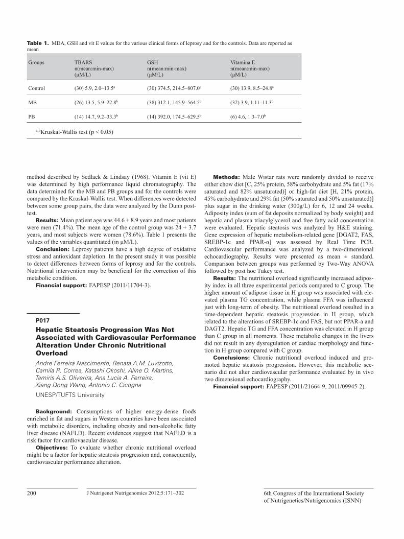
J Nutrigenet Nutrigenomics 2012;5:171–302200 6th Congress of the International Society of Nutrigenetics/Nutrigenomics (ISNN)
method described by Sedlack & Lindsay (1968). Vitamin E (vit E) was determined by high performance liquid chromatography. The data determined for the MB and PB groups and for the controls were compared by the Kruskal-Wallis test. When differences were detected between some group pairs, the data were analyzed by the Dunn post-test.
Results: Mean patient age was 44.6 + 8.9 years and most patients were men (71.4%). The mean age of the control group was 24 + 3.7 years, and most subjects were women (78.6%). Table 1 presents the values of the variables quantitated (in μM/L).
Conclusion: Leprosy patients have a high degree of oxidative stress and antioxidant depletion. In the present study it was possible to detect differences between forms of leprosy and for the controls. Nutritional intervention may be beneficial for the correction of this metabolic condition.
Financial support: FAPESP (2011/11704-3).
P017
Hepatic Steatosis Progression Was Not Associated with Cardiovascular Performance Alteration Under Chronic Nutritional Overload Andre Ferreira Nascimento, Renata A.M. Luvizotto, Camila R. Correa, Katashi Okoshi, Aline O. Martins, Tamiris A.S. Oliverira, Ana Lucia A. Ferreira, Xiang Dong Wang, Antonio C. Cicogna
UNESP/TUFTS University
Background: Consumptions of higher energy-dense foods enriched in fat and sugars in Western countries have been associated with metabolic disorders, including obesity and non-alcoholic fatty liver disease (NAFLD). Recent evidences suggest that NAFLD is a risk factor for cardiovascular disease.
Objectives: To evaluate whether chronic nutritional overload might be a factor for hepatic steatosis progression and, consequently, cardiovascular performance alteration.
Methods: Male Wistar rats were randomly divided to receive either chow diet [C, 25% protein, 58% carbohydrate and 5% fat (17% saturated and 82% unsaturated)] or high-fat diet [H, 21% protein, 45% carbohydrate and 29% fat (50% saturated and 50% unsaturated)] plus sugar in the drinking water (300g/L) for 6, 12 and 24 weeks. Adiposity index (sum of fat deposits normalized by body weight) and hepatic and plasma triacylglycerol and free fatty acid concentration were evaluated. Hepatic steatosis was analyzed by H&E staining. Gene expression of hepatic metabolism-related gene [DGAT2, FAS, SREBP-1c and PPAR-α] was assessed by Real Time PCR. Cardiovascular performance was analyzed by a two-dimensional echocardiography. Results were presented as mean ± standard. Comparison between groups was performed by Two-Way ANOVA followed by post hoc Tukey test.
Results: The nutritional overload significantly increased adipos-ity index in all three experimental periods compared to C group. The higher amount of adipose tissue in H group was associated with ele-vated plasma TG concentration, while plasma FFA was influenced just with long-term of obesity. The nutritional overload resulted in a time-dependent hepatic steatosis progression in H group, which related to the alterations of SREBP-1c and FAS, but not PPAR-a and DAGT2. Hepatic TG and FFA concentration was elevated in H group than C group in all moments. These metabolic changes in the livers did not result in any dysregulation of cardiac morphology and func-tion in H group compared with C group.
Conclusions: Chronic nutritional overload induced and pro-moted hepatic steatosis progression. However, this metabolic sce-nario did not alter cardiovascular performance evaluated by in vivo two dimensional echocardiography.
Financial support: FAPESP (2011/21664-9, 2011/09945-2).
Table 1. MDA, GSH and vit E values for the various clinical forms of leprosy and for the controls. Data are reported as mean
Groups TBARSn(mean:min-max)(μM/L)
GSHn(mean:min-max)(μM/L)
Vitamina En(mean:min-max)(μM/L)
Control (30) 5.9, 2.0–13.5a (30) 374.5, 214.5–807.0a (30) 13.9, 8.5–24.8a
MB (26) 13.5, 5.9–22.8b (38) 312.1, 145.9–564.5b (32) 3.9, 1.11–11.3b
PB (14) 14.7, 9.2–33.3b (14) 392.0, 174.5–629.5b (6) 4.6, 1.3–7.0b
a,bKruskal-Wallis test (p < 0.05)
J Nutrigenet Nutrigenomics 2012;5:171–302 201Abstracts
P018Indicated for the Young Investigator AwardModerate Alcohol Consumption Alters Hepatic Metabolism-Related Markers in Rats with Pre-Existing NASH Andre Ferreira Nascimento, Blanche Ip, Renata A.M. Luvizotto, Xiang Dong Wang
UNESP/TUFTS University
Background: It has been shown that moderate consumption of alcohol in rats with pre-existing nonalcoholic steatohepatitis (NASH) increased hepatic inflammation.
Objectives: To investigate whether moderate alcohol intake alters hepatic lipid metabolism, SIRT1 expression and Adiponectin/Adiponectin receptor (AdipoR)-related signaling in a pre-existing NASH status.
Methods: Sprague-Dawley rats were fed with a high-fat diet (HFD, 71% energy from fat) for 6 weeks to induce NASH. Rats were then equally divided into 2 sub-groups: fed either a modified high-fat diet (HFD, 55% energy from fat) or a modified high-fat alcoholic diet (HFA, 55% energy from fat and 16% energy from ethanol), for addi-tional 4 weeks. Genes involved in hepatic lipid metabolism and Adiponectin/AdipoR-related signaling as well as SIRT1 protein were determined. Plasma adiponectin, triaciglycerol (TG) and free fatty acids (FFA) were analyzed. Hepatic TG and FFA concentration were also evaluated.
Results: In comparison to HFD group: 1) HFA significantly decreased hepatic AdipoR1 but not AdipoR2, 2) plasma adiponectin was lower in the HFA group although this change only reached a sta-tistical tendency (p = 0.06), and 3) FAS gene expression, SREBP-1c and SIRT1 protein expression were significantly up-regulated, while DGAT1/2 and CPT-I gene expression were significantly down-regu-lated in HFA. Unexpectedly, HFA did not alter the phosphorylations of AMPK and ACC as well as PPARa protein level. In addition, there were no differences on plasma and hepatic TG and FFA concentra-tions between two groups.
Conclusions: These data suggest that the down-regulation of Adiponectin/AdipoR-related signaling may contribute to the moder-ate alcohol consumption-exacerbated liver injuries in rats with pre-existing NASH.
Financial support: FAPESP / NIH / USDA (2011/21664-9 / R01CA104932 / 1950-51000-064S).
P019
Tributyrin Treatment Leads to Alteration in Gut Microbiota Composition and in Gut-Associated Lymphoid Tissue Andrea Caricilli, Angela Castoldi, Vinicius Andrade, Enio Bassi, Marco Aurélio Vinolo, Tiago de Souza, Susan Da Silva, Mario Saad, Rui Curi, Niels Câmara
University of São Paulo
Background: Low-grade inflammation, found in obese individ-uals, is associated with impaired intestinal barrier and increased lipo-polysaccharide serum concentration. Obesity is also associated with alteration in gut microbiota composition. However, the mechanism by which altered bacterial composition may lead to increased gut per-meability is not yet understood. Short-chain fatty acids, such as butyr-ate, are fermentation products of some types of bacteria and studies have shown that they may interfere with the modulation of the immune system.
Objectives: Our study aims to investigate the modulation of the immune system found in the gastrointestinal tract of diet-induced obese mice and correlate it with changes in intestinal barrier function after treatment with butyrate prodrug, tributyrin (TB).
Methods: C57BL/6 male mice fed a standard chow or high-fat diet (HFD) were treated with TB (2g/kg, by gavage) during 10 weeks and evaluated for body weight gain, glucose homeostasis, intestinal immune cell profile by flow citometry, gut microbiota composition by 16S mRNA sequencing and intestinal tight-junction protein expression by Western Blot.
Results: TB-treated mice fed a HFD gained less weight and pre-sented increased glucose tolerance than saline-treated mice. However, no significant differences were observed concerning food intake of TB-treated animal, neither in standard chow nor in HFD group. TB-treated mice on a HFD showed increased expression of T regula-tory cells (CD4 + CD25 + Foxp3 + ) and decreased TNF-alpha and IFN-gamma producing cells (TCD8 + and TCD4 + ) in mesenteric limph nodes. Moreover, TB-treated mice on a HFD showed increased expression of IL-17 producing cells in mesenteric limph nodes. After HFD feeding and TB treatment, alteration in the gut microbiota com-position was also observed, with decrease in Firmicutes and increase in Verrucomicrobia phyla. Intestinal barrier was altered by TB treat-ment, with increase of tight-junction proteins, such as ZO-1 and occludin.
Conclusions: Our results suggest that TB treatment leads to an improvement of the intestinal barrier by reducing proinflammatory cytokine production and increasing inflammatory suppression by regulatory T cells. TB treatment also triggers changes in the gut microbiota composition, which might contribute to the modulation of the intestinal immune system. Thus, tributyrin may present itself as a potential drug to be used in the treatment of obesity.
Financial support: FAPESP (2011/19247-0).
J Nutrigenet Nutrigenomics 2012;5:171–302202 6th Congress of the International Society of Nutrigenetics/Nutrigenomics (ISNN)
P020
Camu-Camu Extracts (Myrciaria Dubia McVaugh) Inhibits ROS and iNOS in Lipopolysaccharide-Stimulated Mouse Macrophages Any Elisa Schmidt Gonçalves, Bruno Marcotte, Marie Julie Dubois, Maria Inés Genovese, Franco Maria Lajolo, Yves Desjardins, André Marette
Universidade de São Paulo
Inflammation is involved in numerous diseases, including chronic inflammatory diseases and the development of cancer. Macrophages play an important role in inflammation through the release of inflam-matory mediators involved in the immune response. Inappropriate and prolonged macrophage activation is largely responsible for the pathology of acute and chronic inflammatory conditions. It has been already described that fruits bioactive compounds possess a variety of biological activities, including antifungal, antibacterial and anti-inflammatory activities. Camu-camu is an Amazonia fruit with a high content of antioxidants, specially phenolics compounds and vitamin C. However, there is no information of the anti-inflammatory effects of camu-camu. A hydromethanolic extract (Crude Extract – CE) was prepared and fractionated by solid-phase extraction, providing two different fractions: Total Polyphenolics (TP) and Total Flavonoids (TF). Phenolic compounds of each fraction were identified and quan-tified by HPLC. Anti-inflammatory properties of CE (500 μg/mL), TP (500 μg/ml) and TF (500 μg/ml), were investigated using lipopoly-saccharide (LPS)-stimulated J774.1 macrophages for 6 and 24 hours as in vitro model. As inflammatory parameters, nitric oxide (NO) pro-duction was evaluated by Griess reaction, as well as effects on induc-ible NO synthase (iNOS) expression on intracellular signaling pathways activation, which were analyzed by Western blot using spe-cific antibodies. At the time of 6 hours of LPS stimulation, crude extract (CE) inhibited NO production and iNOS expression. Both TP and TF fraction significantly inhibited NO production, however they were not able to inhibit iNOS expression. At 24 hours of stimulation, CE was the most effective by inhibiting NO production, but none of three fractions were able to modulate iNOS expression. Our data sup-port the usage of Camu-camu extracts as a natural source of anti-inflammatory and antioxidants compounds.
Financial support: CNPQ.
P021
Offspring from Dams Fed High-Fat Diet During Pregnancy and Lactation Presented Activation of Endoplasmic Reticulum Stress Arine Mattos Melo, Rafaela Oliveira Benatti, Nicole Guimarães Ashino, Letícia Martins Ignácio-Souza, Janaína Cursino, Gustavo Aparecido Santos, Licio Augusto Velloso, Adriana Souza Torsoni, Marcio Alberto Torsoni
Faculdade de Ciências Aplicadas – Unicamp
Background: Obesity is associated to increase in the serum pro-inflammatory cytokines and activation of serine-kinases, as JNK and IKK, responsible for the impairment in insulin signaling. Obese sub-jects also present proteins related to unfolded protein response (UPR) activated, an indicator of endoplasmic reticulum stress and responsi-ble by activation of JNK and IKK. Maternal obesity and the high fat diet consumption during pregnancy and lactation has been linked to metabolic damage in the offspring. However, the contribution of UPR to metabolic disorders in offspring of high fat fed mothers remains unknown.
Objectives: Evaluate the activation of UPR in the offspring of dams fed with high fat diet during pregnancy and lactation.
Methods: Females of Swiss mice were fed with either standard chow (SC) or high-fat diet (HFD) during gestation and lactation. After weaning the offspring was divided in SC-offspring (SC-O) and HFD-offspring (HFD-O) and both groups were fed with standard chow until the experiment period. The analyses were performed on the 28th day of life. Initially, we evaluated the body weight. The phosphorylation of the proteins PERK, JNK, eIF2α, in the liver and hypothalamus were analyzed through Western Blot technique. Relative gene expression of CHOP, GRP78, IL1β, TNFα and IL10 were measured in the hypothalamus by real time PCR.
Results: No differences in body weight between the groups on day (d) 2 after birth, but from d7 the HFD-O showed an increase in body weight compared to SC-O. In both tissues from HFD-O the level of p.JNK were increased. Besides, the hypothalamus also showed increase NFkB expression and gene expression of pro-inflammatory cytokines (IL1β, TNFα) and decrease of IL10, an anti-inflammatory cytokine, compared to SC-O. The gene expression of CHOP e GRP78 was also higher in HFD-O compared to SC-O. In both tissues the level of p-PERK, p-eIF2α in HFD-O were increased compared to SC-O mice. Besides, hypothalamic insulin-induced phosphorylation of AKT of HFD-O was reduced compared to SC-O.
Conclusions: Recently weaned offspring from high fat diet dam present activation of proteins related to UPR in liver and hypothala-mus, suggesting that these tissues are under metabolic stress condi-tion. This cellular response may be related to diminished activation of the AKT by insulin in the hypothalamus leading to damage to the signal of satiety and glucose homeostasis in the offspring contributing to the development of obesity in adulthood.
J Nutrigenet Nutrigenomics 2012;5:171–302 203Abstracts
P022
Protein or Methionine Restriction Affects Dicer Expression and MicroRNA Processing in Adipose Tissue of Rodents Beatriz Alves Guerra, Carmen E. Perrone, Thiago Martins Batista, Everardo Magalhaes Carneiro, Marcelo Alves Mori
Universidade Federal de São Paulo
Background: Caloric restriction promotes beneficial health effects and prolongs lifespan in diverse species, from yeast to pri-mates. Our group recently demonstrated that microRNA (miRNA) processing is progressively down-regulated with age across the evo-lutionary spectrum. In particular, this phenomenon is characterized by a decrease in Dicer expression in adipose tissue, and this can be prevented by caloric restriction by mechanisms that are not fully understood. Methionine restriction has been shown to promote bene-ficial health effects that mimic the effects of caloric restriction in rodents. Thus, we sought to analyze whether the restriction of dietary protein or, more specifically, methionine had an effect on the miRNA processing pathway (i.e. Dicer, Argonaute-2 and miRNAs) in adipose tissue of rodents.
Objectives: To identify how nutritional interventions (i.e. pro-tein or methionine restriction) affect the miRNA processing pathway in adipose tissue of rodents.
Methods: Four-week old C57Bl/6 male mice were subjected to protein restriction (PR) or ad libitum chow diet for 14 weeks. A sub-group of these mice were put on a high fat diet (HF) with 10 weeks of age and sacrificed after 8 weeks. Methionine restriction (MR) started with eight-week old Fischer 344 rats and was carried out for 12 weeks. We tested the expression of Dicer and Argonaute-2 (Ago2) by RT-qPCR and Western blotting. The expression of miR-125b and miR-145 was assessed by RT-qPCR.
Results: Our preliminary results indicate that PR resulted in a 12.8-fold increase in Dicer expression at the mRNA, but not at the protein level, in the subcutaneous white adipose tissue of mice. HF decreased Dicer protein levels and attenuated by 32% the effect of PR on Dicer mRNA expression in these mice. The expression of miR-125b and miR-145 also increased with PR and PR followed by HF. To test whether MR could explain some of the phenotypes observed in calorie or protein restricted animals, we measured Dicer and miRNAs in fat of methionine restricted rats. MR resulted in a 5.2-fold increase in Dicer and a 1.6-fold increase in Ago2 in the epididymal white adi-pose tissue. Moreover, miR-125b increased by 2.2-fold in the MR group in relation to the control group.
Conclusions: The effects of caloric restriction on the miRNA processing pathway can be mimicked at least in part by protein or methionine restriction.
Financial support: FAPESP (2012/07259-7 E 2010/52557-0).
P023
Green Tea Attenuates Oxidative Stress in Heart Remodeling Induced by Myocardial Infarction Beatriz H.B. Lustosa, Diego F. Batista, Ana A.H. Fernandes, Katashi Okoshi, Marcos Minicucci, Luiz Matsubara, Leonardo Zornoff, Paula S.A. Gaiolla
UNESP – Faculdade de Medicina de Botucatu
Objectives: The objective of this study is to evaluate whether green tea attenuates cardiac remodeling after acute myocardial infarc-tion.
Methods: wistar rats were allocated in 4 groups: C (control), GT (diet with green tea 0.25%), MI (infarcted group) IGT (infarcted group with green tea 0.25%). After 3 months, animals were submitted to echocardiography and analysis of oxidative strees. Two way anova and Holm Sidak tests were performed.
Results: Both infarcted groups showed the same infarction size (p = 0.7). Echocardiographic data without interaction: left atrium/weight (with MI 14.4 ± 0.5, without MI 10.7 ± 0.6) (p < 0,001) (with GT 13.0 ± 0.5, without GT 12.1 ± 0.6) (p = 0.2), and diastolic ventri-cle diameter/weight (with MI 21.2 ± 0.6, without MI 17.4 ± 0.7) (p < 0.001) (with GT 19.1 ± 0.6, without GT 19.7 ± 0.7) (p = 0.5). Systolic function: fractional area change (with MI 41.2 ± 1.8, without MI 70.8 ± 2) (p < 0.001) (with GT 56.1 ± 1.8, without GT 55.8 ± 2) (p = 0.9). Echocardiographic data with interaction (p = 0.04) E wave decelera-tion time (EDT) (C 48.5 ± 2.9, GT 45 ± 2.6, MI 35.3 ± 2.5, MIGT 43 ± 2.4). Oxidative stress data without interaction: hydroperoxide (HP) (p = 0.28) (with MI 204, without MI 165.3) (p = 0.002) (with GT 174.3, without GT 195) (p = 0.068), and glutathione peroxidase (GSH-PX) (p = 0.12) (with MI 36.8, without MI 34.8)(p = 0.43) (with GT 38.3, without GT 33.3) (p = 0.06).Data with interaction catalase (p < 0.001) (C 67.9, GT 45.8, MI 35.1, MIGT 52.3), superoxide dis-mutase (SOD) (p = 0.002) (C 0.64, GT 0.76, MI 1.04, MIGT 0.88).
Conclusions: There was no reduction in the formation of HPX, but is numerically smaller. IGT decreases SOD and catalase increases. It may be because SOD has already been consumed and then increases catalase to try to hold the oxidative stress. There was no difference in GPX.
Acknowledgment: This work is supported by Fapesp.
P024
Effect of Methylglyoxal, Glyoxal and Resveratrol on Breast Cancer Cells MCF-7 Viability Betina Schmidt, Carolina Agostinho, Eliane Filaho de Oliveira
Universidade Federal do Rio de Janeiro
Background: Breast cancer is the second most common cancer worldwide. Since the discovery of high aerobic glycolysis in tumors, it has remained unclear how to exploit this in chemotherapy. During the glycolysis, metabolites are formed including glyoxal and methyl-
J Nutrigenet Nutrigenomics 2012;5:171–302204 6th Congress of the International Society of Nutrigenetics/Nutrigenomics (ISNN)
glyoxal, Advanced Glycation End-products (AGEs) precursors. The AGEs promote oxidative stress, morphological and functional changes and increase inflammatory mediators becoming aggressive to the healthy cells, but potentially chemotherapeutic. Recent studies show that resveratrol, a polyphenol, has anti-inflammatory and anti-oxidants properties becoming a promising chemopreventive agent.
Objectives: This study aimed to verify the action of methylgly-oxal, glyoxal and resveratrol, alone and in combination on viability of MCF-7 cells.
Methods: MCF-7 cells were grown in Dulbecco’s Modified Eagle Medium supplemented and grown at 37°C in an atmosphere of 5% CO2. On reaching 70–80% confluency they were treated with dif-ferent concentrations of resveratrol, glyoxal and methylglyoxal. Cell viability was determined by MTT reduction assay.
Results: IC 50 values for methylglyoxal, glyoxal and resveratrol were 3.1 mM, 3.0 mM and 133 μM, respectively. To examine a pos-sible synergistic effect of the precursors of AGEs with resveratrol the treatments were associated. The association enhances the decrease of viable cells, demonstrating a synergistic effect of drugs.
Conclusions: Methylglyoxal and glyoxal have a detoxification system composed of two major enzymes, glyoxalase I and II. The cytotoxic action of glyoxals, found in this study, reinforces the find-ings reported in the literature with others cancer cells. Increasing speed of the glycolytic pathway in cases of cancer, there is an increase of glyoxals formation, which would, hypothetically, have a protective effect and induce cancer cells apoptosis, but the literature explained that cancer cells undergo an adaptation of glyoxalase I expression and can detoxify the glyoxals without undergoing cytotoxic injury. It was proved that curcumin minimize glyoxalase I gene expression, increas-ing the amounts of glyoxals, justifying the chemotherapy power of the polyphenol. It is possible that resveratrol has a similar action to curcumin and the synergistic effect of resveratrol and glyoxals, shown in this study, is related to decreased expression of glyoxalase I. Studies involving the expression of the gene and protein have been made by our group to confirm the hypothesis.
Financial support: CAPES, FAPEJ, CNPQ, Fundação do Câncer.
P025
Resveratrol Stimulates the Production of Reactive Oxygen Species in Breast Cancer Cells Patrícia Severo Ramos, Paula Seixas Costa, Jerson Lima Silva, Eliane Fialho
UFRJ
Background: It is estimated that in 2020, the worldwide inci-dence of cancer is on the order of 15 million, of which 60% of new cases will occur in developing countries. The breast cancer is the sec-ond most deadly cancer among women. Resveratrol, a phytoalexin found in grapes and red wine has been reported to exhibit a wide range of pharmacological properties and is believed to play a role as anti-cancer agent but the precise mechanism of action, however, is not fully elucidated. In the present study, the anticancer activity of
resveratrol towards MCF-7 human breast cancer cells was investi-gated.
Materials and Methods: One breast cancer cell line (MCF-7) was cultured using standard techniques. Cell viability was determined by MTT assay. In order to demonstrate the decrease of the cell viabil-ity towards the production of reactive oxygen species (ROS), the cells were treated with resveratrol (RV) (200μM) for 24h. The generation of ROS was measured by using the cell-permeable nonfluorescent compound H2DCF-DA. N-Acetyl-L-cysteine (NAC) was used in dif-ferent concentrations (0–5mM) for 24 h to evaluate the capability of reverse the pro-oxidant effect of RV (200μM), the same was done using L-Glutathione reduced (GSH) in different concentrations (10mM–300mM).
Results: RV inhibited cellular proliferation in time and dose-dependent manner with an IC50 of 238μM and 151 μM for 24 and 48 h, respectively. ROS production was increased when MCF-7 cells were treated with RV 200μM for 24h. Recently, the effects of several drugs that produce ROS and interfere directly with apoptosis in can-cer cells such as MCF-7 cells have been described. NAC and GSH completely reverted the ROS generation, promoted by high concen-trations of RV, demonstrating a pro oxidant effect of RV which caused the decrease of the MCF-7 cell viability.
Conclusions: Taken together, our results demonstrate that RV inhibited the growth of MCF-7 cells in a dose-dependent manner and that this reduction in cell viability resulted from ROS generation which was reverted using different concentrations of NAC and GSH.
Financial support: CAPES, FAPEJ, CNPQ, Fundação do Câncer.
P026
The Advanced Glycation End-Products (AGES) and the Complications of Diabetes Mellitus – A New Therapeutic Target for Nutrition Betina Schmidt, Eliane Fialho de Oliveira, Carolina Agostinho
UFRJ
Diabetes mellitus is a significant concern in prevalence world-wide. A possible mechanism of tissue damage induced by hypergly-cemia in Diabetes Mellitus is the formation of advanced glycation end-products (AGEs). The pathological effects of AGEs are related to its ability to modify the chemical properties and functions of various biological structures. During the Maillard reaction, some intermedi-ate products are formed such as dycarbonils and α-oxaldehydes as glyoxal, methylglyoxal and 3-D-glyoxuglucosone, precursos of AGEs. The intracellular increase in oxidative stress also contributes to the formation of AGEs. The preparation of food containing high fat and sugars at high temperatures (130 °C) is the main exogenous source of AGEs. It is known that approximately 10% of AGEs ingested with diet is absorbed although the mechanisms pertaining to such absorption are not fully elucidated. The fraction absorbed, about two thirds are retained in the body and one third is excreted in the urine, within 48 hours, by individuals with normal renal function. There is evidence that dietary AGEs are added to the pool of endoge-
J Nutrigenet Nutrigenomics 2012;5:171–302 205Abstracts
nous AGEs, favoring the emergence and progression of several com-plications of diabetes mellitus. Some anti-AGEs substances have been identified as N-acetylcysteine and penicillamine. Polyphenols acts as hostages of methylglyoxal and glyoxal, precursors of AGEs, and may act as a new target for prevention of diseases associated with diabetes mellitus. Understanding the contribution of AGEs to the development of complications of Diabetes Mellitus will show new lines of research in medicine and functional foods in order to inhibit the formation or absorption of AGEs.
Financial support: CAPES, FAPEJ, CNPQ, Fundação do Câncer.
P027
Vitamin A Modulation of IL-10 and IL-17 in Human CD4+ Cells Stimulated with Soluble Leishmania Infantum Antigens Bruna Leal Lima Maciel, Francisco P. Freire Neto, João Firmino R. Neto, Tatjana Keesen, Selma Maria B. Jerônimo
Universidade Federal do Rio Grande do Norte
Backgound: Vitamin A supplementation has long been used in developing countries to prevent mortality by diarrheal and respiratory diseases. Infection by Leishmania infantum is endemic in Northeast Brazil and may cause a spectrum of clinical outcomes that ranges from asymptomatic self-healing infection to visceral leishmaniasis (VL), a disease that is fatal even with treatment. VL is characterized by hepatosplenomegaly, fever, pancytopenia and altered hepatic func-tion with increase in acute phase proteins and decrease in albumin synthesis. Determinants of the disease severity are not clearly under-stood, but nutritional status and the immune response influence the outcome of infection and disease. Cytokines as IL-10 and IL-17 are associated with parasite persistence/disease and protection against infection, respectively. IL-10 is a suppressive cytokine produced by effector CD4 + cells and T regulatory (Treg) cells. IL-17 is produced by inflammatory CD4 + (Th17) cells.
Objectives: This work analyzed the effect of retinoic acid (RA), an active vitamin A metabolite, and soluble Leishmania infantum crude antigens (SLA) in CD4 + cells isolated from healthy children.
Methods: PBMCs isolated from children (n = 9) with no signs of Leishmania infection (negative antibody to SLA and negative Montenegro skin test) were stimulated with SLA (10μg/mL) or RA (0.25nM) or SLA (10μg/mL) and RA (0.25nM). Cells were cultured for 20 hours and the production of IL-10, IL-17 in effector CD4 + cells and IL-10 in Treg cells (CD4 + CD25highFOXP3 + cells) was analyzed by flow cytometry.
Results: PBMCs stimulates in presence of RA + SLA increased the expression of CD25 in CD4 + cells, more than SLA alone (32.4% and 22.6%, respectively, p = 0.006). RA elevated the production of IL-10 in CD4 + cells more than SLA (4.1% and 2.5%, respectively, p = 0.036). SLA stimulated IL-17 production by CD4 + CD25 + (6.2% vs 3.4% without any stimulation), while RA addition down regulated IL-17 production to 2.8% of CD4 + CD25 + cells (p = 0.015). Stimulation by either SLA or RA + SLA did not result in sig-nificant changes in Treg IL-10.
Conclusions: Data show increased production of IL-10 and decreased IL-17 in CD4 + cells, which may suggest negative modula-tion of the immune response by vitamin A in presence of Leishmania antigens.
Financial support: National Institutes of Health, CNPQ.
P028
Arg213Gly Polymorphism in EC-SOD Gene and Its Relationship with Cardiovascular Disease Bruna Zavarize Reis, Graziela Biude Silva, Isabela Saraiva Almeida, Silvia Maria Franciscato Cozzolino
University of São Paulo
Superoxide dismutases (SOD) are considered the first line of anti-oxidant enzyme defense systems. They are responsible for dismuta-tion of superoxide anions to hydrogen peroxide, which in turn is catalyzed in water by catalase, glutathione peroxidase and peroxire-doxins. There are three isoforms of this enzyme in mammals: copper-zinc (CuZnSOD, SOD1), found in the cytosol, manganese (MnSOD, SOD2), found almost exclusively in mitochondria, and the extracel-lular superoxide dismutase (EC-SOD, SOD3) in the extracellular space and fluids. EC-SOD is highly expressed in blood vessels, lung, kidney and uterus. In vascular tissue, EC-SOD is synthesized by vas-cular smooth muscle cells and fibroblasts, being located on the sur-face of the endothelial membrane bound to sulfated glycoproteins such as heparan sulfate and heparin. EC-SOD is the main arterial SOD isoform and thus makes a major contribution to cardiovascular defense. Some individuals may exhibit extremely high plasma levels of EC-SOD as a result of a SNP in the enzyme gene, the Arg213Gly. The polymorphism is located in the heparin binding domain, causing a decrease in affinity for glycosaminoglycans. As a result, there is an increase 10 to 30-fold plasma concentration of SOD3 in polymorphic subjects. Consequently, despite high plasma concentration, the arte-rial wall may be deficient in EC-SOD and thus may have insufficient antioxidative capacity against superoxide anions and LDL oxidation, which could lead to increased atherogenesis. In fact, it was shown that heterozygous individuals have an increased risk of developing ischemic heart disease. In patients undergoing angiography, there was an association between the polymorphism and the severity of coro-nary artery disease and the risk for myocardial infarction. Polymorphic patients with diabetes and end-stage renal disease had significantly higher incidence of ischemic heart disease and cerebrovascular dis-ease. In another study, uremic patients who carried the variant allele showed accelerated progression in renal failure and atherosclerosis. It was also observed a positive correlation between the presence of the polymorphism and severity of macro and microvascular complica-tions in both diabetic and healthy subjects. Studies suggest that the presence of Arg213Gly variant in EC-SOD gene reduces the protec-tive role of the enzyme in vascular wall, constituting an independent risk factor for cardiovascular disease.
J Nutrigenet Nutrigenomics 2012;5:171–302206 6th Congress of the International Society of Nutrigenetics/Nutrigenomics (ISNN)
P029
Effects of n-3 Polyunsaturated Fatty Acids in Treatment of Hypertriglyceridemia in Subjects with Diabetes Mellitus Caio E.G. Reis, Karla C. Ladim, Aline C.S. Nunes, Jane Dullius
University of Brasília
Diabetes mellitus increases the risk of hypertriglyceridemia and is an independent risk factor for cardiovascular diseases. Current litera-ture reveals the beneficial effects of n-3 polyunsaturated fatty acids (n-3 PUFA) in hypertriglyceridemia treatment, however its efficacy for subjects with diabetes mellitus are still debatable. This literature review discusses the effects of n-3 PUFA supplementation in the treatment of hypertriglyceridemia and its effects on glycemic control in subjects with diabetes mellitus (type 1 and 2). A search was con-ducted in the LILACS, MEDLINE, and COCHRANE databases. Eighteen articles (fourteen clinical trials and four review studies) were selected for this review. All articles showed triglycerides reduc-tions ranging from 18 to 35% (final vs. initial values) and 15 to 29% (placebo vs. intervention values). Furthermore the articles showed a reduction between 26–36% in very low-density lipoprotein and 5–22% in low-density lipoprotein, and an increase of 5–19% in high-density lipoprotein levels. There was no significant alteration of total cholesterol levels in most of the articles (86%, n = 16). The articles that evaluated fasting plasma glucose and glycated hemoglobin showed an increase of 18% and 16% respectively. These findings pro-vide preliminary evidence that n-3 PUFA supplementation can be a promising tool in the treatment of hypertriglyceridemia in diabetes mellitus subjects. However may cause a slight increase in glucose and glycated hemoglobin levels. Further clinical trials are needed to investigate the complex mechanisms relating n-3 PUFA to the reduc-tion of triglycerides levels and to understand its effects on lipid pro-file and glycemic control in people with diabetes.
P030
Effect of Fish Oil Supplementation on Neutrophil Death in Wheelchair Basketball Players Camila Garcia Marques, Adriana Cristina Levada-Pires, Vinicius Coneglian Santos, Sâmia Rocha Alves, Rui Curi, Tânia Cristina Pithon-Curi, Maria Fernanda Cury-Boaventura
Universidade Cruzeiro do Sul
Background: Individuals with spinal cord injuries commonly develop infections of the urinary tract, respiratory tract, and skin, indicating that their host defense mechanisms may be diminished. The engaging of paraplegics in sports programs combined with an adequate food plan can minimize the risk of related diseases. Evidence suggests that fish oil rich in n-3 polyunsaturated fatty acids (n-3 PUFA) has a beneficial effect on immune response.
Objectives: We investigated the effect of fish oil supplementa-tion on neutrophil death in athletes with spinal cord injuries at rest and after basketball play.
Methods: Nine paraplegic athletes were supplemented with 3g of fish oil per day. The athlete’s blood samples were collected on rest and immediately after basketball play (60 minutes) before and after supplementation. Neutrophils were isolated by density gradient, using Histopaque-1077. The following determinations were carried out: loss of membrane integrity to evaluate necrosis and DNA frag-mentation, phosphatidylserine externalization to evaluate apoptosis.
Results: Were studied nine athletes with following characteris-tics: age: 33.8 + 2.7 years, height: 170 + 4 cm, body fat: 22 + 5%, BMI 23.9 ± 3.16 kg/m2 and training time: 6.9 + 5.4 years. Basketball play induced an increase in the percentage of necrotic neutrophils (2.57 + 0.37 to 17.5 + 3.5%) but had no effect on percentage of cells with DNA fragmentation and phosphatidylserine externalization. Fish oil supplementation prevents the necrosis induced by training but induced a slight increase in the percentage of apoptotic neutro-phils (2.91 + 1.01 to 5.42 + 1.47%).
Conclusions: Basketball play induced neutrophil death in wheelchair athletes by necrosis. Fish oil supplementation may be a strategy for basketball players in wheelchairs to promote improves in athletic performance and health for paraplegia condition.
Financial support: FAPESP (2010/04440-7).
P031
Polyphenol-Rich Cagaita (Eugenia Dysenterica DC) Extract Promotes Pancreatic Beta Cell Proliferation and Improves Insulin Sensitivity in Diabetic Rats Camilo Lellis-Santos, Laila Romagueira Santos, Any Elisa Souza Gonçalves, Lucas Carminatti Pantaleão, Ingrid Candido Garofolo, Marcela Roquim, Renata Gorjão, William Tadeu Festuccia, Franco Maria Lajolo, Maria Inés Genovese, Silvana Bordin, Rui Curi
University of São Paulo
Pancreatic beta cells failure and apoptosis are key events in the development and progression of Diabetes Mellitus (DM). Many bio-active compounds from natural sources have been described to delay or prevent DM through inhibition of oxidative stress and inflamma-tory processes in metabolism-related tissues, such as liver, muscle and adipose tissue. However, there is no data addressing the effects of phenolic extracts from the native Brazilian fruit Cagaita (Eugenia dysenterica DC) over pancreatic beta cell physiology. Pancreatic beta INS1e cells were treated with different concentrations (100, 500 or 1000 μg/mL) of polyphenol-rich Cagaita extract (PCE). PCE were also given to control and streptozotocin (SZT)-induced diabetic rats by gavage at dose of 3g/Kg body weight for 30 days. Phosphorylation of ERK1/2 was significantly increased after treatment with 100 and 500 μg/mL, but the lower dose induced more effectively cell cycle progression and beta cell proliferation, measured by flow cytometry and [3H]-thymidine incorporation, respectively. Treatment of INS1e cells with 100 μg/mL of PCE for 24h increased gene expression of proliferating cell markers (mcm7 and CDK4), and pre-incubation
J Nutrigenet Nutrigenomics 2012;5:171–302 207Abstracts
with ERK1/2 inhibitor UO126 for 1h abrogated this effect (P < 0.05). Among the seven phenolic compounds identified by HPLC, only quercetin significantly increased both phosphorylation of ERK1/2 and Akt at levels of PCE. And quercetin and the polyphenol mixture of all compounds at dose equivalent to PCE (100 μg/mL) were able to partially reproduce the effects of PCE on pancreatic beta cell prolif-eration. Control rats gavaged with PCE showed increased insulin content and mRNA expression in isolated islets, as well as Cyclin D1 expression, a mark of cell proliferation, when compared to saline-treated animals. Besides, islets from PCE-treated animals were more resistant to apoptosis induced by serum deprivation. PCE treatment was not able to counteract streptozotocin (SZT)- induced pancreatic beta cell death. Nevertheless, diabetic rats treated with PCE showed improved glycemia, insulin sensitivity, plasma antioxidant capacity and lipid profile when compared to diabetic rats. Cagaita extract induces pancreatic beta cell proliferation in an ERK1/2 and Akt dependent manner, and quercetin was the major compound to induce these effects. The extract also ameliorates SZT-induced damages in rats. Thus, PCE should be considered as new nutraceuticals to prevent or manage DM.
Financial support: FAPESP (2010/10778-0).
P032
Nutrition in the Post Genomic Era: Genetic Aspects Related to Obesity Carlos Henrique Miotto
Faculdades Integradas ASMEC
Obesity has reached epidemic proportions worldwide, with more than one billion adults being overweight, and is a major contributor to the global burden of chronic disease and disability, affecting virtually all ages and socioeconomic groups. The completion of the ambitious Human Genome Project in April 2003, has the ability for the first time, be held to read the complete model of the genetic makeup of a human being. The post-genome Era resulted in the development of several new “omics” technologies, applicable in many areas of sci-ence. Defined as nutritional genomics or nutrigenomics, this area aims to provide an understanding on how nutrition can affect the bal-ance between health and disease by altering the expression or genetic structure of an individual. No human, except for identical twins have the same genome than others. Each charge is a set of genetic poly-morphisms that makes them unique. The metabolism of drugs and nutrients is associated with the polymorphic profile of an individual, which may explain the adversity of reactions and responses to phar-macological interventions and nutritional. Like other chronic dis-eases, obesity is caused by multiple molecular pathways. Thus, except in special behavioral conditions, it was noticed that obesity usually results from small and continuous accumulation of body fat over the years due to changes in dietary habits and increased sedentary life-style associated with the polymorphism of several genes. Studies of monogenic and rare forms of human obesity may provide important information regarding endocrine physiology and the most common forms of obesity. In 2007, an association of SNPs – known as Fat Mass and Obesity-Associated (FTO) – the genetic regions linked to BMI and the risk of obesity, was identified in various populations,
becoming the first locus associated with adiposity. The nutrigenomics will be the driving force of the future of nutritional research and will entail a major impact on public health.
P033
Effects of Maternal Plane of Nutrition and Weaning Age of the Offspring on Total RNA Yield in Subcutaneous Adipose Tissue Fernanda Trindade Rosa, Sonia Moisa, Johan Osorio, Carolina Bespalhok Jacometo, Eduardo Schmitt, Marcio Nunes Corrêa, Juan J. Loor
University of Illinois
Background: One of the carcass traits that influence meat qual-ity is the intramuscular fat deposition which begins its development before birth. Therefore, maternal nutrition can potentially result in alterations of the development not only of the offspring but of adipose tissue including changes in gene expression. Overall, assess the RNA yield from tissue samples can generate important information for the gene expression analysis.
Objectives: Evaluate the total RNA yield in fat tissue from off-spring of beef cows that were on different planes of nutrition during late gestation.
Materials and Methods: Thirty six cows at late gestation ( ± 90 days from expected calving) were allocated into three planes of nutrition: 1) high plane of nutrition, fed with endophyte-infected tall fescue (F)/red clover pastures (C) plus 9.1 kg of a diet based on Distiller’s Dried Grains with Solubles (DDGS) and soyhulls, 2) medium plane of nutrition, fed with F/C, 2.3 kg of DDGS and soy-hulls, and 3) low plane of nutrition, fed with F/C without supplemen-tation. The offspring from each plane of nutrition were separated into an early weaning (EW, 79 ± 2 days of age) (n = 7) group housed in a feedlot, and a normal weaning (NW) (n = 7) group in which calves remained with the dams until 186 ± 2 days of age. Biopsies from the tail fat pad were harvested at EW and NW and at the mid-point during the finishing phase. RNA was extracted from approximately 100 mg of tissue using the Ambion miRNA/total RNA isolation kit as described by the manufacturer. The spectrophotometric absorbance at 260 and 280 nm was used to determine RNA quality, concentration and total yield. Statistical analysis was performed using the Proc Mixed Procedure in SAS, and a value of P < 0.05 considered signifi-cant.
Results: At the point of EW the total RNA yield (μg/mg tissue) was greater (P < 0.0001) for offspring from dams in the high plane of nutrition than the medium and low planes (119 μg, 66 μg and 42 μg, respectively). In addition, offspring from dams in the high plane of nutrition had greater (P = 0.0009) total RNA yield (116 μg) at the moment of NW than the medium and low planes (75 μg and 63 μg, respectively).
Conclusions: The maternal plane of nutrition during late gesta-tion appears to affect the total RNA content of subcutaneous adipose tissue in the offspring.
J Nutrigenet Nutrigenomics 2012;5:171–302208 6th Congress of the International Society of Nutrigenetics/Nutrigenomics (ISNN)
P034
Prenatal Exposure to Fatty Acids and Its Influence on the Developmental Biology Fernanda Trindade Da Rosa, Carolina Bespalhok Jacometo, Eduardo Schmitt, Juan J. Loor, Marcio Nunes Corrêa
Universidade Federal de Pelotas
Dietary exposure during pregnancy could influence the offspring development. The maternal diet could cause changes in the offspring health and growth, these changes provided by molecular mechanisms and the effects could be across generations. The effects of a fetal pro-gramming can be multigerational, if the individual had a direct expo-sure to the nutrition or transgenerational if the effects are maintained throughout generations that were not directly exposed. DNA methyla-tion and histone modifications are genetics processes that can regu-late the gene expression. Based on environmental factors, like nutritional prenatal exposure, these alterations on gene expression potentially could cause alterations on the growth and on the metabolic status of the progenies. Approaches have focused on the benefits of fatty acids by maternal nutrition. Among the fatty acids, mammal requires essential ones, the polyunsaturated fatty acids (PUFAs), like linoleic (C18:2) and alpha linolenic (C18:3), which cannot be synthe-sized by the mammalian organism, therefore they must be obtained from the diets. These fatty acids are involved on the regulation of lipid metabolism, cell growth and differentiation and immune func-tion. The beneficial effects of C18:2 and C18:3 on fetal development and on the offspring metabolism are well documented. As the main biology function of the PUFAs is to provide energy, researches dem-onstrated that these fatty acids can regulate the lipid metabolism due the activation of target transcription factors that stimulates fatty acid oxidation and/or inhibits the lipogenesis. Also, studies have demon-strated that the ratio between C18:2 and C18:3 are important for maintenance of polyunsaturated fatty acids homeostasis and for liver enzymatic control. In addition, observations showed that the metabo-lites of fatty acid desaturation, like arachidonic acid and docosa-hexaenoic acid are involved with the activity of brain membranes. However, there are studies with alpha linolenic acid that showed an anti-inflammatory effect avoiding some chronic diseases on the adult health. In the context of healthy development, the maternal diet with polyunsaturated fatty acids might be an important genetic agent within the adult life.
P035Indicated for the Young Investigator AwardRegulation of Lipid Metabolism Transcription Factors by Polyunsaturated Fatty Acids on the Postpartum Period of Successive Generations in Rats Carolina Bespalhok Jacometo, Fernanda Trindade Da Rosa, Patrícia Mattei, Eduardo Schmitt, Luiz Francisco Machado Pfeifer, Augusto Schneider, Nelson José Laurino Dionello, Francisco A. Burkert Del Pino, Marcio Nunes Corrêa
Universidade Federal de Pelotas
Background: Polyunsaturated fatty acids (PUFAs) nutrition exposure during fetal life could represent an important factor related to the offspring metabolic status.
Objectives: To evaluate the expression of transcription factors related to lipid metabolism by essential fatty acids and their hepatic profile through consecutive generations.
Methods: Eighteen female Rattus norvegicus – Wistar/UFPel, 8 weeks old, were separated in two groups: 1) diet rich in omega-6, with soybean oil (CTL) and, 2) diet rich in omega-3, with flaxseed oil (OM). The diets were elaborated in accordance with AIN-93 recom-mendations. After 30 d of adaptation the rats were mated. The males received only the control diet. The F1 females were selected at wean-ing (n = 16/group) and divided into three groups: females from OM group that continued receiving the same diet (OM-OM), females from OM group that began to receive diet containing soybean oil (OM-CTL), and females from CTL group that continued to receive the same diet (CTL-CTL). These animals were mated at 60 d old as described (G0). The F2 (n = 16/group) was selected as F1 and the same groups were maintained: OM-OM-OM, OM-CTL-CTL, and CTL-CTL-CTL. The F2 was mated as F1. Female postpartum eutha-nasia (21 days) was conducted for each generation (n = 6/group) to collect liver samples. Real-Time quantitative PCR was performed to evaluate the peroxisome proliferator-activated receptor alpha (PPARα), retinoid X receptor alpha (RXRα), liver X receptor alpha (LXRα), sterol regulatory element binding protein 1c (SREBP-1c). ACTβ, G3PDH and 18S rRNA were used as internal controls. The fatty acid profile of the hepatic tissue was evaluated by the adapted Bligh Dyer Method.
Results: The hepatic omega-3 profile was 10 folds higher for the OM group than the OM-CTL (P < 0.001) and 15 folds than the CTL (P < 0.001). An upregulation was observed in LXRα expression across generations (P < 0.001). F1 had higher expression of RXRα than F2 (P < 0.001), however, it was not different from G0. PPARα expression was not affected by OM treatment, although a reduction was observed in the CTL group from G0 to F2 (P = 0.03). SREBP-1c expression was higher in the CTL-CTL than the OM-OM group for F1 generation (P = 0.04), and an upregulation was observed for all groups from F1 to F2 (P < 0.001).
Conclusions: PUFAs affected the expression of transcription factors related to lipolysis and lipogenesis, and this effect seems to be cumulative throughout generations.
J Nutrigenet Nutrigenomics 2012;5:171–302 209Abstracts
P036
The Omega-3/Omega-6 Ratio in the Regulation of Lipogenesis in the Prepartum Period of Successive Generations in Rats Carolina Bespalhok Jacometo, Diego A.V. Acosta, Patrícia Mattei, Eduardo Schmitt, Francisco A.B. Del Pino, Marcio Nunes Corrêa
Universidade Federal de Pelotas
Background: Besides the polyunsaturated fatty acids effect on gene expression regulation, the omega-3/omega-6 ratio could influ-ence the relationship between lipogenic and lipolytic effects.
Objectives: To evaluate the expression of a transcription factor and a target gene related to lipid metabolism by the omega-3/omega-6 ratio in the prepartum period through consecutive generations.
Methods: Eighteen female Rattus norvegicus – Wistar/UFPel, 8 weeks old, were separated in two groups: 1) diet rich in omega-6, with soybean oil (CTL), 0.4/1 ratio, and, 2) diet rich in omega-3, with flaxseed oil (OM), 13.6/1 ratio. The diets were elaborated in accor-dance with AIN-93 recommendations. After 30 d of adaptation the rats were mated. The males received only the control diet. The F1 females were selected at weaning (n = 16/group) and divided into three groups: females from OM group that continued receiving the same diet (OM-OM), females from OM group that began to receive diet containing soybean oil (OM-CTL), and females from CTL group that continued to receive the same diet (CTL-CTL). These animals were mated at 60 d old as described (G0). The F2 (n = 16/group) was selected as F1 and the same groups were maintained: OM-OM-OM, OM-CTL-CTL, and CTL-CTL-CTL. The F2 was mated as F1. Female prepartum euthanasia (19 ± 1 days of pregnancy) was con-ducted for each generation (n = 4/group) to collect liver samples. Real-Time quantitative PCR was performed to evaluate the sterol regulatory element binding protein 1c (SREBP-1c), fatty acid syn-thase (FAS) and acetyl-CoA carboxylase α (ACACA). ACTβ, G3PDH and 18S rRNA were used as internal controls.
Results: An upregulation across generations was observed in SREBP-1c (G0-F2, P < 0.01, F1-F2, P = 0.01) and in FAS (G0-F2 and F1-F2, P < 0.01), with no differences between groups. A contrasting behavior was observed for ACACA expression, with differences between groups in the F1, a higher expression in OM-OM, an inter-mediated level for OM-CTL and a lower for CTL-CTL (P < 0.01 for all pairs) and along generations, the OM increased from G0 to F1 (P < 0.01) while CTL decreased (P < 0.01).
Conclusions: Both omega3/omega6 ratio affected the expres-sion of transcription factors related to lipogenesis, and this effect seems to be cumulative throughout generations.
P037
Nutritional Status of Children and Adolescents in Ribeirão Preto, Sao Paulo, Brazil Carolina de Almeida Coelho, Jacqueline Pontes Monteiro
Departamento de Puericultura e Pediatria HCFMRPUSP
Introduction: Despite the increasing rate of overweight and obesity in children and adolescents in a lot of cities in Brazil, nutri-tional deficiencies are still a concern. Even paradoxical, micronutri-ents deficiencies are associated with overweight and obesity.
Aim: To determine the nutritional status and analise the body composition of a sample of children and adolescents in Ribeirão Preto, Sao Paulo, Brazil and compare data collected in public and private schools. In a second study these same individuals will be ana-lyzed in the way they respond to a set of supplementation of vitamins through proteomics analysis.
Methodology: During April to June of 2012 individuals of 9–13 years were selected from two public schools and one private school in the city of Ribeirão Preto. We measured weight, height and body composition. BMI was calculated using the formula weight/height2. We used a digital scale (Plenna), stadiometer (Alturexata) and bio-electrical impedance device (TBW Biodynamic 450). To classify the nutritional status were used charts BMI/age of WHO (2007) for 5–19 years for each sex. To assess the adequacy of the percentage of body fat were used reference for individuals of 7–17 years: 10.01 to 20% for boys and 15.01 to 25% for girls (Bristish Journal of Nutrition, v.63, n.2, 1990).
Results: We evaluated 166 boys and 168 girls. The overall prev-alence of underweight, normal weight, overweight and obesity was 5.3%, 54.2%, 24.4% and 16.1%, respectively, in girls and 7.8%, 50.6%, 22 3% and 19.3%, respectively, in boys. Excess body fat was found in 91% of girls and 92.1% of boys. In private school the preva-lence of underweight, normal weight, overweight and obesity was 3.8%, 54.7%, 30.2% and 11.3%, respectively, in girls and 0%, 42.8%, 26.2% and 31% in boys, while the prevalence of excess body fat was 90.6% for girls and 92.8% in boys. In public schools, the prevalence of underweight, normal weight, overweight and obesity was 6.1%, 53.9%, 21.7% and 18.3%, respectively, and 10.5% girls, 53, 2%, 21% and 15.3%, respectively, in boys, and that excess body fat was 91.3% in girls and 91.9% in boys.
Conclusions: The prevalence of overweight in individuals of 9–13 years in the city of Ribeirão Preto-SP is high, and there is no difference between the prevalence of excess body fat in the private and public schools, in both sexes. From this information it will be possible to investigate the possible association between excess weight and nutritional deficiencies.
Financial support: FAPESP (2012/00783-2).
J Nutrigenet Nutrigenomics 2012;5:171–302210 6th Congress of the International Society of Nutrigenetics/Nutrigenomics (ISNN)
P038
Evidence of Weight Regain after 36 Months of Roux En Y Gastric Bypass Carolina Ferreira Nicoletti, Carolina Hunger Malek-Zadeh, Wilson Salgado-Filho, Julio Sergio Marchini, Carla Barbosa Nonino
Faculty of Medicine of Ribeirao Preto
Background: Obesity is a complex disease and involves genetic, behavioral and environmental factors. Studies have shown a 30 to 70% genetic influence on body mass. Research allowed the identifi-cation of single nucleotide polymorphisms (SNPs) that may influence individual responses to food. The number of bariatric surgeries per-formed has greatly increased in recent decades and weight loss main-tenance is difficult to achieve for some patients that have regained weight in the late postoperative period. Also, regardless of the strat-egy used in the control/treatment of obesity, the individual response to interventions is highly variable.
Objectives: Analyze evidence of weight regain after 36 months of roux-en y gastric bypass.
Methods: Anthropometric data were analyzed (weight, height and Body Mass Index – BMI) in the preoperative and postoperative period (12, 24 and 36 months after surgery) in 150 patients undergo-ing Roux enY gastric bypass.
Preliminary Results: 95.5% of patients had grade III obesity before surgical treatment. It was observed weight loss of 47.2 ± 13.6 kg (30.7%) at the end of 12 months and 6.4 ± 9.3 kg (7.8%) from 12 to 24 months. After 36 months it was observed that 51.7% of the patients had weight regain: 32% regained more than 10% of lost weight (18.3 ± 9.3 %) and 68% had regained from 0.2 to 9.9%.
Conclusions: Bariatric surgery promotes important weight change in the postoperative period, as evidenced by the great weight loss after 12 months of surgery. However, it is observed that a number of patients recover weight in the late postoperative period. Based on the findings of weight evolution, the second step of this study is to detect the SNPs in obese subjects submitted to GRYB and their asso-ciation with the different responses to the surgical procedure during the late postoperative period (36 months). This study will identify some SNPs (Trp64Arg in gene ADRB3, – Pro12Ala in gene PPARG2, – 3826A/G in gene UCP1, – 866G/A and Ala55Val in gene UCP2, – 11482G > A in gene PLIN4, – 393T > C and C825T in the protein G gene, GNAS1and GNB3 subunits) that determine weight changes after GRYB. Genotype analysis will be performed by the method of allele discrimination using the real-time polymerase chain reaction (PCR).
Financial support: FAPESP (2012/05539-2).
P039
Therapeutic Role of Acetate-Producing Probiotic on Allergic Airway Inflammation Caroline M. Ferreira, Eduardo Mendes, Fernanda Yamamoto Silva, Niels O. Câmara, Mauro Martins Teixeira, Wothan Tavares de Lima, Rui Curi
Universidade de São Paulo
Background: Asthma is a chronic inflammatory lung disease that affects about 300 million people worldwide. Genetic and envi-ronmental factors have been associated with triggering the disease. An important envirommental factors, it is the exposure to microbes in early life that protects against allergies, called as hygiene hypothesis. Since this hypothesis was proposed, many studies have investigated the role of intestinal microbiota on the modulation of the immune system and allergy. Additionally, studies have shown differences in the composition of the microflora between healthy and allergic infants. Alongside scientific findings about gastrointestinal micorbi-ota influence on allergy, probiotic market is expanding with promises for the treatment and prevention of disease. However, it is unclear the role for probiotics in treating allergic disorders. Evidences in the lit-erature support the possibility that short chain fatty acids (SCFA) pro-duced by some intestinal bacteria, as acetate, it could be a mechanism involved in the immune system modulation by intestinal microbiota. The hypothesis of the present study is that probiotic bacteria could modulate allergic airway inflammation via production of SCFA, such as acetate.
Methods: We investigated the role of two different probiotics, B. longum and B. adolescentis on airway inflammation.The choice of two probiotics was based on the fact that B. longum is associated with allergy protection and great acetate production, and B. adolescentis is not associated with allergy protection and it is not a relevant acetate producer. We also investigated wether host genetics may influence on the probiotic effects.
Results: We found that while B. longum reduced eosinophils cells, airway hyperresposiveness and mucus production in Balb/c and C57BL/6 mice. Surprisely, B. adolescentis treatment also decreased airway inflammation in Balb/c mice but did not change mucus pro-duction. Also, B. adolescentis did not affect Th2 mediated airway inflammation in C57BL/6 mice.
Conclusions: In conclusion, our data show that B. longum can affect Th2 mediated airway inflammation independent of host genetic. However, B. adolescentis changes some characteristics of airway inflammation in Balb/c but did not change in C57BL/6. Thus, it is possible that great acetate production by B. longum may be an important key to modulate all factors involved on Th2 mediated air-way inflammation, independently of host genetics variation.
Financial support: CAPES.
J Nutrigenet Nutrigenomics 2012;5:171–302 211Abstracts
P040
Iodide Excess Reduces the Activity of Sodium-Iodide Symporter (NIS) Promoter: the Role of PAX8 and P65 Transcription Factors on the Inhibitory Effects of This Oligoelement Caroline Serrano-Nascimento, Juan Pablo Nicola, Ana Maria Masini-Repiso, Maria Tereza Nunes
Universidade de São Paulo/Universidad de Córdoba
Background: Iodide is an essential micronutrient for thyroid hormone (TH) synthesis and an important regulator of thyroid func-tion. Iodide is transported across the basolateral membrane of thyro-cytes by NIS, which is the limiting step for TH synthesis. However, iodide excess rapidly reduces NIS mRNA poly(A) tail length and consequently its half-life and expression, mechanisms that take part in the autoregulation of thyroid function. In longer periods of treat-ment (12–24h) the reduction of NIS expression persists, even though there is no alteration in the transcript poly(A) tail length. This indi-cates that mechanisms other than posttranscriptional are involved on NIS expression regulation by iodide. NIS gene expression is upregu-lated mainly by two transcriptional factors, PAX8 and p65. Thus, this study aimed to investigate if these transcriptional factors are involved on the negative effects of iodide excess on NIS gene expression.
Methods: Rat thyroid cells (PCCl3 cells) were transfected with plasmids containing NIS full promoter region, its enhancer, or NIS promoter region with mutations on PAX8 or p65 binding sites. Cells were treated 12 and 24h with NaI (10–3M). NIS promoter activity was evaluated by luciferase assay. Imunocytochemistry and fractionation of nuclear and cytoplasmic proteins were also performed to identify the cellular localization of these transcriptional factors. ChIP assay was done to investigate the binding of these factors to their specific regions on NIS promoter.
Results: NIS promoter activity was reduced 12 and 24h after iodide treatment. This effect was abrogated in cells transfected with both NIS mutated promoters. Furthermore, iodide reduced the amount of PAX8 and p65 in the nucleus, and increased their content in the cytoplasm. It was also observed a reduction in the association of these factors to their specific binding regions on NIS promoter.
Conclusions: The results presented herein demonstrated that transcriptional mechanisms are involved in the iodide negative effects on NIS expression, when cells are exposed to iodide for longer peri-ods of time. The reduced PAX8/p65 nuclear content and binding to NIS promoter seem to underlie these effects. These data indicated that multiple mechanisms are part of the iodide autoregulatory effect on thyroid, which guarantees its uptake for proper TH synthesis. Moreover, this mechanism also prevents the harmful effects that could be triggered by iodide excess on thyroid.
Financial support: FAPESP (2009/50175-6).
P041
Effect of Lycopene on Oxidative Damage Induced by Hyperglycemia in Human Erythrocytes in Vitro Caroline Silveira Martinez, Hemerson Silva Rosa, Márcia Rósula Poetini, Ana Ceolin Colpo, Maria Eduarda Lima, Felipa Melgarecho Bassante, Julio Cesar Mendes Soares
Federal University of Pampa
Background: The chronic hyperglycemia determines a series of biochemical phenomena, which are involved in the genesis of the oxi-dative stress in diabetes mellitus. Lycopene is a dietary carotenoid synthesized by plants and microorganisms. The antioxidant activity of lycopene has been extensively evaluated based on its ability to scavenge free radicals in cell culture and in animal models.
Objectives: The aim of this study was to evaluate the effects of the Lycopene on the oxidative damage induced by different fructose concentrations in human erythrocytes in vitro.
Methods: Samples of human red blood cells, donated from the hospital of Itaqui (RS, Brazil) were used. The erythrocytes were incu-bated at 37°C for 24 hours with the following concentrations of fruc-tose 5, 10, 20, 30 and 50 mM with or without lycopene. Lycopene (10% powder, Deg Chemical Products Importation LTDA, São Paulo, Brazil) was added into erythrocytes in increasing concentrations (0.01, 0.02 and 0.03 mM). After incubation, the lipid peroxidation was determined according to the method of Thiobarbituric Acid Reactive Substances (TBARS) formation. Data were analyzed by Analysis of Variance (ANOVA) followed by a Tukey’s test. p values ≤0.05 were considered as significant.
Results: The TBARS assay showed that with increasing concen-trations of fructose, the lipid peroxidation also increased, reaching a peak at 50 mM fructose. We observed that all concentrations of lyco-pene tested demonstrated a significant reduction of TBARS in any fructose concentration evaluated, so there were no differences between concentrations of lycopene. Therefore, it was found that the lowest lycopene concentration has the same effect of the highest con-centration tested being able to reduce until 60% of TBARS.
Conclusions: The results showed a dose-dependent effect of the concentration of fructose and TBARS production, and that all con-centrations of lycopene were able to significantly reduce oxidative damage caused by hyperglycemia in human erythrocyte like tested in vitro.
J Nutrigenet Nutrigenomics 2012;5:171–302212 6th Congress of the International Society of Nutrigenetics/Nutrigenomics (ISNN)
P042
Effects of Milk Oligosaccharides During Pregnancy on Mice Mono-Associated with Bifidobacterium Bifidum Caroline Thum, Kikuji Itoh, Adrian L. Cookson, Wayne Young, Don E. Otter, Warren C. McNabb, Alison J. Hodgkinson, Jolon Dyer, Nicole C. Roy
Agresearch Grasslands
Epidemiological studies over the past 20 years have shown how changes in maternal food intake during fetal and early postnatal development affect the offspring’s susceptibility to metabolic dis-eases later in life. Recently, changes in maternal intestinal microbiota by supplementation with probiotics and prebiotics have been reported to beneficially impact the development, maturation and microbial colonisation of the neonatal gastrointestinal tract (GIT). Among rumi-nant milk, goat milk has the oligosaccharides profile and structure most similar to human milk. These oligosaccharides act as prebiotics by increasing the population of health promoting bacteria such as bifidobacteria. Research done in our laboratory showed that bifido-bacterial strains (Bifidobacterium bifidum, B. longum ssp longum and B. breve) isolated from baby faecal samples were able to ferment goat milk oligosaccharides (GO) purified (35%) from goat whey. Of the isolated strains, B. bifidum (AGR 2166) was the best at fermenting purified GO based on growth and short chain fatty acids profiles. The objective of this study was to determine if dietary GO change physi-ological characteristics of the large bowel in pregnant mice mono-associated with the B. bifidum. A suspension of B. bifidum (107 CFU) or a sterile phosphate buffer saline control was orally inoculated into germ-free mice fed either with GO or control diet throughout mating and pregnancy. B. bifidum concentration in faeces remains stable throughout the pregnancy of colonised groups. Mice were euthanised 1 to 3 days before delivery. No differences in maternal spleen, liver, and mesenteric lymph nodes weight were found among treatments. There were no changes in colonic crypt length and goblet cell num-bers in transverse sections of the colon for any of the treatments. The combination of GO feeding and B. bifidum inoculation (compared to B. bifidum inoculation alone) increased weight and length of the maternal GIT. Future work will include the analysis of maternal colon and foetal gene and protein expression profiles from these groups. Together, these analyses will provide insights into how maternal sup-plementation with GO and B. bifidum colonisation may improve the intestinal development and colonisation of the newborn.
Financial support: Agresearch Grasslands.
P043
Folic Acid Ingestion and Risk of Experimental Colorectal Carcinogenesis in Rats Celia Cohen, João Felipe Rito Cardoso, Fernanda Aparecida Domenici, Raquel Alves Dos Santos, Mônica Silva De Souza Meirelles, Sérgio Britto Garcia, Helio Vannucchi
School of Medicine of Ribeirão Preto, USP
Background: Recently the role of folic acid food fortification in colorectal cancer has been discussed. Data from experimental studies suggested that folic acid may have a dual modulating role in colorec-tal cancer development and progression, depending on time and dose.
Objectives: This study aimed to evaluate the effects of folic acid fortification and supplementation followed by fortification in colorec-tal carcinogenesis in rats.
Matherials and Methods: Wistar rats received either Standard AIN-93M diet during all experiment (G1), diet without folic acid dur-ing all experiment (G2), diet with 3.25 mg of folic acid during all experiment (G3) or diet with 5 mg of folic acid before carcinogenesis induction followed by a diet with 3.25 mg of folic acid through the experimental period (G4). Colorectal carcinogenesis was induced by 4 doses of MNNG (5 mg/mL) twice a week during 2 weeks. Analysis: serum folic acid and plasma homocysteine, colonic S-adenosylmetionine (SAM) and S-adenosylhomocysteine (SAH), caspase-3 and DNA damage by Comet Assay. Statistical analysis: ANOVA, p < 0.05.
Results: Both supplemented groups had increased serum con-centration of folic acid (p < 0.05). G2 presented a reduced concentra-tion of this vitamin and an increased level of homocysteine (p < 0.05). G3 had higher SAM than the other groups (p < 0.05). G2 showed an increased SAH when compared to the other groups (p < 0.001) and G3 had lower SAH than G4 (p < 0.01).The comet assay showed that group G3 had lower DNA basal damage, followed by G2 and G4 (p < 0.05). G1exhibited higher DNA basal damage than the other groups studied (p < 0.05).
Conclusions: Folic acid fortification (G3) does not seem to increase colorectal carcinogenesis risk in rats. Moreover, fortification with folic acid after supplementation with the higher dose (G4) increases DNA basal damage and SAH colonic levels and may be hazardous to chemical colorectal carcinogenesis.
Financial support: FAPESP (2010/50164-1).
J Nutrigenet Nutrigenomics 2012;5:171–302 213Abstracts
P044
Effects of Strength Training Associated with Fish Oil Supplementation on Lymphocyte Intracellular Signaling Cesar Miguel Momesso, Gustavo Barquilha, Adriano Santos, Rui Curi, Tania Pithon-Curi, Sandro Hirabara, Renata Gorjão
UNICSUL – ICAFE
Background: High intensity strength exercise may induce an inflammatory response that can alter lymphocyte activation mecha-nisms (RAMEL, 2003). Studies have shown that n-3 fatty acid sup-plementation reduces inflammatory process in subjects submitted to aerobic exercise (PEDERSEN, 2000). However, there are no studies related to strength exercise.
Objectives: Evaluate the effects of strength exercise training and n-3 fatty acid supplementation on lymphocyte activation mecha-nisms.
Methods: Sixteen untrained male subjects (25 ± 5 years of age) were evaluated in this study. The strength training protocol was per-formed during seven weeks, three times a week alternation between hypertrophy and strength sessions. Eight subjects were supplemented for 2 months with 3 g/day of fish oil (FO) (54% EPA and 23% DHA). Blood was taken from trained subjects before and immediately after the first session of hypertrophy exercise and after 8 weeks of training and supplementation. CD25 expression in CD4 + lymphocytes, DNA fragmentation and membrane integrity were evaluated by flow cytometry, proliferative capacity were determinated by incorporation of [2-14C]-thymidine. Akt, ERK1/2 and PKC-Zeta phosphorylation were detected by western-blotting.
Results: The proliferative capacity of lymphocytes decreased after the first hypertrophy session (4362 ± 370 to 2630 ± 304 cpm). After two months of training no difference was observed. The supple-mentation did not alter proliferation. There were no differences in DNA fragmentation and membrane integrity in any situation. Percentage of CD25 positive cells was decreased after the first hyper-trophy session (22% to 9% of total lymphocytes) but returned after 8 weeks of training. Akt and ERK1/2 (proteins related to lymphocyte proliferation) phosphorylation were decreased after the first hypertro-phy session, however, no effect was observed in the end of the train-ing. Likewise, fish oil supplementation had no effect. Discussion: The present study indicates that there is a reduction in lymphocyte func-tion after a week of high intensity session of hypertrophy exercise in untrained individuals. This is due to an inhibition of intracellular pathways related to proliferation. Fish oil did not alter lymphocyte response.
Conclusions: The n-3 fatty acid supplementation associated with strength training did not alter lymphocyte signaling pathways. However, other studies showed that isolated n-3 fatty acids modulate lymphocyte signaling pathways.
Financial support: FAPESP, CAPES, CNPQ.
P045
Dietary Combination of Conjugated Linoleic Acid and Phytosterols Modulates the Body Composition in Lean and Obese Sprague-Dawley Rats Rafaela da Silva Marineli, Cibele Priscila Busch Furlan, Anne Y. Castro Marques, Camilla Bertuzzo Veiga, Mário Roberto Maróstica Júnior
UNICAMP
Conjugated linoleic acid (CLA) and phytosterols have been stud-ied for their beneficial effects on health in animal models and clinical studies. Modulation of body composition has been attributed to these lipid compounds with contradictory findings. We investigated the effect of CLA and/or phytosterols supplementation on food intake, body weight gain and body composition in lean and obese rats. Seventy-five Sprague-Dawley rats were fed with high-fat diet (35%) or normal-fat diet (9%) supplemented with 2% of each lipid com-pound. Animals were divided into nine groups: Standard group (P), Control high-fat group (HF), Control linoleic acid group (LA), three high-fat groups fed diets added CLA (HC), phytosterol (HP) and CLA plus phytosterols (HC + P), three normal-fat goups fed diets added CLA (C), phytosterol (P) and CLA plus phytosterols (C + P) for 9 weeks. The weight gain was monitored weekly and the food intake every 2 days during the experimental period. Animals were sacrificed by decapitation preceded by 12-h-fasting. For the assessment of body composition the carcasses were lyophilized and stored at –20°C. Body protein was determined by the semi-micro Kjeldahl method and ashes were quanti?ed by incineration in a muf?e furnace. Body total fat was determined by the method of Bligh and Dyer extraction. Daily food intake was statistically lower in CLA groups, 13% for lean and 10% for obese rats (p < 0.05). The association between CLA and phy-tosterols also significantly decreased food intake (10%) and body weight gain (14%) in obese rats compared to control groups (p < 0.05). Furthermore, the body weight gain was 5% decreased in C group compared to control group in lean rats. In relation to body com-position, phytosterols supplementation significantly increased the lean mass (30%) and decreased the body fat (19%) in obese rats (p < 0.05). In addition, the supplementation with CLA, phytosterols and their mixtures decreased the body fat, 14%, 15% and 13%, respec-tively, in lean rats. There was no significant difference among the experimental groups for ash content for both obese and lean rats. The supplementation with CLA and/or phytosterols showed an important effect in reduce food intake, body weight gain and body fat leading to an improvement in the prevention and treatment of obesity. In sum-mary these finds suggest that the dietary association between CLA and phytosterols could be beneficial for the prevention of metabolic syndrome by suppressing fat accumulation.
Financial support: CNPQ/FAPESP.
J Nutrigenet Nutrigenomics 2012;5:171–302214 6th Congress of the International Society of Nutrigenetics/Nutrigenomics (ISNN)
P046
Plasma Leptin and Insulin Concentrations Are Decreased in Obesity Rats Fed with High-Fat Diet When Added Conjugated Linoleic Acid and/or Phytosterol Cibele Priscila Busch Furlan, Rafaela Silva Marineli, Anne Castro Marques, Mário Roberto Maróstica Júnior
School of Food Engineering, University of Campinas
Conjugated linoleic acid (CLA) has several benefits and an impor-tant role in inflammation and body alterations principally in adipose tissue, in obesity. Phytosterols also have been shown effects in reduce the formation of atherosclerosis and inflammation. This study aimed to determine the effect of CLA and/or phytosterols on leptin and insu-lin of Sprague-Dawley rats fed with high-fat diet. Twenty-one-days-old Sprague-Dawley rats were divided into five groups (n = 5): standard group (P), standard High-Fat group (HF) and three high-fat groups fed diets added CLA (HC), phytosterol (HP) and phytosterols plus CLA (HS). The experimental period lasted 65 days. Analyses of leptin and insulin were performed using enzyme-linked immunosor-bent assay (ELISA) method. One-way ANOVA with the Tukey test for post-hoc analyses was applied to evaluate statistical differences between groups. All results were compared to HF group. Leptin decreased 23% in HC, 4% in HP and 10% in HS groups (p < 0.0001). Insulin, among all the high fat groups, did not presented statistically significant difference, but decreased 7% in HC, 15% in HP and 19% in HS groups (p < 0.05). Probably, these diminishments of the leptin and insulin have been associate the reduction inflammatory condition in the obesity proportionate by phytosterols and CLA, as well as to reduction the fat tissue in rats fed CLA, as vastly reported in litera-ture. All groups supplemented the CLA and/or phytosterols showed an important effect in reduce leptin and insulin leading to an improve-ment in the obesity.
Financial support: FAPESP, CNPQ.
P047
Supplementation with Conjugated Linoleic Acid and/or Phytosterol Reduced Organs and Body Weight of Diet-Induced Obese Sprague-Dawley Rats Cibele Priscila Busch Furlan, Rafaela Silva Marineli, Mário Roberto Maróstica Júnior
Universidade Estadual de Campinas
CLA and phytosterols are recognized to reduce body weight. This study investigated the effect of CLA and/or phytosterols on organs and body weight of rats fed with high-fat diet. Twenty-one-days-old Sprague-Dawley rats were divided into five groups (n = 5): standard group (P), standard High-Fat group (HF) and three high-fat groups fed diets added CLA (HC), phytosterol (HP) and phytosterols plus CLA (HS). The experimental period lasted 65 days. During the exper-iment the rats was weighed three times a week. After sacrificed the organs were removed and weighed. All results were compared to HF
group. Supplemented groups HC, HP and HS showed a decrease on body weight of 6%, 13.5% and 17% respectively (p = .0001). Spleen, kidney, pancreas, femur, heart and adiposity-tissue decreased signifi-cantly in all supplemented groups (p < 0.05). In average heart and spleen decreased 7%, pancreas 13%, kidney 11% and femur 17%. The epididymal fat content decreased 25% in HC, 43% in HP, and 45% in HS group and retroperitoneal fat decreased 52% in HC, 40% in HP and 53.5% in HS group (p < 0.0001). The intestine weight increased 8% in HP and HS and decreased 0.5% in HC (p < 0.05). Probably, these reductions on organs weight could be due to modula-tion peroxisome-proliferator-activated-receptors-gamma in groups supplemented with CLA, and inhibition of the ACAT activity and competition for cholesterol transport and esterase activity promoted by phytosterols, which are reported in the literature. CLA and phytos-terol have contributed to the reduction of body weight and affected the weight of organs, not only weight the adipose tissue.
Financial support: FAPESP/CNPQ.
P048
Obesity: The Role of Free Fatty Acids in Subclinical Inflammation Crislaine Graças Almeida, Josefina Bressan
Universidade Federal de Viçosa
Obesity and metabolic disorders are associated with inflamma-tion, that is characterized by high levels of pro-inflammatory cyto-kines such as tumor necrosis factor-alpha (TNF-α), interleukins 6 and 1β (IL-6 and IL-1β), and macrophage infiltration in the adipose tis-sue. The activation of that inflammation occurs by increased expres-sion of nuclear factor kappa B (NFκB) and activation of c-Jun N-terminal kinase (JNK), with participation of toll like receptors (TLR). Therefore, in order to study the role of different fatty acids in the activation of these mechanisms, we identified publications from searches on electronic data-bases, using the following keywords: “inflammation”, and/or “TLR”, “high-fat diet”, “saturated fatty acid”, “unsaturated fatty acid”, “arachidonic acid”, “obesity”. The titles and abstracts of all identified studies by the electronic searches were screened, overall, 15 studies in vivo (n = 8) e in vitro (n = 7) were included. According to the studies found, the long chain saturated fatty acids promote adipocyte hypertrophy and stimulate pro-inflam-matory genes such as IL-1β, IL-6 and IL-8, TNF-α, monocyte chemo-tactic protein-1 (MCP-1) and matrix metallopeptidase 9 (MMP-9), it also increases resistin secretion, serum amyloid A 1 (SAA1), the pro-duction of reactive oxygen species (ROS) and the activity of JNK and NFκB, besides, reduces the expression of IL-10. The monounsatu-rated fatty acids are associated with adypocyte hypertrophy and with the increase of the triglyceride content. Polyunsaturated n-3 fatty acids (n-3 PUFAs), however, exerts anti-inflammatory effects, reduc-ing the expression of sTNF-R1 and sTNF-R2 (soluble tumor necrosis factor receptors 1 and 2), MCP-1, SAA3, suppressor of cytokine sig-naling 3 (SOCS-3) and the synthesis of hyaluronic acid, and increas-ing secretion of adiponectin. These effects of dietary n-3 PUFAs are dependent on the intake of n-6 PUFAs, which suggests that at low levels of n-3 fatty acid intake, n-6 fatty acids are associated with high levels of inflammatory markers. Furthermore, linoleic acid, a n-6
J Nutrigenet Nutrigenomics 2012;5:171–302 215Abstracts
essential fatty acid, leads to production of arachidonic acid, which is the substrate for proinflammatory and prothrombotic eicosanoids. The high adipose tissue arachidonic acid content is associated with an increasing risk of the metabolic syndrome. We conclude that the con-trol of the lipid profile of the diet is a powerful strategy to control inflammation and metabolic disorders.
P049
Influences of Circus Physical Activity Program on Cardiorespiratory Fitness (VO2 max) in Overweight Children Cristina Neves Borges-Silva, Cristiane Cassoni Goncalves-Santo, Silvia Helena Guirado-Rodrigu, Cesar Miguel Momesso Santos, Renata Gorjao, Tania Cristina Pithon-Curi, Thelma Hoehne Peres Polato
University Cruzeiro do Sul
Background: Obesity and sedentary lifestyles have a negative impact on cardiorespiratory function, flexibility, strength, resistance and body fat in children and adolescents. Programs that are designed to maintain high childhood physical activity levels may improve physical fitness to mitigate the adverse effects of obesity.
Objectives: The influence of circus physical activity program on cardiorespiratory fitness in normal weight and overweight children were investigated.
Patients and Methods: This study involving sixty girls, 8 to 11 years old, participated in this study. The pubertal girls were divided into four groups: 1) Normal Weight, Control (NWCON), which included 18 girls partaking in no structured physical activity, 2) Overweight, Control (OWCON), which included 15 girls partaking in no structured physical activity, 3) Normal Weight, Physical Exercise (NWEX), which included 15 normal weight girls partaking in circus activity (two days a week in sessions lasting for 60 minutes for 2 years) and 4) Overweight, Physical Exercise (OWEX), which included 12 overweight girls partaking in circus activity. The cardiorespiratory fitness assessment was determined using a maximal multistage 20m shuttle run test. To assess the intensity of the circus physical activity, each child wore a heart rate (HR) monitor Polar during the 12-week training period. The following values were determined: a) average of the resting HR each week (HR rest), in beats per minute (bpm), mea-sured during the initial rest interval of 60 seconds, b) average HR during class each week (HR during class), in bpm, measured during the circus class, c) average HR after one minute in the recovery period each week (HR after 1 min), in bpm, measured one minute after the end of class and d) variations of HR from one week to the next (Δ = HR), in bpm, calculated for three situations (rest, during class and after one min). The data were analyzed by a two-way analysis of vari-ance (ANOVA).
Results: The OWCON did not show any differences in the VO2 max than the NWCON. The OWEX showed less VO2max than the NWEX. The relative gain in performance was greater for the NWEX (16% in VO2max) than for the OWEX (6.8 % in VO2max). The OWEX had a lower variation of heart rate (ΔHR) at rest and during the ses-sions of the circus physical activity program than the NWEX.
Conclusions: This study, being overweight did not affect the functional aspects but did diminish the benefits from physical exer-cise.
P050
The Influence of Circus Physical Program on Insulin Sensitivity and Biochemical Parameters in Overweight Children Cristina Neves Borges-Silva, Silvia Helena Guirado-Rodrigu, Cristiane Cassoni Gonçalves-Santo, Cesar Miguel Momesso Santos, Thelma Hoehne Peres Polato, Renata Gorjao, Tania Cristina Pithon-Curi
University Cruzeiro Do Sul
Background: An association between obesity and insulin resis-tance has been reported in the young, as has the link between physical exercise and improves insulin sensitivity. However, the role of the circus physical program of insulin resistance and obesity in adoles-cents is less clearly defined.
Objectives: The goal of this study was to investigate the influ-ence of a circus physical activity program in the response of insulin sensitivity and biochemical in normal and overweight children. Patients and Methods: Sixty girls, 8 to 11 years old, participated in this study. The pubertal girls (Tanner stage II to IV) were divided into four groups: 1) Normal Weight, Control (NWCON), which included 18 girls partaking in no structured physical activity, 2) Overweight, Control (OWCON), which included 15 girls partaking in no structured physical activity, 3) Normal Weight, Physical Exercise (NWEX), which included 15 normal weight girls partaking in circus activity and 4) Overweight, Physical Exercise (OWEX), which included 12 overweight girls partaking in circus activity. The volun-teers practiced the circus activity two days a week, in sessions lasting 60 minutes, during 2 years. An estimate of insulin resistance was determined by HOMA-IR. glycemia, cholesterol, triglycerides and insulin plasma concentration were also determined.
Results: Overweight children presented hyperinsulinemia, increased HOMA-IR and triacylglycerols levels. On the other hand, overweight exercised children presented a reduction of HOMA-IR (30.5%), glycemia and triglycerides.
Conclusions: This study shows that the overweight condition is linked with metabolic profile alteration. However, the participation in the program of circus activity improved insulin sensitivity.
J Nutrigenet Nutrigenomics 2012;5:171–302216 6th Congress of the International Society of Nutrigenetics/Nutrigenomics (ISNN)
P051
Kefir Grains Incorporated in Different Food Matrices Reduce the Levels of Lipid Peroxidation Products in SHR Rats Induced Metabolic Syndrome Damiana Diniz Rosa, Mariana Moura Dias, Sandra Aparecida Reis, Ana Carolina Machado Fonseca, Nathane Pais Siqueira, Lisiane Lopes Conceição, Luciana Nogueira Gontijo, Leticia Linhares Silva, Mayra Eugênio Rodrigues, Maria do Carmo Gouveia Peluzio
Universidade Federal de Viçosa
The fermented milk has often been studied for its possible benefi-cial physiological effects, such as its antimutagenicity, immuno-potentiating activity, antitumor activity, prevention of pathogenic infection and antioxidant role. Kefir has antioxidant potential that interacts with a wide range of species directly responsible for oxida-tive damage. The objective was to evaluate the antioxidant potential of probiotic kefir produced in different food matrix (cow’s whole milk and skim milk) in SHR rats with metabolic syndrome induced by monosodium glutamate (MSG). For the preparation of the solution Kefir grains were inoculated at a concentration of 5% (w/v) of fresh milk (whole cow’s milk and skimmed milk) and cultivated at 26°C for 24h daily. Forty male SHR rats received intradermal injections of MSG (4 g of body weight/g) during the first 5 days of life. After 90 days, the rats were divided into groups of 10 animals each and received daily by gavage in 1 mL solution: G1: whole milk, G2: kefir of whole milk, G3: skim milk and G4: kefir of skim milk, for 10 weeks. At the end of the experiment, the enzymes of the endogenous antioxidant system (catalase and superoxide dismutase) and lipid per-oxidation products (malondialdehyde and hydroperoxides) were eval-uated in liver tissue. The treatments did not differ in weight gain, food consumption and food efficiency ratio (p > 0.05). Animals that received kefir of whole milk were not different from those who received whole milk according to the levels of catalase (p = 0.929), superoxide dismutase (p = 0.376) and hydroperoxides (p = 0.692). However, lower levels of malondialdehyde were found in kefir of whole milk group (p < 0.001). When compared with the group that received skim milk, the animals that received kefir of skim milk showed lower levels of lipid hydroperoxides (p = 0.028). Both fer-mented whole and skim milk were unable to alter levels of the endog-enous liver enzymes of the antioxidant system. Nevertheless, lower levels of lipid oxidation products were observed in animals treated with kefir. Therefore, kefir is a useful potential candidate as natural antioxidant supplements in human diet.
Financial support: CNPQ, FAPEMIG.
P052
Effects of the Maternal Intake of Palm Oil, Interesterified Fat or Trans Fat, During Gestation and Lactation, on the Adult Offspring’s Hypothalamic Insulin Sensitiveness Daniela de Barros Mucci, Letícia Oliveira Rodrigues, Érica Soares Souza, Kenia Pereira Bispo, Fátima Lúcia Carvalho Sardinha, Kelse Tibau Albuquerque
Universidade Federal do Rio De Janeiro
Background: Changes in the fatty acid (FA) composition of the diet, during the critical phases of fetal development and neonatal period, may lead to permanent metabolic adaptations, with changes in the homeostasis of different tissues and systems.
Objectives: Evaluate the effects of the maternal intake of differ-ent trans fat substitutes, during gestation and lactation, on the adult offspring’s hypothalamic insulin sensitiveness.
Methods: Female Wistar rats received isocaloric and normoli-pidic diet during pregnancy and lactation and their pups were divided in 4 groups, according to the mother’s lipid source: palm oil (PG), Interesterified fat (IG), partial hydrogenated (trans group – TG) or soybean oil (control group – CG). A standard diet was given to the post-weaning pups. During gestation and lactation, mothers’ weight and diet intake were accessed. The pups’ weight was evaluated from birth to weaning. P90 rats received intracerebroventricular (icv) insu-lin injection, food intake was measured 12 and 24h later and adipose tissues were taken and weighted. Proteins involved in the insulin sig-naling cascade (IR and Akt) were quantified. ANOVA one-way and Newman Keuls or Student’s t tests were used as appropriate. The level of significance was set at P < 0.05.
Results: Food intake and weight were not significantly different among groups for both mothers and pups. The changes in the FA maternal diet composition did not interfere with the offsprings’ adi-pose tissues total weight. After icv insulin injection, only CG pre-sented significantly lower food intake, compared to icv saline injection. PG’s IR content was 64,1% and 69,19% higher, compared to CG and TG, respectively, p < 0.05. Akt quantification was not sig-nificantly different. The increase in the IR content observed in the PG was not enough to lower their food intake.
Conclusions: These findings suggest that the different trans fat substitutes used in this study decreased the insulin anorexigenic effect, even though changes in the content of the proteins involved in the early steps of the insulin’s transduction cascade were not found.
Financial support: FAPERJ.
J Nutrigenet Nutrigenomics 2012;5:171–302 217Abstracts
P053
Evaluation of C677T and A1298C Polymorphisms of MTHFR Gene and Its Association with Alcohol Consumption and Smoking in Patients with Head and Neck Tumors Daniela Fernandes Torralbo, Francine Rossetti Delboni, Bianca Bianco, André Nimtz Rodrigues, Kelly Oliveira, Fernando Claret Alcadipa, Monica Vannucci Lipay
Universidade Ferderal de São Paulo
Background: Among the risk factors related to development of head and neck cancer are alcohol and smoking using, who are respon-sible for a deficit of micronutrients such as folic acid, vitamin B12, vitamin B6, iron, selenium and zinc. Genetic factors, such as poly-morphisms of methylenetetrahydrofolate reductase (MTHFR) gene (C677T and A1298C), involved with folate metabolism, also may be related to the genesis of tumor.
Objectives: Establish the frequency of C677T and A1298C polymorphisms of MTHFR gene in patients with head and neck tumors and in a control group and correlate them to the occurrence of tumors in individuals distinguished with alcohol consumption and their smoking habit.
Methods: We evaluated 55 patients with head and neck tumors with respect to MTHFR genotypes and smoking habits and alcohol consumption, from Outpatient Clinic of the Medicine College in Jundiaí (FMJ) – Sao Paulo, Brazil, as well as 581 healthy individuals selected from the general population without history of associated diseases. Patients with cancer were divided into four groups regard-ing quantity of alcohol consumed daily (group 1: no consumption, group 2: 1.0 -200 mL, group 3: 201.0 to 500.0 mL, group 4: 501.0 to 2000.0 mL). Genotyping of polymorphisms was performed by PCR-RFLP (C677T) and qPCR (A1298C). For statistical analysis was used chi-square test, with significance level of p < 0.05.
Results: The number of packs of cigarettes smoked per day did not differ significantly between individuals with cancer (mean of 1.2 pack/day). There was no significant association between the fre-quency of C677T and A1298C polymorphisms in the different groups with cancer, compared to the control group. However, the T allele (C677T) occurred with higher prevalence in group 2 (p = 0.0197, OR 2.24, 95% CI 1.18 to 4.27) compared to the control group.
Conclusions: The presence of polymorphisms of genes involved in the metabolism of micronutrients, as the MTHFR, can exacerbate the nutritional deficiency generated by alcohol consumption, since such genetic alterations leads to a reduction of enzymatic activity. The T allele is present 2.24 times more in group 2 in the subjects studied, suggesting an association with the development of this tumor despite lower alcohol consumption. In the present study, there is a tendency of association between C677T polymorphism and alcohol consumption, which requires an evaluation of a larger number of patients, since these data is incomplete.
P054
Excess of Iodine and Dietary Sodium in Brazilian Schoolchildren: A Review of the Literature Danielle Eveline Quadros, Lúcia Gomes Rodrigues
Universidade Federal do Estado do Rio De Janeiro
The iodination of salt is an important factor for the reduction of iodine deficiency, for many years considered a serious public health problem worldwide, associated with damage to brain development in fetuses and children, congenital abnormalities, cretinism, goiter and hypothyroidism. However, recent research has shown an increase in urinary iodine excretion in Brazilian schoolchildren, where the aver-age iodine excretion is above 300 μg/L per day and up to 600 μg/L per day. The expected daily urinary excretion of iodine in children in the 4–10 years range is 100 to 200 μg/L per day. Acording to the World Health Organization an iodine excretion greater than 300 μg/L per day can lead to the emergence of chronic autoimmune thyroiditis and hypothyroidism. Chronic thyroiditis is an autoimmune disorder that occurs when autoantibodies are directed against self antigens (thyro-globulin, thyroid peroxidases and thyroid-stimulating hormone recep-tors) responsible for stimulating the thyroid gland to produce thyroxine and triiodothyronine. It can be caused by excess iodine which can saturate tireoperoxidases receptors, and block the release of hormones from the thyroid gland, causing their progressive failure and triggering hypothyroidism. The increase in urinary iodine may be attributed to a high intake of dietary salt and certain industrialized food products, coupled with the exponential growth of the prevalence of overweight in the population, especially among children and ado-lescents, leading to an early emergence of autoimmune thyroiditis, in addition to hypertension, dyslipidemia, glucose intolerance, among others.
P055Indicated for the Young Investigator AwardDocosahexaenoic Acid Regulates Genes Related with Lipid Metabolism Differently in Normal and Breast Cancer Cell Line: A Microarray Approach Danielle Fontes Almeida, Rita de Cássia Borges Castro, Rosimeire Aparecida Roela, Tatiane Katsue F. Mazzotti, Graziela Rosa Ravacci, Dan Linetzky Waitzberg
Faculdade de Medicina da Universidade de São Paulo
Background: Breast cancer is one of the most common cancers in women throughout the world. Activation of lipid metabolism is an early event in carcinogenesis and a central hallmark of many cancers. Breast cancer cells can produce significantly high amounts of fats, because of overexpression of the peroxisome proliferator-activated receptor (PPAR)γ-binding protein and the nuclear receptor NR1D1 (nuclear receptor subfamily 1, group D, member 1, Rev-erbα). These genes upregulate de novo fatty acid synthesis, which is a critical path-way for the energy production and survival of these cells. There are
J Nutrigenet Nutrigenomics 2012;5:171–302218 6th Congress of the International Society of Nutrigenetics/Nutrigenomics (ISNN)
both epidemiologic and experimental evidences that the long-chain omega-3 fatty acids (specially docosahexaenoic acid -DHA), which occur at high levels in some fish oils, exert protective effects against some common cancers, especially the breast one. DHA has been shown to inhibit cancer cell growth in part by modifying cell signal-ing. However, the gene expression profile resulting from the action of DHA in breast cancer has not been elucidated yet.
Objectives and Methods: We aimed to examine the effects of DHA on breast cancer cell line (SKBR-3) and normal breast cell line (HB4a) using Affymetrix Human Gene 1.0 ST microarray chip. We adopted as significant p values lower than 0.01, corrected by FDR (false discovery rates).
Results: We identified 174, and 126 differentially expressed genes after DHA treatment (with 100 μM of DHA for 72 hours), in normal cells (HB4a), and metastatic cells (SKBR3), respectively. Notably, the molecular pathways for the differentially expressed genes included mostly those related to lipid metabolism. In the nor-mal cell line we found overexpressed genes (PLIN2, FABP3, CD36, ANGPTL4) related with lipid metabolism , however other genes involved in lipid metabolism were down-expressed (THRSP, INSIG1, SCD, LDLR, DHCR7) in breast cancer cell line after treatment.
Conclusions: We suggest a possible mechanism of DHA effect in breast cancer by lipid metabolism control. These molecular annota-tions collectively contribute to a growing knowledgebase on the puta-tive mechanisms of action of DHA in breast cancer pathogenesis.
P056
Influence of Metabolic Syndrome Components on Biomarkers of Oxidative Stress Danielle Venturini, Andrea Name C. Simão, Isaías Dichi
University of Londrina
Background: Metabolic syndrome (MetS) is characterized by increased adiposity, insulin-resistance, atherogenic dyslipidemia, oxi-dative stress, and elevated cardiovascular risk and frequently involves low-grade inflammation and hyperuricemia.
Objectives: The objective of this study was evaluated which metabolic syndrome components were related to oxidative stress markers.
Methods: The present was a cross-sectional study and evaluated 76 subjects with MetS (15 male and 61 female) aged (49.8 ± 9.09), selected among Internal Medicine ambulatory patients and healthy workers of the University Hospital of Londrina, Paraná, Brazil.
Results: Patients with increased glucose levels and hypertriacyl-glycerolemia showed higher advanced oxidation protein products (AOPP) (p < 0.05 and p < 0.0001 respectively) and lower total anti-oxidant capacity (TRAP)/uric acid ratio (p < 0.05), whereas patients with decreased HDL-cholesterol levels demonstrated higher AOPP (p < 0.05), Patients with increased C-reactive protein presented higher AOPP and higher hydroperoxide and hydrogen peroxide concentra-tions determined by ferrous oxidation-xylenol orange assay (FOX) levels (p < 0.05), whereas patients with hyperuricemia had increased AOPP and nitric oxide (NO) levels (p < 0.05). Obese patients showed higher FOX levels (p < 0.05).
Conclusions: This study provides evidence that hypertriyacylg-lycerolemia, hyperglycemia, low levels of HDL-C, inflammation and high levels of uric acid are closely linked to elevated systemic oxida-tive stress in MetS.
P057
Overweight and Dyslipidemia in Adolescents from the City of Londrina (PR), Brazil Crisieli M. Tomeleri, Danilo R.P. Silva, Mariana S. Carnelossi, Helena Scheler, Danielle Venturini, Alessandra M. Okino, Décio Sabbatini Barbosa, Enio R.V. Ronque, Edilson S. Cyrino
Universidade Estadual de Londrina
Background: Overweight has grown markedly in the Brazilian population, affecting increasingly younger population. In children and adolescents being overweight body is configured as one of the main risk factors for cardiovascular disease, due to their excessive growth and strong association with other risk factors.
Objectives: To evaluate the association between overweight and dyslipidemia in a representative sample of adolescents from public schools from the city of Londrina- PR, Brazil.
Methods: Cross sectional study with 1024 adolescents aged 14 to 17 years, both sexes, attending public schools in the city of Londrina / PR. For the characterization of overweight, we adopted the values of body mass index (BMI) according to the cutoff points pro-posed by Cole et al. (2000). The presence of dyslipidemia was char-acterized by a change in the variable components of the lipid profile (total cholesterol, HDL-cholesterol, LDL-cholesterol and triglycer-ides) after 12h of fasting, following the reference values of R Guidelines for the Prevention of Atherosclerosis in Childhood and Adolescents (SBC, 2005). For statistical analyzes, we applied ini-tially Pearson x2 test to determine possible associations between nutritional status and dyslipidemia (P < 0.05). Subsequently, the mul-tivariate model was structured with the binary logistic regression, with adjustment of potential confounders (age, sex, maturation and physical activity level).
Results: Analyses revealed that dyslipidemias were significantly associated with nutritional status (P < 0.05), except only to dyslipid-emia characterized by high total cholesterol (P = 0.31). Thus, after regression analysis was observed that the LDL-C [β(IC95%) = 1.45 (1.01–2.08)], HDL-C [β(IC95%) = 4.52 (3.11–6.53)], TG [β(IC95%) = 5.56 (2.61–11.83)] was independently related to nutri-tional status.
Conclusions: These results suggest that the nutritional status was significantly associated with the majority of dyslipidemia, even after adjustment for confounding variables, and thus, it is recom-mended that preventive measures are taken to prevent more children and adolescents will become obese increases the risk of acquiring car-diovascular risk factors.
Financial support: CNPQ E CAPES.
J Nutrigenet Nutrigenomics 2012;5:171–302 219Abstracts
P058
Oxidative Stress in Overweight Subjects with or without Metabolic Syndrome Danielle Venturini, Nicole A. Scripes, Petrônio A.S. Melo, Francine M. Belinetti, Marcell A.B. Lozovoy, Tathiana N.C. Simão, Andrea N.C. Simão, Isaías Dichi
University of Londrina, University of North Paraná
Background: Metabolic syndrome (MetS) consists of pathologi-cal conditions including insulin resistance, arterial hypertension, obe-sity and dyslipidemia, which accelerate atherosclerosis and increase cardiovascular disease risk. Although several studies have been per-formed to verify the role of oxidative stress in obese subjects with MetS, few studies have reported its role in overweight subjects.
Objectives: The aim of this study was to verify the influence of metabolic syndrome on lipid and protein oxidation and on antioxidant defenses in overweight subjects.
Methods: This cross sectional study was performed with 123 subjects (50 controls and 73 overweight). The control group included 50 healthy individuals (body mass index – BMI between 20 and 24.9 kg/m2) and without metabolic syndrome (MetS). Overweight group consisted of 73 subjects (BMI between 25 and 29.9 kg/m2). Overweight subjects were divided into two groups: with MetS (29 subjects) and without MetS (44 subjects).
Results: Control group and overweight subjects without meta-bolic syndrome showed no differences in the oxidative stress param-eters and total antioxidant capacity (TRAP). Overweight subjects with metabolic syndrome had higher hydroperoxide concentrations measured by chemiluminescence compared to the control group (p < 0.05), higher hydroperoxide and hydrogen peroxide concentrations determined by ferrous oxidation-xylenol orange assay compared to overweight subjects without metabolic syndrome (p < 0.001), and higher advanced oxidation protein products (AOPP) concentrations (p < 0.001) compared to the other groups. AOPP was directly corre-lated with uric acid concentrations. Overweight subjects with meta-bolic syndrome had lower TRAP concentrations compared to the control group (p < 0.001), when TRAP was corrected by to uric acid concentrations.
Conclusions: This study showed that the presence of metabolic syndrome is the main determinant for the redox imbalance, character-ized by increased plasma oxidation and reduced antioxidant capacity, verified in overweight subjects.
Financial support: FAEPE.
P059Indicated for the Young Investigator AwardThe IFAT-1 Mouse Model: Studying Temporal and Tissue Specific Effects of n-3 Polyunsaturated Fatty Acid Enrichment Shannon E. Clarke, Jing X. Kang, David W.L. Ma
University Of Guelph/ MGH Harvard Medical School
Background: Clarification of the role of dietary n-3 polyunsatu-rated fatty acids (PUFA) for the prevention and treatment of chronic human diseases is of great research interest. The fat-1 transgenic mouse, which endogenously synthesizes n-3 PUFA from n-6 PUFA, provides a unique framework to specifically investigate the molecular and cellular mechanisms by which n-3 PUFA affect cell function. To date, research employing the fat-1 mouse model has provided invalu-able insight into the chemo-, cardio- and neuro-protective effects of lifelong n-3 PUFA enrichment. However, a detailed understanding of the mechanisms by which n-3 PUFA temporally regulate health related outcomes is lacking. Therefore, an inducible fat-1 (ifat-1) mouse model, which carries a Cre recombinase dependent version of the fat-1 transgene, has recently been developed.
Objectives: To determine the utility of the ifat-1 transgene as a model for conditional n-3 PUFA enrichment.
Methods: To screen transgene function in-vitro, HEK293T cells were co-transfected with the ifat-1 transgene and either, a Cre expres-sion plasmid or a control empty vector. For in-vivo analysis, ifat-1 transgenic females were bred with R26-Cre-ERT2 mice, a tamoxifen inducible Cre expression model. All mice were fed a 10% safflower diet. At 6 weeks of age ifat-1/R26-Cre-ERT2 double hybrid males (n = 2–3/group) were transiently treated with either tamoxifen or vehicle (corn-oil). Tissues were collected at 9 weeks of age. Total lipids were extracted and phospholipid fatty acid composition was determined using gas chromatography.
Results: Co-transfection of cells with ifat-1 and Cre significantly reduced the n-6/n-3 PUFA ratio in major phospholipid fractions, phosphatidylethanolamine (PE, –91% p < 0.01) and phosphatidylcho-line (PC, –98% p < 0.01). Similarly, tamoxifen treated double hybrid males had a lower n-6/n-3 ratio in the liver (PE, –73% p < 0.05, PC, –58% p < 0.01), kidney (PE, –58% p < 0.01, PC, -58% p < 0.01) and muscle (PE, –51% p < 0.05, PC, –48% p < 0.01) relative to vehicle treated controls. Within each of these tissues the major n-3 PUFA enriched and n-6 PUFA reduced was 22:6n3 and 22:5n6, respectively. Total saturated, monounsaturated and PUFA did not differ between groups.
Conclusions: The ifat-1 plasmid and transgenic mouse have potential application to address the temporal and/or tissue specific effects of n-3 PUFA in disease prevention and treatment.
J Nutrigenet Nutrigenomics 2012;5:171–302220 6th Congress of the International Society of Nutrigenetics/Nutrigenomics (ISNN)
P060Indicated for the Young Investigator AwardStimulation of Muscle Growth and Carbohydrate Metabolism Genes by Dietary Brassinosteroids Debora Esposito, Haley Holway, Slavko Komarnytsky
North Carolina State University
Background: Muscle mass and strength are progressively lost during natural aging (sarcopenia) or disease (dystrophy). There is no uniform strategy to prevent or treat muscle loss. Our previous studies have shown that plant-specific polyhydroxylated derivatives of 5a-cholestane (brassinosteroids) stimulate protein synthesis and myo-genic stem function in skeletal muscle cell culture by yet unknown mechanism(s) that may involve Akt/mTOR and Akt/FOXO signaling pathways.
Objectives: The present study was designed to determine effects of brassinosteroid supplementation on measures of myogenic and metabolic transcriptional profiles in healthy rat model.
Methods: Three groups of 6 wk old male Wistar rats (n = 6) fed normal diet (protein content 23.9%) were gavaged daily for 24 d with 1 ml of vehicle (5% DMSO in corn oil), 20 or 60 mg/kg body weight of homobrassinolide (HB) naturally found in cabbage and mustards. Body composition was assessed by dual-energy X-ray absorptiome-try, while muscle samples were subjected to the pathway-specific qPCR array analysis.
Results: Oral administration of HB to healthy rats increased food intake, body weight gain, lean body mass, and gastrocnemius muscle mass as compared with vehicle-treated controls. Three sub-sets of genes were expressed higher in skeletal muscles of HB-treated animals than in the controls including PI3K/Akt signaling pathway (Adra1d, Igfbp1, Srebf1), carbohydrate metabolism and growth (Fbp2, Igf2), and myogenic differentiation (Myod1, Myf5, Myf6, Myog). The R1d adrenergic receptor Adra1d known to mediate endogenous functions of catecholamines showed the highest upregu-lation in response to the treatment (6.5 fold).
Conclusions: The skeletal muscle gene expression profile is positively modified by dietary HB and reveals the adrenergic receptor R1d (Adra1d) gene as a novel biomarker for muscle survival and dif-ferentiation. These findings underscore the importance of dietary brassinosteroids as putative agents for prevention and treatment of muscle loss.
P061
High Fructose Diet Stimulates Neutrophil Recruitment to Hepatic and Adipose Tissues in the Postprandial State Débora Fernandes Rodrigues, Pedro Elias Marques, Jaqueline Pereira Lana, Gustavo Batista Menezes, Mauro Martins Teixeira, Adaliene Versiani M. Ferreira
Universidade Federal de Minas Gerais
Background: Recent decades have witnessed an enormous rise in fructose consumption, a carbohydrate massively used in the food industry due to its sweeter taste and low cost. Some evidences associ-ate the increased consumption of fructose with growth rates of obe-sity and metabolic syndrome. In fact, the chronic consumption of this carbohydrate increases visceral adipose deposition and de novo lipo-genesis and also induces dyslipidemia, insulin resistance and a state of low-grade chronic inflammation. A previous study of our labora-tory have shown that animals fed a fructose-rich diet presented a sys-temic proinflammatory state and high levels of cytokines in liver and adipose tissue.
Objectives: To investigate whether a diet rich in fructose increases neutrophil recruitment in metabolically active organs, spe-cifically liver and adipose tissue, in the postprandial state.
Methods: The experimental protocol was approved by the “Ethics Committee in Animal Experimentation at the Universidade Federal de Minas Gerais” (protocol N°: 63/2010). Male Lysm-eGFP mice (green fluorescent neutrophils) at 10w of age were housed in individual cages for 15h in fasting and divided into 2 groups: control diet, AIN-93 diet with 20% of sucrose, and high fructose diet, AIN-93 diet with 20% of fructose. Animals were fed for 1h with 1g of the respective diets. After 2h postprandial, mice were anesthetized with ketamine (130 mg/kg) and xylazine (0.3 mg/kg) and injected i.v. with the fluorophore Rhodamine 6G (0.5 mg/mL) for hepatocyte and adi-pocyte visualization. The liver and epididymal adipose tissue were exposed and the microcirculation was visualized under confocal microscopy. After that, animals were euthanized by cervical disloca-tion and samples of liver were collected and stored at -20 °C for Myeloperoxidase (MPO) analysis.
Results: Mice fed control diet showed a small number of adhered neutrophils through the imaging time in liver and adipose tissue. However, animals fed the fructose-rich diet exhibited a high flow of neutrophils in blood vessels of both tissues. MPO assay demonstrated a high activity of this enzyme in animals fed the fructose-rich diet (0.79 ± 0.03) compared with the control group (0.52 ± 0.05).
Conclusions: Our results support the idea that a diet rich in fruc-tose induces an acute proinflammatory response which may lead to recruitment of immune cells in the postprandial state to key metabolic organs involved in the development of obesity and associated dis-eases.
J Nutrigenet Nutrigenomics 2012;5:171–302 221Abstracts
P062Indicated for the Young Investigator AwardInfluence of Yerba Mate (Ilex Paraguariensis) in Insulin Signaling Pathway Demetrius Paiva Arçari, Lucio F. Caldas Ferraz, Juliana Carvalho Santos, Marcelo Lima Ribeiro
UNICAMP/USF
Background: Many studies have been reported that, yerba mate (Ilex paraguariensis), one of the most widely consumed plants in South America, are rich in several bioactive compounds. This studies have shown that the compounds found in yerba mate has several bio-logical functions, such as antioxidant, anti-inflammatory, immuno-modulatory, anticancer, modification of cholesterol metabolism, glucose homeostasis, loss-weight and others. Although several stud-ies highlight the biological functions of yerba mate, there is lack of evidence providing their ability to modulate expression of genes related with insulin signaling pathway.
Objectives: The aim of this study was to evaluate in high fat diet mice the effects of yerba mate toasted in gene expression and tran-scription factor related with insulin signaling pathway.
Methods: Forty five mice were introduced to either standard or high fat diets (HFD). After 8 weeks on a high fat diet, mice were ran-domly assigned to one of the two treatment conditions, water or yerba mate extract at 1.0 g kg−1. After 8 weeks in treatment, hepatic insulin response were evaluated, liver samples were submitted a PCR screen-ing array for 84 genes related with PI3K-AKT insulin pathway. Genes that had a modified expression pattern in PCR Array have validated by real time PCR and normalized by the expression of constitutive gene and the nuclear translocation of FOXO1 was determined by electrophoretic mobility shift assay (EMSA).
Results: This study showed that HFD causes a significant decrease of gene expression in AKT-2, IRS-1, IRS-2, PI3KCA, PI3KCG and PDK-1 involved in deferent steps of the insulin signal-ing pathway in liver and causes a significant increase expression of PEPCK and G6Pase the major regulators of gluconeogenesis process activated by FOXO1 transcription factor. After treatment yerba mate as able to restore this gene expression levels and decreased expres-sion of PEPCK and G6Pase. To check nuclear translocation of FOXO1 our results showed that Yerba maté extract was able to reduce the nuclear translocation of FOXO1 in HFD mice.
Conclusions: This study show for the first time the specific action of mate on major isoforms existing in hepatic insulin signaling and gluconeogenesis process, contributing to the reversal of insulin resistance observed after treatment.
P063
Yerba Mate (Ilex Paraguariensis) Prevents Obesity Through Modulation of Genes Related with Adipogenesis Demetrius Paiva Arçari, Juliana Carvalho Santos, Marcelo Lima Ribeiro
UNICAMP/USF
Background: Obesity is a problem of public health, resulting in significant morbidity and mortality. Several strategies have been used in order to reduce the total body weight, among these stands out the yerba mate. Mate beverages have been positively related with loss-weight, however there are scarce studies that demonstrate the mecha-nisms involved in the action of yerba mate in the adipogenesis.
Objectives: The aim of this study was to evaluate in 3T3-L1 cells and high fat diet (HFD) mice, the effects of yerba mate and some bioactive compounds in gene expression related with adipogen-esis.
Methods: 3T3-L1 cells after differentiation were randomly assigned in two conditions: differentiation stage – with yerba mate and bioactive compounds (clorogenic acid, quercetin and rutin) and maturation stage – with the same compounds inserted after 96 hours. Triglyceride content was determined by Oil red O. Forty-five mice were introduced to either standard or HFD, after 8 weeks on a HFD, mice were randomly assigned to one of the two treatment conditions, water or yerba mate extract at 1.0 g kg−1. After 8 weeks in treatment adipose tissue were evaluated. Samples were submitted a PCR screen-ing array for 84 genes related with adipogenesis. Genes that had a modified expression pattern in PCR Array have validated by real time PCR.
Results: The results of oil red O in 3T3-L1 cells, indicate that the compounds evaluated showed an effect in reduction of adipogenesis most significant in stage of differentiation compared to the stage of maturation of adipocytes, however this effect is most evident in yerba mate than bioactive compounds isolated. This study showed that yerba mate decreased CREB-1, E2F1, C/EBPα, PPARγ2 and increased DLK-1, GATA-3, LRP-5, SFRP-1, TCF7L2, WNT10b, WNT3a gene expression in 3T3-L1, on the other hand the amount of genes modulated by the compounds isolated was less than the regu-lated by yerba mate. In addition this study evaluated the same genes in mice adipose tissue. The results showed that after HFD yerba mate inhibited gene expression of CREB-1, C/EBPα, PPARγ2 and enhanced DLK1, GATA2, GATA3, KLF-2, WNT10b, WNT3a, LRP5, SFRP1 and TCF7L2 anti-adipogenic genes.
Conclusions: This study show in vitro and in vivo the specific action of yerba mate and some bioactive compounds on adipogenesis. Furthermore, it was observed that the bioactive compounds in mate act by modulating different genes related to adipocyte differentiation process.
J Nutrigenet Nutrigenomics 2012;5:171–302222 6th Congress of the International Society of Nutrigenetics/Nutrigenomics (ISNN)
P064
Antimicrobial Activity and Proteomic Analyses of Crude Extract of Germinated Yellow Soybean (Glycine Max) Paula Martins Pedrote, Lemuel Araújo de Souza, Luciana dos Santos Gomes, Denise Marie Delgado Bouts, Luiza Gervazoni Ferreira de Oliveira, Elmo Eduardo de Almeida Amaral, Russolina Benedeta Zingali, Eliane Fialho
UFRJ
Introduction: Plants when exposed to injuries, produces antimi-crobial compounds called phytoalexins. These compounds are pres-ent constitutively or can be induced, and thus, provide a broad spectrum of antimicrobial action. In this context, proteomic is a promising approach and has been successfully used to identify pro-teins of seeds in different conditions, like germination.
Objectives: Analyze the antimicrobial activity of germinated extract of yellow soybean (Glycine max) cv. BRS 258 exposed to the parasite Leishmania amazonensis and evaluate method of protein extraction for 2-D gel reference map.
Material and Methods: Soybeans were soaked in sterile dis-tilled water and germinated for 48 hours. After that, they were crushed and solubilized in buffer Tris-HCl 30 mM pH 8.0. The L. amazonen-sis promastigote (MHOM/BR/77/LTB0016 strain) were grown in Schneider medium, pH 7.2. The parasites were kept in an incubator at 26°C. After the promastigotes cells reached 1x10⁶ cells/ml concen-tration, they were cultured in the absence or presence of different con-centrations of samples for a period of 96 hours. In order to obtain the bidimensional gel, proteins were extracted with TCA/acetone, Tris-HCl and thioureia/ureia procedures. After gel digestion with trypsin, proteins were subsequently identified by MALDI-TOF/TOF.
Results: Germinated extract of BRS 258 inhibited 80% of Leishmania amazonensis growth. In addition thiourea/urea method showed enhanced solubilization of proteins and higher protein resolu-tion and spot intensity. Also, proteins involved in developmental were most abundant.
Conclusions: Results showed that proteins involved in embry-onic developmental have antileishmania activity. Furthermore, refer-ence map could provide information on the biosynthesis of natural products with biological activity in soybean.
Financial support: CAPES, FAPERJ.
P065
In Silico Analyses of Human Genes Involved in the Diabesin Triad (Diabetes, Obesity and Inflammation) and their Association to Imprinted Genes Ana Paula Moreira Bezerra, Samara Cardoso Silva-Santiago, Elton José Rosas Vasconcelos, Ana Carolina Landim Pacheco, Monica Moraes Silva, Diana Magalhaes Oliveira
Universidade Estadual do Ceara – UECE
The term diabesity has been used to describe concomitant morbid conditions of obesity, insulin resistance (IR), metabolic syndrome (MetS) and type 2 diabetes (T2DM). Here we have coined the term “diabesin” to also include the inflammation response. A key genetic process, genomic imprinting, is also interesting to consider on diabe-sin, since a relatively small number of human genes are imprinted and they are recognized as important players of major gene-environment interactions.
Objectives: Aiming to identify a panel of human genes impli-cated in diabesin, here we have attempted to associate imprinted genes that might serve as potential indicators for establishing poten-tial risk haplotypes for this increasingly prevalent condition. A haplo-type is a combination of alleles or of single nucleotide polymorphisms (SNPs) tending to be inherited together.
Methods: Bioinformatics analyses involved exhaustive genomic searches, performed using queries not limited to “genomic imprint-ing,” “imprinted”, “obesity”, “inflammation”, and “diabetes”. A gene catalog was compiled, for each imprinted or predicted-to-be imprinted gene, we extracted annotations, references, and sequences by per-forming comprehensive searches in relevant databases. Association features of the studied conditions, not found in existing databases, were obtained by analyzing related publications.
Results: We have compiled over 80 unique human genes that could be unambiguously associated to diabesin. They have been, then, divided in subgroups to comprise a full repertoire of diabesin gene features (gene product and architecture, sequence length and assembly, SNP content, copy number variant, etc).
Conclusions: Regardless of all obvious (and not underesti-mated) environmental influences, a so-called “diabesin” risk haplo-type would, indeed, be an ideal genomic profile to be searched for and this is the main, preliminary contribution of the present work.
Financial support: CNPQ (560264/2010-5).
P066
Bioinformatic Tool for User Friendly Large Scale Genotyping with SNP-Arrays Diego Martinez Salvanha, Ricardo Zorzetto N. Vêncio
University of Sao Paulo
One of the most promising tools in nutrigenomics, capable of investigating personal genetical configurations and, therefore, pro-vide the link between phenotype and genotype, is the SNP-array plat-
J Nutrigenet Nutrigenomics 2012;5:171–302 223Abstracts
form. This technology allow researchers to interrogate several SNPs and other polymorphism simultaneously and laid the foundation of high throughput, low cost, sample rich genotyping, an emerging field know as variomics. However, this promise can only be delivered if variomics meets nutrigenomics mediated by bioinformatics. Bioinformatics covers all aspects of acquisition, processing, storage, distribution, analysis and interpretation of biological information through a combination of procedures and techniques from mathemat-ics, statistics and computer science. In the present work we present our groups effort to deploy a bioinformatics framework aiming biolo-gists whose research may take advantage of high throughput genotyp-ing data and have no special training in computational sciences. The general goal of preprocessing and analyzing SNP arrays data is to normalize and summarize feature intensities to predict the genotype of each allele of a SNP interrogated. Existing methodologies are responsible for performing genotyping accuracy well over 99.5% with 99.9% of reproducibility – however they are not user friendly. Therefore, we were motivated to provide such accurate methods in a one-stop-shop software integrated into consolidated frameworks, such as Gaggle Genome Browser or Gaggle-boss (http://gaggle.sys-temsbiology.net). We developed an integrated software that allows extraction from SNP-array technology based on usability, in a trans-parent and automated way for the end user. This software was devel-oped using Java Language and can be briefly described as: (i) use of gaggle-boss source-code to communicate and integrate the Gaggle Genome Browser tool, (ii) use of rJava to integrate R-language with this developed software. All implementation are freely available at http://labpib.ffclrp.usp.br/~dmartinez/base/. Understanding that the availability of tools is key to the proper development of a scientific field, we hope to contribute to efforts in nutrigenomics providing the means to make variomics accessible to a broader community.
Financial support: FAPESP/CAPES.
P067
Effects of Undernutrition During Pregnant and Lactation on Glucose Metabolism, Insulin Signaling, and Global DNA Methylation in Skeletal Muscle from Male Offspring Diogo Antonio Alves Vasconcelos, Sandro Massao Hirabara, Carlos Hermano Justa Pinheiro, Renato Tadeu Nachbar, Carlos Flores Rodrigues Junio, Amanda Rabello Crisma, Cátia Lira Do Amaral, Phablo Sávio Abreu Teixeira, Adriano Bento-Santos, Filippe Tebas-Falcão, Alice Cristina Rodrigues, Carol Gois Leandro, Rui Curi, Tania Cristina Pithon-Curi
University of São Paulo
Deficiency of nutrients in critical phases of growth and develop-ment, including gestation and lactation, promotes short- and long-term effects on health. Protein undernutrition during gestation, lactation, and childhood predisposes the adults for cardiovascular dis-eases, type II diabetes mellitus, dislypidemias and obesity. Skeletal muscle has fundamental function on development and worsening of these effects. For example, alteration in insulin pathway in skeletal
muscle tissue results in important changes in glucose metabolism. Therefore, the objectives of this work were to investigate the effects of undernutrition during pregnant and lactation on glucose metabo-lism, insulin signaling, and global DNA methylation in skeletal mus-cle from male offspring. Wistar rats were submitted to low protein diet (8% protein) during gestation and lactation. Body weight was significantly decrease at the 21° day: 56.16 ± 2.30 for control group (co) and 32.72g ± 1.04 for undernutrition group (dn), 58.16% of dif-ference (p < 0.001). But, body weight at 7 months was not different (co 417.1g ± 15.2 vs dn 412.8g ± 12.90). Insulin-stimulated glucose uptake and metabolism (oxidation and glycogen synthesis) was decreased in undernutrition group: co 4.47 ± 0.33 vs 2.38 ± 0.33 (p < 0.01) for glucose uptake, co 0.604 ± vs dn 0.295 ± (p < 0.001) for glucose oxidation, and co 2.00 ± vs dn 1.471 ± (p < 0.05). Akt phos-phorylation was increased by insulin (co– 9.48 ± 1.50 vs co + 23.23 ± 1.66, p < 0.001) and (nd– 7.77 ± 1.89, nd + 15.48 ± 2.32, p < 05), but undernutrition decreased the insulin-stimulated Akt phosphorylation when compared to control group. Total Akt protein content was also reduced by undernutrition (co– 7.30 ± 1.00 vs dn– 4.02 ± 0.76 p < 0.05) and (co + 7.40 ± 0.5 vs nd + 3.09 ± 0.88 p < 0.05). However, GSK-3β protein was not modified by undernutrition, but it was only phosphorylated by insulin in the control group (co– 15.43 ± 2.96 vs co + 26.12 ± 1.46 p < 0.05). Global DNA methylation in the EDL muscle was not modified. We concluded that the undernutrition in early phases was able to induce insulin resistance at adult phase in rat skeletal muscle associated with decreased expression and phosphory-lation of the Akt protein, without modification of global DNA meth-ylation.
Financial support: CNPQ.
P068
The Role of Macadamia Oil Supplementation on Insulin Sensitivity in the Skeletal Muscle of Mice Fed with a High-Fat Diet. The Role of Palmitoleic Fatty Acid? Edson Alves de Lima Junior, José Cesar Rosa Neto, Rodrigo Xavier das Neves, Sandro Massao Hirabara, Marília C. Leite Seelaender, Rui Curi
Institute of Biomedical Sciences, USP
Background: Obesity is one of the diseases most associated with the modern lifestyle and has a range of comorbidities as a conse-quence. In turn, the consumption of some fatty acids, mono or poly-unsaturated, seem to bring a benefit on insulin sensitivity. High content of lipids in the diet is associated with insulin resistance.The knowledge on the effect of palmitoleic acid (ω-7), in the mechanism of action involved on the insulin sensitivity in the skeletal muscle is still incipient.With that, this project is aimed at investigating the role of the supplementation with macadamia oil, on the insulin resistance induced by a high-fat diet.
The objective of this research was to investigate the role of sup-plementation of macadamia oil, rich in palmitoleic acid, on insulin sensitivity (systemic and muscle) in mice of the C57BL / 6.
Methods: The animals received a balanced diet, supplemented (CTMAC) or not with macadamia oil (CTH2O) during 12 weeks
J Nutrigenet Nutrigenomics 2012;5:171–302224 6th Congress of the International Society of Nutrigenetics/Nutrigenomics (ISNN)
(weeks 0 to 12). The animals received a balanced diet during the first 4 weeks, from the weeks 5 to 12, a group of animals continued receiv-inga balanced diet and the other group received a high-fat diet (HFH2O/HFMAC). For that, the following parameters were ana-lyzed: systemic sensitivity to insulin estimated in vivo by the insulin tolerance test (iTT), glucose tolerance test (GTT), basal glycemia and insulinemia. The in vitro experiments (glucose uptake) were carried out in isolated soleus muscle.
Results: Fasted glucose of CTMAC animals were statistically lower for both hyperlipidic diet (p < 0.001), while animals CTH2Oonly to groupHFH2O (p < 0.05).The basal insulin showed no significant difference between the groups. High-fat diet was able to induce a decreased glucose tolerance (p < 0.001), being the supple-mentation unable to lead to statistically significant differences in GTT.However, the AUC of the iTT shows that the CTMAC group have a greater sensitivity to insulin for both high fat diet (p < 0.01), whereas the group CTH2O showed no change. After stimulation by insulin, only group CTMAC has increased glucose uptake for all other groups (p < 0.05).
Conclusions: Our data suggest potential benefits of supplemen-tation effects of the macadamia oil (rich in ω-7) on insulin sensitivity, a possible effect mediated by increased glucose uptake stimulated in animals that received the control diet.
Financial support: FAPESP (2011/14026-6).
P069
The Role of Macadamia Oil Supplementation on Morphometric Characteristics of Adipocytes José Cesar Rosa Neto, Edson Alves de Lima Junior, Rodrigo Xavier das Neves, Sandro Massao Hirabara, Marília C. Leite Seelaender, Rui Curi
Institute of Biomedical Sciences, USP
Background: Obesity is one of the diseases most associated with the modern lifestyle and has a range of comorbidities as a conse-quence. Indeed, the appearance of adipocytes large diameter is closely related to insulin resistance and secretion of adipokines. The knowl-edge on the effect of palmitoleic acid (ω-7), and their role on the mor-phometric characteristics of adipocytes is still incipient.
The objective of this research was to investigate the role of sup-plementation of macadamia oil, rich in palmitoleic acid, over the area and average diameter adipocyte of mice C57BL / 6.
Methods: The animals received a balanced diet, supplemented or not with macadamia oil (CTMAC/CTH2O) during 12 weeks. The animals supplemented or not with macadamia oil (HFH2O/HFMAC) received a balanced diet during the first 4 weeks and from 5 to 12 week received high fat diet. For morphometric analysis the tissue sec-tions was stained with hematoxylin and eosin and morpholmetrical analysis was performed using Image Pro-Plus 6.0.
Results: The administration of a diet with high content of lipids, as expected, was able to increase the final weight of retroperitoneal adipose tissue (p < 0.001), however supplementation was ineffective in attenuating the mass of adipose pads. The histology confirmed the hypertrophic effect on adipose cells when there is exposure to high fat
diet, only in the group HFH2O (p < 0.05). There was no significant difference over the area and mean diameter of adipocytes between the control groups. Interestingly HFMAC group appears to have a attenu-ate effect on these morphological properties, since there is no statisti-cal difference for the both control groups (p > 0.05). Indeed we verified the distribution of adipocytes smaller in this group.
Conclusions: Our data suggest potential effects of supplementa-tion of the macadamia oil (rich in ω-7), on attenuate the size of adipo-cytes of animals fed high-fat diet and supplementation.
Financial support: FAPESP (2010/15124-9).
P070
Dietary PUFAS Interact with UGT1A1 Promoter Genotype to Determine Serum Bilirubin Levels of Healthy Women in Child-Bearing Age Eduardo de Carli, Carina de Fátima Rodrigues
Universidade Federal de Pelotas
Background: Bilirubin is an Hb degradation product that has cardiovascular protective properties. UGT1A1 is the major gene con-trolling serum bilirubin levels. The presence of a Timine Adenine (TA)-repeat polymorphism in the gene’s promoter, an allele desig-nated UGT1A1*28, causes a lower gene activity and higher serum bilirubin levels. Dietary PUFAs also has been linked to the reduction in cardiovascular disease risk, however, the physiological mecha-nisms behind this association are yet not full elucidated.
Objectives: Our objective was to determine, in an observational study, whether habitual dietary PUFAs intake is associated with serum bilirubin levels of healthy women in childbearing age with dif-ferent UGT1A1 promoter genotypes.
Methods: One hundred sixteen healthy Caucasian women (21 ± 3 years old) participated in a Portuguese cohort study that investigate genetic and non-genetic determinants factors of bilirubin levels. A validated quantitative food-frequency questionnaire was used to esti-mate habitual dietary PUFAs intake. These data were adjusted for energy and analyzed as continuous (g/day) or categorical variable (tertiles of intake). Statistical analyses was performed by one way-ANOVA test, simple linear correlations and general univariate regres-sion modeling adjusting for Hb levels and total dietary fat intake using the SPSS software (version 16).
Results: Mean PUFAs intake was not different between geno-type groups. Independently of other variables, a significant positive correlation between PUFAs and serum bilirubin levels was observed in the group of wild homozygotes (r = 0.293, P = 0.024), but not in heterozygote or homozygote UGT1A1*28 carriers. A significant interaction between UGT1A1 genotype and PUFAs intake was found (P = 0.037), women with wild genotype who consumed the higher tertile of PUFAs had ~10% higher serum bilirubin than those with the same genotype at the lower tertile (P = 0.087).
Conclusions: Our data indicate that higher dietary PUFAs intake is associated with increased bilirubin levels in women with homozygous wild UGT1A1 promoter genotype. With regard to car-diovascular diseases, these results imply that UGT1A1*28 non-carri-ers women may have greater possibility to augment their
J Nutrigenet Nutrigenomics 2012;5:171–302 225Abstracts
cardiovascular protection through dietary intervention. Further stud-ies are necessary to elucidate the role of dietary PUFAs in the UGT1A1 activity modulation, taking account the effect of different UGT1A1 promoter genotypes.
Financial support: FCT AND FSE (SFRH/BD/42791/2007).
P071
Role of Sulforaphane in the Reduction of Oxidative Stress and Cancer Chemoprevention Eduardo Emanuel Sátiro Vieira, Vanessa Brito Lira de Carvalho, Ana Karolinne da Silva Brito, Ágatha C. Silva de Carvalho, Larisse Monteles Nascimento, Gabriela De Sousa Silva Rios, Kyria Jayanne Clímaco Cruz, Dilina do N. Marreiro
Universidade Federal do Piauí
The phytochemicals in foods with chemopreventive activity have been of great interest as an alternative to help control cancer. Sulforaphane (SNF) is an isothiocyanate found in cruciferous vegeta-bles, especially broccoli and broccoli sprouts. This compound was considered as a potential anticancer agent for promoting the inhibi-tion of cytochrome P450 monooxygenases – and the reduction of oxi-dative stress, by inhibiting phase I enzymes involved in the carcinogenic activation and the induction of enzymes of phase II detoxification, such as glutathione S-transferase and UDP-glucuronosyltransferase. Some studies show the ability of sulfora-phane to modulate the Nrf2-Keap1 pathway, to induce cell cycle arrest in G2 phase by the regulation of mitogen-activated protein kinase dependent on cyclin- cdc2 or p34cdc2, to stimulate the process of apoptosis in cancer cells, probably through the family of proapop-tic proteins Bcl-2 such as Bax and Bak, and inhibit tubulin polymer-ization and the activity of histone deacetylase. It is suggested that sulforaphane may act as a ‘suppressor’ during the post-initiation stages of the carcinogenesis process. This review shows the impor-tance of sulforaphane on molecular mechanisms involved in chemo-prevention, which highlights its role as a promising bioactive compound in foods in inhibiting the development and progression of various cancers.
P072
Hydroxytyrosol Suppresses MMP-9 and COX-2 Activity and Expression Through PKCA and PKCß1 Inhibition in Human Monocytes Egeria Scoditti, Marika Massaro, Nadia Calabriso, Carlo Storelli, Raffaele De Caterina, Maria Annunziata Carluccio
National Research Council (CNR) IFC
Background: Hydroxytyrosol (HT), the major olive oil antioxi-dant polyphenol in cardioprotective Mediterranean diets, is endowed with anti-inflammatory and anti-atherosclerotic activity. The produc-tion of cyclooxygenase (COX)-2-dependent inflammatory eico-sanoids and the functionally linked release of matrix metalloproteinase (MMP)-9 by macrophages likely contribute to plaque instability lead-ing to acute coronary events.
Objectives: Since both MMP-9 and COX-2 are redox-regulated, we studied the effects of HT on MMP-9 and COX-2 activity and expression in activated human monocytes, and explored underlying mechanisms.
Methods: Human peripheral blood mononuclear cells (PBMC) and U937 cells were treated with 1–10 micromol/L HT before activa-tion with phorbol myristate acetate (PMA). MMP-9 levels in the cul-ture medium were determined by gelatin zymography and ELISA, while MMP-9 and COX-2 cell protein and mRNA expression were assessed by Western blotting and real time PCR, respectively. Prostaglandin (PG)E2 production and nuclear factor(NF)-κB activa-tion were measured by an enzyme immunoassay, while protein kinase C (PKC) membrane translocation, an index of activation, was deter-mined by Western blotting.
Results: HT reduced PMA-induced MMP-9 release, as well as cell protein and mRNA expression with an IC50 = 10 micromol/L, without affecting tissue inhibitor of metalloproteinases (TIMP)-1. Correspondingly, HT blunted PMA-induced MMP-9 enzymatic activity and macrophages gelatin invasion. Moreover, HT inhibited PMA-induced PGE2 production and COX-2 expression, without affecting COX-1. These effects were mediated by inhibition of tran-scription factor NF-κB and PKCalpha and PKCbeta1 activation. Despite the ability of exogenous PGE2 to induce MMP-9, the observed HT suppression of MMP-9 was accounted for by both COX-2-dependent and -independent pathways.
Conclusions: HT, at nutritionally relevant concentrations, blunts MMP-9 and COX-2 expression and activity in activated human monocytes, thus featuring novel anti-inflammatory and plaque-stabi-lizing effects. Such results contribute to explaining the vascular pro-tective effects by olive oil in Mediterranean diets.
J Nutrigenet Nutrigenomics 2012;5:171–302226 6th Congress of the International Society of Nutrigenetics/Nutrigenomics (ISNN)
P073
Oleic Acid and Hydroxytyrosol Restore TNF-�-Reduced Adiponectin Expression in Adipocytes: Protective Effects of Olive Oil Components in Obesity-Related Inflammation Egeria Scoditti, Marika Massaro, Mariangela Pellegrino, Nadia Calabriso, Carlo Storelli, Maria Annunziata Carluccio, Raffaele De Caterina
National Research Council (CNR), IFC
Background: Obesity is associated with increased risk of car-diometabolic disorders. Excess adiposity is accompanied by chronic inflammation and oxidative stress, with increased production of pro-inflammatory cytokines including tumor necrosis factor (TNF)-α and dysregulated secretion of adiponectin, an adipokine with insulin-sen-sitizing, anti-inflammatory and anti-atherosclerotic properties. The olive oil antioxidant phenol hydroxytyrosol (HT) and the MUFA oleic acid (OA, 18:1 n-9 cis) possess anti-inflammatory and vasculoprotec-tive properties.
Objectives: We examined HT and OA effects on TNF-α-reduced adiponectin expression in murine 3T3-L1 adipocytes and in human Simpson-Golabi-Behmel syndrome (SGBS) adipocytes, a cell strain that derives from subcutaneous white adipose tissue and behaves sim-ilarly to human primary adipocytes.
Methods: Differentiated adipocytes were treated with 100 μmol/L OA for 24 h or 0.1–10 μmol/L HT for 1 h before 24 h stimula-tion with 10 ng/mL TNF-α. Adiponectin protein levels in the culture medium, cell protein and mRNA expression were measured by ELISA, Western blotting and real time PCR, respectively. Peroxisome proliferator-activated receptor (PPAR)γ protein and mRNA levels as well as PPARγ activation were assayed by Western blotting, real time PCR, and ELISA. Finally, c-Jun N-terminal kinase (JNK) phosphory-lation and reactive oxygen species (ROS) production were detected by Western blotting and a fluorescent assay, respectively.
Results: Pre-treatment with OA or HT, alone and – even more – in combination, prevented TNF-α-induced suppression of adiponec-tin secretion as well as cell protein and mRNA levels in both murine and human adipocytes (TNF-α: 40% vs control, HT or OA: 80% vs control, P < 0.05). HT and OA also prevented (by 30%) TNF-α-induced downregulation of PPARγ, a master regulator of adiponectin transcription, in association with blunted basal and TNF-α-induced ROS production. Since the activation of JNK is crucial for both adi-ponectin and PPARγ suppression by TNF-α, we explored HT and OA effect on TNF-α-induced JNK phosphorylation: here HT or OA sig-nificantly attenuated TNF-α-mediated JNK phosphorylation.
Conclusions: OA and HT, at nutritionally relevant concentra-tions, can prevent adiponectin downregulation in inflamed adipocytes via the attenuation of ROS production and JNK-mediated PPARγ suppression. These effects may contribute to the metabolic and vascu-lar beneficial role of olive oil in Mediterranean diets.
P074Indicated for the Young Investigator AwardCarbon Metabolism Genomic Changes Are Caused by Reduced ATP Production in Biotin Starvation Estefania Ochoa-Ruiz, Alain de J. Hernandez-Vazquez, Rodrigo Diaz-Ruiz, Daniel Ortega-Cuellar, Salvador Uribe-Carvajal, Antonio Velazquez-Arellano
Universidad Nacional Autonoma de Mexico
We have previously found ATP deficit in biotin starvation (BtDEF), with changes in carbon metabolism gene expression.
Objectives: To investigate the mechanisms of energy and genetic expression changes in biotin deficiency.
Methods: Rats were made biotin deficient with an avidin con-taining diet. TCA cycle anaplerosis was determined by substrate enrichment of 13C-pyruvate. Isolated liver mitochondria respiratory capacity (incubated with glutamate, malate and succinate) was mea-sured by polarography. Citochrome oxidase c activity was assayed with TMPD and ascorbate as substrates. Concentration of respiratory chain citochromes cc1, b and aa3 was calculated in isolated mito-chondria by differential spectra. Number of mitochondria was deter-mined with the activity of citrate synthase and the expression of the mitochondrial transcription factor (Tfam) by colorimetry and western blots, respectively in hepatocytes isolated from BtDEF and control rats. Mitochondria capacity to produce ATP was evaluated as percent-age of oxygen consumption inhibited by oligomycin in isolated hepa-tocytes.
Results: 13C was enriched in lactate and diminished in malate and glutamate in BtDEF indicating defective anaplerosis. Oxidative phosphorylation was curtailed. Mitochondrial respiratory capacity was reduced accompanied by complex IV deficiency. Concentration of citochromes cc1, b and aa3 were lower in mitochondria from BtDEF rats compared with control rats. Citrate synthase activity and Tfam expression were considerably reduced in the BtDEF hepato-cytes indicating reduced quantity of mitochondria. Decreased OXPHOS was evidenced by larger inhibition of oxygen consumption by oligomycin in control hepatocytes (95%) than in BtDEF ones (83%), reduced ATP being associated with AMPK stimulation, and with increased transcripts of genes for β-oxidation and gluconeogen-esis, and diminished ones for glycolysis and lipogenesis, in spite of sufficient glucose provision.
Conclusions: BtDEF causes ATP deficit by reduced anaplerosis and mitochondrial OXPHOS, and leads to genomic dysfunction prob-ably by AMPK activation.
J Nutrigenet Nutrigenomics 2012;5:171–302 227Abstracts
P075
Short-Term of Nutritional Overload Alters Gene Expression of Inflammatory Mediators in Visceral, Epydidimal and Retroperitonial Adipose Tissue of Wistar Rats Fabiane Valentini Francisqueti, André Ferreira Nascimento, Renata A. Melo Luvizotto, Camila Renata Corrêa
Universidade Estadual Paulista-UNESP Botucatu
Background: Recently has been described that proinflammatory cytokines such as TNF-α, IKKβ and NFκB can be activated by toll-like receptors (TLR) in obesity status.
Objectives: The aim was to determine gene expression of inflammation-related gene (TNF- α, IKKβ, NFkB, TLR4) in epididy-mal (E), visceral (V) and retroperitoneal (R) adipose tissue of rats under short-term of nutritional overload. Wistar rats (n = 30) were randomly divided to receive either chow diet (C, 5% fat) or nutri-tional overload (H, high-fat diet with 29% fat plus sugar in the drink-ing water – 300g/L) for six weeks. Gene expression of TNF- α, IKKβ, NFκB, TLR-4 and adiponectin (ADP) was assessed in three fat deposits by Real Time PCR. Results were presented as median ± semi-range. Comparison between groups was performed by Mann-Whitney test.
Results: There was no significant differences in final body weight between two groups (C = 457 ± 37 vs. H = 463 ± 46, p = 0.708). In contrast, the nutritional overload increased the adiposity index in H group compared to C (C = 4.6 ± 0.9% vs H = 7.7 ± 1.5%, p < 0.001). The gene expression for inflammation-related genes was: 1) TNF-α E (C = 0.912 ± 0.59 vs H = 1.15 ± 1.61, p = 0.798), TNF-α V (C = 0.88 ± 0.64 vs H = 0.68 ± 0.52, p = 0.09), TNF- α R (C = 0.99 ± 0.93 vs H = 0.23 ± 0.31, p = 0.005), 2) IKKβ E (C = 0.82 ± 0.60 vs H = 0.70 ± 0.79 , p = 0.50), IKKβ V (C = 0.93 ± 0.81 vs H = 0.314 ± 0.15, p = 0.005), IKKβ R (C = 1.17 ± 0.42 vs H = 0.62 ± 0.11, p = 0.161), 3) NFκB E (C = 1.00 ± 0.38 vs H = 0.74 ± 0.44, p = 0.21), NFκB V (C = 1.00 ± 0.14 vs H = 0.47 ± 0.28, p < 0.001 ), NFκB R (C = 0.73 ± 0.72 vs H = 0.50 ± 0.27, p = 0.03), 4) TLR-4 E (C = 0.942 ± 0.58 vs H = 1.02 ± 0.78, p = 0.75), TLR-4 V (C = 1.08 ± 0.65 vs H = 1.00 ± 0.64, p = 0.80), TLR-4 R (C = 1.04 ± 0.72 vs H = 1.58 ± 0.90, p = 0.03), 5) ADP E (C = 1.15 ± 0.61 vs H = 0.86 ± 0.55, p = 0.47), ADP V (C = 0.17 ± 1.87 vs H = 1.16 ± 2.26, p = 0.23 ), ADP R (C = 1.17 ± 0.76 vs H = 0.88 ± 0.43, p = 0.58). In conclusion, short term of nutritional overload was able to alter gene expression of some inflam-matory mediators in an obesity status but the tissue-dependent man-ner.
Financial support: FAPESP (14132-0).
P076
Evaluation of Zinc and Vitamin E Supplementation on Bone Metabolism in Diabetic and Osteopenic Rat Model Fabricio Melo Santos, Raul Hernandes Bortolin, Francisco Paulo Freire-Neto, João Felipe Bezerra, Marcela Abbott Gal Ururahy, Karla Simone Souza, Melina B. Loureiro, Leandro Vinicius Morais, Anne Caroline S. Barbosa, Yonara M. Oliveira, Heglayne P. Silva, Thamara Rodrigues Melo, Aglauciele M.L. Oliveira, Rosario D.C. Hirata, Mario H. Hirata, Maria G. Almeida, Luciana A.Rezende, Adriana A. Rezende
Federal University of Rio Grande do Norte
Background: Diabetic osteopenia is one of the chronic compli-cations associated to type 1 diabetes. Minerals and antioxidants sup-plementation plays an important role in bone growth and mineralization.
Objectives: We evaluated the effect of mineral and antioxidant supplementation in diabetic and osteopenic model.
Methods: Diabetes was induced by streptozotocin (STZ – 40 mg/Kg of body weight), male Wistar rats were distributed in four groups: Control (n = 5), Diabetic (n = 5), DI [Diabetic treated with insulin (n = 5)] and DSI [Diabetic supplemented with 2.5 times of calcium and phosphorus, 20 times of vitamin E and 500 mg/Kg/diet of zinc and treated with insulin (n = 5)] and evaluated at 60 days fol-lowing the induction of diabetes by histological, biochemical and molecular analysis.
Results: Ionized calcium was reduced in the Diabetic, DI and DSI groups compared with Control group (p < 0.032, p < 0.0001, p < 0.0001, respectively) and no differences in the phosphorus concentra-tions was observed. The serum alkaline phosphatase (ALP) activity was increased in Diabetic group (p = 0.0007), besides an increased Tb.Sp. (p < 0.001) and decreased Tb.Th. (p = 0.0065), compared to Control group. The possible protective effect of supplementation and anabolic effect of insulin was observed in the reduction of ALP activ-ity and Tb.Sp. in DI (p = 0.0203, p < 0.001, respectively) and DSI groups (p = 0.0108, p < 0.001, respectively) compared to Diabetic group, whereas Tb.Th and BV/TV were increased in the DI (p = 0.0065, p = 0.0006 respectively) and DSI (p = 0.0088, p = 0.0264 respectively) groups compared to Diabetic group. A reduction of mRNA expression of RANK and OPG was observed in DI and DSI groups when compared with Diabetic group (p < 0.0001), whereas for RANKL no difference was observed. It was observed also a positive correlation between glucose concentration with ionized calcium (r = 0.665, p = 0.001), RANK (r = 0.721, p < 0.001), OPG (r = 0.415, p = 0.049) and Tb.Sp. (r = 0.735, p < 0,001) and a negative correlation between glucose concentration with BV/TV (r = –0.529, p = 0.017) supporting the bone loss associated to diabetes.
Conclusions: The downregulation of OPG mRNA expression associated to increased bone volume and protection against bone loss suggest the possible anti-resorptive effect of supplementation on bone besides the insulin therapy.
J Nutrigenet Nutrigenomics 2012;5:171–302228 6th Congress of the International Society of Nutrigenetics/Nutrigenomics (ISNN)
P077
Antioxidant Properties of Yerba Mate (Ilex Paraguariensis St. Hil.) Extracts and Its Ability to Chelate Fe2+ Felipa Elzira Melgarecho Bassante, Ana Ceolin Colpo, Maria Eduarda Lima, Hemerson Silva Rosa, Vanderlei Folmer
Universidade Federal do Pampa-UNIPAMPA
Introduction: Mate tea or simply ‘‘mate” is a traditional bever-age prepared as infusions of the dried and minced leaves and twigs of the native South America Ilex paraguariensis (St. Hill), Aquifoliaceae (yerba mate). Recently, researchers had demonstrated a high antioxi-dant activity of aqueous extracts of plant.
Objectives: This study aimed to evaluate and compare the anti-oxidant properties of three yerba mate samples (El Chasqui, Romance and Yemaipe) that are consumed in Argentine.
Materials and Methods: The herbal compounds were bought from a commercial establishment in Argentina. Aqueous extracts were obtained mimicking the “mate” using a medium size gourd, yerba mate, “mate pump”, Kitasato flask and a vacuum pump and extracts of 1°,2°,5°,10°,15° infusions (mates) were collected for anal-ysis. The scavenging activity was determined by the DPPH assay, nitric oxide-scavenging activity, Fe2 + chelating ability and TBARS based on egg yolk assay system. Statistical analysis was done using analysis of variance ANOVA two-way followed by Tukey’s test, p ≤ 0.05 were considered as significant.
Results: Results obtained showed excellent ability of the Ilex paraguariensis extract to scavenge free radicals and reactive interme-diates. Was found that all samples showed a significant inhibition of DPPH radical, but the yerba mate El chasqui demonstrated the great-est potential for inhibition, into > 70%. To nitric oxide-scavenging activity and Fe2 + chelating ability the % inhibition was significant for all the mates and samples. The egg yolk TBARS assay showed that all herbs extracts were able to keep levels at baseline malondialde-hyde formation and lipid peroxidation.
Conclusions: The three brands of mate analyzed showed an excellent antioxidant and scavenger free radical activity. Only in the DPPH assay significant differences between the brands tested. Also, was detected a tendency to decrease in its potential in mates 10º and 15º.
P078
Chemoprotective Activity of Grape Juice Concentrate (G8000) in Chemically Induced Oral Carcinogenesis by 4-Nitroquinoline 1-Oxide: Role of Inflammatory Mediators Flávia Andressa Pidone Ribeiro, Gustavo Protásio Pacheco de Jesus, Carolina Foot Gomes de Moura, Andrea Pittelli Boiago Gollucke, Daniel Araki Ribeiro
Universidade Federal de São Paulo-UNIFESP
Background: In recent decades, several beneficial health effects have been attributed to phenolic compounds present in fruits, vegeta-bles, teas and wines. The grape is considered a major source of phe-nolic compounds when compared to other fruits and vegetables.
Objectives: The objective of this study was to evaluate the che-moprotective potential of grape juice concentrate (G8000TM) rich with polyphenols during rat tongue carcinogenesis induced by 4-nitroquinoline 1-oxide by means of mRNA expression of eNOS, iNOS, TNF-alpha and COX-2.
Methods: A total of 24 male Wistar rats aged 8 weeks of age were used. The animals were distributed into four groups as follows: Group 1 – received G8000TM orally by gavage for eight consecutive weeks only, Group 2 – received 4NQO at 20 ppm dose during 8 weeks in drinking water and treated with G8000TM at a 1% dose orally by gavage between the 1st and 4th weeks daily, Group 3 – received 4NQO at 20 ppm dose for 8 weeks in drinking water and treated with G8000TM at 1% dose orally by gavage between the 5th and 8th weeks daily, Group 4 (control) received 4NQO at 20 ppm dose for 8 weeks in drinking water daily.
Results: The results showed a decrease in gene expression of eNOS, iNOS, TNF-alpha and COX-2, in which statistically signifi-cant differences (p < 0.05) were observed in groups 2 and 3 when compared with group 4 (control).
Conclusion: In summary, these results suggest that G8000TM is able to modulate the oral carcinogenesis as a result of the anti-inflam-matory potential demonstrated by modulating the expression of TNF-alpha, COX-2, iNOS and eNOS. This work was support by CNPq and Capes.
P079
Maternal Food Restriction in Utero Alters GDNF and WNT11 mRNAS in Kidneys During Nephrogenesis Carmen Grigoletti Mir, José Antonio Rocha Gontijo, Flávia Mesquita
School of Medical Science-UNICAMP
A maternal poor diet is frequently associated with increased blood pressure and reduced nephron number in adult offspring. Despite the ubiquity of this model there is uncertainty as to when in development these effects manifest. Mesenchymal to epithelial transformation begins to occur from rat embryo day 14 with ureteric branching and mesenchymal transdifferentiation occurring actively at 20th day
J Nutrigenet Nutrigenomics 2012;5:171–302 229Abstracts
(e20). The objective of this study was to investigate how these tran-scription aand growth factors are controlling fetal nephrogenesis fol-lowing maternal food restriction during pregnancy. Sprague Dawley dams were mated and after sperm detected in vaginal smears, they were separated in 2 gropus: CTRL (control, n = 3 ) and FR50% (food restricted – received 15gof food/day, n = 4). At e20, dams were anes-thetized and foetuses were quickly removed. Fetus were dissected and kidneys removed and placed at RNA later®. Total renal tissue RNA was isolated using RNeasy kit (Qiagen). cDNA was synthetizes using TaqMan®High Capacity cDNA trsncription kit (Life Technologies). The analysis of expression of Pax2, WT1, GDNF and Wnt11 was carried out with TaqMan Gene Expression Master Mix (Life Technologies), and ACTB as endogenous control, and per-formed in the StepOne Plus PCR system (Life Technologies). The mRNA expression obtained for each gene was compared with CTRL group using the comparative quantification method. All relative quan-tifications were assessed using DataAssist software, using ∆∆Ct method. Pregnant rats from CTRL group gained more weight than FR50% group (114.3 ± 11.05 vs. 48.80 ± 8.11, p = 0.0028). Following the mothers pattern, the offspring from FR50% group was 32% lighter than from CTRL group (p < 0.0001). Pax2 and WT1 were nor-mally expressed (0.805-fold, P = 0.1706 and 0.8376-fold, P = 0.1706, respectively) in FR50% as compared with CRL e20 kidneys. Pax 2 is a transcriptional activator expressed in mesenchyme and ureteric bud and is regulated by WT1. Wnt11 is a transcription factor regulated by Pax2 and mRNA is increased (1.16382-fold, P = 0.0004) in FR50% group. GDNF is also upregulated (1.1235-fold, P = 0.033). Nephrogenesis is softly regulated by different genes and at the e20 these modifications found in some of these genes can be a key to elu-cidate why maternal food restriction leads a low nephron number that is one of the causes of adult hypertension.
Financial support: FAPESP (2011/23508-4).
P080
Poor Maternal Diet Alters Fetal Kidney miRNAS Related to Renal Development Flavia Mesquita, José Antonio Rocha Gontijo
School of Medical Science-UNICAMP
The consequences of fetal programming by maternal low protein diet have been investigated deeply and the hypothesis that the kidney is one of the organs most affected is well accepted by worldwide sci-entists. We are working in the hypothesis that nephrogenesis is impaired because of changes on pattern of microRNAs that control important genes of development. miR-192, miR-194 and miR-215 have been cited by literature as microRNAs that can be found in the kidney during the development, specially because the targets of these microRNAs are important genes for nephrogenesis. The objective of this study is to investigate how specific microRNAs are expressed on offspring kidney during gestation after maternal low protein diet. The microRNAs that were studied are miR-192, miR-194, miR-215. 8 female rats (Sprague-Dawley, 12w/a) were divided in 2 groups: nor-mal protein (NP) and low protein (LP). Rats from LP group received diet with 6% of casein, while NP group received diet with 17% of casein. They were mated and at the day 20.5 of gestation, kidneys
were harvested from fetus and processed for real-time PCR. miRNA were extracted with miRVana (Ambion) and real-time PCR processed using TaqMan Universal PCR Master Mix (Applied). The method used was ∆∆Ct, using U87 as endogenous control. Real-time PCR quantification showed that miR-192, miR-194 and miR-215 expres-sion level were significantly down regulated in offspring kidneys at the end of gestational period. The activin receptor type 2 is a target of these miRNAs and develops a crucial step on the activation of activin, protein of TGFβ pathway that controls the ureteric branching. We conclude that the fetal programming of adult kidney chronic disease through undernutrition begins with modifications on miRNAs pat-terns in the fetal stage.
Financial support: FAPESP (2010/05169-5).
P081
Taurine Supplementation Effects on Homocysteine Metabolism and Oxidative Stress in Rats Fed Choline-Deficient Diet Flávia Troncon Rosa, Lucas Vieira Francisco, Thama Borges, Lilian E.C.M. Silva, Alceu Afonso Jordão, Rafael Deminice
University of São Paulo
Background: Choline-deficient diet impairs methionine metab-olism what may increase homocysteine (Hcy) and lipid peroxidation. Taurine is a S-containing amino acid and studies have shown that tau-rine supplementation can modulate Hcy levels. Also, recent data have shown antioxidant capacity of taurine. However, the effects of taurine supplementation on CDD have been poorly explored.
Objectives: To examine the effects of taurine supplementation on Hcy metabolism in rats fed choline-deficient diet.
Methods: Twenty four rats were divided into 3 groups of 8 rats each: control diet (C), choline-deficient diet (CDD), choline-deficient diet supplemented with taurine (CDDT). The CDD diet was AIN-93 without the choline recommended content. Taurine was supplemented by the inclusion of 2.5% in the diet. The rats received the diets for 4 weeks.
Results: CDD diet significantly impaired methionine metabo-lism as evident in increased plasma Hcy (50%) and decreased hepatic S-adenosylmethionine (25%) and SAM/SAH ratio (44%). It was also found decreased liver Bhmt and Gnmt mRNA abundance compared to control. In addition, increased plasma MDA (24%) and ALT (30%) and decreased GSH (16%) and GSH/GSSG ratio (37%) were also found in the liver of CDD rats compared to control. Surprisingly, tau-rine supplementation did not prevent any of these perturbations. Taurine supplementation returned Bhmt and Gnmt gene expression to control level. However, it was not enough to prevent increased Hcy concentration and oxidative stress characterized by elevated MDA and decreased GSH and GSH/GSSG ratio. It was not found changes in methionine and cysteine plasma levels or CBS gene expression.
Conclusions: Taurine supplementation did not prevent impaired methionine metabolism and toxicity in rats fed choline deficient diet.
Financial support: PRODOC/CAPES – Brazil.
J Nutrigenet Nutrigenomics 2012;5:171–302230 6th Congress of the International Society of Nutrigenetics/Nutrigenomics (ISNN)
P082
Baccharis Trimera Extract Increases Oxidative Stress Resistance and Glutathione S-Transferase Expression in Caenorhabditis Elegans Franciny Aparecida Paiva, Riva de Paula Oliveira, Maria Lúcia Pedrosa, Joyce Costa Guerra
Universidade Federal de Ouro Preto
Carqueja (Baccharis trimera) is a plant widely used in folk medi-cine as a tea and to treat gastrointestinal diseases and inflammatory processes. The aqueous and hydroalcoholic extracts are rich in anti-oxidants, such as, esters of quinic acid and flavonoids nepetin, quer-cetin and isoquercetin. Here we use Caenorhabditis elegans as a model organism to investigate the effects of carqueja hydroalcoholic extract (CHE) on oxidative stress resistance and longevity. First, we determined the CHE capacity to neutralize the DPPH radical as indic-ative of its capacity to neutralize free radicals. We observed that CHE at 0.5 mg/ml inhibited only 19.95% of DPPH radicals while 5 and 50 mg/ml of CHE quenched 74.27% and 82.86% of DPPH radicals respectively. The DPPH scavenging activity of CHE at 5 and 50 mg/ml did not alter when it was mixed neither with Escherichia coli, the food source for C. elegans, or E. coli killed with the antibiotic kana-mycin. To examine the effects of CHE on oxidative stress resistance, wild-type animals were treated for 48 h in growth medium seeded with E. coli ressupended with either 0.5, 5 or 50 mg/ml of CHE and then transferred to 7.5 mM of t-butyl hydroperoxide (t-booh) solu-tion. Only animals treated with 50 mg/ml of CHE showed increased survival after 12 hours exposure to 7.5 mM t-booh. To exclude non-specific effects from bacterial metabolization, survival on stress con-ditions were repeated using E. coli killed with kanamycin. In these experiments, the oxidative stress resistance induced by 50 mg/ml of CHE was abolished indicating that bacteria metabolism increases CHE antioxidant power without interfering with its DPPH scaveng-ing activity. To examine the effects of CHE on lifespan, we used the C. elegans strain, fem-1(hc17), which is a temperature-sensitive mutant which develops into sterile adults at 25 °C and has lifespan similar to wild-type animals. There was no statistical difference in the longevity between control and treated animals with any of the three concentrations tested. We also examined the expression of two reporter genes encoding Phase II detoxification enzymes: superoxide dismutase (sod-3::GFP) and glutathione S-transferase (gst-4::GFP). We observed that the expression of gst-4::GFP, but not of sod-3::GFP, in animals treated with CHE were significantly increased. These results suggest that CHE treatment increases C. elegans oxida-tive stress resistance and that activation of gst-4 might be involved in this protection.
P083
Effects of Oleic and Linoleic Acids in Keratinocytes Gilson Masahiro Murata, Renata Gorjão, Rui Curi, Elaine Hatanaka
Universidade de São Paulo
Introduction: Wound healing depends on the balance of restitu-tion, proliferation, and differentiation of epithelial cells such as kera-tinocytes adjacent to the wounded area. We recently demonstrated that the topical treatment of wounds with oleic and linoleic acids has-tens the healing process, however, the mechanisms of action remain unclear.
Aims: Our purpose was to investigate the effect of oleic and lin-oleic acids on HaCaT cells. We focused on assays that reveal the effect of these fatty acids on the proliferation, loss of membrane integrity and DNA fragmentation of HaCaT cells. We also analyzed the release of TNF-alpha, IL-1beta and IL-8 by cells treated with fatty acids and the signaling pathways involved.
Methods: Cells were cultured in the absence and presence of oleic acid or linoleic acid (10–400uM) to evaluate: (a) toxicity by clonogenic assay, (b) apoptosis/necrosis by flow cytometry, (c) acti-vating of signaling pathways by Western Blot and, (d) the release of cytokines by ELISA.
Results: Levels above 150uM and 100uM of oleic and linoleic acids, respectively, significantly reduce the number of colony-form-ing units with toxic effects. These results were confirmed by cell via-bility and DNA fragmentation assays by flow cytometry. Nontoxic (20uM) and toxic (200uM) doses of oleic and linoleic acids increased the phosphorylation of ERK1/2 and Akt proteins. The release of inter-leukin-8 and interleukin-6 was found to be dose-dependent.
Conclusions: Oleic and linoleic acids can modulate the prolif-eration and death of keratinocytes. This study was supported by FAPESP (2008/50310-5), CNPq (475841/2011-5 and 306041/2011).
Financial support: CAPES (No 23038.039417/2008-04).
P084
Fish Oil Decreases COX-2, PPARgamma and RELA Gene Expression in Walker 256 Tumor-Bearing Rats Gina Borghetti, Ricardo Yamazaki, Adriana Yamaguchi, Isabela Coelho, Danielle Pequito, Sandro Bonatto, Julia Aikawa, Gleisson Brito, Luiz Claudio Fernandes
UFPR
Background: Epidemiological and experimental evidences have shown the pharmacological potential and the nutritional value of diets enriched in fish oil, a high n-3 polyunsaturated fatty acid (n-3 PUFA) content. Alterations in the expression of genes related with inflamma-tion could be an important mechanism whereas n-3 PUFA applies its beneficial effects in pathological conditions, such as cancer.
Objectives: The objective of this study was to investigate the effect of diet supplemented with fish oil in cyclooxygenases 2 (COX-
J Nutrigenet Nutrigenomics 2012;5:171–302 231Abstracts
2), peroxisome proliferator-activated receptor gamma (PPARγ) and v-rel reticuloendotheliosis viral oncogene homolog A (RelA) gene expression in Walker 256 tumor-bearing rats.
Methods: Male Wistar rats at 70 days of age were fed with regu-lar chow (control W) and supplemented with fish oil (WFO, long chain n-3 PUFAs: eicosapentaenoic acid (EPA) and docosahexaenoic acid (DHA)), (1g/kg body weight) until they reached 100 days of age. Then, they were inoculated with a suspension of Walker 256 ascitic tumor cells (3×10⁷ mL) and after 14 days they were orthothanized. Total RNA was isolated from tumor tissues and the relative mRNA expression was measured by quantitative real-time reverse transcrip-tion (qRT-PCR) to compare the relative expression, using the 2delta-delta CT method.
Results: Fish oil supplementation resulted in a significant decrease in mRNA expression of COX-2 (5.06 ± 1.10 fold), PPARγ (2.70 ± 0.39 fold) and RelA (1.21 ± 0.07) in tumor tissue when com-pared with control group W (p < 0.05).
Conclusions: These results demonstrate the decrease of COX-2, PPARγ and RelA gene expression in tumor tissue in Walker 256 tumor-bearing rats by fish oil. Despite the molecular mechanisms involved in mRNA regulation, are not fully understood, the data sug-gests that n-3 PUFA may exert their effects via their capacity to atten-uate the expression of inflammatory genes, associated in carcinogenisis.
P085
Nutritional Genomics in Chronic Diseases Gisele Oliveira Guaita, Laylla Marques Coelho
FEPAR/UFPR
The completion of the Human Genome Project has enabled the development of a new era in the field of nutrition, nutritional genom-ics. It studies the interactions between nutrition and the genome, including both Nutrigenomics, an area that studies the effects of nutrition on gene expression and function, and nutrigenetics, that studies the effects of genetic variation in response to diet. Food is one of the factors that influence the functioning of the genome, nutritional genomics will then appear in order to understand how diet interacts with the human genome to influence health and disease and, more-over, how genetic variability influences response to diet. The objec-tive of this study was to demonstrate through a literature review, the main electronic databases, the development of nutritional genomics and its progress to the present day, emphasizing the main studies car-ried out in relation to chronic diseases. Many studies confirm the existence of an association of nutrient intake with the incidence and severity of chronic diseases, so the union of nutrition and genetics can bring great benefits to using the most appropriate interventions and customized to each patient. This work was performed by means of literature, scientific PubMed and Scielo, during the period 2000 to 2012, using the following keywords: Nutritional Genomics, Nutrigenomics, Nutrigenetics, Chronic diseases, Cancer, Obesity, Diabetes Mellitus type 2. Data obtained suggests that carbohydrates, lipids, vitamins and minerals involved in the regulation of gene expression. It is very important to understand the nutrient gene inter-actions, because the substances present in food can modulate gene
expression by modifying the phenotype. The studies reviewed dem-onstrate the important role of nutritional genomics associated with chronic diseases and confirm the existence of interaction between genes and nutrients, indicating the need for new studies to assess the influence of different nutrients on the effects related to the presence of genetic polymorphisms. With the data set expected to arise in this field, it will be possible in future, a dietary intervention tailored to each individual’s genetic profile, which will enable a major improve-ment in quality of life and prevention of chronic diseases.
P086
Antitumor Activity of Morinda Citrifolia L. (Noni) Graciana Freitas Palioto, Juliana Ceccon Voll, Fabio Santos Barros, Tiago Henrique dos Santos Garbim, Suelen Mendes Leles, Priscila da Silva Fraire, Alessandra Paim Berti, Carmem Lucia M.S.C. Rocha
Universidade Estadual de Maringá
Noni (Morinda citrifolia L.) has been consumed for thousands of years by Polynesians, because of its nutritional and therapeutic com-ponents. Many studies have reported that fruits, leaves and roots of this plant have been used to prevent and cure several diseases due to nutritional and therapeutic values it has. The fruit juice is the main way noni is marketed in many countries around the world and it is growing. Among the biological activities related to noni, we can men-tion the antiviral, analgesic, hypotensive, immune modulation, anti-oxidant, hypoglycemic and anti-inflammatory activity. However, noni is mainly studied due to its antitumor activity. Studies have dem-onstrated the efficacy of noni in lung cancer, Lewis carcinoma, liver cancer, colorectal cancer and breast cancer. The antitumor action was demonstrated through several ways: by stimulation of immune sys-tem, production of T-cells, thymocytes and macrophages that are important mediators for cytotoxicity and blockage of tumor growth, inhibitory activity for Ras protein, inhibition of protein AP-1 transac-tivation, preventing the formation of DMBA-DNA adducts, induction of apoptosis in tumor cells and decreasing the proliferation rate of tumor cells. Other studies were conducted with the combination of noni and chemotherapeutics, the results indicated that noni can be considered an adjunct in the treatment of some cancers such as breast cancer. It was also found an increased survival time of animals and reduction of DNA damage caused by the chemotherapeutic. There are about 200 publications in indexed journals on international databases in the last five years, which demonstrates the great potential of the study and application of this plant on human health, with great emphasis on nutrigenomics.
J Nutrigenet Nutrigenomics 2012;5:171–302232 6th Congress of the International Society of Nutrigenetics/Nutrigenomics (ISNN)
P087
Effect of the Noni Juice (Morinda Citrifolia Linn) on the Genotoxicity and Morphophysiological Aspects of Alloxan Induced Diabetes in Rats Juliana Ceccon Voll, Fabiane Apareci Vilar Mendes, Graciana Freitas Palioto, Cecilia Edna Mareze da Costa, Carmem Lucia M.S.C. Rocha
Universidade Estadual de Maringá
Hypoglycemic agents and natural antioxidants are important adjuncts in the treatment of the diabetes mellitus (DM), because they can mitigate the harmful effects of the excessive production of free radicals and reactive species of oxygen, associated to the oxidative stress. The latter has an important role in the pathogenesis of the type 1 DM and its complications. Studies have shown that noni (Morinda citrifolia Linn), a potential source of nutrients and antioxidants, can be complementary in the treatment of the complications of DM. In this work we utilized an experimental diabetes model induced by alloxan in order to evaluate the effects of the noni juice on genotoxic, physiological and morphological parameters of diabetic rats. DNA damage in diabetic rats, as well as the effects of the noni juice on the recovery of these damages, was evaluated with the alkaline comet assay. It were assessed the effects of noni juice on morphological aspects of pancreatic islet, hyperglycemia and plasmatic levels of aspartate aminotransferase (AST) and alanine aminotransferase (ALT). Male Wistar rats with 50 days of age were divided into four groups (each group with N = 10): diabetic treated with noni (DT), diabetic control (DC), normal treated with noni (NT) and normal con-trol (NC). A pre-post treatment was realized with noni juice at a dose of 0.50 mL/kg by a period of ten days before the induction of DM by alloxan and ten days after the induction. The results of the comet assay indicated genotoxic potential of the type 1 diabetes in allox-anized rats. The noni juice decreased the damage index, improved the morphological aspects of the pancreatic islets, reduced the hypergly-cemia and also reduced the plasmatic levels of AST and ALT in dia-betic rats.
P088
Toxic and Genotoxic Effect of Cafeteria Diet in Wistar Rats and Its Reversion by Rat Chow Diet and Noni Supplementation (Morinda Citrifolia) Gabriela Nunes Marsiglio Libra, Cecília Edna Mareze da Costa, Graciana Freitas Palioto, Ana Cláudia Munhoz, Rodrigo Factori, Bruno Carabelli, Carmem Lucia M.S.C. Rocha
Universidade Estadual de Maringá
Toxicologic genetics search the damage on DNA which can be caused by food additives. The cafeteria food is rich in lipides and salt and poor in fibers. Because of this composition plus the high quantity of synthetic aditives, this type of food is characterized as carcino-
genic diet. Many researchers have been made with nutraceuticals and phytotherapicals, showing their capability of genome protection. The fruit juice of noni has been studied because it’s medicinal and nutri-tional properties. One important and known activity of this juice is the antitumoral and antigenotoxic property. The work studied the pos-sibility of reversion of the toxicity with a change of the diet to a bal-anced diet supplemented or no with noni juice. The analysis was made by physiologic parameters and comet test for genotoxicity. Wistar rats male and female, received controlled diet since 21 days, for 23 weeks. The experiment was divided into two steps. First, the rats were divided in two groups, one receiving control alimentation (rat chow control – RAco) and other receiving cafeteria diet (cafeteria control – CAco), for 20 weeks. These rats were analyzed for physio-logic parameters once a week and by comet assay one time each two weeks. The second step was developed during the following 50 days. At this time, the previous 2 groups were subdivided into other, each with 4 animals: rat chow control (RAco), rat chow noni (RAno), caf-eteria control (CAco), cafeteria noni (CAno), cafeteria-rat chow con-trol (CRco) e cafeteria-rat chow noni (CRno). The cafeteria-rat chow groups during these three weeks were feed with rat chow and the noni groups where supplemented with noni juice (0.50 mL/Kg) by esoph-agic catheter daily. The cafeteria diet led to obesity by hypertrophy and hiperplasy of adipose tissue. There were decrease on glucose tol-erance and insulin sensitivity and increase on jejune glicemic. It was detected reduction on lecocites number, HDL and AST. All these parameters were changed to normal levels when rats were submitted to rat chow diet. The comet test results had shown that cafeteria diet presents genotoxic effects and female was more susceptible to geno-toxicity. When animals of cafeteria group had change their diet to rat chow, the comet damage parameters ID and FD were decreased, showing reversion. The noni juice had decreased genotoxicity in all diet groups: rat chow, cafeteria, and cafeteria- rat chow, showing anti-genotoxic potential.
P089
Proteomics Analysis of the Bone Marrow Extracellular Matrix of Mice Submitted to Protein Malnutrition Graziela Batista Silva, Jackeline Oliveira S. Beltran, Iana Suly S. Katz, Layra Lucy Albuquerque, Orlando Garcia Ribeiro, Primavera Borelli
Faculty of Pharmaceutical Sciences-USP
Introduction and Objectives: Hematopoiesis is a dynamic process that is modulated by various factors as the extracellular matrix (ECM) that provides a support structure and important role in many functions for hematopoietic cells such as differentiation, prolif-eration, apoptosis and migration. Mice subjected to protein malnutri-tion show changes in the cell cycle of hematopoietic stem/progenitor cell, resulting in a hypoplasia marrow leading to anemia and leukope-nia. The aim of this study is to evaluate possible changes in bone marrow of ECM, established through long-term cultures of bone mar-row cells (LTBMC), by analyzing two-dimensional gel.
Methods: Male mice C57BI/6J, adults, were divided into two groups. The control group (C) received a diet with 12% casein and the
J Nutrigenet Nutrigenomics 2012;5:171–302 233Abstracts
malnourished group (M) with 2% casein for 5 weeks. After this period were collected bone marrow cells to LTBMC for 28th and 35th days then was performed protein extraction for two-dimensional gel analy-sis.
Results and Conclusions: In the analysis of two-dimensional gel of LTBMC in both groups we select regions where there were some intensity changes. We observed a difference in the proteomic profile between groups and days analyzed. LTBMC M group in 28th days failed or decreased production proteins that are produces the C group. In 35th days of LTBMC the M group maintains a profile of some proteins found in 28th days, but produced proteins that C group did not produce. This proteomic profile difference between the groups suggests a remodeling of ECM that leads to changes in the cell cycle of hematopoietic stem/progenitor cell thus resulting in bone marrow hypoplasia. The sequencing these proteins will help to elucidate the involvement of ECM in the maintenance of hematopoiesis.
Financial support: FAPESP (09/53902-6).
P090
Anti-Inflammatory Effects of Omega-3 Fatty Acids on Colorectal Carcinogenesis Occurs Through Lipid Sensor GPR-120 in DIO Mice Gustavo Duarte Pimentel, Guilherme Z. Rocha, Juliana Alves Camargo, Thayana Oliveira Micheletti, Maria Carolina Mendes, Carlos K. Katashima, Vagner Rodrigues Silva, Eduardo Rochette Ropelle, Mário Abdalla Saad, José Barreto Carvalheira
Department of Internal Medicine, UNICAMP-Brazil
Rationale: Obesity-associated inflammation is associated with an increased risk of several types of cancer, such as the colon cancer. However, the polyunsaturated fatty acid, Omega-3 has anti-neoplastic effects. Thus, we sought to determine the effects of Omega-3 on colorectal carcinogenesis in diet-induced obesity (DIO) mice.
Methods: Four-wk-old male mice (C57BL6/J) were placed in high-fat diet (DIO group) (n = 6) or high-fat diet supplemented with 30% kcal from fat with omega-3 (ROPUFA 75’®) (Omega-3 group) (n = 5). After one wk of diet, the mice were injected i.p. with 12.5 mg/kg azoxymethane. After 5 days, 2% DSS was given in the drinking water for 5d, followed by 14d of regular water. This cycle was repeated twice and mice were sacrificed eight days after the last cycle. Colon and tumour tissues were removed for immunobloting and serum for cytokines analysis by ELISA. O2 consumption and CO2 production were measured through of Gas Analyzer (Panlab®). Statistical analysis: Student’s test t.
Results: We found that Omega-3 group non-significantly reduced body weight (20.7 ± 2.7 vs. 24.8 ± 1.3g, p = 0.10) and adi-pose tissue weight (0.36 ± 0.03 vs. 0.88 ± 0.06 g/100g b.w., p = 0.09) compared to DIO group. However, no change was found in O2 con-sumption and CO2 production. Tumour size and number were reduced in the Omega-3 group (2.4 ± 0.5 vs. 3.2 ± 0.2mm, p = 0.049) and (3.5 ± 0.7 vs. 8.0 ± 1.0 units, p = 0.006), respectively. Serum analysis showed no significant lessening of TNF-α in Omega-3 group (13.0 ± 4.4 vs. 26.0 ± 15.0 pg/mL, p = 0.09), with similar values of IL-6 and IL-10. In the other hand, Western blot analysis revealed that
Omega-3 reduced IKK and JNK phosphorylation in both tumour and colon tissues. It was accompanied by the reduction of TNF-α expres-sion in the tumor tissue and IL-1β and INOS expression in the colon of DIO mice. Furthermore, we sought to investigate as the omega-3 could to reduce the inflammatory process and interestingly was observed the of omega-3 fatty acids acts as a ligant to G-coupling protein receptor (GPR120) and it receptor is associated with β-arrestin protein reducing the anti-inflammatory markers.
Conclusions: Omega-3 promotes anti-inflammatory effects, reduction of size and number of tumor in colorectal cancer probably through of GPR120.
Disclosure of Interest: None Declared.Financial support: FAPESP (2011/02806-7).
P091
Genetic Risk Score as a Predictor of Weight Loss During a Lifestyle Intervention in Obese Adults Harry Freitag Luglio, Erik Aller, Freek Bowman, Edwin Mariman, Marleen Van Baak
Maastricht University
Background: Studies showed that certain Single Nucleotide Polymorphisms (SNPs) can increase the risk of obesity. However the role of SNPs in weight loss of overweight and obese individual dur-ing a lifestyle intervention is still unclear. This might due to weak associations between these SNPs and their phenotype.
Objectives: The aim of this study was to investigate genetic pre-disposition as predictor of weight changes during a lifestyle interven-tion program provided by an obesity treatment centre, both by single SNP analysis and by compilation of genetic risk score (GRS) using multiple SNPs.
Methods: This is an observational study in 554 clients of the obesity treatment centre CO-EUR (Centre for Obesity Europe, Heerlen, The Netherlands) who followed a lifestyle intervention pro-gram for a minimum of 6 months. Seven SNPs, ADRB2 (rs1042713), FTO (rs9939609), PPARg (rs1801282), Near GNPDA2 (rs10938397), LEP (rs4731426), PLIN6 (rs1052700) and CNTF (rs1800169), were chosen based on previous reports, which showed their association with weight or BMI changes (loss or regain) during an intervention program. For each of the 7 SNPs we evaluated the differences in weight and changes in BMI after 3 months and 6 months of interven-tion. For each subject a GRS was calculated from the number of risk alleles of the 7 SNPs.
Results: No SNPs were related to changes in the first 3 months. The A allele of ADRB2 was related to BMI loss in the first 6 months (allele effect size = 0.54, precessive = 0.0004), but not to weight loss (allele effect size = 0.95, p > 0.05). The C allele of PPARg was related to weight loss in the first 6 months (allele effect size = 0.62, pdomi-nant = 0.02), but not to BMI loss (allele effect size = 0.31, p > 0.05). The SNP’s in FTO, Near GNPDA2, LEP, PLIN6 and CNTF were not related to BMI and weight loss after the first 6 months of intervention (p > 0.05 for all). A cut-off point of 10 was used based on the mean value of the GRS. After 6 months of lifestyle intervention, subjects with 10 or more GRS points lost more weight than those who had less
J Nutrigenet Nutrigenomics 2012;5:171–302234 6th Congress of the International Society of Nutrigenetics/Nutrigenomics (ISNN)
than 10 GRS points (mean differences = 1.65 ± 0.82 kg, p = 0.045). The differences were not seen after the first 3 months of intervention.
Conclusions: GRS based on these 7 SNPs is a potential predic-tor of 6-month weight loss during a lifestyle intervention program. Further research is needed to search for the optimal composition of SNPs for the prediction of weight loss.
P092
Effects of Fruit Juice Intake on Inflammatory and Oxidative Stress Related Promoter Genes Raquel Cristina L. Assis, Helen Hermana M. Hermsdorff, Josefina Bressan
Federal University of Viçosa
A low-chronic inflammatory status is a recognized link between excessive adiposity and metabolic syndrome features and atheroscle-rosis. In fact, an increased expression of nuclear-factor-kappa-B (NFκB), a redox-sensitive and proinflammatory transcription factor, has been found jointly with increased expression of cytokines, adhe-sion molecules, and oxidative stress markers in obese individuals. In turn, high-fruit diets have resulted in protective effects against meta-bolic and cardiovascular diseases. Polyphenols from this food-group have been largely responsible for their antiatherogenic activities, mainly through modulating expression of genes encoding anti-inflammatory and antioxidant proteins. Thus, we performed a litera-ture review to identify the current evidence about the effect of fruit juice intake in expression of genes involved in inflammatory response and oxidative stress. We identified publications from electronic data-bases. Key-words used were: “juice” (included in title/abstract) and/or “inflammation”, inflammatory”, “gene expression”, “oxidative stress”. The titles and abstracts of all identified studies by the elec-tronic searches were screened, and full texts of potentially relevant studies were evaluated for inclusion. Review articles, editorials, non-English articles, articles which described in vitro effects of fruit com-pounds, juice fruit effects on not inflammatory or oxidative stress related promoter genes were excluded. Overall, 18 studies accessing effects of fruit juice intake in gene expression/concentrations of inflammatory and oxidative stress markers in vivo were included. The researched juices were bilberry (n = 4), pomegranate (n = 4), açaí (n = 1), apple (n = 2), and orange juice (n = 7). Bilberry juice was associated with significant decreased concentrations of interleukins-6 and 15: all target genes for NFκB. Intake of pomegranate and açaí juices were associated with reduction of lipid oxidation. Expressions of antioxidant enzymes genes (glutathione reductase, glutathione per-oxidase) were up-regulated by açaí and apple juices. Orange juice consumption induced changes in expression of 3,422 genes and even neutralized the proinflammatory effect of a high-fat meal. In conclu-sion, this review suggests habitual fruit juice consumption may pro-mote a beneficial modulation of inflammatory and oxidative stress related promoter genes.
Acknowledgments: To the CAPES and the FAPEMIG by research grant and financial support, respectively.
Financial support: CAPES/ FAPEMIG.
P093Indicated for the Young Investigator AwardMethylation of IL6 Gene Promoter in Peripheral White Blood Cells Is Associated with Truncal Fat and Plasma Free Fatty Acids in Young Women Helen Hermana M. Hermsdorff, Fermín I. Milagro, Javier Campión, M. Ángeles Zulet, J. Alfredo Martínez
Federal University of Viçosa/University of Navarra
Background: Increased serum and gene expression levels of interleukin-6 (IL6) have been associated with obesity and metabolic disorders. However, no studies have analyzed the methylation status of IL6 promoter in relation to these pathologies.
Objectives: We assessed the cytosine methylation levels of IL6 gene promoter in peripheral white blood cells (WBC) of young women, and its relation with central adiposity (truncal fat – TF%) and serum lipid profile.
Methods: A group of 40 healthy young women (21 ± 3 years old) was assigned into two groups according to the median: 20 with low truncal fat (LTF, < 52.3%) and 20 with high truncal fat (HTF, ≥ 52.3%). Anthropometry variables were recorded, while the plasma IL6 concentrations were quantified. DNA from WBC was isolated and cytosine methylation levels of the CpG sites found in two regions of IL6 gene promoter (called regions 8 and 14) analyzed by Sequenom EpiTyper. In addition, the global CpG methylation level for each region was calculated by the average of CpGs values (3 CpGs sites for region 8 and 7 CpGs for region 14). Truncal fat was estimated by the equation: [Σ (subscapular and suprailiac thicknesses)/ Σ(triceps, biceps, subscapular, and suprailiac thicknesses)] x 100.
Results: The women with HTF showed lower global cytosine methylation levels in the region 8 of IL6 gene promoter when were compared to women with LTF (0.82 ± 0.04 vs. 0.85 ± 0.04, P = 0.032), while there was no difference (0.30 ± 0.05 vs. 0.29 ± 0.05, P = 0.588) in the global methylation levels of the region 14. In addition, global cytosine methylation of the region 8 was negatively correlated with truncal fat (r = -0.35, P = 0.028) and plasma free-fatty acids (r = -0.33, P = 0.037). In the linear regression model (R2 = 0.20, P = 0.012), TF % values were negatively associated with global DNA methylation of region 8 (β = -0.429, P = 0.011), independently from plasma IL6 and free-fatty acids concentrations. There was no signifi-cant correlation between plasma IL6 and methylation of any GpG of IL6 gene promoter.
Conclusions: Women with higher truncal fat and higher plasma free-fatty acids showed lower methylation levels in region 8 of IL6 promoter in WBC. However, further studies are needed to understand the intrinsic mechanisms and its importance in the regulation of IL6 secretion and functions.
Funding: Línea Especial about Nutrition, Obesity and Health (University of Navarra LE/97) and FAPEMIG (Brazil).
J Nutrigenet Nutrigenomics 2012;5:171–302 235Abstracts
P094
Fibrates and Fish Oil, But Not Corn Oil, Upregulate the Expression of Cholesteryl Ester Transfer Protein (CETP) Gene Helena Fonseca Raposo, Patrícia Riva Patrício, Mariana Costa Simões, Helena Coutinho F. Oliveira
Depto Biologia Estrutural e Funcional, UNICAMP
Background: CETP acts in the reverse cholesterol transport mediating the transfer of esterified cholesterol from HDL, in exchange for triglycerides, to other lipoproteins that deliver this cholesterol to the liver. In this way, CETP reduces plasma HDL-cholesterol and may increase atherosclerosis risk. Fibrates and fish oil are used to treat hypertriglyceridemia and their actions are mediated by the acti-vation of the nuclear receptors named PPARα. n-3 and n-6 polyun-saturated fatty acid found in fish and corn oils, respectively, are natural ligands whereas fibrates are synthetic ligands for PPARα.
Objectives: To study the effects of PUFA and fibrates on the CETP expression.
Methods: CETP transgenic mice were treated for 2 weeks with 9 g/Kg of fish oil (FO, n-3 PUFA rich), corn oil (CO, n-6 PUFA rich) and saline. In addition, hypertriglyceridemic CETP/apo CIII double transgenic mice were treated for 2 weeks with gemfibrozil (GEM, 200 mg/Kg), fenofibrate (FENO, 100 mg/Kg), bezafibrate (BEZA, 100 mg/Kg) and vehicle (control). Levels of plasma lipids, CETP, adipokines and liver CETP mRNA were determined.
Results: Compared to control, all fibrates treatment elevated plasma cholesterol (CHOL) levels and only FENO significantly diminished triglyceridemia (50%). Elevation of CETP liver mRNA levels and plasma activity were observed in FENO (53%) and GEM (75%) groups. Concerning the oil supplemented studies, compared to saline, FO reduced the plasma levels of non-esterified fatty acid (NEFA) (24 to 28%), CHOL (15%) and HDL-CHOL (30%) levels. CO treatment had no effect on NEFA and HDL-CHOL, but increased slightly CHOL (7%, p < 0.05). None of the two oil treatments affected the plasma triglycerides levels. Compared to saline, FO increased adiponectin and reduced leptin plasma levels, whilst CO increased leptin levels. FO, but not CO, significantly increased plasma CETP mass (85% in male and 96% in female) and activity (26% in male and 20% in female) as well as raised liver CETP mRNA (19–36%).
Conclusions: Fibrates and fish oil, but not corn oil, upregulate CETP expression at both mRNA and protein levels. Since they are known as PPARa ligands, we postulate that there is a functional PPAR responsive element in the CETP promoter region.
Financial support: FAPESP, CNPQ, CAPES.
P095
Cupuaçu Phenolic Compounds Prevented the Development of Glucose Intolerance Associated with a High Fat/High Sucrose Diet Helena Rudge de M. Barros, William Festuccia, Franco Lajolo, Rui Curi, Maria Inés Genovese
Faculdade de Ciências Farmacêuticas USP
The incidence of obesity and type 2 diabetes has reached epi-demic proportions affecting billion people worldwide. The discovery of innovative ways to prevent and treat these diseases is essential to minimize its impact on population’s health and the economy of coun-tries. Many studies have shown that bioactive compounds in foods influence our overall health. Cupuaçu is a native fruit from the Amazon region, with a large number of phytochemicals that can act on body metabolism. Thus, in this study we aimed to investigate the effects of phenolic extracts of cupuaçu in the prevention of insulin resistance and obesity induced by high fat/ high sucrose diet (HF/HS) in mice. A phenolic extract phenolic of cupuaçu purified by solid phase extraction in resin C18 was administered to mice at 2 different concentrations (0.1 or 0.3 mg of gallic acid equivalent/mice/day) by oral daily gavage for a period of 65 days. Mice were evaluated for insulin and glucose tolerance tests, antioxidant capacity, lipid and glucose levels, urea, creatinine, aspartate aminotransferase e alanine aminotransferase. Cupuaçu extracts did not affect body weight gain or mass of several fat depots, skeletal muscle, liver and heart indicat-ing that extracts of cupuaçu were not effective on preventing obesity. Despite of the absence of effects on body weight and adiposity, cupuaçu extract at lower, but not higher dose, completely prevented the development of glucose intolerance associated with HF/HS feed-ing. In conclusion, phenolic extracts of cupuaçu have preventive effects against the glucose intolerance associated with HF/HS diet, but the mechanisms underlying these actions need to be further inves-tigated.
P096
High-Fat Diet Enriched with Fish Oil Modulates Lymphocyte Intracellular Pathways Heloísa Helena Alves, Cesar Miguel Momesso, Jarlei Fiamoncini, Kim Guimarães Caçula, Maria Fernanda Cury-Boaventura, Tania Cristina Pithon Curi, Rui Curi, Sandro Massao Hirabara, Renata Gorjão
Universidade Cruzeiro do Sul
Background: Lymphocyte activation plays an important role in several inflammatory diseases. High-fat diets rich in saturated and n-6 fatty acids are related to obesity development and immune system dysfunction.
J Nutrigenet Nutrigenomics 2012;5:171–302236 6th Congress of the International Society of Nutrigenetics/Nutrigenomics (ISNN)
Objectives: The aim of this study was to compare the effect of diets with different contents of total lipids and n-3 fatty acids on lym-phocyte activation in C57BL/6 mice.
Methods: C57/BL6 mice were fed with control diet (chow) or diets containing either fish oil at 4% (NF) and 40% (HF) or lard at 4% (NL) and 40% (HL) (wt/wt) for four weeks (n = 29). Afterwards, lym-phocytes were isolated from the mesenteric lymph nodes and cell pro-liferative capacity (BrDU incorporation), percentage of T regulatory (Treg) cells and phosphatidylserine externalization (PE) were evalu-ated by flow cytometry (BD FACS Aria II) and analyzed using BD-Diva software. Protein kinase B (Akt), 3-phosphoinositide dependent protein kinase-1 (PDK-1) and protein kinase zeta (PKC-zeta) phosphorylation and expression was performed by western-blotting.
Results: Lymphocytes from HL group presented a higher per-centage of cells in apoptosis when compared to NL (40.6% higher), NF (55.32%) and HF (36%) with p < 0.05 for all comparison. Concanavalin A stimulated lymphocyte proliferation from NL groups was higher than chow, NF and HF groups (p < 0.01 for all compari-son). Lymphocyte proliferation from HL group was 73% higher than HF (p < 0.001). HF group presented an increase in the percentage of Treg cells in relation to the other groups. In the same way, PDK1 and Akt phosphorylation was higher in HL group when compared with the others. PDK-1 phosphorylation was higher in NL and HL groups.
Conclusions: High fat diet with low content of omega-3 fatty acids leads to disruption of lymphocyte regulation characterized by a decrease of Treg cells and increase of phosphorylated protein expres-sion related to lymphocyte proliferation. High fat diet enriched with fish oil promoted an increase on the percentage of Treg cells decreas-ing phosphorylation of proteins related to proliferation.
Financial support: FAPESP (2011/15260-2).
P097
In Vitro Antioxidant Activity and Chromatographic Analysis of Extracts Obtained from Sida Tuberculata R.E. Fries (Malvaceae) Hemerson Silva Rosa, Caroline Silveira Martinez, Vanessa Brum de Camargo, Ana Ceolin Colpo, Maria Eduarda Lima, Felipa Melgarecho Bassante, Felipa Melgarecho Bassante, Vanderlei Folmer, V. Cechinel-Filho, Andreas Loureiro Mendez
Universidade Federal do Pampa
Background: Sida tuberculata R.E. Fries. is a herbaceous spe-cies extensively found on Pampa Biome region of Rio Grande do Sul, Brazil. Popularly, Sida species are known as “guanxuma”, being used in folk medicine for several illnesses like ulcer, abscesses, cardiovas-cular diseases and urinary disorders.
Aims: The present study proposed the evaluation of extracts from S. tuberculata, investigating the in vitro antioxidant activity and the chromatographic profile by HPLC.
Methods: Plant material of S. tuberculata was collected in August of 2011 in Uruguaiana (RS, Brazil). The hydroethanolic and aqueous extracts of leaves and roots were obtained by percolation and
infusion, respectively. The HPLC analyses were performed in a by acetonitrile and phosphoric acid 0.05%, pH 3.0, 0.8 mL/min flow rate, and detection by UV system, 340 nm. In vitro antioxidant activ-ity was evaluated using DPPH and TBARS based on egg yolk assay system.
Results: In antioxidant activity assay, the results demonstrated that the smallest significant concentrations for the DPPH radical assay were 0.06, 0.3 and 0.6 mg/mL for hydroethanolic leaf, hydro-ethanolic root and aqueous leaf and root extracts, respectively. The hydroethanolic extracts were the most effective in the inhibition of malondialdehyde formation and lipid peroxidation, showed by TBARS assay. According to the chromatographic profiles obtained, the phenolic compounds could be detected on leaves and roots extracts, being chemically related to kaempferol and quercetin glyco-sides.
Conclusions: The analytical system proposed allowed the investigation of the phenolic composition in the studied material, which can be relation to the great antioxidant activity observed for the hydroethanolic and aqueous extracts.
Financial support: CAPES.
P098
Maternal Methyl Donor Supplementation and High-Fat Sucrose Diet During Lactation Modify Adult Offspring Response to an Obesogenic Diet Paúl Cordero, Fermín I. Milagro, Javier Campión, J. Alfredo Martínez
University of Navarra, Spain
Background: Perinatal maternal nutrition affects the offspring to develop future diseases in adult life. Novel studies have described the preventive effects of methyl donor supplementation on obesity-induced liver fat accumulation in adult rats.
Objectives: The aim was to analyze the response to an obeso-genic diet during adulthood depending on maternal nutrition during lactation, by feeding mothers with a high-fat-sucrose (HFS) diet with or without methyl donor supplementation.
Methods: Thirty-six female Wistar rats were fed a control diet during pregnancy. After weaning, mothers were assigned to four dietary groups, chow diet (Control), chow diet supplemented with a methyl donor cocktail (Control supp), HFS diet (HFS) and HFS diet supplemented with a methyl donor cocktail (HFS supp). Supplementation cocktail includes choline, betaine, folic acid and vitamin B12. After lactation, male offspring were transferred to chow diet and at week 12th they were fed a Control or HFS diet during eight weeks. Body weight and fat percentage, tissue weights and fat content, plasma biochemical markers and a liver gene expression pro-file were determined.
Results: Adult animals whose mothers were fed an obesogenic diet were heavier and with higher length and adiposity content. Maternal supplementation with methyl donors in HFS dams increased offspring´s fat depots when fed the HFS diet in adulthood and showed a decrease in liver fat accumulation. Furthermore, Dnmt1, Dnmt3a, Srebf1 and Mif expression levels in liver were altered by the adult
J Nutrigenet Nutrigenomics 2012;5:171–302 237Abstracts
diet, whereas Lepr, Mif and Dnmt1 mRNA levels showed changes depending on maternal diet and methyl donor supplementation.
Conclusions: The intake of an obesogenic diet during lactation influences offspring development and their dietary response during adulthood. Maternal supplementation with methyl donors could redistribute fat storages preventing fat accumulation in liver. Maternal and adult dietary intakes also impaired hepatic mRNA levels of genes related with obesity and liver metabolism, as well as with epigenetic related genes such as DNA methyltransferases.
P099
Effect of Protein Malnutrition and the Mechanisms of Apoptosis, Necrosis and Autophagy in Marrow Hypoplasia Jackeline Soares O. Beltran, Graziela Batista Silva, Dalila Cunha Oliveira, Ed Wilson Santos, Primavera Borelli
Faculty of Pharmaceutical Sciences USP
Introduction and Objectives: Protein restriction alters physi-ological responses including proliferation and cell death. We have shown histological changes in protein malnutrition and hypoplasia in the bone marrow (BM) and this study evaluated the participation of apoptosis, necrosis and autophagy in hypoplasia in malnutrition.
Materials and Methods: C57BL/6J mice were fed a normal diet (12% protein, Control group) or low protein (2% protein, Malnourished group) for 5 weeks. Apoptosis and necrosis were eval-uated by flow cytometry (FC), cells in total BM labeled Annexin V and PI caspase 3, Bcl-2 was measured by Western Blot (WB). Autophagosomes were evaluated by FC and mTOR, p-mTOR, Akt, p-Akt and LC3B-II by WB. Serum samples were assessed by HPLC method, liquid chromatography.
Results: The BM of the group M presented hypocellular (C:13.98 ± 1.4 n = 10, M:4.72 ± 0.46, n = 15). There was no differ-ence between groups in the apoptosis and necrosis, but the expression of total mTOR (C:59.23 ± 2.28 n = 4 , M:81.98 ± 3.66 n = 5) was significantly higher in group M and p-mTOR (C:72.13 ± 11.45 n = 7, M:34.81 ± 9.45 n = 5) was significantly lower in group M. There was no difference in total Akt, but p-Akt(C:63.85 ± 4.64 n = 6, M:45.42 ± 7.52 n = 6) was significantly lower in group M. There was an increase in autophagosomes group M (C:32.37 ± 4.71 n = 3, M: 48.90 ± 5.02 n = 6) and significant increase in expression of LC3B-II in group M (C:45.60 ± 10.67 n = 6 , M:74.92 ± 5.74 n = 5). Amino acids: Methionine (C: 6.50 ± 0.46, M = 4.52 ± 0.30) , Valine ( C: 17.86 ± 1.05, M: 11.03 ± 0.17) , Taurine (C: 70.50 ± 9.66, M: 38.26 ± 5.86), Lysine ( C: 35.05 ± 1.41 , M: 26.05 ± 1.4 ), Isoleucine ( C: 8.73 ± 0.59 , M: 6.63 ± 0.28) was significantly lower in group M. (C n = 5, M n = 3).
Conclusions: The hypoplasia in group M is not, at least at this stage of malnutrition, caused by apoptosis and necrosis. We suggest the hypothesis that autophagy might be involved in cytopenia observed.
Financial support: FAPESP, CNPQ.
P100
Effect Antioxidant of Euterpe Oleracea Mart. (Açaí) Extracts on Cell Janaína Carvalho Guimarães, Nathália P.C. Soares, Camila C. Brand, Ana Paula M. Rocha, Roberto S. Moura, Anderson Junger Teodoro
UNIRIO
Cancer is a complex disease caused by progressive accumulation of genetic mutations and other factors. This disease has a high mortal-ity and prevention strategies have been studied throughout the world. Natural antioxidants, particularly in fruit and vegetables have gained increasing interest among consumers and researchers because epide-miological studies have indicated that frequent consumption of natu-ral antioxidants is associated with a lower risk of cardiovascular disease and cancer. Açaí has antioxidant properties and ability to pro-tect vital cellular components from oxidative stress. The aim of the study was to evaluate the antioxidant activity of seed extract açaí (ESA) on cell viability of human breast, colon and lung adenocarci-noma cell lines (MCF-7, MDA-MB-231, HT- 29 and A549). The anti-oxidant potential of the ESA assay was used DPPH free radicals. Cells were treated with different concentrations of ESA (0.3–100 mcg/mL) for 48 hours. For analysis of cell proliferation, we used the MTT method. ESA were exhibit antioxidant activity around 90% with an IC50 value of 13.08 mg/ml in DPPH radical scavenging method. In breast cancer cell lines (MCF-7 and MDA-MB-231), the mean percentage of inhibition of proliferation was 52% and 74%, respectively. ESA showed a potent reduction on cell viability of lung cancer cell line (A549) in concentrations greater than 50mcg/ml with a maximum inhibition of 60.22%. Treatment with ESA in HT-29 cell line showed a lower inhibitory effect and dose-dependent when com-pared to other cell lines, with reduction up to 44.56% on cell viability at doses higher than 100mcg/ml. Our findings showed the capacity of açaí to inhibit cell proliferation in different human cancer cell lines after 48h. In this context, the chemoprevention of seed extract açaí through the action emerges as an important tool in preventing and controlling cancer. Taken together, these data indicated that the poten-tial anticancer effect of açaí was cellular type and dose-dependent.
P101
Insulin Signaling in LDL Receptor Knockout Mice (LDLR-/-) Fed a Chow or a High-Fat-Diet Jane Cristina Souza, Emerielle Cristine Vanzela, Everardo Magalhães Carneiro, Helena Coutinho F. Oliveira, Antonio Carlos Boschero
UNICAMP
Background: Frequently, diabetic patients exhibit dyslipidemia characterized by low levels of high-density lipoprotein, hypertriglyc-eridemia and increased small dense LDL particles. These modifica-tions in lipoproteins profile may contribute to development of insulin resistance, β-cell failure, and type II diabetes (T2DM). The use of animal models that exhibit alterations in lipoprotein profile allow us
J Nutrigenet Nutrigenomics 2012;5:171–302238 6th Congress of the International Society of Nutrigenetics/Nutrigenomics (ISNN)
to examine how factors that regulate lipid metabolism influences the susceptibility to development of insulin resistance and T2DM.
Objectives: In this study we investigated the insulin signaling in liver, muscle and adipose tissue of LDLR-/- mice, fed a chow or a high-fat diet (HFD).
Methods: Male LDLR-/- and wild-type (WT) mice, 4 months of age, were fed with HFD (western-type, 40% Kcal by fat) or a chow-diet for 30 days. Total cholesterol (CHOL), triglycerides (TG), non-esterified fatty acids (NEFA) and plasma insulin were measured by standard commercial kits. Glucose homeostasis was analyzed using oral glucose tolerance test (oGTT) (1.5 g/ Kg body weight). Islets were isolated by collagenase and insulin secretion was measured by radioimmunoassay (RIA). Insulin signaling was analyzed by immu-nopreciptation and immunoblotting of insulin receptor beta (IRβ) and phosphorylation of protein kinase B (AKT).
Results: LDLR-/- mice showed high plasma levels of CHOL, TG, glucose and increased mass of epididymal fat depot compared with WT mice fed a chow diet. The former mice secreted less insulin, were glucose intolerants but more sensitive to insulin than WT, espe-cially in muscle and liver. In LDLR-/- mice HFD promoted a higher body weight gain. These mice showed a worsened glucose tolerance and impaired insulin signaling as judged by a lower phosphorylation of IRβ and AKT. In WT mice HFD increased plasma insulin levels but not changed the insulin signaling in liver and adipose tissues.
Conclusions: The results indicate that LDLR-/- mice fed a chow diet are more sensitive to insulin probably due to adaptive mecha-nisms that compensate lower insulin secretion. However, these changes are not sufficient to maintain glucose homeostasis since these mice were glucose intolerants. When fed a high-fat diet, they became also insulin resistant probably due a large increase in visceral adipose tissue mass.
Financial support: FAPESP (2011/23370-2).
P102
Contribution of Peroxisomes for the Prevention of Diet Induced Obesity and Glucose Intolerance by Fish Oil – Metabolomics Based Evidence Jarlei Fiamoncini, Thaís Martins de Lima, William Tadeu Festuccia, Sandro Massao Hirabara, Renata Gorjão, Talita Romanatto, Alexander Haag, Hannelore Daniel, Rui Curi
University of São Paulo
Background and Objectives: Fish oil (FO) supplementation has been shown to prevent the development of glucose intolerance and dyslipidemia induced by high fat feeding in mice. These effects are followed by peroxisome proliferation and increases in their activ-ity in the liver. To better understand the FO effects on lipid and carbo-hydrate metabolism, C57/BL6 mice were fed hyperlipidic diets containing either lard or fish oil (60 % of energy from fat) for 4 weeks.
Methods: After the treatment period, the plasma concentration of glucose, insulin, triacylglycerols, cholesterol and non-esterified fatty acids (NEFA) were quantified. In the liver, the content of triac-
ylglycerols, NEFA, acyl carnitines, alkyl-acyl phosphatidylcholines (AAPC) and the expression of genes related to peroxissomal function were measured.
Results: Mice fed the lard-containing diet displayed hyperglyce-mia and hyperinsulinemia when compared to FO-fed animals. Mice on FO diet had reduced concentrations of plasma triacylglycerols, NEFA and cholesterol as well as lowest concentration of triacylgycer-ols and NEFA in the liver. Evidence for increased peroxisomal func-tion in the liver of FO fed mice was obtained by a 3-fold increase in mRNA levels for PEXIII (involved in peroxisome biogenesis) and acyl coA oxidase and 2-fold increase for PMP70 (a marker of per-oxissomal membrane). Metabolomics applications via LC-MS/MS profiling revealed highest concentration of medium and short-chain acyl carnitines in the liver of FO fed mice, taken as an indicator of increased beta-oxidation of fatty acids by increased activity of peroxi-somes. Regardless of the differences in the content of short-chain acyl carnitines, the content of long-chain acyl carnitines remained the same between the groups. In the liver of FO fed mice, the hepatic concentration of total AAPC was increased 2-fold and there was a 3-fold increase in the AAPC containing only saturated fatty acids. We obtained a very distinct pattern of the AAPC which asks for more studies to define the origin of these changes in particular of the ether lipid fraction of the phospholipids in liver of FO fed mice.
Conclusions: Despite demonstrating a hepatic adaptation with a suggested increase in peroxisome density and activity to a fish oil rich diet in our rodent model, metabolite profiling revealed major changes in various phosphatidylcholine species as well as in acylcarnitines that also affect a variety of other metabolic pathways.
Financial support: FAPESP (PD 2010/20322-4).
P103
Wnt/Beta-Catenin Signaling Is Downregulated But Restored by Nutritional Intervention in Aged Heart in Mice Johnny Li, Steven S. Hannah
Nestle Purina Research
Background: Aging in general is characterized by decrease in stem cell function and its ability for tissue regeneration. Consequently, the aging heart shows limited ability to adapt to disease or other envi-ronmental changes.The Wnt/β-catenin signalling pathway has emerged as a key player in cellular aging in recent years. Wnt activity increases in aged tissues including skin, serum, muscle and artery. In heart, it is hypothesized that Wnt signalling increases with aging and this signalling drives cardiac progenitor cells into fibrogenic lineage. However, the molecular mechanism underlying heart aging is poorly understood. Conflicting observations have been reported by various research groups.
Objectives: In this study, we seek to understand the molecular mechanism underlying heart aging in rodent. Methods of nutrition intervention are also explored.
Methods: We take a bioinformatics data mining approach to study rodent gene expression changes in heart aging. Two indepen-dent microarray gene expression data sets were identified for this study. A database search is performed to identify nutritents that inter-
J Nutrigenet Nutrigenomics 2012;5:171–302 239Abstracts
act with beta-catenin, a key regulator of the Wnt signaling. A litera-ture mining is conducted in an attempt to understand the mechanism of this gene and nutrition interactions.
Results/Conclusions: Contrary to the current hypothesis, our study shows that the Wnt/β-catenin signalling is down-regulated in aged heart in mice. Nutrition treatment, with calorie restriction (CR) and Resveratrol supplementation, known to retard aging, opposes heart aging by restoring Wnt/β-catenin signalling level in the old heart. In addition, the expression of β-catenin gene, a key regulator of the Wnt/β-catenin signaling pathway, decreases up to three fold in aged heart, but is restored to levels found in young heart with meth-ods of nutrition intervention. Combined with database search, our study suggests some of bioflavonoids may have potential therapeutic benefits to heart aging.
P104
Gene Expression of IL1R1, IL-18 and ICAM-1 in PBMC Has a Positive Predictive Effect on Circulating Levels of TNF-A in Clinically Healthy Adults José Luiz Marques Rocha, Kiriaque Barra Ferr Barbosa, Ana Carolina Pinheiro Volp, Denise Machado Mourão, Caroline Müller, Josefina Bressan
Universidade Federal de Viçosa
Background: Peripheral biomarkers of metabolic diseases have been studied in order to identify tools which indicate a possible meta-bolic prognosis. However, there are gaps about how the inflammatory process could lead to the development of chronic diseases in healthy individuals.
Objectives: To evaluate the gene expression of receptor type-1 of interleukin-1 (IL1R1), interleukin eighteen (IL-18), intercellular adhesion molecule-1 (ICAM1) and tumor necrosis factor alpha (TNF-α) in peripheral blood mononuclear cells (PBMC) of clinically healthy adults.
Methods: This study evaluated 92 women and 68 men with a mean age of 23.2 ± 3.5 years and mean BMI of 22.0 ± 2.9 kg/m2. Blood collection was performed by venipuncture after fasting for 12 hours. Isolation of PBMC was accomplished by the method of gradi-ent centrifugation. The extraction of total RNA was made using Trizol® and its concentration and its purity were determined by spec-trometer. The integrity of the extracted RNA was verified by agarose gel electrophoresis. The gene expression was performed using the method threshold cycle (TC) comparing, using the real-time PCR (Applied Biosystems), with fluorescence detection SYBR Green. The mean values of triplicate TC’s of each sample was used to calculate the expression of the gene of interest normalized by the average of two genes controls (β-actin and GPDH). The level of TNF-α was per-formed by ELISA in EDTA plasma. According to the distribution of variables the Student’s t test or the Mann-Whitney-U test was adopted for comparison between the groups categorized by the median of the concentration of TNF-α. It was used simple linear regression model to track associations and predictive factors for this biomarker. The software SAS version 9.0 was used to statistical analysis, and it was considered the 5% of probability.
Results: It was found that expression of IL1R1, IL-18, ICAM1 were significantly correlated with the levels of circulating TNF-α (r = 0.16, p < 0.001, r = 0.16, p < 0.001, r = 0 20, p < 0.001, respectively). After adjustment for possible confounding factors, it was observed that the expression of IL1R1, IL-18 and ICAM1 had a positive pre-dictive effect on circulating levels of TNF-α (p < 0.05).
Conclusions: Even in clinically healthy adults, there is a posi-tive correlation between the various markers of inflammation, as evi-denced by the prediction of levels of TNF-α from the values of gene expression of mediators involved in this process.
P105
Dietary Açai Up-Regulates Antioxidant Enzymes Expression in Rat Liver Joyce Ferreira Guerra, Maisa Silva, Marcelo Eustáquio Silva, Maria Lúcia Pedrosa
Universidade Federal de Ouro Preto
Background: Euterpe oleraceae (açai) is a typical fruit from the Amazon region, largely consumed in Brasil and exported to many countries. Açai gained popularity in United States and others coun-tries after being promoted as a “Superfood” and many health claims have been reported for açai. Such claims are based on phytochemical composition of the berries. Açai is rich in polyphenols most notably anthocyanins, proanthocyanidins and other flavonoids. Dietary poly-phenols have been shown antioxidant properties and might offer an indirect protection against oxidative stress by modulate a number of cellular processes including stimulate the transcription of antioxidant defense system. Therefore, açai represents a promising dietary source of phytochemicals and may improve the endogenous antioxidant bal-ance.
Objectives: The present study was undertaken to evaluate the possible protective effects of açai on the liver antioxidant enzymes expression in control and streptozotocin- induced diabetic rats, repre-senting normal physiological conditions and conditions of potentially high oxidative stress.
Methods: Female Fischer rats were divided into four groups, control (C), açai (A), diabetic (D), diabetic + açai (DA). Diabetes was induced by a single intraperitoneal injection of streptozotocin (35 mg/kg body weight). Animals in groups C and D were fed a standard diet (AIN-93), those in groups A and DA were given the standard diet with 2% (w/w) açai pulp added for 30 days. The mRNA expression of liver antioxidant enzymes catalase (CAT), glutathione peroxidase (GPx), gamma-glutamylcysteine synthetase (γ-GCS), Zn-superoxide dismutase (Zn-SOD) and Mn-SOD was evaluated by qRT-PCR.
Results: Diet supplementation with 2% açai was found to increase mRNA levels for gamma-glutamylcysteine synthetase and glutathione peroxidase in liver tissue relative to control. Diabetic rats exhibited lower levels of mRNA coding for Zn-superoxide dismutase, glutathione peroxidase and gamma-glutamylcysteine synthetase. The mRNA levels for γ-GCS in diabetic rats that received acai supple-mentation did not differ from those in controls.
Conclusions: These findings suggest that acai has a potential protective effect against oxidative stress through the upregulation of liver antioxidant defense system in vivo.
Financial support: CNPQ.
J Nutrigenet Nutrigenomics 2012;5:171–302240 6th Congress of the International Society of Nutrigenetics/Nutrigenomics (ISNN)
P106
Regulation of Gene Expression During Weight Loss Júlia Cristina Cardoso Carraro, Josefina Bressan
Universidade Federal de Viçosa
About sixty percent of the adult western population search for weight loss, however, the prevalence of obesity still rising. This sce-nario illustrates the difficulty of overweight individuals maintain the weight lost for long-term. This occurs because the body works to reverse the change of weight, and can be regulated by several factors. It is assumed that in times of food scarcity, individuals have devel-oped, adaptively more gene promoters of food intake and energy stor-age and that minimize energy expenditure, gene regulators of adipocyte differentiation and, glucose and lipids metabolism. However, this evolution that allowed survival in ancient times, repre-sents an obstacle to weight loss in Western society, seen a sedentary lifestyle and easy access to food. The decrease in body weight pro-motes an equivalent reduction in energy expenditure, regulated by several factor, such as central nervous system, leptin, gastrointestinal hormones, enzymes involved in lipid metabolism, neuropeptides and transcription factors, such as PPAR gamma 2, C/EBPa and ADD1/SREPB1. Some studies describe the effect of the weight loss on gene expression of several of these factors in order to reverse the catabolic state: a) the presence of glucose in cell culture 3T3-L1 decreases gaddl53 expression, that regulates negatively the C/EBPa, optimizing the action on adipogenesis, b) weight loss after bariatric surgery pro-motes decreased expression of 11β-HSD1, an enzyme related to metabolism of glucocorticoids, whose disorders are associated with obesity, c) in subjects who followed a restricted diet for weight reduc-tion, were found 100 genes regulated by energy restriction, including FABP4, NR3C1, SIRT3 and others, d) obese subjects following weight reduction program had levels of lipoprotein lipase and mRNA in adipose tissue increased, e) obese men and women who underwent bariatric surgery had reduced weight, while women showed lower expression of hormone-sensitive lipase and increased lipoprotein lipase, suggesting a differential response between gender, f ) mice with diet-induced obesity were subjected to calorie restriction for 6 weeks had higher expression of orexigenic neuropeptides NPY and AgRP after weight loss, wich was negatively correlated with leptin levels. All these results illustrate the difficulty of maintaining weight loss in response to therapies used in treatment of obesity and justify the need to study new interventions.
Financial support: FAPEMIG and CNPQ.
P107
MTOR Modulates the Lipogenesis and the Expression of Nuclear Receptor Coregulators in 3T3-L1 Juliana Magdalon, Andressa Bolsoni Lopes, Vivian Almeida Paschoal, Thiago Belchior de Oliveira, Rui Curi, William Festuccia
Institute of Biomedical Sciences – USP
The comprehension of the mechanisms underlying adipocyte maintenance is essential for the development of therapeutic agents aiming to control obesity and related diseases. This process is modu-lated by several transcription factors and epigenomic events, which finely regulate gene transcription. Although chronic inhibition of mTORC1 leads to a reduced TAG accumulation in adipocytes, it is still unknown whether acute inhibition of mTORC1 and mTORC2 would trigger the same effects and which mechanisms are involved. Therefore, the aim of this study was to investigate the role of mTORC1 and mTORC2 in the regulation of mature adipocyte lipo-genesis and nuclear receptor coregulator expression. For this, 8 days differentiated 3T3-L1 treated for 24 h with DMSO (control), 100 nM rapamycin (inhibition of mTORC1) or 250 nM torin (inhibition of mTORC1 and 2) were evaluated by either gene expression, or the incorporation of D-[U-14C]-glucose in triacylglycerol (TAG), fatty acids (FA) and glycerol or the incorporation of D-[U-14C]-acetate in fatty acids. mTORC1 and 2 inhibition with torin reduced insulin-stimulated glucose incorporation in TAG, FA and glycerol and basal and insulin-stimulated acetate incorporation in FA. The acute inhibi-tion of mTORC1 with rapamycin did not alter lipogenesis in the pres-ent conditions. Interestingly, changes in lipogenesis induced by torin were associated with an increase in the mRNA levels of HDAC1, HDAC4, SRC3 and GCN5. The involvement of such gene expression phenotype to the changes in lipogenesis induced by torin is under investigation. In conclusion, mTOR is an important regulator of lipo-genesis in mature 3T3-L1 adipocytes.
P108
Effects of Methylseleninic Acid and Selenite on Growth, Histone Post-Translational Modifications and DNA Methylation Status, and Gene Expression of MCF-7 Human Breast Cancer Juliana Xavier de Miranda, Fábia Oliveira Andrade, Aline de Conti, Maria Lúcia Zaidan Dagli, Tiago Franco de Oliveira, Ana Paula de Melo Loureiro, Fernando Salvador Moreno, Thomas Prates Ong
University of São Paulo (USP), Brazil
Background: Selenium (Se) is a promising dietary anticancer agent. Modulation of epigenetic processes may represent relevant molecular effects by this micronutrient.
Objectives: To investigate effects of two forms of Se [meth-ylseleninic acid (MSA) and selenite (ST)] in MCF-7 human breast adenocarcinoma cells.
J Nutrigenet Nutrigenomics 2012;5:171–302 241Abstracts
Methodology: Evaluated parameters included cell proliferation (violet crystal assay) and viability (trypan blue exclusion assay), plasma membrane integrity (Flow cytometry), DNA fragmentation (Flow cytometry), apoptosis (Flow cytometry – AnnexinV/Propidium iodide staining), cell cycle progression (Flow cytometry), acetylated (H3K9ac) and trimethylated (H3K9me3) lysine 9 levels on histone H3, acetylated (H4K16ac) lysine 16 level on histone H4 (Western blot), global DNA methylation (HPLC-DAD), tumor suppressor gene expression (RASSF1a, qPCR) and promoter methylation (RASSF1a, RARβ, MS-PCR), DNA methyltransferase 1 (DNMT1) expression (Western blot).
Results: Compared to untreated cells (controls), both MSA and ST inhibited (p < 0.05) MCF-7 cell proliferation and viability in a dose- and time-dependent manner. Treatments with MSA favored MCF-7 cell death by apoptosis, that was associated with increased (p < 0.05) DNA fragmentation level, and reduced plasma membrane rupture associated with high (p < 0.05) phosphatidylserine exposure. On the other hand, ST increased (p < 0.05) DNA fragmentation, enhanced (p < 0.05) propidium iodide positivity and necrosis (p < 0.05). Se compounds induced cell cycle arrest, increasing (p < 0.05) the proportion of cells in G2/M phase and reducing (p < 0.05) the proportion of those in G0/G1 and S phases. MSA reduced (p < 0.05) H3K9ac and H3K9me3 and increased (p < 0.05) H4K16ac. Neither MSA nor ST altered (p > 0.05) global DNA methylation, while both compounds reduced (p < 0.05) DNMT1 protein expression. ST, but not MSA, increased (p < 0.05) RASSF1a gene expression. In control and Se-treated cells promoter regions of RASSF1a and RARβ were predominantly methylated.
Conclusions: MSA and ST inhibited MCF-7 cell growth through different actions. Epigenetic modulation of histone post-translational modification seems to represent a relevant feature of MSA inhibitory effects in breast cancer cells.
Financial support: FAPESP (2010/03994-9).
P109
Endurance Training Blocks UCP1 Upregulation in Brown Adipose Tissue While Increasing UCP3 in the Muscle Tissue of Rats Fed with a High-Sugar Diet Karina Barbosa Queiroz, Amanda Rios Ferreira, Gisele Vieira Rodovalho, Daniel Carvalho de Lima, Juliana Bohnen Guimarães, Cândido Celso Coimbra, Elísio Alberto Evangelista, Renata Guerra-Sá Cota
Universidade Federal de Ouro Preto
Background: The mitochondrial uncoupling proteins (UCPs) of interscapular brown adipose tissue (iBAT) and of muscles play important roles in energy balance. For instance, the expression of UCP1 and UCP3 are modulated by free fatty acid gradients induced by high-sugar diets and acute exercise that is dependent on sympa-thetic stimulation. However, the effects of endurance training in ani-mals fed with high-sugar diets are unknown.
Objectives: This study aims to evaluate the long-term effects of diet and exercise on UCP1 and UCP3 levels and energy balance effi-ciency.
Methods: Weaned Wistar rats fed with standard (STD) or high-sugar (containing 33% sweetened condensed milk, HSD) diets were simultaneously subjected to running training (1 hour/day, 5 days/ week in a motor-driven treadmill) over an 8-week period. After the training period, the rats were decapitated, and the iBAT and gastroc-nemius muscle tissues were removed for evaluation of the β3-receptor, Ucp1, and Ucp3 mRNA and protein expression, which were analyzed by qRT-PCR and western blot, respectively.
Results: Groups fed with an HSD displayed a higher adiposity index and iBAT weight (P < 0.05), whereas exhibited an upregulation of Ucp1 mRNA and protein levels (P < 0.05). Training increased β3-receptor mRNA in iBAT and reduced the Ucp3 mRNA in muscle tissues. In association with an HSD, training restored the increasing β3-receptor mRNA and greatly upregulated the levels of Ucp3 mRNA. Therefore, training blocked the HSD-induced upregulation of UCP1 expression in iBAT, whereas it upregulated the expression of Ucp3 mRNA in muscle.
Conclusions: These results suggest that training enhances the relationship between Ucp1/Ucp3 mRNA levels, which could result in higher energy efficiency, but not when HSD-induced elevated sympa-thetic activity is maintained.
Financial support: CAPES, FAPEMIG, UFOP.
P110
ß-Ionone (ßi) Modulates MIR-7A, 18A and 183 in Wistar Rats Submitted to the Resistant Hepatocyte (RH) Model of Hepatocarcinogenesis Kelly Silva Furtado, Clarissa Scolastici, Aline de Conti, Adriana Campos, Aline Henriques, Robson Francisco Carvalho, Thomas Prates Ong, Fernando Salvador Moreno
University of São Paulo (USP)
Background: Micro-RNAs are small nonconding RNAs that could inhibit target gene expression by binding to the 3′untranslated region of target mRNA resulting in either mRNA degradation or inhi-bition of translation. miRNAs play important roles in many normal biological processes, however, studies have also shown that aberrant miRNA expression is correlated with the development and progres-sion of cancers. Dietary isoprenic derivatives such as β-ionone, a cyclic isoprenoid present in grapes, represent a promising class of cancer chemopreventive agent.
Objectives: Previous studies have demonstrated the chemopre-ventive activity of βI when administered to rats submitted to the RH model. Therefore, the aim of this study was to analyze the possible modulation of expression of miRNAs in hepatocarcinogenesis by the natural chemopreventive βI.
Methods: 10 male Wistar rats were submitted to the RH model and treated (n = 5, 16 mg/Kg b.w., βI group) with βI or corn oil (n = 5, controls, CO group) during the promotion phase for 4 consecutive weeks. Frozen (-80oC) liver samples were analyzed by the real time PCR (RT2-qPCR) technique. Plates ready for RT2-qPCR (Qiagen®) were used, containing components and controls of the reaction for detection of 88 miRNAs involved in carcinogenesis. Targets of miR-
J Nutrigenet Nutrigenomics 2012;5:171–302242 6th Congress of the International Society of Nutrigenetics/Nutrigenomics (ISNN)
NAs that changed were predicted using the programs TargetScanS, miRanda and Pictar-4.
Results: Compared to CO group, βI group showed downregula-tion of miR-7a and upregulation of miR-18a and 183 expression. GATA6 (promoter of cell differentiation) and GLI3 (related to inhibi-tion of activation of β-catenin) consist in predicted targets of miR-7a. HIF1A (hypoxia-inducible factor-1alpha, related to the increase of cellular proliferation and apoptosis inhibition) and HMGCS1 (related to the synthesis of cholesterol) are targets of miR-18a, and LRP6 (related to activation of β-catenin in hepatocarcinogenesis) of miR-183.
Conclusions: βI modulates the expression of miRNAs that regu-late genes related to cell differentiation and cancer progression. These actions could contribute to the hepatocarcinogenesis chemopreven-tive effects of the isoprenoid during promotion.
Financial support: FAPESP (2011/03132-0).
P111
Molecular Aspects of Zinc in Obesity Kyria Jayanne Clímaco Cruz, Luana Mota Martins, Ana Raquel Soares Oliveira, Dilina Nascimento Marreiro
Federal University of Piauí
Obesity is characterized by the presence of a low grade chronic inflammatory process. In recent decades, research has been carried out in the view of understanding hormonal, biochemical and nutri-tional imbalance present in obese individuals. After sequencing the human genome, genomic studies have focused on elucidating the function of all genes and characterizing their interactions with nutri-tional factors. Nutrigenomics is one of the most advanced methods that assists in better understanding the cellular homeostasis of zinc. The increasing progress of studies that evaluate the molecular aspects of zinc cell biology by means of proteins that transport zinc (ZnTs and Zips) is a new tool for investigating the nutritional status related to this mineral. These specialized proteins in the uptake, efflux and compartmentalization of zinc appear to be influenced by pro-inflam-matory cytokines. The complexity and importance of zinc homeosta-sis is reflected by the large number of proteins that are potentially dedicated to the transporting of zinc of which at least ten are members of the ZnT family (Zinc Transporter) and 15 are members of the Zip family (ZRT-and IRT-like proteins) and the distinct isoforms of metallothionein. In this sense, zinc acts by modifying the gene expres-sion through its binding to the Metal-responsive transcription factor 1 (MTF-1). With regards to changes in zinc metabolism in obesity, its redistribution from the blood to the liver has been seen. The ongoing inflammatory process favors the increase of the expression of ZIP 14, a protein that promotes the uptake of this mineral from extra intracel-lular compartment, contributing to hypozincemia in obese individu-als. The use of tools such as nutrigenomics may contribute to a better understanding of the influence of obesity on zinc metabolism.
P112
Molecular Mechanisms of Selenium in Breast Cancer Kyria Jayanne Clímaco Cruz, Sueli Maria Teixeira Lima, Lívia Patrícia Rodrigues Batista, Leiliane Rodrigues Batista, Dilina Nascimento Marreiro
Federal University of Piauí
The pathogenesis of breast cancer involves biochemical and met-abolic disorders which contribute to the manifestation of oxidative stress. In recent decades, studies have been carried out with a view to clarify the involvement of micronutrients in the prevention of breast cancer. Selenium, in particular, has been an element of great interest for its role as an antioxidant and anticarcinogenic nutrient. The actions of this mineral are exerted through selenoproteins among which thioredoxin reductase (TRxR), glutathione peroxidase (GPx) and selenoprotein P (SePP) stand out. The enzyme TRxR, when asso-ciated with thioredoxin, forms a redox system with multiple func-tions, including the formation of deoxyribonucleotides for DNA synthesis and regeneration of ubiquinol-10 antioxidant, which is important in preventing lipid peroxidation. This system has three forms: cytosolic TRxR1, mitochondrial TRxR2 and TRxR3 with activity of glutathione and thioredoxin reductase. This system plays a central role in regulating gene expression through control of tran-scription factors such as nuclear factor κB, protein activator AP-1 and kinases in apoptosis regulation which acts indirectly by modulating cellular activities such as proliferation, programmed death of cells and activation of the immune response. The GPx group has four cur-rently known members: cytosolic GPx1, which reduces hydrogen peroxides and hydroperoxides, gastrointestinal GPx2, renal GPx3 and GPx4, which reduce lipid hydroperoxides. These enzymes exert their action from the reduced glutathione. The SePP has three basic func-tions: carry selenium from the liver to the kidneys, bind to heavy met-als, especially mercury, and have antioxidant activity in vitro and in vivo with the reduction of phospholipid hydroperoxides and inhibi-tion of the oxidation of low density lipoproteins. In this regard, varia-tion in the genes that encode the selenoproteins could influence interindividual differences in the metabolism of selenium and in mechanisms involved in the synthesis of these selenoproteins. Recent studies have shown that reduced expression of selenoproteins increases the susceptibility to the development of mammary tumors, demonstrating the chemopreventive action of selenium. Thus, nutrig-enomics is an important tool for understanding the molecular and metabolic disorders present in breast cancer.
J Nutrigenet Nutrigenomics 2012;5:171–302 243Abstracts
P113
Role of Vitamin D in Molecular Mechanisms in the Secretion and Action of Insulin in the Obese Kyria Jayanne Clímaco Cruz, Fabiane Araújo Sampaio, Dilina Nascimento Marreiro
Federal University of Piauí
Obesity is a chronic disease characterized by an excessive accu-mulation of body fat and the presence of insulin resistance and low-grade chronic inflammation. Some studies have shown an anti-inflammatory role for vitamin D in adipogenesis and insulin resis-tance in obese individuals. The mechanisms that may clarify the role of this vitamin in the manifestation of these disorders include indirect post-transcriptional effects by stimulating calcium influx in pancreatic β-cells and in the direct transcriptional regulation mediated by activa-tion of the nuclear receptor of vitamin D, VDR. The action of the VDR is based on the recognition of a specific sequence in the DNA strand, the element responsive to vitamin D, where there are several genes, such as insulin, which allow the coupling of VDR with retinoic acid receptor (RXR). The VDR receptor is expressed by many cell types involved in the inflammatory process, such as monocytes and macro-phages, which suggests the effect of vitamin D deficiency in increas-ing the inflammatory response associated with insulin resistance in the obese. Vitamin D regulates IKβ-α, a β-NFK inhibtor, which is a tran-scription factor involved in gene regulation that encode proinflamma-tory cytokines related to insulin resistance. In this respect, it has been observed that the increase in the secretion of proinflammatory cyto-kines induces stress in pancreatic β cells contributing to apoptosis. Supplementation with vitamin D reduces the concentrations of IL-1β and IL-15 and blocks the apoptosis of pancreatic β cells, which con-tribute to the improvement of insulin secretion. This vitamin reduces the migration and the recruitment of T cells and macrophages to the islets, modulating the effects and production of cytokines. However, some studies have found no reduction in cytokine concentrations in obese individuals supplemented with vitamin D. Therefore, it is neces-sary to carry out further studies on the subject to clarify the benefits of the use of vitamin D supplements in order to favor insulin secretion and action and reduce the inflammation present in the obese.
P114
The Influence of Gustin Gene Polymorphism on the Nutritional Status Sueli Maria Teixeira Lima, Kyria Jayanne Clímaco Cruz, Lívia Patricia Rodrigues Batista, Leiliane Rodrigues Batista, Nadir Nogueira Nascimento, Dilina Nascimento Marreiro
Federal University of Piauí
In addition to the metabolic conditions and lifestyle, the existence of differences in the perception of taste that may determine food choices between obese and normal weight individuals has been sug-gested. The salivary proteins are involved in taste and could be cor-related with the different perceptions. Among them, Gustin or
carbonic anhydrase VI (CA6), a zinc metalloproteinase secreted by the serous acinar cells of the parotid, submandibular and von Ebner glands, may play a crucial role in sensitivity to taste. Some authors have suggested that gustin is a trophic factor that promotes growth and development of taste buds. A reduction in the secretion of sali-vary gustin was associated with decreased or distorted function of taste and smell. The enzymatic function of gustin depends on the presence of zinc in its active site. Accordingly, treatment with zinc can improve the function of flavor in the elderly, increase the concen-tration of gustin in subjects with hypogeusia and normalize the func-tion of taste and morphology of the taste buds in patients with deficient CA6. Recent discoveries have shown that the polymorphism rs2274333 (A / G) of gustin is involved in the modulation of its activ-ity due to the location of this polymorphism in exon 3 of the gustin gene, which encodes amino acid residues of the active zinc site. Therefore, the zinc binding protein is essential for the function of gustin. Studies indicate that the recessive and less functional poly-morphism rs2274333 (A / G) is related to decreasing the function of taste, high zinc concentration of saliva and thus poor nutritional sta-tus. Some researchers have found a correlation between excess weight and a decrease in the perception of basic tastes in adults. Gustin gene genotyping may represent a specific and reliable marker for individ-ual differences in the perception of flavor and constitutes a research tool in studies which aim to assess eating behavior, impairment of taste function and nutritional status.
P115
Body Mass Index and Global Methylation in Leukocyte DNA in a Prospective Cohort L. Joseph Su, Sonja I. Berndt, Lee E. Moore, Hormuzd A. Katki, Srinivasan Yegnasubramania, Mark P. Purdue, Wen-Yi Huang
National Cancer Institute
Background: It has been hypothesized that there is a link between epigenetics and obesity. Studies have demonstrated that ani-mals fed with methyl-deficient diet resulted in alterations of promoter DNA methylation and leads to offspring obesity. However, evidence from epidemiologic studies is scarce, prospective investigations of this relationship is particularly lacking.
Objectives: This study examines whether obesity, measured by body mass index (BMI), is associated with epigenetic changes, mea-sured by the extent of methylation in cytosine (5-mC) and LINE-1 among healthy subjects.
Methods: Leukocyte DNA was measured for methylation of 5-mC and LINE-1, among 519 control subjects, who were 55–74 year old at study entry and free of any cancer at the time of blood collec-tion, in a nested case-control study of colon cancer within the Prostate, Lung, Colorectal, and Ovarian (PLCO) Cancer Screening Trial. Self-reported height and weight at study baseline, age 50, and age20 were used to calculate BMI at three time points in life.
Results: Spearman correlation adjusted for age, gender, race, study year, and smoking status showed that LINE-1 methylation, when analyzed as a continuous variable, was significantly and inversely correlated with BMI at baseline (r = -0.11, p = 0.01) and age
J Nutrigenet Nutrigenomics 2012;5:171–302244 6th Congress of the International Society of Nutrigenetics/Nutrigenomics (ISNN)
50 (r = -0.11, p = 0.02) and marginally significantly with BMI at age 20. Similarly, when examining the methylation status categorized by tertile, LINE-1 methylation was inversely correlated with BMI in more recent measurements but not BMI at age 20. Multivariate logis-tic regression models demonstrated that low LINE-1 methylation level was positively associated with obesity (BMI > 30) at baseline (3rd vs. 1st tertile: odds ratio = 2.23, 95% confidence interval: 1.24, 3.98, test for trend p = 0.007). No clear association was found between 5-mC and obesity.
Conclusions: This unique study examined the relationship between BMI at three time points in life and two different global methylation measurements in Leukocyte DNA in a cancer-free popu-lation. Recent obesity appeared to correlate with LINE-1 hypometh-ylation status.
P116
Contribution of the TNF-Alpha Receptor 1 in Insulin Sensitivity and Glucose Tolerance in Mice Fed a High Refined Carbohydrate-Containing Diet Laís Bhering Martins, Zélia Menezes Garcia, Marina Chaves Oliveira, Raquel Duque Arifa, Renata Lacerda Lima, Mauro Martins Teixeira, Leda Quercia Vieira, Danielle Glória Souza, Adaliene Versiani Ferreira
UFMG
Background: The Tumor Necrosis Factor-alpha (TNF-α) is a proinflammatory cytokine produced mainly by macrophages. This cytokine modulates the metabolism of nutrients by stimulates lipoly-sis, regulates adipocyte function and insulin signaling (Volp, A. et al., 2008, Olson, N. et al., 2012).
Objectives: The aim of this study was to verify whether the lack of TNF-alpha receptor 1 alters the body weight gain and adiposity, as well as peripheral insulin sensitivity and glucose metabolism in mice fed a high refined carbohydrate-containing diet (HC).
Methods: Mice with genetic deletion of TNF-alpha receptor 1 (TNF-αR1-/-) and wild-type mice (WT) were divided into groups according to experimental diets: chow diet (C) or high refined carbo-hydrate-containing diet (HC). After 8 week of dietary treatment, fast-ing serum glucose was assessed from tail blood samples and the animals were submitted to Insulin Sensitivity Test (IST) (75Ul/ kg body weight) and Oral Glucose Tolerance Test (OGTT) (2 g of D-glucose / kg body weight). Three days after testing, mice were euthanized, the epididymal, retroperitoneal and mesenteric adipose tissue were weighed and the adiposity index determined.
Results: Mice fed HC diet presented increased adiposity. No sig-nificant difference in fasting serum glucose was observed between groups. WT-HC mice presented lower glucose tolerance and insulin sensitivity when compared to the WT-C. However, TNF-αR1-/-HC mice presented similar insulin sensitivity and tolerance to glucose when compared to mice fed chow diet.
Conclusions: The results showed that despite the absence of TNF-alpha receptor 1 did not change the obesity state, it can be asso-ciated to the development of insulin resistance induced by high refined carbohydrate-containing diet.
P117
New Evidence for Anthocyanins, Atherosclerosis and Nutrigenetics Larissa Lovatto Amorin, Natália Cruz Campos, Marina Andrade Batista, Gisele Araújo Magalhães, Marialice Pinto Silvestre
Universidade Federal de Minas Gerais
This review is about the benefits of anthocyanins in modulating atherosclerosis, which is the leading cause of morbimortality world-wide. This pathologic condition is related to vessel-wall adhesion, platelet activation, aggregation and the development of inopportune thrombi1. Recent studies have shown that foods rich in anthocyanins like black rice, red propolis and bilberry are able to inhibit platelet function1,2,3. Dyslipidemic rats receiving dietary supplementation with anthocyanin extract from black rice significantly presented reduced body weight gain and restored serum triglyceride levels and platelet function by decreasing calmodulin and sP- selectin levels, thromboxane A2 production and thromboxane A2: prostacyclin ratio1. In another study, the bilberry anthocyanin-rich extract added to the diet of apo E-/- knockout mice showed hypocholesterolemic effects2. The green, red and brown propolis containing polyphenols were given by gavage (250 mg/kg per day) to LDL receptor gene (LDLr−/−) knockout mice and showed to reduce atherosclerotic lesions through mechanisms including the modulation of inflamma-tory and angiogenic factors, specially the red one3. Therefore, further studies are needed involving anthocyanins to prove its positive actions in preventing or reducing atherosclerotic lesions and to explain the molecular mechanisms involved.
References: 1. Yang Y, Andrews MC, Hu Y, et al. Anthocyanin extract from black rice significantly ameliorates platelet hyperactivity and hypertriglyceridemia in dyslipidemic rats induced by high fat diets. Journal of Agricultural and Food Chemistry 2011;59:6759–6764.
2. Mauray A, Felgines C, Morand C, et al. Bilberry anthocyanin-rich extract alters expression of genes related to atherosclerosis devel-opment in aorta of apo E-deficient mice. Nutrition, Metabolism & Cardiovascular Diseases 2012;22:72–80.
3. Daleprane JB, Freitas VS, Pachecoc A, et al. Anti-atherogenic and anti-angiogenic activities of polyphenols from propolis. Journal of Nutritional Biochemistry 2012;23:557–566.
Financial support: CNPQ and FAPEMIG.
J Nutrigenet Nutrigenomics 2012;5:171–302 245Abstracts
P118
The Effect of Fish Oil Supplementation on Insulin Sentivity Involves a TLR-4-Dependent Mechanism Laureane Nunes Masi, Amanda Roque Martins, Amanda Rabelo Crisma, Catia Lira do Amaral, Jose Cesar Rosa Neto, William Festuccia, Sandro Massao Hirabara, Rui Curi
Universidade de Sao Paulo
Toll-like receptors (TLRs) are a family of pattern-recognition receptors that play a critical role in the innate immune system by acti-vating proinflammatory signaling pathways in response to microor-ganisms or endogenous ligand nonesterified fatty acids. Although some authors demonstrated that TLR4-mutated mice are protected against inflammation and insulin resistance (IR) when challenged with high-fat diets (HFD), others showed that TLR4 deletion pro-motes obesity, IR and a diabetic phenotype in mice. The incorpora-tion of n-3 polyunsaturated fatty acids into an obesogenic diet attenuates the accumulation of body fat, IR and inflammation in C57Bl/6 mice but the mechanism involved remains to be found. The aim of this study was to investigate the effect of the pre-treatment with fish oil (FO) on obesity and IR induced by a HFD in TLR-4 knockout (KO) mice. Control (C57Bl/6) and TLR4 KO male mice were used. The FO supplementation (EPA/DHA 5:1, 2 g/Kg b.w, 3 times per week) was carried out for 12 weeks. The animals received a balanced diet (BD) until to the fourth week of FO supplementation. After this period, the animals were maintained in a BD or submitted to a HFD (59% fat) for 8 weeks. The following parameters were mesured: body weight gain, fat depots weight gain, food intake, glu-cose tolerance and insulin sensitivity tests. HFD given for 8 weeks increased the body weight, the mesenteric, retroperitoneal and epi-didymal fat depots, and 12h-fasting blood glucose levels (by 20%), all compared to BD. Concomitantly, impairments in glucose toler-ance and insulin sensitivity were found. The FO supplementation for 12 weeks attenuated the increase in body weight induced by HFD and improved the insulin sensitivity. In TLR4 KO mice, the HFD increased the body weight gain, the retroperitoneal and epididymal fat depots, and 12h-fasting blood glucose levels (by 30%), and impaired the glucose tolerance test, all compared to BD. The FO sup-plementation did not protect against the effects induced by the HFD in TLR4 KO as observed in wild-type C57Bl/6 mice. In fact, TLR4 KO fed the HFD for 8 weeks even under supplementation with FO showed increased body weight gain, raised mesenteric fat depot, and impairments in glucose tolerance and insulin sensitivity tests, all compared with wild-type mice under HFD. In conclusion, FO pro-tected C57Bl/6 mice from the increased body weight gain induced by a HFD and improved the insulin sensitivity through a TLR4 mediated mechanism.
Financial support: FAPESP (2010/08872-9).
P119
Anti-Inflammatory Effect of Vitamin D on Prostate Cancer Leiliane Rodrigues Batista, Lívia Patrícia Rodrigues Batista, Kyria Jayanne Clímaco Cruz, Sueli Maria Texeira Lima, Aline Naiara Sousa Santos, Nara Vanessa Anjos Barros, Adriana Azevedo Paiva
Universidade Federal do Piauí
Cancer is an important public health issue, prostate cancer is the most prevalent among men, especially in the elderly. Inflammation in the prostate has been proposed as an etiological factor in the develop-ment of this cancer, because activation of signaling pathways in tumor tissue. There is evidence of antineoplastic activity of calcitriol (the active form of vitamin D), both through receptor VDR (vitamin D receptor) as for its anti-inflammatory, with beneficial effects in this type of cancer. Research developed in prostate tumor cells indicated that the calcitriol exerts regulatory effects on major molecular path-ways related to inflammation. These include inhibition of synthesis and biological actions of prostaglandin (PG), chemical mediator responsible for the activation ways of proliferation, angiogenesis and other pro-carcinogens. The mechanisms by which calcitriol inhibits the action of pro-inflammatory prostaglandins occurs through the suppression of expression of cyclooxygenase-2 (COX-2), an enzyme that synthesizes PGs, upregulation of expression of the enzyme that inactivates PGs, 15-hydroxyprostaglandin dehydrogenase (15-PGDH) and downregulation of PG receptors essential in their signal-ing mechanisms. Another molecular way in which the calcitriol acts is MKP5. Calcitriol increases the expression of MKP5, the enzyme responsible for inactivation of mitogen-activated protein kinase (MAPK), specifically p38 MAPK, responsible for the production of inflammatory mediators such as cytokines and IL-6, implicated in the pathogenesis of cancer, reducing them. Other mechanisms are, block-ing the activation of NF-kB with consequent reduction of interleu-kin-8, a reduction in the expression of matrix metalloproteinase 9 (MMP-9), suppression of pro-angiogenic factors such as vascular endothelial growth factor (VEGF), leading to inhibition of invasion and metastasis by these routes. Therefore the anti-inflammatory activity of vitamin D appears as an important additional way for the prevention and treatment of prostate cancer, being necessary to the achievement of a greater number of studies to clarify how it should be given their participation in the oncologic therapy type of this cancer.
P120
Current Aspects of Calcium in Obesity Leiliane Rodrigues Batista, Lívia Patrícia Rodrigues Batista, Jéssica A. Soares Carvalho, Micaelle Oliveira Vieira, Tatiana Abreu Barros, Kyria Jayanne Clímaco Cruz, Sueli Maria Texeira Lima
Universidade Federal do Piauí
Obesity is a chronic disease of multifactorial etiology, with emphasis on genetic and environmental factors associated with the risk of hypertension, cardiovascular disease, type 2 diabetes, some
J Nutrigenet Nutrigenomics 2012;5:171–302246 6th Congress of the International Society of Nutrigenetics/Nutrigenomics (ISNN)
cancers and other comorbidities. Recent studies suggest that the metabolism of calcium and probably other components of dairy prod-ucts, can help change the energy balance and thus act in the regulation of body weight. One mechanism that may explain the influence of calcium on weight and body fat percentage is your relationship with levels of parathyroid hormone (PTH) and 1,25-dihydroxy vitamin D3 or calcitriol. Thus, low dietary intake of calcium and its lower absorp-tion induces increased levels of PTH and D3. These, in turn, increase the intracellular influx of calcium in adipose tissue, which stimulates the expression of the enzyme fatty acid synthase, promoting lipogen-esis, and inhibits the phosphorylation of hormone sensitive lipase, reducing lipolysis and oxidation of fats. The intracellular calcium stimulates the production of proinflammatory cytokines in adipose tissue and skeletal muscle. The calcium supplementation may also promote increased body temperature and the expression of uncou-pling protein UCP-2 and decrease in efficiency of energy use. It can also bind to the fatty acids in the intestine, forming insoluble com-plexes with feces excreted by the consequent reduction of fat absorp-tion.
Therefore, further studies about the nutrigemonics use are needed in order to contribute to the understanding of the calcium action on obesity.
P121
Molecular Mechanisms of Chromium on Insulin Action in Type 2 Diabetes Mellitus Leiliane Rodrigues Batista, Lívia Patrícia Rodrigues Batista, Kyria Jayanne Clímaco Cruz, Jéssica A. Soares Carvalho, Tatiana Abreu Barros, Micaelle Oliveira Vieira, Sueli Maria Texeira Lima
Universidade Federal do Piauí
The Type 2 Diabetes mellitus is a chronic disease characterized by abnormalities in insulin secretion, hepatic glucose overproduction and insulin resistance in peripheral tissues. Some studies have shown chromium as a nutrient that plays role of potentiating effects of insu-lin in diabetic individuals. A proposed mechanism of action for chro-mium has as a key player chromium oligopeptide which binds the chromodulin. In response to increased plasma insulin levels after intake of carbohydrates, transferrin receptor translocate to the plasma membrane while the chromium on transferrin, moves to the cells, the complex being internalized by endocytosis and separated by pH changes in the vesicle. The chromodulin, stored in the cytosol and nucleus in its inactive form (apo-chromodulin), binds to chromium free, forming the holocromodulina, active form. Insulin binds to its receptor, activating it and allowing it to exercise its functions. The holocromodulina binds to β subunit of the insulin receptor (IR), assisting in the maintenance of its conformation and amplifying its activity cinásica. Amplification of the phosphorylation of IR by chro-mium has been demonstrated in vivo and appears to increase in a dependent dose. Research indicates that holocromodulina also inhib-its the phosphotyrosine phosphatase 1 B, which is responsible for the dephosphorylation of a substrate and the IR of the insulin receptor (IRS-1). The activation of the kinase and phosphatase inhibition results in increased phosphorylation of IR and hence greater sensitiv-
ity to the hormone. When insulin concentrations reduce the chromod-ulin is eliminated from the cells, which is consistent with the fact that after carbohydrate intake, urinary chromium levels increase. Another possible mechanism of action includes chrome effect post-receptor phosphorylation by protein kinase B and activation of adenosine monophosphate kinase (AMPK), promoting the translocation of vesi-cles containing GLUT-4, which results in increased glucose utiliza-tion However, the scientific evidence concerning the action of chromium in diabetic patients remains controversial, and therefore there is need for further studies to clarify the actual mechanism by which this mineral promotes improvement to the insulin sensitivity in these patients.
P122
Identification and Expression Analysis of FBPA Gene in Lactobacillus Delbrueckii UFV H2B20 Lisiane Lopes Conceição, Eliana Santos Leandro, Fernanda Souza Freitas, Tatiana Fiche Teixieira, Marcelo Nagem Oliveira, Maria Do Carmo Gouveia Peluzio, Alessandra Barbosa Ferreira, Arnaldo Chaer Borges, Célia Alencar Moraes
Universidade Federal de Viçosa
Bacteria of the genus Lactobacillus are inhabit the human gastro-intestinal tract (GIT) and species such as Lactobacillus delbrueckii UFV H2b20, isolated from feces of infant exclusively fedbreast milk, have physiological, technological, immunological and genetic char-acteristics desirable to probiose. Probiotics are microorganisms that when administered in adequate amounts confer a health benefit on the host.The objective of this work was to study the expression of the gene fbpA, encoding the binding protein fibronectin, involved in adhesion to the human gastrointestinal tract.Total DNA extracted from L. delbrueckii UFV H2b20 was submitted to PCR with primers based on the fbpA sequence from L. delbrueckii subsp. bulgaricus ATCC 11842. The resulting amplicon was cloned and sequenced. The PCR inverse was used in order to identify and amplify the DNA sequences flanking the internal region of the gene fbpA obtained by PCR. Phylogenetic tree was reconstructed by Bayesian Inference. Cells of L. delbrueckii UFV H2b20 incorporated in fermented milk was exposed to the conditions prevailing in the GIT and RNA was extracted from the cells according to the manufacturer’s instruction for using Trizol (Invitrogen®). cDNA synthesis was performed using the kit ImProm-IITM Reverse Transcription System (Promega®-) and Real-time PCR using the kit Platinum® SYBR® Green qPCRSuper-MIx-UDG (Invitrogen®) and primers constructed based on the fbpA sequence from L. delbrueckii UFV H2b20.The 1099 base pairs sequence revealed a fbpA homolog, confirming its presence in L. del-brueckii UFV H2b20. This sequence presented 98 % identity with the gene encoding the FbpA in L. delbrueckii subsp. bulgaricus ATCC 11842 available in GenBank with accession number CN: 114 846. The presence of fbpA in L. delbrueckii UFV H2b20 can mean advan-tage its ability to colonize the intestine.The fbpA gene from L. del-brueckii UFV H2b20 grouped with the gene from L. delbrueckii
J Nutrigenet Nutrigenomics 2012;5:171–302 247Abstracts
subsp. bulgaricus ATCC 11842. The expression of the fbpA gene in L. delbrueckii UFV H2b20, incorporated in fermented milk, after 240 minutes exposure to the conditions prevailing in the GIT, was reduced but this result does not rule out the involvement of this gene in response to stress imposed by the simulation of the human GIT. The results demonstrate that L. delbrueckii UFV H2b20 has the fbpA gene, which may mean a comparative advantage in capacity to colo-nize the intestine.
P123
Cardioprotective Effect of Maná-Cubiu Fruit (Solanum Sessiliflorum Dunal) in Wistar Rats Lívia Cristina Hernandes, Mara Ribeiro Almeida, Joana D. Arc Castania Darin, Regislaine Valéria Burim, Eliseu Rodrigues, Adriana Zerlotti Mercadante, Maria Lourdes Bianchi, Lusânia Maria Greg Antunes
Universidade de São Paulo – FCFRP
Background: Diet plays a pivotal role in promoting health. Higher intake of fruits and vegetables has been associated with reduced risk of cardiovascular diseases, diabetes and even cancer. A range of studies have investigated the protective effects of fruits against DNA damage in different organs as liver, heart and kidneys. However, many fruits remain unexploited by scientific community despite their potential action on preventing genomic instability. Maná-cubiu (Solanum sessiliflorum Dunal), a native fruit from Amazon Forest, is used in folk medicine to control blood glucose and cholesterol levels. Nevertheless, lack of scientific information and its antioxidant potential represent some of the reasons to evaluate this promising fruit.
Objectives: The aim of this study was to investigate the effects of maná-cubiu in the heart of Wistar rats by the comet assay, used to detect genomic instability.
Methods: The tests were performed with three doses of maná-cubiu pulp (125, 250 or 375 mg/kg b.w.) by gavage for 14 days, fol-lowed by intraperitoneal injection (i.p.) of saline or doxorubicin (DXR, 16 mg/kg b.w), just after the last gavage and 24 hours before euthanasia. Heart was removed and weighed to establish the relation-ship between heart and body weight, which can reveal toxicological alterations. The comet assay was performed under alkaline conditions and 100 nucleoids were analyzed per animal by Comet Assay IV™, using the Tail intensity parameter.
Results: The animals treated with maná-cubiu did not differ sig-nificantly (p > 0.05) in body and organ weight compared with the control group. There was no genotoxic effects in the animals treated with maná-cubiu at all doses, in comparison with negative control (p > 0.05). Moreover, the administration of maná-cubiu resulted in a significant reduction (p < 0.05) in DNA damages index induced by DXR, when compared with animals treated with a single dose of DXR.
Conclusions: The administration of maná-cubiu fruit at all three doses tested was effective against DXR-induced DNA-damage in heart cells. The cardioprotective effects of maná-cubiu must be better investigated in others conditions and we are currently performing an evaluation of iNOS and COX-2 gene expression in the heart. Here,
we suggest that bioactive compounds such as carotenoids and pheno-lic compounds could be responsible for these protective effects. Financial Support: FAPESP and CAPES.
Financial support: FAPESP (2011/01982-6).
P124
Genomic Action of Retinoic Acid in Cervical Cancer Lívia Patrícia Rodrigues Batista, Leiliane Rodrigues Batista, Micaelle Oliveira Vieira, Jéssica A. Soares Carvalho, Tatiana Abreu Barros, Kyria Jayanne Clímaco Cruz, Sueli Maria Texeira Lima, Adriana Azevedo Paiva
Universidade Federal do Piauí
The cervical cancer is the third most common malignancy among women, as primary etiological factor Human Papillomavirus (HPV), considered only an initial step, being insufficient to promote cancer progression. Some studies have confirmed the anticarcinogenic effect of retinoic acid (RA), active metabolite of vitamin A which regulates the rate of growth, differentiation and apoptosis and suppresses the effects of nuclear transcription factors such as activator protein 1 and NF-IL6, expressed capable of proliferation and inflammation. The action to prevent cervical cancer by RA may be a consequence of its ability to modulate gene expression. The effects of retinoids are medi-ated by two classes of receptors, the retinoic acid receptors (RARs) and retinoid X’s (RXRs), each subclass β, α and y. RAR receptors have a strong affinity for all isomers and trans 9-cis RA whereas RXRs showed strong specificity only for the 9-cis isomers. These nuclear receptors induce the transcription of target gene by binding to response elements in the promoter region. RARs and RXRs are able to interact with other nuclear receptors, and thus expand its action on gene expression. However, the efficiency depends on the RA recep-tors and their interaction with other factors and cofactors. The RARs are complexed by heterodimerization with RXRs and act as ligand-inducible response elements (Rares) and elements of response to reti-noid X (RXREs) in the promoter regions of genes responsive to RA. Both the positive and negative regulation of gene retinoids have been identified. The high level of RARα favors the binding of activated heterodimer RXR / RAR RAR selective retinoids. However, RAR coactivators and co-repressors are also involved in the efficient tran-scription of genes responsive to RA. Thus, studies of vitamin A showed its protective effect, especially in the early stages of carcino-genesis, against the persistence and subsequent progression of HPV infections. However, there is still controversy about the action of vita-min A, being necessary to conduct new studies on the subject in order to clarify the benefits of vitamin A supplements in order to favor the prevention of cervical cancer uterus.
J Nutrigenet Nutrigenomics 2012;5:171–302248 6th Congress of the International Society of Nutrigenetics/Nutrigenomics (ISNN)
P125
Molecular Aspects of Vitamin D on Cancer Lívia Patrícia Rodrigues Batista, Leiliane Rodrigues Batista, Kyria Jayanne Clímaco Cruz, Aline Naiara Sousa Santos, Suely Maria Texeira Lima, Adriana Azevedo Paiva
Universidade Federal do Piauí
Cancer is a chronic disease characterized by abnormal growth of cells due to changes in genetic material, related to endogenous and exogenous factors. Some studies have shown that vitamin D exerts non calcemics activities associated with the gene modulation, with physiological and pathological implications for the development of cancer, especially breast, prostate and colorectal. Among the main evidence for the anticancer action of vitamin D, it is included direct transcriptional effects mediated by direct transcriptional activation of the nuclear vitamin D receptor (VDR). The active form of vitamin, 1.25 (OH) 2D, upon binding to its receptor induces the heterodimer-ization of the retinol receptor (RXR), forming the RXR-VDR com-plex, is then translocated to the nucleus, where it promotes transcription genes that have a response element of vitamin D (VDRE) in its promoter region. The 1,25 (OH) 2D has anti-prolifera-tive effect by means of gene expression CDKN1 which encoding the tumor suppressor CKI p21 and repression factor pro-proliferative tumor, such as c-myc and K16. In addition it provides anti-tumor effect resulting from the adjustment key mediators of apoptosis, as the repression of expression of proteins anti-apoptotic BCL2 and BCLXL and induction of pro-apoptotic protein BAX, BAK and BAD. In normal cells, pro-apoptotic properties of 1,25 (OH) 2 D are essen-tial in order to control the hyperplastic growth. Other antineoplastic mechanism of vitamin D is the deletion of genes encoding proinflam-matory mediators such as cytokines, chemokines and prostaglandins, which are capable of increasing the tumorigenesis through the activa-tion of signaling pathways in tumor tissue. Finally, studies have shown an association between low levels of calcitriol (1,25 (OH) 2D) and presence of cancer and improve the prognosis and patient sur-vival when supplemented with vitamin D. Many aspects related to the antineoplastic effect of vitamin D have to be clarified so that its sup-plementation can be inserted as a clinical practice routine in the treat-ment of neoplasia.
P126
Role of Vitamin D in Type 1 Diabetes Mellitus Lívia Patrícia Rodrigues Batista, Aline Naiara de Sousa Santos, Leiliane Rodrigues Batista, Janaína Guimarães Barbosa, Kytia Jayanne Rodrigues Batista, Sueli Maria Texeira Lima
Universidade Federal do Piauí
Several studies have shown activities or functions of vitamin D is not related to bone metabolism, with effects on various types of ill-nesses, including autoimmune disorders. Among these, the Type 1 Diabetes Mellitus (Type 1DM) is a complex disease, involving
genetic and environmental factors that lead to autoimmune destruc-tion of beta cells. Recently, it has been demonstrated that vitamin D shows, among other functions, immunomodulating activity, which acts by stimulating phagocytosis and killing of bacteria and the reduc-tion capacity of antigen presentation by dendritic cells in vitro and production of cytokines. The enzyme 1-alpha hydroxylase responsi-ble for the activation of the metabolite 25 (OH) D to 1,25 (OH)2D3, is present in most immune cell, while the vitamin D receptor (VDR) is expressed in macrophages, antigen-presenting cells and activated T lymphocytes. Recent studies have considered as markers of genetic susceptibility of Type 1DM four polymorphisms (BsmI, FokI, ApaI, TaqI) in the VDR gene. Results suggest that the genotype “BB” of the BsmI polymorphism, genotype “tt” Talq polymorphism and higher frequency of allele F in polymorphism Fokl are associated with type 1DM. Many studies have demonstrated an increased susceptibility to Type 1DM in the presence of the BsmI polymorphism in certain pop-ulations. This can be explained by genetic influences and environ-mental factors that are critical in the activation of genes for certain diseases. In contrast, other studies found no association between polymorphisms Fokl, ApaI and Talq with the presence of Type 1 DM. Although more research is needed to confirm these findings. The vitamin D deficiency appears to be related to the pathogenesis of type 1 diabetes, because of its effect shaping of the immune system and regulating the differentiation and proliferation. The supplementation of this vitamin since childhood, reduces the risk of Type 1 DM. Thus, the effects that vitamin D has on the immune system, some studies highlight the important role of this hormone in the prevention of this disease. Besides the influence of environment, genetic polymor-phisms implicated in autoimmune diseases determine variations in their susceptibility and bioavailability of vitamin D among individu-als. The clear identification of genotypes at risk for Type 1DM and understanding of the role of vitamin D on this pathology may be criti-cal in defining therapeutic and preventive strategies against the dis-ease.
P127
Creatine Supplementation and Homocysteine Metabolism in Rats Fed Choline-Deficient Diet Lucas Vieira Francisco, Rafael Deminice, Alceu Afonso Jordão Jr., Lilian Eslaine da Silva
University of São Paulo
Backgrounds: In recent years, hyperhomocysteinemia (HHcy) has been related to the development of chronic kidney disease, diabe-tes and nonalcoholic steatohepatitis (NASH), and it is considered an independent risk factor for developing cardiovascular disease. The choline-deficient diet (CDD) causes changes in methionine metabo-lism and increases the concentration of homocysteine (Hcy) in blood and fat accumulation in the liver. Recent studies have shown that cre-atine supplementation can modulate the balance of transmethylation reactions and decrease the concentration of Hcy. However, the effects of creatine supplementation in models of CDD are little known.
J Nutrigenet Nutrigenomics 2012;5:171–302 249Abstracts
Objectives: To evaluate the effects of creatine supplementation on Hcy metabolism and fat accumulation in the liver induced by cho-line-deficient diet.
Methods: Separated into individual cages with controlled light and temperature, 24 rats (Wistar) were divided into three groups: con-trol (C), choline-deficient diet (CDD) and choline-deficient supple-mented with creatine (CR). The DDC was composed of control diet (AIN-93) exempts from the recommended amount of choline. Creatine supplementation was performed by adding 2% (weight / vol) in the diet of the group CR. The diets were given to animals for 28 days on an ad libitum.
Results: The CDD increased significantly (p < 0.05) plasma homocysteine concentration (50%) and total fat (38%), triglycerides (42%) and lipid peroxidation (23%) in the liver as well as decrease the concentration of S-adenosylmethionine (SAM, 25%). Creatine supplementation significantly increased plasma concentration (303%) and liver creatine (478%) and significantly decreased its endogenous synthesis (80% share of kidney AGAT). This mechanism caused a reduction in plasma homocysteine levels (11.2 ± 0.7 C, CDD 16.9 ± 1.3, Cr 10.1 ± 0.6, umol / L) and liver fat (76.9 ± 4 C, 5, CDD 106.4 ± 12.3, Cr 76.7 ± 11.8 mg / g tissue). The determination of gene expression of enzymes involved in homocysteine metabolism and choline showed that creatine supplementation normalized the mRNA of the enzymes BHMT and GNMT decreased by CDD. However, it has been found restoring the concentration of SAM in the CR group. Also, there were no significant changes in the concentrations of cys-teine, methionine and vitamin B12.
Conclusions: Creatine supplementation prevents the increase in Hcy and fat accumulation caused by CDD, probably by modulation of enzymes involved in homocysteine metabolism.
P128
Association of PPAR-Gamma2 Polymorphism on Lipid Profile in Berardinelli-Seip Syndrome Maria de Fátima Paiva Baracho, Lucia Dantas Leite, Sancha Helena Lima Vale, Camila Xavier Alves, Maria Goretti Nascimento Santos, Cristina Moreno Farjado, Mario Hiroyuki Hirata, Jose Brandao-Neto
Federal University of Rio Grande Do Norte
Background: Berardinelli-Seip syndrome (BSS) is a rare auto-somal recessive disorder characterized by near-total lack of adipose tissue, prominent muscularity, insulin resistance, diabetes mellitus, dyslipidemia, and other clinical manifestations. Nearly 300 cases among several ethnic groups have been described in the literature, with significant prevalence in Northeastern Brazil. PPARα and PPARγ are crucial for lipid and glucose metabolism, respectively, and have important role in this syndrome.
Objectives: Although the presence of PPAR-γ2 Pro12Ala poly-morphism has not been associated with plasma lipids concentrations, we aimed to verify possible association between this polymorphism and lipid profile of BSS patients.
Methods: We investigated twenty-four patients with BSS, treated at the Endocrinology Ambulatory of Onofre Lopes University
Hospital. The project was approved by the Ethics Committee. Biochemical analyses (glucose, cholesterol, triglycerides, HDL-c, LDL-c and VLDL-c) were conducted with enzymatic-colorimetric methodology. DNA was extracted as previously described. Analysis of PPAR-γ2 polymorphism was conducted using polymerase chain reaction (PCR). To analyze Pro12Ala polymorphism of the PPAR-γ2 gene, amplification products were digested by PCR and visualized in 8% polyacrymalide gel. The Kruskall-Wallis and the Fisher’s test were applied. The GraphPad Prism was used for statistical data analy-sis with significance level of 5% (p < 0.05).
Results: Among patients 37.5% were males and 62.5% females, with median age of 18.5 years. All had normal BMI for age. Diabetes was present in 54.2% of patients. In relation to lipids, all subjects showed high levels of triglycerides. Cholesterol levels were altered in 70.8%, HDL-c in 83.3%, LDL-c in 58.3% and VLDL-c in 91.7% of the patients. The Pro/Pro genotype of the Pro12Ala polymorphism was found in 87.5% of patients and the Ala/Ala variant was not observed in any of them. A significant association was found between the Pro/Pro genotype and higher levels of triglycerides (p = 0.010), VLDL (p = 0.008) and glycemia (p = 0.045) among subjects.
Conclusions: There was an association between Pro12Ala poly-morphism of the PPAR-γ2 gene and glycemia was found how expected. Moreover, there also was significant association with higher triglycerides and VLDL-c levels, suggesting an interrelation-ship of these parameters in lipid metabolism.
Financial support: CNPQ (471795/2010-0).
P129
Aging and L-Arginine Chronic Administration Modulate BMP-9 Expression in Hepatic Tissue of Rats Mayra Domiciano, Maíra Bispo, João Gilberto Kamia Menezes, Carolina Scatena Sena, Rachel Souza Luiz, Lucas Carminati Pantaleão, Camila Schelles, Thais Castro-Barbosa, Bruno Cogliati, Maria Lucia Dagli, Camilo Lellis-Santos, Maria Tereza Nunes, Silvana Bordin, Luciana Caperuto
UNIFESP – Campus Diadema/ICB – USP/FMVZ – USP
Background: BMPs are members of the TGF-beta superfamily, which regulates several biological processes, as morphogenesis and cell proliferation. BMP-9 is predominantly expressed in non-paren-chymal liver cells and was identified as a regulator of glucose metab-olism. Purified recombinant BMP-9 was able to enhance insulin action in skeletal muscle and adipocytes, and to inhibit hepatic glu-cose production. L-arginine is a conditionally essential amino acid with controversial effects related to glycemic metabolism. In rats, chronic oral supplementation of L-arginine (35mg/day for 30 days) induces insulin resistance, hyperinsulinemia, and increased levels of growth hormone (GH). Aging is a model that exhibits insulin resis-tance and low levels of GH.
Objectives: The aim of this study was to assess BMP-9 expres-sion in two models of insulin resistance with different levels of GH: aging (50 wk) and L-arginine chronic supplementation (A35).
J Nutrigenet Nutrigenomics 2012;5:171–302250 6th Congress of the International Society of Nutrigenetics/Nutrigenomics (ISNN)
Methods: RT-PCR and Western Blotting were used to assess BMP-9 genic and proteic liver regulation in rats after chronic supple-mentation of L-arginine or aging. L-arginine was administered orally for 30 days. Effects of aging were evaluated in 50 weeks old rats. Insulin tolerance test (ITT) was performed to evaluate glycemic metabolism.
Results: There was no difference in growth rate between supple-mented and control rats, which showed that L-arginine supplementa-tion did not promote obesity. Rats became insulin resistant after L-arginine supplementation with 35 mg (CTL = 4.04 ± 0.24, A35 = 2.47 ± 0.31, p < 0.05). Aging rats developed insulin resistance (CTL = 3.30 ± 0.11, 50wk = 2.07 ± 0.05, p < 0.05). There was no difference in BMP-9 genic expression after L-arginine supplementation. However, BMP-9 genic expression was decreased in aging rats (CTL = 1.74 ± 0.03, 50wk = 1.12 ± 0.01, p < 0.05). Expression of unprocessed BMP-9 after L-arginine supplementation was not altered, while aging induced an increased expression of unprocessed BMP-9 (CTL = 3085 ± 557, 50wk = 11064 ± 1624, p < 0.05). Mature BMP-9 was increased with 35 mg supplementation (CTL = 8207 ± 621, A35 = 11166 ± 723, p < 0.05), as well as in aging (CTL = 1126 ± 105, 50wk = 2371 ± 68, p < 0.05).
Conclusions: Regulation of genic and proteic expression of BMP-9 were different in L-arginine supplementation and aging rats. Both models show particular GH levels. This modulation of BMP-9 expression probably participates in the insulin resistance observed in these models.
Financial support: FAPESP / CNPQ (2009/50041-0 / 475091/2008 6).
P130
Nutrigenomics Approach in Children Nutritional Education Luciana Pereira Souza, Nilce Naomi Hashimoto
Universidade Cruzeiro do Sul
Background: Nutrigenomics studies the influence of nutrients in the expression of some genes responsible for the manifestation of some diseases and how these can be prevented through a healthy diet. Such prevention can be accomplished through effective actions in children nutritional education.
Objectives: Promote interactions between the new knowledge of Nutrigenomics and Children Nutritional Education, characterize the presence of non-communicable chronic diseases (NCDs) in stu-dents and their families, develop and evaluate didactic material for the prevention of NCDs since childhood, analyze the school snacks taken by students before and during nutritional intervention.
Methods: longitudinal and descriptive research in a public school with 32 children (7–8 years old) who had their school snacks analysed for sixty days. The observation, registered through a check-list, occurred at two different times, thirty days without nutritional intervention, and the following thirty days with nutritional interven-tion and the use of didactic material for the understanding of concepts about DNA, genetics, NCD and healthy feeding. There was a ques-tionnaire to parents with closed questions about family history regard-ing NCDs and their opinion about the food offered at home and
snacks taken at school. The data obtained was processed by the statis-tical analysis and presented in tables and graphs. The research was approved by the Ethics Committee, respecting the legal standards for studies with humans.
Results: All students had a relative suffering from an NCD, for example 65,63% have arterial hypertension and 21,88% obesity. About school snacks, 65,63% of parents say there are chosen by the children themselves and 37,50% consider them unhealthy. After the dietary interventions considerable improvement in the quality of school snacks was revealed, with reduced consumption of food with low nutritional value such as soda (35,72%) and chips (43,48%) and increased consumption of healthy food like bananas (72,88%) and yoghurt (49,16%).
Conclusions: During all the process the understanding of the new concepts was observed, such fact is confirmed by the change of behavior refleted in changes in the quality os school snacks and chil-dren’s reports about their own history of NCDs in their families. Therefore, nutritional education based on Nutrigenomics can be effective even for children, if done by the appropriate techniques and didactic materials.
P131
Polymorphism in the Transcription Factor NRF2 Influence on Gene Expression of Antioxidant Enzymes Luciane Luca Alencar, Thomas Prates Ong, Silvia M.F. Cozzolino
Universidade de São Paulo
The cellular enzymatic antioxidant defense system displays a major pathway, Nrf2/ARE (NF-E2 related factor 2 / antioxidant response element), which through transcription of Phase II enzymes such as glutathione peroxidase, glutathione transferase, catalase, qui-none oxidoreductase 1, heme oxygenase 1, prevents cellular damage caused by reactive oxygen species (ROS). Thus, this study aims to review the influence of single nucleotide polymorphisms (SNP) in the gene that encodes the transcription factor Nrf2. Original articles (2005 to the present) have been reviewed in the pubmed databases. With genetic sequencing, it is possible to evaluate the responses resulting from genotype. Until now, nine SNPs were identified in the gene that encodes the transcription factor Nrf2, and two of these, –617 C/A and –651 G/A, are located in the promoter region and are related to the reduction of gene transcription of antioxidant enzymes. In humans, polymorphisms –617 C/A, –651 G/A, 336 T/C have been associated with both a reduction in gene expression of enzymes involved in antioxidant defense system as well as with chronic dis-eases, such as diabetes mellitus, atherosclerosis, cancer and respira-tory diseases. Currently it is known that there is an interaction between genes and other DNA sequences with environmental factors, and these interactions may influence gene expression and/or protein and its function. The presence of these polymorphisms has been asso-ciated with functional alterations of translated proteins. When relat-ing the presence of polymorphisms –617, –651 and –653 with nutritional aspects, it was found that coffee consumption increased expression of Nrf2. Similarly, some medications, such as phenytoin,
J Nutrigenet Nutrigenomics 2012;5:171–302 251Abstracts
also increased the gene expression of the transcription factor Nrf2 in the presence of polymorphisms-3089G/A. Thus, Nrf2 represent and im.
P132
Macadamia Oil Prevents the Increase in the Expression of Genes Related to Muscle Proteolytic Degradation in a Diet-Induced Obesity Murine Model: Role of E3 Ubiquitin Enzyme Ligases, MURF-1 and ATROGIN-1 Luís Gustavo de Sousa, Carlos Flores Rodrigues Jr., José Cesar Rosa Neto, Alice Cristina Rodrigues, Edson Lima, Luiz Roberto Grassmann Bechara, Patricia Chakur Brum, Tania Pithon-Curi, Rui Curi, Sandro Massao Hirabara
Institute of Biomedical Sciences
Background: Increased activity of the ubiquitin proteasome sys-tem (UPS) has been observed on skeletal muscle in several murine models of obesity. This increase can be mediated by elevated expres-sion of the E3 ubiquitin enzyme ligases, MuRF-1 and atrogin-1/ MAFbx. Macadamia oil is rich in omega-7 polyunsaturated fatty acid (n-7 PUFA), which is associated with decreased inflammatory pro-cess and improved lipid profile. However, effect of macadamia oil on regulation of protein degradation pathway on skeletal muscle has not been investigated yet.
Objectives: The aim of the present study was to assess the effect of macadamia oil on expression of genes involved in proteolytic path-ways (Murf-1 and Atrogin-1) in diet-induced obese C57/BL6J mice.
Methods: C57BL/6J mice were fed a control or a high-fat diet and supplemented or not with macadamia oil (2.5 mg/kg) for eight weeks. mRNA levels of E3 ubiquitin enzyme ligases, MuRF-1 and atrogin-1/ MAFbx, were evaluated in EDL muscle by real-time PCR.
Results: Mice fed a high-fat diet presented decreased weight of EDL muscle when compared to control diet (0.2783 vs 0.3940, respectively), suggesting skeletal muscle atrophy. Supplementation with macadamia oil prevented this effect (0.2860 vs control 0.3483, p < 0.05). In order to evaluate skeletal muscle proteolytic systems, we measured MuRF1 and Atrogin-1/MAFbx mRNA levels, in EDL mus-cle from high-fat diet or control mice, treated or not with macadamia oil. We observed a significant two-fold increase in mRNA levels of E3 ubiquitin enzyme ligases, MuRF-1 and atrogin-1/ MAFbx, in EDL muscles from mice fed a high-fat diet compared to mice fed a control diet. On the other hand, supplementation with macadamia oil restored mRNA levels of Murf-1 and Atrogin-1 in mice fed a high-fat diet to control levels, suggesting a protection of macadamia oil on the path-ways involved in the diet-induced proteolysis.
Conclusions: Our data suggest that the increase in skeletal mus-cle degradation in obesity is associated with elevated expression of genes involved in muscle proteolysis, which is prevented by macada-mia oil supplementation.
Financial support: FAPESP (2010/18921-7).
P133
Antioxidant Capacity of Dietary Polyphenols and their Impact on Gene Expression in CACO2 Cell Lines Luiza Nicolosi Guido, Aïchatou Dior, Abalo Chango, Thais M.F.S. Vieira
Institut Politechnique Lasalle Beauvais – France
Colon cancer is the third most common type of cancer and second leading cause of death worldwide. Among the risk factors for devel-oping colon cancer, diet is one of the most important. In the last decades, the link between nutrition and health began to be studied at a genetic and epigenetic level. The nutrients presents in food can influ-ence on genetic and epigenetic process that occur during life altering gene expression and cell multiplication, survival and differentiation. Thus, some nutrients can show a harmful effect for health, leading to the development of diseases or a benefic effect, protecting the organ-ism against malignant cell transformation. Phenolic compounds pres-ents in plants are responsible for protection against oxidative stress, pests and predators. Besides that, in human, they have shown anti inflammatory, antioxidant and anticancer properties. Recent evi-dences have demonstrated that phenolic compounds present in plants also influence on epigenetic alterations. This study relates the effect of phenolic compounds extract from Sorghum and Grape on CaCo2 cell line, based on nutrigenomics approaches, gene expression and epigenetic process. The phenolic compounds were extracted from Sorghum and Grape, with quantification and determination of anti-oxidant power. Cell line CaCo2 was treated with phenolic compounds at 40 mM and protection against oxidative stress by lipid oxidation, evaluation of the effect on gene expression and on DNA structure were measured. The phenolic compounds found in Sorghum and Grapes have shown an antioxidant activity, protecting cells against oxidative stress during their life cycle, by inhibiting lipid oxidation. The compounds also had effect on the expression of genes related to epigenetic alterations, as DNMTs, MBD2 and GAMT, all related to DNA methylation, and presented activity on DNA structure, provid-ing an increase on chromatin accessibility and decreasing the level of gene silencing of tumor suppressor gene as P16IK4a, related to cell cycle control. The present results show that the treatment of CaCo2 cells with phenolic compounds from Sorghum and Grape are related to the cell protection against oxidative stress, damage and epigenetic alterations, altering the expression of genes related with epigenetic alterations and cell cycle control, which could be protective for cell differentiation, avoiding malignant transformation.
J Nutrigenet Nutrigenomics 2012;5:171–302252 6th Congress of the International Society of Nutrigenetics/Nutrigenomics (ISNN)
P134
Associations of the TNF-Alpha -308G/A, IL6 -174G/C and ADIPOQ 45T/G Polymorphisms with Inflammatory and Metabolic Responses to Lifestyle Intervention in Brazilians at High Cardiometabolic Risk Maira Ladeia Rod Curti, Milena Monfort Pires, Camila Riso Barros, Antonela Siqueira-Catani, Sandra Roberta G. Ferreira
Department of Nutrition, School of Public Health, University of Sao Paulo, Brazil
Background: Cytokines secreted by the adipose tissue influence inflammation and insulin sensitivity, and lead to metabolic distur-bances. How certain single-nucleotide polymorphisms (SNPs) inter-fere on lifestyle interventions is unclear. We assessed associations of selected SNPs with changes induced by a lifestyle intervention.
Methods: This 9-month intervention on diet and physical activ-ity included 180 Brazilians at high cardiometabolic risk, genotyped for the TNF-α -308G/A, IL-6 -174G/C and AdipoQ 45T/G SNPs. Changes in metabolic and inflammatory variables were analyzed according to these SNPs. Individuals with at least one variant allele were grouped and compared with those with the reference genotype.
Results: In the entire sample (66.7% women, mean age 56.5 ± 11.6 years), intervention resulted in lower energy intake, higher phys-ical activity, and improvement in anthropometry, plasma glucose, HOMA-IR, lipid profile and inflammatory markers, except for IL-6 concentrations. After intervention, variant allele carriers of the TNF-α -308G/A decreased plasma glucose. Regarding the IL6 -174G/C SNP, carriers of the variant allele had a better response of lipid profile and adiponectin concentration, but only the reference genotype group decreased plasma glucose. In contrast to individuals with the refer-ence genotype, carriers of variant allele of AdipoQ 45T/G SNP did not change plasma glucose, apolipoprotein B, HDL-c and adiponectin concentrations in response to intervention.
Conclusion: The TNFα -308G/A SNP may predispose a better response of glucose metabolism to lifestyle intervention. The IL-6 -174 G/C SNP may confer a beneficial effect on lipid but not on glu-cose metabolism. Our findings reinforce unfavorable effects of the AdipoQ 45T/G SNP in lipid profile and glucose metabolism after intervention in Brazilians at cardiometabolic risk. Further studies are needed to direct lifestyle intervention to subsets of individuals at car-diometabolic risk.
Financial support: FAPESP.
P135Indicated for the Young Investigator AwardFTO T/A, PPARgamma PRO12ALA but not APOA1-75 Polymorphisms are Associated with Better Response to Lifestyle Intervention in Brazilians at High Cardiometabolic Risk Maíra Ladeia R. Curti, Marcelo Macedo Rogero, Camila Riso Barros, Antonela Siqueira-Catani, Valéria Troncoso Baltar, Sandra Roberta G. Ferreira
School of Public Health, University of Sao Paulo
Background: The role of obesity-related polymorphisms on weight loss and inflammatory responses to interventions is unclear. We investigated associations of certain polymorphisms with response to a lifestyle intervention.
Methods: This 9-month intervention on diet and physical activ-ity included 180 Brazilians at high cardiometabolic risk, genotyped for the FTO T/A (rs 9939609), PPARγ Pro12Ala (rs 1801282) and Apo A1-75G/A (rs 670) polymorphisms. Changes in metabolic and inflammatory variables were analyzed according to them, adjusted for age, gender, antihypertensive and lipid-lowering medications.
Results: The intervention resulted in lower energy intake and higher physical activity. Anthropometric measurements, 2-h plasma glucose, insulin, HDL-c and Apo B improved significantly for total sample and these benefits did not depend on the genotypes. Only variant allele carriers of FTO T/A decreased fasting plasma glucose after intervention (100.0 ± 1.3 to 95.6 ± 1.4 mg/dL, p = 0.021). Mean blood pressure reduced after intervention in variant allele carriers of the PPARγ Pro12Ala (109.4 ± 2.1 to 101.3 ± 2.1 mmHg, p < 0.001). Improvement in lipid variables was not significant after adjustment for medication. Only the reference genotype of PPARγ Pro12Ala increased Apo A1 after intervention (134.3 ± 2.4 to 140.6 ± 2.3 mg/dL, p < 0.001). Only variant allele carriers of FTO reduced CRP con-centration (0.366 ± 0.031 to 0.286 ± 0.029 mg/dL, p = 0.023).
Conclusion: In Brazilian individuals, the FTO T/A induces a favorable impact on inflammatory status and glucose metabolism. The reference genotype of PPARγ Pro12Ala seems to favor a better lipid profile, while the variant allele decreases blood pressure. Our data did not support benefits of the variant allele of Apo A1 -75G/A polymorphism. Further studies are needed to direct lifestyle interven-tion to subsets of individuals at cardiometabolic risk.
Financial support: FAPESP.
J Nutrigenet Nutrigenomics 2012;5:171–302 253Abstracts
P136
Hepcidin Expression and Oxidative Stress in Diabetic and Diabetic Iron-Supplemented Rat Liver Maisa Silva, Joyce Ferreira Costa Guerra, Marcelo Estáquio Silva, Maria Lúcia Pedrosa
Universidade Federal de Ouro Preto
Increased oxidative stress is widely accepted as a participant in the development and progression of diabetes and its complications. A range of biochemical disturbances may result from dysregulated iron metabolism, including metabolic disorders affecting glucose and insulin that lead to diabetes. Moreover, diabetes mellitus is associated with altered iron homeostasis in both human and animal diabetic models. Accumulating evidence suggests that dysregulation of hepci-din expression is one of the primary causes of hepatic iron overload. Thus, we investigated whether the expression of liver hepcidin is altered in diabetes, as this could potentially affect oxidative stress. Rats were divided into four groups: rats in the control (C) group received a standard AIN-93 diet, rats in the control iron (CI) group received a standard diet supplemented with 0.83% carbonyl iron, rats in the diabetic (D) group received a single injection of streptozotocin (STZ, 35 mg/Kg) on day 15, and rats in the diabetic iron (DI) group were streptozotocin treated animals that received a diet supplemented with 0.83% carbonyl iron. The diets were given for 45 days. Animals from the D (62.41 ± 23.9 μmol/L), CI (92.86 ± 26.8 μmol/L) and DI (70.32 ± 23.4 μmol/L) group showed higher levels of serum iron compared with C (24.71 ± 7.08 μmol/L). Diabetes (9.58 ± 1.2 U/mg protein) and diabetes iron (8.74 ± 1.22 U/mg protein) rats presented an increased concentration of carbonyl protein liver in relation to the group control (4.38 ± 0.46 U/mg protein). TBARs levels in liver were increased in groups receiving iron overload associated with diabetes or not (83% and 71% respectively). Quantitative PCR analysis showed increased expression of hepcidin in 81% higher in DI rats than in control rats. The results presented here suggest that diabetes associated or not with iron can cause perturbations in iron status and oxidative stress.
Financial support: CNPQ, CAPES, FAPEMIG, UFOP.
P137
Hypercholesteloremic Diet Increases Expression of the NADPH Oxidase Subunit p22PHOX in Rat Liver Isabel C.M.E. de Abreu, Maisa Silva, Joyce F.C. Guerra, Marcelo Eustaquio Silva, Maria Lucia Pedrosa
Universidade Federal de Ouro Preto
Hypercholesteloremic diet induces oxidative stress. Reactive oxy-gen species (ROS) may be generated by multiple sources including NADPH oxidases. The NADPH oxidases are a group of plasma membrane associated enzymes found in a variety of cells of mesoder-mal origin. It catalyzes the production of superoxide (O2-) by the one electron reduction of oxygen, using NADPH as the electron donor.
Activation of the NADPH oxidase requires the targeting of a cyto-plasmic p40-p47-p67(phox) complex to the membrane bound het-erodimeric p22-gp91(phox) flavocytochrome. The p22 PHOX subunit is an essential component of the NADPH oxidase, which is considered the major source of oxidative stress products. This way, an increase in an expression of NADPH subunit p22 PHOX can suggest an increase in a production of ROS. The current study aimed to inves-tigate if an increase in oxidative stress generated by hypercolesterol-emic diet was associated with changes in the expression of NADPH oxidase subunit 22PHOX in a rat model. Female rats were divided into two groups: Control (C), which received a standard AIN 93 diet and Hypercolesterolemic (H), composed of animals that received a hypercholesterolemic diet with 25% of soybean oil and 1% choles-terol. The diets were given for 56 days. Quantitative PCR analysis showed increased expression of NADPH oxidase subunit p22PHOX in hypercolesterolemic rats compared to the control group (p = 0.006). The result presented here suggests that hypercolesterolemic diet induces oxidative stress by NADPH oxidase increasing the amount of superoxide anion available.
Financial support: CNPQ, CAPES, FAPEMIG, REUNI, UFOP.
P138
Phenolic Compounds of Jaboticaba (Myrciaria Jaboticaba) Improve Insulin Sensitivity and Glucose Tolerance in Obese Rats Marcela Roquim Alezandro, William Tadeu Lara Festuccia, André Marette, Franco Maria Lajolo, Maria Inés Genovese
Universidade de São Paulo
Strong evidences indicate that dietary polyphenols influence car-bohydrate metabolism through several mechanisms including inhibi-tion of carbohydrate digestion and glucose absorption in the intestine, stimulation of insulin secretion from the pancreatic beta-cells, modu-lation of hepatic glucose production, activation of insulin receptors and glucose uptake in the insulin responsive-tissues, and modulation of intracellular signaling pathways and gene expression. To test whether polyphenols have beneficial actions in the prevention of diet-induced insulin resistance and type 2 diabetes, C57BL/6J mice fed with a high-fat/high sucrose (HF/HS) diet were treated by oral daily gavage with water or polyphenols from jaboticaba purified in PA or C18 for 8 weeks. Mice were evaluated for glucose and insulin toler-ances (GTT and ITT, respectively), antioxidant capacity, lipid profile, glycemia and hepatic and renal function (urea, creatinine, AST (aspartate aminotransferase) and ALT (alanine aminotransferase)). Simultaneously, L6 myocytes were treated with crude extract (CE) and the purified fractions in polyamide (PA) and C18 resin, in the concentrations of 0.5, 5.0 and 50.0 ppm, with and without insulin stimulation. In mice, polyphenols from jaboticaba improved glucose tolerance and insulin sensitivity, without affecting body weight gain or food intake. Mice treatment with jaboticaba polyphenols signifi-cantly reduced plasma triacylglycerol, but it did not affect other circu-lating lipids or hepatic and renal functions. In L6 myotubes, the treatment with jaboticaba extract increased glucose uptake, especially the lowest dose (0.5 ppm). This effect was observed in the three dif-
J Nutrigenet Nutrigenomics 2012;5:171–302254 6th Congress of the International Society of Nutrigenetics/Nutrigenomics (ISNN)
ferent extracts and doses. In the absence of insulin, there was an increase of 76% for CE, 114% for PA and 82% for C18. In the pres-ence of insulin, glucose uptake was raised in 73% for CE, 71% for PA and 57% for C18. In conclusion, polyphenols from jaboticaba pro-tected mice from the development of glucose intolerance and insulin resistance induced by a HF/HS diet, such an effect that may involve an improvement in basal and insulin stimulated glucose uptake in skeletal muscle.
Financial support: FAPESP (2009/01775-0).
P139
Polyphenols Content and Antioxidant Activity of Cooking Spices Curcuma, Curry Powder, Black Pepper and Hot Paprika Marcelo Chaves de Azevedo, Christiane Queiroz, Renata Madureira Polinati, Eliane Fialho
Universidade Federal do Rio De Janeiro
Background: Cooking spices such as curcuma, curry powder, black pepper and red pepper have been casting on the kitchen for a long time because of their food coloring and flavoring properties. However, these spices go further beyond the kitchen and present a great deal of antioxidant and anti-inflammatory activities due to the role of their bioactive compounds curcumin, piperine and capsaicin.
Objectives: To determine the polyphenols content and the anti-oxidant activity of three different brands of each curcuma, curry pow-der, black pepper and hot paprika available at the local market.
Methods: Soluble (SP) and hydrolysable (HP) polyphenols con-tent was determined by the method of Folin-Ciocalteu reagent and the results were expressed in mg of galic acid equivalent (mgGAE/g). The antioxidant activity was determined by the methods of DPPH which was expressed in % of inhibition and FRAP results were expressed in trolox equivalent millimolar (mM ETX).
Results: The polyphenols content in curcuma ranged from 1.4 to 1.8 mgGAE/g (SP) and 2.7 to 3.7 mgGAE/g (HP), in curry powder it ranged from 1.5 to 1.8 mgGAE/g (SP) and 2.7 to 3.7 mgGAE/g (HP), in black pepper it ranged from 1.7 to 2,1 mgGAE/g (SP) and 3.5 to 3.6 mgGAE/g (HP) and in hot paprika it did range from 1.5 to 2.2 mgGAE/g (SP) and 2.5 to 3.7 mgGAE/g (HP). The HP extracts pre-sented the highest antioxidant activity, being curcuma 65%, curry powder 67%, black pepper 64% and hot paprika 62% DPPH inhibi-tion. FRAP results also showed the HP extracts with the highest anti-oxidant activity, being curcuma 13 mM ETX, curry powder 14 mM ETX, black pepper 11 mM ETX and hot paprika 6 mM ETX.
Conclusions: The four cooking spices turned out to present a satisfactory amount of polyphenols and antioxidant activity enough to be considered as an effective health promoting tool available on a daily basis.
P140
Nutrigenomics: Influences of Genetic Variation and Nutrition in Obesity Márcia Fábia Andrade Santos, Luciana Yuki Tomita
UNIP
The pandemic of obesity has generated a term established by WHO to define this situation: “globesity”.With the completion of the Genome Project new areas were created in Nutrition: Nutrigenomics and the nutrigenetics, suggesting customized diets based on individ-ual genotype. The strongest predictor gene associated with obesity, known so far is the gene FTO. Its variants are the most important genetic determinants of obesity. Of SNP’s that were reported and were associated with obesity and BMI, the SNP rs9939609 is of par-ticular interest. It is formed by the alleles A > T, and the A allele is considered hazardous and is associated with a higher fat accumula-tion, mainly in the homozygote form. Studies show the influence of the FTO gene on weight gain, although the mechanism for fat accu-mulation is unknown. However, there is an influence of environmen-tal factors. For populations with high rates of physical activity, or who have lived in less obesogenic environments, or who have had food insecurity there was no risk for overweight and obesity.The studies investigated the relationship of FTO with chronic diseases, but there were no associations. Chronic diseases appear to be associ-ated with excess body fat. The SNP’s list of the FTO gene with obe-sity and its association with appetite suggest the presence of an important interaction between genes and nutrients. In conclusion based on the studies presented, the homozygotes had the worst food choices. This allele has been associated with an impaired capacity to satiety response. In relation to physical activity, the results found that the association of genotype on the body composition is much smaller and less statistically significant in subjects who have higher levels of physical activity.The association of the FTO gene with human obesity is well established in Europeans. A convergence of evidence indicates that obesity has strong influence of lifestyle and genetics, which may favor weight gain in patients with the risk allele, which seem to have choices for energy-dense foods and lower response to satiety. In the near future it may be useful to know if this is the individual risk allele, to determine the most effective way to develop a personalized eating plan with the genotype for the reduction of chronic diseases such as obesity. However more studies are needed to clarify the genetic influ-ence on weight gain in different ethnic groups.
J Nutrigenet Nutrigenomics 2012;5:171–302 255Abstracts
P141
Effect of Tributyrin on the Skeletal Muscle Regeration and Hypertrophy Vitor da Silveira Alves, Diogo Vasconcelos, Phablo Savio Teixeira, Carlos Hermano Pinheiro, Luís G.O. de Sousa, Lucas Guimarães Ferreira, Rui Curi, Marco Aurélio Ramirez Vinolo
Institute of Biomedical Sciences
Tributyrin (Tb) is a pro-drug of butyrate, a short chain fatty acid that, through inhibition of histone desacetylase (HDAC) activity, modulates inflammation, cell proliferation, differentiation and apop-tosis. Regarding skeletal muscle cells, previous studies have shown that inhibition of HDAC activity increase MyoD acetylation and myoblast differentiation in miotubes. Therefore, the aim of the pres-ent study was to analyse the effect of oral administration of Tb on the skeletal muscle healing and hypertrophy. Male Wistar adult rats were treated dailly with Tb (2g/kg) by gavage. Animals were pre-treated during one week and then submitted to the surgical procedures. The treatment persisted until the end of the protocol. Two protocols were used: in the model of hypertrophy, the muscle extensor tibialis ante-rior (ETL) was removed. Other group of animals had their tibialis anterior muscle lacerated (model of lesion). In both cases, the other leg (contralateral leg) was used as control. After 28 days, the animals were sacrificed and had their muscles functionally and histologycally analysed. Administration of Tb has only a slight effect on the recover of muscle strenght after lesion. However, a clear improvement was observed in the model of hipertrophy: a increase of more than 50% and 36% for isotonic and tetanic muscle forces was present in the animals treated with Tb compared with their control. As expected, an increase of EDL (extensor digitorum longus) weight was observed in the ETL ablated legs compared with the contralateral leg. However, no change was found when comparing the muscles of treated and non-treated rats. Histologycally, an increase in fibers diameters was found in animals that received Tb. The results obtained herein indi-cate that Tb administration improve muscle hypertrophy in rats, an effect that may be dependent on the inhibition of HDAC activity.
Financial support: FAPESP (2011/12089-0).
P142
SNP 45TG Polymorphism in Macrosomic Children Silleno José Dias Júnior, Ana Luisa Silva Rodrigues, Caroline Kroll, Sandra Ana Czarnobay, Silmara Salete B.S. Mastroeni, Marco Fabio Mastroeni
University of Joinville Region – UNIVILLE
Background: Fetal macrosomia is defined as birth weight equal or greater than 4.000g, regardless of gestational age. The risk for macrosomia increases with maternal weight and height. The serum concentration of adiponectin, an adipocytokine released from adipose tissue, is inversely correlated with insulin resistance and is conse-quently reduced in obesity and type 2 diabetes mellitus. It is therefore
possible that maternal serum adiponectin in early pregnancy may be a biomarker of the common metabolic derangement observed in obe-sity and GDM causing fetal macrosomia. Some studies indicate that a mutation in the gene SNP45TG is associated with changes in the expression of this cytokine, causing metabolic disorders. There are no studies that investigated the association between the gene SNP45TG and macrosomia.
Objectives: This study aimed to describe the potential of SNP 45TG polymorphism in the prediction of fetal macrosomia.
Methods: The sample was drawn from a larger study that inves-tigated 435 pregnant women aged 18 years or over, gestational age 37–42 weeks and live births. For this study we considered only 24 macrosomic children. Genomic DNA was extracted from venous blood samples stored in the card “clonesaver,” according to the manu-facturer’s instructions. Gene amplification SNP45TG was performed by applying the PCR-RFLP techniques. Amplicons were digested by endonucleases and SmaI and PCR products were subjected to electro-phoresis in submerged gel 1,5% agarose.
Results: Regarding the genotype frequencies, genotype TT was the most prevalent (83.3%), and the prevalence of TG genotype was 16.7%. There were no cases of children with GG genotype. The allele frequency of allele “G” was 0.92, and allele “A” was 0.08.
Conclusions: The low frequency of allele “G” found in this study suggests that the allele “G” does not influence the development of the children with macrossomia. Further tests involving serum adi-ponectin of mothers will still be made to complete the study.
Financial support: Fund to support research – UNIVILLE.
P143
BIP Attenuates Endoplasmic Reticulum Stress Induced by Fatty Acids Maria Anete Santana Valente, Patrícia A. Fontes Vieira, Meire Oliveira Barbosa, Gabrielle Luck Araujo
UFMG/FAMINAS/UFV/UFLA
The endoplasmic reticulum (ER) is the first compartment of the secretory pathway of eucaryotic exerting important functions, such as synthesis and modification of proteins and lipid biosynthesis. BiP (binding protein) is a multifunctional protein involved in regulating cellular processes associated with the endoplasmic reticulum. The overload of cholesterol in the human body can affect the homeostasis of the ER, and consequently predispose to organelle stress that subse-quently leads to accumulation of poorly folded proteins in the lumen of the ER. High levels of free fatty acids are also associated with dysfunction and apoptosis in adipose tissue is not a mechanism known as lipotoxicity. The accumulation of entrapped proteins incor-rectly leads to activation of the response to poorly folded proteins, called the UPR pathway, which includes adaptive mechanisms to restore the normal functions of RE. However, prolonged and intense stress of the ER triggers cell death, usually in the form of apoptosis. Deleterious effects on cell viability due to stress of the ER have also been correlated with high levels of free fatty acids, particularly satu-rated fatty acids (such as palmitic acid) into cells of various tissues, including cardiac cells, pancreatic cells, adipocytes and hepatocytes. Recent investigations have centered on the stress of the ER as a
J Nutrigenet Nutrigenomics 2012;5:171–302256 6th Congress of the International Society of Nutrigenetics/Nutrigenomics (ISNN)
potential mediator of apoptosis induced by high levels of fatty acids. However, the mechanisms are not fully understood. Obesity is associ-ated with the pathophysiology of the ER stress, which plays a role in cardiac dysfunction in obese patients. Liver cells treated with satu-rated fatty acids showed reduced levels of BiP protein expression, whereas cells treated with unsaturated fatty acid did not change expression of this protein. It is known that overexpression of the pro-tein BiP mitigates stress of the ER-induced free fatty acids and leads to a significant reduction in fatty acid-mediated apoptosis, suggesting a correlation between stress of the ER and lipotoxicity in liver cells. According to these data, it is important to emphasize the importance of balance of power, especially in regards to be saturated fatty acids in the control of obesity and chronic degenerative diseases.
P144
The Protective Role of Selenium and Vitamins C and E Against Cisplatin-Induced Nephrotoxicity Maria Anete Santana Valente, Gabrielle Luck Araujo, Patricia A. Fontes Vieira, Carlos Alberto Tagliati
UFMG
Cisplatin (cis-diamminedichloroplatinum II) is an effective che-motherapeutic agent used in the treatment of neoplasms. Despite its effectiveness as an antitumor drug, nephrotoxic side effects have sig-nificantly restricted its clinical use. Although prophylactic measures are widely used, such as moisture and intensive forced diuresis, irre-versible renal damage occurs in about one third of patients treated with this drug. Research suggests that one of the main mechanisms of nephrotoxicity caused by cisplatin are renal changes of mitochondria and reactive oxygen species (ROS), substances potentially harmful to the body. Although mitochondria contain an antioxidant defense sys-tem, also seems to be the main source of ROS such as superoxide and hydroxyl radicals. In most cases, as an aggravating factor, the patient under treatment presents malnourished and with a poor diet, lacking in reserves and intake of vitamins and minerals, which decreases the antioxidant defense mechanisms and promotes the installation of renal oxidative stress, with the formation of ROS. The chemical reac-tivity of ROS with other molecules is very high, can cause lipid per-oxidation, oxidation of nucleic acids and proteins and carbohydrates. The lipid peroxidation promotes, for example, the destruction of cell membranes. Recently, strategies are being developed for control of the antioxidant system of mitochondria with the purpose of this organelle reducing injuries due to poisoning caused by oxidative stress. Control of gene expression glutathione peroxidase and catalase are important for the ROS produced by the cellular metabolism are maintained in low concentrations. In investigations on the modulation of antioxidant power in the antitumor cisplatin nephrotoxicity, there are the vitamins C, E and Selenium. These antioxidants in the diet showed protection on the biomarkers nephrotoxicity, as in the expres-sion of catalase and glutathione reductase. Thus, it is important to highlight the protective role of these nutrients, in patients receiving chemotherapy with cisplatin, against oxidative stress and subsequent renal toxicity.
P145
Association Between the Leptin in Maternal Blood and Placenta and the New Born’s Weight Maria Carmen Neves Souza Carmo, Solange Silveira Pereira, Danielle Fernandes Durso, Raquel Neves Ruas, Jacqueline Alvarez Leite
Universidade Federal de Minas Gerais
Leptin is a hormone secreted by white adipose tissue, mainly sub-cutaneous tissue. Blood leptin levels and adipose tissue mass are positively related. Leptin is an afferent signal of satiety, suppressing food intake and stimulating energy expenditure. Leptin is mainly expressed in adipose tissue. However, it can also be found in other tissues such as ovaries, placenta, brown adipose tissue, and bones. Leptin levels are higher in women than in men and even higher dur-ing pregnancy. Leptin levels in maternal blood begin to rise early in pregnancy and return to pre pregnancy levels 24 hours after delivery. Leptin levels are significantly higher in umbilical vein than in umbili-cal arteries. Although the regulation of maternal and fetal weight dur-ing pregnancy is not yet clear, it is possible that leptin has a role as a regulating factor. Evidences strongly suggest that placenta is an important source of leptin during pregnancy. The goal of this study was to investigate if the leptin levels in maternal blood and in umbili-cal vein and arteries and the expression of ob gene in placenta are related with new borns’ weight in normal risk pregnancies (without presence of diabetes, pre eclampsia, obesity, etc.). This study used the data of 27 patients. We collected samples of maternal blood in a peripheral vein, blood from umbilical vein and arteries and samples of placental tissue right after delivery. We found a positive relation between maternal leptin levels and gain of weight by the mother. The data also showed a negative relation between maternal leptin levels and the new born’s weight.
P146
Antioxidant Potential of Unpublished Xylofuranoside Front to Mn Toxicity in C. Elegans Willian Goulart Salgueiro, Suzi Giliane Wollenhaupt, Ana Thalita Gonçalves Soares, Maria Eduarda Lima, Daniela Freitas Câmara, Daiandra Fagundez, Daiana Silva Avila
Universidade Federal do Pampa
Introduction: The nematode C. elegans has been widely used due to it is a low cost experimental model with great possibilities in the area of molecular biology because its genome is completely known. It is transparent and has a small life cycle, large reproductive capacity and developments quickly. Organochalcogens have been widely studied due to their well-known antioxidant potential, they may provide neuroprotection, antiulcer and antidiabetic properties, which are closely related to their antioxidant activity. Manganese (Mn), an abundant element in the crust, is necessary for reproduction
J Nutrigenet Nutrigenomics 2012;5:171–302 257Abstracts
regulation and normal brain function, being required for activation of many enzymes. Nevertheless, continuous exposure to Mn can lead to cell death due to the activation of necrosis and/or apoptosis pathways.
Objectives: Determine whether new organochalcogens belong-ing to the xylofuranosides class can protect against Mn-induced tox-icity in C.elegans and also determine whether the levels of Mn absorbed by the nematode are reduced by these compounds.
Methodology: For survival assay, 2500 synchronized L1 worms were divided into three groups: 1) tellurium xylofuranoside only, 2) with MnCl2 only and 3) MnCl2 and followed by the xylofuranoside. The worms were treated for 30 min and after 3 washes transferred to recovery plates containing E. coli OP50, being scored or survival after 24h. To measure Mn levels, 10000 L1 worms were pre-treated with MnCl2, washed three times with NaCl 0.5%, and treated with the compound. After eight washes with NaCl 0.5% samples were stored at 20ºC. Mn levels were measured with a graphite furnace atomic absorption spectrophotometry. Statistical analysis was performed one-way ANOVA and Tukey post hoc. P values of < 0.05 were con-sidered statistically significant.
Results: In the survival assay, no tested concentration of tellu-rium xylofuranoside was capable of reversing the damage induced by Mn. Mn levels of treated groups was increased dramatically, but the tellurium compound showed no significant capacity to decrease the absorption of the metal in the nematode.
Conclusions: It is known that Mn is absorbed by the worm through divalent metal transporters and thus, continuous exposure to Mn can cause toxicity to the worm, as observed by decreased survival followed Mn exposure. The tellurium xylofuranoside did not seem to compete with Mn for the same transporters, not capturing or even reversing the damage caused by this metal.
P147
Evaluation of Extracts of Ilex Paraguariensis on Survival, Reproduction and Stress Resistance with Paraquat in Caenorhabditis Elegans Maria Eduarda Lima, Ana Ceolin Colpo, Felipa Melgarecho Bassante, Willian Goulart Salgueiro, Hemerson Silva Rosa, Caroline Silveira Martinez, Daiana Silva Avila, Vanderlei Folmer
Universidade Federal do Pampa
Background: Ilex paraguariensis, popularly known as yerba-mate, is a native specie of tree in South America, used to prepare a peculiar drink. Recent studies have shown that phenolic compounds presented in yerba mate have antioxidant properties. Caenorhabiditis elegans a free-living nematode found in the liquid portion of the soil and is considered a suitable model to study aging and longevity because it has short life cycle, large reproductive capacity and rapid development.
Objectives: This study investigated the effect of acute exposure to extracts of different brands of yerba mate (I. paraguariensis) from Brazil on the survival, reproduction and stress resistance in C. ele-gans.
Methods: The herbal compounds were purchased from a com-mercial establishment in Brazil. The brands analyzed weres: Barão, Madrugada and Vier. The aqueous extracts were obtained mimicking the “mate” preparation, by using a medium size gourd, yerba mate, “mate pump”, Kitasato flask and a vacuum pump. The extracts of the 1st, 2nd,5th,10th,15th infusions were filtered and stored forsubsequent experiments. Assays of survival, reproduction and oxidative stress with Paraquat in C. elegans were performed with Bristol wild-type strain (N2). Worms were treated in the first larval stage (L1), which were obtained by a synchronization process. The larvae (2.000 per group) were exposed for half an hour (acute exposure) and after washing with buffered saline (NaCl 85mm). For survival analysis we performed a dose-response curve and plotted sigmoidal dose response curve using nonlinear regression followed by Bonferroni post hoc test. For brood size and Paraquat-induced stress we performed one-way ANOVA and Tukey post hoc. P values < 0.05 were considered statistically significant.
Results: The extracts of yerba mate increased survival without altering brood size, in C. elegans at all tested concentrations. There was also an increased resistance to paraquat in these nematodes.
Conclusions: The extracts of yerba mate showed no toxic effects on the nematode C. elegans and also exerting a protective activity against the oxidative damage induced by Paraquat, suggesting that the preparation has an antioxidant potential in this model.
P148
Evaluation of Toxic Effects of Iron in Caenorhabiditis Elegans Daiandra Fagundez, Daniela Freitas Câmara, Maria Eduarda Lima, Willian Goulart Salgueiro, Ana Thalita Gonçalves Soares, Daiana Silva Avila
Universidade Federal do Pampa
Iron is a biologically important metal, being the most abundant metal in the human body, obtained in the diet. This metal has been described as an important element in the pathogenesis of the mecha-nisms of neurodegeneration. The Caenorhabditis elegans, are tiny worms that live in soil, with a length up to 1.2 mm in adulthood. Among the main advantages of using this model points to its easy handling and maintenance in laboratory and the fact that they have several homologies with mammals, making it an extremely effective model can be an alternative method of great potential to replace other animal models. The objective of this study was to evaluate the toxic effects of iron in C. elegans. The nematodes used in this study were wild-type N2, strains were maintained on nematode growth medium (NGM) / E. coli OP50 at 20 ° C. The worms in the first larval stage (2500 per group) obtained by synchronization process were exposed to concentrations of FeSO4 of 0.05 mM, 0.1 mM, 0.5 mM 1 mM, 1.5 mM and 2.0 mM for 30 minutes (acute exposure). After the exposure period the worms were washed with 0.5% buffer saline and placed on NGM/OP50 new plate. In the survival tests animals grown for 24 hours and then were counted and the average survival were compared to those of untreated animals, for the testing of egg-laying (Brood Size) one animal of each NGM plate and the control group highest concentration were transferred to individual plates, the egg lay was
J Nutrigenet Nutrigenomics 2012;5:171–302258 6th Congress of the International Society of Nutrigenetics/Nutrigenomics (ISNN)
monitored daily until the end of the fertile period of four days. After 48 hours was performed to evaluate the behavior of animals. Three experiments were performed in duplicate. The survival curves shown decay of the number of live nematodes with increasing concentra-tions of iron. In the egg-laying trials there was a delay of about one day in the posture of the animals treated compared to untreated. The behavior assay shows that the treated animals have lower motility (indicated by the number of head movement) and a lower response to touch stimulus. New experiments are being conducted to elucidate the neurotoxicity effects of iron, this study may help to understand the pathogenic processes caused by iron intoxication in humans.
P149
Palmitoleic Acid (Omega-7) Increases ATGL and HSL Gene Expression and Improves Lipolysis in Adipocytes Via PPARA Andressa Bolsoni Lopes, William Tadeu Festuccia, Talita S.M. De Faria, Leonardo Torres Leal, Patrícia Chimin, Rui Curi, Fabio Bessa Lima, Maria Isabel Cardoso Alonso-Vale
Federal University of Sao Paulo, Diadema, SP
Background: Palmitoleic acid (16:1, ω−7), a fatty acid produced in white adipose tissue (WAT) by de novo lipogenesis, was recently shown to modulate insulin sensitivity and triacylglycerol (TAG) metabolism in muscle and liver. However, its effects on WAT metabo-lism are not fully clarified.
Objectives: To investigate the effects of ω−7 on TAG lipolysis and the lipolitic proteins expression in 3T3-L1 cels and in adipocytes isolated from mice [wild type (WT) and PPARα knockout (KO)].
Methods: For primary culture studies, epididimal adipocytes were isolated from C57 B6 mice WT and PPARα KO mice supple-mented with ω-7 (300mg/kg/day) or H2O by gavage during 10 days. In addition, 3T3-L1 preadipocytes were cultured in growth medium (DMEM) until confluence. Differentiation was induced two days post-confluence by adipogenic cocktail for 48h. Six days after differ-entiation, the cells were treated (or not) with ω−7 (200μM) for 24h. The rate of lipolysis was estimated by Free Glycerol Kit. PPAR-α (GW6741) and PPAR-γ (GW9662) antagonists were associated to the treatment in order to access the involvement of PPARs in the lipolytic response. The mRNA of the lipolytic proteins (ATGL, HSL, MGL, CGI-58, G0S2 and perilipin) were estimated by real time PCR.
Results: Treatment with ω−7 stimulated the lipolysis in both, 3T3-L1 cells (ω−7: 245.3 ± 24.1 vs vehicle: 172.2 ± 12.1 and ω−7: 776.5 ± 42.5 vs vehicle: 567.8 ± 39 nmol/mg protein, basal and iso-proterenol stimulated, respectively) and in primary adipocytes iso-lated from WT mice supplemented with ω−7 (WT + ω−7: 1925 ± 116 vs WT + H2O: 1580 ± 127 and WT + ω−7: 3825 ± 54 vs WT + H2O: 2823 ± 382 nmol/106 cells, basal and isoproterenol stimulated, respectively). In agreement with these data, treatment with ω−7 increased the expression of the lipolytic enzymes HSL and ATGL (around 50%), but did not influence the expression of MGL, G0S2, CGI58 and perilipin. When PPAR-α antagonist was associated to the 3T3-L1 culture treatments, the effect of the ω−7 on lipolysis was abolished. In the same way, oral supplementation of PPARα KO mice
with the palmitoleic acid resulted in absence of the lipolytic effect in the WT mice, suggesting that PPARα is an important mediator of the positive effects of ω−7 on lipolysis.
Conclusions: Our data suggest that palmitoleic acid (ω−7) increases the lipolytic activity of 3T3-L1 and primary adipocytes. This increament is accompanied by an increase of ATGL and HSL mRNA expression and seems to be mediated by PPAR-α pathway.
Financial support: FAPESP (11/51627-8).
P150
Nutritional Comparison Between Traditional Cookie and Cookie Modified with Green Banana Flour for Celiac Consumer Mariana Galvão B. Cadioli, M. Auxiliadora de Brito Rodas, Maria Lima Garbelotti
Instituto Adolfo Lutz
Celiac disease (CD) is an immune-mediated enteropathy that cause permanent intolerance to gluten in genetically predisposed individuals, and which affects approximately 0.3 to 1.0% of the popu-lation. It is characterized by chronic inflammation on the small intes-tine, and may develop neoplasm in serious cases. Gluten is a protein fraction present in wheat, rye, oats and other cereals. The intake of individuals and intolerance celiac gluten prevent the adequate absorp-tion of nutrients. A totally gluten-free diet is hard to be followed and may be transgressed voluntarily or involuntarily. The only current treatment for CD is a gluten-free diet, which is not a simple task due to problems adapting to modified products as well as difficulty find-ing gluten-free products on the market. Furthermore, modified food products lose sensory and technological characteristics, and have increased lipid fractions in order to compensate the modifications. The green banana flour is gluten-free and successfully used by the industry. It is rich in fiber, resistant starch, protein and minerals. The work studied cookie formulations based on wheat flour in compari-son to modified with green banana flour, assessing nutritional compo-sition. Traditional cookie formulations were developed based on flour (CFT) and total replacement of this by green banana flour (CFB). Processing and incorporation of ingredients based on data from the literature, whose ratio was calculated on the basis of farinaceous from 100% w/w.The physico-chemical (IAL, 2005) encompassed mois-ture, ash, lipids, protein and dietary fiber, with calculation of carbo-hydrates and energy value. Statistical analysis employed the Student t test a 5% error. There was a slight difference (p < 0.05) between the nutritional composition of CFT and CFB. The modified product showed higher carbohydrate content (77,80g/100g) and fiber (4,45g/100g) and lower in protein (3.74g/100g). There was no sig-nificant variation in energy value. The higher protein content of the CFT is characteristic of wheat flour, however, the CFB had higher fiber content, a source in the nutrient. The modified product gluten-free food can be seen as a viable alternative for celiac food individu-als, acting in the diet for the functional, nutritional and energy, in addition to low-cost access to important poorest people.
J Nutrigenet Nutrigenomics 2012;5:171–302 259Abstracts
P151
Mechanisms of Antidiabetic Action of Pomegranate (Punica Granatum L.) Mariana Séfora B. Sousa, Amanda C.C.C. Carlos, Michel Cardoso de Angelis-Pereira
Sao Paulo University
Diabetes mellitus (DM) is a metabolic disorder, in which the body either fails to generate insulin (type 1) or the insulin that is formed is not effectively used (type 2), resulting in hyperglycemia. It is associ-ated with lipid metabolism disorder, oxidative stress and inflamma-tion. Various parts of Pomegranate (Punica granatum L.) have been target of current interest as a medicinal agent with antidiabetic action (hypoglycemic, antioxidant and antiinflammatory), probably due to the phenolic compounds. Pomegranate extracts (PE) are rich sources of ellagitannins, gallotannins (punicalin, pedunculagin, granatin B) and anthocyanins (delphinidin, cyanidine and pelargonidin). However, the molecular basis of the observed effects is not fully revealed. Pomegranate peels and flowers were reported to decrease blood glucose, improve insulin levels and number of beta cells in dia-betic rats. Studies also suggest that PE leaves improve postprandial hyperglycemia in type 2 diabetes, at least in part, by inhibiting intes-tinal alpha glycosidase activity. Regarding to in vitro analyzes, seed and hull extracts inhibit aldose reductase enzyme. Furthermore, it has been recently shown that flowers of pomegranate enhance expression of PPAR-γ mRNA and restore the reduced expression of glucose transporter (GLUT)-4 mRNA in diabetic rats. Juice, seed and pome-granate peel possess potent antioxidant properties by decreasing nitric oxide and increasing of paraoxonase 1, superoxide dismutase and glutathione peroxidase expression in diabetic patients. Moreover, administration of PE significantly decreases the concentrations of malondialdehyde, hydroperoxides and inhibits low-density lipopro-tein (LDL) oxidation. Recent studies also have documented that PE inhibit inflammation in vivo by inhibition of phosphorylation of ERK1/2, p38 and JNK1/2, as well as activation of IKK-α and tran-scription factor NF-κB and phosphorylation and degradation of IKB-α. Activation of mitogen activated protein kinases (MAPK) and NF-κB is intimately associated with the increased gene expression of TNF-α, IL-1β, MCP-1, inducible nitric oxide synthase and cyclooxy-genase 2. Moreover, delphinidin inhibits the histone acetyltransferase (HAT) p300/CBP acting as an antiinfammatory by blocking p65 acet-ylation. Therefore, pomegranate shows therapeutic promise as an agent against the DM. However, further studies are necessary to understand out the exact mechanism by which PE could be exerting its effects.
P152
Molecular Mechanisms of Action of Grape and Red Wine Polyphenols Against Oxidative and Inflammatory Processes in Atherosclerosis Mariana Séfora B. Sousa, Amanda C.C.C. Carlos, Michel Cardoso de Angelis-Pereira
Sao Paulo University
Research has shown that initiation and progression of atheroscle-rosis are associated with the oxidation of LDL-c and secretion of inflammatory cytokines. On the other hand, epidemiological studies have reported association between the consumption of phenolic com-pounds and reducing of incidence of cardiovascular diseases, which may occur by antiinflammatory and antioxidant effects of polyphe-nols, for instance resveratrol. Although the mechanisms are not fully understood, studies indicate that phenolic compounds possibly com-plex with metals (such as iron and copper), or else they increase the antioxidant activity of plasma and also preserve the activity of enzyme paraoxonase. Moreover, the polyphenols neutralize free radi-cals and activate transcription factors, for example nuclear factor-erythroid 2-related factor 2 (Nrf2), which increases the expression of genes that encode important proteins in antioxidant defense (such as superoxide dismutase and glutathione peroxidase). Grape and red wine polyphenols modulate the activity of activator protein 1 (AP-1) by inhibiting protein kinase C (PKC) activation and mitogen acti-vated protein kinases (MAPK) and they can also inhibit the activity of cyclooxygenase and lipooxygenase enzymes. Additionally, resvera-trol inhibits IκBα phosphorylation, p65 phosphorylation and NF–κB p65 subunit translocation from cytosol to nucleus. The combined inhibition of NF-kB and AP-1 results in the block of the synthesis of cytokines, especially TNF-α, chemokines and adhesion molecules. Regarding in vitro analyses, the down regulation of gene and protein expression of VCAM-1 and interleukin-6 and -8 by extracts of grape was associated with increased expression of the microRNA-126. Another proposed mechanism is the action of polyphenols over deacetylases. Resveratrol activates histone deacetylase type 2 (HDAC2) and sirtuin 1 (SIRT-1), proteins able to remove acetyl groups from histones, and thereby preventing binding of transcription factors, such as NF-kB, to the DNA. SIRT-1 activation by resveratrol is also involved in the induction of transcription factor Krüppel-like factor 2 (KLF-2) in human endothelial cells. Although the actions of KLF-2 are not fully understood, its activation has been implicated in the modulation of angiogenesis by antithrombotic, antiadhesive and antiinflammatory properties. These actions together reduce oxidation of LDL-c and inflammatory process, attenuating the atherogenic pro-cess.
J Nutrigenet Nutrigenomics 2012;5:171–302260 6th Congress of the International Society of Nutrigenetics/Nutrigenomics (ISNN)
P153
Domain Swap Chymeras Between Thyroid Hormone Receptor Alpha and Beta Isoforms: Strategy to Understand Structure-Function Differences Marie Togashi, Aleksandra Cvoro, Rilva G.P. Soares, Geilliany De O. Campos, Douglas Sieglaff, Paul Webb
Universidade de Brasilia
The understanding of structure-function characteristics of differ-ent thyroid hormone receptor (TR) isoforms is regarded as an impor-tant tool to aid the design of selective thyromimetics. There is a high homology between the two isoforms and the few differences observed in primary sequences account for well-known distinguished responses. Preliminary results using TR alpha and TR beta stable cell lines have shown that TR subtypes activate highly similar gene sets but with wide variations in the magnitude, kinetics/potency of response and response to selective ligands. We performed domain swaps between the two TR isoforms (chymeras) and deletions of the A/B domain in expression vectors to address which regions of the receptor are responsible for differences in TR action on ideal consen-sus thyroid hormone positively regulated response elements (REs), DR4 and IP6, and a negatively regulated one, TSH alpha. Preliminary results obtained by gene reporter assays in transiently transfected HeLa cells suggest that TR responses on DR4 seem not to be influ-enced by A/B domain deletion, but that higher levels of TR beta acti-vation at IP6 elements and greater hormone dependency of repression at the TSH alpha element are functions of A/B domains. We are per-forming a detailed characterization of the TR alpha and TR beta chi-meras from cofactor exchange and affinity, transcriptional activation to oligomeric formation on REs to understand the role of each domain in different REs and use them to understand TR action in natural pro-moters.
Financial support: Fundação de apoio à pesquisa do df (FAPDF) (193.000.527/2009).
P154
Association of PPARG, LEPR and APM1 Gene Polymorphisms with Dietary Intake and Blood Lipids Levels in Children Marília Remuzzi Zandoná, Raquel Olmedo Rodrigues, Silvana Almeida, Paula Dal Bó Campagnolo, Márcia Regina Vitolo, Vanessa Suñe Mattevi
UFCSPA
Background: The prevalence of childhood obesity is increasing worldwide and confers risks for early type 2 diabetes, cardiovascular disease and metabolic syndrome. Increased energy intake is a signifi-cant risk factor of overweight and obesity. Peroxisome proliferator-activated receptor (PPARG) influence energy intake, satiety responsiveness, intake of palatable energy-dense food and body mass index determination. Furthermore, leptin and its receptor, LEPR, are essential in the regulation of food intake and energy expenditure.
Adiponectin (APM1), the most abundant adipocyte-derived protein, exhibits anti atherosclerosis, anti diabetic as insulin sensitizer, and anti inflammatory properties and hypoadiponectinemia has been observed in association with obesity.
Objectives: We analyzed these polymorphisms, rs1801282 (PPARG gene), rs1137101 (LEPR gene) and rs17300539 (APM1 gene), to investigate association these genetic variations with food intake, anthropometric and metabolic parameters in 305 Brazilian children at eight years old, who were enrolled in a cohort study since birth.
Methods: Polymorphisms were analyzed using polymerase chain reaction-based procedures and their association with dietary, anthropometric and metabolic data was evaluated by t-tests, analysis of variance, Mann-Whitney test, Kruskal Wallis test and general lin-ear models.
Results: The PPARG rs1801282 and APM1 rs17300539 poly-morphisms were associated with palatable foods intake. Children car-rying the PPARG Ala-allele presented higher intake of lipid-dense foods (P = 0.010), sugar-dense foods (P = 0.020), total energy intake (P = 0.037), total amount of fat intake (P = 0.039) and marginal differ-ences in total carbohydrates intake (P = 0.056) when compared with the Pro/Pro genotype. For APM1 rs17300539, the amount of total car-bohydrates intake were higher in G/G individuals (P = 0.019) and a trend of association was observed in total energy intake (P = 0.059). The LEPR rs1137101 was associated with plasma lipids. Carriers of Gln/Gln genotype presented higher HDL cholesterol levels than Arg-allele carriers (P = 0.044), and higher LDL cholesterol levels and lower triglycerides levels in relation to Arg/Arg homozygotes (P = 0.021 and 0.024, respectively).
Conclusions: This study provides indications that genetic vari-ants in PPARG and APM1 genes might be involved in determination of palatable foods intake, and LEPR gene polymorphisms might influence metabolic profile in children.
P155
Gut Microbiota, Dairy Foods and Obesity: A New Way for Applying Nutrigenomics to Chronic Diseases Marina Andrade Batista, Larissa Lovatto Amorin, Natália Cruz Campos, Marialice Pinto Silvestre
Universidade Federal de Minas Gerais
This review is about gut microbiota, nutrigenomics and obesity. Obesity is considered the major global public health problem, origi-nated from the actual life style, which contributes to the development of many other diseases1. According to a recent study, gut microbiota, food and human host interact and play a response in nutrigenomics allowing the optimization of a personalized nutrition2. A review paper showed that most studies with animals and humans clearly specify that gut microbiota is different between lean and obese individuals1. This is important since the gut microbiota regulates glucose uptake from the small intestine, energy metabolism, de novo lipogenesis in the liver, fat storage, serum triglycerides, enterohepatic circulation of bile acids1 and blood pressure3. Obesity is associated with a low-grade inflammation, which might be molecularly modulate by using
J Nutrigenet Nutrigenomics 2012;5:171–302 261Abstracts
probiotics1,3. Thus, the intestine is promising as a target organ to design foods with novel health-promoting function, such as probiot-ics, prebiotics and short-chain fatty acids3, which regulate satiety and food intake, through control of gut hormone expression1. Peptides derived from milk and dairy products, for example, appear to act in gut microbiota and may have beneficial properties in blood pressure, oxidative stress, homeostasis, appetite, and lipid metabolism2. Humans are characterized by genetic pre-disposition and inter-indi-vidual variability in terms of response to nutritional interventions, epigenetic, metabolic programming at certain life stages, and acute genomic expression as a holistic response to diet, monitored at gene transcription, protein and metabolite level2. In fact, more studies are needed to appreciate details concerning the role of bioactive peptides derived from milk in gut microbiota and obesity, being an innovator alternative to several chronic diseases.
References: 1. Conterno L, Fava F, Viola R, Tuohy KM. Obesity and the gut microbiota: does up-regulating colonic fermentation pro-tect against obesity and metabolic disease? Genes Nutr 2011;6:241–260.
2. Kussmann M, Van Bladeren PJ. The extended nutrigenomics – understanding the interplay between the genomes of food, gut microbes, and human host. Front. Genet 2011;2:1–13.
3. Shimizu M, Hachimura S. Gut as a target for functional food. Trends Food Sci Tech 2011;22:646–650.
Financial support: CNPQ and FAPEMIG.
P156
Recent News About Obesity, Nutrigenomics and Functional Compounds Marina Andrade Batista, Larissa Lovatto Amorin, Natália Cruz Campos, Marialice Pinto Silvestre
Universidade Federal de Minas Gerais
This review is about novelties concerning the obesity, nutrige-nomics and functional compounds. Actually, we know that obesity is a global health problem, which is associated with several risk factors, chronic diseases, and has environmental and genetic causes1. Inflammation, oxidative stress and lower blood flow in the large adi-pose tissue masses are also related to this pathologic condition2. The literature has shown some alternatives to treat this impacting disease, such as the action of functional compounds in genes expression. Proanthocyanidins attenuate mitochondrial dysfunction caused by diet-induced obesity in rats either by improving sirtuin 1, nuclear respiratory factor 1, isocitrate dehydrogenase 3γ and COX5α or by decreasing the levels of mitochondrial respiration both with pyruvate and canitine–palmitoyl-CoA1. PUFA supplementation reduces LDL-C and TAG levels, while MUFA increases adiponectin expres-sion by PPAR-γ-activating ligands in subjects at increased cardiomet-abolic risk3. The adiponectin promotes insulin sensitivity by activating AMP kinase and PPAR-α in liver and skeletal muscle3. A study with obese individuals observed that polymorphism of the interferon-gamma-inducible protein 30 gene in the visceral adipose tissue may be associated with cardiovascular disease risk factors, such as hyper-glycemia and diabetes4. The search for functional compounds and adequate nutrition is a strategy that might modulate metabolism path-
ways and immunity at the molecular level in obesity, which promotes health and allows a personalized nutrition to prevent chronic diseases.
References: 1. Pajuelo D, Quesada H, Díaz S et al. Chronic dietary supplementation of proanthocyanidins corrects the mitochon-drial dysfunction of brown adipose tissue caused by diet-induced obesity in Wistar rats. Br J Nutr 2012;107:170–178.
2. Alemany M. Regulation of adipose tissue energy availability through blood flow control in the metabolic syndrome. Free Radical Biol Med 2012;52:2108–2119.
3. AlSaleh A, Sanders TAB, O’Dell SD. Effect of interaction between PPARG, PPARA and ADIPOQ gene variants and dietary fatty acids on plasma lipid profile and adiponectin concentration in a large intervention study. Proc Nutr Soc 2012;71:141–153.
4. Turcot V, Bouchard L, Faucher G et al. A polymorphism of the interferon-gamma-inducible protein 30 gene is associated with hyper-glycemia in severely obese individuals. Hum Genet 2012;131:57–66.
Financial support: CNPQ and FAPEMIG.
P157
Effect of High Refined Carbohydrate-Containing Diet on Metabolic and Inflammatory Parameters in Adipose Tissue of Mice Marina Chaves Oliveira, Zélia Menezes-Garcia, Frederico Marianetti Soriani, Vanessa Pinho, Danielle Glória Souza, Mauro Martins Teixeira, Adaliene Versiani M. Ferreira
Universidade Federal de Minas Gerais
Background: Diet composition affects the expression of some genes and, consequently, the phenotypic expression. However, the effect of genes expression caused by high refined carbohydrate-con-taining diet (HC) on inflammatory parameters and metabolic disar-rangement of adipose tissue are poorly understood.
Objectives: To evaluate the timing and progression of metabolic and inflammatory dysfunction induced by HC diet in mice.
Methods: BALB/c mice were fed chow or HC diet for 1 and 3 days, 2, 8 and 12 weeks. Food intake, body weight gain, adiposity index and adipose morphometry were performed. Adipose gene expression of ACC, LPL, PPARγ, SREBP-1 and ChREBP were ana-lyzed by real-time PCR. Serum levels of total cholesterol, triglycer-ides, glucose, adiponectin, resistin and leptin were measured. To evaluate the systemic and adipose tissue inflammation the total and differential count of blood leukocytes were analyzed, the concentra-tion of TNF-α, IL-6, IL-10, TGF-ß1, CCL2, CXCL1 and intravital microscopy in adipose tissue performed.
Results: On average, body weight gain and food intake did not change in mice given chow or HC diet at all times evaluated. However, mice given HC diet exhibited a rapid (after 1 day) increase in adipose tissue mass and adipocyte hypertrophy. The expression of ACC, LPL, PPARy, SREPB-1 and ChREBP increased at 1 day after HC diet and return to basal levels in the other times evaluated. The mice presented hyperlipemia, hyperglycemia and altered circulating levels of leucocytes, adiponectin, resistin and leptin. There was leu-cocyte rolling and adhesion on adipose tissue microvessels at 3 days
J Nutrigenet Nutrigenomics 2012;5:171–302262 6th Congress of the International Society of Nutrigenetics/Nutrigenomics (ISNN)
and until 8 weeks of HC diet. Concentrations of TNF-α and IL-6 increased significantly in adipose tissue of mice fed HC diet through-out the experimental period. However, IL-10 and TGF-β1 increased continuously till the second week of the start of HC diet and tended to fall thereafter to baseline levels at week 12. Concentrations of CCL2 increased from day 1 after the start of HC diet and peaked at week 1. Thereafter levels fell very significantly but were still above baseline levels in mice given HC diet at all time points. The adipose CXCL1 levels were transiently increased in mice fed HC diet throughout the experimental period.
Conclusions: The acute inflammatory response in adipose tis-sue occurs very early and is sustained suggesting that adipose tissue inflammation is a homeostatic mechanism to regulate nutrient over-load and adipose expansion.
Financial support: CAPES, CNPQ and FAPEMIG.
P158
Effect of Isocaloric Intake of a High-Fat Diet on Insulin Signaling Pathway in White Adipose Tissue in Wistar Rats Marina Maintingue Norde, Mônica Yamada, Francisco Leonardo Torres-Leal, Maria Isabel Cardoso Alonso-Vale, Mirian Helena Fonseca-Alaniz, Tatiane Mieko Fujii, Patrícia Silva Jacob, Maria Carolina Borges, Julio Tirapegui, Primavera Borelli, Ricardo Ambrósio Fock, Marcelo Macedo Rogero
Faculdade de Saúde Pública da USP
Background: Obesity, type 2 diabetes, and cardiovascular dis-ease share a metabolic milieu characterized by insulin resistance and chronic subacute inflammation. The amount and the type of dietary fatty acid can modulate inflammatory signaling pathways in white adipose tissue (WAT). High-fat diet (HFD) impairs insulin-stimulated glucose transport and uptake rates, in part, due to increased insulin receptor substrate-1 (IRS-1) serine phosphorylation.
Objectives: The aim of this study was to investigate the effect of isocaloric intake from a HFD on insulin signaling pathway in white adipose tissue in rats.
Material and Methods: Male Wistar rats, aged two-months old, were randomly assigned to one of two groups: control (CON) group (9% of calories from fat) and the HFD group (60% of calories from fat) (n = 6/group). Food intake was measured three times a week. After 12 weeks, the oral glucose tolerance test (oGTT) and the intraperitoneal insulin tolerance test (ipITT) were performed. Also, homeostasis model assessment (HOMA) was used as an indicator of peripheral insulin resistance, using blood glucose (mmol/L) multi-plied by insulin (μIU/mL) fasting as parameters, and divided by 22.5. Periepididymal adipose tissue homogenate was used to measure the activation of insulin signaling pathway by quantifying the expres-sions of IRS-1 and AKT, in its total and phosphorylated forms, by the Western Blotting technique. Prior to analysis, results were checked for distribution normality and homoscedasticity, and means from the control and HFD groups were compared by unpaired Student’s t-test.
Results: Food intake (g) in the HFD group was lower in com-parison to the CON group, however, there were no differences in
energy intake between the groups (p < 0.05). Fasting glucose was significantly higher in rats fed a HFD (p < 0.05), but there was no statistical difference in plasma insulin levels or HOMA compared to the CON group. Furthermore, the consumption of a HFD resulted in glucose intolerance and impaired insulin sensitivity when animals were submited to the oGTT and the ipITT (p < 0.05). However, no statistical difference was found related to the phosphorylation of IRS-1 (Ser307) and AKT (Ser473) proteins between the groups.
Conclusions: Isocaloric intake of HFD induced insulin resis-tance and glucose intolerance in rats, however, this diet was not asso-ciated with altered insulin signaling pathway in white adipose tissue.
Financial support: FAPESP and CNPQ (2009/54395-0 AND 470418/2009-5).
P159
Effects of Increased Antioxidant Capacity in Cells of Skeletal Muscle Insulin Resistance Marina Rodrigues Barbosa, Luciane C. Alberici, Isis Kettelhut, Igor Sampaio, Leonardo dos Reis Silveira
Universidade de São Paulo
Reactive oxygen species are involved in various physiological processes in skeletal muscle. However, the high availability of fatty acids induces insulin resistance. The pathogenesis of insulin resis-tance involves disorders of lipid metabolism. Although the mecha-nism is still unclear, high levels of ROS are involved in the condition of insulin resistance. Therefore, we examined whether the reduction of mitochondrial capacity and the insulin response imposed by the high availability of fatty acids are associated with an increase of intra-cellular hydrogen peroxide (H2O2). The skeletal muscle cells were isolated from quadriceps muscle from Wistar rats and cultured in DMEM medium. The cells were transfected with pcDNA3 plasmid in medium containing Lipofectamine (1:4). After differentiation, the cells were exposed to the palmitic acid (700μm) during 72h. After this period, the cells were collected and the CAT content and mRNA were determined as a control experiment. In addition, the citrate syn-thase activity (CS) and the mRNA level of PGC1 alfa were examined as mitochondrial biogenesis factors. Protein content of insulin recep-tor (IR) and Akt defined by the Western Blotting. The extracellular ROS production were determined by Amplex fluorescent probe. Whereas the oxygen consumption was measured at presence of glu-cose (5.6 mM) with or without insulin (0.1U) using a Clark-type elec-trode. In addition, glucose uptake and fatty acids oxidation determinated by radioactive method. The catalase transfection was successfully transfected as indicated by catalase mRNA and content (p < 0.05). The palmitic acid treatment markedly reduced the citrate synthase activity as well as the mRNA levels of PGC1α (p < 0.05). This effect was accompanied by an elevated production of H2O2 but reduction of oxygen consumption, glucose uptake and fatty acid oxi-dation (p < 0.05). In addition, the levels of PGC1α, PPARβ, phos-phorylated Akt and citrate lyase were markedly reduced. However, the catalase transfection was observed to attenuate this effect (p < 0.05). Our results showed that elevated fatty acid availability reduces mitochondrial oxygen consumption favoring ROS production and insulin resistance. The improvement in the oxidative capacity of the
J Nutrigenet Nutrigenomics 2012;5:171–302 263Abstracts
cells by CAT overexpression improved mitochondrial biogenesis, oxygen consumption, as well as glucose uptake, suggesting that the control of intracellular ROS is an important therapeutic target in pro-tecting against type 2 diabetes in muscle cells.
Financial support: CAPES, CNPQ E FAPESP.
P160
Protein Restriction in Early Life Impairs the Increase in First-Phase Insulin Secretion During Pregnancy with No Change in MRNA of Proteins Related to Exocytosis Bianca Karine Marin, Sílvia Regina de Lima Reis, Albina de Fátima Silva Ramalho, Simone Ferreira Lemes, Leonardo Marin, Emerielle Cristine Vanzela, Everardo Magalhães Carneiro, Márcia Queiroz Latorraca, Vanessa Cristina Arantes, Marise A. De Barros Reis
Universidade Federal de Mato Grosso-UFMT
Background: During normal pregnancy gradual maternal insu-lin resistance, which is accompanied by increased insulin secretion, develops to provide fuel to the developing fetus. It is known that mal-nutrition in intrauterine life may reduce insulin secretion. Nutritional deficiencies during prenatal or postnatal life lead to metabolic pro-gramming that may increase the chances of developing diabetes mel-litus and metabolic syndrome in adult life. Alterations such as reduced islet size, β-cell proliferation and decreased capacity of glucose to increase Ca2 + uptake and/or to reduce its efflux have been associated with the lower insulin secretion observed as result of malnutrition.
Objectives: Investigate the kinetics of glucose-induced insulin secretion and its relationship with intracellular calcium concentration ([Ca2 + ]i) and gene expression of proteins from exocytotic machin-ery in pancreatic islets from recovered pregnant and long-term pro-tein-restricted pregnant rats.
Methods: Female rats were separated in control-fed pregnant (CP) and non-pregnant (CNP) or protein-restricted pregnant (LPP) and non-pregnant (LPNP) rats, from fetal to adult life, and in protein-restricted rats that were recovered after weaning (RP and RNP). Islets were isolated by collagenase method to evaluation of insulin secre-tion, [Ca2 + ]i by fluorescence, protein expression by Western-blot and quantitative Real Time-PCR.
Results: In RP and CP rats, glucose-induced biphasic insulin secretion increased with a rapid 1st-phase release after exposure to high level of glucose, followed by decrease, and then an increase dur-ing the second phase. LPP islets exhibited a monophasic pattern with a small peak of insulin release, followed by maintenance of the con-stant values. Insulin secretion during the 1st-phase was similar in RP and RNP as well as in LPP and LPNP groups but was higher in CP than in CNP group. The [Ca2 + ]i from LPP and LPNP islets was lower than in control groups and both were lower in relation to RP and RNP groups. SNAP-25 was increased in islets from pregnant rats independently of their nutritional status and syntaxin-1 level was reduced in RP in relation to RNP rats. VAMP2 content was similar among the groups. No change was observed in mRNA of these pro-teins.
Conclusions: Protein restriction during intrauterine life and lac-tation increased [Ca2 + ]i, but impaired the classical first phase of insulin release during pregnancy, due at least in part to reduced Sintaxin-1 protein expression.
Financial support: CNPQ, CAPES (CNPQ-620106/2008-5, 479362/2011-4, PROCAD 022/2007).
P161
Proteomic Approach to Evaluate the Role of Cocoa Consumption in the Inflammatory Pathway Marta Roccio, Giorgio Grosa, Silvio Aprile, Stefania Moia, Gillian Elisabeth Walker, Flavia Prodam, Simonetta Bellone, Marco Arlorio, Gianni Bona
Department of Health Sciences, UNIPMN
Background: Diet has been demonstrated to play a crucial role not only for the reduction of excessive energy intake but also for the normalization of tissue hyper-inflammation, that is characteristic of obesity. Dietary components such as polyphenols present in cocoa (Theobroma cacao L.) have important health properties including antioxidant properties, decreased platelet activation and function, modulation of immune function and inflammation. Monocytes are key to this inflammatory status and offer an important key in investi-gating the role of dietary components and their metabolites.
Objectives: The main target of this research was to study the key molecular mechanisms through which the bioactive components of cocoa may modulate the inflammatory state in healthy subjects.
Methods and Results: At present, there is no specific report on the efficacy of bioavailable flavanols on inflammation in humans, despite promising data in animal models. For this reason a liquid chromatography-mass spectrometry (LC-MS) analysis was per-formed to evaluate the bioavailability of epicatechin, in particular the glucuronide metabolite, in the plasma of five healthy volunteers (young men aged 22–27 years) treated with acute and chronic levels of cocoa. Cocoa was administered in the form of chocolate bars, derived from two batches of chocolate with different percentages of cocoa (40% and 80%).
At the same time, peripheral mononuclear cells were isolated from the blood of the same subject, before and after administration. Cells were lysed and analyzed by 2D-electrophoresis with IPG strips 5–8 pH, with the respective proteomic profiles compared using PDQuest statistical software. Overall, the analysis showed an average total of 705 spots (n = 5) present within the monocytes isolated. Following a deeper analysis, our attention was drawn to a cluster of spots localized between 30 to 60 KDa, where we observed differences in protein expression after three hours of administration of cocoa, when compared with the basal profile.
Conclusions: The data obtained from the bioavailability experi-ments and proteomic analyses represent the initial phase of an ongo-ing study that will contribute to understanding the role of diet on inflammatory mechanisms linked to cocoa consumption. In this regard confirmational experiments will be performed to investigate the hypothetical role of these proteins with inflammatory pathways.
J Nutrigenet Nutrigenomics 2012;5:171–302264 6th Congress of the International Society of Nutrigenetics/Nutrigenomics (ISNN)
P162
Functional Foods Based on Whey Proteins with Bioactive Properties Play a Key Role in Helping to Combat Obesity Mayara Franzoi Moreno, Gabriel Inacio de Moraes Honorato de Souza, Ana Claudia Losinskas Hachul, Marcos Hiromu Okuda, Bruno Santos, Elisa Esposito, Lila Missae Oyama
UNIFESP
Introduction: Obesity may be partly responsible for increased oxidative stress and consequent alteration of the expression of proin-flammatory cytokines in different tissues, which can trigger diseases related to metabolic syndrome. Functional foods with bioactive prop-erties could be an important role in helping to combat obesity, decreasing risks for diseases and improve the physiological positively modulating inflammatory pathways. The aim of this study was to investigate the effects the supplementation of protein coacervate milk whey on the probable mechanisms involved in proinflammatory pro-cesses in mice with obesity induced by high fat diet.
Methods: After 12 weeks of treatment (8 weeks with diets con-trol (C) or hyperlipidic (HF) + 4 weeks of intervention with coacer-vate (36 mg protein/kg body weight/day) or water administered daily through gavage, under controlled light-dark cycle and 22–25°C. The animals were sacrificed by decapitation and adipose tissues depots and liver were removed. The serum lipid profile was determined using a commercial kit from Labtest. The tissue concentrations of tumor necrosis factor-α (TNF-α), interleukin-6 (IL-6), interleukin-10 (IL-10) were determined by ELISA using commercial kits and are expressed as total protein. Data analysis was performed using the software Statistica 6.0. The mean and standard deviation (MSD) were calculated for all variables. We used mann-whitney test. The signifi-cance level used for all analyzes will be less than 5%, p < 0.05.
Results: High fat diet increased the serum concentration of triac-ylglycerol: C coacervate 144.23 ± 16.2, C 153.2 ± 8.4, HF Coacervate 159.1 ± 9.6, HF 185.8 ± 19.3. Supplementation with coacervate pro-moted the normalization of this parameter, suggesting that intake of this complex may in part improve the lipid metabolism disorders trig-gered by eating high-fat diet. In the liver, the concentration of TNF-α (p = 0.004) and IL-10 (p = 0.001) had increased in the HF group compared to animals treated with HF coacervate. The increased con-centration of IL-10 in liver of animals treated high fat diets may sug-gest an attempt to decrease the body’s inflammatory process triggered by high-fat diet, increasing the production of an antiinflammatory cytokine.
Conclusions: The high fat diet increases serum concentrations of triacylglycerol and liver concentrations of TNF and in general the association with the coacervate improves the proinflammatory pro-file.
Financial support: CAPES/FAPESP (2009/53801-5 E 2011/ 04310-9).
P163
Anti-Inflammatory Effects of Polyphenol-Rich Araçá (Psidium Cattleianum Sabine) Extract on Macrophages J774 Mayra Domiciano, Ingrid Candido Gorofolo, Franco Maria Lajolo, Maria Inés Genovese, Silvana Bordin, Rui Curi, Camilo Lellis-Santos
University of São Paulo
Several researches have presented the benefits of phenolic com-pounds rich diets due to anti-inflammatory and antioxidant effects of these molecules, which might neutralize free radicals, preventing or ameliorating the damages of metabolic diseases, such as obesity, dia-betes and insulin resistance syndrome (SRI). Diets containing fruits from North hemisphere (berries fruits) or Mediterranean foods (olive oil and wine) have been extensively studied. However, putative func-tional foods or nutraceuticals from Brazilian flora is poorly investi-gated. Araçá or strawberry guava (Psidium cattleianum Sabine) is a native Brazilian fruit that has not been thoroughly studied or com-mercially exploited, but it has significant amounts of polyphenols. Attempting to demonstrate an anti-inflammatory effect of the poly-phenol rich Araçá extract (PAE), macrophages J774 were treated with doses of 100, 500 or 1000 μg/mL of PAE in the presence or absence of 1 μg/mL lipopolysaccharide (LPS) for 24 hours. Cellular viability was not reduced by any dosage, analyzed through flow cytometry. The phenolic extract at 100 μg/mL significantly reduced nitric oxide (NO) produced by LPS-stimulated macrophages, measured by Griess method. This result correlates with the decreased expression of induc-ible nitric oxide synthase (iNOS), and phosphorylation of p38 Mitogen-activated Protein Kinase, which are markers of activation and oxidative stress in macrophages, respectively. After high perfor-mance liquid chromatography (HPLC) identification of the phenolic compounds from Araçá extract, LPS-stimulated macrophages were treated with catechin (264nM), rutin (16.7nM) and ellagic acid (43.35nM) or polyphenol mixture at dose equivalent to 100 μg/mL treatment. Only catechin was able to decrease NO production, iNOS e p38. The polyphenol rutin increased proinflammatory actions induced by LPS. The polyphenol mixture was able to reproduce the effects of the extract in all measured parameters. This is the first description of the catechin presence in this fruit. Our data reinforce the benefits that the consumption of catechin-containing foods or nutraceuticals could promote on inflammation status. Therefore, polyphenol-rich Araçá extract should be considered as a putative nutraceutical to prevent and treat metabolic diseases such as diabetes and obesity.
J Nutrigenet Nutrigenomics 2012;5:171–302 265Abstracts
P164
Topical Antimicrobial Activity of Oleic and Linoleic Acids in Wounds Maysa Braga, Gilson Masahiro Murata, Rui Curi, Andrea Maria Spessoto, Renata Gorjão, Elaine Hatanaka
Universidade Cruzeiro do Sul
Background: The benefits of topically treat wounds using fatty acids include the low cost of the treatment and the fact that the oily damp dressings prevent tissue dehydration and reduce trauma when dressings are changed. Previous studies have demonstrated the immu-nomodulatory nature of fatty acids in signaling tissue repair. Recently, our interest has been focused on the role of fatty acids as anti-micro-bial agents to eliminate microorganisms that appear at wound site.
Purpose: The aim of this study was to determine whether oleic and linoleic acids offer a natural barrier against microorganisms in the wound healing, based on in vivo and in vitro assays.
Methods: In vivo: In rats with wounds and treated with oleic and linoleic acids, the predominant bacterial species were isolated, selected, and identified. In vitro: Bacteria growth (S. aureus, E. coli, and S. epidermidis) was evaluated by using oleic and linoleic acids (250–4000uM) in TSB medium. Additionally, oleic and linoleic acids (250–4000uM) were tested in an agar-based disc diffusion assay and the inhibition halos were measured.
Results: At 250uM and higher concentrations, oleic and linoleic acids inhibited the growth of S. aureus and S. epidermidis. The main antibacterial effects of both fatty acids occurred in the lag phase.
Conclusions: Oleic and linoleic acids can be considered as anti-bacterial agents for the topical treatment of wounds. This study was conducted with the approval of the Ethics Committee of UNICSUL (165/102011) and was financially supported by FAPESP (2008/50310-5 and 2011/19589-9) and CNPq (475841/2011-5 and 306041/2011).
Financial support: FAPESP (2011/19589-9).
P165
Nutritional Genomics: Concepts and Applications Meire Oliveira Barbosa, Maria Anete Santana Valente
Universidade Federal de Lavras
The nomenclature for the field of nutritional genomics is still evolving, and it is common to see “nutrigenomics” used as a short-hand version of “nutritional genomics”. There are two major subcat-egories of nutritional genomics: nutrigenetics and nutrigenomics. Nutrigenetics is concerned with the effect of gene variations on the organism’s functional ability, specifically its ability to digest, absorb, and use food to sustain life. Depending on the specific gene variant, the person’s likelihood of developing a disorder may be increased or reduced. These genes are the primary focus of nutritional genomics, because they are common within the global population, they affect dietary recommendations about the types and amounts of food that best fit a person, and practical interventions are possible. For exam-
ple, the 677C > T variation in the gene for 5,10-methylenetetrahydro-folate reductase (MTHFR) results in an altered enzyme that, in the absence of adequate folate, increases the risk for colon cancer, fetal neural tube defects, and possibly cardiovascular disease. People with this variation must be particularly diligent in maintaining adequate folate intake and, quite possibly, may need more than the standard recommended amount. Nutrigenomics, in contrast, is concerned with how bioactive components within food affect gene expression and function, reflecting the complex communication that occurs between the environment and an organism’s command center. However, keep-ing the concepts separate can be helpful when sorting out the underly-ing mechanisms involved. For example, transcription of a number of genes involved in lipid metabolism and oxidation requires the coordi-nated binding of two transcription factors: the retinoic acid receptor (RXR) and the peroxisome proliferator activator receptor gamma (PPARgamma). These two proteins must form a heterodimer in order to bind to the response element in the regulatory region of these genes. To form the heterodimer, each protein must bind its respective ligand. For RXR, the ligand is a vitamin A derivative, for PPAR gamma, the ligand is a polyunsaturated fatty acid, such as an omega-3 fat. Thus, food components are integral in communicating the state of the environment to the command center and effecting the appropriate response. Such an understanding of the underlying mechanisms, cou-pled to information about a person’s genotype, forms the basis for developing targeted nutritional interventions.
Financial support: FAPEMIG.
P166
Proteomics as a Tool for Functional Genomics: Applications in Nutrition Meire Oliveira Barbosa, Maria Anete Santana Valente, Patrícia Aparecida Fontes Vieira
Universidade Federal de Lavras
Nutrigenomics seeks to understand the influence of nutrients or bioactive food compounds in the nutrient-gene interaction in response to diet. Proteomics is defined as a set of all proteins expressed in a tissue or cell or biological system at some moment cell. The proteome analysis consists in four essential steps: preparation of the sample, separation, identification and determination of the amino acid sequence. Obtaining and preparing of samples represent a critical step in proteomic analysis. The methods of separation of proteins are one electrophoresis or two-dimensional (2-DE) and liquid chromatogra-phy (LC). In 2-DE, proteins are separated according to their isoelec-tric point and molecular weight. To make visible the protein spots, gels are stained, and after this procedure are scanned and the images are analyzed by specific software to identify the differential spots. Parallel the LC has been used to reduce the complexity of samples, separating protein groups with similar characteristics and sample enrichment. The principal method of identifying proteins is used to mass spectrometry. The technique consists of ionization of the pro-tein/peptide and evaluating the mass/charge (m/z) ions. Two methods used for ionization proteins are: MALDI and ESI. After ionization the ions are separated by analyzers like TOF, quadrupole or ion trap. The end result is a graph of m/z versus intensity are compared with infor-
J Nutrigenet Nutrigenomics 2012;5:171–302266 6th Congress of the International Society of Nutrigenetics/Nutrigenomics (ISNN)
mation available in databases such as MASCOT and SEQUEST, which enables identification of the protein of interest by peptide mass fingerprinting. The peptides obtained in the spectrum may undergo fragmentation spectrum and the graph MS/MS can provide the result-ing amino acid sequence of proteins by de novo sequencing or by peptide fragment fingerprinting. In nutrition, proteomics study aimed at identifying cellular responses to dietary constituents, as well as their molecular targets and how they influence the metabolism and also applied in the qualitative analysis of food and the study of pro-tein-protein interaction in raw foods and/or processed, and the inter-actions between proteins and other dietary components. It is noteworthy that a single proteomic analysis of a biological material is not able to reflect its entirety, given the variability between individu-als and materials and technologies inherent limitations, however the accumulation of data is extremely important to help in customizing diet and diagnosis of disease in the future.
P167
Does Physical Activity Behavior Modulate the Impact of Rs9939609 SNP of FTO on Adiposity-Induced Disturbances? Milena Monfort-Pires, Maira L.R. Curti, Luciana Dias Folchetti, Camila Risso Barros, Antonela Siqueira-Catani, Emanuel Péricles Salvador, Sandra Roberta Gouvea Ferreira
School of Public Health/University of São Paulo
Background: The rs9939609 SNP of FTO (T > A) has been associated with obesity and its comorbidities. In carriers of the A allele, physical activity (PA) minimizes the deleterious impact on body weight, which could reduce cardiovascular risk. How sedentary behavior influence this association is unknown.
Objectives: To investigate whether PA level and the time spent watching television modulate the effects of A allele of the FTO on adiposity and markers of inflammation in individuals at high cardio-metabolic risk.
Methods: This cross-sectional study included 158 prediabetic individuals (103 women, 54.7 ± 12.5 years, BMI 30.5 ± 5.6 kg/m²). Physical activities were measured by the long version of IPAQ, indi-viduals were genotyped and stratified according to total PA ( < 150 or ≥150 min/week) and to time spent watching television ( < 15 or ≥15 hours/week). Anthropometric, biochemical and inflammatory data were obtained. Subgroups of individuals with or without A allele were compared by student’s t test and Pearson coefficient used to test correlations.
Results: Among individuals who watched TV < 15 hours/week, those carrying the A allele had significantly higher concentrations of total and LDL-cholesterol, apolipoprotein B (88.3 ± 27.0 vs 96.8 ± 21.1 mg/dL) and interleukin-6 (2.37 ± 2.06 vs 3.54 ± 3.07 pg/mL), but did not differ regarding anthropometric measures. Among those who watched TV ≥15 hrs/week, no difference in any clinical data was detected comparing carriers and non-carriers of A allele. Regarding total PA, the presence of the variant allele did not influence the meta-bolic profile. In A allele carriers, time watching TV was correlated to
CRP (r = 0.227, p = 0.039), reaching borderline significance for BMI (p = 0.187, p = 0.09).
Conclusions: The presence of the A allele at FTO gene seems to favor a deleterious metabolic and inflammatory profiles particularly for individuals less exposed to sedentary activities such as watching TV. For inactive individuals, the presence of this allele might not affect predisposition to adiposity-induced disturbances.
P168
Ginkgo Biloba Extract (EGB) Increases AKT Phosphorylation in Retroperitoneal Fat Depot of Diet-Induced Obese (DIO) Rats Renata Mancini Banin, Bruna Kelly Sousa Hirata, Caio César Sousa Ribeiro, Samira Alves Castiglioni, Gabriella A. Alves Marian, Iracema Senna Andrade, Juliane C. Silva Zemdegs, Ana Paula Grotti Clemente, Ana Paula Dornelas Segantine, Lila Missae Oyama, Cláudia Maria Oller Nascimento, Eliane Beraldi Ribeiro, Monica Marques Telles
UNIFESP – Universidade Federal de São Paulo
Introduction: Consumption of high-fat diets is considered a risk factor for obesity and related co-morbidities among which stands out peripheral insulin resistance. It has been suggested that long-term high-fat intake may affect the insulin signaling cascade, impairing the translocation of GLUT4 (glucose transporters) to the cell surface and thus reducing glucose uptake by insulin-dependent tissues. We have previously demonstrated that EGb significantly reduced food intake and body adiposity and improved both insulin sensitivity and lipid profile of DIO rats. In addition, other studies have suggested that EGb761 therapy might be efficient to treat diabetes mellitus type 2. In this context, the aim of the present study was to evaluate the effect of prolonged treatment with EGb on Akt phosphorylation in retroperito-neal fat depot and gastrocnemius muscle of DIO rats.
Methods: Wistar rats were fed from 2 to 4-mo-old with high-fat diet (obese groups) or standard chow (Control group – C). After that, obese rats were divided in 2 groups: obese rats daily gavaged with saline (O) and obese rats gavaged with 500mg/kg of EGb (OG) dur-ing a 14-day period treatment. C group was gavaged with saline. After that, rats were anesthetized and the abdominal cavity was opened, the portal vein was exposed and 6μg of insulin was injected. Retroperitoneal fat pad and gastrocnemius muscle were removed before and 90 seconds after the infusion of insulin. The quantification of Akt phosphorylation was performed by Western blotting tech-nique.
Results: Prolonged administration of EGb significantly increased insulin-stimulated Akt phosphorylation levels in retroperitoneal fat pad by 28.6% (p = 0.009) in comparison to O group. In gastrocne-mius muscle, insulin infusion tended to increase Akt phosphorylation levels in OG group (p = 0.052) while in O group Akt phosphorylation levels remained unaltered.
Conclusions: Ginkgo biloba therapy stimulated Akt phosphory-lation in retroperitoneal fat depot of DIO rats. Those results combined to our previous finding which reported a significant improvement of
J Nutrigenet Nutrigenomics 2012;5:171–302 267Abstracts
insulin sensitivity suggest that this herbal medicine might be efficient as a hypoglycaemic therapy. Other studies are needed to better under-stand the effects of this phytotherapic medicine on insulin sensitivity.
Financial support: FAPESP (2011/06375-0).
P169
Effect of Isocaloric Intake of a High-Fat Diet on Adiposity, Lipid Profile and Adipokines Gene Expression in White Adipose Tissue from Rats Monica Yamada, Marina Norde, Maria Carolina Borges, Miriam Helena Fonseca-Alaniz, Maria Isabel Cardoso Alonso-Vale, Francisco Leonardo Torres-Leal, Tatiane Mieko De Meneses Fujii, Patrícia Silva Jacob, Lucas Carminatti Pantaleão, Silvana Bordin, Rui Curi, Julio Tirapegui, Primavera Borelli, Ricardo Ambrósio Fock, Marcelo Macedo Rogero
São Paulo University
Background: High-fat diet (HFD) is a relevant risk factor for the development of metabolic diseases. Studies have shown that hyper-phagia induces body weight gain, leading to an increase in body fat deposition in rodents and humans. Obesity is associated with a state of chronic and low-grade inflammation, which is characterized by abnormal cytokine production, increased acute-phase reactants, and activation of inflammatory signaling pathways.
Objectives: The aim of this study was to investigate the effect of isocaloric intake of a HFD on adiposity, lipid profile and adipokines gene expression in periepididymal adipose tissue from rats. .
Material and Methods: Two months old male Wistar rats were randomly assigned to one of two groups: control (CON) group (9% of calories from fat) and the HFD group (60% of calories from fat) (n = 6/group). After 12 weeks, body lipid composition was measured by carcass analysis. Serum lipid profile and C-reactive protein (CRP) were measured using commercial kits. Periepididymal adi-pose tissue was used to verify the expression of tumor necrosis fac-tor (TNF)-α, monocyte chemotactic protein (MCP)-1 and adiponectin genes by Real-time PCR analysis. Prior to analysis, results were checked for distribution normality and homoscedasticity, and means from the control and HFD groups were compared by unpaired Student’s t-test.
Results: Food intake (g) in the HFD group was lower in com-parison to the CON group, however there were no differences in energy intake between the groups (p < 0.05). Animals fed a HFD increased periepididymal (p < 0.05) and retroperitoneal (p < 0.05) fat depots and total body lipid content (p < 0.05). The consumption of HFD increased serum LDL and total cholesterol levels (p < 0.05), but not modified HDL, VLDL-cholesterol and triacylglycerols levels. The HFD group showed increased serum CRP level in comparison to the CON group (p < 0.05). However, there were no statistically differ-ences in TNF-α, MCP-1 and adiponectin gene expression between the groups. Conclusions: Dietary fat content, independent from caloric intake, induced an increase in adiposity and impaired lipid metabo-
lism, without changes in adipokines gene expression in intra-abdomi-nal white adipose tissue.
Financial support: FAPESP (09/50269-0) and CNPq (470418/2009-5).
P170
Determination Cell Free Plasma DNA in Breast Cancer Patients: Comparison Between Genes Encoding GAPDH and ß-Actin Catarina M.N.O. Sediyama, Luís Fernando S. Moraes, Sérgio O. De Paula, Andreia Q. Ribeiro, Maria do Carmo G. Peluzio
Federal University of Viçosa
Breast cancer is a major cause of morbidity and mortality among women worldwide. In developed countries, the increase in its inci-dence is accompanied by a decrease in its mortality However, in developing countries, such as Brazil, the high incidence is still accompanied by increasing mortality. The worldwide primary screen-ing method used is the clinical breast exam and mammography. The discovery of cell-free DNA (CFDNA) in peripheral blood and its cor-relation with breast cancer raised the possibility of its use as a diag-nostic tool in women with this condition. The aim of this study was to evaluate the use of two markers genes encoding glyceraldehyde-3-phosphate dehydrogenase (GAPDH) and β-actin for the quantifica-tion of plasmatic CFDNA in women with breast cancer (GCA) and healthy controls (GCO) in order to compare the diagnostic ability of the method and identify the best marker to classify patients with breast cancer. We evaluated 50 women, 30 cases and 20 healthy con-trols. It was performed CFDNA´s plasmatic dosage using real time PCR. The association between several independent variables and the presence of breast cancer was evaluated by Student’s t test, Mann-Whitney and chi square test, with a significance level of 5% to reject the null hypothesis. We estimated the odds ratio and its confidence intervals. The results showed that both β-actin and the GAPDH CFDNA was increased in plasma of women with breast cancer and controls, but the β-actin identified more accurately the patients with breast cancer. The CFDNA´s concentrations found using the β-actin gene were lower in GCO and higher in GCA, with statistically sig-nificant difference between them (p < 0.000). The dosage of CFDNA using β-actin was the best marker for identifying patients with breast cancer compared to the healthy control group.
J Nutrigenet Nutrigenomics 2012;5:171–302268 6th Congress of the International Society of Nutrigenetics/Nutrigenomics (ISNN)
P171
Vitamins Levels in Women with Recurrent Miscarriages and DNA Methylation in IFNG Gene Promoter Nathalia Sierra Monteiro, Jessica Carrilho Britto, Kelma Cordeiro S. Giusti, Mario Henrique B. Carvalho, Antonio Amorim Filho, Marcelo Zugaib, Elvira Maria Guerra-Shinohara
University of Sao Paulo (USP)
Background: Folate and cobalamin are vitamins involved in the cycle of remethylation of homocysteine to methionine, which is the precursor of S-adenosyl methionine, universal donor of methyl groups for DNA methylation, an epigenetic event that can regulate gene expression. IFNG encodes the cytokine interferon gamma and is regulated by epigenetic changes in promoter site. Disturbances in IFNG could lead to a change in cytokines balance, needed for fetal tolerance and lead to recurrent miscarriage (RM), defined by the occurrence of at least 3 spontaneous consecutive abortions until 22th week.
Objectives: To access DNA methylation in IFNG and SFN genes in women with primary RM (only abortions), secondary RM (at least one viable fetus) and in healthy women with at least 2 viable fetuses without known miscarriages and compare to folate, cobalamin (Cbl) and homocysteine (hcy) concentrations.
Methods: Fifty one women with history of primary RM, 35 women with secondary RM and 91 healthy women with normal deliveries were included. DNA was extracted of leukocytes and DNA methylation was detected by qMSP after bissulfite conversion. Cbl and folate were measured by microbiological method and Hcy by HPLC.
Results: No difference was found in DNA methylation between groups. DNA methylation in IFNG and SFN showed a strong correla-tion (pRM: r = 0.895 p < 0.001, sRM: r = 0.883, p < 0.001, Control: r = 0.805, p < 0.001). IFNG methylation variability was influenced inversely by Cbl in primary RM (p = 0.046), directly by Hcy in sec-ondary RM (p = 0.004) and inversely by folate in control (p = 0.032). SFN methylation variability was influenced inversely by Cbl (p = 0.018) in primary RM and directly by Hcy in secondary RM (p = 0.007).
Conclusions: DNA methylation in IFNG and SFN genes is influenced by folate, Cbl and Hcy levels, although there was no dif-ference between groups.
Ethical approval by Ethical Committee of Pharmaceutical Sciences.
Financial support: CNPQ, FAPESP.
P172
Relationship Between VDR Gene Polymorphisms and the Components of Metabolic Syndrome Natielen Jacques Schuch, Vivian Cristina Garcia, Sandra Roberta Ferreira Vivolo, Lígia Araújo Martini
Faculdade de Saúde Pública – USP
Background/Aims: The vitamin D receptor gene (VDR) is expressed in many tissues and when it is in its activated form modu-lates the expression of several genes. The aim of the present study was to investigate the potential association between metabolic syn-drome (MetSyn) with the presence of FokI or BsmI polymorphisms in VDR gene in Brazilian adult.
Methods: In a cross-sectional study, 243 Brazilian adult indi-viduals were included. MetSyn was classified using the criteria pro-posed by NCEP-ATP III. Insulin resistance and β cell secretion were estimated by calculating HOMA IR and HOMA β, respectively. The BsmI (A/G rs1544410) and FokI (C/T rs2228570) polymorphisms were detected by enzymatic digestion with specific enzymes and con-firmed by allele specific PCR (ASPCR) or amplification of refractory mutation (ARM) in individuals with or without MetSyn.
Results: In patients with MetSyn, there is no association among BsmI polymorphism and components of MetSyn, HOMA IR and β, 25(OH)D3, and PTH. However, subjects without MetSyn, but with homozygosis for BsmI polymorphism (recessive bb genotype), pre-sented lower levels of 25(OH)D3 than those with normal BB geno-type. In addition, individuals with MetSyn and heterozygosis for FokI polymorphism (Ff genotype) have higher concentrations of PTH and HOMA β than those with normal FF genotype. In this same group, subjects with the recessive ff genotype have higher insulin resistance than those with Ff genotype. On the other hand, patients without MetSyn, but carrying the Ff genotype, have higher concentration of triglycerides and lower levels of HDL than those with FF genotype. Interestingly, the presence of one allele f in the genotype (Ff or ff) is apparently enough to increase triglycerides levels and insulin resis-tance, when compared to the normal FF genotype.
Conclusions: The results show that FokI polymorphism in the VDR gene is associated to insulin resistance and higher concentra-tions of PTH in patients with MetSyn. Moreover, BsmI polymor-phism is related to a lower concentration of 25(OH)D3 in individuals without MetSyn. Therefore, the results indicated that VDR gene poly-morphisms are associated to different phenotypes of MetSyn compo-nents.
J Nutrigenet Nutrigenomics 2012;5:171–302 269Abstracts
P173
Brown and Golden Flaxseed Effects on Body Composition, Lipid Profile and Inflammation in Overweight Adolescents Adriane Moreira Machado, Janayra Maris Teixeira, Marjorie Ostrowski, Heberth de Paula, Luciane Daniele Cardoso, Neuza Maria Brunoro Costa
Federal University of Espirito Santo
Background: The comparative effect of golden and brown flax-seeds on lipid and inflammatory profile in overweight subjects has not been reported in the literature.
Objectives: The objective of the study was to evaluate the effects of golden and brown flaxseeds on lipid profile, inflammation and body composition of overweight adolescents.
Methods: 75 adolescents (33 boys and 42 girls), from 10 to 18 years of age and BMI/age > score-z + 1 and < score-z + 2, were recruited. The volunteers were divided into 3 groups (n = 25) and received 28 g of brown flaxseed (BF), golden flaxseed (GF) or wheat bran (Control – CG) from Monday to Friday for 11 weeks. Anthropometric and biochemical (lipid profile and C-reactive pro-tein) analyses were performed before and after intervention. The nor-mal distributed data were analyzed by one-way ANOVA and pared t-test while the non-parametric data were submitted to Wilcoxon Signed and Kruskal-Wallis tests, by using SPSS, version 19.0.
Results: 61 volunteers concluded the study (BF = 20, GF = 20, and CG = 21). The mean daily intake of flaxseed was 14.4 g (BF) and 14.5 g (GF). No significant difference was observed between groups at baseline and the lipid profile and C-reactive protein did not change after intervention. The control group (CG) showed no variation in body weight, but there was a reduction on the percentage of fat after intervention, based on the measurement of triciptal and subscapular skinfolds. The flaxseed groups (BF and GF) showed significant increase on body weight and hight, but reduced body fat and no chance in BMI.
Conclusions: The benefits of flaxseed were not observed in the current study concerning lipid profile and C-reactive protein and no difference between brown and golden flaxseeds was found. Other inflammatory parameters are necessary to find out if the amount of flaxseed consumed is suficient to reduce the risk of chronic diseases in overweight adolescents, in association with the reduced body fat.
Financial support: CNPQ (472104/2010-1).
P174
Polyphenols from Propolis Reduces Matrix Metalloproteinase-9 Gene Expression and Activity in Activated Macrophages Nicolas Saavedra, Alejandro Cuevas, Marcela Frota Cavalcante, Dulcineia Saes Parra Abdalla, Luis Antonio Salazar
University of São Paulo-Universidad De La Frontera
Background: Polyphenols from propolis has shown diverse bio-logical activities such as anti-inflammatory and atheroprotective actions. In the context of atherosclerosis, macrophages have an important role, including the synthesis and secretion of matrix metal-loproteinases and other enzymes involved in degradation of matrix extracellular components, influencing the stability of atherosclerotic plaques.
Objectives: To evaluate the in vitro effect of polyphenols from propolis on Mmp9 gene expression and proteolytic activity in LPS stimulated macrophages.
Methods: An ethanolic extract of polyphenols from Chilean brown propolis was prepared. To evaluate the cellular toxicity associ-ated to these polyphenols MTT assay and apoptosis/necrosis detec-tion by flow cytometry were used. These assays were performed using variable concentrations of extract and the highest vehicle con-centration. RAW 264.7 cells were cultured and stimulated with LPS (100 ng/mL) to allow the cell activation and achieve an increment on Mmp9 production. To evaluate the in vitro activity of the extract on gene expression and protelytic activity, along with the activator, the cells were co-incubated with the corresponding treatment (extract or vehicle). Gene expression was quantified by qRT-PCR using relative quantification and the protelytic activity was measured by zymogra-phy.
Results: The highest non citotoxic concentrations to macro-phages were identified as 10 and 7.5 ug/mL in the MTT and apopto-sis/necrosis assays by flow cytometry, respectively. The total polyphenols extract promoted a dose-dependent significant inhibition on Mmp9 gene expression and gelatinolytic activity compared to the control (P < 0.05).
Conclusions: Polyphenols from Chilean propolis showed a sig-nificant inhibition of Mmp9 gene expression and activity, suggesting a potential role in the control of extracellular matrix degradation in atherosclerotic plaques.
J Nutrigenet Nutrigenomics 2012;5:171–302270 6th Congress of the International Society of Nutrigenetics/Nutrigenomics (ISNN)
P175
Effect of Bark Extract of Bathysa Cuspidata on Hepatic Oxidative Damage in Wistar Rats Induced to Pre-Neoplasic Lesions Patrícia Aparecida Fontes Vieira, Alessandra Barbosa Ferreira Machado, Natália Filardi Tafuri, Elisa Sialino Muller, Letícia Linhares Da Silva, Ana Carolina Machado Fonseca, Nathane Pais Siqueira, Lisiane Lopes da Conceição, Maria do Carmo Gouveia Peluzio
Universidade Federal de Viçosa
Bathysa cuspidata (A. St. Hil.) Hook f. belongs to the Rubiaceae family and your bark extract has coumarins, flavonoids, tannins and alkaloids. These secondary metabolites appear to be responsible for the protective effects of the extract on the free radicals damages. It’s well established that basal levels of reactive oxygen species (ROS) are elevated in chronic diseases, especially cancer (CA). In the endog-enous antioxidant defense system, the enzymes superoxide dismutase (SOD) and catalase (CAT) act scavenging ROS. This study evaluated the ethanolic extract effect of B. cuspidata on the activity of SOD and CAT in the liver of rats submitted with the carcinogen 1.2 dimethyl-hydrazine (DMH), also hepatotoxic enzymes such as aspartate ami-notransferase (AST) and alanine aminotransferase (ALT) were analyzed in serum of animals. The powdered stem bark of Bathysa cuspidata (600 g) was exhaustively extracted by percolation with eta-nol 95%. Freeze dried extract was administered by gavage to the ani-mals on alternate days for 70 days. Rats with 9 weeks old were divided into 4 groups: G1 (non-induced treated with 400mg/kg extract), G2 (induced with pre-neoplasic colorectal lesions treated with 200mg/kg extract), G3 (induced with pre-neoplasic colorectal lesions treated with extracts 400 mg/kg) and G4 (non-induced treated with saline). Were used the spectrophotometric method for the data collection activity of SOD and CAT at 510nm and 240nm, respec-tively. Analysis of ALT and AST enzymes were performed using kits. Data were analyzed using ANOVA, and means tested by Tukey test at 5% of significance. An activity (U/mg protein) of SOD in G2 was significantly higher (P < 0.001) when compared to other groups. The CAT from CA induced groups treated with extract was significantly higher compared with G1 and G4 (P = 0.002). There was no change in AST enzyme activity (P = 0.05) and ALT activity increase signifi-cantly in groups with pre-neoplasic colorectal lesions and treated with the extract (P = 0.006). The AST reduced activity in serum sug-gests liver protection. This type of investigation has been shown to represent an effective strategy for selecting medicinal plants for the development of new drugs, as part of the world population uses prod-ucts derived from plants in their basic healthcare.
Financial support: CAPES / FAPEMIG.
P176
Gestacional Protein Restriction Induces Dendritic Atrophy in Dorsal Hippocampal Neurons and Neurochimical Changes But Does Not Alter Learning and Memory Performance in Adult Offispring Agnes Silva Lopes, Daniele Braz Torres, Ana João Rodrigues, João José Cerqueira, José Miguel Pêgo, José Antonio R. Gontijo, Nuno Sousa, Patrícia Aline Boer
UNESP
Background: Studies have demonstrated that maternal nutri-tional restriction during pregnancy or in early postnatal life results in structural abnormalities in the offspring hippocampus and cognitive impairment.
Objectives: To analyze whether gestational protein restriction might induce learning and memory impairment associated with struc-tural and neurochemistry changes in the hippocampus.
Methods: Virgin female Wistar rats were fed during pregnancy with normal-protein diet (control – 17% casein) or protein-restricted diet (LP 6% casein). In male offspring with 16-wk old we carried out Morris Water Maze (MWM) test and a detailed morphometric analy-sis of dendritic cytoarchitecture of the hippocampus. In addition, we analyzed by western blot and immunohistochemistry the dorsal and ventral hippocampal expression and localization of mineralo- (MR) and glucocorticoid (GR), type 1 angiotensina II receptor (AT1) and serotonin receptors (5HT1A and 5HT2A).
Results: By MWM we did not found significant differences between LP and NP groups, in any of the parameters analyzed, sug-gesting that such functions of hippocampus were not altered by gesta-tional protein restriction. However, by applying 3-dimensional analysis of dendrites from the dorsal hippocampus, this study demon-strates that gestational protein restriction leads to decreases in total basal dendritic length and in apical intersections of CA3 pyramidal neurons. The dendritic architecture of CA1 and dentate gyros was unchanged. We found different patterns of dorsal and ventral expres-sion of analyzed receptors. MR expression was not altered but GR, 5HT1A and AT1 expression was reduced. We also found enhanced 5HT2A expression.
Conclusions: The current study revealed a clear dissociation between behavioral test response and hippocampal neuron changes as consequence of fetal programming. We suggest that reduced GR and 5HT1A and enhanced 5HT2A expression are involved in anxious behavior and that AT1 down regulation may have a protective effect. These neurochemical alterations may have important consequences for anxiety- and depressive-like behavior.
J Nutrigenet Nutrigenomics 2012;5:171–302 271Abstracts
P177
Gestacional Protein Restriction Triggers Hyperanxiety in Adulthood Due to Changes in the Bed Nucleus of the Stria Terminalis (BNST) Daniele Braz Torres, Agnes Silva Lopes, Ana João Rodrigues, João José Cerqueira, José Miguel Pêgo, Antonio Francisco Godinho, Nuno Sousa, José Antonio R. Gontijo, Patricia Aline Boer
UNESP
Background: The BNST is located in a key position to regulate stress response and has been implicated in anxiety behavior. Evidence suggests that this brain region is highly plastic and responsive to del-eterious stimuli in terms of neuronal morphology.
Objectives: Investigate in adult rats submitted to protein restric-tion in utero compared with controls: behavioral parameters, 3D den-dritic structure in neurons and the neurochemistry compounds of BNST.
Methods: Virgin female Wistar rats were fed during pregnancy with normal-protein diet (control – 17% casein) or protein-restricted diet (LP 6% casein). In male offspring with 16-wk old we carried out open field and elevated plus maze tests. The brains were processed for Golgi–Cox staining Three-dimensional reconstructions of repre-sentative Golgi-impregnated neurons from the BNST. Western Blot was used to evaluate in isolated BNST, the expression of 11-βHSD2, AT1 (angiotensin II receptor type 1), 5 HT1A, 5 HT2A (serotonin receptors), MR (mineralocorticoid receptor) e GR (glucocorticoid receptor).
Results: LP offspring presented significant reduction in dendritic tree length in BNST neurons in parallel to the rise in the expression of ansiogenic receptors and reduction in ansiolytic. Additionally, the elevated plus maze analyze showed elevated anxiety-like behavior.
Conclusions: Our results demonstrate for the first time that ges-tational protein restriction can induce significant morphological and neurochemical changes in the BNST that can be related to an anxious behavior in the progeny.
P178Indicated for the Young Investigator AwardInvolvement of Glomerular Micro-RNAs Expression on Epithelial-to-Mesenchymal Transition in Adult Maternal Low Protein Diet Programmed Rats Letícia Barros Sene, Flávia Mesquita Vieira, Leonardo Nazário Moraes, Robson Francisco Carvalho, José Antônio Gontijo, Patrícia Aline Boer
Universidade Estadual Paulista “Júlio de Mesquita”
Background: Nutritional restriction may result in intrauterine growth retardation associated with impaired nephrogenesis and neph-ron underdosing. Recently, study has been show that maternal pro-tein-restricted (LP) 16-wk-age offspring, present pronounced
reduction of nephron number associated with decreased fractional urinary sodium excretion and arterial hypertension.
Objectives: To investigate the effects of maternal low protein diet exposure in utero on glomerular function, ultrastructure and the expression of miRNAs, mRNAs and proteins involved in fibrosis and epithelial-to-mesenchymal transition (EMT).
Methods: Virgin female Wistar rats were fed during pregnancy with normal-protein diet (control – 17% casein) or protein-restricted diet (LP 6% casein). In male offspring with 16-wk old was evaluated the protein urinary excretion rates. Transmission and scanning elec-tron microscopy were used to characterize the glomerular ultrastruc-ture. Glomeruli were isolated to investigate the expression of miR 200 family and miR 192 and mRNAs (col 1α1 e 2, desmin, E-caderin, fibronectin, TGF-β1, ZEB1/2 and ZO-1) by real time-PCR. Proteins markers of fibrosis and epithelial-to-mesenchymal transition (EMT) were studied by western blot and immunohistochemistry.
Results: LP rats presented heavy proteinuria associated to podo-cytes simplification and foot process effacement, increased thickness of glomerular basement membrane. Isolated glomeruli shows reduced level of miR-200a, miR-141 and miR-429 in LP offspring and miR-200c and miR-192 expression are not altered. ZEB2 mRNA was upregulated in glomeruli from LP rats. Collagen 1α1/2mRNA was upregulated but the other mRNAs for EMT markers studied are not altered. The glomeruli shows a reduced immunoreactivity to nephrin and enhanced to TGF- β1, fibronectin, and collagen type I in LP rats. By western blot analyses of total kidney tissue we found significant reduction of both podocin and nephrin and enhanced TGF- β1 expres-sion in LP rats.
Conclusions: The fetal programmed adult rats presented a strik-ing structural glomerular disorder with an accentuated and advanced stage of fibrosis that led us to state that glomerular miR200 family would be early upregulated by TGF- β1 action inducing type I colla-gen expression. In this way, the increased renal collagen expression may subsequently cause glomeruli EMT by decreasing these miR-NAs at later time points.
P179
Expression of Cell Adhesion Molecules in Atherogenesis: an Analysis of the Interfering Factors Patrícia Feliciano Pereira, Franciane Rocha Faria, Maria Do Carmo Gouveia Peluzio, Sylvia do Carmo Castro Franceschini, Silvia Eloiza Priore
Federal Univervisity of Viçosa
Atherogenesis may start during fetal life and its course consists of inter-relationships between traditional risk factors and new biomark-ers. It has been suggested that atherosclerosis is a specific form of chronic inflammation. This review was focused on vascular (VCAM-1) and intercellular (ICAM-) cell adhesion molecules, markers whose crucial roles in all phases of the development of atherosclerosis still need to deepen the scientific knowledge. These molecules mediate adhesion and migration of monocytes to arterial intima, the early stages of atherogenesis. They are expressed by monocytes, macro-phages, T and B lymphocytes, fibroblasts, epithelial and endothelial
J Nutrigenet Nutrigenomics 2012;5:171–302272 6th Congress of the International Society of Nutrigenetics/Nutrigenomics (ISNN)
cells. Longitudinal studies document as a predictor of cardiovascular disease, high circulating levels of ICAM-1 and VCAM-1, the latter being less consistently. Lifestyle factors and genetic induce the secre-tion of these molecules. Being obese and male gender was associated with higher levels of ICAM-1. The level of these markers appears to reduce with age, which suggests caution in interpreting values. Regarding diet, there was an inverse association between diet and VCAM-1, and direct link between western pattern with ICAM-1 and VCAM-1. Investigators from the Nurses’ Health Study confirmed these findings, but other studies haven’t observed these differences. In a recent study in mice with diet rich in cholesterol levels were increased ICAM-1 but not VCAM-1. The type of dietary fatty acids is also an influence factor, once the palmitic acid in human activated expression of adhesion molecules while linoleic acid reduced. The garlic extract has shown in vitro to be able to modulate the expression of ICAM-1 and VCAM-1. Alcohol use, smoking and physical inac-tivity are associated with higher levels of adhesion molecules. Some genetic polymorphisms have also been identified and appear to influ-ence the circulating levels of these molecules, including G241R, K469E, rs5496, rs5498, rs3093030 and rs1799969. It is known that TNF-alpha, fibrinogen, IL-1, IL-4, interferon-γ, angiotensin II, LDL and the transcriptional factor NF-kappaB modulate the expression of adhesion molecules. The adhesion molecules may add prognostic information to traditional risk factors and allow for early identifica-tion of atherosclerosis, allowing for primary prevention that is pri-mordial and should be encouraged.
Financial support: CNPQ and FAPEMIG.
P180
Omega-3 in the Regulation of Non-Esterefied Fatty Acids (NEFa) and the Expression of the Lipogenics Transcription Factors Patrícia Mattei, Carolina Bespalhok Jacometo, Eduardo Schmitt, Augusto Schneider, Luiz Francisco Machado Pfeifer, Nelson José Laurino Dionello, Francisco Burkert Del Pino, Marcio Nunes Corrêa
Universidade Federal de Pelotas
Background: The polyunsaturated fatty acids (PUFAs) are essential for the tissues development, and their supplementation dur-ing pregnancy can induce some alterations on the pregnant and off-spring lipid metabolism, at biochemical and gene expression levels.
Objectives: To evaluate if a diet rich in PUFAs can regulate pre and postpartum NEFA concentrations and the effect on the expression of lipogenic transcription factors in consecutive generations.
Methods: Eighteen female Rattus norvegicus – Wistar/UFPel, 8 weeks old, were separated in two groups: 1) diet rich in omega-6, with soybean oil (CTL) and, 2) diet rich in omega-3, with flaxseed oil (OM). The diets were elaborated in accordance with AIN-93 recom-mendations. After 30 days of adaptation the rats were mated. The males received only the control diet. The F1 females were selected at weaning (n = 16/group) and divided into three groups: females from OM group that continued receiving the same diet (OM-OM), females from OM group that began to receive diet containing soybean oil
(OM-CTL), and females from CTL group that continued to receive the same diet (CTL-CTL). These animals were mated at 60 days old as described (G0). The F2 (n = 16/group) was selected as F1 and the same groups were maintained: OM-OM-OM, OM-CTL-CTL, and CTL-CTL-CTL. The F2 was mated as F1. Female pre-partum (19 days of pregnancy, n = 4/group) and postpartum euthanasias (21 days, n = 6/group) were conducted for each generation to collect blood and liver samples. NEFA concentrations were measured by colorimetric method. Real-Time quantitative PCR was performed to evaluate the liver X receptor alpha (LXRα) and sterol regulatory element binding protein 1c (SREBP-1c).
Results: Comparing NEFA postpartum concentration between the generations, the OM group maintained a constant, while the CTL group had a decrease in concentration from G0 to F1 (P = 0.04) and increased from F1 to F2 (P = 0.005). However, no differences were observed between groups in the prepartum period. LXRα pre-partum expression increased from F1 to F2 (CTL, P < 0.001, OM-CTL, P < 0.001), except to the OM group, and postpartum expression increased in all groups across generations (P < 0.001). SREBP-1c expression increased for all groups, both pre and postpartum, from F1 to F2 (P < 0.001).
Conclusions: PUFAs had an effect on the plasmatic NEFA con-centrations and a cumulative effect across generations at the expres-sion of transcription factors related to lipogenesis.
P181
Influence of GPX4 in Male Infertility Priscila Pereira Pessoa, Christielle Félix Barroso, Luana Mara Silva Castro, Alana Flávia Fernandes Santos, Larissa Bezerra Santos, Carla Soraya Costa Maia
Universidade Estadual do Ceará
The reactive oxygen species (ROS) have pathophysiological importance in the etiology of male infertility. The human sperm pres-ent the major antioxidant defense systems the phospholipid hydroper-oxide glutathione peroxidase (GPx4), which directly reduces oxidized phospholipids. GPx4, which has selenium (Se) on its active site, is found in the mitochondria of the testis and sperm. The articles cited in this literature review were indexed in the databases PubMed, Scielo, Science Direct between the years 2008 and 2012, using the following keywords: GPx4, male infertility, mice, selenium. Schneider et al. (2009) demonstrated that the GPx4 disorders caused a severe struc-tural impairment in the middle piece of rats spermatozoa, arguing that this selenoenzyme confers the vital role of selenium in male fertility in mammals. Study of Imai et al. (2009) demonstrated that GPx4 depletion of mice spermatocytes resulted in infertility. Both sperm motility and mitochondrial membrane potential showed significantly reduction, which are accompanied by structural changes in the middle piece of spermatozoa. A study by Gautrey et al. (2011) showed that T/C polymorphism in the 3’UTR region of the GPx4 gene affects the expression of selenoprotein and the viability of cells transfected into Caco-2 cells. When Se supply was low, cells overexpressing the C variant 3’UTR showed lower viability after oxidative stimulation, increased levels of ROS, lower GPx activity and mRNA expression compared with cells overexpressing the T variant. After Se supple-
J Nutrigenet Nutrigenomics 2012;5:171–302 273Abstracts
mentation, cell viability and GPx4 expression were higher in cells overexpressing the C variant. A more recent study of Atig et al. (2012) evaluated the antioxidant profile in men with idiopathic infertility, which showed a significant association of sperm motility in the pres-ence of Se in seminal fluid and an antioxidant action impaired by Se deficiency. We conclude that depletion of GPx4 causes severe abnor-malities in sperm and may be a cause of infertility in rats and humans. Therefore, higher intake of Se leads to high expression of the seleno-protein, being a protective factor.
P182
Perinatal Undernutrition and Fluoxetine: Gene Expression Analysis of 5HT-1a, MAO-A e BDNF in the Hippocampus of Adult Rats Priscilla M. Ferreira Vieira, Matheus Cavalcanti Pinho, Tercya Lucidi de Araújo Silva, Ricardo Orozco Solis, Sandra Lopes Souza, Raul Manhães de Castro, Rhowena Jane Barbosa de Matos
Universidade Federal de Pernambuco CAV/ UFPE
Background: The undernutrition during the period of gestation and lactation alters the developing process of the offspring that could results in cognitive deficiencies of learning memory. In these pro-cesses of neuronal plasticity participates both, the serotoninergic receptors 5HT-1A and the neurotropic factors BDNF and IGF-1. These genes are overexpressed in response to antidepressants such as fluoxetine an inhibitor of the serotonin reuptake .
Objectives: To determine the long-lasting effects of protein undernutrition in the expression of the genes 5HT-1A, MAO-A, BDNF and IGF-1 in the hippocampus of adult rats in response to fluoxetine.
Methods: Wistar rats (90–100 days) were divided according with the received diet during the gestation and lactation until weaning as follows: control (C, casein 17%, n = 3) undernourished (D, casein 8%, n = 3). Offspring males were divided at 158 days of life accord-ing with the treatment received during 22 days: (CF and DF, fluox-etine 10mg/Kg, s.c., n = 5) or saline (CS and DS, 0.9% NaCl, s.c., n = 5). At 180 days rats were scarified by decapitation and RNA were extracted from hippocampus and real-time PCR were used to quan-tify the expression of the genes 5ht-1a, mao-a, bdnf e igf-1 and b-actin as housekeeping gene. Relative gene expression were calculated with the 2-ΔΔCT method, averages were compared by two-way ANOVA, P < 0.05. Results, we found significant interaction diet x treatment in the undernourished animals that received chronic injection of fluox-etine, showing an increase in the gene expression of the genes: 5ht-1a, 40% (CS = 1.00 ± 0.03, CF = 1.21 ± 0.15, DS = 1.00 ± 0.08, DF = 1.40 ± 0.21, p < 0,05, n = 5 ), mao-a, 30% (CS = 1.01 ± 0.13, CF = 1.03 ± 0.14, DS = 0.93 ± 0.11, DF = 1.31 ± 0.09, p < 0.001, n = 5 ), bdnf, 90% (CS = 1.01 ± 0.19, CF = 0.71 ± 0.20, DS = 1.01 ± 0.19, DF = 1.92 ± 0.32, p < 0.001, n = 5 each group), and not alteration for igf-1 (CS = 1.01 ± 0.16, CF = 0.88 ± 0.10, DS = 1.01 ± 0.16, DF = 0.91 ± 0.10, p > 0,05, n = 5 each group).
Conclusions: The stimulus by fluoxetine increase the serotonin-ergic activation in the undernourished animals, which suggests that these animals presents an intrinsic over activation of the serotoniner-
gic system that could improve the mechanisms of hippocampal plas-ticity and memory formation. Implying that perinatal protein undernutrition may have an effect during the development of the serotoninergic system in the hippocampus. Additional experiments are being done to determine if these animals have improved memory capacities.
Financial support: FACEPE/ CNPQ (APQ-0087-4.05/10).
P183
Creatine Supplementation in Fatty-Liver Disease Rafael Deminice, René L. Jacobs, Margaret E. Brosnan, John T. Brosnan, Alceu Afonso Jordao
University of Sao Paulo
Background: Fat accumulation and the progression of nonalco-holic fatty-liver disease (NAFLD) have been associated to impaired hepatic methionine metabolism. Creatine is a methylated compound responsible for a considerable consumption of S-adenosylmethionine in the liver. Recently, creatine intake has shown to modulated methyl balance. However, the effect of creatine supplementation on fatty-liver disease is unknown.
Objectives: Examine the effects of creatine supplementation on liver fat accumulation and dysfunction in methionine metabolism, induced by a high-fat diet in rats. We also examined the effects of betaine supplementation as a positive control.
Methods: 24 rats were divided into 4 groups: control diet (C), high-fat diet (HF), high-fat diet supplemented with betaine (HFB), and high-fat diet supplemented with creatine (HFC). The C and HF liquid diets contained, respectively, 35 and 71% of energy derived from fat. Betaine and creatine were supplemented in the diet (1% wt/vol) for 3 weeks.
Results: The HF diet significantly increased total liver fat con-centration (25%), liver triglycerides (37%), liver TBARS (14%) and decreased hepatic S-adenosylmethionine concentration (SAM) (18%) and SAM/SAH (24%). Betaine and creatine supplementation normal-ized these perturbations. As previously documented, betaine supple-mentation increased hepatic SAM (276%) and phosphatidylcholine (PC) formation and prevented fat accumulation. Creatine supplemen-tation greatly down-regulated endogenous creatine synthesis, as evi-dent from a 90% decrease in renal arginine:glycine amidinotransferase activity (AGAT). It also returned hepatic SAM levels to normal. However, increased liver phenylethanolamine (PE) and decreased the PC/PE ratio were observed in HFC group compared to rats fed the other diets. Thus, we examined the abundance of genes involved in the metabolism of S-containing amino acids, fatty acids and phospho-lipids. Creatine normalized the expression of Gnmt and Bhmt. It also reduced Ppara mRNA levels as well as those of its downstream tar-gets Cpt1a, Lcad and Cd36 genes of mitochondrial ß-oxidation, and fatty acids transporter, respectively.
Conclusions: Betaine and creatine supplementation prevented the fatty-liver induced by HF by different mechanisms. Betaine increased SAM availability and PC formation. Creatine normalized the expression of Gnmt and Bhmt and key genes of beta-oxidation. To
J Nutrigenet Nutrigenomics 2012;5:171–302274 6th Congress of the International Society of Nutrigenetics/Nutrigenomics (ISNN)
our knowledge, this is the first demonstration of this effect of cre-atine.
Financial support: Supported by Cihr, Canada and FAPESP, Brazil.
P184
Maternal Consumption of High-Fat Diet During Pregnancy and Lactation Leads to Modulation of MicroRNA Expression Related to Lipid Metabolism Rafaela Oliveira Benatti, Arine Mattos Melo, Nicole Guimarães Ashino, Letícia Martins Ignácio-Souza, Lício Augusto Velloso, Márcio Alberto Torsoni, Adriana Souza Torsoni
Universidade Estadual de Campinas – UNICAMP
Introduction: Obesity has been pointed as a ubiquitous disease in society that advances quickly in all races, genders, social levels and age groups. Children of mothers who consume high-fat diet during pregnancy and lactation accumulate lipids in the liver and activate inflammatory pathways that culminate in the development of insulin resistance in various tissues. Recent studies using a model of diet-induced obesity showed the relationship to microRNA expression and metabolic disturbance. The miR-122, an important microRNA related to lipid metabolism and highly expressed in liver, showed altered expression in obese individuals, concomitant to altered expression of genes involved to lipogenesis. It is really important to understanding whether the conditions for development of the fetus in obese mothers could contribute to the microRNAs activation that promote metabolic damage observed in the adult offspring.
Methodology: For the present study was used male Swiss mice (28 days), from dams that consumed either control diet (SC-O) or high fat diet (HFD-O) during pregnancy and lactation. After weaning, the offspring SC-O and HFD-O were fed with control diet for 7 days. Liver fragments were used to evaluate the expression of miR-122 using real time PCR (RT-PCR). Lipogenic proteins (FAS, SCD-1, HMGCR) were also investigated using RT-PCR and/or Western Blot technique (WB).
Results: HFD-O mice presented increased body weight (7d) compared to SC-O. Furthermore, the results obtained by RT-PCR and WB showed higher HMGCR (1.5-fold to WB and 587-fold to RT-PCR), SCD-1 (1.3-fold WB and 708–fold to RT -PCR) and FAS (1.4-fold to WB and 86-fold to RT-PCR) expression in liver of HFD-O than SC-O mice. The liver expression of miR-122 in HFD-O was lower (99.22 %) than SC-O mice.
Discussion: The results of the present study showed that the consumption of a high fat diet during pregnancy and lactation lead to diminished expression of liver miR-122 in the offspring. This result was accompanied by increased expression of lipogenic genes. This molecular alteration may be related to the fatty liver observed in the adult offspring of dams fed with a high fat diet during pregnancy and lactation.
Financial support: FAPESP (2011/13947-0).
P185
Dietary Combination of Conjugated Linoleic Acid with Phytosterols Reduces Adipose Tissues in Lean and Obese Rats Rafaela da Silva Marineli, Cibele Priscila Busch Furlan, Larissa G. Bajay, Mário Roberto Maróstica-Júnior
UNICAMP
It is established that adipokines play a role in the maintenance of an inflammatory state in adipose tissue and in the development of obesity and comorbidities. We investigated the effects of dietary CLA and phytosterols supplementation in adipose tissue weight and in leptin concentration in lean and obese rats. Seventy-five Sprague-Dawley rats were fed with high-fat diet (35%) or normal-fat diet (9%) supplemented with 2% of each lipid compound. Animals were divided into nine groups: Standard group (P), Control high-fat group (HF), Control linoleic acid group (LA), three high-fat groups fed diets added CLA (HC), phytosterol (HP) and CLA plus phytosterols (HC + P), three normal-fat goups fed diets added CLA (C), phytosterol (P) and CLA plus phytosterols (C + P) for 9-weeks. Animals were sacri-ficed by decapitation preceded by 12-h-fasting. Epididymal adipose tissue was removed and weighed. Blood samples were collected and serum leptin was analyzed using enzyme-linked immunosorbent assay method. The animals that received the combination of CLA and phytosterols showed a clear decrease on epididymal adipose tissue weight, 44% for obese and 35% for lean rats compared to control groups (P < 0.05). The serum leptin concentration in CLA groups was significantly lowered, 22% for obese and 60% for lean rats compared to control groups (P < 0.05). It is known that leptin concentration is directly associated with adiposity and an important role for adipose tissue as an endocrine organ. In summary these finds suggest that the dietary association between CLA and phytosterols acted in a comple-mentary and synergistic way and could be beneficial for the preven-tion of metabolic syndrome by suppressing fat accumulation.
Financial support: FAPESP.
P186
Freeze-Dried Jaboticaba Peel (Myrciaria Jaboticaba (Vell.) Berg) Add to High-Fat Diet Ameliorate Insulin Resistance in Obese Rats Sabrina Alves Lenquiste, Ângela Giovana Batista, Anne Castro Marques, Rafaela da Silva Marineli, Mário Roberto Maróstica-Júnior
School of Food Engineering, University of Campinas
Jaboticaba is a typical fruit of the Brazilian cerrado, very rich in anthocyanins, which is attributed powerful effect antioxidant, anti-obesogenic and combat insulin resistance. This study evaluated the effect of add freeze-dried jaboticaba peel (FJP) in high-fat diets in serum glucose, insulin and insulin resistance in rats. Thirty-five Sprague Dawley rats, divided in 5 groups were fed with the diets: Normal Control (NC) – diet AIN 93-G, High-Fat control (HF) – diet AIN 93-G with 35% fat (31% lard and 4% soybean oil), HFJ1 – HF
J Nutrigenet Nutrigenomics 2012;5:171–302 275Abstracts
diet + 1% FJP, HFJ2 – HF diet + 2% FJP, HFJ4 – HF diet + 4% FJP. Supplemented groups were fed for 28 days with HF diet and for more 42 days with the diets with FJP. After the experiment, the animals were fasted for 12 hours and killed by decapitation. The blood was collected to obtain serum and the pancreas and epididymal adipose tissue (EAT) were removed and weighed. We analyzed the serum glu-cose and insulin by enzymatic and ELISA kit, respectively. The blood glucose and insulin were used to calculate HOMA-IR (Homeostasis Model Assessment). The percentage weight of the pancreas and EAT in relation to final body weight of animals was calculated. Pancreas showed no statistical difference between groups (p > 0.05), but the EAT showed the higher values in the HF group and significantly dim-inution in HFJ1. The serum glucose was significant lower (p < 0.05) in CN. Serum insulin and HOMA-IR index were significantly lower (p < 0.05) in groups fed with FJP. We conclude that the consumption of FJP reduced hyperinsulinemia in animals, acting positively in pre-vention of the insulin resistance.
Financial support: FAPESP.
P187
PPAR Alpha and Gamma Modulation: FAS and CPT-2 Gene Expression in Liver and Adipose Tissue by Dietary Conjugated Linoleic Acid in Short and Long Supplementation Period in Healthy Rats Lilia Ferreira Santos-Zago, Adriana Prais Botelho, Rafaela da Silva Marineli, Sabrina Alves Lenquiste, Fernanda Seyr Pozza, Luiza Bernardi Pardo, Cibele Priscila Busch Furlan, Soely Maria P.M. Reis, Admar C. Oliveira, Mário Roberto Maróstica-Júnior
School of Food Engineering, University of Campinas
The effects of conjugated linoleic acid (CLA) on lipid metabolism have been explained by modulation of peroxissome proliferators-acti-vated receptors (PPAR), a nuclear receptor family fatty acid-regulated that represents important regulators of genes. Two members of this family, PPAR alpha and PPAR gamma have critical role in regulation of lipolysis and lipogenesis by activating expression of genes involved in fatty acid oxidation and fatty acid biosynthesis such as carnitine palmitoyltransferase-2 (CPT-2) and fatty acid synthase (FAS) genes, respectively. The objective of this study was to evaluate the influence of dietary CLA supplementation on gene expression of PPAR alpha and gamma, FAS and CPT-2 in liver and adipose tissue of rats. Forty male healthy Wistar rats were divided into four groups (n = 10) and received diet supplemented with 2% of a commercial mixture of c9 t11 and t10 c12 CLA Tonalin® isomers (EE) or 2% of safflower oil as placebo (EP). Euthanasia was carried out after 4 (n = 20) and 8 weeks (n = 20) of supplementation. Samples of liver and epididymal adipose tissue were immediately removed and frozen in liquid nitrogen for RNA extraction. Experiments were performed using real time polymerase chain reaction (RT-PCR) and a commer-cially available test system was used to determine the gene expression of PPAR and enzymes. After 4 weeks, there was an increase of gene expression of PPAR alpha (87% in liver and 63% in adipose tissue) and PPAR gamma (64% in liver and 78% in adipose tissue) and a
reduction of gene expression of FAS (60% in liver and 75% in adi-pose tissue) and CPT-2 (83% in liver and 60% in adipose tissue) in the group supplemented with CLA (p < 0.05) compared to control. Furthermore, after 8 weeks, there was an increase of gene expression of PPAR alpha (65% in liver and 134% in adipose tissue) and PPAR gamma (80% in liver and 159% in adipose tissue) and a reduction of gene expression of FAS (69% in liver and 64% in adipose tissue) and CPT-2 (76% in liver and 60% in adipose tissue) in the group supple-mented with CLA (p < 0.05) compared to control. It was concluded that the increased gene expression of PPAR alpha and gamma not necessarily resulted in increased expression of genes under the tran-scriptional control of PPAR, such as CPT-2 and FAS, suggesting that CLA can influence fatty acid oxidation and fatty acid biosynthesis by PPAR dependent and independent mechanisms in short and long sup-plementation period.
Financial support: FAPESP.
P188
Butyrate Attenuates Glomerular Injury in an Experimental Model of Focal and Segmental Glomerulosclerosis Raphael J.F. Felizardo, Rafael Luis Pereira, Clarice S.T. Origassa, Marcus Antonio Cenedeze, Vinícius Andrade Oliveira, Marco Aurélio Vinolo, Rui Curi, Alvaro Pacheco-Silva, Niels O.S. Câmara
Universidade Federal de São Paulo
Butyrate is a short-chain fatty acid produced by colonocytes through anaerobic fermentation of dietary non digested fibers, and immunologically important despite its well known properties in mod-ulate immune response. Several studies show the capacity of butyrate in modulating intestinal and extra-intestinal inflammatory response, however the literature lacks information about its activity in renal tis-sue. Renal diseases are considered important public health problems with high potential to lead to end-stage renal disease and often to death. Focal and segmental glomerulosclerosis (FSGS) is one of the most important causes of end stage chronic renal disease. FSGS is a glomerular disease characterized by foot effacement of visceral epi-thelial cells (podocytes) and reducing the slit diaphragm-related pro-tein, an important and selective structure involved on filtration barrier. These alterations lead to proteinuria and development of histological findings as vascular collapse, mesangial sclerosis and progressive deterioration of some segments of some glomeruli. Therefore, this study aims to evaluate the antiinflammatory and antifibrotic potential of butyrate in an experimental model of FSGS. Experimental FSGS mimics the classic signs of human disease and we used a single intra-venous injection of chemotherapic adriamycin to induce FSGS in Balbc mice. After, the animals were treated with doses of butyrate with objective to evaluate its antiinflammatory and renoprotective properties. The treatment with butyrate in adriamycin-induced FSGS in mice reduces dramatically total urine proteins, including albumin, a marker of glomerular lesion, in addition to decreased expression of classic inflammatory markers as TGF-β e TNF-α. Expression of podocytes markers, such as podocin and nephrin, once reduced in dis-ease, were re-established after butyrate treatment. Histological find-
J Nutrigenet Nutrigenomics 2012;5:171–302276 6th Congress of the International Society of Nutrigenetics/Nutrigenomics (ISNN)
ings showed glomerular damage was attenuated and preserved form of the glomeruli in butyrate-treated animals when compared with controls. Here, we demonstrate that butyrate has an important effect on glomerulopathies, thus it could be a new accessible approach to treat this kind of disease.
Financial support: FAPESP CAPES (2012/02270-2).
P189
Perinatal Protein Restriction Alters in the Long Term the Metabolic Plasticity of Skeletal Muscle Raquel da Silva Aragão, Omar Guzmán-Quevedo, Georgina Perez-Garcia, Raul Manhães-De-Castro, Francisco Bolaños-Jimenez
UFPE and Nantes University
Background: Skeletal muscle has a remarkable ability to adapt its metabolism in response to caloric intake and the energy demands of the body. Thus, in response to fasting or exercise, skeletal muscle favors the use of lipids as an energy source at the expense of glucose oxidation. This metabolic adaptation, when combined with impaired mitochondrial function, leads to the development of obesity and type 2 diabetes and is associated with enhanced expression of genes involved in lipid metabolism, oxidative phosphorylation and mito-chondrial biogenesis. On the other hand, it is now well established that a poor nutrient environment in utero or during neonatal life, is a predisposing factor for the development of obesity and related meta-bolic diseases at adulthood.
Objectives: The aim of this study was to determine whether the enhanced disease susceptibility induced by perinatal undernutrition is associated with impaired metabolic plasticity of skeletal muscle.
Methods: Wistar rats were fed isocaloric diets containing either 17% (control) or 8% protein (LP) throughout pregnancy and lacta-tion. At weaning offspring received standard chow and at 4 months of age the effects of fasting (48h) on the expression levels in skeletal muscle of several genes involved in fatty acid metabolism and mito-chondrial function were assessed by real-time PCR.
Results: In the fed state, LP rats showed enhanced expression levels of key regulatory genes of mitochondrial function and fatty acid metabolism but these effects differed from one muscle type to another. Thus, whereas the expression of CPT-1a, PGC-1α, UCP3 and PPARα were increased in the soleus of LP rats in relation to controls, only the mRNA levels of this latter gene were increased in EDL. In contrast, we detected higher expression levels of PPARδ in EDL but not in soleus. LP animals exhibited also impaired transcriptional responses to acute food restriction but again these responses differed between muscle types. Actually, early protein restriction suppressed the fasting-induced expression of CPT-1a in soleus but not in EDL.
Conclusions: Perinatal protein restriction alters in the long term the metabolic plasticity of skeletal muscle. This impaired capacity of muscle cells to adapt their metabolism in response to an energy chal-lenge might contribute to the development of metabolic disorders associated with malnutrition during early life.
Financial support: CAPES-COFECUB (657/09).
P190
Energy Value for Overweight or Obese Children and Adolescents: a Systematic Review Raquel Sylvestre Ribeiro, Lúcia Gomes Rodrigues
Universidade Federal do Estado do Rio De Janeiro
The prevalence of childhood obesity has increased in recent decades and has now turned into a public health problem. The strate-gies for its treatment are poorly documented, comparing with the existing work on adults, and there are doubts in calculating diet energy value of overweight or obese children and adolescents. To describe the energy intake established in nutritional intervention stud-ies with overweight or obese children and/or adolescents. A system-atic review performed by the electronic search in the databases LILACS, IBECS, MEDLINE, Cochrane Library, SciELO. The inclu-sion criteria were: clinical trial study design, dietary intervention combined or not with physical activity, the target population of over-weight/obesity children and/or adolescents and energy value quanti-tative changes in participants’ diet. There were no year, genre or language specifications. The exclusion criteria were: review studies, animal experimental studies, studies with adults or old people, indi-viduals with any syndrome or metabolic disease or in use of any med-ication that could influence feeding, weight and/or body composition. The review afforded 315 articles, 16 being selected to be read, 9 of whom matched the aforementioned selection criteria. All of them were published between 2004 to 2010, representing four geographic regions: Asia (44.4%), Europe (33.3%), North America (11.1%) and South America (11.1%). Three studies (33.3%) underwent interven-tions with low-calorie diet, one (11.1%) used normocaloric diet and five (55.5%) did not classify the diet offered, only mentioning your calorie and/or distribution of macronutrients. The energy value estab-lished in the studies ranged from 1200 to 2000 Kcal. For calculating the energy needs for this population, the analyzed studies did not make clear how it was done. There was heterogeneity in the establish-ment of energy intake in the analyzed studies. There was no explana-tion from the authors of the formula for calculating energy requirements for the population in question.
P191
Lycopene Supplementation Modulates Leptin and Resitin in Diet-Induced Obese Rats Renata A.M. Luvizotto, André F. Nascimento, Damiana T. Pierine, Erika Imaizumi, Natália C.M. Miranda, Paula T. Presti, Célia R. Nogueira, Camila R. Correa, Kyung Jin Yeum, Ana Lúcia Anjos Ferreira
UNESP/TUFTS University
Background: Obesity is characterized by a chronic low-grade inflammation and lycopene has been reported to display anti-inflam-matory effects. However, it is not clear whether lycopene supplemen-tation modulates adipokines levels in vivo under obesity condition.
J Nutrigenet Nutrigenomics 2012;5:171–302 277Abstracts
Objectives: To evaluate whether lycopene supplementation can regulate leptin and resistin expression in obese rats.
Methods: Male Wistar rats were randomly separated for receiv-ing control diet (C, n = 7) or hypercaloric diet (DIO) for 6 weeks. After this period, DIO animals were randomized into two groups: DIO (n = 7) or DIO supplemented with lycopene (DIO + L, n = 7). The lycopene used was oil-resin mixed with corn oil (10 mg/Kg body weight/day), untreated groups (C and DIO) received appropriate vol-umes of corn oil, both via gavage, for 6 weeks. The rats were killed by decapitation and blood samples and adipose tissue were collected for hormonal determinations and gene expression evaluation. For data analysis was used one-way ANOVA followed by Tukey post hoc tests. P < 0.05 was considered to be statistically significant.
Results: Plasma lycopene concentration was undetectable in C and DIO groups, while in supplemented animals it was able to be detected (165 ± 64 nmol/L). Lycopene supplementation did not affect body weight or adiposity, however decreased leptin (DIO: 1.31 ± 0.12 vs DIO + L: 0.97 ± 0.22, p < 0.05) and resistin (DIO: 1.04 ± 0.17 vs DIO + L: 0.56 ± 0.07, p < 0.05) gene expression and plasma levels (leptin-DIO: 10.2 ± 2.2 ng/mL vs DIO + L: 7.1 ± 1.3 ng/mL, p < 0.05, resistin-DIO: 10.3 ± 2.1 ng/mL vs DIO + L: 7.7 ± 1.3 ng/mL, p < 0.05) in vivo, also reduced gene expression of IL-6 (DIO: 3.00 ± 0.40 vs DIO + L: 0.70 ± 0.18, p < 0.05) and monocyte che-moattractant protein-1 (MCP-1) (DIO: 1.95 ± 0.35 vs DIO + L: 1.01 ± 0.31, p < 0.05) in adipose tissue from obese rats.
Conclusions: These data suggest that lycopene affects adipo-kines expression and release, improving inflammatory markers, which may be indicated as an effective strategy to reduce the risk of progression of obesity-associated comorbidities.
Financial support: FAPESP (2011/22786-0).
P192
Association of Bioactive Compounds of Spices with the Viability of MCF-7 Breast Cancer Cell Line Renata Madureira Polinati, Christiane Queiroz, Marcelo Chaves Azevedo, Heloise Sierra Cordeiro, Jerson Lima Silva, Eliane Fialho
Universidade Federal do Rio De Janeiro
Background: Spices have been used for thousands years to improve the sensory quality of foods and the quantity and variety consumed in tropical countries is particularly extensive. Among spices, black pepper is the main spice exported worldwide from India, followed by red pepper, turmeric, ginger, cardamom, curry powder, among others. These spices have BCs, such as piperine, capsaicin, curcumin and 6-gingerol which are responsible for their antioxidant and anti-inflammatory effects, contributing to their chemopreventive and chemotherapeutic activities. Breast cancer is the second most fre-quent type in the world and the most common among women.
Objectives: The aim of this study was to investigate the effect of piperine, capsaicin, curcumin and 6-gingerol as well as the melphalan chemotherapy, either alone or associated with the viability of the breast cancer cells (MCF-7).
Methods: For analysis of cell viability (CV), cells were treated with concentrations ranging from 1 to 300 μM of BCs. For treatment with melphalan doses were 50% lower. After treatment, cells were incubated with 0.5 mg/ml MTT dissolved in PBS for 3 hours at 37°C. After incubation, MTT solution was removed and the formazan crys-tals were dissolved in isopropanol acid (0.04 M). The amount that MTT reduced was calculated based on the difference between the absorbance of 570 and 650 nm.
Results: Due to the large volume of data, we chose the concen-tration of 50μM in 24 hours of treatment for the presentation of results. When it comes to the effects of the isolated compounds, we observed that curcumin decreased the CV in 57%, followed by piper-ine, capsaicin,and gingerol. In the 25μM concentration, melphalan showed a decrease of 26% on CV. After the association of BCs with melphalan we observed that for almost all combinations there was an additive effect instead of a potentiating effect, except for the combi-nation with melphalan + piperine and melphalan + capsaicin. For the BCs combinations only an additive effect was observed rather than potentiator and for a few combinations such as piperine + gingerol the effect was less if compared to isolated use.
Conclusions: The results suggest that the combination of BCs, piperine and capsaicin, and melphalan chemotherapy can potentiate the cytotoxic effects on MCF-7 breast cancer cells rather than when they are used alone.
P193
Genetic Ancestry Is Associated with Systolic Blood Pressure and Glucose in Children and Adolescents from an Admixed Brazilian Population Erica M. Queiroz, Ana Paula C. Cândido, Ana Maria Santos, Ieso M. Castro, George L.L. Machado-Coelho, Tailce M. Leite, Rinaldo W. Pereira, Renata N. Freitas
Federal University of Ouro Preto
Background: Epidemiological studies in admixed population have shown that the prevalence of obesity and related diseases such as type 2 diabetes and hypertension may vary by ethnic group, prob-ably due to differences in the genetic Background: Risk factors for the above referred conditions are consistently found in pediatric pop-ulations and may persist until adult life.
Objectives: The aim of this study was to assess the relationship between genetic ancestry and risk variables in children and adoles-cents of an admixed Brazilian population.
Methods: Genetic ancestry was determined by genotyping 15 ancestry informative markers (AIMs) in 189 children and adolescents from the Ouro Preto, Brazil from which we also obtained data on anthropometric, clinical and biochemical variables. Individuals were categorized into three groups (predominantly African – PAFR, pre-dominantly Multiethnic – PMIS and predominantly European – PEUR) using the proportion of AIMs > 0.650 as the cutoff point. Mean values of anthropometric, clinical and biochemical variables were compared between groups. Simple linear regression model was used to test differences in mean values of variables adjusting for con-founders.
J Nutrigenet Nutrigenomics 2012;5:171–302278 6th Congress of the International Society of Nutrigenetics/Nutrigenomics (ISNN)
Results: The contribution of European (EUR), African (AFR) and Amerindian ancestries in the population accounted for 50.3%, 33.3% and 16.4%, respectively. PAFR individuals had higher mean blood pressure and glucose level compared to PEUR. In the linear regression model, the difference in systolic blood pressure between the groups was independent of sex, age, body mass index, waist cir-cumference, glucose and serum lipids. Otherwise, the difference in glucose concentrations was independent of the same variables and also of blood pressure.
Conclusions: Systolic blood pressure and glucose are positively and independently associated with a higher contribution of African ancestry in this admixed Brazilian population.
Financial support: FAPEMIG (CDS-APQ00519-09), CAPES, CNPQ (474965/2004-0), UFOP.
P194
Variants in the APM1, IGF2, LEPR, POMC and PPARGC1 Genes Are Associated with Components of Metabolic Syndrome in Brazilian Children and Adolescents Erica M. Queiroz, Ana Paula C. Cândido, Ieso M. Castro, Alínia Q.A. Bastos, George L.L. Machado-Coelho, Renata N. Freitas
NUPEB/Federal University of Ouro Preto
Background: Obesity can be associated with phenotypes of the metabolic syndrome since early ages. It is well known that these phe-notypes can result of the interaction between genetic and environ-mental factors. Several single nucleotide polymorphisms (SNPs) have been related to a higher susceptibility for metabolic syndrome phenotypes.
Objectives: To investigate whether 12 SNPs located within ADRB2, APM1, IGF2, KCNJ11, LEPR, POMC, PPARG, PPARGC1, SLC6A14 and UCP2 genes are associated with obesity or obesity-related phenotypes including metabolic syndrome components in children and adolescents from Ouro Preto, MG.
Methods: A case-control study was conducted nested to a sec-tional population based study in which 192 individuals (7 to 14 years old) were categorized as overweight (body fat percentage ≥29.56% and ≥32.87% for boys and girls, respectively) and 211 as normal weight. Genotyping and data on clinical and biochemical variables were obtained. Carriers of the less frequent alleles for each SNP were grouped for the statistical analysis. Individuals were categorized according to cut offs of variables used to characterize metabolic syn-drome in children and adolescents. Odds ratios and 95% confident intervals for metabolic syndrome phenotypes were calculated for the polymorphic allele carriers with the most frequent allele homozigous groups as reference. For all analyses P < 0.05 was considered.
Results: No difference in the SNPs distribution was observed between normal and overweight groups. Binary logistic regression revealed that the carriers of the less frequent alleles for APM1 rs266729, POMC rs28932472 and LEPR rs1137101 had higher odds for presenting high concentrations of triacylglycerides (OR = 1.65, P = 0.05) and LDL-C (OR = 2.85, P < 0.01) and low concentrations of HDL-C (OR = 2.83, P = 0.02), respectively. The POMC rs28932472
was also marginally associated with low HDL-C (OR = 2.24, P = 0.05) and the IGF2 rs680 had reduced odds for high LDL-C (OR = 0.55, P = 0.02). The LEPR rs1137101 (OR = 2.50, P = 0.02) and PPARGC1 rs8192678 (OR = 1.91, P = 0.02) were associated with higher B BMI and LEPR rs1137101 was associated with high waist circumference (OR = 3.46, P = 0.01).
Conclusions: We found associations between four SNPs with at least one risk phenotype that is used to characterize metabolic syn-drome in children and adolescents, which might cause later in life, higher risk for cardiovascular diseases or type 2 diabetes.
Financial support: FAPEMIG, CAPES, CNPq and UFOP.
P195
Association Between Adiponectin Variant (T45G) and DNA Damage in Morbid Obese Women Bruno Cesar Ottoboni Luperini, Danielle Cristina Almeida, Marília de Paula Porto, Renato Paschoal Prado, João Paulo de Castro Marcondes, Patrícia Fátima Souza Novais, Irineu Rasera Junior, Daisy Maria Fávero Salvadori
Botucatu Medical School, UNESP
Background: Obesity is a major health problem that increases the risk of several common diseases such as increased risk of type 2 diabetes mellitus, stroke, heart disease, hypertension, metabolic syn-drome, and cancer. Some studies have reported that obesity can lead to the production of oxygen reactive species, which could interact with DNA causing genotoxic damage. It is known that various hor-mones and cytokines are secreted by adipose tissue, among them, adi-ponectin. This is an important adipokine secreted by adipocytes and plays an important role in glucose regulating homeostasis. Moreover, its plasma concentration is inversely proportional to the mass of adi-pose tissue and is closely related to the development of metabolic syndrome (MS) and insulin resistance. Despite adiponectin’s pre-sumed role in fatty acid oxidation and energy homeostasis, little is known about the effect gene variants on DNA damage.
Objectives: Considering that, we examined the effects of adipo-nectin genetic variation and its relationship with DNA damage.
Methods: The ADIPOQ 45T-G single-nucleotide polymorphism (SNP) was analyzed in 300 morbid obese women living in the Southeast region of Brazil, and most of them were Caucasian. These obese women were distributed into three groups according to the gen-otypes: TT (n = 226, age 33.53 ± 6.43 years), TG (n = 68, age 33.06 ± 7.07 years) and GG (n = 6, age 33.2 ± 2.86 years). The control group consisted of 300 age-matched healthy Caucasian individuals with the following ADIPOQ 45T-G genotypes: TT (n = 208, age 31 ± 4.61 years), TG (n = 86, age 30 ± 6.98 years) and GG (n = 9, age 31.67 ± 6.95 years). DNA damage, as depicted by the comet assay, was evalu-ated from a maximum of 30 individuals randomly selected from each genotype, both from control and obese women.
Results: Overall, we did not observe significant differences in genotype or allele frequencies between obese and controls. Furthermore, ADIPOQ 45T-G SNP do not influence body mass index and weight in both groups. However, in obese (37.44 ± 32.12, n = 30)
J Nutrigenet Nutrigenomics 2012;5:171–302 279Abstracts
and in control (17.76 ± 22.15, n = 30), individuals with the TG + GG genotypes had higher DNA lesions than those with TT genotype.
Conclusions: Our data demonstrated an increased DNA damage associated to the ADIPOQ 45T-G TG + GG genotypes. Nevertheless, the higher amount of DNA damage would not be related to morbid obesity, since in the control group increased genotoxicity was also detected in individuals with same genotypes.
P196
Influence of B-Carotene, Lycopene and Polymorphisms of the GENES GSTM1, GSTT1, GSTP1, CYP1A1, CYP2E1, XRCC1, TS and MTHFR on the Level of DNA Damage, Misincorporated Uracil and DNA Repair Capability in Individuals with Antagonic Diets Renato Paschoal Prado, Bruna Fornazari Santos, Daisy Maria Favero Salvadori, Marcelo Sady Plácido Ladeira
Botucatu Medical School-UNESP
Background: The contribution of diet to cancer can vary from 10% to 80%. Possibly, due to the low ingestion of antioxidants and enzymatic cofactors involved in reactions of DNA repair and meth-ylation and the high ingestion of chemical addictive presented in the modern diet, which associated to the genetic factors, could lead to genomic instability and hipomethylation contributing to cancer devel-opment.
Objectives: We evaluated the influence of β-carotene, lycopene and polymorphisms of the genes GSTM1, GSTT1, GSTP1, CYP1A1, CYP2E1, XRCC1, TS and MTHFR on the level of oxidative DNA damage and uracil incorporation, efficiency of DNA repair in lym-phocytes of two groups of individuals with different diets.
Methods: Group I (GI): 49 individuals with diet rich in organic products, integral grains, fruit and vegetables and poor in industrial-ized products, Group II (GII): 56 individuals with diet rich in indus-trialized products and poor in fruit and vegetables. Oxidative DNA damage, uracil incorporation and the efficiency of DNA repair were assessed by the Comet assay and polymorphisms by PCR or PCR-RFLP. The micronutrients were measured by high-performance liquid chromatography (HPLC).
Results: The serum levels of all micronutrients were higher in individuals of GI than GII. GII presented a negative correlation between strand breaks (SBs) and β-carotene and between misincorpo-rated uracil and β-carotene. The serum concentration of lycopene was correlated negatively with misincorporation uracil in both groups. With regard to individuals of GI: a) carriers of the MTHFR C1298C genotype showed higher levels of DNA damage and b) carriers TS TSER3R/3R genotype had higher levels of oxidized purines. With respect to individuals of the GII: a) carriers of the GSTT1 (-/-) or CYP2E1 C1/C2 or XRCC1 Arg399Arg genotype showed higher lev-els of oxidized purines and pyrimidines, uracil incorporated and lower efficiency of the DNA repair, b) carriers of the MTHFR C1298C genotype showed higher levels of broken DNA strand and c) geno-type carriers TS TSER3R/3R had higher levels of oxidized purines and lower efficiency of DNA repair.
Conclusions: Polymorphisms of genes of metabolism, DNA repair and metabolism of folic acid could modulate levels of DNA damage and efficiency of DNA repair, especially in subjects with lower intake of micronutrients. In addition, diet rich in fruit and veg-etables, rich in β-carotene and lycopene, can be capable of protecting cells against DNA damage.
Financial support: FAPESP (05/54450-0).
P197
Micronutrients, Total Antioxidant Capacity and Oxidative DNA Damage in Patients Without Comorbidities Undergoing Elective Surgery with Total Intravenous Anesthesia Mariana Gobbo Braz, Renato Paschoal Prado, Leandro Gobbo Braz, Ana Lucia Dos Anjos Ferreira, José Reinaldo C. Braz, Kyung-Jin Yeum, Daisy Maria Fávero Salvadori
Botucatu Medical School and TUFTS University
Background: It is suggested that invasive surgical procedure can enhance reactive oxygen species (ROS) in patients. On the other hand, there are a few data in literature regarding elective surgeries, and the adjuvant effect of general anesthesia. However, it is known that propofol, an intravenous anesthetic widely used for general anes-thesia, has an antioxidant potential.
Objectives: To determine the antioxidant defense and the level of oxidative DNA damage in patients without comorbidities undergo-ing minor surgery with intravenous propofol anesthesia.
Methods: Fifteen adult patients, of both genders, aged between 18–44 years old, classified by the American Society of Anesthesiologists as physical status I (healthy patient, with no disease other than surgical abnormality and without systemic disturbances) and scheduled for elective and minimally invasive otorhinological surgery under total intravenous anesthesia with propofol were enrolled in the study. Blood samples were drawn before (T0) and 120 min after the beginning of anesthesia (T1). Plasma was obtained to measure lipophilic antioxidants such as carotenoids, tocopherols and retinol by a high performance liquid chromatography (HPLC), hydro-philic antioxidant uric acid using a dry chemical analysis, and total antioxidant performance (TAP) by fluorometry. Oxidative DNA was evaluated in isolated lymphocytes using the alkaline comet assay.
Results: Patients presented significant decreases of carotenoids (p≤0.01), alpha-tocopherol (p = 0.007), retinol (p < 0.001) and uric acid (p < 0.001) concentrations, whereas significant increase of gamma-tocopherol (p < 0.001) and TAP (p = 0.015) was observed at T1. There was no significant change in the amount of DNA damage.
Conclusions: Despite of the reduction of endogenous antioxi-dants during elective surgery with propofol anesthesia, patients with-out comorbidities did not present significant change in the level of oxidative DNA damage in lymphocytes. Antioxidant activity of pro-pofol and its intralipid solution containing soybean oil may be resulted in increases of plasma total antioxidant capacity and gamma-tocopherol concentration during anesthesia in these patients.
Financial support: Supported by FAPESP/Brazil and USDA/USA (2010/05611-0 AND 58-1950-7-07).
J Nutrigenet Nutrigenomics 2012;5:171–302280 6th Congress of the International Society of Nutrigenetics/Nutrigenomics (ISNN)
P198
Influence of Protein Malnutrition on Diferentiation of Bone Marrow Multipotent Mesenchymal Stem Cells Ricardo A. Fock, Mayara C.R. Cunha, Stela Penalva, Fabiana Lima, Primavera Borelli
Faculdade de Ciências Farmacêuticas USP
It is estimated that over 30% of humanity will suffer from some degree of malnutrition was found more in children, the elderly, patients with malignancies, chronic diseases or undergoing chemo-therapy. Data showed that protein malnutrition (PM) compromises lympho-hematopoietic organs and modifies the immune response. Knowing this and the bone marrow (BM) microenvironment has dis-tinct modulators that act synergistically to influence hematopoietic tissue, we evaluated some aspects of the complex regulation of BM microenvironment evaluating the expression of transcription factors involved in the differentiation of MSC, and the production capacity of cytokines which are important in the regulation of the hematopoi-esis. Two-month-old male BALB/c mice were submitted to PM with a low-protein diet (2%), compared to 12% protein in the control group. Serum total protein levels and albumin as well as complete blood count and BM aspirate smear were evaluated. MSC from BM were isolated, characterized and the expression of PPARγ, RUNX2 were evaluated. Supernatants of the MSC culture were collected and the production of Stem cell factor (SCF), granulocyte colony stimu-lating factor (G-CSF) and granulocyte/macrophage colony stimulat-ing factor (GM- CSF) were quantified. The malnourished groups reduced levels of total serum protein and albumin associated with anemia, leukopenia and hypoplasia of BM. MSC characterized by flow cytometry showing cells populations in both groups stained pos-itive for CD90, CD271, Sca1, CD49e, CD13, low marking for CD34 and negative for CD14, CD45. In the measurement of transcription factors by Western Blot found that MSC of malnourished animals showed higher expression of RUNX2 (0.71 ± 0.08, 0.49 ± , 0.07UA control group, p≤0.05) and PPARγ (0.67 ± 0.05 UA, 0.51 ± 0.07 UA control group, p≤0.05). MSC supernatants of malnourished mice quantified by Elisa presented a higher concentration of SCF (144 ± 7 pg/mL.day, p≤0.05) and a reduced concentration of G-CSF (42 ± 19 pg/mL.day, p≤0.05) and GM-CSF (2,2 ± 0.8 pg/mL.day, p≤0.05) when compared to the control group(119.1 ± 4.9 pg/mL.dia, 210.0 ± 94 pg/mL.dia and 12.9 ± 3.4 pg/mL.dia, respectively). The alterations allow us to conclude that malnutrition commits MSC contributing to an impaired hematopoietic microenvironment and inducing the bone marrow failure observed in PM states.
Financial support: FAPESP.
P199
STAT1 and STAT3 Expression in Spleen Lymphocytes from a Protein Malnourished Mice Alexandra Siqueira Mello, Dalila Cunha Oliveira, Jackeline Soares O. Beltran, Stela Penalva, Primavera Borelli, Ricardo Ambrósio Fock
Department of Clinical and Toxicological Analyses, Faculty of Pharmaceutical Sciences, University of São Paulo
Protein malnutrition (PM) modifies resistence to infection alter-ing a wide range of physiological processes, including hematopoiesis and immune response. The STATs are transcription factors involved in signaling pathways and has been implicated in a variety of cellular immune functions like synthesis of different cytokines and growth factors which are important for the immune response. In this study, we evaluated the effect of PM on the Lymphocytes Stat1 and Stat3 expression. Two-month-old male BALB/c mice were submitted to PM. When the experimental group lost approximately 20% of their initial body weight, hemogram, myelogram and spleenogram were evaluated. The spleen cells populations were analyzed by flow cytom-etry and the STAT1 and 3 were quantified. PM group presented ane-mia, leucopenia and reduction in total spleen cells cellularity. PM group showed a higher percentage of spleen CD4 positive cells (75.5 ± 6.8 %) and more CD3 positive spleen cells (28.5 ± 2.3 %) when compared to control animals (65.0 ± 7.2 % and 17.8 ± 5.0 %, p≤0.05). STAT1 and STAT3 expression did not show difference when com-pared control to PM group. However cells from PM group stimulated with LPS showed higher expression of STAT 1 (0.8500 ± 0.1500) and lower expression of STAT3 (0.2725 ± 0.03065) when compared to control group (0.2067 ± 0.1468, 0.8575 ± 0.1425, p≤0.05). These findings show that malnourished mice present a deficient immune response when challenged.
Financial support: FAPESP (2010/51921-0).
P200
The Influence of Protein Malnutrition on the Expression of TNFRI Receptors and IL-1ß Production in Macrophages Stimulated with TNF-�
Dalila Cunha Oliveira, Alexandra Siqueira Mello, Jackeline Soares Oliveira Beltra, Ed Wilson Santos, Primavera Borelli, Ricardo Ambrósio Fock
Department of Clinical and Toxicological Analyses, Faculty of Pharmaceutical Sciences, University of São Paulo
Protein malnutrition (PM) decreases resistance to infection, previ-ous studies have demonstrated in malnourished animals a decreased capacity of pro-inflammatory cytokines production. TNF-α is a pro inflammatory cytokine primarily produced by monocytes and macro-phages and it is able to initiate inflammatory and immunoloregula-tory responses by binding to TNF cell surface receptors inducing NF-κB activation and transcription of pro-inflammatory cytokines.
J Nutrigenet Nutrigenomics 2012;5:171–302 281Abstracts
Our work purpose to investigate the capacity of macrophages have to produce IL-1β after TNF-α stimulus and the expression of TNF receptor type I (TNFRI) in a murine model of PM. Two-month-old male BALB/c mice were submitted to PM with a low-protein diet (2%), compared to 12% in the control group. Serum total protein lev-els, albumin, hemogram and peritoneal cellularity count were evalu-ated. Macrophages from both groups were stained with F4/80, Mac-1 and CD14 and stimulated with 10pg/mL of TNF-α and after 2 hours of stimulus the IL-1β production and expressions of TNFRI were quantified. Malnourished animals presented reduced serum total pro-tein (5.0 ± 0.24g/dL, p≤0.05) and albumin (1.8 ± 0.14g/dL, p≤0.05) levels as well as anemia (10.1 ± 1.1 Hb.g/dL, p≤0.05), leucopenia (1050 ± 170leuc./mm3, p≤0.05) and reduction in peritoneal cellular-ity (1.8 ± 0.6x10⁶cel/mL, p≤0.05) when compared to control animals (6.551 ± 0.2813prot. g/dL, 2.811 ± 0.1327alb. g/dL, 13.2 ± 1.4 Hb. g/dL, 3240 ± 320leuc./mm3, 2.7 ± 0.7 x10⁶cel/mL, respectively). Macrophages from malnourished group after TNF-α stimulus showed a reduced production of IL-1β (39.5 ± 21.8pg/mL, p≤0.05) when compared to animals from controls groups stimulated (116.8 ± 29.8pg/mL) or not with TNF-α (122.0 ± 22.7pg/mL). The quantifica-tion of F4/80, Mac-1 did not show difference beteween groups, how-ever macrophages from malnourished animals showed a reduced expression of CD14 (50.1 ± 2.5%) when compared to control animals (65.1 ± 3.2%, p≤0.05). The expression of TNFRI showed reduced values in malnourished group without (0.36 ± 0.12U/A, p≤0.05) or with (0.36 ± 0.06U/A, p≤0.05) TNF-α stimulus when compared to the respective control animals (0.80 ± 0.09U/A and 0.76 ± 0.15U/A). Our results show that cells from malnourished animals have reduced expression of receptors and low cytokine production these results infer us to say that these reduced receptors expression could be one of the cause that malnourished individuals would not respond with the same intensity and/or manner as well-nourished individuals.
Financial support: FAPESP (2011/14247-2).
P201
Docosahexaenoic Acid (DHA) Induces Epigenetic Changes Involved in the Transcriptional Activation of Tumor Suppressor Gene in Breast Cancer Cell Line Rita de Cássia Borges Castro, Aline Conti, Danielle Fontes Almeida, Fernando Salvador Moreno, Thomas Prates Ong, Graziela Rosa Ravacci, Dan Linetzky Waitzberg
Faculdade de Medicina da Universidade De São Paulo
Background: DNA hypermethylation and alterations in post-translational histones modifications contribute to silencing of tumor-suppressor genes. These alterations are commonly seen in human cancers and have an important role in mammary tumorigenesis. Several genes are hypermethylated in breast cancer, especially the tumor suppressor gene RASSF1A, which contribute to its reduced expression in breast tumors. Epigenetic events are important as thera-peutic targets, because of its reversible nature after dietary com-pounds that can modulate the epigenetic processes in cancer. Experimentally, docosahexaenoic acid (DHA), a member of the
omega-3 fatty acids, can reduce proliferation, induce apoptosis and decrease the invasive potential of breast tumor cells. However, the antitumor mechanism of DHA and its ability to modulate epigenetic events are not completely understood.
Objectives: Our objective was to verify, in vitro, the action of DHA in epigenetic events related to transcriptional reactivation of tumor suppressor gene, such as RASSF1A, in different human breast cancer cell lines.
Methods: Three different breast cell lines (MCF-7, MDA-MB231, SKBR-3) were treated with DHA (100 μM) or vehicle (ethanol) for 72 hours. Real time quantitative PCR was performed for gene expression quantification of RASSF1A. DNA methylation of the promoter region of RASSF1A was evaluated by methylation specific PCR. Western blot was used to analyze histone modification, as his-tone H3 lysine 9 (H3K9ac) and histone H4 lysine 16 (H4K16ac) acet-ylation. Moreover, we evaluated the phases of the cell cycle by flow cytometry.
Results: Compared to control cells, DHA increased RASSF1A expression on MCF-7 (1,98 fold, p = 0.03), but not in MDA-MB231 and in SKBR-3 cells. These three transformed breast cells lines show methylation in specific region of RASSF1A promoter. DHA treatment increased RASSF1A promoter region hypomethylation in MCF-7 and SKBR3. Moreover, DHA induced H4K16ac in MCF7 (p = 0.04) and MDA-MB231 (p = 0,03), demonstrating a role in facilitating gene transcription.
Conclusions: From this perspective, we can suggest that DHA could act by epigenetic mechanisms related to transcriptional reacti-vation and reactivate the tumor gene supressor, as RASSF1A, previ-ously silenced.
Financial support: FAPESP (2010/19706-2).
P202
Açai (Euterpe Oleracea Mart.) Extract Induces the Expression of Glutathione S-Transferase and Protects Caenorhabditis Elegans Against Oxidative Stress Conditions Larissa F. Bonomo, Alvaro G.J. Torres, Patricia Boasquivis, Joyce F.C. Guerra, Maria L. Pedrosa, Marcelo E. Silva, Riva P. Oliveira
Universidade Federal de Ouro Preto
Açai (Euterpe oleracea Mart.) is a native palm plant in northern South America which has recently emerged as a promising source of natural antioxidants, mainly polyphenols. The pulp from the açai fruit has received much attention in recent years as one of the new “super fruits” due to its high antioxidant capacity and potential anti-inflam-matory activities. In this work the Caenorhabditis elegans was used as a model organism to investigate açai aqueous extract (AAE) effects on oxidative stress resistance and its possible mechanisms of action. We determined the AAE capacity to inhibit DPPH radical as indica-tive of its capacity to neutralize free radicals. We observed that 100 mg/mL of AAE inhibited 72,16% of DPPH radicals and this capacity was not significantly altered when the extract was mixed with Escherichia coli, the food source for C. elegans. Next, we examined whether AAE induced the expression of gst-4, a glutathione
J Nutrigenet Nutrigenomics 2012;5:171–302282 6th Congress of the International Society of Nutrigenetics/Nutrigenomics (ISNN)
S-transferase enzyme, using a transgenic line carrying the gst-4 pro-moter fused to the Green Fluorescent Protein (GFP) gene. The fluo-rescence signal of gst-4::GFP animals treated with 100 mg/mL of AAE for 48 hours were significantly increased compared to control group. This same treatment was neither able to change catalase activ-ity nor to increase the levels of total protein sulfhydryl groups in wild-type animals. However, when the worms were first exposed to AAE and then submitted to stress condition with t-butyl hydroperox-ide (t-booh), this pretreatment was able to prevent the reduction of protein sulfhydryl groups level´s induced by t-booh. In addition, AAE pretreatment increased significantly the animal´s survival after 12 hours on t-booh. To further investigate how AAE increased the oxida-tive stress resistance, survival on stress conditions were performed using C. elegans deletion mutants for the p38 mitogen-activated pro-tein (MAP) kinase family, a pathway which is activated by a variety of environmental stresses. By using nsy-1 and sek-1 mutants, which encodes the MAPKKK and MAPKK respectively, we observed that AAE exposure also enhanced their resistance to oxidative stress. Our data indicates that p38 MAPK does not seem to be required for AAE-mediated stress resistance.
Financial support: FAPEMIG and CNPQ.
P203
Identification G1793A Mutation of the Gene Encoding the Enzyme Methylenetetrahydrofolate Reductase and Environmental Factors in Women with Breast Cancer Sandra Soares Melo, Mariel de Barros, Cláudia Helena Jasper, Mônica Testoni Cardozo, Bruno H. Gern
Universidade do Vale Do Itajaí
Background: The etiology of breast cancer may be influenced by genetic and environmental risk factors, being related to mutations, family history of the disease, hormonal, nutritional and behavioral issues.
Objectives: The study aimed to characterize the mutation G1793A of the gene encoding the enzyme methylenetetrahydrofolate reductase (MTHFR) and environmental factors in women with breast cancer.
Methods: The sample consisted of 35 women with breast cancer, treated at a Basic Health Unit in the city of Itajai – SC. Gene and allelic frequency were determined from polymorphism G1793A. Anthropometric data was collected and a clinical and a food fre-quency questionnaire were apllied. The presence of the mutation G1793A was determined through the amplification of the fragment exon 11 of the MTHFR gene by PCR-RFLP.
Results: The results showed heterozygous genotype frequency of 5.71% for the G1793A mutation. The average age of the sample was 56 years, most women were overweight, had no family history of breast cancer, past use of oral contraceptive, had gone through mam-mography and had breast fed, multiparity and were postmenopausal. Coffee, milk and soy bean oil were the daily most consumed foods.
Conclusions: It concludes that G1793A genotype showed a low frequency in the population studied. The environmental factors checked in the women were unfavorable socioeconomic conditions, physical inactivity, obesity, multiparity, low exclusive breastfeeding duration, high fat intake including high red meat consumption and low consumption of fruits and vegetables. Considering the low geno-type frequency of the G1793A mutation, we suggest the development of case-control studies with a larger sample size, to be possible to do associations between environmental risk factors and gene mutation.
Financial support: Governo do estado sc programa pesquisa artigo 170.
P204
Lipidemia and Blood Lactate Assesment in Patients with Colorectal Cancer Supplemented for 8 Weeks with Fish Oil Sandro J.R. Bonatto, Isabela Coelho, Daniele Pequito, Adriana Aya Yamaguchi, Gina Borghetti, Júlia Aikawa, Luciano Vale Sabóia, João Repka, Jorge Itsu Fukushima, Luiz Claudio Fernandes
Instituto de Pesquisa Pelé Pequeno Príncipe
Objective: The chemotherapeutic agents can alter the plasma lipid profile. The polyunsaturated fatty acid (PUFA) n-3 is able to minimize this situation. This study investigated whether supplemen-tation for 8 weeks with 2 g / day of fish oil rich in PUFA n-3 alters the blood lipid and blood lactate concentration in plasma of cancer patients.
Methods: This experimental protocol was approved by the Ethics Committee of Angelina Caron Hospital, Campina Grande do Sul, Parana, Brazil, and was endorsed by the Committee of the Universidade Federal do Paraná. Receiving chemotherapy after exci-sion of gastrointestinal tumors (n = 38) were randomized, and one group was supplemented (n = 19) with 2 g of fish oil (Herbarium) daily for 8 weeks and the other was not supplemented (n = 19). Blood samples were collected prior to initiation of supplementation (T0) and after 8 weeks (T8) in both groups. The plasma was separated by centrifugation, and is measured (mg / dL), total cholesterol, HDL, LDL, VLDL triacylglycerols by commercial systems (LABEST) and lactate (mmol / ml) by the enzymatic method. The results were expressed as mean ± SEM. Total cholesterol in control group T0 was 162.4 ± 14.5, T8 was 196.3 ± 10.2, in fish oil group T0S was 174.1 ± 9.5, T8S was 154.4 ± 9.4. Triacylglycerol T0 was 144.6 ± 19.4, T8 was 170.7 ± 12.6, T0S was 134.4 ± 12.8, T8S was 131 ± 10.4. HDL T0 was 35.3 ± 4.7, T8 was 33.9 ± 5.1, T0S was 30.6 ± 3.1, T8S was 44.46 ± 3.9. LDL T0 was 105.8 ± 11.2, T8 was 127 ± 10.2, T0S was 109.6 ± 8.3, T8S was 93.5 ± 7.1. VLDL T0 was 29.16 ± 4.6, T8 was 35.8 ± 3.1, T0S was 26.1 ± 2.6, T8S was 25 ± 1.2. Lactatemia T0 was 1.83 ± 0.07, T8 was 1.74 ± 0.1, T0S was 1.75 ± 0.1, T8S was 1.37 ± 0.07.
Conclusion: Supplementation with fish oil was effective in reducing blood lactate concentration and increase HDL in relation to the start of supplementation. And also to reduce total and LDL cho-lesterol compared with subjects not supplemented.
Financial support: Pronex-CNPq, CAPES and Fundação Araucaria.
J Nutrigenet Nutrigenomics 2012;5:171–302 283Abstracts
P205
Effect of Fish Oil on Insulin Sensitivity and Global DNA Methylation in Rats Supplemented for Two Generations Renata Gorjao, Sandro M. Hirabara, Alessandra Folador, Jarlei Fiamoncini, Rafael H. Lambertucci, Carlos F. Rodrigues Jr., Marlene S. Rocha, Julia Aikawa, Ricardo K. Yamazaki, Amanda R. Martins, Alice C. Rodrigues, Angelo R. Carpinelli, Tania C. Pithon-Curi, Luiz C. Fernandes, Rui Curi
ICB-USP and ICAFE-UNICSUL
Evidence suggests that omega-3 (n-3) polyunsaturated fatty acids (PUFA), mainly eicosapentaenoic (EPA) and docosahexaenoic (DHA) acids, have positive impact on health in humans and animals models, but the underling mechanisms are not completely known yet. Here, we investigated the effect of fish oil supplementation for two consecutive generations on systemic and muscle insulin sensitivity in rats associated to total DNA methylation profile in skeletal muscle and liver. Female Wistar rats were supplemented with fish oil or water prior to mating and then throughout pregnancy and gestation. Supplementation was daily and orally given at 1 g per kg body weight as a single bolus. Same treatment was performed by the two follow-ing generations. At 75 days of age, male offspring were used. Plasma concentrations of glucose, insulin, and lipids were determined at the end of experimental protocol. HOMA-IR, ITT, muscle insulin sensi-tivity (glucose uptake and metabolism, insulin signaling, and GLUT-4 translocation), and muscle lipid composition were also evaluated. Data were analysed by t test or One-Way Anova followed by Tukey post-test. Long-term fish oil supplementation reduced glycemia and lipidemia and improved in vivo insulin sensitivity (HOMA-IR and ITT) as well as in vitro insulin sensitivity in soleus muscle (glucose uptake and metabolism and GLUT-4 translocation). The last effect was associated with increased insulin-stimulated p38 MAP kinase phosphorylation and lower n-6/n-3 fatty acid ratio, but not with IR, IRS-1, and Akt phosphorylation induced by insulin. Global DNA methylation was significantly decreased in liver from G2 rats by 50% (p < 0.05), but it was not modified in soleus skeletal muscle. These results suggest that long-term fish oil supplementation improves insu-lin sensitivity in association with increased insulin-stimulated p38 activation, decreased n-6:n-3 fatty acid ratio in skeletal muscle, and decreased global DNA methylation in liver.
Financial support: FAPESP, CNPQ, CAPES, and INOD.
P206
Prevention of High-Fat Diet-Induced Insulin Resistance by Fish Oil Supplementation Is Associated with Modulation of Genes Related to Muscle Mitochondrial Function and Oxidative Capacity in C57BL/6 Mice Amanda R. Martins, Alice C. Rodrigues, Laureane N. Masi, Cátia L. Amaral, Amanda R. Crisma, Rui Curi, Sandro M. Hirabara
ICB-USP and ICAFE-UNISCUL
Obesity induced by high-fat diet is closely related to the develop-ment of several diseases, including insulin resistance syndrome, type 2 diabetes mellitus, dyslipidemias, and inflammatory diseases. Although the factors leading to insulin resistance in these conditions are not completely defined yet, evidence suggests that the increased available of fatty acids (FA) plays an important role in the develop-ment of muscle insulin resistance. On the other hand, intake of poly-unsaturated fatty acids (PUFA), specially n-3 PUFA, is associated with beneficial effects on insulin resistance, but the involved mecha-nisms remain to be clarified. In this study, we evaluated if the effects of the fish oil supplementation, a source of n-3 PUFA, on insulin resistance induced by high-fat diet in C57Bl/6 male mice is associ-ated with alteration in the expression of genes related to glucose and lipid metabolism. C57BL/6 male mice (8 weeks) were divided in four groups: a) control diet (CD), b) HFD, c) CD supplemented with n-3 (CD + n-3), and d) HFD supplemented with n-3 (HFD + n-3). CD + n-3 and HFD + n-3 were supplemented with fish oil (n-3 source) by oral gavage at 2 g per Kg of body weight, three times per week. CD and HFD were supplemented with water instead at the same dose. Our results showed that fish oil supplementation increased insulin sensitivity, reduced weight gain and improved lipid profile in animals submitted to high-fat diet. Moreover, expression of oxidative and lipogenic genes was modified in soleus muscle from these animals. IRS-1, UCP3, PLIN5, and PGC-1a genes were significantly down-regulated, GLUT4 gene was not modified, and CPT1 gene was upreg-ulated in animals fed a high-fat diet and supplemented with fish oil. In summary, the results suggest that the prevention of skeletal muscle insulin resistance induced by high-fat diet by fish oil supplementation is associated with modulation of expression of genes related to the mitochondrial function and oxidative capacity. Acknowledgements: FAPESP, CNPq, CAPES, Dean’s Office for Research/USP, INOD, NEL, and Center of Lipid Education and Research.
J Nutrigenet Nutrigenomics 2012;5:171–302284 6th Congress of the International Society of Nutrigenetics/Nutrigenomics (ISNN)
P207
Influence of Modifier Genes Pre-Selected in BRAC1 Carriers on Cancer Risk in Patients with CHEK2 Mutations Satish Gupta, Katarzyna Jaworska, Katarzyna Durda, Magdalena Muszynska, Ania Jakubowska, Jan Lubinski
Medical University of Warsaw
The major inherited risks of development of breast and other can-cers are germline mutations in either the high (BRCA1 BRCA2) or moderate (CHEK2) risk genes. The substantial variability in cancer penetrance in mutation carriers probably can be explained by influ-ence of other genetic or environmental factors. The knowledge of risk-modifying factors in addition to mutation status would aid in the effective application of risk prediction or cancer prevention strategies in patient carrying these mutations. In Poland the three common founder mutations in BRCA1 gene which account for the majority (~ 90%) of BRCA1 mutations in breast-ovarian cancer families and four mutations in CHEK2 gene (1100delC, IVS2 + 1G > A, I157T, del5395) which are present in ~6% of general Polish population have been identified. Studies on our population provided evidence that CHEK2 truncating mutations are associated with increased risk of cancer in the thyroid, breast and prostate. The I157T has been found to be associated with increased risk of breast, prostate, colon, kidney and thyroid cancer. Presence of founder mutations of BRCA1 and CHEK2 genes in our population makes it ideal for association studies on other genes modifying cancer risk not influenced by ethnic varia-tions. We have chosen group of Selenoprotein coding genes for this kind of association study, as Selenium have been studied extensively in different kind of cancers with respects to its protective capabilities. We found lower Se concentration in cancer patients than in controls (73.25μg/L vs. 82.05 μg/L). Next we divided the group according to type of cancer: breasts, prostate, lung/larynx and other cancer-control pairs. We compared the genotype frequencies for 4 most polymorphic tested SNPs (rs1139793, rs713041, rs1050450 and rs5845). We found some evidence that specific genotypes in tested genes may be associ-ated with different cancer risk depending on Se concentration, how-ever it has to be verified in larger number of pairs.
P208
Nutrigenetic Interaction Between Uncoupling Proteins and Dietary Plant Steroids Alters Energy Metabolism Mickey Wilson, Debora Esposito, Slavko Komarnytsky
North Carolina State University
Background: Metabolic syndrome is defined as the pathophysi-ologic condition characterized by the presence of co-existing tradi-tional risk factors for cardiovascular disease such as obesity and insulin resistance. Lifestyle modification can improve insulin resis-tance and mitigate risk factors associated with the metabolic syn-drome, yet the success of maintaining lifestyle changes in humans over a long period is poor. Our previous studies have shown that plant
steroids attenuate lipid accumulation in the adipocytes and mitigate obesity by an unknown mechanism.
Objectives: The cellular mechanisms that contribute to meta-bolic syndrome are not fully understood, but evolving concepts sug-gest that hypothalamus, liver, and adipose tissue defects play major roles. The present study was designed to determine the potential of plant steroid supplementation to modify metabolic gene expression profiles in a mouse model of diet-induced obesity.
Methods: Three groups of 6 wk old male C57BL/6J mice (n = 10) fed low fat (10%) or high fat (60%) diet were gavaged daily for 28 d with 0.2 ml of vehicle (5% DMSO in corn oil) or 50 mg/kg body weight of plant steroids. Body composition was assessed by dual-energy X-ray absorptiometry, while hypothalamus, liver, and adipose tissue samples were subjected to the pathway-specific real time PCR analysis.
Results: Oral administration of plant steroids decreased food intake, body weight gain, and fat mass as compared with vehicle-treated controls. Expression levels of the brain-derived neutotrophic factor (Bdnf) mRNA were increased in adipose (4.5 fold), liver (4.5 fold), and hypothalamic (2 fold) tissues, suggesting enhanced norepi-nephrine turnover and whole body carbohydrate oxidation. Uncoupling protein 1 (Ucp1) transcript was strongly upregulated only in the adipose tissue (16 fold), consistent with alterations in lipid and fatty acid metabolism.
Conclusions: The fat tissue gene expression profile indicates a positive shift in carbohydrate and lipid metabolism associated with dietary plant steroids and begins to elucidate their putative cellular targets and mechanisms.
P209
Effects of Safflower Oil on Body Fat and Biochemical Parameters in Rats Fed a Hypercaloric Diet Elisângela M.S. Pérez, Fernanda T. Benevides, Nylane M.N. Alencar, Ingrid F.T. Figueiredo, Karoline S. Aragão, Otacílio B. Deocleciano, Socorro V.F. Madeira
Universidade Federal do Ceará
Background: Obesity is a medical condition that increases the risk of cardiovascular disease. In some industrialized countries, the safflower oil has been used to reduce the excess of body fat. Safflower oil is a natural oil made from safflower seeds, rich in the essential n-6 polyunsaturated fatty acid (PUFA) linoleic acid.
Objectives: The aim of this work was to evaluate the effect of safflower oil on body fat and serum parameters in rats.
Methods: Male Wistar rats (90–110g) were randomly divided into three groups (n = 11). Groups A and B were fed with a normoca-loric diet and group C with a hypercaloric diet for 31 weeks. Groups B and C were treated with safflower oil (40mg/Kg) by gavage, during the last seven weeks. Weekly body weights, daily water and food con-sumption were recorded and at the end of 31 weeks heparinized blood samples were collected for determination at serum levels of total cho-lesterol (TC), glucose (GLU), and triglycerides (TG) by commercial kits. Data were expressed as mean ± standard error from mean
J Nutrigenet Nutrigenomics 2012;5:171–302 285Abstracts
(S.E.M.) adopting as significant results had presented p < 0.05 (ANOVA and Tukey post test). Experimental protocols were approved by the Institutional Ethics Committee.
Results: After 23 weeks with hypercaloric diet group C showed a significant (p < 0.05) increase in body weight equivalent to 75 % and 115 %, when compared to animals fed on standard diet, group A and B, respectively. The administration of safflower oil in the begin of 24 week reduced significantly the increase of weight in group B (311.75 ± 14.00 g to 319.75 ± 12.15 g, 102.57 %) compared to group A (321.00 ± 10.12 g to 346.00 ± 9.45 g, 107.78 %), but has no sig-nificantly effect in group C. No significant differences among the experimental groups were found TC and GLU. The serum levels of TG were significantly increased in group C with hypercaloric diet (96.25 ± 5.65 mg/dL) compared to normocaloric diet group A (61.92 ± 2.78 mg/dL). The safflower oil reduced significantly TG in group B (46.05 ± 4.45 mg/dL) compared to group A, but has no significantly effect in group C.
Conclusions: These results suggest that safflower oil reduced TG but has no significantly effect in obesity.
Financial support: CNPQ.
P210
Class III Obesity Is Related to Alterations in Expression of Leptin, Insulin Receptor Substrate-1 (IRS-1) in the Visceral But Not in the Subcutaneous Fat Solange Silveira Pereira, Lilian Gonçalves Teixeira, Marina Chaves Oliveira, Alexandre Lages Savassi Rocha, Marco Túlio Costa Diniz, Adaliene V. Matos Ferreira, Jacqueline Isaura Alvarez Leite
Universidade Federal de Minas Gerais
Background: Visceral fat is more related to the production of pro-inflammatory adipokines, whereas the subcutaneous fat is linked to the production of leptin and adiponectin. It is known that expan-sion of visceral fat is more deleterious than the subcutaneous fat. In the extreme obesity the adipokines’ profile in those tissues is not fully clarified.
Objectives: To evaluate the profile of production and expression of adipokines in visceral (omental) and subcutaneous (abdominal) fat in class III obese patients (BMI > 40 kg/m2) compared with tissues from eutrophic ones.
Methods: Sixteen obese patients (undergoing bariatric surgery) and 14 normal ones (undergoing gynecologic or abdominal hernia surgeries) at the Hospital das Clínicas of UFMG participated of the study. At the time of surgery were collected samples of abdominal subcutaneous(SC) and omental(OM) fats. Body weight, BMI and percentage body fat (by BIA) were measured. Adipocytes area was presented as the average of 100 cells/subject. Expression of leptin, adiponectin and its receptor-1, GLUT-4 and IRS-1 were measured by RT-PCR. Normal distribution data were expressed as mean ± SE, and analyzed by t test while other data were expressed as median/inter-quartile range and analyzed by Mann-Whitney test.
Results: The anthropometric data of eutrophic(CT) and obese(OB) patients were: weight = 60.9 (51–74.5) kg and 131.7
(109.3- 245) kg, BMI = 23.7 ± 0.52 kg/m2 and 51.2 ± 1.06 kg/m2, body fat = 29.9 ± 2.0% and 49.4 ± 0.9%. The adipocytes area was 62% higher in SC of obese (8054 ± 351μm2) compared to control (4959 ± 434μm2) subjects. This increase was even greater (86%) in OM of obese (7126 ± 121μm2) compared to controls (3835 ± 543μm2). Leptin expression, characteristically produced by SC fat was similar between Leptin expression, characteristically produced by SC fat was similar between groups in that site. In the OM fat, leptin’s expression was 218% higher in obese (1.267 ± 0.21) com-pared to control (0.40 ± 0.15) patients. GLUT4 expression was simi-larly reduced in SC and OM of obese (± 75%) compared to control ones. IRS-1 expression did not differ between both SC, while in OM fat, it was reduced in obese subjects [0.33 (0.01- 0.69)] compared to controls [0.82 (0.57 – 1.04)]. There were no differences between expression of adiponectin and its receptor 1.
Conclusion: Despite the expansion of both visceral and subcuta-neous fat, increased expression of some important marker occurs mainly in the visceral tissue, including leptin that is essentially pro-duced in subcutaneous tissue.
Financial support: CNPQ.
P211
Protective Effect of Lithospermum Erythrorhizon Against Diet-Induced Obesity Through the Modulation of Hepatic Gene Expressions Involved in Lipid Metabolism So Young Gwon, Jiyun Ahn, Chang Hwa Jung, Taeyoul Ha
Korea Food Research Institute
Lithospermum erythrorhizon, which has traditionally been used as a vegetable and to make the liquor Jindo Hongju, contains several naphthoquinone pigments, including shikonin. This study aimed to evaluate the anti-obesity effects of Lithospermum erythrorhizon etha-nol extract (LE) and elucidate the underlying mechanism. C57BL/6J mice were fed a normal or high-fat diet with or without LE supple-mentation for 8 weeks. LE reduced high-fat diet-induced increases in body weight, white adipose tissue mass, serum triglyceride and total cholesterol levels, and hepatic lipid levels while decreasing lipogenic and adipogenic gene expression. Furthermore, acetylshikonin sup-pressed adipocyte differentiation in a dose-dependent manner and significantly attenuated adipogenic transcription factor expression in 3T3-L1 cells. These findings suggest that Lithospermum erythrorhi-zon prevents obesity by inhibiting adipogenesis through downregula-tion of genes involved in the adipogenesis pathway and may be useful dietary supplement for the prevention of obesity.
J Nutrigenet Nutrigenomics 2012;5:171–302286 6th Congress of the International Society of Nutrigenetics/Nutrigenomics (ISNN)
P212
Protective Effect of Lycopene on High Fat Diet-Induced Hepatic Steatosis and Involvement of MicroRNA-21 Mediated Repression of FABP7 Jiyun Ahn, Hyunjung Lee, Chang Hwa Jung, Taeyoul Ha
Korea Food Research Institute
Non-alcoholic fatty liver disease (NAFLD) is a chronic disorder characterized by hepatic fat accumulation and abnormal lipid metab-olism. Here, we investigated the protective effect of lycopene on high-fat diet-induced hepatic steatosis and fatty acid-induced intra-cellular lipid accumulation by miRNA regulation. C57BL/6J mice were fed high-fat diet with or without 0.05% lycopene for 8 weeks. Hepa 1–6 cells were treated with stearic acid (SA) after 24h pretreat-ment with lycopene. Treatment of lycopene improved hepatic steato-sis in high-fat diet fed mice and reduced intracellular lipid accumulation induced by SA in Hepa 1–6 cells. We demonstrated that miR-21 expression was decreased in livers from high-fat diet-fed mice and Hepa 1–6 cells treated with SA. Lycopene normalized the downregulation of miR-21, which led to the downregulation of fatty acid binding protein 7 (FABP7), a direct target of miR-21, at both the transcriptional and translational levels. This specific negative regula-tion of miR-21 was achieved by targeting the FABP7 3′UTR. Upregulation of miR-21 markedly blocked SA-induced intracellular lipid accumulation by blocking FABP7 expression. Moreover, silenc-ing of FABP7 effectively reduced SA-evoked lipid accumulation in Hepa 1–6 cells. The results suggest that lycopene may be a useful functional compound for treating NAFLD by regulating hepatic lipid metabolism.
P213
The Polymorphisms of the Vitamin D Receptor and Inflammatory Bowel Disease Tainá Nunes Rodrigues, Nathália Marques Barros, Márcia Soares Mota, Cyrla Zaltman
Universidade Federal do Estado Do Rio De Janeiro
The increased prevalence of inflammatory bowel disease (DII) associated with limitations in the knowledge of its etiology has brought attention to important modulator of the immune response associated with chronic inflammatory processes. One of the most important facts is that vitamin D can accumulate in the microenviron-ment of lymphoid organs, where it exerts specific autocrine and/or paracrine effects, and activated T cells can perform only the last vita-min D-activating reaction, hydrolyzing 25(OH)D3 to 1,25(OH)2D3, whereas macrophages and some dendritic cells (DCs) have both of the enzymes needed to convert vitamin D to 1,25(OH)2D3. Macrophages and activated DCs have the enzyme 1-alpha-hydroxy-lase, which promotes the local production of 1,25(OH)2D3, leading to paracrine effects. Vitamin D receptor (VDR) gene and vitamin D appear to be involved in increased susceptibility to autoimmune and inflammatory diseases, including the Crohn´s Disease (CD) and
Ulcerative Colitis (UC). Furthermore, there is an increase of studies about de prevalence of several polymorphisms in the vitamin D receptor gene that my influence on the evoluation and risck of these diseases. The aim of this study was an update on the possible associa-tion of nutritional status of vitamin D, VDR gene polymorphisms and DII. We have done a review of the literature in the period from janu-ary 2000 to may 2012 includes articles that describe the action of vitamin D and VDR polymorphism in etiopathogenesis of DII. The results demonstrate that there is a required of vitamin D and the VDR for normal numbers of regulatory T-cells that have been shown to suppress experimental DII. There is also a possible association of genetic polymorphism of the type Apa I, Taq I and Bsm I in exon 8, and Fok I in exon 2. Prevalence of genotypic polymorphisms in the vitamin D receptor associated with IBD has yielded conflicting results, possibly due to ethnic differences in the populations studied. There is an increase in CD risk for Europeans carrying TaqI tt geno-type and significant decrease in CD risk for all carriers of the ApaI “a” allele. The TaqI tt genotype is associated with susceptibility to both UC and CD in males. In conclusion, this study demonstrates that vitamin D exerts immunomodulating effects that may hold promise in many diseases, characterized by anti-Th1 and anti-Th17 effects, pro-Th2 effects, and induction of Tr1 cells. The development of IBD, as well as the role of impaired vitamin D.
P214
Prolonged Exposure to Omega 3 Fatty Acids Improves Insulin Sensitivity and Modulates Genes Involved in Insulin Signaling Pathway in Mice Talita Romanatto, Bin Wang, Rui Curi, Jing X. Kang
University of São Paulo/ Harvard Medical School
Background and aims: A growing body of evidence suggests that the amount and type of fat included in the diet contribute to the development of insulin resistance and that the reduction of the tissue n-6/n-3 fatty acid ratio could effectively suppress the development of many chronic diseases, including inflammatory disorders, diabetes and neurodegenerative diseases. The fat-1 transgenic mouse is capa-ble of converting omega-6 (n-6) to omega-3 (n-3) fatty acids, leading to an increase in n-3 fatty acid content with a balanced n-6/n-3 fatty acid ratio in all tissues, independent of diet. In the present study we investigate the effects of n-3 on insulin sensitivity as well as the expression of insulin pathway-related genes in aging mice.
Materials and Methods: The total number of C57BL/6 mice were divided in four study groups: wild-type at age 8 weeks (n = 4), fat-1 at age 8 weeks (n = 4), wild-type at age 8 months (n = 4) and fat-1 at age 8 months (n = 4). The tissue fatty acid composition was measured using gas chromatography (GC). After the treatment period, body mass, food intake and the plasma concentration of glucose and insulin were measured. Glucose tolerant testing (GTT), insulin toler-ant testing (ITT) and heart gene expression by Real Time PCR were performed in all groups.
Results: Lipid analysis confirmed that n-3 tissue levels of fat-1 mice are much higher than that of wild type mice. Fat-1 mice at 8 months of age showed decreased body weight and food intake,
J Nutrigenet Nutrigenomics 2012;5:171–302 287Abstracts
decreased basal glucose and insulin levels and displayed an improve-ment in glucose tolerance and insulin sensitivity during GTT and ITT, respectively, compared to wild-type mice of the same age. Evidence for increased insulin related genes in the heart tissue of fat-1 mice (age 8 months) was obtained by a 2-fold increase in mRNA levels for insulin receptor (IR) and protein kinase B (Akt) and 3-fold increase in mRNA levels for phosphatidylinositol 3-kinase (PI3K-p85) and nitric oxide synthase (e-NOS) compared with all other groups.
Conclusions: Aged fat-1 mice are more insulin sensitive than the other groups tested in this study, despite the age-related insulin resistance observed in the aged wild type mice. This suggests that a prolonged increase in n-3 fatty acid tissue content with a balanced of the n-6/n-3 fatty acid ratio may be an important approach for the pre-vention and treatment of insulin resistance and related chronic dis-eases.
Financial support: FAPESP (2009/50621-6).
P215
The Relationship Between FTO Polymorphisms and Anthropometric and Metabolic Profile in Individuals with MS Enrolled in a Nutritional Orientation Program Tamiris Invencioni Moraes, Raquel Oliveira, Rebecca Kristina Kiyokawa, Marcela Correia Sousa, Alvaro Cerda, Mario Hiroyuki Hirata, Cristina Moreno Fajardo, Egidio Lima Dorea, Nagila R. Teixeira Damasceno, Monica Ines Elias Jorge, Rosario D. Crespo Hirata
University of São Paulo
Background: Metabolic syndrome (MS) is a set of metabolic disorders characterized by central obesity, insulin resistance, dysgly-cemia, dyslipidemia, and hypertension, which are important risk fac-tors for cardiovascular disease. Common polymorphisms in Fat Mass and Obesity Associated gene (FTO) have been found to be associated with obesity and MS, however there is limited information in our population.
Objectives: We have investigated the relationship between FTO polymorphisms and anthropometric and metabolic profile in individ-uals with MS enrolled in a nutritional orientation program (NOP) to reduce body weight.
Methods: Anthropometric, body fat composition and clinical data were obtained from 62 MS patients, before and after the NOP lasting nine weeks. Blood samples were also obtained to evaluate metabolic variables (lipid, glucose, leptin and other markers) and genomic DNA extraction. FTO polymorphisms rs1558902 T > A, rs17817449 T > G, rs9930506A > G, rs9939609T > A and rs8050136 C > A were detected by Real Time PCR.
Results: Allele frequencies of FTO variants were similar to that found in European populations.
MS patients carrying FTO rs1558902 TT genotype had higher baseline body weight than the A allele carriers (TA + AA genotypes) (p = 0,010). After NOP, carriers of FTO rs1558902 A allele (TA + AA genotypes) had higher reduction of weight, body mass index (BMI) and abdominal circumference (p < 0.05) than TT genotype carriers. In
addition, MS patients carrying FTO rs9039609 TT genotype had greater reductions in body weight (p = 0.010) than the allele A (AA + TA genotypes) carriers, after NOP. There was no relationship of SNPs with lipid profile, glucose, leptin and other markers.
Conclusions: These results are suggestive that the FTO 1558902 T > A and rs9039609 T > A polymorphisms contribute with a reduc-tion of body weight in response to a nutritional orientation program in MS patients.
Financial support: FAPESP.
P216
Methionine Imbalance in the Diet Affects the DNA Stability Tarsila D.U.H. Gomes, Alexandre Ferro Aissa, Joana D. Arc C. Darin, Maria de Lourde Pires Bianchi, Lusânia Maria Greggi Antunes
FCFRP/USP
Background: Environmental factors during intrauterine devel-opment, such as maternal diet, may alter the metabolism of the off-spring. One of these nutrients is the amino acid methionine (Met) that is required for protein synthesis and for methyl donation to the DNA methylation process, especially in the early life development. It is metabolized by liver to homocysteine, a compound that might inter-fere with reactive oxygen species (ROS) levels and cause damages to the DNA. Therefore, it is important to evaluate how a Met imbalance may alter the DNA stability.
Objectives: This study aimed at evaluating the DNA damage induced by Met imbalance in mice, by the comet assay in hepatic and renal tissues.
Methods: Male mice offspring of dams, that previously fed dif-ferent Met diets (control, 0.3% DL-Met, supplemented, 2.0% DL-Met), were treated with control, supplemented or deficient (0% DL-Met) Met diets ad libitum for 18 weeks. Then, the animals were euthanized and the liver and kidney were collected to carry out the comet assay under alkaline conditions. The comet assay parameter used was the % tail DNA (CometAssay IV™software) scored in 100 nucleoids per animal.
Results: The Met supplementation and deficiency in the off-spring diet, regardless the maternal diet, increased (p < 0.05) the DNA instability in both tissues when compared to the offspring of control dams group treated subsequently with control diet.
Conclusions: The DNA damage induced by Met supplementa-tion might be due to a ROS production increased in response to a homocysteine metabolism. Besides, it might be possible that occurred a saturation of the transulfuration pathway and the glutathione pro-duced in this pathway did not neutralize all the ROS generated. The DNA instability caused by Met deficiency may be due to the uracil misincorporation in the genetic material when methyl group is depleted. Further studies related to genes expression may elucidate how Met imbalance can interfere in its metabolism cycle, and other biomarkers.
Financial support: CAPES/CNPQ (CNPQ 471078/2010-7).
J Nutrigenet Nutrigenomics 2012;5:171–302288 6th Congress of the International Society of Nutrigenetics/Nutrigenomics (ISNN)
P217
Characterization of the Fecal Levels of Bifidobacterium and Lactobacillus Species from Lean and Obese Adult Women Tatiana Fiche S. Teixeira, Lisiane Lopes Conceição, Seppo Salminen, Kirsi Laitinen, Josefina Bressan, Maria do Carmo Gouveia Peluzio
Universidade Federal de Viçosa
Gut microbiota can affect the gene expression of the host and may predispose to obesity. Characterization of gut microbiota from lean and obese individuals at species is scant, particularly in the case of Bifidobacterium and Lactobacillus. The identification of species that are naturally present in higher concentrations in lean individuals might direct the search for strains that can have higher potential to positively affect body weight or nutritional status management.
The aim of this study was to characterize specific Bifidobacterium and Lactobacillus species in feces from female adults.
This was a cross-sectional study in which seventeen lean (BMI 19–24, 99 kg/m2) and fifteen obese females (BMI > 30 kg/m2) of similar age participated. Anthropometric and body composition mea-surements were assessed. Specific bacterial groups were analyzed in fecal samples by qPCR. Bifidobacterium genus, B. longum, Bifidobacterium catenulatum, Bifidobacterium bifidum, Bifidobacterium adolescentis, Bifidobacterium breve, Lactobacillus acidophilus, Lactobacillus paracasei, Lactobacillus plantarum, Lactobacillus rhmanosus and Lactobacillus casei species were char-acterized and compared regarding the prevalence and count number. Data are expressed as median (interquartile range). Anthropometric and body composition of lean and obese women were as follows: BMI 21.2 (20.6–21.9) vs 34.5 (32.8–36.7), waist (cm) 68 (66.5–72) vs 93.5 (89.5–97), body fat (%) 22.7 (18–2.8) vs 37.5 (36.7–38.9) (p < 0.05). Lactobacillus casei was the only bacterial specie not iden-tified in the fecal samples from either group. The only specie that differed regarding the prevalence was Lactobacillus plantarum, whose presence was more common in the lean group (76% vs 25%, p = 0.005). A tendency for lower counts of L plantarum in obese sub-jects was also observed (p = 0.06). B genus and B longum counts were lower in obese subjects (p < 0.05). The other bacterial species analyzed did not differ between groups. B genus, B longum and L plantarum inversely correlated with weight, BMI and fat percentage (p < 0.05). Specific species from the Bifidobacterium and Lactobacillus genus are naturally present in higher levels in lean women, and since increased numbers of B longum and L plantarum correlated with lower weight, BMI and fat percentage, these species might be explored in future human studies as potential probiotics for obesity management.
Financial support: FAPEMIG and academy of Finland (APQ0131310).
P218
Endotoxins and Chronic Diseases Tatiana Fiche S. Teixeira, Raquel Duarte M. Alves, Ana Paula Boroni, Viviane Silva Macedo, Maria do Carmo Gouveia Peluzio
Universidade Federal de Viçosa
The term endotoxin refers to any toxin associated with bacterial cells, more specifically to the lipopolisaccharide (LPS), a structural molecule of the outer part of the cell’s wall of gram-negative bacteria. Gut microbiota is a huge reservoir of endotoxins and alteration of its composition has been associated with obesity and diabetes. LPS molecule comprises three domains: O-antigen, core R and lipid A. The lipid A portion determines the biological activity of LPS. It has been verified that a chronic infusion of low levels of LPS in mice promoted a low grade inflammatory status, a key aspect of chronic diseases. Obese and diabetic patients show higher levels of circulat-ing LPS. Specific receptors in the cell membrane of immune, epithe-lial and adipocytes cells known as toll-like receptors (TLR-4) interact with LPS and activate intracellular signaling cascades activating the nuclear factor kappa beta (NFkB). This transcriptional factor stimu-lates the expression of pro-inflammatory cytokines such as tumor necrosis factor alpha (TNF-α) and interleukins (IL-1, IL-6 and IL-6), that maintains the low grade inflammation. Subclinical increment in the circulatory levels of endotoxins is associated with weight gain, insulin resistance, glucose intolerance and oxidative stress, similarly to the effects of a high fat diet (HF). This dietary pattern modifies the gut microbiota composition by increasing the population of gram-negative bacteria. It also leads to an increment in the intestinal perme-ability which might be a route to LPS to access the circulation. Besides, the HF also stimulates the formation of chylomicrons, which incorporates LPS from the intestinal lumen into its structure, trans-porting LPS through the lymphatic circulation. The type of dietary fat differently influences the endotoxin levels and consequently the gene expression of host cells. Different lipid sources (milk, palm, rapeseed or sunflower oil) were administrated to mice and it was shown that palm oil intake resulted in the most active transport of LPS toward tissues. Consequently, those mice had the highest inflammatory response and expression of the receptors TLR4 and CD4 in white adi-pose tissue. Thus, LPS seems to act synergistically with fatty acids on the secretion of proinflammatory cytokines, endotoxin transporters and inflammation. It is important to understand the link between the dietary fatty acids profile and endotoxemia in the chronic diseases development and management.
Financial support: FAPEMIG (CDS-APQ-01313-10).
J Nutrigenet Nutrigenomics 2012;5:171–302 289Abstracts
P219
Nutrigenetics and Memory: the Interaction Between Zinc Intake and the Variability of the Gene SLC30A3 on Memory Scores in Aging Tatiane Jacobsen da Rocha, Pâmela Camini Constantin, Cláudia Blehm, Camila Korb, Daiani Bamberg, Andrea Guedes, Tainá Ludmila Ramos Fonseca, Alcyr Oliveira Junior, Luciana Alves Tisser, Fabiana Michelsen de Andrade, Marilu Fiegenbaum
Universidade Federal de Ciências da Saúde de Poa
Zinc is an essential micronutrient and plays roles in cell metabo-lism and gene expression, besides its important role in neurogenesis. The SLC30A3 gene encodes the zinc transporter protein 3 (ZnT3) and is expressed exclusively in brain regions like the hippocampus and neocortex, regions important for memory consolidation. The objective of this study is to evaluate the interaction between the vari-ability in the gene SLC30A3 and zinc intake on the memory of volun-teers over 50 years. For this cross-sectional study, 197 volunteers were selected from groups of elderly people in the region of the Vale dos Sinos (Rio Grande do Sul, Brazil) for which five types of memo-ries were analyzed by tests of the WAIS-III and Weschesler Verbal Learning Test of Rey. These volunteers responded a food frequency recall, based on two periods of life: before age 40, and from that age until the present age. This food recall contained information on the frequency of consumption in 3 zinc-rich foods (beef cattle, chicken and fish). For statistical analysis food groups were grouped into two class of consumption alone (day vs twice a week or less). The deter-mination of the genotypes of SLC30A3 gene polymorphisms (rs11126936 and rs73924411) was performed by allelic discrimina-tion assays in real-time PCR. Nutrigenetic interactions were investi-gated using the factorial design, using the level of education (in years) and sex as co-adjustment factors for memory scores. All statistical analyzes were performed using SPSS version 19.0. It was possible to perceive an apparent interaction (p = 0.050) between the past con-sumption of zinc and rs11126936 polymorphism of the SLC30A3 gene, we found that GG homozygotes who consumed foods rich in zinc daily, showed lower immediate verbal memory scores than T allele carriers (-0.18 ± 1.39 vs. + 0.29 ± 1.18) although this difference was not observed in individuals with less than the consumption of zinc daily. The power may interact with individual genetic profile and determine the susceptibility of an individual to develop greater degrees of memory deficit. However, to date, no study investigated the influence nutrigenetics consumption of zinc and SLC30A3 gene polymorphisms in memory deficits. The data presented in this study should be confirmed with increasing sample size, and indicate a pos-sibility for further personalization of diet-related modulation of mem-ory.
P220
Nutrigenetics Evaluation of the Interaction Between the Consumption of Tryptophan and Polymorphism Ins/Del in the SLC6A4 Gene and Its Influence on Memory Scores Tatiane Jacobsen da Rocha, Renata Biachi Marian, Ana Paula Nazario, Camila Korb, Daiani Bamberg, Pâmela Camini Constantin, Luciana Alves Tisser, Alcyr Oliveira Junior, Fabiana Michelsen de Andrade, Marilu Fiegenbaum
Universidade Federal de Ciências da Saude de Poa
The synthesis of brain serotonin may be modulated by offering dietetically macronutrients, among the main tryptophan. The sero-tonin transporter (5-HTT) is responsible for the reuptake of serotonin released into the synaptic cleft and is encoded by the gene SLC6A4. Some studies suggest the role of serotonin in modulating learning and memory processes, however, investigations on the interaction between the intake of foods rich in tryptophan and the gene SLC6A4 on memory scores missing in the literature are scarce. The objective of this study is to evaluate the interaction between the variability in the gene SLC6A4 and consumption of foods rich in tryptophan, on the memory of volunteers over 50 years. For this cross-sectional study, 197 volunteers were selected from groups of elderly people in the region of the Valley of the Bells, for which five types of memories were analyzed by tests of the WAIS-III and Weschesler Verbal Learning Test of Rey These volunteers responded a food frequency questionnaire, based on two periods of life: before age 40, and from that age until the present age. This food recall contained information on the frequency in the consumption of foods rich in tryptophan (egg, cassava, meat and cheese). The determination of genotypes of poly-morphism ins/del (rs25531) of the SLC6A4 gene was performed by PCR assay – RFLP using the restriction enzyme. The influence of interactions on scores nutrigenéticas memory was tested using facto-rial designs, with level of education (in years) and sex as covariates. All statistical analyzes were performed using SPSS version 19.0. We detected a significant interaction (p = 0.012) between the genetic variant and the consumption of foods rich in tryptophan before age 40 scores on verbal learning ability Rey: individuals who reported infre-quent consumption of tryptophan have these scores decreased only when homozygous for the long allele. Already in short allele carriers, the consumption of this nutrient did not affect the scores of learning ability. Traditionally nutrition provides general information about the nutritional needs of the population. However, from studies on nutri-genetics, the genetic variability of the individual or population sub-groups may be used to promote adequate diet and optimal state of health. The data presented in this study should be confirmed with increasing sample size, and indicate a possibility for further personal-ization of diet-related modulation of memory.
J Nutrigenet Nutrigenomics 2012;5:171–302290 6th Congress of the International Society of Nutrigenetics/Nutrigenomics (ISNN)
P221
Nutrigenetics Interaction Between Selenium Intake and the Variability of the Gene 15 SEP on Memory Scores Tatiane Jacobsen da Rocha, Daiani Pires Bamberg, Pâmela Camini Constantin, Camila Korb, Cláudia Blehm, Paola Dornelles, Tainá Ludmila Ramos Fonseca, Alcyr Oliveira Junior, Luciana Alves Tisser, Fabiana Michelsen de Andrade, Marilu Fiegenbaum
Universidade Federal de Ciências da Saúde de Poa
Selenium is an important micronutrient and antioxidant, and stud-ies show that selenium deficiency in the diet is associated with mem-ory deficits in aging. However, nutrigenetic interaction on this characteristic has never been investigated. The gene of the selenopro-tein 15k (SEP15) is responsible for the transport of about 50% of selenium found in plasma, and this concentration is influenced by diet. The objective of this study is to evaluate the interaction between the variability in gene SEP15 and consumption of selenium on the memory of volunteers over 50 years. For this cross-sectional investi-gation, 197 volunteers were selected from groups of elderly people in the region of the Vale do Sinos (Rio Grande do Sul, Brazil), for which five types of memories were analyzed by tests of the WAIS-III and Weschesler Verbal Learning Test of Rey. These volunteers responded a food frequency questionnaire, based on two periods of life: before age 40, and from that age until the present age. This food recall con-tained information on the frequency in the consumption of four foods rich in selenium (Brazilian nuts, cabbage, spinach and flaxseed). The determination of the genotypes of the SEP15 gene polymorphism (rs5859 rs5845 and rs561104) was performed by allelic discrimina-tion assays in real-time PCR. The influence of nutrigenetic interac-tions on memory scores was tested using factorial designs, with level of education (in years) and sex as covariates. All statistical analyzes were performed using SPSS version 19.0. Of the five types of mem-ory assessed only the immediate verbal memory was associated with genetic variability investigated (rs5845) and with selenium consump-tion. To this variant, it was seen that the selenium few frequent con-sumption was associated to decreased scores to this specific type of memory, but only in GG homozygotes, but this effect was not seen in allele A carriers (p = 0,035). Low selenium intake has been associated with various diseases, so understanding the interaction between the genetic profile of individual or population subgroups and selenium intake in disease susceptibility is essential. The data presented in this study should be confirmed with increasing sample size, and indicate a possibility for further personalization of diet-related modulation of memory.
P222
Yerba Maté (Ilex Paraguariensis) Aqueous Extract Modulates the Inflammatory Response in Wistar Rats Fed a High-Fat Diet Tatiane Mieko de Meneses Fujii, Monica Yamada, Patrícia Silva Jacob, Maria Carolina Borges, Lucas Carminati Pantaleão, Primavera Borelli, Ricardo Ambrósio Fock, Julio Tirapegui, Marcelo Macedo Rogero
University of Sao Paulo
Background: The consumption of high-fat diet (HFD), which is rich in saturated fatty acids, can increase body adiposity and contrib-ute to the development of chronic diseases, such as obesity. Yerba maté (Ilex paraguariensis) has a high concentration of phenolic com-pounds, which can modulate the inflammatory response through inhi-bition of the nuclear factor κB (NF-κB) signaling pathway.
Objectives: The purpose of this study was to investigate the effect of yerba mate aqueous extract (YMAE) on inflammatory response in rats fed a isocaloric HFD.
Methods: Male Wistar rats were fed a HFD (n = 12/group) or control (CON) diet (n = 12/group) for 12 weeks. After this period, animals were randomly distributed in groups, which received water (CON1 and HFD1) or were treated with 1 g.kg-1 of YMAE (CON2 and HFD2) for more 4 weeks. From blood, C reactive protein (CRP), lipid profile and free fatty acids levels were quantified. Carcass chemical composition was determined. Extraction of total proteins of soleus muscle was carried out and used for evaluating the expression and phosphorylation of c-Jun N terminal (JNK) and IkappaB kinase beta (IKK-β) and phosphorylation of NF-κB by the Western Blotting technique. Concerning the statistical analysis, comparisons between groups were initially performed by ANOVA. The alpha level was set for 0.05. Significant differences were then compared using Tukey’s test with a significance coefficient of 0.05.
Results: The total energy intake did not differ between the groups. The treatment with YMAE reduced the percentage of weight gain, but adipose fat pads and plasma total fatty acid concentration did not differ between the groups. The CON2 group presented lower fat and higher lean mass percentage in comparison to the CON1 group. Plasma CRP, total cholesterol and triglycerides levels was lower in HFD2 group in comparison to the HFD1 group. There were no statistical differences between groups in relation to the expression and phosphorylation of JNK and IKK-β and phosphorylation of NF-κB.
Conclusions: YMAE promoted reduction in weight gain and in adiposity, which may be associated with reduction of inflammatory response. These results suggesting a potential benefit of yerba maté in reducing cardiometabolic risk.
Financial support: FAPESP (2009/54395-0) and CNPQ (470418/2009-5).
J Nutrigenet Nutrigenomics 2012;5:171–302 291Abstracts
P223
Growth Hormone Secretagogue Receptor (GHSR) Gene Polymorphisms Association with Food Intake and Metabolic Features in Obese Children and Adolescents Thais Arthur, Eliana Frazzatto, Isabel Guazzelli, Simone Diniz, Sandra Villares
University of São Paulo Medical School, Brazil
Introduction: Ghrelin, a hormone-releasing peptide, enhances appetite and increases food intake in healthy men. The endogenous ligand ghrelin is the growth hormone secretagogue receptor (GHSR), and effects of ghrelin are mediated via the GHSR. Ghrelin and the GHSR are believed to have important roles in energy homeostasis and metabolic parameters. Common polymorphisms in GHSR have been associated with obesity. The GHSR gene 477G/A (rs572169) polymorphism is a good candidate for susceptibility to obesity, and subjects carrying the minor A allele had a higher dietary restraint and more disinhibition than subjects homozygous for the G allele.
Objective: Determine possible associations between the 477G/A (rs572169) polymorphism of GHSR with energy intake, macronutri-ent intakes and metabolic features in obese children and adolescents.
Methods: A total of 347 obese children and adolescents (37.2% boys, aged 10.6 ± 1,4 years, BMI 30.5 ± 4.7 kg/m2, ZBMI 2.3 ± 0.3, 46.3% pubertal) were genotyped for GHSR by PCR. Metabolic mea-sures were obtained and. energy and macronutrient intakes were assessed by using 3-day food records. The statistical analysis was performed with Independent-samples T test by SPSS software.
Results: The population was in Hardy-Weinberg equilibrium (GG: 67.4%, GA:29.7%, AA:2.9%, p = 0.74). GG children had higher energy intake that children carrying the A allele (GA + AA) (2127.7 ± 641.2 Kcal vs. 1815.3 ± 562.0 Kcal, p = 0.006) and carbohydrate intake (261.0 ± 86.9 g vs. 218.0 ± 71.5 g, p = 0.005). GG carrier had also a higher total cholesterol (160.7 ± 31.2 mg/dL vs. 152.5 ± 27.4 mg/dL, p = 0.02), higher LDLc (98.8 ± 27.1 mg/dL vs. 89.7 ± 25.7 mg/dL, p = 0.003) and lower HDLc (41.1 ± 8.8 mg/dL vs. 43.5 ± 10.1, p = 0.03) when compared with children carrying the A allele.
Conclusions: The results support the notion that the studied 477 G/A (rs572169) polymorphism of GHSR may be involved in obesity, food intake and with a more severe metabolic profile in obese chil-dren and adolescents. Our findings also suggest a complex genotype – environmental interactions on obesity risk.
P224
Growth Hormone Secretagogue Receptor (GHSR) Gene Polymorphisms Association with Metabolic Features in Obese Children and Adolescents Sophie Deram, Eliana Frazzatto, Isabel Guazzelli, Sandra Villares, Thais Arthur
University of São Paulo Medical School, Brazil
Introduction: Ghrelin, hormone-releasing peptide, enhances appetite and increases food intake in healthy men. The endogenous ligand ghrelin is the growth hormone secretagogue receptor (GHSR), and effects of ghrelin are mediated via GHSR. The GHSR system plays significant role in regulating the process of obesity and type 2 diabetes that are important components of metabolic syndrome (MS). Therefore, the GHSR gene 171C/T (rs495225) polymorphism is a good candidate for susceptibility to obesity and excellent for studying MS.
Objectives: To assess the frequencies of 171C/T (rs495225) polymorphism of GHSR in obese children and adolescents (OCA) as compared to normal weight healthy controls and to investigate that polymorphism are associated with MS and features metabolic in OCA.
Methods: A total of 428 Brazilian children, 91 normal weight healthy (43.9% boys, aged 10.6 ± 1.5 years, BMI 17.0 ± 2.4 kg/m2, ZBMI –0.3 ± 0.9, 40.6% pubertal) and 337 obese (35.5% boys, aged 10.7 ± 1.4 years, BMI 30.6 ± 4.7 kg/m2, ZBMI 2.3 ± 0.3, 46.3% pubertal, 43.0% with MS) participated into the study. The polymor-phic region of GHSR gene was amplified by polymerase chain reac-tion (PCR) using primers. MS was diagnosed by Adult Treatment Panel III (ATP III) adapted for children. The statistical analysis was performed with ANOVA by SPSS software.
Results: Genotype frequencies did not significantly differ between normal weight controls and obese children (X2 = 0.66, d.f. = 2, p = 0.7). However, obese patients carrying the TT genotype in 171C/T polymorphism had significantly higher values of HOMA (4.21 ± 0.19 vs 3.66 ± 0.26, p = 0.02), higher frequency of MS (44.1% vs 15.2%, p = 0.04), hypertriglyceridemia (47.7% vs 14.1%, p < 0.001) and a tendency for hyperglycemia (56.5% vs 17.4%, p = 0.08) as compared to this carrying the CC genotype.
Conclusions: The results support the notion that the studied 171C/T (rs495225) polymorphism of GHSR might be a genetic risk factor for MS in OCA. Our findings also suggest a complex geno-type– environmental interactions on obesity risk.
J Nutrigenet Nutrigenomics 2012;5:171–302292 6th Congress of the International Society of Nutrigenetics/Nutrigenomics (ISNN)
P225
Acute Physical Exercise Reduces Food Intake and Activates Tub, Insulin and Leptin Signaling Probably Via Hypothalamic IL-6 Action in Mice Thayana Oliveira Micheletti, Paula Gabriele Fernandes Quaresma, Lais Weissmann, Gustavo Duarte Pimentel, Tanyara Silva Baliane, Manoela Ramalho, Nayara Reencober, Andressa de Cassia dos Santos, Mario José Abdalla Saad, Patricia Oliveira Prada
Universidade Estadual de Campinas
Background: It is known that the regulation of food intake is mediated by the central nervous system (CNS) and insulin and leptin have a major role in this process. Studies showed that the protein Tub is express in hypothalamic areas which regulate food intake and energy expenditure. Deletion or mutation of Tub induces obesity and insulin resistance. Furthermore, studies from our group demonstrated that Tub is a substrate of insulin and leptin signaling in mice hypo-thalamus. Currently, it is known that physical exercise increases inter-leukin 6 (IL-6) levels and IL-6 may regulate energy homeostasis by acting in the hypothalamus.
Aim: To investigate the effect of acute physical exercise and hypothalamic IL-6 injections in the regulation of food intake, and insulin or leptin induced Tub, IR and Obr phosphorylation in the hypothalamus and in the adipose tissue of control mice.
Methods: Adults C57BL6/J mice fed with chow were cannu-lated in order to receive intracerebroventricular (ICV) injections of insulin, leptin or saline. A group of mice was submitted to 2 hours of exercise (swimming) and another group was not exposed to exercise (sedentary). Sedentary mice received saline or IL-6 peptide and exer-cised mice received saline or IL-6 antibody. One hour after treatment mice received insulin, leptin or saline ICV and the food intake, tub phosphorylation in hypothalamus and adiposity were measured.
Results: We observed a decrease in food intake in the exercised group treated with insulin and leptin. However, IL-6 antibody blocked this effect. Decreased food intake was accompanied with an increase in tub, IR and Obr phosphorylation in the hypothalamus. Similarly to hypothalamus, there was an increase in tub, IR and Obr phosphoryla-tion after insulin and leptin ICV injections in the epididimal adipose tissue of exercised and IL-6 peptide treated mice. In addition, epididi-mal adipose mass was decreased and hormone-sensitive lipase (HSL) protein levels was increased in this tissue, suggesting a possible increase in lipolysis. No differences were found in adipose tissue mass and HSL protein levels in exercised and IL-6 antibody treated mice.
Conclusions: Our results suggest that IL-6 and physical exer-cise are able to induce reduction intake food, in addition to modulate Tub, IR and Obr phosphorylation in hypothalamus and in adipose tis-sue and may play a role in lipolysis.
Financial support: FAPESP (2010/18918-6).
P226
Anticarcinogenic Properties of Zinc and Selenium Thiago Leal B. Hipólito, Laís Lima Castro, Kyria Jayanne C. Cruz, Sueli Maria T. Lima, Maria do Carmo Carvalho Martins, Betânia Jesus S.A. Freitas
Federal University of Piauí
The completion of the human genome project has contributed to a major breakthrough in research aimed at finding answers about the relationship between the pathogenesis of chronic diseases and nutri-tion, more specifically on the possible interactions between genes and nutrients. Cancer is a disease in which cells with genetic alterations grow abnormally, invading other tissues and losing its original func-tion. Evidence has shown that metabolic disorders are common in cancer cells, leading to increased oxidative stress. The increasing in production of reactive oxygen species associated with low antioxi-dant activity has been linked to several cancers. In recent decades, studies have been conducted with a view to clarify the involvement of micronutrient antioxidants and anticarcinogenic mechanisms in can-cer development. Zinc in particular has been the element of greatest interest to most researchers. This mineral is involved in biochemical processes in antioxidant defense by being structural component of the superoxide dismutase and metallothionein, it also acts as a transcrip-tion factor and in activities of enzymes involved in DNA and RNA synthesis such as RNA and DNA polymerase, and thus seems to have effect inhibitory on the growth of neoplastic cells. Selenium, micro-nutrient antioxidant, can function as an antimutagenic agent, prevent-ing malignant transformation of normal cells. This protective effect of selenium was first associated with its presence in glutathione peroxi-dase and thioredoxin reductase, enzymes that are known to protect DNA and other cellular components from oxidative damage. Some mechanisms have been suggested to explain the anticarcinogenic effect of selenium: the action of selenoenzymes in reducing DNA damage, oxidative stress reduction, inflammation reduction, detoxifi-cation, improved immune response, increased tumor suppressor pro-tein p53, inactivation of protein kinase C, changes in DNA methylation, blocking the cell cycle, inducing apoptosis of cancer cells and inhibition of angiogenesis. Despite evidence from epidemi-ological and laboratory anticarcinogenic properties these minerals, further studies should be conducted in order to better clarify the mechanisms of action of these nutrients in the genesis and prevention of cancer.
J Nutrigenet Nutrigenomics 2012;5:171–302 293Abstracts
P227
Dietary Compounds with Anticarcinogenic Properties Thiago Leal B. Hipólito, Laís Lima Castro, Kyria Jayanne C. Cruz, Sueli Maria T. Lima, Maria do Carmo Carvalho Martins, Betânia Jesus S.A. Freitas
Federal University of Piauí
Several epidemiologic and experimental investigations have shown that bioactive compounds in food products (CBAs) may decrease the risk of many kinds of cancers. The CBAs present in food may act in different ways, both as regard to physiological targets as their mechanisms of action. The resveratrol found in peanuts, blue-berries, grapes and plant species have antioxidant, anti-inflammatory, anticancer and cardioprotective. The mechanisms that explain these properties relate to the power to modulate gene expression by inhibit-ing the activation of Nuclear Factor Kappa B (NF-kB), the Activator Protein 1 (AP-1), of Inducible Nitric Oxide Synthase (iNOS) of Jun-N-terminal Kinase (JNK) and 5-lipoxygenase (5-LOX), promoting improvement of inflammatory processes and oxidative stress and suppressing the progression of tumors. The limonoids present in cit-rus fruits (citron, orange and lemon) whose main mechanism of action is the induction of glutathione-S-transferase (GST), an enzyme that promotes the conjugation of carcinogens that have suffered pre-vious bioactivation by phase I enzymes in the metabolism of xenobi-otics within the cytochrome P450, and as a result the decrease in toxicity of mutagenic substances. Quercetin (apple and citrus), the tyrosol (olive oil) and lycopene (tomatoes, watermelon and guava) inhibit in vitro gene the expression of COX-2 and iNOS, and, by decreasing the translocation of the transcription factor NF- κB from the cytoplasm to the nucleus, thereby reducing the inflammatory response. Minerals such as manganese, magnesium, zinc, copper and molybdenum may protect DNA, as well as present immunomodula-tory functions, but little is known about specific antineoplastic prop-erties. Calcium plays an important role antineoplastic. In the digestive tract, this mineral inhibits proliferation and increased differentiation of intestinal cells, inhibits activation of the enzyme ornitinadescar-boxilase and promotes the activation of apoptosis of tumor cells through both the activation of caspase enzymes, which foster nuclear disintegration and organelles, as the activation of transglutaminases. From these literature data, it becomes possible to identify the impor-tance of nutrigenomics as a tool to evaluate interactions between nutrients and bioactive food compounds in modulating gene and, especially, in contributing to the reduction of cancer.
P228
Hypolactasia in Adult: Genetic Test as a Comfortable Diagnostic Method Thiago Leal B. Hipólito, Artemízia Francisca Sousa, Julianne Viana F. Portela, Laís Lima Castro, Lívio Rodrigues Leal, Claricy Barbosa Leal, Lívia Patrícia Rodrigues Batista, Kyria Jayanne C. Cruz, Sueli Maria T. Lima
Federal University of Piauí
Hypolactasia is defined by the decline in enzymatic activity of the lactase, which affects a major part of the population after weaning as a irreversible and genetically programmed process, causing primary lactose malabsorption. In consequence, bacteria in the colon metabo-lize non digested lactose within the intestine lumen producing hydro-gen, carbon dioxide and methane, leading to abdominal bloating and cramps, flatulence and diarrhea. Advances made on researches have shown that this disturb is associated with a polymorphism on the LCT-13910C > T nucleotide, the DNA region responsible for lactase persistence or nonpersistence. The diagnosis of lactose intolerance has been difficult to establish because of unsatisfactory diagnostic methods, including the hydrogen breath test, considered gold-stan-dard. The validation of the LCT-13910C > T polymorphism prompted the use of genetic test as screening test to diagnose hypolactasia. Its mechanism is based on measuring the lactase activity with a Quick Lactase Test kit with a sensitivity and specificity of 100% and 86% respectively, when compared to the hydrogen breath test. It also offers other advantages, as not requiring a pre-exam diet, lactose tests or fasting and being cheaper and more comfortable for the patient. Thus, facing these facts on literature review, genetic test can be used as a new available tool to diagnose hypolactasia among a population of patients suspected of lactose malabsorption.
P229
Sphingosine 1-Phosphate Receptor 1/STAT3 Signaling Is Crucial for Regulation of Food Intake in Lean and Obese Rodents Vagner Rodrigues Silva, Érico Moreto Lins, Luciene Lenhare, Thayana Oliveira Micheletti, Guilherme Z. Rocha, Carlos K. Katashima, Gustavo Duarte Pimentel, Patrícia Oliveira Prada, Joseane Morari, Dennys Esper Cintra, José Rodrigo Pauli, Lício Augusto Velloso, Mário José Saad, José Barreto Carvalheira, Eduardo Rochette Ropelle
Department Of Internal Medicine, UNICAMP-Brazil
Rationale: Food intake and energy expenditure are regulated by neurons expressed in the hypothalamus. The leptin signaling (Jak2/STAT3) is crucial for appetite regulation and energy expenditure. Recently, the sphingolipid S1P (sphingosine-1-phosphate) through of receptor S1PR1 (sphingosine-1-phosphate receptor-1) was described to act as a potent activator of leptin pathway in tumor cells. Therefore, we sought to investigate the effects of receptor S1PR1 on the activa-
J Nutrigenet Nutrigenomics 2012;5:171–302294 6th Congress of the International Society of Nutrigenetics/Nutrigenomics (ISNN)
tion of Jak/STAT signaling in hypothalamus of lean and obese rodents.
Methods: Western blot, immunohistochemistry and the dissec-tion of the hypothalamic regions techniques were combined to char-acterize the expression of S1PR1 in hypothalamus of lean and obese (diet-induced obesity) male Wistar rats and C57BL6/J and ob/ob mice. The intracerebroventricular (ICV) injection of S1P was per-formed to measure the food intake and to evaluate the leptin signaling in hypothalamus of both lean and obese rats. For statistical analysis were used the test t Student and ANOVA one-way.
Results: Initially, we observed higher expression of S1PR1 in hypothalamus of rats when compared with other tissues, such as mus-cle, liver, heart and adipose tissue. The dissection of several hypotha-lamic nuclei showed an increased expression of S1PR1 in arcuate and ventromedial nuclei when compared to others hypothalamic area. Using the double staining in the immunohistochemistry approach, we observed that most neurons expressing S1PR1 in arcuate and ventro-medial nuclei were shown to possess pSTAT3. Interestingly, the ICV infusion of S1P reduced the food intake dose-dependent manner in lean rats. The immunoprecipitation and Western blotting analysis showed a strong association of S1PR1/Jak2 and persistent phosphor-ylation of STAT3 in hypothalamus. In the other hand, the ICV infu-sion of curcubitacin, a pharmacological inhibitor of STAT3, abolished the effects of S1P on food intake. Furthermore, in obese rats and ob/ob mice was found lower S1PR1 expression and leptin resistance in hypothalamus. Besides, the S1P ICV injection was able to activate Jak2/STAT3 pathway, to reduce the food intake and potentiate the leptin effects in obese rats.
Conclusions: The present study demonstrated that S1PR1 is able to control food intake through the interaction with the Jak2/STAT3 pathway in the hypothalamus. In addition, S1P administration was sufficient to potentiate the anorexigenic effect of leptin in obese rodents.
Financial support: FAPESP (11/13828-1).
P230
Nutritional Recovery Promotes Hypothalamic Inflammation in Adult Life Hellen Barbosa Farias Silva, Ana Paula Carli, Katarine Barbosa Cardoso, Letícia Martins Ignacio-Souza, Silvia Regina Lima Reis, Andressa Coope, Marise Auxillia Barros Reis, Márcia Queiroz Latorraca, Marciane Milanski, Vanessa Cristina Arantes
Universidade Federal de Mato Grosso
Background: Epidemiological studies have demonstrated a rela-tionship between low birth weight and obesity in adult life. Low pro-tein diet during important phases of life promotes hyperphagia, and the hypothalamus is an important regulator of food intake.
Objectives: We aimed to assess whether protein restriction in fetal life and after nutritional recovery during lactation alters the expression of hypothalamic cytokines (IL1-β and TNF-α), neuropep-tides (POMC and NPY) and the AKT signaling.
Methods: Rats were divided into three groups: a control group (C) consisting of rats born to and suckled by dams fed a control diet
during pregnancy, lactation and after weaning, a recovered group (R) consisting of the offspring of dams fed the LP diet during pregnancy but fed the control diet during lactation and after weaning, and an LP group consisting of the offspring of dams fed a LP diet during preg-nancy, lactation and after weaning. After 120 d old were evaluated nutritional and biochemical parameters, gene expression involved in hypothalamic inflammatory pathway, as well as, AKT signaling in different tissues.
Results: The relative food intake was greater in LP and R rats than C rats. The liver glycogen content was similar in R and LP rats and both showed greater liver glycogen concentration than the C group. Serum albumin was diminished in LP. Both R and LP rats, showed diminished serum leptin concentrations than C rats. Mean total areas under the ΔG were similar in R and C rats and higher than LP rats. The HOMA-IR index was decreased in R and C compared to the LP group. In contrast, the Kitt in R was similar from that in C, and both lower than in LP rats. AKT phosphorylation in the hypothalamus and liver were similar in R and C rats and higher than LP. In muscle, AKT phosphorylation was higher in R and LP than the C rats. PCR array in hypothalamus showed that, 52 inflammatory genes were up-regulated in R and 59 down-regulated in LP group compared to C group. TNF-α expression was higher in R group, whereas IL- β did not differ among three groups. Hypothalamus of LP showed greater NPY mRNA expression than of C rats. The R group showed higher POMC mRNA expression in relation to both LP and C groups.
Conclusions: Our results suggest the nutritional recovery con-tributed to hypothalamic inflammation and the protein restriction dur-ing all life promotes hyperphagia, but not hypothalamic inflammation.
Financial support: FAPEMAT (Processo No. 282448/2010).
P231
The Cytoprotective Role of Short Chain Fatty Acids in Acute Kidney Injury Model Involves Modulation of Inflammatory Process and Epigenetic Modifications Vinicius Andrade-Oliveira, Mariane T. Amano, Matheus Correa-Costa, Angela Castoldi, Pedro M. Moraes-Vieira, Jean S. Peron, Ênio J. Bassi, Danilo C. Almeida, Clarice Origassa, Meire I. Hiyane, Caroline M. Ferreira, Marco A. Vinolo, Rui Curi, Niels Olsen Camara
University of Sao Paulo
Background: Short chain fatty acids (SCFA) such as acetate, propionate and butyrate, are the end-products released from the fer-mentation of complex carbohydrates by anaerobic bacteria of the intestinal tract, for which have been ascribed anti-inflammatory roles.
Objectives: to evaluate the role of SCFA in model of acute kid-ney injury whereas inflammation plays a major role.
Methods: Mice were submitted to model of AKI injury (isch-emia-reperfusion injury (IRI). SCFA were given individually (200 mg/kg) or in pool (200 mg/kg of each), 0.5h before ischemia and at the time of reperfusion. After 24h, samples were collected for analy-ses. A p value was considered significant when ≤ 0.05.
Results: SCFA induced better renal function, when administered isolated or in pool when compared to untreated group, (n = 5 for all
J Nutrigenet Nutrigenomics 2012;5:171–302 295Abstracts
groups and p < 0.0001), being acetate associated with the best protec-tion. This improvement was due to lower protein levels of inflamma-tory molecules as IL1-β, IL-6 and MCP-1. A reduction in the influx of macrophages, activated dendritic cells and neutrophils was also seen in SCFA-treated animals. Interestingly, an amelioration of the sys-temic inflammatory response was also achieved, better seen by the reduced serum levels of MCP-1, IL1A and RANTES. All together, these data was corroborated by a decreased activation of NF-κB path-way. The level of apoptosis also reduced in kidney of animals treated with SCFA. In vitro, it was observed that these SCFA modulate of the activation and the generation of bone marrow derived dendritic cells. LPS-pulsed dendritic cells treated with SCFA also induced less lym-phocyte (CD4 + and CD8 + ) proliferation in allogeneic context and SCFA treatment inhibited lymphocyte proliferation under specific or polyclonal stimuli. In a kidney epithelial cell line, the SCFA treatment reduces NFK-b activation in inflammatory conditions. We observed that treatment with acetate modulated the expression of genes related to chromatin modification (epigenetic regulation) in the kidneys of animals submitted to IRI. Finally, the treatment with acetate-produc-ing bacteria ameliorates kidney function after IRI injury.
Conclusions: The SCFA is a promising tool in the protection of the AKI.
Support: FAPESP (process number: 2011/01016-2 and 12/02270-2), CNPq (LIA, CNPq/Inserm), Complex Fluids INCT, and Cepid-Fapesp.
P232
Muscle Gene Expression in Response to Sepsis and Supplementation with Free L-Glutamine Plus L-Alanine or L-Alanyl-L-Glutamine Vinicius Fernandes Cruzat, Lucas Carminatti Pantaleão, Paulo Ivo Homem Bittencourt Jún, Julio Orlando Tirapegui
University of São Paulo
Background: Sepsis is the leading cause of death in intensive care units (ICUs) in the world and in Brazil. The availability of gluta-mine is reduced in sepsis, possibly altering the gene expression response of many tissues, especially skeletal muscles, which seems to correlate with increased morbidity and mortality. Oral or enteral sup-plementations with L-glutamine dipeptides, such as L-alanyl-L-glutamine (DIP) or solutions containing L-glutamine plus L-alanine, both in the free form (ALA + GLN) can represent an efficient way to provide glutamine for the body in some catabolic situations.
Objectives: We investigated the effects of ALA + GLN and DIP supplementation on skeletal muscle gene expression in a model of sepsis.
Methods: C57B6 mice were subjected to endotoxemia (LPS, Escherichia Coli, 5 mg.kg-1) and supplemented orally with L-glutamine (1 g.kg-1) plus L-alanine (0.61 g.kg-1) both in the free form (GLN + ALA-LPS) or DIP (1.49 g.kg-1) (DIP-LPS). Plasma and skeletal muscle concentrations of glutamine were evaluated by ELISA kit, and the expression rate of 41 genes in gastrocnemius mus-cle was analyzed by Real Time-PCR Array.
Results: LPS group exhibited lower concentration of glutamine in plasma and gastrocnemius muscle (p < 0.05), when compared to control and supplemented groups. We detected a differential skeletal muscle gene expression profile in LPS group with up or down-regula-tion of 68.3% genes when compared to control. Among these genes, the expression of 31.7% was at least partially recovered in GLN + ALA-LPS and DIP-LPS groups. MRNA of genes related to inflam-mation (E.g: NFkB, Myd88, IRAK) and that codify for chaperone proteins (E.g: HSF1 and HSPs) were more expressed in LPS group than in controls, GLN + ALA-LPS and DIP-LPS groups (p < 0.05). Significantly less mRNAs of genes related to protein synthesis (E.g: Eif4e, mTOR) and more mRNAs of genes related to protein break-down (E.g: Foxo3, Trim63, Casp 1 and 3) were observed in LPS group, compared to controls (p < 0.05). However, genes related to protein synthesis and degradation did not differ in LPS group com-pared to GLN + ALA-LPS and DIP-LPS groups.
Conclusions: Our results indicate that L-glutamine plus L-alanine in the free form or L-alanyl-L-glutamine dipeptide repre-sent an efficient way to provide glutamine for the body, modifying muscle gene expression profile related to inflammatory response, which provide additional protection during endotoxemia.
Financial support: FAPESP and CNPQ (09/52853-1).
P233
Involvement of the Complexes 1 and 2 of MTOR in the Changes in Gene Expression Induced by LPS in Macrophages (Peritoneal and Raw 264.7) and 3T3l1 Adipocytes Vivian Almeida Paschoal, Thiago Belchior Oliveira, Juliana Magdalon, Rui Curi, William Tadeu Festuccia
ICB – USP
Introduction: Previous studies found an increased activity of the mTOR complex 1 (mTORC1) in some cell types after treatment with lipopolysaccharide (LPS) and cytokines such as TNF-alpha and IL1beta. In this study we evaluated the contribution of mTOR com-plexes 1 and 2 to the changes in gene expression induced by LPS treatment of peritoneal and RAW 264.7 macrophages and 3T3L1 dif-ferentiated adipocytes.
Methods: C57BL6 peritoneal mouse macrophages and RAW 264.7 cells treated with LPS (5 mg/mL) in association or not with rapamycin (mTORC1 100 nM inhibitor) and torin (250 nM inhibitor mTORC1 and 2) for 4 hours. 3T3L1 cells treated with LPS (100 ng/ml) in association or not with rapamycin (100 nM) or Torin (250 nM) for 24 h were assessed for mRNA levels from several cytokines by QPCR.
Results: LPS treatment significantly increased peritoneal macro-phages mRNA levels of COX2, IL1β, TNFα and MCP1 and reduced those of lipin. Among these genes, the upregulation in MCP-1 and TNFα mRNA levels induced by LPS was partially (MCP-1) or com-pletely (TNFα) attenuated by mTORC1 inhibition with rapamycin, while LPS-induced IL1β upregulation was impaired by inhibition of mTORC1 and 2 by torin. Similarly, treatment with either rapamycin or torin blocked the reduction of lipin levels induced by LPS. In RAW 264.7 cells, LPS significantly increased mRNA levels of COX2,
J Nutrigenet Nutrigenomics 2012;5:171–302296 6th Congress of the International Society of Nutrigenetics/Nutrigenomics (ISNN)
IP10, IL1β, TNFα and MCP1. In contrast to the peritoneal macro-phages, however, treatment with torin attenuated the increase in IL-1β induced by LPS alone. In 3T3L1 adipocytes, LPS significantly increased mRNA levels of COX2, MCP1, PAI1. In contrast to macro-phages, however, LPS induced COX2 upregulation was completely attenuated by Torin and rapamycin. Torin, also attenuated LPS induced upregulation of PAI1 and resistin mRNA levels.
Conclusions: Our data suggest that complexes 1 and 2 of mTOR seem to actively participate in the modulation of gene expression associated with treatment with LPS in macrophages and 3T3L1 cells. RAW 264.7 cells respond differently compared to peritoneal macro-phages when treated with rapamycin and torin.
Financial support: FAPESP.
P234
N-3 Fatty Acids Protects from the Glucose Intolerance Associated with High Fat Feeding by Activating PPAR? Thiago Belchior Oliveira, Juliana Magdalon, Vivian Almeida Paschoal, Talita Farias, André Marette, Rui Curi, William Tadeu Festuccia
ICB – USP
Introduction: Supplementation with n-3 fatty acids was previ-ously demonstrated to have beneficial actions against the develop-ment of chronic metabolic diseases such as diabetes and obesity. In this sense, genetically modified mice (FAT-1) with an increased endogenous production of n-3 from n-6 fatty acids due the transgen-esis of a fatty acid desaturase were protected from the development of diet-induced obesity. Here we tested the hypothesis that n-3 fatty acids protect from the development of diet-induced obesity and insu-lin resistance by activating the nuclear receptor peroxisome prolifera-tor activated receptor γ (PPARγ).
Methods: Wild type and FAT1 C57BL/6 mice fed with high-fat diet supplemented or not with a PPARγ antagonist GW9662 (0.1% w/w) for 8 weeks were evaluated for body weight gain, adiposity, glucose tolerance (GTT) and adipose tissue glucose metabolism.
Results: FAT-1 mice were protected from obesity and glucose intolerance (GTT) associated with high fat feeding. Pharmacological PPARγ inhibition, however, impaired glucose tolerance of FAT-1 mice to levels close to high fat fed wild type mice. Surprisingly, this impairment of glucose tolerance induced by PPARγ inhibition in FAT-1 mice was not associated with changes in adiposity or glucose incorporation into triacylglycerol and its components: fatty acids and glycerol.
Conclusions: Our data suggest that the beneficial effects of n-3 fatty acids against the development of the glucose intolerance associ-ated with high fat feeding are, at least in part, associated with an acti-vation of PPARγ by these fatty acids.
Financial support: FAPESP.
P235
Association of Vitamin D Receptor Gene Polymorphisms with Blood Pressure Vivian Cristina Garcia, Natielen Jacques Schuch, Sandra R.G. Ferreira, Lígia Araújo Martini
University of Sao Paulo
Findings suggest that vitamin D receptor (VDR) gene polymor-phisms are associated with higher susceptibility to several diseases, including hypertension. To evaluate the potential association of pres-ence of polymorphisms in VDR gene with high blood pressure in adults. In a cross-sectional study, 242 individuals (62% women) were evaluated. Anthropometric measurements, blood pressure and a fast-ing blood sample were obtained. Serum concentration of 25(OH)D3 was measured by HPLC and iPTH by electrochemiluminescence. BP was obtained by mean of two measures. Vitamin D insufficiency was defined by 25(OH)D3 ≤75 nmol/. The VDR rs1544410 (BsmI) and rs2228570 (FokI) polymorphisms were performed by allele specific PCR. Individuals with chronic kidney disease were excluded. Hardy-Weinberg equilibrium test was performed. Statistical analysis was performed by SPSS (v.17.0), significance level was p < 0.05. Mean age 51(14) years, BMI 29(6)kg/m², waist circumference 98(14)cm and iPTH 40(18)pg/mL. Mean blood pressure was 130/80(18/11)mmHg. Mean vitamin D was 58(17)nmol/L. Vitamin D insufficiency was present in 82% of the sample and 54% of individuals had blood pressure above of 140 and/or 90mmHg. Genotype variation of BsmI and FokI was, respectively: BB, 14%, Bb, 41%, bb, 45% and FF, 9%, Ff, 53%, ff, 38%. No differences were observed between BsmI and FokI polymorphism and BMI, waist circumference, vitamin D, PTH and blood pressure. iPTH was correlated with systolic (r = 0.265, p = 0.000) and diastolic blood pressure (r = 0.216, p = 0.001) in whole sample. However, in individuals homozygous ff genotypes, the vita-min D was correlated with systolic (r = 0.283, p = 0.007) and diastolic blood pressure (r = 0.293, p = 0.005). Individuals heterozygous Bb, vitamin D was correlated with diastolic blood pressure (r = 0.200, p = 0.047). Previous data suggests that VDR polymorphisms could par-ticipate of elevation of blood pressure, increasing the susceptibility of hypertension. In our study, the relationship between vitamin D and blood pressure only appear in individuals homozygous ff genotype and in individuals heterozygous Bb genotype, showing the possible influence that polymorphisms could exert in hypertension.
J Nutrigenet Nutrigenomics 2012;5:171–302 297Abstracts
P236
Physico-Chemical Characterization of Yacon Wanise Maria S. Cruz, Shizuko Kajishima, Matilde Pumar, Carolina Oliveira Coutinho, Sabrina Mattos Guimarães, Natália Alvarenga Borges
Universidade Federal Fluminense
Introduction: The yacon (Polymnia sonchifolia Poepp) is a tuberous root, originated in the Andean region of South America It is considered functional food due to the high content of fructooligosac-charides (FOS), precursor of inulin, a prebiotic. The functional effects attributed to yacon are: Improves the immune response due to the stimulation of growth of bifidobacteria, better glycemic control and hyperlipidemia. FOS are fermented by the microflora in the colon, producing fatty acids and organic acids which lower the pH in the intestinal lumen, increasing the concentration of ionized minerals, including calcium, which favors uptake by passive diffusion (Caprilles et al. 2005).
Objectives: To physically and chemically characterize yacon, in natura.
Methods: Samples were acquired in Niteroi / RJ. Were deter-mined pH, titratable acidity (TA), total soluble solids (° Brix), mois-ture, protein, lipid and ash, according to the methods Instituto Adolfo Lutz (2005). The percentage of total carbohydrate was determined by the difference of the percentage of the sums of moisture, fat, protein and ash (USP, 2004). To determine the energy value was considering the conversion factors of Atwater (USP, 2003).
Results: The analysis results were compared with the results found in the literature, with some variations: pH 5.51, ATT 0.318, 1.332 º Brix, moisture 90.08%, ash 0.33%, 0.05% lipids and proteins 3 20%. ° Brix values and ATT were different from literature, the pH values were closer to those found by Ventura (2004) and Michels (2005). Despite the lack of literary data, the determination of these is important for the assessment of post-harvest quality of yacon. The value obtained was 6.34% carbohydrates, different from those found by Lachman (2003), 13.8%, and Michels (2005), 12.5%. This differ-ence can be explained by variations in the composition of sugars and fructans of yacon in different harvest times and growing conditions. The energy value found was 38.6 kcal/100g close to that obtained by Michels (2005), 34.7 kcal.
Conclusions: The yacon is a food low in energy. For its func-tional properties it is an alternative feed for prevention and treatment of various diseases nutrition.
P237
Soy Protein-Based Infant Formulas and Its Adverse Effects: A Systematic Review Yasmin N.V. Amaral, Lucia Gomes Rodrigues
Universidade Federal do Estado do Rio de Janeiro
Cow’s milk allergy is the most common food allergy in infants, because they have an immature immune system and rely heavily on antibodies in breast milk. The use of hypoallergenic formulas is con-sidered the alternative proposed. Currently there are three types of formulas that can be indicated in the dietary treatment of allergy in infants: Soy Protein based formulas, extensively hydrolyzed casein formulas, and free amino acids based formulas. The soy formulas have high amounts of phytate, aluminum and phytoestrogens that can bring undesirable effects to child health. Therefore, the objective of this review was to describe the adverse effects of the use of soy for-mula in infants. This study was a systematic review by searching the electronic databases Pubmed and Biblioteca Virtual em Saúde, whose sources of information to Cochrane, SciELO, LILACS and MEDLINE. The articles were selected from the reading of the abstracts that should provide the following information: the content of phytoestrogens and / or aluminum in soy formula compared to other formulas and breast milk and its possible consequences, evalua-tion of growth of infants fed with soy formula and review articles were excluded. Have been created three cards from extraction of information from selected articles, first of all have been identified 210 articles, 19 were selected for full reading, and only 14 were included in the review for meeting all inclusion criteria. The articles dated from 1994 to 2012 and represented 7 countries: United States (42.9%), Italy (14.3%), United Kingdom (14.3%), Spain (7.1%), Finland (7.1%), Canada (7.1%) and New Zealand (7.1%). Five stud-ies addressed the issue of growth and development of infants fed with soy formula, five discussed the amount of phytoestrogens in soy for-mula and other four aluminum. As for weight of the infants, only one study reported a reduction in infants fed with soy formula. Other studies showed extremely higher amounts of isoflavones and alumi-num in soy formula when compared to milk-based formula or breast milk. Studies have shown the existence of high levels of isoflavones (genistein and daidzein) and aluminum in both formulas as in infants fed with soy formula and this fact may lead to adverse effects on human health both during childhood and adulthood.
Abdalla, D.S.P. 195, 269Acosta, D.A.V. 209Agostinho, C. 203, 204Ahn, J. 285, 286Aikawa, J. 230, 282, 283Aissa, A.F. 287Alberici, L.C. 191, 262Albuquerque, K.T. 216Albuquerque, L.L. 232Alcadipa, F.C. 217Alencar, L.L. 250Alencar, N.M.N. 284Alezandro, M.R. 253Allayee, H. 182Aller, E. 233Almeida, C.G. 214Almeida, D.C. 278, 294Almeida, D.F. 217, 281Almeida, I.S. 205Almeida, M.G. 227Almeida, M.R. 247Almeida, S. 260Alonso-Vale, M.I.C. 258, 262,
267Alves, C.X. 249Alves, H.H. 235Alves, R.D.M. 288Alves, S.R. 206Amano, M.T. 294Amaral, C.L. 196, 198, 283Amaral, Y.N.V. 297Amorin, L.L. 244, 260, 261Andrade, F.O. 240Andrade, I.S. 266Andrade-Oliveira, V. 294Andrade, V. 201Angelis-Pereira, M.C. 197Antoine, Jean-M. 191Antunes, L.M.G. 247, 287Aprile, S. 263Aragão, K.S. 284Arantes, V.C. 263, 294Araujo, G.L. 255, 256Araújo, T.M.C. 195Arçari, D.P. 221Arifa, R.D. 244Arlorio, M. 263Arthur, T. 291Ashino, N.G. 202, 274Assis, R.C.L. 234Avila, D.S. 256, 257Azevedo, M.C. 277
Bajay, L.G. 274Baliane, T.S. 292Baltar, V.T. 252Bamberg, D. 289, 290Banin, R.M. 266Barale, R. 192Barbosa, A.C.S. 227Barbosa, D.S. 218Barbosa, J.G. 248Barbosa, K.B.F. 198, 239Barbosa, M.O. 255, 265Barbosa, M.R. 191, 262Barquilha, G. 213Barrett, K.E. 190Barros, C.R. 252, 266Barros, F.S. 231Barros, N.M. 286Barros, N.V.A. 245Barros, T.A. 245, 246, 247Barroso, C.F. 272Bassante, F.E.M. 228Bassante, F.M. 199, 211, 236,
257Bassi, E. 201, 294Bastos, A.Q.A. 278Batista, Â.G. 274Batista, D.F. 203Batista, K.J.R. 248Batista, L.P.R. 242, 243, 245,
246, 247, 248, 293Batista, L.R. 242, 243, 245,
246, 247, 248Batista, M.A. 244, 260, 261Batista, T.M. 203Bechara, L.R.G. 251Beger, R. 180Belinetti, F.M. 219Bellone, S. 263Beltra, J.S.O. 280Beltran, J.O.S. 232Beltran, J.S.O. 237, 280Benatti, R.O. 202, 274Benevides, F.T. 284Bento-Santos, A. 223Berndt, S.I. 243Berti, A.P. 231Bezerra, A.P.M. 222Bezerra, J.F. 227Bianchi, M.L. 247Bianco, B. 217Bispo, K.P. 216Bispo, M. 249
Blehm, C. 289, 290Boasquivis, P. 281Boer, P.A. 270, 271Bogle, M. 180Bolaños-Jiménez, F. 187, 276Bollati, M. 190Bona, G. 263Bonatto, S. 230Bonatto, S.J.R. 282Bonomo, L.F. 281Bordin, S. 206, 249, 264, 267Borelli, P. 232, 237, 262, 267,
280, 290Borges, A.C. 246Borges, M.C. 262, 267, 290Borges, N.A. 297Borges-Silva, C.N. 215Borges, T. 229Borghetti, G. 230, 282Boroni, A.P. 288Bortolin, R.H. 227Boschero, A.C. 237Botelho, A.P. 275Bouts, D.M.D. 222Bowman, F. 233Braga, M. 265Brand, C.C. 237Brandao-Neto, J. 249Braz, J.R.C. 279Braz, L.G. 279Braz, M.G. 279Bressan, J. 198, 214, 234, 239,
240, 288Brito, G. 230Britto, J.C. 268Brosnan, J.T. 273Brosnan, M.E. 273Brum, P.C. 251Bukhari, A. 194Burgueño, A.L. 193Burim, R.V. 247
Cabe-Sellers, B.Mc, 180Caçula, K.G. 235Cadioli, M.G.B. 258Calabriso, N. 225, 226Caldas, D.R.C. 193Câmara, D.F. 256, 257Câmara, N. 187, 201, 210, 275,
294Camargo, J.A. 233Camargo, V.B. 199
Campa, D. 192Campagnolo, P.D.B. 260Campión, J. 234, 236Campos, A. 241Campos, G.D.O. 260Campos, N.C. 244, 260, 261Cândido, A.P.C. 277, 278Canzian, F. 192Caperuto, L. 249Carabelli, B. 232Cardoso, J.F.R. 212Cardoso, K.B. 294Cardoso, L.D. 269Cardozo, M.T. 282Caricilli, A. 201Carli, A.P. 294Carlos, A.C.C.C. 197, 259Carluccio, M.A. 225, 226Carmo, M.C.N.S. 256Carneiro, E.M. 191, 203, 237,
263Carnelossi, M.S. 218Carpinelli, A.R. 283Carrai, M. 192Carraro, J.C.C. 240Carvalheira, J.B. 233, 293Carvalho, F.M.C. 195Carvalho, J.A.S. 245, 246, 247Carvalho, M.H.B. 268Carvalho, R.F. 241, 271Casey, J. 190Castiglioni, S.A. 266Castoldi, A. 201, 294Castro-Barbosa, T. 249Castro, I.M. 277, 278Castro, L.L. 292, 293Castro, L.M.S. 272Cavalcante, M.F. 269Cechinel-Filho, V. 236Cenedeze, M.A. 275Cerda, A. 287Cerqueira, J.J. 270, 271Chambaud, I. 190Champagne, C. 180Chango, A. 251Chiang, N. 184Chimin, P. 258Cicogna, A.C. 200Cintra, D.E. 293Clarke, S.E. 219Clemente, A.P.G. 266Coelho, I. 230, 282
Author Index
Numbers refer to page number
Fax �41 61 306 12 34E-Mail [email protected]
© 2012 S. Karger AG, Basel
Accessible online at:www.karger.com/jnn

J Nutrigenet Nutrigenomics 2012;5:171–302 299Author Index
Coelho, L.M. 231Cogliati, B. 249Cohen, C. 212Coimbra, C.C. 241Colpo, A.C. 199, 211, 228, 236,
257Conceição, L.L. 216, 246, 288Constantin, P.C. 289, 290Conti, A. 281Cookson, A.L. 212Coope, A. 294Cordeiro, H.S. 277Cordero, P. 236Correa-Costa, M. 294Correa, C.R. 200, 227, 276Corrêa, M.N. 207, 208, 209,
272Costa, N.M.B. 269Costa, P.S. 204Cota, R.Guerra-S. 241Coutinho, C.O. 297Cozzolino, S.M.F. 205, 250Crisma, A.R. 198, 223, 245,
283Crocco, P. 192Cruz, K.J.C. 193, 225, 242,
243, 245, 246, 247, 248, 292, 293
Cruz, W.M.S. 297Cruzat, V.F. 295Cuevas, A. 195, 269Cunha, M.C.R. 280Curi, R. 183, 191, 193, 194,
196, 198, 201, 206, 210, 213, 223, 224, 230, 235, 238, 240, 245, 251, 255, 258, 264, 265, 267, 275, 283, 286, 294, 295, 296
Curi, T.C.P. 235Cursino, J. 202Curti, M.L.R. 252, 266Cury-Boaventura, M.F. 206,
235Cvoro, A. 260Cyrino, E.S. 218Czarnobay, S.A. 255
da Conceição, L.L. 270da Costa, C.E.M. 232da Rocha, T.J. 289, 290Da Rosa, F.T. 207, 208da Silva Aragão, R. 187, 276da Silva Brito, A.K. 193, 225da Silva Fraire, P. 231da Silva, L.E. 248Da Silva, L.L. 270da Silva Marineli, R. 213, 274,
275Da Silva, S. 201da Silveira Alves, V. 255
Dagli, M.L. 240, 249Damasceno, N.R.T. 287Daniel, H. 188, 238Darin, J.D.A.C. 247, 287das Neves, R.X. 223, 224de Abreu, I.C.M.E. 253de Almeida Amaral, E.E. 222de Almeida Coelho, C. 209de Andrade, F.M. 289, 290de Angelis-Pereira, M.C. 197,
259de Araújo Silva, T.L. 273de Azevedo, M.C. 254de Barros, M. 282de Barros Mucci, D. 216De Barros Reis, M.A. 263de Brito Rodas, M.A. 258de Camargo, V.B. 236de Carli, E. 224de Carvalho, Á.C.S. 193, 225de Carvalho, V.B.L. 193, 225de Cássia Borges Castro,
R. 217, 281de Cassia dos Santos, A. 292de Castro Marcondes, J.P. 278de Castro, R.M. 187, 273De-Castro, R.Manhães-, 276De Caterina, R. 181, 225, 226de Conti, A. 240, 241De Faria, T.S.M. 258de Fátima Paiva Baracho,
M. 249de Fátima Rodrigues, C. 224de Fátima Silva Ramalho,
A. 263de J. Hernandez-Vazquez,
A. 226de Jesus, G.P.P. 228de Lima, D.C. 241de Lima Junior, E.A. 223, 224de Lima Reis, S.R. 263de Lima, T.M. 238de Lima, W.T. 210de Lourde Pires Bianchi,
M. 287de M. Barros, H.R. 235de Matos, R.J.B. 273de Melo Loureiro, A.P. 240de Meneses Fujii, T.M. 267,
290de Miranda, J.X. 240de Moraes Honorato de Souza,
G.I. 264de Moura, C.F.G. 228de Oliveira, L.G.F. 222de Oliveira, E.F. 203, 204de Oliveira, T.B. 240de Oliveira, T.F. 240de Paula, H. 269de Paula Oliveira, R. 230
de Paula Porto, M. 278De Paula, S.O. 267De Rango, F. 192de Sousa, L.G. 196, 251, 255de Sousa Santos, A.N. 248De Sousa Silva Rios, G. 193,
225de Souza, L.A. 222de Souza, L.E. 194De Souza Meirelles, M.S. 212de Souza, T. 201Delboni, F.R. 217Deminice, R. 229, 248, 273Dennis, E.A. 184Deocleciano, O.B. 284Deram, S. 291Desjardins, Y. 202de~Souza~Jr. A.L. 194Dias, M.M. 216Diaz-Ruiz, R. 226Dichi, I. 218, 219Diniz, M.T.C. 285Diniz, S. 291Dionello, N.J.L. 208, 272Dior, A. 251do Amaral, C.L. 223, 245do N. Marreiro, D. 193, 225Domenici, F.A. 212Domiciano, M. 249, 264Dorea, E.L. 287Dornelles, P. 290dos Reis Silveira, L. 262dos Santos Garbim, T.H. 231dos Santos Gomes, L. 222Draper, C. 179Dubois, M.J. 202Dullius, J. 206Durda, K. 284Durso, D.F. 256Dyer, J. 212
Esposito, D. 220, 284Esposito, E. 264Evangelista, E.A. 241
Factori, R. 232Fagundes, C.T. 183Fagundez, D. 256, 257Fajardo, C.M. 287Faria, F.R. 271Farias, T. 296Farjado, C.M. 249Felizardo, R.J.F. 275Ferguson, L.R. 186Fernandes, A.A.H. 203Fernandes, L.C. 230, 282, 283Fernandez, T. 190Ferraz, L.F.C. 221Ferreira, A.B. 246Ferreira, A.L.A. 200, 276
Ferreira, A.L.D.A. 279Ferreira, A.R. 241Ferreira, A.V. 220, 244, 261,
285Ferreira, C.M. 210, 294Ferreira, L.G. 255Ferreira, S.R.G. 252, 266, 296Festuccia, W. 186, 235, 240,
245, 253Festuccia, W.T. 196, 206, 238,
258, 295, 296Fialho, E. 204, 222, 254, 277Fiamoncini, J. 235, 238, 283Fiegenbaum, M. 289, 290Figueiredo, I.F.T. 284Filho, A.A. 268Fock, R.A. 262, 267, 280, 290Folador, A. 283Folchetti, L.D. 266Folmer, V. 199, 228, 236, 257Fonseca, A.C.M. 216, 270Fonseca-Alaniz, M.H. 262, 267Fonseca, T.L.R. 289, 290Foss, N.T. 199Frade, M.A. 199Franceschini, S.D.C.C. 271Francisco, L.V. 229, 248Francisqueti, F.V. 227Frazzatto, E. 291Freire-Neto, F.P. 227Freitas, B.J.S.A. 292, 293Freitas, F.S. 246Freitas, R.N. 277, 278Fujii, T.M. 262Fukushima, J.I. 282Furlan, C.P.B. 213, 214, 274,
275Furtado, K.S. 241
Gaiolla, P.S.A. 203Galvão, B.C.O. 195Garbelotti, M.L. 258García, G.P. 187Garcia, S.B. 212Garcia, V.C. 268, 296Garcia, Z.M. 244Garofolo, I.C. 206Genovés, S. 190Genovese, M.I. 202, 206, 235,
253, 264Gern, B.H. 282Giano, T.F. 193Giusti, K.C.S. 268Godinho, A.F. 271Gollucke, A.P.B. 228Gomes, T.D.U.H. 287Gonçalves, A.E.S. 202, 206Goncalves-Santo, C.C. 215Gontijo, J.A. 228, 229, 270, 271Gontijo, L.N. 216
J Nutrigenet Nutrigenomics 2012;5:171–302300 6th Congress of the International Society of Nutrigenetics/Nutrigenomics (ISNN)
Gorjao, R. 206, 213, 215, 230, 235, 238, 265, 283
Gorofolo, I.C. 264Green, B. 180Grompone, G. 190Grosa, G. 263Guaita, G.O. 231Guazzelli, I. 291Guedes, A. 289Guerra, B.A. 203Guerra, J.C. 230Guerra, J.F. 239, 253, 281Guerra-Shinohara, E.M. 268Guido, L.N. 251Guimarães, J.B. 241Guimarães, J.C. 237Guimarães, S.M. 297Guirado-Rodrigu, S.H. 215Gupta, S. 284Guzmán-Quevedo, O. 187, 276Gwon, S.Y. 285
Ha, T. 285, 286Haag, A. 238Hachul, A.C.L. 264Hannah, S.S. 238Hashimoto, N.N. 250Hatanaka, E. 183, 230, 265Henriques, A. 241Hermsdorff, H.H.M. 234Hernandes, L.C. 247Hernández, A. 196Hipólito, T.L.B. 292, 293Hirabara, S. 213Hirabara, S.M. 196, 223, 224,
235, 238, 245, 251, 283Hirata, B.K.S. 266Hirata, M.H. 227, 249, 287Hirata, R.D.C. 182, 227, 287Hiyane, M.I. 294Hodgkinson, A.J. 212Holway, H. 220Huang, T. 192Huang, Wen-Y. 243
Ignacio-Souza, L.M. 202, 274, 294
Imaizumi, E. 276Ip, B. 201Irigoyen, M.C. 194Itoh, K. 212
Jacob, P.S. 262, 267, 290Jacobs, R.L. 273Jacometo, C.B. 207, 208, 209,
272Jakubowska, A. 284Jasper, C.H. 282Jaworska, K. 284Jenkins, G. 190
Jerônimo, S.M.B. 205Jordao, A.A. 229, 273Jordão Jr. A.A. 248Jorge, M.I.E. 287Jún, P.I.H.B. 295Jung, C.H. 285, 286Junio, C.F.R. 223Junior, A.J. 199Junior, A.O. 289, 290Junior, I.R. 278Júnior, M.R.M. 213, 214Júnior, S.J.D. 255
Kajishima, S. 297Kang, J.X. 188, 189, 219, 286Kaput, J. 180, 190Katashima, C.K. 233, 293Katki, H.A. 243Katz, I.S.S. 232Keesen, T. 205Kettelhut, I. 191, 262Kiyokawa, R.K. 287Komarnytsky, S. 220, 284Korb, C. 289, 290Kroll, C. 255
Ladeira, M.S.P. 279Ladim, K.C. 206Laitinen, K. 288Lajolo, F. 235Lajolo, F.M. 202, 206, 253, 264Lambertucci, R.H. 283Lana, J.P. 220Latorraca, M.Q. 263, 294Leal, C.B. 293Leal, L.R. 293Leal, L.T. 258Leandro, C.G. 223Leandro, E.S. 246Lee, H. 286Leite, J.A. 256Leite, J.I.A. 285Leite, L.D. 249Leite, T.M. 277Leles, S.M. 231Lellis-Santos, C. 206, 249, 264Lemes, S.F. 263Lenhare, L. 293Lenquiste, S.A. 274, 275Levada-Pires, A.C. 193, 206Li, D. 192Li, J. 238Libra, G.N.M. 232Lima, E. 251Lima, F. 280Lima, F.B. 258Lima, M.E. 199, 211, 228, 236,
256, 257Lima, R.L. 244Lima-Salgado, T.M. 193
Lima, S.M.T. 242, 243, 245, 246, 247, 248, 292, 293
Lins, É.M. 293Lipay, M.V. 217Loor, J.J. 207, 208Lopes, A.B. 240, 258Lopes, A.S. 270, 271Loureiro, M.B. 227Lozovoy, M.A.B. 219Lubinski, J. 284Luglio, H.F. 233Luiz, R.S. 249Luperini, B.C.O. 278Lustosa, B.H.B. 203Luvizotto, R.A.M. 200, 201,
227, 276
Ma, D.W.L. 219Macedo, V.S. 288Machado, A.B.F. 270Machado, A.M. 269Machado-Coelho, G.L.L. 277,
278Maciel, B.L.L. 205Madeira, S.V.F. 284Magalhães, G.A. 244Magdalon, J. 183, 240, 295,
296Maia, C.S.C. 272Malek-Zadeh, C.H. 210Malfitano, C. 194Mancini, D.A.P. 199Mancini-Filho, J. 199Mansil, N.L.G. 193Marchini, J.S. 210Marcotte, B. 202Mardiana, M. 194Marette, A. 188, 202, 253, 296Marian, G.A.A. 266Marian, R.B. 289Mariman, E. 233Marin, B.K. 263Marin, L. 263Marineli, R.S. 214Marniar, M. 194Maróstica-Júnior, M.R. 274,
275Marques, A.C. 214, 274Marques, A.Y.C. 213Marques, C.G. 206Marques, P.E. 220Marreiro, D.N. 242, 243Martin, R.L. 180Martinez, C.S. 211, 236, 257Martinez, J.A. 188, 234, 236Martini, L.A. 268, 296Martins, A.O. 200Martins, A.R. 196, 198, 245,
283Martins, L.B. 244
Martins, L.M. 242Martins, M.D.C.C. 292, 293Martorell, P. 190Masi, L.N. 198, 245, 283Masini-Repiso, A.M. 211Massaro, M. 225, 226Mastroeni, M.F. 255Mastroeni, S.S.B.S. 255Mathers, J.C. 185Matsubara, L. 203Mattei, P. 208, 209, 272Mattevi, V.S. 260Mazzotti, T.K.F. 217Mello, A.S. 280Melo, A.M. 202, 274Melo, I.L.P. 199Melo, P.A.S. 219Melo, S.S. 282Melo, T.R. 227Mendes, A. 199Mendes, E. 210Mendes, F.A.V. 232Mendes, M.C. 233Mendez, A.L. 236Menezes-Garcia, Z. 261Menezes, G.B. 220Menezes, J.G.K. 249Mercadante, A.Z. 247Mesquita, F. 228, 229Micheletti, T.O. 233, 292, 293Milagro, F.I. 234, 236Milanski, M. 294Minicucci, M. 203Miotto, C.H. 207Mir, C.G. 228Miranda, N.C.M. 276Moia, S. 263Moisa, S. 207Momesso, C.M. 213, 235Monfort-Pires, M. 266Monteiro, J.P. 180, 209Monteiro, N.S. 268Montesanto, A. 192Moore, L.E. 243Moraes, C.A. 246Moraes, L.F.S. 267Moraes, L.N. 271Moraes, T.I. 287Moraes-Vieira, P.M. 294Morais, A.H.A. 195Morais, L.V. 227Morari, J. 293Moreno, F.S. 240, 241, 281Moreno, M.F. 264Mori, M.A. 203Morine, M. 180Moriscot, A. 191Mota, M.S. 286Moura, R.S. 237Mourão, D.M. 198, 239
J Nutrigenet Nutrigenomics 2012;5:171–302 301Author Index
Mulet, A.P. 190Müller, C. 198, 239Muller, E.S. 270Munhoz, A.C. 232Murata, G.M. 230, 265Muszynska, M. 284
Nabb, W.C.Mc, 212Nachbar, R.T. 223Nascimento, A.F. 200, 201, 227,
276Nascimento, C.M.O. 266Nascimento, L.M. 193, 225Nascimento, N.N. 243Navarro, A.M. 199Nazario, A.P. 289Neto, F.P.F. 205Neto, J.C.R. 223, 224, 245, 251Neto, J.F.R. 205Nicola, J.P. 211Nicoletti, C.F. 210Nogueira, C.R. 276Nonino, C.B. 210Norde, M. 262, 267Novais, P.F.S. 278Nunes, A.C.S. 206Nunes, M.T. 211, 249
Ochoa-Ruiz, E. 226Okino, A.M. 218Okoshi, K. 200, 203Okuda, M.H. 264Oliveira, A.C. 275Oliveira, A.M.L. 227Oliveira, A.R.S. 242Oliveira, D.C. 237, 280Oliveira, D.M. 222Oliveira, F.M. 199Oliveira, H.C.F. 235, 237Oliveira, K. 217Oliveira, M.C. 244, 261, 285Oliveira, M.N. 246Oliveira, R. 287Oliveira, R.P. 281Oliveira, T.B. 295, 296Oliveira, V.A. 275Oliveira, Y.M. 227Oliverira, T.A.S. 200Ong, T. 179, 185, 240, 241,
250, 281Origassa, C. 275, 294Ortega-Cuellar, D. 226Osorio, J. 207Ostrowski, M. 269Otter, D.E. 212Oyama, L.M. 264, 266Ozanne, S. 185
Pacheco, A.C.L. 222Pacheco-Silva, A. 275
Paiva, A.A. 245, 247, 248Paiva, F.A. 230Palioto, G.F. 231, 232Pantaleão, L.C. 206, 249, 267,
290, 295Pardo, L.B. 275Paschoal, V.A. 240, 295, 296Passarino, G. 192Patrício, P.R. 235Pauli, J.R. 293Pedrosa, M.L. 230, 239, 253,
281Pedrote, P.M. 222Pêgo, J.M. 270, 271Pellegrino, M. 226Peluzio, M.D.C.G. 216, 246,
267, 270, 271, 288Penalva, S. 280Pence, L. 180Pequito, D. 230, 282Percival, S.S. 199Pereira, P.F. 271Pereira, R.L. 275Pereira, R.W. 277Pereira, S.S. 256, 285Pérez, E.M.S. 284Perez-Garcia, G. 276Peron, J.S. 294Perrone, C.E. 203Pérusse, L. 189Pessoa, P.P. 272Pfeifer, L.F.M. 208, 272Pierine, D.T. 276Pimentel, G.D. 233, 292, 293Pinheiro, C.H. 255Pinheiro, C.H.J. 223Pinho, M.C. 273Pinho, V. 261Pino, F.A.B.D. 208, 209Pino, F.B.D. 272Pires, M.M. 252Pirola, C.J. 193Pithon-Curi, T. 193, 206, 213,
215, 223, 251, 283Poetini, M.R. 211Polato, T.H.P. 215Polinati, R.M. 254, 277Portela, J.V.F. 293Pozza, F.S. 275Prada, P.O. 292, 293Prado, R.P. 278, 279Prasad, C. 179, 181Presti, P.T. 276Priami, C. 180Priore, S.E. 271Prodam, F. 263Pumar, M. 297Purdue, M.P. 243
Quadros, D.E. 217
Quaresma, P.G.F. 292Queiroz, C. 254, 277Queiroz, E.M. 277, 278Queiroz, K.B. 241
Rabelo, L.M.A. 195Radiah, R. 194Ramalho, M. 292Ramon, D. 190Ramos, P.S. 204Raposo, H.F. 235Ravacci, G.R. 217, 281Reencober, N. 292Reis, B.Z. 205Reis, C.E.G. 206Reis, M.A.B. 294Reis, S.A. 216Reis, S.M.P.M. 275Reis, S.R.L. 294Repka, J. 282Rezende, A.A. 227Rezende, L.A. 227Ribeiro, A.Q. 267Ribeiro, C.C.S. 266Ribeiro, D.A. 228Ribeiro, E.B. 266Ribeiro, F.A.P. 228Ribeiro, M.L. 221Ribeiro, O.G. 232Ribeiro, R.S. 276Rizzato, C. 192Roccio, M. 263Rocha, A.L.S. 285Rocha, A.P.M. 237Rocha, C.L.M.S.C. 231, 232Rocha, G.Z. 233, 293Rocha, J.L.M. 198, 239Rocha, M.S. 283Rodovalho, G.V. 241Rodrigues, A.C. 196, 223, 251,
283Rodrigues, A.J. 270, 271Rodrigues, A.L.S. 255Rodrigues, A.N. 217Rodrigues, D.F. 220Rodrigues, E. 247Rodrigues, H.G. 183Rodrigues Jr. C.F. 251, 283Rodrigues, L.G. 217, 276, 297Rodrigues, L.O. 216Rodrigues, M.E. 216Rodrigues, R.O. 260Rodrigues, T.N. 286Roela, R.A. 217Rogero, M.M. 252, 262, 267,
290Rohani, R. 194Romanatto, T. 238, 286Ronque, E.R.V. 218Ropelle, E.R. 233, 293
Roquim, M. 206Rosa, D.D. 216Rosa, F.T. 229Rosa, H. 199, 211, 228, 236,
257Rose, G. 192Rowe, C.A. 199Roy, N.C. 212Ruas, R.N. 256Rudnicki, M. 195
Saad, M. 201Saad, M.J. 233, 292, 293Saavedra, N. 195, 269Sabóia, L.V. 282Salazar, L.A. 195, 196, 269Salgado-Filho, W. 210Salgueiro, W.G. 256, 257Salminen, S. 288Salvador, E.P. 266Salvadori, D.M.F. 278, 279Salvanha, D.M. 222Sampaio, F.A. 243Sampaio, I. 191, 262Santos, A. 213Santos, A.F.F. 272Santos, A.M. 277Santos, A.N.S. 245, 248Santos, B. 264Santos, B.F. 279Santos, C.M.M. 215Santos, E.A. 195Santos, E.W. 237, 280Santos, F.M. 227Santos, G.A. 202Santos, J.C. 221Santos, L.B. 272Santos, L.R. 206Santos, M.F.A. 254Santos, M.G.N. 249Santos, R.A.D. 212Santos, V.C. 206Santos-Zago, L.F. 275Sardinha, F.L.C. 216Scheler, H. 218Schelles, C. 249Schmidt, B. 203, 204Schmitt, E. 207, 208, 209, 272Schneider, A. 208, 272Schuch, N.J. 268, 296Scoditti, E. 225, 226Scolastici, C. 241Scotti, M. 180Scripes, N.A. 219Sediyama, C.M.N.O. 267Seelaender, M.C.L. 223, 224Segantine, A.P.D. 266Sena, C.S. 249Sene, L.B. 271Serhan, C.N. 184
J Nutrigenet Nutrigenomics 2012;5:171–302302 6th Congress of the International Society of Nutrigenetics/Nutrigenomics (ISNN)
Serquiz, A.C. 195Serrano-Nascimento, C. 211Shelby, B. 180Sieglaff, D. 260Silva, A.M.O. 199Silva, D.R.P. 218Silva, F.Y. 210Silva, G.B. 205, 232, 237Silva, H.B.F. 294Silva, H.P. 227Silva, J.L. 204, 277Silva, L.E.C.M. 229Silva, L.L. 216Silva, M. 239, 253Silva, M.E. 239, 253, 281Silva, M.M. 222Silva-Santiago, S.C. 222Silva, V.R. 233, 293Silveira, L.R. 191Silvestre, M.P. 244, 260, 261Simão, A.N.C. 218, 219Simão, T.N.C. 219Simões, M.C. 235Simopoulos, A.P. 181Siqueira-Catani, A. 252, 266Siqueira, N.P. 216, 270Soares, A.T.G. 256, 257Soares, J.C.M. 211Soares, N.P.C. 237Soares, R.G.P. 260Solis, R.O. 273Sookoian, S.C. 193Soriani, F.M. 261Sousa, A.F. 293Sousa, J.M.A. 195Sousa, M.C. 287
Sousa, M.S.B. 197, 259Sousa, N. 270, 271Souza, D.G. 183, 244, 261Souza, É.S. 216Souza, J.C. 237Souza, K.S. 227Souza, L.P. 250Souza, S.L. 273Spessoto, A.M. 265Stanilka, J.M. 199Storelli, C. 225, 226Su, L.J. 243Sun, J. 192
Tafuri, N.F. 270Tagliati, C.A. 256Tebas-Falcão, F. 223Teixeira, J.M. 269Teixeira, L.G. 285Teixeira, M.M. 183, 210, 220,
244, 261Teixeira, P.S. 223, 255Teixieira, T.F. 246,118Telles, M.M. 266Teodoro, A.J. 237Thum, C. 212Tirapegui, J. 262, 267, 290, 295Tiscornia, I. 190Tisser, L.A. 289, 290Togashi, M. 260Tomeleri, C.M. 218Tomita, L.Y. 254Torralbo, D.F. 217Torres, A.G.J. 281Torres, D.B. 270, 271Torres-Leal, F.L. 262, 267
Torsoni, A.S. 202, 274Torsoni, M.A. 202, 274Turner, J. 180
Uribe-Carvajal, S. 226Ururahy, M.A.G. 227
Vale, S.H.L. 249Valente, M.A.S. 255, 256, 265Van Baak, M. 233van den Berg, R. 190Vannucchi, H. 212Vanzela, E.C. 237, 263Vasconcelos, D. 196, 223, 255Vasconcelos, E.J.R. 222Veiga, C.B. 213Velazquez-Arellano, A. 226Velloso, L.A. 187, 202, 274,
293Vêncio, R.Z.N. 222Venturini, D. 218, 219Vieira, A.M. 183Vieira, E.E.S. 193, 225Vieira, F.M. 271Vieira, L.Q. 244Vieira, M.O. 245, 246, 247Vieira, P.A.F. 255, 256, 265,
270Vieira, P.M.F. 273Vieira, T.M.F.S. 251Villares, S. 291Villarim, A.L.S.C. 195Vinolo, M.A. 183, 196, 201,
275, 294Vinolo, M.R. 198, 255, 260Vivolo, S.R.F. 268
Voll, J.C. 231, 232Volp, A.C.P. 198, 239
Wainwright, L.J. 190Waitzberg, D.L. 217, 281Walker, G.E. 263Wang, B. 286Wang, X.D. 200, 201Webb, P. 260Weissmann, L. 292White, P.J. 188Williams, A. 180Wilson, M. 284Wise, C. 180Wollenhaupt, S.G. 256
Yamada, M. 262, 267, 290Yamaguchi, A. 230, 282Yamazaki, R. 230Yamazaki, R.K. 283Yegnasubramania, S. 243Yeum, K.J. 276, 279Young, W. 212
Zaltman, C. 286Zambonatto, R. 193Zandoná, M.R. 260Zemdegs, J.C.S. 266Zingali, R.B. 222Zoppi, C. 191Zornoff, L. 203Zugaib, M. 268Zulet, M.Á. 234


