Embed Size (px)
Citation preview
Review Article
Dermatology 2020;236:571–592
Vitiligo: A Review
Christina Bergqvist
a Khaled Ezzedine
a, b a
Department of Dermatology, AP-HP, Henri Mondor University Hospital, UPEC, Créteil, France; b
EA 7379 EpidermE, Université Paris-Est Créteil (UPEC), Créteil, France
Received: December 9, 2019Accepted after revision: January 23, 2020Published online: March 10, 2020
Khaled EzzedineEA EpidermE, Université Paris-Est Créteil (UPEC)51, avenue du Maréchal-de-Lattre-de-TassignyFR–94010 Créteil (France)khaled.ezzedine @ aphp.fr
© 2020 S. Karger AG, [email protected]/drm
DOI: 10.1159/000506103
KeywordsVitiligo, non-segmental · Vitiligo, segmental · Pathogenesis · Epidemiology · Management
AbstractVitiligo, a common depigmenting skin disorder, has an esti-mated prevalence of 0.5–2% of the population worldwide. The disease is characterized by the selective loss of melano-cytes which results in typical nonscaly, chalky-white macules. In recent years, considerable progress has been made in our understanding of the pathogenesis of vitiligo which is now clearly classified as an autoimmune disease. Vitiligo is often dismissed as a cosmetic problem, although its effects can be psychologically devastating, often with a considerable bur-den on daily life. In 2011, an international consensus classi-fied segmental vitiligo separately from all other forms of vit-iligo, and the term vitiligo was defined to designate all forms of nonsegmental vitiligo. This review summarizes the current knowledge on vitiligo and attempts to give an overview of the future in vitiligo treatment. © 2020 S. Karger AG, Basel
Introduction
Vitiligo, a depigmenting skin disorder, is character-ized by the selective loss of melanocytes, which in turn leads to pigment dilution in the affected areas of the skin. The characteristic lesion is a totally amelanotic, nonscaly,
chalky-white macule with distinct margins. Considerable recent progress has been made in our understanding of the pathogenesis of vitiligo, and it is now clearly classified as autoimmune disease, associated with genetic and en-vironmental factors together with metabolic, oxidative stress and cell detachment abnormalities [1, 2]. Vitiligo should not be dismissed as a cosmetic or insignificant dis-ease, as its effects can be psychologically devastating, of-ten with a considerable burden on daily life [3].
In 2011, an international consensus classified vitiligo into two major forms: nonsegmental vitiligo (NSV) and segmental vitiligo (SV) [2]. The term vitiligo was defined to designate all forms of NSV (including acrofacial, mu-cosal, generalized, universal, mixed and rare variants). Distinguishing SV from other types of vitiligo was one of the most important decisions of the consensus, primarily because of its prognostic implications.
Epidemiology
Vitiligo is the most common depigmenting skin disor-der, with an estimated prevalence of 0.5–2% of the popu-lation in both adults and children worldwide [4–7]. One of the earliest and largest epidemiological surveys to have been reported was performed on the Isle of Bornholm, Denmark, in 1977, where vitiligo was reported to affect 0.38% of the population [4]. Vitiligo affects ethnic groups and people of all skin types with no predilection [1, 8, 9]. However, there seem to be large geographic differences.

Bergqvist/EzzedineDermatology 2020;236:571–592572DOI: 10.1159/000506103
For example, a study in the Shaanxi Province of China reported a prevalence as low as 0.093% [10], whereas re-gions of India had rates as high as 8.8% [11, 12]. This high value could be due to the inclusion of cases with chemical and toxic depigmentation [12], or because these data might reflect the prevalence of a single skin institute in Delhi [11]. Moreover, the disparity in the prevalence data may be due to higher reporting of data in places where social and cultural stigma are common, or where lesions are more evident in darker-skinned individuals [12]. An extensive in-depth review of prevalence data from more than 50 worldwide studies has demonstrated that the prevalence of vitiligo ranges from a low of 0.06% to a high of 2.28% [7]. A meta-analysis assessing the prevalence of vitiligo which included a total of 103 studies found that the pooled prevalence of vitiligo from 82 population- or community-based studies was 0.2% and from 22 hospital-based studies 1.8% [13]. SV accounts for 5–16% of overall vitiligo cases [14, 15]; however, its incidence and preva-lence are not well established. The prevalence of SV rang-es from 5 to 30% in published reports [14, 16–18]. This variability in epidemiological data could be accounted for by differences in disease classification due to the lack of consensus in previous years, inconsistent reporting by patients and varied populations.
Males and females are equally affected, although wom-en and girls often seek consultation more frequently, pos-sibly due to the greater negative social impact than for men and boys [6, 19]. NSV develops at all ages but usu-ally occurs in young people between the ages of 10 and 30 years [12, 20, 21]. Twenty-five percent of vitiligo patients develop the disease before the age of 10 years, almost half of patients with vitiligo develop the disease before the age of 20 years and nearly 70–80% before the age of 30 years [12, 22]. Most populations have mixed age-of-onset groups and double peaks as has been noted [23]. SV tends to occur at a younger age than NSV [21]: before the age of 30 years in 87% of cases and before the age of 10 years in 41.3% [14]. In the report of Hann and Lee [14], the mean age of onset was 15.6 years. The earliest reported onset was immediately after birth, whereas the latest was 54 years. Most cases were less than 3 years in duration at referral, ranging from 2 months to 15 years [14].
Pathogenesis
Vitiligo is a multifactorial disorder characterized by the loss of functional melanocytes [2, 24–27]. Multiple mecha-nisms have been proposed for melanocyte destruction in
vitiligo. These include genetic, autoimmune responses, ox-idative stress, generation of inflammatory mediators and melanocyte detachment mechanisms. Both innate and adaptive arms of the immune system appear to be involved. None of these proposed theories are in themselves suffi-cient to explain the different vitiligo phenotypes, and the overall contribution of each of these processes is still under debate, although there is now consensus on the autoim-mune nature of vitiligo. Several mechanisms might be in-volved in the progressive loss of melanocytes, and they con-sist either of immune attack or cell degeneration and de-tachment. The “convergence theory” or “integrated theory” suggests that multiple mechanisms may work jointly in vit-iligo to contribute to the destruction of melanocytes, ulti-mately leading to the same clinical result [1, 8, 24, 28, 29].
NSV and SV were believed to have distinct underlying pathogenetic mechanisms due to their different clinical presentations, with the neuronal hypothesis or somatic mosaicism favored for the segmental form [30]. However, more recent evidence points towards an overlapping in-flammatory pathogenesis for both SV and NSV. Both seem to involve a multistep process, which involves initial release of proinflammatory cytokines and neuropeptides elicited by external or internal injury, with subsequent vascular dilatation and immune response [1, 31, 32].
Some authors have suggested that the nervous system contributes to vitiligo pathogenesis, referred to as the “neural hypothesis.” This hypothesis relied on the unilat-eral distribution pattern of SV [27]. However, the distri-bution pattern of SV is not entirely similar to any other skin disease, and it is rarely, if ever, dermatomal [31, 33]. Furthermore, there is not enough evidence to support such a hypothesis. Moreover, melanocyte-specific T-cell infiltrations identical to NSV were found in SV further suggesting that it is also mediated by autoimmunity [34].
Genetics of VitiligoStrong evidence from multiple studies indicates the
importance of genetic factors in the development of vit-iligo, although it is clear that these influences are com-plex. Epidemiological studies have shown that vitiligo tends to aggregate in families [9, 35–37]; however, the genetic risk is not absolute. Around 20% of vitiligo pa-tients have at least 1 first-degree relative with vitiligo, and the relative risk of vitiligo for first-degree relatives is in-creased by 7- to 10-fold [37]. Monozygotic twins have a 23% concordance rate, which highlights the importance of additional stochastic or environmental factors in the development of vitiligo [37]. Large-scale genome-wide association studies performed in European-derived
Vitiligo 573Dermatology 2020;236:571–592DOI: 10.1159/000506103
whites and in Chinese have revealed nearly 50 different genetic loci that confer a vitiligo risk [38–46].
Several corresponding relevant genes have now been identified. They are involved in immune regulation, me-lanogenesis and apoptosis; they are associated with other pigmentary, autoimmune and autoinflammatory disor-ders [38–48]. Several loci are components of the innate and adaptive immune system and are shared with other autoimmune disorders, such as thyroid disease, type 1 di-abetes and rheumatoid arthritis [42, 47, 49, 50].
Tyrosinase, which is encoded by the TYR gene, is an enzyme that catalyzes the rate-limiting steps of melanin biosynthesis [51]. Tyrosinase is a major autoantigen in generalized vitiligo [52–54]. A genome-wide association study has discovered a susceptibility variant for NSV in TYR in European white people that is rarely seen in mel-anoma patients [43]. It seems that there is a mutually ex-clusive relationship between susceptibility to vitiligo and susceptibility to melanoma, suggesting a genetic dysregu-lation of immunosurveillance against the melanocytic system [38, 43, 47]. The NALP1 gene on chromosome 17p13, encoding the NACHT leucine-rich repeat protein 1, is a regulator of the innate immune system. It has been linked to vitiligo-associated multiple autoimmune dis-ease, a group of diseases including various combinations of vitiligo, autoimmune thyroid disease, and other auto-immune and autoinflammatory syndromes [42]. On an-other hand, the production of large amounts of protein during melanin synthesis increases the risk of misfolding of those proteins, which activates a stress pathway within the cell called the unfolded protein response. XBP1P1 (the gene encoding X-box binding protein 1) has been as-sociated with vitiligo [49, 55]. It plays a pivotal role in mitigating the unfolded protein response, as well as driv-ing stress-induced inflammation in vivo [39]. Although many of the specific mechanisms arising from these ge-netic factors are still being explored, it is now evident that vitiligo is an autoimmune disease implicating a complex relationship between programming and function of the immune system, aspects of the melanocyte autoimmune target and dysregulation of the immune response [38].
Oxidative StressResearch into the pathogenesis of vitiligo suggests that
oxidative stress may be the initial event in the destruction of melanocytes [56–59]. Indeed, melanocytes from pa-tients with vitiligo were found to be more susceptible to oxidative stress than those from unaffected individuals and are more difficult to culture ex vivo than those from healthy controls [60].
Reactive oxygen species (ROS) are released from me-lanocytes in response to stress. In turn, this causes wide-spread alteration of the antioxidant system: An imbalance of elevated oxidative stress markers (superoxide dis-mutase, malondialdehyde, ROS) and a significant deple-tion of antioxidative mechanisms (catalase, glutathione peroxidase, glutathione reductase, thioredoxin reductase and thioredoxin, superoxide dismutases, and the repair enzymes methionine sulfoxide reductases A and B) in the skin and in the blood [26, 57, 61–67]. It has been sug-gested that this imbalance between pro-oxidants and an-tioxidant in vitiligo is responsible of the increased sensi-tivity of melanocytes to external pro-oxidant stimuli [57, 58, 68] and, over time, to induce a presenescent status. The generation and buildup of ROS can in turn cause DNA damage, protein oxidation and fragmentation, and lipid peroxidation, thus impairing their cellular function [68, 69].
Both endogenous and exogenous stimuli can poten-tially generate ROS in vitiligo [29]. The production of melanin itself is toxic to melanocytes. Melanogenesis is an energy-consuming process performed by melanocytes, which generates a pro-oxidant state in the skin [70]. Ty-rosine-related protein 1 is an important protein for mela-nin synthesis. Oxidative stress causes tyrosine-related protein 1 to interact with the calnexin complex, which in turn leads to reduced tyrosine-related protein 1 stability with subsequent production of toxic melanin intermedi-ates [58]. Dihydropteridin reductase is the last enzyme in the recycling process of an essential cofactor 6-tetrahy-drobiopterin [71]. Oxidative stress leads to modifications of the active site dihydropteridin reductase which in turn leads to altered biopterin synthesis and recycling [71]. Defective recycling of 6-tetrahydrobiopterin increases production of hydrogen peroxide and decreases catalase levels, which further contributes to cell death.
Mitochondria seem to be the key inducer of ROS, and patients with vitiligo have an altered mitochondrial func-tionality [72]. An alteration in the mitochondrial trans-membrane potential and in the electron transport chain complex causes a marked increase in the expression of mitochondrial malate dehydrogenase activity and a mod-ification of the membrane lipid components. Oxidative stress impairs the function of membrane lipids and cel-lular proteins [58, 68]. Redox variations of membrane lip-ids disturb lipid rafts, which disrupt the function of mem-brane receptors, and electron transfer and ATP pro-duction in mitochondria [26, 56, 68, 73]. Furthermore, oxidative stress promotes the expression of the transient receptor potential cation channel subfamily M member
Bergqvist/EzzedineDermatology 2020;236:571–592574DOI: 10.1159/000506103
2 and thus facilitates mitochondria dependent apoptosis of melanocytes by increasing calcium influx [74].
Exogenous stimuli can also generate oxidative byprod-ucts [29]. Monobenzone is the most widely used depig-menting agent [75]; it has been shown to induce the release of melanosomal related antigen-containing exosomes fol-lowing overproduction of ROS from melanocytes [76].
Decreased melanocyte adhesiveness due to oxidative stress has been detected at the borders of vitiligo lesions possibly explaining the Koebner phenomenon [77–79]. Melanocyte-keratinocyte interaction does not require specific adhesive structures such as desmosomes, but simple adhesion molecules such as integrins and cadher-ins. In nonlesional skin of patients with vitiligo, the ex-pression of e-cadherins is decreased and that of tenascin, an antiadhesion molecule, increased [77, 78]. In vitiligo skin, chronic friction can activate epithelial cells, which in turn convert the mechanical forces into biochemical signals [78], producing intracellular stress and subse-quent altered cadherin expression [79].
Innate ImmunityInnate immunity in vitiligo bridges the gap between
oxidative stress and adaptive immunity in vitiligo. It is likely that the activation of innate immune cells occurs early in vitiligo, by sensing exogenously or endogenously induced stress signals released from melanocytes and possibly keratinocyte [25, 76, 80]. As mentioned above, there is an association between vitiligo susceptibility and genetic changes in NALP1, a regulator of the innate im-mune system [42, 81]. Genomic expression analysis on the skin of patients with vitiligo has highlighted an abnor-mally heightened innate immunity in the local microen-vironment of melanocytes in vitiligo skin, particularly natural killer cells [80]. Indeed, natural killer cells have been found to infiltrate clinically normal skin of patients with vitiligo, suggesting that natural killer cells are early responders to melanocyte stress [80].
Melanocytes seem to communicate stress to the innate immune system through the excretion of exosomes. Hu-man melanocytes were found to secrete exosomes in re-sponse to chemically induced stress [76]. These exosomes contain melanocyte-specific antigens, miRNAs, heat shock proteins and other proteins that act as damage-as-sociated molecular patterns [82]. These exosomes deliver vitiligo target antigens to nearby dendritic cells and in-duce their maturation into efficient antigen-presenting cells [76, 83–85]. Among these damage-associated mo-lecular patterns, inducible heat shock protein 70 is unique as it acts as a chaperone to peptides specific to the origi-
nating host cells that protects cells from undergoing apoptosis [86]. Inducible heat shock protein 70 has been shown to play a central role in vitiligo pathogenesis in a mouse model by inducing dendritic cells to present me-lanocyte-specific antigens to T cells in lymphoid tissues [83, 87]. This has been proposed to be the key link be-tween innate and adaptive immunity leading to the T cell-mediated autoimmune destruction of melanocytes [88, 89]. A modified version of inducible heat shock protein 70, Hsp70iQ435A, was found to repigment vitiligo le-sions in Sinclair swine recently, opening the door to a po-tential new treatment for vitiligo patients [90, 91].
Adaptive ImmunityBoth humoral and cell-mediated immune abnormali-
ties are implicated in the pathogenesis of vitiligo.Antibodies to surface and cytoplasmic melanocyte an-
tigens have been identified in the past in the sera of vit-iligo patients [92–94]. These antibodies can induce the destruction of melanocytes grown in culture by comple-ment-mediated lysis and antibody-dependent cellular cytotoxicity [92, 93].
Cytotoxic CD8+ T cells that target melanocytes spe-cifically are responsible for the destruction of melano-cytes. CD8+ T-cell infiltration of the epidermis and der-mis has been demonstrated histologically [95, 96]. High-er numbers of cytotoxic CD8+ T cells are found in the blood of patients with vitiligo compared with healthy controls, and these numbers correlate with vitiligo activ-ity [1, 88, 95–97]. High numbers of CD8+ T cells are found in perilesional skin, and these cells exhibit antime-lanocyte cytotoxic reactivity [96]. Infiltrating T cells iso-lated from biopsies of the perilesional margins show an enrichment of cells that recognize melanocyte antigens. When these cells were isolated and reintroduced in nor-mally pigmented autologous skin, they induced melano-cyte apoptosis [98]. By contrast, CD8+ T cell-depleted perilesional T cells were unable to induce cytotoxicity and apoptosis of melanocytes, whereas CD8-purified popula-tions were even more potent [98]. CD8+ T cells also ex-press the skin-homing marker cutaneous lymphocyte an-tigen [99, 100]. The destruction of melanocytes was found to be associated with the prominent presence of cutane-ous lymphocyte antigen-positive T cells at the perilesion-al site, the majority of which expressed perforin and gran-zyme-B. So far, some antigenic proteins derived from normal or stressed melanocytes involved in the melanin synthesis have been identified in vitiligo and include gp100, Melan-A/MART-1, tyrosinase, and tyrosinase- related proteins 1 and 2 [101].
Vitiligo 575Dermatology 2020;236:571–592DOI: 10.1159/000506103
The CD8+ T cells from vitiligo lesions produce several cytokines such as interferon-γ (IFN-γ) and tumor necro-sis factor, among other cytokines [98, 102–104]. IFN-γ is central to disease pathogenesis and helps to promote au-toreactive CD8+ T-cell recruitment into the skin [102]. The IFN-γ-induced CXC chemokine ligand 9 (CXCL9), CXCL10 and CXCL11 were the most highly expressed genes in a transcriptional profile of lesional skin of vitili-go patients, whereas other chemokine pathways were not [103]. These IFN-γ-induced CXC chemokines were also reported to be increased in the serum of patients [103]. Analysis of chemokine expression in mouse skin showed that CXCL9 and CXCL10 expression strongly correlates with disease activity, whereas CXCL10 alone correlates with severity, supporting them as potential biomarkers
for following disease progression. Likewise, serum CXCL10 in patients with vitiligo also correlates with dis-ease activity and severity and may be a novel biomarker in monitoring disease activity [105, 106]. Neutralization of CXCL10 in mice with established, widespread depig-mentation leads to repigmentation, suggesting a critical role for CXCL10 in both the progression and mainte-nance of vitiligo [103]. Indeed, CXCL9 promotes the bulk recruitment of melanocyte-specific CD8+ T cells to the skin whereas CXCL10 is required for localization within the epidermis where melanocytes reside and effector function [103, 107]. Interestingly, both CXCL9 and CXCL10 share a single receptor, CXCR3. Melanocyte-specific autoreactive T cells in vitiligo patients express CXCR3 in both the blood and in lesional skin [107]. Tar-
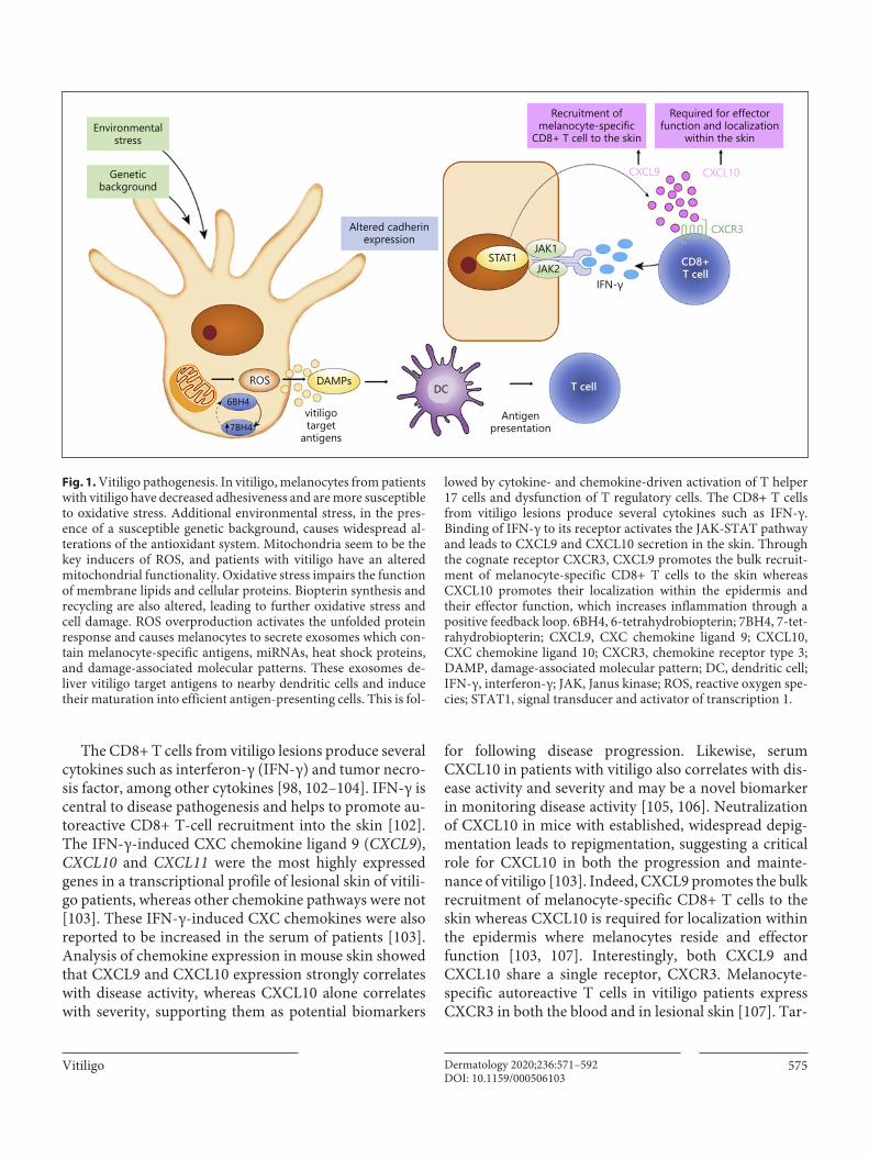
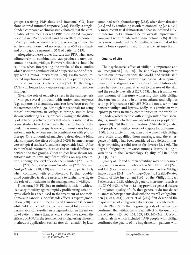
Environmentalstress
Altered cadherinexpression
Recruitment ofmelanocyte-specific
CD8+ T cell to the skin
Required for effectorfunction and localization
within the skin
ROS DAMPs
STAT1JAK1
JAK2IFN-γ
DC T cell
CD8+T cell
CXCR3
CXCL10CXCL9
vitiligotarget
antigens
Antigenpresentation
6BH4
7BH4
Geneticbackground
Fig. 1. Vitiligo pathogenesis. In vitiligo, melanocytes from patients with vitiligo have decreased adhesiveness and are more susceptible to oxidative stress. Additional environmental stress, in the pres-ence of a susceptible genetic background, causes widespread al-terations of the antioxidant system. Mitochondria seem to be the key inducers of ROS, and patients with vitiligo have an altered mitochondrial functionality. Oxidative stress impairs the function of membrane lipids and cellular proteins. Biopterin synthesis and recycling are also altered, leading to further oxidative stress and cell damage. ROS overproduction activates the unfolded protein response and causes melanocytes to secrete exosomes which con-tain melanocyte-specific antigens, miRNAs, heat shock proteins, and damage-associated molecular patterns. These exosomes de-liver vitiligo target antigens to nearby dendritic cells and induce their maturation into efficient antigen-presenting cells. This is fol-
lowed by cytokine- and chemokine-driven activation of T helper 17 cells and dysfunction of T regulatory cells. The CD8+ T cells from vitiligo lesions produce several cytokines such as IFN-γ. Binding of IFN-γ to its receptor activates the JAK-STAT pathway and leads to CXCL9 and CXCL10 secretion in the skin. Through the cognate receptor CXCR3, CXCL9 promotes the bulk recruit-ment of melanocyte-specific CD8+ T cells to the skin whereas CXCL10 promotes their localization within the epidermis and their effector function, which increases inflammation through a positive feedback loop. 6BH4, 6-tetrahydrobiopterin; 7BH4, 7-tet-rahydrobiopterin; CXCL9, CXC chemokine ligand 9; CXCL10, CXC chemokine ligand 10; CXCR3, chemokine receptor type 3; DAMP, damage-associated molecular pattern; DC, dendritic cell; IFN-γ, interferon-γ; JAK, Janus kinase; ROS, reactive oxygen spe-cies; STAT1, signal transducer and activator of transcription 1.
Bergqvist/EzzedineDermatology 2020;236:571–592576DOI: 10.1159/000506103
geting CXCR3 in a mouse model using depleting antibod-ies reduces autoreactive T-cell numbers and reverses the disease [108]. Furthermore, keratinocytes were shown to be the major chemokine producers throughout the course of disease in both mouse model and human patients [109]. Functional studies using a conditional signal trans-ducer and activator of transcription (STAT) 1 knockout mouse revealed that keratinocyte-derived chemokines and IFN-γ signaling drives vitiligo and proper autoreac-tive T-cell homing to the epidermis. In contrast, epider-mal immune cells such as endogenous T cells, Langerhans cells, and γδ T cells are not required [109]. IFN-γ in turn inhibits melanogenesis and directly induces melanocyte apoptosis [110]. Further functional studies in a mouse model found that IFN-γ, the IFN-γ receptor, STAT1, CXCL10 and CXCR3 are critical for the development of hypopigmentation in vitiligo [102, 103, 107, 111].
Many cytokines that bind type I and type II cytokine receptors use the Janus kinase (JAK) and STAT pathway to achieve their effect [112]. Extracellular binding of cyto-kines activates their receptors, inducing apposition of JAKs and self-activation by autophosphorylation. Activated JAKs bind STATs, which undergo JAK-mediated phos-phorylation leading to STAT dimerization, translocation to the nucleus, DNA binding and regulation of gene ex-pression. In vitiligo, IFN-γ-bound receptor complex re-cruits JAK1 and JAK2 kinases, leading to phosphorylation and nuclear translocation of STAT, which in turn tran-scriptionally activates downstream IFN-γ-inducible genes. Lesional skin from patients with vitiligo showed much more intense and diffuse JAK1 expression compared with healthy tissue. Moreover, high JAK1 expression was asso-ciated with short disease duration and a lower percentage of surviving melanocytes [113, 114]. These results thereby support investigation of therapies that disrupt the pathway targeting IFN-γ, the IFN-γ receptor, the downstream sig-naling proteins JAK1, JAK2 and STAT1, and the chemo-kine CXCL10 and its receptor CXCR3 [115–117].
Regulatory T cells (Tregs) are crucial to the develop-ment of self-tolerance. Tregs have been found to be less abundant in vitiligo skin and their functional activity compromised [118–120]. The paucity of Tregs in vitiligo skin is likely crucial for perpetual antimelanocyte reactiv-ity in this progressive and chronic disease. Indeed, Tregs show lower expression of transforming growth factor β1 in active vitiligo patients [119]. The number of Tregs ex-pressing FoxP3, the transcription factor that downregu-lates T-cell activation, is reduced significantly in lesional skin [120]. Furthermore, the expression of homing recep-tor CCL22 was found to be remarkably reduced in vitiligo
skin [121], and conversely, expression of CCL22 can pro-mote Treg skin homing to suppress depigmentation [122].
Functional CD8 tissue-resident memory T cells were found in both stable and active vitiligo, suggesting that those that remain in stable disease could account for the disease reactivation [123].
Figure 1 summarizes the main mechanisms in vitiligo pathogenesis.
Classification
In 2011, an international consensus classified SV sepa-rately from all other forms of vitiligo, and the term vitili-go was defined to designate all forms of NSV [2]. “Mixed vitiligo” in which SV and NSV coexist in one patient, is classified as a subgroup of NSV (Table 1). Distinguishing SV from other types of vitiligo was one of the most im-portant decisions of the consensus, primarily because of its prognostic implications.
NSV includes the acrofacial, mucosal, generalized, universal, mixed and rare variants. Generalized and acro-facial vitiligo are the most common subtypes.• Generalized vitiligo is characterized by bilateral, often
symmetrical, depigmented macules or patches occur-ring in a random distribution over the entire body sur-face. It often affects areas that tend to experience pres-
Table 1. Classification of vitiligo (adapted from Ezzedine et al. [2])
Type of vitiligo Subtypes
NSV Focal1MucosalAcrofacialGeneralizedUniversalRare variants of vitiligo (leukoderma punctata, hypochromic vitiligo, follicular vitiligo)
SV Focal1UnisegmentalBi- or multisegmental
Mixed (NSV + SV) Concomitant occurrence of SV and NSVAccording to severity of SV
Unclassified Focal at onset, multifocal asymmetrical nonsegmental, mucosal (one site),
1 Can evolve into segmental (SV) or nonsegmental vitiligo (NSV).
Vitiligo 577Dermatology 2020;236:571–592DOI: 10.1159/000506103
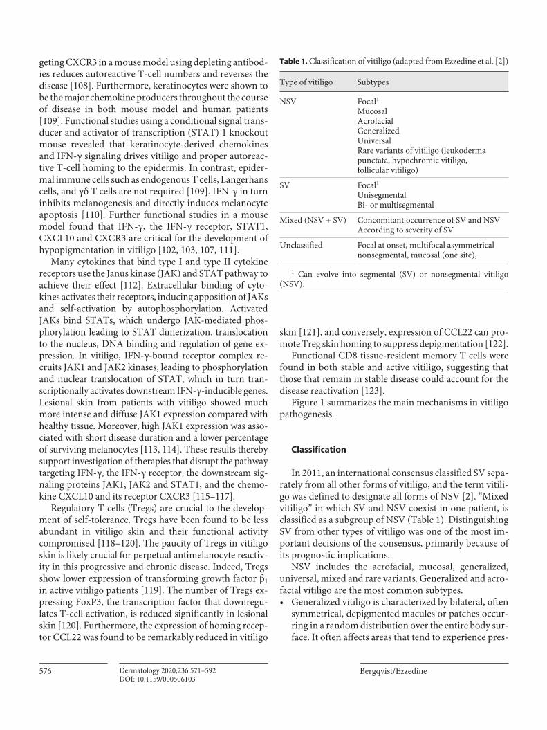
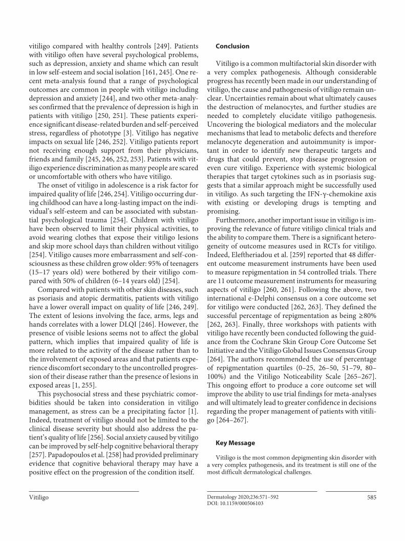
sure, friction and/or trauma. It may begin in childhood or early adulthood (Fig. 2).
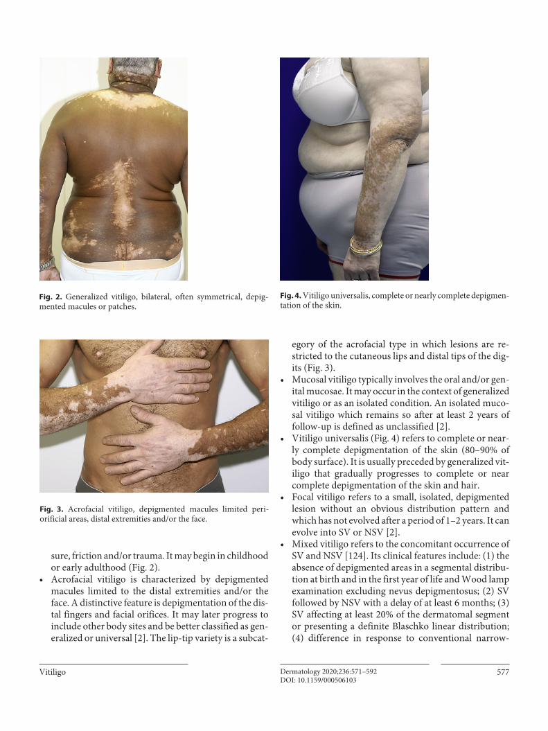
• Acrofacial vitiligo is characterized by depigmented macules limited to the distal extremities and/or the face. A distinctive feature is depigmentation of the dis-tal fingers and facial orifices. It may later progress to include other body sites and be better classified as gen-eralized or universal [2]. The lip-tip variety is a subcat-
egory of the acrofacial type in which lesions are re-stricted to the cutaneous lips and distal tips of the dig-its (Fig. 3).
• Mucosal vitiligo typically involves the oral and/or gen-ital mucosae. It may occur in the context of generalized vitiligo or as an isolated condition. An isolated muco-sal vitiligo which remains so after at least 2 years of follow-up is defined as unclassified [2].
• Vitiligo universalis (Fig. 4) refers to complete or near-ly complete depigmentation of the skin (80–90% of body surface). It is usually preceded by generalized vit-iligo that gradually progresses to complete or near complete depigmentation of the skin and hair.
• Focal vitiligo refers to a small, isolated, depigmented lesion without an obvious distribution pattern and which has not evolved after a period of 1–2 years. It can evolve into SV or NSV [2].
• Mixed vitiligo refers to the concomitant occurrence of SV and NSV [124]. Its clinical features include: (1) the absence of depigmented areas in a segmental distribu-tion at birth and in the first year of life and Wood lamp examination excluding nevus depigmentosus; (2) SV followed by NSV with a delay of at least 6 months; (3) SV affecting at least 20% of the dermatomal segment or presenting a definite Blaschko linear distribution; (4) difference in response to conventional narrow-
Fig. 2. Generalized vitiligo, bilateral, often symmetrical, depig-mented macules or patches.
Fig. 3. Acrofacial vitiligo, depigmented macules limited peri- orificial areas, distal extremities and/or the face.
Fig. 4. Vitiligo universalis, complete or nearly complete depigmen-tation of the skin.
Bergqvist/EzzedineDermatology 2020;236:571–592578DOI: 10.1159/000506103
band ultraviolet B (NB-UVB) treatment between SV (poor response) and NSV (good response). Leuko-trichia and halo nevi at onset may be risk factors for developing MV in patients with SV [125]. The co- occurrence of SV and NSV in a same patient has been viewed as a superimposed segmental manifestation of a generalized polygenic disorder, in which segmental involvement precedes disease generalization and is more resistant to therapy [126, 127].In a study of latent class analyses, two phenotypes of
NSV have been differentiated: the first consists of early onset of disease (before 12 years of age) and is often asso-ciated with halo nevi and a familial background of prema-ture hair graying; the second is of late onset and is most often characterized by an acrofacial distribution [23, 128].
Several conditions are difficult to classify into the two classical forms of NSV and SV.• “Punctate vitiligo” refers to sharply demarcated depig-
mented punctiform 1- to 1.5-mm macules involving any area of the body [129]. If these lesions do not co-exist with classical vitiligo macules, they should be referred to as “leukoderma punctata.”
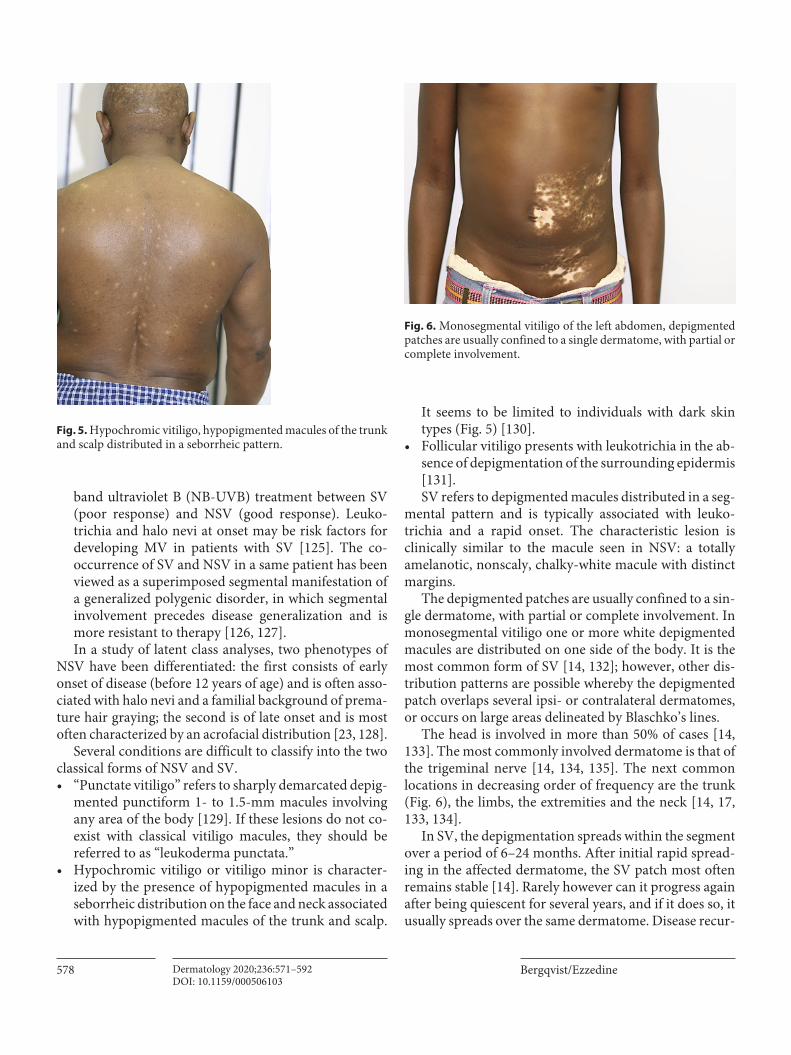
• Hypochromic vitiligo or vitiligo minor is character-ized by the presence of hypopigmented macules in a seborrheic distribution on the face and neck associated with hypopigmented macules of the trunk and scalp.
It seems to be limited to individuals with dark skin types (Fig. 5) [130].
• Follicular vitiligo presents with leukotrichia in the ab-sence of depigmentation of the surrounding epidermis [131].SV refers to depigmented macules distributed in a seg-
mental pattern and is typically associated with leuko-trichia and a rapid onset. The characteristic lesion is clinically similar to the macule seen in NSV: a totally amelanotic, nonscaly, chalky-white macule with distinct margins.
The depigmented patches are usually confined to a sin-gle dermatome, with partial or complete involvement. In monosegmental vitiligo one or more white depigmented macules are distributed on one side of the body. It is the most common form of SV [14, 132]; however, other dis-tribution patterns are possible whereby the depigmented patch overlaps several ipsi- or contralateral dermatomes, or occurs on large areas delineated by Blaschko’s lines.
The head is involved in more than 50% of cases [14, 133]. The most commonly involved dermatome is that of the trigeminal nerve [14, 134, 135]. The next common locations in decreasing order of frequency are the trunk (Fig. 6), the limbs, the extremities and the neck [14, 17, 133, 134].
In SV, the depigmentation spreads within the segment over a period of 6–24 months. After initial rapid spread-ing in the affected dermatome, the SV patch most often remains stable [14]. Rarely however can it progress again after being quiescent for several years, and if it does so, it usually spreads over the same dermatome. Disease recur-
Fig. 5. Hypochromic vitiligo, hypopigmented macules of the trunk and scalp distributed in a seborrheic pattern.
Fig. 6. Monosegmental vitiligo of the left abdomen, depigmented patches are usually confined to a single dermatome, with partial or complete involvement.
Vitiligo 579Dermatology 2020;236:571–592DOI: 10.1159/000506103
rence can occur after years of stability [136]. However, in very rare cases, lesions may become generalized, and be-come part of mixed vitiligo [124, 136].
Diagnosis
The diagnosis of vitiligo is generally straightforward, made clinically based upon the finding of acquired, amel-anotic, nonscaly, chalky-white macules with distinct margins in a typical distribution: periorificial, lips and tips of distal extremities, penis, segmental and areas of friction [6, 8, 137].
The diagnosis of vitiligo does not usually require con-firmatory laboratory tests. A skin biopsy or other tests are not necessary except to exclude other disorders [6, 138, 139]. The absence of melanocytes in a lesion can be as-sessed noninvasively by in vivo confocal microscopy or by a skin biopsy. The histology of the center of a vitiligo lesion reveals complete loss of melanin pigment in the epidermis and absence of melanocytes. Occasional lym-phocytes may be noted at the advancing border of the lesions [34, 140].
The diagnosis of vitiligo may be facilitated by the use of a Wood’s lamp, a hand-held ultraviolet (UV) irradia-tion device that emits UVA [141]. It helps identify focal melanocyte loss and detect areas of depigmentation that may not be visible to the naked eye, particularly in pale skin [142]. Under the Wood’s light, the vitiligo lesions emit a bright blue-white fluorescence and appear sharply demarcated.
Dermoscopy can be used to differentiate vitiligo from other depigmenting disorders. Vitiligo typically shows residual perifollicular pigmentation and telangiectasia, which are absent in other hypopigmentation disorders [143]. More importantly, it can be useful in assessing dis-ease activity in vitiligo and the stage of evolution: progres-sive lesions display perifollicular pigmentation, whereas stable or remitting lesions display perifollicular depig-mentation [144].
The differential diagnosis of vitiligo is broad (Table 2). Many common and uncommon conditions present with areas of depigmentation that may mimic vitiligo. It is im-portant to differentiate vitiligo from melanoma-associat-ed leukoderma and to prevent its misdiagnosis as vitiligo especially that it may precede melanoma detection. Al-though clinically similar, antibodies against melanoma antigen recognized by T cells 1 (MART1) in melanoma-associated depigmentation can help differentiate it from vitiligo [145]. Nevus depigmentosus is segmental hy-
popigmentation usually present at birth or detectable in the first year of life. It is stable although it may enlarge in proportion to the child’s growth. It is a common differ-ential diagnosis of SV, but nevi usually contain a normal number of melanocytes with reduced melanin produc-tion [146]. Under Wood’s lamp examination, the contrast between lesional and normal skin is less striking than in vitiligo [147].
Table 2. Differential diagnosis of vitiligo
Chemically-induced leukoderma (occupational)Phenols and other derivatives
Topical or systemic drug-induced depigmentationGenetic syndromesHypomelanosis of ItoPiebaldismTuberous sclerosisVogt-Koyanagi-Harada syndromeWaardenburg syndromeHermanski-Pudlak syndromeMenke’s syndromeZiprkowski-Margolis syndromeGriscelli’s syndrome
Postinflammatory hypopigmentationPityriasis albaAtopic dermatitis/allergic contact dermatitisPsoriasisLichen planusToxic drug reactionsPosttraumatic hypopigmentation (scar)Phototherapy- and radiotherapy-induced
Neoplasm-related hypomelanosesMelanoma-associated leukodermaMycosis fungoidesInfection-related hypomelanosesLeprosyPityriasis versicolorLeishmaniasisOnchocerciasisTreponematoses (pinta and syphilis)
IdiopathicIdiopathic guttate hypomelanosisProgressive (or acquired) macular hypomelanosis
CongenitalNevus anemicusNevus depigmentosus
OthersLichen sclerosus et atrophicusMelasma (caused by contrast between lighter and darker skin)
Bergqvist/EzzedineDermatology 2020;236:571–592580DOI: 10.1159/000506103
Assessment
The management of a patient with vitiligo requires time for a careful initial assessment. The evaluation of the patient with vitiligo entails a detailed history and a com-plete skin examination to assess disease severity and indi-vidual prognostic factors. An assessment form created by the Vitiligo European Task Force summarizes the per-sonal and family history elements and the clinical exami-nation items which may be useful for evaluation [141]. Patients should routinely be asked about family history of vitiligo and premature hair graying and about family or personal history of thyroid disease or other autoimmune diseases [148]. Skin phototype, disease duration, extent, activity, rate of progression or spread of lesions, presence of Koebner’s phenomenon, presence of halo nevi, previ-ous treatments including their type, duration and effec-tiveness, previous episodes of repigmentation, occupa-tional history/exposure to chemicals and effects of disease on the quality of life should all be assessed.
Some areas of the body are more susceptible to Koeb-ner’s phenomenon and are related to daily life activities such as hygiene or clothing and occupation [8]. Assessing for the presence of Koebner’s phenomenon (vitiligo fol-lowing mechanical trauma) can prove to be useful in the prevention of vitiligo [8, 149]. A scoring evaluating the probability of Koebner’s phenomenon, the K-VSCOR, has been developed and validated [150]. Patients with high scores should be counseled about mechanical stress avoidance.
Many studies have demonstrated the associations of vitiligo with thyroid disorders and other associated auto-immune diseases, such as alopecia areata, rheumatoid ar-thritis, adult-onset diabetes mellitus, Addison’s disease, pernicious anemia, systemic lupus erythematosus, pso-riasis and atopic background [9, 128, 151, 152].
Because of the increased risk of autoimmune thyroid disease in NSV, especially Hashimoto’s thyroiditis [153], antibodies to thyroid peroxidase should be screened initially, and the thyrotropin levels should be measured regularly, especially in patients with antibod-ies to thyroid peroxidase at the initial screening. The susceptibility to autoimmune diseases in patients with vitiligo varies with ethnic background and family his-tory of autoimmune diseases [9, 154]. The presence of signs or symptoms of organ-specific autoimmune dis-eases should prompt an appropriate investigation and referral to specialists [155].
The most extensively characterized clinical markers of active, progressive disease include: Koebner’s phenome-
non, trichrome lesions, inflammatory lesions and confet-ti-like depigmentation [27, 156–159].
Finally, an overall assessment of the psychological fea-tures and quality of life is warranted as the patient’s per-sonality and perceived severity of vitiligo are predictors of quality of life impairment [160, 161]. A vitiligo-specif-ic quality-of-life instrument has been developed and val-idated [162]. All patients with vitiligo should be offered psychological support and counseling [142].
Management
The treatment of vitiligo is still one of the most difficult dermatological challenges. An important step in the man-agement of vitiligo is to first acknowledge that it is not merely a cosmetic disease and that there are safe and ef-fective treatments available [163]. These treatments in-clude phototherapy, topical and systemic immunosup-pressants, and surgical techniques, which together may help in halting the disease, stabilizing depigmented le-sions and stimulating repigmentation [164, 165].
Choice of treatment depends on several factors includ-ing: the subtype of the disease, the extent, distribution and activity of disease as well as the patient’s age, photo-type, effect on quality of life and motivation for treat-ment. The face, neck, trunk and mid-extremities respond best to therapy, while the lips and distal extremities are more resistant [166]. Repigmentation appears initially in a perifollicular pattern or at the periphery of the lesions. Treatment for at least 2–3 months is needed to determine efficacy of treatment. UV light-based therapy is the most common treatment for vitiligo and, when combined with an additional therapy, is associated with an improved outcome [165].
Management requires a personalized therapeutic ap-proach whereby patients should always be consulted, as most of the therapeutic options are time consuming and require long-term follow-up. Advice on cosmetic camou-flage by a cosmetician or a specialized nurse should be of-fered and can be beneficial for patients with vitiligo affect-ing exposed areas. These include foundation-based cosmet-ics and self-tanning products containing dihydroxyacetone which provides lasting color for up to several days.
Several guidelines have been published for the man-agement of vitiligo [142, 167–169]. In 2008, the British Association of Dermatologists published user-friendly clinical guidelines for the diagnosis and management of vitiligo [142] which were established based on the first Cochrane review and expert consensus on vitiligo reflect-
Vitiligo 581Dermatology 2020;236:571–592DOI: 10.1159/000506103
ing patient choice and clinical expertise [169, 170]. The Cochrane reviews of 2010 and 2015 underscored the ab-sence of cure for vitiligo and the inability of current treat-ment options to restrict the spread of the disease in a lasting way [170–172]. However, most randomized con-trolled trials (RCTs) included in the review had had fewer than 50 participants. They concluded that due to the heterogeneity in the design of trials and the small numbers of participants, no firm clinical recommenda-tions could be made.
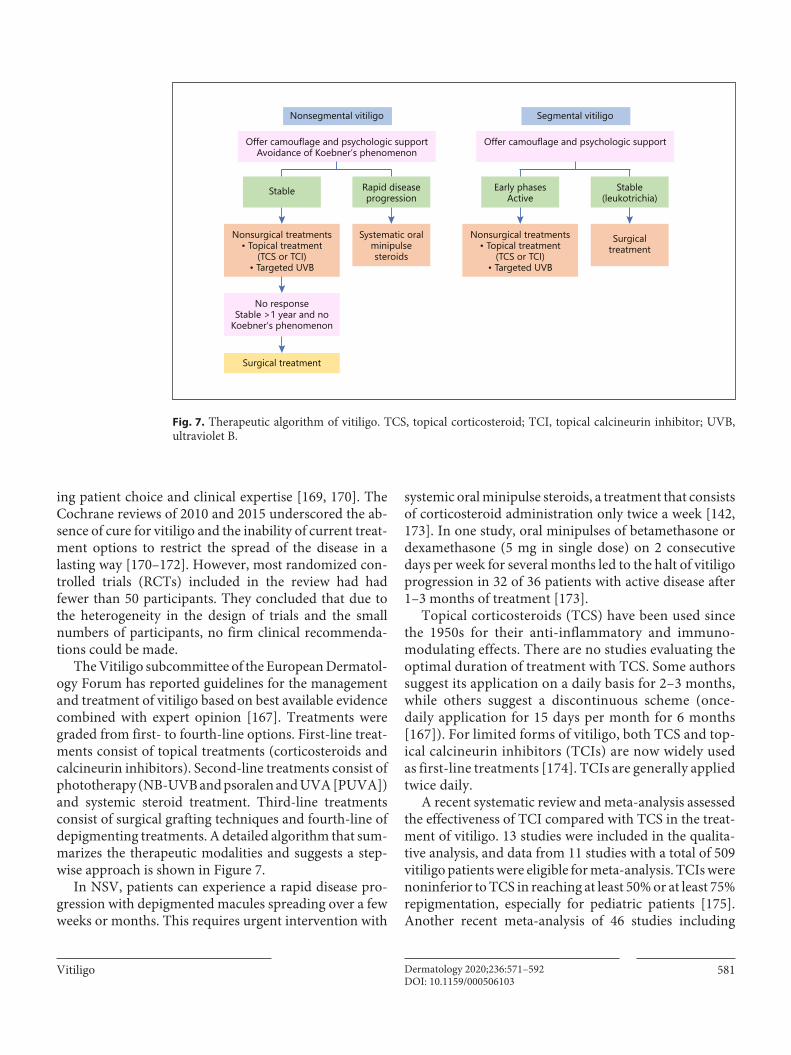
The Vitiligo subcommittee of the European Dermatol-ogy Forum has reported guidelines for the management and treatment of vitiligo based on best available evidence combined with expert opinion [167]. Treatments were graded from first- to fourth-line options. First-line treat-ments consist of topical treatments (corticosteroids and calcineurin inhibitors). Second-line treatments consist of phototherapy (NB-UVB and psoralen and UVA [PUVA]) and systemic steroid treatment. Third-line treatments consist of surgical grafting techniques and fourth-line of depigmenting treatments. A detailed algorithm that sum-marizes the therapeutic modalities and suggests a step-wise approach is shown in Figure 7.
In NSV, patients can experience a rapid disease pro-gression with depigmented macules spreading over a few weeks or months. This requires urgent intervention with
systemic oral minipulse steroids, a treatment that consists of corticosteroid administration only twice a week [142, 173]. In one study, oral minipulses of betamethasone or dexamethasone (5 mg in single dose) on 2 consecutive days per week for several months led to the halt of vitiligo progression in 32 of 36 patients with active disease after 1–3 months of treatment [173].
Topical corticosteroids (TCS) have been used since the 1950s for their anti-inflammatory and immuno-modulating effects. There are no studies evaluating the optimal duration of treatment with TCS. Some authors suggest its application on a daily basis for 2–3 months, while others suggest a discontinuous scheme (once- daily application for 15 days per month for 6 months [167]). For limited forms of vitiligo, both TCS and top-ical calcineurin inhibitors (TCIs) are now widely used as first-line treatments [174]. TCIs are generally applied twice daily.
A recent systematic review and meta-analysis assessed the effectiveness of TCI compared with TCS in the treat-ment of vitiligo. 13 studies were included in the qualita-tive analysis, and data from 11 studies with a total of 509 vitiligo patients were eligible for meta-analysis. TCIs were noninferior to TCS in reaching at least 50% or at least 75% repigmentation, especially for pediatric patients [175]. Another recent meta-analysis of 46 studies including
Offer camouflage and psychologic supportAvoidance of Koebner’s phenomenon
Rapid diseaseprogression
Nonsegmental vitiligo
Stable
Systematic oralminipulsesteroids
Nonsurgical treatments• Topical treatment
(TCS or TCI)• Targeted UVB
No responseStable >1 year and no
Koebner’s phenomenon
Surgical treatment
Offer camouflage and psychologic support
Stable(leukotrichia)
Segmental vitiligo
Early phasesActive
Surgicaltreatment
Nonsurgical treatments• Topical treatment
(TCS or TCI)• Targeted UVB
Fig. 7. Therapeutic algorithm of vitiligo. TCS, topical corticosteroid; TCI, topical calcineurin inhibitor; UVB, ultraviolet B.
Bergqvist/EzzedineDermatology 2020;236:571–592582DOI: 10.1159/000506103
1,499 patients showed that TCI monotherapy appears to have significant therapeutic effects on vitiligo and pro-duced at least mild response in 55.0% of the patients, at least moderate response in 38.5% and a marked response in 18.1% after a median treatment duration of 3 months [176]. The treatment responses of TCIs combined with phototherapy were higher than those of TCI monothera-py and those of phototherapy alone, which supports the synergistic effects of this combination therapy. TCI monotherapy could be useful for the treatment of face and neck lesions, particularly in children, when photo-therapy is not available. Another meta-analysis on 7 RCTs involving 240 patients suggested that adding TCI on NB-UVB does not yield significantly superior outcomes com-pared to NB-UVB monotherapy for treatment of vitiligo; except for the face and neck where addition of TCI to NB-UVB may increase treatment outcomes [177].
The Vitiligo Working Group has recently published a unified set of recommendations for NB-UVB photo-therapy treatment of vitiligo based on prescribing prac-tices of phototherapy experts from around the world [178]. These included the dosing protocol (initiate dose at 200 mJ/cm2 regardless of constitutive skin type, then increase by 10–20% per treatment), the frequency of administration (optimal 3 times per week), the maximal acceptable doses (1,500 mJ/cm2 for the face, 3,000 mJ/cm2 for the body), the course and the follow-up. They reported that the minimum number of doses needed to determine lack of response was 48 exposures, and that because of the existence of slow responders, ≥72 expo-sures may be needed to determine lack of response to phototherapy [178].
Due to its good safety profile in both children and adults and lack of systemic toxicity, NB-UVB has emerged as the initial treatment of choice for patients with vitiligo involving > 10% of the body surface area. A 2017 meta-analysis of 35 randomized and nonrandomized studies including 1,428 patients compared the repigmentation rates of NB-UVB and PUVA by treatment duration. For NB-UVB, a ≥75% repigmentation was achieved by 19 and 36% of patients at 6 and 12 months of treatment, re-spectively, compared to 9 and 14% with PUVA. This con-firmed the superiority of NB-UVB over PUVA and sug-gested that phototherapy should be continued for at least 12 months to achieve a maximal response [179].
Targeted phototherapy using 308-nm monochromatic excimer lamps or lasers is useful for the treatment of lo-calized vitiligo. These devices deliver high-intensity light only to the affected areas while avoiding exposure of the healthy skin and lowering the cumulative UVB dose.
A systematic review of 6 randomized trials (411 pa-tients with 764 lesions) found that excimer lamps and ex-cimer lasers are equally effective as NB-UVB in inducing ≥50% and ≥75% repigmentation [180]. Although more frequent weekly treatments lead to more repigmentation, the ultimate repigmentation and final result seems to de-pend entirely on the overall number of treatment sessions rather than their frequency [181]. As with NB-UVB, TCIs can work synergistically with targeted phototherapy [182, 183]. A meta-analysis which included 8 RCTs comprising a total of 425 patches/patients found that TCIs in con-junction with excimer light/laser are more effective com-pared with excimer light/laser monotherapy [184].
Surgical methods can be offered as a therapeutic op-tion to patients with SV and those with NSV with stable disease after at least a year of documented nonresponse to medical interventions and absence of Koebner’s phe-nomenon. A minigraft test to assess stability, spread of pigment at the recipient site and no koebnerization at the donor site after 2–3 months can also assist in patient se-lection. The purpose of the transplantation is to transfer to the vitiliginous skin a reservoir of healthy melanocytes for proliferation and migration into areas of depigmenta-tion [185]. The surgical techniques that are mentioned in the European guidelines [167] include tissue grafts (full-thickness punch, split-thickness and suction blister grafts) and cellular grafts (autologous melanocyte cul-tures and noncultured epidermal cellular grafts). Other techniques include cultured epidermal suspensions [186, 187] and hair follicle transplantation [188–190]. Tissue grafts use unprocessed pigmented epidermis and dermis, which are transplanted to depigmented areas; they are ideal for treating smaller areas [185]. In contrast, cellular transplants involve more complex processing of the grafts before surgery.
An evidence-based review concluded that split-thick-ness grafting and blister grafts are the most effective and safe techniques [185]. An old systematic review of ran-domized trials and observational studies of autologous transplantation methods for vitiligo concluded that split-thickness and epidermal blister grafting were the most effective and safest techniques [191]. Both treatment groups achieved success rates of 90% repigmentation. They could not draw conclusions about the effectiveness of culturing techniques because only a small number of patients have been studied. The benefits of transplanta-tion of autologous melanocyte cultures and epidermal suspensions have been reported in some studies [186, 192, 193]. In an RCT comparing autologous noncultured epidermal cell suspension with suction blister grafts in 41
Vitiligo 583Dermatology 2020;236:571–592DOI: 10.1159/000506103
patients, both treatment groups reached a repigmenta-tion of ≥75% in over 85% of lesions [193]. However, more lesions in the noncultured epidermal cell suspension group (70%) achieved a 90–100% repigmentation com-pared with those in the suction blister group (27%) [193]. Important advantages of cellular grafting are the possibil-ity of treating large areas and the better cosmetic results than with tissue grafts [194, 195]. Cellular grafts seem to have less frequently associated adverse events than with punch grafting, followed by split-thickness grafting [196].
Depigmenting treatment of residual areas of pigmen-tation should only be considered in select cases such as: widespread, refractory, and disfiguring vitiligo, or highly visible recalcitrant facial or hand vitiligo [8]. Monobenzyl ether of hydroquinone (monobenzone) has been used as a depigmenting agent for patients with extensive vitiligo since the 1950s [197]. Other skin-bleaching methods in-clude laser treatment (e.g., 755-nm Q-switched alexan-drite or 694-nm Q-switched ruby) [198–200] and cryo-therapy [75].
Reliable data regarding the treatment of SV are limited since most studies do not differentiate between these types of vitiligo. SV was previously considered to be resis-tant to treatment. However, recent studies have been re-porting promising results; especially during the early stage. Within the first 6 months, patients should be of-fered potent TCS or topical immune modulators com-bined with NB-UVB or targeted excimer lamp or laser. Oral steroid minipulse therapy is another option if the lesion is still in its active phase. In contrast, if these med-ical therapies fail, or at a later stage of the disease, surgery should be offered. Overall, stable SV is a good indication for surgical grafting, especially as the presence of leuko-trichia in SV makes it more resistant to standard medical therapies.
Emerging TherapiesAfamelanotide, a potent and longer-lasting synthetic
analog of α-melanocyte-stimulating hormone, has been shown to be synergistic with NB-UVB in promoting re-pigmentation [201, 202]. Prostaglandin E2 controls the proliferation of melanocytes by means of stimulant and immunomodulatory effects. In one study of 56 consecu-tive patients with stable and limited vitiligo, repigmenta-tion, treatment with prostaglandin E2 0.25 mg/g gel twice daily for 6 months led to repigmentation in 40 patients; the response was excellent in 22 patients, and the repig-mentation was complete in 8 [203]. Bimatoprost, a syn-thetic analog of prostaglandin F2α approved for the topi-cal treatment of glaucoma and hypotrichosis of the
eyelashes, was shown in an RCT to provide greater repig-mentation than treatment with mometasone [204].
Besides, JAK inhibitors have shown promise in the treatment of vitiligo [116, 205]. Ruxolitinib is a JAK1 and JAK2 inhibitor. In a phase 2, proof-of-concept trial, topi-cal ruxolitinib 1.5% cream was applied twice daily to 11 adult patients with vitiligo involving at least 1% of the body surface area for 20 weeks [206]. Eight of 11 patients achieved a response with a mean improvement of the Vit-iligo Area Scoring Index of 23%. The best response was observed in patients with facial vitiligo. Five patients who completed the trial were then followed up at 6 months after treatment discontinuation, and all of them main-tained response, with a maximum duration of > 40 weeks [205].
Alternative Treatment OptionsThere are limited data regarding the use of systemic
immunosuppressants other than corticosteroids in the treatment of vitiligo. A randomized comparative study performed on 52 patients with vitiligo showed that meth-otrexate is equally effective as oral minipulse therapy with betamethasone or dexamethasone in controlling the dis-ease activity, suggesting that methotrexate could be used in patients with active vitiligo if corticosteroids are con-traindicated [207]. Twice daily oral cyclophosphamide (50 mg) was shown to cause repigmentation in 29 pa-tients, including the difficult-to-treat areas such as acral sites; however, significant side effects were reported [208]. Although some authors have suggested that anti-tumor necrosis factor-α can stabilize the disease in progressive vitiligo [209], many studies have demonstrated that these agents do not improve the disorder, and that they may even cause initiation and worsening of the disease [210–214].
Platelet-rich plasma (PRP) is an autologous prepara-tion of platelets in concentrated plasma which contains various growth factors. It is hypothesized that these growth factors promote melanocyte stimulation [215]. Earlier studies showed conflicting results. Lim et al. [216] reported that PRP alone is not effective in treating vitiligo. However, Ibrahim et al. [217] carried out a trial compar-ing the combination of PRP with NB-UVB and found better results than treatment with NB-UVB alone. Seven-ty-five percent of patients in the NB-UVB and PRP group had more than 50% repigmentation compared to none of the patients in the NB-UVB group. More recently, a pro-spective, open-label, randomized trial has shown that combining fractional CO2 laser with PRP injection led to at least 50% repigmentation in all of the patients, whereas
Bergqvist/EzzedineDermatology 2020;236:571–592584DOI: 10.1159/000506103
groups receiving PRP alone and fractional CO2 laser alone showed minimal response [218]. Finally, a single-blinded comparative clinical study showed that the com-bination of excimer laser with PRP injection led to a good response in 50% of patients and an excellent response in 35% of patients, whereas the group receiving excimer la-ser treatment alone had no response in 65% of patients and only a good response in 35% of patients [219].
Altogether, these studies indicate that PRP, when used adjunctively in combination, can produce better out-comes in treating vitiligo. However, clinicians should be cautious when interpreting the results of these studies which used the combination of a superior mode of ther-apy with a minor intervention [220]. Furthermore, re-peated injections at short intervals are a painful proce-dure and can induce koebnerization [221]. Further larger RCTs with longer follow-up are required to confirm these findings.
Given the role of oxidative stress in the pathogenesis of vitiligo, several products with antioxidant enzymes (e.g., superoxide dismutase, catalase) have been used for the treatment of vitiligo. Although the rationale for using topical antioxidants in vitiligo is strong, studies have shown conflicting results, probably owing to the difficul-ty of delivering active antioxidants directly into the skin. Some studies have looked into the use of topical anti-oxidants as monotherapy; however, in most cases topical antioxidants have been used in combination with photo-therapy. One randomized, matched-paired, double-blind trial compared the effect of topical 0.05% betamethasone versus topical catalase/dismutase superoxide [222]. After 10 months of treatment, there was no statistical difference between the two groups. Other studies have shown oral antioxidants to have significant effects on repigmenta-tion, although the level of evidence is limited [223]. Vita-min E [224, 225], Polypodium leucotomos [226, 227] and Ginkgo biloba [228, 229] seem to be useful, particularly when combined with phototherapy. Further double-blind controlled trials are necessary to further investigate the role of antioxidants in the management of vitiligo.
Fluorouracil (5-FU) has an antimitotic activity with se-lective cytotoxicity against rapidly proliferating keratino-cytes which has been used in the treatment of nonmela-noma skin cancers. One of its side effects is hyperpigmen-tation [230]. Back in 1985, Tsuji and Hamada [231] found, while 5-FU alone had no effect, applying it following epi-dermal abrasion resulted in repigmentation in the major-ity of patients. Since then, several studies have shown the efficacy of 5-FU in the treatment of vitiligo using different methods of application, such as after skin ablation by laser
combined with phototherapy [232], after dermabrasion [233] and by combining it with microneedling [234, 235]. A more recent trial demonstrated that in localized NSV, intradermal 5-FU showed better overall improvement compared with intradermal triamcinolone [236]. Its ef-fects were maintained for 6 months, whereas that of tri-amcinolone stopped at 1 month after the last injection.
Quality of Life
The psychosocial effect of vitiligo is important and well recognized [1, 8, 160]. The skin plays an important role in our interaction with the world, and visible skin disorders can limit healthy psychosocial development owing to the stigma these disorders create. Historically, there has been a stigma attached to diseases of the skin and the people they affect [237, 238]. There is an impor-tant amount of literature witnessing that with vitiligo since ancient times and in different cultural and religious settings. Hippocrates (460–355 BC) did not discriminate between vitiligo and leprosy. Sadly, this confusion with leprosy persists in many communities in the world up until today, where people with vitiligo suffer from social stigma, similarly to the same age-old way as people with leprosy [8]. Old Buddhist literature (624–544 BC) stated that people with vitiligo were not eligible for ordainment [160]. Since ancient times, men and women with vitiligo were often disqualified from marriage, and the emer-gence of vitiligo has been considered as a defect in mar-riage, providing a solid reason for divorce [8, 160]. The degree of stigmatization varies among cultures, leading to variations in the Dermatology Quality of Life Index (DLQI) [239].
Quality of life and burden of vitiligo may be measured by generic assessment tools such as Short Form-12 [240] and DLQI or by more specific tools such as the Vitiligo Impact Scale [241], the Vitiligo-Specific Health-Related Quality of Life Instrument [162] or the Vitiligo Impact Patient scale [242]. Although generic instruments such as the DLQI or Short Form-12 may provide a general picture of impaired quality of life, they generally do not detect nuances in how patients deal with the overall vitiligo bur-den [3, 243, 244]. Porter et al. [245] first described the major impact of vitiligo on patients’ quality of life back in the late 1970s. Since then, a growing body of evidence has confirmed that vitiligo has a major effect on the quality of life of patients [3, 160, 161, 169, 243, 246–248]. A recent meta-analysis which included 1,799 people with vitiligo confirmed the quality of life impairment in patients with
Vitiligo 585Dermatology 2020;236:571–592DOI: 10.1159/000506103
vitiligo compared with healthy controls [249]. Patients with vitiligo often have several psychological problems, such as depression, anxiety and shame which can result in low self-esteem and social isolation [161, 245]. One re-cent meta-analysis found that a range of psychological outcomes are common in people with vitiligo including depression and anxiety [244], and two other meta-analy-ses confirmed that the prevalence of depression is high in patients with vitiligo [250, 251]. These patients experi-ence significant disease-related burden and self-perceived stress, regardless of phototype [3]. Vitiligo has negative impacts on sexual life [246, 252]. Vitiligo patients report not receiving enough support from their physicians, friends and family [245, 246, 252, 253]. Patients with vit-iligo experience discrimination as many people are scared or uncomfortable with others who have vitiligo.
The onset of vitiligo in adolescence is a risk factor for impaired quality of life [246, 254]. Vitiligo occurring dur-ing childhood can have a long-lasting impact on the indi-vidual’s self-esteem and can be associated with substan-tial psychological trauma [254]. Children with vitiligo have been observed to limit their physical activities, to avoid wearing clothes that expose their vitiligo lesions and skip more school days than children without vitiligo [254]. Vitiligo causes more embarrassment and self-con-sciousness as these children grow older: 95% of teenagers (15–17 years old) were bothered by their vitiligo com-pared with 50% of children (6–14 years old) [254].
Compared with patients with other skin diseases, such as psoriasis and atopic dermatitis, patients with vitiligo have a lower overall impact on quality of life [246, 249]. The extent of lesions involving the face, arms, legs and hands correlates with a lower DLQI [246]. However, the presence of visible lesions seems not to affect the global pattern, which implies that impaired quality of life is more related to the activity of the disease rather than to the involvement of exposed areas and that patients expe-rience discomfort secondary to the uncontrolled progres-sion of their disease rather than the presence of lesions in exposed areas [1, 255].
This psychosocial stress and these psychiatric comor-bidities should be taken into consideration in vitiligo management, as stress can be a precipitating factor [1]. Indeed, treatment of vitiligo should not be limited to the clinical disease severity but should also address the pa-tient’s quality of life [256]. Social anxiety caused by vitiligo can be improved by self-help cognitive behavioral therapy [257]. Papadopoulos et al. [258] had provided preliminary evidence that cognitive behavioral therapy may have a positive effect on the progression of the condition itself.
Conclusion
Vitiligo is a common multifactorial skin disorder with a very complex pathogenesis. Although considerable progress has recently been made in our understanding of vitiligo, the cause and pathogenesis of vitiligo remain un-clear. Uncertainties remain about what ultimately causes the destruction of melanocytes, and further studies are needed to completely elucidate vitiligo pathogenesis. Uncovering the biological mediators and the molecular mechanisms that lead to metabolic defects and therefore melanocyte degeneration and autoimmunity is impor-tant in order to identify new therapeutic targets and drugs that could prevent, stop disease progression or even cure vitiligo. Experience with systemic biological therapies that target cytokines such as in psoriasis sug-gests that a similar approach might be successfully used in vitiligo. As such targeting the IFN-γ-chemokine axis with existing or developing drugs is tempting and promising.
Furthermore, another important issue in vitiligo is im-proving the relevance of future vitiligo clinical trials and the ability to compare them. There is a significant hetero-geneity of outcome measures used in RCTs for vitiligo. Indeed, Eleftheriadou et al. [259] reported that 48 differ-ent outcome measurement instruments have been used to measure repigmentation in 54 controlled trials. There are 11 outcome measurement instruments for measuring aspects of vitiligo [260, 261]. Following the above, two international e-Delphi consensus on a core outcome set for vitiligo were conducted [262, 263]. They defined the successful percentage of repigmentation as being ≥80% [262, 263]. Finally, three workshops with patients with vitiligo have recently been conducted following the guid-ance from the Cochrane Skin Group Core Outcome Set Initiative and the Vitiligo Global Issues Consensus Group [264]. The authors recommended the use of percentage of repigmentation quartiles (0–25, 26–50, 51–79, 80–100%) and the Vitiligo Noticeability Scale [265–267]. This ongoing effort to produce a core outcome set will improve the ability to use trial findings for meta-analyses and will ultimately lead to greater confidence in decisions regarding the proper management of patients with vitili-go [264–267].
Key Message
Vitiligo is the most common depigmenting skin disorder with a very complex pathogenesis, and its treatment is still one of the most difficult dermatological challenges.
Bergqvist/EzzedineDermatology 2020;236:571–592586DOI: 10.1159/000506103
Disclosure Statement
The authors have no conflicts of interest to declare.
Funding Sources
This paper did not receive any funding.
Author Contributions
Christina Bergqvist wrote the manuscript. Khaled Ezzedine su-pervised the work and revised the manuscript for critical revision for important intellectual content.
References
1 Picardo M, Dell’Anna ML, Ezzedine K, Hamzavi I, Harris JE, Parsad D, et al. Vitiligo. Nat Rev Dis Primers. 2015 Jun; 1(1): 15011.
2 Ezzedine K, Lim HW, Suzuki T, Katayama I, Hamzavi I, Lan CC, et al.; Vitiligo Global Is-sue Consensus Conference Panelists. Revised classification/nomenclature of vitiligo and re-lated issues: the Vitiligo Global Issues Con-sensus Conference. Pigment Cell Melanoma Res. 2012 May; 25(3):E1–13.
3 Ezzedine K, Grimes PE, Meurant JM, Seneschal J, Léauté-Labrèze C, Ballanger F, et al. Living with vitiligo: results from a national survey in-dicate differences between skin phototypes. Br J Dermatol. 2015 Aug; 173(2): 607–9.
4 Howitz J, Brodthagen H, Schwartz M, Thom-sen K. Prevalence of vitiligo. Epidemiological survey on the Isle of Bornholm, Denmark. Arch Dermatol. 1977 Jan; 113(1): 47–52.
5 Boisseau-Garsaud AM, Garsaud P, Calès-Quist D, Hélénon R, Quénéhervé C, Claire RC. Epidemiology of vitiligo in the French West Indies (Isle of Martinique). Int J Derma-tol. 2000 Jan; 39(1): 18–20.
6 Alikhan A, Felsten LM, Daly M, Petronic-Rosic V. Vitiligo: a comprehensive overview Part I. Introduction, epidemiology, quality of life, diagnosis, differential diagnosis, associa-tions, histopathology, etiology, and work-up. J Am Acad Dermatol. 2011 Sep; 65(3): 473–91.
7 Krüger C, Schallreuter KU. A review of the worldwide prevalence of vitiligo in children/adolescents and adults. Int J Dermatol. 2012 Oct; 51(10): 1206–12.
8 Ezzedine K, Eleftheriadou V, Whitton M, van Geel N. Vitiligo. Lancet. 2015 Jul; 386(9988):
74–84. 9 Alkhateeb A, Fain PR, Thody A, Bennett DC,
Spritz RA. Epidemiology of vitiligo and asso-ciated autoimmune diseases in Caucasian probands and their families. Pigment Cell Res. 2003 Jun; 16(3): 208–14.
10 Lu T, Gao T, Wang A, Jin Y, Li Q, Li C. Vit-iligo prevalence study in Shaanxi Province, China. Int J Dermatol. 2007 Jan; 46(1): 47–51.
11 Behl PN, Bhatia RK. 400 cases of vitiligo. A clinico-therapeutic analysis. Indian J Derma-tol. 1972 Jan; 17(2): 51–6.
12 Sehgal VN, Srivastava G. Vitiligo: compendi-um of clinico-epidemiological features. Indi-an J Dermatol Venereol Leprol. 2007 May-Jun; 73(3): 149–56.
13 Zhang Y, Cai Y, Shi M, Jiang S, Cui S, Wu Y, et al. The Prevalence of Vitiligo: A Meta-Analysis. PLoS One. 2016 Sep; 11(9):e0163806.
14 Hann SK, Lee HJ. Segmental vitiligo: clinical findings in 208 patients. J Am Acad Dermatol. 1996 Nov; 35(5 Pt 1): 671–4.
15 Silverberg NB. Update on childhood vitiligo. Curr Opin Pediatr. 2010 Aug; 22(4): 445–52.
16 Koga M, Tango T. Clinical features and course of type A and type B vitiligo. Br J Der-matol. 1988 Feb; 118(2): 223–8.
17 el-Mofty AM, el-Mofty M. Vitiligo. A symp-tom complex. Int J Dermatol. 1980 Jun; 19(5):
237–44.18 Wang X, Du J, Wang T, Zhou C, Shen Y, Ding
X, et al. Prevalence and clinical profile of vit-iligo in China: a community-based study in six cities. Acta Derm Venereol. 2013 Jan;
93(1): 62–5.19 Das SK, Majumder PP, Chakraborty R, Ma-
jumdar TK, Haldar B. Studies on vitiligo. I. Epidemiological profile in Calcutta, India. Genet Epidemiol. 1985; 2(1): 71–8.
20 Ezzedine K, Diallo A, Léauté-Labrèze C, Seneschal J, Boniface K, Cario-André M, et al. Pre- vs. post-pubertal onset of vitiligo: multi-variate analysis indicates atopic diathesis as-sociation in pre-pubertal onset vitiligo. Br J Dermatol. 2012 Sep; 167(3): 490–5.
21 Nicolaidou E, Antoniou C, Miniati A, Lagogi-anni E, Matekovits A, Stratigos A, et al. Child-hood- and later-onset vitiligo have diverse epidemiologic and clinical characteristics. J Am Acad Dermatol. 2012 Jun; 66(6): 954–8.
22 Lee H, Lee MH, Lee DY, Kang HY, Kim KH, Choi GS, et al. Prevalence of vitiligo and as-sociated comorbidities in Korea. Yonsei Med J. 2015 May; 56(3): 719–25.
23 Ezzedine K, Le Thuaut A, Jouary T, Ballanger F, Taieb A, Bastuji-Garin S. Latent class anal-ysis of a series of 717 patients with vitiligo al-lows the identification of two clinical sub-types. Pigment Cell Melanoma Res. 2014 Jan;
27(1): 134–9.24 Le Poole IC, Das PK, van den Wijngaard RM,
Bos JD, Westerhof W. Review of the etio-pathomechanism of vitiligo: a convergence theory. Exp Dermatol. 1993 Aug; 2(4): 145–53.
25 Schallreuter KU, Bahadoran P, Picardo M, Slominski A, Elassiuty YE, Kemp EH, et al. Vitiligo pathogenesis: autoimmune disease,
genetic defect, excessive reactive oxygen spe-cies, calcium imbalance, or what else? Exp Dermatol. 2008 Feb; 17(2): 139–60.
26 Dell’anna ML, Picardo M. A review and a new hypothesis for non-immunological pathoge-netic mechanisms in vitiligo. Pigment Cell Res. 2006 Oct; 19(5): 406–11.
27 Rodrigues M, Ezzedine K, Hamzavi I, Pandya AG, Harris JE; Vitiligo Working Group. New discoveries in the pathogenesis and classifica-tion of vitiligo. J Am Acad Dermatol. 2017 Jul;
77(1): 1–13.28 Sandoval-Cruz M, García-Carrasco M, Sán-
chez-Porras R, Mendoza-Pinto C, Jiménez-Hernández M, Munguía-Realpozo P, et al. Immunopathogenesis of vitiligo. Autoim-mun Rev. 2011 Oct; 10(12): 762–5.
29 Richmond JM, Frisoli ML, Harris JE. Innate immune mechanisms in vitiligo: danger from within. Curr Opin Immunol. 2013 Dec; 25(6):
676–82.30 Taïeb A, Morice-Picard F, Jouary T, Ezzedine
K, Cario-André M, Gauthier Y. Segmental vitiligo as the possible expression of cutane-ous somatic mosaicism: implications for common non-segmental vitiligo. Pigment Cell Melanoma Res. 2008 Dec; 21(6): 646–52.
31 van Geel N, Mollet I, Brochez L, Dutré M, De Schepper S, Verhaeghe E, et al. New insights in segmental vitiligo: case report and review of theories. Br J Dermatol. 2012 Feb; 166(2):
240–6.32 Attili VR, Attili SK. Segmental and general-
ized vitiligo: both forms demonstrate inflam-matory histopathological features and clinical mosaicism. Indian J Dermatol. 2013 Nov;
58(6): 433–8.33 van Geel N, Speeckaert R, Melsens E, Toelle
SP, Speeckaert M, De Schepper S, et al. The distribution pattern of segmental vitiligo: clues for somatic mosaicism. Br J Dermatol. 2013 Jan; 168(1): 56–64.
34 van Geel NA, Mollet IG, De Schepper S, Tjin EP, Vermaelen K, Clark RA, et al. First histo-pathological and immunophenotypic analysis of early dynamic events in a patient with seg-mental vitiligo associated with halo nevi. Pig-ment Cell Melanoma Res. 2010 Jun; 23(3):
375–84.35 Majumder PP, Nordlund JJ, Nath SK. Pattern
of familial aggregation of vitiligo. Arch Der-matol. 1993 Aug; 129(8): 994–8.

Vitiligo 587Dermatology 2020;236:571–592DOI: 10.1159/000506103
36 Das SK, Majumder PP, Majumdar TK, Haldar B, Rao DC. Studies on vitiligo. II. Familial aggregation and genetics. Genet Epidemiol. 1985; 2(3): 255–62.
37 Nath SK, Majumder PP, Nordlund JJ. Genet-ic epidemiology of vitiligo: multilocus reces-sivity cross-validated. Am J Hum Genet. 1994 Nov; 55(5): 981–90.
38 Spritz RA, Andersen GH. Genetics of Vitiligo. Dermatol Clin. 2017 Apr; 35(2): 245–55.
39 Spritz RA. Modern vitiligo genetics sheds new light on an ancient disease. J Dermatol. 2013 May; 40(5): 310–8.
40 Spritz RA. Shared genetic relationships un-derlying generalized vitiligo and autoimmune thyroid disease. Thyroid. 2010 Jul; 20(7): 745–54.
41 Czajkowski R, Męcińska-Jundziłł K. Current aspects of vitiligo genetics. Postepy Dermatol Alergol. 2014 Aug; 31(4): 247–55.
42 Jin Y, Mailloux CM, Gowan K, Riccardi SL, LaBerge G, Bennett DC, et al. NALP1 in vit-iligo-associated multiple autoimmune dis-ease. N Engl J Med. 2007 Mar; 356(12): 1216–25.
43 Jin Y, Birlea SA, Fain PR, Gowan K, Riccardi SL, Holland PJ, et al. Variant of TYR and au-toimmunity susceptibility loci in generalized vitiligo. N Engl J Med. 2010 May; 362(18):
1686–97.44 Jin Y, Andersen G, Yorgov D, Ferrara TM,
Ben S, Brownson KM, et al. Genome-wide as-sociation studies of autoimmune vitiligo identify 23 new risk loci and highlight key pathways and regulatory variants. Nat Genet. 2016 Nov; 48(11): 1418–24.
45 Jin Y, Birlea SA, Fain PR, Mailloux CM, Ric-cardi SL, Gowan K, et al. Common variants in FOXP1 are associated with generalized vitili-go. Nat Genet. 2010 Jul; 42(7): 576–8.
46 Jin Y, Birlea SA, Fain PR, Ferrara TM, Ben S, Riccardi SL, et al. Genome-wide association analyses identify 13 new susceptibility loci for generalized vitiligo. Nat Genet. 2012 May;
44(6): 676–80.47 Spritz RA. The genetics of generalized vitiligo:
autoimmune pathways and an inverse rela-tionship with malignant melanoma. Genome Med. 2010 Oct; 2(10): 78.
48 Shen C, Gao J, Sheng Y, Dou J, Zhou F, Zheng X, et al. Genetic susceptibility to vitiligo: GWAS approaches for identifying vitiligo susceptibility genes and loci. Front Genet. 2016 Feb; 7: 3.
49 Birlea SA, Jin Y, Bennett DC, Herbstman DM, Wallace MR, McCormack WT, et al. Compre-hensive association analysis of candidate genes for generalized vitiligo supports XBP1, FOXP3, and TSLP. J Invest Dermatol. 2011 Feb; 131(2): 371–81.
50 Spritz RA. Six decades of vitiligo genetics: ge-nome-wide studies provide insights into au-toimmune pathogenesis. J Invest Dermatol. 2012 Feb; 132(2): 268–73.
51 Spritz RA, Hearing VJ Jr. Genetic disorders of pigmentation. Adv Hum Genet. 1994; 22: 1–45.
52 Baharav E, Merimski O, Shoenfeld Y, Zigel-man R, Gilbrud B, Yecheskel G, et al. Tyrosi-nase as an autoantigen in patients with vitili-go. Clin Exp Immunol. 1996 Jul; 105(1): 84–8.
53 Kemp EH, Gawkrodger DJ, Watson PF, Weetman AP. Immunoprecipitation of mela-nogenic enzyme autoantigens with vitiligo sera: evidence for cross-reactive autoantibod-ies to tyrosinase and tyrosinase-related pro-tein-2 (TRP-2). Clin Exp Immunol. 1997 Sep;
109(3): 495–500.54 Rezaei N, Gavalas NG, Weetman AP, Kemp
EH. Autoimmunity as an aetiological factor in vitiligo. J Eur Acad Dermatol Venereol. 2007 Aug; 21(7): 865–76.
55 Ren Y, Yang S, Xu S, Gao M, Huang W, Gao T, et al. Genetic variation of promoter se-quence modulates XBP1 expression and ge-netic risk for vitiligo. PLoS Genet. 2009 Jun;
5(6):e1000523.56 Dell’Anna ML, Maresca V, Briganti S, Cam-
era E, Falchi M, Picardo M. Mitochondrial impairment in peripheral blood mononuclear cells during the active phase of vitiligo. J In-vest Dermatol. 2001 Oct; 117(4): 908–13.
57 Maresca V, Roccella M, Roccella F, Camera E, Del Porto G, Passi S, et al. Increased sensitiv-ity to peroxidative agents as a possible patho-genic factor of melanocyte damage in vitiligo. J Invest Dermatol. 1997 Sep; 109(3): 310–3.
58 Jimbow K, Chen H, Park JS, Thomas PD. In-creased sensitivity of melanocytes to oxidative stress and abnormal expression of tyrosinase-related protein in vitiligo. Br J Dermatol. 2001 Jan; 144(1): 55–65.
59 Speeckaert R, Dugardin J, Lambert J, Lapeere H, Verhaeghe E, Speeckaert MM, et al. Criti-cal appraisal of the oxidative stress pathway in vitiligo: a systematic review and meta-analy-sis. J Eur Acad Dermatol Venereol. 2018 Jul;
32(7): 1089–98.60 Puri N, Mojamdar M, Ramaiah A. In vitro
growth characteristics of melanocytes ob-tained from adult normal and vitiligo sub-jects. J Invest Dermatol. 1987 Apr; 88(4): 434–8.
61 Morrone A, Picardo M, de Luca C, Terminali O, Passi S, Ippolito F. Catecholamines and vit-iligo. Pigment Cell Res. 1992 Mar; 5(2): 65–9.
62 Kostyuk VA, Potapovich AI, Cesareo E, Brescia S, Guerra L, Valacchi G, et al. Dys-function of glutathione S-transferase leads to excess 4-hydroxy-2-nonenal and H(2)O(2) and impaired cytokine pattern in cul-tured keratinocytes and blood of vitiligo pa-tients. Antioxid Redox Signal. 2010 Sep;
13(5): 607–20.63 Ozturk IC, Batcioglu K, Karatas F, Hazneci E,
Genc M. Comparison of plasma malondialde-hyde, glutathione, glutathione peroxidase, hydroxyproline and selenium levels in pa-tients with vitiligo and healthy controls. In-dian J Dermatol. 2008; 53(3): 106–10.
64 Liu L, Li C, Gao J, Li K, Zhang R, Wang G, et al. Promoter variant in the catalase gene is as-sociated with vitiligo in Chinese people. J In-vest Dermatol. 2010 Nov; 130(11): 2647–53.
65 Sravani PV, Babu NK, Gopal KV, Rao GR, Rao AR, Moorthy B, et al. Determination of oxidative stress in vitiligo by measuring su-peroxide dismutase and catalase levels in viti-liginous and non-vitiliginous skin. Indian J Dermatol Venereol Leprol. 2009 May-Jun;
75(3): 268–71.66 Schallreuter KU, Wood JM, Berger J. Low cat-
alase levels in the epidermis of patients with vitiligo. J Invest Dermatol. 1991 Dec; 97(6):
1081–5.67 Bulut H, Pehlivan M, Alper S, Tomatir AG,
Onay H, Yüksel SE, et al. Lack of association between catalase gene polymorphism (T/C exon 9) and susceptibility to vitiligo in a Turk-ish population. Genet Mol Res. 2011 Oct;
10(4): 4126–32.68 Dell’Anna ML, Ottaviani M, Albanesi V, Vi-
dolin AP, Leone G, Ferraro C, et al. Mem-brane lipid alterations as a possible basis for melanocyte degeneration in vitiligo. J Invest Dermatol. 2007 May; 127(5): 1226–33.
69 Bickers DR, Athar M. Oxidative stress in the pathogenesis of skin disease. J Invest Derma-tol. 2006 Dec; 126(12): 2565–75.
70 Denat L, Kadekaro AL, Marrot L, Leachman SA, Abdel-Malek ZA. Melanocytes as instiga-tors and victims of oxidative stress. J Invest Dermatol. 2014 Jun; 134(6): 1512–8.
71 Hasse S, Gibbons NC, Rokos H, Marles LK, Schallreuter KU. Perturbed 6-tetrahydrobi-opterin recycling via decreased dihydropteri-dine reductase in vitiligo: more evidence for H2O2 stress. J Invest Dermatol. 2004 Feb;
122(2): 307–13.72 Dell’Anna ML, Urbanelli S, Mastrofrancesco
A, Camera E, Iacovelli P, Leone G, et al. Al-terations of mitochondria in peripheral blood mononuclear cells of vitiligo patients. Pig-ment Cell Res. 2003 Oct; 16(5): 553–9.
73 Dell’Anna ML, Ottaviani M, Bellei B, Albane-si V, Cossarizza A, Rossi L, et al. Membrane lipid defects are responsible for the generation of reactive oxygen species in peripheral blood mononuclear cells from vitiligo patients. J Cell Physiol. 2010 Apr; 223(1): 187–93.
74 Kang P, Zhang W, Chen X, Yi X, Song P, Chang Y, et al. TRPM2 mediates mitochon-dria-dependent apoptosis of melanocytes un-der oxidative stress. Free Radic Biol Med. 2018 Oct; 126: 259–68.
75 AlGhamdi KM, Kumar A. Depigmentation therapies for normal skin in vitiligo universa-lis. J Eur Acad Dermatol Venereol. 2011 Jul;
25(7): 749–57.76 van den Boorn JG, Picavet DI, van Swieten PF,
van Veen HA, Konijnenberg D, van Veelen PA, et al. Skin-depigmenting agent monoben-zone induces potent T-cell autoimmunity to-ward pigmented cells by tyrosinase haptena-tion and melanosome autophagy. J Invest Dermatol. 2011 Jun; 131(6): 1240–51.
77 Le Poole IC, van den Wijngaard RM, Wester-hof W, Das PK. Tenascin is overexpressed in vitiligo lesional skin and inhibits melanocyte adhesion. Br J Dermatol. 1997 Aug; 137(2):
171–8.

Bergqvist/EzzedineDermatology 2020;236:571–592588DOI: 10.1159/000506103
78 Wagner RY, Luciani F, Cario-André M, Ru-bod A, Petit V, Benzekri L, et al. Altered E-Cadherin Levels and Distribution in Melano-cytes Precede Clinical Manifestations of Vit-iligo. J Invest Dermatol. 2015 Jul; 135(7):
1810–9.79 Gauthier Y, Cario-Andre M, Lepreux S, Pain
C, Taïeb A. Melanocyte detachment after skin friction in non lesional skin of patients with generalized vitiligo. Br J Dermatol. 2003 Jan;
148(1): 95–101.80 Yu R, Broady R, Huang Y, Wang Y, Yu J, Gao
M, et al. Transcriptome analysis reveals mark-ers of aberrantly activated innate immunity in vitiligo lesional and non-lesional skin. PLoS One. 2012; 7(12):e51040.
81 Wang CQ, Cruz-Inigo AE, Fuentes-Duculan J, Moussai D, Gulati N, Sullivan-Whalen M, et al. Th17 cells and activated dendritic cells are increased in vitiligo lesions. PLoS One. 2011 Apr; 6(4):e18907.
82 Thulasingam S, Massilamany C, Gangaplara A, Dai H, Yarbaeva S, Subramaniam S, et al. miR-27b*, an oxidative stress-responsive mi-croRNA modulates nuclear factor-kB path-way in RAW 264.7 cells. Mol Cell Biochem. 2011 Jun; 352(1-2): 181–8.
83 Kroll TM, Bommiasamy H, Boissy RE, Her-nandez C, Nickoloff BJ, Mestril R, et al. 4-Ter-tiary butyl phenol exposure sensitizes human melanocytes to dendritic cell-mediated kill-ing: relevance to vitiligo. J Invest Dermatol. 2005 Apr; 124(4): 798–806.
84 Al-Shobaili HA, Rasheed Z. Mitochondrial DNA acquires immunogenicity on exposure to nitrosative stress in patients with vitiligo. Hum Immunol. 2014 Oct; 75(10): 1053–61.
85 Passeron T, Ortonne JP. Activation of the un-folded protein response in vitiligo: the miss-ing link? J Invest Dermatol. 2012 Nov;
132(11): 2502–4.86 Vega VL, Rodríguez-Silva M, Frey T, Gehr-
mann M, Diaz JC, Steinem C, et al. Hsp70 translocates into the plasma membrane after stress and is released into the extracellular en-vironment in a membrane-associated form that activates macrophages. J Immunol. 2008 Mar; 180(6): 4299–307.
87 Mosenson JA, Zloza A, Nieland JD, Garrett-Mayer E, Eby JM, Huelsmann EJ, et al. Mutant HSP70 reverses autoimmune depigmentation in vitiligo. Sci Transl Med. 2013 Feb; 5(174):
174ra28.88 Toosi S, Orlow SJ, Manga P. Vitiligo-inducing
phenols activate the unfolded protein re-sponse in melanocytes resulting in upregula-tion of IL6 and IL8. J Invest Dermatol. 2012 Nov; 132(11): 2601–9.
89 Mosenson JA, Eby JM, Hernandez C, Le Poole IC. A central role for inducible heat-shock protein 70 in autoimmune vitiligo. Exp Der-matol. 2013 Sep; 22(9): 566–9.
90 Henning SW, Fernandez MF, Mahon JP, Duff R, Azarafrooz F, Guevara-Patiño JA, et al. HSP70iQ435A-encoding DNA repigments vitiligo lesions in Sinclair swine. J Invest Der-matol. 2018 Dec; 138(12): 2531–9.
91 Frisoli ML, Harris JE. Treatment with Mod-ified Heat Shock Protein Repigments Vitili-go Lesions in Sinclair Swine. J Invest Derma-tol. 2018 Dec; 138(12): 2505–6.
92 Naughton GK, Eisinger M, Bystryn JC. De-tection of antibodies to melanocytes in vit-iligo by specific immunoprecipitation. J In-vest Dermatol. 1983 Dec; 81(6): 540–2.
93 Norris DA, Kissinger RM, Naughton GM, Bystryn JC. Evidence for immunologic mechanisms in human vitiligo: patients’ sera induce damage to human melanocytes in vi-tro by complement-mediated damage and antibody-dependent cellular cytotoxicity. J Invest Dermatol. 1988 Jun; 90(6): 783–9.
94 Ongenae K, Van Geel N, Naeyaert JM. Evi-dence for an autoimmune pathogenesis of vitiligo. Pigment Cell Res. 2003 Apr; 16(2):
90–100. 95 Ogg GS, Rod Dunbar P, Romero P, Chen JL,
Cerundolo V. High frequency of skin-hom-ing melanocyte-specific cytotoxic T lym-phocytes in autoimmune vitiligo. J Exp Med. 1998 Sep; 188(6): 1203–8.
96 Wańkowicz-Kalińska A, van den Wijngaard RM, Tigges BJ, Westerhof W, Ogg GS, Ce-rundolo V, et al. Immunopolarization of CD4+ and CD8+ T cells to Type-1-like is as-sociated with melanocyte loss in human vit-iligo. Lab Invest. 2003 May; 83(5): 683–95.
97 Benzekri L, Gauthier Y. Clinical markers of vitiligo activity. J Am Acad Dermatol. 2017 May; 76(5): 856–62.
98 van den Boorn JG, Konijnenberg D, Del-lemijn TA, van der Veen JP, Bos JD, Melief CJ, et al. Autoimmune destruction of skin melanocytes by perilesional T cells from vit-iligo patients. J Invest Dermatol. 2009 Sep;
129(9): 2220–32. 99 Le Gal FA, Avril MF, Bosq J, Lefebvre P, De-
schemin JC, Andrieu M, et al. Direct evidence to support the role of antigen-specific CD8(+) T cells in melanoma-associated vitiligo. J In-vest Dermatol. 2001 Dec; 117(6): 1464–70.
100 van den Wijngaard R, Wankowicz-Kalinska A, Le Poole C, Tigges B, Westerhof W, Das P. Local immune response in skin of general-ized vitiligo patients. Destruction of mela-nocytes is associated with the prominent presence of CLA+ T cells at the perilesional site. Lab Invest. 2000 Aug; 80(8): 1299–309.
101 Xie H, Zhou F, Liu L, Zhu G, Li Q, Li C, et al. Vitiligo: how do oxidative stress-induced autoantigens trigger autoimmunity? J Der-matol Sci. 2016 Jan; 81(1): 3–9.
102 Harris JE, Harris TH, Weninger W, Wherry EJ, Hunter CA, Turka LA. A mouse model of vitiligo with focused epidermal depig-mentation requires IFN-γ for autoreactive CD8⁺ T-cell accumulation in the skin. J In-vest Dermatol. 2012 Jul; 132(7): 1869–76.
103 Rashighi M, Agarwal P, Richmond JM, Har-ris TH, Dresser K, Su MW, et al. CXCL10 is critical for the progression and maintenance of depigmentation in a mouse model of vit-iligo. Sci Transl Med. 2014 Feb; 6(223):
223ra23.
104 Bertolotti A, Boniface K, Vergier B, Mossa-layi D, Taieb A, Ezzedine K, et al. Type I in-terferon signature in the initiation of the im-mune response in vitiligo. Pigment Cell Mel-anoma Res. 2014 May; 27(3): 398–407.
105 Abdallah M, El-Mofty M, Anbar T, Rasheed H, Esmat S, Al-Tawdy A, et al.; Egyptian Vit-iligo Group. CXCL-10 and Interleukin-6 are reliable serum markers for vitiligo activity: A multicenter cross-sectional study. Pigment Cell Melanoma Res. 2018 Mar; 31(2): 330–6.
106 Wang XX, Wang QQ, Wu JQ, Jiang M, Chen L, Zhang CF, et al. Increased expression of CXCR3 and its ligands in patients with vit-iligo and CXCL10 as a potential clinical marker for vitiligo. Br J Dermatol. 2016 Jun;
174(6): 1318–26.107 Harris JE. Cellular stress and innate inflam-
mation in organ-specific autoimmunity: les-sons learned from vitiligo. Immunol Rev. 2016 Jan; 269(1): 11–25.
108 Richmond JM, Masterjohn E, Chu R, Tedstone J, Youd ME, Harris JE. CXCR3 Depleting An-tibodies Prevent and Reverse Vitiligo in Mice. J Invest Dermatol. 2017 Apr; 137(4): 982–5.
109 Richmond JM, Bangari DS, Essien KI, Cur-rimbhoy SD, Groom JR, Pandya AG, et al. Keratinocyte-Derived Chemokines Orches-trate T-Cell Positioning in the Epidermis during Vitiligo and May Serve as Biomark-ers of Disease. J Invest Dermatol. 2017 Feb;
137(2): 350–8.110 Yang L, Wei Y, Sun Y, Shi W, Yang J, Zhu L,
et al. Interferon-gamma Inhibits Melano-genesis and Induces Apoptosis in Melano-cytes: A Pivotal Role of CD8+ Cytotoxic T Lymphocytes in Vitiligo. Acta Derm Vene-reol. 2015 Jul; 95(6): 664–70.
111 Agarwal P, Rashighi M, Essien KI, Rich-mond JM, Randall L, Pazoki-Toroudi H, et al. Simvastatin prevents and reverses depig-mentation in a mouse model of vitiligo. J In-vest Dermatol. 2015 Apr; 135(4): 1080–8.
112 Schwartz DM, Bonelli M, Gadina M, O’Shea JJ. Type I/II cytokines, JAKs, and new strate-gies for treating autoimmune diseases. Nat Rev Rheumatol. 2016 Jan; 12(1): 25–36.
113 Nada HR, El Sharkawy DA, Elmasry MF, Rashed LA, Mamdouh S. Expression of Janus Kinase 1 in vitiligo & psoriasis before and af-ter narrow band UVB: a case-control study. Arch Dermatol Res. 2018 Jan; 310(1): 39–46.
114 Abdou AG, Maraee A, Yassien H, Sarhan M. Immunohistochemistry of Janus Kinase 1 (JAK1) Expression in Vitiligo. J Pathol Transl Med. 2018 Nov; 52(6): 363–8.
115 Rashighi M, Harris JE. Interfering with the IFN-γ/CXCL10 pathway to develop new tar-geted treatments for vitiligo. Ann Transl Med. 2015 Dec; 3(21): 343.
116 Craiglow BG, King BA. Tofacitinib Citrate for the Treatment of Vitiligo: A Pathogene-sis-Directed Therapy. JAMA Dermatol. 2015 Oct; 151(10): 1110–2.
117 Howell MD, Kuo FI, Smith PA. Targeting the Janus kinase family in autoimmune skin diseases. Front Immunol. 2019 Oct; 10: 2342.
Vitiligo 589Dermatology 2020;236:571–592DOI: 10.1159/000506103
118 Ben Ahmed M, Zaraa I, Rekik R, Elbeldi-Ferchiou A, Kourda N, Belhadj Hmida N, et al. Functional defects of peripheral regula-tory T lymphocytes in patients with progres-sive vitiligo. Pigment Cell Melanoma Res. 2012 Jan; 25(1): 99–109.
119 Lin M, Zhang BX, Shen N, Dong XJ, Zhang C, Qi XY, et al. Regulatory T cells from active non-segmental vitiligo exhibit lower sup-pressive ability on CD8+CLA+ T cells. Eur J Dermatol. 2014 Nov-Dec; 24(6): 676–82.
120 Dwivedi M, Kemp EH, Laddha NC, Mansuri MS, Weetman AP, Begum R. Regulatory T cells in vitiligo: implications for pathogene-sis and therapeutics. Autoimmun Rev. 2015 Jan; 14(1): 49–56.
121 Klarquist J, Denman CJ, Hernandez C, Wainwright DA, Strickland FM, Overbeck A, et al. Reduced skin homing by functional Treg in vitiligo. Pigment Cell Melanoma Res. 2010 Apr; 23(2): 276–86.
122 Eby JM, Kang HK, Tully ST, Bindeman WE, Peiffer DS, Chatterjee S, et al. CCL22 to Ac-tivate Treg Migration and Suppress Depig-mentation in Vitiligo. J Invest Dermatol. 2015 Jun; 135(6): 1574–80.
123 Boniface K, Jacquemin C, Darrigade AS, Dessarthe B, Martins C, Boukhedouni N, et al. Vitiligo Skin Is Imprinted with Resident Memory CD8 T Cells Expressing CXCR3. J Invest Dermatol. 2018 Feb; 138(2): 355–64.
124 Ezzedine K, Gauthier Y, Léauté-Labrèze C, Marquez S, Bouchtnei S, Jouary T, et al. Seg-mental vitiligo associated with generalized vitiligo (mixed vitiligo): a retrospective case series of 19 patients. J Am Acad Dermatol. 2011 Nov; 65(5): 965–71.
125 Ezzedine K, Diallo A, Léauté-Labrèze C, Sé-neschal J, Prey S, Ballanger F, et al. Halo nae-vi and leukotrichia are strong predictors of the passage to mixed vitiligo in a subgroup of segmental vitiligo. Br J Dermatol. 2012 Mar; 166(3): 539–44.
126 Happle R. [Segmental type 2 manifestation of autosome dominant skin diseases. Devel-opment of a new formal genetic concept]. Hautarzt. 2001 Apr; 52(4): 283–7.
127 Happle R. Superimposed segmental mani-festation of polygenic skin disorders. J Am Acad Dermatol. 2007 Oct; 57(4): 690–9.
128 Ezzedine K, Diallo A, Léauté-Labrèze C, Seneschal J, Mossalayi D, AlGhamdi K, et al. Halo nevi association in nonsegmental vit-iligo affects age at onset and depigmentation pattern. Arch Dermatol. 2012 Apr; 148(4):
497–502.129 Falabella R, Escobar CE, Carrascal E,
Arroyave JA. Leukoderma punctata. J Am Acad Dermatol. 1988 Mar; 18(3): 485–94.
130 Ezzedine K, Mahé A, van Geel N, Cardot-Leccia N, Gauthier Y, Descamps V, et al. Hy-pochromic vitiligo: delineation of a new en-tity. Br J Dermatol. 2015 Mar; 172(3): 716–21.
131 Gan EY, Cario-André M, Pain C, Goussot JF, Taïeb A, Seneschal J, et al. Follicular vit-iligo: A report of 8 cases. J Am Acad Derma-tol. 2016 Jun; 74(6): 1178–84.
132 Lerner AB. Vitiligo. J Invest Dermatol. 1959 Feb; 32(2, Part 2): 285–310.
133 Barona MI, Arrunátegui A, Falabella R, Al-zate A. An epidemiologic case-control study in a population with vitiligo. J Am Acad Der-matol. 1995 Oct; 33(4): 621–5.
134 Hann SK, Park YK, Chun WH. Clinical fea-tures of vitiligo. Clin Dermatol. 1997 Nov-Dec; 15(6): 891–7.
135 Hann SK, Chang JH, Lee HS, Kim SM. The classification of segmental vitiligo on the face. Yonsei Med J. 2000 Apr; 41(2): 209–12.
136 Park JH, Jung MY, Lee JH, Yang JM, Lee DY, Park KK. Clinical course of segmental vitili-go: a retrospective study of eighty-seven pa-tients. Ann Dermatol. 2014 Feb; 26(1): 61–5.
137 Ezzedine K, Silverberg N. A Practical Ap-proach to the Diagnosis and Treatment of Vitiligo in Children. Pediatrics. 2016 Jul;
138(1): 138.138 Oiso N, Fukai K, Kawada A, Suzuki T. Pie-
baldism. J Dermatol. 2013 May; 40(5): 330–5.139 Ardigo M, Malizewsky I, Dell’anna ML, Be-
rardesca E, Picardo M. Preliminary evalua-tion of vitiligo using in vivo reflectance con-focal microscopy. J Eur Acad Dermatol Ve-nereol. 2007 Nov; 21(10): 1344–50.
140 Faria AR, Tarlé RG, Dellatorre G, Mira MT, Castro CC. Vitiligo—Part 2—classification, histopathology and treatment. An Bras Der-matol. 2014 Sep-Oct; 89(5): 784–90.
141 Taïeb A, Picardo M; Vitiligo European Task Force Members. The definition and assess-ment of vitiligo: a consensus report of the Vitiligo European Task Force. Pigment Cell Res. 2007 Feb; 20(1): 27–35.
142 Gawkrodger DJ, Ormerod AD, Shaw L, Mauri-Sole I, Whitton ME, Watts MJ, et al.; Therapy Guidelines and Audit Subcommit-tee, British Association of Dermatologists; Clinical Standards Department, Royal Col-lege of Physicians of London; Cochrane Skin Group; Vitiligo Society. Guideline for the di-agnosis and management of vitiligo. Br J Dermatol. 2008 Nov; 159(5): 1051–76.
143 Thatte SS, Khopkar US. The utility of der-moscopy in the diagnosis of evolving lesions of vitiligo. Indian J Dermatol Venereol Lep-rol. 2014 Nov-Dec; 80(6): 505–8.
144 Kumar Jha A, Sonthalia S, Lallas A, Chaud-hary RK. Dermoscopy in vitiligo: diagnosis and beyond. Int J Dermatol. 2018 Jan; 57(1):
50–4.145 Teulings HE, Willemsen KJ, Glykofridis I,
Krebbers G, Komen L, Kroon MW, et al. The antibody response against MART-1 differs in patients with melanoma-associated leu-coderma and vitiligo. Pigment Cell Melano-ma Res. 2014 Nov; 27(6): 1086–96.
146 Kim YC, Kim YJ, Kang HY, Sohn S, Lee ES. Histopathologic features in vitiligo. Am J Dermatopathol. 2008 Apr; 30(2): 112–6.
147 Lee HS, Chun YS, Hann SK. Nevus depig-mentosus: clinical features and histopatho-logic characteristics in 67 patients. J Am Acad Dermatol. 1999 Jan; 40(1): 21–6.
148 Mason CP, Gawkrodger DJ. Vitiligo presen-tation in adults. Clin Exp Dermatol. 2005 Jul;
30(4): 344–5.149 van Geel N, Speeckaert R, Taieb A, Picardo
M, Böhm M, Gawkrodger DJ, et al.; VETF members. Koebner’s phenomenon in vitili-go: european position paper. Pigment Cell Melanoma Res. 2011 Jun; 24(3): 564–73.
150 Diallo A, Boniface K, Jouary T, Seneschal J, Morice-Picard F, Prey S, et al. Development and validation of the K-VSCOR for scoring Koebner’s phenomenon in vitiligo/non-seg-mental vitiligo. Pigment Cell Melanoma Res. 2013 May; 26(3): 402–7.
151 Schallreuter KU, Lemke R, Brandt O, Schwartz R, Westhofen M, Montz R, et al. Vitiligo and other diseases: coexistence or true association? Hamburg study on 321 pa-tients. Dermatology. 1994; 188(4): 269–75.
152 Birlea SA, Fain PR, Spritz RA. A Romanian population isolate with high frequency of vitiligo and associated autoimmune diseas-es. Arch Dermatol. 2008 Mar; 144(3): 310–6.
153 Kakourou T, Kanaka-Gantenbein C, Papa-dopoulou A, Kaloumenou E, Chrousos GP. Increased prevalence of chronic autoim-mune (Hashimoto’s) thyroiditis in children and adolescents with vitiligo. J Am Acad Dermatol. 2005 Aug; 53(2): 220–3.
154 Liu JB, Li M, Yang S, Gui JP, Wang HY, Du WH, et al. Clinical profiles of vitiligo in Chi-na: an analysis of 3742 patients. Clin Exp Dermatol. 2005 Jul; 30(4): 327–31.
155 Betterle C, Caretto A, De Zio A, Pedini B, Veller-Fornasa C, Cecchetto A, et al. Inci-dence and significance of organ-specific au-toimmune disorders (clinical, latent or only autoantibodies) in patients with vitiligo. Dermatologica. 1985; 171(6): 419–23.
156 van Geel N, Speeckaert R, De Wolf J, Bracke S, Chevolet I, Brochez L, et al. Clinical sig-nificance of Koebner phenomenon in vitili-go. Br J Dermatol. 2012 Nov; 167(5): 1017–24.
157 Hann SK, Kim YS, Yoo JH, Chun YS. Clini-cal and histopathologic characteristics of tri-chrome vitiligo. J Am Acad Dermatol. 2000 Apr; 42(4): 589–96.
158 Le Poole IC, van den Wijngaard RM, Westerhof W, Das PK. Presence of T cells and macrophages in inflammatory vitiligo skin parallels melanocyte disappearance. Am J Pathol. 1996 Apr; 148(4): 1219–28.
159 Sosa JJ, Currimbhoy SD, Ukoha U, Sirigna-no S, O’Leary R, Vandergriff T, et al. Confet-ti-like depigmentation: A potential sign of rapidly progressing vitiligo. J Am Acad Der-matol. 2015 Aug; 73(2): 272–5.
160 Elbuluk N, Ezzedine K. Quality of Life, Bur-den of Disease, Co-morbidities, and System-ic Effects in Vitiligo Patients. Dermatol Clin. 2017 Apr; 35(2): 117–28.
161 Kostopoulou P, Jouary T, Quintard B, Ezze-dine K, Marques S, Boutchnei S, et al. Objec-tive vs. subjective factors in the psychologi-cal impact of vitiligo: the experience from a French referral centre. Br J Dermatol. 2009 Jul; 161(1): 128–33.
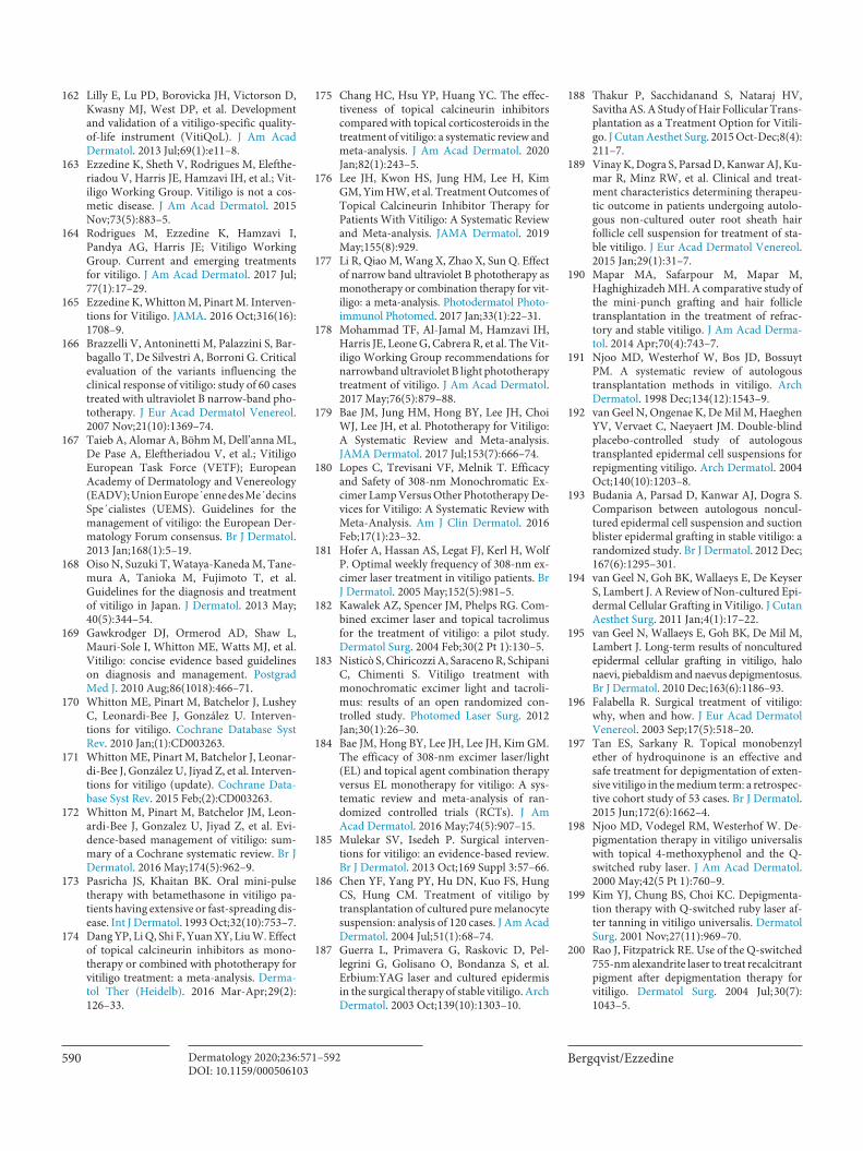
Bergqvist/EzzedineDermatology 2020;236:571–592590DOI: 10.1159/000506103
162 Lilly E, Lu PD, Borovicka JH, Victorson D, Kwasny MJ, West DP, et al. Development and validation of a vitiligo-specific quality-of-life instrument (VitiQoL). J Am Acad Dermatol. 2013 Jul; 69(1):e11–8.
163 Ezzedine K, Sheth V, Rodrigues M, Elefthe-riadou V, Harris JE, Hamzavi IH, et al.; Vit-iligo Working Group. Vitiligo is not a cos-metic disease. J Am Acad Dermatol. 2015 Nov; 73(5): 883–5.
164 Rodrigues M, Ezzedine K, Hamzavi I, Pandya AG, Harris JE; Vitiligo Working Group. Current and emerging treatments for vitiligo. J Am Acad Dermatol. 2017 Jul;
77(1): 17–29.165 Ezzedine K, Whitton M, Pinart M. Interven-
tions for Vitiligo. JAMA. 2016 Oct; 316(16):
1708–9.166 Brazzelli V, Antoninetti M, Palazzini S, Bar-
bagallo T, De Silvestri A, Borroni G. Critical evaluation of the variants influencing the clinical response of vitiligo: study of 60 cases treated with ultraviolet B narrow-band pho-totherapy. J Eur Acad Dermatol Venereol. 2007 Nov; 21(10): 1369–74.
167 Taieb A, Alomar A, Böhm M, Dell’anna ML, De Pase A, Eleftheriadou V, et al.; Vitiligo European Task Force (VETF); European Academy of Dermatology and Venereology (EADV); Union Europe´enne des Me´decins Spe´cialistes (UEMS). Guidelines for the management of vitiligo: the European Der-matology Forum consensus. Br J Dermatol. 2013 Jan; 168(1): 5–19.
168 Oiso N, Suzuki T, Wataya-Kaneda M, Tane-mura A, Tanioka M, Fujimoto T, et al. Guidelines for the diagnosis and treatment of vitiligo in Japan. J Dermatol. 2013 May;
40(5): 344–54.169 Gawkrodger DJ, Ormerod AD, Shaw L,
Mauri-Sole I, Whitton ME, Watts MJ, et al. Vitiligo: concise evidence based guidelines on diagnosis and management. Postgrad Med J. 2010 Aug; 86(1018): 466–71.
170 Whitton ME, Pinart M, Batchelor J, Lushey C, Leonardi-Bee J, González U. Interven-tions for vitiligo. Cochrane Database Syst Rev. 2010 Jan;(1):CD003263.
171 Whitton ME, Pinart M, Batchelor J, Leonar-di-Bee J, González U, Jiyad Z, et al. Interven-tions for vitiligo (update). Cochrane Data-base Syst Rev. 2015 Feb;(2):CD003263.
172 Whitton M, Pinart M, Batchelor JM, Leon-ardi-Bee J, Gonzalez U, Jiyad Z, et al. Evi-dence-based management of vitiligo: sum-mary of a Cochrane systematic review. Br J Dermatol. 2016 May; 174(5): 962–9.
173 Pasricha JS, Khaitan BK. Oral mini-pulse therapy with betamethasone in vitiligo pa-tients having extensive or fast-spreading dis-ease. Int J Dermatol. 1993 Oct; 32(10): 753–7.
174 Dang YP, Li Q, Shi F, Yuan XY, Liu W. Effect of topical calcineurin inhibitors as mono-therapy or combined with phototherapy for vitiligo treatment: a meta-analysis. Derma-tol Ther (Heidelb). 2016 Mar-Apr; 29(2):
126–33.
175 Chang HC, Hsu YP, Huang YC. The effec-tiveness of topical calcineurin inhibitors compared with topical corticosteroids in the treatment of vitiligo: a systematic review and meta-analysis. J Am Acad Dermatol. 2020 Jan; 82(1): 243–5.
176 Lee JH, Kwon HS, Jung HM, Lee H, Kim GM, Yim HW, et al. Treatment Outcomes of Topical Calcineurin Inhibitor Therapy for Patients With Vitiligo: A Systematic Review and Meta-analysis. JAMA Dermatol. 2019 May; 155(8): 929.
177 Li R, Qiao M, Wang X, Zhao X, Sun Q. Effect of narrow band ultraviolet B phototherapy as monotherapy or combination therapy for vit-iligo: a meta-analysis. Photodermatol Photo-immunol Photomed. 2017 Jan; 33(1): 22–31.
178 Mohammad TF, Al-Jamal M, Hamzavi IH, Harris JE, Leone G, Cabrera R, et al. The Vit-iligo Working Group recommendations for narrowband ultraviolet B light phototherapy treatment of vitiligo. J Am Acad Dermatol. 2017 May; 76(5): 879–88.
179 Bae JM, Jung HM, Hong BY, Lee JH, Choi WJ, Lee JH, et al. Phototherapy for Vitiligo: A Systematic Review and Meta-analysis. JAMA Dermatol. 2017 Jul; 153(7): 666–74.
180 Lopes C, Trevisani VF, Melnik T. Efficacy and Safety of 308-nm Monochromatic Ex-cimer Lamp Versus Other Phototherapy De-vices for Vitiligo: A Systematic Review with Meta-Analysis. Am J Clin Dermatol. 2016 Feb; 17(1): 23–32.
181 Hofer A, Hassan AS, Legat FJ, Kerl H, Wolf P. Optimal weekly frequency of 308-nm ex-cimer laser treatment in vitiligo patients. Br J Dermatol. 2005 May; 152(5): 981–5.
182 Kawalek AZ, Spencer JM, Phelps RG. Com-bined excimer laser and topical tacrolimus for the treatment of vitiligo: a pilot study. Dermatol Surg. 2004 Feb; 30(2 Pt 1): 130–5.
183 Nisticò S, Chiricozzi A, Saraceno R, Schipani C, Chimenti S. Vitiligo treatment with monochromatic excimer light and tacroli-mus: results of an open randomized con-trolled study. Photomed Laser Surg. 2012 Jan; 30(1): 26–30.
184 Bae JM, Hong BY, Lee JH, Lee JH, Kim GM. The efficacy of 308-nm excimer laser/light (EL) and topical agent combination therapy versus EL monotherapy for vitiligo: A sys-tematic review and meta-analysis of ran-domized controlled trials (RCTs). J Am Acad Dermatol. 2016 May; 74(5): 907–15.
185 Mulekar SV, Isedeh P. Surgical interven-tions for vitiligo: an evidence-based review. Br J Dermatol. 2013 Oct; 169 Suppl 3: 57–66.
186 Chen YF, Yang PY, Hu DN, Kuo FS, Hung CS, Hung CM. Treatment of vitiligo by transplantation of cultured pure melanocyte suspension: analysis of 120 cases. J Am Acad Dermatol. 2004 Jul; 51(1): 68–74.
187 Guerra L, Primavera G, Raskovic D, Pel-legrini G, Golisano O, Bondanza S, et al. Erbium:YAG laser and cultured epidermis in the surgical therapy of stable vitiligo. Arch Dermatol. 2003 Oct; 139(10): 1303–10.
188 Thakur P, Sacchidanand S, Nataraj HV, Savitha AS. A Study of Hair Follicular Trans-plantation as a Treatment Option for Vitili-go. J Cutan Aesthet Surg. 2015 Oct-Dec; 8(4):
211–7.189 Vinay K, Dogra S, Parsad D, Kanwar AJ, Ku-
mar R, Minz RW, et al. Clinical and treat-ment characteristics determining therapeu-tic outcome in patients undergoing autolo-gous non-cultured outer root sheath hair follicle cell suspension for treatment of sta-ble vitiligo. J Eur Acad Dermatol Venereol. 2015 Jan; 29(1): 31–7.
190 Mapar MA, Safarpour M, Mapar M, Haghighizadeh MH. A comparative study of the mini-punch grafting and hair follicle transplantation in the treatment of refrac-tory and stable vitiligo. J Am Acad Derma-tol. 2014 Apr; 70(4): 743–7.
191 Njoo MD, Westerhof W, Bos JD, Bossuyt PM. A systematic review of autologous transplantation methods in vitiligo. Arch Dermatol. 1998 Dec; 134(12): 1543–9.
192 van Geel N, Ongenae K, De Mil M, Haeghen YV, Vervaet C, Naeyaert JM. Double-blind placebo-controlled study of autologous transplanted epidermal cell suspensions for repigmenting vitiligo. Arch Dermatol. 2004 Oct; 140(10): 1203–8.
193 Budania A, Parsad D, Kanwar AJ, Dogra S. Comparison between autologous noncul-tured epidermal cell suspension and suction blister epidermal grafting in stable vitiligo: a randomized study. Br J Dermatol. 2012 Dec;
167(6): 1295–301.194 van Geel N, Goh BK, Wallaeys E, De Keyser
S, Lambert J. A Review of Non-cultured Epi-dermal Cellular Grafting in Vitiligo. J Cutan Aesthet Surg. 2011 Jan; 4(1): 17–22.
195 van Geel N, Wallaeys E, Goh BK, De Mil M, Lambert J. Long-term results of noncultured epidermal cellular grafting in vitiligo, halo naevi, piebaldism and naevus depigmentosus. Br J Dermatol. 2010 Dec; 163(6): 1186–93.
196 Falabella R. Surgical treatment of vitiligo: why, when and how. J Eur Acad Dermatol Venereol. 2003 Sep; 17(5): 518–20.
197 Tan ES, Sarkany R. Topical monobenzyl ether of hydroquinone is an effective and safe treatment for depigmentation of exten-sive vitiligo in the medium term: a retrospec-tive cohort study of 53 cases. Br J Dermatol. 2015 Jun; 172(6): 1662–4.
198 Njoo MD, Vodegel RM, Westerhof W. De-pigmentation therapy in vitiligo universalis with topical 4-methoxyphenol and the Q-switched ruby laser. J Am Acad Dermatol. 2000 May; 42(5 Pt 1): 760–9.
199 Kim YJ, Chung BS, Choi KC. Depigmenta-tion therapy with Q-switched ruby laser af-ter tanning in vitiligo universalis. Dermatol Surg. 2001 Nov; 27(11): 969–70.
200 Rao J, Fitzpatrick RE. Use of the Q-switched 755-nm alexandrite laser to treat recalcitrant pigment after depigmentation therapy for vitiligo. Dermatol Surg. 2004 Jul; 30(7):
1043–5.

Vitiligo 591Dermatology 2020;236:571–592DOI: 10.1159/000506103
201 Grimes PE, Hamzavi I, Lebwohl M, Ortonne JP, Lim HW. The efficacy of afamelanotide and narrowband UV-B phototherapy for re-pigmentation of vitiligo. JAMA Dermatol. 2013 Jan; 149(1): 68–73.
202 Lim HW, Grimes PE, Agbai O, Hamzavi I, Henderson M, Haddican M, et al. Afamela-notide and narrowband UV-B phototherapy for the treatment of vitiligo: a randomized multicenter trial. JAMA Dermatol. 2015 Jan;
151(1): 42–50.203 Kapoor R, Phiske MM, Jerajani HR. Evalua-
tion of safety and efficacy of topical prosta-glandin E2 in treatment of vitiligo. Br J Der-matol. 2009 Apr; 160(4): 861–3.
204 Grimes PE. Bimatoprost 0.03% Solution for the Treatment of Nonfacial Vitiligo. J Drugs Dermatol. 2016 Jun; 15(6): 703–10.
205 Harris JE, Rashighi M, Nguyen N, Jabbari A, Ulerio G, Clynes R, et al. Rapid skin repig-mentation on oral ruxolitinib in a patient with coexistent vitiligo and alopecia areata (AA). J Am Acad Dermatol. 2016 Feb; 74(2):
370–1.206 Rothstein B, Joshipura D, Saraiya A, Abdat
R, Ashkar H, Turkowski Y, Sheth V, Huang V, Au SC, Kachuk C, Dumont N, Gottlieb AB, Rosmarin D. Treatment of vitiligo with the topical Janus kinase inhibitor ruxoli-tinib. J Am Acad Dermatol. 2017; 76: 1054–60. e1051.
207 Singh H, Kumaran MS, Bains A, Parsad D. A Randomized Comparative Study of Oral Corticosteroid Minipulse and Low-Dose Oral Methotrexate in the Treatment of Un-stable Vitiligo. Dermatology. 2015; 231(3):
286–90.208 Gokhale BB. Cyclophosphamide and vit-
iligo. Int J Dermatol. 1979 Jan-Feb; 18(1):
92.209 Webb KC, Tung R, Winterfield LS, Gottlieb
AB, Eby JM, Henning SW, et al. Tumour ne-crosis factor-α inhibition can stabilize dis-ease in progressive vitiligo. Br J Dermatol. 2015 Sep; 173(3): 641–50.
210 Alghamdi KM, Khurrum H, Taieb A, Ezze-dine K. Treatment of generalized vitiligo with anti-TNF-α Agents. J Drugs Dermatol. 2012 Apr; 11(4): 534–9.
211 Maruthappu T, Leandro M, Morris SD. De-terioration of vitiligo and new onset of halo naevi observed in two patients receiving adalimumab. Dermatol Ther (Heidelb). 2013 Jul-Aug; 26(4): 370–2.
212 Alghamdi KM, Khurrum H, Rikabi A. Worsening of vitiligo and onset of new pso-riasiform dermatitis following treatment with infliximab. J Cutan Med Surg. 2011 Sep-Oct; 15(5): 280–4.
213 Speeckaert R, Speeckaert MM, van Geel N. Why treatments do(n’t) work in vitiligo: an autoinflammatory perspective. Autoimmun Rev. 2015 Apr; 14(4): 332–40.
214 Rigopoulos D, Gregoriou S, Larios G, Mous-tou E, Belayeva-Karatza E, Kalogeromitros D. Etanercept in the treatment of vitiligo. Dermatology. 2007; 215(1): 84–5.
215 Parambath N, Sharma VK, Parihar AS, Sahni K, Gupta S. Use of platelet-rich plasma to suspend noncultured epidermal cell sus-pension improves repigmentation after au-tologous transplantation in stable vitiligo: a double-blind randomized controlled trial. Int J Dermatol. 2019 Apr; 58(4): 472–6.
216 Lim HK, Shin MK, Lee MH. Clinical appli-cation of platelet-rich plasma in vitiligo: a pilot study. Poster Presentation. XXIst Inter-national Pigment Cell Conference. 2011. Bordeaux, France.
217 Ibrahim ZA, El-Ashmawy AA, El-Tatawy RA, Sallam FA. The effect of platelet-rich plasma on the outcome of short-term nar-rowband-ultraviolet B phototherapy in the treatment of vitiligo: a pilot study. J Cosmet Dermatol. 2016 Jun; 15(2): 108–16.
218 Abdelghani R, Ahmed NA, Darwish HM. Combined treatment with fractional carbon dioxide laser, autologous platelet-rich plas-ma, and narrow band ultraviolet B for vitili-go in different body sites: A prospective, ran-domized comparative trial. J Cosmet Der-matol. 2018 Jun; 17(3): 365–72.
219 Khattab FM, Abdelbary E, Fawzi M. Evalua-tion of combined excimer laser and platelet-rich plasma for the treatment of nonsegmen-tal vitiligo: A prospective comparative study. J Cosmet Dermatol. 2019 Sep;jocd.13103.
220 Sardana K, Verma G. Overview of Medical Therapies and Phototherapy in Vitiligo Based on Their Pathogenetic Action and the Role of Platelet-Rich Plasma. J Cutan Aesthet Surg. 2018 Oct-Dec; 11(4): 167–8.
221 Ejjiyar M, Sahibi M, El Gueouatri M, Bhihi A, Mahrouch M, Yafi I, et al. [Vitiligo and Koebner phenomenon following platelet-rich plasma injections]. Pan Afr Med J. 2019 Feb; 32: 58. French.
222 Sanclemente G, Garcia JJ, Zuleta JJ, Diehl C, Correa C, Falabella R. A double-blind, ran-domized trial of 0.05% betamethasone vs. topical catalase/dismutase superoxide in vit-iligo. J Eur Acad Dermatol Venereol. 2008 Nov; 22(11): 1359–64.
223 Grimes PE, Nashawati R. The Role of Diet and Supplements in Vitiligo Management. Dermatol Clin. 2017 Apr; 35(2): 235–43.
224 Elgoweini M, Nour El Din N. Response of vitiligo to narrowband ultraviolet B and oral antioxidants. J Clin Pharmacol. 2009 Jul;
49(7): 852–5.225 Akyol M, Celik VK, Ozcelik S, Polat M,
Marufihah M, Atalay A. The effects of vita-min E on the skin lipid peroxidation and the clinical improvement in vitiligo patients treated with PUVA. Eur J Dermatol. 2002 Jan-Feb; 12(1): 24–6.
226 Reyes E, Jaén P, de las Heras E, Carrión F, Alvarez-Mon M, de Eusebio E, et al. System-ic immunomodulatory effects of Polypodi-um leucotomos as an adjuvant to PUVA therapy in generalized vitiligo: A pilot study. J Dermatol Sci. 2006 Mar; 41(3): 213–6.
227 Middelkamp-Hup MA, Bos JD, Rius-Diaz F, Gonzalez S, Westerhof W. Treatment of vit-
iligo vulgaris with narrow-band UVB and oral Polypodium leucotomos extract: a ran-domized double-blind placebo-controlled study. J Eur Acad Dermatol Venereol. 2007 Aug; 21(7): 942–50.
228 Parsad D, Pandhi R, Juneja A. Effectiveness of oral Ginkgo biloba in treating limited, slowly spreading vitiligo. Clin Exp Derma-tol. 2003 May; 28(3): 285–7.
229 Szczurko O, Shear N, Taddio A, Boon H. Ginkgo biloba for the treatment of vitilgo vulgaris: an open label pilot clinical trial. BMC Complement Altern Med. 2011 Mar;
11(1): 21.230 Hrushesky WJ. Letter: serpentine supra-
venous fluorouracil hyperpigmentation. JAMA. 1976 Jul; 236(2): 138.
231 Tsuji T, Hamada T. Topically administered fluorouracil in vitiligo. Arch Dermatol. 1983 Sep; 119(9): 722–7.
232 Anbar TS, Westerhof W, Abdel-Rahman AT, Ewis AA, El-Khayyat MA. Effect of one session of ER:YAG laser ablation plus topical 5Fluorouracil on the outcome of short-term NB-UVB phototherapy in the treatment of non-segmental vitiligo: a left-right compar-ative study. Photodermatol Photoimmunol Photomed. 2008 Dec; 24(6): 322–9.
233 Sethi S, Mahajan BB, Gupta RR, Ohri A. Comparative evaluation of the therapeutic efficacy of dermabrasion, dermabrasion combined with topical 5% 5-fluorouracil cream, and dermabrasion combined with topical placentrex gel in localized stable vit-iligo. Int J Dermatol. 2007 Aug; 46(8): 875–9.
234 Mina M, Elgarhy L, Al-Saeid H, Ibrahim Z. Comparison between the efficacy of mi-croneedling combined with 5-fluorouracil vs microneedling with tacrolimus in the treatment of vitiligo. J Cosmet Dermatol. 2018 Oct; 17(5): 744–51.
235 Jha AK, Sonthalia S. 5-Fluorouracil as an ad-juvant therapy along with microneedling in vitiligo. J Am Acad Dermatol. 2019 Apr;
80(4):e75–6.236 Zohdy HA, Hussein MS. Intradermal injec-
tion of Fluorouracil versus triamcinolone in localized vitiligo treatment. J Cosmet Der-matol. 2018 Nov.
237 Wu JH, Cohen BA. The stigma of skin dis-ease. Curr Opin Pediatr. 2019 Aug; 31(4):
509–14.238 Roman J. Beauty That Is More Than Skin
Deep. JAMA Dermatol. 2016 May; 152(5):
562–3.239 Krüger C, Schallreuter KU. Cumulative life
course impairment in vitiligo. Curr Probl Dermatol. 2013; 44: 102–17.
240 Ware J Jr, Kosinski M, Keller SD. A 12-Item Short-Form Health Survey: construction of scales and preliminary tests of reliability and validity. Med Care. 1996 Mar; 34(3): 220–33.
241 Krishna GS, Ramam M, Mehta M, Sreenivas V, Sharma VK, Khandpur S. Vitiligo impact scale: an instrument to assess the psychoso-cial burden of vitiligo. Indian J Dermatol Ve-nereol Leprol. 2013 Mar-Apr; 79(2): 205–10.

Bergqvist/EzzedineDermatology 2020;236:571–592592DOI: 10.1159/000506103
242 Salzes C, Abadie S, Seneschal J, Whitton M, Meurant JM, Jouary T, et al. The Vitiligo Im-pact Patient Scale (VIPs): Development and Validation of a Vitiligo Burden Assessment Tool. J Invest Dermatol. 2016 Jan; 136(1):
52–8.243 Linthorst Homan MW, Spuls PI, de Korte J,
Bos JD, Sprangers MA, van der Veen JP. The burden of vitiligo: patient characteristics as-sociated with quality of life. J Am Acad Der-matol. 2009 Sep; 61(3): 411–20.
244 Osinubi O, Grainge MJ, Hong L, Ahmed A, Batchelor JM, Grindlay D, et al. The preva-lence of psychological comorbidity in people with vitiligo: a systematic review and meta-analysis. Br J Dermatol. 2018 Apr; 178(4):
863–78.245 Porter J, Beuf AH, Nordlund JJ, Lerner AB.
Psychological reaction to chronic skin disor-ders: a study of patients with vitiligo. Gen Hosp Psychiatry. 1979 Apr; 1(1): 73–7.
246 Parsad D, Dogra S, Kanwar AJ. Quality of life in patients with vitiligo. Health Qual Life Outcomes. 2003 Oct; 1(1): 58.
247 Ongenae K, Beelaert L, van Geel N, Naeyaert JM. Psychosocial effects of vitiligo. J Eur Acad Dermatol Venereol. 2006 Jan; 20(1):
1–8.248 Bhandarkar SS, Kundu RV. Quality-of-life
issues in vitiligo [viii.]. Dermatol Clin. 2012 Apr; 30(2): 255–68.
249 Morrison B, Burden-Teh E, Batchelor JM, Mead E, Grindlay D, Ratib S. Quality of life in people with vitiligo: a systematic review and meta-analysis. Br J Dermatol. 2017 Dec;
177(6):e338–9.250 Lai YC, Yew YW, Kennedy C, Schwartz RA.
Vitiligo and depression: a systematic review and meta-analysis of observational studies. Br J Dermatol. 2017 Sep; 177(3): 708–18.
251 Wang G, Qiu D, Yang H, Liu W. The preva-lence and odds of depression in patients with vitiligo: a meta-analysis. J Eur Acad Derma-tol Venereol. 2018 Aug; 32(8): 1343–51.
252 Sukan M, Maner F. The problems in sexual functions of vitiligo and chronic urticaria patients. J Sex Marital Ther. 2007 Jan-Feb;
33(1): 55–64.253 Porter J, Beuf A, Nordlund JJ, Lerner AB.
Personal responses of patients to vitiligo: the importance of the patient-physician interac-tion. Arch Dermatol. 1978 Sep; 114(9): 1384–5.
254 Ingordo V, Cazzaniga S, Gentile C, Iannaz-zone SS, Cusano F, Naldi L. Dermatology Life Quality Index score in vitiligo patients: a pilot study among young Italian males. G Ital Dermatol Venereol. 2012 Feb; 147(1):
83–90.255 Noh S, Kim M, Park CO, Hann SK, Oh SH.
Comparison of the psychological impacts of asymptomatic and symptomatic cutaneous diseases: vitiligo and atopic dermatitis. Ann Dermatol. 2013 Nov; 25(4): 454–61.
256 Rzepecki AK, McLellan BN, Elbuluk N. Be-yond Traditional Treatment: The Impor-tance of Psychosocial Therapy in Vitiligo. J Drugs Dermatol. 2018 Jun; 17(6): 688–91.
257 Shah R, Hunt J, Webb TL, Thompson AR. Starting to develop self-help for social anxi-ety associated with vitiligo: using clinical sig-nificance to measure the potential effective-ness of enhanced psychological self-help. Br J Dermatol. 2014 Aug; 171(2): 332–7.
258 Papadopoulos L, Bor R, Legg C. Coping with the disfiguring effects of vitiligo: a prelimi-nary investigation into the effects of cogni-tive-behavioural therapy. Br J Med Psychol. 1999 Sep; 72(Pt 3): 385–96.
259 Eleftheriadou V, Thomas KS, Whitton ME, Batchelor JM, Ravenscroft JC. Which out-comes should we measure in vitiligo? Results of a systematic review and a survey among patients and clinicians on outcomes in vit-iligo trials. Br J Dermatol. 2012 Oct; 167(4):
804–14.
260 Vrijman C, Linthorst Homan MW, Limpens J, van der Veen W, Wolkerstorfer A, Terwee CB, et al. Measurement properties of out-come measures for vitiligo. A systematic review. Arch Dermatol. 2012 Nov; 148(11):
1302–9.261 Wolkerstorfer A. The long road to valid out-
comes in vitiligo. Br J Dermatol. 2019 Mar;
180(3): 454–5.262 Eleftheriadou V, Thomas K, van Geel N,
Hamzavi I, Lim H, Suzuki T, et al.; Vitiligo Global Issues Consensus Group. Developing core outcome set for vitiligo clinical trials: international e-Delphi consensus. Pigment Cell Melanoma Res. 2015 May; 28(3): 363–9.
263 Gan EY, Eleftheriadou V, Esmat S, Hamzavi I, Passeron T, Böhm M, et al.; VGICC. Re-pigmentation in vitiligo: position paper of the Vitiligo Global Issues Consensus Con-ference. Pigment Cell Melanoma Res. 2017 Jan; 30(1): 28–40.
264 Kottner J, Jacobi L, Hahnel E, Alam M, Bal-zer K, Beeckman D, et al.; International Cochrane Skin Group Core Outcome Set Initiative (CSG-COUSIN) group. Core outcome sets in dermatology: report from the second meeting of the International Cochrane Skin Group Core Outcome Set Initiative. Br J Dermatol. 2018 Apr; 178(4): e279–85.
265 Batchelor JM, Tan W, Tour S, Yong A, Montgomery AA, Thomas KS. Validation of the Vitiligo Noticeability Scale: a patient- reported outcome measure of vitiligo treat-ment success. Br J Dermatol. 2016 Feb;
174(2): 386–94.266 Ezzedine K, Pandya A. Time for a patient-
oriented outcome in vitiligo: the vitiligo no-ticeability scale. Br J Dermatol. 2016 Feb;
174(2): 255–6.267 Eleftheriadou V, Hamzavi I, Pandya AG,
Grimes P, Harris JE, Huggins RH, et al. In-ternational Initiative for Outcomes (INFO) for vitiligo: workshops with patients with vitiligo on repigmentation. Br J Dermatol. 2019 Mar; 180(3): 574–9.


