Embed Size (px)
Citation preview

�����������������
Citation: Liu, W.; Zheng, X.; Dai, X.;
Zhang, Z.; Zhang, W.; Xiao, T.;
Huang, L. Isolation and
Characterization of the First Temperate
Virus Infecting Psychrobacillus from
Marine Sediments. Viruses 2022, 14,
108. https://doi.org/10.3390/
v14010108
Academic Editor: Evelien
Adriaenssens
Received: 19 November 2021
Accepted: 6 January 2022
Published: 8 January 2022
Publisher’s Note: MDPI stays neutral
with regard to jurisdictional claims in
published maps and institutional affil-
iations.
Copyright: © 2022 by the authors.
Licensee MDPI, Basel, Switzerland.
This article is an open access article
distributed under the terms and
conditions of the Creative Commons
Attribution (CC BY) license (https://
creativecommons.org/licenses/by/
4.0/).
viruses
Article
Isolation and Characterization of the First Temperate VirusInfecting Psychrobacillus from Marine SedimentsWang Liu 1,2, Xiaowei Zheng 1, Xin Dai 1,2, Zhenfeng Zhang 1 , Wenyan Zhang 3,4,5, Tian Xiao 3,4,5
and Li Huang 1,2,*
1 State Key Laboratory of Microbial Resources, Institute of Microbiology, Chinese Academy of Sciences,Beijing 100101, China; [email protected] (W.L.); [email protected] (X.Z.);[email protected] (X.D.); [email protected] (Z.Z.)
2 College of Life Science, University of Chinese Academy of Sciences, Beijing 100049, China3 Key Laboratory for Marine Ecology and Environmental Sciences, Institute of Oceanology, Chinese Academy
of Sciences, Qingdao 266071, China; [email protected] (W.Z.); [email protected] (T.X.)4 Center for Ocean Mega-Science, Chinese Academy of Sciences, Qingdao 266071, China5 Laboratory for Marine Ecology and Environmental Science, Qingdao National Laboratory for Marine Science
and Technology, Qingdao 266273, China* Correspondence: [email protected]
Abstract: Viruses are far more abundant than cellular microorganisms in the marine ecosystem.However, very few viruses have so far been isolated from marine sediments, especially hydrothermalvent sediments, hindering the understanding of the biology and ecological functions of these tinyorganisms. Here, we report the isolation and characterization of a temperate bacteriophage, namedPVJ1, which infects Psychrobacillus from a hydrothermal vent field in Okinawa Trough. PVJ1 belongsto the Myoviridae family of the order Caudovirales. The tailed phage possesses a 53,187 bp lineardsDNA genome, with 84 ORFs encoding structural proteins, genome replication, host lysis, etc. in amodular pattern. The phage genome is integrated into the host chromosome near the 3′-end of deoD,a gene encoding purine nucleoside phosphorylase (PNP). The phage integration does not appear todisrupt the function of PNP. The phage DNA is packaged by the headful mechanism. Release of PVJ1from the host cell was drastically enhanced by treatment with mitomycin C. Phages encoding anMCP sharing significant similarity (≥70% identical amino acids) with that of PVJ1 are widespread indiverse environments, including marine and freshwater sediments, soils, artificial ecosystems, andanimal intestines, and primarily infect Firmicutes. These results are valuable to the understanding ofthe lifestyle and host interactions of bacterial viruses at the bottom of the ocean.
Keywords: Psychrobacillus; bacteriophage; marine sediments; Myoviridae
1. Introduction
Viruses are the most abundant biological entities in the ocean, and those in marinesediments are especially abundant and extraordinarily diverse [1–4]. Virus-like particles(VLPs) in some marine sediments were found to exceed 109/mL, which is three ordersof magnitude higher than VLPs in seawater at the same depth [5]. VLPs of differentmorphologies, predominated by the head-tail shape, were observed under an electronmicroscope in the eluates of marine sediment samples [1–3], but the vast majority of theVLP sequences retrieved from the metagenomic data of the sediment samples showed nosignificant matches to known sequences [3]. Although the virosphere of marine sedimentsremains largely a mystery, its role in geobiochemical processes in the oceans has beenincreasingly recognized. Clearly, the isolation and characterization of these viruses areessential to the elucidation of their ecological functions.
Hydrothermal vents support unique ecosystems, in which considerably diverse mi-croorganisms driving elemental cycling thrive [6]. However, fewer than a dozen of bacterio-phages have been isolated from hydrothermal vents and characterized so far [7–11]. Four
Viruses 2022, 14, 108. https://doi.org/10.3390/v14010108 https://www.mdpi.com/journal/viruses

Viruses 2022, 14, 108 2 of 14
of them infect Bacillus or Geobacillus, and they are all lytic [8–11]. These phages possesshead-tailed morphotypes similar to those of the majority of phages isolated from terrestrialhot springs [12]. Therefore, head-and-tailed viruses of the order Caudovirales, which accountfor most of the prokaryotic viruses [13], appear to be also well represented in the extremeenvironments on Earth. Metagenomic analysis shows that viruses in hydrothermal ventsare predominantly lysogenic and carry auxiliary metabolic genes that function to regulatethe physiological activities of the host [14]. It is suggested that proviruses contribute to thefitness of the host strains, whereas the integrated state represents a strategy employed bythe viruses to survive harsh conditions [15].
Here, we report the isolation and characterization of PVJ1, a bacteriophage that infectsa mesophilic Psychrobacillus strain, from a sediment sample taken from a hydrothermal ventfield in Okinawa Trough. PVJ1 is a temperate phage of the Myoviridae family of the orderCaudovirales, and capable of integrating its 53,187 bp linear dsDNA genome into the hostgenome at a unique site within a gene encoding purine nucleoside phosphatase. Phagesencoding a MCP sharing significant similarity (≥70% identical amino acids) with that ofPVJ1 specifically infected Firmicutes and are widespread in aquatic environments, soils,and animal intestines.
2. Materials and Methods2.1. Strain Isolation
Samples used in this study were collected in July 2018, during the cruise conductedby the scientific research vessel KEXUE in Okinawa Trough (GPS coordinates: 126.55◦ E,27.78◦ N, depth: 958 m, temperature in situ: 4 ◦C). The sediments were collected by TVgrab (TVG) equipped on a KEXUE vessel. After removing the superficial 5 cm layer ofsediments, surface sediments (upper 10 cm) were transferred in aseptic sampling bags(Haibo, Qingdao, China). Sediments were stored at −80 ◦C and kept on dry ice duringtransportation. Then, 5 g sediment samples were inoculated into marine 2216E medium [16].After incubation at 18 ◦C for 1–2 weeks, the enrichment cultures were plated onto marineagar 2216E. Following further incubation at 18 ◦C for 1 week, colonies were picked, andisolates were purified by streaking repeatedly on 2216E plates. The 16S rRNA gene of eachisolate was amplified by PCR using the primers 16S-27F/1492R (Table S1) and sequenced.Psychrobacillus sp. GC2J1, the native host of bacteriophage PVJ1, was obtained.
2.2. Isolation and Purification of PVJ1
Psychrobacillus sp. GC2J1 was grown with shaking in marine 2216E medium at 28 ◦Cto an OD600 of 0.4~0.6. Mitomycin C (Amresco) was added to a final concentration of1 µg/mL to induce the replication and release of PVJ1. After incubation for 9 h, the culturewas centrifuged at 10,000× g for 20 min at 4 ◦C. The supernatant was filtered through amembrane with a 0.22 µm pore size (Merck Millipore). Virus particles in the filtrate werecollected by ultracentrifugation at 120,000× g for 1 h at 4 ◦C and gently resuspended inbuffer SM (10 mM Tris-HCl, pH 7.5; 100 mM NaCl; 10 mM MgSO4). The particles weresubjected to purification by cesium chloride density gradient centrifugation at 200,000× gfor 24 h at 4 ◦C. The purified PVJ1 particles were resuspended in buffer SM and storedat 4 ◦C.
2.3. Transmission Electron Microscopy (TEM)
A sample (10 µL) of the phage PVJ1 was deposited on a carbon-coated 230-mesh grid,negatively stained for 1 min with 2% (w/v) uranyl acetate, and observed under a JEM-1400transmission electron microscope (JEOL, Tokyo, Japan).
2.4. DNA Isolation and Genome Analysis
Purified PVJ1 particles were digested with proteinase K (0.6 mg/mL) in the presenceof 2% (wt/vol) SDS at 55 ◦C for 3 h. NaCl and cetyltrimethylammonium bromide (CTAB)were added to 800 mM and 1%, respectively. After incubation for 10 min at 65 ◦C, the
Viruses 2022, 14, 108 3 of 14
sample was extracted twice with phenol:chloroform:isoamylalcohol (25:24:1), and the DNAwas precipitated with ethanol. Paired-end (PE) sequencing (2 × 150 bp) was conducted onan Illumina Hiseq-1500 platform at Microbial Genome Research Center, Institute of Micro-biology, Chinese Academy of Sciences, Beijing, China. Raw reads were adapter trimmedand quality controlled using fastp (v0.19.4) with default parameters [17] and subsequentlyassembled by SPAdes 3.11 [18] with a series of kmers (i.e., 21, 31, 55, 77, 99, 121). Theresulting PVJ1 genome was submitted to RAST (http://rast.nmpdr.org/ accessed on 11 De-cember 2021) for an initial annotation [19] and further checked by BLASTp analysis againstNCBI non-redundant (nr) protein database. Hypothetical proteins were also screenedfor distinct homologs in HHpred (https://toolkit.tuebingen.mpg.de/tools/hhpred ac-cessed on 11 December 2021) [20]. Putative tRNA genes were predicted with tRNAscan-SE(http://lowelab.ucsc.edu/tRNAscan-SE/ accessed on 11 December 2021). Gene domainarchitectures were predicted with SMART (http://smart.embl-heidelberg.de accessed on11 December 2021). Transmembrane domains and signal peptides were predicted us-ing TMHMM (http://www.cbs.dtu/services/TMHMM/ accessed on 11 December 2021)and Signal P-3.0 Server (http://www.cbs.dtu.dk/services/SignalP-3.0/ accessed on 11December 2021), respectively.
2.5. Quantification of Phage DNA by qPCR
The PVJ1 DNA was quantified by qPCR with the primer pair PVJ1-qPCR-F/R (Table S1)targeting PVJ1 ORF81, which encodes a portal protein. To prepare a standard curve forPVJ1 DNA quantification, the PCR fragment was cloned into plasmid pEASY-T1 (TransGenBiotech, Beijing, China). The construct was then serially diluted and amplified as theknown control to generate a standard curve relating the copy number of PVJ1 DNA withCT (threshold cycle) values. The standard reaction (20 µL) contained 2 µL of total DNAtemplate, 10 µL of TB GreenTM Premix Ex TaqTM II (Tli RNaseH Plus, Takara Bio, Shiga,Japan), 0.8 µL each of the primers (10 µM), and 6.4 µL of DNase-free water. The two-stepqPCR reaction was performed on a Light Cycler fluorescence quantitative PCR instrument(Roche Life Science, Indianapolis, IN, USA) with pre-denaturation for 30 s at 95 ◦C, followedby 40 cycles of denaturation for 5 s at 95 ◦C, and annealing and extension for 30 s at 60 ◦Cin the first step, and denaturation for 5 s at 95 ◦C, annealing and extension for 1 min at60 ◦C, and denaturation for heating to 95 ◦C, and cooling to 50 ◦C in the second step [21].
2.6. Host Range Determination
The plaque assays and qPCR quantification were used to screen the phage's ability tolyse different strains. Tested strains included Psychrobacillus sp. GC2J1 (CGMCC 1.19103),Psychrobacillus insolitus DSM5 (CGMCC 1.3683), Psychrobacillus lasiicapitis NEAU-3TGS17T
(CGMCC 1.15308), Lysinibacillus antri SYSU K30002T (CGMCC 1.13504), Paenisporosarcinaantarctica N-05 (CGMCC 1.6503), and Planococcus rifietoensis N14 (CGMCC 1.8017). Forplaque assays, phage suspension (10 µL) was detected on lawns of the strains grown onplates. After overnight incubation at 25 ◦C, plates were observed for the presence of clearor turbid plaques. For qPCR quantification, 10 µL phage suspension was added into 500 µLbacterial cultures at exponential growth phase, Mitomycin C was added after culturing tothe stationary phase, and the total PVJ1 DNA before and after induction was quantifiedby qPCR.
2.7. Protein Analysis
The purified PVJ1 virions were subjected to 12% sodium dodecyl sulfate-polyacrylamidegel electrophoresis (SDS–PAGE). The gel was stained with Coomassie Brilliant Blue G250,and gel slices containing protein bands were excised. Proteins in the gel slices were digestedwith trypsin (12.5 ng/µL), and the resulting peptides were analyzed by matrix-assistedlaser desorption ionization-time of flight (MALDI-TOF) mass spectrometry (AB 533 Sciex5800 TOF/TOF 5800, Foster City, CA, USA) as described previously [21].
Viruses 2022, 14, 108 4 of 14
2.8. Atomic Force Microscopy (AFM)
The purified PVJ1 DNA molecules were dissolved in nuclease-free water, adsorbedonto the mica substrate in the presence of 5 mM Mg2+, and observed under an atomicforce microscope (Nanoscope V Multimode, Bruker, USA). Length measurements of DNAmolecules were performed manually using the software Image J (Wayne Rasband, NationalInstitute of Health, USA). A total of 50 fields were examined.
2.9. Phylogenetic Analysis
The amino acid sequences of the terminase large subunits of PVJ1 and 32 other viruseswith a clear classification of DNA packaging strategies [22] were aligned by Clustal W [23],and a neighbor-joining phylogenetic tree was constructed using MEGA 7.0 with a bootstrapof 1000 replicates.
Bacterial and viral proteins were downloaded from the NCBI reference sequencedatabase (ftp://ftp.ncbi.nlm.nih.gov/genomes/refseq/bacteria/ accessed on 13 September2021) and IMG/VR database (Integrated Microbial Genomes with Virus, https://img.jgi.doe.gov/vr/ accessed on 13 September 2021). The habitat information of these sequenceswas manually parsed from the NCBI and IMG/VR databases. Proteins annotated as capsidwere extracted to create a local database using Diamond (V.0.9.10.111) [24]. Homologsof the major capsid protein (ORF75) of PVJ1 were extracted after aligning ORF75 againstthe local capsid database using “diamond blastp” (≥70% identity and ≥50% alignment,E-value of 1 × 10⁻³). The obtained capsid sequences were multi-aligned by the programMAFFT 7.455 with the “auto” algorithm [25], followed by sequence trimming using trimAl1.4.1 in default parameters [26]. Additionally, a maximum likelihood (ML) phylogenetictree was constructed for capsid proteins with FastTree v.2.1.10 in the WAG + GAMMAmodel [27]. Further, integrated prophage sequences in bacterial genomes were predictedusing PHASTER [28]. Those viral sequences containing capsid clustered closely withORF75, as observed in the ML phylogenetic tree of the capsid, were selected for syntenyanalysis using EasyFig 3.0.5 [29].
3. Results and Discussion3.1. Isolation of Psychrobacillus sp. GC2J1 and Bacteriophage PVJ1
Strain GC2J1 was isolated during a survey of microorganisms in sediment samplescollected from a hydrothermal vent field in Okinawa Trough. The strain grew at 4–40 ◦C(opt: 25–30 ◦C) and pH 5–12 (opt: pH 7–8) in the presence of 0–5% (w/v) NaCl (opt: 2%).GC2J1 was capable of forming endospores, a property that presumably allows the strain towithstand occasional exposure to high temperatures in the habitat. The sequence of the16S rRNA gene of strain GC2J1 (GenBank accession no. MZ959810) is 99.9% identical tothat of Psychrobacillus lasiicapitis NEAU-3TGS17T (GenBank accession no. NR_159144), astrain isolated from the head of an ant [30]. However, digital DNA–DNA hybridizationand average nucleotide identity (ANI) values for the two strains were only 36.6% and 89%,respectively, suggesting that GC2J1 is probably a new species in the genus Psychrobacillus.It should be noted that, although Psychrobacillus strains were isolated from a variety ofenvironments, e.g., oil-contaminated soil, red fox feces, the Antarctic iceberg, etc., Psy-chrobacillus sp. GC2J1 represents the first member of the genus Psychrobacillus to be isolatedfrom high-temperature habitats.
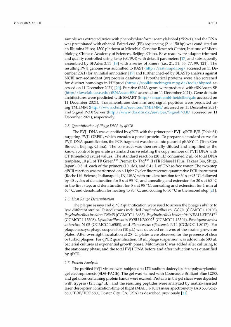
The strain denoted Psychrobacillus sp. GC2J1 released virus-like particles (VLPs)following treatment with mitomycin C (1 µg/mL; Figure 1A,B). We analyzed 50 VLPsby TEM to measure diameter, length, and width. VLPs possess an icosahedral head of60 ± 2 nm in diameter and a contractile tail of 120 ± 5 and 20 ± 1 nm in length and width,respectively (Figure 1A). A cluster of terminal knobs of 20 ± 2 nm in length was observed.Based on its morphological characteristics, the bacteriophage belongs to the Myoviridaefamily of the order Caudovirales. We named the phage Psychrobacillus virus J1 (PVJ1). Todemonstrate that PVJ1 indeed existed in the sediment sample from the hydrothermal ventfield in Okinawa Trough, we extracted metagenomic DNA from the sample and conducted
Viruses 2022, 14, 108 5 of 14
a PCR assay using a primer pair targeting PVJ1 (PVJ1-ORF75F/R, Table S1). Our resultsshow that PVJ1 was present in the original sediment sample (Figure S1). Members of thefamily Bacillaceae are known to be infected by bacteriophages. However, there were noreports of bacteriophages infecting the genus Psychrobacillus of this family. Therefore, PVJ1is the first bacteriophage isolated from Psychrobacillus.
Viruses 2022, 14, x FOR PEER REVIEW 5 of 14
nm in diameter and a contractile tail of 120 ± 5 and 20 ± 1 nm in length and width, respec-tively (Figure 1A). A cluster of terminal knobs of 20 ± 2 nm in length was observed. Based on its morphological characteristics, the bacteriophage belongs to the Myoviridae family of the order Caudovirales. We named the phage Psychrobacillus virus J1 (PVJ1). To demon-strate that PVJ1 indeed existed in the sediment sample from the hydrothermal vent field in Okinawa Trough, we extracted metagenomic DNA from the sample and conducted a PCR assay using a primer pair targeting PVJ1 (PVJ1-ORF75F/R, Table S1). Our results show that PVJ1 was present in the original sediment sample (Figure S1). Members of the family Bacillaceae are known to be infected by bacteriophages. However, there were no reports of bacteriophages infecting the genus Psychrobacillus of this family. Therefore, PVJ1 is the first bacteriophage isolated from Psychrobacillus.
Figure 1. Morphology and induction of PVJ1: (A) transmission electron micrographs of PVJ1; (B) thin section of a Psychrobacillus sp. GC2J1 cell following treatment with mitomycin C. CM, cytoplas-mic membrane; NB, nuclear body; C, cytoplasm; CW, cell wall; VLP, virus-like particle; (C) effect of mitomycin C treatment on the growth of Psychrobacillus sp. GC2J1; (D) titer of PVJ1 during mitomy-cin C induction experiment.
As revealed quantitatively by qPCR and observed by TEM, PVJ1 virions were re-leased at a low frequency without induction under our experimental conditions (Figure 1D). A brief heat treatment at 60 °C or growth at pH4 resulted in a drop in the noninduced release of the phage (Figure S2). The PVJ1 DNA increased drastically following treatment with mitomycin C, which inhibited the growth of the host cells (Figure 1C, D). The phage production, which showed a latent period of ~4 h, peaked at 1.93 × 10¹¹ copies/mL at 17 h after the addition of mitomycin C (Figure 1D).
The host range of PVJ1 was tested using plaque assays as well as qPCR on six differ-ent strains including Psychrobacillus sp. GC2J1 (CGMCC 1.19103), Psychrobacillus insolitus DSM5 (CGMCC 1.3683), Psychrobacillus lasiicapitis NEAU-3TGS17T (CGMCC 1.15308), Lysinibacillus antri SYSU K30002T (CGMCC 1.13504), Paenisporosarcina antarctica N-05 (CGMCC 1.6503), and Planococcus rifietoensis N14 (CGMCC 1.8017). VLPs were readily
Figure 1. Morphology and induction of PVJ1: (A) transmission electron micrographs of PVJ1; (B) thinsection of a Psychrobacillus sp. GC2J1 cell following treatment with mitomycin C. CM, cytoplasmicmembrane; NB, nuclear body; C, cytoplasm; CW, cell wall; VLP, virus-like particle; (C) effect ofmitomycin C treatment on the growth of Psychrobacillus sp. GC2J1; (D) titer of PVJ1 during mitomycinC induction experiment.
As revealed quantitatively by qPCR and observed by TEM, PVJ1 virions were releasedat a low frequency without induction under our experimental conditions (Figure 1D).A brief heat treatment at 60 ◦C or growth at pH4 resulted in a drop in the noninducedrelease of the phage (Figure S2). The PVJ1 DNA increased drastically following treatmentwith mitomycin C, which inhibited the growth of the host cells (Figure 1C,D). The phageproduction, which showed a latent period of ~4 h, peaked at 1.93 × 10¹¹ copies/mL at 17 hafter the addition of mitomycin C (Figure 1D).
The host range of PVJ1 was tested using plaque assays as well as qPCR on six differentstrains including Psychrobacillus sp. GC2J1 (CGMCC 1.19103), Psychrobacillus insolitus DSM5(CGMCC 1.3683), Psychrobacillus lasiicapitis NEAU-3TGS17T (CGMCC 1.15308), Lysinibacil-lus antri SYSU K30002T (CGMCC 1.13504), Paenisporosarcina antarctica N-05 (CGMCC1.6503), and Planococcus rifietoensis N14 (CGMCC 1.8017). VLPs were readily observed onlyin Psychrobacillus sp. GC2J1 after MMC induction and none of the other tested strains wereinfected by PVJ1.

Viruses 2022, 14, 108 6 of 14
3.2. General Features of the PVJ1 Genome
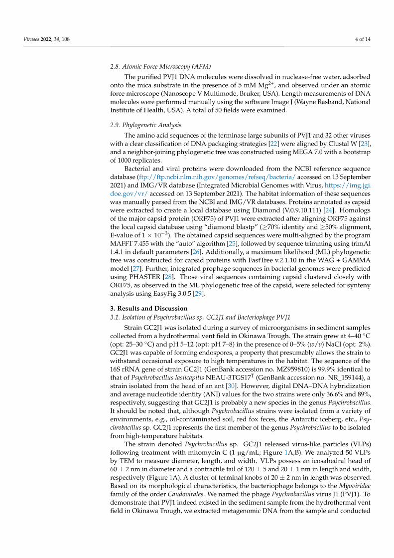
PVJ1 harbors a 53,187 bp linear dsDNA genome (see below) with a GC content of37.43%, which is similar to that of the genome of Psychrobacillus sp. GC2J1 (37.1%). PVJ1 ispredicted to encode 84 proteins, of which 77 have homologous sequences (E-value < 1 ×10⁻³) in the NCBI database and 39 have an annotating function (Table S2). Genes encodingDNA replication, transcription, repair, and integration proteins (blue), proteins related tothe host cell lysis (red), structural proteins (purple), and DNA packaging (brown) exist incassettes in the genome (Figure 2). Most of the genes are arranged in the same orientation.
Viruses 2022, 14, x FOR PEER REVIEW 6 of 14
observed only in Psychrobacillus sp. GC2J1 after MMC induction and none of the other tested strains were infected by PVJ1.
3.2. General Features of the PVJ1 Genome PVJ1 harbors a 53,187 bp linear dsDNA genome (see below) with a GC content of
37.43%, which is similar to that of the genome of Psychrobacillus sp. GC2J1 (37.1%). PVJ1 is predicted to encode 84 proteins, of which 77 have homologous sequences (E-value < 1 × 10⁻³) in the NCBI database and 39 have an annotating function (Table S2). Genes encoding DNA replication, transcription, repair, and integration proteins (blue), proteins related to the host cell lysis (red), structural proteins (purple), and DNA packaging (brown) exist in cassettes in the genome (Figure 2). Most of the genes are arranged in the same orientation.
Figure 2. Genome organization of PVJ1. ORFs are shown with arrows oriented in the direction of transcription. Gene cassettes with putative functions are color coded as indicated.
In the gene cassette responsible primarily for DNA replication, transcription, repair, and integration, ORF12 is a homolog of Holliday junction resolvase, an enzyme that both resolves the Holliday structures resulting from the recombination process and serves roles in debranching phage DNA structures prior to phage genome packaging into head parti-cles and in degrading bacterial host DNA [31]. ORF29 encodes an AAA+ family protein [32]. Both ORF33 and ORF34 code for HTH domain-containing transcriptional regulatory factors of the xenobiotic response element (XRE) family. These transcriptional factors are often encoded by phages of spore-forming bacteria and may be related to the regulation of integrase [33]. ORF35 encodes a site-specific integrase. A 42-bp integration core se-quence exists downstream of ORF35, allowing the phage DNA to be integrated at a spe-cific site in the host genome. ORF45, ORF46, ORF47, and ORF49 encode a YolD-like family protein, DNA polymerase V, regulatory protein LexA, and DNA ligase, respectively, and may play key roles in phage DNA replication and repair.
The second cassette of genes appears to be involved in PVJ1 lysis. ORF50 encodes an N-acetylmuramoyl-L-alanine amidase, which acts on the cell wall of the host when the virions are released [34]. Holin encoded by ORF51 presumably disrupts the host cell wall once the phage entered the lytic cycle, releasing the progeny phage particles [35]. ORF56 encodes a peptidoglycan/LPS O-acetylase, which comprises acyltransferase and SGNH hydrolase domains [36]. ORF57 encodes a polygalacturonase, which is also involved in the lysis of the cell wall.
In the gene cassette for structure proteins, ORF65 encodes a tape measure protein (TMP). It has been suggested that TMP is involved in the thermal stability of some phages [37]. The tail core protein, encoded by ORF67, and the tail sheath protein, encoded by ORF68, are the major structural proteins of the phage tail. The head-to-tail linking protein, encoded by ORF73, connects the head and tail of the virion and presumably forms a cen-tral channel for the injection of the phage DNA. ORF75 encodes MCP, from which the
Figure 2. Genome organization of PVJ1. ORFs are shown with arrows oriented in the direction oftranscription. Gene cassettes with putative functions are color coded as indicated.
In the gene cassette responsible primarily for DNA replication, transcription, repair,and integration, ORF12 is a homolog of Holliday junction resolvase, an enzyme that bothresolves the Holliday structures resulting from the recombination process and serves rolesin debranching phage DNA structures prior to phage genome packaging into head particlesand in degrading bacterial host DNA [31]. ORF29 encodes an AAA+ family protein [32].Both ORF33 and ORF34 code for HTH domain-containing transcriptional regulatory factorsof the xenobiotic response element (XRE) family. These transcriptional factors are oftenencoded by phages of spore-forming bacteria and may be related to the regulation ofintegrase [33]. ORF35 encodes a site-specific integrase. A 42-bp integration core sequenceexists downstream of ORF35, allowing the phage DNA to be integrated at a specific site inthe host genome. ORF45, ORF46, ORF47, and ORF49 encode a YolD-like family protein,DNA polymerase V, regulatory protein LexA, and DNA ligase, respectively, and may playkey roles in phage DNA replication and repair.
The second cassette of genes appears to be involved in PVJ1 lysis. ORF50 encodes anN-acetylmuramoyl-L-alanine amidase, which acts on the cell wall of the host when thevirions are released [34]. Holin encoded by ORF51 presumably disrupts the host cell wallonce the phage entered the lytic cycle, releasing the progeny phage particles [35]. ORF56encodes a peptidoglycan/LPS O-acetylase, which comprises acyltransferase and SGNHhydrolase domains [36]. ORF57 encodes a polygalacturonase, which is also involved in thelysis of the cell wall.
In the gene cassette for structure proteins, ORF65 encodes a tape measure protein(TMP). It has been suggested that TMP is involved in the thermal stability of somephages [37]. The tail core protein, encoded by ORF67, and the tail sheath protein, en-coded by ORF68, are the major structural proteins of the phage tail. The head-to-tail linkingprotein, encoded by ORF73, connects the head and tail of the virion and presumably formsa central channel for the injection of the phage DNA. ORF75 encodes MCP, from which thephage head is constructed. ORF76 encodes a head decoration protein. ORF80 encodes ahead morphological protein of the SPP1 family, in agreement with the finding that PVJ1 isclosely related to Bacillus phage SPP1 in a phylogenetic analysis of TerL (see below).
Viruses 2022, 14, 108 7 of 14
In the fourth gene cassette, ORF82 and ORF83 encode the large and small subunits ofthe terminal enzyme, respectively, serving important roles in phage DNA packaging.
BLASTN analysis revealed no significant similarity of the phage PVJ1 genome to othersequences in the viral database. However, comparative proteomic analysis revealed thatthe structural proteins of PVJ1 were similar to proteins encoded by various Firmicutesand phages. By sequence searching with a cutoff of 70% identity, 50% coverage, andE-value of 1 × 10⁻³, we retrieved from public databases a total of 77 phage sequences(16 from the IMG/VR database and 61 from the NCBI database) encoding homologs of PVJ1MCP (Figure S3, Table S3). Overall, 98.7% of these homologs are from Caudovirales withFirmicutes, predominantly Bacillaceae (40.25%) and Paenibacillaceae (37.66%), as putativehosts. In agreement with the widespread distribution of Firmicutes, phages encodingan MCP sharing significant similarity (70% identical amino acids) with that of PVJ1 aredetected in samples collected from marine and freshwater environments, terrestrial soil,plants, and animals, as well as bioreactors (Figure S3, Table S3).
We then analyzed synteny between the genome of PVJ1 and those of the other nineprophages. All of the nine phage sequences, identified as intact prophages by PHASTERfrom the NCBI reference sequence database, encode MCPs, sharing ≥80% identity to thatof PVJ1 at the amino acid sequence level. As shown in Figure 3, all of these phage genomesincluding that of PVJ1 exhibit similar modular organization, and gene cassettes for structureproteins are highly conserved among these prophages. A modular organization would facil-itate viral genome recombination following horizontal gene transfer. Phage head proteinsincluding MCP, SPP1 family phage head morphogenesis protein, and portal protein areconserved in all 10 prophages, with the sole exception that the phage head morphogenesisprotein is absent in Lysinibacillus macroides DSM 54 prophage 876,855–942,549, suggestingthat these phages have a similar head structure. Similarities in additional genes, such asthose encoding functions involved in the genetic processes, were detected between PVJ1and Bacillus sp. S/N-304-OC-R1 Contig 9 164,151–206,780 (prophage BS) and Psychrobacillussp. FJAT-21963 super1 735,434–790,565 (prophage PF). For example, the AAA family AT-Pase of PVJ1 (ORF29) shows 85.52% and 50.79% identities with prophage BS and prophagePF, respectively, and the putative metallo-hydrolase YycJ of PVJ1 (ORF25) is 84.98% and65.24% identical to prophage BS and prophage PF, respectively. Despite these similarities,PVJ1 differs significantly from other known bacteriophages in the genome sequence.
The structural proteins of PVJ1 were also identified by SDS–PAGE and subsequentMALDI–TOF mass spectrometry. Five protein bands were obtained, suggesting that theyare present in relative abundance in the virion. The five proteins correspond to thoseencoded by ORF81, ORF68, ORF75, ORF67, and ORF76 (Figure 4). As expected, MCP,encoded by ORF75, was the most abundant of the five proteins. A head decoration protein,identified as the product of ORF76, migrated faster than expected from its theoreticalmolecular mass (12,390 Da), presumably as a result of a posttranslational modification.

Viruses 2022, 14, 108 8 of 14Viruses 2022, 14, x FOR PEER REVIEW 8 of 14
Figure 3. Genome comparison between PVJ1 and nine other prophages encoding an MCP highly similar to that of PVJ1. ORFs with putative functions are color coded as indicated. The nine pro-phage genomes used for synteny analysis include Bacillus sp. S/N-304-OC-R1 Contig 9 164,151–206,780, Psychrobacillus sp. FJAT-21963 super1 735,434–790,565, Cytobacillus praedii 2017H2G3 NODE 60,844–22,856, Sporosarcina sp. resist 1,399,660–1,454,488, Schinkia azotoformans LMG 9581 Contig 4,650–29,367, Schinkia azotoformans MEV2011 M670 Contig 7 355,275–410,977, Peribacillus loiseleuriae FJAT–27997 Scaffold1 1,438,757–1,468,868, Bacillus ndiopicus isolate FF3 Contig 4 110,928–157,228, and Lysinibacillus macroides DSM 54 prophage 876,855–942,549.
The structural proteins of PVJ1 were also identified by SDS–PAGE and subsequent MALDI–TOF mass spectrometry. Five protein bands were obtained, suggesting that they
Figure 3. Genome comparison between PVJ1 and nine other prophages encoding an MCP highlysimilar to that of PVJ1. ORFs with putative functions are color coded as indicated. The nineprophage genomes used for synteny analysis include Bacillus sp. S/N-304-OC-R1 Contig 9164,151–206,780, Psychrobacillus sp. FJAT-21963 super1 735,434–790,565, Cytobacillus praedii 2017H2G3NODE 60,844–22,856, Sporosarcina sp. resist 1,399,660–1,454,488, Schinkia azotoformans LMG 9581 Con-tig 4650–29,367, Schinkia azotoformans MEV2011 M670 Contig 7 355,275–410,977, Peribacillus loiseleuriaeFJAT–27997 Scaffold1 1,438,757–1,468,868, Bacillus ndiopicus isolate FF3 Contig 4 110,928–157,228, andLysinibacillus macroides DSM 54 prophage 876,855–942,549.

Viruses 2022, 14, 108 9 of 14
Viruses 2022, 14, x FOR PEER REVIEW 9 of 14
are present in relative abundance in the virion. The five proteins correspond to those en-coded by ORF81, ORF68, ORF75, ORF67, and ORF76 (Figure 4). As expected, MCP, en-coded by ORF75, was the most abundant of the five proteins. A head decoration protein, identified as the product of ORF76, migrated faster than expected from its theoretical mo-lecular mass (12,390 Da), presumably as a result of a posttranslational modification.
Figure 4. Structural proteins of PVJ1 as revealed by SDS–PAGE. A sample of purified PVJ1 virions was subjected to 12% SDS–PAGE. The gel was stained with Coomassie brilliant blue G250. Gel slices containing protein bands were excised. Proteins in the gel slices were digested with trypsin, and the resulting peptides were analyzed by MALDI–TOF mass spectrometry. The identity of the proteins is shown.
3.3. The PVJ1 Genome Is Linear and Packaged by a Headful Strategy To determine whether the PVJ1 genome was linear or circular in the virion, the phage
DNA was extracted from the purified phage particles. Five pairs of primers were designed to generate overlapping fragments, which together would cover the entire PVJ1 genome. We found that PCR with each primer pair was able to generate a product of the expected length based on the assumption that the phage genome was circular (data not shown). Surprisingly, however, molecules of the PVJ1 genome appeared linear with an average size of ~17 μm, as would be expected from the 53 kb genome length under an atomic force microscope (Figure 5A).
Figure 4. Structural proteins of PVJ1 as revealed by SDS–PAGE. A sample of purified PVJ1 virionswas subjected to 12% SDS–PAGE. The gel was stained with Coomassie brilliant blue G250. Gel slicescontaining protein bands were excised. Proteins in the gel slices were digested with trypsin, and theresulting peptides were analyzed by MALDI–TOF mass spectrometry. The identity of the proteinsis shown.
3.3. The PVJ1 Genome Is Linear and Packaged by a Headful Strategy
To determine whether the PVJ1 genome was linear or circular in the virion, the phageDNA was extracted from the purified phage particles. Five pairs of primers were designedto generate overlapping fragments, which together would cover the entire PVJ1 genome.We found that PCR with each primer pair was able to generate a product of the expectedlength based on the assumption that the phage genome was circular (data not shown).Surprisingly, however, molecules of the PVJ1 genome appeared linear with an average sizeof ~17 µm, as would be expected from the 53 kb genome length under an atomic forcemicroscope (Figure 5A).
To understand the above discrepancy, the genome DNA from the purified PVJ1 virionswas analyzed by restriction digestion with SacI and BlpI. Substoichiometric fragments wereobserved in the restriction digest (Figure 5B). These fragments appear to have resulted frompackaging initiation cleavage by the terminase large subunits at one end and restrictioncleavage at the other end, and, therefore, are the pac fragments [22]. The site of packagingcleavage was found in the middle of the TerS gene by sequencing the restriction fragments(Figure 5C). The restriction pattern suggests that the PVJ1 genome is linear and is probablypackaged by a headful mechanism [22]. Since TerL, often used in phage classification,permits the classification of phages by DNA-packaging strategies and DNA ends [38], wecompared the TerL sequences of 33 viruses, including PVJ1, thermophilic Geobacillus virusesD6E, and GVE2 [22]. As shown in Figure 6, the TerL protein of PVJ1 belongs to the groupincluding TerL from Samonella virus P22, which has been shown to employ a headful DNApackaging strategy [39]. It is worth noting that TerL from Geobacillus virus D6E, whichwas also isolated from a hydrothermal vent, shows a high similarity to that from PVJ1 atthe amino acid sequence level (73%), suggesting that the two TerL proteins share a recentcommon ancestor. Our phylogenetic analysis supports the notion that the PVJ1 genome ispackaged by a headful mechanism.

Viruses 2022, 14, 108 10 of 14
Viruses 2022, 14, x FOR PEER REVIEW 10 of 14
Figure 5. Analysis of the structure of the PVJ1 genome: (A) AFM images of the purified genomic DNA of PVJ1. White arrows point to the two ends of the linear genome; (B) restriction digestion of the PVJ1 DNA. The purified PVJ1 DNA was digested with either BlpI or SacI. The restriction frag-ments were subjected to electrophoresis in 0.8% agarose gel. The gel was stained with ethidium bromide and photographed under UV light. Expected restriction fragments are shown by thick hor-izontal lines. The pac fragments generated by SacI and BlpI cleavage are indicated with a star (*); (C) a diagram showing the sizes of the pac fragments generated through packaging initiation cleavage and restriction cleavage with SacI and BlpI.
To understand the above discrepancy, the genome DNA from the purified PVJ1 viri-ons was analyzed by restriction digestion with SacI and BlpI. Substoichiometric fragments were observed in the restriction digest (Figure 5B). These fragments appear to have re-sulted from packaging initiation cleavage by the terminase large subunits at one end and restriction cleavage at the other end, and, therefore, are the pac fragments [22]. The site of packaging cleavage was found in the middle of the TerS gene by sequencing the restriction fragments (Figure 5C). The restriction pattern suggests that the PVJ1 genome is linear and is probably packaged by a headful mechanism [22]. Since TerL, often used in phage clas-sification, permits the classification of phages by DNA-packaging strategies and DNA ends [38], we compared the TerL sequences of 33 viruses, including PVJ1, thermophilic Geobacillus viruses D6E, and GVE2 [22]. As shown in Figure 6, the TerL protein of PVJ1 belongs to the group including TerL from Samonella virus P22, which has been shown to employ a headful DNA packaging strategy [39]. It is worth noting that TerL from Geobacil-lus virus D6E, which was also isolated from a hydrothermal vent, shows a high similarity to that from PVJ1 at the amino acid sequence level (73%), suggesting that the two TerL proteins share a recent common ancestor. Our phylogenetic analysis supports the notion that the PVJ1 genome is packaged by a headful mechanism.
Figure 5. Analysis of the structure of the PVJ1 genome: (A) AFM images of the purified genomicDNA of PVJ1. White arrows point to the two ends of the linear genome; (B) restriction digestionof the PVJ1 DNA. The purified PVJ1 DNA was digested with either BlpI or SacI. The restrictionfragments were subjected to electrophoresis in 0.8% agarose gel. The gel was stained with ethidiumbromide and photographed under UV light. Expected restriction fragments are shown by thickhorizontal lines. The pac fragments generated by SacI and BlpI cleavage are indicated with a star(*); (C) a diagram showing the sizes of the pac fragments generated through packaging initiationcleavage and restriction cleavage with SacI and BlpI.

Viruses 2022, 14, 108 11 of 14Viruses 2022, 14, x FOR PEER REVIEW 11 of 14
Figure 6. Phylogenetic analysis of the large terminase subunits of PVJ1 and selected phages. The phylogenetic tree was constructed by using the neighbor-joining method. Bootstrap analysis was performed with 1000 repetitions.
3.4. PVJ1 Integrates into the Host Genome at the Purine Nucleoside Phosphorylase Gene To identify the site of integration of PVJ1, we sequenced the genome of Psychrobacillus
sp. GC2J1. As shown in Figure 6, the attL and attR sites of PVJ1 are present at the ends of the integrated prophage sequence. The attB site contains a 42 bp core sequence (CTG-GATTGCTGCTTCTAATGCAACAACGATCATGTCGTTGAA), which was duplicated upon PVJ1 integration (Figure 7). The core sequence occurs once in the GC2J1 genome and is located near the 3’-end of the host deoD gene, which encodes purine nucleoside phos-phorylase (PNP). Interruption of the deoD gene by phage insertion was reported in Esche-richia coli [40]. The attP site is situated between ORF35, the integrase gene, and ORF36, which encodes a hypothetical protein. Integration of the PVJ1 genome may not affect the function of host-encoded PNP because a new stop codon is generated so that the altered PNP polypeptide differs only slightly from the original one at the C-terminus (Figure 7). By comparison, the integration site of the thermophilic virus GVE3 is in the gene encoding a pyrimidine nucleoside phosphorylase [41]. The tRNA or tmRNA genes are known to be preferred sites of phage integration in prokaryotes [42]. Our observations suggest that host genes for nucleotide metabolism may also serve as a hot spot for phage integration. However, the addition of purine or purine nucleoside did not affect the growth of Psy-chrobacillus sp. GC2J1 or the replication of PVJ1 in the host cells (data not shown).
Figure 6. Phylogenetic analysis of the large terminase subunits of PVJ1 and selected phages. Thephylogenetic tree was constructed by using the neighbor-joining method. Bootstrap analysis wasperformed with 1000 repetitions.
3.4. PVJ1 Integrates into the Host Genome at the Purine Nucleoside Phosphorylase Gene
To identify the site of integration of PVJ1, we sequenced the genome of Psychrobacillus sp.GC2J1. As shown in Figure 6, the attL and attR sites of PVJ1 are present at the ends of theintegrated prophage sequence. The attB site contains a 42 bp core sequence (CTGGATTGCT-GCTTCTAATGCAACAACGATCATGTCGTTGAA), which was duplicated upon PVJ1 in-tegration (Figure 7). The core sequence occurs once in the GC2J1 genome and is locatednear the 3’-end of the host deoD gene, which encodes purine nucleoside phosphorylase(PNP). Interruption of the deoD gene by phage insertion was reported in Escherichia coli [40].The attP site is situated between ORF35, the integrase gene, and ORF36, which encodes ahypothetical protein. Integration of the PVJ1 genome may not affect the function of host-encoded PNP because a new stop codon is generated so that the altered PNP polypeptidediffers only slightly from the original one at the C-terminus (Figure 7). By comparison,the integration site of the thermophilic virus GVE3 is in the gene encoding a pyrimidinenucleoside phosphorylase [41]. The tRNA or tmRNA genes are known to be preferredsites of phage integration in prokaryotes [42]. Our observations suggest that host genesfor nucleotide metabolism may also serve as a hot spot for phage integration. However,the addition of purine or purine nucleoside did not affect the growth of Psychrobacillus sp.GC2J1 or the replication of PVJ1 in the host cells (data not shown).

Viruses 2022, 14, 108 12 of 14Viruses 2022, 14, x FOR PEER REVIEW 12 of 14
Figure 7. Sites of integration by PVJ1 and GVE3. Host genes at the site of phage integration in the host genome are shown with grey arrows. The attL and attR sequences are underlined. The stop codons are in red. The gene sequences encoding the purine nucleoside phosphorylase of Psychroba-cillus sp. GC2J1 and pyrimidine nucleoside phosphorylase of G. thermoglucosidasius are in grey. PNP, purine nucleoside phosphorylase or pyrimidine nucleoside phosphorylase; PNPN’ and PNPC’ are the N- and C-terminal portions of the G. thermoglucosidasius pyrimidine nucleoside phosphorylase.
4. Conclusions Hydrothermal vents, one of the most extreme environments on Earth, are abundantly
inhabited by diverse microorganisms. However, our knowledge of the viral diversity in these unique ecosystems remains limited. In this article, we reported the isolation and characterization of PVJ1, the first-tailed temperate phage-infecting Psychrobacillus. PVJ1 possesses a 53,187 bp linear dsDNA genome. The phage packages DNA by a headful mechanism and integrates its genome into the host chromosome at the 3′-end of the deoD gene. Phages with an MCP highly similar (≥70% identical amino acids) to that of PVJ1 are widespread in various environments and primarily infect Firmicutes. These findings will facilitate the study of the biology of bacteriophages and their ecological roles in marine hydrothermal vents.
Supplementary Materials: The following are available online at www.mdpi.com/xxx/s1, Table S1: Primers used in this study, Table S2: Predicted ORFs of PVJ1, Table S3: Information of phages hom-ologs of MCP (ORF75) of PVJ1, Figure S1: Detection of the PVJ1 genome sequence in the original hydrothermal sediment sample, Figure S2: Effects of temperature and pH on the growth of Psychro-bacillus sp. GC2J1 and the titer of PVJ1, Figure S3: Maximum likelihood (ML) phylogenetic tree of the homologs of PVJ1 MCP.
Author Contributions: W.L., X.Z., X.D. and L.H. designed the study and wrote the manuscript. W.L., X.Z., Z.Z. and W.Z. carried out the experiments. L.H. and T.X. reviewed the manuscript. All authors have read and agreed to the published version of the manuscript.
Funding: This research was funded by the National Key R&D Program of China (2018YFC0310703), the Strategic Priority Research Program of the Chinese Academy of Sciences (XDB42000000), the China Ocean Mineral Resources R&D Association (DY135-B2-02), the National Natural Science Foundation of China (No. 31970170, No. 92051109 and No. 92151001), and the Senior User Projects of RV KEXUE (No. KEX-UE2019GZ05 and No. KEXUE2019GZ03).
Institutional Review Board Statement: Not applicable.
Informed Con sent Statement: Not applicable.
Data Availability Statement: The genome sequence of PVJ1 is available in the GenBank database under accession numbers MZ983385. The accession number of the host strain’s 16S rRNA gene se-quence available in the GenBank database is MZ959810 (Psychrobacillus sp. GC2J1).
Conflicts of Interest: The authors declare no conflict of interest.
Figure 7. Sites of integration by PVJ1 and GVE3. Host genes at the site of phage integration in the hostgenome are shown with grey arrows. The attL and attR sequences are underlined. The stop codonsare in red. The gene sequences encoding the purine nucleoside phosphorylase of Psychrobacillus sp.GC2J1 and pyrimidine nucleoside phosphorylase of G. thermoglucosidasius are in grey. PNP, purinenucleoside phosphorylase or pyrimidine nucleoside phosphorylase; PNPN’ and PNPC’ are the N-and C-terminal portions of the G. thermoglucosidasius pyrimidine nucleoside phosphorylase.
4. Conclusions
Hydrothermal vents, one of the most extreme environments on Earth, are abundantlyinhabited by diverse microorganisms. However, our knowledge of the viral diversity inthese unique ecosystems remains limited. In this article, we reported the isolation andcharacterization of PVJ1, the first-tailed temperate phage-infecting Psychrobacillus. PVJ1possesses a 53,187 bp linear dsDNA genome. The phage packages DNA by a headfulmechanism and integrates its genome into the host chromosome at the 3′-end of the deoDgene. Phages with an MCP highly similar (≥70% identical amino acids) to that of PVJ1 arewidespread in various environments and primarily infect Firmicutes. These findings willfacilitate the study of the biology of bacteriophages and their ecological roles in marinehydrothermal vents.
Supplementary Materials: The following are available online at https://www.mdpi.com/article/10.3390/v14010108/s1, Table S1: Primers used in this study, Table S2: Predicted ORFs of PVJ1, Table S3:Information of phages homologs of MCP (ORF75) of PVJ1, Figure S1: Detection of the PVJ1 genomesequence in the original hydrothermal sediment sample, Figure S2: Effects of temperature and pH onthe growth of Psychrobacillus sp. GC2J1 and the titer of PVJ1, Figure S3: Maximum likelihood (ML)phylogenetic tree of the homologs of PVJ1 MCP.
Author Contributions: W.L., X.Z., X.D. and L.H. designed the study and wrote the manuscript. W.L.,X.Z., Z.Z. and W.Z. carried out the experiments. L.H. and T.X. reviewed the manuscript. All authorshave read and agreed to the published version of the manuscript.
Funding: This research was funded by the National Key R&D Program of China (2018YFC0310703),the Strategic Priority Research Program of the Chinese Academy of Sciences (XDB42000000), theChina Ocean Mineral Resources R&D Association (DY135-B2-02), the National Natural ScienceFoundation of China (No. 31970170, No. 92051109 and No. 92151001), and the Senior User Projects ofRV KEXUE (No. KEX-UE2019GZ05 and No. KEXUE2019GZ03).
Institutional Review Board Statement: Not applicable.
Informed Consent Statement: Not applicable.
Data Availability Statement: The genome sequence of PVJ1 is available in the GenBank databaseunder accession numbers MZ983385. The accession number of the host strain’s 16S rRNA genesequence available in the GenBank database is MZ959810 (Psychrobacillus sp. GC2J1).

Viruses 2022, 14, 108 13 of 14
Conflicts of Interest: The authors declare no conflict of interest.
References1. Cai, L.; Jorgensen, B.B.; Suttle, C.A.; He, M.; Cragg, B.A.; Jiao, N.; Zhang, R. Active and diverse viruses persist in the deep
sub-seafloor sediments over thousands of years. ISME J. 2019, 13, 1857–1864. [CrossRef] [PubMed]2. Engelhardt, T.; Orsi, W.D.; Jorgensen, B.B. Viral activities and life cycles in deep subseafloor sediments. Environ. Microbiol. Rep.
2015, 7, 868–873. [CrossRef] [PubMed]3. Zheng, X.; Liu, W.; Dai, X.; Zhu, Y.; Wang, J.; Zhu, Y.; Zheng, H.; Huang, Y.; Dong, Z.; Du, W.; et al. Extraordinary diversity of
viruses in deep-sea sediments as revealed by metagenomics without prior virion separation. Environ. Microbiol. 2021, 23, 728–743.[CrossRef]
4. Breitbart, M.; Felts, B.; Kelley, S.; Mahaffy, J.M.; Nulton, J.; Salamon, P.; Rohwer, F. Diversity and population structure of anear-shore marine-sediment viral community. Proc. Biol. Sci. 2004, 271, 565–574. [CrossRef] [PubMed]
5. Danovaro, R.; Serresi, M. Viral density and virus-to-bacterium ratio in deep-sea sediments of the Eastern Mediterranean. Appl.Environ. Microbiol. 2000, 66, 1857–1861. [CrossRef]
6. Hourdez, S.; Weber, R.E. Molecular and functional adaptations in deep-sea hemoglobins. J. Inorg. Biochem. 2005, 99, 130–141.[CrossRef]
7. Mercier, C.; Lossouarn, J.; Nesbo, C.L.; Haverkamp, T.H.A.; Baudoux, A.C.; Jebbar, M.; Bienvenu, N.; Thiroux, S.; Dupont, S.;Geslin, C. Two viruses, MCV1 and MCV2, which infect Marinitoga bacteria isolated from deep-sea hydrothermal vents: Functionaland genomic analysis. Environ. Microbiol. 2018, 20, 577–587. [CrossRef] [PubMed]
8. Liu, B.; Wu, S.; Song, Q.; Zhang, X.; Xie, L. Two novel bacteriophages of thermophilic bacteria isolated from deep-sea hydrothermalfields. Curr. Microbiol. 2006, 53, 163–166. [CrossRef] [PubMed]
9. Liu, B.; Zhang, X. Deep-sea thermophilic Geobacillus bacteriophage GVE2 transcriptional profile and proteomic characterizationof virions. Appl. Microbiol. Biotechnol. 2008, 80, 697–707. [CrossRef] [PubMed]
10. Wang, Y.; Zhang, X. Genome analysis of deep-sea thermophilic phage D6E. Appl. Environ. Microbiol. 2010, 76, 7861–7866.[CrossRef] [PubMed]
11. Yoshida-Takashima, Y.; Takaki, Y.; Shimamura, S.; Nunoura, T.; Takai, K. Genome sequence of a novel deep-sea vent epsilon-proteobacterial phage provides new insight into the co-evolution of Epsilonproteobacteria and their phages. Extremophiles 2013,17, 405–419. [CrossRef] [PubMed]
12. Zablocki, O.; van Zyl, L.J.; Kirby, B.; Trindade, M. Diversity of dsDNA Viruses in a South African Hot Spring Assessed byMetagenomics and Microscopy. Viruses 2017, 9, 348. [CrossRef]
13. Ackermann, H.W.; Prangishvili, D. Prokaryote viruses studied by electron microscopy. Arch. Virol. 2012, 157, 1843–1849.[CrossRef] [PubMed]
14. Anderson, R.E.; Sogin, M.L.; Baross, J.A. Evolutionary strategies of viruses, bacteria and archaea in hydrothermal vent ecosystemsrevealed through metagenomics. PLoS ONE 2014, 9, e109696. [CrossRef]
15. Williamson, S.J.; Rusch, D.B.; Yooseph, S.; Halpern, A.L.; Heidelberg, K.B.; Glass, J.I.; Andrews-Pfannkoch, C.; Fadrosh, D.;Miller, C.S.; Sutton, G.; et al. The Sorcerer II Global Ocean Sampling Expedition: Metagenomic characterization of viruses withinaquatic microbial samples. PLoS ONE 2008, 3, e1456. [CrossRef]
16. Ruan, C.J.; Wang, J.; Zheng, X.W.; Song, L.; Zhu, Y.X.; Huang, Y.; Lu, Z.J.; Du, W.B.; Huang, L.; Dai, X. Halovulum marinum sp.nov., isolated from deep-sea water of the Indian Ocean, and emended description of the genus Halovulum. Int. J. Syst. Evol.Microbiol. 2020, 70, 4523–4530. [CrossRef]
17. Chen, S.; Zhou, Y.; Chen, Y.; Gu, J. fastp: An ultra-fast all-in-one FASTQ preprocessor. Bioinformatics 2018, 34, i884–i890. [CrossRef][PubMed]
18. Bankevich, A.; Nurk, S.; Antipov, D.; Gurevich, A.A.; Dvorkin, M.; Kulikov, A.S.; Lesin, V.M.; Nikolenko, S.I.; Pham, S.;Prjibelski, A.D.; et al. SPAdes: A new genome assembly algorithm and its applications to single-cell sequencing. J. Comput. Biol.2012, 19, 455–477. [CrossRef] [PubMed]
19. Altschul, S.F.; Madden, T.L.; Schaffer, A.A.; Zhang, J.; Zhang, Z.; Miller, W.; Lipman, D.J. Gapped BLAST and PSI-BLAST: A newgeneration of protein database search programs. Nucleic Acids Res. 1997, 25, 3389–3402. [CrossRef]
20. Zimmermann, L.; Stephens, A.; Nam, S.Z.; Rau, D.; Kubler, J.; Lozajic, M.; Gabler, F.; Soding, J.; Lupas, A.N.; Alva, V. A CompletelyReimplemented MPI Bioinformatics Toolkit with a New HHpred Server at its Core. J. Mol. Biol. 2018, 430, 2237–2243. [CrossRef][PubMed]
21. Zhang, J.; Zheng, X.; Wang, H.; Jiang, H.; Dong, H.; Huang, L. Novel Sulfolobus Fuselloviruses with Extensive Genomic Variations.J. Virol. 2020, 94, e01624-19. [CrossRef]
22. Casjens, S.R.; Gilcrease, E.B. Determining DNA packaging strategy by analysis of the termini of the chromosomes in tailed-bacteriophage virions. Methods Mol. Biol. 2009, 502, 91–111.
23. Sievers, F.; Higgins, D.G. Clustal Omega, accurate alignment of very large numbers of sequences. Methods Mol. Biol. 2014,1079, 105–116.
24. Buchfink, B.; Xie, C.; Huson, D.H. Fast and sensitive protein alignment using DIAMOND. Nat. Methods 2015, 12, 59–60. [CrossRef][PubMed]

Viruses 2022, 14, 108 14 of 14
25. Katoh, K.; Standley, D.M. MAFFT multiple sequence alignment software version 7: Improvements in performance and usability.Mol. Biol. Evol. 2013, 30, 772–780. [CrossRef]
26. Capella-Gutierrez, S.; Silla-Martinez, J.M.; Gabaldon, T. trimAl: A tool for automated alignment trimming in large-scalephylogenetic analyses. Bioinformatics 2009, 25, 1972–1973. [CrossRef]
27. Price, M.N.; Dehal, P.S.; Arkin, A.P. FastTree 2–approximately maximum-likelihood trees for large alignments. PLoS ONE 2010,5, e9490. [CrossRef]
28. Arndt, D.; Grant, J.R.; Marcu, A.; Sajed, T.; Pon, A.; Liang, Y.; Wishart, D.S. PHASTER: A better, faster version of the PHASTphage search tool. Nucleic. Acids Res. 2016, 44, W16–W21. [CrossRef] [PubMed]
29. Sullivan, M.J.; Petty, N.K.; Beatson, S.A. Easyfig: A genome comparison visualizer. Bioinformatics 2011, 27, 1009–1010. [CrossRef]30. Shen, Y.; Fu, Y.; Yu, Y.; Zhao, J.; Li, J.; Li, Y.; Wang, X.; Zhang, J.; Xiang, W. Psychrobacillus lasiicapitis sp. nov., isolated from the
head of an ant (Lasius fuliginosus). Int. J. Syst. Evol. Microbiol. 2017, 67, 4462–4467. [CrossRef] [PubMed]31. Green, V.; Curtis, F.A.; Sedelnikova, S.; Rafferty, J.B.; Sharples, G.J. Mutants of phage bIL67 RuvC with enhanced Holliday junction
binding selectivity and resolution symmetry. Mol. Microbiol. 2013, 89, 1240–1258. [CrossRef]32. Frickey, T.; Lupas, A.N. Phylogenetic analysis of AAA proteins. J. Struct. Biol. 2004, 146, 2–10. [CrossRef]33. Stamereilers, C.; Fajardo, C.P.; Walker, J.K.; Mendez, K.N.; Castro-Nallar, E.; Grose, J.H.; Hope, S.; Tsourkas, P.K. Genomic
Analysis of 48 Paenibacillus larvae Bacteriophages. Viruses 2018, 10, 377. [CrossRef] [PubMed]34. LeBlanc, L.; Nezami, S.; Yost, D.; Tsourkas, P.; Amy, P.S. Isolation and characterization of a novel phage lysin active against
Paenibacillus larvae, a honeybee pathogen. Bacteriophage 2015, 5, e1080787. [CrossRef]35. Young, R. Phage lysis: Three steps, three choices, one outcome. J. Microbiol. 2014, 52, 243–258. [CrossRef]36. Pearson, C.R.; Tindall, S.N.; Herman, R.; Jenkins, H.T.; Bateman, A.; Thomas, G.H.; Potts, J.R.; Van der Woude, M.W. Acetylation of
Surface Carbohydrates in Bacterial Pathogens Requires Coordinated Action of a Two-Domain Membrane-Bound Acyltransferase.MBio 2020, 11, e01364-20. [CrossRef] [PubMed]
37. Geagea, H.; Labrie, S.J.; Subirade, M.; Moineau, S. The Tape Measure Protein Is Involved in the Heat Stability of Lactococcuslactis Phages. Appl. Environ. Microbiol. 2018, 84, e02082-17. [CrossRef] [PubMed]
38. Casjens, S.R. Comparative genomics and evolution of the tailed-bacteriophages. Curr. Opin. Microbiol. 2005, 8, 451–458. [CrossRef]39. Wu, H.; Sampson, L.; Parr, R.; Casjens, S. The DNA site utilized by bacteriophage P22 for initiation of DNA packaging. Mol.
Microbiol. 2002, 45, 1631–1646. [CrossRef]40. Buxton, R.S.; Hammer-Jespersen, K.; Hansen, T.D. Insertion of bacteriophage lambda into the deo operon of Escherichia coli K-12
and isolation of plaque-forming lambdadeo+ transducing bacteriophages. J. Bacteriol. 1978, 136, 668–681. [CrossRef] [PubMed]41. van Zyl, L.J.; Sunda, F.; Taylor, M.P.; Cowan, D.A.; Trindade, M.I. Identification and characterization of a nvel Geobacillus
thermoglucosidasius bacteriophage, GVE3. Arch. Virol. 2015, 160, 2269–2282. [CrossRef] [PubMed]42. Williams, K.P. Integration sites for genetic elements in prokaryotic tRNA and tmRNA genes: Sublocation preference of integrase
subfamilies. Nucleic Acids Res. 2002, 30, 866–875. [CrossRef] [PubMed]


