Embed Size (px)
Citation preview
Biosemiotics (2014) 7:203–222DOI 10.1007/s12304-014-9211-2
ORIGINAL PAPER
Is the “Histone Code” an Organic Code?
Stefan Kuhn ·Jan-Hendrik S. Hofmeyr
Received: 22 May 2013 / Accepted: 29 June 2013 / Published online: 29 August 2014© Springer Science+Business Media Dordrecht 2014
Abstract Post-translational histone modifications and their biological effects havebeen described as a ‘histone code’. Independently, Barbieri used the term ‘organiccode’ to describe biological codes in addition to the genetic code. He also providedthe defining criteria for an organic code, but to date the histone code has not beentested against these criteria. This paper therefore investigates whether the histonecode is a bona fide organic code. After introducing the use of the term ‘code’ in biol-ogy, the criteria a putative organic code such as the histone code must conform toin order to be recognised as an organic code are described. Our current knowledgeof histones and their major post-translational modifications, and the specific proteinbinding domains that recognise and translate these into specific biological effects, isthen reviewed in detail. The histone modification system is then placed in the con-text of an organic code and it is concluded that it fulfils all the requirements of anorganic code. The marks produced on histones by processes such as acetylation andmethylation act as organic signs that are translated into unique biological effects, theirbiological meanings. These translations are accomplished by effector proteins thatconsist of a binding domain that recognises a specific histone mark and a regulatorydomain that mediates the biological effect. Crucially, these domains can be experi-mentally interchanged between different effector proteins, thus altering the rules thatspecify the relationships between sign and meaning. The effector proteins thereforefulfil the role of adaptor molecules.
Keywords Histone code · Organic codes · Code biology · Histone modifications
S. Kuhn · J.-H. S. Hofmeyr (�)Department of Biochemistry, Stellenbosch University, Stellenbosch, 7600, South Africae-mail: [email protected]
S. Kuhne-mail: [email protected]
204 S. Kuhn and J.-H. S. Hofmeyr
Introduction
The concept of codes in biology is by no means new; the mRNA-tRNA-amino acidtranslation code, known as the genetic code, was the first code to be discovered andelucidated (Crick et al. 1961; Nirenberg et al. 1965; Soll et al. 1965). After a hiatusof a decade the use of the term ‘code’ reared its head again in the 1970s in the contextof a ‘metabolic code’ (Tomkins 1975) and an ‘epigenetic code’ (Elder 1979). Thecode concept only really started to gain momentum in the early days of the new mil-lennium, when Turner (2000) proposed an ‘epigenetic code’, Strahl and Allis (2000)the ‘histone code’ and Gabius (2000) the ‘sugar code’. Recently, there has been talkof a ‘cytoskeleton code’ (Gimona 2008) and a ‘ubiquitin code’ (Komander and Rape2012). Despite these uses, the codes they refer to lacked a general framework thatdefines what a biological code is and which components it should have in order tobe classified as such; it was not at all clear whether these putative codes were reallytrue biological codes. Such a unifying framework was provided by Barbieri with hisgeneral concept of an organic code (Barbieri 2003), one that arose from his earlierwork on semantic biology (Barbieri 1985) and which now forms the basis for the newresearch field of code biology (Barbieri 2012). Code biology recognises that biologi-cal codes are ubiquitous and absolutely essential for life, that coding in fact providesfor a mechanism of evolution by natural conventions (Barbieri 1998) that is dis-tinct from the copying mechanism that underlies evolution by natural selection. Theestablishment of new organic codes introduce absolute novelties into the evolution-ary process and are associated with major evolutionary transitions and increases inbiocomplexity; natural selection only provides for relative novelties (Barbieri 2008).
One of the major aims of code biology, besides that of discovering and eluci-dating new biological codes, is to examine all previously proposed biological codesin the light of Barbieri’s framework in order to test whether they truly are organiccodes. In order to do this for the histone code, we first provide a detailed overview ofthe complexities of the post-translational modifications of histones, the subsequentrecognition of these modifications by specialised protein domains, and the result-ing biological effects. This then allows us to tackle this paper’s main objective oftesting the histone code against the criteria that characterise a true organic code. Asexplained in the next section, this implies that we must be able to identify particularelements of the histone code with organic signs, biological meanings, and adaptorsthat translate sign into meaning.
Organic Codes
An organic code is a molecular system for translating an organic sign into its biolog-ical meaning. In the genetic code, which has been shown to be a true organic code(Barbieri 2003), the organic signs are sequences of three nucleotides in mRNA whichhas been transcribed from DNA and subsequently processed into a mature form. The64 possible triplet sequences are called codons. These codons are recognised by com-plementary nucleotide triplets, called anticodons, on tRNA molecules that have beencharged with amino acids. Each anticodon corresponds to a particular amino acidaccording to a convention called the genetic code; the amino acid is therefore the

Is the “Histone Code” an Organic Code? 205
biological meaning of the codon sign. Since more than one anticodon can be associ-ated with a particular amino acid the genetic code is a degenerate code. A sequenceof mRNA codons is translated into a corresponding sequence of amino acids in apolypeptide in a process called translation, which is catalysed by a ribosome. On ahigher level a particular mRNA nucleotide sequence can be regarded as the organicsign that is decoded into its biological meaning, here a specific polypeptide. It shouldbe remembered that the worlds of nucleotide sequences on the one hand and aminoacid sequences on the other are completely independent of each other. The set of rulesof the genetic code that associate codons with amino acids are conventional in naturesince the specificity of this correspondence is not dictated by the laws of chemistrybut have been fixed in the course of an evolutionary process. There are no determin-istic reasons for the rules of the genetic code; in this sense they are arbitrary, but oncefixed they remain frozen.
The signature component of any organic code is the adaptor molecule that linksthe world of organic signs to the world of its biological meanings. In the genetic codethis role is played by the charged tRNAs. One could say the genetic code is realisedin these adaptors. However, the ‘writers’ of the genetic code are the aminoacyl-tRNAsynthetases that charge tRNAs with their correct amino acids. All of these compo-nents of the genetic code are produced by the cell itself; the cell is therefore whatBarbieri calls the codemaker.
The example of the genetic code makes clear against which criteria one shouldjudge putative organic codes:
1. Demonstrate that the code links two independent worlds, namely that of organicsigns to their biological meanings. The organic signs will be biomolecules, buttheir biological meaning need not necessarily be; instead of molecules theycan, for example, be biological effects such as activation or repression of genetranscription, which is relevant in the case of histone modifications.
2. Identify the set of adaptor molecules that instantiate the rules of the putativeorganic code. On the one hand, such an adaptor must specifically recognise theorganic sign molecule and, on the other hand, translate this sign into its biologicalmeaning, either directly or indirectly. The charged tRNA in the genetic codeis an example of indirect translation: tRNA on its own can only recognise acodon; it needs another agent, a specific aminoacyl-tRNA synthetase, to createthe translation to an amino acid. The signal transduction code (Barbieri 2003)is an example of direct translation, where the adaptor, here a protein spanningthe cell membrane, both recognises the external organic sign (first messenger)and catalyses the production of the internal second messenger, the biologicalmeaning of the first messenger.
3. Show that this set of rules is conventional in nature in that it can be experimen-tally altered and still act as a code, albeit now with different rules.
What are Histones and the ‘Histone Code’?
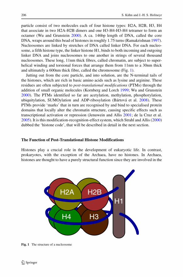
Histones are small, basic proteins that complex together to form a core particlearound which DNA wraps to form a nucleosome (Ramakrishnan 1997). This core
206 S. Kuhn and J.-H. S. Hofmeyr
particle consist of two molecules each of four histone types: H2A, H2B, H3, H4that associate in two H2A-H2B dimers and one H3-H4-H3-H4 tetramer to form anoctamer (Wu and Grunstein 2000). A ca. 146bp length of DNA, called the coreDNA, wraps around this bead of histones in roughly 1.75 turns (Ramakrishnan 1997).Nucleosomes are linked by stretches of DNA called linker DNA. For each nucleo-some, a fifth histone type, the linker histone H1, binds to both incoming and outgoinglinker DNA and joins nucleosomes to one another in strings of several thousandnucleosomes. These long, 11nm thick fibres, called chromatin, are subject to super-helical winding and torsional forces that arrange them from 11nm to a 30nm thickand ultimately a 600nm thick fibre, called the chromosome (Fig. 1).
Jutting out from the core particle, and into solution, are the N-terminal tails ofthe histones, which are rich in basic amino acids such as lysine and arginine. Theseresidues are often subjected to post-translational modifications (PTMs) through theaddition of small organic molecules (Kornberg and Lorch 1999; Wu and Grunstein2000). The PTMs identified so far are acetylation, methylation, phosphorylation,ubiquitylation, SUMOylation and ADP-ribosylation (Bartova et al. 2008). ThesePTMs provide ‘marks’ that in turn are recognised by and bind to specialised proteindomains that locally alter the chromatin structure, causing specific effects such astranscriptional activation or repression (Jenuwein and Allis 2001; de la Cruz et al.2005). It is this modification-recognition-effect system, which Strahl and Allis (2000)dubbed the ‘histone code’, that will be described in detail in the next section.
The Function of Post-Translational Histone Modifications
Histones play a crucial role in the development of eukaryotic life. In contrast,prokaryotes, with the exception of the Archaea, have no histones. In Archaea,histones are thought to have a purely structural function since they are involved in the
H2A H2B
H4 H3
H1
Fig. 1 The structure of a nucleosome
Is the “Histone Code” an Organic Code? 207
AM M A A
A R T K Q T A R K S T G G K A P R K1 2 3 4 5 6 7 8 9 10 11 12 13 14 15 16 17 18
A M MQ L A T K A A E K S A P A T G G V K19 20 21 22 23 24 25 26 27 28 29 30 31 32 33 34 35 36
MK D F K T D L37 . . . 77 78 79 80 81 82
A A AS G R G K G G K G L G K G G A K R H1 2 3 4 5 6 7 8 9 10 11 12 13 14 15 16 17 18
MR K V L R D19 20 21 22 23 24
UA V L L P K K T E S H H113 114 115 116 117 118 119 120 121 122 123 124
U UK V T K Y T S S K120 121 122 123 124 125 126 127 128
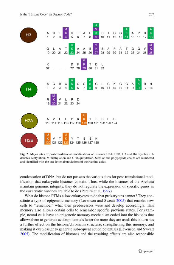
Fig. 2 Major sites of post-translational modifications of histones H2A, H2B, H3 and H4. Symbols: Adenotes acetylation, M methylation and U ubiquitylation. Sites on the polypeptide chains are numberedand identified with the one-letter abbreviations of their amino acids
condensation of DNA, but do not possess the various sites for post-translational mod-ification that eukaryotic histones contain. Thus, while the histones of the Archaeamaintain genomic integrity, they do not regulate the expression of specific genes asthe eukaryotic histones are able to do (Pereira et al. 1997).
What do histone PTMs allow eukaryotes to do that prokaryotes cannot? They con-stitute a type of epigenetic memory (Levenson and Sweatt 2005) that enables newcells to “remember” what their predecessors were and develop accordingly. Thismemory also allows certain cells to remember specific previous states. For exam-ple, neural cells have an epigenetic memory mechanism coded into the histones thatallows them to generate action potentials faster the more they are used; this in turn hasa further effect on the histone/chromatin structure, strengthening this memory, andmaking it even easier to generate subsequent action potentials (Levenson and Sweatt2005). The modification of histones and the resulting effects are also responsible
208 S. Kuhn and J.-H. S. Hofmeyr
for the spatio-temporal regulation of genetic activity during cell differentiation anddevelopment. In higher eukaryotes, for example, the Hox genes, which are respon-sible for proper embryonic segmentation and development, are regulated throughhistone modifications (Zhu et al. 2005; Wang et al. 2007). Regulation through thehistone code extends to virtually every gene and is absolutely crucial to the normalfunctioning of an organism.
Histone PTMs have been of particular importance in recent advances in stem cellresearch. Without the proper epigenetic programming provided by histone PTMs (interms of timing, localisation and specificity), the project of inducing stem cells to dif-ferentiate into the desired tissue type would be impossible to realise. Those researchgroups that have realised this are now making the necessary effort to understand andharness the histone PTMs (Gifford et al. 2013).
The Histone Post-Translational Modification Zoo
What follows is a discussion of the major histone modifications through acetylation,methylation and ubiquitylation (Fig. 2). The list of modifications is not exhaus-tive; we chose those modifications for which there is enough information abouttheir recognition and effects to enable us to judge them as possible elements ofan organic code. For most modifications by phosphorylation, SUMOylation andADP-ribosylation the required information is to our knowledge not yet available.
The best-studied post-translational modifications are the histone acetylations atlysine residues (Wang et al. 2008). Originally it was thought that the decrease in pos-itive charge brought about by the acetylation of lysine residues in histones wouldaffect the electrochemical association between histones and DNA, and that this wouldresult in the variety of biological effects so far observed (Berger 2002; Shahbazianand Grunstein 2007). While partly true, we now know that this is not the entire pic-ture. Several more recent discoveries have cast reasonable doubt on this hypothesis(Agalioti et al. 2002; Peterson and Laniel 2004). The discovery of modificationsthrough methylation, ubiquitylation and phosphorylation, none of which introduce achange in charge, provided sufficient evidence that if there is an effect on the electri-cal charge of histones, it would be ancillary to the main event (Peterson and Laniel2004).
In what follows the nomenclature convention HxKy is used, where x identifies thehistone in question, and y the lysine position (K is the one-letter abbreviation for theamino acid lysine, the three-letter abbreviation is Lys). Acetylation, methylation, andubiquitylation are indicated by HxKyac, HxKymet, and HxKyq respectively.
Acetylation
First identified almost 50 years ago by Allfrey et al. (1964), histone acetylationsby the histone acetyltransferases (HATs) have become the best characterised of his-tone PTMs (Turner 2000). Of the histones, H3 and H4 are most often acetylated,with lysine residues being the common target Rice and Allis (2001). Currently
Is the “Histone Code” an Organic Code? 209
it seems that only lysine residues in histones are acetylated (Kouzarides 2007).These acetylated histones are mostly associated with the activation of transcription(Toth et al. 2004). The acetylation of histones is reversible through the action ofhistone deacetylases (HDACs) (Brownell et al. 1996), which keeps them in a stateof high turnover. Furthermore, it seems that the functions associated with acety-lated histones (mostly restricted to transcriptional activation) are less diverse thanthose associated with methylated histones. Table 1 summarises the major histoneacetylations, which are discussed in detail in the following.
H3K9 Acetylation of H3K9 to H3K9ac is associated with the initiation and elonga-tion phases of transcription (Agalioti et al. 2002; Wang et al. 2008; Pokholok et al.2005). This mark acts oppositely to methylation of the Lys9 residue, which codesfor transcriptional repression. Strasak et al. (2009) have shown that H3K9ac is alsoimportant for nuclear reorganisation of the chromatin, since the inhibition of HDACscaused a spike in acetylation and prevented the binding of heterochromatin protein1 (HP1) to H3K9met. The Gcn5 HAT enzyme binds to H3K9 prior to initiation oftranscription and there is a subsequent peak in acetylated histones associated with thetranscriptional start site of active genes (Pokholok et al. 2005).
H3K14 Like H3K9ac, H3K14ac is associated with the initiation of transcription andis similarly tied to Gcn5 recruitment at the transcriptional start sites of various genes(Pokholok et al. 2005). This mark also correlates with an increase in transcriptionrates.
Table 1 The major histone acetylations and ubiquitylations, the binding domains that specificallyrecognise them, and their corresponding cellular effects
Modification Binding domain Biological effect References
H3K9ac bromodomain Transcriptional initiation (Agalioti et al. 2002)
H3K14ac bromodomain Transcriptional initiation (Pokholok et al. 2005)
H4K5ac bromodomain Transcriptional initiation (Rundlett et al. 1998;
in embryonic genes Chen et at. 2012)
H4K8ac bromodomain Transcriptional initiation (Agalioti et al. 2002)
and chromatin remodelling
H4K16ac bromodomain Transcriptional initiation (Gelbart et al. 2009;
(prevents the binding of the ISWI Kurdistani and Grunstein 2003;
and Sir3 transcriptional silencers) Kurdistani 2004)
H2BK120ac bromodomain Provides a binding site for an E3 (Gatta et al. 2011)
ubiquitin ligase to ubiquitylate
H2AK119
H2Bk120q Cps35 Methylation of H3K79, transcriptional (Jeltsch and Rathert 2008;
activation, transcriptional repression Osley 2004; Yuan et al. 2011)
H2BK119q unkown domain Transcriptional repression (Wang et al. 2004)
210 S. Kuhn and J.-H. S. Hofmeyr
H3K18 Of all the histones tested by Kurdistani et al. (2004), acetylation of H3K18increased transcriptional activity to the highest degree. Acetylation of H3K18 seemsto prevent that of H4K16 and vice versa, although by which mechanism is not known.
H3K23 H3K23ac is acetylated at Lys23 and with, no methyl group at Lys36, bindsto TRIM24, a chromatin and estrogen response modulator. These two sites are recog-nised by the two binding domains of the PHD-bromo cassette on TRIM24 (Tsai et al.2010) (the plant-homeo finger and bromo domains of proteins that bind to histoneswill be discussed in a later section). This TRIM24-H3K23ac complex is associatedwith the up-regulation of estrogen-related genes associated with cell-proliferationand tumorigenesis. Aberrant TRIM24 expression (and the resulting disruption inH3K23ac patterns) have adverse consequences on breast cancer survival rate (Tsaiet al. 2010). Dimethylated and trimethylated H3K4met are other marks that counteractivation by H3K23ac by preventing the binding of TRIM24 and thus suppressingactivation of transcription (Pray-Grant et al. 2005; Tsai et al. 2010).
H4K5 H4K5ac plays a crucial role in the earliest stages of embryonic development(Shi et al. 2008; Chen et al. 2012) and is, like most other acetylated histones, stronglycorrelated with transcriptional activation (Grunstein 1997; Rundlett et al. 1998).
H4K8 H4K8ac is associated with SWI/SNF recruitment, which in turn is responsiblefor ATP-dependent chromatin remodelling (Agalioti et al. 2002).
H4K16 Unlike other acetylated histones, the absence as well as the presence of anacetylated Lys16 on H4K16 has a function. Like most acetylated histones, H4K16acis linked to the up-regulation of transcription (Strahl and Allis 2000; Gelbart et al.2009). However, removing the acetyl group from H4K16ac allows for the interactionof the Sir3 transcription-silencing protein with H4 (Kurdistani and Grunstein 2003).Similarly the ISWI protein, which is part of a nucleosome-remodelling complex asso-ciated with silent chromatin, interacts with de-acetylated H4K16 (Kurdistani et al.2004). Further, as mentioned before, it appears that there is an antagonistic rela-tionship between H4K16ac and H3K18ac in that an increase in H4K16ac decreasesH3K18ac and vice versa (Kurdistani et al. 2004). The precise reason and mechanismfor this is not yet known.
H4K16ac also engages in extensive cross-talk with the transcriptionally repres-sive mark, H3K36met2/3. The latter recruits an H4K16-specific deacetylase whichabolishes the transcriptional activation of H4K16ac (Bell et al. 2007).
H2BK120 Acetylation of H2BK120 is closely related to its ubiquitylation (Gattaet al. 2011). The addition of ubiquitin to H2BK120 needs the presence of anacetyl group at Lys120 since the inhibition of KAT3A/B, the acetylase that acety-lates Lys120, by siRNA prevents ubiquitylation of H2BK120. However, these twomarks are inversely correlated in that an increase in H2BK120q is accompaniedby a decrease in H2BK120ac. Preliminary evidence suggests that the acetylationof H2BK120 acts as a ‘hot’ switch, essentially keeping the position primed forubiquitylation by an E3-ligase.
Is the “Histone Code” an Organic Code? 211
Methylation
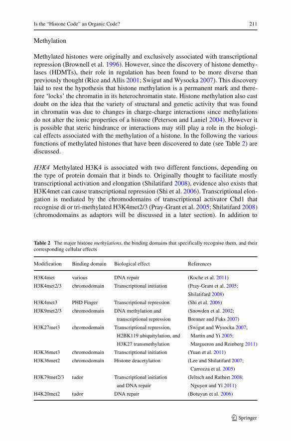
Methylated histones were originally and exclusively associated with transcriptionalrepression (Brownell et al. 1996). However, since the discovery of histone demethy-lases (HDMTs), their role in regulation has been found to be more diverse thanpreviously thought (Rice and Allis 2001; Swigut and Wysocka 2007). This discoverylaid to rest the hypothesis that histone methylation is a permanent mark and there-fore ‘locks’ the chromatin in its heterochromatin state. Histone methylation also castdoubt on the idea that the variety of structural and genetic activity that was foundin chromatin was due to changes in charge-charge interactions since methylationsdo not alter the ionic properties of a histone (Peterson and Laniel 2004). However itis possible that steric hindrance or interactions may still play a role in the biologi-cal effects associated with the methylation of a histone. In the following the variousfunctions of methylated histones that have been discovered to date (see Table 2) arediscussed.
H3K4 Methylated H3K4 is associated with two different functions, depending onthe type of protein domain that it binds to. Originally thought to facilitate mostlytranscriptional activation and elongation (Shilatifard 2008), evidence also exists thatH3K4met can cause transcriptional repression (Shi et al. 2006). Transcriptional elon-gation is mediated by the chromodomains of transcriptional activator Chd1 thatrecognise di or tri-methylated H3K4met2/3 (Pray-Grant et al. 2005; Shilatifard 2008)(chromodomains as adaptors will be discussed in a later section). In addition to
Table 2 The major histone methylations, the binding domains that specifically recognise them, and theircorresponding cellular effects
Modification Binding domain Biological effect References
H3K4met various DNA repair (Koche et al. 2011)
H3K4met2/3 chromodomain Transcriptional initiation (Pray-Grant et al. 2005;
Shilatifard 2008)
H3K4met3 PHD Finger Transcriptional repression (Shi et al. 2006)
H3K9met2/3 chromodomain DNA methylation and (Snowden et al. 2002;
transcriptional repression Brenner and Fuks 2007)
H3K27met3 chromodomain Transcriptional repression, (Swigut and Wysocka 2007;
H2BK119 ubiquitylation, and Martin and Yi 2005;
H3K27 transmethylation Margueron and Reinberg 2011)
H3K36met3 chromodomain Transcriptional initiation (Yuan et al. 2011)
H3K36met2 chromodomain Histone deacetylation (Lee and Shilatifard 2007;
Carrozza et al. 2005)
H3K79met2/3 tudor Transcriptional initiation (Jeltsch and Rathert 2008;
and DNA repair Nguyen and Yi 2011)
H4K20met2 tudor DNA repair (Botuyan et al. 2006)

212 S. Kuhn and J.-H. S. Hofmeyr
possessing two chromodomains, Chd1 also contains a DNA-binding domain and ahelicase domain (Simic et al. 2003). When bound to H3K4met2/3 via its chromod-omain, Chd1 associates with various transcription elongation factors (Spt5, Pob3,Rtf1) (Simic et al. 2003; McDaniel et al. 2008). Evidence also suggests that in highereukaryotes Chd1 is also responsible for transcriptional regulation and termination(McDaniel et al. 2008). Schneider et al. (2003) have discovered that the presence ofH3K4met decreases toward the 3′ end of genes, suggesting that the absence of thismodification provides a signal for the termination of transcription.
On the other hand, proteins that bind to H3K4met3 via a PHD finger usually causeactive repression of genes (Shi et al. 2006). However, even this role is not as clear-cutas it seems, as there are several instances where the PHD finger is part of a proteinthat is involved in the activation of gene transcription (Shilatifard 2008). Here is aclear case of the flexibility often encountered in regulatory mechanisms. Not onlyis H3K4met associated with activation and repression of transcription, but also withactivities such as DNA repair, chromatin remodelling, and sporulation (Roest et al.2004; Koche et al. 2011). Even the unmethylated H3K4 has a known function: bind-ing to the PHD domain of the autoimmune regulator (AIRE) resulting in chromatinremodelling and active transcription (Org et al. 2008). In Drosophila, trimethylatedH3K4met3 prevents binding of H3K27met3 to polycomb group (PcG) proteins and,therefore, repression of transcription caused by PcG proteins (Schwartz and Pirrotta2008).
H3K9 To date, di- and tri-methylated H3K9 has been associated predominantlywith transcriptional repression (Snowden et al. 2002; Turner 2002). This is due toH3K9met3 acting as a ‘beacon’ to which several DNA-methylases (DNMTs) arerecruited (Brenner and Fuks 2007). It appears that repression is due to a coopera-tion between HP1, the protein that recognises the H3K9met3 mark, and the variousDNMTs (1, 3a, 3b) (Smallwood et al. 2007).
Vakoc et al. (2005), however, reported that H3K9met2/3 can also be associatedwith active transcription. It appears that H3K9met2/3 seems to stabilise the openreading frame to permit transcriptional elongation to occur, rather than being associ-ated with the initiation of transcription. Certain genes show increases in H3K9met3and H3K4met3 levels upon initiation of transcription, but, despite this apparent cor-relation, the evidence that suggests that these methyl marks interact with one anotheris weak (Vakoc et al. 2005).
H3K27 H3K27 is methylated by the SET-containing E(Z) (EZH2 in humans) pro-teins which form part of the Polycomb group (PcG) of proteins (Martin and Yi 2005).H3K27met3 is read by the Polycomb repressor-complex 1 (PRC1), which containsa chromodomain. H3K27met3 is mainly associated with the repression of transcrip-tion, X chromosome inactivation and genomic imprinting (Martin and Yi 2005).Furthermore, it appears that H3K27met is an important signal for the localisation of aPRC1-like E3 ligase that ubiquitylates histone 2A at Lys119 since, in the absence ofE(Z), H2AK119 ubiquitylation does not occur (Martin and Yi 2005). However whilethey are related, the methyl mark is not dependent on the ubiquitin mark, indicatingthat H2AK119q depends on H3K27met3, and not vice-versa (Wang et al. 2004).
Is the “Histone Code” an Organic Code? 213
So far, H3K27met3 has been linked only to transcriptional repression (Swigut andWysocka 2007), which is due to the interactions between H3K27met3 and variousregulatory complexes, and not to alteration of the nucleosomal architecture (Simonand Kingston 2009). This holds for mono-, di-, and tri-methyl marks (Martin andYi 2005). It was first thought that these methyl marks cause permanent repression,but the recent discovery of a H3K27-specific demethylase undermines this idea (DeSanta et al. 2007).
Another interesting feature of H3K27met3 is that it is a self-perpetuating mark(Margueron and Reinberg 2011) like H3K9. H3K27me3 recruits a histone methyl-transferase (HMT) to monomethylated H3K27me2 to its trimethyl form. Of thethree types of methyl marks, dimethylated H3K27me2 is the most abundant, beingpresent in roughly 50 % of nucleosomes (Schwartz and Pirrotta 2008; Margueron andReinberg 2011). While H3K27met2 itself is of limited importance in gene repression,evidence suggests that it may not only act as an inactive precursor of H3K27met3,but also prevents methylated H3K27 from being acetylated to H3K27ac(Margueronand Reinberg 2011), a mark associated with active transcription and antagonistic toH3K27met3 (Cui et al. 2009).
H3K36 H3K36met prevents the methylation of H3K27 and is commonly associatedwith the activation of transcription. Experimental evidence shows that the repressivemark H3K27met3 rarely co-exists with the activating H3K36met2/3 in the same his-tone (Yuan et al. 2011). This study also showed that histones rarely, if ever, existwithout one of these modifications, and that nucleosomes therefore do not exist in a‘blank’ state. It also showed that although a pre-existing H3K36-methyl mark doesinhibit the methylation of H3K27 by PRC1, the reverse is not true. This indicates thatthe unmethylated H3K36 position could play a role in the binding of PRC1 or, alter-natively, that the methylated H3K36 somehow prevents the binding of PRC1. Theexact mechanism is not known, but it is clear that the Lys36 position in H3 is impor-tant since a mutation of this lysine to alanine decreases PRC1 activity considerably(Yuan et al. 2011).
The Eaf3 protein subunit of the deacetylase Rpd3 contains a chromodomain essen-tial to the recognition of H3K36met2 by Rpd3. The absence of this subunit orits chromodomain has been shown to leave histone acetylation levels unchanged(Lee and Shilatifard 2007). Interestingly, Carrozza et al. (2005) have identified theH3K36me2 mark in actively transcribed regions of the genome and it has beenpositively associated with transcription elongation. However, the deacetylation thatresults from the binding of Rpd3 is linked to transcriptional repression (Lee andShilatifard 2007).This mark has also been strongly associated with the recruitmentof a HDAC to H4K16ac during active transcription (Bell et al. 2007) effectivelyinducing transcriptional silencing. This suggests an intricate web of cross-talk andinter-regulation between the various histone modifications. A possible function ofthis deacetylation is to prevent spurious transcription from being initiated (Leeand Shilatifard 2007) or to regulate the length of the open reading frame in orderto allow alternate transcripts to be produced or simply to signal the end of tran-scription, allowing the euchromatic area to condense to its heterochromatin stateagain.
214 S. Kuhn and J.-H. S. Hofmeyr
H3K79 Methylated H3K79 is associated mostly with transcriptional activation, how-ever it has also been found to occasionally result in transcriptional repression (Sunand Allis 2002; Steger et al. 2008). The most interesting aspect of H3K79 is theextensive crosstalk with H2BK120q (Jeltsch and Rathert 2008). H2BK120q has beenshown to recruit the Dot1 methylase, which is responsible for more than 90 % of theH3K79 methylations (McGinty et al. 2008; Jeltsch and Rathert 2008; Mellor 2009).Khan and Hampsey (2002) have shown that it is indeed H3K79met that is associatedwith transcriptional activation, since the replacement of Lys79, or the deletion of thedot1 gene, both result in the silencing of a particular genic region. The methylatedLys79 seems to be particularly enriched on the histone variant H3.3, which is preva-lent in actively transcribed regions of the genome, but there is no clear understandingas to why this is so (McKittrick et al. 2004).
H4K20: Unlike the previously mentioned histone methylations, the methylation ofH4K20 is possesses a single function only; trimethylated H4K20 is associated withtranscriptional repression (Schotta et al. 2004).
Ubiquitylation
Ubiquitylation of histones involves the addition of ubiquitin, a highly conserved, 76amino acid protein molecule, to the lysine residues in a histone. It was the ubiquity-lation of H2A that first heralded the discovery of ubiquitin and the modification ofhistones (Schlesinger et al. 1975; Osley 2004). Unfortunately the importance of bothdiscoveries has been underestimated for some time. Unlike acetylated or methylatedhistones, the two instances of histone ubiquitylation which have been studied in somedetail (see Table 1) seem to be recognised by structurally unrelated binding domains,although this is not yet certain. Ubiquitin is ubiquitous (hence the name) anddiverse enough in function to perhaps warrant the investigation of a code of its own(Komander and Rape 2012). Originally it was proposed that histone-ubiquitylationaffected transcription via three possible mechanisms: firstly ubiquitin itself, due to itsrelatively large size, directly affected the chromatin structure and histone/DNA affin-ity, secondly that ubiquitin acted as a beacon for the recruitment of various regulatoryproteins, and thirdly that ubiquitin affected transcription by directly influencing theother histone modifications (Zhang 2003). Currently the evidence seems to favourthe third option, however the first two have not been entirely ruled out.
H2AK119 In most most eukaryotes, the ubiquitylated H2AK119 (H2AK119q) ispresent in 5–15 % of histones (de Napoles et al. 2004). H2AK119q is overwhelm-ingly associated with transcriptional repression, either directly or through the indirectmechanism mentioned earlier that involves the recruitment of ubiquitin to transcrip-tionally silent chromatin (such as a Barr body) that has already been marked byH3K27met3 (Wang et al. 2004; de Napoles et al. 2004). Ubiquitin is ligated to H2Aby the PRC1-like proteins Ring1A and Ring1B, both of which are crucial for formingand maintaining the H2AK119q mark (de Napoles et al. 2004). These PRC1-like pro-teins contain a chromodomain that specifically reads the H3K27met3 mark, directlyimplicating it in the ubiquitylation of H2AK119 (Martin and Yi 2005). However,
Is the “Histone Code” an Organic Code? 215
Tavares et al. (2012) have recently shown that while H3K27met3 does code forH2AK119q, it is not essential since PRC2-null mutants, which abolish H3K27met3entirely, show near normal levels of H2AK119q. The precise mechanism by whichH2AK119q is able to facilitate the repression of transmission is not yet known.
H2BK120 H2B is ubiquitylated in higher eukaryotes at Lys120 and in lower eukary-otes at Lys123. Both H2BK120q and H2BK123q have been shown to directlyinfluence the methylation of H3K79 by the Dot1 methylase (McGinty et al. 2008;Jeltsch and Rathert 2008) and the methylation of H3K4 by COMPASS, a Set1methyltransferase (Osley 2004; Latham et al. 2011). Both H3K79met and H3K4metare associated with transcriptional regulation, however the majority of H2BK120q-mediated methylation is responsible for transcriptional activation. It is interesting thatthe presence of H2BK120q does not affect the methylation of H3K36, which is alsolinked to active transcription (Yuan et al. 2011). It also seems that H2K123q is notessential for the monomethylation of histones, since in the absence of H2BK123q,Dot1 and COMPASS still monomethylate H3K4 and H3K79, however what isimpeded is their ability to di and trimethylated these positions in the presence of apre-existing monomethyl mark (Shahbazian et al. 2005). Osley (2004) has shownthat H2BK120q/H2BK123q can also function as a transcription-repressing mark,although the precise mechanism for this is unknown. It is thought that, as with tran-scriptional activation, this is due to the effect that ubiquitylated histones have on theother histone modifications (Dover et al. 2002; Zhang 2003).
Binding Domains: The Adaptors of the Histone Code
Acetyl-Recognising Domains
Currently, the only protein domain known to be capable of recognising acetyl-lysinesis the bromodomain (Mujtaba et al. 2007). For each acetylated Lys in a particularhistone there is a specific bromodomain. Within their ca. 110 amino acid structure,bromodomains contain a conserved hydrophobic pocket of aromatic amino acidsthat specifically recognises a specific acetyl-lysine (Owen et al. 2000; Mujtaba et al.2007). Studies have shown that if one or more of the critical residues in this pocketare mutated, the bromodomain loses its ability to recognise a specific acetyl-lysine(Mujtaba et al. 2002). This shows that the bromodomain is absolutely essential forthe recognition of acetyl-lysines and that this structural domain fulfils one of crite-ria for being an adaptor in the histone code, recognising the sign posed by a specificacetyl-lysine.
Methyl-Recognising Domains
Unlike acetylated histones, the methylated histones recognised, depending on theirlocation and degree of methylation, by a much greater variety of protein domains,such as the Royal family of protein domains (Maurer-Stroh et al. 2003). The mostcommon of these are the chromodomains, Tudor, plant-Agenet, MBT and PHD
216 S. Kuhn and J.-H. S. Hofmeyr
finger domains, as well as several smaller domains, such as PWWP and JMJ (Cavalliand Paro 1998; Mellor 2006; Kim et al. 2006). Each of these domains discriminateaccording to the degree of methylation, the position of the lysine and frequently evenaccording to the surrounding residues, although there is evidence that suggests thatthe latter serve only to strengthen binding: the crucial element remains the modifiedresidue (Kouzarides 2007).
Evidence is emerging which suggests that the chromodomains can be exper-imentally exchanged between proteins (Fischle et al. 2003). In this study thechromodomains of the protein Polycomb (Pc) and of HP1 were interchanged,giving PcHP1 and HP1Pc respectively. As a result, PcHP1 recognised the origi-nal target of HP1 and HP1Pc recognised that of Pc (H3K27met and H3K9metrespectively).
This suggests that although these domains give each protein a specific identityin terms of being able to recognise a specific modification, they are not ‘locked’ toa protein. If these findings can be confirmed, and even expanded to include inter-domain exchanges, they would further cement the claim that a protein domain acts asthe molecular adaptor for the histone code. We can however say with confidence thatthose domains involved in the recognition of methyl-lysines are molecular adaptorsfor the histone code. They ably recognise the methyl-lysines as organic signs andsubsequently translate them into their corresponding biological effect.
Ubiquitin-Recognising Domains
While the number of domains that interact with ubiquitin is large, very few have beenfound in proteins that specifically interact with histone-bound ubiquitin. One of theseis the zinc-finger (ZnF), ubiquitin-specific processing protease (UBP) which is foundin the HDAC6 deacetylase (Hurley et al. 2006; Husnjak and Dikic 2012).
Unfortunately it seems that, currently, no ubiquitin-binding domain has been iden-tified on the Dot1 or COMPASS methylases which bind to H2BK120q. However ifone is to be found, it is likely to be found on Cps35 which binds to H2BK120q andthen recruits the COMPASS methylase (Lee et al. 2007). The domain responsible forthe binding of Dot1 to ubiquitin is to the best of our knowledge not known.
How Does it all Fit Together? Is the Histone Code an Organic Code?
As discussed earlier, for the histone modification system to act as an organic codewe need to demonstrate that not only does it consist of two independent worlds, herethat of histone modifications (which would act as organic signs) and their biologicaleffects (the meaning of the signs), but that there are chemical molecules, called adap-tors, that recognise the signs with absolute specificity and translate them into theirmeanings. Furthermore, it must be possible in principle (and, preferably, by experi-ment) to alter the rules of the code, i.e., the relationships between organic signs andtheir meanings, by interchanging those parts of the adaptor molecules that recognisethe signs.
Is the “Histone Code” an Organic Code? 217
From the above discussion of histone post-translational modifications it is clearthat each of these modifications is linked to a highly specific biological effect; toour knowledge there are no instances where a particular PTM in a particular organ-ism results in more than one biological effect. These relationships can therefore beregarded as a set of rules between the independent worlds of PTMs (the organicsigns) and biological effects (biological meanings). In order for this set of rules to beregarded as code, it is, however, necessary to establish that are molecules that act asthe adaptors that translate signs into their meanings. From the details of the histonePTMs it is clear that the role of adaptors is played by the effector proteins that consistof a binding domain that specifically recognises the PTM and a domain that acts as amediator of biological effect associated with that PTM, albeit transcriptional regula-tion, structural remodelling of chromation, or even a post-translational modificationof another histone.
As explained previously, the relationship between an organic sign and its meaningin an organic code must be arbitrary in the sense that it is not determined by the lawsof chemistry or physics (although completely compatible with these laws), but ratherhas the nature of a convention that arose naturally through evolution. The relationshipbetween a histone PTM and its biological effect fulfils this criterion. The recogni-tion of a specific histone PTM by its corresponding binding domain is analogous tothe interaction of a codon on mRNA with its corresponding anticodon on an tRNA.Without the binding domain being part of the effector protein, the effect specifiedby a certain PTM will not come to be. For example, the mammalian Brd4 protein, aprotein involved in transcriptional regulation, contains two bromodomains, and thedeletion of even one of these bromodomains abolishes the interaction of Brd4 withacetylated histones (Dey et al. 2003) and prevents the biological effect of Brd4. Inanother study, Flanagan et al. (2005) showed that the mutation of tryptophan 64 or 67in the active site of one of the two chromodomains of a CHD1 protein significantlyreduced the ability of this protein to bind H3Kmet4.
That the histone code exhibits the required arbitrariness of an organic code hasbeen proven by experimentally altering the coding scheme. As previously mentioned,Fischle et al. (2003) replaced the binding domain of one effector protein with thatof a different effector protein; the modified effector protein now had the bindingspecificity of the other one. More specifically, they interchanged the chromodomainof the polycomb (Pc) protein with the chromodomain of heterochromatin protein 1(HP1). The hybrid HP1Pc now only recognised the H3K27met mark, the target ofPc, instead of the original H3K9met. Similarly, the hybrid PcHP1 now recognisedH3K9met instead of H3K27met.
These considerations show that the histone code fulfils all the criteria for anorganic code. Although we probably do not yet know the complete histone code, wehave, as argued in this paper, more than enough information to be able to recognisethe histone code as a bona fide organic code.
As with the genetic code, which after its discovery came as a “bolt from the blue”but was quickly surrounded by a “protective belt” that emptied it from all its revolu-tionary potential (Barbieri 2008), we hope that we have ensured with this paper thatthe histone code does not suffer the same fate.
218 S. Kuhn and J.-H. S. Hofmeyr
Acknowledgements The authors thank Marcello Barbieri and Joachim de Beule for their boundless sup-port and guidance. The authors acknowledge financial support from the South African National ResearchFoundation (NRF). Any opinion, findings, and conclusions or recommendations expressed in this materialare those of the authors, and therefore the NRF does not accept any liability in regard thereto.
References
Agalioti, T., Chen, G., Thanos, D. (2002). Deciphering the transcriptional histone acetylation code for ahuman gene. Cell, 111(3), 381–392.
Allfrey, V.G., Faulkner, R., Mirsky, A.E. (1964). Acetylation and methylation of histones and their possi-ble role in the regulation of RNA synthesis. Proceedings of the National Academy of Sciences of theUnited States of America, 51(5), 786.
Barbieri, M. (2008). Biosemiotics: a new understanding of life. Naturwissenschaften, 95(7), 577–599.Barbieri, M. (2012). Code Biology–A New Science of Life.Barbieri, M. (1985). The semantic theory of evolution: Chur: Harwood Academic Publishers.Barbieri, M. (1998). The organic codes. The basic mechanism of macroevolution. Rivista di Biologia,
91(3), 481.Barbieri, M. (2003). The organic codes: An introduction to semantic biology. Cambridge: Cambridge
University Press.Bartova, E., Krejcı, J., Harnicarova, A., Galiova, G., Kozubek, S. (2008). Histone modifications and
nuclear architecture: a review. Journal of Histochemistry and Cytochemistry, 56(8), 711–721.Bell, O., Wirbelauer, C., Hild, M., Scharf, A.N.D., Schwaiger, M., MacAlpine, D.M., Zilbermann,
F.rederic., van Leeuwen, F., Bell, S.P., Imhof, A., Garza, D., Peters, A.H.F.M., Schubeler, D.(2007). Localized H3K36 methylation states define histone H4K16 acetylation during transcriptionalelongation in Drosophila. EMBO Journal, 26(24), 4974–4984.
Berger, S.L. (2002). Histone modifications in transcriptional regulation. Current Opinion in Genetics andDevelopment, 12(2), 142–148.
Botuyan, M.V., Lee, J., Ward, I.M., Kim, J.-E., Thompson, J.R., Chen, J., Mer, G. (2006). Structural basisfor the methylation state-specific recognition of histone H4-K20 by 53BP1 and Crb2 in DNA repair.Cell, 127(7), 1361–1373.
Brenner, C., & Fuks, F. (2007). A methylation rendezvous: reader meets writers. Developmental Cell,12(6), 843–844.
Brownell, J.E., Zhou, J., Ranalli, T., Kobayashi, R., Edmondson, D.G., Roth, S.Y., Allis, C.D. (1996).Tetrahymena histone acetyltransferase A: a homolog to yeast Gcn5p linking histone acetylation togene activation. Cell, 84(6), 843–852.
Carrozza, M.J., Li, B., Florens, L., Suganuma, T., Swanson, S.K., Lee, K.K., Shia, W.-J., Anderson, S.,Yates, J., Washburn, M.P., Workman, J.L. (2005). Histone H3 methylation by Set2 directs deacety-lation of coding regions by Rpd3S to suppress spurious intragenic transcription. Cell, 123(4), 581–592.
Cavalli, G., & Paro, R. (1998). Chromo-domain proteins: linking chromatin structure to epigeneticregulation. Current Opinion in Cellular Biology, 10(3), 354–360.
Chen, C.-H., Chang, W.-F., Liu, C.-C., Su, H.-Y., Shyue, S.-K., Cheng, W.T.K., Chen, E.Y., Wu, S.-C.,Du, F., Sung, L.-Y. (2012). Spatial and temporal distribution of Oct-4 and acetylated H4K5 in rabbitembryos. Reproductive BioMedicine Online, 24(4), 433–442.
Crick, F.H.C., Barnett, L., Brenner, S., Watts-Tobin, R.J. (1961). General nature of the genetic code forproteins. Nature, 192(4809), 1227–1232.
Cui, K., Zang, C., Roh, T.-Y., Schones, D.E., Childs, R.W., Peng, W., Zhao, K. (2009). Chromatin sig-natures in multipotent human hematopoietic stem cells indicate the fate of bivalent genes duringdifferentiation. Cell Stem Cell, 4(1), 80–93.
de la Cruz, X., Lois, S., Sanchez-Molina, S., Martınez-Balbas, M.A. (2005). Do protein motifs read thehistone code? Bioessays, 27(2), 164–175.
de Napoles, M., Mermoud, J.E., Wakao, R., Tang, Y.A., Endoh, M., Appanah, R., Nesterova, T.B., Silva,J., Otte, A.P., Vidal, M., Koseki, H., Brockdorff, N. (2004). Polycomb group proteins Ring1A/B linkubiquitylation of histone H2A to heritable gene silencing and X inactivation. Developmental Cell,7(5), 663–676.
Is the “Histone Code” an Organic Code? 219
De Santa, F., Totaro, M.G., Prosperini, E., Notarbartolo, S., Testa, G., Natoli, G. (2007). The histone H3lysine-27 demethylase Jmjd3 links inflammation to inhibition of polycomb-mediated gene silencing.Cell, 130(6), 1083–1094.
Dey, A., Chitsaz, F., Abbasi, A., Misteli, T., Ozato, K. (2003). The double bromodomain protein Brd4binds to acetylated chromatin during interphase and mitosis. Proceedings of the National Academy ofSciences of the United States of America, 100(15), 8758–8763.
Dover, J., Schneider, J., Tawiah-Boateng, M.A., Wood, A., Dean, K., Johnston, M., Shilatifard, A. (2002).Methylation of histone H3 by COMPASS requires ubiquitination of histone H2B by Rad6. Journal ofBiological Chemistry, 277(32), 28368–28371.
Elder, D. (1979). An epigenetic code. Differentiation, 14(1), 119–122.Fischle, W., Wang, Y., Jacobs, S.A., Kim, Y., Allis, C.D., Khorasanizadeh, S. (2003). Molecular basis
for the discrimination of repressive methyl-lysine marks in histone H3 by Polycomb and HP1chromodomains. Genes & Development, 17(15), 1870–1881.
Flanagan, J.F., Mi, L.-Z., Chruszcz, M., Cymborowski, M., Clines, K.L., Kim, Y., Minor, W., Rastinejad,F., Khorasanizadeh, S. (2005). Double chromodomains cooperate to recognize the methylated histoneH3 tail. Nature, 438(7071), 1181–1185.
Gabius, H.J. (2000). Biological information transfer beyond the genetic code: the sugar code. Naturwis-senschaften, 87(3), 108–121.
Gatta, R., Dolfini, D., Zambelli, F., Imbriano, C., Pavesi, G., Mantovani, R. (2011). An acetylation-monoubiquitination switch on lysine 120 of H2B. Epigenetics, 6(5), 630–637.
Gelbart, M.E., Larschan, E., Peng, S., Park, P.J., Kuroda, M.I. (2009). Drosophila MSL complex glob-ally acetylates H4K16 on the male X chromosome for dosage compensation. Nature Structural andMolecular Biology, 16(8), 825–832.
Gifford, C.A., Ziller, M.J., Gu, H., Trapnell, C., Donaghey, J., Tsankov, A., Shalek, A.K., Kelley, D.R.,Shishkin, A.A., Issner, R., Zhang, X., Coyne, M., Fostel, J.L., Holmes, L., Meldrim, J., Guttman, M.,Epstein, C., Park, H., Kohlbacher, O., Rinn, J., Gnirke, A., Lander, E.S., Bernstein, B.E., Meissner,A. (2013). Transcriptional and epigenetic dynamics during specification of human embryonic stemcells. Cell, 153(5), 1149–1163.
Gimona, M. (2008). Protein linguistics and the modular code of the cytoskeleton. In The codes of life(pp. 189–206), Springer.
Grunstein, M. (1997). Histone acetylation in chromatin structure and transcription. Nature, 389(6649),349–352.
Hurley, J., Lee, S., Prag, G. (2006). Ubiquitin-binding domains. Biochemical Journal, 399, 361–372.Husnjak, K., & Dikic, I. (2012). Ubiquitin-binding proteins: decoders of ubiquitin-mediated cellular
functions. Annual Review of Biochemistry, 81, 291–322.Jeltsch, A., & Rathert, P. (2008). Putting the pieces together: histone H2B ubiquitylation directly stimulates
histone H3K79 methylation. Chembiochem, 9(14), 2193–2195.Jenuwein, T., & Allis, C.D. (2001). Translating the histone code. Science’s STKE, 293(5532), 1074.Khan, A.U., & Hampsey, M. (2002). Connecting the DOTs: covalent histone modifications and the
formation of silent chromatin. Trends in Genetics, 18(8), 387–389.Kim, J., Daniel, J., Espejo, A., Lake, A., Krishna, M., Li, X., Yi, Z., Bedford, M.T. (2006). Tudor, MBT
and chromo domains gauge the degree of lysine methylation. EMBO Reports, 7(4), 397–403.Koche, R.P., Smith, Z.D., Adli, M., Gu, H., Ku, M., Gnirke, A., Bernstein, B.E., Meissner, A. (2011).
Reprogramming factor expression initiates widespread targeted chromatin remodeling. Cell Stem Cell,8(1), 96–105.
Komander, D., & Rape, M. (2012). The ubiquitin code. Annual Review of Biochemistry, 81, 203–229.Kornberg, R.D., & Lorch, Y. (1999). Twenty-five years of the nucleosome, fundamental particle of the
eukaryote chromosome. Cell, 98, 285–294.Kouzarides, T. (2007). Chromatin modifications and their function. Cell, 128(4), 693.Kurdistani, S.K., & Grunstein, M. (2003). Histone acetylation and deacetylation in yeast. Nature Reviews
Molecular Cell Biology, 4(4), 276–284.Kurdistani, S.K., Tavazoie, S., Grunstein, M. (2004). Mapping global histone acetylation patterns to gene
expression. Cell, 117(6), 721–733.Latham, J.A., Chosed, R.J., Wang, S., Dent, S.Y.R. (2011). Chromatin signaling to kinetochores:
transregulation of Dam1 methylation by histone H2B ubiquitination. Cell, 146(5), 709–719.Lee, J.-S., & Shilatifard, A. (2007). A site to remember: H3K36 methylation a mark for histone
deacetylation. Mutation Research: Fundamental and Molecular Mechanisms, 618(1), 130–134.
220 S. Kuhn and J.-H. S. Hofmeyr
Lee, J.-S., Shukla, A., Schneider, J., Swanson, S.K., Washburn, M.P., Florens, L., Bhaumik, S.R., Shilati-fard, A. (2007). Histone crosstalk between H2B monoubiquitination and H3 methylation mediated byCOMPASS. Cell, 131(6), 1084–1096.
Levenson, J.M., & Sweatt, J.D. (2005). Epigenetic mechanisms in memory formation. Nature ReviewsNeuroscience, 6(2), 108–118.
Margueron, R., & Reinberg, D. (2011). The Polycomb complex PRC2 and its mark in life. Nature,469(7330), 343–349.
Martin, C., & Yi, Z. (2005). The diverse functions of histone lysine methylation. Nature Reviews MolecularCell Biology, 6(11), 838–849.
Maurer-Stroh, S., Dickens, N.J., Hughes-Davies, L., Kouzarides, T., Eisenhaber, F., Ponting, C.P. (2003).The Tudor domain ‘Royal Family’: tudor, plant agenet, chromo, PWWP and MBT domains. Trendsin Biochemical Sciences, 28(2), 69–74.
McDaniel, I.E., Lee, J.M., Berger, M.S., Hanagami, C.K., Armstrong, J.A. (2008). Investigations of CHD1function in transcription and development of Drosophila melanogaster. Genetics, 178(1), 583–587.
McGinty, R.K., Kim, J., Chatterjee, C., Roeder, R.G., Muir, T.W. (2008). Chemically ubiquitylated histoneH2B stimulates hDot1L-mediated intranucleosomal methylation. Nature, 453(7196), 812–816.
McKittrick, E., Gafken, P.R., Ahmad, K., Henikoff, S. (2004). Histone H3.3 is enriched in covalent mod-ifications associated with active chromatin. Proceedings of the National Academy of Sciences of theUnited States of America, 101(6), 1525–1530.
Mellor, J. (2006). It takes a PHD to read the histone code. Cell, 126(1), 22–24.Mellor, J. (2009). Linking the cell cycle to histone modifications: Dot1, G1/S, and cycling K79me2.
Molecular Cell, 35(6), 729–730.Mujtaba, S., Zeng, L., Zhou, M.M. (2007). Structure and acetyl-lysine recognition of the bromodomain.
Oncogene, 26(37), 5521–5527.Mujtaba, S., He, Y., Zeng, L., Farooq, A., Carlson, J.E., Ott, M., Verdin, E., Zhou, M.-M. (2002). Structural
basis of lysine-acetylated HIV-1 Tat recognition by PCAF bromodomain. Molecular Cell, 9(3), 575–586.
Nguyen, A.T., & Yi, Z. (2011). The diverse functions of Dot1 and H3K79 methylation. Genes &Development, 25(13), 1345–1358.
Nirenberg, M., Leder, P., Bernfield, M., Brimacombe, R., Trupin, J., Rottman, F., O’neal, C. (1965).Rna codewords and protein synthesis, vii. on the general nature of the rna code. Proceedings of theNational Academy of Sciences of the United States of America, 53(5), 1161.
Org, T., Chignola, F., Hetenyi, C., Gaetani, M., Rebane, A., Liiv, I., Maran, U., Mollica, L., Bottomley,M.J., Musco, G., Peterson, P. (2008). The autoimmune regulator PHD finger binds to non-methylatedhistone H3K4 to activate gene expression. EMBO Reports, 9(4), 370–376.
Osley, M.A. (2004). H2B ubiquitylation: the end is in sight. BBA Gene Structural Expression, 1677(1),74–78.
Owen, D.J., Ornaghi, P., Yang, J.-C., Lowe, N., Evans, P.R., Ballario, P., Neuhaus, D., Filetici, P., Travers,A.A. (2000). The structural basis for the recognition of acetylated histone H4 by the bromodomain ofhistone acetyltransferase Gcn5p. EMBO Journal, 19(22), 6141–6149.
Pereira, S.L., Grayling, R.A., Lurz, R., Reeve, J.N. (1997). Archaeal nucleosomes. Proceedings of theNational Academy of Sciences of the United States of America, 94(23), 12633–12637.
Peterson, C.L., & Laniel, M.-A. (2004). Histones and histone modifications. Current Biology, 14(14),546–551.
Pokholok, D.K., Harbison, C.T., Levine, S., Cole, M., Hannett, N.M., Lee, T.I., Bell, G.W., Walker, K.,Rolfe, A., Herbolsheimer, E., Zeitlinger, J., Lewitter, F., Gifford, D.K., Young, R.A. (2005). Genome-wide map of nucleosome acetylation and methylation in yeast. Cell, 122(4), 517–527.
Pray-Grant, M.G., Daniel, J.A., Schieltz, D., Yates, J.R., Grant, P.A. (2005). Chd1 chromodomain linkshistone H3 methylation with SAGA and SLIK-dependent acetylation. Nature, 433(7024), 434–438.
Ramakrishnan, V. (1997). Histone structure and the organization of the nucleosome. Annual Review ofBiophysics and Biomolecular Structure, 26(1), 83–112.
Rice, J.C., & Allis, C.D. (2001). Histone methylation versus histone acetylation: new insights intoepigenetic regulation. Current Opinion in Cellular Biology, 13(3), 263–273.
Roest, H.P., Baarends, W.M., de Wit, J., van Klaveren, J.W., Wassenaar, E., Hoogerbrugge, J.W., van Cap-pellen, W.A., Hoeijmakers, J.H.J., Grootegoed, J.A. (2004). The ubiquitin-conjugating DNA repair
Is the “Histone Code” an Organic Code? 221
enzyme HR6A is a maternal factor essential for early embryonic development in mice. Molecular andCellular Biology, 24(12), 5485–5495.
Rundlett, S.E., Carmen, A.A., Suka, N., Turner, B.M., Grunstein, M. (1998). Transcriptional repressionby UME6 involves deacetylation of lysine 5 of histone H4 by RPD3. Nature, 392(6678), 831–835.
Schlesinger, D.H., Goldstein, G., Niall, H.D. (1975). Complete amino acid sequence of ubiquitin, anadenylate cyclase stimulating polypeptide probably universal in living cells. Biochemistry (Moscow),14(10), 2214–2218.
Schneider, R., Bannister, A.J., Myers, F.A., Thorne, A.W., Crane-Robinson, C., Kouzarides, T. (2003).Histone H3 lysine 4 methylation patterns in higher eukaryotic genes. Nature Cell Biology, 6(1), 73–77.
Schotta, G., Lachner, M., Sarma, K., Ebert, A., Sengupta, R., Reuter, G., Reinberg, D., Jenuwein, T.(2004). A silencing pathway to induce H3-K9 and H4-K20 trimethylation at constitutive heterochro-matin. Genes & Development, 18(11), 1251–1262.
Schwartz, Y.B., & Pirrotta, V. (2008). Polycomb complexes and epigenetic states. Current Opinion inCellular Biology, 20(3), 266–273.
Shahbazian, M.D., & Grunstein, M. (2007). Functions of site-specific histone acetylation and deacetyla-tion. Annual Review of Biochemistry, 76, 75–100.
Shahbazian, M.D., Zhang, K., Grunstein, M. (2005). Histone H2B ubiquitylation controls processivemethylation but not monomethylation by Dot1 and Set1. Molecular and Cellular, 19(2), 271–277.
Shi, L.H., Ai, J.S., Ouyang, Y.C., Huang, J.C., Lei, Z.L., Wang, Q., Yin, S., Han, Z.M., Sun, Q.Y., Chen,D.Y. (2008). Trichostatin A and nuclear reprogramming of cloned rabbit embryos. Journal of AnimalSciences, 86(5), 1106–1113.
Shi, X., Hong, T., Walter, K.L., Ewalt, M., Michishita, E., Hung, T., Carney, D., Pena, P., Lan, F., Kaadige,M.R., et al. (2006). ING2 PHD domain links histone H3 lysine 4 methylation to active gene repression.Nature, 442(7098), 96–99.
Shilatifard, A. (2008). Molecular implementation and physiological roles for histone H3 lysine 4 (H2K4)methylation. Current Opinion in Cellular Biology, 20(3), 341–348.
Simic, R., Lindstrom, D.L., Tran, H.G., Roinick, K.L., Costa, P.J., Johnson, A.D., Hartzog, G.A., Arndt,K.M. (2003). Chromatin remodeling protein Chd1 interacts with transcription elongation factors andlocalizes to transcribed genes. EMBO Journal, 22(8), 1846–1856.
Simon, J.A., & Kingston, R.E. (2009). Mechanisms of polycomb gene silencing: knowns and unknowns.Nature Reviews Molecular Cell Biology, 10(10), 697–708.
Smallwood, A., Esteve, P.-O., Pradhan, S., Carey, M. (2007). Functional cooperation between HP1 andDNMT1 mediates gene silencing. Genes & Development, 21(10), 1169–1178.
Snowden, A.W., Gregory, P.D., Case, C.C., Pabo, C.O. (2002). Gene-specific targeting of H3K9 methyla-tion is sufficient for initiating repression in vivo. Current Biology, 12(24), 2159–2166.
Soll, D., Ohtsuka, E., Jones, D.S., Lohrmann, R., Hayatsu, H., Nishimura, S., Khorana, H.G. (1965).Studies on polynucleotides, xlix. stimulation of the binding of aminoacyl-srna’s to ribosomes byribotrinucleotides and a survey of codon assignments for 20 amino acids. Proceedings of the NationalAcademy of Sciences of the United States of America, 54(5), 1378.
Steger, D.J., Lefterova, M.I., Ying, L., Stonestrom, A.J., Schupp, M., Zhuo, D., Vakoc, A.L.,Kim, J.-E., Chen, J., Lazar, M.A., Blobel, G.A., Vakoc, C.R. (2008). DOT1L/LMT4 recruitment andH3K79 methylation are ubiquitously coupled with gene transcription in mammalian cells. Molecularand Cellular Biology, 28(8), 2825–2839.
Strahl, B.D., & Allis, C.D. (2000). The language of covalent histone modifications. Nature, 403(6765), 41.Strasak, L., Bartova, E., Harnicarova, A., Galiova, G., Krejcı, J., Kozubek, S. (2009). H3K9 acetylation
and radial chromatin positioning. Journal of Cellular Physiology, 220(1), 91–101.Sun, Z.-W., & Allis, C.D. (2002). Ubiquitination of histone H2B regulates H3 methylation and gene
silencing in yeast. Nature, 418(6893), 104–108.Swigut, T., & Wysocka, J. (2007). H3K27 demethylases, at long last. Cell, 131(1), 29–32.Tavares, L., Dimitrova, E., Oxley, D., Webster, J., Poot, R., Demmers, J., Bezstarosti, K., Taylor, S., Ura,
H., Koide, H., Wutz, A., Vidal, M., Elderkin, S., Brockdorff, N. (2012). RYBP-PRC1 complexesmediate H2A ubiquitylation at polycomb target sites independently of PRC2 and H3K27me3. Cell,148(4), 664–678.
Tomkins, G.M. (1975). The metabolic code. Science, 189(4205), 760–763.
222 S. Kuhn and J.-H. S. Hofmeyr
Toth, K.F., Knoch, T.A., Wachsmuth, M., Frank-Stohr, M., Stohr, M., Bacher, C.P., Muller, G., Rippe, K.(2004). Trichostatin A-induced histone acetylation causes decondensation of interphase chromatin.Journal of Cell Science, 117(18), 4277–4287.
Tsai, W.-W., Wang, Z., Yiu, T.T., Akdemir, K.C., Xia, W., Winter, S., Tsai, C.-Y., Shi, X., Schwarzer, D.,Plunkett, W., Aronow, B., Or, G., Fischle, W., Hung, M.-C., Patel, D.J., Barton, M.C. (2010). TRIM24links a non-canonical histone signature to breast cancer. Nature, 468(7326), 927–932.
Turner, B.M. (2000). Histone acetylation and an epigenetic code. Bioessays, 22(9), 836–845.Turner, B.M. (2002). Cellular memory and the histone code. Cell, 111(3), 285–291.Vakoc, C.R., Mandat, S.A., Olenchock, B.A., Blobel, G.A. (2005). Histone H3 lysine 9 methylation and
HP1γ are associated with transcription elongation through mammalian chromatin. Molecular Cell,19(3), 381–391.
Wang, G.G., Cai, L., Pasillas, M.P., Kamps, M.P. (2007). NUP98–NSD1 links H3K36 methylation toHox-A gene activation and leukaemogenesis. Nature Cell Biology, 9(7), 804–812.
Wang, H., Wang, L., Erdjument-Bromage, H., Vidal, M., Tempst, P., Jones, R.S., Yi, Z. (2004). Role ofhistone h2a ubiquitination in polycomb silencing. Nature, 431(7010), 873–878.
Wang, Z., Zang, C., Rosenfeld, J.A., Schones, D.E., Barski, A., Cuddapah, S., Cui, K., Roh, T.-Y., Peng,W., Zhang, M.Q., Zhao, K. (2008). Combinatorial patterns of histone acetylations and methylationsin the human genome. Nature Genetics, 40(7), 897–903.
Wu, J., & Grunstein, M. (2000). 25 years after the nucleosome model: chromatin modifications. Trends inBiochemical Sciences, 25(12), 619–623.
Yuan, W., Mo, X., Huang, C., Liu, N., Chen, S., Zhu, B. (2011). H3K36 methylation antagonizes PRC2-mediated H3K27 methylation. Journal of Biological Chemistry, 286(10), 7983–7989.
Zhang, Y. (2003). Transcriptional regulation by histone ubiquitination and deubiquitination. Genes &Development, 17(22), 2733–2740.
Zhu, B., Zheng, Y., Pham, A.-D., Mandal, S.S., Erdjument-Bromage, H., Tempst, P., Reinberg, D.(2005). Monoubiquitination of human histone H2B: the factors involved and their roles in HOX generegulation. Molecular Cell, 20(4), 601–611.