Embed Size (px)
Citation preview

Extremophiles (1999) 3:239–245 © Springer-Verlag 1999
ORIGINAL PAPER
Alla S. Kostyukova · Georgi M. GongadzeYaroslava Y. Polosina · Elizaveta A. Bonch–OsmolovskayaMargarita L. Miroshnichenko · Nikolai A. ChernyhMaria V. Obraztsova · Vitaly A. SvetlichnyPaul Messner · Uve B. Sleytr · Stephane L’HaridonChristian Jeanthon · Daniel Prieur
Investigation of structure and antigenic capacities of Thermococcales cellenvelopes and reclassification of “Caldococcus litoralis” Z-1301 asThermococcus litoralis Z-1301
Received: February 5, 1999 / Accepted: May 11, 1999
Abstract Fourteen strains of hyperthermophilic orga-notrophic anaerobic marine Archaea were isolated fromshallow water and deep-sea hot vents, and four of themwere characterized. These isolates, eight previously pub-lished strains, and six type strains of species of the orderThermococcales were selected for the study of cell wallcomponents by means of thin sectioning or freeze-etchingelectron microscopy. The cell envelopes of most isolateswere shown to consist of regularly arrayed surface proteinlayers, either single or double, with hexagonal lattice (p6)symmetry, as the exclusive constituents outside the cyto-plasmic membrane. The S-layers studied differed in center-to-center spacing and molecular mass of the constituentprotein subunits. Polyclonal antisera raised against the cellsof 10 species were found to be species-specific and allowed12 new isolates from shallow water hot vents to be identifiedas representatives of the species Thermococcus litoralis,
Communicated by G. Antranikian
A.S. Kostyukova · G.M. Gongadze · Y.Y. PolosinaInstitute of Protein Research, Russian Academy of Sciences,Pushchino, Moscow Region, Russia
G.M. GongadzeInstitute of Biophysics, Russian Academy of Sciences, Pushchino,Moscow Region, Russia
E.A. Bonch–Osmolovskaya (*) · M.L. Miroshnichenko ·N.A. Chernyh · M.V. Obraztsova · V.A. SvetlichnyInstitute of Microbiology, Russian Academy of Sciences, Prospect 60Letiya, Oktyabrya 7/2, 117811, Moscow, RussiaFax 17-095 135 65 30e-mail: [email protected]
P. Messner · U.B. SleytrZentrum für Ultrastrukturforschung und Ludwig Boltzmann-Institutfür Molekulare Nanotechnologie, Universität für Bodenkultur Wien,Vienna, Austria
S. L’Haridon · C. Jeanthon · D. PrieurStation Biologique, UPR 9042, CNRS and Université Pierre et MarieCurie, Roscoff, France
D. PrieurUniversité de Bretagne Occidentale, UFR des Sciences et Techniques,Brest Cedex, France
Thermococcus stetteri, Thermococcus chitonophagus, andThermococcus pacificus. Of the 7 deep-sea isolates, only1 was identified as a T. litoralis strain. Thus, hyperther-mophilic marine organotrophic isolates obtained fromdeep-sea hot vents showed greater diversity with regard totheir S-layer proteins than shallow water isolates.
Key words Submarine hot vents · HyperthermophilicArchaea · S-layers · Immunochemical identification
Introduction
Organotrophic marine Archaea have been actively studiedover the last 15 years and represent now the most numerousgroup of hyperthermophiles. They are represented by fivegenera belonging to three orders: Thermococcus (Zilliget al. 1983) and Pyrococcus (Fiala and Stetter 1986) of theorder Thermococcales; Staphylothermus (Fiala et al. 1986),belonging to Desulfurococcales; and Pyrodictium (Stetteret al. 1983) and Hyperthermus (Zillig et al. 1991), whichare members of Pyrodictiales (Stetter 1996). Among thesegroups of organisms, the order Thermococcales is the mostwidely represented one, including a total of 18 species. De-spite taxonomic diversity, all these organisms are pheno-typically very similar. Their cells are irregular cocci, in mostcases motile by means of a bundle of flagella. The physio-logy of hyperthermophilic organotrophic marine Archaea isalso quite similar (Schönheit and Schafer 1995): they areobligate anaerobes, organotrophs fermenting complex or-ganic substrates, amino acids, or pyruvate, and, being sensi-tive to hydrogen formed in the course of fermentation,require elemental sulfur as an external electron acceptorwhen grown in closed vessels. All representatives are ex-treme thermophiles or hyperthermophiles with growth opti-mum at 85°–88°C (Thermococcus and Staphylothermus), or100°–103°C (Pyrococcus, Pyrodictium, and Hyperthermus).In an attempt to find phenotypic criteria for species differ-entiation within this group of Archaea, cell protein profileswere suggested as the most reliable feature (Marteinsson et

al. 1995). The objectives of this work were to compare theS-layer proteins as exclusive wall components of differentrepresentatives of hyperthermophilic marine organotrophicArchaea and to elaborate a species-specific identificationmethod based on antibody typing.
Materials and methods
Sources of isolation
Samples from Guaymas were kindly provided by V.F.Galchenko (Institute of Microbiology, Russian Academyof Sciences, Moscow) and were obtained from a depth of2000m by means of a Pisces submersible apparatus duringthe voyage of the D. Keldysh research vessel. Samples fromshallow water hot vents of Matupi Harbor, New Guinea,and the Bay of Plenty, New Zealand, were obtained duringthe 18th voyage of the A. Nesmeyanov scientific vessel. Thedescription of sampling sites and sampling procedures werereported elsewhere (Namsaraev et al. 1994). Samples ofwater and sand from Kuril Islands were kindly provided byV.G. Tarasov (Institute of Marine Biology, Vladivostok,Russia).
Strains
Type strains of Pyrococcus or Thermococcus species used inthis study were either maintained in the collections of au-thors or obtained from DSMZ: Pyrococcus furiosus DSM3638T, Pyrococcus abyssi GE5, Thermococcus celer DSM2476T, Thermococcus litoralis DSM 5474T, Thermococ-cus stetteri K3 (DSM 5262T), Thermococcus profundusDT 5432T, Thermococcus peptonophilus JCM 9653T,Thermococcus chitonophagus DSM 10152T, Thermococcusgorgonarius W12 (DSM10395T), and Thermococcuspacificus P4 (DSM 10394T). The two other identified strainsalso used in this work were Pyrococcus abyssi GE23(Marteinsson et al. 1995) and Thermococcus stetteri K15(Miroshnichenko et al. 1989). We also used some uniden-tified or not validated strains previously published:“Caldococcus litoralis” (Svetlichny et al. 1987) and isolatesGE3, GE6, GE20, GE21, and GE25 (Marteinsson et al.1995).
Isolation and cultivation
Isolation of pure cultures of hyperthermophilic orga-notrophic marine Archaea was done either by transfer ofseparate colonies from the surface of Gelrite (Sigma) -so-lidified medium (Miroshnichenko et al. 1998) or by serialdilutions in a liquid medium of the same composition.Hyperthermophilic organotrophic marine isolates were cul-tured in 15-ml Hungate tubes on the same medium. Thephysiology of new isolates, the G 1 C content of the DNA,and the DNA–DNA homology were studied as describedelsewhere (Miroshnichenko et al. 1998).
Electron microscopy studies
Thin sections were prepared as described earlier(Miroshnichenko et al. 1994). For the freeze-etching, thecells were frozen in Freon and specimens were prepared asdescribed by Sleytr et al. (1988), or they were frozen inliquid propane and cleaving and platinum carbon shadow-ing were done as described by Borovjagin et al. (1987).Samples were etched for 2–4 min at 2100°C. Preparationswere examined in a JEM-100C (Jeol) electron microscope.
Characterization of cell envelope proteins
Cells of strains studied were centrifuged for 20min at10000g. Pellets were resuspended in distilled water andsonicated (22 kHz, 2min). Then lysates were centrifuged for30min at 15000g. Pellets were resuspended in 8M urea.SDS-PAGE was performed according to Laemmli (1970).The following molecular weight standards (Biolabs,Bevezly, MA, USA) were used: 212000, 158000, 116000,97000, 66000, 55000, 42000, and 36000.
Immunological methods
For the immunization experiments, cells of the eighttype strains, Thermococcus stetteri K15, and “Caldococcuslitoralis,” were grown in 15-ml Hungate tubes, centrifuged,and then washed twice with 0.15M NaCl. The density of thefinal suspensions was not less than 109 cellsml21. Polyclonalantisera were raised in rabbits by injection of 1ml of culturesuspensions: hypodermic injection: after 7 days intramuscu-lar injection; after 7 days intravenous injection. Seven daysafter the last immunization, about 20ml of blood was takenfrom an ear vein. Blood cells were pelleted by centrifuga-tion (5000g, 20min) and supernatants were used for immu-nological experiments. Immunodiffusion was performedaccording to Ouchterlony (1953). For immunoblotting, pro-teins from SDS gels were transferred for 2–2.5h at 300 mAin a transfer buffer (25mM Tris, 190 mM glycine, 20%methanol) to nitrocellulose membranes (0.45µm; Biorad).After transfer, membranes were incubated in 20mM Tris-HCl, pH 7.5, 150mM NaCl, and 0.5% Tween 20 (TBST)containing 1% blot qualified bovine serum albumin (BSA),at 4°C overnight. Membranes were incubated with the di-luted antisera for 1h, washed, incubated with alkaline phos-phatase conjugated second antibodies, and visualized byusing color-developing substrates, nitro blue tetrazolin and5-bromo-4-chloro-3-indolyl-phosphate, according to manu-facturer’s (Promega, Madison, WI, USA) instructions.
Results
Isolation and characterization of new isolates
Fourteen strains of hyperthermophilic organotrophicmarine Archaea were obtained, which showed anaerobic
240
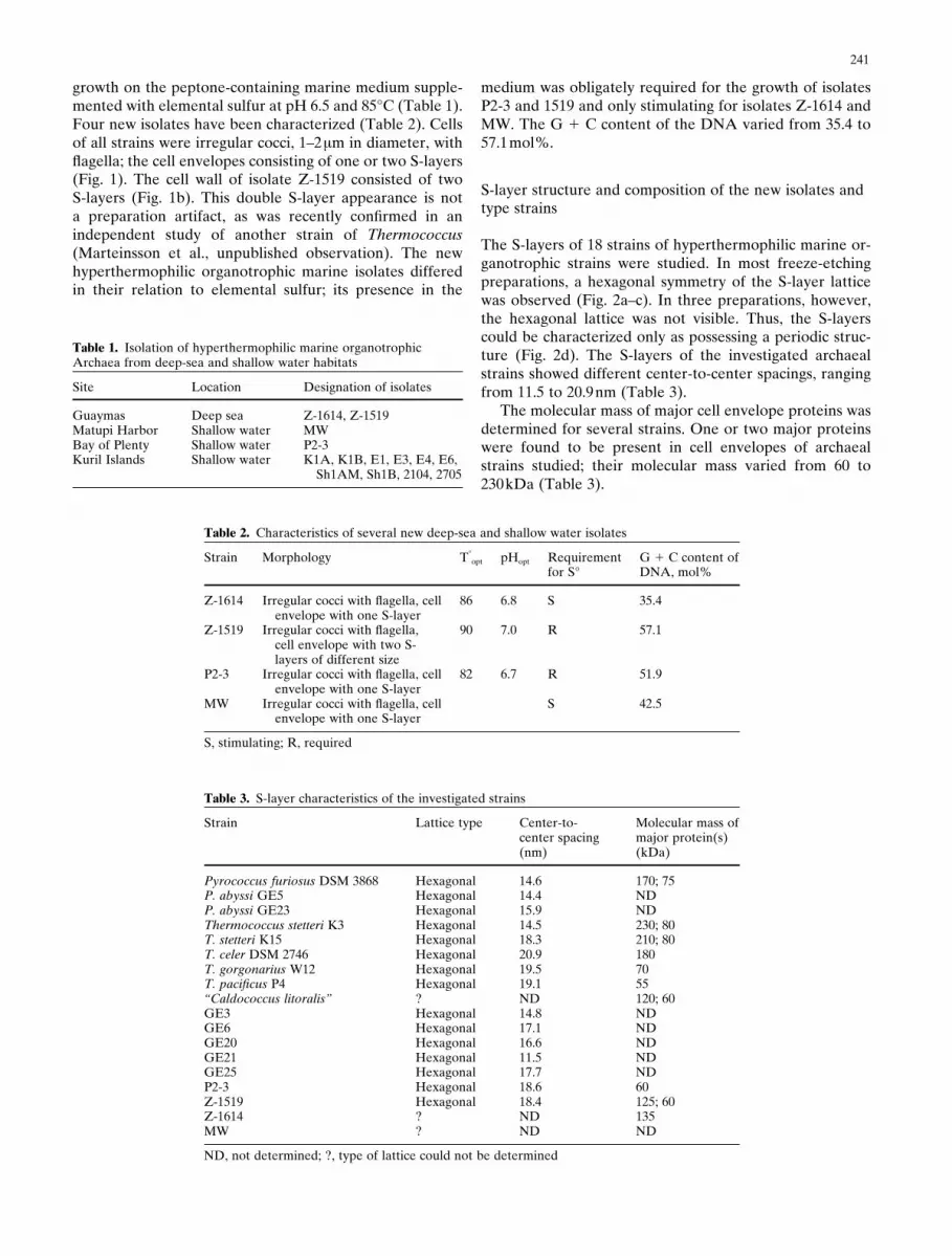
growth on the peptone-containing marine medium supple-mented with elemental sulfur at pH 6.5 and 85°C (Table 1).Four new isolates have been characterized (Table 2). Cellsof all strains were irregular cocci, 1–2µm in diameter, withflagella; the cell envelopes consisting of one or two S-layers(Fig. 1). The cell wall of isolate Z-1519 consisted of twoS-layers (Fig. 1b). This double S-layer appearance is nota preparation artifact, as was recently confirmed in anindependent study of another strain of Thermococcus(Marteinsson et al., unpublished observation). The newhyperthermophilic organotrophic marine isolates differedin their relation to elemental sulfur; its presence in the
medium was obligately required for the growth of isolatesP2-3 and 1519 and only stimulating for isolates Z-1614 andMW. The G 1 C content of the DNA varied from 35.4 to57.1mol%.
S-layer structure and composition of the new isolates andtype strains
The S-layers of 18 strains of hyperthermophilic marine or-ganotrophic strains were studied. In most freeze-etchingpreparations, a hexagonal symmetry of the S-layer latticewas observed (Fig. 2a–c). In three preparations, however,the hexagonal lattice was not visible. Thus, the S-layerscould be characterized only as possessing a periodic struc-ture (Fig. 2d). The S-layers of the investigated archaealstrains showed different center-to-center spacings, rangingfrom 11.5 to 20.9nm (Table 3).
The molecular mass of major cell envelope proteins wasdetermined for several strains. One or two major proteinswere found to be present in cell envelopes of archaealstrains studied; their molecular mass varied from 60 to230kDa (Table 3).
Table 1. Isolation of hyperthermophilic marine organotrophicArchaea from deep-sea and shallow water habitats
Site Location Designation of isolates
Guaymas Deep sea Z-1614, Z-1519Matupi Harbor Shallow water MWBay of Plenty Shallow water P2-3Kuril Islands Shallow water K1A, K1B, E1, E3, E4, E6,
Sh1AM, Sh1B, 2104, 2705
Table 2. Characteristics of several new deep-sea and shallow water isolates
Strain Morphology T°opt pHopt Requirement G 1 C content of
for S° DNA, mol%
Z-1614 Irregular cocci with flagella, cell 86 6.8 S 35.4envelope with one S-layer
Z-1519 Irregular cocci with flagella, 90 7.0 R 57.1cell envelope with two S-layers of different size
P2-3 Irregular cocci with flagella, cell 82 6.7 R 51.9envelope with one S-layer
MW Irregular cocci with flagella, cell S 42.5envelope with one S-layer
S, stimulating; R, required
Table 3. S-layer characteristics of the investigated strains
Strain Lattice type Center-to- Molecular mass ofcenter spacing major protein(s)(nm) (kDa)
Pyrococcus furiosus DSM 3868 Hexagonal 14.6 170; 75P. abyssi GE5 Hexagonal 14.4 NDP. abyssi GE23 Hexagonal 15.9 NDThermococcus stetteri K3 Hexagonal 14.5 230; 80T. stetteri K15 Hexagonal 18.3 210; 80T. celer DSM 2746 Hexagonal 20.9 180T. gorgonarius W12 Hexagonal 19.5 70T. pacificus P4 Hexagonal 19.1 55“Caldococcus litoralis” ? ND 120; 60GE3 Hexagonal 14.8 NDGE6 Hexagonal 17.1 NDGE20 Hexagonal 16.6 NDGE21 Hexagonal 11.5 NDGE25 Hexagonal 17.7 NDP2-3 Hexagonal 18.6 60Z-1519 Hexagonal 18.4 125; 60Z-1614 ? ND 135MW ? ND ND
ND, not determined; ?, type of lattice could not be determined
241

Fig. 1a–d. Electron photomicrographs of thin-sectioned cells of the new hyperthermophilic organotrophic marine isolates: Z-1614 (a), Z-1519 (b),P2-3 (c), and MW (d). Bar 500nm
Fig. 2a–d. Electron photo-micrographs of freeze-etched cells of hyperther-mophilic organotrophicmarine Archaea: Pyroco-ccus furiosus (a), Thermo-coccus celer (b), Z-1519 (c),and “Caldococcuslitoralis” (d).Bar 32nm
242

Immunochemical analyses
Antisera to whole cells of ten archaeal strains wereraised, including the eight type strains of P. furiosus, T.celer, T. stetteri, T. profundus, T. peptonophilus, T.chitonophagus, T. gorgonarius, and T. pacificus; and twoother strains, T. stetteri K15 and “Caldococcus litoralis.”Quality of antisera obtained was checked by theOuchterlony method, and antisera specificity was testedby immunoblotting.
It was found, in immunoblotting analysis, that antiseradiluted 1 :100 or 1 :1000 cross-reacted with cells of alltype strains tested (data not shown). When diluted 1 : 5000,antisera reacted strongly only with the strains againstwhich they had been raised (Fig. 3). Antisera to T. stetteristrain K3 and strain K15 cross-reacted with cells of bothstrains, thus confirming that this method was species-rather than strain-specific. Antiserum to “Caldococcuslitoralis” reacted with cells of T. litoralisT DSM 5474,indicating that “Caldococcus litoralis” was a strain of T.litoralis.
Cells of all new and unidentified isolates were used in theimmunoblotting analyses with nine antisera (Table 4). Thir-teen isolates gave positive reaction with four antisera: iso-lates Z-1614, MW, Sh1AM, and Sh1B, with antiserum to T.litoralis; E1, E4, and 2705, with that to T. chitonophagus;isolates K1A, K1B, as well as strains E3, E6, and 2104, wereidentified as strains of T. stetteri and isolate P2-3 as T.pacificus. Some of these results were tested by parallelDNA–DNA hybridization analyses. A high level of DNA–DNA homology between the evaluated strains and the typestrains confirmed the results of immunochemical analyses(Table 5). Six deep-sea isolates gave no positive reactionwith any of the antisera used.
Discussion
Crystalline protein layers on the surface of prokaryotic cellshave been termed surface layers (S-layers), and have beenfound in many bacteria belonging to all phylogeneticbranches and virtually all archaeal species studied (for re-cent compilation, see Messner and Sleytr 1992; Sleytr et al.1996). In many archaeal species, the S-layers constitute the
Table 4. Results of immunoblotting reaction of antisera with cells of the new isolates
Strain Antiserum to
P. furiosus T. celer T. stetteri “C. litoralis” T. T. T. profundus T. gorgonarius T. pacificusK3 chitonophagus peptonophilus
GE3 2 2 2 2 2 2 2 2 2GE6 2 2 2 2 2 2 2 2 2GE20 2 6 6 6 6 2 6 6 6GE21 2 2 2 2 2 2 2 2 2GE25 2 2 2 2 2 2 2 2 2Z-1614 2 2 2 1 2 2 2 2 2Z-1519 2 2 2 2 2 2 2 2 2P2-3 2 2 2 2 2 2 2 2 1MW 2 2 2 1 2 2 2 2 2K1A 2 2 1 2 2 2 6 2 2K1B 2 2 1 2 6 2 2 2 6E1 2 2 2 6 1 2 2 2 2E3 2 2 1 2 2 2 2 2 2E4 2 2 2 2 1 2 2 2 2E6 2 2 1 2 2 2 2 2 2Sh1AM 2 2 2 1 6 2 2 2 2Sh1B 2 2 2 1 6 2 2 2 22104 2 2 1 2 2 2 2 2 22705 2 2 6 2 1 2 2 2 2
1, strong reaction; 6, weak reaction; 2, no reaction
Fig. 3. Western blot analysis of different strains with antiserum againstP. furiosus showing strong reaction with P. furiosus cell proteins (lane4), weak reaction with T. celer cell proteins (lane 3), and no reactionwith cell proteins of Z-1614 (lane 1), “C. litoralis” (lane 2), K1B (lane5), K1A (lane 6), E1 (lane 7), E3 (lane 8), E6 (lane 9), 2104 (lane 10),and 2705 (lane 11). Antiserum was diluted 1 :5000
243

only cell wall structure, which is attached to the cytoplasmicmembrane (Sleytr et al. 1988; König 1988; Phipps et al.1991; Baumeister and Lembcke 1992). Most S-layers pos-sess hexagonal (p3 or p6) lattice symmetry. Up to now,oblique and square (p2 or p4) lattice symmetry was shownonly for several hyperthermophilic Archaea such as repre-sentatives of the Crenarchaeota, Desulfurococcus mobilis(Baumeister et al. 1990) and Pyrolobus fumarii (Blöchl etal. 1997), and Ferroglobus placidus (Hafenbradl et al. 1996),belonging to the Euryarchaeota.
This study was aimed at the characterization of thecell walls of hyperthermophilic Archaea of the generaPyrococcus and Thermococcus, representing the orderThermococcales of the kingdom Euryarchaeota. All isolatesof this group described so far possess cell walls consisting ofone or two S-layers outside the cytoplasmic membrane. Thedouble S-layer was studied in detail (by freeze-etching andisolation of sheets) only for Pyrobaculum organotrophum(Phipps et al. 1991). However, numerous thin sections re-vealed that the double S-layer could be recognized asa common feature of Thermococcales. This structure wasfirst shown for Pyrococcus woesii (Zillig et al. 1987) butwas found later to be fairly widespread among representa-tives of Thermococcus and Pyrococcus genera: T. stetteri(Miroshnichenko et al. 1989), T. celer (Baumeisteret al. 1990), Pyrococcus abyssi (Erauso et al. 1993), T.chitonophagus (Huber et al. 1995), T. peptonophilus(Gonzales et al. 1995), T. fumicolans (Godfroy et al. 1997),and T. pacificus (Miroshnichenko et al. 1989). The cell wallsof the four new isolates described here also possess eitherone or two S-layers. Isolates Z-1614 and MW, identified asmembers of T. litoralis, are covered with a single S-layer.We did not observe double S-layers on thin sections ofstrain P2-3, which was identified as T. pacificus. Althoughreported for the type strain of this species, the S-layer of thisorganism was described as very fragile and easily destructed(Miroshnichenko et al. 1989). This fact might explain itsabsence on thin sections of strain P2-3.
With the exception of three strains, all other strainsexamined in this study were shown to have S-layers withhexagonal lattice symmetry. These three isolates belong toT. litoralis, and their cell wall structures need further char-acterization. S-layers of all the other strains differed in thecenter-to-center spacing and molecular mass of major pro-teins (see Table 3). In several cases, the values obtained forstrains belonging to the same species were fairly close, butin other cases these differed significantly (T. stetteri K3 and
K15, GE20, and GE21). Although in Bacteria the structureand even the occurrence of an S-layer are strain-specificfeatures, in Archaea the available data might be exploitedfor differentiating species. Their presence could be associ-ated with a very important common role of S-layers inarchaeal cells, where they often occur as the only cell wallcomponent. For example, in Thermoproteus tenax (Messneret al. 1986; Wildhaber and Baumeister 1987), Pyrobaculumorganotrophum (Phipps et al. 1991), and Methano-corpusculum sinense (Pum et al. 1991) it was demonstratedthat S-layers are involved in cell shape determinationand cell division. However, more evidence is needed toprove the species-specificity of cell wall components ofThermococcales.
Immunochemical experiments indicated that the anti-genic capacity of Thermococcales cell envelopes might beused for species identification. Immunoblotting analyseswith highly diluted antisera provided reliable differentiationof species belonging to the genus Thermococcus and allowedidentification of several new and previously publishedstrains. Three isolates identified as the strains of T. litoralisoriginated from shallow water hot vents of Kurils and NewGuinea and the fourth was isolated from deep-sea hot ventsof Guaymas. The new strains of T. stetteri and T. pacificusoriginated from the same sites as the type strains of thesespecies (Kurils and New Zealand, respectively). By contrast,T. chitonophagus, originating from Guaymas deep-sea vents,was now found only among shallow water isolates. Thus, all12 shallow water isolates tested could be identified, whereassix of seven deep-sea strains failed to react with any of thenine species-specific antisera used. These strains either arerepresentatives of the recently described species ofThermococcales that are not yet included in our antiserabank or belong to new taxa. Our findings confirm the consid-erable taxonomic diversity among deep-sea microbial com-munities in comparison to shallow water populations.
“Caldococcus litoralis” (Svetlichny et al. 1987) was previ-ously published as a representative of a novel genus, basedon a significant 14% difference in the G 1 C content withthe only representative of Thermococcus—T. celer—knownat that time. Later a new species, Thermococcus litoralis,was published (Neuner et al. 1990). Immunoblotting analy-ses reported here indicated that “C. litoralis” and the typespecies of T. litoralis represent the same species. Thisevidence is supported by the results of the DNA–DNAhybridization (96% homology between two strains). Thus,we propose to reclassify “Caldococcus litoralis” asThermococcus litoralis Z-1301.
Phenotypically, strain Z-1301 is very close to the typestrain of Thermococcus litoralis, as it has the same pH andtemperature range of growth, ferments peptides, and doesnot obligately require elemental sulfur for growth. The onlysignificant difference is the presence of flagella in strain Z-1301, whereas the type strain of T. litoralis is not flagellated.However, the presence of flagella is a strain-specific feature,as was shown for T. stetteri (Miroshnichenko et al. 1989).There is also a 3% difference in G 1 C content of the DNA ofboth strains. We include these strain-specific differencesin the following emended description of Thermococcuslitoralis.
Table 5. The DNA–DNA cross-hybridization reaction between thenew and reference strains of the genus Thermococcus
Strain % of DNA–DNA cross-hybridization with
T. litoralis DSM 5474 T. steteri K3
“C. litoralis” 96 NDZ-1614 71 NDMW 69 NDK1B ND 97T. litoralis 100 NDT. stetteri ND 100
ND, not determined
244
Emended description of Thermococcus litoralis
Thermococcus litoralis sp. nov., Neuner, Jannasch, Belkinand Stetter. Cells are regular or irregular cocci 0.5–3.0µm,not flagellated, or with a tuft of flagella. Obligate or-gantrophs, utilizing peptides. The presence of elementalsulfur in the medium stimulates growth but is not obligatelyrequired. Hyperthermophiles, growing between 55° and100°C with an optimum at 85°–88°C. pH for growth rangesfrom 4.0 and 8.0 with optimum at 6.0–6.4. Growth occurs atNaCl concentration from 1.8% to 6.5%, with the optimumat 2.5%. G 1 C content of DNA, 38–41mol%. Inhabitsshallow and deep-sea submarine hot vents. Type strain:Thermococcus litoralis NS-C (DSM 5473).
Acknowledgments This work was supported by the INTAS grantno. 94-1717 and by the Russian Ministry of Science (project“New hyperthermophiles”). The authors are grateful to NadezhdaKostrikina, who prepared the ultrathin sections, and to ValeryGalchenko (Institute of Microbiology, Moscow) and Vitaly Tarasov(Institute of Marine Biology, Vladivostok), who provided samplesfrom deep-sea and shallow water hot vents.
References
Baumeister W, Lembcke G (1992) Structural features ofarchaebacterial cell envelopes. J Bioenerg Biomembr 24:567–575
Baumeister W, Santarius U, Volker S, Dürr R, Lembcke G,Engelhardt H (1990) The surface protein of Hyperthermus butylicus:three-dimensional structure and comparison with other archae-bacterial surface proteins. Syst Appl Microbiol 13:105–111
Blöchl E, Rachel R, Burggraf S, Hafenbradl D, Jannasch HW, StetterKO (1997) Pyrolobus fumarii, gen. and sp. nov., represents a novelgroup of archaea, extending the upper temperature limit for life to113°C. Extremophiles 1:14–21
Borovjagin VL, Sabelnikov AG, Tarahovsky YS, Vasilenko IA (1987)Polymorphic behaviour of gram-negative bacteria membranes. JMembr Biol 100:229–242
Erauso G, Reysenbach A-L, Godfroy A, Meunier J-R, Crump B,Partensky F, Baross JA, Marteinsson V, Barbier G, Pace NR, PrieurD (1993) Pyrococcus abyssi sp. nov., a new hyperthermophilicarchaeon isolated from a deep-sea hydrothermal vent. ArchMicrobiol 160:338–349
Fiala G, Stetter KO (1986) Pyrococcus furiosus sp. nov. represents anovel genus of marine heterotrophic archaebacteria growing opti-mally at 100°C. Arch Microbiol 145:56–61
Fiala G, Stetter KO, Jannasch HW, Langworthy TA, Madon J (1986)Staphylothermus marinus sp. nov. represents a new genus of marineheterotrophic archaebacteria growing up to 98°C. Syst ApplMicrobiol 8:106–113
Godfroy A, Lesongeur F, Raguenes G, Querellou J, Antoine E,Meunier J-R, Guezennec J, Barbier G (1997) Thermococcus hydrot-hermalis sp. nov., a new hyperthermophilic archaeon isolated from adeep-sea hydrothermal vent. Int J Syst Bacteriol 47:6223–6226
Gonzales GM, Kato C, Horikoshi K (1995) Thermococcuspeptonophilus sp. nov., a fast growing, extremely thermophilicarchaebacterium isolated from deep-sea hydrothermal vents. ArchMicrobiol 164:159–164
Hafenbradl D, Keller M, Dirmeier R, Rachel R, Rossnagel P, BurggrafS, Huber H, Stetter KO (1996) Ferroglobus placidus gen nov. a novelhyperthermophilic archaeum that oxidizes Fe21 at neutral pH underanoxic conditions. Arch Microbiol 166:308–314
Huber R, Stohr J, Honenhaus J, Rachel R, Burggraf S, Jannasch HW,Stetter KO (1995) Thermococcus chitonophagus sp. nov., a novel,chitin-degrading, hyperthermophilic archaeum from a deep-seahydrothermal environment. Arch Microbiol 164:255–264
König H (1988) Archaebacterial cell envelopes. Can J Microbiol34:395–406
Laemmli UK (1970) Cleavage of structural proteins during the assem-bly of the head of bacteriophage T4. Nature (Lond) 227:680–685
Marteinsson VT, Watrin L, Prieur D, Caprais JC, Raguenes G, ErausoG (1995) Phenotypic characterization, DNA similarities, and proteinprofiles of twenty sulfur-metabolizing hyperthermophilic anaerobicarchaea isolated from hydrothermal vents in the southern PacificOcean. Int J Syst Bacteriol 46:1113–1119
Messner P, Pum D, Sára M, Stetter KO, Sleytr UB (1986) Ultrastruc-ture of the cell envelope of the archaebacteria Thermoproteus tenaxand Thermoproteus neutrophilus. J Bacteriol 166:1046–1054
Messner P, Sleytr UB (1992) Crystalline bacterial cell-surface layers.Adv Microb Physiol 33:213–275
Miroshnichenko ML, Bonch–Osmolovskaya EA, Neuner A,Kostrikina NA, Alekseev VA (1989) Thermococcus stetteri sp. nov.,a new extremely thermophilic marine sulfur-metabolizing archae-bacterium. Syst Appl Microbiol 12:257–262
Miroshnichenko ML, Gongadze GM, Lysenko AM, Bonch-Osmolovskaya EA (1994) Desulfurella multipotens sp. nov., a newsulfur-respiring thermophilic eubacterium from Raoul Island(Kermadec archipelago, New Zealand). Arch Microbiol 161:88–93
Miroshnichenko ML, Gongadze GM, Rainey FA, Kostyukova AS,Lysenko AM, Chernyh NA, Bonch–Osmolovskaya EA (1998)Thermococcus gorgonarius sp. nov. and Thermococcus pacificus sp.nov.: heterotrophic extremely thermophilic archaea from NewZealand submarine hot vents. Int J Syst Bacteriol 48:23–29
Namsaraev BB, Bonch–Osmolovskaya EA, Kaehalkin VI, Propp LN,Tarasov VG (1994) Microbiological processes of carbon cycle inshallow-water hot vents in the western part of Pacific Ocean.Mikrobiologia 63:100–111
Neuner A, Jannasch HW, Belkin S, Stetter KO (1990) Thermococcuslitoralis sp. nov., a new species of extremely thermophilic marinearchaebacteria. Arch Microbiol 153:205–207
Ouchterlony O (1953) Antigen-antibody reaction in gels. Acta PatholMicrobiol Scand 32:231–242
Phipps BM, Huber R, Baumeister W (1991) The cell envelope of thehyperthermophilic archaebacterium Pyrobaculum organotrophumconsists of two regularly arrayed protein layers: three-dimensionalstructure of the outer layer. Mol Microbiol 5:253–265
Pum D, Messner P, Sleytr UB (1991) Role of the S layer in morphogen-esis and cell division of the archaebacterium Methanocorpusculumsinense. J Bacteriol 173:6865–6873
Schönheit P, Schäfer T (1995) Metabolism of hyperthermophiles.World J Microbiol Biotechnol 11:26–57
Sleytr UB, Messner P, Pum D (1988) Analysis of crystalline bacterialsurface layers by freeze-etching, metal shadowing, negative stainingand ultrathin sectioning. Methods Microbiol 20:29–60
Sleytr UB, Messner P, Pum D, Sára M (eds) (1996) Crystalline bacte-rial cell surface layers. Landes/Academic Press, Austin, TX
Stetter KO (1996) Hyperthermophilic prokaryotes. FEMS MicrobiolRev 18:149–158
Stetter KO, König H, Stackebrandt E (1983) Pyrodictium gen. nov., anew genus of submarine disk-shaped sulfur-reducing archaebacteriagrowing optimally at 105°C. Syst Appl Microbiol 4:535–551
Svetlichny VA, Slesarev AI, Svetlichnaya TP, Zavarzin GA (1987)Caldococcus litoralis gen. nov., sp. nov.—new marine extremelythermophilic archaebacterium reducing elemental sulfur. Mikro-biologiya 56:831–838
Wildhaber I, Baumeister W (1987) The cell envelope ofThermoproteus tenax: three-dimensional structure of the surfacelayer and its role in shape maintainance. EMBO J 6:1475–1480
Zillig W, Holz I, Janekovic D, Schäfer W, Reiter WD (1983) Thearchaebacterium Thermococcus celer represents a novel genuswithin the thermophilic branch of archaebacteria. Syst ApplMicrobiol 4:88–94
Zillig W, Holz I, Janekovic D, Klenk H-P, Imsel E, Trent J, Wunderl I,Forjaz VH, Couyinho R, Ferreira T (1991) Hyperthermus butylicus,a hyperthermophilic sulfur-reducing archaebacterium that fermentspeptides. J Bacteriol 172:3959–3965
Zillig W, Holz I, Klenk H-P, Trent J, Wunderl I, Janekovic D, Imsel E,Haas B (1987) Pyrococcus woesii sp. nov. an ultra-thermophilic ma-rine archaebacterium representing a novel order Thermococcales.Syst Appl Microbiol 9:62–70
245


