Embed Size (px)
Citation preview

Water Research 38 (2004) 1197–1206
ARTICLE IN PRESS
*Correspond
7247-826-858.
E-mail addr
(T. Schwartz).
0043-1354/$ - se
doi:10.1016/j.w
Investigation of natural biofilms formed during the productionof drinking water from surface water embankment filtration
Farahnaz Emtiazi, Thomas Schwartz*, Silke Mareike Marten,Peter Krolla-Sidenstein, Ursula Obst
Department of Environmental Microbiology, Forschungszentrum Karlsruhe GmbH, Institute for Technical Chemistry–Water Technology
and Geotechnology Division, P.O. Box 3640, Karlsruhe D-76021, Germany
Received 5 June 2003; received in revised form 8 October 2003; accepted 13 October 2003
Abstract
Populations of bacteria in biofilms from different sites of a drinking water production system were analysed.
Polymerase chain reaction (PCR) and denaturing gradient gel electrophoresis (DGGE) analyses revealed changing
DNA band patterns, suggesting a population shift during bank filtration and processing at the waterworks. In addition,
common DNA bands that were attributed to ubiquitous bacteria were found. Biofilms even developed directly after
UV disinfection (1–2m distance). Their DNA band patterns only partly agreed with those of the biofilms from
the downstream distribution system. Opportunistic pathogenic bacteria in biofilms were analysed using PCR and
Southern blot hybridisation (SBH). Surface water appeared to have a direct influence on the composition of biofilms
in the drinking water distribution system. In spite of preceding filtration and UV disinfection, opportunistic pathogens
such as atypical mycobacteria and Legionella spp. were found in biofilms of drinking water, and Pseudomonas
aeruginosa was detected sporadically. Enterococci were not found in any biofilm. Bacterial cell counts in the
biofilms from surface water to drinking water dropped significantly, and esterase and alanine-aminopeptidase activity
decreased. b-glucosidase activity was not found in the biofilms. Contrary to the results for planktonic bacteria,
inhibitory effects were not observed in biofilms. This suggested an increased tolerance of biofilm bacteria against toxic
compounds.
r 2003 Elsevier Ltd. All rights reserved.
Keywords: Biofilms; Population shifts; Opportunistic pathogenic bacteria; Enzyme activities; Molecular-biological techniques
1. Introduction
Bank filtration and artificial groundwater enrichment
are frequently employed for the production of drinking
water. When surface water enters the aquifer via an
underground passage due to potential gradients gener-
ated by wells, this is referred to as bank filtration.
During underground passage, a variety of chemical and
biological processes occur, by which compounds are
ing author. Tel.: +49-7247-826-802; fax: +49-
ess: [email protected]
e front matter r 2003 Elsevier Ltd. All rights reserve
atres.2003.10.056
reduced and eliminated. Research results obtained with
respect to the hydraulic, physico-chemical, and chemi-
cal/biological processes revealed a good and stable long-
term cleaning efficiency of bank filtration. This also
applies to the removal of particles, pathogens, a number
of organic, and most trace substances [1] except for some
individual organic polar compounds which are persis-
tent in a nearly unrestricted manner [2]. Microorganisms
significantly contribute to cleaning during the under-
ground passage by enzymatic degradation or partial
metabolism of water impurities and by physico-chemical
processes, e.g. adsorption. However, specific manipula-
tion and use of these microbial elimination processes
have failed, and knowledge of the processes is too
d.

ARTICLE IN PRESSF. Emtiazi et al. / Water Research 38 (2004) 1197–12061198
incomplete. Still these biological processes during
natural underground passage are suited as environmen-
tally compatible and low-cost stage for sustainable
drinking water processing. This is not only true for
Central Europe with its large river basins, but also for
many Third World and threshold countries that have to
produce drinking water from highly contaminated
surface waters. Yet few data are available for the
comparative description of function and population
changes of adhesive bacteria (biofilms) during the
underground passage and in the downstream drinking
water production facilities.
Among the major ecological units of aquatic systems,
which affect water quality, are biofilms that cover
practically all accessible wet surfaces. Biofilms may be
composed of algae, bacteria, fungi and other eukaryotic
microorganisms, and cover surfaces, e.g. in storage
basins, filter systems, and drinking water distribution
lines. As biofilms represent the predominating biological
form of life in habitats of water and soil, it is urgently
required to improve the understanding of the structure
and function of these biocoenoses. There is a lack of
methods for the detection of adhesive bacteria in
classical drinking water analysis. Cultivation processes
cover a small part of the natural planktonic population
only [3]. The limitation of nutrients and environmental
stress situations may induce physiological and morpho-
logical changes in many aquatic bacteria. A dormant
status was described for pathogenic bacteria, such
as Campylobacter spp., E. coli, and Legionella pneumo-
phila [4,5] which makes their isolation and identi-
fication problematic. Therefore, cultivation-independent
molecular-biological methods targeting nucleic acids are
required in addition to biochemico-metabolic analyses
[5].
Our investigations covered biofilms from surface
water, raw water after bank filtration, processed
drinking water prior to and after UV disinfection as
well as from the downstream municipal distribution
system. As an alternative to classical cultivation
methods, molecular-biological methods were applied
with the DNA as target molecule, which exists in each
cell irrespective of its physiological state. Oligonucleo-
tide probes developed for a number of environmental
bacteria and pathogens allowed to comprehensively
describe bacterial populations or identify pathogens
without a cultivation pre-enrichment. By means of the
polymerase chain reaction (PCR), denaturing gradient
gel electrophoresis (DGGE), and southern blot hybridi-
sation (SBH), population shifts of the bacterial biofilms
were investigated and the occurrence of facultatively
pathogenic bacteria, such as legionellae, mycobacteria,
and enterococci, was studied. Enzyme activities were
measured in biofilm samples to determine the metabolic
performance and physiological states of the bacterial
biocoenoses.
2. Material and methods
2.1. Biofilm sampling method
For biofilm formation in drinking water distribution
systems, special devices were used [6,7]. The core of these
devices consisted of a hollow stainless steel cylindrical
element, where stainless steel bolts holding steel platelets
for biofilm growth were screwed into place. The platelet
(15mm� 30mm� 2.5mm) was attached to the end of a
bolt with a small screw. To study natural biofilms, these
devices were installed at different sampling points in the
waterworks, i.e. downstream of the granular activated
carbon filtration (GAC) and downstream of UV
disinfection (DIS). Two devices were installed in house
branch connections within the municipal drinking water
distribution system (DW1 and DW2) 1–2 km down-
stream of the waterworks. Additionally, platelets
(170mm� 20mm� 2mm) were incubated in surface
water used for embankment filtration (SW, Rhine river)
and raw water (RW) from a well downstream of bank
filtration. To examine the biofilm populations, the
platelets were removed after 3–4 weeks, the biofilms
were scraped from the surface using a sterile cell scraper
(Nunc, Wiesbaden, Germany) and suspended in 10ml
water from the sampling site. The biofilm suspensions
were centrifuged for 10min at 10 000g. The pellet was
resuspended in 1ml sterile water. DNA was extracted
from biofilms from the surface water and horizontal well
(QIAamps DNA Mini Kit-50, Qiagen). These DNA
preparations were used as template in PCR. The biofilm
suspensions from GAC, DIS, and DW were used
without extraction for subsequent molecular-biological
analysis.
2.2. PCR-DGGE analysis of different aquatic biofilms
PCR primers targeting the regions (V1-3) of 16S
rDNA of bacteria were used for biofilm analysis
(Table 1) [8,9]. For DGGE analysis, a sequence of
multiple guanines (G) and cytosines (C) was attached to
the 50end of the forward primer [10]. A ‘‘touch-down’’
PCR profile published by Kilb et al. [9] was applied. The
final 100ml reaction mixture contained 2.5 U of HotStar
Taq-DNA polymerase (Qiagen, Germany), 30 pmol of
each primer, 1�PCR buffer, 1.5mM MgCl2, 200mMdNTPs, and 10 ml biofilm suspension or template DNA.
A GeneAmp PCR System 9700 (Applied Biosystems)
was used for PCR. Aliquots of 5 ml were analysed by
electrophoresis in a 1% agarose gel containing ethidium
bromide to check the sizes and amounts of the
amplicons. PCR products were purified using phenol-
chloroform-isoamyl alcohol (25:24:1 vol.), precipitated
with isopropanol, washed with 70% ethanol, dried, and
resuspended in 20 ml sterile water. DGGE analysis of
PCR products (B526 bp and 349 bp) was performed
ARTICLE IN PRESS
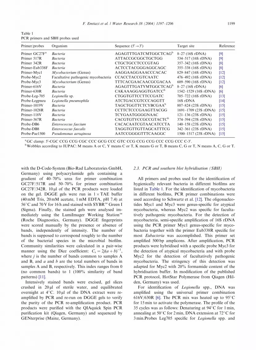
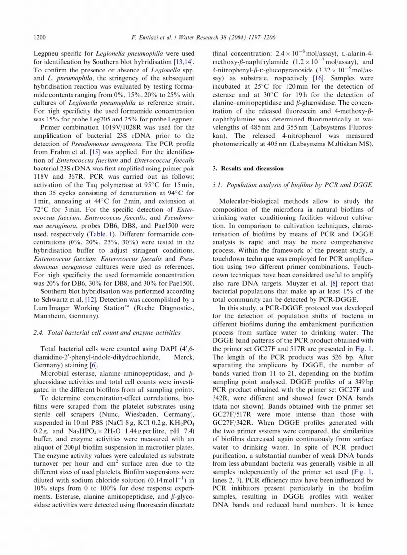
Table 1
PCR primers and SBH probes used
Primer/probes Organism Sequence (50-30) Target site Reference
Primer GC27Fa Bacteria AGAGTTTGATCMTGGCTCAGb 8–27 (16S rDNA) [9]
Primer 517R Bacteria ATTACCGCGGCTGCTGG 534–517 (16S rDNA) [9]
Primer 342R Bacteria CTGCTGCCTCCCGTAG 357–342 (16S rDNA) [8]
Primer-Eub338F Bacteria ACTCCTACGGGAGGCAGC 355–338 (16S rDNA) [8]
Primer-Myc1 Mycobacterium (Genus) AAGGAAGGAAACCCACAC 829–847 (16S rDNA) [12]
Probe-Myc2 Facultative pathogenic mycobacteria CCACCTACCGTCAATC 476–492 (16S rDNA) [12]
Probe-Myc3 Mycobacterium (Genus) TTTCACGAACAACGCGACAA 609–590 (16S rDNA) [12]
Primer-616V Bacteria AGAGTTTGATYMTGGCTCAGb 8–27 (16S rDNA) [6]
Primer-630R Bacteria CAKAAAGGAGGTGATCCb 1542–1529 (16S rDNA) [6]
Probe-Leg-705 Legionella sp. CTGGTGTTCCTTCCGATC 705–722 (16S rDNA) [13]
Probe-Legpneu Legionella pneumophila ATCTGACCGTCCCAGGTT 16S rDNA [14]
Primer-1019V Bacteria TAGCTGGTTCTCYBCGAAb 807–824 (23S rDNA) [15]
Primer-1028R Bacteria CCTTCTCCCGAAGTTACGG 1691–1709 (23S rDNA) [15]
Primer-118V Bacteria TCYGAATGGGGNAAC 121–136 (23S rDNA) [15]
Primer-367R Bacteria CACGTGTYCCGCCGTACTCb 374–394 (23S rDNA) [15]
Probe-DB6 Enterococcus faecium CACACAATCGTAACATCCTA 140–158 (23S rDNA) [15]
Probe-DB8 Enterococcus faecalis TAGGTGTTGTTAGCATTTCG 342–361 (23S rDNA) [15]
Probe-Pae1500 Pseudomonas aeruginosa AATCCGGGGTTTCAAGGC 1500–1517 (23S rDNA) [15]
aGC clamp: 50-CGC CCG CCG CGC CCC GCG CCC GTC CCG CCG CCG CCC CCG CCC C-30.bWobbles according to IUPAC: M means A or C, Y means C or T, K means G or T, B means C, G or T, N means A, C, G or T.
F. Emtiazi et al. / Water Research 38 (2004) 1197–1206 1199
with the D-Code-System (Bio-Rad Laboratories GmbH,
Germany) using polyacrylamide gels containing a
gradient of 40–70% urea for primer combination
GC27F/517R and 50–70% for primer combination
GC27F/342R. 10 ml of the PCR products were loaded
on the gel. DGGE gels were run in 1�TAE buffer
(40mM Tris, 20mM acetate, 1mM EDTA, pH 7.4) at
56�C and 70V for 16 h and stained with SYBRtGreen I
(Sigma). Finally, the stained gels were analysed im-
mediately using the LumiImager Working Stationt
(Roche Diagnostics, Germany). DGGE fingerprints
were scored manually by the presence or absence of
bands, independently of intensity. The number of
bands is supposed to correspond roughly to the number
of the bacterial species in the microbial biofilm.
Community similarities were calculated in a pair-wise
manner using the Dice coefficient: Cs ¼ 2jða þ bÞ�1;where j is the number of bands common to samples A
and B, and a and b are the total numbers of bands in
samples A and B, respectively. This index ranges from 0
(no common bands) to 1 (100% similarity of band
patterns) [11].
Intensively stained bands were excised, gel slices
crushed in 20 ml of sterile water, and equilibrated
overnight at 4�C. 10ml of the DNA extract were re-
amplified by PCR and re-run on DGGE gels to verify
the purity of the PCR re-amplification product. PCR
products were purified with the QIAquick Spin PCR
purification kit (Qiagen, Germany) and sequenced by
GENterprise (Mainz, Germany).
2.3. PCR and southern blot hybridisation (SBH)
All primers and probes used for the identification of
hygienically relevant bacteria in different biofilms are
listed in Table 1. For the identification of mycobacteria
in different biofilms, PCR primer combinations were
used according to Schwartz et al. [12]. The oligonucleo-
tides Myc1 and Myc3 were genus-specific for atypical
mycobacteria, whereas Myc2 was specific for faculta-
tively pathogenic mycobacteria. For the detection of
mycobacteria, semi-specific amplification of 16S rDNA
using the PCR primer Myc1 genus-specific for myco-
bacteria together with the primer Eub338R specific for
most Eubacteria was accomplished. This primer set
amplified 500 bp amplicons. After amplification, PCR
products were hybridised with a specific probe Myc3 for
the detection of atypical mycobacteria and with probe
Myc2 for the detection of facultatively pathogenic
mycobacteria. The stringency of this detection was
adapted for Myc2 with 20% formamide content of the
hybridisation buffer. In modification of the published
PCR protocol, HotStar Polymerase from Qiagen (Hil-
den, Germany) was used.
For identification of Legionella spp., DNA was
amplified using the universal primer combination
616V/630R [6]. The PCR mix was heated up to 95�C
for 15min to activate the polymerase. The profile of the
35 cycles was as follows: Denaturing at 94�C for 1min,
annealing at 50�C for 2min, DNA extension at 72�C for
3min.Probes Leg705 specific for Legionella spp. and
ARTICLE IN PRESSF. Emtiazi et al. / Water Research 38 (2004) 1197–12061200
Legpneu specific for Legionella pneumophila were used
for identification by Southern blot hybridisation [13,14].
To confirm the presence or absence of Legionella spp.
and L. pneumophila, the stringency of the subsequent
hybridisation reaction was evaluated by testing forma-
mide contents ranging from 0%, 15%, 20% to 25% with
cultures of Legionella pneumophila as reference strain.
For high specificity the used formamide concentration
was 15% for probe Leg705 and 25% for probe Legpneu.
Primer combination 1019V/1028R was used for the
amplification of bacterial 23S rDNA prior to the
detection of Pseudomonas aeruginosa. The PCR profile
from Frahm et al. [15] was applied. For the identifica-
tion of Enterococcus faecium and Enterococcus faecalis
bacterial 23S rDNA was first amplified using primer pair
118V and 367R. PCR was carried out as follows:
activation of the Taq polymerase at 95�C for 15min,
then 35 cycles consisting of denaturation at 94�C for
1min, annealing at 44�C for 2min, and extension at
72�C for 3min. For the specific detection of Enter-
ococcus faecium, Enterococcus faecalis, and Pseudomo-
nas aeruginosa, probes DB6, DB8, and Pae1500 were
used, respectively (Table. 1). Different formamide con-
centrations (0%, 20%, 25%, 30%) were tested in the
hybridisation buffer to adjust stringent conditions.
Enterococcus faecium, Enterococcus faecalis and Pseu-
domonas aeruginosa cultures were used as references.
For high specificity the used formamide concentration
was 20% for DB6, 30% for DB8, and 30% for Pae1500.
Southern blot hybridisation was performed according
to Schwartz et al. [12]. Detection was accomplished by a
LumiImager Working Stationt (Roche Diagnostics,
Mannheim, Germany).
2.4. Total bacterial cell count and enzyme activities
Total bacterial cells were counted using DAPI (40,6-
diamidine-20-phenyl-indole-dihydrochloride, Merck,
Germany) staining [6].
Microbial esterase, alanine–aminopeptidase, and b-glucosidase activities and total cell counts were investi-
gated in the different biofilms from all sampling points.
To determine concentration-effect correlations, bio-
films were scraped from the platelet substrates using
sterile cell scrapers (Nunc, Wiesbaden, Germany),
suspended in 10ml PBS (NaCl 8 g, KCl 0.2 g, KH2PO4
0.2 g, and Na2HPO4� 2H2O 1.44 g per litre, pH 7.4)
buffer, and enzyme activities were measured with an
aliquot of 200 ml biofilm suspension in microtiter plates.
The enzyme activity values were calculated as substrate
turnover per hour and cm2 surface area due to the
different sizes of used platelets. Biofilm suspensions were
diluted with sodium chloride solution (0.14mol l�1) in
10% steps from 0 to 100% for dose response experi-
ments. Esterase, alanine–aminopeptidase, and b-glyco-sidase activities were detected using fluorescein diacetate
(final concentration: 2.4� 10�8mol/assay), l-alanin-4-
methoxy-b-naphthylamide (1.2� 10�7mol/assay), and
4-nitrophenyl-b-d-glucopyranoside (3.32� 10�8mol/as-
say) as substrate, respectively [16]. Samples were
incubated at 25�C for 120min for the detection of
esterase and at 30�C for 19 h for the detection of
alanine–aminopeptidase and b-glucosidase. The concen-tration of the released fluorescein and 4-methoxy-b-naphthylamine was determined fluorimetrically at wa-
velengths of 485 nm and 355 nm (Labsystems Fluoros-
kan). The released 4-nitrophenol was measured
photometrically at 405 nm (Labsystems Multiskan MS).
3. Results and discussion
3.1. Population analysis of biofilms by PCR and DGGE
Molecular-biological methods allow to study the
composition of the microflora in natural biofilms of
drinking water conditioning facilities without cultiva-
tion. In comparison to cultivation techniques, charac-
terisation of biofilms by means of PCR and DGGE
analysis is rapid and may be more comprehensive
process. Within the framework of the present study, a
touchdown technique was employed for PCR amplifica-
tion using two different primer combinations. Touch-
down techniques have been considered useful to amplify
also rare DNA targets. Muyzer et al. [8] report that
bacterial populations that make up at least 1% of the
total community can be detected by PCR-DGGE.
In this study, a PCR-DGGE protocol was developed
for the detection of population shifts of bacteria in
different biofilms during the embankment purification
process from surface water to drinking water. The
DGGE band patterns of the PCR product obtained with
the primer set GC27F and 517R are presented in Fig. 1.
The length of the PCR products was 526 bp. After
separating the amplicons by DGGE, the number of
bands varied from 11 to 21, depending on the biofilm
sampling point analysed. DGGE profiles of a 349 bp
PCR product obtained with the primer set GC27F and
342R, were different and showed fewer DNA bands
(data not shown). Bands obtained with the primer set
GC27F/517R were more intense than those with
GC27F/342R. When DGGE profiles generated with
the two primer systems were compared, the similarities
of biofilms decreased again continuously from surface
water to drinking water. In spite of PCR product
purification, a substantial number of weak DNA bands
from less abundant bacteria was generally visible in all
samples independently of the primer set used (Fig. 1,
lanes 2, 7). PCR efficiency may have been influenced by
PCR inhibitors present particularly in the biofilm
samples, resulting in DGGE profiles with weaker
DNA bands and reduced band numbers. It is hence

ARTICLE IN PRESS
Fig. 1. DGGE band pattern of biofilm bacteria from different sampling sites using primer combination GC27F/517R after SYBR
Green staining. Lane 1: Eschericha coli as positive control; lane 2: surface water SW1; lane 3: surface water SW2; lane 4: raw water RW;
lane 5: drinking water after activated carbon filtration GAC; lane 6: drinking water after disinfection DIS1; lane 7: drinking water after
disinfection DIS2; lane 8: distribution system DW1; lane 9: distribution system DW2. Arrows indicate bands excised for sequencing.
F. Emtiazi et al. / Water Research 38 (2004) 1197–1206 1201
pointed out that the different biofilm preparation
techniques (DNA extraction or direct analysis of biofilm
aliquots) may have had an effect on the abundance of
DNA band patterns. Generally, the PCR-DGGE
fingerprints of the biofilms did not exhibit any unique
pattern from one sampling point to the next. Different
patterns were determined even within the distribution
system, although the distribution area was supplied with
the same drinking water (Fig. 1, lanes 8 and 9).
Dice coefficients (Cs) for the pair-wise comparison of
biofilm community composition similarities are listed in
Table 2. As expected, the Cs index for similar aquatic
habitats was higher than the similarity index between
different aquatic compartments, with similarity Cs
values decreasing continuously from surface water to
drinking water. This indicates that surface water
bacteria passing the different conditioning steps also
occur in the subsequent drinking water facilities.
Oligotrophic drinking water systems may be preferred
by certain environmental groups of bacteria from
surface water. The lowest Cs values were measured
between raw water biofilms after embankment filtration
and biofilms within the distribution system. This may be
due to the more anaerobic conditions during under-
ground passage. In some cases, the similarity values
generated with the two primer sets differed considerably.
This may be explained by differences in primer
mismatches between the two PCR systems.
To obtain more detailed information about some of
the community members in these biofilms, a number of
strong DGGE bands (see arrows in Fig. 1) were excised
from the gel, re-amplified, subjected to electrophoresis,
and sequenced. Comparison of the 16S rDNA sequences
with sequences available in GenBank databases indi-
cated that beta-Proteobacteria were most abundant
(similarity between 87% and 99%) (Table 3). Sequences
with high similarity to Dechloromonas (97–99%) were
found in high abundance in the biofilms from raw water,
drinking water after UV disinfection, and the municipal
drinking water distribution system. Sequences with
similarity to Pseudomonas sp. and Acidovorax sp. were
identified in biofilms from surface water only, and
sequences similar to Azospirillum doebereinerae and
Pseudomonas diminuta (96–99%) in biofilms from raw
water and disinfected drinking water. One DNA band of
a gamma-Proteobacterium (99% similarity to Pseudo-
monas marginalis) originated from a biofilm from the
municipal drinking water distribution system. The

ARTICLE IN PRESS
Table 2
Comparison of PCR-DGGE fingerprints from different biofilms by the Dice coefficient
Sample SW RW GAC DIS DW1 DW2
SWa 1.0
SWb 1.0
RWa 0.50 1.0
RWb 0.48 1.0
GACa 0.50 0.29 1.0
GACb 0.46 0.59 1.0
DISa 0.37 0.28 0.47 1.0
DISb 0.33 0.40 0.54 1.0
DW1a 0.32 0.26 0.45 0.46 1.0
DW1b 0.30 0.33 0.33 0.36 1.0
DW2a 0.41 0.37 0.46 0.46 0.56 1.0
DW2b 0.36 0.24 0.38 0.25 0.45 1.0
Two primer combinations GC27F/517R (a) and GC27F/342R (b) were used; for each primer combination 2-3 experiments were
performed per sampling point. SW: surface water; RW: raw water; GAC: downstream of granular activated carbon filters; DIS:
downstream of UV disinfection; DW1 and DW2: within the distribution system.
Table 3
Similarity of sequences of selected DGGE bands, as determined by BLAST nucleotide search
Selected DGGE bandsa Related sequence Similarity (%) Subclass Habitatb
P1 Nitrosomonas oligotropha 97 Beta-Proteobacteria RW
P2 Beta Proteobacterium CRE-PA84 91 Beta-Proteobacteria DW1
P3 Beta Proteobacterium Spb298 99 Beta-Proteobacteria DIS
P4 Pseudomonas marginalis, NZCX27 99 Gamma-Proteobacteria DW2
P5 Azospirillum doebereineri 99 Alpha-Proteobacteria DIS
P6 Sludge bacterium S21 99 Beta-Proteobacteria DIS
P7 Beta Proteobacterium A0640 97 Beta-Proteobacteria RW
P8 Beta Proteobacterium UCT N117 99 Beta-Proteobacteria GAC
P10 Pseudomonas sp. C96E 99 Gamma-Proteobacteria SW
P11 Acidovorax sp. G8B1 97 Beta-Proteobacteria SW
P12 Pseudomonas diminuta 98 Alpha-Proteobacteria RW
P13 Dechloromonas spp. 98 Beta-Proteobacteria DW2
P14 Eubacterium F13.40 94 Beta-Proteobacteria GAC
B1 Bacterium GKS2-174 87 Beta-Proteobacteria DIS
B4b Azospirillum sp. Mat2-1a 95 Alpha-Proteobacteria DIS
B6 Dechloromonas spp. 99 Beta-Proteobacteria DIS
B8 Dechloromonas spp. 98 Beta-Proteobacteria DW2
B9 Bacterium clone IAFDn47 84 Beta-Proteobacteria DW1
B11 Pseudomonas spinosa, ATCC 93 Beta-Proteobacteria SW
B12 Pseudomonas sp. clone Pseud3a 99 Gamma-Proteobacteria SW
B12b Pseudomonas sp. clone Pseud3a 99 Gamma-Proteobacteria SW
B13 Bacterium BVB72 98 Beta-Proteobacteria SW
B15a Dechloromonas spp. 98 Beta-Proteobacteria RW
B17a Dechloromonas spp. 96 Beta-Proteobacteria DW2
B17b Dechloromonas spp. 97 Beta-Proteobacteria DW2
B18a Brevundimonas sp. Dcm7A 99 Alpha-Proteobacteria DW2
B18b Alpha Proteobacterium FL14F11 96 Alpha-Proteobacteria DW2
aBand numbers P1 to P14 were amplified using primer set GC27F/517R (arrows in Fig. 1) and band numbers B1 to B18b were
amplified using primer set GC27F/342R (not shown).bSW, surface water; RW, raw water; GAC, drinking water after activated carbon filtration; DIS, drinking water after disinfection;
DW, drinking water distribution system.
F. Emtiazi et al. / Water Research 38 (2004) 1197–12061202

ARTICLE IN PRESSF. Emtiazi et al. / Water Research 38 (2004) 1197–1206 1203
results confirm previous studies on biofilms grown at the
same drinking water sampling points within the
distribution system. In situ hybridisation experiments
using subclass-specific labelled probes for Proteobacteria
revealed that beta-Proteobacteria were most frequently
found, but also alpha- and gamma-Proteobacteria could
be detected in significant minor percentages in biofilms
from embankment filtered drinking water [6]. In
drinking water biofilm communities from distribution
systems supplied with conditioned groundwater in
Hamburg, Berlin, Mainz, and drinking water in Stock-
holm, which is gained from surface water, a higher
number of beta-Proteobacteria, particularly Aquabacter-
ium commune, was detected [17]. In our analyses none of
the dominant PCR amplicons showed any significant
homology with A. commune. Kawai et al. [18] reported
constrastingly that alpha- and gamma-Proteobacteria
were dominant in purified water which had been
prepared by ion exchange from tap water.
3.2. Identification of hygienically relevant bacteria in
different biofilms
Bacterial re-growth in biofilms can become a problem
in water distribution systems. In the present study, the
occurrence of hygienically relevant bacteria, such as
facultatively pathogenic mycobacteria, Legionella spp.,
enterococci and Pseudomonas aeruginosa was analysed
by PCR and Southern blot hybridisation (SBH) with
specific primers and probes in biofilms from surface
water to drinking water (Table 1). Saprophytic myco-
bacteria were detected in all samples (Fig. 2b1), and
Fig. 2. Identification of saprophytic and facultatively pathogenic atyp
Eub338R/Myc1; (2b1) SBH with the probe Myc3 for identification of
identification of facultatively pathogenic atypical mycobacteria; lane
water RW; lane 4: drinking water after activated carbon filtration
distribution system DW1; lane 7: distribution system DW2; lanes 8
positive controls.
facultatively pathogenic mycobacteria in the biofilm
from drinking water directly after UV disinfection and
in 1 sample from the distribution system (Fig. 2b2).
Previously, Schwartz et al. [12] demonstrated the
presence of non-tuberculous and non-pathogenic myco-
bacteria in native biofilm samples from the same
drinking water distribution systems. Atypical mycobac-
teria occurred more frequently in biofilms from bank-
filtered drinking water than in biofilms from drinking
water conditioned from groundwater. Hall-Stoodley
et al. [19] found the rapidly growing facultative
pathogens Mycobacterium fortuitum and Mycobacterium
chelonae in soil, freshwater, seawater, wastewater and
potable water. The environment and even drinking
water must hence be considered as a potential source of
infection with facultatively pathogenic mycobacteria.
Bacteria of the genus Legionella and Legionella
pneumophila, the etiologic agent of the Legionnaires’
disease, normally inhabit fresh water or wet soil and can
live as intracellular parasites of amoebae and ciliates or
cyanobacteria. They are found both in natural and man-
made environments, such as cooling towers [20,21]. Lye
et al. [22] detected significant amounts of Legionella spp.
in groundwater and potable water. Legionella spp. and
especially Legionella pneumophila were described to be
adapted to warm water systems, where they multiply
most effectively [20,21]. In larger water distribution
systems Legionella spp. find good survival conditions,
increased temperatures, and nutrients (such as sediments
and biofilms). In the present study, Legionella spp. were
detected using genus-specific probes in biofilms from
surface water to drinking water, but there was no
ical mycobacteria by PCR and SBH: (2a) PCR with primer set
saprophytic mycobacteria; (2b2) SBH with the probe Myc2 for
1: surface water SW1; lane 2: surface water SW2; lane 3: raw
GAC; lane 5: drinking water after disinfection DIS; lane 6:
and 9: Mycobacterium avium and Mycobacterium kansasii as

ARTICLE IN PRESS
Table 4
Enzyme activities and total cell counts in different biofilms
Biofilm samples from Enzyme activities (mol h�1 cm�2) Total cell countsa
Esterase Alanine-aminopeptidase (DAPI counts cm�2)
Surface water 8.8� 10�6 (74.3� 10�7) 4.0� 10�6 (73.4� 10�8) 7.2� 106
Raw water 8.4� 10�6 (71.0� 10�7) 1.9� 10�6 (72.6� 10�7) 5.6� 106
Drinking water after activated carbon filtration 5.7� 10�6 (71.8� 10�7) 3.4� 10�7 (71.7� 10�8) 7.4� 105
Drinking water after UV disinfection 2.7� 10�6 (72.9� 10�7) 2.3� 10�7 (72.0� 10�9) 3.0� 105
Drinking water from the distribution system 4.9� 10�6 (71.1� 10�7) 6.6� 10�7 (72.6� 10�8) 5.7� 105
aStandard error, 20–30%; n ¼ 223:
F. Emtiazi et al. / Water Research 38 (2004) 1197–12061204
evidence for Legionella pneumophila. Using PCR and
SBH techniques, Schwartz et al. [6] also detected
Legionella spp. in different biofilms of a cold drinking
water distribution system.
Pseudomonas aeruginosa is an important pathogen in
nosocomial infections and its frequent presence in
recreational and drinking water is a significant threat
to public health [23]. We detected P. aeruginosa in one
biofilm sample from drinking water, but not in the
whole distribution system. The indicators of fecal
contamination, Enterococcus faecium and Entercoccus
faecalis, were not detected in the investigated biofilms.
In previous molecular biological and conventional
microbiological studies, enterococci were detected only
sporadically after pipe bursts in biofilms from these
drinking water distribution systems [6].
These results indicate that saprophytic as well as
facultatively pathogenic atypical mycobacteria, Legio-
nella spp., and Pseudomonas aeruginosa may have been
transferred from their natural habitat of surface water to
the drinking water distribution system, although water
treatment involved bank filtration and disinfection.
3.3. Microbial cell counts and enzyme activities
Survival and physiology of bacteria depend on
nutrient supply, and this affects the purification
performance during raw water conditioning. Bacterial
counts per biofilm surface area measured by DAPI
staining decreased by 1–2 orders of magnitude from
surface water to raw water and further from non-
disinfected drinking water after filtration to disinfected
drinking water directly after UV disinfection (Table 4)
probably due to filtration and disinfection. In the
downstream water distribution system, bacterial counts
per biofilm surface area were slightly increased.
Enzyme activities of esterases for general metabolic
activities, b-glucosidase for carbohydrate metabolism
and alanine-amino-peptidase for protein metabolism
were measured to physiologically characterise biofilms
in surface water and drinking water treatment steps. In
all biofilm samples, activity of alanine-amino-peptidase
was much lower than of esterase, and glucosidase
activity was very low, indicating a high general cell
and protein but a low carbohydrate metabolism. In
parallel with the DAPI counts, enzymatic substrate
utilisation was decreased after filtration and disinfection
but still detectable (Table 4). For the downstream
distribution system esterase and alanine-amino-pepti-
dase activities slightly increased. The decrease of enzyme
activity was proportional to the decrease of bacterial
numbers. Hence, the biofilms did not differ in specific
enzyme activities.
To identify effects of soluble water components on
biofilm enzyme activities, serial dilutions of the sus-
pended biofilms were tested. There was a linear
correlation between enzyme activities and biofilm
biomass (not shown) indicating non-toxic conditions.
A non-linear relationship was expected, if inhibitors
were present.
Lehman et al. [24] reported increased amino-peptidase
activity of attached organisms compared to unattached
organisms in artificial systems supplied with ground-
water. Enzyme activities depend on bacterial numbers,
water quality, natural substrates, and other environ-
mental factors (e.g. slow sand filtration, bank filtration
of groundwater) [24–26]. Many of these studies focussed
on free-living bacteria without considering attached
biocoenoses. In contrast to previous studies with
planktonic systems, which described dose-dependent
inhibition effects in bulk water [27], no enzyme
inhibition was observed in our biofilm samples. This
suggests that biofilms are more resistant to toxic
compounds than free living bacteria. Miettinen et al.
[25] reported that many xenobiotics inhibited exoenzyme
activities. This inhibition has been used as a biochemical
toxicity test for the pollution of surface waters [16].
Hendel et al. [26] reported that the microbial community
in the sand filter probably acts as a biological buffer
against increases in the loads of organic matter and
nutrients in the recharge plant.
High extracellular enzyme activities reflect high
substrate availability and typically are characteristic of
untreated water. Reductions in cell counts and enzyme
ARTICLE IN PRESSF. Emtiazi et al. / Water Research 38 (2004) 1197–1206 1205
activities of biofilms during the purification process from
surface water to drinking water (Table 4) reflected an
improvement in its trophic status, indicating that the
purification process principally fulfilled its function.
However, the presence of (facultative) pathogens in
biofilms after UV treatment and in the distribution
system is a potential threat to human health. Therefore,
microbiological, molecular biological and physiological
controls are useful tools for monitoring the quality of
drinking water and the efficiency of the conditioning
processes.
4. Conclusions
* The characterisation of biofilms by means of PCR,
DGGE, and sequence analysis is a rapid cultivation-
independent method to gain insight into the micro-
bial population composition of aquatic habitats.* The biofilms of the different drinking water con-
ditioning sampling sites showed different band
patterns, and were apparently made up by different
populations. Even within one compartment, the
biofilms were not unique.* The similarity index in similar aquatic habitats was
higher than the similarity index between different
aquatic compartments, indicating that similar ecolo-
gical niches are inhabited by similar bacterial
populations.* Most of the bacteria identified belonged to beta-
Proteobacteria.* Saprophytic mycobacteria and legionellae were de-
tected in all biofilms studied from surface to drinking
water within the distribution system, indicating a
possible passage of bacteria from surface to drinking
water. Positive Southern Blots for Pseudomonas
aeruginosa and facultatively pathogenic mycobacter-
ia were obtained from some biofilms of drinking
water directly after UV disinfection and from the
downstream distribution system. This potential
source of infection is hygienically of concern,
drinking water may need improvement.* Legionella pneumophila and the indicators of faecal
contamination, Enterococcus faecium and E. faecalis,
were not detected in the biofilms by specific probing.
Data for fecal indicators hence do not allow to draw
conclusions on the presence of pathogenic bacteria in
biofilms.* Effects of toxic compounds, as typically observed for
bulk water samples, were not detected for our biofilm
samples, indicating an increased tolerance of biofilm
communities against toxic compounds.* The reduction in enzyme activities during the
purification process from surface water to drinking
water reflect an improvement in its trophic status.
Acknowledgements
We thank the EU for funding the study (EVK1-CT-
1999-00001). Special thanks are given to Dr. Birgit
Kuhlmann, Dr. Beate Kilb, Dr. Holger Volkmann, and
Erik Ziemann for their helpful support. We also thank
Silke Kirchen for her technical assistance during
sampling and laboratory analyses. We thank the
technical staff of the municipal drinking water supplier
for installing the modified devices.
References
[1] Brauch H, Sacher F, Denecker E, Tacke T. The effective-
ness of bank filtration for the removal of polar organic
trace elements. Wasser Abwasser 2000;141(4):226–34.
[2] Heberer T, Schmidt-B.aumler K, Stan H-J. Occurrence
and distribution of organic contaminants in the aquatic
system in Berlin. Acta Hydrochim Hydrobiol 1998;26(5):
272–8.
[3] Byrd J, Xu H, Colwell R. Viable but nonculturable
bacteria in drinking water. Appl Environ Microbiol
1991;57(3):875–8.
[4] Buswell C, Herlihy Y, Lawrence L, McGuiggan J, Marshc
P, Keevil C, Leach S. Extended survival and persistence of
Campylobacter spp. in water and aquatic biofilms and their
detection by immunofluorescent-antibody and rRNA
staining. Appl Environ Microbiol 1998;64(2):733–41.
[5] Theron J, Cloete T. Emerging waterborne infections:
contributing factors, agents and detection tools. Crit Rev
Microbiol 2002;28:1–26.
[6] Schwartz T, Hoffmann S, Obst U. Formation and
bacterial composition of young, natural biofilms obtained
from public bank-filtered drinking water systems. Water
Res 1998;32(9):2787–97.
[7] Kalmbach S, Manz W, Szewzyk U. Dynamics of biofilm
formation in drinking water: phylogenetic affiliation and
metabolic potential of single cells assessed by formazan
reduction and in situ hybridization. FEMS Microbiol Ecol
1997;22:265–79.
[8] Muyzer G, Waal EC, Uitterlinden AG. Profiling of
complex microbial population by denaturing gradient gel
electrophoresis analysis of polymerase chain reaction—
amplified genes coding for 16S rRNA. Appl Environ
Microbiol 1993;59(3):695–700.
[9] Kilb B, Kuhlmann B, Eschweiler B, PreuX G, Ziemann E,
Sch .ottler U. Community structures of different ground-
water habitats investigated using methods of molecular
biology. Acta Hydrochim Hydrobiol 1998;26(6):349–54.
[10] Sheffield VC, Cox DR, Myers RM. Attachment of a 40-bp
G+C rich sequence (GC-clamp) to genomic DNA
fragments by polymerase chain reaction results in im-
proved detection of single-base changes. Proc Natl Acad
Sci USA 1989;86:232–6.
[11] Murray AE, Hollibaugh JT, Orrego C. Phylogenetic
composition of bacterioplankton from two California
estuaries compared by denaturing gradient gel electro-
phoresis of 16S rDNA fragments. Appl Environ Microbiol
1996;62:2676–80.
ARTICLE IN PRESSF. Emtiazi et al. / Water Research 38 (2004) 1197–12061206
[12] Schwartz T, Kalmbach S, Hoffmann S, Szewzyk U, Obst
U. PCR-based detection of mycobacteria in biofilms from
a drinking water distribution system. J Microbiol Meth
1998;34:113–23.
[13] Manz W, Amann R, Szewzyk R, Szewzyk U, Stenstr .om
TA, Hutzler P, Schleifer KH. In situ identification of
Legionellaceae using 16S rRNA-targeted oligonucleotide
probes and confocal laser scanning microscopy. Micro-
biology 1995;141:29–39.
[14] Grimm D, Merkert H, Ludwig W, Schleifer KH, Hacker J,
Brand BC. Specific detection of Legionella pneumophila:
construction of a new 16S rRNA targeted oligonucleotide
probe. Appl Environ Microbiol 1998;64(7):2686–90.
[15] Frahm E, Heiber I, Ludwig W, Obst U. Rapid parallel
detection of hygienically relevant microorganisms in water
samples by PCR and specific hybridisation in microtiter
plates. Syst Appl Microbiol 2001;24:423–9.
[16] Obst U, WeXler A,Wiegand-Rosinus M. Enzyme inhibition
for examination of toxic effect in aquatic systems. In: Wells
P, Lee K, Blaise C, editors. Microscale testing in aquatic
toxicology. Boca Raton: CRC Press; 1997. p. 77–96.
[17] Kalmbach S, Manz W, Bendinger B, Szewzky U. In situ
probing reveals Aquabacterium commune as a widespread
and highly abundant bacterial species in drinking water
biofilms. Water Res 2000;34(2):575–81.
[18] Kawai M, Matsutera E, Kanda H, Yamaguchi N, Tani K,
Nasu M. 16S Ribosomal DNA-based analysis of bacterial
diversity in purified water used in pharmaceutical manu-
facturing processes by PCR and denaturing gradient gel
electrophoresis. Appl Environ Microbial 2002;68(2):
699–704.
[19] Hall-Stoodley L, Keevil CM, Lappin-Scott HM. Myco-
bacterium fortuitum and Mycobacterium chelonae biofilm
formation under high and low nutrient conditions. J Appl
Microbiol Symp Suppl 1999;85:60–9.
[20] Atlas RM. Legionella: from environmental habitats to
disease pathology, detection, and control. Environ Micro-
biol 1999;1:283–95.
[21] States SJ, Wadowsky RM, Kuchta JM, Wolford RS,
Colnley LF, Yee RB. Legionella in drinking water. In:
McFeters GA, editor. Drinking water microbiology. New
York: Springer; 1990. p. 340–67.
[22] Lye D, Fout S, Crout SR, Danielson R, Thio CL, Paszko-
Kolva CM. Survey of ground, surface, and potable waters
for the presence of Legionella species by EnviroampR PCR
Legionella Kit, culture, and immunofluorescent staining.
Water Res 1997;31:287–93.
[23] Trautmann M, Michalsky T, Wiedeck H, Radosavljevic V,
Ruhnke N. Tap water colonization with Pseudomonas
aeruginosa in a surgical intensive care unit (ICU) and
relation to Pseudomonas infection of ICU patients. Infect
Control Hosp Epidemiol 2001;22(1):49–52.
[24] Lehman RM, O’Connell SP. Comparison of extracellular
enzyme activities and community composition of attached
and free-living bacteria in porous medium columns. Appl
Environ Microbiol 2002;68(4):1569–75.
[25] Miettinen IT, Vartiainen T, Martikainen PJ. Bacterial
enzyme activities in ground water during bank filtration of
lake water. Water Res 1996;30:2495–501.
[26] Hendel B, Marxsen J, Fiebig D, PreuX G. Extracellular
enzyme activities during slow sand filtration in a water
recharge plant. Water Res 2001;35(10):2484–8.
[27] Schmitt-Biegel B, Obst U. Inhibition of the microbial
purification in the river Rhine and in the groundwater
influenced by the river Rhine. Vom Wasser 1989;73:
315–22.


