Embed Size (px)
Citation preview
©2004 FASEB
The FASEB Journal express article 10.1096/fj.04-1625fje. Published online September 2, 2004.
Interleukin-6: a cytokine to forget D. Balschun,* W. Wetzel,* A. del Rey,† F. Pitossi,‡ H. Schneider,† W. Zuschratter,* and H. O. Besedovsky†
*Leibniz Institute for Neurobiology, 39008 Magdeburg; †Institute of Physiology, Division of Immunophysiology, Philipps-University Marburg, 35037 Marburg, Germany; and ‡Institute for Biomedical Research, Fundacion Campomar, Buenos Aires, Argentina
Corresponding author: H. O. Besedovsky, Institute of Physiology, Division of Immunophysiology, Philipps-University Marburg, Deutschhausstr. 2, 35037 Marburg, Germany. E-mail: [email protected]
ABSTRACT
It is known that proinflammatory cytokines such as interleukin-6 (IL-6) are expressed in the central nervous system (CNS) during disease conditions and affect several brain functions including memory and learning. In contrast to these effects observed during pathological conditions, here we describe a physiological function of IL-6 in the “healthy” brain in synaptic plasticity and memory consolidation. During long-term potentiation (LTP) in vitro and in freely moving rats, IL-6 gene expression in the hippocampus was substantially increased. This increase was long lasting, specific to potentiation, and was prevented by inhibition of N-methyl-D-aspartate receptors with (±)-2-amino-5-phosphonopentanoic acid (AP-5). Blockade of endogenous IL-6 by application of a neutralizing anti-IL6 antibody 90 min after tetanus caused a remarkable prolongation of LTP. Consistently, blockade of endogenous IL-6, 90 min after hippocampus-dependent spatial alternation learning resulted in a significant improvement of long-term memory. In view of the suggested role of LTP in memory formation, these data implicate IL-6 in the mechanisms controlling the kinetics and amount of information storage.
Key words: IL-6 • LTP • spatial learning • hippocampus • rat
igh levels of proinflammatory cytokines (PICs) such as interleukin-1β (IL-1β) and interleukin-6 (IL-6) as attained during systemic infection and inflammation have been recognized to produce many of the behavioral symptoms that are collectively termed “sickness behavior” (1). A hallmark of these symptoms is a deterioration of cognition
(see ref 2 for review). As supported by several studies, learning tasks thought to depend on the integrity of the hippocampus appear to be particularly vulnerable to high, sickness-inducing levels of IL-1 and IL-6, irrespective of whether these high concentrations are generated endogenously or by pharmacological intervention (refs 3–8, but see ref 2). Although this pathophysiological function of PICs is well established, their role in learning and memory under basal or nonpathological conditions is only scarcely documented (9, 10). In view that PICs can affect brain functions and are produced in the “normal” brain (11), it is tempting to hypothesize a physiological role in learning and memory. Indirect support for this hypothesis comes from recent studies of the role of PICs in long-term potentiation (LTP), an established model of the cellular mechanisms underlying memory storage. Induction of LTP by high-frequency
H
Page 1 of 20(page number not for citation purposes)
stimulation causes an increase of the low constitutively expressed levels of PICs such as IL-1β (12, 13), and these enhanced levels appear to sustain the potentiation in vivo (12, 14) and in vitro (12–15). Since IL-6 and IL-1 are closely functionally related and IL-1 is able to trigger an increased expression of IL-6 (16), we tested in the present study the hypothesis of a putative role of IL-6 in LTP and learning and memory. IL-6 is known to affect multiple functions in the brain. Recent evidence suggests that this cytokine is produced within the central nervous system (CNS) by certain types of neurons and glial cells, not only under pathological but also physiological conditions (17, 18). Furthermore, IL-6 receptors have been identified in different regions of the CNS (19, 20) and pharmacological studies demonstrated the capacity of IL-6 to alter the release and turnover rate of neurotransmitters and neuromodulators (refs 21–24; for review see ref 25).
Here we show that during LTP in the hippocampus, the brain region with the highest abundance of IL-6 receptors (20), the expression of the IL-6 gene is markedly enhanced. This rise in gene expression could be detected in vitro and in vivo and was specifically related to potentiation. Application of a neutralizing anti-IL-6 antibody (IL-6Ab) 90 min after tetanization resulted in a reinforcement of LTP in freely-moving animal. In accordance with these findings, immunoneutralization of endogenous IL-6 90 min after acquisition of a spatial alternation task resulted in an improvement of retention when tested 24 h later under the same conditions. Thus, our results suggest a role of IL-6 in the mechanism controlling the consolidation of long-term synaptic plasticity and hippocampus-dependent spatial learning that is opposite to the function of the related cytokine IL-1.
MATERIALS AND METHODS
Animals
Male Wistar rats (8-10 wk old, 230-330 g) were used for in vitro and in vivo experiments. Animals were fed ad libitum and housed in temperature and light-controlled rooms (12-h light/dark cycle). Rats were treated in accordance to institutional guidelines and approved by the Ethics Committee of the State of Sachsen-Anhalt.
LTP in vitro
Animals were killed by decapitation, the brain was quickly removed and placed in cold, oxygenated artificial cerebrospinal fluid (ACSF, containing 124 mM NaCl, 4.9 mM KCl, 1.2 mM KH2PO4, 1.3 mM MgSO4, 2.5 mM CaCl2, 25.6 mM NaHCO3, and 10 mM D-glucose) and the right hippocampus was dissected out. Transverse slices, 400 µm thick, were cut with a tissue chopper and allowed to recover for at least 5 h at 33-35°C in oxygenated ACSF. This recovery time was necessary because previous experiments indicated a transient, nonspecific up-regulation of cytokine gene expression during the first hours after dissection of the tissue (12). Slices were then transferred to a recording chamber, placed underneath a grid of nylon threads adjusted within a U-formed platinum frame, and superfused with oxygenated ACSF (1-2 ml/min). Bipolar stimulation electrodes were lowered to the stratum radiatum of the CA1 area, and population spikes were recorded from the pyramidal layer with glass electrodes (1-4 MΩ). Every 5 min before and 10 min after tetanization three test stimuli (pulse width: 200 µs) were delivered with an interval of 10 s and averaged. The strength of the test stimuli was adjusted to elicit a population spike of ~30-40% of its maximum amplitude. Robust LTP was induced by
Page 2 of 20(page number not for citation purposes)
three trains at 100 Hz for 1 s (pulse width: 400 µs, intertrain interval: 1 min). Control slices received only test stimulation. At the times indicated in the figure, hippocampal slices were removed from the recording chamber, immediately homogenized in TRIzol reagent (Gibco-BRL), and used to determine IL-6 gene expression by semiquantitative RT-PCR (see below).
LTP in vivo
Experiments were performed on freely-moving rats. A monopolar recording electrode (coordinates AP -2.8, L 1.8) and a bipolar stimulation electrode (coordinates AP -6.9, L 4.1) were implanted stereotaxically under Nembutal anesthesia (40 mg/kg ip) into the granule cell layer of the dentate gyrus and into the perforant path, respectively, of the right hemisphere. For drug application, a brass cannula was chronically inserted into the right lateral ventricle (coordinates AP -0.8, L 1.6). Animals were allowed to recover from surgery for at least 1 wk. The stimulus intensity was adjusted to evoke 40% of the maximum population spike amplitude. Five test stimuli were applied every 10 min and averaged. Once stable responses were obtained for 45 min, LTP was induced by strong or weak tetanic stimulation. The strong tetanus consisted of 10 bursts of 15 pulses (200 Hz, 0.2 ms duration each stimulus, interburst interval 10 s) resulting in a “saturated,” late-LTP, while the weak tetanus, comprising only 3 200-Hz bursts, induced a decremental LTP, which usually returned to baseline between 3 and 7 h after tetanization (26). One minute after tetanization until 15 min post-tetanus recordings were taken every 3 min; afterwards a 15 min interval was used.
For examination of IL-6 mRNA expression, rats were killed 8 h after tetanic stimulation. In one group of rats, the induction of LTP was prevented by intracerebroventricular application of the NMDA receptor antagonist (±)-2-amino-5-phosphonopentanoic acid (AP-5; 19.7 ng) 15 min before tetanic stimulation. Control animals received physiological saline (0.9% NaCl) instead of AP-5. After decapitation, ipsi- and contralateral hippocampi were quickly dissected and processed for the evaluation of IL-6 mRNA.
Y-maze spatial alternation task
For intracerebroventricular injection, a microcannula was chronically implanted in the right lateral ventricle under nembutal anesthesia (40 mg/kg). Footshock-motivated right-left spatial alternation learning was tested in a computer-controlled Y-maze, as described earlier (27). In this task, animals are forced by a footshock to acquire an alternation between two arms of the maze in complete darkness. At the beginning, a footshock (0.7-1.3 mA, depending on the individual sensitivity) was given to the stainless-steel grid floor of the start arm and the animal had to escape into the right alley (correct run, no foot shock in this arm), whereas entry into the left alley (error) was punished by further footshock. In the next trial, the former goal arm served as start arm and the animal had to run into the left alley to avoid punishment. In the third trial, the animal had to run into the right alley, and so on. After the 20th of 40 trials (intertrial interval 1 min), the animal is removed from the goal arm and replaced by hand into the formerly wrong alley, now serving as start arm for the next series of trials. This design is used to avoid that the animals learn the spatial alternation reaction simply by avoiding one particular arm of the Y-maze. Twenty four hours after the training session, retention of spatial alternation is tested using the same behavioral procedure as during training.
Page 3 of 20(page number not for citation purposes)
Semiquantitative RT-PCR
RNA was extracted from the rat hippocampus according to standard protocols by homogenizing the samples in TRIzol reagent (Gibco-BRL). Total RNA was reverse transcribed with SuperScript II (Gibco-BRL) and oligo dT. CDNA was amplified by 30 cycles of PCR with a competitive fragment as described previously (17). Controls performed before semiquantifying IL-6 by RT-PCR included a parallelism test, the determination of the efficiency of the RT reaction, the accuracy of the determination of the RNA concentration, and the use of the multispecific competitive fragment pRat6 (17). The primer sequences used were: IL-6, sense: TGTTCTCAGGGAGATCTTGG, and antisense: TCCAGGTAGAAACGGAACTC; b2-microglobulin (b2-m), sense: TGACCGGCTTGTATGCTATC, and antisense: CGATGTGAGCCAGGATATAG. Amplicons were separated by electrophoresis in 1.5% agarose gels containing 5 µg/ml ethidium bromide and 10% PAGE at 80 V for 3 h. Bands were checked in the agarose gels by excitation at 316 nm and documented. Acrylamide gels were dried and exposed to a phosphor screen, and the signals were quantified using a Phosphorimager device (Molecular Dynamics) with an Image Quant program. Background levels were subtracted from the detected signals. Each individual sample was amplified by PCR at least three times. In all samples, ratios were normalized to the levels of the housekeeping gene β2-microglobulin.
Immunoneutralization of IL-6
The anti-IL-6 antibody (IL-6Ab) or the preimmune sheep serum (kindly provided by S. Poole, National Institute for Biological Standards and Control, UK) was administered intracerebroventricularly in a final volume of 5 µl (1 µl/min). This antibody was shown to recognize natural rat IL-6 but does not cross-react with rat IL-1α, IL-1β, and TNF-α (28). In the LTP experiments in vivo, the IL-6 antibody and control serum were injected either 30 min before the induction of a nonsaturated LTP, immediately after tetanization or 90 min post-tetanus. To investigate the effect of immunoneutralization of IL-6 on spatial alternation learning, IL-6Ab was applied 90 min after completion of the training session. Controls rats received the same amount of preimmune serum instead.
Penetration of the IL-6 antibody
At various time intervals (1, 6, and 12 h) after intracerebroventricular injection of IL-6Ab, rats were deeply anaesthetized with nembutal and perfusion-fixed transcardially with phosphate buffered saline (0.1 M, pH 7.4) followed by a mixture of 4% formaldehyde and 0.2% picric acid (dissolved in the same buffer). Subsequently, brains were removed from the skull, postfixed in 4% formaldehyde overnight, and cut into 60 µm thick coronal sections on a vibratome. After being washed in PBS three times, free floating sections were preincubated in a 1:1 methanol/PBS mixture with 1% H2O2 for 15 min to block endogenous peroxidases. Thereafter, they were further washed and preincubated in 5% donkey serum in PBS for 45 min to prevent unspecific binding. Sections containing the dorsal hippocampus were subjected to three additional washing steps in PBS and incubated with an affinity-purified biotinylated donkey-anti-sheep IgG (H+L) (1:200; Jackson ImmunoResearch) for 2 h. After washing with PBS, IL-6 antibody binding was visualized using a vectarstain ABC-elite-standard detection kit (Vector Laboratories, Burlingame, VT) containing 0.02% diaminobenzidine (DAB) as chromogen. Subsequently, sections were transferred to microscope slides, air dried, briefly dehydrated with isopropanol (5
Page 4 of 20(page number not for citation purposes)
min) and mounted with Merckoglas (Merck, Darmstadt). Immunoperoxidase stained sections were examined under a LEICA DM RXE (Leica, Wetzlar) microscope with bright-field illumination and digital images at various magnifications were taken with a SPOT RT CCD camera (1600×1200 pixel resolution) using MetaView image software (Universal Imaging Corporation).
Statistics
The results of IL-6 gene expression were analyzed using ANOVA, followed by the Fischer’s test for multiple comparisons. For statistical evaluation of LTP and learning studies, ANOVA with repeated measures and the Mann-Whitney U-test were used for between-group comparisons whereas the Wilcoxon matched-pairs signed rank test and the Friedman test were applied for within-group comparisons.
RESULTS
IL-6 gene expression is increased during LTP in hippocampal slices and in freely moving rats
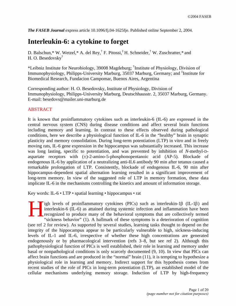
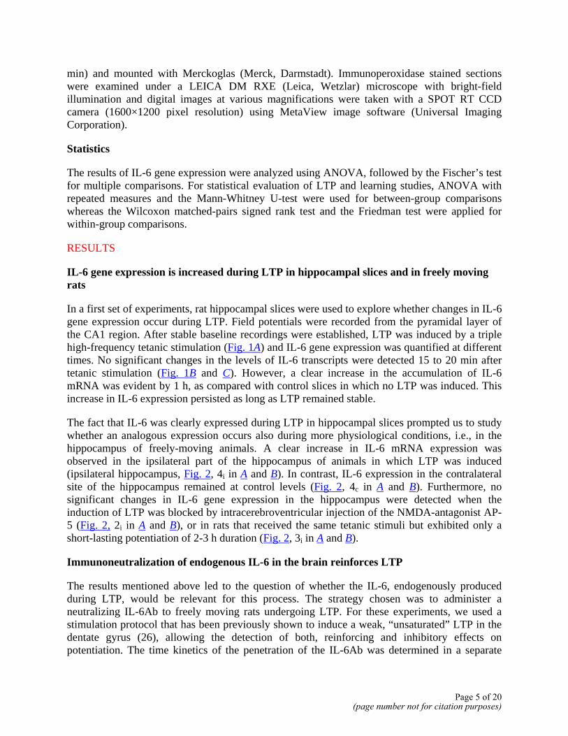
In a first set of experiments, rat hippocampal slices were used to explore whether changes in IL-6 gene expression occur during LTP. Field potentials were recorded from the pyramidal layer of the CA1 region. After stable baseline recordings were established, LTP was induced by a triple high-frequency tetanic stimulation (Fig. 1A) and IL-6 gene expression was quantified at different times. No significant changes in the levels of IL-6 transcripts were detected 15 to 20 min after tetanic stimulation (Fig. 1B and C). However, a clear increase in the accumulation of IL-6 mRNA was evident by 1 h, as compared with control slices in which no LTP was induced. This increase in IL-6 expression persisted as long as LTP remained stable.
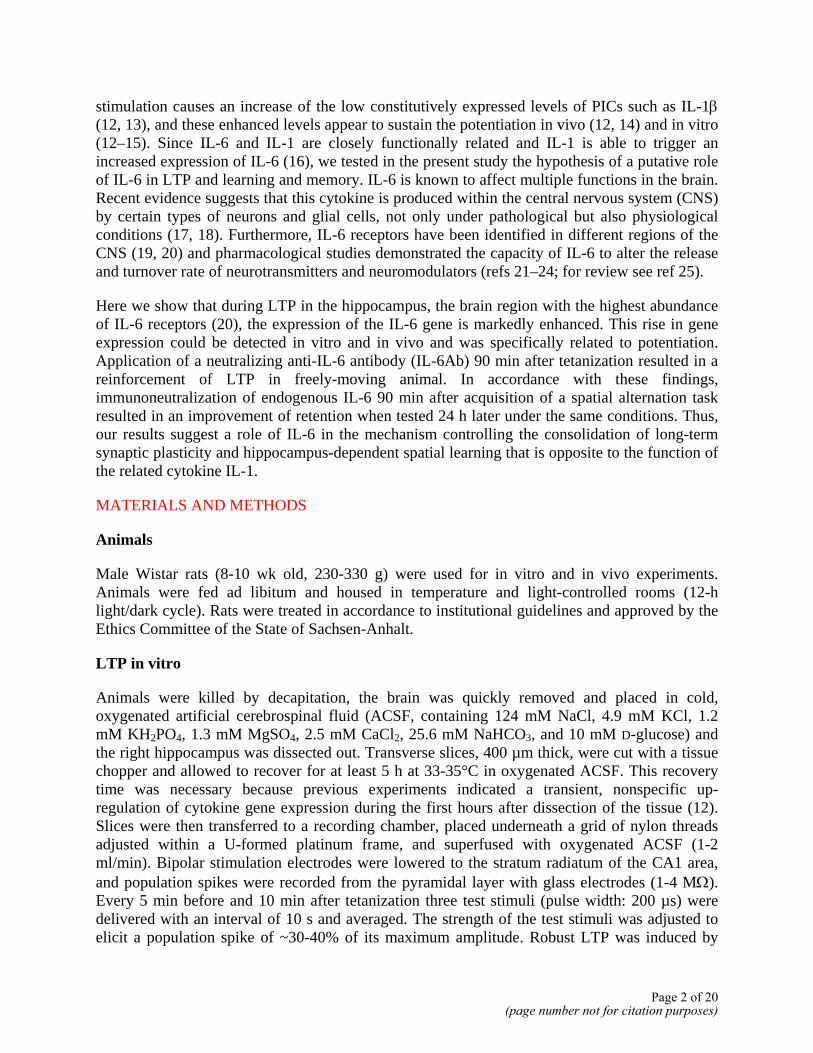
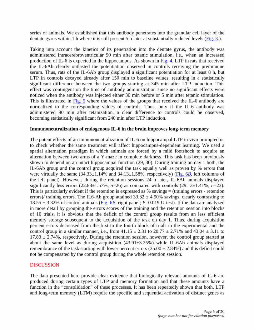
The fact that IL-6 was clearly expressed during LTP in hippocampal slices prompted us to study whether an analogous expression occurs also during more physiological conditions, i.e., in the hippocampus of freely-moving animals. A clear increase in IL-6 mRNA expression was observed in the ipsilateral part of the hippocampus of animals in which LTP was induced (ipsilateral hippocampus, Fig. 2, 4i in A and B). In contrast, IL-6 expression in the contralateral site of the hippocampus remained at control levels (Fig. 2, 4c in A and B). Furthermore, no significant changes in IL-6 gene expression in the hippocampus were detected when the induction of LTP was blocked by intracerebroventricular injection of the NMDA-antagonist AP-5 (Fig. 2, 2i in A and B), or in rats that received the same tetanic stimuli but exhibited only a short-lasting potentiation of 2-3 h duration (Fig. 2, 3i in A and B).
Immunoneutralization of endogenous IL-6 in the brain reinforces LTP
The results mentioned above led to the question of whether the IL-6, endogenously produced during LTP, would be relevant for this process. The strategy chosen was to administer a neutralizing IL-6Ab to freely moving rats undergoing LTP. For these experiments, we used a stimulation protocol that has been previously shown to induce a weak, “unsaturated” LTP in the dentate gyrus (26), allowing the detection of both, reinforcing and inhibitory effects on potentiation. The time kinetics of the penetration of the IL-6Ab was determined in a separate
Page 5 of 20(page number not for citation purposes)
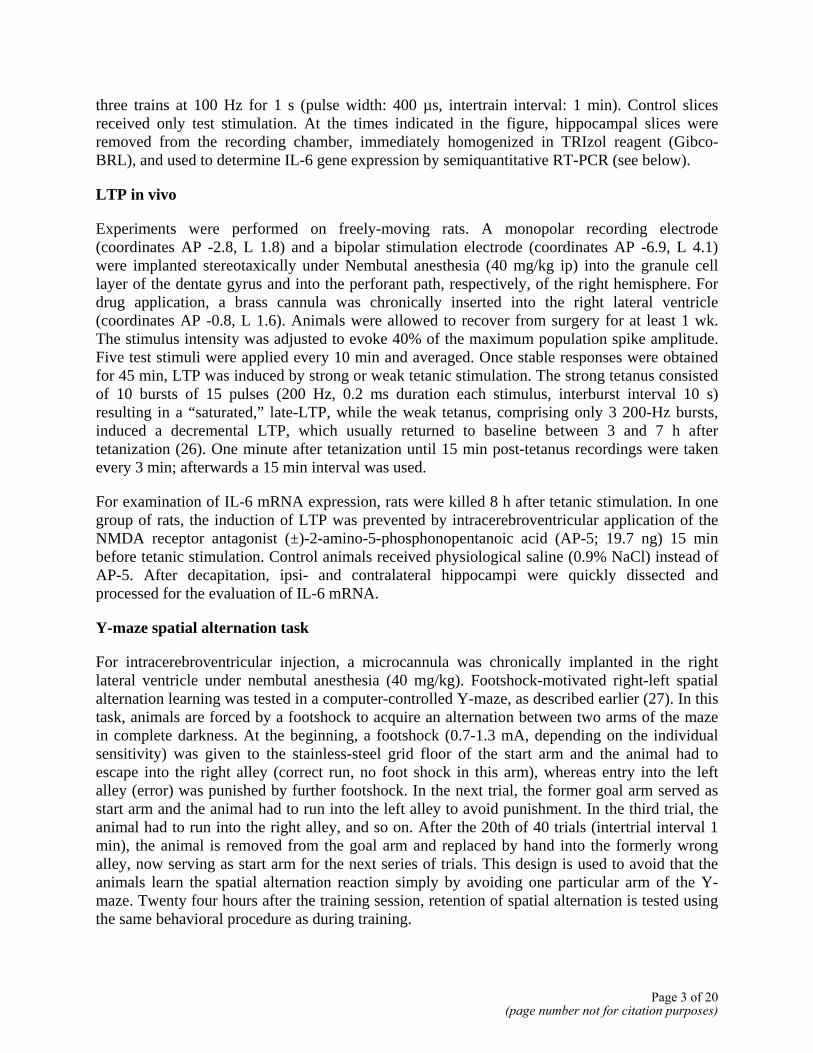
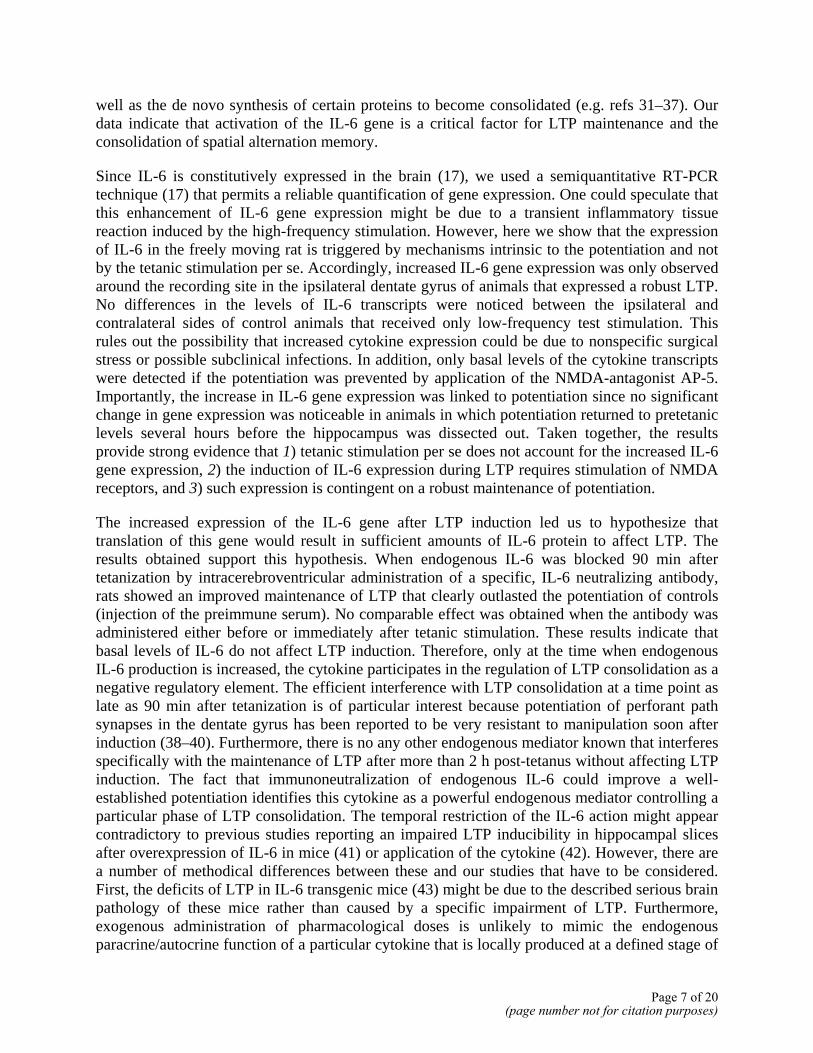
series of animals. We established that this antibody penetrates into the granular cell layer of the dentate gyrus within 1 h where it is still present 5 h later at substantially reduced levels (Fig. 3.).
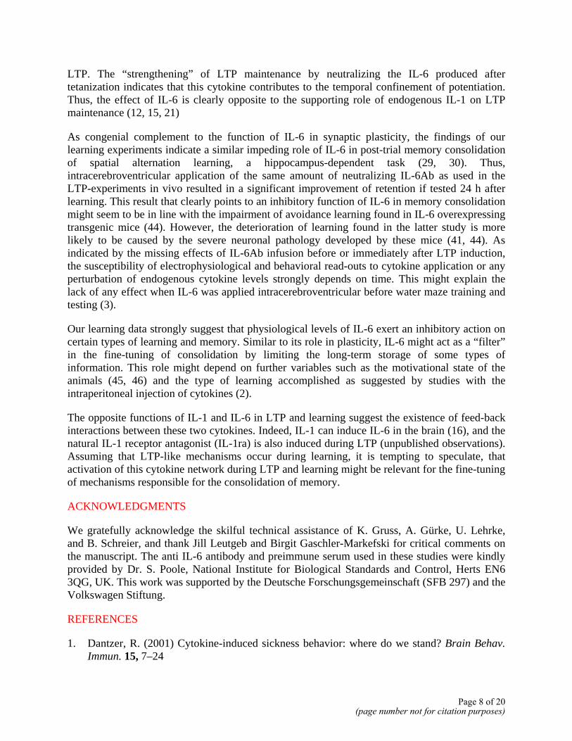
Taking into account the kinetics of its penetration into the dentate gyrus, the antibody was administered intracerebroventricular 90 min after tetanic stimulation, i.e., when an increased production of IL-6 is expected in the hippocampus. As shown in Fig. 4, LTP in rats that received the IL-6Ab clearly outlasted the potentiation observed in controls receiving the preimmune serum. Thus, rats of the IL-6Ab group displayed a significant potentiation for at least 8 h, but LTP in controls decayed already after 150 min to baseline values, resulting in a statistically significant difference between the two groups starting at 345 min after LTP induction. This effect was contingent on the time of antibody administration since no significant effects were noticed when the antibody was injected either 30 min before or 5 min after tetanic stimulation. This is illustrated in Fig. 5 where the values of the groups that received the IL-6 antibody are normalized to the corresponding values of controls. Thus, only if the IL-6 antibody was administered 90 min after tetanization, a clear difference to controls could be observed, becoming statistically significant from 240 min after LTP induction.
Immunoneutralization of endogenous IL-6 in the brain improves long-term memory
The potent effects of an immunoneutralization of IL-6 on hippocampal LTP in vivo prompted us to check whether the same treatment will affect hippocampus-dependent learning. We used a spatial alternation paradigm in which animals are forced by a mild footshock to acquire an alternation between two arms of a Y-maze in complete darkness. This task has been previously shown to depend on an intact hippocampal function (29, 30). During training on day 1 both, the IL-6Ab group and the control group acquired the task equally well as proven by % errors that were virtually the same (34.33±1.14% and 34.13±1.58%, respectively) (Fig. 6B, left columns of the left panel). However, during the retention sessions 24 h later, IL-6Ab animals displayed significantly less errors (22.88±1.57%, n=26) as compared with controls (29.13±1.41%, n=23). This is particularly evident if the retention is expressed as % savings = (training errors - retention errors)/ training errors. The IL6-Ab group attained 33.32 ± 4.50% savings, clearly contrasting to 18.55 ± 3.32% of control animals (Fig. 6B, right panel; P=0.019 U-test). If the data are analyzed in more detail by grouping the errors scores of the training and the retention session into blocks of 10 trials, it is obvious that the deficit of the control group results from an less efficient memory storage subsequent to the acquisition of the task on day 1. Thus, during acquisition percent errors decreased from the first to the fourth block of trials in the experimental and the control group in a similar manner, i.e., from 41.15 ± 2.31 to 20.77 ± 2.71% and 43.04 ± 3.11 to 17.83 ± 2.74%, respectively. During the retention session, however, the control group started at about the same level as during acquisition (43.91±3.25%) while IL-6Ab animals displayed remembrance of the task starting with lower percent errors (35.00 ± 2.84%) and this deficit could not be compensated by the control group during the whole retention session.
DISCUSSION
The data presented here provide clear evidence that biologically relevant amounts of IL-6 are produced during certain types of LTP and memory formation and that these amounts have a function in the “consolidation” of these processes. It has been repeatedly shown that both, LTP and long-term memory (LTM) require the specific and sequential activation of distinct genes as
Page 6 of 20(page number not for citation purposes)
well as the de novo synthesis of certain proteins to become consolidated (e.g. refs 31–37). Our data indicate that activation of the IL-6 gene is a critical factor for LTP maintenance and the consolidation of spatial alternation memory.
Since IL-6 is constitutively expressed in the brain (17), we used a semiquantitative RT-PCR technique (17) that permits a reliable quantification of gene expression. One could speculate that this enhancement of IL-6 gene expression might be due to a transient inflammatory tissue reaction induced by the high-frequency stimulation. However, here we show that the expression of IL-6 in the freely moving rat is triggered by mechanisms intrinsic to the potentiation and not by the tetanic stimulation per se. Accordingly, increased IL-6 gene expression was only observed around the recording site in the ipsilateral dentate gyrus of animals that expressed a robust LTP. No differences in the levels of IL-6 transcripts were noticed between the ipsilateral and contralateral sides of control animals that received only low-frequency test stimulation. This rules out the possibility that increased cytokine expression could be due to nonspecific surgical stress or possible subclinical infections. In addition, only basal levels of the cytokine transcripts were detected if the potentiation was prevented by application of the NMDA-antagonist AP-5. Importantly, the increase in IL-6 gene expression was linked to potentiation since no significant change in gene expression was noticeable in animals in which potentiation returned to pretetanic levels several hours before the hippocampus was dissected out. Taken together, the results provide strong evidence that 1) tetanic stimulation per se does not account for the increased IL-6 gene expression, 2) the induction of IL-6 expression during LTP requires stimulation of NMDA receptors, and 3) such expression is contingent on a robust maintenance of potentiation.
The increased expression of the IL-6 gene after LTP induction led us to hypothesize that translation of this gene would result in sufficient amounts of IL-6 protein to affect LTP. The results obtained support this hypothesis. When endogenous IL-6 was blocked 90 min after tetanization by intracerebroventricular administration of a specific, IL-6 neutralizing antibody, rats showed an improved maintenance of LTP that clearly outlasted the potentiation of controls (injection of the preimmune serum). No comparable effect was obtained when the antibody was administered either before or immediately after tetanic stimulation. These results indicate that basal levels of IL-6 do not affect LTP induction. Therefore, only at the time when endogenous IL-6 production is increased, the cytokine participates in the regulation of LTP consolidation as a negative regulatory element. The efficient interference with LTP consolidation at a time point as late as 90 min after tetanization is of particular interest because potentiation of perforant path synapses in the dentate gyrus has been reported to be very resistant to manipulation soon after induction (38–40). Furthermore, there is no any other endogenous mediator known that interferes specifically with the maintenance of LTP after more than 2 h post-tetanus without affecting LTP induction. The fact that immunoneutralization of endogenous IL-6 could improve a well-established potentiation identifies this cytokine as a powerful endogenous mediator controlling a particular phase of LTP consolidation. The temporal restriction of the IL-6 action might appear contradictory to previous studies reporting an impaired LTP inducibility in hippocampal slices after overexpression of IL-6 in mice (41) or application of the cytokine (42). However, there are a number of methodical differences between these and our studies that have to be considered. First, the deficits of LTP in IL-6 transgenic mice (43) might be due to the described serious brain pathology of these mice rather than caused by a specific impairment of LTP. Furthermore, exogenous administration of pharmacological doses is unlikely to mimic the endogenous paracrine/autocrine function of a particular cytokine that is locally produced at a defined stage of
Page 7 of 20(page number not for citation purposes)
LTP. The “strengthening” of LTP maintenance by neutralizing the IL-6 produced after tetanization indicates that this cytokine contributes to the temporal confinement of potentiation. Thus, the effect of IL-6 is clearly opposite to the supporting role of endogenous IL-1 on LTP maintenance (12, 15, 21)
As congenial complement to the function of IL-6 in synaptic plasticity, the findings of our learning experiments indicate a similar impeding role of IL-6 in post-trial memory consolidation of spatial alternation learning, a hippocampus-dependent task (29, 30). Thus, intracerebroventricular application of the same amount of neutralizing IL-6Ab as used in the LTP-experiments in vivo resulted in a significant improvement of retention if tested 24 h after learning. This result that clearly points to an inhibitory function of IL-6 in memory consolidation might seem to be in line with the impairment of avoidance learning found in IL-6 overexpressing transgenic mice (44). However, the deterioration of learning found in the latter study is more likely to be caused by the severe neuronal pathology developed by these mice (41, 44). As indicated by the missing effects of IL-6Ab infusion before or immediately after LTP induction, the susceptibility of electrophysiological and behavioral read-outs to cytokine application or any perturbation of endogenous cytokine levels strongly depends on time. This might explain the lack of any effect when IL-6 was applied intracerebroventricular before water maze training and testing (3).
Our learning data strongly suggest that physiological levels of IL-6 exert an inhibitory action on certain types of learning and memory. Similar to its role in plasticity, IL-6 might act as a “filter” in the fine-tuning of consolidation by limiting the long-term storage of some types of information. This role might depend on further variables such as the motivational state of the animals (45, 46) and the type of learning accomplished as suggested by studies with the intraperitoneal injection of cytokines (2).
The opposite functions of IL-1 and IL-6 in LTP and learning suggest the existence of feed-back interactions between these two cytokines. Indeed, IL-1 can induce IL-6 in the brain (16), and the natural IL-1 receptor antagonist (IL-1ra) is also induced during LTP (unpublished observations). Assuming that LTP-like mechanisms occur during learning, it is tempting to speculate, that activation of this cytokine network during LTP and learning might be relevant for the fine-tuning of mechanisms responsible for the consolidation of memory.
ACKNOWLEDGMENTS
We gratefully acknowledge the skilful technical assistance of K. Gruss, A. Gürke, U. Lehrke, and B. Schreier, and thank Jill Leutgeb and Birgit Gaschler-Markefski for critical comments on the manuscript. The anti IL-6 antibody and preimmune serum used in these studies were kindly provided by Dr. S. Poole, National Institute for Biological Standards and Control, Herts EN6 3QG, UK. This work was supported by the Deutsche Forschungsgemeinschaft (SFB 297) and the Volkswagen Stiftung.
REFERENCES
1. Dantzer, R. (2001) Cytokine-induced sickness behavior: where do we stand? Brain Behav. Immun. 15, 7–24
Page 8 of 20(page number not for citation purposes)
2. Gibertini, M. (1998) Cytokines and cognitive behavior. Neuroimmunomodulation 5, 160–165
3. Oitzl, M. S., van Oers, H., Schobitz, B., and De Kloet, E. R. (1993) Interleukin-1 beta, but not interleukin-6, impairs spatial navigation learning. Brain Res. 613, 160–163
4. Aubert, A., Vega, C., Dantzer, R., and Goodall, G. (1995) Pyrogens specifically disrupt the acquisition of a task involving cognitive processing in the rat. Brain Behav. Immun. 9, 129–148
5. Pugh, C. R., Nguyen, K. T., Gonyea, J. L., Fleshner, M., Wakins, L. R., Maier, S. F., and Rudy, J. W. (1999) Role of interleukin-1 beta in impairment of contextual fear conditioning caused by social isolation. Behav. Brain Res. 106, 109–118
6. Matsumoto, Y., Yoshida, M., Watanabe, S., and Yamamoto, T. (2001) Involvement of cholinergic and glutamatergic functions in working memory impairment induced by interleukin-1beta in rats. Eur. J. Pharmacol. 430, 283–288
7. Barrientos, R. M., Higgins, E. A., Sprunger, D. B., Watkins, L. R., Rudy, J. W., and Maier, S. F. (2002) Memory for context is impaired by a post context exposure injection of interleukin-1 beta into dorsal hippocampus. Behav. Brain Res. 134, 291–298
8. Song, C. (2002) The effect of thymectomy and IL-1 on memory: implications for the relationship between immunity and depression. Brain Behav. Immun. 16, 557–568
9. Maier, S. F., and Watkins, L. R. (1995) Intracerebroventricular interleukin-1 receptor antagonist blocks the enhancement of fear conditioning and interference with escape produced by inescapable shock. Brain Res. 695, 279–282
10. Yirmiya, R., Winocur, G., and Goshen, I. (2002) Brain interleukin-1 is involved in spatial memory and passive avoidance conditioning. Neurobiol. Learn. Mem. 78, 379–389
11. Vitkovic, L., Bockaert, J., and Jacque, C. (2000) Inflammatory cytokines: neuromodulators in normal brain? J. Neurochem. 74, 457–471
12. Schneider, H., Pitossi, F., Balschun, D., Wagner, A., del Rey, A., and Besedovsky, H. O. (1998) A neuromodulatory role of interleukin-1beta in the hippocampus. Proc. Natl. Acad. Sci. USA 95, 7778–7783
13. Jankowsky, J. L., Derrick, B. E., and Patterson, P. H. (2000) Cytokine responses to LTP induction in the rat hippocampus: a comparison of in vitro and in vivo techniques. Learn. Mem. 7, 400–412
14. Avital, A., Goshen, I., Kamsler, A., Segal, M., Iverfeldt, K., Richter-Levin, G., and Yirmiya, R. (2003) Impaired interleukin-1 signaling is associated with deficits in hippocampal memory processes and neural plasticity. Hippocampus 13, 826–834
Page 9 of 20(page number not for citation purposes)
15. Coogan, A. N., O’Neill, L. A., and O’Connor, J. J. (1999) The P38 mitogen-activated protein kinase inhibitor SB203580 antagonizes the inhibitory effects of interleukin-1beta on long-term potentiation in the rat dentate gyrus in vitro. Neuroscience 93, 57–69
16. Reyes, T. M., and Coe, C. L. (1998) The proinflammatory cytokine network: interactions in the CNS and blood of rhesus monkey. Am. J. Physiol. 274, R139-R144
17. Pitossi, F., del Rey, A., Kabiersch, A., and Besedovsky, H. (1997) Induction of cytokine transcripts in the central nervous system and pituitary following peripheral administration of endotoxin to mice. J. Neurosci. Res. 48, 287–298
18. Krueger, J. M., Obal, F. J., Fang, J., Kubota, T., Taishi, P. (2001) The role of cytokines in physiological sleep regulation. Ann. NY Acad. Sci. 933, 211-221
19. Schobitz, B., De Kloet, E. R., Sutanto, W., and Holsboer, F. (1993) Cellular localization of interleukin 6 mRNA and interleukin 6 receptor mRNA in rat brain. Eur. J. Neurosci. 5, 1426–1435
20. Gadient, R. A., and Otten, U. (1994) Expression of interleukin-6 (IL-6) and interleukin-6 receptor (IL-6R) mRNAs in rat brain during postnatal development. Brain Res. 637, 10–14
21. Besedovsky, H. O., del Rey, A., Klusman, I., Furukawa, H., Monge, A. G., and Kabiersch, A. (1991) Cytokines as modulators of the hypothalamus-pituitary-adrenal axis. J. Steroid Biochem. Mol. Biol. 40, 613–618
22. Wang, J., and Dunn, A. J. (1999) The role of interleukin-6 in the activation of the hypothalamo-pituitary-adrenocortical axis and brain indoleamines by endotoxin and interleukin-1 beta. Brain Res. 815, 337–348
23. D’Arcangelo, G., Tancredi, V., Onofri, F., D’Antuono, M., Giovedi, S., and Benfenati, F. (2000) Interleukin-6 inhibits neurotransmitter release and the spread of excitation in the rat cerebral cortex. Eur. J. Neurosci. 12, 1241–1252
24. De Laurentiis, A., Pisera, D., Lasaga, M., Diaz, M., Theas, S., Duvilanski, B., and Seilicovich, A. (2000) Effect of interleukin-6 and tumor necrosis factor-alpha on GABA release from mediobasal hypothalamus and posterior pituitary. Neuroimmunomodulation 7, 77–83
25. Besedovsky, H. O., and del Rey, A. (1996) Immune-neuro-endocrine interactions: facts and hypotheses. Endocr. Rev. 17, 64–102
26. Seidenbecher, T., Reymann, K. G., and Balschun, D. (1997) A post-tetanic time window for the reinforcement of long-term potentiation by appetitive and aversive stimuli. Proc. Natl. Acad. Sci. USA 94, 1494–1499
27. Balschun, D., Manahan-Vaughan, D., Wagner, T., Behnisch, T., Reymann, K. G., and Wetzel, W. (1999) A specific role for group I mGluRs in hippocampal LTP and hippocampus-dependent spatial learning. Learn. Mem. 6, 138–152
Page 10 of 20(page number not for citation purposes)
28. Cartmell, T., Poole, S., Turnbull, A. V., Rothwell, N. J., and Luheshi, G. N. (2000) Circulating interleukin-6 mediates the febrile response to localised inflammation in rats. J. Physiol 526, 653-661
29. Stevens, R., and Cowey, A. (1973) Effects of dorsal and ventral hippocampal lesions on spontaneous alternation, learned alternation and probability learning in rats. Brain Res. 52, 203-224
30. Aggleton, J. P., Hunt, P. R., and Rawlins, J. N. (1986) The effects of hippocampal lesions upon spatial and non-spatial tests of working memory. Behav. Brain Res. 19, 133–146
31. Agranoff, B. W., Davis, R. E., and Brink, J. J. (1966) Chemical studies on memory fixation in goldfish. Brain Res. 1, 303–309
32. Krug, M., Lossner, B., and Ott, T. (1984) Anisomycin blocks the late phase of long-term potentiation in the dentate gyrus of freely moving rats. Brain Res. Bull. 13, 39–42
33. Stanton, P. K., and Sarvey, J. M. (1984) Blockade of long-term potentiation in rat hippocampal CA1 region by inhibitors of protein synthesis. J. Neurosci. 4, 3080–3088
34. Mizumori, S. J., Rosenzweig, M. R., and Bennett, E. L. (1985) Long-term working memory in the rat: effects of hippocampally applied anisomycin. Behav. Neurosci. 99, 220–232
35. Nguyen, P. V., Abel, T., and Kandel, E. R. (1994) Requirement of a critical period of transcription for induction of a late phase of LTP. Science 265, 1104–1107
36. Frey, U., and Morris, R. G. (1997) Synaptic tagging and long-term potentiation. Nature 385, 533–536 (see comments)
37. Scharf, M. T., Woo, N. H., Lattal, K. M., Young, J. Z., Nguyen, P. V., and Abel, T. (2002) Protein synthesis is required for the enhancement of long-term potentiation and long-term memory by spaced training. J. Neurophysiol. 87, 2770–2777
38. Errington, M. L., Bliss, T. V., Richter-Levin, G., Yenk, K., Doyere, V., and Laroche, S. (1995) Stimulation at 1-5 Hz does not produce long-term depression or depotentiation in the hippocampus of the adult rat in vivo. J. Neurophysiol. 74, 1793–1799
39. Abraham, W. C., Mason-Parker, S. E., and Logan, B. (1996) Low-frequency stimulation does not readily cause long-term depression or depotentiation in the dentate gyrus of awake rats. Brain Res. 722, 217–221
40. Kulla, A., Reymann, K. G., and Manahan-Vaughan, D. (1999) Time-dependent induction of depotentiation in the dentate gyrus of freely moving rats: involvement of group 2 metabotropic glutamate receptors. Eur. J. Neurosci. 11, 3864–3872
41. Bellinger, F. P., Madamba, S. G., Campbell, I. L., and Siggins, G. R. (1995) Reduced long-term potentiation in the dentate gyrus of transgenic mice with cerebral overexpression of interleukin-6. Neurosci. Lett. 198, 95–98
Page 11 of 20(page number not for citation purposes)
42. Li, A. J., Katafuchi, T., Oda, S., Hori, T., and Oomura, Y. (1997) Interleukin-6 inhibits long-term potentiation in rat hippocampal slices. Brain Res. 748, 30–38
43. Campbell, I. L., Abraham, C. R., Masliah, E., Kemper, P., Inglis, J. D., Oldstone, M. B., and Mucke, L. (1993) Neurologic disease induced in transgenic mice by cerebral overexpression of interleukin 6. Proc. Natl. Acad. Sci. USA 90, 10061–10065
44. Heyser, C. J., Masliah, E., Samimi, A., Campbell, I. L., and Gold, L. H. (1997) Progressive decline in avoidance learning paralleled by inflammatory neurodegeneration in transgenic mice expressing interleukin 6 in the brain. Proc. Natl. Acad. Sci. USA 94, 1500–1505
45. Cahill, L. (2000) Neurobiological mechanisms of emotionally influenced, long-term memory. Prog. Brain Res. 126, 29–37
46. McGaugh, J. L. (2000) Memory–a century of consolidation. Science 287, 248–251
Received February 18, 2004; accepted June 12, 2004.
Page 12 of 20(page number not for citation purposes)
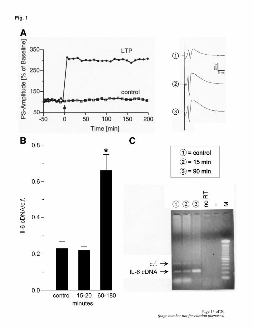
Fig. 1
Page 13 of 20
(page number not for citation purposes)
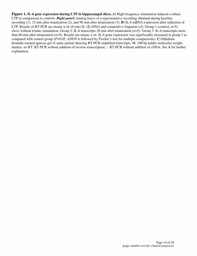
Figure 1. IL-6 gene expression during LTP in hippocampal slices. A) High-frequency stimulation induced a robust LTP in comparison to controls. Right panel) Analog traces of a representative recording obtained during baseline recording (1), 15 min after tetanization (2), and 90 min after tetanization (3). B) IL-6 mRNA expression after induction of LTP. Results of RT-PCR are means ± SE of ratio IL-1β cDNA and competitive fragment (cf). Group 1 (control, n=5): slices without tetanic stimulation. Group 2: IL-6 transcripts 20 min after tetanization (n=5). Group 3: IL-6 transcripts more than 60 min after tetanization (n=9). Results are means ± SE. IL-6 gene expression was significantly increased in group 3 as compared with control group (P<0.05; ANOVA followed by Fischer’s test for multiple comparisons). C) Ethidium bromide-stained agarose gel of same animal showing RT-PCR amplified transcripts. M: 100 bp ladder molecular weight marker; no RT: RT-PCR without addition of reverse transcriptase; -: RT-PCR without addition of cDNA. See A for further explanation.
Page 14 of 20(page number not for citation purposes)
Fig. 2
Page 15 of 20(page number not for citation purposes)
Figure 2. IL-6 gene expression during LTP in freely moving rats (A) IL-6 mRNA expression 8 h after tetanization. Thirty minutes before tetanization, animals received either physiological saline (groups 1, 3, and 4) or AP-5 (group 2) administered icv. Gray bars are measurements performed in ipsilateral (i) and black bars in contralateral (c) hippocampi. Group 1: hippocampi of animals recorded under baseline conditions without tetanic stimulation. Group 2: hippocampi of animals in which expression of LTP after tetanization was blocked by AP-5. Group 3: hippocampi of animals showing a potentiation that returned to baseline within 3 h. Group 4: hippocampi of animals with a robust LTP lasting for 8 h. Results of RT-PCR are means ± SE of ratio IL-6 cDNA and competitive fragment (cf), n=4 per group. Group 4i differs significantly from all other groups (ANOVA followed by Fischer’s test for multiple comparisons). B) Representative ethidium bromide-stained agarose gel showing amplified transcripts of a RT-PCR obtained from ipsi- and contralateral hippocampi that were subjected to different experimental conditions (lane numbers correspond to groups shown in A). Line graphs are representative analog traces recorded during baseline (top), immediately after tetanization (middle), and 8 h after tetanus (bottom). M: 100 bp ladder molecular weight marker; no RT: RT-PCR without addition of reverse transcriptase; -: RT-PCR without addition of cDNA.
Page 16 of 20(page number not for citation purposes)
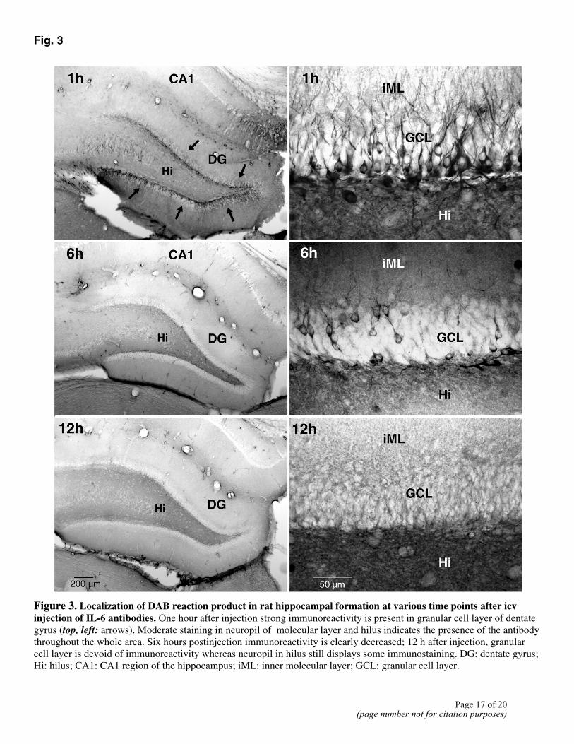
Fig. 3
Figure 3. Localization of DAB reaction product in rat hippocampal formation at various time points after icv injection of IL-6 antibodies. One hour after injection strong immunoreactivity is present in granular cell layer of dentate gyrus (top, left: arrows). Moderate staining in neuropil of molecular layer and hilus indicates the presence of the antibody throughout the whole area. Six hours postinjection immunoreactivity is clearly decreased; 12 h after injection, granular cell layer is devoid of immunoreactivity whereas neuropil in hilus still displays some immunostaining. DG: dentate gyrus; Hi: hilus; CA1: CA1 region of the hippocampus; iML: inner molecular layer; GCL: granular cell layer.
Page 17 of 20
(page number not for citation purposes)
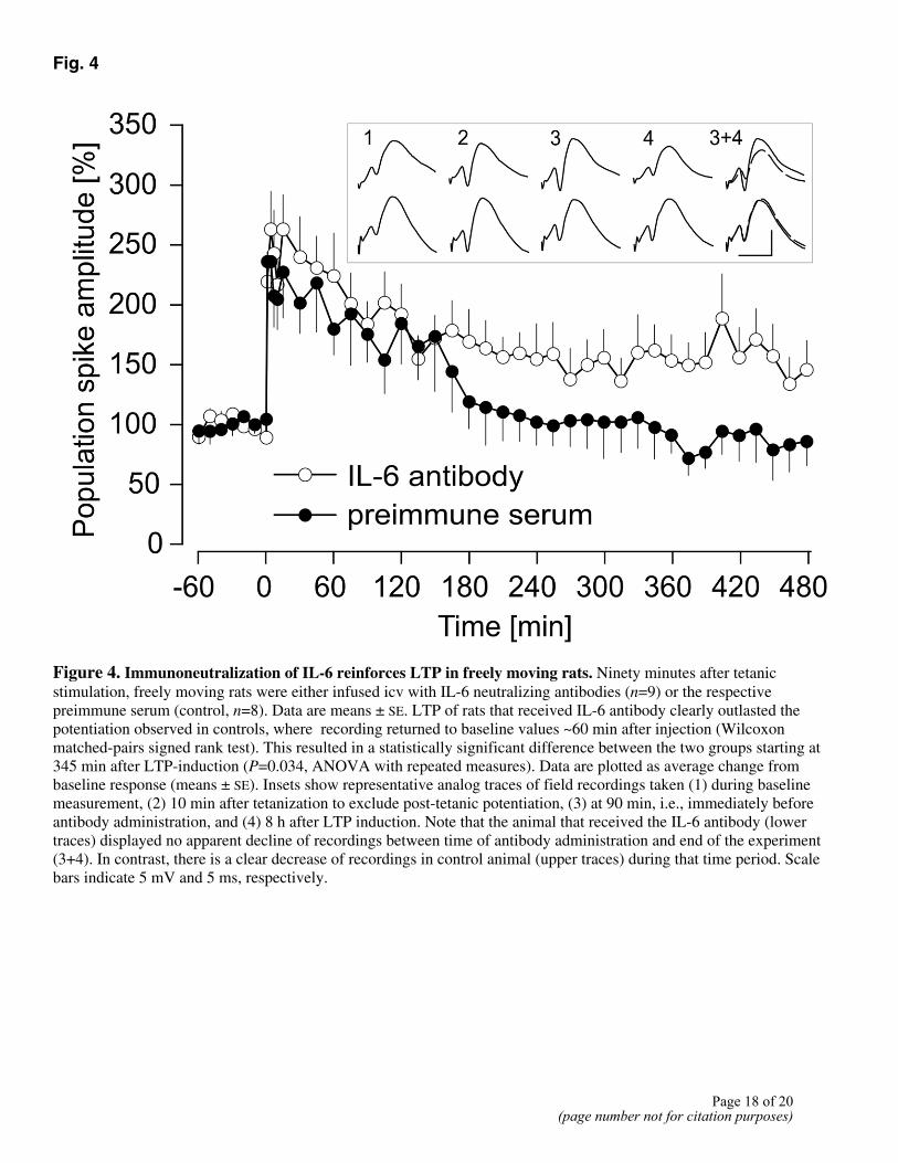
Fig. 4
Figure 4. Immunoneutralization of IL-6 reinforces LTP in freely moving rats. Ninety minutes after tetanic stimulation, freely moving rats were either infused icv with IL-6 neutralizing antibodies (n=9) or the respective preimmune serum (control, n=8). Data are means ± SE. LTP of rats that received IL-6 antibody clearly outlasted the potentiation observed in controls, where recording returned to baseline values ~60 min after injection (Wilcoxon matched-pairs signed rank test). This resulted in a statistically significant difference between the two groups starting at 345 min after LTP-induction (P=0.034, ANOVA with repeated measures). Data are plotted as average change from baseline response (means ± SE). Insets show representative analog traces of field recordings taken (1) during baseline measurement, (2) 10 min after tetanization to exclude post-tetanic potentiation, (3) at 90 min, i.e., immediately before antibody administration, and (4) 8 h after LTP induction. Note that the animal that received the IL-6 antibody (lower traces) displayed no apparent decline of recordings between time of antibody administration and end of the experiment (3+4). In contrast, there is a clear decrease of recordings in control animal (upper traces) during that time period. Scale bars indicate 5 mV and 5 ms, respectively.
Page 18 of 20(page number not for citation purposes)
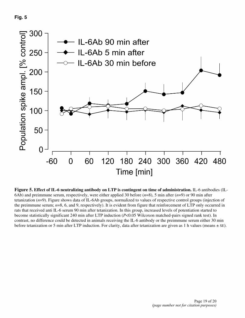
Fig. 5
Figure 5. Effect of IL-6 neutralizing antibody on LTP is contingent on time of administration. IL-6 antibodies (IL-6Ab) and preimmune serum, respectively, were either applied 30 before (n=8), 5 min after (n=9) or 90 min after tetanization (n=9). Figure shows data of IL-6Ab groups, normalized to values of respective control groups (injection of the preimmune serum; n=8, 6, and 9, respectively). It is evident from figure that reinforcement of LTP only occurred in rats that received anti IL-6 serum 90 min after tetanization. In this group, increased levels of potentiation started to become statistically significant 240 min after LTP induction (P<0.05 Wilcoxon matched-pairs signed rank test). In contrast, no difference could be detected in animals receiving the IL-6 antibody or the preimmune serum either 30 min before tetanization or 5 min after LTP induction. For clarity, data after tetanization are given as 1 h values (means ± SE).
Page 19 of 20(page number not for citation purposes)
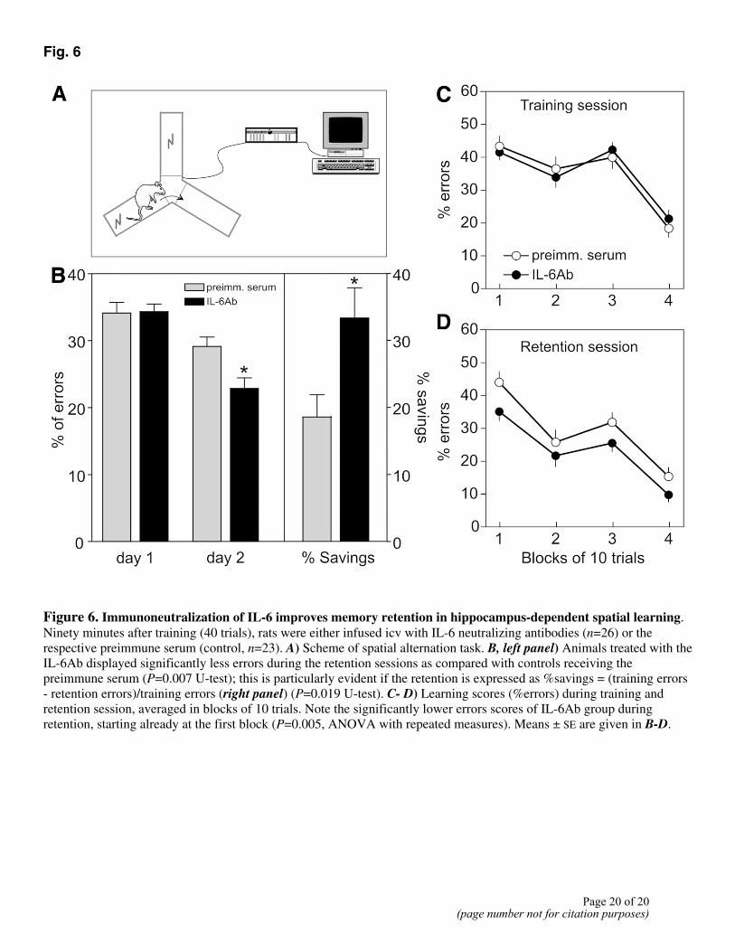
Fig. 6
Figure 6. Immunoneutralization of IL-6 improves memory retention in hippocampus-dependent spatial learning. Ninety minutes after training (40 trials), rats were either infused icv with IL-6 neutralizing antibodies (n=26) or the respective preimmune serum (control, n=23). A) Scheme of spatial alternation task. B, left panel) Animals treated with the IL-6Ab displayed significantly less errors during the retention sessions as compared with controls receiving the preimmune serum (P=0.007 U-test); this is particularly evident if the retention is expressed as %savings = (training errors - retention errors)/training errors (right panel) (P=0.019 U-test). C- D) Learning scores (%errors) during training and retention session, averaged in blocks of 10 trials. Note the significantly lower errors scores of IL-6Ab group during retention, starting already at the first block (P=0.005, ANOVA with repeated measures). Means ± SE are given in B-D.
Page 20 of 20(page number not for citation purposes)


