Embed Size (px)
Citation preview

ACADEMIA ROMÂNĂ
Revue Roumaine de Chimie
http://web.icf.ro/rrch/
Rev. Roum. Chim.,
2012, 57(4-5), 361-368
Dedicated to Professor Ionel Haiduc on the occasion of his 75th anniversary
INFLUENCE OF SAM STRUCTURE ON DIRECT ELECTRON TRANSFER AT Au ELECTRODES MODIFIED WITH CELLOBIOSE DEHYDROGENASE
FROM NEUROSPORA CRASSA
Gábor KOVÁCSa, Roberto ORTIZb, Vasile COMANa, Wolfgang HARREITHERc, Ionel Cătălin POPESCUa*, Roland LUDWIGc and Lo GORTONb
a Departament of Physical Chemistry, Faculty of Chemistry and Chemical Engineering, Babeş-Bolyai University, 11, Arany János, RO-400084 Cluj-Napoca, Roumania
bDepartment of Analytical Chemistry / Biochemistry and Structural Biology, Lund University P.O. Box 124, 22100 Lund, Sweden, cFood Biotechnology Laboratory, Department of Food Sciences and Technology, Boku-University of Natural Resources
and Life Sciences Vienna, Muthgasse 18, A-1190 Wien, Austria
Received January 27, 2012
Cellobiose dehydrogenase (CDH) isolated from Neurospora crassa was immobilized on Au electrodes, covered with self-assembled monolayers (SAMs) made of different thiolic compounds bearing -OH, -COOH or -NH2 as terminal functional groups. From cyclic voltammetric measurements, performed at different pH values, the catalytic efficiencies towards lactose oxidation were estimated for the immobilized CDH. The observed variations were explained in terms of: (i) the influence of the SAM structure on the direct electron transfer (DET) between CDH and the Au electrode surface; (ii) the variation of the catalytic activity of the enzyme, induced by changes of enzyme’s conformation. The dependence on pH, found for the standard formal potential of the heme group, confirms that, when CDH is deposited on thiolic SAMs, it is able to sustain an efficient DET process.
INTRODUCTION∗
Ever since the discovery of direct electron transfer (DET), the intriguing phenomenon, which refers to the direct electronic coupling between the redox cofactor of a protein and an electrode surface,1-5 a lot of interest emerged in obtaining information about the thermodynamic and kinetic properties of the protein with implications for catalytic and sensor applications6-8 and for a better understanding of biological electron transfer (ET) processes.9, 10 In order to obtain an efficient DET coupling between the redox centers of the protein/enzyme and the electrode, a suitable orientation of the enzyme on the electrode surface is crucial. According to the Marcus theory,11 DET ∗ Corresponding author: [email protected]
between a protein and an electrode is dependent on three major factors: i) the distance between the redox site and the electrode surface; ii) the reorganization energy, which reflects the structural rigidity of the redox site in its oxidized and reduced forms; iii) the thermodynamic driving force of the ET, which is related to a proper synchronization between the redox potential of the protein and the polarization of the electrode surface.6, 12, 13
In the case of metal and especially gold electrodes,14 the discovery of the strong adsorption of thiols, sulfides, disulfides and related molecules on surfaces offered multiple choices for protein immobilization and orientation. A self-assembled monolayer (SAM), an organized molecular

362 Gábor Kovács et al.
assembly of amphiphilic molecules, is formed by adsorption onto a solid surface from a homogeneous solution.15-18 The organization is given by the affinity of the head group for the surface and from the slow two dimensional orientations of the hydrophobic tail groups. Concerning ET to large biomolecules, the groups of Taniguchi and Hill19, 20 pioneered the field of Au modification with organic compounds that yielded diffusion controlled DET of cytochrome c. The electronic pairing between the protein and the electrode can be varied by changing the length of the spacer or the functionality of the tail-group of the SAM modifier, controlling in this way the orientation of the redox active protein on the electrode surface and the distance between the redox active site of the protein and the electrode.
Among the enzymes known to exhibit DET characteristics at various electrodes, cellobiose dehydrogenase (CDH, cellobiose: acceptor 1-oxidoreductase, EC 1.1.99.18) was thoroughly studied in recent years.21-24 CDH is an extracellular oxidoreductase, produced by many basidiomycetes (class I) and ascomycetes (class II) fungi.24, 25
CDHs are monomeric enzymes consisting of two domains connected by a mobile linker.26 The larger catalytic domain contains flavin adenine dinucleotide (FAD), DHCDH, and belongs to the glucose-methanol-choline (GMC) family, while the smaller cytochrome domain hosts a heme b (protoheme IX), CYTCDH, with an unusual pair (His and Met) of axial ligands.26, 27
A schematic representation of the functioning principle, involving a DET process, illustrated in the case of a bioelectrode constructed by immobilizing CDH on a SAM-modified Au electrode is shown in Fig. 1A. In the presence of a substrate (e.g. lactose) CDH oxidizes the sugar to the corresponding lactone at the FAD containing DHCDH and the resulting electrons are transferred through an internal electron transfer (IET) pathway to the CYTCDH. If the enzyme is properly oriented on the modified electrode surface (Fig. 1B), the CYTCDH transfers the electrons further to the electrode surface through an efficient DET process.
Fig. 1 – (A) The schematic diagram of DET (underlying the internal ET) for adsorbed CDH on a SAM-modified Au electrode; (B) the electrochemical interface structure of the bioelectrode based on CDH adsorbed at different SAM-modified Au electrodes, where Y= -(CH2)2-, -C6H4-, -(CH2)11-, and X= -OH, -NH2, -COOH.
Cellobiose dehydrogenase 363
The best studied CDHs up to date are the ones from basidiomycetes (class I), while only limited information is available on class II CDHs.23, 25 The DET behavior at SAM modified Au electrodes for a number of CDHs (predominantly belonging to class I) have been studied previously using cyclic voltammetry and UV-Vis spectroelectrochemis-try.28-34 In this context, it is worth to mention that for each investigated CDH the optimal response was obtained for a specific thiolic SAM. Therefore, it was considered interesting to get a clear picture of the DET process in the case of a new CDH, isolated from Neurospora crassa.25
The aim of the current paper was to provide more information on the DET process in the case of Neurospora crassa CDH. For this purpose, CDH was trapped under a permselective membrane applied onto the surface of SAM modified Au electrodes, as it was previously described.28,29,31,33,35 Cyclic voltammetric measure-ments, performed at different pH values, were used to estimate the catalytic efficiencies towards lactose. The observed behavior was explained in terms of the influence of the SAM structure on DET process occurring between CDH and the Au electrode surface. At the same time, the pH dependence of the standard formal potential of the heme group of the CYTCDH, corroborated with data
for electrocatalytic efficiency, confirms once again that CDH, deposited on thiolic SAMs, is able to sustain an efficient DET process.
RESULTS AND DISCUSSION
The enzyme used in the present study, is a class IIA CDH carrying a carbohydrate binding module. This enzyme was recently extracted and purified from Neurospora crassa, a well characterized laboratory model organism, using a protocol described elsewhere.25 The electrochemical behavior of this enzyme, adsorbed on graphite electrodes, was studied under direct and mediated electron transfer modes.36, 37
1. Voltammetric behavior of AU-SYX-CDH modified electrodes
In this context, taking into account the possibility to better control the enzyme orientation by its immobilization on Au surfaces modified with thiolic SAMs,28-34 it was interesting to carry out a detailed electrochemical study on the influence of the SAM structure on DET process at Au-SAM-CDH electrodes.
HS-(CH2)2-X HS-C6H4-X HS-(CH2)11-X
-OH
-300 -200 -100 0 100 200-50
0
50
100
150
200
I /
nA
E / mV vs. SCE -300 -200 -100 0 100 200 300
-150
0
150
300
450
I /
nA
E / mV vs. SCE -200 -100 0 100 200 300
-200
0
200
400
600
800
I /
nA
E / mV vs. SCE
-NH
2
-300 -200 -100 0 100 200-200
0
200
400
600
800
I /
nA
E / mV vs. SCE -300 -200 -100 0 100 200
-50
0
50
100
I /
nA
E / mV vs. SCE
-CO
OH
-200 -100 0 100 200
0
300
600
900
1200
1500
I /
nA
E / mV vs. SCE -200 -100 0 100 200 300
-100
-50
0
50
100
I /
nA
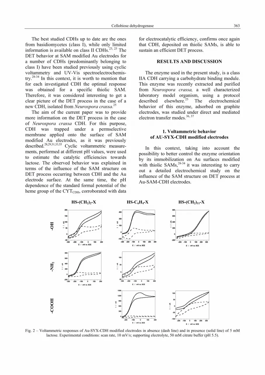
E / mV vs. SCE Fig. 2 – Voltammetric responses of Au-SYX-CDH modified electrodes in absence (dash line) and in presence (solid line) of 5 mM lactose. Experimental conditions: scan rate, 10 mV/s; supporting electrolyte, 50 mM citrate buffer (pH 5.5).
364 Gábor Kovács et al.
As can be seen from Fig. 2, three types of voltammetric behaviors were recorded for the investigated Au-SYX-CDH modified electrodes: (i) a well shaped electrocatalytic response, observed for Au electrodes covered with HS-(CH2)2-NH2 and HS-C6H4-X (X= -OH, -COOH); (ii) a mixed response, observed in the case of HS-(CH2)2-OH, HS-C6H4-NH2 and HS-(CH2)11-COOH, which can be considered an overlapping of the electrocatalytic response with a partial direct oxidation of the substrate on the unmodified Au surface; (iii) a poor electrocatalytic response, noticed in the case of HS-(CH2)11-OH, being due to the direct oxidation of the substrate on the unmodified Au electrode surface.31, 38 Additionally, it should be mentioned that the Au-S-(CH2)2-COOH-CDH modified electrode showed a high instability and, for this reason, all corresponding results were disregarded. At this moment, the real reason for this peculiar behavior is not known.
These behaviors could be understood as resulting from a complex combination of two factors: the distance between the electrode surface and the enzyme redox active center, which is controlled by the length of the thiolic molecule; the interactions existing, for a given SAM, between the CDH molecule and the terminal functional group.
Thus, according to the Marcus theory,11 the increase in the length of an insulating hydrocarbon chain will induce a decrease in the electrocatalytic efficiency. Indeed, this was the case when the electrocatalytic activity of CDH deposited on Au-S-(CH2)2-X (X= -OH, -NH2) is compared to that of CDH deposited on Au-S-(CH2)11-X (X= -OH, -COOH). It is interesting to notice that a positive deviation from this rule can be observed when between HS- and the terminal functional group is intercalated a structural unit bearing delocalized electrons. This was the case of the SAM built of HS-C6H4-X (X= -OH, -COOH), when a clear enhancement of the electrocatalytic activity of CDH occurred.
For a given SAM, the electrostatic interactions between the CDH molecule and the terminal functional group depend strongly on the surrounding pH, which controls the ionization state of both the terminal functional groups and the enzyme surface. An example illustrating this situation is the case of HS-(CH2)2-NH2, which at pH 5.5 is positively charged, and consequently, will develop attractive interactions with the CDH
molecules, promoting the DET process.28 The apparent discrepancy observed between the electrocatalytic efficiencies observed for Au-S-C6H4-NH2-CDH and Au-S-(CH2)2-NH2-CDH modified electrodes should be due to the higher basicity of the aliphatic amines compared to the aromatic ones.
However, besides the electrostatic nature, other interactions such as hydrogen bonds, van der Waals and hydrophilic/hydrophobic interactions etc., should be considered in order to understand why, for example, the Au-S-C6H4-COOH-CDH modified electrode showed the highest catalytic activity among the investigated modified electrodes. Additionally, one must keep in mind that for efficient bioelectrocatalysis both an intimate coupling and correct orientation between the CYTCDH-domain and the electrode must prevail as well as a flexibility between the two subunits allowing rapid IET from DHCDH and CYTCDH.
2. Electrocatalytic behavior of AU-SYX-CDH modified electrodes
In order to compare the electrocatalytic activity of the investigated modified electrodes at different pH values, the electrocatalytic efficiency was estimated as (I[S]-I[0])/I[0]), where I[S] and I[0] stand for the catalytic current and the background current, respectively. For all investigated electrodes, except the Au-S-(CH2)11-COOH-CDH modified electrode, the maximum electrocatalytic efficiency was observed around pH 5 (Fig. 3). Thus, the already reported preference of CDH isolated from Neurospora crassa25, 36, 37 for acidic media was confirmed once again. The presence of a maximum efficiency for a certain pH (around 5) reflects a combination of two factors: (i) the increase of the thermodynamic driving force due to the decrease in the formal standard potential (E0’) of the heme of the CYTCDH, when the pH increases until pH ~5.5;28, 29, 33 (ii) the decrease in the catalytic activity of the enzyme caused by the changes occurring in the enzyme’s conformation with the pH increase, involving a less intimate coupling between DHCDH and CYTCDH.23, 24 Concerning the last factor, this is related to the efficiency of the internal ET between DHCDH and CYTCDH, which is strongly affected by the change of the distance between the two enzyme domains.

Cellobiose dehydrogenase 365
4 5 6 7 80.0
2.5
5.0
7.5
10.0
12.5
(I [S] -
I [0])
/ I[0
]
pH
-NH2
-OHA
4 5 6 7 80
20
40
60
(I [S]
-I [0])/I
[0]
pH
-NH2
-OH -COOH
B
4 5 6 7 8 90
3
6
9
12
15
(I [S]
-I [0])/I
[0]
pH
-OH -COOHC
Fig. 3 – pH dependence of the electrocatalytic efficiencies, calculated as (I[S]-I[0])/I[0]), for Au-SYX-CDH modified electrodes, where Y = -(CH2)2- (A); -C6H4- (B) and -(CH2)11- (C). Experimental conditions: applied potential, +200 mV vs. SCE; substrate, 5 mM lactose.
0
5
10
40
45
50
-(CH2)11--(C6H4)-
-NH2
-NH2
-COOH
-COOH
-OH
-OH
(I [S] -
I [0]) /
I [0]
-(CH2)2-
-OH
Fig. 4 – Influence of the SAM type on the electrocatalytic efficiencies for Au-SYX-CDH modified electrodes. Experimental conditions: applied potential, +200 mV vs. SCE; pH 5.5; substrate, 5 mM lactose.
Concerning the peculiar behavior of the Au-S-(CH2)11-COOH-CDH modified electrode (Fig. 3C), the pH increase induces a gradual ionization of the carboxyl terminal group causing a monotone decrease in the electrocatalytic efficiency. At pH values higher than 5, the electrocatalytic activity of the electrode vanishes, probably because the unfavorable interactions between the CDH molecule and the negatively charged surface of the SAM.
The effect of the terminal functional group on the electrocatalytic efficiency can be better put in evidence when the maximum values of the electrocatalytic efficiencies, estimated for all Au-SYX-CDH modified electrodes, are plotted grouped for the same -Y- unit (Fig. 4). Thus, within the limits of experimental errors, it can be stated that: (i) when the –COOH group is connected to the thiol group (HS-) via a conducting
unit (-C6H4-) it will clearly enhance the electrocatalytic activity; (ii) the higher basicity of the aliphatic amines, compared to their aromatic counterparts, will favor attractive interactions between CDH and the modified electrode surface, resulting in an increase in the electrocatalytic efficiency of the electrode.
3. pH influence on E0’ of the heme redox couple from CDH
The voltammetric responses corresponding to the heme redox couple, hosted by CYTCDH,28-32 were recorded at different pH values for the Au-SYX-CDH modified electrodes. Their characteristic electrochemical parameters were estimated at pH 5.5 and are summarized in Table 1.
366 Gábor Kovács et al.
Table 1
The electrochemical parameters of the heme voltammetric response observed at different Au-SYX modified electrodes (pH 5.5)
Thiolic compound X ∆Ep (mV) E0’ (mV) -OH 50 -135 HS-(CH2)2-X -NH2 40 -130 -OH 70 -135
-COOH 30 -135 HS-C6H4-X -NH2 70 -135
4.5 5.0 5.5 6.0 6.5 7.0
-150
-140
-130
-120
-110
-100
E0'
/ m
V vs
. SC
E
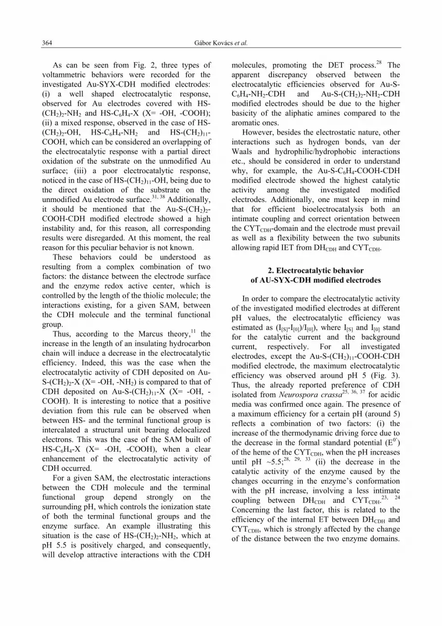
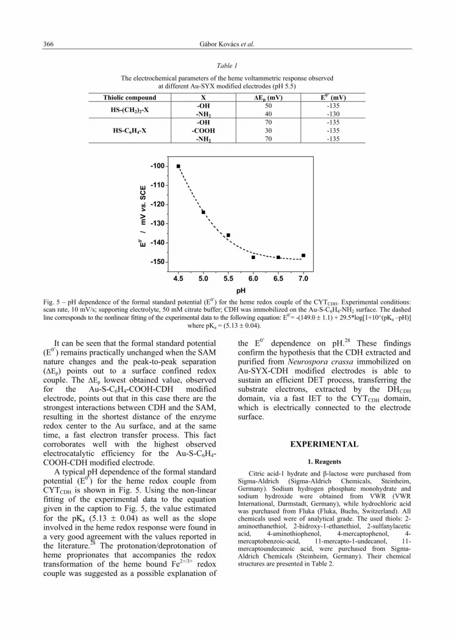
pH Fig. 5 – pH dependence of the formal standard potential (E0’) for the heme redox couple of the CYTCDH. Experimental conditions: scan rate, 10 mV/s; supporting electrolyte, 50 mM citrate buffer; CDH was immobilized on the Au-S-C6H4-NH2 surface. The dashed line corresponds to the nonlinear fitting of the experimental data to the following equation: E0’= -(149.0 ± 1.1) + 29.5*log[1+10^(pKa –pH)] where pKa = (5.13 ± 0.04).
It can be seen that the formal standard potential
(E0’) remains practically unchanged when the SAM nature changes and the peak-to-peak separation (∆Ep) points out to a surface confined redox couple. The ∆Ep lowest obtained value, observed for the Au-S-C6H4-COOH-CDH modified electrode, points out that in this case there are the strongest interactions between CDH and the SAM, resulting in the shortest distance of the enzyme redox center to the Au surface, and at the same time, a fast electron transfer process. This fact corroborates well with the highest observed electrocatalytic efficiency for the Au-S-C6H4-COOH-CDH modified electrode.
A typical pH dependence of the formal standard potential (E0’) for the heme redox couple from CYTCDH is shown in Fig. 5. Using the non-linear fitting of the experimental data to the equation given in the caption to Fig. 5, the value estimated for the pKa (5.13 ± 0.04) as well as the slope involved in the heme redox response were found in a very good agreement with the values reported in the literature.28 The protonation/deprotonation of heme proprionates that accompanies the redox transformation of the heme bound Fe2+/3+ redox couple was suggested as a possible explanation of
the E0’ dependence on pH.28 These findings confirm the hypothesis that the CDH extracted and purified from Neurospora crassa immobilized on Au-SYX-CDH modified electrodes is able to sustain an efficient DET process, transferring the substrate electrons, extracted by the DHCDH domain, via a fast IET to the CYTCDH domain, which is electrically connected to the electrode surface.
EXPERIMENTAL
1. Reagents
Citric acid-1 hydrate and β-lactose were purchased from Sigma-Aldrich (Sigma-Aldrich Chemicals, Steinheim, Germany). Sodium hydrogen phosphate monohydrate and sodium hydroxide were obtained from VWR (VWR International, Darmstadt, Germany), while hydrochloric acid was purchased from Fluka (Fluka, Buchs, Switzerland). All chemicals used were of analytical grade. The used thiols: 2-aminoethanethiol, 2-hidroxy-1-ethanethiol, 2-sulfanylacetic acid, 4-aminothiophenol, 4-mercaptophenol, 4-mercaptobenzoic-acid, 11-mercapto-1-undecanol, 11-mercaptoundecanoic acid, were purchased from Sigma-Aldrich Chemicals (Steinheim, Germany). Their chemical structures are presented in Table 2.

Cellobiose dehydrogenase 367
Table 2
Formulae of the thiolic compounds used for SAM construction
Terminal functional group (X) Compound (HS-Y-X) -OH -COOH -NH2
2-hydroxy-1-ethanethiol 2-sulfanylacetic acid 2-aminoethanethiol
HS-(CH2)2-X
4-mercaptophenol 4-mercaptobenzoic-acid 4-aminothiophenol
HS-C6H4-X HS OH
11-mercapto-1-undecanol 11-mercapto-1-undecanoic acid -
HS-(CH2)11-X HS OH
O
Cellobiose dehydrogenase from Neurospora crassa
(CDH) was obtained and purified as previously described.25 The protein concentration was estimated using the Bradford assay as 13.9 mg/mL. The enzyme activity, at pH 4 and 30°C, was found to be 118.73 U/ml and 63.6 U/ml by using DCIP assay and cyt c assay, respectively.
The buffer solutions used in all experiments were prepared using either a 50 mM citric acid solution (for pHs ranging from 4.0 to 6.5) or a 50 mM sodium hydrogen phosphate (for pHs placed in the 6.0 to 8.5 interval). The desired pH value was adjusted with 4 M NaOH or 5 M HCl. The pH values were measured with a pH meter (Metrohm 827 pH Lab, Herisau, Switzerland). Before use, the buffer and the substrate solutions were carefully degassed.
The thiol solutions were prepared using ethanol (99.7%); buffers and other solutions were prepared using water purified and deionized (18 MΩ) with a Milli-Q system (Millipore, Bedford, MA, USA).
2. Cyclic voltammetry measurements
Cyclic voltammetry (CV) experiments were performed under anaerobic conditions (assured by a previous degassing of the solutions and by using a flow of pure argon gas over the working solution) with a BAS 100W Electrochemical Analyzer (Bioanalytical Systems, West Lafayette, IN, USA). A three electrode cell was used with the working electrode, a saturated calomel electrode (SCE) as the reference electrode and a Pt wire as the auxiliary electrode. A scan rate of 300 mV/s was used for the electrochemical cleaning procedure of the Au electrodes and 10 mV/s for all other measurements.
3. Preparation of Au-SYX-CDH modified electrodes
The cleaning of the disc Au electrode (CH-Instruments, Cordova, TN, USA; Ø 2 mm and area of 0.033 cm2) started by dipping the Au electrode in “piranha” solution (3:1 v/v H2SO4:H2O2) for 5 min. Caution: piranha solution reacts violently with most organic materials and must be handled with extreme care. Then, the electrode was mirror-like polished with aqueous alumina FF slurry (1 and 0.1 µm,
Struers, Copenhagen, Denmark) deposited on Microcloth (Buehler, Lake Bluff, IL, USA). Furthermore, the electrodes were carefully rinsed with water, ultrasonicated for 5 min in Milli-Q water, and electrochemically cleaned in 0.5 M H2SO4 by performing 20 cycles with a scan rate of 300 mV/s between −100 and 1700 mV vs. SCE. Finally, they were rinsed again with Milli-Q water.
The preparation of CDH-thiol-modified electrodes started by 60 min immersion of the clean Au electrodes in a 1 mM ethanolic solution of the thiol. The treatment resulted in the formation of the SAM of the thiol on the electrode surface. Before CDH deposition, the Au-SAM electrodes were carefully rinsed with ethanol in order to remove the weakly adsorbed thiols. Then, they were dried with Ar. The CDH deposition on the Au-SAM modified electrodes was made by spreading 2 µL of enzyme solution onto the thiol-modified Au surface. The enzyme drop was allowed to gently dry in order to avoid enzyme spreading outside the electrode area. A dialysis membrane (Spectrum Laboratories Inc., Rancho Dominguez, CA, USA, molecular weight cut off 6000–8000), pre-soaked in the buffer solution, was applied onto the electrode and fitted tightly to the electrode surface with a rubber O-ring. For three equivalently prepared electrodes the enzymatic activities were found to be reproducible within the error limits of 5%.
CONCLUSIONS
Cyclic voltammetric measurements, performed at different pHs for Au-SYX-CDH modified electrodes (Y = -(CH2)2-, -C6H4- and -(CH2)11-; X = -OH, -COOH, -NH2) in absence or in presence of the CDH substrate (lactose), allow estimation of the electrocatalytic efficiencies of the immobilized Neurospora crassa CDH towards lactose. The variations observed between the different electrocatalytic efficiencies were interpreted in

368 Gábor Kovács et al.
terms of the influence of the SAM structure on the DET between CDH and the Au electrode surface.
The obtained results reveal, in a systematic manner, that the amperometric response recorded at CDH modified Au electrodes can be explained as a complex combination of three factors: (i) the distance between the electrode surface and the redox active center of the CYTCDH, which is controlled by the length of the thiolic molecule; (ii) the interactions existing, for a given SAM, between the CDH molecule and the terminal functional group; (iii) the connection flexibility between the two subunits of the bound enzyme, which controls the rate of the IET.
The pH dependence of the standard formal potential of the heme group, validates the hypothesis that the CDH extracted from Neurospora crassa and immobilized on Au-SYX-CDH modified electrodes is able to sustain an efficient DET process, which consists of three consecutive steps: (i) the substrate oxidation by the DHCDH domain; (ii) the internal electron transfer to the CYTCDH domain via a fast IET; (iii) the electrical connection between the CYTCDH domain and the modified electrode surface.
Acknowledgments: The authors acknowledge the financial
supports as follows: (GK) The Sectoral Operational Program for Human Resources Development 2007 – 2013, contract no. POSDRU/6/1.5/S/3, “Doctoral Studies: through Science towards Society”; (VC) contract no. POSDRU/89/1.5/S/60189, “Postdoctoral Programs for Sustainable Development in a Knowledge Based Society”; (LG and RO) The Swedish Research Council (project number 2010-5031) and The European Commission, project “3D-Nanobiodevice” NMP4-SL-2009-229255; (ICP) CNCSIS–UEFISCSU (project PNII-ID-PCCE-129/2008).
REFERENCES
1. P. Yeh and T. Kuwana, Chem. Lett., 1977, 1145-1148. 2. M. J. Eddowes and H. A. O. Hill, J. Chem. Soc., Chem.
Commun., 1977, 771-772. 3 I. V. Berezin, V. A. Bogdanovskaya, S. D. Varfolomeev,
M. R. Tarasevich and A. I. Yaropolov, Dokl. Akad. Nauk SSSR, 1978, 240, 615-618.
4. M. R. Tarasevich, A. I. Yaropolov, V. A. Bogdanovskaya and S. D. Varfolomeev, Bioelectrochem. Bioenerg., 1979, 6, 393-403.
5 A. I. Yaropolov, V. Malovik, S. D. Varfolomeev and I. V. Berezin, Dokl. Akad. Nauk SSSR, 1979, 249, 1399-1401.
6. L. Gorton, A. Lindgren, T. Larsson, F. D. Munteanu, T. Ruzgas and I. Gazaryan, Anal. Chim. Acta, 1999, 400, 91-108.
7 W. Zhang, G. Li, Anal. Sci., 2004, 20, 603-610. 8. J. A. Cracknell, K. A. Vincent and F. A. Armstrong,
Chem. Rev., 2008, 108, 2439-2461. 9. C. C. Page, C. C. Moser, X. Chen and P. L. Dutton,
Nature, 1999, 402, 47-52.
10. L. J. C. Jeuken, Biochim. Biophys. Acta, 2003, 1604, 67-76.
11. R. A. Marcus and N. Sutin, Biochim. Biophys. Acta, 1985, 811, 265-322.
12. I. Willner and E. Katz, Angew. Chem. Int. Ed., 2000, 39, 1181-1218.
13. F. A. Armstrong, Russ. J. Electrochem., 2002, 38, 49-62. 14. J. J. Gooding, F. Mearns, W. Yang and J. Liu,
Electroanalysis, 2003, 15, 81-96. 15. K. L. Prime and G. M. Whitesides, Science, 1991, 252,
1164-1167. 16. G. E. Poirier and E. D. Pylant, Science, 1996, 272, 1145-
1148. 17. A. Ulman, Chem. Rev., 1996, 96, 1533-1554. 18. H. O. Finklea, Electroanalytical Chemistry, Vol. 19 (Ed.:
A. J. B. a. I. Rubinstein), Marcel Decker, New York, 1996, p. 109-335.
19. I. Taniguchi, K. Toyosawa, H. Yamaguchi and K. Yasukouchi, J. Electroanal. Chem. Interfacial Electrochem., 1982, 140, 187-193.
20. P. M. Allen, H. Allen, O. Hill and N. J. Walton, J. Electroanal. Chem., 1984, 178, 69-86.
21. G. Henriksson, G. Johansson and G. Pettersson, J. Biotechnol., 2000, 78, 93-113.
22. M. D. Cameron and S. D. Aust, Enzyme Microb. Technol., 2001, 28, 129-138.
23. M. Zamocky, R. Ludwig, C. Peterbauer, B. M. Hallberg, C. Divne, P. Nicholls and D. Haltrich, Curr. Prot. Pept. Sci., 2006, 7, 255-280.
24. R. Ludwig, W. Harreither, F. Tasca and L. Gorton, ChemPhysChem, 2010, 11, 2674-2697.
25. W. Harreither, C. Sygmund, M. Augustin, M. Narciso, M. L. Rabinovich, L. Gorton, D. Haltrich and R. Ludwig, Appl. Environ. Microb., 2011, 77, 1804-1815.
26. B. M. Hallberg, G. Henriksson, G. Pettersson and C. Divne, J. Mol. Biol., 2002, 315, 421-434.
27. B. M. Hallberg, T. Bergfors, K. Backbro, G. Pettersson, G. Henriksson and C. Divne, Structure, 2000, 8, 79-88.
28. A. Lindgren, T. Larsson, T. Ruzgas and L. Gorton, J. Electroanal. Chem., 2000, 494, 105-113.
29. A. Lindgren, L. Gorton, T. Ruzgas, U. Baminger, D. Haltrich and M. Schulein, J. Electroanal. Chem., 2001, 496, 76-81.
30. T. Larsson, A. Lindgren and T. Ruzgas, Bioelectrochem., 2001, 53, 243-249.
31. L. Stoica, N. Dimcheva, D. Haltrich, T. Ruzgas and L. Gorton, Biosens. Bioelectron., 2005, 20, 2010-2018.
32. L. Stoica, R. Ludwig, D. Haltrich and L. Gorton, Anal. Chem., 2006, 78, 393-398.
33. V. Coman, W. Harreither, R. Ludwig, D. Haltrich and L. Gorton, Chem. Anal. (Warsaw), 2007, 52, 945-960.
34. D. Sarauli, R. Ludwig, D. Haltrich, L. Gorton and F. Lisdat, Bioelectrochem., 2011, in press.
35. J. Haladjian, P. Bianco, F. Nunzi and M. Bruschi, Anal. Chim. Acta, 1994, 289, 15-20.
36. G. Kovács, R. Ortiz, V. Coman, W. Harreither, I. C. Popescu, R. Ludwig and L. Gorton, Bioelectrochem., 2012, submitted.
37. W. Harreither, E. Lackner, P. Nicholls, L. Gorton and R. Ludwig, Langmuir, 2012, submitted.
38. T. Vidaković-Koch, I. Ivanov, M. Falk, S. Shleev, T. Ruzgas and K. Sundmacher, Electroanalysis, 2011, 23, 927-930.


