Embed Size (px)
Citation preview
Indonesian Day
1A-S01-01
GMP VS ISO IN A BLOOD ESTABLISHMENTVA Armstrong
Vee Armstrong Quality and Regulatory Consultant, Perth, Australia
Blood Establishments have undergone a significant cultural change over the past
25 years, moving from a predominantly scientifically focussed culture to that of a
manufacturer of therapeutic goods.
Part of this transition has been driven by the emergence of, initially, quality man-
agement systems (QMS), and subsequently, principles of good manufacturing prac-
tice (GMP) as requirements for blood establishments. The manufacture of fresh blood
and blood components has also become highly regulated in many countries, with
Codes of Good Manufacturing Practice written specifically for the industry.
Certification to the international Standard ISO 9001: Quality management systems –Requirements has been undertaken by a number of Blood Establishments in coun-
tries where regulation of the Blood Establishments has not yet been introduced. The
Standard sets out the framework for a QMS that could be used by any industry that
produces a product for a customer and as such, the requirements are general. ISO
implementation has the advantage of auditing by a third party to determine the level
of compliance, and formal certification once the level is acceptable.
Not only do Codes of GMP for Blood require a QMS to be implemented, but they
also set out principles for the manufacture of therapeutic goods, focussing on safety
through risk management and error minimisation. Additionally, the Codes include
requirements specifically for each part of the manufacturing chain. Successful GMP
implementation in regulated countries is formally acknowledged by documented
accreditation or the award of a licence to operate, however in non-regulated coun-
tries, there is often no formal acknowledgement of successful compliance or third
party auditing. Additionally, the concept of GMP and particularly the differences
between it and practices already in place can be difficult to grasp.
As a result, some Blood Establishments in non-regulated countries that wish to
improve practices have opted to use ISO implementation as a driver for quality
improvement. However, while the ISO Standard and Code of GMP both set out the
requirements for a QMS, ISO does not include the critical requirement that manufac-
ture of blood and blood components must follow principles of GMP to ensure opti-
misation of safety through managing risks and minimising errors. Blood
Establishments will therefore ultimately need to implement GMP regardless of ISO
certification to be assured of safe, consistent and efficacious components.
The starting point for a Blood Establishment’s implementation of GMP will depend
on its ISO status if any. If it has already been ISO certified, a gap analysis should
be conducted between ISO and GMP and a plan developed to close the gaps. If it
is currently implementing ISO, the gap analysis should include the systems still to
be developed so that GMP is built into those systems at the same time. If it has
not implemented ISO, it is recommended that the focus be on GMP implementa-
tion.
The similarities and differences between ISO and GMP, and the implementation of
GMP into an ISO QMS will be considered in more detail.
1A-S01-02
THE ROLE OF THE NATIONAL BLOOD CENTRE IN A COUNTRYWITH A MIXED SYSTEM OF BLOOD SERVICESU Charoonruangrit
National Blood Centre, the Thai Red Cross Society, Bangkok, Thailand
Background: National Blood Centre (NBC), the Thai Red Cross Society is a charita-
ble organization under principles of the International Federation of Red Cross and
Red Crescent Societies (IFRC). It is designated by the Government to implement the
national blood transfusion service with safe and adequate supply. Apart from NBC
headquarters in Bangkok, there are 12 Regional Blood Centres (RBCs) in the provin-
cial regions running blood collection and testing services for the network provinces
in each particular region. Blood donation at NBC and RBCs are from voluntary non-
remunerated donors (VNRD). Six government hospitals in Bangkok (under universi-
ties, militaries and Bangkok Metropolitans) and also approximately 160 government
provincial hospitals operate blood collections for their own uses. Clinical blood
transfusion practices run by the hospitals.
National blood policy: The Government adopted the current National Blood Policy
3rd version in 2010. It consists of 9 policy objectives including effective manage-
ment, adequacy of blood supply from VNRD, safety of blood in accordance with the
principles of the World Health Organization, promoting quality assurance systems,
appropriate blood utilization, implementing related laws and regulations, conducting
the researches and developments, producing plasma derived medicinal products and
providing the stem cell from unrelated donor volunteers.
Blood donation: Coordinating with National Blood Centre, each Provincial Red
Cross Chapter led by Red Cross Chapter President, shall conduct blood recruitment
program. Red Cross Chapters together with provincial hospitals, which are assigned
as Blood Centre Branches, shall arrange blood donation schedules to meet their need
and also supply to neighboring districts. Due to effective public relations campaign
during the past 4 years, NBC, RBCs and also hospital blood centre branches all over
the country attained blood collections beyond the setting goal. By the year 2014,
overall whole blood collections were 2,274,788 units which was equal to 3.5% of
the number of the whole country populations.
Blood testing: Centralization of donor blood testing is one of the national policy
strategies for safe and standard blood supply. All 12 RBCs are the centralized labs
for their regions and 10 of them are Nucleic Acid Testing (NAT) lab centres. At pre-
sent, nearly 95% of donated blood in the country is NAT tested.
Standards and guidelines: NBC and experts on Transfusion Medicine had revised
and published the 5th edition of Donor Selection Guideline, the 1st edition of Physi-
cian Handbook on The Appropriate use of Blood and Blood Components, the 3rd
edition of the Standard Blood Banks and Transfusion Services and the 1st edition of
Hemovigilance Handbook. They are the references for national blood service prac-
tices.
Quality systems: NBC provides training and accreditation on Blood Collection
Technique and Blood Donation Administration course for hospital staff who operate
blood donations. All hospitals shall follow quality assurance programs such as
Hospital Accreditation (HA), ISO etc. and shall be regularly assessed by the profes-
sional council committees.
1A-S01-03
CONSOLIDATION OF LABORATORIES FOR BLOOD TESTINGJ Chen
Taiwan Blood Services Foundation, Taipei, Taiwan
Background: Annually we collect 1.8 million of donor bloods, either whole blood or
apheresis platelets, through six regional centers. Before the release of blood and
blood components, samples of each donation are taken to test for infectious diseases
and blood grouping. Since the population of the service area of certain center is low
and the number of specimens to be tested by the center is few, they are in a difficult
position in recruitment of qualified staff and in use of advanced technology for
donor blood screening. Aims: We report a laboratory consolidation program that,
with minimal cost, improve the quality of laboratory screening for donor blood col-
lected by each centers. Methods: Before laboratory consolidation, donor bloods were
tested in the laboratories of each center. The laboratory of the smallest center per-
formed screening for around 200 donors per day. In 2001, we initiated a laboratory
consolidation program. The fundamentals of the program were (1) centralization of
donor blood screening at two laboratories, (2) each equipped with advanced screen-
ing analyzers, (3) blood specimens arrived at laboratory before 09:00 on the next
morning, (4) result report before 17:00 on the next day, (5) uniformity in testing kits
and equipment between laboratories, (6) back-up plan in place. As a part of risk
management, the program was carried out with two phases that began with the tran-
sition from 6 to 5 laboratories, and after 6 months, to 2 laboratories. Results: After
consolidation, the two laboratories each performs an average of 2,500 donor blood
screening and the subsequent confirmatory testing per day in the work hours from
08:00 to 17:00. These include blood group serology (ABO, RhD, antibody screening),
chemistry, viral immunoassay and nucleic acid testing (NAT). Before 17:00 on the
second day of collection, test results are available for regional center to release
blood and blood components except that donations are subject to test for bacteria
contamination. Since that culture-based bacteria detection requires specimens for
analysis being taken during 24–36 h of platelet storage, for this reason, we detect
bacteria contamination for platelets in the laboratory of each regional center rather
than centralization. In addition, it is essential that contingency back-up plans are in
place for donor blood screening. Up to the present time, we had experienced a num-
ber of events that switched the usual procedure for donor blood screening to the
back-up procedure.
Summary/conclusion: Through laboratory consolidation, we are able to provide
high-quality and cost-effective donor blood screening and, most importantly, to
ensure the safety of donor blood collected by each individual center. At present
the two laboratories, together with a head laboratory that performs reference test-
ing service, ensure blood safety and provide specialized transfusion support in
Taiwan.
© 2015 The AuthorVox Sanguinis © 2015 International Society of Blood Transfusion
Vox Sanguinis (2015) 109 (Suppl. 2), 1–96
Indonesian Day 1

1A-S02-01
BLOOD APHAERESIS IN BEIJINGY Qiu
Beijing Red Cross Blood Center, Beijing, China
In July 1978, Beijing Red Cross Blood Center (BRCBC) collected the first aphaeresis
platelet with Haemonetics M30. Since then BRCBC has imported Haemonetics PCS+,MCS3P, MCS+, Baxter AMICUS and Caridian BCT Trima successively into blood col-
lection and components preparation. We collect platelet and plasma with aphaeresis
machine only for clinical usage. The number of plateletpheresis has been increased
annually. In 1994 we only collected 2249 therapeutic unit plateletpheresis and
reached to 65,088 units in 2014. At present the SDP supply had replaced of the RDP
totally in Beijing.
We follow the national regulation ‘whole blood and component donor selection
requirements GB 18467-2011’ t o screen and select the aphaeresis donors and set up
a efficient SDP donation workflow including donor identification, pre-donation test-
ing and health evaluation, donor acceptance, aphaeresis collection preparation, col-
lection process and post-donation treatment, etc. The rate of plateletpheresis donors
deferred is 17.56 in 2014. The choice of aphaeresis procedures can be collecting pla-
telet only, including single, double platelet or both plasma and platelet component
is collected at the same time. We program the aphaeresis procedure depending on
the donor intention and health examination results. The rate of double platelet col-
lection is 62.7% and the number of plasma for transfusion units derived from
aphaeresis collections is 4950 units in 2014.
In order to meet the increasing progressively clinical consumption of platelets annu-
ally, we take advantage of any accessible Medias to promote the blood donation and
try to recruit and retain the blood donors in Beijing. Plateletpheresis donation fre-
quency per donor in Beijing is about 3 times per year. Both the new guidelines of
shortened the plateletpheresis donation interval and the increasing ratio of repeated
aphaeresis donor help to overcome the shortage of platelet supply in Beijing. The
platelet issues per 1000 population in Beijing are 5.69 in 2014.
PCs product random test monthly is a method of quality surveillance and also a part
work of the established blood quality management system. ‘Standards for whole
blood and blood components quality: GB 18469-2012’ is a statutory national guide-
line for blood services to implement quality control and supervise the Labeling,
appearance, volume, pH value, platelets count, residual of WBC, residual of RBC and
the sterility of all labile blood products.
Immunohaemotology lab is responsible for investigating frequencies of HLA and
HPA antibodies among patients who transfusion the platelet. Patient’s characteristics
and the positive % of platelet-specific antibody push us to screen the platelet-speci-
fic and HLA antibodies screening routinely. Platelet cross-match is the mainly way
to prevent the platelet transfusion refractory in Beijing.
By now we have not organized an integrated heamovigilance system in Beijing.
Optimization the blood aphaeresis system deeply, fulfilling the clinical demand,
expanding the HLA and HPA typed platelet donors pool and traceability the
aphaeresis blood product will be the goal in the near future.
1A-S02-02
HEPATITIS B UPDATE ON DIAGNOSTIC AND SCREENING TESTMW Thedja
Eijkman Institute for Molecular Biology, Jakarta, Indonesia
Hepatitis B remains a major health concern worldwide with impact on various clini-
cal manifestations including chronic infection, cirrhosis, fulminant hepatitis, and
hepatocellular carcinoma. Further, hepatitis B virus (HBV) accounts as the most
common transfusion-transmitted viral infection that might give significance impact
on morbidity and mortality. Thus, diagnostic and screening tests for HBV infection
are the entry point to reduce the burden of clinical and socio-economic conse-
quences of HBV complication. Hepatitis B surface Antigen (HBsAg) serology test is
still used as the first-line diagnostic marker for hepatitis B, and also the first-line
blood-donor screening test for HBV infection. However, in some situations HBsAg
might not be detected in serum either in chronic HBV-patient or in blood donation
despite the presence of HBV DNA. This condition might be due to window period,
HBV mutant particularly in surface gene, and occult carriage of HBV infection (OBI)
that usually corresponds with low titer of HBV DNA. In addition to HBsAg, anti-HBc
test has been widely used for blood donation screening in some countries; however,
the implementation of anti-HBc screening is inefficient for those countries with HBV
endemic in term of blood donation loss. Mutations in HBsAg that is associated with
conformational changes may account solely or in conjunction with the failure of
HBsAg detection by immunoassay. In the last few years, NAT (nucleic acid testing)
multiplex assay that combines simultaneous HBV, hepatitis C virus (HCV), and
human immunodeficiency virus (HIV) detection has been introduced and
implemented either in minipools or more efficiently in individual samples. NAT dis-
criminatory assay for HBV/HCV/HIV can be applied for samples with positive NAT
multiplex. In comparison with serological tests, HBV NAT has the ability to reduce
the window period and to detect OBI cases. However, for countries with high
endemicity of HBV infection, the implementation of NAT multiplex/discriminatory
may not be affordable due to the high operational cost. Alternatives to simplify
methods and to reduce NAT multiplex/discriminatory operational cost should be a
challenge for scientific community, and also develop more sensitive serological tests
for HBsAg mutants.
1A-S02-03
PLATELET ANTIBODY TESTING AS PART OF PRE-TRANSFUSION TESTING FOR MULTI-TRANSFUSION PATIENTSWHO HAD PLATELET REFRACTORINESSNK Ritchie
Jakarta Blood Transfusion Unit, DKI Jakarta, Indonesia
Platelets as part of blood cells, which are multi-antigenic, have human platelet anti-
gen (HPA), human leukocyte antigen (HLA) class I beside ABO antigen on its surface.
The polymorphism of those antigens are very high, especially HLA antigen. The
patients could develop alloantibody to antigen which not exists on his platelets. At
next transfusion, that alloantibody could react with donor’s platelet, resulting in pla-
telet refractoriness. Beside alloantibody, there were other factors contributed to pla-
telet refractoriness. The clinicians could use corrected count increment at 1 h and
24 h after platelet transfusion to predict the cause of platelet refractoriness. Then,
they could ask the reference laboratory to screen and identify the platelet antibody.
In Indonesia, according to Chunaeni study, 20–70% of hemato-oncology patients in
Dharmais Hospital had platelet refractoriness due to platelet transfusion. Most of
them had platelet transfusion from random donors. Another study by Lubis et al has
shown 39% of hemato-oncology patients who received repeatedly platelets transfu-
sion, developed anti-HLA class I antibody and only one anti-gpIIb/IIIa (3%). In other
cases, we might find anti-HLA class I antibody in patients with no history of platelet
transfusion. In such cases, the source of HLA class I antigens were leukocytes which
contaminated the PRC. If the alloantibodies detected, there is a probability, patients
could get a compatible platelet so the transfusion would be effective.
Platelet antibody testing consists of antibody screening and identification test. The
principle of this test is testing the patient’s serum with intact/whole platelets. There
are many methods for this test, e.g. enzyme immunoassay, monoclonal antibody
immobilization platelet antigen (MAIPA), solid-phase enzyme immunoassay (SPEIA),
platelet immunofluorescence tests, and radioactive-monoclonal anti-IgG assay. Each
method has its advantages and disadvantages. When we detect antibodies in
patients, we should do platelet antigen typing either with molecular testing or sero-
logic testing. If the patient needs blood then we select the antigen-negative platelet
or do platelet cross-matching to get the most compatible platelet for the patient.
So, if we implemented platelet antibody testing in Indonesia, it would help multi-
transfusion patients who have developed allo-antibodies to get the effective platelet
transfusion.
Plenary Session: OvercomingResource Limitations
1B-PL1-01
OVERCOMING RESOURCE LIMITATIONS OF BLOOD SERVICESIN INDONESIAY Soedarmono
Ministry of Health of Indonesia, Jakarta, Indonesia
Background: Indonesia with 250 million population is the fourth most populated
country in the world. Blood service is an important part of health services especially
for Indonesia with a high maternal mortality rate which one of the cause is inade-
quate and limited access to blood supply. In the last ten years, strong government
commitment and good strategies have made blood services improved significantly in
its supply, quality and access.Aims. To describe strategies and steps of improvement
of blood services in Indonesia.
Methods: Indonesia with 539 district has various conditions of geography and
infrastructure. Since 1950 the blood services in 210 districts were run by the Indone-
© 2015 The AuthorVox Sanguinis © 2015 International Society of Blood TransfusionVox Sanguinis (2015) 109 (Suppl. 2), 1–96
2 Indonesian Day
sian Red Cross Blood Transfusion Services (IRC BTSs). In 1997–2002, Indonesia
received a project from Japan Bank for International Cooperation (JBIC) on
Strengthening of Safe Blood Supply System in Sulawesi. This project was a mile-
stone to start government attention on the need for improving of blood services in
the country. There were three main problems of blood services identified: (1) inade-
quate organization of blood services; (2) limited access and inadequate blood supply;
and (3) varying and poor control of quality of blood product. To cope with these
problems, the Ministry of Health (MOH) using the WHO guidelines, has made some
strategies and actions.Results: To improve the organization of blood services, in
2005 a National Committee of Blood Services has been established to formulate pol-
icy and design strategies for MOH’s approval. After the committee has been estab-
lished, the government regulation on blood services and MOH decree on BTS,
Hospital Blood Bank (HBB) and blood supply network have also been issued. These
regulations emphasized that blood should 100% come from voluntary donors, and
their quality should be ensured. Under these regulations, the BTSs were classified
based on their level of services; every hospital should establish a HBB to maintain
cold chain of blood; and National Agency for Food and Drugs Control (NAFDC) was
appointed to control the quality of blood products. Moreover, MOH decree on Func-
tional Standard of BTS; plasma fractionation policy; and personnel resource and
development will be issued. The functional Standard of BTS guides implementation
of quality system and GMP of all aspects of BTS operational. To increase access and
achieve adequate blood supply, mixed organization of BTS has been chosen by
building 164 Hospital Based Blood Transfusion Services (HBTS) in the districts with
no IRC BTSs. And nonetheless, the NAFDC in cooperation with the WHO, MOH and
relevant stakeholders has set up the GMP audit tools of blood establishment. In the
future the national system on GMP accreditation is expected to be established.
Conclusions: Blood services is a complex activity that should be well organized,
maintained and controlled. A mixed organization of blood services run by IRC and
hospitals might be the best choice for Indonesia. However, clear and precise govern-
ment regulations with strong commitment of government and IRC is key to achieve
adequate and qualified blood services in Indonesia.
1B-PL1-02
OVERCOMING RESOURCE LIMITATION IN MYANMART Aung1 and I Nozaki2
1Government, Yangon, Myanmar, Burma 2NCGM, Tokyo, Japan
Background: As many of other developing countries, Myanmar has been straggled
with improving the blood transfusion services in resource limited setting. It was
started in 1935 with paid donor and Central National Blood Bank committee, estab-
lished in 1962, introduced voluntary blood donation. The frequent repeated donors
who donate more than 50 times started to be awarded by Ministry of Health, and
the ceremony for awarding has been conducted 38 times since then. Transfusion
transmissible infections (TTIs) screening was started with HIV in 1995 by the support
of National AIDS Program and extended to HBs Antigen and HCV Antibody screen-
ing in 2002 with cost sharing basis. Major challenges in those days were (1) lack of
awareness on blood transfusion among both administrators and Public, (2) relatively
high prevalence of TTIs in general (3) weakness in human, technical and financial
resources.
Aim: To raise quality status of Blood transfusion services with available resources.
Method: Retrospective review of system.
Result: To overcome those challenges, Blood and Blood Product Law were enacted
in 2003, and National Blood Center (NBC) was assigned to strengthen the nation-
wide service.
Firstly, NBC has focused on awareness rising on blood transfusion service including
importance of voluntary donation. HIV serology screening reactive rate was 1.08%
in 2000 and was decreasing as increasing voluntary donors. Donor deferral and sys-
tematic donor registration were introduced to strengthen the donor selection since
down trend was obtained in 2004. After that, it has been decreasing to 0.18% in
2013. Hepatitis B vaccine program was also introduced as motivation and retention
program for safer donors. As a result, voluntary donations were increased from 8035
(34.9%) in 2000 to 38 814 (88.5%) in 2013.
Secondly, NBC has tackled quality improvement of the services. NBC participated in
International External Quality Assessment Scheme (EQAS) for TTI testing since 2002.
NBC has a chance to change HBs Antigen testing method which used locally pro-
duced reagents, in 2003 because of false negative results from that program.
National EQAS Program for HIV testing by National Health Laboratory covered other
hospital blood banks. Monitoring for those hospital blood banks has been conducted
since 2008. Finally, the guidelines on blood transfusion services, which adapted to
country‘s situation, was published in 2010 and started to be utilized in the training
as reference.
Summary/Conclusions: Looking back the journey of improving blood transfusion
service in Myanmar, we would like to emphasize three key points. First, social mobi-
lization was the most important fundamental for developing blood transfusion ser-
vices. Without the support from community, private sectors, and publics, we cannot
do anything. Second, technical supports from expert groups were crucial for quality
improvement in various areas and various ways. Third, the political commitment
from the government was the most essential for long time sustainability. We are still
in the middle of the journey and continue to work harder to improve quality of BTS
for our patients, remembering those three keys.
1B-PL1-03
OVERCOMING RESOURCE LIMITATIONS IN PAPUA NEWGUINEAML Mathias
National Blood Service, National Capital District, Papua New Guinea
Background: Papua New Guinea is the larget island in the South Pacific with a
population of 7.8milliion. It is made up of 600 islands and 50% of the land is moun-
tainous with many areas inaccessible by road. Communication is very difficult in
some places and 87% of the population is rural.
Current situation: The National Blood Service faces many challenges in making
safe blood available. There are 35 blood banks scattered throughout the country.
Annually, 30,000 units of blood is collected, 50% of these from VNRBD whilst many
small centres rely entirely on FR donors. A quater of these bloods colected is dis-
carded due to TTIs.
Due to the isolation of these centres, blood banks are set up to be self sufficient in
recruiting, bleeding of donors and screening blood for transfusion.
The NBS has recently been taken over by the DoH after being managed by NGOs in
the las 50 years. It continues to struggle with lack of a management team, a central
office, and the relevant logistical support. Funding has always been a main issue.
The issues that NBS faces are in effectively coordinating the activities of all the 35
blood centres, improve and maintaining quality systems, improving and maintaing
an adequate blood supply, reduce the rate of TTIs and improve VNRBD to 100%.
Achievements: NBS has completed the Blood Policy that has been endosed by the
National Health Board awaiting Government approval for funding. The Quality Man-
agement Checklist and revised Donor Management SOP has been approved for
implementation. 120 persons have been trained in various activities of blood bank-
ing. NBS is gradually establishing regular contact with the blood centres.
Way forward:
1. National Blood Policy and Strategic Plan 2015–2019 to be approved and funded
by the Government
2. Establish a fully functional office
3. Finalise Clinical Blood Guidelines to be implemented
Acronyms: FR, family replacement donors; NBS, National Blood Service; PNG,
Papua New Guinea; SOP, Standard Operating Procedures; VNRBD, Voluntary Non-
remunerated Blood Donor; WHO, World Health Organization.
Academy Session: The Whatand How ofImmunohaematology
1C-S03-01
MOLECULAR APPLICATIONS IN ROUTINE TRANSFUSIONTESTINGCE van der Schoot, B Veldhuisen, L Haer-Wigman and M de Haas
Sanquin Research, Amsterdam, The Netherlands
For safe transfusion practices it is important to transfuse compatible donor red blood
cells to an immunized patient. Blood group antigen status in donors and recipients
is most often determined via serological typing. Nevertheless, serological typing is
not always possible, for instance, in patients who recently received a red blood cell
transfusion or in individuals with auto-antibodies. Also for fetal typing, serology is
impossible and genotyping is the method of first choice. Furthermore, serological
typing can be cumbersome when a large set of antigens needs to be determined or
© 2015 The AuthorVox Sanguinis © 2015 International Society of Blood Transfusion
Vox Sanguinis (2015) 109 (Suppl. 2), 1–96
Academy Session: The What and How of Immunohaematology 3
when an antigen has variable expression levels. Blood group antigen prediction via
genotyping can overcome situations in which serology is impossible or impractical.
At the moment some blood centers have implemented genotyping assays to accu-
rately predict the blood group status of blood donors and recipients. More frequently
blood group genotyping has also been used to screen donors for rare blood types in
a high-throughput fashion. Nevertheless, most blood group genotyping assays are
still used as a tool to facilitate serological typing. In this presentation an overview
will be given on the presently available genotyping methods, and the potential
advantages as well as drawbacks of genotyping in routine transfusion testing will be
discussed.
1C-S03-02
HOW DO WE APPROACH DILEMMAS IN RED CELL TESTING?VS Nadarajan
University Malaya, Kuala Lumpur, Malaysia
Despite the large number of serologically identified red cell antigens to date, it is
reassuring to note that in the common clinical scenario, the transfusion laboratory
would only likely deal with a narrow range of clinically significant antibodies.
Accurate ABO and RhD typing still remains the most crucial step in pre-transfusion
testing followed by the antibody screen. The correct identification of single antibod-
ies or multiple antibodies and whether they are allo- or autoantibodies is necessary
so that corresponding antigen-negative red cells can be supplied. Clinically insignifi-
cant antibodies, or what some would denote as ‘nuisance’ antibodies would need to
be differentiated from clinically significant antibodies. Conversely, the laboratory
should also be able to rapidly recognize situations where an antibody to a high-inci-
dence antigen developing within null phenotype individuals may occur.
When discrepancies occur between forward and reverse ABO typing, it is crucial that
laboratories resolve the discrepancy before a specific ABO blood group is assigned.
The inclusion of A2 cells and O cells in reverse grouping, anti-H and -A1 lectins in
the forward, performing the typing on a range of temperatures using washed red cells
and careful interpretation of agglutination patterns would usually provide an answer.
On occasions, a saliva test may be necessary and rarely molecular typing. With
regards to RhD typing, test systems should be in place to identify RhD-negative and
D-variant individuals, so that D-negative units can be issued to them. Testing with
anti-sera from different clones, use of adsorption-elution techniques and RhD.CcEe
typing aids in coming to a conclusion, before molecular testing becomes necessary.
Performing an extended red cell phenotype is indicated especially if the patient har-
bours or is at risk for developing a red cell antibody. Using good quality validated
anti-sera together with appropriate controls is always necessary. Washed red cells
should always be used for phenotyping as substances in the plasma may either inter-
fere with the anti-sera or cause false positive reactions. It is also important that both
antithetical antigens are tested in order not to miss a null-phenotype. In a DAT-posi-
tive patient, phenotyping may be complicated especially when performed using an
AHG phase. In such situations, we choose to use acid-eluted cells although other
methods of stripping antibodies from the red cell surface may be employed as long
as one is cognizant of the effect of the technique on antigen expression. Another
issue that often arises with red cell phenotyping is when patients have been recently
transfused. Careful interpretations of mixed field populations using column aggluti-
nations are often helpful in assigning a preliminary phenotype without having to
resort to molecular genotyping. Techniques for harvesting reticulocytes, which are
more likely of patient origin in a post transfused sample are available but are often
cumbersome and has largely been abandoned in favour of molecular typing. On
occasions, atypical phenotyping patterns may be observed. Common red cell mem-
brane abnormalities among Asians such as South East Asian ovalocytosis may down
regulate red cell antigen expressions as would some acquired disorders in particular
leukemia and myelodysplastic syndromes.
1C-S03-03
HOW DO WE MANAGE PATIENTS WITH RARE BLOOD TYPESZ Zhu
Shanghai Blood Center, Shanghai, China
In many countries, rare blood type is defined as red blood cell antigen phenotype
rate below one of thousand. As their rare rate among population, clinical blood
transfusion is always a major challenge for all countries, especially with a high titer
antigen-specific antibody in the circulation.
As for blood services that supply blood components for clinical usage, selecting and
determining rare blood type among donors is the most effective way of obtaining.
Although there are different strategy and methods of rare blood screening, founda-
tion of a unified rare blood donor information system is a common pattern. From
80s in last century, ISBT set up a working party for rare blood donors. This party
connects several major rare blood banks in the world, so as to form an international
rare blood exchange system.
Currently, more and more laboratories are applying molecular engineering methods
for blood type gene test. As technique evolves, more new blood type is found in
genetic level. By means of high-flux genetic screening technology, such as genechip,
mass diagnosis, etc, we conduct overall screening for all populations. This greatly
remedies the lack of antiserum in rare blood screening.
According to the regional rate of blood type antigen and alloantibody if occur, it is
needed to predict regional rare blood type and their possible demanding. If national
blood center or regional blood center can estimate rare blood type demanding and
therefore screen and prepare in advance, this will greatly cut the clinical waiting
time and meet most clinical blood transfusion requirement. Meanwhile, as for scarce
and unique blood type, national-wide registration and allocation system is required,
as well as international exchange system.
Of course, not all rare blood types can determine its specificity in a short period.
Sometimes even specificity is determined; you cannot obtain antiserum right away.
Therefore, emergency blood transfusion, including monocyte monolayer assay
(MMA), Chemiluminescence Test(CLT) and different blood substitute transfusion is
needed.
Rare blood type is not only restricted to red blood cell or platelet. In plasma, rare
blood type also matters. For instance, IgA deficiency patients usually suggest IgA
negative blood transfusion. Therefore, setting up IgA negative donor bank is another
emphasis in clinical blood transfusion.
Scientific Advances in BloodDonation and Components
1C-S04-01
APPLYING BEHAVIOUR CHANGE THEORY TO BLOOD DONORRECRUITMENT AND RETENTIONC Tan
Singapore Red Cross, Singapore, Singapore
A successful system, based on voluntary, non-remunerated blood donation (VNRBD)
to ensure a safe and sustainable blood supply for the country, requires acceptance
and support from all stakeholders. In order for this to happen, communications and
engagement of stakeholders, especially existing blood donors and potential blood
donors are key tenets of success. In this digital age, where content is king, blood ser-
vices that have embarked on social media, in addition to traditional media, to com-
municate and engage potential donors have seen many successes in the recruitment
of new blood donors. The challenge, however, is in bringing about more repeat
donations. Studies and researches have been conducted to better understand donor
behaviours; with the aim of enhancing communications with target audiences. In
Singapore, we incorporated the 5W’s and 1H in examining the 5 steps of behavioral
change, from sharing the idea to bringing about comprehension and conviction; to
taking action and repeating the action of voluntary blood donation; using both tra-
ditional and digital media to communicate our ideas and to heighten interest and
acceptance of blood donation as an integral part of life, especially among the youth.
1C-S04-03
NEW DEVELOPMENTS IN PLASMA DERIVATIVESPRODUCTIONT Burnouf
Taipei Medical University, Taipei, Taiwan
Human plasma derivatives obtained by large-scale fractionation represent a unique
range of approximately 20 therapeutic protein bioproducts. They are used to treat
patients affected with life threatening conditions due to trauma, rare bleeding or
metabolic disorders, immunodeficiency, auto-immune diseases, or bacterial or viral
infections. Some plasma derivatives obtained by genetic engineering technologies
are used mostly in developed economies due to limited supply and high cost. Plasma
fractionation is thus required to prepare many protein products and to contribute to
the worldwide supply.
The production of plasma derivatives is an integral manufacturing process starting
from donors screening and blood/plasma collection. Plasmas obtained from whole
blood (‘recovered plasma’) or collected by apheresis are both suitable for the produc-
© 2015 The AuthorVox Sanguinis © 2015 International Society of Blood TransfusionVox Sanguinis (2015) 109 (Suppl. 2), 1–96
4 Oral Abstracts

tion of fractionated plasma derivatives, as long as they meet specific specifications
defined by the fractionator and approved by regulatory authorities overlooking
plasma collection and plasma product marketing authorization.
In line with the clinical needs and considerations for cost-effectiveness, plasma frac-
tionation involves integrated manufacturing steps to extract multiple products from
plasma pool of typically 3000–6000 l. The current fractionation process remains lar-
gely influenced by technological choices made some 70 years ago when a technol-
ogy (known as the Cohn method) using sequential ethanol precipitations under
defined conditions of pH, negative temperature, and conductivity was developed to
prepare albumin and immunoglobulins. Combined with cryoprecipitation to isolate
Factor VIII, it still remains the core methodology in use to date. Multiple products
are obtained through the incorporation of chromatographic procedures to extract
coagulation factors, protease inhibitors, anticoagulants from the cryoprecipitate-poor
plasma or early fractions of the ‘Cohn process’. As a means to increase immunoglob-
ulin recovery without affecting other products, current plasma fractionators have
successfully engaged into and achieved process modifications that replace dedicated
ethanol precipitation steps of IgG by chromatographic purifications. The current
fractionation process of plasma combining refined purification tools and robust viral
reduction treatments allow preparing a range of plasma products with established
quality and safety profile, albeit sometimes at sub-optimal yield.
Several attempts are on going to bring to the market new extraction processes of
plasma derivatives. Most are intending to change the economics of plasma fractiona-
tion, mostly by allowing (a) higher recovery of the leading protein (currently
Immunoglobulin) or that of key value proteins (such as alpha 1-antitrypsin) that suf-
fer from very low yield during the current ethanol fractionation process or (b)
extraction of novel plasma proteins. Such methodologies are based on core processes
using integrated affinity chromatography, expanded bed chromatography, membrane
electrophoresis, aqueous two-phase system, or salt precipitation and diafiltration.
Regardless the technology used to improve product yield, the needed implementation
of robust viral reduction treatments does require extra manufacturing steps with a
toll on protein yield, therefore leveling off the initial recovery claims. Finally, a
mini-pool fractionation/viral inactivation approach using disposable equipment is
under development to facilitate access to the plasma fractionation technology in
developing economies with minimal capital investment.
This presentation will summarize the current technology in place for industrial
plasma fractionation and present an update on the methodologies in development.
1C-S04-04
THERAPEUTIC APHERESIST Triyono
Faculty of Medicine, Gadjah Mada University/ Sardjito Hospital, Yogyakarta,
Indonesia
Implementation of therapeutic apheresis varies in any areas around the world. It is a
routine procedure in developed countries, but still a special procedure in many
developing countries.
Apheresis may be performed to collect a therapeutic dose of a particular component,
therapeutically reduce the circulating amount of a particularly harmful component,
or collect a particular blood cell/ precursor from a patient for re-infusion. Many
therapeutic apheresis procedures can be applied i.e. Therapeutic plasma exchange
(TPE), Leukocytapheresis, Thrombocytapheresis, Erythrocytapheresis, RBC exchange,
LDL apheresis, Adsorptive cytapheresis, Lymphocytapheresis, ECP, and Rheopheresis.
Therapeutic plasma exchange is the most common procedure performed among ther-
apeutic apheresis procedures. It is a therapeutic procedure in which blood of the
patient is passed through a medical device which separates out plasma from other
components of blood, the plasma is removed and replaced with a replacement solu-
tion such as colloid solution (e.g., albumin and/or plasma) or combination of crys-
talloid/colloid solution. In general,TPE is used for the treatment of autoimmune or
immune mediated diseases or disorders to remove monoclonal antibodies, parapro-
teins, autoimmune antibodies, or antigen-antibody complexes.Some aspects should
be considered i.e. indication, venous access, anticoagulant used, doses of exchange,
number of procedures and its frequency. Other important aspects were replacement
fluid and the complications may be happened.
Red cell exchange is usually done in case of sickle cell disease, and other cytaphere-
sis is aimed to reduced pathologic or excess leukocytes/ platelets.
Technical aspects of the procedure, socialization, and economical coverage should
be considered to improve implementation of therapeutic apheresis.
1C-S04-05
THE EFFECTS OF 22°C AND 4°C STORAGE OF PLATELETS ONVASCULAR ENDOTHELIAL INTEGRITY IN VITRO AND IN VIVOG Baimukanova and S Pati
BSRI, San Francisco, United States of America
Background: With the ever increasing demand for blood components in transfusion,
methods of extending the short platelet shelf life have been examined recently, with
a particular emphasis on cold storage (4°C). While most studies are focused on plate-
let hemostatic function, the effect of 4°C platelets on endothelial vascular integrity
is still not known. Maintenance of endothelial barrier stability is a critical goal of
especially in prophylactic transfusion and the context of an injury or inflammatory
response.
Aims: In this study, we compared the effects of platelets stored 1 up to 7 days at
22°C or 4°C on the vascular endothelial barrier function, in vitro and in vivo in
mice.
Methods: Leukoreduced apheresis platelets were used fresh (whiting 1 day) or aged
5, 7 days in plasma at 22°C or 4°C with a horizontal agitation. In vitro, platelet
effects on HUVEC monolayers was assessed by analyzing transendothelial electrical
resistance (TEER), adhesion, adherens junctions assembly revealed by VE-cadherin
staining and impedance aggregometry. Cytokines in plasma have been detected by
using Milliplex cytokine/chemokine panels. In vivo, platelets were investigated in a
vascular permeability leakage model in NSG mice, and platelet circulation was mea-
sured by flow cytometry.
Results: Treatment of HUVEC monolayers with 5-day stored 22°C and 4°C platelets
resulted in a similar enhance resistance via TEER, while at 7 days of storage, some
donors of 4°C stored platelets showed increased barrier function over 22°C platelets.
In response to VEGF-A, 4°C platelets demonstrated stronger inhibition of endothelial
permeability over their 22°C counterparts. Cytokine measurements revealed a signifi-
cant decline in pro-inflammatory mediators in plasma during cold storage. 4°C pla-
telets demonstrated diminished non-specific platelet-endothelial adhesion and loss of
aggregation over time to relevant agonists: ADP (adenosine diphosphate), collagen,
TRAP-6 (thrombin receptor activated peptide-6), and ASPI (arachidonic acid). In
mice, while 22°C and 4°C platelets both revealed protection against VEGF-chal-
lenged vascular leak, 22°C platelets demonstrated stronger protection after 5 and
7 days of storage compared to 4°C platelets. Systemic circulating levels of 4°C plate-
lets were diminished compared to 22°C platelets.
Summary/Conclusions: Treatment of endothelial cells with platelets results in
increased endothelial barrier stability. In vitro, 4°C-stored platelets exhibit greater
capacity to maintain vascular stability than 22°C-stored platelets. In vivo, 22°C plate-
lets provide stronger control of VEGF-induced vascular leak. This discrepancy could
possibly be due earlier clearance of 4°C platelets from the systemic circulation.
Basic Science in TransfusionMedicine
1D-S05-01
A MURINE MODEL OF FOETAL/NEONATAL ALLOIMMUNETHROMBOCYTOPENIA CAUSED BY CD36 ANTIBODIESX Xu1, X Ye2, W Xia3, D Chen3 and S Santoso4
1Guangzhou Blood Center, Guangzhou, China 2Guangzhou Blood Centre,Institute of
Blood Transfusion, Guangzhou, China 3Guangzhou Blood Centre, Institute of Blood
Transfusion, Guangzhou, China 4Institute for Clinical Immunology and Transfusion
Medicine, Giessen, Germany
Background: Foetal/neonatal alloimmune thrombocytopenia (FNAIT) is bleeding
disorder of foetus/neonates caused by maternal antibodies which recognize paternal-
derived antigen on foetal platelets leading to platelet destruction. The majority of
FNAIT cases in Caucasians are caused by anti-HPA-1a antibodies reacted with the
b3 integrin subunit (CD 61). Recent data indicate that anti-Naka isoantibodies
against CD36 seem to be the most frequent antibodies responsible for FNAIT in
Asian populations. However, little is known about the pathomechanism of anti-CD36
mediated FNAIT. Whereas CD36 is found on different cells including platelets, ery-
throcytes, monocytes and endothelial cells, CD61 expression is only found on plate-
lets and endothelial cells.
Aim: In this study, we developed a murine model of FNAIT to investigate the role
of maternal anti-CD36 on foetal platelets and bleeding tendency of the foetus.
© 2015 The AuthorVox Sanguinis © 2015 International Society of Blood Transfusion
Vox Sanguinis (2015) 109 (Suppl. 2), 1–96
Basic Science in Transfusion Medicine 5
Methods: CD36�/� deficient female mice were immunized two or three times with
1x10⁸ wild-type (WT) platelets at weekly intervals. After immunization, anti-CD36
antibodies in serum of immunized mice were detected by flow cytometry using
wild-type platelets as targets. Subsequently, the immunized CD36�/� female mice
were bred with WT male mice. During pregnancy and following delivery, miscar-
riage, platelet counts and bleeding disorders were analyzed.
Results: After breeding with WT mouse, we found that anti-CD36 antibodies can
only be measured in some CD36�/� female mice (1/5) in the third and subsequent
deliveries. In contrast, immunization CD36�/� female mice with WT platelets led to
anti-CD36 antibodies in the majority of immunized mice (12/12). In the control
experiment, CD36�/+ heterozygous mice did not produce anti-CD36 antibodies after
immunization (0/4). When the immunized female CD36�/� mice were bred with WT
male mice, miscarriages, head bleeding and massive skin bleeding in some delivered
pups were observed. In addition, the number of delivered pups was lower than con-
trol mice (immunized CD36�/+ female mice), and the mortality rate was 21.57%
(Table 1). Interestingly, the platelet counts of the pups delivered from immunized
CD36�/� female mice were significantly lower when compared to control mice
(335.9 � 50.90 9 109/l vs 594.8 � 64.05 9 109/l; P < 0.05). In comparison to anti-
CD61 mediated thrombocytopenia (132.2 � 10.5 9 109/l vs 618.3 � 42.5 9 109/l;
P < .001), the level of the platelet counts in these pups are higher.
Conclusions: In this study, we established an animal model of FNAIT using CD36
knockout mice (CD36�/� mice) and observed that anti-CD36 antibodies can induce
severe bleeding and miscarriage although only mild thrombocytopenia was observed
in the foetus. This result indicates that other mechanism, besides platelet clearance,
may participate on the mechanism of severe bleeding and miscarriage induced by
anti-CD36 antibodies.
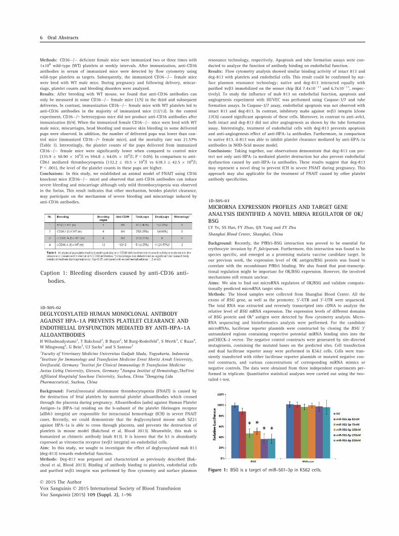
Caption 1: Bleeding disorders caused by anti-CD36 anti-
bodies.
1D-S05-02
DEGLYCOSYLATED HUMAN MONOCLONAL ANTIBODYAGAINST HPA-1A PREVENTS PLATELET CLEARANCE ANDENDOTHELIAL DYSFUNCTION MEDIATED BY ANTI-HPA-1AALLOANTIBODIESH Wihadmadyatami1, T Bakchoul2, B Bayat3, M Burg-Roderfeld3, S Werth3, C Ruan4,
W Mingwang5, G Bein3, UJ Sachs3 and S Santoso3
1Faculty of Veterinary Medicine Universitas Gadjah Mada, Yogyakarta, Indonesia2Institute for Immunology and Transfusion Medicine Ernst Moritz Arndt University,
Greifswald, Germany 3Institut for Clinical Immunology & Transfusion Medicine
Justus Liebig University, Giessen, Germany 4Jiangsu Institut of Hematology,TheFirst
Affiliated Hospitalof Soochow University, Suzhou, China 5Dongying Lida
Pharmaceutical, Suzhou, China
Background: Foetal/neonatal alloimmune thrombocytopenia (FNAIT) is caused by
the destruction of fetal platelets by maternal platelet alloantibodies which crossed
through the placenta during pregnancy. Alloantibodies (aabs) against Human Platelet
Antigen-1a (HPA-1a) residing on the b-subunit of the platelet fibrinogen receptor
(aIIbb3 integrin) are responsible for intracranial hemorrhage (ICH) in severe FNAIT
cases. Recently, we could demonstrate that the deglycosylated mouse mab SZ21
against HPA-1a is able to cross through placenta, and prevents the destruction of
platelets in mouse model (Bakchoul et al, Blood 2013). Meanwhile, this mab is
humanized as chimeric antibody (mab 813). It is known that the b3 is abundantly
expressed as vitronectin receptor (avb3 integrin) on endothelial cells.
Aim: In this study, we sought to investigate the effect of deglycosylated mab 813
(deg-813) towards endothelial function.
Methods: Deg-813 was prepared and characterized as previously described (Bak-
choul et al, Blood 2013). Binding of antibody binding to platelets, endothelial cells
and purified avb3 integrin was performed by flow cytometry and surface plasmon
resonance technology, respectively. Apoptosis and tube formation assays were con-
ducted to analyze the function of antibody binding on endothelial function.
Results: Flow cytometry analysis showed similar binding activity of intact 813 and
deg-813 with platelets and endothelial cells. This result could be confirmed by sur-
face plasmon resonance technology; native and deg-813 interacted equally with
purified avb3 immobilized on the sensor chip (Kd 7.4x10�11 and 6.7x10�11, respec-
tively). To study the influence of mab 813 on endothelial function, apoptosis and
angiogenesis experiment with HUVEC was performed using Caspase-3/7 and tube
formation assays. In Caspase-3/7 assay, endothelial apoptosis was not observed with
intact 813 and deg-813. In contrast, inhibitory mabs against avb3 integrin (clone
23C6) caused significant apoptosis of these cells. Moreover, in contrast to anti-avb3,
both intact and deg-813 did not alter angiogenesis as shown by the tube formation
assay. Interestingly, treatment of endothelial cells with deg-813 prevents apoptosis
and anti-angiogenesis effect of anti-HPA-1a antibodies. Furthermore, in comparison
to native 813, d-813 was able to inhibit platelet clearance mediated by anti-HPA-1a
antibodies in NOD-Scid mouse model.
Conclusions: Taking together, our observations demonstrate that deg-813 can pro-
tect not only anti-HPA-1a mediated platelet destruction but also prevent endothelial
dysfunction caused by anti-HPA-1a antibodies. These results suggest that deg-813
may represent a novel drug to prevent ICH in severe FNAIT during pregnancy. This
approach may also applicable for the treatment of FNAIT caused by other platelet
antibody specificities.
1D-S05-03
MICRORNA EXPRESSION PROFILES AND TARGET GENEANALYSIS IDENTIFIED A NOVEL MIRNA REGULATOR OF OK/BSGLY Ye, SS Han, FY Zhao, QX Yang and ZY Zhu
Shanghai Blood Center, Shanghai, China
Background: Recently, the PfRh5-BSG interaction was proved to be essential for
erythrocyte invasion by P. falciparum. Furthermore, this interaction was found to be
species specific, and emerged as a promising malaria vaccine candidate target. In
our previous work, the expression level of OK antigen/BSG protein was found to
correlate with the recombinant PfRh5 binding. We also found that post-transcrip-
tional regulation might be important for OK/BSG expression. However, the involved
mechanisms still remain unclear.
Aims: We aim to find out microRNA regulators of OK/BSG and validate computa-
tionally predicted microRNA target sites.
Methods: The blood samples were collected from Shanghai Blood Center. All the
exons of BSG gene, as well as the promoter, 50-UTR and 30-UTR were sequenced.
The total RNA was extracted and reversely transcripted into cDNA to analyze the
relative level of BSG mRNA expression. The expression levels of different domains
of BSG protein and Oka antigen were detected by flow cytometry analysis. Micro-
RNA sequencing and bioinformatics analysis were performed. For the candidate
microRNAs, luciferase reporter plasmids were constructed by cloning the BSG 30
untranslated regions containing respective potential miRNA binding sites into the
psiCHECK-2 vector. The negative control constructs were generated by site-directed
mutagenesis, containing the mutated bases on the predicted sites. Cell transfection
and dual luciferase reporter assay were performed in K562 cells. Cells were tran-
siently transfected with either luciferase reporter plasmids or mutated negative con-
trol constructs, and various concentrations of corresponding miRNA mimics or
negative controls. The data were obtained from three independent experiments per-
formed in triplicate. Quantitative statistical analyses were carried out using the two-
tailed t-test.
6 Oral Abstracts
Figure 1: BSG is a target of miR-501-3p in K562 cells.
© 2015 The AuthorVox Sanguinis © 2015 International Society of Blood TransfusionVox Sanguinis (2015) 109 (Suppl. 2), 1–96
Results: A significantly high level of BSG mRNA was found in one sample with
lower OK/BSG expression. MicroRNA sequencing was performed in this sample and
another one bearing similar BSG polymorphisms but with higher OK/BSG expression
and lower BSG mRNA level, used as a control. Among all miRNAs predicted to be
the potential miRNAs to target BSG, miR-15b-5p, miR-99b-5p, and miR-501-3p
were up-regulated in the experimental sample compared with the control. The
respective dual luciferase reporter and control plasmids were constructed for lucifer-
ase assays, confirmed by sequencing. Overexpression of miR-501-3p mimic rather
than other mimics significantly suppressed the luciferase activity, and the inhibition
was rescued when the binding sites were mutated (Fig. 1). These results suggest that
miR-501-3p targets the 30-UTR of BSG in K562 cells, by directly binding to the pre-
dicted target sites.
Conclusions: For the first time, our results suggest that miR-501-3p might be a
novel negative regulator of BSG gene, which enriches the knowledge of the regula-
tion of OK/BSG.
1D-S05-04
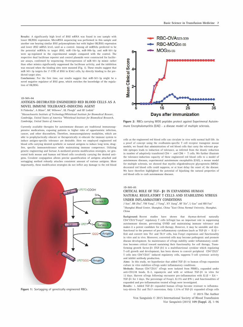
ANTIGEN-DECORATED ENGINEERED RED BLOOD CELLS AS ANOVEL IMMUNE TOLERANCE-INDUCING AGENTN Pishesha1, A Bilate2, MC Wibowo1, HL Ploegh1 and HF Lodish1
1Massachusetts Institute of Technology/Whitehead Institute for Biomedical Researc,
Cambridge, United States of America 2Whitehead Institute for Biomedical Research,
Cambridge, United States of America
Currently available therapies for autoimmune diseases are traditional immunosup-
pressive medications, exposing patients to higher risks of opportunistic infection,
cancer, and other discomforts. Therefore, immunoregulatory modalities, which are
able to prophylactically educate or therapeutically re-educate the immune system to
induce antigen-specific tolerance are desirable. Here we employed engineered red
blood cells carrying desired synthetic or natural antigens to induce long term, drug-
free, specific immunotolerance while maintaining immune competence. Utilizing
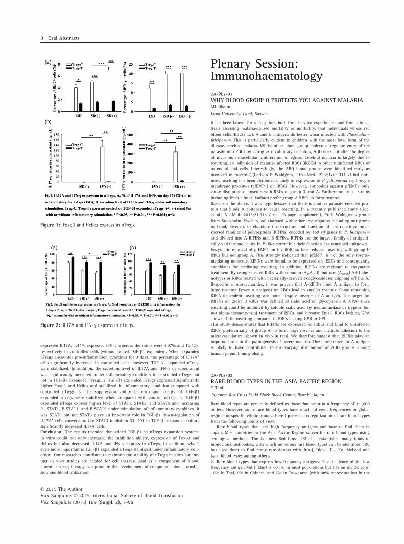
genetic engineering and Sortase A-mediated protein modification strategies, we gen-
erated both mouse and human red blood cells covalently carrying the desired anti-
gens. Covalent conjugation allows precise quantification of antigens attached and
sortagging method robustly attaches consistent amount of various antigens. More
importantly, these modification strategies do not inflict any damage to the red blood
cells as the engineered red blood cells can circulate in vivo with normal half-life. As
a proof of concept using the ovalbumin-specific T cell receptor transgenic mouse
models, we found that administration of red blood cells that carry the relevant pep-
tide epitopes leads to induction of tolerance, as inferred from the drastic reduction
in number of adoptively transferred CD4 + and CD8 + T cells. We further illustrate
the tolerance-induction capacity of these engineered red blood cells to a model of
autoimmune diseases, experimental autoimmune encephalitis (EAE), a mouse model
for multiple sclerosis, we showed that myelin oligodendrocyte glycoprotein (MOG)-
decorated red blood cells could suppress, or at least delay the onset of, the disease.
We have therefore highlighted the potential of hijacking the natural properties of
red blood cells to curb autoimmune diseases.
1D-S05-05
CRITICAL ROLE OF TGF- b1 IN EXPANDING HUMANNATURAL REGULATORY T CELLS AND STABILIZING NTREGSUNDER INFLAMMATORY CONDITIONSJ Sun1, BB Zhu2, YM Yang1, J Yang1, XY Jiang1, RF Xie1, L Gao1 and HH Fan1
1Shanghai Blood Center, Shanghai, China 2East China Normal Univesity, Shanghai,
China
Background: Recent studies have shown that thymus-derived naturally
CD4+CD25+Foxp3+ regulatory T cells (nTregs) has an important role in suppressing
autoimmune disease, preventing GVHD and maintaining immune tolerance and
makes it a potent candidate for cell-therapy. However, it may be unstable and dys-
functional in the presence of pro-inflammatory cytokines (such as TGF-b1 + IL1b +IL6) and convert into Th1 and Th17 cells, loss Foxp3 expression and functionality
in vitro and in vivo. Moreover, converted cells may become pathogenic and promote
disease development. So maintenance of nTregs stability under inflammatory condi-
tion becomes critical toward sustaining their functionality for cell therapy. Trans-
forming growth factor-b1 (TGF-b1) is a multifunctional cytokine which regulating
T-cell growth and development, has been shown to convert peripheral CD4+CD25+
T cells into CD4+CD25+ induced regulatory cells, suppress T-cell cytotoxic activity
and inhibit antibody production.
Aims: In this study, we hypothesize that added TGF-b1 to human nTregs expansion
culture in vitro stabilizes nTregs under inflammatory conditions.
Methods: Human CD4+CD25+ nTregs were isolated from PBMCs, expanded under
anti-CD3/28 beads, IL-2, rapamycin and with or without TGF-b1 in vitro for
12 days. Harvest nTregs following encounter pro-inflammation with IL1b + IL6 +TGF-b1 for 3 days. The percentage of Foxp3, IL17A and IFN-c and functionalities of
expanded and pro-inflammation treated nTregs were investigated.
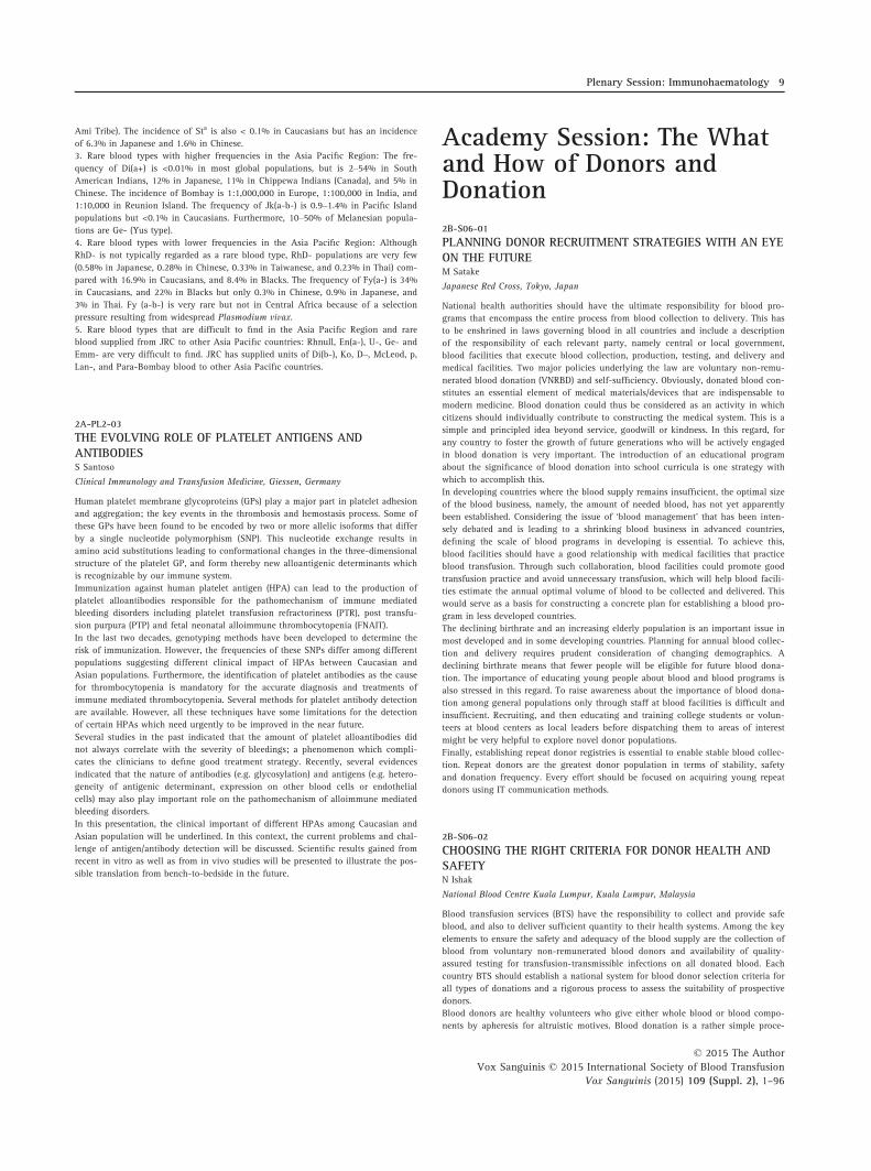
Results: 1. Added TGF-b1 expanded human nTregs become resistant to inflamma-
tory-driven Th1 and Th17 conversion. Only 1.31% of TGF-b1 expanded nTregs cellsFigure 1: Sortagging of genetically engineered RBCs.
Figure 2: RBCs carrying MOG peptides protect against Experimenal Autoim-
mune Encephalomyelitis (EAE) – a disease model of multiple sclerosis.
© 2015 The AuthorVox Sanguinis © 2015 International Society of Blood Transfusion
Vox Sanguinis (2015) 109 (Suppl. 2), 1–96
Basic Science in Transfusion Medicine 7
expressed IL17A, 1.44% expressed IFN-c whereas the ratios were 4.07% and 12.42%
respectively in controlled cells (without added TGF-b1 expanded). When expanded
nTregs encounter pro-inflammation cytokines for 3 days, the percentage of IL17A+
cells significantly increased in controlled cells, however, TGF-b1 expanded nTregs
were stabilized. In addition, the secretion level of IL17A and IFN-c in supernatant
was significantly increased under inflammatory condition in controlled nTregs but
not in TGF-b1 expanded nTregs. 2. TGF-b1 expanded nTregs expressed significantly
higher Foxp3 and Helios and stabilized in inflammatory condition compared with
controlled nTregs. 3. The suppression ability in vitro and anergy of TGF-b1expanded nTregs were stabilized when compared with control nTregs. 4. TGF-b1expanded nTregs express higher level of STAT1, STAT3, and STAT5 and increasing
P- STAT1, P-STAT3, and P-STAT5 under stimulation of inflammatory cytokines. It
was STAT3 but not STAT5 plays an important role in TGF-b1 down-regulation of
IL17A+ cells conversion. Use STAT3 inhibition S3I-201 in TGF-b1 expanded culture
significantly increased IL17A+cells.
Conclusion: The results revealed that added TGF-b1 in nTregs expansion systems
in vitro could not only increased the inhibition ability, expression of Foxp3 and
Helios but also decreased IL17A and IFN-c express in nTregs. In addition, what’s
even more important is TGF-b1 expanded nTregs stabilized under inflammatory con-
dition. Our researches contribute to maintain the stability of nTregs in vitro but fur-
ther in vivo studies are needed for cell therapy. And as a component of blood,
potential nTreg therapy can promote the development of component blood transfu-
sion and blood utilization.
Plenary Session:Immunohaematology
2A-PL2-01
WHY BLOOD GROUP O PROTECTS YOU AGAINST MALARIAML Olsson
Lund University, Lund, Sweden
It has been known for a long time, both from in vitro experiments and from clinical
trials assessing malaria-caused mortality or morbidity, that individuals whose red
blood cells (RBCs) lack A and B antigens do better when infected with Plasmodium
falciparum. This is particularly evident in children with the most fatal form of the
disease, cerebral malaria. Whilst other blood group molecules regulate entry of the
parasite into RBCs by acting as involuntary receptors, ABO does not alter the degree
of invasion, intracellular proliferation or egress. Cerebral malaria is largely due to
rosetting, i.e. adhesion of malaria-infected RBCs (iRBCs) to other uninfected RBCs or
to endothelial cells. Interestingly, the ABO blood groups were identified early as
involved in rosetting (Carlson & Wahlgren, J.Exp.Med. 1992;176:1311-7) but until
now, rosetting has been attributed mainly to expression of P. falciparum erythrocyte
membrane protein-1 (pfEMP1) on iRBCs. However, antibodies against pfEMP1 only
cause disruption of rosettes with RBCs of group O, not A. Furthermore, most strains
including fresh clinical isolates prefer group A RBCs to form rosettes.
Based on the above, it was hypothesized that there is another parasite-encoded pro-
tein that binds A epitopes to cause rosetting. In a recently published study (Goel
et al., Nat.Med. 2015;21:314-7 + a 15-page supplement), Prof. Wahlgren’s group
from Stockholm, Sweden, collaborated with other investigators including our group
in Lund, Sweden, to elucidate the structure and function of the repetitive inter-
spersed families of polypeptides (RIFINs) encoded by 150 rif genes in P. falciparum
and divided into A-RIFINs and B-RIFINs. RIFINs are the largest family of antigeni-
cally variable molecules in P. falciparum but their function has remained unknown.
Enzymatic removal of pfEMP1 on the iRBC surface reduced rosetting with group O
RBCs but not group A. This strongly indicated that pfEMP1 is not the only rosette-
mediating molecule. RIFINs were found to be expressed on iRBCs and consequently
candidates for mediating rosetting. In addition, RIFINs are resistant to enzymatic
treatment. By using selected RBCs with common (A1,A2,O) and rare (Aweak) ABO phe-
notypes or RBCs treated with bacterially-derived exoglycosidases clipping off the A/
B-specific monosaccharides, it was proven that A-RIFINs bind A antigen to form
large rosettes. Fewer A antigens on RBCs lead to smaller rosettes. Some remaining
RIFIN-dependent rosetting was noted despite absence of A antigen. The target for
RIFINs on group O RBCs was defined as sialic acid on glycophorin A (GPA) since
rosetting could be inhibited by soluble sialic acid, by neuraminidase or trypsin (but
not alpha-chymotrypsin) treatment of RBCs, and because En(a-) RBCs lacking GPA
showed little rosetting compared to RBCs lacking GPB or GPC.
This study demonstrates that RIFINs are expressed on iRBCs and bind to uninfected
RBCs, preferentially of group A, to form large rosettes and mediate adhesion to the
microvasculature (shown in vivo in rats). We therefore suggest that RIFINs play an
important role in the pathogenesis of severe malaria. Their preference for A antigen
is likely to have contributed to the varying distribution of ABO groups among
human populations globally.
2A-PL2-02
RARE BLOOD TYPES IN THE ASIA PACIFIC REGIONY Tani
Japanese Red Cross Kinki Block Blood Center, Ibaraki, Japan
Rare blood types are generally defined as those that occur at a frequency of 1:1,000
or less. However, some rare blood types have much different frequencies in global
regions or specific ethnic groups. Here I present a categorization of rare blood types
from the following points of view.
1. Rare blood types that lack high frequency antigens and how to find them in
Japan: Most countries in the Asia Pacific Region screen for rare blood types using
serological methods. The Japanese Red Cross (JRC) has established many kinds of
monoclonal antibodies, with which numerous rare blood types can be identified. JRC
has used them to find many rare donors with Jr(a-), Di(b-), D–, Ko, McLeod and
Lan- blood types among others.
2. Rare blood types that express low frequency antigens: The incidence of the low
frequency antigen MiIII (Mur) is <0.1% in most populations but has an incidence of
10% in Thai, 6% in Chinese, and 7% in Taiwanese (with 88% representation in the
Figure 1: Foxp3 and Helios express in nTregs.
Figure 2: IL17A and IFN-c express in nTregs.
8 Oral Abstracts
© 2015 The AuthorVox Sanguinis © 2015 International Society of Blood TransfusionVox Sanguinis (2015) 109 (Suppl. 2), 1–96
Ami Tribe). The incidence of Sta is also < 0.1% in Caucasians but has an incidence
of 6.3% in Japanese and 1.6% in Chinese.
3. Rare blood types with higher frequencies in the Asia Pacific Region: The fre-
quency of Di(a+) is <0.01% in most global populations, but is 2–54% in South
American Indians, 12% in Japanese, 11% in Chippewa Indians (Canada), and 5% in
Chinese. The incidence of Bombay is 1:1,000,000 in Europe, 1:100,000 in India, and
1:10,000 in Reunion Island. The frequency of Jk(a-b-) is 0.9–1.4% in Pacific Island
populations but <0.1% in Caucasians. Furthermore, 10–50% of Melanesian popula-
tions are Ge- (Yus type).
4. Rare blood types with lower frequencies in the Asia Pacific Region: Although
RhD- is not typically regarded as a rare blood type, RhD- populations are very few
(0.58% in Japanese, 0.28% in Chinese, 0.33% in Taiwanese, and 0.23% in Thai) com-
pared with 16.9% in Caucasians, and 8.4% in Blacks. The frequency of Fy(a-) is 34%
in Caucasians, and 22% in Blacks but only 0.3% in Chinese, 0.9% in Japanese, and
3% in Thai. Fy (a-b-) is very rare but not in Central Africa because of a selection
pressure resulting from widespread Plasmodium vivax.
5. Rare blood types that are difficult to find in the Asia Pacific Region and rare
blood supplied from JRC to other Asia Pacific countries: Rhnull, En(a-), U-, Ge- and
Emm- are very difficult to find. JRC has supplied units of Di(b-), Ko, D–, McLeod, p,
Lan-, and Para-Bombay blood to other Asia Pacific countries.
2A-PL2-03
THE EVOLVING ROLE OF PLATELET ANTIGENS ANDANTIBODIESS Santoso
Clinical Immunology and Transfusion Medicine, Giessen, Germany
Human platelet membrane glycoproteins (GPs) play a major part in platelet adhesion
and aggregation; the key events in the thrombosis and hemostasis process. Some of
these GPs have been found to be encoded by two or more allelic isoforms that differ
by a single nucleotide polymorphism (SNP). This nucleotide exchange results in
amino acid substitutions leading to conformational changes in the three-dimensional
structure of the platelet GP, and form thereby new alloantigenic determinants which
is recognizable by our immune system.
Immunization against human platelet antigen (HPA) can lead to the production of
platelet alloantibodies responsible for the pathomechanism of immune mediated
bleeding disorders including platelet transfusion refractoriness (PTR), post transfu-
sion purpura (PTP) and fetal neonatal alloimmune thrombocytopenia (FNAIT).
In the last two decades, genotyping methods have been developed to determine the
risk of immunization. However, the frequencies of these SNPs differ among different
populations suggesting different clinical impact of HPAs between Caucasian and
Asian populations. Furthermore, the identification of platelet antibodies as the cause
for thrombocytopenia is mandatory for the accurate diagnosis and treatments of
immune mediated thrombocytopenia. Several methods for platelet antibody detection
are available. However, all these techniques have some limitations for the detection
of certain HPAs which need urgently to be improved in the near future.
Several studies in the past indicated that the amount of platelet alloantibodies did
not always correlate with the severity of bleedings; a phenomenon which compli-
cates the clinicians to define good treatment strategy. Recently, several evidences
indicated that the nature of antibodies (e.g. glycosylation) and antigens (e.g. hetero-
geneity of antigenic determinant, expression on other blood cells or endothelial
cells) may also play important role on the pathomechanism of alloimmune mediated
bleeding disorders.
In this presentation, the clinical important of different HPAs among Caucasian and
Asian population will be underlined. In this context, the current problems and chal-
lenge of antigen/antibody detection will be discussed. Scientific results gained from
recent in vitro as well as from in vivo studies will be presented to illustrate the pos-
sible translation from bench-to-bedside in the future.
Academy Session: The Whatand How of Donors andDonation
2B-S06-01
PLANNING DONOR RECRUITMENT STRATEGIES WITH AN EYEON THE FUTUREM Satake
Japanese Red Cross, Tokyo, Japan
National health authorities should have the ultimate responsibility for blood pro-
grams that encompass the entire process from blood collection to delivery. This has
to be enshrined in laws governing blood in all countries and include a description
of the responsibility of each relevant party, namely central or local government,
blood facilities that execute blood collection, production, testing, and delivery and
medical facilities. Two major policies underlying the law are voluntary non-remu-
nerated blood donation (VNRBD) and self-sufficiency. Obviously, donated blood con-
stitutes an essential element of medical materials/devices that are indispensable to
modern medicine. Blood donation could thus be considered as an activity in which
citizens should individually contribute to constructing the medical system. This is a
simple and principled idea beyond service, goodwill or kindness. In this regard, for
any country to foster the growth of future generations who will be actively engaged
in blood donation is very important. The introduction of an educational program
about the significance of blood donation into school curricula is one strategy with
which to accomplish this.
In developing countries where the blood supply remains insufficient, the optimal size
of the blood business, namely, the amount of needed blood, has not yet apparently
been established. Considering the issue of ‘blood management’ that has been inten-
sely debated and is leading to a shrinking blood business in advanced countries,
defining the scale of blood programs in developing is essential. To achieve this,
blood facilities should have a good relationship with medical facilities that practice
blood transfusion. Through such collaboration, blood facilities could promote good
transfusion practice and avoid unnecessary transfusion, which will help blood facili-
ties estimate the annual optimal volume of blood to be collected and delivered. This
would serve as a basis for constructing a concrete plan for establishing a blood pro-
gram in less developed countries.
The declining birthrate and an increasing elderly population is an important issue in
most developed and in some developing countries. Planning for annual blood collec-
tion and delivery requires prudent consideration of changing demographics. A
declining birthrate means that fewer people will be eligible for future blood dona-
tion. The importance of educating young people about blood and blood programs is
also stressed in this regard. To raise awareness about the importance of blood dona-
tion among general populations only through staff at blood facilities is difficult and
insufficient. Recruiting, and then educating and training college students or volun-
teers at blood centers as local leaders before dispatching them to areas of interest
might be very helpful to explore novel donor populations.
Finally, establishing repeat donor registries is essential to enable stable blood collec-
tion. Repeat donors are the greatest donor population in terms of stability, safety
and donation frequency. Every effort should be focused on acquiring young repeat
donors using IT communication methods.
2B-S06-02
CHOOSING THE RIGHT CRITERIA FOR DONOR HEALTH ANDSAFETYN Ishak
National Blood Centre Kuala Lumpur, Kuala Lumpur, Malaysia
Blood transfusion services (BTS) have the responsibility to collect and provide safe
blood, and also to deliver sufficient quantity to their health systems. Among the key
elements to ensure the safety and adequacy of the blood supply are the collection of
blood from voluntary non-remunerated blood donors and availability of quality-
assured testing for transfusion-transmissible infections on all donated blood. Each
country BTS should establish a national system for blood donor selection criteria for
all types of donations and a rigorous process to assess the suitability of prospective
donors.
Blood donors are healthy volunteers who give either whole blood or blood compo-
nents by apheresis for altruistic motives. Blood donation is a rather simple proce-
© 2015 The AuthorVox Sanguinis © 2015 International Society of Blood Transfusion
Vox Sanguinis (2015) 109 (Suppl. 2), 1–96
Plenary Session: Immunohaematology 9
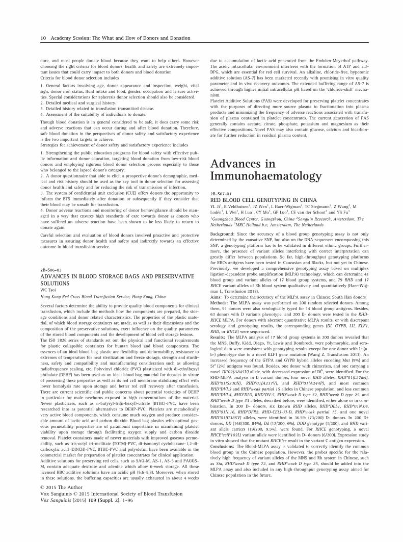
dure, and most people donate blood because they want to help others. However
choosing the right criteria for blood donors’ health and safety are extremely impor-
tant issues that could carry impact to both donors and blood donation
Criteria for blood donor selection includes
1. General factors involving age, donor appearance and inspection, weight, vital
sign, donor iron status, fluid intake and food, gender, occupation and leisure activi-
ties. Special considerations for apheresis donor selection should also be considered.
2. Detailed medical and surgical history.
3. Detailed history related to transfusion transmitted disease.
4. Assessment of the suitability of individuals to donate.
Though blood donation is in general considered to be safe, it does carry some risk
and adverse reactions that can occur during and after blood donation. Therefore,
safe blood donation in the perspectives of donor safety and satisfactory experience
is the two important targets to achieve.
Strategies for achievement of donor safety and satisfactory experience includes
1. Strengthening the public education programs for blood safety with effective pub-
lic information and donor education, targeting blood donation from low-risk blood
donors and employing rigorous blood donor selection process especially to those
who belonged to the lapsed donor’s category.
2. A donor questionnaire that able to elicit a prospective donor’s demographic, med-
ical and risk history should be used as the key tool in donor selection for assessing
donor health and safety and for reducing the risk of transmission of infection.
3. The system of confidential unit exclusion (CUE) offers donors the opportunity to
inform the BTS immediately after donation or subsequently if they consider that
their blood may be unsafe for transfusion.
4. Donor adverse reactions and monitoring of donor hemovigilance should be man-
aged in a way that ensures high standards of care towards donor as donors who
have suffered an adverse reaction have been shown to be less likely to return to
donate again.
Careful selection and evaluation of blood donors involved proactive and protective
measures in assuring donor health and safety and indirectly towards an effective
outcome in blood transfusion service.
2B-S06-03
ADVANCES IN BLOOD STORAGE BAGS AND PRESERVATIVESOLUTIONSWC Tsoi
Hong Kong Red Cross Blood Transfusion Service, Hong Kong, China
Several factors determine the ability to provide quality blood components for clinical
transfusion, which include the methods how the components are prepared, the stor-
age conditions and donor related characteristics. The properties of the plastic mate-
rial, of which blood storage containers are made, as well as their dimensions and the
composition of the preservative solutions, exert influence on the quality parameters
of the stored blood components and the development of blood cell storage lesions.
The ISO 3826 series of standards set out the physical and functional requirements
for plastic collapsible containers for human blood and blood components. The
essences of an ideal blood bag plastic are flexibility and deformability, resistance to
extremes of temperature for heat sterilization and freeze storage, strength and sturdi-
ness, safety and compatibility and manufacturing consideration such as allowing
radiofrequency sealing, etc. Polyvinyl chloride (PVC) plasticized with di-ethylhexyl
phthalate (DEHP) has been used as an ideal blood bag material for decades in virtue
of possessing these properties as well as its red cell membrane stabilizing effect with
lower hemolysis rate upon storage and better red cell recovery after transfusion.
There are current scientific and public concerns about potential toxicities of DEHP
in particular for male newborns exposed to high concentrations of the material.
Newer plasticizers, such as n-butyryl-tri(n-hexyl)-citrate (BTHC)-PVC, have been
researched into as potential alternatives to DEHP-PVC. Platelets are metabolically
very active blood components, which consume much oxygen and produce consider-
able amount of lactic acid and carbon dioxide. Blood bag plastics with optimal gas-
eous permeability properties are of paramount importance in maintaining platelet
viability upon storage through facilitating oxygen supply and carbon dioxide
removal. Platelet containers made of newer materials with improved gaseous perme-
ability, such as tris-octyl tri-mellitate (TOTM)-PVC, di-isononyl cyclohexane-1,2-di-
carboxylic acid (DINCH)-PVC, BTHC-PVC and polyolefin, have been available in the
commercial market for preparation of platelet concentrates for clinical application.
Additive solutions for preserving red cells, such as SAG-M, AS-1, AS-5 and PAGGS-
M, contain adequate dextrose and adenine which allow 6-week storage. All these
licensed RBC additive solutions have an acidic pH (5.6–5.8). Moreover, when stored
in these solutions, the buffering capacities are usually exhausted in about 4 weeks
due to accumulation of lactic acid generated from the Embden-Meyerhof pathway.
The acidic intracellular environment interferes with the formation of ATP and 2,3-
DPG, which are essential for red cell survival. An alkaline, chloride-free, hypotonic
additive solution (AS-7) has been marketed recently with promising in vitro quality
parameter and in vivo recovery outcomes. The extended buffering range of AS-7 is
achieved through higher initial intracellular pH based on the ‘chloride-shift’ mecha-
nism.
Platelet Additive Solutions (PAS) were developed for preserving platelet concentrates
with the purposes of directing more source plasma to fractionation into plasma
products and minimizing the frequency of adverse reactions associated with transfu-
sion of plasma contained in platelet concentrates. The current generation of PAS
generally contains acetate, citrate, phosphate, potassium and magnesium as their
effective compositions. Novel PAS may also contain glucose, calcium and bicarbon-
ate for further reduction in residual plasma content.
Advances inImmunohaematology
2B-S07-01
RED BLOOD CELL GENOTYPING IN CHINAYL Ji1, B Veldhuisen2, JZ Wen1, L Haer-Wigman2, TC Stegmann2, Z Wang1, M
Lod�en3, L Wei1, H Luo1, CY Mo1, GP Luo1, CE van der Schoot2 and YS Fu1
1Guangzhou Blood Center, Guangzhou, China 2Sanquin Research, Amsterdam, The
Netherlands 3MRC-Holland b.v, Amsterdam, The Netherlands
Background: Since the accuracy of a blood group genotyping assay is not only
determined by the causative SNP, but also on the DNA-sequences encompassing this
SNP, a genotyping platform has to be validated in different ethnic groups. Further-
more, the presence of variant alleles interfering with correct interpretation can
greatly differ between populations. So far, high-throughput genotyping platforms
for RBCs antigens have been tested in Caucasian and Blacks, but not yet in Chinese.
Previously, we developed a comprehensive genotyping assay based on multiplex
ligation-dependent probe amplification (MLPA) technology, which can determine 41
blood group and variant alleles of 17 blood group systems, and 79 RHD and 17
RHCE variant alleles of Rh blood system qualitatively and quantitatively (Haer-Wig-
man L, Transfusion 2013).
Aims: To determine the accuracy of the MLPA assay in Chinese South Han donors.
Methods: The MLPA assay was performed on 200 random selected donors. Among
them, 91 donors were also serologically typed for 14 blood group antigens. Besides,
63 donors with D variants phenotype, and 200 D- donors were tested in the RHD-
RHCE MLPA. For donors with aberrant quantitative MLPA results, or with discrepant
serology and genotyping results, the corresponding genes (DI, GYPB, LU, KLF1,
RHD, or RHCE) were sequenced.
Results: The MLPA analysis of 17 blood group systems in 200 donors revealed that
the MNS, Duffy, Kidd, Diego, Yt, Lewis and Dombrock, were polymorphic, and sero-
logical data were consistent with genotyping results except for one donor with Lu(a-
b-) phenotype due to a novel KLF1 gene mutation (Wang Z. Transfusion 2013). An
increased frequency of the GYPA and GYPB hybrid alleles encoding Mur (9%) and
Sta (2%) antigens was found. Besides, one donor with chimerism, and one carrying a
novel DI*02(A845V) allele, with decreased expression of Dib, were identified. For the
RHD-MLPA analysis in D variant donors, four novel RHD alleles, RHD*01(L27del),
RHD*01(S230N), RHD*01(A237V), and RHD*01(A244P), and most common
RHD*DVI.3 and RHD*weak partial 15 alleles in Chinese population, and less common
RHD*DVI.4, RHD*DLO, RHD*DV.5, RHD*weak D type 72, RHD*weak D type 25, and
RHD*weak D type 33 alleles, described before, were identified, either alone or in com-
bination. In 200 D- donors, six known RHD alleles, RHD*DEL1, RHD*01N.04,
RHD*01N.16, RHD*DFR2, RHD-CE(3-7)-D, RHD*weak partial 15, and one novel
RHD*01(G385V) alleles, were identified in 36.5% (73/200) D- donors. In 200 D+donors, DD (168/200, 84%), Dd (12/200, 6%), DDD genotype (1/200), and RHD vari-
ant allele carriers (19/200, 9.5%), were found. For RHCE genotyping, a novel
RHCE*ce(P103L) variant allele were identified in D- donors (6/200). Expression study
in vitro showed that the mutant RHCE*ce result in the variant C antigen expression.
Conclusions: The Blood-MLPA assay is validated to correctly identify the common
blood group in the Chinese population. However, the probes specific for the rela-
tively high frequency of variant alleles of the MNS and Rh system in Chinese, such
as Sta, RHD*weak D type 72, and RHD*weak D type 25, should be added into the
MLPA assay and also included in any high-throughput genotyping assay aimed for
Chinese population in the future.
© 2015 The AuthorVox Sanguinis © 2015 International Society of Blood TransfusionVox Sanguinis (2015) 109 (Suppl. 2), 1–96
10 Academy Session: The What and How of Donors and Donation
2B-S07-02
THE RELEVANCE OF PLATELET ANTIGEN/ANTIBODIES INMULTI ETHNIC IN MALAYSIAMI Armawai1 and S Santoso2
1National Blood Center of Kuala Lumpur, Kuala Lumpur, Malaysia 2Institute for
Clinical Immunology and Transfusion Medicine, Justus Liebig Univer, Giessen,
Germany
Background: Immunization against polymorphic structure on Human platelet anti-
gens (HPA) have been implicated in the pathogenesis of fetal/neonatal alloimmune
thrombocytopenia (FNAIT), post-transfusion purpura (PTP) and platelet transfusion
refractoriness (PTR). Malaysia is multi-ethic country; the majority of the inhabitants
are Malays (54.4%), Chinese (25.0%) and Indians (7.5%). Recently, the distribution of
HPA gene frequencies and has been reported in Malaysia (Tan et al, 2012). However,
little is currently known about the relevance of platelet alloantibodies in our hetero-
geneous population.
Aim: In this study, the frequency of platelet reactive alloantibodies in patients with
suspected FNAIT and PTR should be analyzed to estimate their clinical relevance.
Methods: Serum samples were analysed by antigen capture assay, MAIPA (Mono-
clonal Antibody Immobilized Platelet Antigen (MAIPA) using a panel of HPA geno-
typed platelets derived from blood group donors. Five monoclonal antibodies (mabs)
against GPIIb/IIIa, Ib/IX, Ia/IIa, CD109 and HLA Class I were used as capture anti-
bodies. Incompatibility on HPA systems was verified by genotyping of maternal/pa-
ternal or donor/recipient using comnmercial kits (GTI and BAGene).
Results: In our heterogeneous patient’s cohort, we detected anti-HLA Class I as the
major antibody specificity anticipated in FNAIT (20.7%) and PTR (31.5%) cases.
Screening of anti-HPA antibodies indicated that anti-HPA-3a (20.5%) represents the
most frequent platelet alloantibody in all three ethnic groups (Malay, Chinese and
Indians). Importantly, neonates in FNAIT cases associated with anti-HPA-3a alloanti-
bodies presented severe thrombocytopenia; HPA compatible platelet transfusion(s)
was necessary to manage the bleeding in these babies. Although, HPA-1a negative
individual is extremely rare in our population, when compared to Caucasians, we
found one FNAIT case caused by anti-HPA-1a alloantibody. In addition, anti-
HPA-5b and anti-HPA-15b alloantibodies (36.9%) were also found in our cohort.
However, only mild thrombocytopenia in FNAIT babies associated with these alloan-
tibody specificities.
Summary: Our study indicates that anti-HPA-3a, -HPA-5b and -HPA-15b seem to
be the most platelet specific antibodies involved in FNAIT and PTR cases in Malay-
sian population. Among them, anti-HPA-3a appears to be associated with severe
thrombocytopenia. Similar HPA allelic distribution in Malaysian and Indonesian
populations have been observed (Asmarinah et al, 2013), Thus, immunization against
these three HPA systems are expected to be the most potential risk of alloimmune
mediated platelet disorders in Indonesia and probably in other South East Asian
Countries. Collective donor register of HPA and HLA typed platelets may improve
the diagnosis and treatment of alloimmune mediated thrombocytopenic disorders in
our region.
2B-S07-03
GLYCOSYLATION OF ALLOANTIBODIES AGAINST BLOODCELL ANTIGENSCE van der Schoot1, E Sonneveld1, R Kapur1, J Koelewijn1, M de Haas1, M Wuhrer2
and G Vidarsson1
1Sanquin Research, Amsterdam, The Netherlands 2Leiden University Medical Centre,
Leiden, The Netherlands
Background: IgG-alloantibodies against red blood cell(RBC) and platelet antigens
can lead to clearance of the blood cells through Fc-gamma receptor (FccR)- medi-
ated uptake by phagocytes in the foetal spleen and liver. Classically, these antibodies
are detected in the case of RBCs by agglutination assays and in the case of platelets
via immune fluorescence tests or Monoclonal antibody–specific immobilization of
platelet antigens(MAIPA)-assays. The titre of the alloantibody or the level of the
antibody in arbitrary units are used to estimate the risk of cytopenia. However,
although the antibody level is correlated with the severity of disease, this correlation
is not strict, whereas there is an important need for risk assignment. For example, of
children of mothers with anti-fetal-platelet IHPA-1a IgG, only 31% develop fetal
neonatal alloimmune thrombocytopenia(FNAIT) and only 2.3% suffer from severe
hemorrhagic disease. For haemolytic disease of the foetus and newborn(HDFN) a
functional assay (e.g. the antibody dependent cytotoxicity test (ADCC) can be used
to identify cases at high risk of disease. One of the factors determining the func-
tional activity of alloantibodies is the glycosylation pattern of the Fc-tail of the anti-
bodies.
IgG antibodies are glycoproteins containing a variable branched sugar moiety
attached to asparagine (Asn)297 on each chain in the Fc-domain. The carbohydrates
have a direct influence on the effector functions of the antibodies. Galactose and
sialic acid are believed to affect the binding affinity to FccR in general and might
enable binding to DC-SIGN. Notably, the absence of fucose causes markedly
enhanced binding to FccRIIIa and FccRIIIb, increasing the antibody mediated cyto-
toxicity. Likewise, the addition of bisecting GlcNAc has been shown to increase the
antibody-dependent cellular cytotoxicity of recombinant IgG.
Aim: To investigate the glycosylation pattern of alloantibodies and relate this pat-
tern with clinical outcome
Methods: Antigen specific alloantibodies were purified of serum from pregnant
women with alloantbodies against HPA-1a, RhD or Rhc, of which the outcome of
disease was known (Kapur et al. Br.J.Haematol. 2014 166: 936; Kapur et al Blood
2014;123:471; Koelewijn et al. Transfusion. 2008;48:1721; Koelewijn et al. Transfu-
sion. 2008;48:941). The glycosylation pattern was investigated by Mass Spectrometry
as previously described (Kapur, Blood 2014).
Results: Unique skewing in antigen-specific IgG-Fc-glycans after alloimmune
responses against the RhD antigen on red blood cells and the HPA-1a antigen on
platelets was found. Whereas ~90% of IgG-Fc glycan species present in serum are
core fucosylated, the fucosylation of the antigen specific IgG-Fc-glycans was low-
ered to a varying degree, in some patients even down to ~10%. The level of core
fucosylation correlated significantly with the Hb-level and platelet count of the
new-born, respectively and clinical outcome (Kapur,Blood 2014; Kapur,Br.J.Haema-
tol 2014). A less pronounced decreased fucosylation was found in anti-c antibodies,
possibly explaining the lower risk of anaemia in these cases.
Conclusion: Why alloantibodies against blood cell antigens show such unique gly-
cosylation patterns is unknown, and is presently investigated. But also without
understanding the physiological explanation for this observation, IgG-glycosylation
is proving to become an important biomarker in screening for severe cases of HDFN
and FNAIT.
2B-S07-04
ADSORPTION OF AUTO- ANTIBODIES IN PATIENTS WITHAIHA HELPS TO DETECT MASKED ALLO- ANTIBODIES TOIMPROVE SAFETY OF BLOOD TRANSFUSIONA Abd Elrahman
National Blood Transfusion Services, Cairo, Egypt
Background: Warm type auto-antibodies are directed against patients’ own red
blood cells that may interfere with and complicate investigations for the detection of
RBC allo-antibodies. The patients’ phenotype of RBCs can be difficult to be deter-
mined, if the patients were recently transfused.
Finding of compatible blood for patients with warm type AIHA may be impossible
as the auto- antibodies in the patient’s serum can react with all red blood cells
Complicated adsorption procedures in a reference laboratory allow detection and
identification of clinically significant allo- Abs that may be masked by the auto-
Abs.
Aim: To highlight the importance of implementation of adsorption techniques for
detection of co-existing allo-Abs which are present in a high percentage of our
Egyptian patients, to improve safety of blood transfusion for those patients.
Methods: The study done on 292 patients with warm type AIHA, presented to the
Red Cell Reference Lab at Egyptian NBTC, in the period from January 2012 to
December 2014.
According to the Egyptian national testing strategy; routine lab investigations in the
form of screening for red cell Abs and auto-control were done for all these patients
and revealed positive auto-control with pan-positive reactivity with different pheno-
typed RBCs, using Column Agglutination Technique.
The patients were subjected to allo-adsorption; in this procedure adsorption of auto-
antibodies from the patient’s serum is carried out using three samples of allogeneic
red cells of varying phenotypes, these phenotyped samples already prepared monthly
from selected blood units for this purpose. These samples should be complementary
lacking the clinical significant red cell Ags, and are used to adsorb auto-antibodies
from the patient’s serum at 37°C on several phases to ensure complete successful
adsorption of auto-antibodies. The adsorbed serum then tested for the presence allo-
antibodies.
Results: Of the 292 patients; 108(37%) revealed co-existing allo-antibodies, of
which 46.3% of the antibodies were against the (Rh) blood group antigens, 17.6%
against (Kell) blood group antigens, 13.9% against (MNSs) antigens, 7.4% against
(Kidd) antigens, 3.7% against (Duffy) antigens and 11.1% against other blood group
antigens. Of the 292 patients; 184(63%) revealed only auto-Antibodies without no
co-existing allo-antibodies.
© 2015 The AuthorVox Sanguinis © 2015 International Society of Blood Transfusion
Vox Sanguinis (2015) 109 (Suppl. 2), 1–96
Advances in Immunohaematology 11
Clear identification of the masked coexisting allo-Abs directed us to transfuse the
patients with Ag/s free blood units for the relevant identified Ab/s, and hence we
avoided the risk of hemolytic transfusion adverse effect of these allo-Abs
The previous wrong concept of transfusion of the least incompatible blood units to
patients with AIHA was changed accordingly.
Summary/Conclusions: In countries with limited recourses; proper management of
patients with AIHA requires the implementation of adsorption techniques that can
differentiate between auto- & allo-antibodies and allow the identification of co-ex-
isting allo-antibodies; however being time consuming.
Genotyping of RBCs, especially for recently transfused patients and patients with
AIHA can help in that, so we have to think about implementing this technique;
however being expensive.
2B-S07-05
TO STUDY THE EFFICACY OF CROSS-MATCH COMPATIBLEPLATELETS IN MULTI-TRANSFUSED ALLO-IMMUNIZEDHEMATOLOGY-ONCOLOGY PATIENTS REFRACTORY TOPLATELET TRANSFUSIONRR Sharma, A Chavan, P Malhotra, B Saikia and N Marwaha
Postgraduate Institute Medical Education & Research, Chandigarh, India
Background: Platelet refractoriness, which leads to platelet transfusion failure
resulting in morbidity and mortality, long hospital stay and the resultant patient
cost, is not routinely investigated. Platelet Refractoriness occurs in about 20% to
70% of multi-transfused thrombocytopenic patients as a result of immunological or
non-immunological causes.
Aims: This study aimed to determine the efficacy of cross-match compatible plate-
lets in multi-transfused alloimmunized haemato-oncological patients refractory to
platelet transfusion.
Materials and method: A case control study in which 149 ABO compatible single
donor platelet transfusions given to 38 patients with immune causes of refractoriness
were studied. Patients were defined to be refractory if their Corrected Count Incre-
ment (CCI) was <5000 at 1 h following two consecutive platelet transfusions. A
solid-phase red cell adherence method on automated immunohaematology analyzer
was used to determine the alloimmunization status and platelet cross-matching was
performed on the same platform. Post Transfusion Platelet Increment (PPI), CCI and
the Percentage Platelet Recovery (PPR) were used to monitor the effectiveness of
each platelet transfusion. ANOVA test followed by Post hoc test Tukey HSD to com-
pare group means for data classified into three groups depending upon the cross-
matching and compatibility status. Categorical data analyzed for various outcomes
using Pearson’s chi square test or Fischer exact test whichever was applicable.
Result: Patients showed statistically significant and better platelet recovery in terms
of PPI, CCI and PPR at 1 h post SDAP transfusions when they received cross-
matched compatible platelets than on occasions receiving uncross-matched platelet
units and receiving cross-matched incompatible platelets. The 1-h CCI (mean� SD)
was significantly higher for cross-match-compatible platelets (19,173 � 2692) than
for incompatible ones (5888 � 1526) and for uncross-matched ones (8140 � 1480).
Forty four (97.8%) of 45 cross-matched compatible platelet transfusion episodes
showed a satisfactory response in terms of PPI and CCI values as compared to 50%
and 53.9% in uncross-matched group respectively, (P < 0.0001).
Conclusion: Platelet cross-matching is an effective intervention in the management
of multitransfused alloimmunized Haemato-oncological patients, refractory to plate-
let transfusion and may be a beneficial intervention in such patients for better out-
come of transfusion therapy and should be encouraged.
Plenary Session: Donors,Donations and Components
2C-PL3-01
UPDATES ON DONOR RECRUITMENT AND MANAGEMENT INBLOOD SERVICECK Lee
Hong Kong Red Cross Blood Transfusion Service, Hong Kong, Hong Kong
Voluntary Non-Remunerated Blood Donation (VNRD) is the cornerstone of modern
blood service and it relies heavily on the community support and participation. Over
the years most efforts have been made on the adequacy of blood donation and blood
safety. Though significant improvement in blood safety profile has been seen and a
decreasing blood demand in developed countries is noted, ongoing challenges in
safety and adequacy still remain in every country.
Since blood transfusion (and blood donation) will never disappear, a balance in
matching right blood supply and managing donors expectation are area that needs
serious consideration. Besides, counteracting factors including ageing population/
donors, population migration, socio-economic factors, increasing stringent donor
selection criteria could definitely add on the difficulties in maintaining this balance.
At the same time, the traditional donors’ mission for donation – ‘Give Blood Save
Lives’ is no longer adequate to reflect their expectations nowadays. Therefore, trans-
fusion services have been working on ways to refresh and revisit the current donor
recruitment and management. The wide spread use of social media and ‘big data’
analysis in blood donation settings are typical examples which have enabled them a
totally different approach in reaching and understanding donor behaviours. While
those can either be passively originated from the donors or proactively disseminated
from the blood service, all do create a dynamic and interactive culture among
donors, blood services and the communities. With proper strategies, targeted donors
(right age, sex, blood groups or others) are reached, donated blood and returned to
subsequent donation at their expectation. As a result, the right balance can hope-
fully be maintained while donor expectation could be met.
In summary, challenges and possible solutions have been discussed to allow
blood service operators to revisit their needs; adapt and revise for their own
applications.
2C-PL3-02
BAA BAA BLOOD SHEEPYL Fung1 and JP Tung2
1University of the Sunshine Coast, Caloundra, Australia 2Australian Red Cross Blood
Service, Brisbane, Australia
Background: Animal models have been a valuable tool for research into blood
products as well, as the effects of blood transfusions. Small animal models have
been applied extensively and large animal model less frequently. This session
describes the experience and details the findings from a recent series of in vivo
sheep transfusion models.
Aims and method: To discuss the benefits of sheep as a large animal transfusion
model and to review conclusions from a series of sheep transfusion models.
Results and discussion: Similarities in size, anatomy and physiology of sheep com-
pared to humans have supported its use to model a wide range of human diseases.
The large size of the sheep enables continuous physiological monitoring using stan-
dard human techniques. Its size also equates to a higher total blood volume which
permits the collection of multiple and serial samples.
In this series the first sheep model involved transfusion of supernatant (SN) from
human blood products. These studies confirmed the two-hit TRALI mechanism, as
both host factors (lipopolysaccharide modelling bacterial infection) and blood pro-
duct factors (storage duration of either platelet or packed red blood cell (PRBC)
units) contributed to the development of TRALI. The ability to control interventions
in an animal model also enabled demonstration that pathophysiological changes
were more severe in TRALI induced by stored PRBC-SN than in TRALI induced by
stored platelet-SN.
Subsequent sheep transfusion models were more sophisticated with the collection and
use of homologous sheep blood products, and pre-transfusion cross-matching. In
addition, similarities in the storage lesions of sheep and human PRBC units across
42 days storage, supported the use of these models to investigate the effects of stored
PRBC transfusion. In a haemorrhage model, transfusion of stored sheep PRBC affected
pulmonary function, even in the absence of overt tissue injury (first hit), highlighting
the sensitivity and vulnerability of the pulmonary system to transfusions.
Like human PRBC, sheep PRBC have negligible levels of selenium (trace element
involved with antioxidant function). The sheep haemorrhage model demonstrated
that the transfusion of sheep PRBC diluted selenium levels and compromised glu-
tathione peroxidase antioxidant activity, which in turn allowed lipid peroxidation.
Their large size also enabled development of a sheep model of extracorporeal mem-
brane oxygenation (ECMO). In these studies the insults of lung injury and ECMO
reduced selenium levels but transfusion with two units PRBC did not alter the mea-
sured oxidative stress markers.
A recent study has delineated the similarities and differences between ovine and
human haemostasis. The study revealed the sheep had similar primary haemostasis
response, however differences were detected in its secondary haemostasis response.
For examples sheep platelets had reduced ADP and collagen response.
Summary: These findings demonstrate the value of large animal models for transfu-
sion research and reveal the sophistication of current sheep transfusion models. Con-
12 Oral Abstracts
© 2015 The AuthorVox Sanguinis © 2015 International Society of Blood TransfusionVox Sanguinis (2015) 109 (Suppl. 2), 1–96
versely these studies have also identified where sheep differ (e.g. platelet response),
and this information is useful in the design of future sheep transfusion models.
2C-PL3-03
CELLULAR THERAPY ACROSS ASIA: HARMONISATION ANDCHALLANGESM Koh
Health Sciences Authority, Singapore, Singapore
Cell based therapy including the use of stem cells continue to generate considerable
interest worldwide as a viable modality of treatment in cancer, immunologically
mediated diseases and in regenerative medicine. The heterogenenity of economies,
health care systems and regulatory frameworks across Asia present particular chal-
langes and opportunities for the development of such therapies. Valuable lessons
can be learnt from the development and progress in blood transfusion, stem cell
transplantation and solid organ transplantation across this region. Japan continues
to lead the field with its Nobel prize winning discoveries in induced pluripotent cell
(iPS) technologies as well as its novel approaches with regulatory frameworks and
market approvals for cell based therapy in contrast to standard pharmaceutical med-
icines. On the other hand, high cost of cell manufacture, requirements for specialised
infrastructure and the need for manpower expertise have imposed challenges for
other countries. This fast developing field has witnessed a profusion of unproven
therapies for a variety of medical conditions; which coupled with the ease of global
travel, has paradoxically seen a proliferation of “stem cell and cell therapy treatment
hubs”, many of which are concentrated in the Asian region. Other than cost and the
need for continued emphasis on evidence based medicine, the other most pressing
issue is the goal for greater harmonisation of regulatory frameworks and standardis-
ation across the entire continent. Current national and international initiatives direc-
ted at these issues will also be discussed. A spectrum of emerging cell therapies and
products across the continent will also be discussed
Academy Session: The Whatand How of BloodTransfusion Safety
2D-S08-01
ASSESSING THE RISK OF TRANSFUSION-TRANSMITTEDEMERGING INFECTIOUS DISEASESB Custer
Blood Systems Research Institute, San Francisco, United States of America
Background: Emerging infectious diseases (EID) continue to be a source of signifi-
cant concern in blood safety, and are expected to result in continued morbidity and
potential mortality for transfusion recipients.
Aims: The objectives of this presentation and article are to review EIDs of current
concern. The particular issues that make EIDs more challenging for blood safety
compared to other areas of public health will be demonstrated through the use of
specific examples.
Methods: EIDs of known and theoretical concern in transfusion will be described.
The methods used to conduct EID risk assessments will be reviewed, with a focus on
the context of transfusion medicine. Dengue, Chikungunya, and Babesia microti will
serve as the primary examples to demonstrate the challenges of understanding risk
to blood recipients compared to vector- or community-acquired infection.
Results: Current EIDs include mosquito-borne arbovirus infections such as Dengue,
Chikungunya, and Zika virus. In addition, tick-borne infections such as Babesia,
Borrelia, Anaplasma and Severe Fever with Thrombocytopenia Syndrome Virus
(SFTSV) are or have the potential to become significant EIDs in transfusion. Other
zoonoses, such as HEV, are expected to emerge or re-emerge. Given the range of
pathogens that are threats, understanding the actual risk to blood recipients that
each pathogen poses is not straightforward. Often in risk assessment assumptions are
made that transfusion-transmitted infection will have the same disease progression
course as infection transmitted in other ways. This assumption can lead to inaccu-
rate risk assessments which could substantially over- or under-estimate the impact
of an EID in transfusion. A study conducted in Brazil demonstrates that substantial
transfusion-transmission of Dengue virus can occur, but evidence of significant clin-
ical outcome differences between recipients of Dengue RNA(+) and RNA(-) transfu-
sions was not found. The policy implications are important; because clinical disease
from Dengue may be obscured by the underlying reason the patient required trans-
fusion, preventing transfusion-transmitted Dengue may not impact the burden of
disease in endemic countries. Chikungunya poses a different set of concerns. Even
though there have been explosive mosquito-borne epidemics of Chikungunya virus,
no evidence of transfusion-transmission has been documented in the literature. In
contrast, while transfusion-transmission of Babesia microti in the USA is a clear
threat to recipients, there is no consensus on how to screen for or in which parts of
the country donor screening should be implemented.
Summary/Conclusions: The three divergent examples demonstrate the need for risk
assessment of the impact of transfusion-transmitted infection. The lesson from the
assessment of the risk of transfusion-transmitted Dengue virus is that generalizations
from one route of infection acquisition to another and from one pathogen to another
may be appropriate for precautionary reasons, but may lead to ineffective and cost-
ineffective policy decisions. Each pathogen is unique and so is the route of transfu-
sion-transmission. In order for risk assessment and blood safety policy to be evi-
dence-based, studies of the risk and consequences to blood recipients need to be
conducted for each EID.
2D-S08-02
ROLE OF A PERFUSIONIST IN PATIENT BLOODMANAGEMENTSW Tara
National University Hospital, Singapore, Singapore
Perfusionists are essential members of the cardiac surgery team. They provide car-
diopulmonary support for patients undergoing various cardiac surgeries with the use
of the cardiopulmonary bypass (CPB) machine. This takes over the functions of a
patient’s native heart and lungs. Blood from the patient is drained (typically from
the right atrium) into the CPB circuit where it gets oxygenated and then pumped
back to the patient’s aorta. It is important that the extracorporeal circulation of the
CPB meets the oxygen demands of the patient. Central in this oxygen demand-sup-
ply relationship between the patient and CPB is blood and its components. Cardiac
surgery involves many situations that may necessitate transfusion of blood products.
CPB and its circuit components, albeit vital, are one of the major contributors to
transfusion. It represents the single largest acute source of exogenous non-hemic
volume infused into a patient at the commencement of CPB. Anticoagulation is also
fundamental due to the foreign surfaces of CPB. Systemic cooling of the patient on
CPB is another contributor of transfusion. At the National University Hospital, Sin-
gapore, measures, both in terms of practices and devices, have been put in place in
order to minimize transfusion. Switching to albumin prime and maintaining mild
hypothermia may help reduce prime- and hypothermia-induced coagulopathy. Addi-
tionally, devices such as the hemofilter and cell-saver can aid in concentrating and
recycling patient’s own shed blood. Despite each having its own limitations,
together, they help to minimize patients receiving homologous blood in the course
of their surgery.
2D-S08-03
GETTING THE RIGHT UNIT TO THE RIGHT PATIENT – HOWCAN WE FIX THE PROBLEMY Ayob
National Heart Institute, Kajang, Selangor, Malaysia
Blood transfusion is a complex process involving many departments, multiple staff,
and several steps. At every stage of the blood transfusion process, error may occur
but most incidents of wrong blood transfusion are caused by identification errors at
the bedside.
In the laboratory, wrong sample, technical and clerical errors account for less than
ward errors but, error in the laboratory is more likely to result in wrong blood trans-
fusion.
Administration of incorrect blood to the wrong patient may result in fatality due to
ABO incompatibility. Thus it is crucial that all staff understand the importance of
appropriate match of the patient and blood.
Quality system should be implemented in all hospitals that include error manage-
ment. Tracking transfusion-related events can be part of an overall facility quality
improvement program. Identification of errors and analysis can assist the hospital to
take appropriate corrective actions, including correction of latent system flaws and
develop its own local policy and standard operating procedures (SOPs) in accordance
with national guidelines. The Hospital Transfusion Committee plays an important
role in enforcing and implementing these standard procedures.
© 2015 The AuthorVox Sanguinis © 2015 International Society of Blood Transfusion
Vox Sanguinis (2015) 109 (Suppl. 2), 1–96
Academy Session: The What and How of Blood Transfusion Safety 13
Each hospital should make sure that identification procedures are appropriate for the
facility and understood by staff. Special procedures may be needed in areas, such as
the operating room or the emergency department that pose special challenges.
There should be SOPs to ensure accurate labeling at the time of phlebotomy. Labeling
of specimens at the bedside, either manually or using an automated system or on-de-
mand labeling is more reliable than preprinted labels. Before administering the blood,
identity check between patient and the blood unit at the bedside is important as it is
the final step to avoid potentially fatality. This identification must be performed by the
person administering the blood. Wristband systems, blood bag locking device, and bar-
code systems to confirm identity between wristband and blood that include handheld
printers and bar code readers are available to assist identification at the bedside.
In case of any ambiguity, a second sample must be requested by to verify blood
group or to verify patient identity.
There must be procedures should be in place so that the correct units could be
retrieved from the blood fridge in the operating room.
Standard operating procedures must be available at all times in the immediate work
area. All staff should be trained in the procedure they perform and be informed of
the potential consequences if SOPs are not adhered to. They should be also be
trained in identifying and handling transfusion reactions so that appropriate inter-
vention can be instituted. In-service training in hospitals should be regularly done
and kept up-to-date.
National haemovigilance programme that includes collection of near misses provide
information on the frequency of error in the transfusion process but hospitals play a
major role in patient safety relating to transfusion which includes appropriate trans-
fusion and management of adverse events.
New Perspectives in BloodSystems and Organisations
2D-S09-01
FIRST STEPS INTO LEAN AT THE AUSTRALIAN RED CROSSBLOOD SERVICEG Wilkie
Australian Red Cross Blood Service, Sydney, Australia
Background: The Blood Service collects, processes and tests blood throughout Aus-
tralia, supplying fresh product to public and private pathology services as well as
plasma for fractionation to CSL Behring. There are four main processing centres
where blood is tested and processed, approximately eighty static collection sites and
thirty mobile collection facilities. Blood products are distributed from ten distribu-
tion depots nationally.
Aim: The drivers for introducing lean thinking to the Blood Service were:
1. Pressure on the health dollar and a commitment to providing value for taxpayer
funding;
2. sustained growth in demand for plasma for fractionation;
3. significant decline in demand for red blood cells;
4. competitive pressure from cheaper imported fractionated plasma products; and
5. Recognition of process inefficiencies including comparative international bench
marking.
Method: In early 2013, the Manufacturing Division of the Blood Service launched a
series of kaizen or rapid improvement events. Around thirty front line staff and
supervisors were trained in the tools & techniques and encouraged to address some
long-standing improvement opportunities. The aim was to achieve some quick wins
in order to build interest and enthusiasm for a broader lean approach. Despite some
initial success, it quickly became clear that what was needed was a whole of organi-
sation approach, as manufacturing processes interlinked with so many other areas of
the Blood Service.
Results: As part of a planned culture change programme in the Manufacturing Divi-
sion, the Blood Service invested in a number of dedicated roles to drive lean think-
ing. Lean awareness programmes were developed and rolled out to all
manufacturing employees and this was built into the induction programme for new
employees. Visual management, short interval production meetings and gemba walks
were introduced and quickly drew attention from other parts of the Blood Service.
A real breakthrough came when Lean Leadership programmes were run for the
senior teams of all Divisions. This coincided with a refreshed strategic plan which is
explicit in embracing continuous improvement, lean processes and customer value.
Waste reduction, pull, flow, level production and lean production systems all feature
in the new plan as key enablers to the achievement of strategic goals.
Conclusion: The Blood Service is already seeing great benefits from the pursuit of a
lean continuous improvement culture. Whilst most of the gains have been within
the Manufacturing Division to date, momentum is building in other parts of the
business, supported by a small central team of business process improvement spe-
cialists.
The changes in Manufacturing have resulted in improvements in meeting customer
demand; strong improvements in productivity in both processing and testing; a flat-
ter organisation structure; greater levels of collaboration between different depart-
ments; and improving employee engagement.
The key to successful implementation of lean thinking is to invest time and effort at
the beginning in educating people on the concepts and reassuring them that this is
about making processes easier for them and better for customers, rather than cost
cutting.
Acknowledgement: Australian governments fund the Australian Red Cross Blood
Service for the provision of blood, blood products and services to the Australian
community.
2D-S09-02
HEMOVIGILANCE AND PATIENT BLOOD MANAGEMENT: AFRESH PERSPECTIVEC Bianco
ISBT, Bethesda, United States of America
Background: Efforts to prevent another AIDS tragedy drove the development of
approaches to mitigate risk and adverse reactions associated with blood transfusions.
Hemovigilance (HV) was created in France and SHOT (Serious Hazards of Transfu-
sion) in the United Kingdom in 1996. Their concepts spread around the world, simi-
lar programs appeared in other countries and earlier this year the World Health
Organization posted an Aide-M�emoire on national HV systems (www.who.int/blood-
safety/haemovigilance/en/). ISBT contributed to this work through its Hemovigilance
Working Party.
Benefits: HV stretched our focus from prevention of transfusion-transmitted dis-
eases (serious but rare events) to other frequent and preventable clinical conse-
quences of transfusion. Successes of HV included the reduction of TRALI events by
the preferential use of male plasma and the decline in bacterial contamination by
introduction of bags with diversion pouches. Ultimately, clinical trials informed evi-
dence-based guidelines and generated the new vision of Patient Blood Management
(PBM), a program focused on evidence-based practice of transfusion.
Concerns: HV is a one-sided assessment of bad outcomes; it does not quantify suc-
cesses or lives saved. HV and PBM, by their nature, promote reduction of the num-
ber of components transfused and consequently, reduction of costs. PBM led to a
substantial reduction in blood component collection and transfusion in many high
HDI countries including the United States, Canada and European countries, sup-
ported by clinical trials showing that patients subjected to restrictive transfusion
strategies do as well as patients under liberal transfusion strategies. There are fewer
studies of the benefits of transfusion. Trial results have valorized red blood cell
transfusions in sickle cell and thalassemia patients, in trauma and surgical oncology,
and in reduction of mortality of cardiac surgery patients.
Critique: Tragically, the most common measures of success of PBM are not
increases in appropriate transfusions and improved patient outcomes (difficult to
measure); they are simply the percentage reduction of transfusions and costs (easy
to measure). This allowed a perverse convergence of HV and PBM with the financial
interests of health provider organizations. In the United States, cost became the
focus and austerity became the rule. HV and PBM mutated into programs aiming at
reduction of transfusions, instead of aiming at the increase in appropriate transfu-
sions. We need to restore a balanced vision of evidence-based transfusion therapy in
the same way we approach pharmaceuticals, by prescribing transfusions when bene-
fits exceed risks. We need to balance HV and PBM with data about clinical improve-
ments and lives saved because of appropriate use of blood components. Conclusion
and Recommendations
Reduction of the number of components transfused and diminished costs are not
sufficient to define success of HV and PBM. These programs need data to quantify
increases in the proportion of appropriate transfusions and improvement of clinical
outcomes in addition to data about adverse reactions, reduction of transfusions and
cost savings. Our patients deserve well-indicated transfusions with improved clinical
outcomes, not compulsory avoidance of transfusions because of fear of adverse reac-
tions.
© 2015 The AuthorVox Sanguinis © 2015 International Society of Blood TransfusionVox Sanguinis (2015) 109 (Suppl. 2), 1–96
14 Academy Session: The What and How of Blood Transfusion Safety
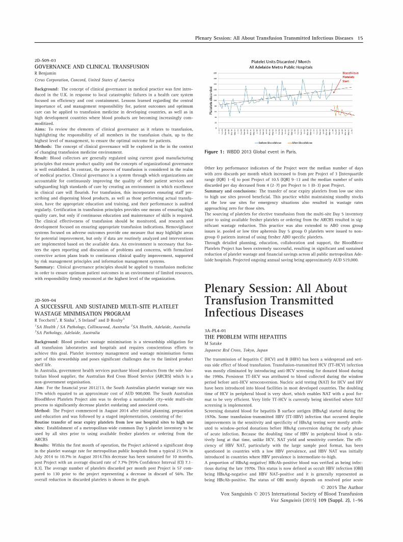
2D-S09-03
GOVERNANCE AND CLINICAL TRANSFUSIONR Benjamin
Cerus Corporation, Concord, United States of America
Background: The concept of clinical governance in medical practice was first intro-
duced in the U.K. in response to local catastrophic failures in a health care system
focused on efficiency and cost containment. Lessons learned regarding the central
importance of, and management responsibility for, patient outcomes and optimum
care can be applied to transfusion medicine in developing countries, as well as in
high development countries where blood products are becoming increasingly com-
moditized.
Aims: To review the elements of clinical governance as it relates to transfusion,
highlighting the responsibility of all members in the transfusion chain, up to the
highest level of management, to ensure the optimal outcome for patients.
Methods: The concept of clinical governance will be explored in the in the context
of changing transfusion medicine environment.
Result: Blood collectors are generally regulated using current good manufacturing
principles that ensure product quality and the concepts of organizational governance
is well established. In contrast, the process of transfusion is considered in the realm
of medical practice. Clinical governance is a system through which organizations are
accountable for continuously improving the quality of their patient services and
safeguarding high standards of care by creating an environment in which excellence
in clinical care will flourish. For transfusion, this incorporates ensuring staff pre-
scribing and dispensing blood products, as well as those performing actual transfu-
sion, have the appropriate education and training, and their performance is audited
regularly. Certification in transfusion principles provides one means of ensuring high
quality care, but only if continuous education and maintenance of skills is required.
The clinical effectiveness of transfusion should be monitored, and research and
development focused on ensuring appropriate transfusion indications. Hemovigilance
systems focused on adverse outcomes provide one measure that may highlight areas
for potential improvement, but only if data are routinely analyzed and interventions
are implemented based on the available data. An environment is necessary that fos-
ters the open reporting and discussion of problems and concerns, with formalized
corrective action plans leads to continuous clinical quality improvement, supported
by risk management principles and information management systems.
Summary: Clinical governance principles should be applied to transfusion medicine
in order to ensure optimum patient outcomes in an environment of limited resources,
with responsibility firmly ensconced at the highest level of the organization.
2D-S09-04
A SUCCESSFUL AND SUSTAINED MULTI-SITE PLATELETWASTAGE MINIMISATION PROGRAMR Tocchetti1, R Sinha1, S Ireland2 and D Roxby3
1SA Health / SA Pathology, Collinswood, Australia 2SA Health, Adelaide, Australia3SA Pathology, Adelaide, Australia
Background: Blood product wastage minimisation is a stewardship obligation for
all transfusion laboratories and hospitals and requires conscientious efforts to
achieve this goal. Platelet inventory management and wastage minimisation forms
part of this stewardship and poses significant challenges due to the limited product
shelf life.
In Australia, government health services purchase blood products from the sole Aus-
tralian blood supplier, the Australian Red Cross Blood Service (ARCBS) which is a
non-government organisation.
Aim: For the financial year 2012/13, the South Australian platelet wastage rate was
17% which equated to an approximate cost of AUD 900,000. The South Australian
BloodMove Platelets Project aim was to develop a sustainable city-wide multi-site
process to significantly decrease platelet outdating and associated costs.
Method: The Project commenced in August 2014 after initial planning, preparation
and education and was followed by a staged implementation, consisting of the:
Routine transfer of near expiry platelets from low use hospital sites to high use
sites: Establishment of a metropolitan-wide common Day 5 platelet inventory to be
used by all sites prior to using available fresher platelets or ordering from the
ARCBS
Results: Within the first month of operation, the Project achieved a significant drop
in the platelet wastage rate for metropolitan public hospitals from a typical 21.5% in
July 2014 to 10.7% in August 2014.This decrease has been sustained for 10 months,
post Project with an average discard rate of 7.7% [95% Confidence Interval (CI) 7.1–8.3]. The average number of platelets discarded per month post Project is 57 com-
pared to 130 prior to the project representing a decrease in discard of 56%. The
overall reduction in discarded platelets is shown in the graph.
Other key performance indicators of the Project were the median number of days
with zero discards per month which increased to from pre Project of 3 [Interquartile
range (IQR) 1–4] to post Project of 10.5 (IQR) 9–13 and the median number of units
discarded per day deceased from 4 (2–7) pre Project to 1 (0–3) post Project.Summary and conclusions: The transfer of near expiry platelets from low use sites
to high use sites proved beneficial. This practice whilst maintaining standby stocks
at the low use sites for emergency situations also resulted in wastage rates
approaching zero for those sites.
The sourcing of platelets for elective transfusion from the multi-site Day 5 inventory
prior to using available fresher platelets or ordering from the ARCBS resulted in sig-
nificant wastage reduction. This practice was also extended to ABO cross group
issues ie. pooled or low titre apheresis Day 5 group O platelets were issued to non-
group O patients instead of using fresher ABO specific platelets.
Through detailed planning, education, collaboration and support, the BloodMove
Platelets Project has been extremely successful, resulting in significant and sustained
reduction of platelet wastage and financial savings across all public metropolitan Ade-
laide hospitals. Projected ongoing annual saving being approximately AUD 519,000.
Plenary Session: All AboutTransfusion TransmittedInfectious Diseases
3A-PL4-01
THE PROBLEM WITH HEPATITISM Satake
Japanese Red Cross, Tokyo, Japan
The transmission of hepatitis C (HCV) and B (HBV) has been a widespread and seri-
ous side effect of blood transfusion. Transfusion-transmitted HCV (TT-HCV) infection
was mostly eliminated by introducing anti-HCV screening for donated blood during
the 1990s. Persistent TT-HCV was attributed to blood collected during the window
period before anti-HCV seroconversion. Nucleic acid testing (NAT) for HCV and HIV
have been introduced into blood facilities in most developed countries. The doubling
time of HCV in peripheral blood is very short, which enables NAT with a pool for-
mat to be very efficient. Very little TT-HCV is currently being identified where NAT
screening is implemented.
Screening donated blood for hepatitis B surface antigen (HBsAg) started during the
1970s. Some transfusion-transmitted HBV (TT-HBV) infection that occurred despite
improvements in the sensitivity and specificity of HBsAg testing were mostly attrib-
uted to window-period donations before HBsAg conversion during the early phase
of acute infection. Because the doubling time of HBV in peripheral blood is rela-
tively long at that time, unlike HCV, NAT yield and sensitivity correlate. The effi-
ciency of HBV NAT, particularly with the large sample pool format, has been
questioned in countries with a low HBV prevalence, and HBV NAT was initially
introduced in countries where HBV prevalence is intermediate-to-high.
A proportion of HBsAg-negative/ HBcAb-positive blood was verified as being infec-
tious during the late 1970s. This status is now defined as occult HBV infection (OBI)
being HBsAg-negative and HBV NAT-positive and it is generally represented as
being HBcAb-positive. The status of OBI mostly depends on resolved prior acute
Figure 1: WBDD 2013 Global event in Paris.
© 2015 The AuthorVox Sanguinis © 2015 International Society of Blood Transfusion
Vox Sanguinis (2015) 109 (Suppl. 2), 1–96
Plenary Session: All About Transfusion Transmitted Infectious Diseases 15
HBV infection and in a small fraction, the late acute phase before anti-HBs conver-
sion and the late stage of HBV carriage losing HBsAg. The extremely low viral load
of OBI-derived blood and the very small fraction of infectious OBI pose problems
from the perspective of screening. Even individual donation NAT (ID-NAT) could
miss infectious OBI blood. The strategy of disqualifying all HBcAb-positive blood
would be a final solution to this issue, but a considerable number of blood donors
would be lost. This could be serious in areas that have > 10% HBcAb positivity rates
among donors. In such areas, ID-NAT or pool format NAT with equivalent sensitivity
would be a second-best strategy to eliminate most infectious donations although
some infectious blood with a viral load below ID-NAT sensitivity might be missed.
Debate is beginning to focus on whether or not hepatitis E virus (HEV) screening
should be implemented in blood facilities because of TT-HEV infection. Despite an
unusually high prevalence of HEV, HEV-hepatitis occurs only occasionally among
the general population. However, HEV-related chronic liver damage is not uncom-
mon among immunocompromised patients. These issues complicate decisions about
which strategy to be implemented, no screening at all, selectively screening for
patients at high risk, or universal screening. Nucleic acid testing for HEV would also
further burden blood facilities from a cost viewpoint.
3A-PL4-02
PATHOGEN REDUCTION TECHNOLOGY – IS IT TIME?SY Kwon
Seoul Nambu Blood Center, Korean Red Cross, Seoul, South-Korea
With the application of stringent donor selection criteria and sophisticated screening
tests, including the introduction of nucleic acid testing (NAT), the risk of the so-
called ‘traditional’ transfusion-transmitted infections (TTI), HBV, HCV and HIV, has
dramatically decreased. However, as no system is 100% safe, breakthrough cases are
still being reported. Known pathogens that are not screened for and new emerging
ones represent a constant threat. Moreover, bacterial contamination still remains the
major infectious risk in blood transfusion. As opposed to our current safety measures
which are reactive, the development of pathogen reduction technologies has pre-
sented a proactive approach to our blood safety paradigm. Pathogen reduction tech-
nology for platelets and plasma has been approved for use in Europe for more than
10 years. Although pathogen reduction efficiency against non-enveloped viruses
and spore forming bacteria is less satisfactory, these technologies show a robust
pathogen reduction capability for a broad spectrum of pathogens. There are concerns
regarding the toxicity of the photochemical agent used to treat the platelets but cur-
rent data indicate that transfusion reactions after transfusion of treated platelets are
comparable to that of non-treated platelets. Another concern is that the treatment
might affect platelet function and increase risk of bleeding. Clinical data have shown
that recovery and survival of treated platelets are decreased compared to non-treated
platelets; therefore an increase in platelet use can be anticipated after the implemen-
tation of pathogen reduction technology. According to current data, transfusion of
treated platelets does not increase bleeding. As most pathogen reduction technolo-
gies target nucleic acids, they are not only effective in reducing infectious risks but
also in inactivating leukocytes, thereby preventing non-infectious risks like transfu-
sion-associated graft-versus-host disease (TA-GVHD) and alloimmunization. Despite
the multiple risks prevented through pathogen reduction, the cost to implement
these technologies remains a major issue, especially in developing countries. On the
other hand, pathogen reduction could be an alternative to bacterial screening and
gamma irradiation. Furthermore, cessation of some of the current screening items
and modification of some donor exclusion criteria might be possible, especially
when pathogen reduction technologies for red blood cells or whole blood become
available. Therefore, if we consider all correlated efforts involved to make blood
transfusion safe, the introduction of the pathogen reduction technology might prove
to be beneficial.
3A-PL4-03
THE CURRENT STATUS OF NUCLEIC ACID AMPLIFICATIONTECHNOLOGY IN TRANSFUSION TRANSMITTED INFECTIOUSDISEASE TESTINGM Vermeulen1 and N Lelie2
1South African National Blood Service, Johannesburg, South Africa 2Lelie Research,
Paris, France
Fully automated multiplex (MPX) Nucleic Acid Amplification Technology (NAT)
enabling individual-donation (ID) or minipool of 6 (MP6) screening for HIV, HCV
and HBV has been available for approximately 10 years. These systems are provided
by two major commercial players in the NAT industry (Grifols/Hologic and Roche).
In addition a few manufacturers provide MP24-96 NAT systems for the local market.
The pool size is indirectly proportional to the sensitivity of the assay. Prior to
August 2014 Grifols was the main supplier of assays for ID-NAT as the Roche s201
platform is not conducive to ID-NAT screening for high numbers. However in
August 2014 Roche released the new MPX assay performed on the high through-put
Cobas 8800 and 6800 instruments (which are also ID-NAT analysers, the former able
to process 1000 donations in an 8 h shift). Grifols also recently released their Ultrio
Elite new assay which includes detection of HIV-2 and is performed on the Panther
platform. This system has a smaller footprint than the former Tigris but allows ran-
dom access of samples and loading of reagents during processing, which streamlines
functionality and improves turn-around time. It is better suited for smaller laborato-
ries than the previous Tigris instrument. At the end of June 2015, 61 countries per-
form NAT to screen the blood supply. Of these 16 and 18 countries screen entirely
with either NAT systems from Roche or Grifols respectively and the remaining 27
countries use a combination of both methods in their country. Twenty countries
screen their entire blood supply using ID-NAT and the remaining 41 countries use
either MP or a combination of MP and ID-NAT. The MPX Roche assay has a 95%
limit of detection (LOD) of 25.7 IU/ml (CI 21.1–32.8), 7.0 IU/ml (CI 5.9–8.6) and
1.4 IU/ml (CI 1.2–1.7) for HIV-1, HCV and HBV respectively. The Ultrio Elite Grifols
assay has a LOD of 18 IU/ml (15–23.5), 3.0 IU/ml (2.5–3.9) and 4.3 IU/ml (CI 3.8–5.0) for HIV, HCV and HBV respectively according to the manufacturers’ package
insert claims. A number of publications most notably in South Africa, Europe, Aus-
tralia and Japan have shown, using modelling or lookback analysis, that a small risk
still remains of transmitting HIV and HBV even when using highly sensitive ID-
NAT. These models have in part been validated by 4 anecdotal cases of proven ID-
NAT breakthrough transmission cases of both window period (WP) and occult HBV
infection (OBI) by red blood cell or fresh frozen plasma (FFP transfusions). In addi-
tion MP-NAT HIV and HBV WP and OBI breakthrough infections have been found
in Brazil, Japan and Thailand, which also could have been missed by ID-NAT. These
latter cases highlight the importance of selective donor recruitment in addition to
state of the art screening.
Academy Session: The Whatand How of BloodComponents
3B-S10-01
AUTOMATING THE PRODUCTION OF BLOOD COMPONENTSM Lozano and J Cid
University Clinic Hospital, Barcelona, Spain
For many years the preparation of blood components from whole blood (WB) dona-
tions was totally manual, from centrifugation of the WB bag to the transfer of the
blood components to the connected bags or the seal of the plastic tubing. Since the
introduction of the buffy coat (BC) method for preparing platelet concentrates from
WB donations, automation started to enter the blood components preparation labo-
ratory of the blood banks. Thus, in the late 1980 several devices were developed that
allow the automation of the first step of blood processing, i.e. the separation of the
centrifuged WB bag into red blood cells (RBC), plasma and BC. The introduction of a
device that performed the sterile connection of the plastic tubing had a tremendous
impact in the blood component preparation laboratory, since it opened the possibil-
ity of pre-storage pooling of the components and allowed the discovery that the
processing of pooled BC instead of individual BC increased significantly platelet
recovery in the platelet concentrates (PC). In 2003, a device that combined the cen-
trifugation of one pooled BC with the expression of the supernatant plasma rich
plasma was introduced in the market (OrbiSac, Terumo BCT). Later a new device was
developed that allowed the processing of up to six pooled BC in each centrifugation
run (TACSI, Terumo BCT). More recently a new generation of devices, which allow
the automation of almost all the necessary steps for preparing blood components
from WB donations has been introduced. The first one was Atreus system (Terumo
BCT) that allowed the processing of one whole blood donation into RBC and plasma
(protocol 2C) or RBC, plasma and BC (protocol 2C+). The system was not refrigerated
and due to high temperatures generated during the centrifugation process in routine
use, RBC vesiculation occurred leading to the appearance of reddish PC. The recently
introduced Reveos system (Terumo BCT) could process simultaneously 4 units of WB
and is refrigerated. The Reveos 2C protocol produces from one WB donation RBC
and plasma while the 3C protocol produces RBC, plasma and one intermediate plate-
© 2015 The AuthorVox Sanguinis © 2015 International Society of Blood TransfusionVox Sanguinis (2015) 109 (Suppl. 2), 1–96
16 Plenary Session: All About Transfusion Transmitted Infectious Diseases
let unit (IPU). The IPUs can be manually pooled with other IPUs using a pooling kit
with integrated leukoreduction filter to form a transfusable adult-size PC without
the need of an additional centrifugation step for RBC removal. As the Atreus 3C, the
Reveos 3C system also includes a platelet yield index, which provides an estimation
of the platelet content in the IPU, making it possible to increase and/or standardize
platelet content of the final PC by selecting the optimal IPUs. Several in vitro and in
vivo studies have shown that the blood components produced using semi-automated
and automated devices met the European quality standards and provide a similar
transfusion response in the recipients compared to those prepared using manual
methods
3B-S10-02
INTRODUCING NOVEL BLOOD COMPONENTS – PRINCIPLESAND PRACTICESM Kirwan
New Zealand Blood Service, Auckland, New Zealand
The development of novel blood components requires input from a number of func-
tional areas within the Blood Service and must be managed in close consultation
with the Regulator. Prior to commencing discussions with the Regulator an evalua-
tion is performed and presented to the Executive management team for discussion
and inclusion into the business strategic plan.
Before approval to implement a new product can be granted, the Regulator needs to
understand what the benefits to the patient and organisation are, what other interna-
tional Blood Services are doing in regard to this component and whether the Blood
Service has the capability to implement the change. The information submitted to
the Regulator for approval will inevitably be comprehensive and likely require sev-
eral detailed submissions.
Initial input is required from the clinical team to provide comment on international
practice and evidence of clinical efficacy of the new component. The technical /op-
erational team will develop the process and procedures to manufacture the product
providing confidence that the organisation has the required expertise and capability.
Validations of new equipment and processes must be completed, reported and
approved including any changes to the Blood Management System. The Quality sys-
tem within the organisation will provide assurance that the development is robust
and as per defined change management procedures. Training plans specific to the
new process must be implemented and delivered to manufacturing staff. Post imple-
mentation plans are developed to review and verify the effectiveness of the process.
Any change requires timely and relevant communications with clinical end users
and all others with a vested interest. Not only is it important to communicate the
benefits of the change, it is unavoidable that costs will rise when creating a new
product so revised pricing and its impact must be announced.
Recognition that the evolution of a de novo product, within the Blood Service, is a
complex and multifaceted process is essential for success, together with effective
communication and careful planning.
3B-S10-03
GMP SUPERVISION ON BLOOD ESTABLISHMENTT Hutadjulu
Badan POM/NADFC, Jakarta Pusat, Indonesia
Blood products are life saving medicine that often used in life threatening situations
of severely ill patients. Therefore, blood safety has become an important issue where
high standards of quality and safety of these products have to be assured prior to
use in human. These high standards can be achieved by implementing the principles
of Good Manufacturing Practice (GMP) in blood establishment, which is providing a
systematic approach for incorporating quality into the entire manufacturing process.
This approach ensuring that quality system are in place for the collection, prepara-
tion, storage, testing and distribution of blood products and consistently produce
product that appropriate to their intended use and meet the required specifications.
GMP in blood establishments also increasing availability of plasma and is one of the
key issues for a successful plasma contract fractionation program. Plasma for frac-
tionation is needed to generate essential plasma-derived medicinal products (e.g.
clotting factors and immune globulins). Implementation of GMP standards are
mandatory for collection of plasma as starting material for plasma-derived products.
By assuring that standards are met, blood regulation serves to protect donors and to
promote and maintain quality and safety of both blood components for transfusion
and plasma for fractionation.
GMP implementation in Blood Establishment is relatively new, especially in develop-
ing countries. Consistent compliance in GMP implementation depends upon a coor-
dinated service with an appropriate national blood policy. Blood regulation within a
legal framework is an essential element of any national blood system. The competent
national regulatory authority (NRA) is responsible for assessing and ensuring the
compliance with GMP in blood establishments in order to ensure quality and safety
of the blood products.
Badan Pengawas Obat dan Makanan (Badan POM) or National Agency of Drug and
Food Control (NADFC) is a regulatory body which is responsible for ensuring the
safety, efficacy and quality of drug and food marketed in Indonesia. Blood products
along with other pharmaceuticals are included as commodities under the supervision
of Badan POM/NADFC. As a developing country which become the pilot project on
WHO’s blood safety program, Indonesia has strong commitment to strenghten the
national blood regulation and enforcing the GMP aspect on blood establishment in
order to provides high quality plasma and plasma derived product.
Advances in Blood Safety
3B-S11-01
A PRIMER ON MALARIA IN THE ASIA PACIFICJ Baird
Eijkman-Oxford Clinical Research Unit, Jakarta, Indonesia
Five species of protozoan parasites in the genus Plasmodium cause acute malaria in
humans and other primates. These parasites follow complex life cycles in the blood
and liver of humans, and are transmitted between humans by the bite of mosquitoes
in the genus Anopheles – the only means of acquiring malaria except for blood
transfusion or tissue and organ transplantation. Malaria remains highly endemic
across much of the Asia Pacific. Among all nations of the region, only Japan, Rus-
sia, Australia and Singapore are free of endemic malaria transmission. All of the five
species of plasmodia occur in the Asia Pacific, putting almost 3 billion people at risk
of infection and typically causing tens of millions of attacks of illness. Each year
many thousands in the region do not survive their malaria attacks, mostly due to
delayed or improper diagnosis and treatment. This lecture will review the plasmodia
species causing malaria, the clinical course and consequences of them, and their
geographic distribution in the Asia Pacific. Diagnostic methods for malaria will be
reviewed, including ultra-sensitive molecular techniques needed to detect the very
often microscopically sub-patent infections. The lecture will also describe the rela-
tionship between glucose-6-phosphate dehydrogenase deficiency and malaria, espe-
cially as regards sensitivity to acute hemolytic anemia caused by the antimalarial
drug primaquine. All of these issues may be considered relevant to the practices of
blood banking and transfusion in the Asia Pacific.
3B-S11-02
NEW STRATEGIES FOR BACTERIAL CONTAMINATIONSM Schmidt and E Seifried
German Red Cross Institute, Frankfurt, Germany
Background: The detection of bacterial contaminations in blood components can be
classified in culture methods, rapid detection methods and experimental new tech-
nologies.
Culture methods: Many countries have currently implemented culture methods into
blood donor screening. Culture methods convinced with easy handling and high
analytical sensitivity. The challenge with culture methods, with the negative-to-date
release concept, is a risk for false negative screening results due to sample failures
and a reduced clinical efficiency. To reduce this risk, the time for sample collection
was extended and the input volume improved to a maximum of 10 ml. An addi-
tional advantage of culture systems is the option to screen all blood components
including stem cells. Nevertheless, due to storage conditions at room temperature,
most countries have implemented bacterial screening only for platelet concentrates.
Rapid detection methods: Within the last decade, rapid bacterial detection systems
(RBDS) were developed as an alternative option to culture systems. In RBDS testing
time is reduced to 2–3 h. Therefore a negative-to-date release concept is not neces-
sary by using these systems. Three of them are currently CE or FDA certified (bacti-
flow system NAT and Verax PGD test). The clinical efficiency is high for the rapid
detection methods but the analytical sensitivity is a little lower than for culture
methods
New technologies: In addition, new technologies are currently in development to
detect bacterial strains in blood components. One option could be the measurement
of the bacterial esterase activity by an automated bio-imaging system. First data
demonstrated an analytical sensitivity of app. 20 CFU/ml with a detection time of
45 min. Another approach focuses on the detection of the bacterial cell wall by an
ELISA technology. A third new technology uses ATP luminometry. Finally, electro-
chemical biosensors were developed for many different analytes, which range in size
© 2015 The AuthorVox Sanguinis © 2015 International Society of Blood Transfusion
Vox Sanguinis (2015) 109 (Suppl. 2), 1–96
Advances in Blood Safety 17
from individual ions and small molecules to nucleic acids and proteins up to whole
viruses and bacteria. This ‘lab-on-a-chip’ method of patient or donor diagnosis and
monitoring provides a more rapid diagnosis, which allows for faster and more effec-
tive therapeutic intervention, thereby preventing full-blown infection and mortality
and also decreasing the spread of disease.
Conclusions: Bacterial detection in blood components, especially in platelets, is
implemented in many blood donor services. Timing of all steps (sample collection,
detection method) is very important to achieve the highest safety level. Although
pathogen reduction methods are available for some blood components like platelets
or packed red cell concentrates, testing methods offer in addition options to screen
also stem cell concentrates. New approaches are promising to come to a next level
to be implemented into routine screening, but the analytical sensitivity has to be
improved for some of the new test systems in the coming years. Due to the fact that
a risk for bacterial contamination still occurs in all blood components, the focus in
transfusion medicine in the next decade will be the development of general princi-
ples and strategies to detect or inactivate bacteria in all blood components. There-
fore, new developments are gladly welcome.
3B-S11-03
DEVELOPMENT OF A HIGHLY SENSITIVE DETECTIONMETHOD FOR BACTERIAL DNA IN BLOOD FOR TRANSFUSIONT Yoshimasa1, H Sakata1, K Matsubayashi1, S Sato1, T Kato1, S Kino1, T Yamamoto2,
H Ikeda2 and S Takamoto1
1Japanese Red Cross Hokkaido Block Blood Center, Sapporo, Japan 2Hokkaido Red
Cross Blood Center, Sapporo, Japan
Background: Bacterial contaminated blood product for transfusion is known to
result in severe side effects in patients. Although the Japanese Red Cross Blood Cen-
ters introduced the diversion of the initial blood collection and leukoreduction in
addition to a short shelf-life of blood products, bacterial contamination of blood
products has not been completely excluded. Therefore additional preventive mea-
sures such as bacterial screening are required. Culture-based bacterial detection
methods are highly sensitive but take much time to obtain the results, whereas PCR-
based methods such as broad-range real-time PCR of 16S rRNA gene (rDNA) are
rapid but not so sensitive because of contamination of the exogenous bacterial DNA
in the PCR reagents.
Aims: We aimed to develop a rapid and highly sensitive method for broad-range
bacterial 16S rDNA detection using real-time PCR.
Methods: A primers/probes targeting the highly conserved region of the bacterial
16S rDNA was used to amplify a 160-bp fragment by real-time PCR (Yang S, et al,
2002). A plasmid including the target region of 16S rDNA was synthesized and used
as a standard 16S rDNA. Three methods for decontamination or inactivation of the
exogenous bacterial DNA in the two reagents, namely an amplification reagent
(FastStart Universal Probe Master, Roche Applied Science, AMP) and a primers/probes
mix (PP), were explored. Method 1, a mixture of AMP and PP was digested with the
Alu I restriction enzyme. Method 2, only AMP was digested with DNase I, and PP
was filtered through a centrifugal ultrafiltration device (Amicon Ultra-0.5 50K, Merck
Millipore). Method 3, the same as the method 2 but AMP was light-inactivated with
ethidium monoazide instead of enzymatic digestion. A serial diluted standard DNA at
0, 1, 2, 3 and 4 log copies/reaction and no template controls (NTCs) were tested by
real-time PCR with reagents treated by the methods described above. For evaluation
of the sensitivity for practical bacterial detection, peripheral blood spiked with Sta-
phylococcus aureus or Escherichia coli at 0, 1, 2, 3 and 4 log bacteria/reaction was
prepared and tested along with NTCs. DNA was extracted from blood spiked with
bacteria using QIAamp MinElute Virus Spin kit (QIAGEN) with bead beating and sub-
jected to real-time PCR using a bacterial DNA-reduced reagents.
Results: The amount of exogenous bacterial DNA contaminated in the PCR reagents
was estimated approximately 100 copies/reaction. The assay sensitivity using stan-
dard DNA was approximately 100, 1 and 10 copies/reaction for the method 1, 2 and
3, respectively. No amplification was also observed for NTC in the method 2. In the
bacterial spiking experiments, the sensitivity of the real-time PCR assay in which the
reagents were treated by the method 2 was approximately 10 bacteria/reaction both
for S. aureus and E. coli.
Conclusions: In the broad-range bacterial detection by real-time PCR, a combina-
tion of DNase I digestion and ultrafiltration was proven to be very effective for
reducing contaminated bacterial DNA in the PCR reagents. Broad-range bacterial
16S rDNA real-time PCR assay using bacterial DNA-reduced reagents was considered
to be feasible for a bacterial screening of blood for transfusion.
3B-S11-04
TRANSFUSION RISK FROM EMERGING PATHOGENS IN THEASIA-PACIFIC REGIONHM Faddy1, E Viennet2 and R Flower1
1Australian Red Cross Blood Service, Brisbane, Australia 2Research School of
Population Health, Australian National University, Canberra, Australia
Background: An emerging infectious disease (EID) refers to a disease that has
recently appeared in a population or one that has rapidly increased in incidence or
geographic range. Examples of EIDs include Ebola, HIV/AIDS, hepatitis E and the
vector-borne diseases caused by Zika and dengue virus. EIDs pose a risk to transfu-
sion safety, which can be direct, if the agent can be transmitted through blood
transfusion, or indirect, where outbreaks reduce the pool of available donors.
Although examples of both have been observed, the former will be the focus of this
review. Many transfusion risks have become global due to globalisation and
increased international travel; however, unique region-specific concerns exist. The
Asia-Pacific region varies in size depending on context, but for the purposes of this
review will be defined as Southeast Asia and Oceania. This region is a hotspot for
genetic diversity, as well as the emergence of a number of infectious diseases. The
Asia-Pacific region is also geographically, culturally, socioeconomically, and climat-
ically diverse. Moreover, the level of sophistication in blood operators within this
region varies, from a national supplier based on voluntary blood donors as is seen
in Australia, to hospital-based services relying on replacement donors, which is seen
in many nations across the pacific.
Aims: To review current EID risks to blood transfusion safety in the Asia-Pacific region.
Methods: Review of available public health reports and literature for the occurrence
of EIDs in the Asia-Pacific region that pose a transfusion risk.
Results: EIDs that pose a transfusion risk occur across the Asia-Pacific region, with
differing levels of endemicity between and within countries. Reports of hepatitis E
virus (HEV) seropositivity vary from 2.2% in Fiji to 15.2% in Papua New Guinea. As
a result of high HEV prevelance in the Hokkaido region of Japan, HEV nucleic acid
testing (NAT) has been implemented for screening blood donations. Dengue infection
is widespread across the Asia-Pacific, with major outbreaks occurring in 2014 in Fiji,
Malaysia, the Philippines, and Thailand. In Australia, dengue is episodic in the
North-east, resulting in donation restrictions in affected areas. Zika virus, the first
arbovirus demonstrated to also be sexually transmitted, emerged in the Asia-Pacific
in early 2007 and outbreaks have spread across the pacific, with epidemics in French
Polynesia, the Cook Islands, Easter Island and New Caledonia. Due to the theoretical
transfusion risk several preventative procedures were implemented in French Polyne-
sia during an outbreak in 2013, including NAT.
Summary/Conclusions: EIDs pose a transfusion risk globally, and in the Asia-Paci-
fic region. A number of EIDs that can be transmitted through blood transfusion are
endemic to the Asia-Pacific, with countries managing the transfusion risk depending
on the level of endemicity of the agent in question, and the maturity of blood oper-
ations in each county. Climate change is predicted to increase the distribution and
density of a range of disease vectors, which is likely to increase the transmission of
a number of vector-borne diseases, representing a significant threat to the mainte-
nance of a safe blood supply in the future.
3B-S11-05
MIDDLE EAST RESPIRATORY SYNDROME CORONAVIRUS(MERS-COV) OUTBREAK IN SOUTH KOREA: RISKMANAGEMENT AT THE KOREAN RED CROSS SEOUL NAMBUBLOOD CENTERSY Kwon, EH Lee, HS Kim, JH Kim, YO Lim and GR Choi
Seoul Nambu Blood Center, Korean Red Cross, Seoul, South-Korea
Background: On 20 May 2015 the first confirmed case of MERS was identified in
South Korea. Unexpectedly this case triggered an outbreak with a total of 186 cases
of which 36 have died. Nearly half of all confirmed cases have been traced to Sam-
sung Medical Center (SMC), which is in the service area of the Seoul Nambu Blood
Center (SNBC) of the Korean Red Cross (KRC).
Aims: To review measures taken both at a national and SNBC level to deal with the
impact of the MERS crisis on blood availability.
Methods: National and SNBC blood collection and supply data were analyzed using
the Blood Information Management System (BIMS) of the KRC. Measures taken to
deal with the decrease in collections were reviewed.
Results: To ensure the safety of the national blood supply, the KRC applied
enhanced donor criteria and guidelines on post-donation information. Donors with
overseas travel history were already deferred for 1 month since 2008. Although
there was no evidence of community transmission, public anxiety caused by the out-
18 Oral Abstracts
© 2015 The AuthorVox Sanguinis © 2015 International Society of Blood TransfusionVox Sanguinis (2015) 109 (Suppl. 2), 1–96
break led to decreased outdoor activities, temporary closure of schools and cancella-
tion of various events. This resulted in a decrease of donors at fixed donation sites
and cancellation of mobile blood drives. From 21 May to 20 June, this includes the
period with a peak in case confirmation, national and SNBC collection at fixed sites
decreased by 2.5% and 2.3%, respectively compared to 2014. Decrease of collection
at mobile blood drives was more pronounced, with a 9.6% and 22.2% decrease at a
national and SNBC level. Overall national and SNBC total collection decreased by
4.9% and 8.1%, respectively. While blood supply still met the demand, to ensure
adequate blood supply level, the KRC made public appeals to participate in blood
donation. Opening time of fixed donation sites was also extended. At SNBC level,
regular blood donation campaigns were held to raise awareness about the decrease
in donation in our area. With these efforts, national and SNBC collection increased
during 21 to 30 June by 9.0% and 7.1%, respectively. National blood demand
decreased by only 0.5% compared to 2014. However, at SNBC the demand dropped
by 9.1%. This was mainly due to the shutdown of SMC after the increase in con-
firmed MERS cases, because about 35% of SNBC’s supply is issued to SMC.
Conclusion: Emerging infections that have not been confirmed to be transmitted by
transfusion can also threaten the safety of the blood supply. Timely measures were
effective in ensuring an adequate blood supply at a national level. Although decrease
in collection at SNBC was more pronounced, adequate supply level could be main-
tained because the demand declined remarkably compared to other regions. In cases of
an emergency, blood collection target should be adjusted according to local needs as
the incident may have different impact on the blood service at a local level. Experience
gained through this crisis will add to our preparedness planning for future incidents.
Academy Session: The Whatand How of Organisation andBlood Supply Management
3C-S12-01
WHAT’S THE RIGHT MODEL FOR A NATIONAL BLOODPROGRAMME?P Flanagan
New Zealand Blood Service, Auckland, New Zealand
The World Health Organisation advocates for a nationally co-ordinated blood system
in order to promote uniform standards, economies of scale, consistency in the qual-
ity and safety of blood and blood products and best transfusion practices. The struc-
ture of the national blood system will however depend on a range of factors
including the size and population of the country, geographical and transport con-
straints, the level of development of the overall health-care system and the level of
funding available to develop and maintain the system. The Ministry of Health will
play a pivotal role in defining the strategy and policy of the overall blood transfu-
sion system and for ensuring that it is fully integrated into the wider health system
of the country.
Internationally a number of different models exist for delivery of blood transfusion
services. There is no one model that fits all circumstances. Each country must deter-
mine its own optimal model and in doing so ensure effective oversight and monitoring
against pre-agreed standards. Governance of the blood system must extend beyond the
activities of the blood transfusion services and include consideration of regulation,
access to plasma fractionation and clinical transfusion systems within hospitals.
A range of options should be considered when defining the optimal organisation
structure within a country. This includes the number of organisations that will pro-
vide the service, the types of organisations that will be involved (government provi-
sion or non-governmental organisation) and the level of integration between the
collection, manufacturing and hospital blood banking activities. Inevitably the type
and effectiveness of pre-existing structures will also need to be considered.
A key consideration is the level of centralization of the national blood transfusion
service. Centralization of the collection and manufacturing aspects of BTS provision
will reduce duplication, enable better national management of inventory and enable
introduction of consistent national standards. Conversely a decentralized approach
whereby individual hospitals undertake broad range of activities will enable closer
and better integration with clinical services and links to University and Teaching
hospitals with improved opportunities for education and research. Either approach
can work. Many countries have adopted a hybrid model.
Three types of organisation may be involved in the delivery of the BTS. These are
government managed providers, non-government organisation providers (Red Cross/
Red Crescent) and hospital/ university providers. In some countries commercial pro-
vides are also involved. Many countries adopt a mixed model. The Red Crescent/Red
Cross is a major provider of BTSs. This avoids a direct link to government and might
provide a useful source of finance for the system. It does however result in a more
complex governance model. Overall direct provision of a centrally co-ordinated BTS
by government is probably the most efficient model. Nonetheless an NGO model
does provide other benefits and might be more appropriate in some circumstances.
An effective model for delivery of a national co-ordinated BTS requires government
commitment and support, clarity of the roles and functions of individual providers,
clear lines of authority, responsibility & accountability and adequate and sustainable
resourcing.
3C-S12-02
PUTTING IN PLACE A RISK-BASED DECISION FRAMEWORKFOR BLOOD SAFETYPC McDonald and ABO Alliance of Blood Operators
Alliance of Blood Operators, Melbourne, Australia
Background: Decisions about blood safety need to take numerous factors into
account, such as risks, benefits, costs, ethical issues, and stakeholder perspectives.
Since the 1980s, numerous measures aimed at maximising blood safety have been
implemented based on varying interpretations of the precautionary principle and an
unsustainable pursuit of ‘zero risk’, without evident consideration of cost effective-
ness and opportunity cost. In 2010, the international Alliance of Blood Operators
(ABO) held an International Consensus Conference on Risk-Based Decision-Making
for Blood Safety to address the issue. Among the consensus panel’s conclusions were
the following principles: that risk is inherent ‘vein to vein’; that zero risk is
unattainable; that the well-being of transfusion recipients must be central to blood
safety decision-making.
Based on these conclusions, ABO has developed an integrated, internationally appli-
cable, risk-based decision-making framework to guide major policy and operational
change. The Framework is: evidence-based and takes advantage of quantitative and
qualitative information; provides measurable outcomes; improves consistency in
decision making and proportional responses to risk; improves confidence and trust
in investment decisions; allows redirection of resources thus improving efficiency
and effectiveness; increases transparency and communication; and enables defensi-
bility and commitment across the system.
Summary: The ABO Risk-based Decision-making (RBDM) Framework is a structured
approach to blood safety risk management. It lays out a step by step methodology for
setting the best course of action under uncertain conditions by identifying, assessing,
acting on and communicating risk. The RBDM Framework has been tailored for the
specific needs of blood operators. The Framework provides a set of approaches to
help users identify and prioritise risks to blood safety and to evaluate the effective-
ness of potential risk management options, such as introducing a new intervention or
withdrawing an existing one. The approaches included in the Framework help the
user analyse and account for a series of contextual, often qualitative, factors that
affect decision making in the management of blood risks. The Framework takes a
societal perspective, enabling consideration of social, economic, and ethical perspec-
tives that go beyond quantitative calculations of risk and can alter risk tolerability.
The RBDM Framework is intended to be embedded in an organisation’s risk management
program. As such, it has two distinct components: policy foundations; and the decision-
making process. Risk management policy foundations are background elements that
express the values, priorities, and policies of a blood service in relation to risk manage-
ment. They guide the decision-making process and must be in place before the process
begins. The policy foundations include: risk management principles; risk communication
and stakeholder participation; assessment principles; and risk tolerability.
The stages of the decision-making process are common to many decision frame-
works. The RBDM Framework steps the user through each of the stages: preparation;
problem formulation; participation strategy; assessments (including health eco-
nomics & outcomes assessments); evaluation; and decision.
3C-S12-03
HAEMOVIGILANCE-SETTING UP ONE FROM NONEN Marwaha
Postgraduate Institute of Medical Education and Research, Chandigarh, India
Haemovigilance is a surveillance system which aims to detect and analyse adverse
events and reactions in the blood transfusion chain to improve blood safety by insti-
tuting corrective and preventive actions. Haemovigilance activities may be organized
at an institutional level to improve clinical transfusion practices, but this does not
contribute to risk assessments of transfusion at a national level. Hence, there is a
© 2015 The AuthorVox Sanguinis © 2015 International Society of Blood Transfusion
Vox Sanguinis (2015) 109 (Suppl. 2), 1–96
Academy Session: The What and How of Organisation and Blood Supply Management 19
need to establish a nationally co-ordinated system of haemovigilance. Setting up
such a system is a complex task, it involves both organizational and operational
aspects, which may vary between countries depending upon the structure and regu-
lation of blood transfusion services.
First and foremost it should be a part of the National Blood Policy. The organiza-
tional structure should ideally have governmental oversight to the program through
its Ministry of Health or equivalent authority. Haemovigilance advisory committees
are required to give direction to the program. All stakeholders need to be identified
and systems worked out for their collaboration and coordination. During the phase
of planning the following issues need to be addressed: (i) whether reporting of all
adverse events in the transfusion chain is required (donors, processes and recipient-
related events) or restricted to recipients, that is, reporting only transfusion reactions
to begin with, whether to include near miss events and/or rapid alerts, (ii) whether
reporting will be voluntary or mandatory, or a combination of both, that is manda-
tory reporting of serious adverse reactions only, rest being voluntary, (iii) what
would be the flow of information – who reports to whom, (iv) adoption of defini-
tions of adverse events and reactions (AEs, ARs) as per international consensus, (v)
uniform reporting formats and (vi) requisite resources both financial and technical
to implement the program. The initial focus of haemovigilance activities could be on
the transfusion recipients, that is, reporting of transfusion reactions.
At the reporting end, awareness and training of all staff who would be involved in
haemovigilance activities needs to be conducted. This would include clinicians and
other bedside transfusion staff, personnel in blood centers and hospital based depart-
ments of transfusion medicine, members of hospital transfusion committees and
donor organizations. A very important aspect to be dealt with during awareness ses-
sions is to allay fears of any punitive action for the reporting staff. The reporting
units initially could be regional blood transfusion centers and their associated hospi-
tals or the tertiary care hospitals and gradually when the system gains ground the
scope could be extended to include other hospitals and blood centers and subse-
quently also the donors.
The haemovigilance reports generate a large amount of data, hence a validated sys-
tem for collection, collation and analysis of data is required. Mechanisms for regular
publications of haemovigilance reports with recommendations for better transfusion
practices should be put in place. This would help in directing policy decisions to
improve blood safety
It should also be aimed to establish linkage with the international haemovigilance
network and database for exchange of information between member countries.
Advances in ClinicalTransfusion
3C-S13-01
HAEMOGLOBINOPATHIES IN INDONESIAI Setianingsih
Eijkman Institute for Molecular Biology, Jl. Diponegoro 69, Jakarta
Around 6000 patients (mostly are beta-thalassaemia) are currently registered for reg-
ular blood transfusion, where 5000 out of these patients are living in Java island.
The management of thalassaemia in Indonesia is far from the standard required.
Most patients were late diagnosed and received inadequate blood transfusion, Only a
very small number of patients received adequate blood transfusion and iron chelat-
ing agent. However, since 2011, Government: has subsidized the optimal manage-
ment of all patients registered.
The management and prevention program for thalassemia in Indonesia is compli-
cated by the diversity of the carrier frequency, the molecular defects and the pheno-
type expression of the disease in this country. The carrier frequency ranged from 5
to 10%, and that of HbE which is 1–33%, in the various ethnic populations. Like-
wise, the spectrum of the underlying b-globin mutations varies widely between
regions. Twenty four mutations accounted for more than 90% alleles in Indonesia.
HbE and IVS1-nt5 are found in all geographical areas, albeit with different frequen-
cies, whilst other mutations are specific for particular ethnic groups such as Hb
Malay and Cd 26 (GAG?TAG) in the Malay populations of Sumatera, IVS1-nt1 in
the Sundanese and Cd-35 in the Javanese (both of the Java island), and large – and
-globin gene deletions in Buginese and Makasarese of South Sulawesi.
The relationship between these mutations, their overall genetic background and their
clinical expression is also of major interests. Clinically the patients showed a wide
range of phenotypic expression, from thalassemia major to the mild type of tha-
lassemia intermedia. All patients with bo/ bo-, bo/ b+- and b+/ b+-mutations presented
at early of age and were clinically b-thalassaemia major. In contrast, the age at first
presentation in patients compound heterozygous for HbE with either bo or b+-tha-lassemia mutations varied from 6 months to 20 years and showed a broad spectrum
of clinical severity, ranging from thalassemia major to mild thalassemia intermedia.
The findings have major implications in the management of thalassaemia in Indone-
sia. The variation in the distribution of carriers in the various ethnic populations
and the size of the populations resulted in unequal anticipated carrier screening and
prenatal diagnosis workload. The diagnostic protocol for carrier screening and for
prenatal diagnosis could be specifically designed on a regional basis.
3C-S13-02
OPTIMISING TRANSFUSION THERAPY IN THE CHRONICALLYTRANSFUSED PATIENTSAL Ang
Health Sciences Authority, Singapore, Singapore
Patients may require regular transfusion due to congenital or acquired causes of
ineffective blood cell production or bone marrow failure. In Asia, the most common
congenital cause is thalassaemia while acquired causes include myelodysplastic syn-
drome (MDS) and severe aplastic anaemia. The major goal of transfusion in these
patients is to minimise the complications from their condition with the lowest possi-
ble amount and risks of transfusion.
Due to the frequent transfusions, their cumulative risks from transfusion are higher
than other patients. These include mild adverse reactions such as febrile non-haemo-
lytic transfusion reactions and urticaria, as well as severe risks such as haemolytic
transfusion reactions, anaphylaxis, transfusion transmitted infections, transfusion
associated circulatory overload and TRALI. Leucodepleted blood products not only
help to reduce the risks of febrile non-haemolytic transfusion, it may also reduce the
risk of HLA-alloimmunisation and hence platelet refractoriness in patients with sig-
nificant thrombocytopaenia requiring platelet transfusion. Washed blood products
may also be needed in those with recurrent severe anaphylactic reactions to transfu-
sion. These chronically transfused patients have a higher frequency of red cell
alloimmunization, which in turn increases their risks of haemolytic transfusion reac-
tions. It is therefore important to regularly monitor for the development of new clin-
ically significant red cell alloantibodies before each transfusion. Patients with
clinically significant red cell alloantibodies need to receive compatible antigen-nega-
tive red cells. An extended red cell phenotyping before commencement of red cell
transfusion may be useful for subsequent antibody identification in case of alloim-
munisation later. If the patient is already transfused regularly, molecular typing
should be carried out instead. Prophylactic matching of blood groups (other than
ABO and RhD) which are associated with high frequencies of alloimmunization helps
to reduce the incidence of this, and is particularly useful for patients with high risks
of red cell alloimmunization. To minimise the risk of transfusion transmitted infec-
tions, the blood should be collected from carefully screened non-remunerated volun-
tary blood donors, and each donation should be screened for HIV, Hepatitis B/C,
syphilis and where relevant, other infections that pose a risk to blood safety. Patients
should also be vaccinated against hepatitis B.
In addition, patients who need regular red cell transfusion such as those with trans-
fusion dependent thalassaemia, are at risk of transfusional haemosiderosis. Transfus-
ing red cell concentrate close to its collection may result in better in vivo recovery
and longer half-life, and this may decrease the red cell transfusion requirements of
these patients. This led to the recommendation of transfusing red cells in additive
solution within 2 weeks from collection. For thalassaemia major patients, maintain-
ing pre-transfusion Hb at 9 to 10.5 g/dl had been shown to provide the most appro-
priate balance between adequate bone marrow suppression and transfusional iron
loading. Thalassaemia patients should also be assessed carefully on the indications
and benefits of regular transfusion before being commenced on this.
3C-S13-03
INTRODUCTION OF POINT-OF-CARE TESTING ANDINTERVENTION IN THE TRANSFUSION UNIT DECREASESPERIOPERATIVE BLEEDING, SHORTENS OPERATIVE TIMEAND FACILITATES APPROPRIATE DOSAGE OF BLOODTRANSFUSIONSC Yamada, H Furumaki, H Fujihara, H Watanabe, M Tsuzuki, T Shinba, M Kaneko, H
Shibata, K Ishizuka, Y Hosaka and A Takeshita
Hamamatsu University School of Medicine, Hamamatsu, Shizuoka, Japan
Background and aims: Perioperative blood components should be supplied on the
basis of the latest clinical symptoms and laboratory data, while many transfusion
© 2015 The AuthorVox Sanguinis © 2015 International Society of Blood TransfusionVox Sanguinis (2015) 109 (Suppl. 2), 1–96
20 Advances in Clinical Transfusion
units have not been always and completely attainable. We introduced a blood cell
and coagulation analyzer into the operating room, which members from the transfu-
sion unit use to analyze samples. We previously reported this system significantly
decreased the total incidence of perioperative bleeding (Yamada et al, AABB, 2014).
In this study, we had deeper analyses on the advantage of adopting this system in
each operative procedure and each blood component.
Methods: Samples were analyzed on the spot by members of the transfusion unit
using the analyzer placed in the operating room, and shared the data with anesthesi-
ologists and surgeons. We analyzed the amount of total bleeding, operative time and
dosage of blood components transfused during each operating procedure, and com-
pared these findings before and after introducing the system. The data was compared
by Chi-square test and Fisher’s exact test (SAS, Tokyo), and P-values of less than
0.05 were considered statistically significant.
Results: The total number of operations performed before and after introducing the
system was 1757 and 2044, respectively. After initiating the new system, the amount
of perioperative bleeding significantly decreased during endovascular aortic repair,
aortic aneurysmectomy, colectomy, valve replacement, cervical and uterine cancer,
spinal fusion, and scoliosis correction (Table 1). Operative time was significantly
shortened in bypass graft, hepatectomy, colectomy, valve replacement, cervical and
uterine cancer surgery and spinal fusion (P < 0.001, 0.007, 0.042, <0.001, <0.001and <0.001, respectively) after introduction of point-of-care testing. In addition,
dosage of RBC and FFP were significantly decreased in bypass graft (P = 0.028 and
0.002, respectively) and valve replacement (P < 0.001 and 0.015, respectively) upon
adoption of the new system. The dosage of RBC was significantly decreased in hepa-
tectomy (P = 0.016). The dosage of FFP, which was adversely increased in scoliosis
correction (P = 0.011) compared to those before introduction of the system,
improved postoperative bleeding.
Conclusion: Perioperative interventions include monitoring the amount of blood loss,
management of coagulopathy, and treatment of adverse effects of transfusion. Accord-
ing to the Perioperative Blood Management Guidelines (Jonathan et al, AABB, 2014),
point-of-care testing has a positive effect on perioperative care and improves patient
outcome. In this study, we moreover clarified the advantage of the system not only
with regard to each operative procedure but also with each blood component. Perioper-
ative bleeding and dosage of blood components varies depending on the operative pro-
cedure (Thompson et al, Transfusion, 2014). Our results suggest it is important to
promptly and continuously provide latest perioperative data, which can then be dis-
seminated to other members of the team in the operating room.
Table 1. The amount of total bleeding and perioperative time during each operating
procedure.
3C-S13-04
RISKS AND BENEFITS OF ANTIFIBRINOLYTIC THERAPY WITHTRANEXAMIC ACID VARY BY CARDIOVASCULAR SURGERYTYPEY Kanaumi, T Maeda, S Seguchi, S Kawamura, M Kodama, T Kawai and S Miyata
National Cerebral and Cardiovascular Center, Suita, Japan
Background: Massive bleeding is associated with increased morbidity and mortality
in cardiovascular surgery patients. Since surgical intervention on the cardiovascular
system, use of cardiopulmonary bypass (CPB), and hypothermia can potentially acti-
vate the fibrinolytic system, hyperfibrinolysis is a major cause of massive bleeding.
Indeed, recent meta-analyses have indicated that antifibrinolytic agents such as
tranexamic acid (TXA) are effective in reducing blood loss in cardiovascular surgery
patients when used prophylactically. However, several concerns remain regarding
appropriate indications and adverse events, such as an increased incidence of post-
operative seizures that may be associated with increased mortality. Recent studies
suggest that the use of higher doses of TXA can be associated with a higher inci-
dence of seizures. Since relatively high doses of TXA are required in cardiovascular
surgery, we discontinued TXA use in April 2013, which permitted us to conduct a
before-and-after study to clarify the balance of risks and benefits of TXA for differ-
ent types of cardiovascular surgery.
Aims: The aim of the study was to investigate the risks and benefits of TXA therapy
for different types of cardiovascular surgery.
Methods: We retrospectively collected the data including surgery type, perioperative
adverse events, and surgical blood loss after the ethical committee of our institution
approved the study. Patients who underwent surgery requiring CPB at our hospital
were included and divided into two groups: cardiac surgery (CS) and aortic surgery
(AS). The groups included patients who underwent coronary artery bypass grafting,
valve replacement, or removal of intracardiac tumors and those who underwent aor-
tic replacement or thoracic aorta aneurysm repair, respectively. Patients with con-
genital heart disease were excluded from the study. We compared the incidence of
postoperative seizures up to postoperative day 28 and total surgical blood loss vol-
ume before (April 2008–March 2013) and after (April 2013–July 2014) April 2013,
when we discontinued TXA use, utilizing Student’s t-test or Fisher’s exact test as
appropriate.
Results: A total of 2387 patients were enrolled (n = 1312 in the CS group and
n = 1075 in the AS group). Overall, the incidence of postoperative seizures signifi-
cantly decreased after April 2013 (before: 4.3% vs after: 2.4%; P = 0.03). However,
mean blood loss was similar (before: 1844 ml vs after: 1993 ml; P = 0.17). In the
AS group, the incidence of postoperative seizures decreased significantly (before:
6.1% vs after: 2.1%; P = 0.01) and mean blood loss increased significantly (before:
2394 ml vs after: 3012 ml; P = 0.003) after April 2013. In contrast, significant alter-
ations were not observed in the CS group.
Summary/conclusions: Antifibrinolytic therapy with TXA may reduce surgical
blood loss only in patients who undergo aortic replacement or thoracic aorta aneur-
ysm repair, probably due to more surgical invasiveness and prolonged CPB time.
However, we must take into account the increased risk of postoperative seizures
associated with TXA use.
3C-S13-05
A COMPARISON OF PLATELETS TRANSFUSION PRACTICE INDENGUE HOSPITALIZED PATIENTS DURING SEPARATEOUTBREAK YEARS – 2005 VS 2013KP Myint and J Chay
Health Sciences Authority, Singapore, Singapore
Background/Introduction: Dengue outbreaks occurred in Singapore in 2005 and
2013. In 2005, there were a total of 14,209 dengue fever cases and 25 people died.
2013 saw 22,318 dengue fever cases, with 8 deaths reported.
Clinical management of dengue in-patients in Singapore has changed significantly
in recent years, with regards to transfusion support. Prior to 2009, the majority of
hospital doctors, including infectious disease physicians, subscribed to the practice
of prophylactic platelets transfusion for non-bleeding dengue patients when platelets
count below 20,000/mm3. Clinical practices have undergone a paradigm shift in the
early 2010s, led by infectious disease physicians; findings from a series of small
studies, as well as the ongoing ADEPT.
Adult Dengue Platelet Study suggest that prophylactic platelet transfusion in patients
with thrombocytopenia (platelet count <20,000/mm3) may not be effective in pre-
venting complications of dengue, including bleeding. On basis of this emerging evi-
dence, prophylactic platelet transfusion triggered by platelet count <20,000/mm3
alone is no longer current standard practice among most hospital departments.
Anecdotally this has led to a dramatic drop in platelet demand for dengue cases.
Aim/Objective: This retrospective audit study compares the platelet usage for den-
gue cases in the two outbreak years 2005 and 2013 to analyze for any significant
drop in platelet demand in dengue cases with the change in transfusion support
practices in dengue management.
Methodology: We analyzed the usage of platelets for Dengue per 100,000 populations
for the Dengue outbreak years of 2005 & 2013, to observe if there is any statistically
significant difference. We also studied the trend of overall platelet usage and platelet
used for Dengue, as well as the case mortality.
Result: Out of 18,254 whole-blood derived platelet units used in 2005, 3857 units
were used for Dengue cases (representing 21.13% of overall platelets usage for
2005). This translates to 271.45 platelet units transfused per 1000 Dengue cases.
In 2013, total platelets usage was 23,592 whole blood derived units, but only 776
platelets units were used for Dengue cases, representing only 3.29% of total platelets
usage. There were only 35 whole blood derived platelet units transfused per 1000
Dengue cases.
Dengue mortality rates in 2005 and 2013 were 0.19% and 0.04% respectively.
Conclusion: This study shows there was a very significant drop in platelet transfu-
sions to dengue patients with the decrease in prophylactic platelet transfusions given
in the absence of significant bleeding. The decline in dengue mortality rates also
suggest that this change in practice has not resulted in harm to dengue patients.
© 2015 The AuthorVox Sanguinis © 2015 International Society of Blood Transfusion
Vox Sanguinis (2015) 109 (Suppl. 2), 1–96
Advances in Clinical Transfusion 21
Plenary Session: ClinicalTransfusion Updates
3D-PL5-01
UPDATE ON MANAGEMENT OF HEMOGLOBINOPATHIES ANDIRON OVERLOAD: BEYOND BLOOD TRANSFUSIONV Viprakasit
Siriraj Hospital, Mahidol University, Bangkok, Thailand
Background: Chronic anemia, ineffective erythropoiesis (IE) and secondary iron
overload due to iron hyperabsorption from reduced hepcidin levels and transfusional
iron influx are three main pathological changes underlie complications in tha-
lassemia patients; both Transfusion Dependent Thalassemia (TDT) and Non-Transfu-
sion Dependent Thalassemia (NTDT). Although stem cell transplantation (SCT) can
offer a curative measure, as it could abolish completely anemia and abnormal ery-
throcyte production. However there is a limitation due to unavailable donors.
Aims: To review alternative treatment options for thalassemia patients.
Methods: Literatures on thalassemia management in the last two decades were
reviewed and evaluated.
Results: Recent success on SCT from haploidentical donors has provided an alterna-
tive source of stem cells, however this approach require further clinical trials before
this could be a mainstream management. Therefore, blood transfusion remain a stan-
dard of care and this requires appropriate red cell typing to avoid potential alloim-
munization and adequate hemovigilance. One unit of blood contains at least 100–120 mg of iron that is much higher than usual iron intake through duodenal absorp-
tion (2–3 mg/day). TDT patients on regular red cell transfusion will soon develop
iron overload. At present, three iron chelators; desferioxamine (DFO), deferiprone
(DFP) and deferasirox (DFX) are available. Together with a better iron monitoring by
MRI with appropriate usage of these iron chelators, optimal iron chelation treatment
could be achieved with acceptable iron status and minimizing risks of iron related
end-organs’ complications and improving a long term survival of these patients.
In recent years, several modalities for thalassemia have been studied and reported.
These include somatic gene therapy using lentivirus. The aim is to transfer b-globininto the patients’stem cells to reduce the a–b imbalance in erythroid cells, eventually
leading to transfusion independence. In an exploratory clinical trial, an adult patient
with severe HbE/b-thalassemia dependent on monthly transfusions since early child-
hood has become transfusin independent for over 5 years. At least 10 additional
patients are currently under clinical trails. Several new molecules that could amelio-
rate anemia in TDT and NTDT are under investigated including: (1) Janus kinase 2
inhibitors: this may limit the overproduction of immature erythroid cells in tha-
lassemia, potentially reversing extramedullary hematopoiesis and preventing
splenectomy, (2) ACE-011 (sotatercept), (3) ACE-536 (luspatercept); these are modi-
fied activin type IIa and -b receptor fusion protein respectively that inhibit signaling
induced by other members of the transforming growth factor b super family in tha-
lassemia and (4) Hepcidin agonist; it replaces endogenous hepcidin and improve reg-
ulation over hepcidin-feroportin axis enhancing iron utilization and reducing
ineffective erythropoiesis. These molecules could result in a promotion of normal
maturation of terminally differentiating erythroblasts and enhancing hemoglobin
production. These compounds are under-investigation at Phase II-III studies on both
TDT and NTDT patients and they were expected from preclinical trials to be effective
and possibly reduce transfusion requirement in some, but not all thalassemia
patients.
Summary: It remains to be seen whether these new modality as a mono or combo
therapy could provide a new pathway for future management of thalassemia patients
when blood transfusion might become a history.
3D-PL5-02
UPDATE ON INDICATIONS FOR RED CELL TRANSFUSIONAL Ang
Health Sciences Authority, Singapore, Singapore
The first attempt of blood transfusion dates back to as early as the 17th century,
and the first successful blood transfusion in human took place in the early 19th cen-
tury. Blood transfusion became an established clinical practice in the 20th century
after World War I which propelled the improvement in knowledge and technological
advancements that made blood transfusion safer and more effective.
The benefits of blood (or red cell) transfusion were first recognised and demonstrated
in patients with massive haemorrhage and haemorrhagic shock. In such patients, red
cell transfusion helps to quickly restore oxygen delivery to the tissues by replacing
the rapidly lost red cells. For patients with chronic isovolaemic anaemia, there may
be differences in their tolerance of anaemia due to variations in their ability to
recruit physiological reserves (eg. increase cardiac output), depending on their age
and comorbidities. Therefore in patients with chronic anaemia, decision to transfuse
red cells should not be based purely on the Hb level, but should also depend on their
clinical assessment.
Unlike massive haemorrhage, the benefits of red cell transfusion in chronic anaemia
are less evident. This has been subsequently confirmed in a few randomised con-
trolled trials. Since the randomized controlled trial on Transfusion Requirements in
Critical Care was first published in the late 1990s, there have been more randomized
controlled trials conducted which showed that a restrictive transfusion strategy is
not inferior to a liberal strategy in other patient groups despite a significantly lower
transfusion requirement. These patient groups include post-operative patients with
history of ischaemic heart disease or risk factors for this, patients with stable upper
gastrointestinal tract bleed, septic and obstetric patients. These studies have gener-
ally led to the consensus that in patients with chronic anaemia, transfusion is not
appropriate when Hb is at least 10 g/dl, transfusion is likely beneficial when the Hb
is less than 7 g/dl, and those with Hb of 7 to 10 g/dl requires individual assessment
of their comorbidities and clinical condition. However, a safe red cell transfusion
trigger for patients with acute coronary syndrome is unknown, and the pilot study
seems to suggest that a liberal transfusion strategy may be better.
3D-PL5-03
AN UPDATE ON INDICATIONS FOR PLATELET TRANSFUSIONEM Wood1, GL Crighton1, LJ Estcourt2, ZK McQuilten1 and SJ Stanworth2
1Monash University, Melbourne, Australia 2NHS Blood & Transplant/Oxford
University Hospitals NHS Trust, Oxford, United Kingdom
Platelet concentrates are the most commonly prescribed blood component after red
cells, and demand for platelet concentrates appears to be increasing. Numerous clini-
cal factors likely influence a patient’s need for platelet support; and patient factors,
donor variables, and product characteristics (such as methods of collection, prepara-
tion and storage) all affect the outcomes from platelet transfusion.
Most platelet transfusions are administered to haemato-oncology patients, where
these serve as an important adjunctive therapy in treatment of hypoproliferative
thrombocytopenia secondary to chemotherapy or haematopoietic stem cell trans-
plantation (HSCT). International guidelines recommend prophylactic platelet transfu-
sions for hypoproliferative thrombocytopenia to reduce the risk of spontaneous
bleeding when platelet count is <10x109/l. A recent Cochrane systematic review in
this patient cohort found that, overall, prophylactic platelet transfusions appeared to
reduce bleeding events and days with significant bleeding. However, the benefit
from prophylactic platelet transfusions differed between treatment groups, with
autologous HSCT patients deriving least benefit. Bleeding frequently occurs in these
patients despite prophylactic platelet transfusions, and other risk factors such as
fever, duration of thrombocytopenia, underlying disease and treatment regimen
should be considered in transfusion decision-making.
In thrombocytopenic patients, platelet concentrates may also be given prophylacti-
cally prior to surgery or other invasive procedures. However, the causes of thrombo-
cytopenia are different between haemato-oncology and other clinical contexts (e.g.
cardiac surgery), and relationship between platelet count and risk of bleeding in
many of these settings is not strong. Evidence to guide transfusion practice remains
limited. There are no randomised controlled trials (RCTs) to guide platelet transfusion
prior to invasive procedures. National guidelines vary. AABB recommends patients
are transfused at a platelet count of <20x109/l prior to elective CVC insertion, or
<50x109/l prior to elective lumbar puncture and major non-neuroaxial surgery.
Higher thresholds may be needed for surgery in critical sites (eye, brain).
Neonates in the neonatal intensive care unit frequently become thrombocytopenic
and intracranial haemorrhage (ICH) is a major concern. However, many platelet
transfusions are prescribed to treat neonatal thrombocytopenia with little evidence
to support this practice. Guidelines directing neonatal platelet transfusion practice
vary considerably, and are generally consensus rather than evidence-based. There
has only been one RCT in preterm neonates; this found no difference in the inci-
dence of ICH between a liberal regimen (platelet transfusion when platelet count
<150x109/l) and a more restrictive regimen (platelet count <50x109/l or clinical con-cern about bleeding). The Planet-2 RCT is comparing a platelet transfusion threshold
of 25 vs 50x109/l in neonates <34 weeks in whom ICH has been excluded.
Therapeutic platelet transfusions are usually indicated in the setting of active bleed-
ing when thrombocytopenia or platelet dysfunction are contributing factors, or in
critical bleeding necessitating large-volume transfusion and with a falling platelet
count. However, platelet transfusions are not indicated in all cases of thrombocy-
topenia. Importantly, conditions where platelet transfusions are generally avoided
unless critical bleeding is present include immune-mediated thrombocytopenia,
© 2015 The AuthorVox Sanguinis © 2015 International Society of Blood TransfusionVox Sanguinis (2015) 109 (Suppl. 2), 1–96
22 Plenary Session: Clinical Transfusion Updates
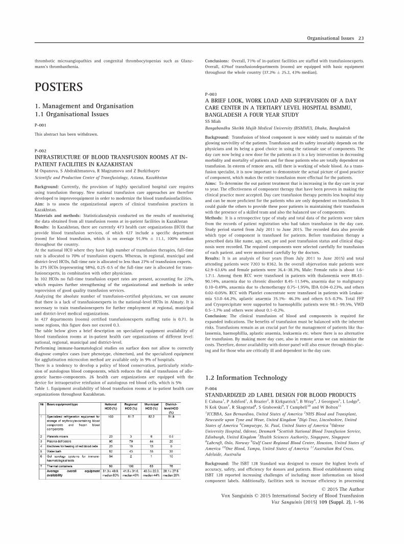
thrombotic microangiopathies and congenital thrombocytopenias such as Glanz-
mann’s thrombasthenia.
POSTERS
1. Management and Organisation1.1 Organisational Issues
P-001
This abstract has been withdrawn.
P-002
INFRASTRUCTURE OF BLOOD TRANSFUSION ROOMS AT IN-PATIENT FACILITIES IN KAZAKHSTANM Ospanova, S Abdrakhmanova, R Magzumova and Z Burkitbayev
Scientific and Production Center of Transfusiology, Astana, Kazakhstan
Background: Currently, the provision of highly specialized hospital care requires
using transfusion therapy. New national transfusion care approaches are therefore
developed to improveequipment in order to modernize the blood transfusionfacilities.
Aim: is to assess the organizational aspects of clinical transfusion practices in
Kazakhstan.
Materials and methods: Statisticalanalysis conducted on the results of monitoring
the data obtained from all transfusion rooms at in-patient facilities in Kazakhstan
Results: In Kazakhstan, there are currently 473 health care organizations (HCO) that
provide blood transfusion services, of which 427 include a specific department
(room) for blood transfusion, which is on average 91.9% � 11.1, 100% median
throughout the country.
At the national HCO where they have high number of transfusion therapies, full-time
rate is allocated to 70% of transfusion experts. Whereas, in regional, municipal and
district-level HCOs, full-time rate is allocated to less than 27% of transfusion experts.
In 275 HCOs (representing 58%), 0.25–0.5 of the full-time rate is allocated for trans-
fusionexperts, in combination with other physicians.
In 102 HCOs no full-time transfusion expert rates are present, accounting for 22%,
which requires further strengthening of the organizational and methods in order
toprovision of good quality transfusion services.
Analyzing the absolute number of transfusion-certified physicians, we can assume
that there is a lack of transfusionexperts in the national-level HCOs in Almaty. It is
necessary to train transfusionexperts for further employment at regional, municipal
and district-level medical organizations.
In 427 departments (rooms) certified transfusionexperts staffing ratio is 0.71. In
some regions, this figure does not exceed 0.3.
The table below gives a brief description on specialized equipment availability of
blood transfusion rooms at in-patient health care organizations of different level:
national, regional, municipal and district-level.
Performing immune-haematological studies on surface does not allow to correctly
diagnose complex cases (rare phenotype, chimerism), and the specialized equipment
for agglutination microcolon method are available only in 9% of hospitals.
There is a tendency to develop a policy of blood conservation, particularly reinfu-
sion of autologous blood components, which reduces the risk of transfusion of allo-
geneic haemo-components. 26 health care organizations are equipped with the
device for intraoperative reinfusion of autologous red blood cells, which is 5%
Table 1. Equipment availability of blood transfusion rooms at in-patient health care
organizations throughout Kazakhstan.
Conclusions: Overall, 71% of in-patient facilities are staffed with transfusionexperts.
Overall, 43%of transfusiondepartments (rooms) are equipped with basic equipment
throughout the whole country (37.2% � 25.2, 43% median).
P-003
A BRIEF LOOK, WORK LOAD AND SUPERVISION OF A DAYCARE CENTER IN A TERTIARY LEVEL HOSPITAL BSMMU,BANGLADESH A FOUR YEAR STUDYSS Miah
Bangabandhu Sheikh Mujib Medical University (BSMMU), Dhaka, Bangladesh
Background: Transfusion of blood component is now widely used to maintain of the
glowing survivility of the patients. Transfusion and its safety invariably depends on the
physicians and its being a good choice in using the rationale use of components. The
day care now being a new door for the patients as it is a key intervention in decreasing
morbidity and mortality of patients and for those patients who are totally dependent on
transfusion. In esteem of remote area, still there is working of whole blood. As a trans-
fusion specialist, it is now important to demonstrate the actual picture of good practice
of component, which makes the entire transfusion more effectual for the patients.
Aims: To determine the out patient treatment that is increasing in the day care in year
to year. The effectiveness of component therapy that have been proven in making the
clinical practice more accepted. Day care transfusion therapy permits less hospital stay
and can be more proficient for the patients who are only dependent on transfusion. It
could guide the others to provide these poor patients in maintaining their transfusion
with the presence of a skilled team and also the balanced use of components.
Methods: It is a retrospective type of study and total data of the patients were taken
from the records of patient registration who had taken transfusion in the day care.
Study period started from July 2011 to June 2015. The recorded data also provide
which type of component is transfused for patients. Before transfusion therapy a
prescribed data like name, age, sex, pre and post transfusion status and clinical diag-
nosis were recorded. The required components were selected carefully for transfusion
of each patient. and were monitored carefully by the doctors.
Results: It is an analysis of four years (from July 2011 to June 2015) and total
attending patients were 7203 to 8362. In the overall objervation male patients were
62.9–63.6% and female patients were 36.4–38.3%, Male: Female ratio is about 1.6–1.7:1. Among them RCC were transfused in patients with thalassemia were 88.43–90.14%, anaemia due to chronic disorder 8.45–11.54%, anaemia due to malignancy
0.10–0.49%, anaemia due to chemotherapy 0.75–1.95%, IDA 0.04–0.23%, and others
0.02–0.05%. RCC with Platelet concentrate were transfused in patients with Leukae-
mia 53.0–64.2%, aplastic anaemia 35.1%- 46.3% and others 0.5–0.7%. Total FFP
and Cryoprecipitate were supported to haemophillic patients were 98.1–99.5%, VWD
0.5–1.7% and others were about 0.1–0.2%.
Conclusion: The clinical transfusion of blood and components is required for
expanded indications. The benefits of transfusion must be balanced with the inherent
risks. Transfusions remain as an crucial part for the management of patients like tha-
lassemia, haemophillia, aplastic anaemia, leukaemia etc. where there is no alternative
for transfusion. By making more day care, also in remote areas we can minimize the
costs. Therefore, donor availability with donor panel will also ensure through this plac-
ing and for those who are critically ill and dependent in the day care.
1.2 Information Technology
P-004
STANDARDIZED 2D LABEL DESIGN FOR BLOOD PRODUCTSE Cabana1, P Ashford1, A Brazier2, B Kirkpatrick3, B Wray4, J Georgsen5, L Lodge6,
N Kok Quan7, R Skagestad8, S Grabowski9, T Campbell10 and W Bolton11
1ICCBBA, San Bernardino, United States of America 2NHS Blood and Transplant,
Newcastle upon Tyne and Wear, United Kingdom 3Digi-Trax, Lincolnshire, United
States of America 4Computype, St. Paul, United States of America 5Odense
University Hospital, Odense, Denmark 6Scottish National Blood Transfusion Service,
Edinburgh, United Kingdom 7Health Sciences Authority, Singapore, Singapore8Labcraft, Oslo, Norway 9Gulf Coast Regional Blood Center, Houston, United States of
America 10One Blood, Tampa, United States of America 11Australian Red Cross,
Adelaide, Australia
Background: The ISBT 128 Standard was designed to ensure the highest levels of
accuracy, safety, and efficiency for donors and patients. Blood establishments using
ISBT 128 reported increasing challenges of including more information on blood
component labels. Additionally, facilities seek to increase efficiency in processing
© 2015 The AuthorVox Sanguinis © 2015 International Society of Blood Transfusion
Vox Sanguinis (2015) 109 (Suppl. 2), 1–96
Organisational Issues 23
hundreds of thousands of units each year without compromising safety. The current
international ISBT 128 blood label was designed over 15 years ago and has four,
sometimes five, standardized linear barcodes. It was proposed that a new label
design, using a single 2D barcode (i.e., Data Matrix), be developed to satisfy these
demands.
Aims: Moving towards a single 2D barcode on the label will:
1. Increase efficiency by reducing the number of barcode scans;
2. Allow for better organization of information;
3. Decrease training and confusion on which barcode to scan.
Methods: An international committee was formed by ICCBBA with experts from
various regions.
They began with published survey results (Transfusion Med. 2014 Apr;24 (2): 89–98)from the UK which determined hospital views on the optimal placement of critical
information on a label. Two of the authors participated on this committee. Then
label information was categorized as either necessary or unnecessary. The Committee
then surveyed blood facilities and hospitals in various regions to identify elements
of the label that could be internationally standardized.
Different scanning mechanisms (e.g., hand held scanners and flatbed scanners) were
taken into consideration when determining the best placement of the 2D barcode.
Several draft labels were distributed for comment and a best-fit design was created
based on feedback.
Results: A new label design was created. Information that is important to the end-
user is in the upper half of the label. This includes the Donation Identification Num-
ber, Product Code, product name, ABO/RhD, expiry date, volume, and the Data
Matrix symbol.
The lower right of the label includes test results and the processing facility (if differ-
ent than the collection facility).
The lower left of the label contains the collection date, storage conditions, regulatory
text, static information, and collection facility. Component specifications may be
provided via a second 2D barcode (using QR symbology) that takes the user to a
website that displays this information.
An ISBT 128 compound message will be used to encode the information into the
Data Matrix symbol. Minimally it will include the Donation Identification Number,
Product Code, ABO/RhD, and expiry date. Additional information may optionally be
encoded.
Summary/Conclusions: The ISBT 128 standardized blood label will move towards
the use of a single Data Matrix barcode and eventually eliminate linear barcodes on
the label. This will improve efficiency at the blood center and hospital by requiring
a single scan to capture all the information currently encoded within 4 or 5 linear
barcodes. It will group key information for the transfusionist in the upper half of the
label, while static information will appear in the lower half of the label. A transition
period of ten years is anticipated. Facility software must be updated before the new
label can be implemented. Design feedback can be submitted to ICCBBA.
P-005
SOLVENT DETERGENT PLASMA STANDARDIZED LABELE Cabana1, P Ashford1, A Brazier2, S Greenwell3, T Laakso4, W LeFresne5, B Quirke6,
M Rechsteiner7, A Riezebos8 and R Skagestad9
1ICCBBA, San Bernardino, United States of America 2NHS Blood and Transplant,
Newcastle upon Tyne and Wear, United Kingdom 3American Red Cross, Washington
DC, United States of America 4Finnish Red Cross Blood Service, Helsinki, Finland5Canadian Blood Services, Nova Scotia, Canada 6Irish Blood Transfusion Service,
Dublin, Ireland 7Octapharma AG, Lachen, Switzerland 8Sanquin, Amsterdam, The
Netherlands 9Labcraft, Oslo, Norway
Background: Unlike most products that are made from plasma from a large number of
donors, ABO blood group is relevant to solvent detergent-treated plasma (SDP). There-
fore, the blood group must be on the affixed label, preferably in both eye- and electron-
ically-readable format. The relevancy of blood group, and the fact the product is
handled in a similar way to Fresh Frozen Plasma, mean it is typically managed by blood
banks rather than pharmacies. Because blood banks use ISBT 128, and because ISBT
128 supports the encoding of blood groups and other key information, both ICCBBA
and GS1 recommend labeling SDP using the ISBT 128 standard on the affixed label.
Aims: The aim of the work was to design a globally-standardized affixed label with
key information presented in both eye- and electronically-readable format. Such a
label would improve patient safety, as well as improve the accuracy and efficiency
of recording information required for traceability.
Methods: ICCBBA, the organisation that manages the ISBT 128 Standard, established
a team of experts to design an internationally-standardized affixed label for SDP. The
team, which met by conference call, comprised professionals responsible for managing
SDP in blood centres and hospitals, ICCBBA staff, and a representative of Octapharma,
a major manufacturer of SDP. A proposed label design was released for public comment
and the comments received were incorporated into a final label design.
Results: The standardized affixed label includes critical information in the upper
half of the label encoded into linear bar codes following the ISBT 128 standard. Cor-
responding text information is placed near each bar code. This critical information
includes:
1. Donation Identification Number (DIN) to uniquely identify each plasma product
2. ABO blood group
3. Product code
4. Expiration date
The relative position of the bar codes is the same as on an ISBT 128 blood label.
DIN and blood group bar codes are at the top of the label placed in a way to support
concatenation, while product code and expiration date bar codes are beneath the
DIN and blood group, also placed to support concatenation.
Regulatory and other country-specific information is just beneath the horizontal
centre of the label and may be bar coded. Manufacturer information, lot number,
and peel-off labels are on the bottom of the label.
Text may be in any language and two languages on a label are possible.
Other electronic information may appear on outer packaging of an SDP unit. Two-
dimensional (2-D) symbols may be used to encode this information.
Summary/Conclusions: An internationally-standardized affixed label for SDP utiliz-
ing ISBT 128 has been designed and approved. ISBT 128 is commonly used in blood
banks around the world and its use on SDP supports automated capture of critical
information by computer systems in these facilities. Accurate entry of key informa-
tion improves patient safety and as well as the accuracy and efficiency of record-
keeping. Organisations are encouraged to request that their supplier of SDP provide
products labelled with ISBT 128.
P-006
METRICS FOR ACCEPTANCE TESTING FOR EGYPTIAN BLOODMANAGEMENT SYSTEM (BMS)A Elgohary, L Borhan and M Labib
National Blood Transfusion Services, Cairo, Egypt
Background: The implementation of the BMS in Egyptian blood transfusion services
is a necessary step towards monitoring, saving time and decreasing human errors.
Acceptance testing (AT) is a vital prerequisite for the implementation of BMS,
needed to fulfill the business rules of any blood transfusion centre.
(AT) is a means to measure quality by challenging the efficiency of the utilized soft-
ware. This can be achieved by choosing reflective in-process metrics which validate
and quantify the ability of the system in place.
Aims: To quantify the accuracy of (AT) performed on the BMS software using in-
process metrics as useful indicators.
To utilize these metrics to highlight problems, encountered in (AT) to ensure the
quality of all BMS application components and their successful module integration
from donation to patient care
Methods: Test data was collected from January to July 2015.
A review of ‘Software requirement specifications’(SRS) was done to:enable the detec-
tion of missing requirements, detect the need for more information, and identify
mistakes in SRS as ‘comments’ which are analyzed in terms of severity.
The ability of the system to meet these comments was evaluated; defects were
recorded and assigned the metric ‘requirement review’.
Test cases (TCs) were developed based on the SRS, subsequently The degree of
requirements coverage by (TCs) was calculated using an equation and named the
metric ‘requirement coverage’
Requirement Coverage = Total no. of requirements supported by TCs/Total no. of
requirements
The third important metric is number of defects vs fixed defects. These defects are
weighted in terms of severity and this evaluation process must be reflected in the
acceptance criteria
Finally TCs were executed, recorded and analyzed, then execution results were cate-
gorized as: pass/ fail. The ‘fail’ status generated a defect record and indicated that
the targeted function hadn’t been achieved. This metric was named ‘Number of
defects/executed TC’.
Result: Regarding the metric ‘Requirements Review’, Our study found 128 missing
requirements, in the SRS which otherwise wouldn’t be included in the final applied
software.
The percentage of requirement coverage found by (TCs) was 81%. However the
remaining 19% lacking coverage need to be addressed by increasing number of TCs
(effectiveness) or enhancing the efficiency of the existing TCs.
© 2015 The AuthorVox Sanguinis © 2015 International Society of Blood TransfusionVox Sanguinis (2015) 109 (Suppl. 2), 1–96
24 Posters

The percentage of fixed defects in relation to total defects was calculated at 88%
(104 fixed /117 Total defects), it is noted that 88% defects fixed, is a positive indica-
tion of software integrity and
Conclusion: These metrics are vital to (AT) in isolating missing requirements, evalu-
ating requirement coverage through test case analysis and achieving quality assur-
ance by assessing number of fixed defects. Our study illustrated how the
development of these metrics was achieved through the customization of already
existing data. Such metrics can be utilized by similar blood banks, in the process of
(AT) provided that they follow the same steps and adapt these metrics to conform to
their own business rules.
In conclusion it’s clear that implementation of these metrics merits serious consider-
ation for efficient running of Blood management projects.
P-007
TESTING SECURITY OF THE BLOOD MANAGEMENT SYSTEM,THROUGH THE USAGE OF NON FUNCTIONAL TESTING, INTHE NATIONAL BLOOD TRANSFUSION CENTRE (NBTC),CAIRO, EGYPTS Abdullah1, A Ahmed2, M Labib2, A Ibrahim1, M Shokri2 and A Abady2
1NBTC, Cairo, Egypt 2National Blood Transfusion Center, Cairo, Egypt
Background: The Blood Management System (BMS) is a project which will auto-
mate manual operations in 17 sites of the Egyptian National Blood Transfusion Ser-
vices (ENBTS). Protecting data is a vital issue to guarantee business continuity.
Security is an essential requirement that is fulfilled by having the suitable hardware
and network architecture. Security functions are granted to each user according to the
degree of their involvement in the blood transfusion system. These user privileges limit
the transfer and sharing of data, and other functionalities amongst users in real time.
Non Functional Security Testing is a method used to measure whether the BMS
application and its infrastructure is secure or not.
Aims: To examine how Non Functional Security Testing can be used as a tool to
measure how far the BMS software, hardware and network are secured.
Methods: The IT team at ENBTS arranged a series of meetings to study the BMS
security requirements, namely ‘Test Basis’ (the network, hardware and application
design) in detail. In order to extract any points of weakness in the design and pro-
vide useful feedback.
A database of Non Functional Security Test cases was generated by using the check
list testing technique, which is a technique that creates a catalog of requirements
that need to be tracked within the BMS, concerning security.
The generated test cases were classified by site (‘headquarters only’ or ‘all sites’) and
by severity, and their impact on the flow of work. The outcome from these test cases
is embedded into the execution plan database.
The execution plan database, challenges the various security steps, detects and also
categorizes defects according to severity. These findings are then relayed and solu-
tions are found, taking security requirements into consideration.
Finally the execution plan in all its revised stages will be run, to achieve user accep-
tance with regards security of the BMS.
Results: The ENBTS IT team created a repository of about 100 test cases extracted
from the security requirements, which highlighted several points:
The importance of firewall and optimal antivirus software installment.
Security lists should be applied on switches.
To protect data, it was found that full identification of each user must be monitored
during login; our system is expected to dictate password changes at a pre-deter-
mined period (30 days).
Locking of the session if users don0t use the system within a set period of time, to
protect confidentiality.
Control of access authorization to: read, update, create and delete data; according to
the user’s personal functionality definition. Especially concerning data defined by
the blood center’s management as sensitive.
Conclusion: Non functional security testing insures the integrity, and safety of any
system, especially one as the BMS. It predicts problems, protects businesses from
data theft and saves money. Such considerations are crucial in low income settings
and an institution as sensitive as a blood transfusion centre.
Achieving strong security is mandatory to achieve confidentiality, build trust, and
strengthen the donor and patient relationship with the ENBTS.
P-008
BENEFITS OF IMPLEMENTATION OF BLOOD DONORS DATABASE MANAGEMENT SYSTEM VS MANUAL RECORDINGSYSTEMS IN DAR EL SALAM REGIONAL BLOODTRANSFUSION CENTERA Selim Sayed, R Farouk, R Farouk Badr and S Mohamed
Dar El Salam Regional Blood Transfusion Center, Cairo, Egypt
Background: Blood bank management system is a browser based system that is
designed to store, retrieve and analyze information concerned with all aspects, start-
ing from recruiting blood donors to issuing of blood units. Prerequisites for imple-
mentation of such technology include: adequate infrastructure, quality system,
appropriate staffing, and sufficient funding. Blood donor module plays an important
role to build secured donor database, recruiting regular donors, encouraging recall
for aphaeresis and rare blood group donors, exclude disqualified donors from further
donations and postpone temporary deferred donors.
© 2015 The AuthorVox Sanguinis © 2015 International Society of Blood Transfusion
Vox Sanguinis (2015) 109 (Suppl. 2), 1–96
Information Technology 25
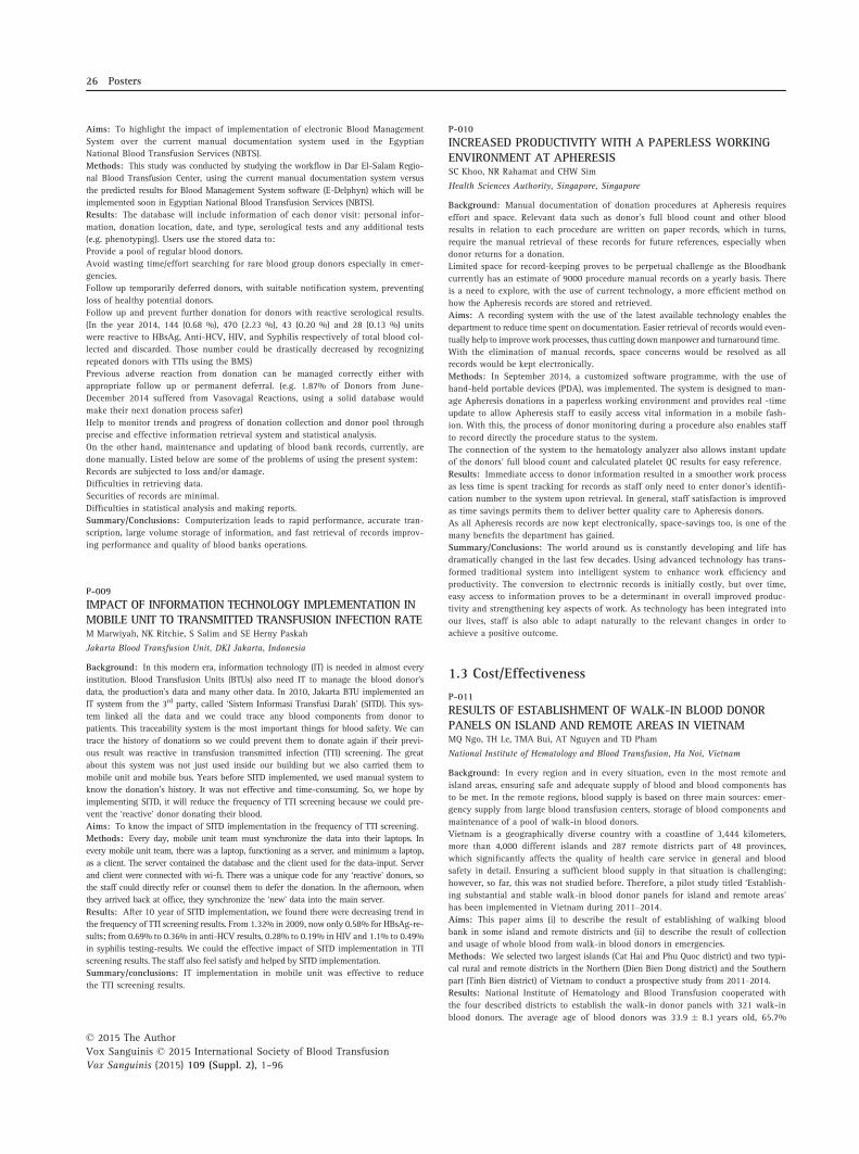
Aims: To highlight the impact of implementation of electronic Blood Management
System over the current manual documentation system used in the Egyptian
National Blood Transfusion Services (NBTS).
Methods: This study was conducted by studying the workflow in Dar El-Salam Regio-
nal Blood Transfusion Center, using the current manual documentation system versus
the predicted results for Blood Management System software (E-Delphyn) which will be
implemented soon in Egyptian National Blood Transfusion Services (NBTS).
Results: The database will include information of each donor visit: personal infor-
mation, donation location, date, and type, serological tests and any additional tests
(e.g. phenotyping). Users use the stored data to:
Provide a pool of regular blood donors.
Avoid wasting time/effort searching for rare blood group donors especially in emer-
gencies.
Follow up temporarily deferred donors, with suitable notification system, preventing
loss of healthy potential donors.
Follow up and prevent further donation for donors with reactive serological results.
(In the year 2014, 144 (0.68 %), 470 (2.23 %), 43 (0.20 %) and 28 (0.13 %) units
were reactive to HBsAg, Anti-HCV, HIV, and Syphilis respectively of total blood col-
lected and discarded. Those number could be drastically decreased by recognizing
repeated donors with TTIs using the BMS)
Previous adverse reaction from donation can be managed correctly either with
appropriate follow up or permanent deferral. (e.g. 1.87% of Donors from June-
December 2014 suffered from Vasovagal Reactions, using a solid database would
make their next donation process safer)
Help to monitor trends and progress of donation collection and donor pool through
precise and effective information retrieval system and statistical analysis.
On the other hand, maintenance and updating of blood bank records, currently, are
done manually. Listed below are some of the problems of using the present system:
Records are subjected to loss and/or damage.
Difficulties in retrieving data.
Securities of records are minimal.
Difficulties in statistical analysis and making reports.
Summary/Conclusions: Computerization leads to rapid performance, accurate tran-
scription, large volume storage of information, and fast retrieval of records improv-
ing performance and quality of blood banks operations.
P-009
IMPACT OF INFORMATION TECHNOLOGY IMPLEMENTATION INMOBILE UNIT TO TRANSMITTED TRANSFUSION INFECTION RATEM Marwiyah, NK Ritchie, S Salim and SE Herny Paskah
Jakarta Blood Transfusion Unit, DKI Jakarta, Indonesia
Background: In this modern era, information technology (IT) is needed in almost every
institution. Blood Transfusion Units (BTUs) also need IT to manage the blood donor’s
data, the production’s data and many other data. In 2010, Jakarta BTU implemented an
IT system from the 3rd party, called ‘Sistem Informasi Transfusi Darah’ (SITD). This sys-
tem linked all the data and we could trace any blood components from donor to
patients. This traceability system is the most important things for blood safety. We can
trace the history of donations so we could prevent them to donate again if their previ-
ous result was reactive in transfusion transmitted infection (TTI) screening. The great
about this system was not just used inside our building but we also carried them to
mobile unit and mobile bus. Years before SITD implemented, we used manual system to
know the donation’s history. It was not effective and time-consuming. So, we hope by
implementing SITD, it will reduce the frequency of TTI screening because we could pre-
vent the ‘reactive’ donor donating their blood.
Aims: To know the impact of SITD implementation in the frequency of TTI screening.
Methods: Every day, mobile unit team must synchronize the data into their laptops. In
every mobile unit team, there was a laptop, functioning as a server, and minimum a laptop,
as a client. The server contained the database and the client used for the data-input. Server
and client were connected with wi-fi. There was a unique code for any ‘reactive’ donors, so
the staff could directly refer or counsel them to defer the donation. In the afternoon, when
they arrived back at office, they synchronize the ‘new’ data into the main server.
Results: After 10 year of SITD implementation, we found there were decreasing trend in
the frequency of TTI screening results. From 1.32% in 2009, now only 0.58% for HBsAg-re-
sults; from 0.69% to 0.36% in anti-HCV results, 0.28% to 0.19% in HIV and 1.1% to 0.49%
in syphilis testing-results. We could the effective impact of SITD implementation in TTI
screening results. The staff also feel satisfy and helped by SITD implementation.
Summary/conclusions: IT implementation in mobile unit was effective to reduce
the TTI screening results.
P-010
INCREASED PRODUCTIVITY WITH A PAPERLESS WORKINGENVIRONMENT AT APHERESISSC Khoo, NR Rahamat and CHW Sim
Health Sciences Authority, Singapore, Singapore
Background: Manual documentation of donation procedures at Apheresis requires
effort and space. Relevant data such as donor’s full blood count and other blood
results in relation to each procedure are written on paper records, which in turns,
require the manual retrieval of these records for future references, especially when
donor returns for a donation.
Limited space for record-keeping proves to be perpetual challenge as the Bloodbank
currently has an estimate of 9000 procedure manual records on a yearly basis. There
is a need to explore, with the use of current technology, a more efficient method on
how the Apheresis records are stored and retrieved.
Aims: A recording system with the use of the latest available technology enables the
department to reduce time spent on documentation. Easier retrieval of records would even-
tually help to improvework processes, thus cutting downmanpower and turnaround time.
With the elimination of manual records, space concerns would be resolved as all
records would be kept electronically.
Methods: In September 2014, a customized software programme, with the use of
hand-held portable devices (PDA), was implemented. The system is designed to man-
age Apheresis donations in a paperless working environment and provides real -time
update to allow Apheresis staff to easily access vital information in a mobile fash-
ion. With this, the process of donor monitoring during a procedure also enables staff
to record directly the procedure status to the system.
The connection of the system to the hematology analyzer also allows instant update
of the donors’ full blood count and calculated platelet QC results for easy reference.
Results: Immediate access to donor information resulted in a smoother work process
as less time is spent tracking for records as staff only need to enter donor’s identifi-
cation number to the system upon retrieval. In general, staff satisfaction is improved
as time savings permits them to deliver better quality care to Apheresis donors.
As all Apheresis records are now kept electronically, space-savings too, is one of the
many benefits the department has gained.
Summary/Conclusions: The world around us is constantly developing and life has
dramatically changed in the last few decades. Using advanced technology has trans-
formed traditional system into intelligent system to enhance work efficiency and
productivity. The conversion to electronic records is initially costly, but over time,
easy access to information proves to be a determinant in overall improved produc-
tivity and strengthening key aspects of work. As technology has been integrated into
our lives, staff is also able to adapt naturally to the relevant changes in order to
achieve a positive outcome.
1.3 Cost/Effectiveness
P-011
RESULTS OF ESTABLISHMENT OF WALK-IN BLOOD DONORPANELS ON ISLAND AND REMOTE AREAS IN VIETNAMMQ Ngo, TH Le, TMA Bui, AT Nguyen and TD Pham
National Institute of Hematology and Blood Transfusion, Ha Noi, Vietnam
Background: In every region and in every situation, even in the most remote and
island areas, ensuring safe and adequate supply of blood and blood components has
to be met. In the remote regions, blood supply is based on three main sources: emer-
gency supply from large blood transfusion centers, storage of blood components and
maintenance of a pool of walk-in blood donors.
Vietnam is a geographically diverse country with a coastline of 3,444 kilometers,
more than 4,000 different islands and 287 remote districts part of 48 provinces,
which significantly affects the quality of health care service in general and blood
safety in detail. Ensuring a sufficient blood supply in that situation is challenging;
however, so far, this was not studied before. Therefore, a pilot study titled ‘Establish-
ing substantial and stable walk-in blood donor panels for island and remote areas’
has been implemented in Vietnam during 2011–2014.Aims: This paper aims (i) to describe the result of establishing of walking blood
bank in some island and remote districts and (ii) to describe the result of collection
and usage of whole blood from walk-in blood donors in emergencies.
Methods: We selected two largest islands (Cat Hai and Phu Quoc district) and two typi-
cal rural and remote districts in the Northern (Dien Bien Dong district) and the Southern
part (Tinh Bien district) of Vietnam to conduct a prospective study from 2011–2014.Results: National Institute of Hematology and Blood Transfusion cooperated with
the four described districts to establish the walk-in donor panels with 321 walk-in
blood donors. The average age of blood donors was 33.9 � 8.1 years old, 65.7%
© 2015 The AuthorVox Sanguinis © 2015 International Society of Blood TransfusionVox Sanguinis (2015) 109 (Suppl. 2), 1–96
26 Posters
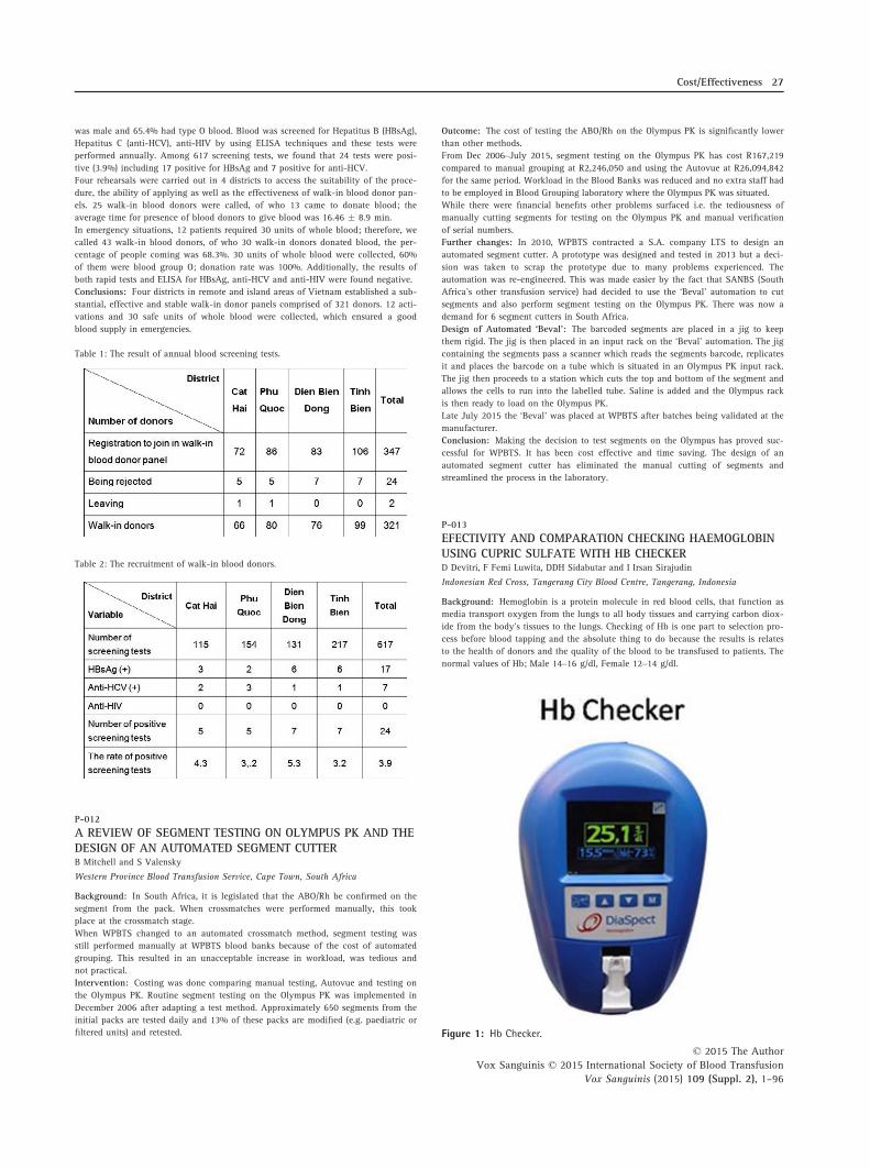
was male and 65.4% had type O blood. Blood was screened for Hepatitus B (HBsAg),
Hepatitus C (anti-HCV), anti-HIV by using ELISA techniques and these tests were
performed annually. Among 617 screening tests, we found that 24 tests were posi-
tive (3.9%) including 17 positive for HBsAg and 7 positive for anti-HCV.
Four rehearsals were carried out in 4 districts to access the suitability of the proce-
dure, the ability of applying as well as the effectiveness of walk-in blood donor pan-
els. 25 walk-in blood donors were called, of who 13 came to donate blood; the
average time for presence of blood donors to give blood was 16.46 � 8.9 min.
In emergency situations, 12 patients required 30 units of whole blood; therefore, we
called 43 walk-in blood donors, of who 30 walk-in donors donated blood, the per-
centage of people coming was 68.3%. 30 units of whole blood were collected, 60%
of them were blood group O; donation rate was 100%. Additionally, the results of
both rapid tests and ELISA for HBsAg, anti-HCV and anti-HIV were found negative.
Conclusions: Four districts in remote and island areas of Vietnam established a sub-
stantial, effective and stable walk-in donor panels comprised of 321 donors. 12 acti-
vations and 30 safe units of whole blood were collected, which ensured a good
blood supply in emergencies.
Table 1: The result of annual blood screening tests.
Table 2: The recruitment of walk-in blood donors.
P-012
A REVIEW OF SEGMENT TESTING ON OLYMPUS PK AND THEDESIGN OF AN AUTOMATED SEGMENT CUTTERB Mitchell and S Valensky
Western Province Blood Transfusion Service, Cape Town, South Africa
Background: In South Africa, it is legislated that the ABO/Rh be confirmed on the
segment from the pack. When crossmatches were performed manually, this took
place at the crossmatch stage.
When WPBTS changed to an automated crossmatch method, segment testing was
still performed manually at WPBTS blood banks because of the cost of automated
grouping. This resulted in an unacceptable increase in workload, was tedious and
not practical.
Intervention: Costing was done comparing manual testing, Autovue and testing on
the Olympus PK. Routine segment testing on the Olympus PK was implemented in
December 2006 after adapting a test method. Approximately 650 segments from the
initial packs are tested daily and 13% of these packs are modified (e.g. paediatric or
filtered units) and retested.
Outcome: The cost of testing the ABO/Rh on the Olympus PK is significantly lower
than other methods.
From Dec 2006–July 2015, segment testing on the Olympus PK has cost R167,219
compared to manual grouping at R2,246,050 and using the Autovue at R26,094,842
for the same period. Workload in the Blood Banks was reduced and no extra staff had
to be employed in Blood Grouping laboratory where the Olympus PK was situated.
While there were financial benefits other problems surfaced i.e. the tediousness of
manually cutting segments for testing on the Olympus PK and manual verification
of serial numbers.
Further changes: In 2010, WPBTS contracted a S.A. company LTS to design an
automated segment cutter. A prototype was designed and tested in 2013 but a deci-
sion was taken to scrap the prototype due to many problems experienced. The
automation was re-engineered. This was made easier by the fact that SANBS (South
Africa’s other transfusion service) had decided to use the ‘Beval’ automation to cut
segments and also perform segment testing on the Olympus PK. There was now a
demand for 6 segment cutters in South Africa.
Design of Automated ‘Beval’: The barcoded segments are placed in a jig to keep
them rigid. The jig is then placed in an input rack on the ‘Beval’ automation. The jig
containing the segments pass a scanner which reads the segments barcode, replicates
it and places the barcode on a tube which is situated in an Olympus PK input rack.
The jig then proceeds to a station which cuts the top and bottom of the segment and
allows the cells to run into the labelled tube. Saline is added and the Olympus rack
is then ready to load on the Olympus PK.
Late July 2015 the ‘Beval’ was placed at WPBTS after batches being validated at the
manufacturer.
Conclusion: Making the decision to test segments on the Olympus has proved suc-
cessful for WPBTS. It has been cost effective and time saving. The design of an
automated segment cutter has eliminated the manual cutting of segments and
streamlined the process in the laboratory.
P-013
EFECTIVITY AND COMPARATION CHECKING HAEMOGLOBINUSING CUPRIC SULFATE WITH HB CHECKERD Devitri, F Femi Luwita, DDH Sidabutar and I Irsan Sirajudin
Indonesian Red Cross, Tangerang City Blood Centre, Tangerang, Indonesia
Background: Hemoglobin is a protein molecule in red blood cells, that function as
media transport oxygen from the lungs to all body tissues and carrying carbon diox-
ide from the body’s tissues to the lungs. Checking of Hb is one part to selection pro-
cess before blood tapping and the absolute thing to do because the results is relates
to the health of donors and the quality of the blood to be transfused to patients. The
normal values of Hb; Male 14–16 g/dl, Female 12–14 g/dl.
Figure 1: Hb Checker.
© 2015 The AuthorVox Sanguinis © 2015 International Society of Blood Transfusion
Vox Sanguinis (2015) 109 (Suppl. 2), 1–96
Cost/Effectiveness 27
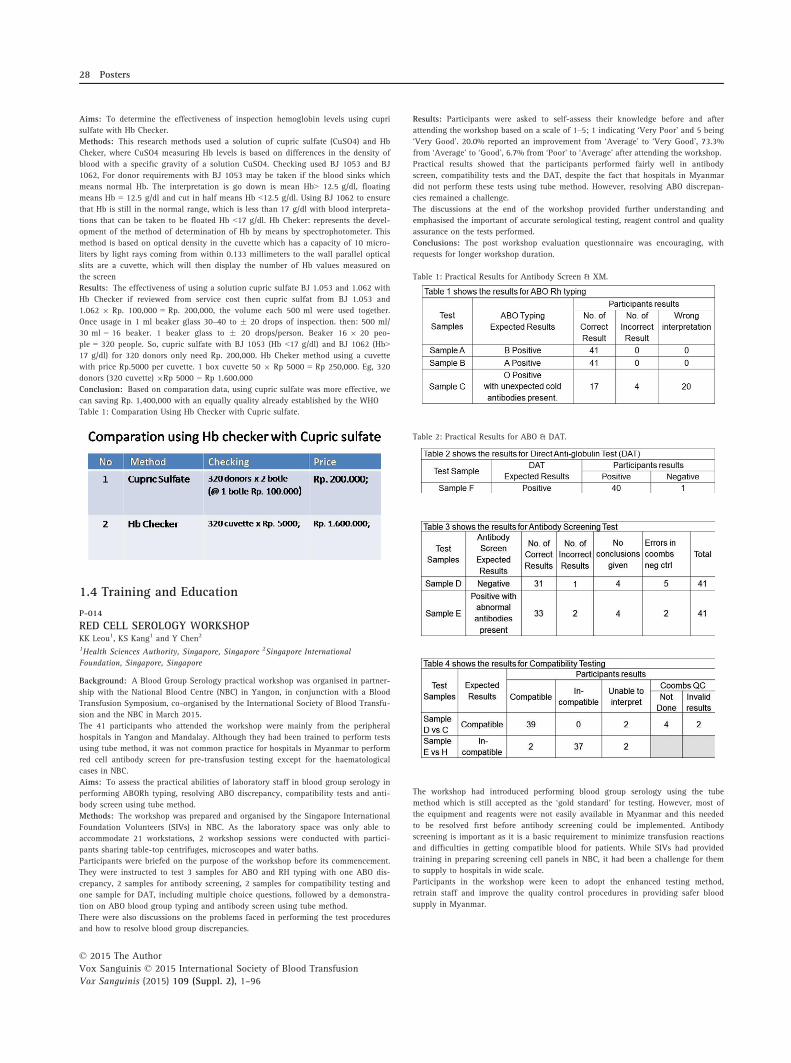
Aims: To determine the effectiveness of inspection hemoglobin levels using cupri
sulfate with Hb Checker.
Methods: This research methods used a solution of cupric sulfate (CuSO4) and Hb
Cheker, where CuSO4 measuring Hb levels is based on differences in the density of
blood with a specific gravity of a solution CuSO4. Checking used BJ 1053 and BJ
1062, For donor requirements with BJ 1053 may be taken if the blood sinks which
means normal Hb. The interpretation is go down is mean Hb> 12.5 g/dl, floating
means Hb = 12.5 g/dl and cut in half means Hb <12.5 g/dl. Using BJ 1062 to ensure
that Hb is still in the normal range, which is less than 17 g/dl with blood interpreta-
tions that can be taken to be floated Hb <17 g/dl. Hb Cheker: represents the devel-
opment of the method of determination of Hb by means by spectrophotometer. This
method is based on optical density in the cuvette which has a capacity of 10 micro-
liters by light rays coming from within 0.133 millimeters to the wall parallel optical
slits are a cuvette, which will then display the number of Hb values measured on
the screen
Results: The effectiveness of using a solution cupric sulfate BJ 1.053 and 1.062 with
Hb Checker if reviewed from service cost then cupric sulfat from BJ 1.053 and
1.062 9 Rp. 100,000 = Rp. 200,000, the volume each 500 ml were used together.
Once usage in 1 ml beaker glass 30–40 to � 20 drops of inspection. then: 500 ml/
30 ml = 16 beaker. 1 beaker glass to � 20 drops/person. Beaker 16 9 20 peo-
ple = 320 people. So, cupric sulfate with BJ 1053 (Hb <17 g/dl) and BJ 1062 (Hb>17 g/dl) for 320 donors only need Rp. 200,000. Hb Cheker method using a cuvette
with price Rp.5000 per cuvette. 1 box cuvette 50 9 Rp 5000 = Rp 250,000. Eg, 320
donors (320 cuvette) 9Rp 5000 = Rp 1.600.000
Conclusion: Based on comparation data, using cupric sulfate was more effective, we
can saving Rp. 1,400,000 with an equally quality already established by the WHO
Table 1: Comparation Using Hb Checker with Cupric sulfate.
1.4 Training and Education
P-014
RED CELL SEROLOGY WORKSHOPKK Leou1, KS Kang1 and Y Chen2
1Health Sciences Authority, Singapore, Singapore 2Singapore International
Foundation, Singapore, Singapore
Background: A Blood Group Serology practical workshop was organised in partner-
ship with the National Blood Centre (NBC) in Yangon, in conjunction with a Blood
Transfusion Symposium, co-organised by the International Society of Blood Transfu-
sion and the NBC in March 2015.
The 41 participants who attended the workshop were mainly from the peripheral
hospitals in Yangon and Mandalay. Although they had been trained to perform tests
using tube method, it was not common practice for hospitals in Myanmar to perform
red cell antibody screen for pre-transfusion testing except for the haematological
cases in NBC.
Aims: To assess the practical abilities of laboratory staff in blood group serology in
performing ABORh typing, resolving ABO discrepancy, compatibility tests and anti-
body screen using tube method.
Methods: The workshop was prepared and organised by the Singapore International
Foundation Volunteers (SIVs) in NBC. As the laboratory space was only able to
accommodate 21 workstations, 2 workshop sessions were conducted with partici-
pants sharing table-top centrifuges, microscopes and water baths.
Participants were briefed on the purpose of the workshop before its commencement.
They were instructed to test 3 samples for ABO and RH typing with one ABO dis-
crepancy, 2 samples for antibody screening, 2 samples for compatibility testing and
one sample for DAT, including multiple choice questions, followed by a demonstra-
tion on ABO blood group typing and antibody screen using tube method.
There were also discussions on the problems faced in performing the test procedures
and how to resolve blood group discrepancies.
Results: Participants were asked to self-assess their knowledge before and after
attending the workshop based on a scale of 1–5; 1 indicating ‘Very Poor’ and 5 being
‘Very Good’. 20.0% reported an improvement from ‘Average’ to ‘Very Good’, 73.3%
from ‘Average’ to ‘Good’, 6.7% from ‘Poor’ to ‘Average’ after attending the workshop.
Practical results showed that the participants performed fairly well in antibody
screen, compatibility tests and the DAT, despite the fact that hospitals in Myanmar
did not perform these tests using tube method. However, resolving ABO discrepan-
cies remained a challenge.
The discussions at the end of the workshop provided further understanding and
emphasised the important of accurate serological testing, reagent control and quality
assurance on the tests performed.
Conclusions: The post workshop evaluation questionnaire was encouraging, with
requests for longer workshop duration.
Table 1: Practical Results for Antibody Screen & XM.
Table 2: Practical Results for ABO & DAT.
The workshop had introduced performing blood group serology using the tube
method which is still accepted as the ‘gold standard’ for testing. However, most of
the equipment and reagents were not easily available in Myanmar and this needed
to be resolved first before antibody screening could be implemented. Antibody
screening is important as it is a basic requirement to minimize transfusion reactions
and difficulties in getting compatible blood for patients. While SIVs had provided
training in preparing screening cell panels in NBC, it had been a challenge for them
to supply to hospitals in wide scale.
Participants in the workshop were keen to adopt the enhanced testing method,
retrain staff and improve the quality control procedures in providing safer blood
supply in Myanmar.
© 2015 The AuthorVox Sanguinis © 2015 International Society of Blood TransfusionVox Sanguinis (2015) 109 (Suppl. 2), 1–96
28 Posters
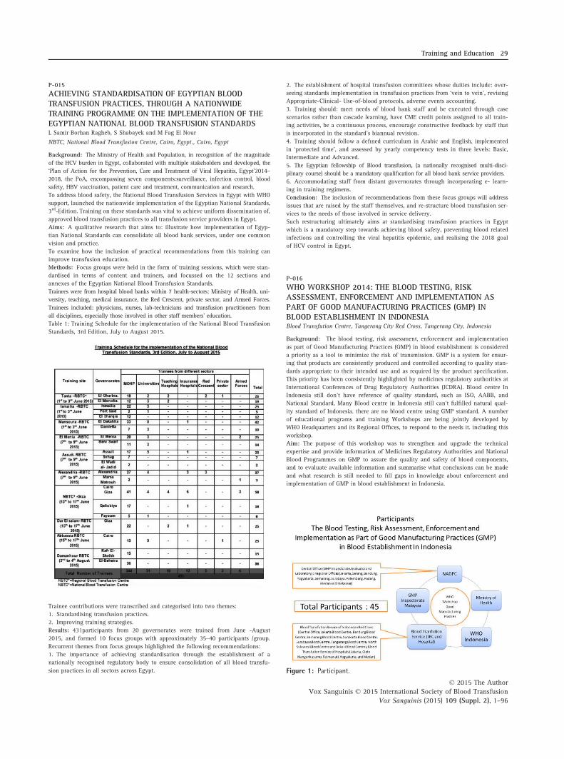
P-015
ACHIEVING STANDARDISATION OF EGYPTIAN BLOODTRANSFUSION PRACTICES, THROUGH A NATIONWIDETRAINING PROGRAMME ON THE IMPLEMENTATION OF THEEGYPTIAN NATIONAL BLOOD TRANSFUSION STANDARDSL Samir Borhan Ragheb, S Shabayek and M Fag El Nour
NBTC, National Blood Transfusion Centre, Cairo, Egypt., Cairo, Egypt
Background: The Ministry of Health and Population, in recognition of the magnitude
of the HCV burden in Egypt, collaborated with multiple stakeholders and developed, the
‘Plan of Action for the Prevention, Care and Treatment of Viral Hepatitis, Egypt’2014–2018, the PoA, encompassing seven components:surveillance, infection control, blood
safety, HBV vaccination, patient care and treatment, communication and research.
To address blood safety, the National Blood Transfusion Services in Egypt with WHO
support, launched the nationwide implementation of the Egyptian National Standards,
3rd-Edition. Training on these standards was vital to achieve uniform dissemination of,
approved blood transfusion practices to all transfusion service providers in Egypt.
Aims: A qualitative research that aims to: illustrate how implementation of Egyp-
tian National Standards can consolidate all blood bank services, under one common
vision and practice.
To examine how the inclusion of practical recommendations from this training can
improve transfusion education.
Methods: Focus groups were held in the form of training sessions, which were stan-
dardised in terms of content and trainers, and focussed on the 12 sections and
annexes of the Egyptian National Blood Transfusion Standards.
Trainees were from hospital blood banks within 7 health-sectors: Ministry of Health, uni-
versity, teaching, medical insurance, the Red Crescent, private sector, and Armed Forces.
Trainees included: physicians, nurses, lab-technicians and transfusion practitioners from
all disciplines, especially those involved in other staff members’ education.
Table 1: Training Schedule for the implementation of the National Blood Transfusion
Standards, 3rd Edition, July to August 2015.
Trainee contributions were transcribed and categorised into two themes:
1. Standardising transfusion practices.
2. Improving training strategies.
Results: 431participants from 20 governorates were trained from June -August
2015, and formed 10 focus groups with approximately 35–40 participants /group.
Recurrent themes from focus groups highlighted the following recommendations:
1. The importance of achieving standardisation through the establishment of a
nationally recognised regulatory body to ensure consolidation of all blood transfu-
sion practices in all sectors across Egypt.
2. The establishment of hospital transfusion committees whose duities include: over-
seeing standards implementation in transfusion practices from ‘vein to vein’, revising
Appropriate-Clinical- Use-of-blood protocols, adverse events accounting.
3. Training should: meet needs of blood bank staff and be executed through case
scenarios rather than cascade learning, have CME credit points assigned to all train-
ing activities, be a continuous process, encourage constructive feedback by staff that
is incorporated in the standard’s biannual revision.
4. Training should follow a defined curriculum in Arabic and English, implemented
in ‘protected time’, and assessed by yearly competency tests in three levels: Basic,
Intermediate and Advanced.
5. The Egyptian fellowship of Blood transfusion, (a nationally recognised multi-disci-
plinary course) should be a mandatory qualification for all blood bank service providers.
6. Accommodating staff from distant governorates through incorporating e- learn-
ing in training regimens.
Conclusion: The inclusion of recommendations from these focus groups will address
issues that are raised by the staff themselves, and re-structure blood transfusion ser-
vices to the needs of those involved in service delivery.
Such restructuring ultimately aims at standardising transfusion practices in Egypt
which is a mandatory step towards achieving blood safety, preventing blood related
infections and controlling the viral hepatitis epidemic, and realising the 2018 goal
of HCV control in Egypt.
P-016
WHO WORKSHOP 2014: THE BLOOD TESTING, RISKASSESSMENT, ENFORCEMENT AND IMPLEMENTATION ASPART OF GOOD MANUFACTURING PRACTICES (GMP) INBLOOD ESTABLISHMENT IN INDONESIABlood Transfution Centre, Tangerang City Red Cross, Tangerang City, Indonesia
Background: The blood testing, risk assessment, enforcement and implementation
as part of Good Manufacturing Practices (GMP) in blood establishment is considered
a priority as a tool to minimize the risk of transmission. GMP is a system for ensur-
ing that products are consistently produced and controlled according to quality stan-
dards appropriate to their intended use and as required by the product specification.
This priority has been consistently highlighted by medicines regulatory authorities at
International Conferences of Drug Regulatory Authorities (ICDRA). Blood centre In
Indonesia still don’t have reference of quality standard, such as ISO, AABB, and
National Standard, Many Blood centre in Indonesia still can’t fulfilled natural qual-
ity standard of Indonesia. there are no blood centre using GMP standard. A number
of educational programs and training Workshops are being jointly developed by
WHO Headquarters and its Regional Offices, to respond to the needs it. including this
workshop.
Aim: The purpose of this workshop was to strengthen and upgrade the technical
expertise and provide information of Medicines Regulatory Authorities and National
Blood Programmes on GMP to assure the quality and safety of blood components,
and to evaluate available information and summarise what conclusions can be made
and what research is still needed to fill gaps in knowledge about enforcement and
implementation of GMP in blood establishment in Indonesia.
Figure 1: Participant.
© 2015 The AuthorVox Sanguinis © 2015 International Society of Blood Transfusion
Vox Sanguinis (2015) 109 (Suppl. 2), 1–96
Training and Education 29
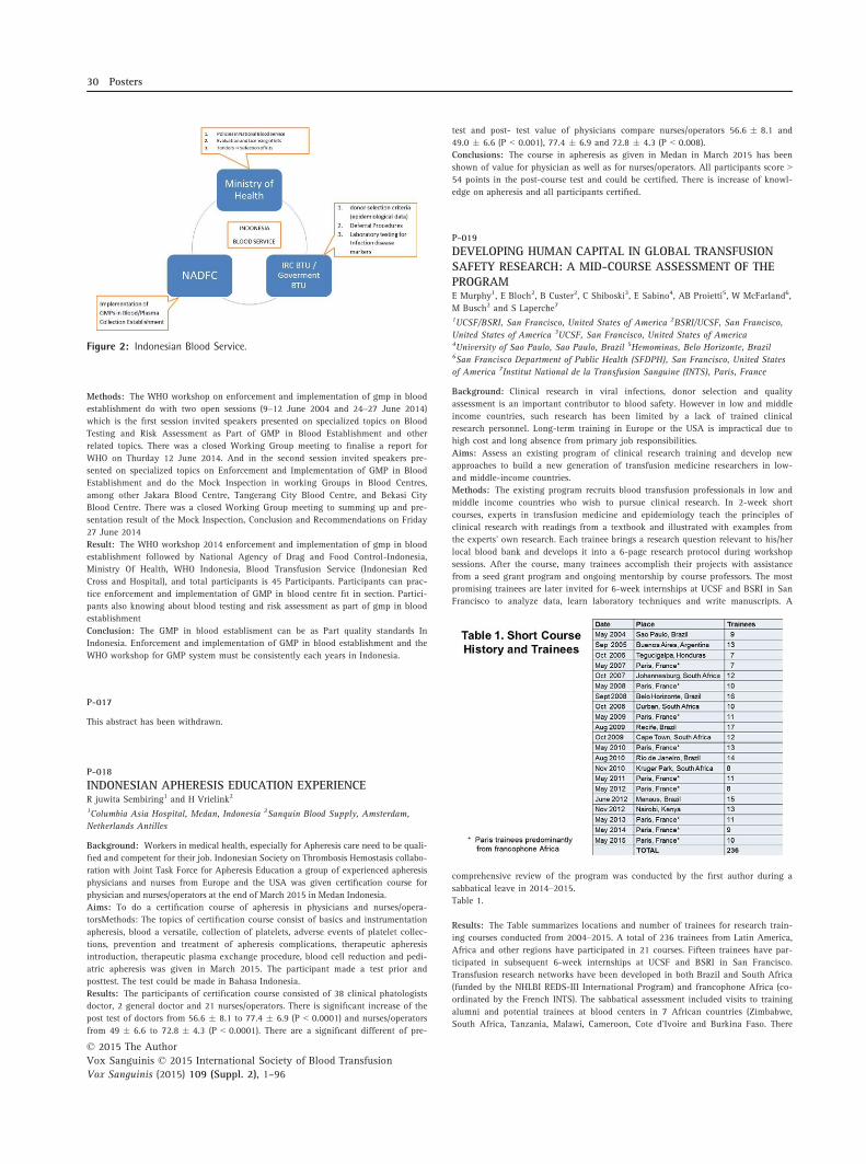
Methods: The WHO workshop on enforcement and implementation of gmp in blood
establishment do with two open sessions (9–12 June 2004 and 24–27 June 2014)
which is the first session invited speakers presented on specialized topics on Blood
Testing and Risk Assessment as Part of GMP in Blood Establishment and other
related topics. There was a closed Working Group meeting to finalise a report for
WHO on Thurday 12 June 2014. And in the second session invited speakers pre-
sented on specialized topics on Enforcement and Implementation of GMP in Blood
Establishment and do the Mock Inspection in working Groups in Blood Centres,
among other Jakara Blood Centre, Tangerang City Blood Centre, and Bekasi City
Blood Centre. There was a closed Working Group meeting to summing up and pre-
sentation result of the Mock Inspection, Conclusion and Recommendations on Friday
27 June 2014
Result: The WHO workshop 2014 enforcement and implementation of gmp in blood
establishment followed by National Agency of Drag and Food Control-Indonesia,
Ministry Of Health, WHO Indonesia, Blood Transfusion Service (Indonesian Red
Cross and Hospital), and total participants is 45 Participants. Participants can prac-
tice enforcement and implementation of GMP in blood centre fit in section. Partici-
pants also knowing about blood testing and risk assessment as part of gmp in blood
establishment
Conclusion: The GMP in blood establisment can be as Part quality standards In
Indonesia. Enforcement and implementation of GMP in blood establishment and the
WHO workshop for GMP system must be consistently each years in Indonesia.
P-017
This abstract has been withdrawn.
P-018
INDONESIAN APHERESIS EDUCATION EXPERIENCER juwita Sembiring1 and H Vrielink2
1Columbia Asia Hospital, Medan, Indonesia 2Sanquin Blood Supply, Amsterdam,
Netherlands Antilles
Background: Workers in medical health, especially for Apheresis care need to be quali-
fied and competent for their job. Indonesian Society on Thrombosis Hemostasis collabo-
ration with Joint Task Force for Apheresis Education a group of experienced apheresis
physicians and nurses from Europe and the USA was given certification course for
physician and nurses/operators at the end of March 2015 in Medan Indonesia.
Aims: To do a certification course of apheresis in physicians and nurses/opera-
torsMethods: The topics of certification course consist of basics and instrumentation
apheresis, blood a versatile, collection of platelets, adverse events of platelet collec-
tions, prevention and treatment of apheresis complications, therapeutic apheresis
introduction, therapeutic plasma exchange procedure, blood cell reduction and pedi-
atric apheresis was given in March 2015. The participant made a test prior and
posttest. The test could be made in Bahasa Indonesia.
Results: The participants of certification course consisted of 38 clinical phatologists
doctor, 2 general doctor and 21 nurses/operators. There is significant increase of the
post test of doctors from 56.6 � 8.1 to 77.4 � 6.9 (P < 0.0001) and nurses/operators
from 49 � 6.6 to 72.8 � 4.3 (P < 0.0001). There are a significant different of pre-
test and post- test value of physicians compare nurses/operators 56.6 � 8.1 and
49.0 � 6.6 (P < 0.001), 77.4 � 6.9 and 72.8 � 4.3 (P < 0.008).
Conclusions: The course in apheresis as given in Medan in March 2015 has been
shown of value for physician as well as for nurses/operators. All participants score >54 points in the post-course test and could be certified. There is increase of knowl-
edge on apheresis and all participants certified.
P-019
DEVELOPING HUMAN CAPITAL IN GLOBAL TRANSFUSIONSAFETY RESEARCH: A MID-COURSE ASSESSMENT OF THEPROGRAME Murphy1, E Bloch2, B Custer2, C Shiboski3, E Sabino4, AB Proietti5, W McFarland6,
M Busch2 and S Laperche7
1UCSF/BSRI, San Francisco, United States of America 2BSRI/UCSF, San Francisco,
United States of America 3UCSF, San Francisco, United States of America4University of Sao Paulo, Sao Paulo, Brazil 5Hemominas, Belo Horizonte, Brazil6San Francisco Department of Public Health (SFDPH), San Francisco, United States
of America 7Institut National de la Transfusion Sanguine (INTS), Paris, France
Background: Clinical research in viral infections, donor selection and quality
assessment is an important contributor to blood safety. However in low and middle
income countries, such research has been limited by a lack of trained clinical
research personnel. Long-term training in Europe or the USA is impractical due to
high cost and long absence from primary job responsibilities.
Aims: Assess an existing program of clinical research training and develop new
approaches to build a new generation of transfusion medicine researchers in low-
and middle-income countries.
Methods: The existing program recruits blood transfusion professionals in low and
middle income countries who wish to pursue clinical research. In 2-week short
courses, experts in transfusion medicine and epidemiology teach the principles of
clinical research with readings from a textbook and illustrated with examples from
the experts’ own research. Each trainee brings a research question relevant to his/her
local blood bank and develops it into a 6-page research protocol during workshop
sessions. After the course, many trainees accomplish their projects with assistance
from a seed grant program and ongoing mentorship by course professors. The most
promising trainees are later invited for 6-week internships at UCSF and BSRI in San
Francisco to analyze data, learn laboratory techniques and write manuscripts. A
comprehensive review of the program was conducted by the first author during a
sabbatical leave in 2014–2015.Table 1.
Results: The Table summarizes locations and number of trainees for research train-
ing courses conducted from 2004–2015. A total of 236 trainees from Latin America,
Africa and other regions have participated in 21 courses. Fifteen trainees have par-
ticipated in subsequent 6-week internships at UCSF and BSRI in San Francisco.
Transfusion research networks have been developed in both Brazil and South Africa
(funded by the NHLBI REDS-III International Program) and francophone Africa (co-
ordinated by the French INTS). The sabbatical assessment included visits to training
alumni and potential trainees at blood centers in 7 African countries (Zimbabwe,
South Africa, Tanzania, Malawi, Cameroon, Cote d’Ivoire and Burkina Faso. There
Figure 2: Indonesian Blood Service.
© 2015 The AuthorVox Sanguinis © 2015 International Society of Blood TransfusionVox Sanguinis (2015) 109 (Suppl. 2), 1–96
30 Posters
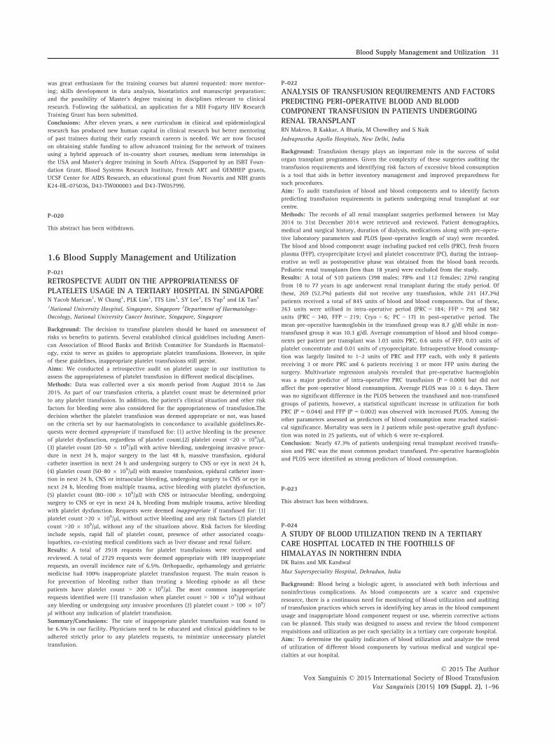
was great enthusiasm for the training courses but alumni requested: more mentor-
ing; skills development in data analysis, biostatistics and manuscript preparation;
and the possibility of Master’s degree training in disciplines relevant to clinical
research. Following the sabbatical, an application for a NIH Fogarty HIV Research
Training Grant has been submitted.
Conclusions: After eleven years, a new curriculum in clinical and epidemiological
research has produced new human capital in clinical research but better mentoring
of past trainees during their early research careers is needed. We are now focused
on obtaining stable funding to allow advanced training for the network of trainees
using a hybrid approach of in-country short courses, medium term internships in
the USA and Master’s degree training in South Africa. (Supported by an ISBT Foun-
dation Grant, Blood Systems Research Institute, French ART and GEMHEP grants,
UCSF Center for AIDS Research, an educational grant from Novartis and NIH grants
K24-HL-075036, D43-TW000003 and D43-TW05799).
P-020
This abstract has been withdrawn.
1.6 Blood Supply Management and Utilization
P-021
RETROSPECTVE AUDIT ON THE APPROPRIATENESS OFPLATELETS USAGE IN A TERTIARY HOSPITAL IN SINGAPOREN Yacob Marican1, W Chang1, PLK Lim1, TTS Lim1, SY Lee2, ES Yap2 and LK Tan2
1National University Hospital, Singapore, Singapore 2Department of Haematology-
Oncology, National University Cancer Institute, Singapore, Singapore
Background: The decision to transfuse platelets should be based on assessment of
risks vs benefits to patients. Several established clinical guidelines including Ameri-
can Association of Blood Banks and British Committee for Standards in Haematol-
ogy, exist to serve as guides to appropriate platelet transfusions. However, in spite
of these guidelines, inappropriate platelet transfusions still persist.
Aims: We conducted a retrospective audit on platelet usage in our institution to
assess the appropriateness of platelet transfusion in different medical disciplines.
Methods: Data was collected over a six month period from August 2014 to Jan
2015. As part of our transfusion criteria, a platelet count must be determined prior
to any platelet transfusion. In addition, the patient’s clinical situation and other risk
factors for bleeding were also considered for the appropriateness of transfusion.The
decision whether the platelet transfusion was deemed appropriate or not, was based
on the criteria set by our haematologists in concordance to available guidelines.Re-
quests were deemed appropriate if transfused for: (1) active bleeding in the presence
of platelet dysfunction, regardless of platelet count.(2) platelet count <20 9 109/ll,(3) platelet count (20–50 9 109/ll) with active bleeding, undergoing invasive proce-
dure in next 24 h, major surgery in the last 48 h, massive transfusion, epidural
catheter insertion in next 24 h and undergoing surgery to CNS or eye in next 24 h,
(4) platelet count (50–80 9 109/ll) with massive transfusion, epidural catheter inser-
tion in next 24 h, CNS or intraocular bleeding, undergoing surgery to CNS or eye in
next 24 h, bleeding from multiple trauma, active bleeding with platelet dysfunction,
(5) platelet count (80–100 9 109/ll) with CNS or intraocular bleeding, undergoing
surgery to CNS or eye in next 24 h, bleeding from multiple trauma, active bleeding
with platelet dysfunction. Requests were deemed inappropriate if transfused for: (1)
platelet count >20 9 109/ll, without active bleeding and any risk factors (2) platelet
count >20 9 109/ll, without any of the situations above. Risk factors for bleeding
include sepsis, rapid fall of platelet count, presence of other associated coagu-
lopathies, co-existing medical conditions such as liver disease and renal failure.
Results: A total of 2918 requests for platelet transfusions were received and
reviewed. A total of 2729 requests were deemed appropriate with 189 inappropriate
requests, an overall incidence rate of 6.5%. Orthopaedic, opthamology and geriatric
medicine had 100% inappropriate platelet transfusion request. The main reason is
for prevention of bleeding rather than treating a bleeding episode as all these
patients have platelet count > 200 9 109/ll. The most common inappropriate
requests identified were (1) transfusion when platelet count > 100 9 109/ll withoutany bleeding or undergoing any invasive procedures (2) platelet count > 100 9 109/
ll without any indication of platelet transfusion.
Summary/Conclusions: The rate of inappropriate platelet transfusion was found to
be 6.5% in our facility. Physicians need to be educated and clinical guidelines to be
adhered strictly prior to any platelets requests, to minimize unnecessary platelet
transfusion.
P-022
ANALYSIS OF TRANSFUSION REQUIREMENTS AND FACTORSPREDICTING PERI-OPERATIVE BLOOD AND BLOODCOMPONENT TRANSFUSION IN PATIENTS UNDERGOINGRENAL TRANSPLANTRN Makroo, B Kakkar, A Bhatia, M Chowdhry and S Naik
Indraprastha Apollo Hospitals, New Delhi, India
Background: Transfusion therapy plays an important role in the success of solid
organ transplant programmes. Given the complexity of these surgeries auditing the
transfusion requirements and identifying risk factors of excessive blood consumption
is a tool that aids in better inventory management and improved preparedness for
such procedures.
Aim: To audit transfusion of blood and blood components and to identify factors
predicting transfusion requirements in patients undergoing renal transplant at our
centre.
Methods: The records of all renal transplant surgeries performed between 1st May
2014 to 31st December 2014 were retrieved and reviewed. Patient demographics,
medical and surgical history, duration of dialysis, medications along with pre-opera-
tive laboratory parameters and PLOS (post-operative length of stay) were recorded.
The blood and blood component usage including packed red cells (PRC), fresh frozen
plasma (FFP), cryoprecipitate (cryo) and platelet concentrate (PC), during the intraop-
erative as well as postoperative phase was obtained from the blood bank records.
Pediatric renal transplants (less than 18 years) were excluded from the study.
Results: A total of 510 patients (398 males; 78% and 112 females; 22%) ranging
from 18 to 77 years in age underwent renal transplant during the study period. Of
these, 269 (52.7%) patients did not receive any transfusion, while 241 (47.3%)
patients received a total of 845 units of blood and blood components. Out of these,
263 units were utilised in intra-operative period (PRC = 184; FFP = 79) and 582
units (PRC = 340, FFP = 219; Cryo = 6; PC = 17) in post-operative period. The
mean pre-operative haemoglobin in the transfused group was 8.7 g/dl while in non-
transfused group it was 10.3 g/dl. Average consumption of blood and blood compo-
nents per patient per transplant was 1.03 units PRC, 0.6 units of FFP, 0.03 units of
platelet concentrate and 0.01 units of cryoprecipitate. Intraoperative blood consump-
tion was largely limited to 1–2 units of PRC and FFP each, with only 8 patients
receiving 3 or more PRC and 6 patients receiving 3 or more FFP units during the
surgery. Multivariate regression analysis revealed that pre-operative haemoglobin
was a major predictor of intra-operative PRC transfusion (P = 0.000) but did not
affect the post-operative blood consumption. Average PLOS was 10 � 6 days. There
was no significant difference in the PLOS between the transfused and non-transfused
groups of patients, however, a statistical significant increase in utilization for both
PRC (P = 0.044) and FFP (P = 0.002) was observed with increased PLOS. Among the
other parameters assessed as predictors of blood consumption none reached statisti-
cal significance. Mortality was seen in 2 patients while post-operative graft dysfunc-
tion was noted in 25 patients, out of which 6 were re-explored.
Conclusion: Nearly 47.3% of patients undergoing renal transplant received transfu-
sion and PRC was the most common product transfused. Pre-operative haemoglobin
and PLOS were identified as strong predictors of blood consumption.
P-023
This abstract has been withdrawn.
P-024
A STUDY OF BLOOD UTILIZATION TREND IN A TERTIARYCARE HOSPITAL LOCATED IN THE FOOTHILLS OFHIMALAYAS IN NORTHERN INDIADK Bains and MK Kandwal
Max Superspeciality Hospital, Dehradun, India
Background: Blood being a biologic agent, is associated with both infectious and
noninfectious complications. As blood components are a scarce and expensive
resource, there is a continuous need for monitoring of blood utilization and auditing
of transfusion practices which serves in identifying key areas in the blood component
usage and inappropriate blood component request or use, wherein corrective actions
can be planned. This study was designed to assess and review the blood component
requisitions and utilization as per each speciality in a tertiary care corporate hospital.
Aim: To determine the quality indicators of blood utilization and analyze the trend
of utilization of different blood components by various medical and surgical spe-
cialties at our hospital.
© 2015 The AuthorVox Sanguinis © 2015 International Society of Blood Transfusion
Vox Sanguinis (2015) 109 (Suppl. 2), 1–96
Blood Supply Management and Utilization 31
Method: The transfusion data was reviewed for all the patients requiring blood com-
ponent transfusion at our hospital for a period of six months from January 2015 to
June 2015 using patient transfusion request forms, hospital information system, cross-
match register, and issue register.The information obtained from the documents
included age, gender, blood group, consultant incharge, speciality, diagnosis and indi-
cation of transfusion, pre transfusion hemoglobin/platelet count/coagulation profile.
The data pertaining to number of whole blood (WB)/packed red blood cells (PRBC)/fresh
frozen plasma (FFP) /random donor platelet concentrates (PC)/single donor apheresis
(SDAP) platelet concentrates requested as well as crossmatched and issued was col-
lected. The quality indicators for blood utilization were calculated as crossmatch to
transfusion ratio (CT ratio), Transfusion Probability (%T) and Transfusion index (TI).
Result: A total of 1210 (795 male and 415 female patients) blood component requisi-
tions were received by Department of Transfusion Medicine during the study period. Of
total of 3711 blood components requested, PRBC, FFP, PC, SDAP and WB requisitions
contributed 49.63%(n = 1842), 28.59%(n = 1061), 21.34%(n = 792), 0.30% (n = 11)
and 0.13%(n = 5) respectively. The requisitions and issue for surgical specialties
[62.97%(n = 762), 67.81%] exceeded the demand from medical specialties [37.02%
(n = 448), 32.19%].The maximum blood component requisitions were received for the
patients of cardiothoracic surgical unit followed by neurosurgery.The transfusion
requests were observed to be maximum for the age group of 51–60 years (19.67%,
n = 238) followed by 61–70 years (18.26%, n = 221).BRh D positive (38.01%) blood
components were maximally requested followed by A Rh Dpositive (25.70%), O Rh
Dpositive (22.72%) and AB Rh Dpositive (10.33%) blood group. The overall mean pre-
transfusion hemoglobin trigger for packed red cell transfusion was 7.42 g/dl.The over-
all average CT Ratio was 1.57, highest being in cardiothoracic surgery department
(1.92), followed by general surgery (1.84) and neurosurgery (1.79) departments. The
Transfusion probability (%T) and Transfusion Index were 79.91% was 0.97 respec-
tively. Of the total blood component utilization, PRBC constituted 44.47%, FFP utiliza-
tion was 33.83% and platelet concentrates as 21.21%.
Summary: The retrospective audit helped in assessment of the transfusion practice
at our centre which is found to be heartening. The CT ratio, Transfusion probability
and Transfusion index demonstrated significant blood utilization at our hospital.
However, excessive blood ordering for elective surgeries so observed leads to
wastage of testing reagent resources, further adding extra cost to the patient .There-
fore, the transfusion auditing should be made a common feature with hospital trans-
fusion committee meetings where maximum surgical blood ordering schedule
(MSBOS) can be modified and implemented progressively.
P-025
SUPERVISION AND COMMUNICATION WITH THE BLOODBANKS EFFECTIVE FOR IMPROVING BLOOD SAFETY INJAKARTA PROVINCEE Pratiwi, P Rini, NK Ritchie and S Salim
Jakarta Blood Transfusion Unit, DKI Jakarta, Indonesia
Background: Jakarta Blood Transfusion Unit (BTU) is the biggest BTU in Indonesia,
served 51.084 blood components per month to 173 Jakarta and 300 other hospitals
and clinics in around Jakarta. According to Ministry of Health Regulation in 2007,
every hospitals should have a blood bank in their hospitals. In 1991, there were only
14 blood banks. The number is increasing and in 2014, there were 42 blood banks.
The responsibility of blood banks were maintaining blood stock in their hospitals and
preparing blood for the patients in their hospitals. Jakarta BTU as the blood provider
should monitor the blood usage in blood banks. The blood banks usually give reports
to BTU and BTU should evaluate it. In 2009, we realize that many blood banks request
whole blood and they produce PRC by open system in not proper procedure. Those
blood components were not safe for the patients and that practice must be stopped.
There should be a more efficient monitoring program for blood usage in blood bank.
Aims: To reduce the production of open system PRC in blood bank.
Methods: We analyze and evaluate annual reports. We set up 2 programs for blood
bank, first is impromptu visit and second is an ‘audit’ visit. We prepared audit
checklist and reports. In internal of Jakarta BTU, we coordinate with logistic teams
to order double bags instead single bags. The blood banks always request whole
blood for their stock because the expiry date was longer than packed red cell (PRC)
in Jakarta BTU. In this case, we already did some research to extend the expiry date
but it seemed difficult unless we used additive solutions. Therefore, if we provide
only whole blood in unprocessed-double bag, they could produce PRC in closed sys-
tem which was safer than the previously. We also held routine meeting, seminars
and workshop to gain their knowledge and skill in blood banking.
Results: After 5 years of supervision, we had a decreasing number of whole-blood
request from 6.2% to 1.6%. Now, opened-system already left behind. We hope the
transfusion reactions in patients would also be reduced although the hemovigilance
system not yet well-established. Since the communication between BTU and blood
banks are more frequent and intense especially for the ‘worst’ blood bank so the
hospital managements now gave their attention to the blood banks and supported
them more than previously. So, we got another value from the supervision programs
that more than 80% of blood banks in Jakarta were in the scale of good and very
good.
Summary/conclusions: Supervision and communication were effective to improve
the quality of blood bank services.
P-026
KOREAN RED CROSS BLOOD SERVICE HEADQUARTERS’ RISKMANAGEMENT OF MERS-COV OUTBREAKSJ Shin, NS Cho, JN Ko, JY Eom and JW Kim
Korean Red Cross, Wonju-si, South-Korea
Background: On May 20, 2015, Korea confirmed the first Middle East Respiratory
Syndrome (MERS) patient. The unexpected MERS outbreak resulted in 36 deaths
among 186 confirmed cases, and 16,693 people quarantined. The increased anxiety
of the emerging outbreak had people limit outdoor and group activities, which led
to the decreasing number of blood donations.
Aims: To assess the risk management of the potential crisis related to blood collec-
tion and supply due to the MERS outbreak.
Methods: The Korean Red Cross Blood Service headquarters (KRC) compared the num-
ber of collected blood and supplied blood with the same period of time (May 20-July
10) in 2014 and assessed if the activities implemented by the KRC were effective.
Results: The KRC conducted various activities to protect blood donors and KRC
employees from the outbreak of MERS. The KRC strengthened strict blood donor
interview, medical examination, and hygiene control at blood collection sites.
Because of temporary closure of schools, cancellation of mobile blood drives, and
the lower number of blood donors, the KRC identified the decreasing number of
blood donations. On June 2, 2015, the KRC recognized that the inventory days of
red blood cells were 7.3 days (the optimal days are 5 days) and predicted that the
inventory days would be less than the optimal days after June 19 (20–30% and 10%
of reduction in the number of blood donations and blood supply respectively).
Therefore, the KRC implemented the risk management including securing blood for
transfusion as a priority at all blood centers, strengthening blood donation promo-
tion, following the risk response manual, and extending blood donation center open-
ing hours. Additionally, the KRC put an effort in protecting the health of employees
and ensuring the safety of blood collection sites by providing the guideline for the
prevention of MERS proliferation. As a result, compared with the same period of
time (May 20-July 10) in 2014, there was no significant difference in blood collec-
tion and supply. While the total number of blood donations decreased by 0.5%, the
number of whole blood and apheresis platelet donations increased by 4.0% and
19.7% respectively. The total number of blood supply decreased by 3.8% because of
the closure of medical institutions, and the blood centers located in the center of
MERS outbreak showed the reduction in blood supply, more than twice the national
average reduction, resulting in significant variances among regions. Contrary to the
prediction, the inventory days of blood products recorded the lowest number of
6.1 days on June 13 and increased to 7.4 days on July 10. Overall, the blood prod-
ucts were maintained over the optimal inventory days.
Summary/Conclusions: Disease outbreak that has high mortality or infection is one
of the causes that threaten blood collection and supply. However, the prediction and
preparation of the risk in advance contributed to overcoming the potential crisis of
blood collection and supply as the KRC responded to the case of MERS outbreak.
1.7 Quality Management
P-027
THE RESULTS OF EXTERNAL PROFICIENCY TESTS FORHBSAG AND ANTI-HCV DONOR TESTING IN BLOOD CENTERSIN KOREA (2012–2014)MN Lee1, JR Kwon1, YS Choi1, QE Park2, YJ Cha3 and JN Kim1
1Korea Centers for Disease Control & Prevention, Cheongju, South-Korea 2Blood
Center, Chung-Ang University Hospital, Seoul, South-Korea 3Department of
Laboratory Medicine, Chung-Ang University College of Medicine, Seoul, South-Korea
Background: Blood donor screening tests for infectious diseases are one of the most
important processes of blood safety management. In Korea, these tests include
© 2015 The AuthorVox Sanguinis © 2015 International Society of Blood TransfusionVox Sanguinis (2015) 109 (Suppl. 2), 1–96
32 Posters
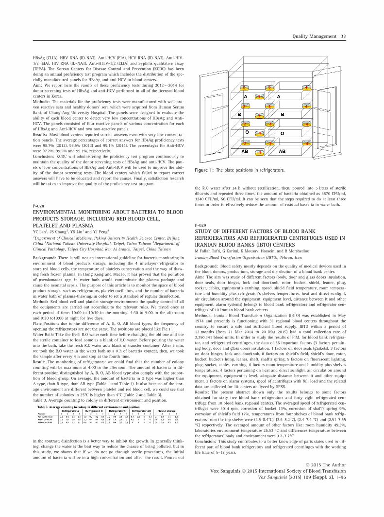
HBsAg (CLIA), HBV DNA (ID-NAT), Anti-HCV (EIA), HCV RNA (ID-NAT), Anti-HIV-
1/2 (EIA), HIV RNA (ID-NAT), Anti-HTLV-1/2 (CLIA) and Syphilis qualitative assay
(TPPA). The Korean Centers for Disease Control and Prevention (KCDC) has been
doing an annual proficiency test program which includes the distribution of the spe-
cially manufactured panels for HBsAg and anti-HCV to blood centers.
Aim: We report here the results of these proficiency tests during 2012� 2014 for
donor screening tests of HBsAg and anti-HCV performed in all of the licensed blood
centers in Korea.
Methods: The materials for the proficiency tests were manufactured with well-pro-
ven reactive sera and healthy donors’ sera which were acquired from Human Serum
Bank of Chung-Ang University Hospital. The panels were designed to evaluate the
ability of each blood center to detect very low concentrations of HBsAg and Anti-
HCV. The panels consisted of four reactive panels of various concentration for each
of HBsAg and Anti-HCV and two non-reactive panels.
Results: Most blood centers reported correct answers even with very low concentra-
tion panels. The average percentages of correct answers for HBsAg proficiency tests
were 98.7% (2012), 98.5% (2013) and 99.1% (2014). The percentages for Anti-HCV
were 97.7%, 99.5% and 99.1%, respectively.
Conclusion: KCDC will administering the proficiency test program continuously to
maintain the quality of the donor screening tests of HBsAg and anti-HCV. The pan-
els of low concentrations of HBsAg and Anti-HCV will be used to improve the abil-
ity of the donor screening tests. The blood centers which failed to report correct
answers will have to be educated and report the causes. Finally, satisfaction research
will be taken to improve the quality of the proficiency test program.
P-028
ENVIRONMENTAL MONITORING ABOUT BACTERIA TO BLOODPRODUCTS STORAGE, INCLUDING RED BLOOD CELL,PLATELET AND PLASMAYC Lan1, JS Chang2, YS Lin1 and YJ Peng3
1Department of Clinical Medicine, Peking University Health Science Center, Beijing,
China 2National Taiwan University Hospital, Taipei, China Taiwan 3Department of
Clinical Pathology, Taipei City Hospital, Ren Ai branch, Taipei, China Taiwan
Background: There is still not an international guideline for bacteria monitoring in
environment of blood products storage, including the 4 interlayer-refrigerator to
store red blood cells, the temperature of platelets conservation and the way of thaw-
ing fresh frozen plasma. In Hong Kong and Macao, it has proved that the pollution
of pseudomonas spp. in water bath would contaminate the plasma package and
cause the neonatal sepsis. The purpose of this article is to monitor the space of blood
product storage, such as refrigerators, platelet oscillators, and the number of bacteria
in water bath of plasma-thawing, in order to set a standard of regular disinfection.
Method: Red blood cell and platelet storage environment: the quality control of all
the equipments are carried out according to the relevant rules. We tested once at
each period of time: 10:00 to 10:30 in the morning, 4:30 to 5:00 in the afternoon
and 9:30 to10:00 at night for five days.
Plate Position: due to the difference of A, B, O, AB blood types, the frequency of
opening the refrigerators are not the same. The positions are placed like Pic.1.
Water Bath: Take the fresh R.O water each time before changing the old one and use
the sterile container to load some as a blank of R.O water. Before pouring the water
into the bath, take the fresh R.O water as a blank of transfer container. After 5 min,
we took the R.O water in the water bath as a 0 h of bacteria content, then, we took
the sample after every 4 h and stop at the fourth time.
Result: The monitoring of refrigerators: we could find that the number of colony
counting will be maximum at 4:00 in the afternoon. The amount of bacteria in dif-
ferent position distinguished by A, B, O, AB blood type also comply with the propor-
tion of blood group. On average, the amount of bacteria in O type was higher than
A type, than B type, than AB type (Table 1 and Table 3). It also because of the stor-
age environment are different between platelet and red blood cell, we could see that
the number of colonies in 25°C is higher than 4°C (Table 2 and Table 3).
Table 3. Average counting to colony in different environment and position.
in the contrast, disinfection is a better way to inhibit the growth. In generally think-
ing, change the water is the best way to reduce the chance of being polluted, but in
this study, we shows that if we do not go through sterile procedures, the initial
amount of bacteria will be in a high concentration and affect the result. Poured out
the R.O water after 24 h without sterilization, then, poured into 5 liters of sterile
diluents and repeated three times, the amount of bacteria obtained as 5870 CFU/ml,
3240 CFU/ml, 50 CFU/ml. It can be seen that the steps required to do at least three
times in order to effectively reduce the amount of residual bacteria in water bath.
P-029
STUDY OF DIFFERENT FACTORS OF BLOOD BANKREFRIGERATORS AND REFRIGERATED CENTRIFUGES USED INIRANIAN BLOOD BANKS (IBTO) CENTERSM Fallah Tafti, G Karimi, K Mousavi Hosseini and R Morshedlou
Iranian Blood Transfusion Organisation (IBTO), Tehran, Iran
Background: Blood safety mostly depends on the quality of medical devices used in
the blood donors, productions, storage and distribution of a blood bank center.
Aim: The aim was study of different factors (body, door and glass doors insulation,
door seals, door hinges, lock and doorknob, rotor, bucket, shield, leaner, plug,
socket, cables, equipment’s earthing, speed, shield field temperature, room tempera-
ture and humidity plus refrigerator’s shelves temperatures, heat and direct sunlight,
air circulation around the equipment, equipment level, distance between it and other
equipment, alarm systems) belongs to blood bank refrigerators and refrigerator cen-
trifuges of 10 Iranian blood bank centers
Methods: Iranian Blood Transfusion Organization (IBTO) was established in May
1974 and presently is functioning with 31 regional blood centers throughout the
country to ensure a safe and sufficient blood supply. IBTO within a period of
12 months (from 21 Mar 2014 to 20 Mar 2015) had a total collection rate of
2,250,341 blood units. In order to study the results of P.M. for blood bank refrigera-
tor, and refrigerated centrifuges, the data of 36 important factors (3 factors pertain-
ing body, door and glass doors insulation, 1 factors on door seals (gaskets), 3 factors
on door hinges, lock and doorknob, 8 factors on shield’s field, shield’s door, rotor,
bucket, bucket’s hung, leaner, shaft, shaft’s spring, 5 factors on fluorescent lighting,
plug, socket, cables, earthing, 6 factors room temperature and humidity plus shelves
temperatures, 4 factors pertaining on heat and direct sunlight, air circulation around
the equipment, equipment is level, adequate distance between it and other equip-
ment, 3 factors on alarm systems, speed of centrifuges with full load and the related
data are collected for 10 centers analyzed by SPSS.
Results: The present abstract shown only the results belongs to some factors
obtained for sixty tree blood bank refrigerators and forty eight refrigerated cen-
trifuge from 10 blood bank regional centers. The averaged speed of refrigerated cen-
trifuges were 5014 rpm, corrosion of bucket 13%, corrosion of shaft’s spring 9%,
corrosion of shield’s field 17%, temperatures from four shelves of blood bank refrig-
erators from the top shelve were (2.5–8.4°C), (2.6–8.2°C), (2.4–7.4 °C) and (2.51–7.55°C) respectively. The averaged amount of other factors like: room humidity 49.3%,
laboratories environment temperature 26.53 °C and differences temperature between
the refrigerators’ body and environment were 3.2–7.7°C.Conclusion: This study contributes to a better knowledge of parts states used in dif-
ferent part of blood bank refrigerators and refrigerated centrifuges with the working
life time of 5–12 years.
Figure 1: The plate positions in refrigerators.
© 2015 The AuthorVox Sanguinis © 2015 International Society of Blood Transfusion
Vox Sanguinis (2015) 109 (Suppl. 2), 1–96
Quality Management 33

Key words: Blood bank refrigerator, refrigerated centrifuge. Speed, rotor, bucket,
leaner, shaft, shield, humidity, laboratories environment temperature, humidity, body
and glass insulator, ribbon and door lock of refrigerator.
P-030
AN INSTITUTIONAL EFFORT AT MONITORING QUALITY OFPACKED RED CELLS AND PLATELET CONCENTRATESRN Makroo, M Chowdhry, A Bhatia and UK Thakur
Indraprastha Apollo Hospitals, New Delhi, India
Background: The Standards for Blood Banks & Blood Transfusion Services, laid
down by the Ministry of Health and Family Welfare, Government of India, outline
recommendations for the quality of blood and blood components in India. Monitor-
ing the quality of blood components prepared establishes the efficacy of procedures
and protocols being followed, aids in identifying problem areas and determining
corrective actions.
Aims: To analyze the quality of blood components processed at our blood bank, to
test whether they meet the recommended national standards.
Design and methods: Our hospital based blood bank routinely collects blood in
quintuple top and bottom bags with integral leucocyte filters. Blood is then pro-
cessed for preparation of blood components as per departmental protocols and man-
ufacturers’ instructions using automated component separation systems. Pre-storage
leucoreduction of Packed Red Cells (PRC) is performed using integral filter. Platelet
Concentrates (PC) are prepared from the Buffy coat.
As per the national standards, a minimum of 1% of the blood components prepared
undergo quality control testing. PRC and PC units, for which no incidents are
reported during collection and processing, are randomly selected for quality control.
Weights are measured and volumes are calculated based on specific gravity. Cell
counts, hemoglobin and hematocrit are performed in a automated cell counter
(Beckman Coulter). In addition, a Nagoette’s chamber is used for manually confirm-
ing WBC counts. The pH is recorded at the day of collection as well as on the 5th
day of storage of PC. Swirling is examined visually.
We retrieved and reviewed the Quality Control data of the cellular blood components
(PRC and PCs) for 2013 and 2014.
Results: The volume of PRC units (Mean�SD) was 286.8 � 12.6 ml. Hemoglobin
levels ranged from 17.8–23.3 g/dl and the mean hematocrit (Hct) was 59.6 � 4%.
None of the units had a Hct of less than 50%, while 2 units had a Hct of over 70%.
All the units tested were adequately leucoreduced as per standards (<5 9 106 WBC/
unit after use of leucocyte filter).
As regards to PCs tested, the mean volume of PC was 73.8 � 5 ml, and the platelet
yield was 6.65 � 1.4 9 1010/unit. Platelet yield in 96.8% of the units tested were in
compliance with the national standards of minimum 4.5 9 1010 platelets/unit. The
pH ranged from 6 to 7.5 on the last day of storage, in compliance with the recom-
mended standards. Swirling was visible in all the units. The mean WBC counts of
the units was 2.8 � 1.6 9 108 per unit and 91.4% of the PC tested were adequately
leucoreduced as per the standards (<5 9 108). The red cell counts were
0.02 � 0.001 9 1012/unit.
Conclusion: Both red cell as well as platelet concentrates prepared on the auto-
mated component separation systems at our centre comply with the recommended
national standards. All PRC units were adequately leucoreduced (upto 3 log reduc-
tion), while 91.4% of the PC achieved over 1 log leucoreduction.
Table 1: Quality Parameters of packed red cells and platelet concentrates.
P-031
ANALYSIS AND CLINICAL AUDIT OF UNUSED PLATELETCONCENTRATES IN H. ADAM MALIK HOSPITAL MEDANINDONESIAI Adhayanti1, T Triyono2 and R Juwita1
1Adam Malik Hospital, Medan, Indonesia 2Sardjito Hospital, Yogjakarta, Indonesia
Background: Platelet concentrate (PC) is given to avoid or cure patients with bleed-
ing disorders. Platelet transfusion must be performed with the right indication for
patient safety reason. One of the quality indicators in H. Adam Malik Hospital Blood
Transfusion Unit is the percentage of unused PC per-month. The data showed that
the percentage of unused PC were 2.63%, 6.45%, 8.74%, and 12.40% for January,
February, March and April 2015 respectively. The analysis and clinical audit were
needed for PC usage.
Aims: To reach the efficacy of PC usage and to fix the unused PC quality indicator
in H. Adam Malik Blood Transfusion Unit.
Method: This research was held in H. Adam Malik Blood Transfusion Unit, Medan
Indonesia using cross sectional design. The data were collected from the hospital
information system in the period of January-June 2015. The data then analyzed and
compared with the data from Hospital transfusion unit.
Result: There was an increased PC used (2604 units) from January to June 2015. The
unused PC were declined but still above the standard (> 1%).The strategies that were
used for increasing quality indicator in Blood Transfusion Unit started on May 2015
were spreading platelet transfusion indication brochures that were signed by the hos-
pital main director, the responsible doctors must sign the platelet concentrate demand
letters, requesting the platelet agitator with incubator device for patient safety reason.
The result still high 4.12% in May and 5.50% in June. The departments with the high-
est number of unused PC were Internal Medicine 28.4%; Surgery 20.4%; Pediatric
19.8%, Obstetrics and Gynecology 17%; Cardiology 10.8%, others 3.4%.
Conclusion: The number of unused PC in H. Adam Malik Hospital Blood Transfu-
sion Unit has not reached the standard. There were still many constraints found,
such as: different education background of the responsible doctors, ineffective com-
munication, unsigned platelet concentrate order letter, broken platelet agitator with
incubator device. By improving the communication, implementing the strategies that
have been made and provisioning platelet agitator with incubator device, the
decrease of unused platelet concentrates was expected.
P-032
QUALITY ANALYSIS OF PRE-OPERATIVE PREPARED PACKEDRED CELLS STUDY OF INDEXES RBC, CULTURE ANDIDENTIFICATION OF BACTERIARA Muhiddin1, T Triyono2 and B Mulyono2
1Wahidin Sudirohusodo Center Hospital, Makassar, Indonesia 2Gadjah Mada
University, Jogjakarta, Indonesia
Background: Pre-operative Packed Red Cell (PRC) is prepared to anticipate acute
blood loss during surgery. In the absence of intraoperative bleeding complication,
the PRC would not be transfused. The handling of pre-operative PRC performed
under the cold chain system to ensure it was safe to be distributed to the patients.
Aims: To determine the differences of red blood cell (RBC) indexes, and the bacte-
rial culture of the pre-operative prepared PRC before being transferred to the operat-
ing room (group I) compared to the unused PRC in operating room then returned to
the blood bank and stored for 3 days (group II). Furthermore, to determine the per-
centage of hemolysis in plasma after 3 days storage at the Blood Bank, and its rela-
tionship with the day of PRC storage after donation.
Method: This study was conducted in the period of August to October 2014, in the
Blood Bank and Clinical Pathology Laboratory of Dr. Wahidin Sudirohusodo General
Hospital Makassar, Indonesia. The RBC indexes was examined by Sysmex XT1800
hematology analyzer. Bacterial culture was examined by Bact/ALERT, Bacterial
identification was examined by VTEK2 Compact.
Results: The Mean Corpuscular Volume (MCV) of group I and group II were
85.95 fl and 86.45 fl respectively (P = 0.109). The Mean Corpuscular Hemoglobin
(MCH) group I was higher (28.05 pg) than group II (28.00 pg), but insignificant
(P = 0.386). The Mean corpuscular hemoglobin concentrate (MCHC) of Group I and
group II were 32.70% and 32.20% respectively (P = 0.001). Red Cell Distribution
Width (RDW) group I was 14.90%, RDW Group II was 15.47% (P = 0.006). There
wasn’t difference positivity bacterial culture in both group I and II. There was 6.67%
samples found percentage hemolysis >1%. There was weak relationship between per-
centage of hemolysis with day of PRC storage after donation (r = 0.128, P = 0.463).
Conclusions: There was a significant difference for MCHC and RDW between group
I and II. There wasn’t difference positivity bacterial culture in both group I and II.
Hemolysis can occur along the length of the storage period PRC.
© 2015 The AuthorVox Sanguinis © 2015 International Society of Blood TransfusionVox Sanguinis (2015) 109 (Suppl. 2), 1–96
34 Posters
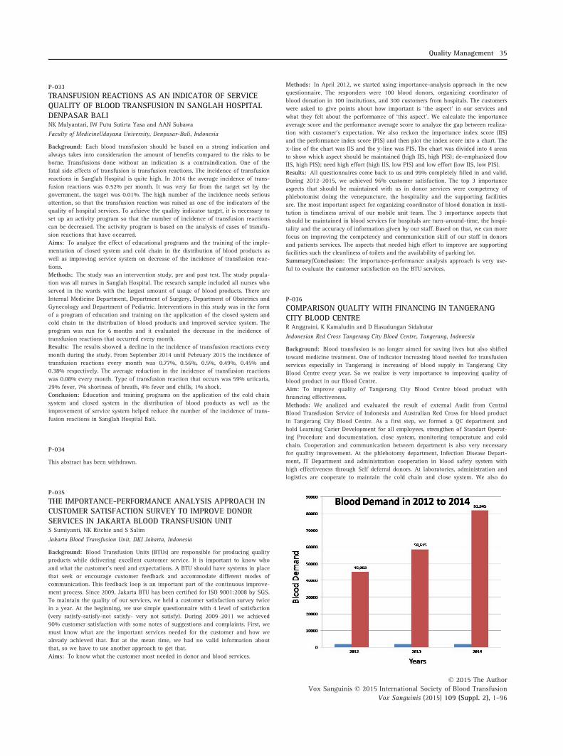
P-033
TRANSFUSION REACTIONS AS AN INDICATOR OF SERVICEQUALITY OF BLOOD TRANSFUSION IN SANGLAH HOSPITALDENPASAR BALINK Mulyantari, IW Putu Sutirta Yasa and AAN Subawa
Faculty of MedicineUdayana University, Denpasar-Bali, Indonesia
Background: Each blood transfusion should be based on a strong indication and
always takes into consideration the amount of benefits compared to the risks to be
borne. Transfusions done without an indication is a contraindication. One of the
fatal side effects of transfusion is transfusion reactions. The incidence of transfusion
reactions in Sanglah Hospital is quite high. In 2014 the average incidence of trans-
fusion reactions was 0.52% per month. It was very far from the target set by the
government, the target was 0.01%. The high number of the incidence needs serious
attention, so that the transfusion reaction was raised as one of the indicators of the
quality of hospital services. To achieve the quality indicator target, it is necessary to
set up an activity program so that the number of incidence of transfusion reactions
can be decreased. The activity program is based on the analysis of cases of transfu-
sion reactions that have occurred.
Aims: To analyze the effect of educational programs and the training of the imple-
mentation of closed system and cold chain in the distribution of blood products as
well as improving service system on decrease of the incidence of transfusion reac-
tions.
Methods: The study was an intervention study, pre and post test. The study popula-
tion was all nurses in Sanglah Hospital. The research sample included all nurses who
served in the wards with the largest amount of usage of blood products. There are
Internal Medicine Department, Department of Surgery, Department of Obstetrics and
Gynecology and Department of Pediatric. Interventions in this study was in the form
of a program of education and training on the application of the closed system and
cold chain in the distribution of blood products and improved service system. The
program was run for 6 months and it evaluated the decrease in the incidence of
transfusion reactions that occurred every month.
Results: The results showed a decline in the incidence of transfusion reactions every
month during the study. From September 2014 until February 2015 the incidence of
transfusion reactions every month was 0.77%, 0.56%, 0.5%, 0.49%, 0.45% and
0.38% respectively. The average reduction in the incidence of transfusion reactions
was 0.08% every month. Type of transfusion reaction that occurs was 59% urticaria,
29% fever, 7% shortness of breath, 4% fever and chills, 1% shock.
Conclusion: Education and training programs on the application of the cold chain
system and closed system in the distribution of blood products as well as the
improvement of service system helped reduce the number of the incidence of trans-
fusion reactions in Sanglah Hospital Bali.
P-034
This abstract has been withdrawn.
P-035
THE IMPORTANCE-PERFORMANCE ANALYSIS APPROACH INCUSTOMER SATISFACTION SURVEY TO IMPROVE DONORSERVICES IN JAKARTA BLOOD TRANSFUSION UNITS Sumiyanti, NK Ritchie and S Salim
Jakarta Blood Transfusion Unit, DKI Jakarta, Indonesia
Background: Blood Transfusion Units (BTUs) are responsible for producing quality
products while delivering excellent customer service. It is important to know who
and what the customer’s need and expectations. A BTU should have systems in place
that seek or encourage customer feedback and accommodate different modes of
communication. This feedback loop is an important part of the continuous improve-
ment process. Since 2009, Jakarta BTU has been certified for ISO 9001:2008 by SGS.
To maintain the quality of our services, we held a customer satisfaction survey twice
in a year. At the beginning, we use simple questionnaire with 4 level of satisfaction
(very satisfy-satisfy-not satisfy- very not satisfy). During 2009–2011 we achieved
90% customer satisfaction with some notes of suggestions and complaints. First, we
must know what are the important services needed for the customer and how we
already achieved that. But at the mean time, we had no valid information about
that, so we have to use another approach to get that.
Aims: To know what the customer most needed in donor and blood services.
Methods: In April 2012, we started using importance-analysis approach in the new
questionnaire. The responders were 100 blood donors, organizing coordinator of
blood donation in 100 institutions, and 300 customers from hospitals. The customers
were asked to give points about how important is ‘the aspect’ in our services and
what they felt about the performance of ‘this aspect’. We calculate the importance
average score and the performance average score to analyze the gap between realiza-
tion with customer’s expectation. We also reckon the importance index score (IIS)
and the performance index score (PIS) and then plot the index score into a chart. The
x-line of the chart was IIS and the y-line was PIS. The chart was divided into 4 areas
to show which aspect should be maintained (high IIS, high PIS); de-emphasized (low
IIS, high PIS); need high effort (high IIS, low PIS) and low effort (low IIS, low PIS).
Results: All questionnaires come back to us and 99% completely filled in and valid.
During 2012–2015, we achieved 96% customer satisfaction. The top 3 importance
aspects that should be maintained with us in donor services were competency of
phlebotomist doing the venepuncture, the hospitality and the supporting facilities
are. The most important aspect for organizing coordinator of blood donation in insti-
tution is timeliness arrival of our mobile unit team. The 3 importance aspects that
should be maintained in blood services for hospitals are turn-around-time, the hospi-
tality and the accuracy of information given by our staff. Based on that, we can more
focus on improving the competency and communication skill of our staff in donors
and patients services. The aspects that needed high effort to improve are supporting
facilities such the cleanliness of toilets and the availability of parking lot.
Summary/Conclusion: The importance-performance analysis approach is very use-
ful to evaluate the customer satisfaction on the BTU services.
P-036
COMPARISON QUALITY WITH FINANCING IN TANGERANGCITY BLOOD CENTRER Anggraini, K Kamaludin and D Hasudungan Sidabutar
Indonesian Red Cross Tangerang City Blood Centre, Tangerang, Indonesia
Background: Blood transfusion is no longer aimed for saving lives but also shifted
toward medicine treatment. One of indicator increasing blood needed for transfusion
services especially in Tangerang is increasing of blood supply in Tangerang City
Blood Centre every year. So we realize is very importance to improving quality of
blood product in our Blood Centre.
Aim: To improve quality of Tangerang City Blood Centre blood product with
financing effectiveness.
Methods: We analized and evaluated the result of external Audit from Central
Blood Transfusion Service of Indonesia and Australian Red Cross for blood product
in Tangerang City Blood Centre. As a first step, we formed a QC department and
hold Learning Carier Development for all employees, strengthen of Standart Operat-
ing Procedure and documentation, close system, monitoring temperature and cold
chain. Cooperation and communication between department is also very necessary
for quality improvement. At the phlebotomy department, Infection Disease Depart-
ment, IT Department and administration cooperation in blood safety system with
high effectiveness through Self deferral donors. At laboratories, administration and
logistics are cooperate to maintain the cold chain and close system. We also do
© 2015 The AuthorVox Sanguinis © 2015 International Society of Blood Transfusion
Vox Sanguinis (2015) 109 (Suppl. 2), 1–96
Quality Management 35
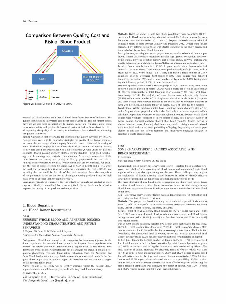
external QC blood product with Central Blood Transfusion Service of Indonesia. The
quality should not be interrupted just in our Blood Centre but also for Patient safety,
therefore we also hold socializations to nurses, doctor and clinicians about blood
transfusions safety and quality. At finance department had to think about the effect
of improving the quality of the costing to effectiveness but it should not damaging
the quality framework.
Result: Calculation that we arrange for improving the quality increased by 141.21%
from previous year, with QC improving strategies the quality of our human resources
increases, the percentage of blood taping failure decreased 12.5%, and increasing of
blood distribution roughly 39.63%. Comparison of test results and quality product
from Whole Blood and Packed Red Cell 3 times external QC with CBTS: not pass the
standarts (87.5%), passing standarts (100%), passing standart (100%) good morphol-
ogy test, hematology and bacterial contamination. As a rough calculation of the
ratio between the costing and quality is directly proportional, but the ratio is
reserved when compared to the risks from products that are not qualified. For exam-
ple, the cost of blood screening by using NAT or CLIA is $20.01 and $11.56 while
for rapid test we using two kinds of reagen for comparison the cost is $23.13, not
including the cost would be the risks of the results obtained. From the comparison
of two parameters it can see the cost to obtain good quality products is not too high
could even be cheaper than the regular product.
Conclusion: Quality is expensive but the effects of unqualified product is more
expensive. Quality is something that is not negotiable. So we should not be afraid to
improve the quality of our products and our services.
2. Blood Donation2.1 Blood Donor Recruitment
P-037
FREQUENT WHOLE BLOOD AND APHERESIS DONORS:UNDERSTANDING CHARACTERISTICS AND RETURNBEHAVIOURA Thijsen, CN Gemelli, D Waller and J Hayman
Australian Red Cross Blood Service, Alexandria, Australia
Background: Blood donor management is supported by examining segments in the
donor population. An essential donor group is the frequent donor population who
provide the largest portion of donations on a regular basis. A few studies have
determined frequent donor characteristics but no analysis has included donation fre-
quency, phlebotomy type and donor return behaviour. Thus, the Australian Red
Cross Blood Service set out a large database research to understand trends in the fre-
quent donor population to provide support for retention and reactivation strategies
of this specific donor group.
Aims: The aim of this study was to identify segments within the frequent donor
population based on phlebotomy type, medical history, and donation history.
Methods: Based on donor records two study populations were identified: (1) fre-
quent whole blood donors who had donated successfully 3 times or more between
December 2010 and November 2011, and (2) frequent apheresis donors who had
donated 6 times or more between January and December 2012. Donors were further
segregated by deferral status, those who started donating in the study period, and
those who had lapsed from blood donation.
Descriptive analysis using means and proportions was conducted on both donor popu-
lations. Donor characteristics examined included age, gender, occupation, socioeco-
nomic status, previous donation history, and deferral status. Survival analysis was
used to determine the probability of lapsing following a temporary medical deferral.
Results: Donor records identified 90,649 frequent whole blood donors who had
donated 3 or more times. These donors were predominately male (51.36%), with a
mean age of 48.07 years (range 16–81). They had made a mean number of 22.67
donations prior to December 2010 (range 0–270). These donors were followed
through to the end of 2013 to determine numbers of lapse with 13.59% lapsing dur-
ing the follow-up period 23.26% of these due to deferral.
Frequent apheresis donors were a smaller group of 17,231 donors. They were found
to have a greater portion of males (64.5%), with a mean age of 50.24 years (range
18–81). The mean number of total donations prior to January 2011 was 45.73 dona-
tions (range 1–334). The majority of these donors were apheresis only donors
(77.7%), with a mean number of 12.15 apheresis donations made in 2012 (range 6–29). These donors were followed through to the end of 2014 to determine numbers of
lapse with 6.15% lapsing during follow-up period, 13.6% of these due to a deferral.
Conclusions: Whilst previous studies have examined donor characteristics of the
entire frequent donor population, this is the first study to examine segments within
this donor population. Compared to frequent apheresis donors, frequent whole blood
donors were younger, consisted of more female donors, and a greater number of
lapsed donors. Survival analysis showed that being younger, female, having a
shorter donation career, donating whole blood only, and receiving a medical deferral
were associated with an increased probability of lapsing. Segmenting the donor pop-
ulation in this way can inform retention and reactivation strategies designed to
maintain a stable blood supply.
P-038
SOME CHARACTERISTIC FACTORS ASSOCIATED WITHDONOR RECRUITMENTIP Rupasena
National Blood Center, Colombo 05, Sri Lanka
Background: Blood supply has always been scarce. Therefore blood donation pro-
grams face challenges in recruiting of blood donors and maintaining their blood
supplies without any shortages throughout the year. These challenges make urgent
the exploration of factors affecting blood donation in order to identify effective
strategies for increasing the donor base and fulfilling future blood needs.
The main strategies of any blood donor programmes are donor motivation, donor
recruitment and donor retention. Donor recruitment is an essential strategy in any
blood donor programme because it aids in maintaining a sustainable and safe blood
donor pool.
Aim: Descriptive study of donor factors such as donor intention, 1st motivation and
advertizing method of donor recruitment.
Methods: The prospective descriptive study was conducted a period of six months
from 01/10/2015 to 30/06/2015 in blood collection campaigns conducted by Blood
Bank, District General Hospital, Negombo, Sri Lanka.
Results: Total of 2759 voluntary blood donors, 81.1% (n = 2237) males and 18.9%
(n = 522) females were donated blood as voluntary non remunerated blood donors
during relevant period. 29.6% (n = 816) was first time donors and 70.4% (n = 1943)
was regular donors.
Out of 2759 donors, randomly selected 879 donors were participated for this study.
40.9% (n = 360) was first time donors and 59.1% (n = 519) was regular donors. Male
donors accounted for 73.3% while the female counterpart was responsible for 26.7%.
Considering the educational level of donors, 79.1% had primary educational level
(school education) and 20.9% had secondary educational level (diploma or higher).
In first time donors, the highest number of donors 12.7% (n = 177) were motivated
for blood donation in their 1st blood donation by printed media (poster/news paper
etc.) while 14.7% (n = 129) in regular donors who were motivated by friends. The
least number of donors motivated by electronic media (TV/Radio) which was 0.6%
(n = 6) in both 1st time and regular donors. 26.9% and 36.2% donors donated blood
for self satisfaction in 1st time and regular donors respectively. 12.9% 1st time
donors and 18.8% regular donors donated blood as a responsibility. 25.7% 1st time
donors and 30% regular donors thought that most effective ways for advertizing the
blood donation campaigns was displaying the poster or banner. Also 7.2% 1st time
and 11.3% regular donors thought it was Facebook/Internet.
Figure 2: Blood Demand in 2012 to 2014.
© 2015 The AuthorVox Sanguinis © 2015 International Society of Blood TransfusionVox Sanguinis (2015) 109 (Suppl. 2), 1–96
36 Posters

When considering the educational level, 12.7% of donors who had primary educa-
tional level were motivated their blood donation by printed media (poster/news
paper etc.) and 14.7% of donors who had secondary educational level were moti-
vated by friends. 26.9% of primary educated donors and 36.2% of secondary edu-
cated donors donated their blood for self satisfaction. Both type of donors believed
that the most effective ways for advertizing the blood donation campaigns was dis-
playing Posters or Banners. (25.6% and 30.% respectively)
Summary: Donor population of Sri Lanka is 100% voluntary non-remunerated
donors. The information about donor’s opinion and feelings towards donor recruit-
ment is more important to make future decisions and improvements.
Donors are found to vary in their behaviors, attitudes and beliefs but not enor-
mously. These data may enable National blood center to optimize recruitment by tai-
loring a more effective strategy.
P-039
RECRUITMENT STRATEGIES FOR INCREASING NON-REMUNERATED VOLUNTARY DONOR AT FASTING MONTH INJAKARTA BLOOD TRANSFUSION UNITS Juwariyah, U Khasanah, D Winarti, NK Ritchie and S Salim
Jakarta Blood Transfusion Unit, DKI Jakarta, Indonesia
Background: The blood stock depends on the availability of blood donors. In
Jakarta Blood Transfusion Unit (BTU), 95% was non-remunerated voluntary donor,
and 5% was replacement donor which increasing in fasting month because 88.1% of
Indonesian population were moslem. At fasting month, the blood collection from
voluntary donor was dropped to 60% compared to other months, voluntary donors
were 99.5%. Usually, we have 10–15 Mobile Unit teams, went to various institution
and got 800–1200 blood donors every day. At fasting month, only 1–2 mobile unit
teams per day, except Saturday and Sunday. This condition affected the blood stock
and delayed the medical treatment of patients. So, we have to make a breakthrough
program to increase the blood collection. In 2013, we have new recruitment teams
and new mobile bus, a facility for collecting blood inside the bus. We have social-
ization program in social media such as radio to socialize fatwa MUI which allow
Muslims donated blood in fasting month. We also placed our mobile bus unit in
some mosques and malls almost every day to get more donors. By those programs,
the number of voluntary donors increased to 65%, but then in 2014 it dropped to
59% because less anticipated programs were conducted before fasting month.
Aims: To increase the blood collection at fasting month in Jakarta BTU.
Methods: We always ask the active institutions and non Muslims organizations to
donate in fasting month. Few weeks before fasting month, we went to some institu-
tions, door to door to explain the situation of unbalancing between blood stock and
demands in fasting month and encouraged them to engage blood donations in fast-
ing month. Other routine programs such as socialization programs still conducted.
Results: During fasting month in 2015, we achieved the highest number of volun-
tary donors ever, 71.3% of total donations or 21.2% higher than 2014. The increas-
ing voluntary donors were seen in our buildings (25.4%) and in mobile unit teams
(16.8%) compared to 2014. The responds from the coordinator of blood donations in
all institutions were excited to help. They also gave us time to campaign about
‘blood donations in the fasting month’ for the next fasting months.
Summary/conclusions: Pro-active communication and good personal skill were
very helpful increasing the number of blood collection in the fasting month.
P-040
This abstract has been withdrawn.
P-041
PUBLIC AWARENESS, ATTITUDE AND PRACTICES OF BLOODDONATION AMONG THE GENERAL POPULATION LIVING INRIYADH, SAUDI ARABIAAW Al-Johar1, MA Abolfotouh2, MH Al-Assiri3, MA Al-Omani2, AM Al-Hakbani1
and AS Alaskar3
1King Saud University, Riyadh, Saudi Arabia 2King Saud bin-Abdulaziz University
for Health Sciences, Riyadh, Saudi Arabia 3King Abdullah International Medical
Research Center, Riyadh, Saudi Arabia
Background: Voluntary donors are the only source of blood donation in Saudi Ara-
bia. Despite frequent blood donation campaigns and encouragement by the media,
lack of blood resources remains significant and problematic in advanced healthcare
centers.
Aim: The aim of this study was to assess the level of public knowledge and attitude
toward blood dona�tion. In addition, to describe the donating and non-donating
populations with identification of motivating and restraining factors in each.
Methods: Using a previously validated questionnaire that comprises 38 questions to
assess the level of knowledge, attitude, and practices towards blood donation, 469
Saudi adults who attended different shopping malls in Riyadh, Saudi Arabia were
interviewed. Based on calculated scores, level of knowledge and attitude of our
study cohort were categorized into different groups. Multiple regression analyses
were used to identify significant predictors of blood donation.
Results: Approximately, half of all subjects (53.3%) reported previous history of
donation, 39% of whom had multiple donations, with predominance of males (66%
vs 13.3%; P < 0.001). Knowledge percentage mean score was 58.0%, denoting poor
level of knowledge. Attitude percentage mean score towards donation was 75.4%,
reflecting neutral attitude. After adjustment of confounders, multiple linear regres-
sion showed that higher knowledge score, higher attitude score, and male gender
were significant predictors of blood donation (P = 0.01, P = 0.001, P < 0.001,
respectively). Correlation between knowledge and attitude mean scores showed sig-
nificant association between the variants (P < 0.001). Notable reasons that refrained
females from donation were inability to reach the blood donation centers and fear
of anemia (49.9% and 35.7%, respectively), whereas lack of time was the only sig-
nificant reason amongst males (59.5%).
Conclusion/Summary: Overall, level of knowledge about blood donation was poor,
and attitude towards blood donation was generally neutral. Higher level of knowl-
edge was associated with more positive attitude for blood donation. After adjust-
ment, significant predictors of blood donation were higher knowledge and attitude
scores, and male gender.
P-042
A STUDY ON THE EFFECTIVENESS OF ADVERTISEMENTS ATTHE WOODLANDS SATELLITE BLOOD BANKR Vamadevan1, LPB ms Tan2 and R ms Othman2
1HSA, Singapore, Singapore 2Health Sciences Authority, Singapore, Singapore
Background: Singapore’s first satellite blood collection centre, Blood Bank at
Woodlands, was opened in 2011. Situated within a shopping cum office building,
with close proximity to a busy shopping mall and subway train station, it was hoped
that this location would be more convenient to donors living in the Northern part of
Singapore and would encourage more donations. Early this year, in order to increase
public awareness regarding its location as well as to serve as reminders to donate
blood, advertisements in the form of a lighted signboard mounted on the building’s
facade and advertorial stickers on the building’s first floor escalator were installed.
Yet blood collection at this satellite site still lags behind all other collection centres,
including newer satellite sites. Faced with increasing blood demand, the staff at
Woodlands were challenged to investigate the effectiveness of the advertisements.
Figure 1: Correlation between knowledge and attitude mean scores.
© 2015 The AuthorVox Sanguinis © 2015 International Society of Blood Transfusion
Vox Sanguinis (2015) 109 (Suppl. 2), 1–96
Blood Donor Recruitment 37
Aims: To determine the effectiveness of advertisements at the building where the
Woodlands Satellite Blood Bank is located.
Methods: A survey was conducted using questionnaire forms given out to all
potential donors who walked into the donation centre. The questionnaire was inten-
tionally kept simple with only three questions so that donors could fill up the forms
while waiting for their turn to donate blood. Sample size was not expected to be
large as data collection was over two months, the intention was to have an overview
of whether donors noticed the advertisements put up and therefore were informed of
the availability of a donation centre in their neighbourhood.
Results: A total of 972 repeat donors and 88 first-time donors were surveyed.
Almost no one rejected the questionnaire forms, because it was simple and quick to
complete. The results showed that 62% of repeat donors and 49% of first-time
donors did not notice the advertisements. There were more repeat donors than first-
time donors who did not notice the advertisements; this could be due to first-timer
donors being keener on looking out for directions to the blood bank. On the ques-
tion of accessibility, the results were encouraging. The vast majority of donors said
the blood bank was accessible via lifts or stairs, even though it is situated on the
fifth floor.
Summary/Conclusions: The role of satellite blood banks is central to overall blood
collection in Singapore, and thus the location and visibility of the blood banks can
have major implications on donor recruitment and retention. The results of this sur-
vey showed that advertisements placed at Woodlands served little to direct people to
the blood bank or to get the blood bank noticed. It is therefore recommended that
further measures should be taken to explore other possible avenues of advertise-
ments as well as being more tactical and creative with the information conveyed.
P-043
KNOWLEDGE, ATTITUDES AND BELIEFS TOWARDSVOLUNTARY BLOOD DONATION AMONGSTUNDERGRADUATE MEDICAL STUDENTS AT A TERTIARYHEALTHCARE INSTITUTION IN UTTARAKHAND, INDIAA Doda, G Negi, DS Gaur and M Harsh
Himalayan Institute of Medical Sciences, Dehradun, India
Background: Blood donors form the backbone of blood transfusion services. Statis-
tics show that there are 234 million major operations in India, 63 million trauma-in-
duced surgeries, 31 million cancer-related procedures and 10 million pregnancy
related complications which require blood transfusions. Apart from these there are
also disorders like sickle cell anemia, thalassemia and haemophilia that require
repeated blood transfusions. Blood donation is the only way of acquiring blood to
meet the requirements. Globally, 80 million units of blood are donated each year.
India, despite being a country with a population of 1.25 billion, faces a blood short-
age of 3 million units every year.
Aims: The objective of this study was to determine the knowledge and attitude of
medical students of a tertiary healthcare institution in Uttarakhand, India towards
voluntary blood donation.
Methods: This descriptive cross-sectional study was conducted amongst second
year MBBS students at Himalayan Institute of Medical Sciences, Dehradun, Uttar-
akhand (India). We assessed awareness, knowledge, attitude, perceptions and fac-
tors affecting voluntary blood donation in the students by a validated and pre-
tested questionnaire. The consented information was analyzed using Microsoft
Excel.
Results: More than half of the respondents had never donated blood. Of those that
had donated, most were voluntary donors. Approximately two-thirds of total respon-
dents had good knowledge of blood donation. Most students showed a positive atti-
tude towards blood donation. Among those that had donated, males were more than
females. Many donated for moral satisfaction in the regularly held blood donation
camps at our institution. The reasons why many did not donate were lack of aware-
ness, lack of opportunity due to tight lecture schedule, lack of knowledge and inade-
quate information about blood collection services along with fear of weakness and
fear related to venepuncture. Some students agreed that IEC material and gift items
such as T-shirts, stationery, mugs and wristbands would motivate them to donate
blood. Use of print, electronic and social media was suggested for donor motivation.
Students mentioned that a simple explanation of the procedure and reassurance
would alleviate fear and apprehension about blood donation.
Conclusion: The role of youth in voluntary blood donation is crucial to meet the
demand of safe blood. It was possible to understand the various factors that influ-
ence blood donation which is the basis for donor mobilization and retention strate-
gies. A positive attitude and practice can be improved by inculcating knowledge
about voluntary donation amongst medical students so as to recruit regular donors.
Owing to ease of access to hospital services, the students organizations in the uni-
versity should include a blood donation drive in their activities. The university
authorities along with the Department of Pathology & Transfusion Medicine should
collaborate in promoting voluntary blood donation among the students more
actively.
P-044
KNOWLEDGE, ATTITUDE AND PRACTICE TOWARDS WALK-INBLOOD DONATION AMONG INHABITANTS OF SOME ISLANDSAND REMOTE DISTRICTS IN VIETNAMMQ Ngo, TH Le, VN Nguyen, TMA Bui and AT Nguyen
National Institute of Hematology and Blood Transfusion, Ha Noi, Vietnam
Background: One of the most important aspects of blood safety for inhabitants of
remote and island areas is to ensure safe, adequate and timely supply of blood and
blood components. In these geographical areas, it is important to ensure that a con-
stant pool of donors is always available to donate, particularly in emergencies.
Therefore, walk-in blood donor panels are set up as alternative and the donors must
commit to donate blood at any time and without the need for an appointment. Dur-
ing 2012–2013, the National Institute of Hematology and Blood Transfusion (Viet
Nam) organized campaigns in some islands and remote districts in order to promote
walk-in blood donation with a variety of activities such as broadcasting, leaflets,
posters and delivery of the information to each family. In addition, 4 walk-in blood
banks were established in 4 districts.
Aims: This paper aims (i) to assess the knowledge, attitude and practice towards
walk-in blood donation of inhabitants of various islands and remote districts in
2013 and (ii) to identify the key factors affecting this.
Methods: A cross-sectional descriptive study was conducted among 810 inhabitants
of the two largest islands (Cat Hai and the Phu Quoc district) and two typical rural
and remote districts in the Northern (Dien Bien Dong district) and Southern part
(Tinh Bien district) of Viet Nam. Responders were 18–60 years of age, lived in the
central area of 4 different districts for more than 2 years and had to complete a
questionnaire.
Results: Among all 810 responders (211 in Cat Hai, 206 in Phu Quoc, 186 in Dien
Bien Dong and 207 in Tinh Bien), 58.3% were male and the average age was
37.4 � 10.47.
67.8% of responders were aware of the walk-in blood donation; 58.3% of respon-
dents had the knowledge of the ABO blood group system, of which, 70.6% were
aware that blood group O was the most needed blood group in recruiting walk-in
blood donors.
96% of the responders agreed that it was necessary to establish a walk-in donation
panel and 64.9% agreed that joining such a blood bank was each person’s own
responsibility. 63.6% were willing to register themselves as donors in a walk-in
donation panel, which was higher in males when compared with females (66.9% and
58.9% respectively). 6.2% participated in walk-in blood donation before.
Factors that positively affected the general knowledge, attitude and practice towards
walk-in blood donation were male gender, educational level of at least high school
diploma, presence of relatives and friends who donated blood or received blood. The
awareness of the walk-in blood donation enhanced the responder’s willingness to
participate in waking blood bank.
Conclusions: Inhabitants of various islands and remote districts were overall well
informed and had a positive attitude about walk-in blood donation; however, good
practices of joining in the walking blood bank can still be improved.
P-045
WHY WOMEN DON’T GIVE BLOOD? BACKGROUNDL Kasraian and MH Karimi
Blood Transfusion Research Centre, High institute for research and education in,
Shiraz, Iran
In Iran, 100% of blood donations are collected from voluntary non-remunerated
blood donors. The distribution of men and women donors is similar in many coun-
tries. However in previous studies in Iran, only 8% of Blood collected from women
blood donors. In shiraz, women account for only 3.2% of blood donors
Aims: The aim of this study was to assess donor motivations and barriers of dona-
tion of blood by women. methodsIn this cross-sectional study, 658 women were
selected by cluster sampling method and asked to fill a specially formatted question-
naire. Data were analysed by chi squre, ANOVA and the t-test.ResultsThe most
important motivation for donation was altruism (98.6%). The most important barriers
for blood donation were fear of needle, fear of anemia, medical unsuitability for
donation anemia, and lack of time..Conclusion Increase in the level of knowledge of
© 2015 The AuthorVox Sanguinis © 2015 International Society of Blood TransfusionVox Sanguinis (2015) 109 (Suppl. 2), 1–96
38 Posters
women and correction of false beliefs could improve blood donation in women. Iron
supplementation therapy can promote blood donation
P-046
DATA ANALYSIS OF BLOOD DONORS POST 2014 KAOHSIUNGGAS EXPLOSIONSKT Lin, CH Yeh, SB Tseng and CM Hung
Kaohsiung Blood Center, Kaohsiung City, Taiwan
Background: A series of gas explosions occurred on July 31, 2014 in Kaohsiung,
Taiwan. A large number of people came to donate blood after the incident as infor-
mation of hundreds of wounded had been reported by the media.
Aims: It is worth noting that the number of first-time blood donors increased
tremendously after the disaster. The aim of this study is to analyze the characteris-
tics and demographics of the blood donors a week post disaster and compare these
to that of the same period in the previous year. The findings of this study will help
direct future publicity and recruitment efforts of blood donation.
Methods: All donors from August 1 to August 7, 2014, were subjects of the study
group, while all donors during the same period in 2013 were subjects of the control
group. The chi-square test and multiple variables logistic regression were used to
analyze throughout the study.
Results: 3425 donors represent a 73.6% increase in the same period from 2013 to
2014. 909 donors who are first time donors in the study group, compared to 171
donors in the control group, an increase of 738 donors and 431.6%. Comparing from
the study group to the control group, the average of age is younger and a positive
correlation was found. The number of female donors was higher than males
(OR = 1.276). The number of 250 ml donors was higher than that of 500 ml donors
(OR = 1.276). More donors came to the fixed sites than mobiles (OR = 3.311). As
aforementioned, there is an increase of the number of first-time donors, as well as
donors who have not donated blood over last three years (OR = 3.525). No difference
was found on the rate of pre-donation deferral between the study and control groups.
Conclusions: Since this disaster did not increase the demand of blood; we immedi-
ately mitigated the volume of donors arriving at various sites to prevent over dona-
tion. Nonetheless, reporting by unprofessional media causing chaos at donation
sites. While both the number of first-time donors and repeat donors increased post
disaster, the latter seemed better at gauging their donation effort based on the
demand of the blood bank and understanding that donating regularly is the key to
keep the blood bank working. Although the actual need of blood did not match the
donors’ expectation for donating blood in this gas explosion disaster, through this
study, this experience can be utilized in establishing future regulations and recruit-
ment efforts.
P-047
ANALYSIS ON THE CHARACTERISTICS OF BLOOD DONORS INKOREA, 2011–2013MJ Kim1, I Park2, YG Kim3, KI Na3, YS Choi3 and JN Kim3
1Seonam University College of Medicine, Goyang, South-Korea 2Pukyong
NatiolUnivrsity College of Natural Science, Busan, South-Korea 3Korean Centers for
Disease Control & Preventon, Cheogju, South-Korea
Background: Transfusion comprises the essential parts of therapy in medical prac-
tice. Various products by many researchers have tried the clinical trials; no truly
safe and effective artificial blood product is currently marketed. So, blood donation
by healthy donors is required to fulfill the supply the adequate amount of blood
component in time. To accomplish this goal, thorough understanding of the charac-
teristics of blood donors is critical to recruit and maintain the donor pools.
Aims: Analysis on the blood donors was performed to identify the characteristics of
majority of blood donors in Korea to be used as base line data to make a strategy to
recruit and maintain the donors.
Methods: Age, gender, body weight, height, past donation history of donors, and
the blood center, mobile/fixed site where the donation was performed from 2011 to
2013 were analyzed. BMI (body mass index) was calculated with donors’ body
weight and height. Descriptive statistics were used to analyze our data.
Results: 3,147,300 donors donated blood, including whole blood, plateletpheresis,
and plasmapheresis, 7,699,232 from 2011 to 2013 in Korea. The average age of
donors was 25.3 in male, and 21.9 in female. The average body weight was 70.4 kg
in male and 56.6 kg in female, and the average height of donors was 174.2 cm in
male and 161.7 cm in female. The average BMI was 23.2 in male and 21.6 in female.
65.8% was donated in fixed sites. 33.1% of male donors donated platelets or plasma
through cell separators, and 98.5% of female donors donated whole blood. The med-
ian number of past donation history was 7 in male and 5 in female, and 10 in fixed
site and 4 in mobile drive.
Conclusions: Most blood donation is performed by young male with reference BMI
in fixed site whose past donation history is seven times. Decreased donation rate in
middle aged population, decreased participation rate in female, apheresis performed
only in male, shows that the strategy customized to middle aged population and
especially for female is needed to increase and maintain the stable donor pool in the
long term.
P-048
DEFERRED DONORS IN R. MACEDONIA IN THE YEAR 2014M Blagoevska, L Mitevska, E Velkova and T Makarovska Bojadzieva
National Institute of Transfusion Medicine, Skopje, Macedonia
Introduction: R. Macedonia is a small Balkan country with around 2 million citi-
zens; 3% of population is registered as regular voluntary blood donors. In the last
5 years, 98% of donations are from voluntary donors and only 2% are family
donors. Even though that at national level is reached self-sufficiency in red blood
cells, albumin, immunoglobulin and coagulation factors are still imported. In the
meantime the criteria for donor selection become more rigorous;the donor popula-
tion is getting older which implicates on the number of collected blood units and
reasons for deferrals.
Aim: To evaluate the number of deferred donors for the year 2014 and the reasons
for deferral at national level; to compare number and reasons for deferral between
National Institute of Transfusion Medicine-Skopje (NITM) and other 19 Blood Trans-
fusion Services (BTSs) from other cities that are part of NITM since integration (year
2011).
Material and method: Data from monthly rapports from all BTSs and from NITM-
Skopje were used. With the method of comparative analyze, average and percentage
were used to estimate the most frequent reasons for blood donors deferral.
Results: At national level for the year 2014 were collected 49.931 units. From total
number of registered donors (55.962), deferred donors are 6.031 (10.8%), out of
which 46.1% are from BTSs and 53.9% from NITM-Skopje. The most frequent rea-
sons for deferral were: low hemoglobin level (women-30.4%, men 24.9%), hypoten-
sion (8.3%), hypertension (7.3%), infections (4.9%, with antibiotic treatment),
infections (3.6%, without antibiotic treatment), cardiovascular diseases (2.3%) and
other (high risk behavior, travels in foreign countries, surgical treatment with
received blood, recent tattoo and piercing, low weight) which represent 18.3%.Per-
manent deferred donors are 3% (first screen positive to TTI, severe chronic diseases
etc.). There are significant variations in the reported data for deferred donors: two
BTSs didn’t report any case of deferral, which implicates on the average number.The
number of deferred donors had variations from maximum14.3%, till minimum 1.6%;
at NITM- Skopje the number of deferred donors is high (14.3%) which is question-
able issue compared with previous years, when the referral rate was around 10%.
Conclusions: In the year 2014 for the first time there is evidence of deferred donor-
sat national level. It is obvious that the data are still incomplete, with big variations
and differences between BTSs and NITM. Due to the lack of integrated informatics
system for registration of blood donor deferrals, not all deferred donors are in the
base. Other important factor is a new employed staff, which is in a process of educa-
tion and has a lack of proper skills.
P-049
EVALUATION AND PATTERN OF DONOR DEFERRAL IN ATERTIARY LEVEL HOSPITAL, BANGABANDHU SHEIKH MUJIBMEDICAL UNIVERSITY, DHAKASS Miah
Bangabandhu Sheikh Mujib Medical University (BSMMU), Dhaka, Bangladesh
Background: Healthy donor recruitment and retention is a challenge that faces the
health today. The most important steps in improving the safety of blood is donor
selection. Hence proper assessment is essential to ensure the safety of the recipient
and on entire transfusion process. By the evaluation of donor questionnaire and
physical examination, the donors are deferred for many reasons which is allied to
our patient and donor safety. Therefore it’s very important to scrutinize the reasons
for such deferral among the potential donors in order to categorize them into tem-
porary and permanent deferrals.
Aims: To evaluate the causes of donor deferral pattern in the centre of Transfusion
Medicine dept.in BSMMU, Dhaka. The main objective is to assess the current reasons
for donor deferral so that temporarily deferred donors with corrective reasons can be
© 2015 The AuthorVox Sanguinis © 2015 International Society of Blood Transfusion
Vox Sanguinis (2015) 109 (Suppl. 2), 1–96
Blood Donor Recruitment 39
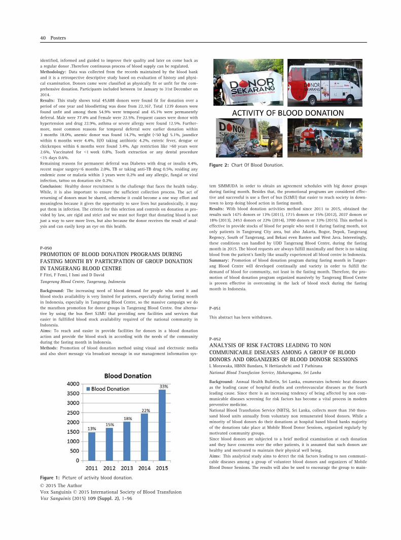
identified, informed and guided to improve their quality and later on come back as
a regular donor .Therefore continuous process of blood supply can be regulated.
Methodology: Data was collected from the records maintained by the blood bank
and it is a retrospective descriptive study based on evaluation of history and physi-
cal examination. Donors came were classified as physically fit or unfit for the com-
prehensive donation. Participants included between 1st January to 31st December on
2014.
Results: This study shows total 45,688 donors were found fit for donation over a
period of one year and bloodletting was done from 22,167. Total 1239 donors were
found unfit and among them 54.9% were temporal and 45.1% were permanently
deferral. Male were 77.4% and Female were 22.5%. Frequent causes were donor with
hypertension and drug 22.9%, asthma or severe allergy were found 12.5%. Further-
more, most common reasons for temporal deferral were earlier donation within
3 months 18.0%, anemic donor was found 14.7%, weight (<50 kg) 5.1%, jaundice
within 6 months were 4.4%, H/O taking antibiotic 4.2%, enteric fever, dengue or
chickenpox within 6 months were found 3.4%, Age restriction like >60 years were
2.6%, Vaccinated for <1 week 0.8%, Tooth extraction or any dental procedure
<15 days 0.6%.
Remaining reasons for permanent deferral was Diabetes with drug or insulin 4.4%,
recent major surgery<6 months 2.0%, TB or taking anti-TB drug 0.5%, residing any
endemic zone or malaria within 3 years were 0.2% and any allergic, fungal or viral
infection, tattoo on donation site 0.2%.
Conclusion: Healthy donor recruitment is the challenge that faces the health today.
While, it is also important to ensure the sufficient collection process. The act of
returning of donors must be shared, otherwise it could become a one way effort and
meaningless because it gives the opportunity to save lives but paradoxically, it may
put them in infection. The criteria for this selection and controls on donation as pro-
vided by law, are rigid and strict and we must not forget that donating blood is not
just a way to save more lives, but also because the donor receives the result of anal-
ysis and can easily keep an eye on this health.
P-050
PROMOTION OF BLOOD DONATION PROGRAMS DURINGFASTING MONTH BY PARTICIPATION OF GROUP DONATIONIN TANGERANG BLOOD CENTREF Fitri, F Femi, I Ismi and D David
Tangerang Blood Centre, Tangerang, Indonesia
Background: The increasing need of blood demand for people who need it and
blood stocks availability is very limited for patients, especially during fasting month
in Indonesia, especially in Tangerang Blood Centre, so the massive campaign we do
the marathon promotion for donor groups in Tangerang Blood Centre. One alterna-
tive by using the bus fleet S2MU that providing new facilities and services that
easier in fullfilled blood stock availability required of the national community in
Indonesia.
Aims: To reach and easier in provide facilities for donors in a blood donation
action and provide the blood stock in according with the needs of the community
during the fasting month in Indonesia.
Methods: Promotion of blood donation method using visual and electronic media
and also short message via broadcast message in our management information sys-
tem SIMMUDA in order to obtain an agreement schedules with big donor groups
during fasting month. Besides that, the promotional programs are considered effec-
tive and successful is use a fleet of bus (S2MU) that easier to reach society in down-
town to keep doing blood action in fasting month.
Results: With blood donation activities method since 2011 to 2015, obtained the
results such 1475 donors or 13% (2011), 1715 donors or 15% (2012), 2037 donors or
18% (2013), 2453 donors or 22% (2014), 3700 donors or 33% (2015). This method is
effective in provide stocks of blood for people who need it during fasting month, not
only patients in Tangerang City area, but also Jakarta, Bogor, Depok, Tangerang
Regency, South of Tangerang, and Bekasi even Banten and West Java. Interestingly,
these conditions can handled by UDD Tangerang Blood Centre, during the fasting
month in 2015. The blood requests are always fulfill maximally and there is no taking
blood from the patient’s family like usually experienced all blood centre in Indonesia.
Summary: Promotion of blood donation program during fasting month in Tanger-
ang Blood Centre will developed continually and variety in order to fulfill the
demand of blood for community, not least in the fasting month. Therefore, the pro-
motion of blood donation program organized massively by Tangerang Blood Centre
is proven effective in overcoming in the lack of blood stock during the fasting
month in Indonesia.
P-051
This abstract has been withdrawn.
P-052
ANALYSIS OF RISK FACTORS LEADING TO NONCOMMUNICABLE DISEASES AMONG A GROUP OF BLOODDONORS AND ORGANIZERS OF BLOOD DONOSR SESSIONSL Morawaka, HBNN Bandara, N Hettiarahchi and T Pathirana
National Blood Transfusion Service, Maharagama, Sri Lanka
Background: Annual Health Bulletin, Sri Lanka, enumerates ischemic heat diseases
as the leading cause of hospital deaths and cerebrovascular diseases as the fourth
leading cause. Since there is an increasing tendency of being affected by non com-
municable diseases screening for risk factors has become a vital process in modern
preventive medicine.
National Blood Transfusion Service (NBTS), Sri Lanka, collects more than 350 thou-
sand blood units annually from voluntary non remunerated blood donors. While a
minority of blood donors do their donations at hospital based blood banks majority
of the donations take place at Mobile Blood Donor Sessions, organized regularly by
motivated community groups.
Since blood donors are subjected to a brief medical examination at each donation
and they have concerns over the other patients, it is assumed that such donors are
healthy and motivated to maintain their physical well being.
Aims: This analytical study aims to detect the risk factors leading to non communi-
cable diseases among a group of volunteer blood donors and organizers of Mobile
Blood Donor Sessions. The results will also be used to encourage the group to main-
Figure 1: Picture of activity blood donation.
Figure 2: Chart Of Blood Donation.
© 2015 The AuthorVox Sanguinis © 2015 International Society of Blood TransfusionVox Sanguinis (2015) 109 (Suppl. 2), 1–96
40 Posters
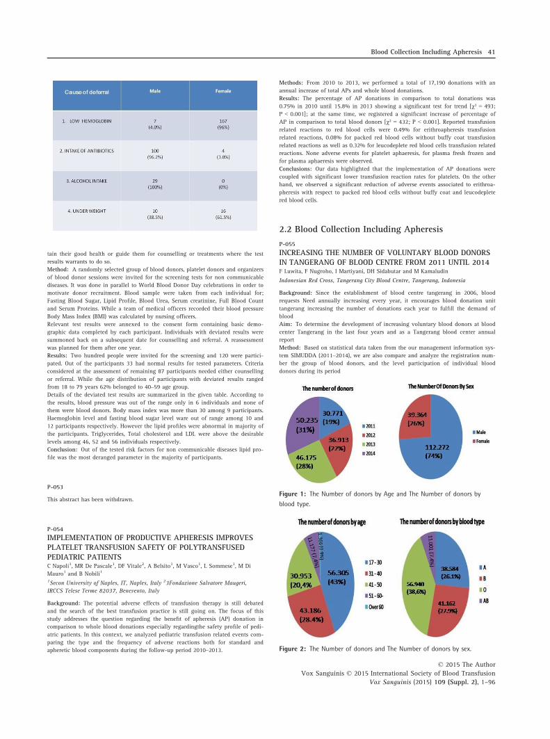
tain their good health or guide them for counselling or treatments where the test
results warrants to do so.
Method: A randomly selected group of blood donors, platelet donors and organizers
of blood donor sessions were invited for the screening tests for non communicable
diseases. It was done in parallel to World Blood Donor Day celebrations in order to
motivate donor recruitment. Blood sample were taken from each individual for;
Fasting Blood Sugar, Lipid Profile, Blood Urea, Serum creatinine, Full Blood Count
and Serum Proteins. While a team of medical officers recorded their blood pressure
Body Mass Index (BMI) was calculated by nursing officers.
Relevant test results were annexed to the consent form containing basic demo-
graphic data completed by each participant. Individuals with deviated results were
summoned back on a subsequent date for counselling and referral. A reassessment
was planned for them after one year.
Results: Two hundred people were invited for the screening and 120 were partici-
pated. Out of the participants 33 had normal results for tested parameters. Criteria
considered at the assessment of remaining 87 participants needed either counselling
or referral. While the age distribution of participants with deviated results ranged
from 18 to 79 years 62% belonged to 40–59 age group.
Details of the deviated test results are summarized in the given table. According to
the results, blood pressure was out of the range only in 6 individuals and none of
them were blood donors. Body mass index was more than 30 among 9 participants.
Haemoglobin level and fasting blood sugar level ware out of range among 10 and
12 participants respectively. However the lipid profiles were abnormal in majority of
the participants. Triglycerides, Total cholesterol and LDL were above the desirable
levels among 46, 52 and 56 individuals respectively.
Conclusion: Out of the tested risk factors for non communicable diseases lipid pro-
file was the most deranged parameter in the majority of participants.
P-053
This abstract has been withdrawn.
P-054
IMPLEMENTATION OF PRODUCTIVE APHERESIS IMPROVESPLATELET TRANSFUSION SAFETY OF POLYTRANSFUSEDPEDIATRIC PATIENTSC Napoli1, MR De Pascale1, DF Vitale2, A Belsito1, M Vasco1, L Sommese1, M Di
Mauro1 and B Nobili1
1Secon University of Naples, IT, Naples, Italy 23Fondazione Salvatore Maugeri,
IRCCS Telese Terme 82037, Benevento, Italy
Background: The potential adverse effects of transfusion therapy is still debated
and the search of the best transfusion practice is still going on. The focus of this
study addresses the question regarding the benefit of apheresis (AP) donation in
comparison to whole blood donations especially regardingthe safety profile of pedi-
atric patients. In this context, we analyzed pediatric transfusion related events com-
paring the type and the frequency of adverse reactions both for standard and
apheretic blood components during the follow-up period 2010–2013.
Methods: From 2010 to 2013, we performed a total of 17,190 donations with an
annual increase of total APs and whole blood donations.
Results: The percentage of AP donations in comparison to total donations was
0.75% in 2010 until 15.8% in 2013 showing a significant test for trend [v² = 493;
P < 0.001]; at the same time, we registered a significant increase of percentage of
AP in comparison to total blood donors [v² = 432; P < 0.001]. Reported transfusion
related reactions to red blood cells were 0.49% for erithroapheresis transfusion
related reactions, 0.08% for packed red blood cells without buffy coat transfusion
related reactions as well as 0.32% for leucodeplete red blood cells transfusion related
reactions. None adverse events for platelet aphaeresis, for plasma fresh frozen and
for plasma aphaeresis were observed.
Conclusions: Our data highlighted that the implementation of AP donations were
coupled with significant lower transfusion reaction rates for platelets. On the other
hand, we observed a significant reduction of adverse events associated to erithroa-
pheresis with respect to packed red blood cells without buffy coat and leucodeplete
red blood cells.
2.2 Blood Collection Including Apheresis
P-055
INCREASING THE NUMBER OF VOLUNTARY BLOOD DONORSIN TANGERANG OF BLOOD CENTRE FROM 2011 UNTIL 2014F Luwita, F Nugroho, I Martiyani, DH Sidabutar and M Kamaludin
Indonesian Red Cross, Tangerang City Blood Centre, Tangerang, Indonesia
Background: Since the establishment of blood centre tangerang in 2006, blood
requests Need annually increasing every year, it encourages blood donation unit
tangerang increasing the number of donations each year to fulfill the demand of
blood
Aim: To determine the development of increasing voluntary blood donors at blood
center Tangerang in the last four years and as a Tangerang blood center annual
report
Method: Based on statistical data taken from the our management information sys-
tem SIMUDDA (2011–2014), we are also compare and analyze the registration num-
ber the group of blood donors, and the level participation of individual blood
donors during its period
Figure 1: The Number of donors by Age and The Number of donors by
blood type.
Figure 2: The Number of donors and The Number of donors by sex.
© 2015 The AuthorVox Sanguinis © 2015 International Society of Blood Transfusion
Vox Sanguinis (2015) 109 (Suppl. 2), 1–96
Blood Collection Including Apheresis 41
Result: There is increasing number of voluntary blood donors each year, such
30.771 people (2011), 36.913 people (2012), 46.175 people (2013), 36.390 people
(2014), so total number of donors until now is 369.302 people. By sex; 112,272 male
(74%), 39.364 female (26%). Based on the age of the donor; 65.305 (43%) age 17–30, 43.186 (28.4%) age 31–40, 30.953 (20.4%) age 41–50, 11.177 (7, 4%) age 51–60,1.366 (0.9%) age over 60 years. Based on blood group, such; blood group A 38.584
(26.1%), blood group B 41.162 (27.9%), blood group O 56.940 (38.6%), blood group
AB 11.001 (7.4%)
Conclusion: The needs of underserved blood demand in blood center tangerang
very influential to the participation of voluntary blood donors. To achieve enhance-
ment number of voluntary blood donors every year, need to increase publicity about
the importance of blood donation, comfortable place with good facilities, promotion
of blood donation, etc.
P-056
THE CAUSE OF THE BLOOD COLLECTION’S FAILURE ONBLOOD DONOR BEGINNERS IN BLOOD DONOR SERVICESR Aprianti, YM Djoechro, U Muktimanah, W Suryawidjaja, Y Mulyati, H Purwati, A
Sriati and R Mulyani
Indonesian Red Cross, Bandung, Indonesia
Background: Until now the failure of the blood collection still occurs, primarily on
blood donor beginners.
One of the main reasons is donor reaction occurs during donation, either mild reac-
tions or moderate reactions, therefore will result in failure of blood collection.
Blood donor beginners are people who donate blood for the first time either volun-
tary or as a replacement donor with the age limit from 17 years old to 60 years old
and weigh equal or more than 45 kg.
Aims:
1. To determine the cause of failure in the blood donor beginners.
2. To identify the unknown factors that may affect the donor beginners.
Methods:
1. Descriptive research design with sample of 100 cases donor beginners.
2. Likert scale questionnaire data collectio, open questionnaire and structured inter-
views.
Result: From 100 donor beginners there are 20 donors failures of blood collection,
caused by:
1. Sleep after 12.00 a.m.
2. Excessive activity before donor.
Conclusions: Blood collection’s failure is have higher rates in donor beginners with
sleep after 12.00 a.m, excessive activity before donor, and having high levels of
anxiety.
Keywords: : failure, blood collection, blood donor beginner
P-057
DONOR ARM PREPARATION TECHNIQUE EFFICACYEVALUATIONCL Toh, SC Khoo and HH Tan
Health Sciences Authority, Singapore, Singapore
Background: Viral transmission via allogeneic blood is very low due to transfusion
safety measures, bacteria contamination is still a great concern. Though the source of
bacterial contamination in blood cannot always be easily identified, donors’ arm nev-
ertheless is a potential source of contamination. Type of antiseptic and cleaning tech-
nique have an effect on the efficacy of skin disinfection.Currently Blood Services
Group, Singapore (BSG) is using a 1 step cleansing procedure with swab stick con-
taining 2% w/v Chlorhexidine Gluconate in 70% v/v Isopropyl Alcohol in circular
motion from within out from the intended venepuncture site for 30 s, covering an
area of about 8 cm by 8 cm and allowed to air dry for at least 30 s.FDA in its options
for Arm Preparation (Method 4), approved scrubbing the intended venepuncture site
with 2% Chlorhexidine Gluconate in 70% Isopropyl Alcohol with repeated back and
forth strokes and paper published in Vox Sanguinis (2001) by C.P. McDonald on
‘evaluation of donor arm disinfection techniques’ found that the up and down motion
of the disinfectant application was more superior to spiral.
Aims: In this evaluation, the efficacies of circular motion technique currently prac-
tise by BSG and the repeated back and forth strokes technique with the same type of
antiseptic (2% w/v Chlorhexidine gluconate and 70% v/v Isopropyl alcohol), were
compared.
Methods: All phlebotomists in BSG were recruited as the subjects for this evalua-
tion to minimise inconvenience to donors. Those who are sensitive to either
Chlorhexidine or alcohol or pre-existing skin conditions on the antecubital fossa
area were excluded.Direct swabbing on the antecubital fossa area of both arms of
each subject was taken prior to skin disinfection as baseline.The antecubital fossa
area of the right arm of each subject was prepared with the antiseptic solution using
circular motion from within out and that of the left arm was prepared using
repeated back and forth strokes.Post disinfection swab on each arm was taken imme-
diately after the required drying time and sent to Pathology Laboratory for plating,
incubation and bacteria count.
Results: Post disinfection culture per plate for both methods had shown no growth
in 93% of the cases. In both methods there were 3 cases each or 7% with colony
growth of 1 to 4 colonies per plate post disinfection.
Summary and conclusion: There was no significant difference in efficacy with both
methods. In view of the findings and FDA’s option for arm preparation with 2%
Chlorhexidine Gluconate in 70% Isopropyl Alcohol, BSG has decided to adopt
repeated back and forth strokes technique for donors’ arm preparation.
P-058
VALIDATION OF THE HAEMOGLOBIN COLOUR SCALE INBLOOD DONORS IN MALAWIB M’baya1, I Mbingwani2, L Mgawi2, V Mkochi2, I Bates3, SA White2 and TJ Allain2
1Malawi Blood Transfusion Service, Blantyre, Malawi 2College of Medicine, Blantyre,
Malawi 3Liverpool School of Tropical Medicine, Liverpool, United Kingdom
Background: The Malawi Blood Transfusion Service (MBTS) was established in
2004 as the nationally coordinated blood service based on voluntary non-remuner-
ated blood donation. Malawi is a densely populated low Human Development Index
(HDI) Southern African country (2009 GDP per capita of USD290) with a population
of 15 million, 85% of whom live in rural areas. It cannot routinely use HemoCue for
anaemia screening due to the high cost of cuvettes. Since 2009, the MBTS uses the
WHO Haemoglobin Colour Scale (HCS) to screen all blood donors to determine their
haemoglobin eligibility for blood donation and to detect those that need additional
testing with HemoCue before their eligibility can be decided. Before 2009, clinical
assessment was used in addition to this but it was removed because it was felt that
this criteria was being abused due to its subjective nature. The 2009 HCS based pro-
tocol is used as follows: those with Hb <12 g/dl are deferred for blood donation;
those with Hb >12 g/dl are accepted for blood donation, while those with
Hb = 12 g/dl are referred for testing with HemoCue and are only accepted for dona-
tion if the Hb is ≥12.5 g/dl.However, both the pre and post 2009 protocols had not
been validated. Published studies of the accuracy of the HCS to screen potential
blood donors show varying levels of accuracy and opinion varies whether this is an
appropriate screening test or not hence the need for this study.
Aims: The aim of the study was to assess the validity of the MBTS protocol of
using the HCS and HemoCue in screening for anaemia in potential blood donors.
Methods: This was a blinded prospective study in potential blood donors aged over
18 years, at MBTS in Blantyre, Malawi. Capillary blood samples were analysed using
the HCS and HemoCue, independent of each other. The sensitivity and specificity of
correctly identifying eligible blood donors as per MBTS protocol were calculated.
Results: From 242 participants 8 (3%), were wrongly allocated on the basis of the
HCS compared to HemoCue. All were subjects that were wrongly accepted as donors
when their haemoglobin results were ≤12.0 g/dl. This gave a sensitivity of 65% and
specificity of 100% to detect eligibility for donation. No subjects were wrongly
excluded as donors.
Conclusions: Initial screening with the HCS correctly predicts eligibility for blood
donation in the majority of potential blood donors at considerable cost saving com-
pared with use of HemoCue as the first line anaemia screening test.
© 2015 The AuthorVox Sanguinis © 2015 International Society of Blood TransfusionVox Sanguinis (2015) 109 (Suppl. 2), 1–96
42 Posters
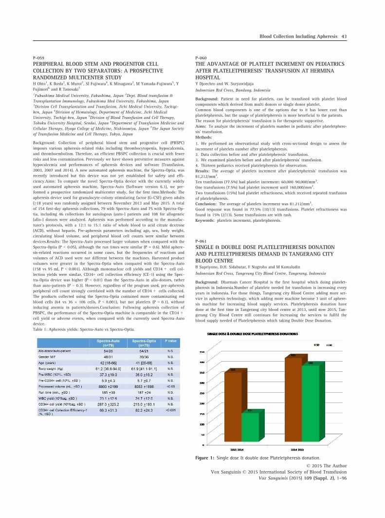
P-059
PERIPHERAL BLOOD STEM AND PROGENITOR CELLCOLLECTION BY TWO SEPARATORS: A PROSPECTIVERANDOMIZED MULTICENTER STUDYH Ohto1, K Ikeda2, K Muroi3, SI Fujiwara4, K Minagawa2, M Yamada-Fujiwara5, Y
Fujimori6 and R Tanosaki7
1Fukushima Medical University, Fukushima, Japan 2Dept. Blood transfusion &
Transplantation Immunology, Fukushima Med University, Fukushima, Japan3Division Cell Transplantation and Transfusion, Jichi Medical University, Tochigi-
ken, Japan 4Division of Hematology, Department of Medicine, Jichi Medical
University, Tochigi-ken, Japan 5Division of Blood Transfusion and Cell Therapy,
Tohoku University Hospital, Sendai, Japan 6Department of Transfusion Medicine and
Cellular Therapy, Hyogo College of Medicine, Nishinomiya, Japan 7The Japan Society
of Transfusion Medicine and Cell Therapy, Tokyo, Japan
Background: Collection of peripheral blood stem and progenitor cell (PBSPC)
imposes various apheresis-related risks including thrombocytopenia, hypocalcemia,
and thromboembolism. Therefore, an efficient PBSPC collection is crucial with fewer
risks and less contamination. Previously we have shown preventive measures against
hypocalcemia and performances of apheresis devices and software (Transfusion,
2003, 2007 and 2014). A new automated apheresis machine, the Spectra-Optia, was
recently introduced but this device was not yet established for safety and effi-
ciency.Aims: To compare the novel Spectra-Optia device with the currently widely
used automated apheresis machine, Spectra-Auto (Software version 6.1), we per-
formed a prospective randomized multicenter study, for the first time.Methods: The
apheresis device used for granulocyte-colony stimulating factor (G-CSF) given adults
(≥18 years) was randomly assigned between November 2013 and May 2015. A total
of 154 first-day apheresis collections, 79 with Spectra-Auto and 75 with Spectra-Op-
tia, including 46 collections for autologous (auto-) patients and 108 for allogeneic
(allo-) donors were analyzed. Apheresis was performed according to the manufac-
turer’s protocols, with a 12:1 to 15:1 ratio of whole blood to acid citrate dextrose
(ACD), without heparin. Pre-apheresis parameters including age, sex, body weight,
circulating blood volume, and peripheral blood cell counts were similar between
devices.Results: The Spectra-Auto processed larger volumes when compared with the
Spectra-Optia (P < 0.05), although the run times were similar (P = 0.6). Mild aphere-
sis-related reactions occurred in some cases, but the frequencies of reactions and
volumes of ACD used were not different between the machines. Harvested product
volumes were greater in the Spectra-Optia when compared with the Spectra-Auto
(158 vs 95 ml, P < 0.001). Although mononuclear cell yields and CD34 + cell col-
lection yields were similar, CD34+ cell collection efficiency (CE-1) using the Spec-
tra-Optia device was higher (P < 0.01) than the Spectra-Auto in allo-donors, rather
than auto-patients (P = 0.3). However, regardless of the program used, pre-apheresis
peripheral cell count strongly correlated with the number of CD34 + cells collected.
The products collected using the Spectra-Optia contained more contaminating red
blood cells (64 vs 36 9 106 cells, P < 0.001), but not platelets (P = 0.1), without
inducing anemia in patients/donors.Conclusion: Following apheresis collection of
PBSPC, the performance of the Spectra-Optia machine is comparable in the CD34 +cell yield or adverse events, when compared with the currently used Spectra-Auto
device.
Table 1: Apheresis yields: Spectra-Auto vs Spectra-Optia.
P-060
THE ADVANTAGE OF PLATELET INCREMENT ON PEDIATRICSAFTER PLATELETPHERESIS’ TRANSFUSION AT HERMINAHOSPITALY Djoechro and W. Suryawidjaja
Indonesian Red Cross, Bandung, Indonesia
Background: Patient in need for platelets, can be transfused with platelet blood
components which derived from multi donors or single donor platelet.
Common blood components is one of the options due to it has lower cost than
plateletpheresis, but the usage of plateletpheresis is more beneficial to the patients.
The reason for plateletpheresis’ transfusion is for therapeutic supportive.
Aims: To analyze the increment of platelets number in pediatric after plateletphere-
sis’ transfusion.
Methods:
1. We performed an observational study with cross-sectional design to assess the
increment of platelets number after plateletpheresis.
2. Data collection before and after plateletpheresis’ transfusion.
3. We examined platelets before and after plateletpheresis’ transfusion.
4. Thirteen pediatrics received plateletpheresis for observation.
Results: The average of platelets increment after plateletpheresis’ transfusion was
81,212/mm3.
Ten tranfusions (77.5%) had platelet increment: 60,000–90,000/mm3.
One transfusions (7.5%) had platelet increment until 160,000/mm3.
Two transfusions (15%) had platelet refractioness, which received repeated tranfusion
of plateletpheresis.
Conclusion: The average of platelets increment was 81.212/mm3.
Good response was found in 77.5% (10/13) transfusions. Platelet refractioness was
found in 15% (2/13). Some transfusions are with rash.
Keywords: platelets increment, plateletpheresis.
P-061
SINGLE & DOUBLE DOSE PLATELETPHERESIS DONATIONAND PLETELETPHERESIS DEMAND IN TANGERANG CITYBLOOD CENTREB Supriyono, D.H. Sidabutar, F Nugroho and M Kamaludin
Indonesian Red Cross, Tangerang City Blood Centre, Tangerang, Indonesia
Background: Dharmais Cancer Hospital is the first hospital which doing platelet-
pheresis in Indonesia.Number of platelete needed for transfusion is increasing every
years in indonesia. For those things, Tangerang city Blood Centre adding more ser-
vice in apheresis technology, which adding more machine become 3 unit of aphere-
sis machine for increasing blood supply services. Plateletpheresis donation have
done at the first time in Tangerang city blood centre at 2013, until now 2015, Tan-
gerang City Blood Centre still continues for increasing the services to fullfil the
blood supply needed of Plateletpheresis which taking Double Dose Donation.
Figure 1: Single dose & double dose Plateletpheresis donation.
© 2015 The AuthorVox Sanguinis © 2015 International Society of Blood Transfusion
Vox Sanguinis (2015) 109 (Suppl. 2), 1–96
Blood Collection Including Apheresis 43
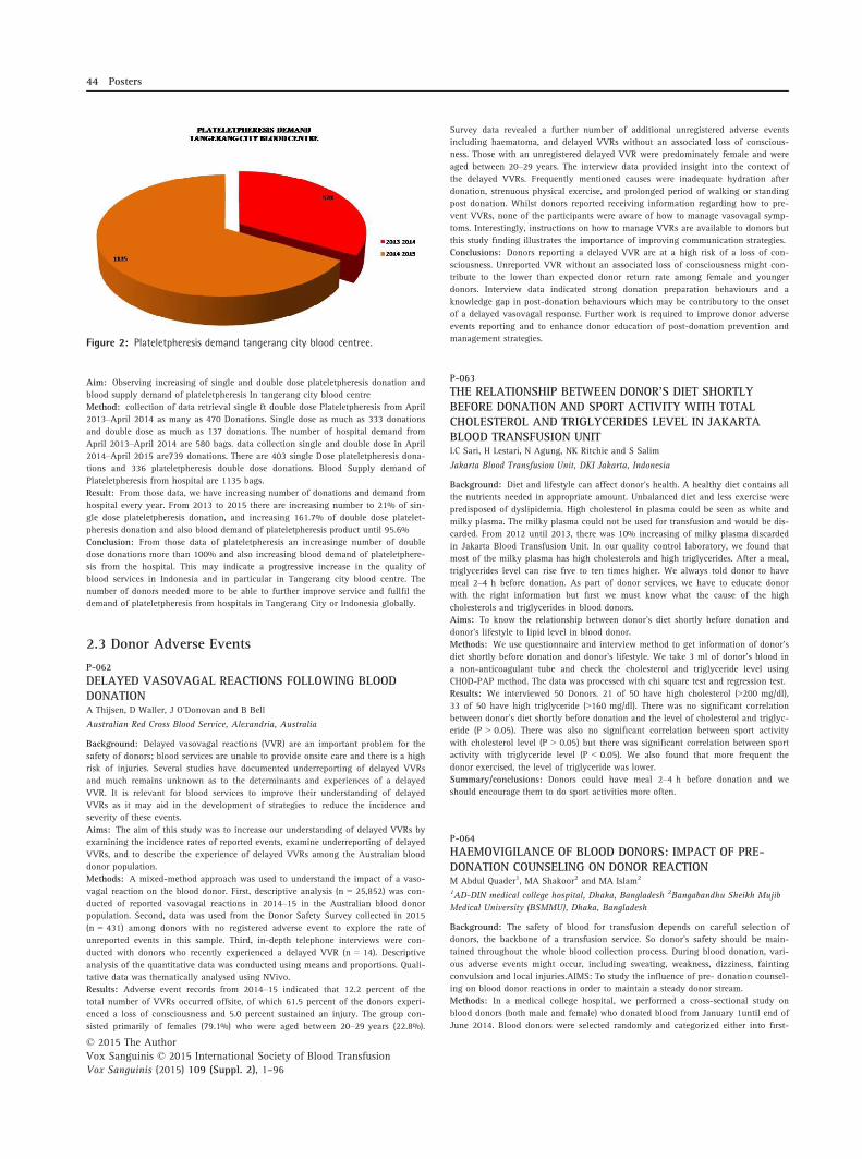
Aim: Observing increasing of single and double dose plateletpheresis donation and
blood supply demand of plateletpheresis In tangerang city blood centre
Method: collection of data retrieval single & double dose Plateletpheresis from April
2013–April 2014 as many as 470 Donations. Single dose as much as 333 donations
and double dose as much as 137 donations. The number of hospital demand from
April 2013–April 2014 are 580 bags. data collection single and double dose in April
2014–April 2015 are739 donations. There are 403 single Dose plateletpheresis dona-
tions and 336 plateletpheresis double dose donations. Blood Supply demand of
Plateletpheresis from hospital are 1135 bags.
Result: From those data, we have increasing number of donations and demand from
hospital every year. From 2013 to 2015 there are increasing number to 21% of sin-
gle dose plateletpheresis donation, and increasing 161.7% of double dose platelet-
pheresis donation and also blood demand of plateletpheresis product until 95.6%
Conclusion: From those data of plateletpheresis an increasinge number of double
dose donations more than 100% and also increasing blood demand of plateletphere-
sis from the hospital. This may indicate a progressive increase in the quality of
blood services in Indonesia and in particular in Tangerang city blood centre. The
number of donors needed more to be able to further improve service and fullfil the
demand of plateletpheresis from hospitals in Tangerang City or Indonesia globally.
2.3 Donor Adverse Events
P-062
DELAYED VASOVAGAL REACTIONS FOLLOWING BLOODDONATIONA Thijsen, D Waller, J O’Donovan and B Bell
Australian Red Cross Blood Service, Alexandria, Australia
Background: Delayed vasovagal reactions (VVR) are an important problem for the
safety of donors; blood services are unable to provide onsite care and there is a high
risk of injuries. Several studies have documented underreporting of delayed VVRs
and much remains unknown as to the determinants and experiences of a delayed
VVR. It is relevant for blood services to improve their understanding of delayed
VVRs as it may aid in the development of strategies to reduce the incidence and
severity of these events.
Aims: The aim of this study was to increase our understanding of delayed VVRs by
examining the incidence rates of reported events, examine underreporting of delayed
VVRs, and to describe the experience of delayed VVRs among the Australian blood
donor population.
Methods: A mixed-method approach was used to understand the impact of a vaso-
vagal reaction on the blood donor. First, descriptive analysis (n = 25,852) was con-
ducted of reported vasovagal reactions in 2014–15 in the Australian blood donor
population. Second, data was used from the Donor Safety Survey collected in 2015
(n = 431) among donors with no registered adverse event to explore the rate of
unreported events in this sample. Third, in-depth telephone interviews were con-
ducted with donors who recently experienced a delayed VVR (n = 14). Descriptive
analysis of the quantitative data was conducted using means and proportions. Quali-
tative data was thematically analysed using NVivo.
Results: Adverse event records from 2014–15 indicated that 12.2 percent of the
total number of VVRs occurred offsite, of which 61.5 percent of the donors experi-
enced a loss of consciousness and 5.0 percent sustained an injury. The group con-
sisted primarily of females (79.1%) who were aged between 20–29 years (22.8%).
Survey data revealed a further number of additional unregistered adverse events
including haematoma, and delayed VVRs without an associated loss of conscious-
ness. Those with an unregistered delayed VVR were predominately female and were
aged between 20–29 years. The interview data provided insight into the context of
the delayed VVRs. Frequently mentioned causes were inadequate hydration after
donation, strenuous physical exercise, and prolonged period of walking or standing
post donation. Whilst donors reported receiving information regarding how to pre-
vent VVRs, none of the participants were aware of how to manage vasovagal symp-
toms. Interestingly, instructions on how to manage VVRs are available to donors but
this study finding illustrates the importance of improving communication strategies.
Conclusions: Donors reporting a delayed VVR are at a high risk of a loss of con-
sciousness. Unreported VVR without an associated loss of consciousness might con-
tribute to the lower than expected donor return rate among female and younger
donors. Interview data indicated strong donation preparation behaviours and a
knowledge gap in post-donation behaviours which may be contributory to the onset
of a delayed vasovagal response. Further work is required to improve donor adverse
events reporting and to enhance donor education of post-donation prevention and
management strategies.
P-063
THE RELATIONSHIP BETWEEN DONOR’S DIET SHORTLYBEFORE DONATION AND SPORT ACTIVITY WITH TOTALCHOLESTEROL AND TRIGLYCERIDES LEVEL IN JAKARTABLOOD TRANSFUSION UNITLC Sari, H Lestari, N Agung, NK Ritchie and S Salim
Jakarta Blood Transfusion Unit, DKI Jakarta, Indonesia
Background: Diet and lifestyle can affect donor’s health. A healthy diet contains all
the nutrients needed in appropriate amount. Unbalanced diet and less exercise were
predisposed of dyslipidemia. High cholesterol in plasma could be seen as white and
milky plasma. The milky plasma could not be used for transfusion and would be dis-
carded. From 2012 until 2013, there was 10% increasing of milky plasma discarded
in Jakarta Blood Transfusion Unit. In our quality control laboratory, we found that
most of the milky plasma has high cholesterols and high triglycerides. After a meal,
triglycerides level can rise five to ten times higher. We always told donor to have
meal 2–4 h before donation. As part of donor services, we have to educate donor
with the right information but first we must know what the cause of the high
cholesterols and triglycerides in blood donors.
Aims: To know the relationship between donor’s diet shortly before donation and
donor’s lifestyle to lipid level in blood donor.
Methods: We use questionnaire and interview method to get information of donor’s
diet shortly before donation and donor’s lifestyle. We take 3 ml of donor’s blood in
a non-anticoagulant tube and check the cholesterol and triglyceride level using
CHOD-PAP method. The data was processed with chi square test and regression test.
Results: We interviewed 50 Donors. 21 of 50 have high cholesterol (>200 mg/dl),
33 of 50 have high triglyceride (>160 mg/dl). There was no significant correlation
between donor’s diet shortly before donation and the level of cholesterol and triglyc-
eride (P > 0.05). There was also no significant correlation between sport activity
with cholesterol level (P > 0.05) but there was significant correlation between sport
activity with triglyceride level (P < 0.05). We also found that more frequent the
donor exercised, the level of triglyceride was lower.
Summary/conclusions: Donors could have meal 2–4 h before donation and we
should encourage them to do sport activities more often.
P-064
HAEMOVIGILANCE OF BLOOD DONORS: IMPACT OF PRE-DONATION COUNSELING ON DONOR REACTIONM Abdul Quader1, MA Shakoor2 and MA Islam2
1AD-DIN medical college hospital, Dhaka, Bangladesh 2Bangabandhu Sheikh Mujib
Medical University (BSMMU), Dhaka, Bangladesh
Background: The safety of blood for transfusion depends on careful selection of
donors, the backbone of a transfusion service. So donor’s safety should be main-
tained throughout the whole blood collection process. During blood donation, vari-
ous adverse events might occur, including sweating, weakness, dizziness, fainting
convulsion and local injuries.AIMS: To study the influence of pre- donation counsel-
ing on blood donor reactions in order to maintain a steady donor stream.
Methods: In a medical college hospital, we performed a cross-sectional study on
blood donors (both male and female) who donated blood from January 1until end of
June 2014. Blood donors were selected randomly and categorized either into first-
Figure 2: Plateletpheresis demand tangerang city blood centree.
© 2015 The AuthorVox Sanguinis © 2015 International Society of Blood TransfusionVox Sanguinis (2015) 109 (Suppl. 2), 1–96
44 Posters
time or regular donors. Adverse donor reactions were documented in donors with or
without counseling. In this study, only blood donors who donated within the last
two years included into the regular donor group.
Results: 256 out of 4145 randomly selected donors were investigated in total. 111/
256 selected donors were first-time and 145/256 were regular donors.54/ 111 first-
time donors were counseled, while 57 were not.76/145 of regular donors were coun-
seled, 69 were not. The mean age of first-time donors was 30.27 � 2.34, for regular
donor’s 31.57 � 1.95 years. In 20.37% of first-time donors who were being coun-
seled, donation reactions occurred, while this was 45.61% in the group who did not
undergo counseling. Among regular donors who received counseling, 15.79% expe-
rienced a donor reaction. In contrast, this was 31.88% who were not counseled. In
total, 27.73% of studied donors experienced a donor reaction. In the counseling
group, this was 8.98% while this was 18.75% in the without counseling group. A
total of 185 (72.27%) showed negative reaction: 107 (41.80%) were part of the coun-
seling group, and 78 (30.47%) in non- counseled group.
Conclusions: We found that in the counseled group less adverse donor reactions
occurred but the donors also responded more negatively towards counseling when
compared with the non- counseled group. Although our null hypothesis was: no
association between donor reaction and counseling, this study did show a strong
association (P<0.001). In general, we found that counseling before blood donation
has great impact on the number of adverse donor reactions. Therefore, in order to
maintain a high quality donor chain, we should therefore implement counseling for
all donors pre-donation.
P-065
SOME CHARACTERISTIC FACTORS ASSOCIATED WITHADVERSE REACTIONS IN WHOLE BLOOD DONATIONIP Rupasena
National Blood Center, Colombo 05, Sri Lanka
Background: Blood donation programme in Sri Lanka is conducted by National
Blood Transfusion Service. The National Blood Transfusion Service is responsible for
the blood collection, processing, testing and distribution of blood to both public and
private hospitals. All the requirements of blood are fulfilled by the voluntary non-
remunerated blood donation.
Blood donors are the backbone of Blood Transfusion Service. Ensuring the safety of
blood donors is of extremely importance. If the donors are ensured of a pleasant expe-
rience during blood donation they could be motivated to become regular blood donors.
This can be accomplished by way of preventing adverse reactions in the donor.
Aims: This study was conducted to determine and estimate the frequency of occur-
rence of adverse reactions in whole blood donors and to identify associated factors
like age, sex, and body weight in the causation of donor adverse reactions.
Methodology: The retrospective study was conducted a period of one year from 01/
10/2013 to 30/09/2014 in National Blood Center, Colombo, Sri Lanka. The data was
collected from four mobile blood collecting in relevant period.
Result: Total of 62,130 voluntary blood donors, 67.3%(n = 41,814) males and
32.7% (n = 20,316) females were studied during relevant period for adverse reaction
of blood donation.
2.22% (n = 1382) of voluntary blood donors reacted to blood donation. Male donors
accounted for 58.18%(n = 804) reactions while the female counterpart was responsi-
ble for 41.82% (n = 578).First time donors were significantly more likely had
adverse effect 60.85% (n = 841) than regular blood donors 39.15% (n = 541).
The age distribution of donors were 24.6%(n = 15,311), 47.9% (n = 29,695), 16.7%
(n = 10,390) and 10.8% (n = 6734) for aged 18–25, 26–35, 36–45 and 46–60 years
respectively. The rate of adverse effect was highest, 3.85% among donors 18–25 years
compared to 1.2%, 3%, and 1.8% respectively for other age groups (P < 0.001).
The rate of adverse reactions was higher among donors who weighing between 50–59 kg than the higher weight group (weight 60 kg or above) (P < 0.001) .
Table 01.
Conclusion: There was a predictable low rate of adverse reaction to blood donation
among voluntary blood donors. First time donors were more prone to get adverse
reactions than regular donors. When considering the age of donors, there was signif-
icant reactions among donors who were in between 18–25 years of age .Adverse
reactions of blood donation was significantly occurred in donors who had 50–59 kg
body weight than donors 60 kg body weight or more. Donor friendly environment,
relieving of peer pressure, shorten the waiting time and good communication with
donor may help to resolve the occurrence of adverse reactions.
Table 02.
P-066
OMAN EXPERIENCE AFTER IMPLEMENTING DONORHEMOVIGILANCE (DHV) AT CENTRAL BLOOD BANK,DEPARTMENT OF BLOOD SERVICES, MUSCATAH Sakr, T Ashraf and A Al-Balushi
Ministry of Health, Sultanate of Oman, Muscat, Oman
Background: Donor health and safety should be of equal concern to blood safety.
Donor hemovigilance is the systemic monitoring of adverse reactions and incidents
in the whole chain of blood donor care, with a view to improving quality and safety
for blood donors.
Aim: The aim of this study is to monitor complications related to blood donation
(adverse reactions), errors in donor care (adverse incidents) and to focus on aspects
related to donor safety.
Methods: Donor hemovigilance reporting system (DHV Form) based on ISBT/IHN
classification is adapted. Full training of all nurses conducted to ensure proper
recognition, reporting and classification of adverse events. Donor reactions are strat-
ified under age, sex and frequency of donation. Age was classified into three cate-
gories (18–24 years), (24–40 years) and (40–65 years). Frequency of donation was
only divided into 1st timers and repeated donors.
Results: This programme started in 20/9/2014 and data given till 31/5/2015.
Through this period 18,878 donors males represented 83.47% while females 16.53%.
Repeated donors were 52.64% against 47.32%. Repeated donors had the upper hand
in males by 53.5% while the 1st timers were in females by 51.63%. As total levels
of reactions we had 440 males (2.79%) and females 254 (8.14%). 1st timers donors
in both sexes were 400 (57.80%) while repeated donors reactions were 292 (42.20%).
According to age classifications the mean of reactions percentage in relation to their
total number in the same age group, males between 18–24 years had reactions per-
centage (5.66%) while females showed (11.94%). Age between 24–40 years males
had (2.48%) while females had (4.97%). Last age group between 40–65 years males
had (1.49%) females had (2.36%). Through the details of reactions types the pro-
gramme of donor vigilance showed that we recorded (1213) form of reactions pre-
sented as hyperventilation (118), Sweating (252), Dizziness (539), loss of
consciousness (72), vomiting (15), convulsions (46), hematomas and re-bleeding (52),
nerve irritation (4), offsite reactions (112) and only one case of hypocalcaemia
symptoms. Many cases of multiple forms of reaction occurred to same donors.
Conclusion: However, the current study covers short time period with limited num-
ber of donors but gives us a conclusive understanding that DHV is an important and
valuable tool in monitoring the quality of blood donation services delivered. Most
frequently recorded reaction remains dizziness. There is notified number of convul-
sions reaction (46) and off-site reactions (112) for further investigations. Upon anal-
ysis, reactions occurred more frequently in younger age, female and first time
donors. The data is comparable to results reported from blood banks in other parts
of the world. With comparable classification (ISBT/IHN), one could not only look
into other countries, but also look into effectiveness of risk reduction strategies and
follow-up trends. We started to implement pre-donation hydration as a method of
intervention to reduce levels of complications.
3. Blood Products3.1 Blood Processing, Storage and Release
P-067
HEMOLYSIS POTENCY OF WASHED ERYTHROCYTE AND IN-LINE RED CELL LEUKODEPLETED COMPONENTSTSA Akbar1, ASM Sofro2 and RSE Gantini3
1Indonesian Red Cross – Banda Aceh Blood Center, Banda Aceh, Indonesia 2Faculty
of Medicine YARSI University, Jakarta, Indonesia 3Center Blood Transfusion Service,
Jakarta, Indonesia
Background: Washed erythrocyte (WE) and red cell leukodepleted (RC-LD) are nor-
mally used in clinical transfusion to prevent transfusion reaction. However, in
Indonesia there is no evidence on hemolysis due to erythrocyte washing and leuko-
cyte filtration that may cause significant clinical impact.
© 2015 The AuthorVox Sanguinis © 2015 International Society of Blood Transfusion
Vox Sanguinis (2015) 109 (Suppl. 2), 1–96
Donor Adverse Events 45
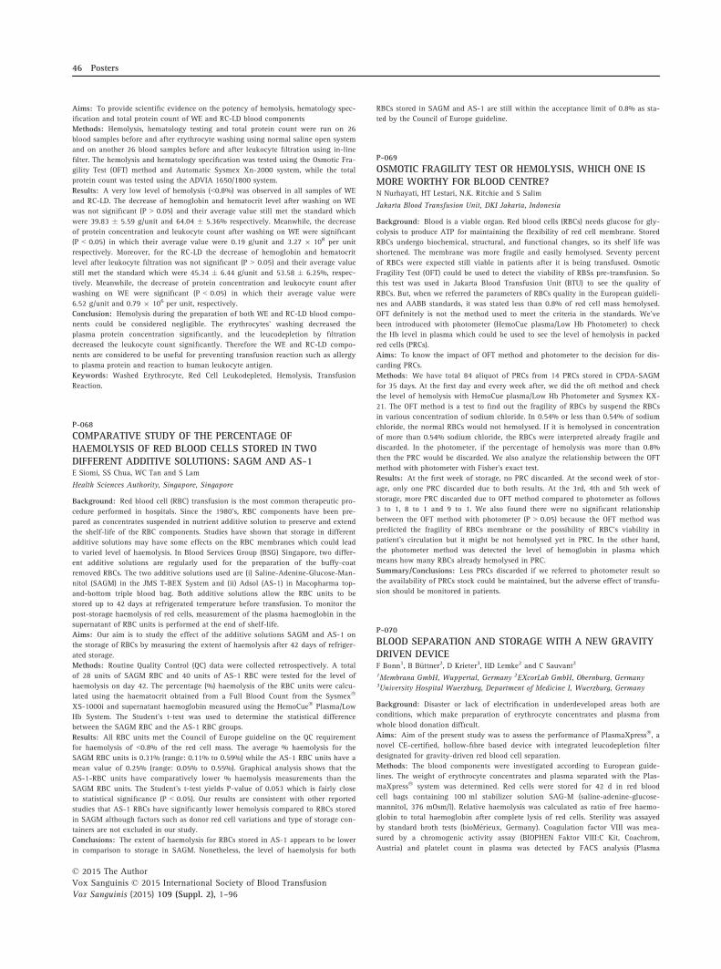
Aims: To provide scientific evidence on the potency of hemolysis, hematology spec-
ification and total protein count of WE and RC-LD blood components
Methods: Hemolysis, hematology testing and total protein count were run on 26
blood samples before and after erythrocyte washing using normal saline open system
and on another 26 blood samples before and after leukocyte filtration using in-line
filter. The hemolysis and hematology specification was tested using the Osmotic Fra-
gility Test (OFT) method and Automatic Sysmex Xn-2000 system, while the total
protein count was tested using the ADVIA 1650/1800 system.
Results: A very low level of hemolysis (<0.8%) was observed in all samples of WE
and RC-LD. The decrease of hemoglobin and hematocrit level after washing on WE
was not significant (P > 0.05) and their average value still met the standard which
were 39.83 � 5.59 g/unit and 64.04 � 5.36% respectively. Meanwhile, the decrease
of protein concentration and leukocyte count after washing on WE were significant
(P < 0.05) in which their average value were 0.19 g/unit and 3.27 9 108 per unit
respectively. Moreover, for the RC-LD the decrease of hemoglobin and hematocrit
level after leukocyte filtration was not significant (P > 0.05) and their average value
still met the standard which were 45.34 � 6.44 g/unit and 53.58 � 6.25%, respec-
tively. Meanwhile, the decrease of protein concentration and leukocyte count after
washing on WE were significant (P < 0.05) in which their average value were
6.52 g/unit and 0.79 9 106 per unit, respectively.
Conclusion: Hemolysis during the preparation of both WE and RC-LD blood compo-
nents could be considered negligible. The erythrocytes’ washing decreased the
plasma protein concentration significantly, and the leucodepletion by filtration
decreased the leukocyte count significantly. Therefore the WE and RC-LD compo-
nents are considered to be useful for preventing transfusion reaction such as allergy
to plasma protein and reaction to human leukocyte antigen.
Keywords: Washed Erythrocyte, Red Cell Leukodepleted, Hemolysis, Transfusion
Reaction.
P-068
COMPARATIVE STUDY OF THE PERCENTAGE OFHAEMOLYSIS OF RED BLOOD CELLS STORED IN TWODIFFERENT ADDITIVE SOLUTIONS: SAGM AND AS-1E Siomi, SS Chua, WC Tan and S Lam
Health Sciences Authority, Singapore, Singapore
Background: Red blood cell (RBC) transfusion is the most common therapeutic pro-
cedure performed in hospitals. Since the 1980’s, RBC components have been pre-
pared as concentrates suspended in nutrient additive solution to preserve and extend
the shelf-life of the RBC components. Studies have shown that storage in different
additive solutions may have some effects on the RBC membranes which could lead
to varied level of haemolysis. In Blood Services Group (BSG) Singapore, two differ-
ent additive solutions are regularly used for the preparation of the buffy-coat
removed RBCs. The two additive solutions used are (i) Saline-Adenine-Glucose-Man-
nitol (SAGM) in the JMS T-BEX System and (ii) Adsol (AS-1) in Macopharma top-
and-bottom triple blood bag. Both additive solutions allow the RBC units to be
stored up to 42 days at refrigerated temperature before transfusion. To monitor the
post-storage haemolysis of red cells, measurement of the plasma haemoglobin in the
supernatant of RBC units is performed at the end of shelf-life.
Aims: Our aim is to study the effect of the additive solutions SAGM and AS-1 on
the storage of RBCs by measuring the extent of haemolysis after 42 days of refriger-
ated storage.
Methods: Routine Quality Control (QC) data were collected retrospectively. A total
of 28 units of SAGM RBC and 40 units of AS-1 RBC were tested for the level of
haemolysis on day 42. The percentage (%) haemolysis of the RBC units were calcu-
lated using the haematocrit obtained from a Full Blood Count from the Sysmex�
XS-1000i and supernatant haemoglobin measured using the HemoCue� Plasma/Low
Hb System. The Student’s t-test was used to determine the statistical difference
between the SAGM RBC and the AS-1 RBC groups.
Results: All RBC units met the Council of Europe guideline on the QC requirement
for haemolysis of <0.8% of the red cell mass. The average % haemolysis for the
SAGM RBC units is 0.31% (range: 0.11% to 0.59%) while the AS-1 RBC units have a
mean value of 0.25% (range: 0.05% to 0.55%). Graphical analysis shows that the
AS-1-RBC units have comparatively lower % haemolysis measurements than the
SAGM RBC units. The Student’s t-test yields P-value of 0.053 which is fairly close
to statistical significance (P < 0.05). Our results are consistent with other reported
studies that AS-1 RBCs have significantly lower hemolysis compared to RBCs stored
in SAGM although factors such as donor red cell variations and type of storage con-
tainers are not excluded in our study.
Conclusions: The extent of haemolysis for RBCs stored in AS-1 appears to be lower
in comparison to storage in SAGM. Nonetheless, the level of haemolysis for both
RBCs stored in SAGM and AS-1 are still within the acceptance limit of 0.8% as sta-
ted by the Council of Europe guideline.
P-069
OSMOTIC FRAGILITY TEST OR HEMOLYSIS, WHICH ONE ISMORE WORTHY FOR BLOOD CENTRE?N Nurhayati, HT Lestari, N.K. Ritchie and S Salim
Jakarta Blood Transfusion Unit, DKI Jakarta, Indonesia
Background: Blood is a viable organ. Red blood cells (RBCs) needs glucose for gly-
colysis to produce ATP for maintaining the flexibility of red cell membrane. Stored
RBCs undergo biochemical, structural, and functional changes, so its shelf life was
shortened. The membrane was more fragile and easily hemolysed. Seventy percent
of RBCs were expected still viable in patients after it is being transfused. Osmotic
Fragility Test (OFT) could be used to detect the viability of RBSs pre-transfusion. So
this test was used in Jakarta Blood Transfusion Unit (BTU) to see the quality of
RBCs. But, when we referred the parameters of RBCs quality in the European guideli-
nes and AABB standards, it was stated less than 0.8% of red cell mass hemolysed.
OFT definitely is not the method used to meet the criteria in the standards. We’ve
been introduced with photometer (HemoCue plasma/Low Hb Photometer) to check
the Hb level in plasma which could be used to see the level of hemolysis in packed
red cells (PRCs).
Aims: To know the impact of OFT method and photometer to the decision for dis-
carding PRCs.
Methods: We have total 84 aliquot of PRCs from 14 PRCs stored in CPDA-SAGM
for 35 days. At the first day and every week after, we did the oft method and check
the level of hemolysis with HemoCue plasma/Low Hb Photometer and Sysmex KX-
21. The OFT method is a test to find out the fragility of RBCs by suspend the RBCs
in various concentration of sodium chloride. In 0.54% or less than 0.54% of sodium
chloride, the normal RBCs would not hemolysed. If it is hemolysed in concentration
of more than 0.54% sodium chloride, the RBCs were interpreted already fragile and
discarded. In the photometer, if the percentage of hemolysis was more than 0.8%
then the PRC would be discarded. We also analyze the relationship between the OFT
method with photometer with Fisher’s exact test.
Results: At the first week of storage, no PRC discarded. At the second week of stor-
age, only one PRC discarded due to both results. At the 3rd, 4th and 5th week of
storage, more PRC discarded due to OFT method compared to photometer as follows
3 to 1, 8 to 1 and 9 to 1. We also found there were no significant relationship
between the OFT method with photometer (P > 0.05) because the OFT method was
predicted the fragility of RBCs membrane or the possibility of RBC’s viability in
patient’s circulation but it might be not hemolysed yet in PRC. In the other hand,
the photometer method was detected the level of hemoglobin in plasma which
means how many RBCs already hemolysed in PRC.
Summary/Conclusions: Less PRCs discarded if we referred to photometer result so
the availability of PRCs stock could be maintained, but the adverse effect of transfu-
sion should be monitored in patients.
P-070
BLOOD SEPARATION AND STORAGE WITH A NEW GRAVITYDRIVEN DEVICEF Bonn1, B B€uttner2, D Krieter3, HD Lemke2 and C Sauvant2
1Membrana GmbH, Wuppertal, Germany 2EXcorLab GmbH, Obernburg, Germany3University Hospital Wuerzburg, Department of Medicine I, Wuerzburg, Germany
Background: Disaster or lack of electrification in underdeveloped areas both are
conditions, which make preparation of erythrocyte concentrates and plasma from
whole blood donation difficult.
Aims: Aim of the present study was to assess the performance of PlasmaXpress�, a
novel CE-certified, hollow-fibre based device with integrated leucodepletion filter
designated for gravity-driven red blood cell separation.
Methods: The blood components were investigated according to European guide-
lines. The weight of erythrocyte concentrates and plasma separated with the Plas-
maXpress� system was determined. Red cells were stored for 42 d in red blood
cell bags containing 100 ml stabilizer solution SAG-M (saline-adenine-glucose-
mannitol, 376 mOsm/l). Relative haemolysis was calculated as ratio of free haemo-
globin to total haemoglobin after complete lysis of red cells. Sterility was assayed
by standard broth tests (bioM�erieux, Germany). Coagulation factor VIII was mea-
sured by a chromogenic activity assay (BIOPHEN Faktor VIII:C Kit, Coachrom,
Austria) and platelet count in plasma was detected by FACS analysis (Plasma
© 2015 The AuthorVox Sanguinis © 2015 International Society of Blood TransfusionVox Sanguinis (2015) 109 (Suppl. 2), 1–96
46 Posters
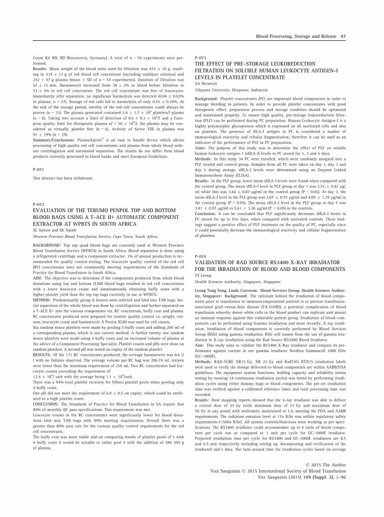
Count Kit BD, BD Biosciences, Germany). A total of n = 54 experiments were per-
formed.
Results: Mean weight of the blood units used for filtration was 474 � 10 g, result-
ing in 319 � 13 g of red blood cell concentrate (including stabilizer solution) and
242 � 47 g plasma (mean � SD of n = 54 experiments). Duration of filtration was
57 � 15 min. Haematocrit increased from 38 � 3% in blood before filtration to
53 � 3% in red cell concentrate. The red cell concentrate was free of leucocytes.
Immediately after separation, no significant haemolysis was detected (0.04 � 0.02%
in plasma; n = 27). Storage of red cells led to haemolysis of only 0.45 � 0.16%. At
the end of the storage period, sterility of the red cell concentrates could always be
proven (n = 23). The plasma generated contained 2.4 � 1.7 9 106 platelets/l plasma
(n = 6). Taking into account a limit of detection of 0.6 � 0.3 9 106/l and a Euro-
pean quality limit for therapeutic plasma of < 50 9 109/l, the plasma may be con-
sidered as virtually platelet free (n = 6). Activity of factor VIII in plasma was
91 � 19% (n = 29).
Summary/Conclusions: PlasmaXpress� is an easy to handle device which allows
processing of high quality red cell concentrates and plasma from whole blood with-
out centrifugation and automated separation. The results do not differ from blood
products currently generated in blood banks and meet European Guidelines.
P-071
This abstract has been withdrawn.
P-072
EVALUATION OF THE TERUMO PENPOL TOP AND BOTTOMBLOOD BAGS USING A T-ACE II+ AUTOMATIC COMPONENTEXTRACTOR AT WPBTS IN SOUTH AFRICASL Sutton and DL Smith
Western Province Blood Transfusion Service, Cape Town, South Africa
BACKGROUND: Top top quad blood bags are currently used at Western Province
Blood Transfusion Service (WPBTS) in South Africa. Blood separation is done using
a refrigerated centrifuge and a component extractor. 1% of annual production is rec-
ommended for quality control testing. The leucocyte quality control of the red cell
(RC) concentrates were not consistently meeting requirements of the Standards of
Practice for Blood Transfusion in South Africa.
AIM: The objective was to determine if the components produced from whole blood
donations using top and bottom (TAB) blood bags resulted in red cell concentrates
with a lower leucocyte count and simultaneously obtaining buffy coats with a
higher platelet yield than the top top bags currently in use at WPBTS.
METHOD: Predominantly group A donors were selected and bled into TAB bags. Ini-
tial separation of the whole blood was done by centrifugation and further separated on
a T-ACE II+ into the various components viz. RC concentrate, buffy coat and plasma.
RC concentrates produced were prepared for routine quality control i.e. weight, vol-
ume, leucocyte count and haematocrit. A Pentra XL80 was used for cell counts.
Six random donor platelets were made by pooling 5 buffy coats and adding 200 ml of
a corresponding plasma, which is our current method. A further twenty one random
donor platelets were made using 4 buffy coats and an increased volume of plasma at
the advice of a Component Processing Specialist. Platelet counts and pHs were done on
random platelets. A second pH was tested on expiry of the random platelet.
RESULTS: Of the 171 RC concentrates produced, the average haematocrit was 0.6 l/
l with no failures observed. The average volume per RC bag was 286.74 ml, sixteen
were lower than the minimum requirement of 250 ml. Two RC concentrates had leu-
cocyte counts exceeding the requirement of
≤2.4 9 109/ unit with the average being 1.1 9 109/unit.
There was a 94% total platelet recovery for fifteen platelet pools when pooling only
4 buffy coats.
One pH did not meet the requirement of 6.9 � 0.5 on expiry, which could be attrib-
uted to a high platelet count.
CONCLUSION: The Standards of Practice for Blood Transfusion in SA require that
80% of monthly QC pass specifications. This requirement was met.
Leucocyte counts in the RC concentrates were significantly lower for blood dona-
tions bled into TAB bags with 99% meeting requirements. Overall there was a
greater than 80% pass rate for the various quality control requirements for the red
cell concentrates.
The buffy coat was more stable and on comparing results of platelet pools of 5 with
4 buffy coats it would be suitable to rather pool 4 with the addition of 280–305 g
of plasma.
P-073
THE EFFECT OF PRE-STORAGE LEUKOREDUCTIONFILTRATION ON SOLUBLE HUMAN LEUKOCYTE ANTIGEN-ILEVELS IN PLATELET CONCENTRATESA Herawati
Udayana University, Denpasar, Indonesia
Background: Platelet concentrates (PC) are important blood components in order to
manage bleeding in patients. In order to provide platelet concentrates with good
therapeutic effect, preparation process and storage condition should be optimized
and maintained properly. To ensure high quality, pre-storage leukoreduction filtra-
tion (PLF) can be performed during PC preparation. Human Leukocyte Antigen-I is a
highly polymorphic glycoprotein which is expressed on all nucleated cells and also
on platelets. The presence of HLA-I antigen in PC is considered a marker of
immunological reactivity and cellular fragmentation; therefore it can be used as an
indicator of the performance of PLF in PC preparation.
Aims: The purpose of this study was to determine the effect of PLF on soluble
human leukocyte antigen-1 (sHLA-I) levels in PC stored for 1, 3 and 5 days.
Methods: In this study 34 PC were enrolled, which were randomly assigned into a
PLF treated and control group. Samples from all PC were taken on day 1, day 3 and
day 5 during storage. sHLA-I levels were determined using an Enzyme Linked
Immunosorbent Assay (ELISA).
Results: In the PLF group, lower mean sHLA-I levels were found when compared with
the control group. The mean sHLA-I level in PLF group at day 1 was 2.75 � 0.82 lg/ml while this was 3.66 � 0.87 lg/ml in the control group (P < 0.05). At day 3, the
mean sHLA-I level in the PLF group was 3.07 � 0.75 lg/ml and 4.05 � 1.29 lg/ml in
the control group (P < 0.05). The mean sHLA-I level in the PLF group at day 5 was
3.81 � 0.97 lg/ml vs 5.61 � 3.26 lg/ml (P < 0.05) in the controls.
Conclusion: It can be concluded that PLF significantly decreases sHLA-I levels in
PC stored for up to five days, when compared with untreated controls. These find-
ings suggest a positive effect of PLF treatment on the quality of PC, especially since
it could potentially decrease the immunological reactivity and cellular fragmentation
of platelets.
P-074
VALIDATION OF RAD SOURCE RS3400 X-RAY IRRADIATORFOR THE IRRADIATION OF BLOOD AND BLOOD COMPONENTSTS Leong
Health Sciences Authority, Singapore, Singapore
Leong Tong Seng, Linda Corcoran: Blood Services Group, Health Sciences Author-
ity, Singapore: Background: The rationale behind the irradiation of blood compo-
nents prior to transfusion in immunocompromised patients is to prevent transfusion-
associated graft-versus-host disease (TA-GvHD), a potential complication of blood
transfusion whereby donor white cells in the blood product can replicate and mount
an immune response against this vulnerable patient group. Irradiation of blood com-
ponents can be performed using Gamma irradiation and more recently, X-ray irradi-
ation. Irradiation of blood components is currently performed by Blood Services
Group (BSG) using gamma irradiation. BSG will transit from the use of gamma irra-
diation to X-ray irradiation using the Rad Source RS3400 Blood Irradiator.
Aim: This study aims to validate the RS3400 X-Ray irradiator and compare its per-
formance against current in use gamma irradiator Nordion Gammacell 1000 Elite
(GC-1000E).
Methods: RAD-SURE XR15 Gy, XR 25 Gy and RadTAG RTX15 irradiation labels
were used to verify the dosage delivered to blood components are within AABB/FDA
guidelines. The equipment system functions, holding capacity and reliability (stress
testing by running 24 continuous irradiation cycles) was tested by performing irradi-
ation cycles using either dummy bags or blood components. The pre-set irradiation
time was verified against a calibrated reference timer and total processing time was
recorded.
Results: Dose mapping reports showed that the x-ray irradiator was able to deliver
a central dose of 25 Gy (with minimum dose of 15 Gy and maximum dose of
50 Gy at any point) with uniformity maintained at 1.6, meeting the FDA and AABB
requirements. The radiation emission level at 17u R/hr was within regulatory safety
requirements (<500u R/hr). All system controls/functions were working as per speci-
fications. The RS3400 irradiator could accommodate up to 6 units of blood compo-
nent per cycle run as compared to 1 unit per cycle for GC-1000E irradiator.
Projected irradiation time per cycle for RS3400 and GC-1000E irradiators are 8.5
and 6.5 min respectively including setting up, documenting and verification of the
irradiated unit’s data. The turn-around time for irradiation cycles based on average
© 2015 The AuthorVox Sanguinis © 2015 International Society of Blood Transfusion
Vox Sanguinis (2015) 109 (Suppl. 2), 1–96
Blood Processing, Storage and Release 47
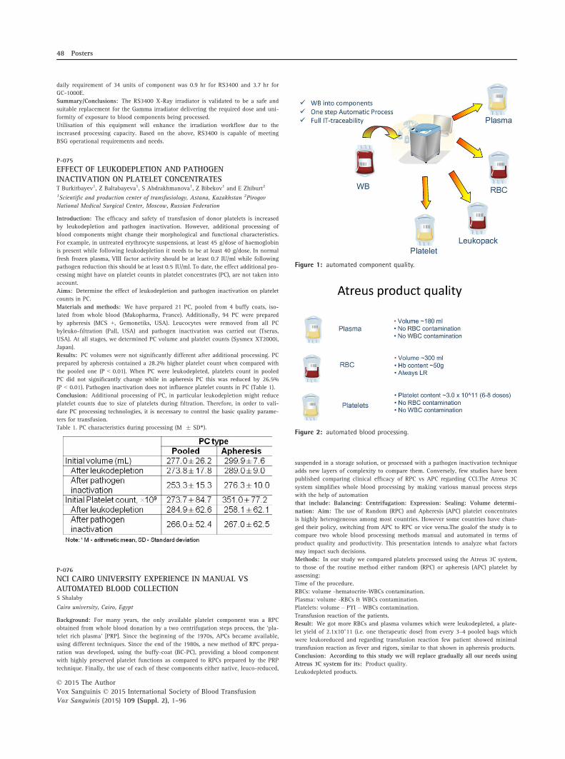
daily requirement of 34 units of component was 0.9 hr for RS3400 and 3.7 hr for
GC-1000E.
Summary/Conclusions: The RS3400 X-Ray irradiator is validated to be a safe and
suitable replacement for the Gamma irradiator delivering the required dose and uni-
formity of exposure to blood components being processed.
Utilisation of this equipment will enhance the irradiation workflow due to the
increased processing capacity. Based on the above, RS3400 is capable of meeting
BSG operational requirements and needs.
P-075
EFFECT OF LEUKODEPLETION AND PATHOGENINACTIVATION ON PLATELET CONCENTRATEST Burkitbayev1, Z Baltabayeva1, S Abdrakhmanova1, Z Bibekov1 and E Zhiburt2
1Scientific and production center of transfusiology, Astana, Kazakhstan 2Pirogov
National Medical Surgical Center, Moscow, Russian Federation
Introduction: The efficacy and safety of transfusion of donor platelets is increased
by leukodepletion and pathogen inactivation. However, additional processing of
blood components might change their morphological and functional characteristics.
For example, in untreated erythrocyte suspensions, at least 45 g/dose of haemoglobin
is present while following leukodepletion it needs to be at least 40 g/dose. In normal
fresh frozen plasma, VIII factor activity should be at least 0.7 IU/ml while following
pathogen reduction this should be at least 0.5 IU/ml. To date, the effect additional pro-
cessing might have on platelet counts in platelet concentrates (PC), are not taken into
account.
Aims: Determine the effect of leukodepletion and pathogen inactivation on platelet
counts in PC.
Materials and methods: We have prepared 21 PC, pooled from 4 buffy coats, iso-
lated from whole blood (Makopharma, France). Additionally, 94 PC were prepared
by apheresis (MCS +, Gemonetiks, USA). Leucocytes were removed from all PC
byleuko-filtration (Pall, USA) and pathogen inactivation was carried out (Tserus,
USA). At all stages, we determined PC volume and platelet counts (Sysmex XT2000i,
Japan).
Results: PC volumes were not significantly different after additional processing. PC
prepared by apheresis contained a 28.2% higher platelet count when compared with
the pooled one (P < 0.01). When PC were leukodepleted, platelets count in pooled
PC did not significantly change while in apheresis PC this was reduced by 26.5%
(P < 0.01). Pathogen inactivation does not influence platelet counts in PC (Table 1).
Conclusion: Additional processing of PC, in particular leukodepletion might reduce
platelet counts due to size of platelets during filtration. Therefore, in order to vali-
date PC processing technologies, it is necessary to control the basic quality parame-
ters for transfusion.
Table 1. PC characteristics during processing (M � SD*).
P-076
NCI CAIRO UNIVERSITY EXPERIENCE IN MANUAL VSAUTOMATED BLOOD COLLECTIONS Shalaby
Cairo university, Cairo, Egypt
Background: For many years, the only available platelet component was a RPC
obtained from whole blood donation by a two centrifugation steps process, the ‘pla-
telet rich plasma’ [PRP]. Since the beginning of the 1970s, APCs became available,
using different techniques. Since the end of the 1980s, a new method of RPC prepa-
ration was developed, using the buffy-coat (BC-PC), providing a blood component
with highly preserved platelet functions as compared to RPCs prepared by the PRP
technique. Finally, the use of each of these components either native, leuco-reduced,
suspended in a storage solution, or processed with a pathogen inactivation technique
adds new layers of complexity to compare them. Conversely, few studies have been
published comparing clinical efficacy of RPC vs APC regarding CCI.The Atreus 3C
system simplifies whole blood processing by making various manual process steps
with the help of automation
that include: Balancing: Centrifugation: Expression: Sealing: Volume determi-
nation: Aim: The use of Random (RPC) and Apheresis (APC) platelet concentrates
is highly heterogeneous among most countries. However some countries have chan-
ged their policy, switching from APC to RPC or vice versa.The goalof the study is to
compare two whole blood processing methods manual and automated in terms of
product quality and productivity. This presentation intends to analyze what factors
may impact such decisions.
Methods: In our study we compared platelets processed using the Atreus 3C system,
to those of the routine method either random (RPC) or apheresis (APC) platelet by
assessing:
Time of the procedure.
RBCs: volume -hematocrite-WBCs contamination.
Plasma: volume -RBCs & WBCs contamination.
Platelets: volume – PYI – WBCs contamination.
Transfusion reaction of the patients.
Result: We got more RBCs and plasma volumes which were leukodepleted, a plate-
let yield of 2.1x10^11 (i.e. one therapeutic dose) from every 3–4 pooled bags which
were leukoreduced and regarding transfusion reaction few patient showed minimal
transfusion reaction as fever and rigors, similar to that shown in apheresis products.
Conclusion: According to this study we will replace gradually all our needs using
Atreus 3C system for its: Product quality.
Leukodepleted products.
Figure 1: automated component quality.
Figure 2: automated blood processing.
© 2015 The AuthorVox Sanguinis © 2015 International Society of Blood TransfusionVox Sanguinis (2015) 109 (Suppl. 2), 1–96
48 Posters
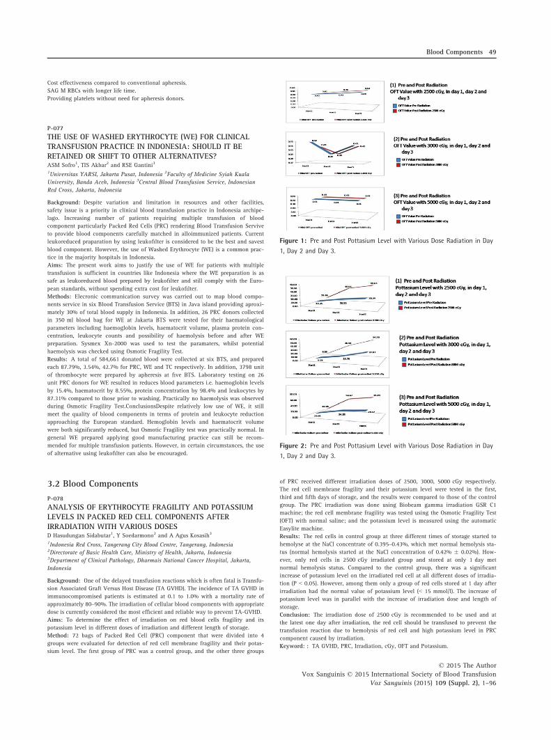
Cost effectiveness compared to conventional apheresis.
SAG M RBCs with longer life time.
Providing platelets without need for apheresis donors.
P-077
THE USE OF WASHED ERYTHROCYTE (WE) FOR CLINICALTRANSFUSION PRACTICE IN INDONESIA: SHOULD IT BERETAINED OR SHIFT TO OTHER ALTERNATIVES?ASM Sofro1, TIS Akbar2 and RSE Gantini3
1Universitas YARSI, Jakarta Pusat, Indonesia 2Faculty of Medicine Syiah Kuala
University, Banda Aceh, Indonesia 3Central Blood Transfusion Service, Indonesian
Red Cross, Jakarta, Indonesia
Background: Despite variation and limitation in resources and other facilities,
safety issue is a priority in clinical blood transfusion practice in Indonesia archipe-
lago. Increasing number of patients requiring multiple transfusion of blood
component particularly Packed Red Cells (PRC) rendering Blood Transfusion Servive
to provide blood components carefully matched in alloimmunized patients. Current
leukoreduced praparation by using leukofilter is considered to be the best and savest
blood component. However, the use of Washed Erythrocyte (WE) is a common prac-
tice in the majority hospitals in Indonesia.
Aims: The present work aims to justify the use of WE for patients with multiple
transfusion is sufficient in countries like Indonesia where the WE preparation is as
safe as leukoreduced blood prepared by leukofilter and still comply with the Euro-
pean standards, without spending extra cost for leukofilter.
Methods: Elecronic communication survey was carried out to map blood compo-
nents service in six Blood Transfusion Service (BTS) in Java island providing aproxi-
mately 30% of total blood supply in Indonesia. In addition, 26 PRC donors collected
in 350 ml blood bag for WE at Jakarta BTS were tested for their haematological
parameters including haemoglobin levels, haematocrit volume, plasma protein con-
centration, leukocyte counts and possibility of haemolysis before and after WE
preparation. Sysmex Xn-2000 was used to test the paramaters, whilst potential
haemolysis was checked using Osmotic Fragility Test.
Results: A total of 584,661 donated blood were collected at six BTS, and prepared
each 87.79%, 3.54%, 42.7% for PRC, WE and TC respectively. In addition, 3798 unit
of thrombocyte were prepared by apheresis at five BTS. Laboratory testing on 26
unit PRC donors for WE resulted in reduces blood parameters i.e. haemoglobin levels
by 15.4%, haematocrit by 8.55%, protein concentration by 98.4% and leukocytes by
87.31% compared to those prior to washing. Practically no haemolysis was observed
during Osmotic Fragility Test.ConclusionsDespite relatively low use of WE, it still
meet the quality of blood components in terms of protein and leukocyte reduction
approaching the European standard. Hemoglobin levels and haematocrit volume
were both significantly reduced, but Osmotic Fragility test was practically normal. In
general WE prepared applying good manufacturing practice can still be recom-
mended for multiple transfusion patients. However, in certain circumstances, the use
of alternative using leukofilter can also be encouraged.
3.2 Blood Components
P-078
ANALYSIS OF ERYTHROCYTE FRAGILITY AND POTASSIUMLEVELS IN PACKED RED CELL COMPONENTS AFTERIRRADIATION WITH VARIOUS DOSESD Hasudungan Sidabutar1, Y Soedarmono2 and A Agus Kosasih3
1Indonesia Red Cross, Tangerang City Blood Centre, Tangerang, Indonesia2Directorate of Basic Health Care, Ministry of Health, Jakarta, Indonesia3Department of Clinical Pathology, Dharmais National Cancer Hospital, Jakarta,
Indonesia
Background: One of the delayed transfusion reactions which is often fatal is Transfu-
sion Associated Graft Versus Host Disease (TA GVHD). The incidence of TA GVHD in
immunocompromised patients is estimated at 0.1 to 1.0% with a mortality rate of
approximately 80–90%. The irradiation of cellular blood components with appropriate
dose is currently considered the most efficient and reliable way to prevent TA-GVHD.
Aims: To determine the effect of irradiation on red blood cells fragility and its
potassium level in different doses of irradiation and different length of storage.
Method: 72 bags of Packed Red Cell (PRC) component that were divided into 4
groups were evaluated for detection of red cell membrane fragility and their potas-
sium level. The first group of PRC was a control group, and the other three groups
of PRC received different irradiation doses of 2500, 3000, 5000 cGy respectively.
The red cell membrane fragility and their potassium level were tested in the first,
third and fifth days of storage, and the results were compared to those of the control
group. The PRC irradiation was done using Biobeam gamma irradiation GSR C1
machine; the red cell membrane fragility was tested using the Osmotic Fragility Test
(OFT) with normal saline; and the potassium level is measured using the automatic
Easylite machine.
Results: The red cells in control group at three different times of storage started to
hemolyse at the NaCl concentrate of 0.395–0.43%, which met normal hemolysis sta-
tus (normal hemolysis started at the NaCl concentration of 0.42% � 0.02%). How-
ever, only red cells in 2500 cGy irradiated group and stored at only 1 day met
normal hemolysis status. Compared to the control group, there was a significant
increase of potassium level on the irradiated red cell at all different doses of irradia-
tion (P < 0.05). However, among them only a group of red cells stored at 1 day after
irradiation had the normal value of potassium level (< 15 mmol/l). The increase of
potassium level was in parallel with the increase of irradiation dose and length of
storage.
Conclusion: The irradiation dose of 2500 cGy is recommended to be used and at
the latest one day after irradiation, the red cell should be transfused to prevent the
transfusion reaction due to hemolysis of red cell and high potassium level in PRC
component caused by irradiation.
Keyword: : TA GVHD, PRC, Irradiation, cGy, OFT and Potassium.
Figure 1: Pre and Post Pottasium Level with Various Dose Radiation in Day
1, Day 2 and Day 3.
Figure 2: Pre and Post Pottasium Level with Various Dose Radiation in Day
1, Day 2 and Day 3.
© 2015 The AuthorVox Sanguinis © 2015 International Society of Blood Transfusion
Vox Sanguinis (2015) 109 (Suppl. 2), 1–96
Blood Components 49
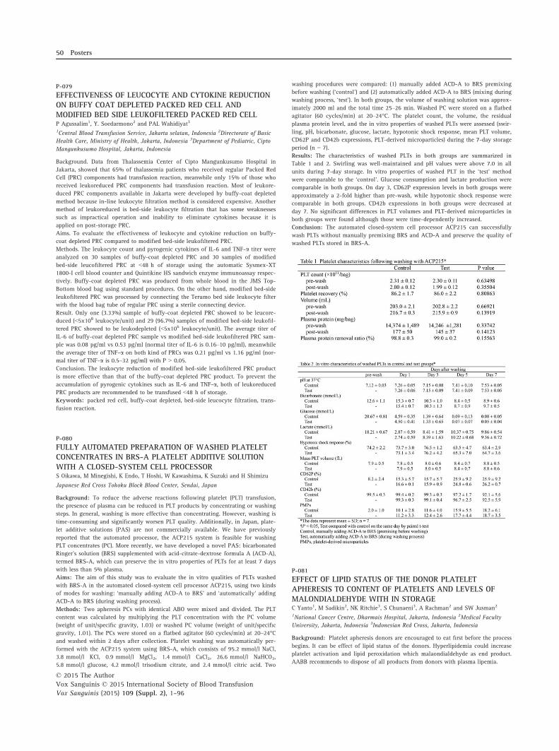
P-079
EFFECTIVENESS OF LEUCOCYTE AND CYTOKINE REDUCTIONON BUFFY COAT DEPLETED PACKED RED CELL ANDMODIFIED BED SIDE LEUKOFILTERED PACKED RED CELLP Agussalim1, Y. Soedarmono2 and PAL Wahidiyat3
1Central Blood Transfusion Service, Jakarta selatan, Indonesia 2Directorate of Basic
Health Care, Ministry of Health, Jakarta, Indonesia 3Department of Pediatric, Cipto
Mangunkusumo Hospital, Jakarta, Indonesia
Background. Data from Thalassemia Center of Cipto Mangunkusumo Hospital in
Jakarta, showed that 65% of thalassemia patients who received regular Packed Red
Cell (PRC) components had transfusion reaction, meanwhile only 15% of those who
received leukoreduced PRC components had transfusion reaction. Most of leukore-
duced PRC components available in Jakarta were developed by buffy-coat depleted
method because in-line leukocyte filtration method is considered expensive. Another
method of leukoreduced is bed-side leukocyte filtration that has some weaknesses
such as impractical operation and inability to eliminate cytokines because it is
applied on post-storage PRC.
Aims. To evaluate the effectiveness of leukocyte and cytokine reduction on buffy-
coat depleted PRC compared to modified bed-side leukofiltered PRC.
Methods. The leukocyte count and pyrogenic cytokines of IL-6 and TNF-a titer were
analyzed on 30 samples of buffy-coat depleted PRC and 30 samples of modified
bed-side leucofiltered PRC at <48 h of storage using the automatic Sysmex-XT
1800-I cell blood counter and Quintikine HS sandwich enzyme immunoassay respec-
tively. Buffy-coat depleted PRC was produced from whole blood in the JMS Top-
Bottom blood bag using standard procedures. On the other hand, modified bed-side
leukofiltered PRC was processed by connecting the Terumo bed side leukocyte filter
with the blood bag tube of regular PRC using a sterile connecting device.
Result. Only one (3.33%) sample of buffy-coat depleted PRC showed to be leucore-
duced (<5x10⁸ leukocyte/unit) and 29 (96.7%) samples of modified bed-side leukofil-
tered PRC showed to be leukodepleted (<5x10⁶ leukocyte/unit). The average titer of
IL-6 of buffy-coat depleted PRC sample vs modified bed-side leukofiltered PRC sam-
ple was 0.08 pg/ml vs 0.53 pg/ml (normal titer of IL-6 is 0.16–10 pg/ml), meanwhile
the average titer of TNF-a on both kind of PRCs was 0.21 pg/ml vs 1.16 pg/ml (nor-
mal titer of TNF-a is 0.5–32 pg/ml) with P > 0.05.
Conclusion. The leukocyte reduction of modified bed-side leukofiltered PRC product
is more effective than that of the buffy-coat depleted PRC product. To prevent the
accumulation of pyrogenic cytokines such as IL-6 and TNF-a, both of leukoreduced
PRC products are recommended to be transfused <48 h of storage.
Keywords: packed red cell, buffy-coat depleted, bed-side leucocyte filtration, trans-
fusion reaction.
P-080
FULLY AUTOMATED PREPARATION OF WASHED PLATELETCONCENTRATES IN BRS-A PLATELET ADDITIVE SOLUTIONWITH A CLOSED-SYSTEM CELL PROCESSORS Oikawa, M Minegishi, K Endo, T Hoshi, W Kawashima, K Suzuki and H Shimizu
Japanese Red Cross Tohoku Block Blood Center, Sendai, Japan
Background: To reduce the adverse reactions following platelet (PLT) transfusion,
the presence of plasma can be reduced in PLT products by concentrating or washing
steps. In general, washing is more effective than concentrating. However, washing is
time-consuming and significantly worsen PLT quality. Additionally, in Japan, plate-
let additive solutions (PAS) are not commercially available. We have previously
reported that the automated processor, the ACP215 system is feasible for washing
PLT concentrates (PC). More recently, we have developed a novel PAS: bicarbonated
Ringer’s solution (BRS) supplemented with acid-citrate-dextrose formula A (ACD-A),
termed BRS-A, which can preserve the in vitro properties of PLTs for at least 7 days
with less than 5% plasma.
Aims: The aim of this study was to evaluate the in vitro qualities of PLTs washed
with BRS-A in the automated closed-system cell processor ACP215, using two kinds
of modes for washing: ‘manually adding ACD-A to BRS’ and ‘automatically’ adding
ACD-A to BRS (during washing process).
Methods: Two apheresis PCs with identical ABO were mixed and divided. The PLT
content was calculated by multiplying the PLT concentration with the PC volume
(weight of unit/specific gravity, 1.03) or washed PC volume (weight of unit/specific
gravity, 1.01). The PCs were stored on a flatbed agitator (60 cycles/min) at 20–24°Cand washed within 2 days after collection. Platelet washing was automatically per-
formed with the ACP215 system using BRS-A, which consists of 95.2 mmol/l NaCl,
3.8 mmol/l KCl, 0.9 mmol/l MgCl2, 1.4 mmol/l CaCl2, 26.6 mmol/l NaHCO3,
5.8 mmol/l glucose, 4.2 mmol/l trisodium citrate, and 2.4 mmol/l citric acid. Two
washing procedures were compared: (1) manually added ACD-A to BRS premixing
before washing (‘control’) and (2) automatically added ACD-A to BRS (mixing during
washing process, ‘test’). In both groups, the volume of washing solution was approx-
imately 2000 ml and the total time 25–26 min. Washed PC were stored on a flatbed
agitator (60 cycles/min) at 20–24°C. The platelet count, the volume, the residual
plasma protein level, and the in vitro properties of washed PLTs were assessed (swir-
ling, pH, bicarbonate, glucose, lactate, hypotonic shock response, mean PLT volume,
CD62P and CD42b expressions, PLT-derived microparticles) during the 7-day storage
period (n = 7).
Results: The characteristics of washed PLTs in both groups are summarized in
Table 1 and 2. Swirling was well-maintained and pH values were above 7.0 in all
units during 7-day storage. In vitro properties of washed PLT in the ‘test’ method
were comparable to the ‘control’. Glucose consumption and lactate production were
comparable in both groups. On day 3, CD62P expression levels in both groups were
approximately a 2-fold higher than pre-wash, while hypotonic shock response were
comparable in both groups. CD42b expressions in both groups were decreased at
day 7. No significant differences in PLT volumes and PLT-derived microparticles in
both groups were found although those were time-dependently increased.
Conclusion: The automated closed-system cell processor ACP215 can successfully
wash PLTs without manually premixing BRS and ACD-A and preserve the quality of
washed PLTs stored in BRS-A.
P-081
EFFECT OF LIPID STATUS OF THE DONOR PLATELETAPHERESIS TO CONTENT OF PLATELETS AND LEVELS OFMALONDIALDEHYDE WITH IN STORAGEC Yanto1, M Sadikin2, NK Ritchie3, S Chunaeni3, A Rachman2 and SW Jusman2
1National Cancer Centre, Dharmais Hospital, Jakarta, Indonesia 2Medical Faculty
University, Jakarta, Indonesia 3Indonesian Red Cross, Jakarta, Indonesia
Background: Platelet apheresis donors are encouraged to eat first before the process
begins. It can be effect of lipid status of the donors. Hyperlipidemia could increase
platelet activation and lipid peroxidation which malaondialdehyde as end product.
AABB recommends to dispose of all products from donors with plasma lipemia.
© 2015 The AuthorVox Sanguinis © 2015 International Society of Blood TransfusionVox Sanguinis (2015) 109 (Suppl. 2), 1–96
50 Posters
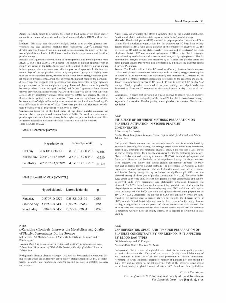
Aims: This study aimed to determine the effect of lipid status of the donor platelet
apheresis to content of platelets and levels of malondialdehyde (MDA) with in stor-
age.
Methods: This study used descriptive analytic design in 31 platelet apheresis con-
centrates. We used apheresis machine from Haemonetic MCS+�. Samples were
divided into two groups, hyperlipidemia and normolipidemia. The assay for the con-
tent of platelets and levels of MDA was tested on the first day, second and fourth of
platelet storage.
Results: The triglyceride concentration of hyperlipidemia and normolipidemia were
240.41 � 95.51 and 89.50 � 30.51 mg/dl. The results of platelet apheresis with in
storage are shown in the table. An increase in the content of platelets during storage
in both groups, which are associated with the process of apoptosis. In the first and
second days of storage platelet counts in hyperlipidemia group was slightly lower
than the normolipidemia group, whereas in the fourth day of storage obtained plate-
let counts in hyperlipidemia group that exceeded the platelet count in the normolipi-
demia group. This suggests that apoptosis occurs more frequently in hyperlipidemia
group compared to the normolipidemia group. Increased platelet count is probably
because platelets have an enlarged (swollen) and further fragments to form platelets
derived procoagulant microparticles (PDMPs) in the apoptotic process but still count
as platelets by hematology analyzer (false positive). PDMPs will increase the risk of
thrombosis in patients who are sensitive. There was no significant correlation
between levels of triglycerides and platelet content. On the fourth day found signifi-
cant differences in the levels of MDA. There were positive and significant correla-
tions between levels of triglyceride to the levels of MDA.
Conclusions: Improved of the lipid status of the donor platelet apheresis will
increase platelet apoptosis and increase levels of MDA. The need to remind donors
platelet apheresis to a low fat dietary before apheresis process implemented. Need
for further research to determine the lipid levels that can still be tolerated.
Table 2. Levels of MDA.
P-082
L-Carnitine effectively Improves the Metabolism and Qualityof Platelet Concentrates During StorageMR Deyhim1, SA Mesbah-Namin1, F Yari1, MR Taghikhani2, A Naser1 and F
Khoshnaghsh1
1Iranian blood transfusion research center, High institute for research and edu.,
Tehran, Iran 2Department of Clinical Biochemistry, Faculty of Medical Sciences,
Tehran, Iran
Background: Human platelets undergo structural and biochemical alternations dur-
ing storage which are collectively called platelet storage lesion (PSL). PSL is charac-
terized metabolic and functionally changes causing decrease in platelet’s recovery
and survival.
Aims: Here, we evaluated the effect L-carnitine (LC) on the platelet metabolism,
function and platelet mitochondrial enzyme activity during platelet storage.
Methods: Platelet rich plasma (PRP) was used to prepare platelet concentrate (PC) in
Iranian blood transfusion organization. For this purpose, ten PC bags from healthy
donors, stored at 22� C with gentle agitation in the presence or absence of LC. The
effects of LC (15 mM) on the platelet quality were assessed by analyzing the levels
of glucose, lactate, ATP and lactate dehydrogenase (LDH) activity. Platelet aggrega-
tion induced by arachidonate and ristocetin were analyzed by aggregometer. Platelet
mitochondrial enzyme activity was measured by MTT assay and platelet count and
mean platelet volume (MPV) were also determined by a hematology analyzer during
5 days of PC storage.
Results: The Results indicated that LC could significantly decrease lactate concen-
tration and glucose consumption accompany with increasing oxygen consumption
in stored PC. LDH activity was also significantly less increased in LC-treated PC on
day 2 and 5 of storage. Platelet aggregation in response to the ristocetin and arachi-
donate was significantly higher in LC-treated PC than in untreated PC on day 5 of
storage. Finally, platelet mitochondrial enzyme activity was significantly less
decreased in LC-treated PC compared to the control group on day 2 and 5 of stor-
age.
Conclusions: It seems that LC would be a good additive to reduce PSL and improve
the platelet metabolism and quality of the stored PC for platelet-transfusion therapy.
Keywords: L-carnitine; Platelet quality; stored platelet concentrates; Platelet stor-
age lesion:
P-083
INFLUENCE OF DIFFERENT METHODS PREPARATION ONPLATELET ACTIVATION IN STORED PLATELETCONCENTRATESA Soleimany ferizhandy
Iranian Blood Transfusion Research Center, High Institute for Research and Educa,
Tehran, Iran
Background: Platelet concentrates are routinely manufactured from whole blood by
differential centrifugation. During this storage period under blood bank conditions,
biochemical, structural and functional changes occur, a process that is also known
as platelet storage lesion. Their quality was assessed using the following parameters:
Platelets, leukocytes and erythrocytes counts, pH, CD63, lactate dehydrogenase and
Annexin V. Materials and Methods: In this experimental study, 25 platelet concen-
trates prepared with platelet rich plasma-platelet concentrates, 25 units via buffy
coat and apheresis-derived platelet methods. The percentages of Annexin V, CD63
expression, lactatedehydrogenase, platelet, leukocytes counts and pH were evalu-
ated.Results: During storage for up to 5 days, no significant pH, difference was
observed among all three type of platelet concentrates (P > 0.05). The mean leuko-
cytes count buffy coat units, platelet rich plasma platelet concentrates and aphere-
sis-derived units were comparable and statistically significant difference was
observed (P < 0.05). During storage for up to 5 days platelet concentrates units dis-
played significant an increase in lactatedehydrogenase, CD63 and Annexin V expres-
sions, as compared with buffy coat units and apheresisderived units preparation on
day 5 (P < 0.05). Discussion: The kinetics of CD63 and annexin V levels are influ-
enced by the method used to prepare platelets for storage. The different levels of
CD63, annexin V and lactatedehydrogenase in three types of units clearly demon-
strating a progressive activation process of platelet concentrates units exceeds that
of buffy coat and apheresis-derived units. Further clinical studies will be necessary
to determine whether meet the quality criteria or is superior in predicting in vivo
viability
P-084
CENTRIFUGATION SPEED AND TIME FOR PREPARATION OFPLATELET CONCENTRATE BY PRP METHOD. IS IT AFFECTEDBY BLOOD BAG TYPE?CG Kohombange and KS Kuruppu
National Blood Center, Colombo, Sri Lanka
Background: Platelet count of a platelet concentrate is the main quality parame-
ter which determines the efficacy of the product. Quality control laboratory of
NBC monitors at least 1% of all the total production of platelet concentrate.
According to AABB standards acceptable number of platelets per unit should be
5.5 9 1010 and according to the EU guideline, 75% of the products tested should
be at least having a platelet count of 6.0 9 1010. Based on these guidelines,
© 2015 The AuthorVox Sanguinis © 2015 International Society of Blood Transfusion
Vox Sanguinis (2015) 109 (Suppl. 2), 1–96
Blood Components 51
NBTS standards is set as 75% from the tested products should contain at least
5.5 9 1010 platelets per unit.
While monitoring the platelet counts in PRP derived platelets prepared at the blood
component preparation laboratory of NBC, it was noted that from July 2014
onwards, the platelet count has not met the required pass rate of 75%. This study
was conducted as to find out the root cause for this trend.
Aim: Aim of this study was to find the root cause for the reduced platelet counts of
the PRPD platelets.
Method: A root cause analysis was conducted to identify any deviations in all the
quality system essentials involved in the process.
Results: Based on the above analysis it was evident that the properties of the blood
bag matters in the final yield of the platelets prepared from PRPD method.
With further testing an optimum centrifugation programe was selected for platelet
preparation by whole blood collected to Terumo Japanese triple blood bag system
Conclusion: Following aspects has to be taken into consideration while preparing
PRPD platelets:
Centrifugation speeds and times of the initial centrifugation cycle of the whole blood
collected to triple blood bag systems should be validated according to the blood bag
type. When procuring blood bags it is recommended to get down supplier recom-
mendations on centrifugation programs and obtain technical assistance of the sup-
pliers in initial validation of the blood bag system. It is feasible to use a single
brand of triple blood bag system in preparation of PRPD platelets in a single produc-
tion cycle, as it may required to change the centrifugation programs when using dif-
ferent brands of blood bags.
Caption 1: Root cause analysis.
P-085
COAGULATION FACTORS, THROMBIN FORMATION AND CLOTFORMATION OF THAWED FRESH FROZEN PLASMAY Naito1, Y Hayashi1, S Wakamoto1, M Fujihara1, M Akino2, M Endo1, C Homma1, T
Yamamoto3, H Ikeda3, S Kino1 and S Takamoto1
1Japanese Red Cross Hokkaido Block Blood Center, Sapporo, Japan 2Japanese Red
Cross Blood Service Headquarters, Tokyo, Japan 3Japanese Red Cross Hokkaido
Blood Center, Sapporo, Japan
Background: In Japan, leukocytes reduced fresh frozen plasma (FFP-LR) must be
used within 3 h after thawing. This is thought to be based on the decline of labile
coagulation factors such as factor (F) V and FVIII activities during post-thaw stor-
age. In 2012, on the other hand, the Food and Drug Administration (USA) approved
extending the storage time of thawed FFP stored at 1 to 6 °C up to 24 h, instead of
6 h. The longer shelf life of thawed FFP-LR makes it applicable for neonatal use or
critical care settings.
Aims: We evaluated coagulation capacity of thawed FFP-LR up to 120 h with a
thrombin generation assay, a viscoelastic assay and measurement of activities of
coagulation factors.
Methods: 15 FFP-LRs (4 A-, 5 O-, 2 B-, 4 AB-type) were stored at �30°C for
approximately 12 months. FFP-LRs were thawed at 37°C, divided into two equal
volumes in plastic bags, and stored at either 4°C or 22°C for up to 120 h. Samples
were taken at 0, 3, 6, 24, 48 and 120 h after thawing. Coagulation factors were
assessed by CS-2000i automated coagulation analyzer (Sysmex Corporation). Throm-
bin generation was measured based on the use of a fluorogenic substrate in the pres-
ence of exogenous tissue factor, phospholipid, and calcium using the calibrated
automated thrombogram system (CAT, Thrombinoscope BV). The thrombin genera-
tion curve was assessed by examining the lag time for thrombin generation to occur,
peak amount of thrombin generated, the area under the thrombin generation curve
(endogenous thrombin potential, ETP), time to peak and time to tail. Clot formation
was assessed by rotational thromboelastometry (ROTEM, Pentapharm GmbH) using
INTEM (ellagic acid/phospholipid activation) and EXTEM (tissue factor activation).
The clotting time (CT, the lag time for clot formation to commence), alpha angle (a,rate of clot formation) and maximum clot firmness (MCF, a measure of clot strength)
were assessed.
Results: The FVIII level decreased from 0.95 � 0.21 to 0.50 � 0.11 IU/ml after
120 h of storage at both 4°C and 22°C, and at the largest degree among other coag-
ulation factors. The level of fibrinogen was almost unchanged after 120 h of storage.
Levels of other coagulation factors in thawed FFP-LR after 120 h remained above
0.70 IU/ml. In the thrombin generation assay, there was a significant increase in the
lag time after 120 h of storage at 4°C and after 48 h and 120 h of storage at 22°Ccompared with 0 h. However, there was no difference in the ability of thawed FFP-
LR to generate thrombin up to 120 h as assessed by the peak and ETP. There was no
difference in clot formation in thawed FFP-LR stored for 120 h as assessed by
ROTEM in terms of a and MCF.
Conclusions: Despite the approximately 50% loss of FVIII, the ability of thawed
FFP-LR generating thrombin or forming a clot was unaltered. Therefore, we suggest
that thawed FFP-LR can be used to treat coagulopathies except FVIII deficiency for
up to 120 h.
P-086
AGGREGATION AND RELEASE OF PRO-COAGULANT TISSUEFACTOR-EXPRESSING MICROPARTICLES BY THP-1MONOCYTIC CELLS ACTIVATED BY PLATELET-DERIVEDMICROPARTICLES IN VITROML Chou1, HC Lin2, HW Chang2, SH Hsiao1, JC Seghatchian3 and T Burnouf1
1Taipei Medical University, Taipei, Taiwan 2Taipei Medical University Hospital,
Taipei, Taiwan 3Audit/Inspection and DDR Strategy, London, United Kingdom
Background: Microparticles (MPs) released by blood or endothelial cells are present
in plasma for transfusion. They originate from the collected donor blood, or are trig-
gered by the collection and production process of blood components. While MPs
may contribute to hemostasis, their presence in transfused plasma may lead to
uncontrolled thrombin generation when transfused to susceptible cancer or patients
with hypercoagulable state. Understanding the biochemical and cellular triggers of
MP-mediated thrombogenesis is therefore needed. We hypothesized that the pro-
thrombotic role of PMPs could encompass interaction with and activation of
immune cells leading to the release of pro-thrombotic microparticles.
Aim: In the current study, we have isolated and compared the characteristics of
PMPs isolated from the human plasma compartment of platelet concentrates (N-
PMPs) or generated by thrombin activation of isolated platelets (T-PMPs) and evalu-
ated their capacity to induce THP-1 leukemia monocytic cell aggregation and release
of pro-coagulant tissue factor-bearing MPs.
Methods: We isolated platelet MPs (PMPs) present in platelet concentrate supernatant
plasma (N-PMPs) or prepared by activation of isolated platelets using 0.1 IU/ml
thrombin (T-PMPs). N-PMPs and T-PMPs were characterized dynamic light scattering
and counted by tunable resistive pulse sensing to determine population size and num-
ber. 1x10⁶ THP-1 cells cultures were treated with 5 ml of N-PMP or 5 ml of T-PMP
(corresponding to a 1 to 500 ratio of THP-1 cells to PMPs) at 37 °C for 4 h. Super-
natants from monocytes stimulated or not by N-PMPs or T-PMPs were collected by
serial centrifugation at 300xg and 3000xg at 4°C for 10 min to remove monocytes
and obtain the supernatant. The functional pro-coagulant activity of TF-MPs released
by monocytes was determined by a TF-capture assay (Zymuphen MP-TF).
Results: THP-1 cells incubated for 5 min at 37°C in growth medium evidenced a
typical regular round shape morphology. There was no immediate change in THP-1
cells morphology after 5 min of incubation following addition of N-PMPs and THP-
1 cells remained as a monodispersed suspension with round shape morphology. By
contrast, T-PMP addition induced, within as little as 5 min of incubation, a strong
cellular aggregation, also characterized by a change in THP-1 cell morphology from
regular to irregular and rough appearance, with an apparent release of MPs budding
from the cell surface. There was no detectable change in the morphology and shape
of THP cells alone or co-cultured with N-PMP over 24 h, whereas aggregation
induced by T-PMPs remained strong. Release of pro-coagulant TF-MPs was signifi-
cantly stimulated by the addition of both N-PMPs (P < 0.05) and T-PMPs
(P < 0.001) compared to THP-1 cell alone. The control samples of growth medium
spiked with N-PMPs and T-PMPs under the same conditions and at the same ratios
did not contain detectable (<1 pg/ml) pro-coagulant TF-MPs, suggesting that the
pro-coagulant TF-bearing MPs were released by THP-1 cells.
Conclusion: Our in vitro data indicate that, in addition to generating thrombin
through protein activation, PMPs may trigger cell-mediated thrombogenicity leading
to the generation of pro-coagulant TF-bearing MPs released by monocytes.
P-087
EFFECT OF FREEZING TECHNIQUE ON THE QUALITY OFFRESH FROZEN PLASMAR Chaudhary, L Dhantole and A Sonker
Sanjay Gandhi Postgraduate Institute of Medical Sciences, Lucknow, India
Background: The quality of plasma depends on many factors, one of which is the
technique used for freezing plasma.
Aim: In order to study effect of freezing method on quality of plasma, we con-
ducted a study in which plasma components were frozen by contact shock freezer
© 2015 The AuthorVox Sanguinis © 2015 International Society of Blood TransfusionVox Sanguinis (2015) 109 (Suppl. 2), 1–96
52 Posters
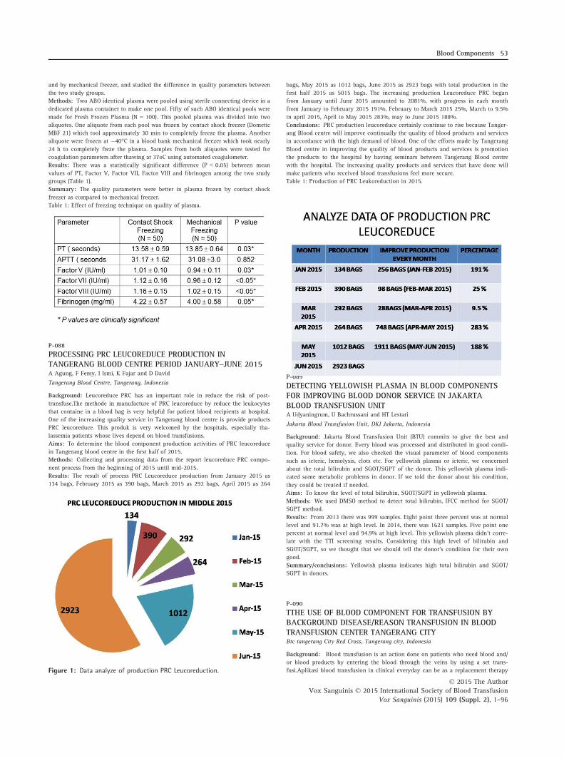
and by mechanical freezer, and studied the difference in quality parameters between
the two study groups.
Methods: Two ABO identical plasma were pooled using sterile connecting device in a
dedicated plasma container to make one pool. Fifty of such ABO identical pools were
made for Fresh Frozen Plasma (N = 100). This pooled plasma was divided into two
aliquotes. One aliquote from each pool was frozen by contact shock freezer (Dometic
MBF 21) which tool approximately 30 min to completely freeze the plasma. Another
aliquote were frozen at �40°C in a blood bank mechanical freezer which took nearly
24 h to completely freze the plasma. Samples from both aliquotes were tested for
coagulation parameters after thawing at 37oC using automated coagulometer.
Results: There was a statistically significant difference (P < 0.05) between mean
values of PT, Factor V, Factor VII, Factor VIII and fibrinogen among the two study
groups (Table 1).
Summary: The quality parameters were better in plasma frozen by contact shock
freezer as compared to mechanical freezer.
Table 1: Effect of freezing technique on quality of plasma.
P-088
PROCESSING PRC LEUCOREDUCE PRODUCTION INTANGERANG BLOOD CENTRE PERIOD JANUARY–JUNE 2015A Agung, F Femy, I Ismi, K Fajar and D David
Tangerang Blood Centre, Tangerang, Indonesia
Background: Leucoreduce PRC has an important role in reduce the risk of post-
transfuse.The methode in manufacture of PRC leucoreduce by reduce the leukocytes
that containe in a blood bag is very helpful for patient blood recipients at hospital.
One of the increasing quality service in Tangerang blood centre is provide products
PRC leucoreduce. This produk is very welcomed by the hospitals, especially tha-
lassemia patients whose lives depend on blood transfusions.
Aims: To determine the blood component production activities of PRC leucoreduce
in Tangerang blood centre in the first half of 2015.
Methods: Collecting and processing data from the report leucoreduce PRC compo-
nent process from the beginning of 2015 until mid-2015.
Results: The result of process PRC Leucoreduce production from January 2015 as
134 bags, February 2015 as 390 bags, March 2015 as 292 bags, April 2015 as 264
bags, May 2015 as 1012 bags, June 2015 as 2923 bags with total production in the
first half 2015 as 5015 bags. The increasing production Leucoreduce PRC began
from January until June 2015 amounted to 2081%, with progress in each month
from January to February 2015 191%, February to March 2015 25%, March to 9.5%
in april 2015, April to May 2015 283%, may to June 2015 188%.
Conclusions: PRC production leucoreduce certainly continue to rise because Tanger-
ang Blood centre will improve continually the quality of blood products and services
in accordance with the high demand of blood. One of the efforts made by Tangerang
Blood centre in improving the quality of blood products and services is promotion
the products to the hospital by having seminars between Tangerang Blood centre
with the hospital. The increasing quality products and services that have done will
make patients who received blood transfusions feel more secure.
Table 1: Production of PRC Leukoreduction in 2015.
P-089
DETECTING YELLOWISH PLASMA IN BLOOD COMPONENTSFOR IMPROVING BLOOD DONOR SERVICE IN JAKARTABLOOD TRANSFUSION UNITA Udyaningrum, U Bachrussani and HT Lestari
Jakarta Blood Transfusion Unit, DKI Jakarta, Indonesia
Background: Jakarta Blood Transfusion Unit (BTU) commits to give the best and
quality service for donor. Every blood was processed and distributed in good condi-
tion. For blood safety, we also checked the visual parameter of blood components
such as icteric, hemolysis, clots etc. For yellowish plasma or icteric, we concerned
about the total bilirubin and SGOT/SGPT of the donor. This yellowish plasma indi-
cated some metabolic problems in donor. If we told the donor about his condition,
they could be treated if needed.
Aims: To know the level of total bilirubin, SGOT/SGPT in yellowish plasma.
Methods: We used DMSO method to detect total bilirubin, IFCC method for SGOT/
SGPT method.
Results: From 2013 there was 999 samples. Eight point three percent was at normal
level and 91.7% was at high level. In 2014, there was 1621 samples. Five point one
percent at normal level and 94.9% at high level. This yellowish plasma didn’t corre-
late with the TTI screening results. Considering this high level of bilirubin and
SGOT/SGPT, so we thought that we should tell the donor’s condition for their own
good.
Summary/conclusions: Yellowish plasma indicates high total bilirubin and SGOT/
SGPT in donors.
P-090
TTHE USE OF BLOOD COMPONENT FOR TRANSFUSION BYBACKGROUND DISEASE/REASON TRANSFUSION IN BLOODTRANSFUSION CENTER TANGERANG CITYBtc tangerang City Red Cross, Tangerang city, Indonesia
Background: Blood transfusion is an action done on patients who need blood and/
or blood products by entering the blood through the veins by using a set trans-
fusi.Aplikasi blood transfusion in clinical everyday can be as a replacement therapyFigure 1: Data analyze of production PRC Leucoreduction.
© 2015 The AuthorVox Sanguinis © 2015 International Society of Blood Transfusion
Vox Sanguinis (2015) 109 (Suppl. 2), 1–96
Blood Components 53
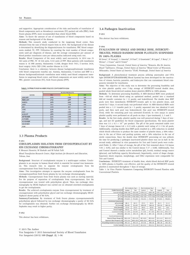
and supportive. Appropriate consideration of the risks and benefits of transfusion of
blood components such as thrombocyt concentrate (TC) packed red cells (PRC), fresh
frozen plasma (FFP), more recommended than whole blood (WB).
Aims: To Know the amount of use of transfusions of blood components based on
reasons and background of the disease.
Methods: This research study conducted in the tangerang blood center. Data
obtained from the sum of blood request form in 2014. The background of the disease
is determined by identifying the diagnosis/reason for transfusion. PRC blood compo-
nents studied, TC, FFP. Utilization by counting the total number of blood compo-
nents used per diagnosis of disease, and the average consumption per amount of
blood product transfusion cases per tahun.analisa indicated using static test.
Results: Blood Component of the most widely used successively in 2014 was 25
503 units of PRC, TC 18 526 units, 5143 units of FFP. Most patients with transfusion
reason is 16 098 anemia, thalassemia 13,568, dengue fever 7451, C-section 3434,
heart surgery 2424, CKD 2398, another disease in 2118.
Conclusion: there is a coherency between disease background/rationale transfusion
with blood components transfus use. Anemia, thalassemia, C-section and DHF is a
disease background/rationale transfusion most widely used blood component trans-
fusion in tangerang blood center, and blood components are most widely used is the
PRC, platelet concentrate (TC), fresh frozen plasma (FFP),
3.3 Plasma Products
P-091
CERULOPLASMIN ISOLATION FROM CRYOSUPERNATANT BYION EXCHANGE CHROMATOGRAPHYK Mousavi Hosseini, M Shahabi and M Fallah Tafti
Blood Transfusion Research Center, High Institute for Research and Education,
Tehran, Iran
Background: Structure of ceruloplasmin enzyme is a multicopper oxidase. Cerulo-
plasmin is an enzyme in human blood which is essential for normal iron homeosta-
sis. This research tries to separate the enzyme ceruloplasmin from the
cryosupernatant from fresh frozen plasma.
Aims: This investigation attempts to separate the enzyme ceruloplasmin from the
cryosupernatantfrom fresh frozen plasma by ion exchange chromatography.
Methods: Cryosupernatant from fresh frozen plasma was used as starting material.
For the purpose of separation of ceruloplasmin from cryosupernatant, first the
cryosupernatant was treated with polyethylene glycol. Then, ion exchange chro-
matography by DEAE-Sephacel was carried out on obtained enriched-ceruloplasmin
to get the ceroluplasmin.
Results: The achieved ceroluplasmin enzyme from cryosupernatant by treatment of
cryosupernatant with polyethylene glycol and ion exchange chromatography DEAE-
Sephacel showe purity of 90–92%.
Summary/Conclusion: By treatment of fresh frozen plasma cryosupernatantwith
polyethylene glycol followed by ion exchange chromatography a purity of 90–92%for ceroluplasmin was obtained. Further ion exchange chromatography by DEAE-
Sephadex may result in higher purity.
P-092
This abstract has been withdrawn.
3.4 Pathogen Inactivation
P-093
This abstract has been withdrawn.
P-094
EVALUATION OF SINGLE AND DOUBLE DOSE, INTERCEPT-TREATED, POOLED RANDOM DONOR PLATELETS SUSPENDEDIN 100% PLASMAM Green1, H Vermeij1, L Amorim2, H Patel3, S Graminske4, M Lopes2, T Berry1, E
Weiner1, J Crane3 and D Triulzi5
1Cerus Corporation, Concord, United States of America 2Hemorio, Rio De Janeiro,
Brazil 3LifeSource, Chicago, United States of America 4Blood Center of Wisconsin,
Milwaukee, United States of America 5ITxM, Pittsburgh, United States of America
Background: A photochemical treatment process utilizing amotosalen and UVA
light (INTERCEPT[TRADEMARK] Blood System) has been developed for the inactiva-
tion of viruses, bacteria, parasites, and leukocytes that can contaminate blood com-
ponents intended for transfusion.
Aims: The objective of this study was to determine the processing feasibility and
in vitro platelet quality over 7 day storage of INTERCEPT-treated double dose,
pooled whole blood-derived random donor platelets (RDPs) in 100% plasma.
Methods: To determine processing feasibility, 8 ABO-identical RDPs were produced
from ~450 ml whole blood using an optimized method, pooled into a standard
600 ml transfer container (n = 12 pools), and leukofiltered. The double dose RDP
pools were then immediately INTERCEPT-treated, split in two platelet doses, and
stored for 7 days. A second study was performed where 16 ABO-identical RDPs were
pooled into a 1.3 l transfer pack (n = 5 pools), separated into two identical 8 unit
pools, and then each pool was leukoreduced. One pool was INTERCEPT-treated
(Test), while the remaining pool was untreated (Control). In vitro assays to assess
platelet quality were performed on all pools on days 1 (pre-treatment), 2, 5 and 7.
Results: In the first study, platelet quality was well preserved during 7 days of stor-
age, and met EU guidelines for blood component specifications. The mean platelet
dose was 3.5 � 0.3 9 1011 per product. The pH of the pools remained stable over
7 days of storage (mean 6.8 � 0.1) with a platelet swirl rating of 2–3 for all pools.
Additionally, creating double-dose RDP pools resulted in a 20% reduction in needed
whole blood collections to produce the same number of platelet doses, a 50% reduc-
tion in the use of filters and pooling systems, and a 30% reduction in the use of
sterile connections. Since the double dose INTERCEPT processing set was utilized,
the time for producing 2 pathogen inactivated platelet doses was cut in half. In the
second study, additional in vitro parameters for Test and Control pools were evalu-
ated (Table 1). After 7 days of storage, the pH of the Test remained above 7.0 (mean
7.05 � 0.05), and was similar to the Control (mean 7.17 � 0.08). Additionally, Test
and Control showed a similar active metabolism (pO2 levels), residual energy source
(glucose), and buffering capacity (bicarbonate). Importantly, extent of shape change,
hypotonic shock response, morphology, and CD62 expression were comparable for
Test and Control.
Conclusions: INTERCEPT-treatment of double dose, whole-blood derived RDP pools
in 100% plasma is feasible, cost effective, and the quality of the INTERCEPT-treated
platelets is maintained throughout 7 days of storage.
Table 1: In Vitro Platelet Parameters Comparing INTERCEPT-Treated Platelets with
Conventional Platelets.
© 2015 The AuthorVox Sanguinis © 2015 International Society of Blood TransfusionVox Sanguinis (2015) 109 (Suppl. 2), 1–96
54 Posters
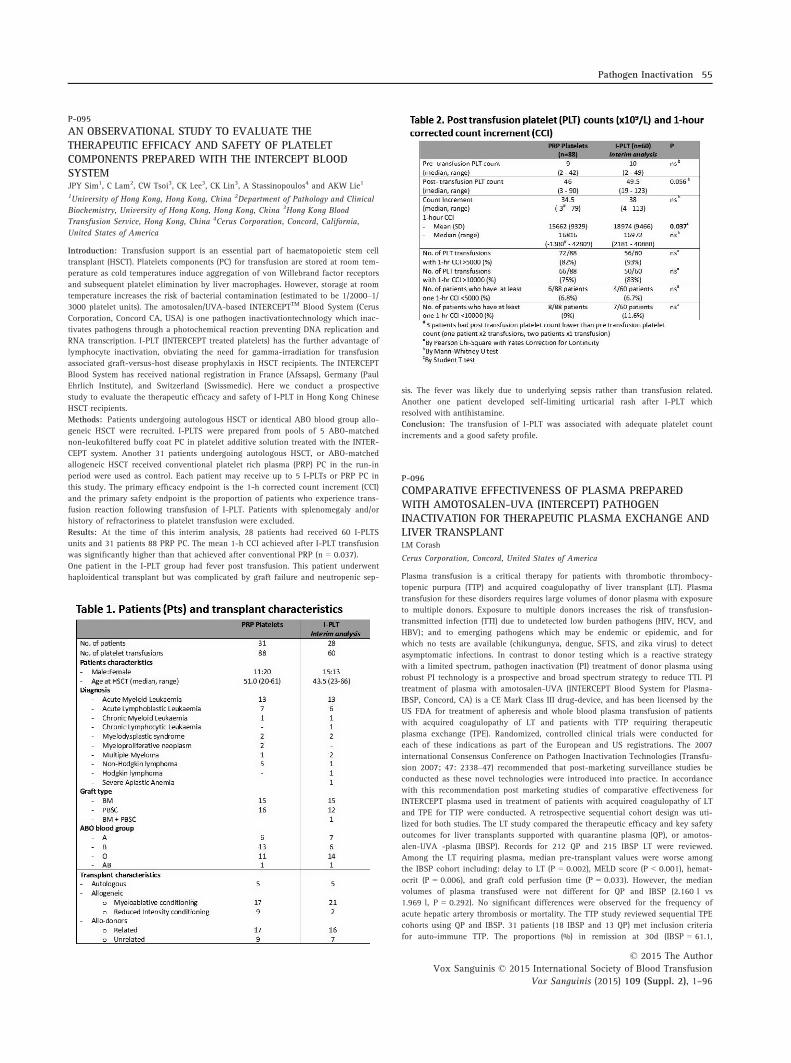
P-095
AN OBSERVATIONAL STUDY TO EVALUATE THETHERAPEUTIC EFFICACY AND SAFETY OF PLATELETCOMPONENTS PREPARED WITH THE INTERCEPT BLOODSYSTEMJPY Sim1, C Lam2, CW Tsoi3, CK Lee3, CK Lin3, A Stassinopoulos4 and AKW Lie1
1University of Hong Kong, Hong Kong, China 2Department of Pathology and Clinical
Biochemistry, University of Hong Kong, Hong Kong, China 3Hong Kong Blood
Transfusion Service, Hong Kong, China 4Cerus Corporation, Concord, California,
United States of America
Introduction: Transfusion support is an essential part of haematopoietic stem cell
transplant (HSCT). Platelets components (PC) for transfusion are stored at room tem-
perature as cold temperatures induce aggregation of von Willebrand factor receptors
and subsequent platelet elimination by liver macrophages. However, storage at room
temperature increases the risk of bacterial contamination (estimated to be 1/2000–1/3000 platelet units). The amotosalen/UVA-based INTERCEPTTM Blood System (Cerus
Corporation, Concord CA, USA) is one pathogen inactivationtechnology which inac-
tivates pathogens through a photochemical reaction preventing DNA replication and
RNA transcription. I-PLT (INTERCEPT treated platelets) has the further advantage of
lymphocyte inactivation, obviating the need for gamma-irradiation for transfusion
associated graft-versus-host disease prophylaxis in HSCT recipients. The INTERCEPT
Blood System has received national registration in France (Afssaps), Germany (Paul
Ehrlich Institute), and Switzerland (Swissmedic). Here we conduct a prospective
study to evaluate the therapeutic efficacy and safety of I-PLT in Hong Kong Chinese
HSCT recipients.
Methods: Patients undergoing autologous HSCT or identical ABO blood group allo-
geneic HSCT were recruited. I-PLTS were prepared from pools of 5 ABO-matched
non-leukofiltered buffy coat PC in platelet additive solution treated with the INTER-
CEPT system. Another 31 patients undergoing autologous HSCT, or ABO-matched
allogeneic HSCT received conventional platelet rich plasma (PRP) PC in the run-in
period were used as control. Each patient may receive up to 5 I-PLTs or PRP PC in
this study. The primary efficacy endpoint is the 1-h corrected count increment (CCI)
and the primary safety endpoint is the proportion of patients who experience trans-
fusion reaction following transfusion of I-PLT. Patients with splenomegaly and/or
history of refractoriness to platelet transfusion were excluded.
Results: At the time of this interim analysis, 28 patients had received 60 I-PLTS
units and 31 patients 88 PRP PC. The mean 1-h CCI achieved after I-PLT transfusion
was significantly higher than that achieved after conventional PRP (n = 0.037).
One patient in the I-PLT group had fever post transfusion. This patient underwent
haploidentical transplant but was complicated by graft failure and neutropenic sep-
sis. The fever was likely due to underlying sepsis rather than transfusion related.
Another one patient developed self-limiting urticarial rash after I-PLT which
resolved with antihistamine.
Conclusion: The transfusion of I-PLT was associated with adequate platelet count
increments and a good safety profile.
P-096
COMPARATIVE EFFECTIVENESS OF PLASMA PREPAREDWITH AMOTOSALEN-UVA (INTERCEPT) PATHOGENINACTIVATION FOR THERAPEUTIC PLASMA EXCHANGE ANDLIVER TRANSPLANTLM Corash
Cerus Corporation, Concord, United States of America
Plasma transfusion is a critical therapy for patients with thrombotic thrombocy-
topenic purpura (TTP) and acquired coagulopathy of liver transplant (LT). Plasma
transfusion for these disorders requires large volumes of donor plasma with exposure
to multiple donors. Exposure to multiple donors increases the risk of transfusion-
transmitted infection (TTI) due to undetected low burden pathogens (HIV, HCV, and
HBV); and to emerging pathogens which may be endemic or epidemic, and for
which no tests are available (chikungunya, dengue, SFTS, and zika virus) to detect
asymptomatic infections. In contrast to donor testing which is a reactive strategy
with a limited spectrum, pathogen inactivation (PI) treatment of donor plasma using
robust PI technology is a prospective and broad spectrum strategy to reduce TTI. PI
treatment of plasma with amotosalen-UVA (INTERCEPT Blood System for Plasma-
IBSP, Concord, CA) is a CE Mark Class III drug-device, and has been licensed by the
US FDA for treatment of apheresis and whole blood plasma transfusion of patients
with acquired coagulopathy of LT and patients with TTP requiring therapeutic
plasma exchange (TPE). Randomized, controlled clinical trials were conducted for
each of these indications as part of the European and US registrations. The 2007
international Consensus Conference on Pathogen Inactivation Technologies (Transfu-
sion 2007; 47: 2338–47) recommended that post-marketing surveillance studies be
conducted as these novel technologies were introduced into practice. In accordance
with this recommendation post marketing studies of comparative effectiveness for
INTERCEPT plasma used in treatment of patients with acquired coagulopathy of LT
and TPE for TTP were conducted. A retrospective sequential cohort design was uti-
lized for both studies. The LT study compared the therapeutic efficacy and key safety
outcomes for liver transplants supported with quarantine plasma (QP), or amotos-
alen-UVA -plasma (IBSP). Records for 212 QP and 215 IBSP LT were reviewed.
Among the LT requiring plasma, median pre-transplant values were worse among
the IBSP cohort including: delay to LT (P = 0.002), MELD score (P < 0.001), hemat-
ocrit (P = 0.006), and graft cold perfusion time (P = 0.033). However, the median
volumes of plasma transfused were not different for QP and IBSP (2.160 l vs
1.969 l, P = 0.292). No significant differences were observed for the frequency of
acute hepatic artery thrombosis or mortality. The TTP study reviewed sequential TPE
cohorts using QP and IBSP. 31 patients (18 IBSP and 13 QP) met inclusion criteria
for auto-immune TTP. The proportions (%) in remission at 30d (IBSP = 61.1,
© 2015 The AuthorVox Sanguinis © 2015 International Society of Blood Transfusion
Vox Sanguinis (2015) 109 (Suppl. 2), 1–96
Pathogen Inactivation 55
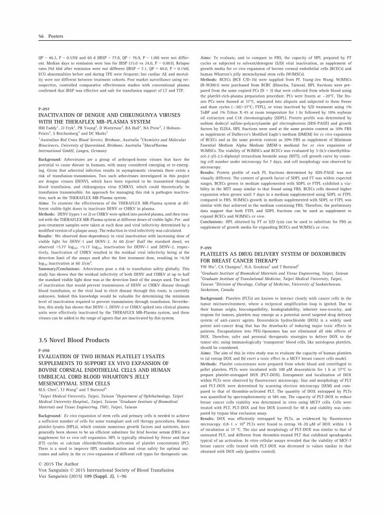
QP = 46.2, P = 0.570) and 60 d (IBSP = 77.8, QP = 76.9, P = 1.00) were not differ-
ent. Median days to remission were less for IBSP (15.0 vs 24.0, P = 0.003). Relapse
rates (%) 60d after remission were not different (IBSP = 7.1, QP = 40.0, P = 0.150).
ECG abnormalities before and during TPE were frequent; but cardiac AE and mortal-
ity were not different between treatment cohorts. Post market surveillance using ret-
rospective, controlled comparative effectiveness studies with conventional plasma
confirmed that IBSP was effective and safe for transfusion support of LT and TTP.
P-097
INACTIVATION OF DENGUE AND CHIKUNGUNYA VIRUSESWITH THE THERAFLEX MB-PLASMA SYSTEMHM Faddy1, JJ Fryk1, PR Young2, D Watterson2, RA Hall2, NA Prow2, J Hobson-
Peters2, S Reichenberg3 and DC Marks1
1Australian Red Cross Blood Service, Brisbane, Australia 2Chemistry and Molecular
Biosciences, University of Queensland, Brisbane, Australia 3MacoPharma
International GmbH, Langen, Germany
Background: Arboviruses are a group of arthropod-borne viruses that have the
potential to cause disease in humans, with many considered emerging or re-emerg-
ing. Given that arboviral infection results in asymptomatic viraemia there exists a
risk of transfusion-transmission. Two such arboviruses investigated in this project
are dengue viruses (DENV), which have been reported to be transmitted through
blood transfusion, and chikungunya virus (CHIKV), which could theoretically be
transfusion transmissible. An approach for managing this risk is pathogen inactiva-
tion, such as the THERAFLEX MB-Plasma system.
Aims: To examine the effectiveness of the THERAFLEX MB-Plasma system at dif-
ferent visible-light doses to inactivate DENV or CHIKV in plasma.
Methods: DENV (types 1 or 2) or CHIKV were spiked into pooled plasma, and then trea-
ted with the THERAFLEX MB-Plasma system at different doses of visible light. Pre- and
post-treatment samples were taken at each dose and viral infectivity determined by a
modified version of a plaque assay. The reduction in viral infectivity was calculated.
Results: We observed dose-dependency in viral inactivation with increasing dose of
visible light for DENV-1 and DENV-2. At 60 J/cm² (half the standard dose), we
observed >5.77 log10, >5.17 log10, inactivation for DENV-1 and DENV-2, respec-
tively. Inactivation of CHIKV resulted in the residual viral infectivity being at the
detection limit of the assays used after the first treatment dose, resulting in >6.58log10 inactivation at 60 J/cm2.
Summary/Conclusions: Arboviruses pose a risk to transfusion safety globally. This
study has shown that the residual infectivity of both DENV and CHIKV at up to half
the standard visible light dose was at the detection limit of the assays used. The level
of inactivation that would prevent transmission of DENV or CHIKV disease through
blood transfusion, or the viral load to elicit disease through this route, is currently
unknown. Indeed this knowledge would be valuable for determining the minimum
level of inactivation required to prevent transmission through transfusion. Neverthe-
less, this study has shown that DENV-1, DENV-2 or CHIKV spiked into clinical plasma
units were effectively inactivated by the THERAFLEX MB-Plasma system, and these
viruses can be added to the range of agents that are inactivated by this system.
3.5 Novel Blood Products
P-098
EVALUATION OF TWO HUMAN PLATELET LYSATESSUPPLEMENTS TO SUPPORT EX VIVO EXPANSION OFBOVINE CORNEAL ENDOTHELIAL CELLS AND HUMANUMBILICAL CORD BLOOD WHARTON’S JELLYMESENCHYMAL STEM CELLSM.S. Chen1, TJ Wang2 and T Burnouf3
1Taipei Medical University, Taipei, Taiwan 2Department of Ophthalmology, Taipei
Medical University Hospital,, Taipei, Taiwan 3Graduate Institute of Biomedical
Materials and Tissue Engineering, TMU, Taipei, Taiwan
Background: Ex vivo expansion of stem cells and primary cells is needed to achieve
a sufficient number of cells for some transplant and cell therapy procedures. Human
platelet-lysates (HPLs), which contain numerous growth factors and nutrients, have
generally been shown to be an efficient substitute for fetal bovine serum (FBS) as a
supplement for ex vivo cell expansion. HPL is typically obtained by freeze and thaw
(FT) cycles or calcium chloride/thrombin activation of platelet concentrates (PC).
There is a need to improve HPL standardization and virus safety for optimal out-
comes and safety in the ex vivo expansion of different cell types for therapeutic use.
Aims: To evaluate, and to compare to FBS, the capacity of HPL prepared by FT
cycles or subjected to solvent/detergent (S/D) viral inactivation, as supplement of
growth media for ex vivo expansion of bovine corneal endothelial cells (BCECs) and
human Wharton’s jelly mesenchymal stem cells (WJMSCs).
Methods: BCECs (BCE C/D-1b) were supplied from Pf. Tsung-Jen Wang. WJMSCs
(H-WJ001) were purchased from BCRC (Hinschu, Taiwan). HPL fractions were pre-
pared from the same expired PCs (N = 3) that were collected from whole blood using
the platelet-rich-plasma preparation procedure. PCs were frozen at �20°C. The fro-
zen PCs were thawed at 37°C, separated into aliquots and subjected to three freeze
and thaw cycles (�20/+37°C; FTPL), or virus inactived by S/D treatment using 1%
TnBP and 1% Triton X-45 at room temperature for 1 hr followed by 10% soybean
oil extraction and C18 chromatography (SDPL). Protein profile was determined by
sodium dodecyl sulfate-polyacrylamide gel electrophoresis (SDS-PAGE) and growth
factors by ELISA. HPL fractions were used at the same protein content as 10% FBS
as supplement of Dulbecco’s Modified Eagle’s medium (DMEM) for ex vivo expansion
of BCECs and as the same protein content as 20% FBS as supplement of Minimum
Essential Medium Alpha Medium (MEM-a medium) for ex vivo expansion of
WJMSCs. The viability of WJMSCs and BCECs was evaluated by 3-(4,5-cimethylthia-
zol-2-yl)-2,5-diphenyl tetrazolium bromide assay (MTT), cell growth curve by count-
ing cell number under microscopy for 7 days, and cell morphology was observed by
microscopy.
Results: Protein profile of each PL fractions determined by SDS-PAGE was not
visually different. The content of growth factor of SDPL and FT was within expected
ranges. BCECs grown in medium supplemented with SDPL or FTPL exhibited a via-
bility in the MTT assay similar to that found using FBS. BCECs cells showed higher
expansion when grown until 7 days in a medium supplemented using SDPL or FTPL
compared to FBS. WJMSCs growth in medium supplemented with SDPL or FTPL was
similar with that achieved in the medium containing FBS. Therefore, the preliminary
data support that both FTPL and SDPL fractions can be used as supplement to
expand BCECs and WJMSCs ex vivo.
Conclusions: HPL obtained by FT or S/D lysis can be used to substitute for FBS as
supplement of growth media for expanding BCECs and WJMSCs ex vivo.
P-099
PLATELETS AS DRUG DELIVERY SYSTEM OF DOXORUBICINFOR BREAST CANCER THERAPYYW Wu1, CA Changou2, H.A. Goubran3 and T Burnouf1
1Graduate Institute of Biomedical Materials and Tissue Engineering, Taipei, Taiwan2Graduate Institute of Translational Medicine, Taipei Medical University, Taipei,
Taiwan 3Division of Oncology, College of Medicine, University of Saskatchewan,
Saskatoon, Canada
Background: Platelets (PLTs) are known to interact closely with cancer cells in the
tumor microenvironment, where a reciprocal amplification loop is ignited. Due to
their human origin, biocompatibility, biodegradability, inherent non-toxicity, and
tropism for tumors, platelets may emerge as a potential novel targeted drug delivery
system of anti-cancer agents. Doxorubicin hydrochloride (DOX) is a widely used
potent anti-cancer drug that has the drawbacks of inducing major toxic effects in
patients. Encapsulation into PEG-liposomes has not eliminated all side effects of
DOX. Therefore, safer and personal therapeutic strategies to deliver DOX to the
tumor site, using immunologically ‘transparent’ blood cells, like autologous platelets,
should be considered.
Aims: The aim of this in vitro study was to evaluate the capacity of human platelets
to (a) entrap DOX and (b) exert a toxic effect in a MCF7 breast cancer cells model.
Methods: Platelet concentrates were prepared from whole blood and centrifuged to
pellet platelets. PLTs were incubated with 100 lM doxorubicin for 1 h at 37°C to
prepare platelet-entrapped DOX (PLT-DOX). Entrapment and localization of DOX
within PLTs were observed by fluorescence microscopy. Size and morphology of PLT
and PLT-DOX were determined by scanning electron microscopy (SEM) and com-
pared to that of thrombin-activated PLT. The quantity of DOX entrapped by PLTs
was quantified by spectrophotometry at 585 nm. The capacity of PLT-DOX to reduce
breast cancer cells viability was determined in vitro using MCF7 cells. Cells were
treated with PLT, PLT-DOX and free DOX (control) for 48 h and viability was com-
pared by trypan blue exclusion assay.
Results: DOX was efficiently entrapped by PLTs, as evidenced by fluorescence
microscopy. 0.8–1 9 106 PLTs were found to entrap 18–20 lM of DOX within 1 h
of incubation at 37 °C. The size and morphology of PLT-DOX was similar to that of
untreated PLT, and different from thrombin-treated PLT that exhibited speudopodes
typical of an activation. In vitro cellular assays revealed that the viability of MCF-7
breast cancer cells treated with PLT-DOX was decreased to values similar to that
obtained with DOX only (positive control).
© 2015 The AuthorVox Sanguinis © 2015 International Society of Blood TransfusionVox Sanguinis (2015) 109 (Suppl. 2), 1–96
56 Posters
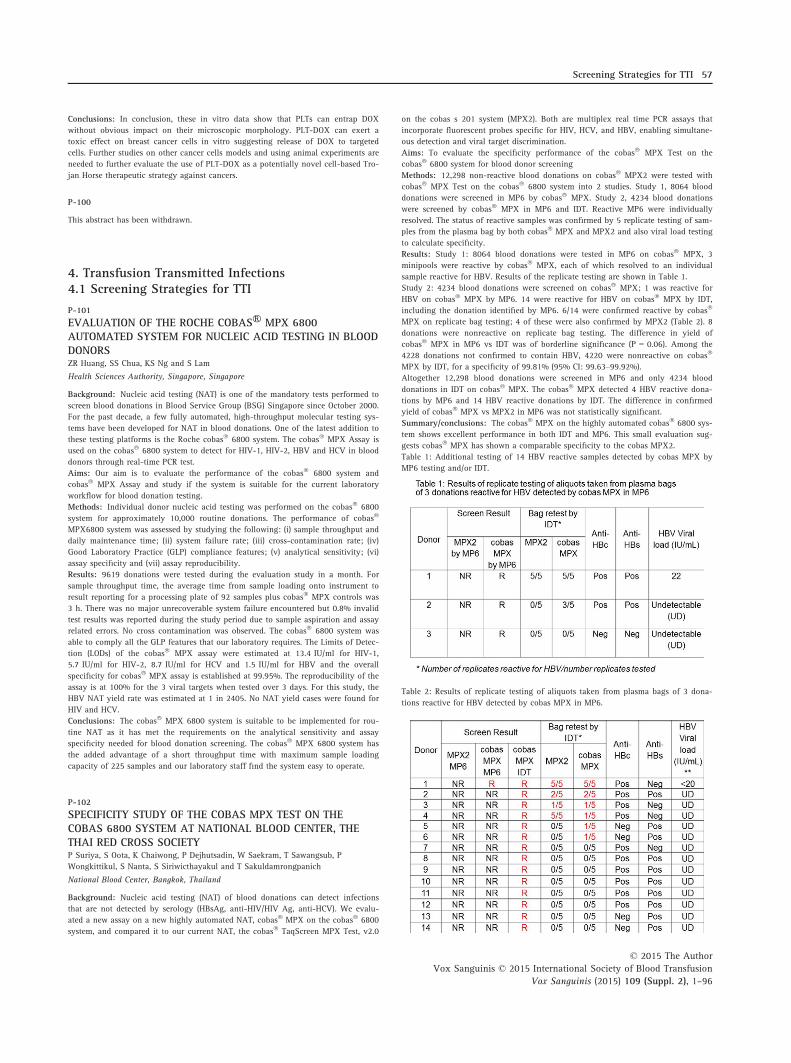
Conclusions: In conclusion, these in vitro data show that PLTs can entrap DOX
without obvious impact on their microscopic morphology. PLT-DOX can exert a
toxic effect on breast cancer cells in vitro suggesting release of DOX to targeted
cells. Further studies on other cancer cells models and using animal experiments are
needed to further evaluate the use of PLT-DOX as a potentially novel cell-based Tro-
jan Horse therapeutic strategy against cancers.
P-100
This abstract has been withdrawn.
4. Transfusion Transmitted Infections4.1 Screening Strategies for TTI
P-101
EVALUATION OF THE ROCHE COBASⓇ MPX 6800AUTOMATED SYSTEM FOR NUCLEIC ACID TESTING IN BLOODDONORSZR Huang, SS Chua, KS Ng and S Lam
Health Sciences Authority, Singapore, Singapore
Background: Nucleic acid testing (NAT) is one of the mandatory tests performed to
screen blood donations in Blood Service Group (BSG) Singapore since October 2000.
For the past decade, a few fully automated, high-throughput molecular testing sys-
tems have been developed for NAT in blood donations. One of the latest addition to
these testing platforms is the Roche cobas� 6800 system. The cobas� MPX Assay is
used on the cobas� 6800 system to detect for HIV-1, HIV-2, HBV and HCV in blood
donors through real-time PCR test.
Aims: Our aim is to evaluate the performance of the cobas� 6800 system and
cobas� MPX Assay and study if the system is suitable for the current laboratory
workflow for blood donation testing.
Methods: Individual donor nucleic acid testing was performed on the cobas� 6800
system for approximately 10,000 routine donations. The performance of cobas�
MPX6800 system was assessed by studying the following: (i) sample throughput and
daily maintenance time; (ii) system failure rate; (iii) cross-contamination rate; (iv)
Good Laboratory Practice (GLP) compliance features; (v) analytical sensitivity; (vi)
assay specificity and (vii) assay reproducibility.
Results: 9619 donations were tested during the evaluation study in a month. For
sample throughput time, the average time from sample loading onto instrument to
result reporting for a processing plate of 92 samples plus cobas� MPX controls was
3 h. There was no major unrecoverable system failure encountered but 0.8% invalid
test results was reported during the study period due to sample aspiration and assay
related errors. No cross contamination was observed. The cobas� 6800 system was
able to comply all the GLP features that our laboratory requires. The Limits of Detec-
tion (LODs) of the cobas� MPX assay were estimated at 13.4 IU/ml for HIV-1,
5.7 IU/ml for HIV-2, 8.7 IU/ml for HCV and 1.5 IU/ml for HBV and the overall
specificity for cobas� MPX assay is established at 99.95%. The reproducibility of the
assay is at 100% for the 3 viral targets when tested over 3 days. For this study, the
HBV NAT yield rate was estimated at 1 in 2405. No NAT yield cases were found for
HIV and HCV.
Conclusions: The cobas� MPX 6800 system is suitable to be implemented for rou-
tine NAT as it has met the requirements on the analytical sensitivity and assay
specificity needed for blood donation screening. The cobas� MPX 6800 system has
the added advantage of a short throughput time with maximum sample loading
capacity of 225 samples and our laboratory staff find the system easy to operate.
P-102
SPECIFICITY STUDY OF THE COBAS MPX TEST ON THECOBAS 6800 SYSTEM AT NATIONAL BLOOD CENTER, THETHAI RED CROSS SOCIETYP Suriya, S Oota, K Chaiwong, P Dejhutsadin, W Saekram, T Sawangsub, P
Wongkittikul, S Nanta, S Siriwicthayakul and T Sakuldamrongpanich
National Blood Center, Bangkok, Thailand
Background: Nucleic acid testing (NAT) of blood donations can detect infections
that are not detected by serology (HBsAg, anti-HIV/HIV Ag, anti-HCV). We evalu-
ated a new assay on a new highly automated NAT, cobas� MPX on the cobas� 6800
system, and compared it to our current NAT, the cobas� TaqScreen MPX Test, v2.0
on the cobas s 201 system (MPX2). Both are multiplex real time PCR assays that
incorporate fluorescent probes specific for HIV, HCV, and HBV, enabling simultane-
ous detection and viral target discrimination.
Aims: To evaluate the specificity performance of the cobas� MPX Test on the
cobas� 6800 system for blood donor screening
Methods: 12,298 non-reactive blood donations on cobas� MPX2 were tested with
cobas� MPX Test on the cobas� 6800 system into 2 studies. Study 1, 8064 blood
donations were screened in MP6 by cobas� MPX. Study 2, 4234 blood donations
were screened by cobas� MPX in MP6 and IDT. Reactive MP6 were individually
resolved. The status of reactive samples was confirmed by 5 replicate testing of sam-
ples from the plasma bag by both cobas� MPX and MPX2 and also viral load testing
to calculate specificity.
Results: Study 1: 8064 blood donations were tested in MP6 on cobas� MPX, 3
minipools were reactive by cobas� MPX, each of which resolved to an individual
sample reactive for HBV. Results of the replicate testing are shown in Table 1.
Study 2: 4234 blood donations were screened on cobas� MPX; 1 was reactive for
HBV on cobas� MPX by MP6. 14 were reactive for HBV on cobas� MPX by IDT,
including the donation identified by MP6. 6/14 were confirmed reactive by cobas�
MPX on replicate bag testing; 4 of these were also confirmed by MPX2 (Table 2). 8
donations were nonreactive on replicate bag testing. The difference in yield of
cobas� MPX in MP6 vs IDT was of borderline significance (P = 0.06). Among the
4228 donations not confirmed to contain HBV, 4220 were nonreactive on cobas�
MPX by IDT, for a specificity of 99.81% (95% CI: 99.63–99.92%).
Altogether 12,298 blood donations were screened in MP6 and only 4234 blood
donations in IDT on cobas� MPX. The cobas� MPX detected 4 HBV reactive dona-
tions by MP6 and 14 HBV reactive donations by IDT. The difference in confirmed
yield of cobas� MPX vs MPX2 in MP6 was not statistically significant.
Summary/conclusions: The cobas� MPX on the highly automated cobas� 6800 sys-
tem shows excellent performance in both IDT and MP6. This small evaluation sug-
gests cobas� MPX has shown a comparable specificity to the cobas MPX2.
Table 1: Additional testing of 14 HBV reactive samples detected by cobas MPX by
MP6 testing and/or IDT.
Table 2: Results of replicate testing of aliquots taken from plasma bags of 3 dona-
tions reactive for HBV detected by cobas MPX in MP6.
© 2015 The AuthorVox Sanguinis © 2015 International Society of Blood Transfusion
Vox Sanguinis (2015) 109 (Suppl. 2), 1–96
Screening Strategies for TTI 57
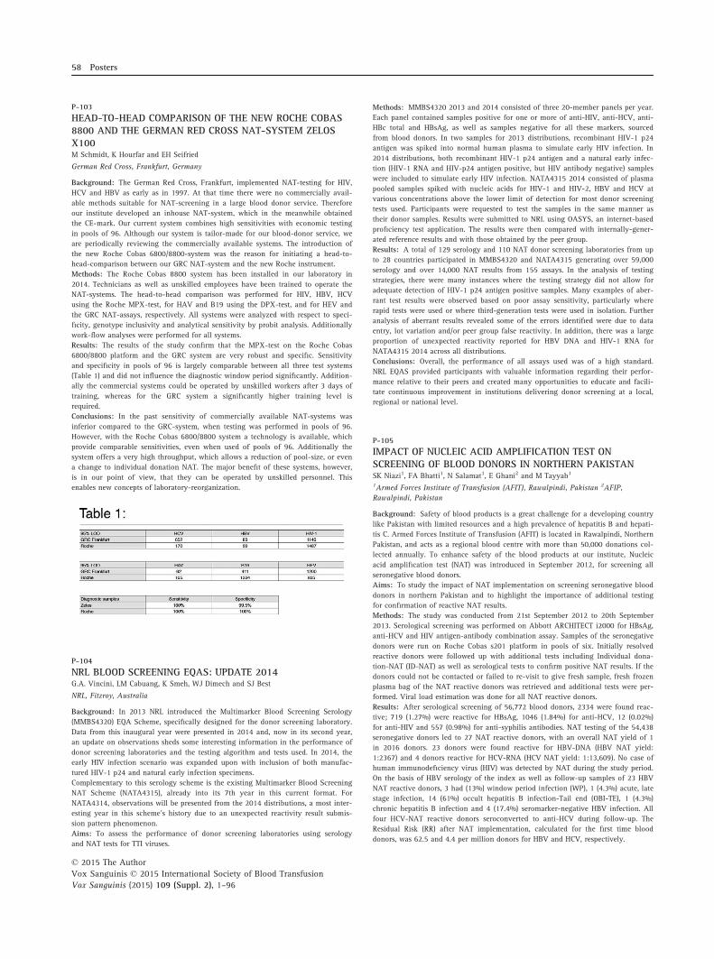
P-103
HEAD-TO-HEAD COMPARISON OF THE NEW ROCHE COBAS8800 AND THE GERMAN RED CROSS NAT-SYSTEM ZELOSX100M Schmidt, K Hourfar and EH Seifried
German Red Cross, Frankfurt, Germany
Background: The German Red Cross, Frankfurt, implemented NAT-testing for HIV,
HCV and HBV as early as in 1997. At that time there were no commercially avail-
able methods suitable for NAT-screening in a large blood donor service. Therefore
our institute developed an inhouse NAT-system, which in the meanwhile obtained
the CE-mark. Our current system combines high sensitivities with economic testing
in pools of 96. Although our system is tailor-made for our blood-donor service, we
are periodically reviewing the commercially available systems. The introduction of
the new Roche Cobas 6800/8800-system was the reason for initiating a head-to-
head-comparison between our GRC NAT-system and the new Roche instrument.
Methods: The Roche Cobas 8800 system has been installed in our laboratory in
2014. Technicians as well as unskilled employees have been trained to operate the
NAT-systems. The head-to-head comparison was performed for HIV, HBV, HCV
using the Roche MPX-test, for HAV and B19 using the DPX-test, and for HEV and
the GRC NAT-assays, respectively. All systems were analyzed with respect to speci-
ficity, genotype inclusivity and analytical sensitivity by probit analysis. Additionally
work-flow analyses were performed for all systems.
Results: The results of the study confirm that the MPX-test on the Roche Cobas
6800/8800 platform and the GRC system are very robust and specific. Sensitivity
and specificity in pools of 96 is largely comparable between all three test systems
(Table 1) and did not influence the diagnostic window period significantly. Addition-
ally the commercial systems could be operated by unskilled workers after 3 days of
training, whereas for the GRC system a significantly higher training level is
required.
Conclusions: In the past sensitivity of commercially available NAT-systems was
inferior compared to the GRC-system, when testing was performed in pools of 96.
However, with the Roche Cobas 6800/8800 system a technology is available, which
provide comparable sensitivities, even when used of pools of 96. Additionally the
system offers a very high throughput, which allows a reduction of pool-size, or even
a change to individual donation NAT. The major benefit of these systems, however,
is in our point of view, that they can be operated by unskilled personnel. This
enables new concepts of laboratory-reorganization.
P-104
NRL BLOOD SCREENING EQAS: UPDATE 2014G.A. Vincini, LM Cabuang, K Smeh, WJ Dimech and SJ Best
NRL, Fitzroy, Australia
Background: In 2013 NRL introduced the Multimarker Blood Screening Serology
(MMBS4320) EQA Scheme, specifically designed for the donor screening laboratory.
Data from this inaugural year were presented in 2014 and, now in its second year,
an update on observations sheds some interesting information in the performance of
donor screening laboratories and the testing algorithm and tests used. In 2014, the
early HIV infection scenario was expanded upon with inclusion of both manufac-
tured HIV-1 p24 and natural early infection specimens.
Complementary to this serology scheme is the existing Multimarker Blood Screening
NAT Scheme (NATA4315), already into its 7th year in this current format. For
NATA4314, observations will be presented from the 2014 distributions, a most inter-
esting year in this scheme’s history due to an unexpected reactivity result submis-
sion pattern phenomenon.
Aims: To assess the performance of donor screening laboratories using serology
and NAT tests for TTI viruses.
Methods: MMBS4320 2013 and 2014 consisted of three 20-member panels per year.
Each panel contained samples positive for one or more of anti-HIV, anti-HCV, anti-
HBc total and HBsAg, as well as samples negative for all these markers, sourced
from blood donors. In two samples for 2013 distributions, recombinant HIV-1 p24
antigen was spiked into normal human plasma to simulate early HIV infection. In
2014 distributions, both recombinant HIV-1 p24 antigen and a natural early infec-
tion (HIV-1 RNA and HIV-p24 antigen positive, but HIV antibody negative) samples
were included to simulate early HIV infection. NATA4315 2014 consisted of plasma
pooled samples spiked with nucleic acids for HIV-1 and HIV-2, HBV and HCV at
various concentrations above the lower limit of detection for most donor screening
tests used. Participants were requested to test the samples in the same manner as
their donor samples. Results were submitted to NRL using OASYS, an internet-based
proficiency test application. The results were then compared with internally-gener-
ated reference results and with those obtained by the peer group.
Results: A total of 129 serology and 110 NAT donor screening laboratories from up
to 28 countries participated in MMBS4320 and NATA4315 generating over 59,000
serology and over 14,000 NAT results from 155 assays. In the analysis of testing
strategies, there were many instances where the testing strategy did not allow for
adequate detection of HIV-1 p24 antigen positive samples. Many examples of aber-
rant test results were observed based on poor assay sensitivity, particularly where
rapid tests were used or where third-generation tests were used in isolation. Further
analysis of aberrant results revealed some of the errors identified were due to data
entry, lot variation and/or peer group false reactivity. In addition, there was a large
proportion of unexpected reactivity reported for HBV DNA and HIV-1 RNA for
NATA4315 2014 across all distributions.
Conclusions: Overall, the performance of all assays used was of a high standard.
NRL EQAS provided participants with valuable information regarding their perfor-
mance relative to their peers and created many opportunities to educate and facili-
tate continuous improvement in institutions delivering donor screening at a local,
regional or national level.
P-105
IMPACT OF NUCLEIC ACID AMPLIFICATION TEST ONSCREENING OF BLOOD DONORS IN NORTHERN PAKISTANSK Niazi1, FA Bhatti1, N Salamat1, E Ghani2 and M Tayyab1
1Armed Forces Institute of Transfusion (AFIT), Rawalpindi, Pakistan 2AFIP,
Rawalpindi, Pakistan
Background: Safety of blood products is a great challenge for a developing country
like Pakistan with limited resources and a high prevalence of hepatitis B and hepati-
tis C. Armed Forces Institute of Transfusion (AFIT) is located in Rawalpindi, Northern
Pakistan, and acts as a regional blood centre with more than 50,000 donations col-
lected annually. To enhance safety of the blood products at our institute, Nucleic
acid amplification test (NAT) was introduced in September 2012, for screening all
seronegative blood donors.
Aims: To study the impact of NAT implementation on screening seronegative blood
donors in northern Pakistan and to highlight the importance of additional testing
for confirmation of reactive NAT results.
Methods: The study was conducted from 21st September 2012 to 20th September
2013. Serological screening was performed on Abbott ARCHITECT i2000 for HBsAg,
anti-HCV and HIV antigen-antibody combination assay. Samples of the seronegative
donors were run on Roche Cobas s201 platform in pools of six. Initially resolved
reactive donors were followed up with additional tests including Individual dona-
tion-NAT (ID-NAT) as well as serological tests to confirm positive NAT results. If the
donors could not be contacted or failed to re-visit to give fresh sample, fresh frozen
plasma bag of the NAT reactive donors was retrieved and additional tests were per-
formed. Viral load estimation was done for all NAT reactive donors.
Results: After serological screening of 56,772 blood donors, 2334 were found reac-
tive; 719 (1.27%) were reactive for HBsAg, 1046 (1.84%) for anti-HCV, 12 (0.02%)
for anti-HIV and 557 (0.98%) for anti-syphilis antibodies. NAT testing of the 54,438
seronegative donors led to 27 NAT reactive donors, with an overall NAT yield of 1
in 2016 donors. 23 donors were found reactive for HBV-DNA (HBV NAT yield:
1:2367) and 4 donors reactive for HCV-RNA (HCV NAT yield: 1:13,609). No case of
human immunodeficiency virus (HIV) was detected by NAT during the study period.
On the basis of HBV serology of the index as well as follow-up samples of 23 HBV
NAT reactive donors, 3 had (13%) window period infection (WP), 1 (4.3%) acute, late
stage infection, 14 (61%) occult hepatitis B infection-Tail end (OBI-TE), 1 (4.3%)
chronic hepatitis B infection and 4 (17.4%) seromarker-negative HBV infection. All
four HCV-NAT reactive donors seroconverted to anti-HCV during follow-up. The
Residual Risk (RR) after NAT implementation, calculated for the first time blood
donors, was 62.5 and 4.4 per million donors for HBV and HCV, respectively.
© 2015 The AuthorVox Sanguinis © 2015 International Society of Blood TransfusionVox Sanguinis (2015) 109 (Suppl. 2), 1–96
58 Posters
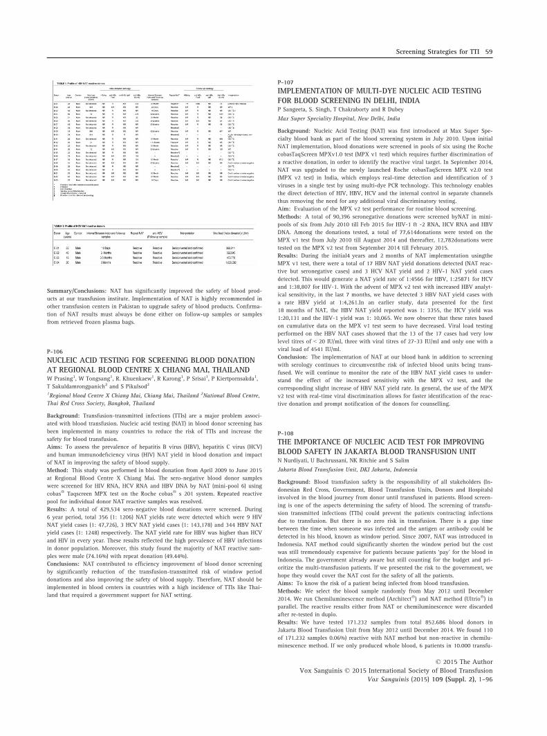
Summary/Conclusions: NAT has significantly improved the safety of blood prod-
ucts at our transfusion institute. Implementation of NAT is highly recommended in
other transfusion centers in Pakistan to upgrade safety of blood products. Confirma-
tion of NAT results must always be done either on follow-up samples or samples
from retrieved frozen plasma bags.
P-106
NUCLEIC ACID TESTING FOR SCREENING BLOOD DONATIONAT REGIONAL BLOOD CENTRE X CHIANG MAI, THAILANDW Prasing1, W Tongsang1, R. Khuenkaew1, R Karong1, P Srisai1, P Kiertpornsakda1,
T Sakuldamrongpanich2 and S Pikulsod2
1Regional blood Centre X Chiang Mai, Chiang Mai, Thailand 2National Blood Centre,
Thai Red Cross Society, Bangkok, Thailand
Background: Transfusion-transmitted infections (TTIs) are a major problem associ-
ated with blood transfusion. Nucleic acid testing (NAT) in blood donor screening has
been implemented in many countries to reduce the risk of TTIs and increase the
safety for blood transfusion.
Aims: To assess the prevalence of hepatitis B virus (HBV), hepatitis C virus (HCV)
and human immunodeficiency virus (HIV) NAT yield in blood donation and impact
of NAT in improving the safety of blood supply.
Method: This study was performed in blood donation from April 2009 to June 2015
at Regional Blood Centre X Chiang Mai. The sero-negative blood donor samples
were screened for HIV RNA, HCV RNA and HBV DNA by NAT (mini-pool 6) using
cobas� Taqscreen MPX test on the Roche cobas� s 201 system. Repeated reactive
pool for individual donor NAT reactive samples was resolved.
Results: A total of 429,534 sero-negative blood donations were screened. During
6 year period, total 356 (1: 1206) NAT yields rate were detected which were 9 HIV
NAT yield cases (1: 47,726), 3 HCV NAT yield cases (1: 143,178) and 344 HBV NAT
yield cases (1: 1248) respectively. The NAT yield rate for HBV was higher than HCV
and HIV in every year. These results reflected the high prevalence of HBV infections
in donor population. Moreover, this study found the majority of NAT reactive sam-
ples were male (74.16%) with repeat donation (49.44%).
Conclusions: NAT contributed to efficiency improvement of blood donor screening
by significantly reduction of the transfusion-transmitted risk of window period
donations and also improving the safety of blood supply. Therefore, NAT should be
implemented in blood centers in countries with a high incidence of TTIs like Thai-
land that required a government support for NAT setting.
P-107
IMPLEMENTATION OF MULTI-DYE NUCLEIC ACID TESTINGFOR BLOOD SCREENING IN DELHI, INDIAP Sangeeta, S. Singh, T Chakraborty and R Dubey
Max Super Speciality Hospital, New Delhi, India
Background: Nucleic Acid Testing (NAT) was first introduced at Max Super Spe-
cialty blood bank as part of the blood screening system in July 2010. Upon initial
NAT implementation, blood donations were screened in pools of six using the Roche
cobasTaqScreen MPXv1.0 test (MPX v1 test) which requires further discrimination of
a reactive donation, in order to identify the reactive viral target. In September 2014,
NAT was upgraded to the newly launched Roche cobasTaqScreen MPX v2.0 test
(MPX v2 test) in India, which employs real-time detection and identification of 3
viruses in a single test by using multi-dye PCR technology. This technology enables
the direct detection of HIV, HBV, HCV and the internal control in separate channels
thus removing the need for any additional viral discriminatory testing.
Aim: Evaluation of the MPX v2 test performance for routine blood screening.
Methods: A total of 90,396 seronegative donations were screened byNAT in mini-
pools of six from July 2010 till Feb 2015 for HIV-1 & -2 RNA, HCV RNA and HBV
DNA. Among the donations tested, a total of 77,614donations were tested on the
MPX v1 test from July 2010 till August 2014 and thereafter, 12,782donations were
tested on the MPX v2 test from September 2014 till February 2015.
Results: During the initial4 years and 2 months of NAT implementation usingthe
MPX v1 test, there were a total of 17 HBV NAT yield donations detected (NAT reac-
tive but seronegative cases) and 3 HCV NAT yield and 2 HIV-1 NAT yield cases
detected. This would generate a NAT yield rate of 1:4566 for HBV, 1:25871 for HCV
and 1:38,807 for HIV-1. With the advent of MPX v2 test with increased HBV analyt-
ical sensitivity, in the last 7 months, we have detected 3 HBV NAT yield cases with
a rate HBV yield at 1:4,261.In an earlier study, data presented for the first
18 months of NAT, the HBV NAT yield reported was 1: 3355, the HCV yield was
1:20,131 and the HIV-1 yield was 1: 10,065. We now observe that these rates based
on cumulative data on the MPX v1 test seem to have decreased. Viral load testing
performed on the HBV NAT cases showed that the 13 of the 17 cases had very low
level titres of < 20 IU/ml, three with viral titres of 27-33 IU/ml and only one with a
viral load of 4541 IU/ml.
Conclusion: The implementation of NAT at our blood bank in addition to screening
with serology continues to circumventthe risk of infected blood units being trans-
fused. We will continue to monitor the rate of the HBV NAT yield cases to under-
stand the effect of the increased sensitivity with the MPX v2 test, and the
corresponding slight increase of HBV NAT yield rate. In general, the use of the MPX
v2 test with real-time viral discrimination allows for faster identification of the reac-
tive donation and prompt notification of the donors for counselling.
P-108
THE IMPORTANCE OF NUCLEIC ACID TEST FOR IMPROVINGBLOOD SAFETY IN JAKARTA BLOOD TRANSFUSION UNITN Nurdiyati, U Bachrussani, NK Ritchie and S Salim
Jakarta Blood Transfusion Unit, DKI Jakarta, Indonesia
Background: Blood transfusion safety is the responsibility of all stakeholders (In-
donesian Red Cross, Government, Blood Transfusion Units, Donors and Hospitals)
involved in the blood journey from donor until transfused in patients. Blood screen-
ing is one of the aspects determining the safety of blood. The screening of transfu-
sion transmitted infections (TTIs) could prevent the patients contracting infections
due to transfusion. But there is no zero risk in transfusion. There is a gap time
between the time when someone was infected and the antigen or antibody could be
detected in his blood, known as window period. Since 2007, NAT was introduced in
Indonesia. NAT method could significantly shorten the window period but the cost
was still tremendously expensive for patients because patients ‘pay’ for the blood in
Indonesia. The government already aware but still counting for the budget and pri-
oritize the multi-transfusion patients. If we presented the risk to the government, we
hope they would cover the NAT cost for the safety of all the patients.
Aims: To know the risk of a patient being infected from blood transfusion.
Methods: We select the blood sample randomly from May 2012 until December
2014. We run Chemiluminescence method (Architect�) and NAT method (Ultrio�) in
parallel. The reactive results either from NAT or chemiluminescence were discarded
after re-tested in duplo.
Results: We have tested 171.232 samples from total 852.686 blood donors in
Jakarta Blood Transfusion Unit from May 2012 until December 2014. We found 110
of 171.232 samples 0.06%) reactive with NAT method but non-reactive in chemilu-
minescence method. If we only produced whole blood, 6 patients in 10.000 transfu-
© 2015 The AuthorVox Sanguinis © 2015 International Society of Blood Transfusion
Vox Sanguinis (2015) 109 (Suppl. 2), 1–96
Screening Strategies for TTI 59
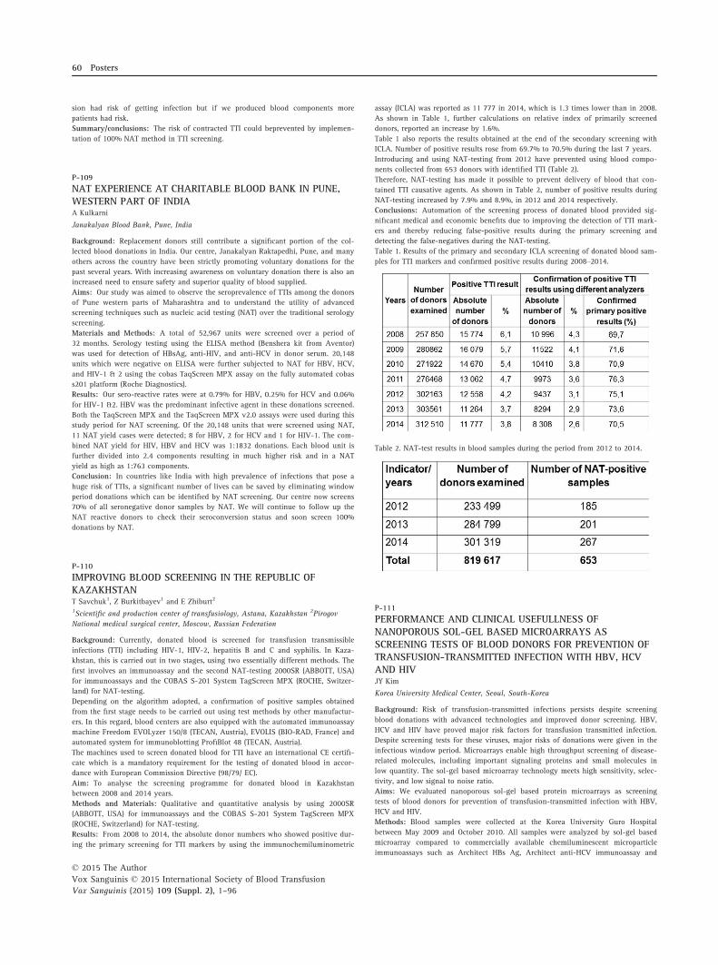
sion had risk of getting infection but if we produced blood components more
patients had risk.
Summary/conclusions: The risk of contracted TTI could beprevented by implemen-
tation of 100% NAT method in TTI screening.
P-109
NAT EXPERIENCE AT CHARITABLE BLOOD BANK IN PUNE,WESTERN PART OF INDIAA Kulkarni
Janakalyan Blood Bank, Pune, India
Background: Replacement donors still contribute a significant portion of the col-
lected blood donations in India. Our centre, Janakalyan Raktapedhi, Pune, and many
others across the country have been strictly promoting voluntary donations for the
past several years. With increasing awareness on voluntary donation there is also an
increased need to ensure safety and superior quality of blood supplied.
Aims: Our study was aimed to observe the seroprevalence of TTIs among the donors
of Pune western parts of Maharashtra and to understand the utility of advanced
screening techniques such as nucleic acid testing (NAT) over the traditional serology
screening.
Materials and Methods: A total of 52,967 units were screened over a period of
32 months. Serology testing using the ELISA method (Benshera kit from Aventor)
was used for detection of HBsAg, anti-HIV, and anti-HCV in donor serum. 20,148
units which were negative on ELISA were further subjected to NAT for HBV, HCV,
and HIV-1 & 2 using the cobas TaqScreen MPX assay on the fully automated cobas
s201 platform (Roche Diagnostics).
Results: Our sero-reactive rates were at 0.79% for HBV, 0.25% for HCV and 0.06%
for HIV-1 &2. HBV was the predominant infective agent in these donations screened.
Both the TaqScreen MPX and the TaqScreen MPX v2.0 assays were used during this
study period for NAT screening. Of the 20,148 units that were screened using NAT,
11 NAT yield cases were detected; 8 for HBV, 2 for HCV and 1 for HIV-1. The com-
bined NAT yield for HIV, HBV and HCV was 1:1832 donations. Each blood unit is
further divided into 2.4 components resulting in much higher risk and in a NAT
yield as high as 1:763 components.
Conclusion: In countries like India with high prevalence of infections that pose a
huge risk of TTIs, a significant number of lives can be saved by eliminating window
period donations which can be identified by NAT screening. Our centre now screens
70% of all seronegative donor samples by NAT. We will continue to follow up the
NAT reactive donors to check their seroconversion status and soon screen 100%
donations by NAT.
P-110
IMPROVING BLOOD SCREENING IN THE REPUBLIC OFKAZAKHSTANT Savchuk1, Z Burkitbayev1 and E Zhiburt2
1Scientific and production center of transfusiology, Astana, Kazakhstan 2Pirogov
National medical surgical center, Moscow, Russian Federation
Background: Currently, donated blood is screened for transfusion transmissible
infections (TTI) including HIV-1, HIV-2, hepatitis B and C and syphilis. In Kaza-
khstan, this is carried out in two stages, using two essentially different methods. The
first involves an immunoassay and the second NAT-testing 2000SR (ABBOTT, USA)
for immunoassays and the COBAS S-201 System TagScreen MPX (ROCHE, Switzer-
land) for NAT-testing.
Depending on the algorithm adopted, a confirmation of positive samples obtained
from the first stage needs to be carried out using test methods by other manufactur-
ers. In this regard, blood centers are also equipped with the automated immunoassay
machine Freedom EVOLyzer 150/8 (TECAN, Austria), EVOLIS (BIO-RAD, France) and
automated system for immunoblotting ProfiBlot 48 (TECAN, Austria).
The machines used to screen donated blood for TTI have an international CE certifi-
cate which is a mandatory requirement for the testing of donated blood in accor-
dance with European Commission Directive (98/79/ EC).
Aim: To analyse the screening programme for donated blood in Kazakhstan
between 2008 and 2014 years.
Methods and Materials: Qualitative and quantitative analysis by using 2000SR
(ABBOTT, USA) for immunoassays and the COBAS S-201 System TagScreen MPX
(ROCHE, Switzerland) for NAT-testing.
Results: From 2008 to 2014, the absolute donor numbers who showed positive dur-
ing the primary screening for TTI markers by using the immunochemiluminometric
assay (ICLA) was reported as 11 777 in 2014, which is 1.3 times lower than in 2008.
As shown in Table 1, further calculations on relative index of primarily screened
donors, reported an increase by 1.6%.
Table 1 also reports the results obtained at the end of the secondary screening with
ICLA. Number of positive results rose from 69.7% to 70.5% during the last 7 years.
Introducing and using NAT-testing from 2012 have prevented using blood compo-
nents collected from 653 donors with identified TTI (Table 2).
Therefore, NAT-testing has made it possible to prevent delivery of blood that con-
tained TTI causative agents. As shown in Table 2, number of positive results during
NAT-testing increased by 7.9% and 8.9%, in 2012 and 2014 respectively.
Conclusions: Automation of the screening process of donated blood provided sig-
nificant medical and economic benefits due to improving the detection of TTI mark-
ers and thereby reducing false-positive results during the primary screening and
detecting the false-negatives during the NAT-testing.
Table 1. Results of the primary and secondary ICLA screening of donated blood sam-
ples for TTI markers and confirmed positive results during 2008–2014.
Table 2. NAT-test results in blood samples during the period from 2012 to 2014.
P-111
PERFORMANCE AND CLINICAL USEFULLNESS OFNANOPOROUS SOL-GEL BASED MICROARRAYS ASSCREENING TESTS OF BLOOD DONORS FOR PREVENTION OFTRANSFUSION-TRANSMITTED INFECTION WITH HBV, HCVAND HIVJY Kim
Korea University Medical Center, Seoul, South-Korea
Background: Risk of transfusion-transmitted infections persists despite screening
blood donations with advanced technologies and improved donor screening. HBV,
HCV and HIV have proved major risk factors for transfusion transmitted infection.
Despite screening tests for these viruses, major risks of donations were given in the
infectious window period. Microarrays enable high throughput screening of disease-
related molecules, including important signaling proteins and small molecules in
low quantity. The sol-gel based microarray technology meets high sensitivity, selec-
tivity, and low signal to noise ratio.
Aims: We evaluated nanoporous sol-gel based protein microarrays as screening
tests of blood donors for prevention of transfusion-transmitted infection with HBV,
HCV and HIV.
Methods: Blood samples were collected at the Korea University Guro Hospital
between May 2009 and October 2010. All samples were analyzed by sol-gel based
microarray compared to commercially available chemiluminescent microparticle
immunoassays such as Architect HBs Ag, Architect anti-HCV immunoassay and
© 2015 The AuthorVox Sanguinis © 2015 International Society of Blood TransfusionVox Sanguinis (2015) 109 (Suppl. 2), 1–96
60 Posters
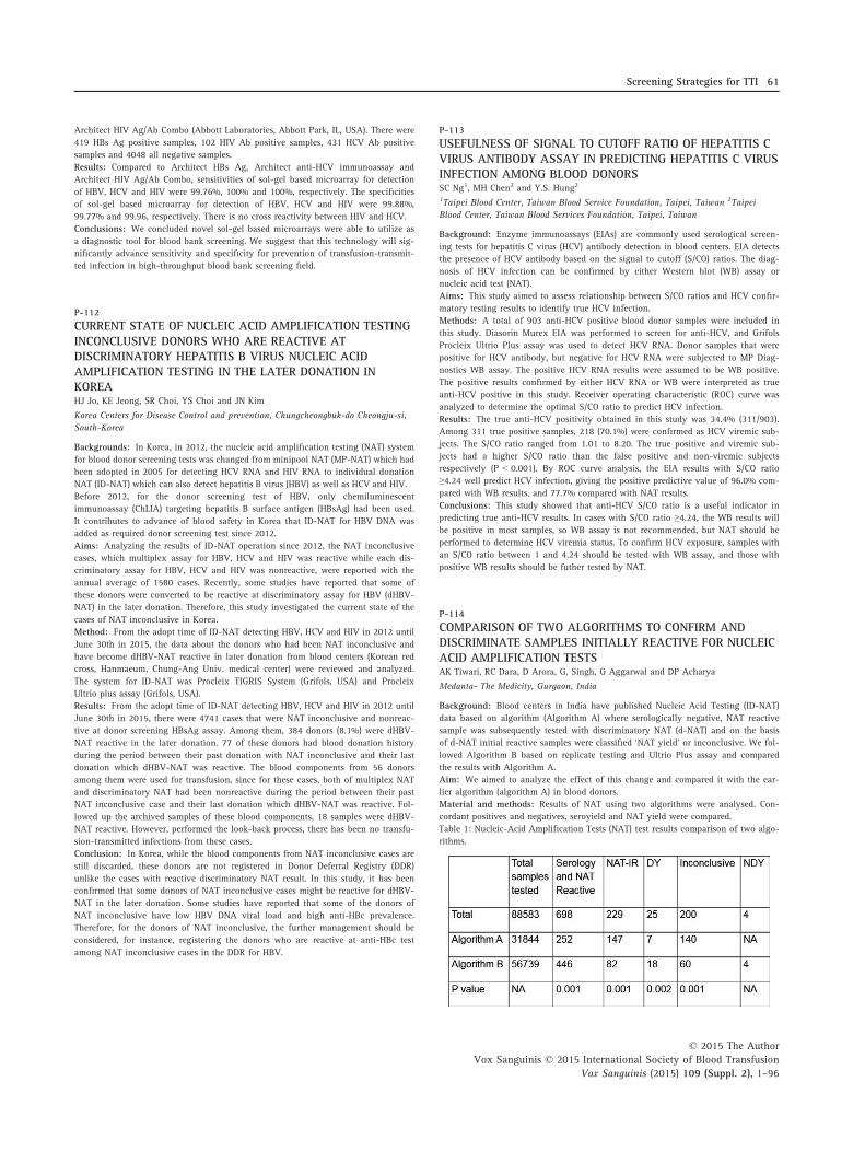
Architect HIV Ag/Ab Combo (Abbott Laboratories, Abbott Park, IL, USA). There were
419 HBs Ag positive samples, 102 HIV Ab positive samples, 431 HCV Ab positive
samples and 4048 all negative samples.
Results: Compared to Architect HBs Ag, Architect anti-HCV immunoassay and
Architect HIV Ag/Ab Combo, sensitivities of sol-gel based microarray for detection
of HBV, HCV and HIV were 99.76%, 100% and 100%, respectively. The specificities
of sol-gel based microarray for detection of HBV, HCV and HIV were 99.88%,
99.77% and 99.96, respectively. There is no cross reactivity between HIV and HCV.
Conclusions: We concluded novel sol-gel based microarrays were able to utilize as
a diagnostic tool for blood bank screening. We suggest that this technology will sig-
nificantly advance sensitivity and specificity for prevention of transfusion-transmit-
ted infection in high-throughput blood bank screening field.
P-112
CURRENT STATE OF NUCLEIC ACID AMPLIFICATION TESTINGINCONCLUSIVE DONORS WHO ARE REACTIVE ATDISCRIMINATORY HEPATITIS B VIRUS NUCLEIC ACIDAMPLIFICATION TESTING IN THE LATER DONATION INKOREAHJ Jo, KE Jeong, SR Choi, YS Choi and JN Kim
Korea Centers for Disease Control and prevention, Chungcheongbuk-do Cheongju-si,
South-Korea
Backgrounds: In Korea, in 2012, the nucleic acid amplification testing (NAT) system
for blood donor screening tests was changed from minipool NAT (MP-NAT) which had
been adopted in 2005 for detecting HCV RNA and HIV RNA to individual donation
NAT (ID-NAT) which can also detect hepatitis B virus (HBV) as well as HCV and HIV.
Before 2012, for the donor screening test of HBV, only chemiluminescent
immunoassay (ChLIA) targeting hepatitis B surface antigen (HBsAg) had been used.
It contributes to advance of blood safety in Korea that ID-NAT for HBV DNA was
added as required donor screening test since 2012.
Aims: Analyzing the results of ID-NAT operation since 2012, the NAT inconclusive
cases, which multiplex assay for HBV, HCV and HIV was reactive while each dis-
criminatory assay for HBV, HCV and HIV was nonreactive, were reported with the
annual average of 1580 cases. Recently, some studies have reported that some of
these donors were converted to be reactive at discriminatory assay for HBV (dHBV-
NAT) in the later donation. Therefore, this study investigated the current state of the
cases of NAT inconclusive in Korea.
Method: From the adopt time of ID-NAT detecting HBV, HCV and HIV in 2012 until
June 30th in 2015, the data about the donors who had been NAT inconclusive and
have become dHBV-NAT reactive in later donation from blood centers (Korean red
cross, Hanmaeum, Chung-Ang Univ. medical center) were reviewed and analyzed.
The system for ID-NAT was Procleix TIGRIS System (Grifols, USA) and Procleix
Ultrio plus assay (Grifols, USA).
Results: From the adopt time of ID-NAT detecting HBV, HCV and HIV in 2012 until
June 30th in 2015, there were 4741 cases that were NAT inconclusive and nonreac-
tive at donor screening HBsAg assay. Among them, 384 donors (8.1%) were dHBV-
NAT reactive in the later donation. 77 of these donors had blood donation history
during the period between their past donation with NAT inconclusive and their last
donation which dHBV-NAT was reactive. The blood components from 56 donors
among them were used for transfusion, since for these cases, both of multiplex NAT
and discriminatory NAT had been nonreactive during the period between their past
NAT inconclusive case and their last donation which dHBV-NAT was reactive. Fol-
lowed up the archived samples of these blood components, 18 samples were dHBV-
NAT reactive. However, performed the look-back process, there has been no transfu-
sion-transmitted infections from these cases.
Conclusion: In Korea, while the blood components from NAT inconclusive cases are
still discarded, these donors are not registered in Donor Deferral Registry (DDR)
unlike the cases with reactive discriminatory NAT result. In this study, it has been
confirmed that some donors of NAT inconclusive cases might be reactive for dHBV-
NAT in the later donation. Some studies have reported that some of the donors of
NAT inconclusive have low HBV DNA viral load and high anti-HBc prevalence.
Therefore, for the donors of NAT inconclusive, the further management should be
considered, for instance, registering the donors who are reactive at anti-HBc test
among NAT inconclusive cases in the DDR for HBV.
P-113
USEFULNESS OF SIGNAL TO CUTOFF RATIO OF HEPATITIS CVIRUS ANTIBODY ASSAY IN PREDICTING HEPATITIS C VIRUSINFECTION AMONG BLOOD DONORSSC Ng1, MH Chen2 and Y.S. Hung2
1Taipei Blood Center, Taiwan Blood Service Foundation, Taipei, Taiwan 2Taipei
Blood Center, Taiwan Blood Services Foundation, Taipei, Taiwan
Background: Enzyme immunoassays (EIAs) are commonly used serological screen-
ing tests for hepatitis C virus (HCV) antibody detection in blood centers. EIA detects
the presence of HCV antibody based on the signal to cutoff (S/CO) ratios. The diag-
nosis of HCV infection can be confirmed by either Western blot (WB) assay or
nucleic acid test (NAT).
Aims: This study aimed to assess relationship between S/CO ratios and HCV confir-
matory testing results to identify true HCV infection.
Methods: A total of 903 anti-HCV positive blood donor samples were included in
this study. Diasorin Murex EIA was performed to screen for anti-HCV, and Grifols
Procleix Ultrio Plus assay was used to detect HCV RNA. Donor samples that were
positive for HCV antibody, but negative for HCV RNA were subjected to MP Diag-
nostics WB assay. The positive HCV RNA results were assumed to be WB positive.
The positive results confirmed by either HCV RNA or WB were interpreted as true
anti-HCV positive in this study. Receiver operating characteristic (ROC) curve was
analyzed to determine the optimal S/CO ratio to predict HCV infection.
Results: The true anti-HCV positivity obtained in this study was 34.4% (311/903).
Among 311 true positive samples, 218 (70.1%) were confirmed as HCV viremic sub-
jects. The S/CO ratio ranged from 1.01 to 8.20. The true positive and viremic sub-
jects had a higher S/CO ratio than the false positive and non-viremic subjects
respectively (P < 0.001). By ROC curve analysis, the EIA results with S/CO ratio
≥4.24 well predict HCV infection, giving the positive predictive value of 96.0% com-
pared with WB results, and 77.7% compared with NAT results.
Conclusions: This study showed that anti-HCV S/CO ratio is a useful indicator in
predicting true anti-HCV results. In cases with S/CO ratio ≥4.24, the WB results will
be positive in most samples, so WB assay is not recommended, but NAT should be
performed to determine HCV viremia status. To confirm HCV exposure, samples with
an S/CO ratio between 1 and 4.24 should be tested with WB assay, and those with
positive WB results should be futher tested by NAT.
P-114
COMPARISON OF TWO ALGORITHMS TO CONFIRM ANDDISCRIMINATE SAMPLES INITIALLY REACTIVE FOR NUCLEICACID AMPLIFICATION TESTSAK Tiwari, RC Dara, D Arora, G. Singh, G Aggarwal and DP Acharya
Medanta- The Medicity, Gurgaon, India
Background: Blood centers in India have published Nucleic Acid Testing (ID-NAT)
data based on algorithm (Algorithm A) where serologically negative, NAT reactive
sample was subsequently tested with discriminatory NAT (d-NAT) and on the basis
of d-NAT initial reactive samples were classified ‘NAT yield’ or inconclusive. We fol-
lowed Algorithm B based on replicate testing and Ultrio Plus assay and compared
the results with Algorithm A.
Aim: We aimed to analyze the effect of this change and compared it with the ear-
lier algorithm (algorithm A) in blood donors.
Material and methods: Results of NAT using two algorithms were analysed. Con-
cordant positives and negatives, seroyield and NAT yield were compared.
Table 1: Nucleic-Acid Amplification Tests (NAT) test results comparison of two algo-
rithms.
© 2015 The AuthorVox Sanguinis © 2015 International Society of Blood Transfusion
Vox Sanguinis (2015) 109 (Suppl. 2), 1–96
Screening Strategies for TTI 61
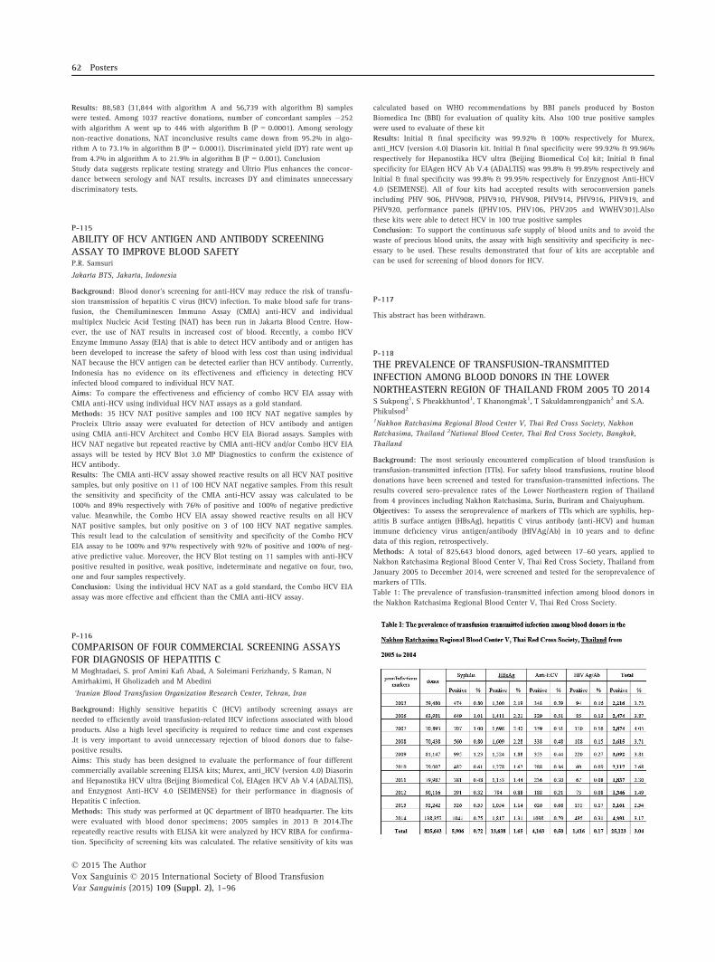
Results: 88,583 (31,844 with algorithm A and 56,739 with algorithm B) samples
were tested. Among 1037 reactive donations, number of concordant samples �252
with algorithm A went up to 446 with algorithm B (P = 0.0001). Among serology
non-reactive donations, NAT inconclusive results came down from 95.2% in algo-
rithm A to 73.1% in algorithm B (P = 0.0001). Discriminated yield (DY) rate went up
from 4.7% in algorithm A to 21.9% in algorithm B (P = 0.001). Conclusion
Study data suggests replicate testing strategy and Ultrio Plus enhances the concor-
dance between serology and NAT results, increases DY and eliminates unnecessary
discriminatory tests.
P-115
ABILITY OF HCV ANTIGEN AND ANTIBODY SCREENINGASSAY TO IMPROVE BLOOD SAFETYP.R. Samsuri
Jakarta BTS, Jakarta, Indonesia
Background: Blood donor’s screening for anti-HCV may reduce the risk of transfu-
sion transmission of hepatitis C virus (HCV) infection. To make blood safe for trans-
fusion, the Chemiluminescen Immuno Assay (CMIA) anti-HCV and individual
multiplex Nucleic Acid Testing (NAT) has been run in Jakarta Blood Centre. How-
ever, the use of NAT results in increased cost of blood. Recently, a combo HCV
Enzyme Immuno Assay (EIA) that is able to detect HCV antibody and or antigen has
been developed to increase the safety of blood with less cost than using individual
NAT because the HCV antigen can be detected earlier than HCV antibody. Currently,
Indonesia has no evidence on its effectiveness and efficiency in detecting HCV
infected blood compared to individual HCV NAT.
Aims: To compare the effectiveness and efficiency of combo HCV EIA assay with
CMIA anti-HCV using individual HCV NAT assays as a gold standard.
Methods: 35 HCV NAT positive samples and 100 HCV NAT negative samples by
Procleix Ultrio assay were evaluated for detection of HCV antibody and antigen
using CMIA anti-HCV Architect and Combo HCV EIA Biorad assays. Samples with
HCV NAT negative but repeated reactive by CMIA anti-HCV and/or Combo HCV EIA
assays will be tested by HCV Blot 3.0 MP Diagnostics to confirm the existence of
HCV antibody.
Results: The CMIA anti-HCV assay showed reactive results on all HCV NAT positive
samples, but only positive on 11 of 100 HCV NAT negative samples. From this result
the sensitivity and specificity of the CMIA anti-HCV assay was calculated to be
100% and 89% respectively with 76% of positive and 100% of negative predictive
value. Meanwhile, the Combo HCV EIA assay showed reactive results on all HCV
NAT positive samples, but only positive on 3 of 100 HCV NAT negative samples.
This result lead to the calculation of sensitivity and specificity of the Combo HCV
EIA assay to be 100% and 97% respectively with 92% of positive and 100% of neg-
ative predictive value. Moreover, the HCV Blot testing on 11 samples with anti-HCV
positive resulted in positive, weak positive, indeterminate and negative on four, two,
one and four samples respectively.
Conclusion: Using the individual HCV NAT as a gold standard, the Combo HCV EIA
assay was more effective and efficient than the CMIA anti-HCV assay.
P-116
COMPARISON OF FOUR COMMERCIAL SCREENING ASSAYSFOR DIAGNOSIS OF HEPATITIS CM Moghtadaei, S. prof Amini Kafi Abad, A Soleimani Ferizhandy, S Raman, N
Amirhakimi, H Gholizadeh and M Abedini
‘Iranian Blood Transfusion Organization Research Center, Tehran, Iran
Background: Highly sensitive hepatitis C (HCV) antibody screening assays are
needed to efficiently avoid transfusion-related HCV infections associated with blood
products. Also a high level specificity is required to reduce time and cost expenses
.It is very important to avoid unnecessary rejection of blood donors due to false-
positive results.
Aims: This study has been designed to evaluate the performance of four different
commercially available screening ELISA kits; Murex, anti_HCV (version 4.0) Diasorin
and Hepanostika HCV ultra (Beijing Biomedical Co), EIAgen HCV Ab V.4 (ADALTIS),
and Enzygnost Anti-HCV 4.0 (SEIMENSE) for their performance in diagnosis of
Hepatitis C infection.
Methods: This study was performed at QC department of IBTO headquarter. The kits
were evaluated with blood donor specimens; 2005 samples in 2013 & 2014.The
repeatedly reactive results with ELISA kit were analyzed by HCV RIBA for confirma-
tion. Specificity of screening kits was calculated. The relative sensitivity of kits was
calculated based on WHO recommendations by BBI panels produced by Boston
Biomedica Inc (BBI) for evaluation of quality kits. Also 100 true positive samples
were used to evaluate of these kit
Results: Initial & final specificity was 99.92% & 100% respectively for Murex,
anti_HCV (version 4.0) Diasorin kit. Initial & final specificity were 99.92% & 99.96%
respectively for Hepanostika HCV ultra (Beijing Biomedical Co) kit; Initial & final
specificity for EIAgen HCV Ab V.4 (ADALTIS) was 99.8% & 99.85% respectively and
Initial & final specificity was 99.8% & 99.95% respectively for Enzygnost Anti-HCV
4.0 (SEIMENSE). All of four kits had accepted results with seroconversion panels
including PHV 906, PHV908, PHV910, PHV908, PHV914, PHV916, PHV919, and
PHV920, performance panels ((PHV105, PHV106, PHV205 and WWHV301).Also
these kits were able to detect HCV in 100 true positive samples
Conclusion: To support the continuous safe supply of blood units and to avoid the
waste of precious blood units, the assay with high sensitivity and specificity is nec-
essary to be used. These results demonstrated that four of kits are acceptable and
can be used for screening of blood donors for HCV.
P-117
This abstract has been withdrawn.
P-118
THE PREVALENCE OF TRANSFUSION-TRANSMITTEDINFECTION AMONG BLOOD DONORS IN THE LOWERNORTHEASTERN REGION OF THAILAND FROM 2005 TO 2014S Sukpong1, S Pheakkhuntod1, T Khanongmak1, T Sakuldamrongpanich2 and S.A.
Phikulsod2
1Nakhon Ratchasima Regional Blood Center V, Thai Red Cross Society, Nakhon
Ratchasima, Thailand 2National Blood Center, Thai Red Cross Society, Bangkok,
Thailand
Background: The most seriously encountered complication of blood transfusion is
transfusion-transmitted infection (TTIs). For safety blood transfusions, routine blood
donations have been screened and tested for transfusion-transmitted infections. The
results covered sero-prevalence rates of the Lower Northeastern region of Thailand
from 4 provinces including Nakhon Ratchasima, Surin, Buriram and Chaiyuphum.
Objectives: To assess the seroprevalence of markers of TTIs which are syphilis, hep-
atitis B surface antigen (HBsAg), hepatitis C virus antibody (anti-HCV) and human
immune deficiency virus antigen/antibody (HIVAg/Ab) in 10 years and to define
data of this region, retrospectively.
Methods: A total of 825,643 blood donors, aged between 17–60 years, applied to
Nakhon Ratchasima Regional Blood Center V, Thai Red Cross Society, Thailand from
January 2005 to December 2014, were screened and tested for the seroprevalence of
markers of TTIs.
Table 1: The prevalence of transfusion-transmitted infection among blood donors in
the Nakhon Ratchasima Regional Blood Center V, Thai Red Cross Society.
© 2015 The AuthorVox Sanguinis © 2015 International Society of Blood TransfusionVox Sanguinis (2015) 109 (Suppl. 2), 1–96
62 Posters
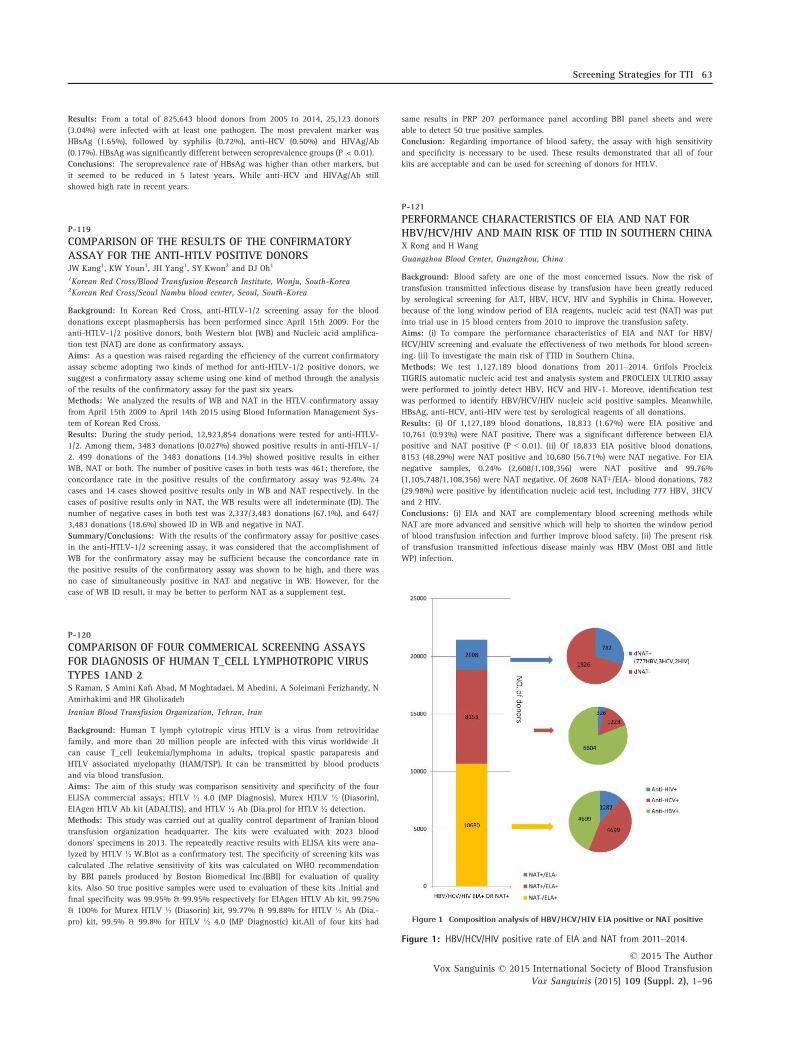
Results: From a total of 825,643 blood donors from 2005 to 2014, 25,123 donors
(3.04%) were infected with at least one pathogen. The most prevalent marker was
HBsAg (1.65%), followed by syphilis (0.72%), anti-HCV (0.50%) and HIVAg/Ab
(0.17%). HBsAg was significantly different between seroprevalence groups (P < 0.01).
Conclusions: The seroprevalence rate of HBsAg was higher than other markers, but
it seemed to be reduced in 5 latest years. While anti-HCV and HIVAg/Ab still
showed high rate in recent years.
P-119
COMPARISON OF THE RESULTS OF THE CONFIRMATORYASSAY FOR THE ANTI-HTLV POSITIVE DONORSJW Kang1, KW Youn1, JH Yang1, SY Kwon2 and DJ Oh1
1Korean Red Cross/Blood Transfusion Research Institute, Wonju, South-Korea2Korean Red Cross/Seoul Nambu blood center, Seoul, South-Korea
Background: In Korean Red Cross, anti-HTLV-1/2 screening assay for the blood
donations except plasmaphersis has been performed since April 15th 2009. For the
anti-HTLV-1/2 positive donors, both Western blot (WB) and Nucleic acid amplifica-
tion test (NAT) are done as confirmatory assays.
Aims: As a question was raised regarding the efficiency of the current confirmatory
assay scheme adopting two kinds of method for anti-HTLV-1/2 positive donors, we
suggest a confirmatory assay scheme using one kind of method through the analysis
of the results of the confirmatory assay for the past six years.
Methods: We analyzed the results of WB and NAT in the HTLV confirmatory assay
from April 15th 2009 to April 14th 2015 using Blood Information Management Sys-
tem of Korean Red Cross.
Results: During the study period, 12,923,854 donations were tested for anti-HTLV-
1/2. Among them, 3483 donations (0.027%) showed positive results in anti-HTLV-1/
2. 499 donations of the 3483 donations (14.3%) showed positive results in either
WB, NAT or both. The number of positive cases in both tests was 461; therefore, the
concordance rate in the positive results of the confirmatory assay was 92.4%. 24
cases and 14 cases showed positive results only in WB and NAT respectively. In the
cases of positive results only in NAT, the WB results were all indeterminate (ID). The
number of negative cases in both test was 2,337/3,483 donations (67.1%), and 647/
3,483 donations (18.6%) showed ID in WB and negative in NAT.
Summary/Conclusions: With the results of the confirmatory assay for positive cases
in the anti-HTLV-1/2 screening assay, it was considered that the accomplishment of
WB for the confirmatory assay may be sufficient because the concordance rate in
the positive results of the confirmatory assay was shown to be high, and there was
no case of simultaneously positive in NAT and negative in WB. However, for the
case of WB ID result, it may be better to perform NAT as a supplement test.
P-120
COMPARISON OF FOUR COMMERICAL SCREENING ASSAYSFOR DIAGNOSIS OF HUMAN T_CELL LYMPHOTROPIC VIRUSTYPES 1AND 2S Raman, S Amini Kafi Abad, M Moghtadaei, M Abedini, A Soleimani Ferizhandy, N
Amirhakimi and HR Gholizadeh
Iranian Blood Transfusion Organization, Tehran, Iran
Background: Human T lymph cytotropic virus HTLV is a virus from retroviridae
family, and more than 20 million people are infected with this virus worldwide .It
can cause T_cell leukemia/lymphoma in adults, tropical spastic paraparesis and
HTLV associated myelopathy (HAM/TSP). It can be transmitted by blood products
and via blood transfusion.
Aims: The aim of this study was comparison sensitivity and specificity of the four
ELISA commercial assays; HTLV ½ 4.0 (MP Diagnosis), Murex HTLV ½ (Diasorin),
EIAgen HTLV Ab kit (ADALTIS), and HTLV ½ Ab (Dia.pro) for HTLV ½ detection.
Methods: This study was carried out at quality control department of Iranian blood
transfusion organization headquarter. The kits were evaluated with 2023 blood
donors’ specimens in 2013. The repeatedly reactive results with ELISA kits were ana-
lyzed by HTLV ½ W.Blot as a confirmatory test. The specificity of screening kits was
calculated .The relative sensitivity of kits was calculated on WHO recommendation
by BBI panels produced by Boston Biomedical Inc.(BBI) for evaluation of quality
kits. Also 50 true positive samples were used to evaluation of these kits .Initial and
final specificity was 99.95% & 99.95% respectively for EIAgen HTLV Ab kit, 99.75%
& 100% for Murex HTLV ½ (Diasorin) kit, 99.77% & 99.88% for HTLV ½ Ab (Dia.-
pro) kit, 99.5% & 99.8% for HTLV ½ 4.0 (MP Diagnostic) kit.All of four kits had
same results in PRP 207 performance panel according BBI panel sheets and were
able to detect 50 true positive samples.
Conclusion: Regarding importance of blood safety, the assay with high sensitivity
and specificity is necessary to be used. These results demonstrated that all of four
kits are acceptable and can be used for screening of donors for HTLV.
P-121
PERFORMANCE CHARACTERISTICS OF EIA AND NAT FORHBV/HCV/HIV AND MAIN RISK OF TTID IN SOUTHERN CHINAX Rong and H Wang
Guangzhou Blood Center, Guangzhou, China
Background: Blood safety are one of the most concerned issues. Now the risk of
transfusion transmitted infectious disease by transfusion have been greatly reduced
by serological screening for ALT, HBV, HCV, HIV and Syphilis in China. However,
because of the long window period of EIA reagents, nucleic acid test (NAT) was put
into trial use in 15 blood centers from 2010 to improve the transfusion safety.
Aims: (i) To compare the performance characteristics of EIA and NAT for HBV/
HCV/HIV screening and evaluate the effectiveness of two methods for blood screen-
ing. (ii) To investigate the main risk of TTID in Southern China.
Methods: We test 1,127,189 blood donations from 2011–2014. Grifols Procleix
TIGRIS automatic nucleic acid test and analysis system and PROCLEIX ULTRIO assay
were performed to jointly detect HBV, HCV and HIV-1. Moreove, identification test
was performed to identify HBV/HCV/HIV nucleic acid positive samples. Meanwhile,
HBsAg, anti-HCV, anti-HIV were test by serological reagents of all donations.
Results: (i) Of 1,127,189 blood donations, 18,833 (1.67%) were EIA positive and
10,761 (0.93%) were NAT positive. There was a significant difference between EIA
positive and NAT positive (P < 0.01). (ii) Of 18,833 EIA positive blood donations,
8153 (48.29%) were NAT positive and 10,680 (56.71%) were NAT negative. For EIA
negative samples, 0.24% (2,608/1,108,356) were NAT positive and 99.76%
(1,105,748/1,108,356) were NAT negative. Of 2608 NAT+/EIA- blood donations, 782
(29.98%) were positive by identification nucleic acid test, including 777 HBV, 3HCV
and 2 HIV.
Conclusions: (i) EIA and NAT are complementary blood screening methods while
NAT are more advanced and sensitive which will help to shorten the window period
of blood transfusion infection and further improve blood safety. (ii) The present risk
of transfusion transmitted infectious disease mainly was HBV (Most OBI and little
WP) infection.
Figure 1: HBV/HCV/HIV positive rate of EIA and NAT from 2011–2014.
© 2015 The AuthorVox Sanguinis © 2015 International Society of Blood Transfusion
Vox Sanguinis (2015) 109 (Suppl. 2), 1–96
Screening Strategies for TTI 63
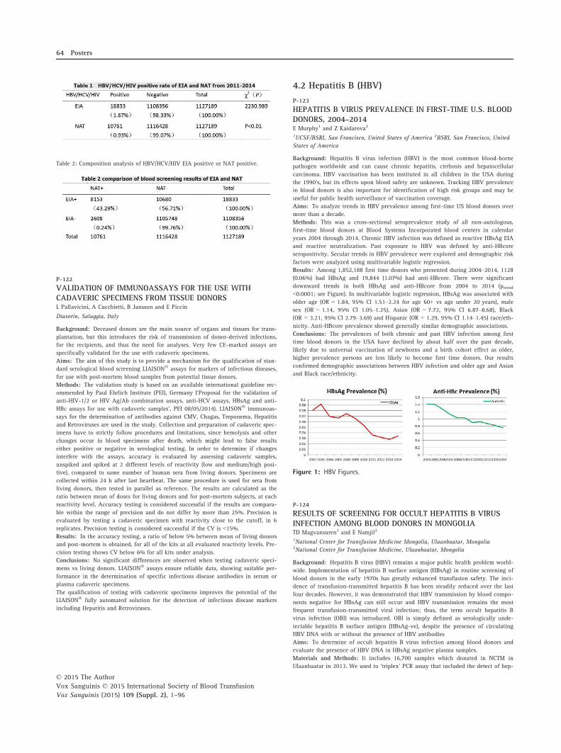
Table 2: Composition analysis of HBV/HCV/HIV EIA positive or NAT positive.
P-122
VALIDATION OF IMMUNOASSAYS FOR THE USE WITHCADAVERIC SPECIMENS FROM TISSUE DONORSL Pallavicini, A Cucchietti, B Jansson and E Piccin
Diasorin, Saluggia, Italy
Background: Deceased donors are the main source of organs and tissues for trans-
plantation, but this introduces the risk of transmission of donor-derived infections,
for the recipients, and thus the need for analyses. Very few CE-marked assays are
specifically validated for the use with cadaveric specimens.
Aims: The aim of this study is to provide a mechanism for the qualification of stan-
dard serological blood screening LIAISON� assays for markers of infectious diseases,
for use with post-mortem blood samples from potential tissue donors.
Methods: The validation study is based on an available international guideline rec-
ommended by Paul Ehrlich Institute (PEI), Germany (‘Proposal for the validation of
anti-HIV-1/2 or HIV Ag/Ab combination assays, anti-HCV assays, HBsAg and anti-
HBc assays for use with cadaveric samples’, PEI 08/05/2014). LIAISON� immunoas-
says for the determination of antibodies against CMV, Chagas, Treponema, Hepatitis
and Retroviruses are used in the study. Collection and preparation of cadaveric spec-
imens have to strictly follow procedures and limitations, since hemolysis and other
changes occur in blood specimens after death, which might lead to false results
either positive or negative in serological testing. In order to determine if changes
interfere with the assays, accuracy is evaluated by assessing cadaveric samples,
unspiked and spiked at 2 different levels of reactivity (low and medium/high posi-
tive), compared to same number of human sera from living donors. Specimens are
collected within 24 h after last heartbeat. The same procedure is used for sera from
living donors, then tested in parallel as reference. The results are calculated as the
ratio between mean of doses for living donors and for post-mortem subjects, at each
reactivity level. Accuracy testing is considered successful if the results are compara-
ble within the range of precision and do not differ by more than 25%. Precision is
evaluated by testing a cadaveric specimen with reactivity close to the cutoff, in 6
replicates. Precision testing is considered successful if the CV is <15%.
Results: In the accuracy testing, a ratio of below 5% between mean of living donors
and post-mortem is obtained, for all of the kits at all evaluated reactivity levels. Pre-
cision testing shows CV below 6% for all kits under analysis.
Conclusions: No significant differences are observed when testing cadaveric speci-
mens vs living donors. LIAISON� assays ensure reliable data, showing suitable per-
formance in the determination of specific infectious disease antibodies in serum or
plasma cadaveric specimens.
The qualification of testing with cadaveric specimens improves the potential of the
LIAISON� fully automated solution for the detection of infectious disease markers
including Hepatitis and Retroviruses.
4.2 Hepatitis B (HBV)
P-123
HEPATITIS B VIRUS PREVALENCE IN FIRST-TIME U.S. BLOODDONORS, 2004–2014E Murphy1 and Z Kaidarova2
1UCSF/BSRI, San Francisco, United States of America 2BSRI, San Francisco, United
States of America
Background: Hepatitis B virus infection (HBV) is the most common blood-borne
pathogen worldwide and can cause chronic hepatitis, cirrhosis and hepatocellular
carcinoma. HBV vaccination has been instituted in all children in the USA during
the 1990’s, but its effects upon blood safety are unknown. Tracking HBV prevalence
in blood donors is also important for identification of high risk groups and may be
useful for public health surveillance of vaccination coverage.
Aims: To analyze trends in HBV prevalence among first-time US blood donors over
more than a decade.
Methods: This was a cross-sectional seroprevalence study of all non-autologous,
first-time blood donors at Blood Systems Incorporated blood centers in calendar
years 2004 through 2014. Chronic HBV infection was defined as reactive HBsAg EIA
and reactive neutralization. Past exposure to HBV was defined by anti-HBcore
seropositivity. Secular trends in HBV prevalence were explored and demographic risk
factors were analyzed using multivariable logistic regression.
Results: Among 1,852,188 first time donors who presented during 2004–2014, 1128(0.06%) had HBsAg and 19,844 (1.07%) had anti-HBcore. There were significant
downward trends in both HBsAg and anti-HBcore from 2004 to 2014 (ptrend<0.0001; see Figure). In multivariable logistic regression, HBsAg was associated with
older age (OR = 1.84, 95% CI 1.51–2.24 for age 60+ vs age under 20 years), male
sex (OR = 1.14, 95% CI 1.05–1.25), Asian (OR = 7.72, 95% CI 6.87–8.68), Black
(OR = 3.21, 95% CI 2.79–3.69) and Hispanic (OR = 1.29, 95% CI 1.14–1.45) race/eth-nicity. Anti-HBcore prevalence showed generally similar demographic associations.
Conclusions: The prevalences of both chronic and past HBV infection among first
time blood donors in the USA have declined by about half over the past decade,
likely due to universal vaccination of newborns and a birth cohort effect as older,
higher prevalence persons are less likely to become first time donors. Our results
confirmed demographic associations between HBV infection and older age and Asian
and Black race/ethnicity.
P-124
RESULTS OF SCREENING FOR OCCULT HEPATITIS B VIRUSINFECTION AMONG BLOOD DONORS IN MONGOLIATD Magvansuren1 and E Namjil2
1National Center for Transfusion Medicine Mongolia, Ulaanbaatar, Mongolia2National Center for Transfusion Medicine, Ulaanbaatar, Mongolia
Background: Hepatitis B virus (HBV) remains a major public health problem world-
wide. Implementation of hepatitis B surface antigen (HBsAg) in routine screening of
blood donors in the early 1970s has greatly enhanced transfusion safety. The inci-
dence of transfusion-transmitted hepatitis B has been steadily reduced over the last
four decades. However, it was demonstrated that HBV transmission by blood compo-
nents negative for HBsAg can still occur and HBV transmission remains the most
frequent transfusion-transmitted viral infection; thus, the term occult hepatitis B
virus infection (OBI) was introduced. OBI is simply defined as serologically unde-
tectable hepatitis B surface antigen (HBsAg-ve), despite the presence of circulating
HBV DNA with or without the presence of HBV antibodies
Aims: To determine of occult hepatitis B virus infection among blood donors and
evaluate the presence of HBV DNA in HBsAg negative plasma samples.
Materials and Methods: It includes 16,700 samples which donated in NCTM in
Ulaanbaatar in 2013. We used to ‘triplex’ PCR assay that included the detect of hep-
Figure 1: HBV Figures.
© 2015 The AuthorVox Sanguinis © 2015 International Society of Blood TransfusionVox Sanguinis (2015) 109 (Suppl. 2), 1–96
64 Posters

atitis B virus HBV-DNA in addition HCV-RNA and HIV1/2-RNA for whom with
absence of serological markers of infection. The studies used molecular biology
methods were performed with the help of equipment (ROCHE COBAS S 201) and
technology based on Real Time PCR (pool size: 6 donation) Then we choose HBsAg
negative, DNA positive samples and determined, anti-HBc and anti-HBs by serologi-
cal methods, of ELISA Wantai HBc and HBs 3.0 tests.
Results: The 14,948 samples were detected serological negative in the total of
16,700 samples. PCR test results show 35 (0.23%) positive by HBV-DNA. 29 (82.9%)
of the 35 DNA positive blood donors were alone anti-HBc positive and 3 (8.6%) were
anti-HBs, anti-HBc positive. 7 (17.1%) were seronegative. Of the 35 OBI cases, 28
(80%) were detected the first time they were screened for HBV DNA while 7 (20%)
gave one more HBV PCR-nonreactive results before detection. Callback studies we
determined 2 cases were pre-HBsAg window period.
Conclusion: The prevalence of HBV DNA positive in HBsAg negative blood donors
is found 0.2%. Its 29 (82.9%) were seropositive, 7 (17.1%) were seronegative. HBV
NAT needs either extreme sensitivity or to be performed on individual donations to
eliminate HBV DNA-containing units.
P-125
ASSESSMENT OF AN HBSAG COMMERCIAL ASSAY WITH APANEL OF WELL CARACHTERIZED MUTANTS SAMPLESRS Tedder, B Haywood and S Ijaz
Blood Borne Virus Unit, Virus Reference Department, Centre for Infections, Health
Prot, London, United Kingdom
Background: HBsAg of hepatitis B virus (HBV) detection is crucial both for diagno-
sis of infection and for screening of blood derivatives and pregnancy. HBV harbor-
ing mutations in its sequence may challenge the ability of detection for laboratory
assays, leading to false negative HBsAg results, and still today HBV vaccine and
diagnostic escape mutants are a growing concern.
Aims: Testing the detection ability of a commercial assay, LIAISON XL murex
HBsAg Quant (DiaSorin S.p.A., Italy), using a set of native mutant HBsAg samples.
Methods: A panel of 30 plasma samples diluted to between 1 and 10 IU/ml HBsAg
containing native surface antigen bearing single or multiple mutations in the a
determinant based on consensus genotype sequencing, have been used to assess the
capability of the DiaSorin HBsAg assay to detect them. Mutation pattern of the pre-
dominant HBsAg sequence in samples has been determined by means of sequencing
analysis, as well as the genotype.
Phenotyping of HBsAg has been undertaken with a set of four mAbs (P2D3 aa 121–129; D2H5 aa 143–147; HB04 aa 137–147, H3F5 aa 131–142). All samples were pre-
viously detected by the Murex HBsAg Version 3, GE34/36, ELISA assay.
LIAISON XL murex HBsAg Quant is a chemiluminescent immunoassay for the quan-
titative determination of HBV surface antigen. The test uses monoclonal antibodies
directed against linear epitopes of the external and highly conserved internal and
transmembrane regions of HBsAg with a balanced reactivity against subtypes ad and
ay. All testing have been performed according to the manufacturing guidelines.
Results: All samples detected as positive by the LIAISON XL murex HBsAg Quant,
with a range of values between 0.050 mIU/ml and 3.90 mIU/ml.
Summary/Conclusions: Occurrence of false negative HBsAg results could impact
diagnostic and blood safety especially in HBV high prevalence countries where
immunization programmes may facilitate the evolution of escape variants. For the
blood donor screening single mutations are the most frequently described. It is
therefore important to assess sensitivity performance of commercially available
assays, with the use of well characterized samples covering both the more frequent
substitutions and also the ones having more impact in HBsAg detection. LIAISON
XL murex HBsAg Quant has been able to detect the range of variants examined.
P-126
COMPARISON STUDY WITH ENZYME IMMUNOASSAY ANDCHEMILUMINESCENCE IMMUNOASSAY FOR HEPATITIS BVIRUS SURFACE ANTIGEN DETECTIONA Soleimany ferizhandy, S Amini Kafi-Abad, M Moghtadaei, H Gholizadeh, M
Abedini, S Raman and N Amirhakimi
Iranian Blood Transfusion Research Center, High Institute for Research and Educa,
Tehran, Iran
Background: The serological detection of the surface antigen (HBsAg) of hepatitis B
virus (HBV) is the basis of detection of HBV infections in blood donors and patients
with hepatitis. The aim of this study was to compare the performance of HBsAg
enzyme-linked immunosorbent assay (ELISA) and HBsAg chemiluminescence
immunoassay evaluated in Iran.
Methods: We compared Two assays: HBsAg Qualitative, Architect i2000 (Abbott
Laboratories, USA), Enzygnost HBsAg (seimens 6.0, Germany). We evaluated the sen-
sitivity and specificity of each assay by testing HBsAg, four commercial seroconver-
sion panels BBI (Boston Biomedica Inc), PHM933, PHM912, PHM935A, PHM908,
and one sensitivity panel BBI (PHA808). For specificity, randomly selected serum
samples were collected from volunteer blood donors at 5 blood-donation centers in
Iran. The assigned values over the 4 seroconversion series were averaged to deter-
mine a mean relative seroconversion sensitivity index for each assay and the 95%
confidence limits were determined.
Results: The lowest HBsAg level detected by each assay using reference material
(BBI panel) was variable from 0.02 to 0.67 IU/ml (for sub type; ad, ay). When testing
21 seroconversion panels’ members, the total number of positive samples was 20
and 22 by Enzygnost HBsAg seimens 6.0 and HBsAg Qualitative, Architect i2000
samples tested. Good correlation q = 0.93 (P < 0.001), was found between the Archi-
tect i2000, seimens 6.0 assays with narrow 95% limits of agreement. The specificity
of HBsAg tests was found to be 99.95% and 99.91% for HBsAg Qualitative, Architect
i2000 and by Enzygnost HBsAg seimens 6.0 methods, respectively. The HBsAg Qual-
itative, Architect i2000 assays showed a higher sensitivity for subtype ad and ay for
the BBI sensitivity panel 808 than did the Enzygnost HBsAg seimens 6.0 assays.
Three samples with low HBsAg concentrations (0.01 to 0.05 IU/ml) tested negative
by the Enzygnost HBsAg seimens 6.0 assays.
Conclusions: The ARCHITECT HBsAg Qualitative is designed to show sensitivity
better than a commercially available HBsAg assay. The ARCHITECT HBsAg Qualita-
tive is designed to show seroconversion better than a commercially available HBsAg
assay.Analytical sensitivity detecting ability among HBsAg assays were variable. We
suggest that each laboratory should select an HBsAg assay based on analytical per-
formance, test throughput, and the applicability of full automation. The presented
data show that the new ARCHITECT HBsAg Qualitative screening assay exhibits a
low initial reactive rate, while displaying sensitivity and a specificity than
P-127
This abstract has been withdrawn.
4.4 HIV
P-128
EFFECT OF THE SAMPLES SENSITIVITY ANTI-HIV SCREENINGF Farida Faridatin Nasriyah
Blood Transfution Centre Tangerang City Red Cross, Tangerang, Indonesia
Background: For standard blood tests healthy and safe Ministry of Health issued a
policy that the examination was screened using a 4 parameters, one of which is HIV
testing. HIV (Human Imunodeviciency Virus) is a virus that causes the immune sese-
oran declined and over time the virus will be AIDS, the emergence of a set of dis-
eases caused by the human immunodeficiency. For screening examination
Tangerang City Blood Centre using screening tools ARCHITECT with CMIA method
and with a sensitivity which reached 99.5%.
Aim: To know the results of the sample of screenings 3000 rpm and 5000 rpm
2 min 5 min to the inspection of Anti-HIV screening test.
Methods: This method uses 10 samples taken from volunteer blood donors with no.
Different bags that have been through the handover of samples from quarantine lab
to lab screening. Sample in a centrifuge rotating at 5000 rpm with a 5-min playback
and in red blood cell sedimentation get perfect, the samples in running the instru-
ment and reagents ARCHITECT and see the results, stelah playback results carried
out on the same sample in different tubes with round 3000 rpm 2 min. Results of
the first round and the second round later in the compare and concluded.
Result: Examination of Anti-HIV screening of 10 samples with no. Different pockets
and with different playback is 5000 rpm to 3000 rpm 5 min 2 min 1 sample found to
Contain samples infected with HIV as a result of screening samples at 3000 rpm 2 min.
Conclusions: There is a 10% change in sample playback results from 3000 rpm to
5000 rpm 2 min 5 min with different results. Evidenced by the sample No.
634I2006A bag with a screening of 3000 rpm 2 min at OD 1.36 can result REAC-
TIVE against Anti-HIV, and from the same sample with the screening of 5000 rpm
5 min there is a change results OD 0.13 NON-REACTIVE terhadapAnti HIV. Changes
in the results that occur in the sample playback speed of 3000 rpm 2 min is likely
to occur as a result of deposition of cells is less than perfect. It can be concluded
that the use of screening samples for the screening of IMLTD would be better if you
use the playback speed of 5000 rpm and a 5 min.
© 2015 The AuthorVox Sanguinis © 2015 International Society of Blood Transfusion
Vox Sanguinis (2015) 109 (Suppl. 2), 1–96
HIV 65
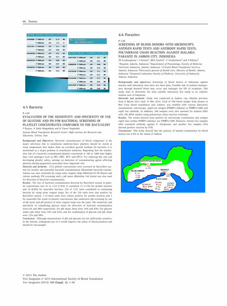
4.5 Bacteria
P-129
EVALUATION OF THE SENSITIVITY AND SPECIFICITY OF USEOF GLUCOSE AND PH FOR BACTERIAL SCREENING OFPLATELET CONCENTRATES COMPARED TO THE BACT/ALERTF Razjou, A Dabir Mogaddam and H Timori Naghadeh
Iranian Blood Transfusion Research Center, High institute for Research and
Education, Tehran, Iran
Background and Objectives: Bacterial contamination of blood component is the
major infectious risk in transfusion medicine.Since platelets should be stored at
room temperature that makes them an excellent growth medium for bacteria; it is
mentioned as a major problem in transfusion medicine. Regarding that the transfu-
sion risk of a bacterial contaminated platelet concentrate is 100 to 1000 time higher
than viral pathogen such as HIV, HBV, HCV and HTLV. For reducing this risk and
developing platelet safety, attempt on detection of contaminating agents affecting
platelets during preparation procedure have important role.
Material and methods: 1332 platelet concentrates were screened by Bact/Alert sys-
tem for aerobic and anaerobic bacterial contamination. Meanwhile bacterial contam-
ination was also evaluated by using urine reagent strips (Multistix10 SG Bayer) and
culture methods. PH screening with a pH meter (Metrohm 744 Swiss) was also used
for detection of bacterial contamination.
Results: The rate of bacterial contamination detected by Bact/Alert system in plate-
let concentrates was 25 in 1332 (1.9%). It contained 15 (1.1%) for aerobic bacteria
and 10 (0.8%) for anaerobic bacteria. 226 of 1332 were considered as containing
bacteria by using urine reagent strips. Six of the 226 units were also positive by
Bact/Alert system. 3 of those units were culture positive for aerobic bacteria and 3
for anaerobic.The result of platelet concentrates that underwent pH screening by use
of pH meter and pH portion of urine reagent strips was the same. The sensitivity and
specificity of considering glucose alone for detection of bacterial contamination
were12% and 98% respectively .For pH alone, these were 24% and 83% .For glucose
and/or pH, these were 24% and 83%; and for combination of glucose and pH, these
were 12% and 98%.
Conclusion: Although measurement of pH and glucose are not sufficiently sensitive,
in the interim, widespread use of it would improve the safety of blood products and
should be encouraged.
4.6 Parasites
P-130
SCREENING OF BLOOD DONORS WITH MICROSCOPY,ANTIGEN RAPID TESTS AND ANTIBODY RAPID TESTS,POLYMERASE CHAIN REACTION AGAINST MALARIAPARASITE IN AMBON CITY, INDONESIAJN Louhenapessy1, I Sutanto2, REG Syafitri3, Y Soedarmono4 and S Rahayu5
1Hospital, Jakarta, Indonesia 2Departement of Parasitology, Faculty of Medicine,
University Indonesia, Jakarta, Indonesia 32Central Blood Transfusion Services,
Jakarta, Indonesia 4Directorate general of Health Care, Ministry of Health, Jakarta,
Indonesia 5Integrated Laboratory Faculty of Medicine, University of Indonesia,
Jakarta, Indonesia
Backgrounds and objectives: Screening of blood donors in Indonesia against
malaria with laboratory tests have not been done. Possible risk of malaria transmis-
sion through donated blood may occur and endanger the life of recipients. This
study aims to determine the most suitable laboratory test using in an endemic
malaria area of Indonesia.
Materials and methods: Study was conducted in Ambon city, Maluku province
from 8 March 2013 until 15 Mei 2014. Total of 550 blood sample from donors at
Red Cross blood transfusion unit Ambon, was testified with various laboratory
examinations: microscopy, rapid test antigen (PfHRP2-aldolase or PfHRP2-LDH) and
rapid test antibody. In addition, 248 samples were also assessed for malaria DNA
with 18S rRNA marker using polymerase chain reaction technique.
Results: The results showed none positive by microscopy examination and antigen
rapid tests neither PfHRP2-aldolase nor PfHRP2-LDH. However, twenty-two samples
(4%) contained antibody against P. falciparum, and another five samples (2%)
showed positive reaction by PCR.
Conclusions: This study showed that the potency of malaria transmission by blood
donors was 4.9% in the island of Ambon.
© 2015 The AuthorVox Sanguinis © 2015 International Society of Blood TransfusionVox Sanguinis (2015) 109 (Suppl. 2), 1–96
66 Posters

4.7 Newly Emerging Pathogens and otherTransfusion Related Pathogens
P-131
COBASⓇ CHIKV/DENV INVESTIGATIONAL ASSAY: A NOVELHIGHLY SENSITIVE AND REPRODUCIBLE ASSAY FOR BLOODSCREENING ON THE COBAS� 6800/8800 SYSTEMP Albrecht1, A Kuehler1, N Dyer2, C Roordink1, S Sudershana2 and J Niemiec2
1Roche Diagnostics International Ltd, Rotkreuz, Switzerland 2Roche Molecular
Systems, Inc., Pleasanton, CA, United States of America
Background: The cobas� Chikungunya/Dengue test (CHIKV/DENV) for use on the
cobas� 6800/8800 Systems is currently in development. This automated real time
PCR qualitative blood screening test is designed to both detect and discriminate
CHIKV/DENV with high sensitivity and specificity.
Like other infectious diseases for which blood donations are screened, blood dona-
tions must be screened with a sensitive assay to detect chikungunya virus (CHIKV)
and dengue virus (DENV) RNA so that infected units may be interdicted and dis-
carded. The cobas� CHIKV/DENV investigational assay will offer novel capability to
detect RNA from these viruses and thereby provide heightened protection from
transfusion-transmitted CHIKV and DENV infection for recipients of donated blood
components or products and will further improve the safety of the blood supply.
Aims: To develop and evaluate the performance of a novel real-time PCR based
CHIKV/DENV investigational test for blood screening utilizing the new cobas� 6800/
8800 fully automated system.
Methods: Automated, nucleic acid extraction was used for isolation of viral RNA
which amplified and detected by automated, real-time PCR. Analytical sensitivity
was determined using Roche Secondary Standards of CHIKV/DENV. Clinical speci-
ficity was assessed by testing 1000 individual specimens from healthy donors.
Results: The 95% limit of detection by Probit analysis was 0.18 PDU/ml for CHIKV
and 71.8 PDU/ml for DENV. Clinical specificity was 100%. The test detected geno-
types Asia and ECSA for CHIKV and all four serotypes for DENV. No interference
was observed in samples with albumin (60 g/l), bilirubin (0.2 g/l), hemoglobin (2 g/
l), triglycerides (33 g/l), human DNA (0.5 mg/l). No cross reactivity was observed
with 27 cultured microorganisms at 1.00E+06 particles, copies, or PFU/ml; including
20 viral isolates, 6 bacterial strains, and 1 yeast isolate.
This study demonstrated consistent performance of the cobas� CHIKV/DENV test
across kit lot and instrument-operators on different days at ~0.5x and ~1x and ~2xLoD. Blood collected in EDTA anticoagulant, CPD and PPT could be stored for up to
5 days at 2–8°C, of which 48 h may be at 2–25°C and 24 h at 2–30°C without
affecting the performance of the test. For storage longer than 5 days, plasma was
shown to be stored at <�18°C for up to 15 days without affecting performance,
including up to three freeze-thaw cycles.
Conclusions: The high specificity and sensitivity and genotype inclusivity of the
novel cobas� CHIKV/DENV assay confirm that this assay is appropriate for blood
and plasma screening for investigational use.
P-132
THE SEARCH FOR DENGUE VIRUS IN AUSTRALIAN BLOODDONATIONS DURING LOCAL OUTBREAKSHM Faddy1, JJ Fryk1, CR Seed2, C Hyland1, JA Holmberg3 and R Flower1
1Australian Red Cross Blood Service, Brisbane, Australia 2Medical Services,
Australian Red Cross Blood Service, Perth, Australia 3Grifols, Emeryville, United
States of America
Background: Dengue virus (DENV) poses a risk to the safe transfusion of blood,
with confirmed reports of transfusion-transmission from Hong Kong, Singapore,
Puerto Rico and Brazil. In Australia, episodic outbreaks occur on a seasonal basis in
north eastern Australia. One of the largest epidemics in over 50 years took place in
the summer of 2008/2009, with over 1000 confirmed cases. Another sizeable out-
break occurred in the summer of 2012/2013. To manage this risk, fresh components
are not manufactured from donors who live in or travel to areas experiencing den-
gue outbreaks, including countries where dengue is endemic. Dengue outbreaks pose
a risk to the safety of Australia’s blood supply, which justifies relatively-high cost
risk mitigation strategies. An alternative approach for managing this risk could be
the use of an appropriate donor screening test.
Aims: The aim of this study was to determine the rate of asymptomatic donor vir-
aemia during local dengue outbreaks by testing plasma samples for the presence of
DENV RNA using transcription mediated amplification (TMA).
Methods: Samples (n = 6182) from donors in North Queensland ‘at-risk’ of expo-
sure to dengue were collected, including donations from two separate periods: the
2008/2009 epidemic (donors in areas of Cairns that recorded more than 20 con-
firmed dengue cases) and the 2012/2013 outbreak (all affected donations). Addi-
tional samples were sourced from a control region in southern Australia, where
DENV transmission does not occur (n = 1601). All samples were tested for DENV
RNA by DENV TMA (95% detection at <30 copies/ml for DENV types 1–4) as per themanufacturer’s instructions. Exact confidence intervals (CI) were calculated for indi-
vidual proportions using a standard method.
Results: None of the samples collected during the 2008/2009 epidemic (0%, 95%
CI: 0–0.55%) were positive for DENV RNA, despite them all being collected from
‘higher-risk’ areas. Likewise, none of the samples collected during the 2012/2013
outbreak (0%, 95% CI: 0–0.07%) were positive for DENV RNA. All of the control
samples were also negative for DENV RNA (0%, 95% CI: 0–0.23%).
Summary/Conclusions: None of the donations tested from donors residing in areas
experiencing dengue outbreaks had detectable DENV RNA. This was concordant with
earlier studies during previous outbreaks, and is consistent with our modelling esti-
mate of 1 in 7147 (95% CI: 1 in 2218 to 1 in 50,021) for the risk of collecting a vir-
aemic donation during an outbreak. The rate of DENV viraemia in blood donors
residing in areas of Australia that experience episodic dengue outbreaks was lower
than that in donors from Puerto Rico, where dengue is endemic, likely reflecting dif-
ferences in viral epidemiology between endemic and non-endemic regions. While
this study is insufficiently powered to draw definitive conclusions, the absence of
DENV RNA positive donations, despite the higher number of reported cases during
these outbreaks, suggests fresh component restriction for ‘at risk’ donors during local
outbreaks is an effective strategy for minimising the risk of transfusion transmitted
DENV in non-endemic regions.
P-133
HEPATITIS E VIRUS INFECTIONS IN TRAVELLERS: A THREATIN AUSTRALIA?AC Shrestha1, RLP Flower1, CR Seed1, AJ Keller1, V Hoad1, R Harley1, R Leader2, B
Polkinghorne2, C Furlong3 and HM Faddy1
1Australian Red Cross Blood Service, Queensland, Australia 2Australian Government
Department of Health, Australian Capital Territory, Australia 3New South Wales
Department of Health, New South Wales, Australia
Background: In developed countries such as Australia, hepatitis E virus (HEV)
infections are predominantly seen in travellers returning from countries endemic for
HEV. HEV can be transfusion transmissible and viral RNA has been detected in
asymptomatic blood donors, demonstrating this virus is a potential threat to blood
safety. To minimise the risk of HEV transfusion transmission in Australia, individu-
als diagnosed with HEV infection are deferred from donating for 12 months from
© 2015 The AuthorVox Sanguinis © 2015 International Society of Blood Transfusion
Vox Sanguinis (2015) 109 (Suppl. 2), 1–96
Newly Emerging Pathogens and other Transfusion Related Pathogens 67
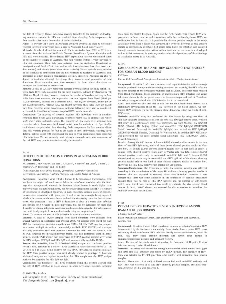
the date of recovery. Donors who have recently travelled to the majority of develop-
ing countries endemic for HEV are restricted from donating fresh components for
four months after return due to risk of malaria infection.
Aims: To describe HEV cases in Australia acquired overseas in order to determine
whether infection in travellers poses a risk to Australian blood supply safety.
Methods: Details of all notified cases of HEV in Australia from 2002 to 2013 were
accessed from the National Notifiable Diseases Surveillance System. Countries with
HEV risk were identified. The estimated HEV importation rate was determined based
on the number of people in Australia who had recently (within 1 year) travelled to
HEV risk countries. These data were obtained from the Australian Department of
Immigration and Border Protection and include Australian travellers (short term resi-
dent returns) and visitors (short term visitor arrivals). Visitor arrivals were included
in this analysis as notification data are not restricted to residents of Australia, and,
providing all other donation requirements are met, visitors to Australia are able to
donate in Australia, although this group likely makes a small proportion of total
donations. These countries were then compared to those where donations are
restricted for travel due to malaria-risk.
Results: A total of 325 HEV cases were acquired overseas during the study period. Tra-
vel to India (149; 46%) accounted for the most infections, followed by Bangladesh (39;
12%) and Nepal (21; 6%). However, based on the number of travellers arriving in Aus-
tralia from these countries, the importation rate was highest from Nepal (22.03 per
10,000 travellers), followed by Bangladesh (18.01 per 10,000 travellers), Sudan (16.09
per 10,000 travellers), Pakistan (5.66 per 10,000 travellers) then India (3.20 per 10,000
travellers). Countries where donations are restricted following travel due to malaria-risk
accounted for 95% (308) of overseas acquired HEV cases in Australia.
Conclusions: The majority of overseas acquired HEV infections were in travellers
returning from South Asia, particularly countries where HEV is endemic and where
large water-borne outbreaks occur. The majority of HEV cases were acquired from
countries where donation-related travel restrictions for malaria exist. Specifically,
such donors are precluded from donating fresh components for four months. Given
that HEV viremia persists for four to six weeks in most individuals, existing travel
deferral policies assist with minimising the risk to fresh components from imported
HEV infections. We are currently undertaking a comprehensive risk assessment of
the risk HEV may pose to transfusion safety in Australia.
P-134
DETECTION OF HEPATITIS E VIRUS IN AUSTRALIAN BLOODDONATIONSAC Shrestha1, RLP Flower1, CR Seed1, AJ Keller1, R Harley1, HT Chan1, V Hoad1, D
Warrilow2, JA Holmberg3 and HM Faddy1
1Australian Red Cross Blood Service, Queensland, Australia 2Queensland
Government, Queensland, Australia 3Grifols, CA, United States of America
Background: Hepatitis E virus (HEV) is known to be transfusion-transmissible. HEV
has been the subject of much recent debate in transfusion medicine with the find-
ings that asymptomatic viraemia in European blood donors is much higher than
expected based on notification rates, and the acknowledgement that HEV is a disease
of importance in developed countries. In such countries, sporadic cases of zoonotic
transmission associated with genotypes 3 and 4 are increasingly being reported,
which is in contrast to the water-borne outbreaks in developing countries that are asso-
ciated with genotypes 1 and 2. HEV is detectable in blood 2–3 weeks after infection
and persists for 4–6 weeks in most individuals, but can be detectable for more than
3 months in chronic infections. Australian notification data suggests HEV infections are
rare, with locally acquired cases predominantly being due to genotype 3.
Aims: To measure the rate of HEV infection in Australian blood donations.
Methods: A total of 14,799 samples from blood donations were collected from
around Australia in September and October 2014. All samples were tested for HEV
RNA by transcription mediated amplification (TMA). All HEV TMA reactive samples
were tested in duplicate with a commercially available HEV RT-PCR, and a sample
was only considered HEV RNA positive if reactive by both TMA and RT-PCR. HEV
RT-PCR targeting the methyltransferase region was also performed using in-house
primers, and the PCR product was sequenced. HEV RNA positive samples were tested
for HEV antigen, HEV IgG and HEV IgM with commercially available ELISAs.
Results: One (0.0068%, 95% CI: 0.0002–0.0376%) sample was confirmed positive
for HEV RNA, resulting in 1 out of 14,799 Australian blood donations (95% CI: 1 in
584,530 to 1 in 2657) being positive for HEV RNA. Phylogenetic analysis indicates
the HEV RNA positive sample was most closely related to genotype 3; however,
additional analyses are required to confirm this. This sample was also HEV antigen
positive, but negative for HEV IgG and IgM.
Conclusions: Our finding of 1 in 14,799 donations being HEV positive is lower than
the rate of HEV infection in blood donors in other developed countries, including
those from the United Kingdom, Spain and the Netherlands. This reflects HEV sero-
prevalence in these countries and is consistent with the considerably lower HEV case
notification data in Australia. It is possible that the one positive sample in this study
could have been from a donor who acquired HEV overseas, however, as this positive
sample is provisionally genotype 3, it seems more likely the infection was acquired
through zoonotic transmission, either within Australia or overseas in a developed
nation. A risk assessment is underway to determine the significance of these findings
to transfusion safety in in Australia.
P-135
COMPARISON OF THE ANTI-HEV SCREENING TEST RESULTSFOR KOREAN BLOOD DONORSKW Youn
Korean Red Cross/Blood Transfusion Research Institute, Wonju, South-Korea
Background: Hepatitis E infection is an acute viral hepatitis infection and was recog-
nized as pandemic mainly in the developing countries. But recently, the HEV infection
has been detected in the developed countries such as Japan, and some cases resulted
from blood transfusion. Blood donation of asymptomatic HEV infection can cause
infectious disease in the pregnant women or immunocompressed patient. Therefore,
the safety strategies related with HEV infection has to be considered.
Aims: This study was the first trial of HEV test for the Korean Blood donors. As a
preliminary investigation about the HEV infection in the blood donors, we per-
formed HEV antibody test for the Korean blood donors by using two kinds of anti-
HEV assay.
Methods: Anti-HEV assay was performed for 630 donors by using two kinds of
anti-HEV IgG/IgM screening assay. For the anti-HEV IgG/IgM positve cases, Western
blot assay as a confirmatory assay was performed. We used Wantai HEV-IgG/IgM
ELISA (Wantai LTD, Beijing, China) and recomWell HEV IgG/IgM (MIKROGEN
GmbH, Neuried, Germany) for anti-HEV IgG/IgM, and recomLine HEV IgG/IgM
(MIKROGEN GmbH, Neuried, Germany) for Western blot. In addition HEV RNA assay
was performed for the same samples using ampliCube HEV (MIKROGEN GmbH,
Neuried, Germany).
Results: Among the 630 donors, 5 donors (0.8%) showed positive results in the two
kinds of anti-HEV IgG assay, and 4 of them (0.6%) showed positive results in Wes-
tern blot. 15 donors (2.4%) showed positive results only in one kind of assay. 6
donors (1.0%) showed positive results only in Wantai anti-HEV IgG, 4 donors (0.6%)
showed positive results only in recomWell anti-HEV IgG, and 5 donors (0.8%)
showed positive results only in recomWell anti-HEV IgM. All of the donors showing
positive results only in one kind of assay showed negative results in Western blot.
There was no HEV RNA positive case among the 630 donors.
Summary/Conclusions: The frequency of anti-HEV positive samples was different
according to the manufacture of the assay kit. 4 donors showing positive results in
Western blot was regarded as recovery phase after infection. However, it was
thought that there was some limitation in the estimation of accurate prevalence
because there was no case of HEV RNA positive and the number of 630 donors
which were tested was considered too small to estimate the risk among blood
donors. At least, 10,000 donors are required for risk evaluation to introduce the
anti-HEV screening test in Korea.
P-136
PREVALENCE OF HEPATITIS E VIRUS INFECTION AMONGIRANIAN BLOOD DONORSZ Sharifi and MA Jalili
Blood Transfusion Research Center, High Institute for Research and Education,
Tehran, Iran
Background: Hepatitis E virus (HEV) is endemic in many developing countries. HEV
is transmitted by the fecal-oral route mainly. Some studies have reported HEV trans-
mission by blood transfusions. HEV infection usually causes a self-limiting, acute ill-
ness. HEV may cause chronic infection and severe liver disease in
immunocompromised patients and pregnant women.
Aims: The aim of this study was to determine the Prevalence of Hepatitis E virus
infection among Iranian blood donors.
Methods: This study was carried out among 400 volunteer blood donors. Total (IgM
and IgM) anti-HEV antibody was tested by ELISA method. The presence of HEV-
RNA was detected by RT-PCR procedure after nucleic acid extraction from plasma
samples.
Results: About 4% (16 of 400) of blood donors had total anti-HEV antibody and
HEV- RNA was detected in all anti-HEV antibody positive samples. The most com-
mon genotype of HEV was genotype 1.
© 2015 The AuthorVox Sanguinis © 2015 International Society of Blood TransfusionVox Sanguinis (2015) 109 (Suppl. 2), 1–96
68 Posters
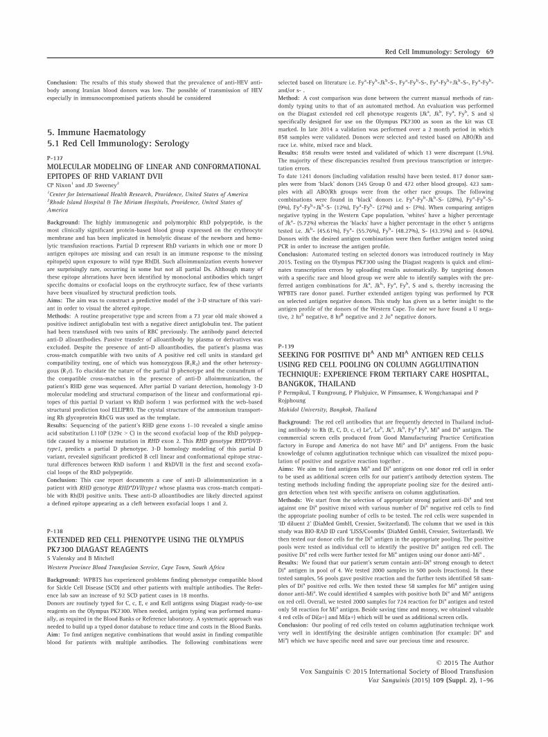
Conclusion: The results of this study showed that the prevalence of anti-HEV anti-
body among Iranian blood donors was low. The possible of transmission of HEV
especially in immunocompromised patients should be considered
5. Immune Haematology5.1 Red Cell Immunology: Serology
P-137
MOLECULAR MODELING OF LINEAR AND CONFORMATIONALEPITOPES OF RHD VARIANT DVIICP Nixon1 and JD Sweeney2
1Center for International Health Research, Providence, United States of America2Rhode Island Hospital & The Miriam Hospitals, Providence, United States of
America
Background: The highly immunogenic and polymorphic RhD polypeptide, is the
most clinically significant protein-based blood group expressed on the erythrocyte
membrane and has been implicated in hemolytic disease of the newborn and hemo-
lytic transfusion reactions. Partial D represent RhD variants in which one or more D
antigen epitopes are missing and can result in an immune response to the missing
epitope(s) upon exposure to wild type Rh(D). Such alloimmunization events however
are surprisingly rare, occurring in some but not all partial Ds. Although many of
these epitope alterations have been identified by monoclonal antibodies which target
specific domains or exofacial loops on the erythrocyte surface, few of these variants
have been visualized by structural prediction tools.
Aims: The aim was to construct a predictive model of the 3-D structure of this vari-
ant in order to visual the altered epitope.
Methods: A routine preoperative type and screen from a 73 year old male showed a
positive indirect antiglobulin test with a negative direct antiglobulin test. The patient
had been transfused with two units of RBC previously. The antibody panel detected
anti-D alloantibodies. Passive transfer of alloantibody by plasma or derivatives was
excluded. Despite the presence of anti-D alloantibodies, the patient’s plasma was
cross-match compatible with two units of A positive red cell units in standard gel
compatibility testing, one of which was homozygous (R1R2) and the other heterozy-
gous (R1r). To elucidate the nature of the partial D phenotype and the conundrum of
the compatible cross-matches in the presence of anti-D alloimmunization, the
patient’s RHD gene was sequenced. After partial D variant detection, homology 3-D
molecular modeling and structural comparison of the linear and conformational epi-
topes of this partial D variant vs RhD isoform 1 was performed with the web-based
structural prediction tool ELLIPRO. The crystal structure of the ammonium transport-
ing Rh glycoprotein RhCG was used as the template.
Results: Sequencing of the patient’s RHD gene exons 1–10 revealed a single amino
acid substitution L110P (329c > C) in the second exofacial loop of the RhD polypep-
tide caused by a missense mutation in RHD exon 2. This RHD genotype RHD*DVII-
type1, predicts a partial D phenotype. 3-D homology modeling of this partial D
variant, revealed significant predicted B cell linear and conformational epitope struc-
tural differences between RhD isoform 1 and RhDVII in the first and second exofa-
cial loops of the RhD polypeptide.
Conclusion: This case report documents a case of anti-D alloimmunization in a
patient with RHD genotype RHD*DVIItype1 whose plasma was cross-match compati-
ble with Rh(D) positive units. These anti-D alloantibodies are likely directed against
a defined epitope appearing as a cleft between exofacial loops 1 and 2.
P-138
EXTENDED RED CELL PHENOTYPE USING THE OLYMPUSPK7300 DIAGAST REAGENTSS Valensky and B Mitchell
Western Province Blood Transfusion Service, Cape Town, South Africa
Background: WPBTS has experienced problems finding phenotype compatible blood
for Sickle Cell Disease (SCD) and other patients with multiple antibodies. The Refer-
ence lab saw an increase of 92 SCD patient cases in 18 months.
Donors are routinely typed for C, c, E, e and Kell antigens using Diagast ready-to-use
reagents on the Olympus PK7300. When needed, antigen typing was performed manu-
ally, as required in the Blood Banks or Reference laboratory. A systematic approach was
needed to build up a typed donor database to reduce time and costs in the Blood Banks.
Aim: To find antigen negative combinations that would assist in finding compatible
blood for patients with multiple antibodies. The following combinations were
selected based on literature i.e. Fya-Fyb-Jkb-S-, Fya-Fyb-S-, Fya-Fyb+Jkb-S-, Fya-Fyb-and/or s- .
Method: A cost comparison was done between the current manual methods of ran-
domly typing units to that of an automated method. An evaluation was performed
on the Diagast extended red cell phenotype reagents (Jka, Jkb, Fya, Fyb, S and s)
specifically designed for use on the Olympus PK7300 as soon as the kit was CE
marked. In late 2014 a validation was performed over a 2 month period in which
858 samples were validated. Donors were selected and tested based on ABO/Rh and
race i.e. white, mixed race and black.
Results: 858 results were tested and validated of which 13 were discrepant (1.5%).
The majority of these discrepancies resulted from previous transcription or interpre-
tation errors.
To date 1241 donors (including validation results) have been tested. 817 donor sam-
ples were from ‘black’ donors (345 Group O and 472 other blood groups). 423 sam-
ples with all ABO/Rh groups were from the other race groups. The following
combinations were found in ‘black’ donors i.e. Fya-Fyb-Jkb-S- (28%), Fya-Fyb-S-
(9%), Fya-Fyb+Jkb-S- (12%), Fya-Fyb- (27%) and s- (7%). When comparing antigen
negative typing in the Western Cape population, ‘whites’ have a higher percentage
of Jka- (5.72%) whereas the ‘blacks’ have a higher percentage in the other 5 antigens
tested i.e. Jkb- (45.61%), Fya- (55.76%), Fyb- (48.27%), S- (43.35%) and s- (4.60%).
Donors with the desired antigen combination were then further antigen tested using
PCR in order to increase the antigen profile.
Conclusion: Automated testing on selected donors was introduced routinely in May
2015. Testing on the Olympus PK7300 using the Diagast reagents is quick and elimi-
nates transcription errors by uploading results automatically. By targeting donors
with a specific race and blood group we were able to identify samples with the pre-
ferred antigen combinations for Jka, Jkb,, Fya, Fyb, S and s, thereby increasing the
WPBTS rare donor panel. Further extended antigen typing was performed by PCR
on selected antigen negative donors. This study has given us a better insight to the
antigen profile of the donors of the Western Cape. To date we have found a U nega-
tive, 2 hrS negative, 8 hrB negative and 2 Joa negative donors.
P-139
SEEKING FOR POSITIVE DIA AND MIA ANTIGEN RED CELLSUSING RED CELL POOLING ON COLUMN AGGLUTINATIONTECHNIQUE: EXPERIENCE FROM TERTIARY CARE HOSPITAL,BANGKOK, THAILANDP Permpikul, T Rungroung, P Plubjuice, W Pimsamsee, K Wongchanapai and P
Rojphoung
Mahidol University, Bangkok, Thailand
Background: The red cell antibodies that are frequently detected in Thailand includ-
ing antibody to Rh (E, C, D, c, e) Lea, Leb, Jka, Jkb, Fya Fyb, Mia and Dia antigen. The
commercial screen cells produced from Good Manufacturing Practice Certification
factory in Europe and America do not have Mia and Dia antigens. From the basic
knowledge of column agglutination technique which can visualized the mixed popu-
lation of positive and negative reaction together .
Aims: We aim to find antigens Mia and Dia antigens on one donor red cell in order
to be used as additional screen cells for our patient’s antibody detection system. The
testing methods including finding the appropriate pooling size for the desired anti-
gen detection when test with specific antisera on column agglutination.
Methods: We start from the selection of appropriate strong patient anti-Dia and test
against one Dia positive mixed with various number of Dia negative red cells to find
the appropriate pooling number of cells to be tested. The red cells were suspended in
‘ID diluent 2’ (DiaMed GmbH, Cressier, Switzerland). The column that we used in this
study was BIO-RAD ID card ‘LISS/Coombs’ (DiaMed GmbH, Cressier, Switzerland). We
then tested our donor cells for the Dia antigen in the appropriate pooling. The positive
pools were tested as individual cell to identify the positive Dia antigen red cell. The
positive Dia red cells were further tested for Mia antigen using our donor anti-Mia .
Results: We found that our patient’s serum contain anti-Dia strong enough to detect
Dia antigen in pool of 4. We tested 2000 samples in 500 pools (reactions). In these
tested samples, 56 pools gave positive reaction and the further tests identified 58 sam-
ples of Dia positive red cells. We then tested these 58 samples for Mia antigen using
donor anti-Mia. We could identified 4 samples with positive both Dia and Mia antigens
on red cell. Overall, we tested 2000 samples for 724 reaction for Dia antigen and tested
only 58 reaction for Mia antigen. Beside saving time and money, we obtained valuable
4 red cells of Di(a+) and Mi(a+) which will be used as additional screen cells.
Conclusion: Our pooling of red cells tested on column agglutination technique work
very well in identifying the desirable antigen combination (for example: Dia and
Mia) which we have specific need and save our precious time and resource.
© 2015 The AuthorVox Sanguinis © 2015 International Society of Blood Transfusion
Vox Sanguinis (2015) 109 (Suppl. 2), 1–96
Red Cell Immunology: Serology 69
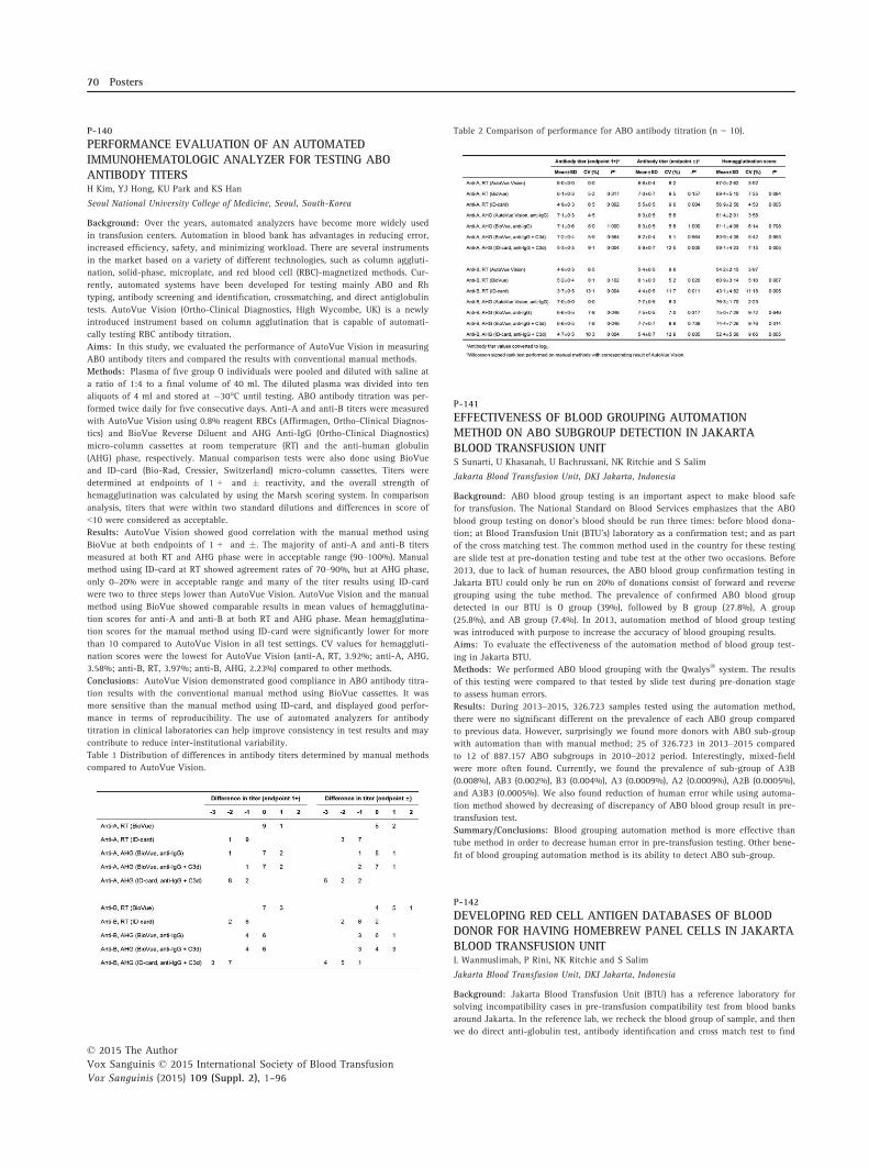
P-140
PERFORMANCE EVALUATION OF AN AUTOMATEDIMMUNOHEMATOLOGIC ANALYZER FOR TESTING ABOANTIBODY TITERSH Kim, YJ Hong, KU Park and KS Han
Seoul National University College of Medicine, Seoul, South-Korea
Background: Over the years, automated analyzers have become more widely used
in transfusion centers. Automation in blood bank has advantages in reducing error,
increased efficiency, safety, and minimizing workload. There are several instruments
in the market based on a variety of different technologies, such as column aggluti-
nation, solid-phase, microplate, and red blood cell (RBC)-magnetized methods. Cur-
rently, automated systems have been developed for testing mainly ABO and Rh
typing, antibody screening and identification, crossmatching, and direct antiglobulin
tests. AutoVue Vision (Ortho-Clinical Diagnostics, High Wycombe, UK) is a newly
introduced instrument based on column agglutination that is capable of automati-
cally testing RBC antibody titration.
Aims: In this study, we evaluated the performance of AutoVue Vision in measuring
ABO antibody titers and compared the results with conventional manual methods.
Methods: Plasma of five group O individuals were pooled and diluted with saline at
a ratio of 1:4 to a final volume of 40 ml. The diluted plasma was divided into ten
aliquots of 4 ml and stored at �30°C until testing. ABO antibody titration was per-
formed twice daily for five consecutive days. Anti-A and anti-B titers were measured
with AutoVue Vision using 0.8% reagent RBCs (Affirmagen, Ortho-Clinical Diagnos-
tics) and BioVue Reverse Diluent and AHG Anti-IgG (Ortho-Clinical Diagnostics)
micro-column cassettes at room temperature (RT) and the anti-human globulin
(AHG) phase, respectively. Manual comparison tests were also done using BioVue
and ID-card (Bio-Rad, Cressier, Switzerland) micro-column cassettes. Titers were
determined at endpoints of 1 + and � reactivity, and the overall strength of
hemagglutination was calculated by using the Marsh scoring system. In comparison
analysis, titers that were within two standard dilutions and differences in score of
<10 were considered as acceptable.
Results: AutoVue Vision showed good correlation with the manual method using
BioVue at both endpoints of 1 + and �. The majority of anti-A and anti-B titers
measured at both RT and AHG phase were in acceptable range (90–100%). Manual
method using ID-card at RT showed agreement rates of 70–90%, but at AHG phase,
only 0–20% were in acceptable range and many of the titer results using ID-card
were two to three steps lower than AutoVue Vision. AutoVue Vision and the manual
method using BioVue showed comparable results in mean values of hemagglutina-
tion scores for anti-A and anti-B at both RT and AHG phase. Mean hemagglutina-
tion scores for the manual method using ID-card were significantly lower for more
than 10 compared to AutoVue Vision in all test settings. CV values for hemaggluti-
nation scores were the lowest for AutoVue Vision (anti-A, RT, 3.92%; anti-A, AHG,
3.58%; anti-B, RT, 3.97%; anti-B, AHG, 2.23%) compared to other methods.
Conclusions: AutoVue Vision demonstrated good compliance in ABO antibody titra-
tion results with the conventional manual method using BioVue cassettes. It was
more sensitive than the manual method using ID-card, and displayed good perfor-
mance in terms of reproducibility. The use of automated analyzers for antibody
titration in clinical laboratories can help improve consistency in test results and may
contribute to reduce inter-institutional variability.
Table 1 Distribution of differences in antibody titers determined by manual methods
compared to AutoVue Vision.
Table 2 Comparison of performance for ABO antibody titration (n = 10).
P-141
EFFECTIVENESS OF BLOOD GROUPING AUTOMATIONMETHOD ON ABO SUBGROUP DETECTION IN JAKARTABLOOD TRANSFUSION UNITS Sunarti, U Khasanah, U Bachrussani, NK Ritchie and S Salim
Jakarta Blood Transfusion Unit, DKI Jakarta, Indonesia
Background: ABO blood group testing is an important aspect to make blood safe
for transfusion. The National Standard on Blood Services emphasizes that the ABO
blood group testing on donor’s blood should be run three times: before blood dona-
tion; at Blood Transfusion Unit (BTU’s) laboratory as a confirmation test; and as part
of the cross matching test. The common method used in the country for these testing
are slide test at pre-donation testing and tube test at the other two occasions. Before
2013, due to lack of human resources, the ABO blood group confirmation testing in
Jakarta BTU could only be run on 20% of donations consist of forward and reverse
grouping using the tube method. The prevalence of confirmed ABO blood group
detected in our BTU is O group (39%), followed by B group (27.8%), A group
(25.8%), and AB group (7.4%). In 2013, automation method of blood group testing
was introduced with purpose to increase the accuracy of blood grouping results.
Aims: To evaluate the effectiveness of the automation method of blood group test-
ing in Jakarta BTU.
Methods: We performed ABO blood grouping with the Qwalys� system. The results
of this testing were compared to that tested by slide test during pre-donation stage
to assess human errors.
Results: During 2013–2015, 326.723 samples tested using the automation method,
there were no significant different on the prevalence of each ABO group compared
to previous data. However, surprisingly we found more donors with ABO sub-group
with automation than with manual method; 25 of 326.723 in 2013–2015 compared
to 12 of 887.157 ABO subgroups in 2010–2012 period. Interestingly, mixed-field
were more often found. Currently, we found the prevalence of sub-group of A3B
(0.008%), AB3 (0.002%), B3 (0.004%), A3 (0.0009%), A2 (0.0009%), A2B (0.0005%),
and A3B3 (0.0005%). We also found reduction of human error while using automa-
tion method showed by decreasing of discrepancy of ABO blood group result in pre-
transfusion test.
Summary/Conclusions: Blood grouping automation method is more effective than
tube method in order to decrease human error in pre-transfusion testing. Other bene-
fit of blood grouping automation method is its ability to detect ABO sub-group.
P-142
DEVELOPING RED CELL ANTIGEN DATABASES OF BLOODDONOR FOR HAVING HOMEBREW PANEL CELLS IN JAKARTABLOOD TRANSFUSION UNITL Wanmuslimah, P Rini, NK Ritchie and S Salim
Jakarta Blood Transfusion Unit, DKI Jakarta, Indonesia
Background: Jakarta Blood Transfusion Unit (BTU) has a reference laboratory for
solving incompatibility cases in pre-transfusion compatibility test from blood banks
around Jakarta. In the reference lab, we recheck the blood group of sample, and then
we do direct anti-globulin test, antibody identification and cross match test to find
© 2015 The AuthorVox Sanguinis © 2015 International Society of Blood TransfusionVox Sanguinis (2015) 109 (Suppl. 2), 1–96
70 Posters
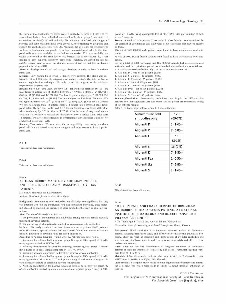
the cause of incompatibility. To screen red cell antibody, we need 2–3 different cell
suspensions derived from individual donors all with blood group O and 8–12 cell
suspensions to identify red cell antibody. The minimum 18 of red cells antigen of
screened and panel cells must have been known. At the beginning we got panel cells
support for antibody detection from CSL Australia. But it is only for temporary, so
we have to develop our own panel cells or buy commercial panel cells. At that time,
panel cells were not available in the Indonesian market. If it was available, the
expiry date would be the issue due to long bureaucracy in the custom. So, it was
decided to have our own homebrew panel cells. Therefore, we started the red cell
antigen phenotyping to know the characterization of red cell antigens in donor’s
population in Jakarta BTU.
Aims: to develop donor’s red cell antigen database in order to have homebrew
panel cells.
Methods: Only routine-blood group O donors were selected. The blood was col-
lected in 10 ml EDTA tube. Phenotyping was conducted using either tube method or
column agglutination technique. We only typed 18 antigens as the minimum
requirements for panel cells.
Results: Since 2001 until 2015, we have 1061 donors in our database. Of 1061, the
most frequent antigens are D (85.8%), C (87.6%), e (97.9%), k (100%), Fya (96.8%), s
(98.9%), M (85.1%) and Jka (77.9%). The less frequent antigens are E (22.5%), Fyb
(24.7%), S (14.8%), and Lea (27.1%). The rare antigens are K (0.94%). The most diffi-
cult types in donors are Jka-b- (0.38%), Fy a-b- (0.28%), R2R2 (1.7%) and SS (1.04%).
We have to arrange these 18 antigens from 2–3 donors into a screened panel (small
panel cells). The big panel cells need 8–12 donors. Sometimes we found difficulties
when combining Fy a-b+ (4.24%) or Jka-b+ (21.87%) because of very small donors
available. So, we have to expand our database to have a perfect panel. With these
18 antigens, we also found difficulties in determining other antibodies which not yet
established in our panel cells.
Summary/Conclusions: We can solve the incompatibility cases using homebrew
panel cells but we should screen more antigens and more donors to have a perfect
panel cells.
P-143
This abstract has been withdrawn.
P-144
This abstract has been withdrawn.
P-145
ALLO-ANTIBODIES MASKED BY AUTO-IMMUNE COLDANTIBODIES IN REGULARLY TRANSFUSED EGYPTIANPATIENTSM Salah, U Khasanah and E Mohammed
National blood transfusion services, Giza, Egypt
Background: Autoimmune cold antibodies are clinically non-significant but they
can interfere with the pre-transfusion tests like (antibodies screening, cross-match-
ing, etc. . ...) by masking the presence of other antibodies that may be clinically sig-
nificant.
Aim: The aim of the study is to find out:
1. The prevalence of autoimmune cold antibodies among male and female regularly
transfused Egyptian patients.
2. The prevalence of allo-antibodies masked by autoimmune cold antibodies.
Methods: The study conducted on transfusion dependent patients (2000 patients)
with: Thalassemia, aplastic anemia, leukemia, renal failure and anemia of chronic
diseases; presented to Egyptian NBTC for blood transfusion.
According to National Egyptian Testing Strategy, Patients were subjected to:
1. Screening for allo-antibodies against group O reagent RBCs (panel of 3 cells)
using appropriate IAT at 37°C by CAT.
2. Antibody identification for positive screening samples against group O reagent
RBCs (panel of 11 cells) using appropriate IAT at 37°C by CAT.
3. Screening at room temperature to detect the presence of cold antibodies.
4. Screening for allo-antibodies against group O reagent RBCs (panel of 3 cells)
using appropriate IAT at strict 37°C with pre-warming of both serum & reagents (in
case of positive results of Screening at room temperature)
5. Antibody identification for positive screening samples to identify the specificity
of allo-antibodies masked by autoimmune cold ones against group O reagent RBCs
(panel of 11 cells) using appropriate IAT at strict 37°C with pre-warming of both
serum & reagents.
Results: A total of 2000 patient (1000 males & 1000 females) were examined for
the presence of autoimmune cold antibodies & allo antibodies that may be masked
by them.
106 out of 1000 (10.6%) male patients were found to have autoimmune cold anti-
bodies.
79 out of 1000 (7.9%) female patients were found to have autoimmune cold anti-
bodies.
Out of a total of 2000 we found that 185 (9.25%) patients had autoimmune cold
antibodies and the co-incident prevalence of masked allo-antibodies was as follows:
1. Autoimmune cold antibodies only:129 out of 185 patients (69.7%).
2. Allo-anti D: 3 out of 185 patients (1.6%).
3. Allo-anti C: 7 out of 185 patients (3.8%).
4. Allo-anti E: 15 out of 185 patients (8.1%).
5. Allo-antic:13 out of 185 patients (7%).
6. Allo-anti K: 7 out of 185 patients (3.8%).
7. Allo-anti Fya: 1 out of 185 patients (0.5%).
8. Allo-anti Jka: 7 out of 185 patients (3.8%).
9. Allo-anti S: 3 out of 185 patients (1.6%).
Summary/Conclusions: Pre-warming techniques are helpful in differentiation
between cold non-significant Abs and warm Abs, for proper pre-transfusion testing
of the patients’ samples
Table 1: co-incident prevalence of masked allo-antibodies.
P-146
This abstract has been withdrawn.
P-147
STUDY ON RATE AND CHARACTERISTIC OF IRREGULARANTIBODIES OF THALASSEMIA PATIENTS AT NATIONALINSTITUTE OF HEMATOLOGY AND BLOOD TRANSFUSION,VIETNAM (2013–2015)H Thi Thanh Nga, B Thi Mai An, NA Anh Tri and VD Duc Binh
National Institute of Hematology and Blood Transfusion, Hanoi, Vietnam
Background: Blood transfusion is an important treatment method for thalassemia
patients. Ensuring transfusion safely and effectively for thalassemia patients is nec-
essary. Study on result of screening and identification of irregular antibodies and
selection matching blood units in order to transfuse more safely and effectively for
thalassemia patients.
Aims: Study on rate and characteristic of irregular antibodies of thalassemia
patients at National Institute of Hematology and Blood Transfusion (NIHBT), Viet-
nam from 2013 to 2015.
Materials: 1.544 thalassemia patients who were treated in Thalassemia center,
NIHBT from 01/01/2013 to 30/06/2015. Methods
Cross-sectional descriptive study. Using column agglutination technique and screen-
ing cell, panel cell which were made in NIHBT to detect irregular antibodies of
patients.
© 2015 The AuthorVox Sanguinis © 2015 International Society of Blood Transfusion
Vox Sanguinis (2015) 109 (Suppl. 2), 1–96
Red Cell Immunology: Serology 71
Results: We found out 88 patients had irregular antibodies among 1.544 tha-
lassemia patients (the rate is 5.7%); Irregular antibodies rate in women with preg-
nancy higher than women without pregnancy (9.7% and 5.3%); Irregular antibodies
rate related to blood transfusion times (no transfusion: 2.5%, less than 5 times:
3.1%, from 5 to 10 times: 7.2%, more than 10 times: 10.7%); There were 59 patients
had single irregular antibody: anti-E (43.2%) and anti-Mia (13.6%) had the highest
rate in this group; There were 29 patients had combined irregular antibodies: 2
irregular antibodies: 19 patients (21.6%), 3 irregular antibodies: 7 patients (7.9%), 4
irregular antibodies: 2 patients (2.3%) and 5 irregular antibodies: 1 patient (1.1%),
the rate of anti-E combined anti-c was the highest (10.2%); The highest irregular
antibodies rate was found in Rh blood group system (anti-E: 68.2%, anti-c: 22.7%),
follow MNS blood group system (anti-Mia: 29.5%), Kidd’s antibodies: anti-Jka: 6.8%,
anti-Jkb: 5.7%, Duffy’s antibodies: anti-Fyb: 1.1%, P1PK’s antibodies: anti-P1: 1.1%.
Conclusions: Irregular antibody rate in thalassemia patients was 5.7%. Irregular anti-
body rate related to pregnancy and blood transfusion times. Irregular antibodies could
appeared single (59 patients) or combination (29 patients). Anti-E (43.2%) and anti-Mia
(13.6%) had the highest rate in single antibody group. The rate of anti-E combined
anti-c was the highest (10.2%) in combined antibodies group, we met a special patient
had 5 irregular antibodies: anti-E combined anti-c, anti-Mia, anti-Jkb v�a anti-Fyb
(1.1%). The highest irregular antibodies rate was found in Rh blood group system (anti-
E: 68.2%, anti-c: 22.7%) and MNS blood group system (anti-Mia: 29.5%) was follow.
P-148
THE IMPLEMENTATION OF ANTIBODY SCREENING IN PRE-TRANSFUSION TESTING FOR PATIENT SAFETY IN JAKARTABLOOD TRANSFUSION UNITM Maryunis, P Rini, NK Ritchie and S Salim
Jakarta Blood Transfusion Unit, DKI Jakarta, Indonesia
Background: Pre-transfusion compatibility test aimed to know the compatibility of
patients and donors or to see whether there was any antigen-antibody reactions. If it
is compatible, the donor’s blood expected to ‘live’ and patients get ‘benefit’ from
transfusions. In other hand, if it’s not compatible, we had chance to prevent transfu-
sion reactions in patients. In general, pre-transfusion test included antibody screen-
ing beside blood group test and cross-match test. But, in Indonesia, antibody
screening was excluded due to the limited of cost. There is risk of undoing antibody
screening. Negative major of cross-match test was not always indicated no free anti-
body in patients. If ‘false negative’ blood was given to patients, they would have
delayed transfusion reactions in the end. Therefore, Jakarta Blood Transfusion Unit
(BTU) wanted to improve the blood safety in pre-transfusion test.
Aims: To know the frequency of ‘false negative’ of major cross test.
Methods: Patient’s sample were collected in 3 ml EDTA Blood, sent to Jakarta BTU.
We screened all patient’s sample using selectogen�, 3 screened cells from Ortho
Clinical Diagnostic�. The next step, we did the major cross-match test in CAT system
from Ortho Clinical Diagnostic�. All test run in Autovue Innova, an automated sys-
tem for pre-transfusion testing from Ortho Clinical Diagnostic�.
Results: We found 387 of 65,045 or 0.5% of patient’s sample from Januari 2014 until
December 2014. From 387 samples contained antibody, 308 (79.6%) samples showed
negative results in major cross-match test and 79 (20.4%) samples showed positive
results in major cross-match test. If we didn’t do antibody screening in 2014, among
10.000 patients, there could be 796 false negative of blood, given to patients.
Summary/Conclusions: Antibody screening in pre-transfusion testing should be
implemented to prevent transfusion reactions in patients.
P-149
This abstract has been withdrawn.
P-150
DETECTION OF CISCIS-AB USING A MICROPLATE-BASEDSYSTEMS AND TWO COLUMN AGGLUTINATION TEST IN AHOSPITAL BLOOD BANKYJ Hong1, SH Park1, T Kim1, N Kim2, H Kim2, KU Park1, J Song1 and KS Han2
1Seoul National University Bundang Hospital, Seongnam, South-Korea 2Seoul
National University College of Medicine, Seoul, South-Korea
Background: The ABO blood group is one of the most important antigens in transfu-
sion medicine. Recently, a microplate-based system for ABO phenotyping has been
developed and is used universally in large-sized blood banks because of its effective-
ness of time and cost. A cis-AB, a rare and interesting ABO genotype, is the result of a
mutated gene. Both the A glycosyltransferase and B glycosyltransferase activity are
carried by the same gene which is a different from common Mendelian inheritance.
This may lead to mistyping of ABO phenotype and adverse transfusion reactions.
Aims: The evaluation of ABO typing using microplate-based system is required to
prevent errors and to standardize protocols.
Methods: Total 28 cis-AB patients’ blood samples collected from May 2011 to
February 2015 were tested for ABO typing with four different methods as follows:
method 1, a microplate-based system (Galileo NEO, Immucor, GA, USA); method 2,
manual plate-tube method; method 3, manual column agglutination test using Ortho
BioVue ABO-Rh/Reverse (Ortho, Raritan, NJ, USA), and method 4, manual column
agglutination test using DG Gel ABO/Rh (2D) (Grifols, Barcelona, Spain). ABO geno-
typing for cis-AB confirmation were performed by direct DNA sequencing on exon
6 and 7 of the ABO gene. ABO phenotype with weak B expression in front typing
and presence of weak anti-B in the back typing was defined as ‘highly suspected
cis-AB’ is defined. We analyzed the detection rate of ‘highly suspected cis-AB’ as
well as front/back typing discrepancies on each methods.
Results: All 28 specimens were confirmed as cis-AB: CisAB/O1, 60.7%; CisAB/O1v,
32.1%; CisAB/A1, 7.1%. Using methods 1 through method 4, 4 (14.3%), 22 (78.6%),
18 (64.3%), and 20 (71.4%) of 28 samples were concluded as ‘highly suspected cis-
AB’, respectively. In detection rate of ABO discrepancies, method 1 showed ‘Not
Typed Determined (NTD)’ in 24 (85.7%) samples and AB in 4 (14.3%). Twenty seven
(96.4%), 28 (100.0%), and 28 (100.0%) specimens resulted in front/back discrepan-
cies in method 2, 3, and 4, respectively. One (3.6%) was showed as phenotype AB in
method 2.
Conclusions: If we perform cis-AB patients’ ABO phenotype using only a micro-
plate method, we can conclude wrong results (failure rate: 14.3% [4 out of 28]).
Therefore, not to miss a cis-AB, the system must be worked together with manual
methods and ABO genotyping, for new arrival and ambiguous patients.
P-151
PAROXYSMAL COLD HEMOGLOBINURIA: ROLE OF HOSPITALTRANSFUSION MEDICINE & IMMUNOHEMATOLOGYDEPARTMENT IN THE DIAGNOSISS Sood and S Rani
Saket City Hospital, New Delhi, India
Background: Paroxysmal cold hemoglobinuria (PCH), an autoimmune haemolytic
anaemia caused due to polyclonal IgG anti-P autoantibody binding to red blood cell
surface antigens and is characterized by hemoglobinuria, typically after exposure to
cold temperatures.
Case Presentation: An 84-year-old female patient started developing breathing dif-
ficulty off and on with generalised weakness and loss of appetite. There was contin-
uous fall in hemoglobin and continuous evidence of intravascular hemolysis since
last 15 days. After ruling out many common diagnosis, Donath -Landsteiner Test
was done which showed Immunoglobulin G, IgG, antibodies, appearing at 4c and
hemolysing at 37�c in the presence of complement. Peripheral smear showed
anisopoikilocytosis and spherocytosis. Reticulocyte count was on the lower side
(0.5%).Forward and reverse blood grouping showed no group discrepancy .Direct
antiglobulin test (direct Coombs test, DAT) was positive with the monoclonal C3
antisera and negative with the monoclonal anti-IgG. ICT, Indirect Coombs Test was
negative. Syphilis was tested negative by Treponema pallidum hemagglutination
assay (TPHA).
Results: Patient was diagnosed as a case of PCH based on the test results.
Conclusions: Detecting blood antibodies, auto, allo, cold or warm, is important. The
role of transfusion medicine laboratory in the diagnosis of patient PCH is empha-
sised. (198 Words)
P-152
PREDICTIVE VALUE OF ANTI-D SUBCLASESS IN HEMOLITICDESEASE OF THE NEWBORNE Velkova, V Dejanova Ilijevska, T Makarovska-Bojadzieva, L Mitevska, M
Blagoevska, I Nikoloska and J Samonikov-Tosevska
Institute of Transfusion Medicine, Skopje, Macedonia
Background: Hemolytic disease of the newborn (HDN) continues to be a clinical
problem in spite of the existing prophylaxis. To date, none of the available tests,
developed to predict the severity of HDN, has provided complete reliability.
© 2015 The AuthorVox Sanguinis © 2015 International Society of Blood TransfusionVox Sanguinis (2015) 109 (Suppl. 2), 1–96
72 Posters
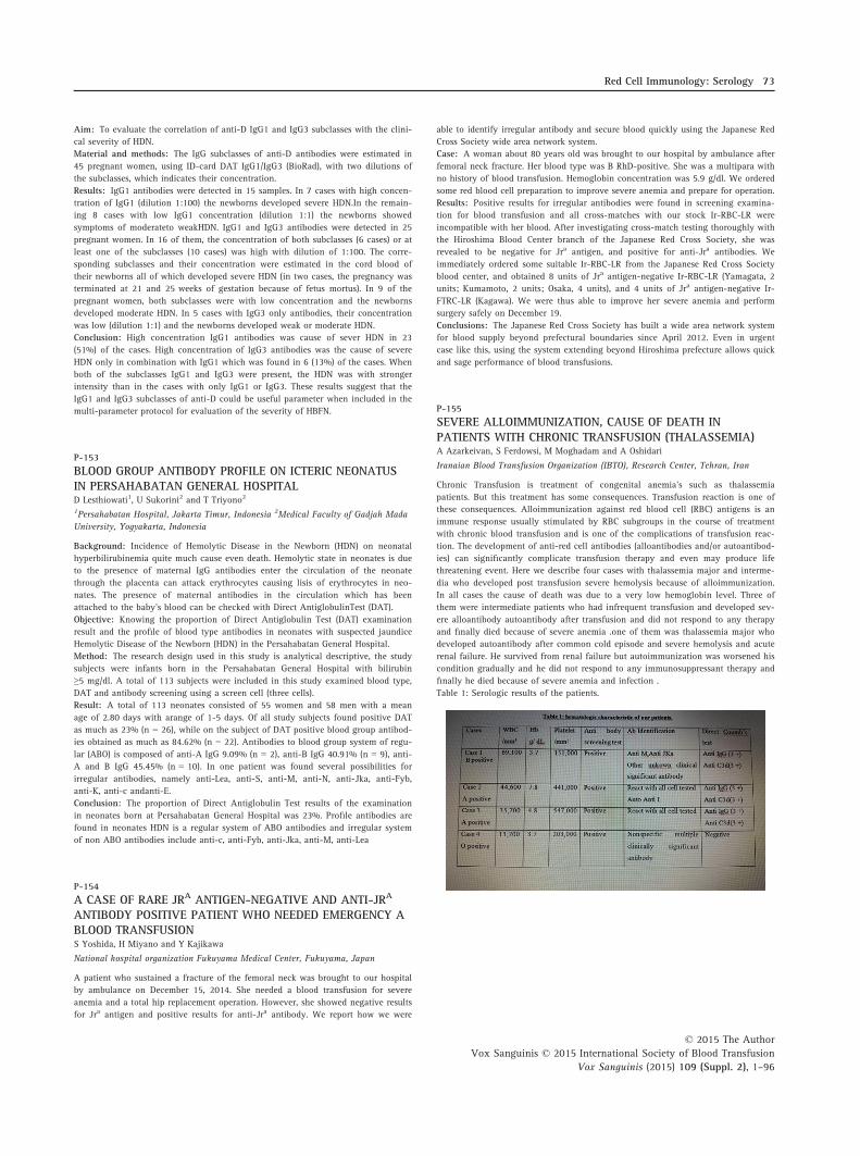
Aim: To evaluate the correlation of anti-D IgG1 and IgG3 subclasses with the clini-
cal severity of HDN.
Material and methods: The IgG subclasses of anti-D antibodies were estimated in
45 pregnant women, using ID-card DAT IgG1/IgG3 (BioRad), with two dilutions of
the subclasses, which indicates their concentration.
Results: IgG1 antibodies were detected in 15 samples. In 7 cases with high concen-
tration of IgG1 (dilution 1:100) the newborns developed severe HDN.In the remain-
ing 8 cases with low IgG1 concentration (dilution 1:1) the newborns showed
symptoms of moderateto weakHDN. IgG1 and IgG3 antibodies were detected in 25
pregnant women. In 16 of them, the concentration of both subclasses (6 cases) or at
least one of the subclasses (10 cases) was high with dilution of 1:100. The corre-
sponding subclasses and their concentration were estimated in the cord blood of
their newborns all of which developed severe HDN (in two cases, the pregnancy was
terminated at 21 and 25 weeks of gestation because of fetus mortus). In 9 of the
pregnant women, both subclasses were with low concentration and the newborns
developed moderate HDN. In 5 cases with IgG3 only antibodies, their concentration
was low (dilution 1:1) and the newborns developed weak or moderate HDN.
Conclusion: High concentration IgG1 antibodies was cause of sever HDN in 23
(51%) of the cases. High concentration of IgG3 antibodies was the cause of severe
HDN only in combination with IgG1 which was found in 6 (13%) of the cases. When
both of the subclasses IgG1 and IgG3 were present, the HDN was with stronger
intensity than in the cases with only IgG1 or IgG3. These results suggest that the
IgG1 and IgG3 subclasses of anti-D could be useful parameter when included in the
multi-parameter protocol for evaluation of the severity of HBFN.
P-153
BLOOD GROUP ANTIBODY PROFILE ON ICTERIC NEONATUSIN PERSAHABATAN GENERAL HOSPITALD Lesthiowati1, U Sukorini2 and T Triyono2
1Persahabatan Hospital, Jakarta Timur, Indonesia 2Medical Faculty of Gadjah Mada
University, Yogyakarta, Indonesia
Background: Incidence of Hemolytic Disease in the Newborn (HDN) on neonatal
hyperbilirubinemia quite much cause even death. Hemolytic state in neonates is due
to the presence of maternal IgG antibodies enter the circulation of the neonate
through the placenta can attack erythrocytes causing lisis of erythrocytes in neo-
nates. The presence of maternal antibodies in the circulation which has been
attached to the baby’s blood can be checked with Direct AntiglobulinTest (DAT).
Objective: Knowing the proportion of Direct Antiglobulin Test (DAT) examination
result and the profile of blood type antibodies in neonates with suspected jaundice
Hemolytic Disease of the Newborn (HDN) in the Persahabatan General Hospital.
Method: The research design used in this study is analytical descriptive, the study
subjects were infants born in the Persahabatan General Hospital with bilirubin
≥5 mg/dl. A total of 113 subjects were included in this study examined blood type,
DAT and antibody screening using a screen cell (three cells).
Result: A total of 113 neonates consisted of 55 women and 58 men with a mean
age of 2.80 days with arange of 1-5 days. Of all study subjects found positive DAT
as much as 23% (n = 26), while on the subject of DAT positive blood group antibod-
ies obtained as much as 84.62% (n = 22). Antibodies to blood group system of regu-
lar (ABO) is composed of anti-A IgG 9.09% (n = 2), anti-B IgG 40.91% (n = 9), anti-
A and B IgG 45.45% (n = 10). In one patient was found several possibilities for
irregular antibodies, namely anti-Lea, anti-S, anti-M, anti-N, anti-Jka, anti-Fyb,
anti-K, anti-c andanti-E.
Conclusion: The proportion of Direct Antiglobulin Test results of the examination
in neonates born at Persahabatan General Hospital was 23%. Profile antibodies are
found in neonates HDN is a regular system of ABO antibodies and irregular system
of non ABO antibodies include anti-c, anti-Fyb, anti-Jka, anti-M, anti-Lea
P-154
A CASE OF RARE JRA ANTIGEN-NEGATIVE AND ANTI-JRA
ANTIBODY POSITIVE PATIENT WHO NEEDED EMERGENCY ABLOOD TRANSFUSIONS Yoshida, H Miyano and Y Kajikawa
National hospital organization Fukuyama Medical Center, Fukuyama, Japan
A patient who sustained a fracture of the femoral neck was brought to our hospital
by ambulance on December 15, 2014. She needed a blood transfusion for severe
anemia and a total hip replacement operation. However, she showed negative results
for Jra antigen and positive results for anti-Jra antibody. We report how we were
able to identify irregular antibody and secure blood quickly using the Japanese Red
Cross Society wide area network system.
Case: A woman about 80 years old was brought to our hospital by ambulance after
femoral neck fracture. Her blood type was B RhD-positive. She was a multipara with
no history of blood transfusion. Hemoglobin concentration was 5.9 g/dl. We ordered
some red blood cell preparation to improve severe anemia and prepare for operation.
Results: Positive results for irregular antibodies were found in screening examina-
tion for blood transfusion and all cross-matches with our stock Ir-RBC-LR were
incompatible with her blood. After investigating cross-match testing thoroughly with
the Hiroshima Blood Center branch of the Japanese Red Cross Society, she was
revealed to be negative for Jra antigen, and positive for anti-Jra antibodies. We
immediately ordered some suitable Ir-RBC-LR from the Japanese Red Cross Society
blood center, and obtained 8 units of Jra antigen-negative Ir-RBC-LR (Yamagata, 2
units; Kumamoto, 2 units; Osaka, 4 units), and 4 units of Jra antigen-negative Ir-
FTRC-LR (Kagawa). We were thus able to improve her severe anemia and perform
surgery safely on December 19.
Conclusions: The Japanese Red Cross Society has built a wide area network system
for blood supply beyond prefectural boundaries since April 2012. Even in urgent
case like this, using the system extending beyond Hiroshima prefecture allows quick
and sage performance of blood transfusions.
P-155
SEVERE ALLOIMMUNIZATION, CAUSE OF DEATH INPATIENTS WITH CHRONIC TRANSFUSION (THALASSEMIA)A Azarkeivan, S Ferdowsi, M Moghadam and A Oshidari
Iranaian Blood Transfusion Organization (IBTO), Research Center, Tehran, Iran
Chronic Transfusion is treatment of congenital anemia’s such as thalassemia
patients. But this treatment has some consequences. Transfusion reaction is one of
these consequences. Alloimmunization against red blood cell (RBC) antigens is an
immune response usually stimulated by RBC subgroups in the course of treatment
with chronic blood transfusion and is one of the complications of transfusion reac-
tion. The development of anti-red cell antibodies (alloantibodies and/or autoantibod-
ies) can significantly complicate transfusion therapy and even may produce life
threatening event. Here we describe four cases with thalassemia major and interme-
dia who developed post transfusion severe hemolysis because of alloimmunization.
In all cases the cause of death was due to a very low hemoglobin level. Three of
them were intermediate patients who had infrequent transfusion and developed sev-
ere alloantibody autoantibody after transfusion and did not respond to any therapy
and finally died because of severe anemia .one of them was thalassemia major who
developed autoantibody after common cold episode and severe hemolysis and acute
renal failure. He survived from renal failure but autoimmunization was worsened his
condition gradually and he did not respond to any immunosuppressant therapy and
finally he died because of severe anemia and infection .
Table 1: Serologic results of the patients.
© 2015 The AuthorVox Sanguinis © 2015 International Society of Blood Transfusion
Vox Sanguinis (2015) 109 (Suppl. 2), 1–96
Red Cell Immunology: Serology 73
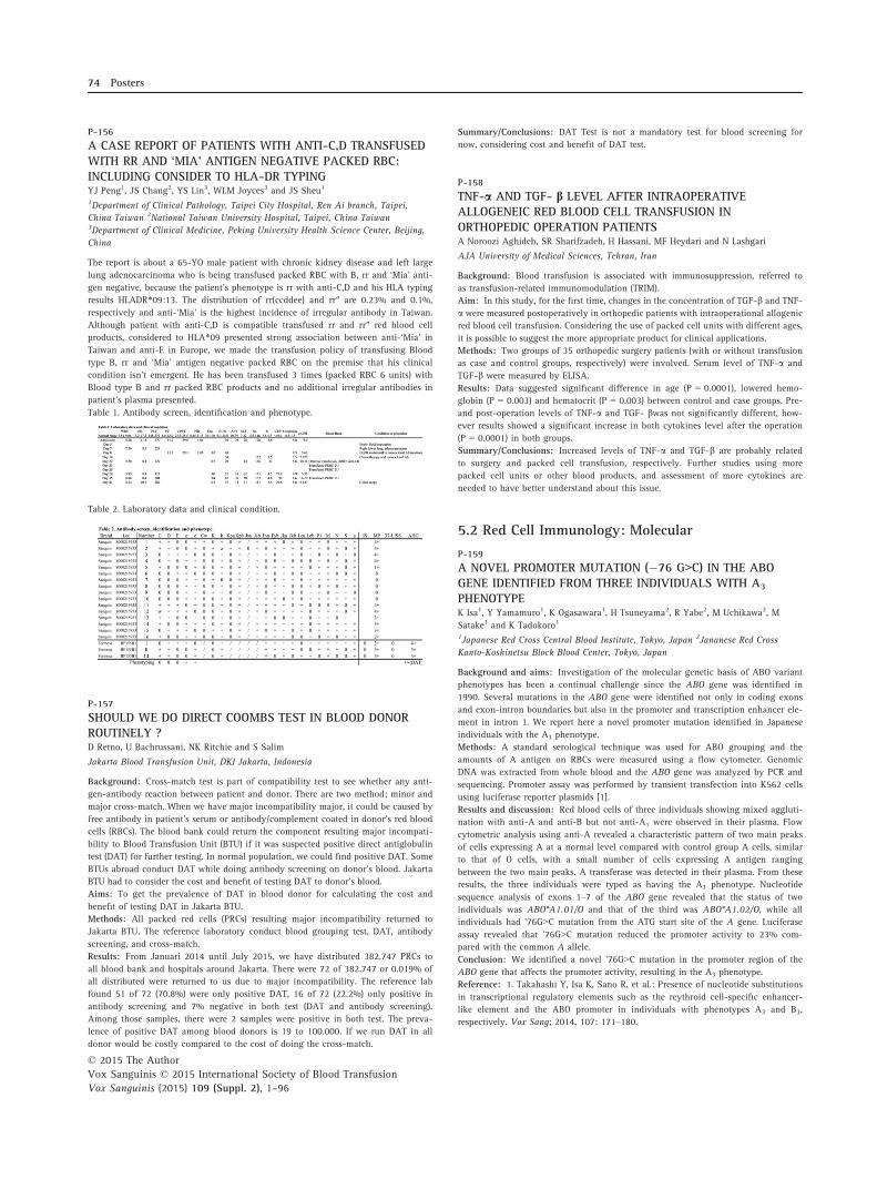
P-156
A CASE REPORT OF PATIENTS WITH ANTI-C,D TRANSFUSEDWITH RR AND ‘MIA’ ANTIGEN NEGATIVE PACKED RBC:INCLUDING CONSIDER TO HLA-DR TYPINGYJ Peng1, JS Chang2, YS Lin3, WLM Joyces3 and JS Sheu1
1Department of Clinical Pathology, Taipei City Hospital, Ren Ai branch, Taipei,
China Taiwan 2National Taiwan University Hospital, Taipei, China Taiwan3Department of Clinical Medicine, Peking University Health Science Center, Beijing,
China
The report is about a 65-YO male patient with chronic kidney disease and left large
lung adenocarcinoma who is being transfused packed RBC with B, rr and ‘Mia’ anti-
gen negative, because the patient’s phenotype is rr with anti-C,D and his HLA typing
results HLADR*09:13. The distribution of rr(ccddee) and rr″ are 0.23% and 0.1%,
respectively and anti-‘Mia’ is the highest incidence of irregular antibody in Taiwan.
Although patient with anti-C,D is compatible transfused rr and rr″ red blood cell
products, considered to HLA*09 presented strong association between anti-‘Mia’ in
Taiwan and anti-E in Europe, we made the transfusion policy of transfusing Blood
type B, rr and ‘Mia’ antigen negative packed RBC on the premise that his clinical
condition isn’t emergent. He has been transfused 3 times (packed RBC 6 units) with
Blood type B and rr packed RBC products and no additional irregular antibodies in
patient’s plasma presented.
Table 1. Antibody screen, identification and phenotype.
Table 2. Laboratory data and clinical condition.
P-157
SHOULD WE DO DIRECT COOMBS TEST IN BLOOD DONORROUTINELY ?D Retno, U Bachrussani, NK Ritchie and S Salim
Jakarta Blood Transfusion Unit, DKI Jakarta, Indonesia
Background: Cross-match test is part of compatibility test to see whether any anti-
gen-antibody reaction between patient and donor. There are two method; minor and
major cross-match. When we have major incompatibility major, it could be caused by
free antibody in patient’s serum or antibody/complement coated in donor’s red blood
cells (RBCs). The blood bank could return the component resulting major incompati-
bility to Blood Transfusion Unit (BTU) if it was suspected positive direct antiglobulin
test (DAT) for further testing. In normal population, we could find positive DAT. Some
BTUs abroad conduct DAT while doing antibody screening on donor’s blood. Jakarta
BTU had to consider the cost and benefit of testing DAT to donor’s blood.
Aims: To get the prevalence of DAT in blood donor for calculating the cost and
benefit of testing DAT in Jakarta BTU.
Methods: All packed red cells (PRCs) resulting major incompatibility returned to
Jakarta BTU. The reference laboratory conduct blood grouping test, DAT, antibody
screening, and cross-match.
Results: From Januari 2014 until July 2015, we have distributed 382.747 PRCs to
all blood bank and hospitals around Jakarta. There were 72 of 382.747 or 0.019% of
all distributed were returned to us due to major incompatibility. The reference lab
found 51 of 72 (70.8%) were only positive DAT, 16 of 72 (22.2%) only positive in
antibody screening and 7% negative in both test (DAT and antibody screening).
Among those samples, there were 2 samples were positive in both test. The preva-
lence of positive DAT among blood donors is 19 to 100.000. If we run DAT in all
donor would be costly compared to the cost of doing the cross-match.
Summary/Conclusions: DAT Test is not a mandatory test for blood screening for
now, considering cost and benefit of DAT test.
P-158
TNF-a AND TGF- b LEVEL AFTER INTRAOPERATIVEALLOGENEIC RED BLOOD CELL TRANSFUSION INORTHOPEDIC OPERATION PATIENTSA Noroozi Aghideh, SR Sharifzadeh, H Hassani, MF Heydari and N Lashgari
AJA University of Medical Sciences, Tehran, Iran
Background: Blood transfusion is associated with immunosuppression, referred to
as transfusion-related immunomodulation (TRIM).
Aim: In this study, for the first time, changes in the concentration of TGF-b and TNF-
a were measured postoperatively in orthopedic patients with intraoperational allogenic
red blood cell transfusion. Considering the use of packed cell units with different ages,
it is possible to suggest the more appropriate product for clinical applications.
Methods: Two groups of 35 orthopedic surgery patients (with or without transfusion
as case and control groups, respectively) were involved. Serum level of TNF-a and
TGF-b were measured by ELISA.
Results: Data suggested significant difference in age (P = 0.0001), lowered hemo-
globin (P = 0.003) and hematocrit (P = 0.003) between control and case groups. Pre-
and post-operation levels of TNF-a and TGF- bwas not significantly different, how-
ever results showed a significant increase in both cytokines level after the operation
(P = 0.0001) in both groups.
Summary/Conclusions: Increased levels of TNF-a and TGF-b are probably related
to surgery and packed cell transfusion, respectively. Further studies using more
packed cell units or other blood products, and assessment of more cytokines are
needed to have better understand about this issue.
5.2 Red Cell Immunology: Molecular
P-159
A NOVEL PROMOTER MUTATION (�76 G>C) IN THE ABOGENE IDENTIFIED FROM THREE INDIVIDUALS WITH A3
PHENOTYPEK Isa1, Y Yamamuro1, K Ogasawara1, H Tsuneyama2, R Yabe2, M Uchikawa2, M
Satake1 and K Tadokoro1
1Japanese Red Cross Central Blood Institute, Tokyo, Japan 2Jananese Red Cross
Kanto-Koshinetsu Block Blood Center, Tokyo, Japan
Background and aims: Investigation of the molecular genetic basis of ABO variant
phenotypes has been a continual challenge since the ABO gene was identified in
1990. Several mutations in the ABO gene were identified not only in coding exons
and exon-intron boundaries but also in the promoter and transcription enhancer ele-
ment in intron 1. We report here a novel promoter mutation identified in Japanese
individuals with the A3 phenotype.
Methods: A standard serological technique was used for ABO grouping and the
amounts of A antigen on RBCs were measured using a flow cytometer. Genomic
DNA was extracted from whole blood and the ABO gene was analyzed by PCR and
sequencing. Promoter assay was performed by transient transfection into K562 cells
using luciferase reporter plasmids [1].
Results and discussion: Red blood cells of three individuals showing mixed aggluti-
nation with anti-A and anti-B but not anti-A1 were observed in their plasma. Flow
cytometric analysis using anti-A revealed a characteristic pattern of two main peaks
of cells expressing A at a normal level compared with control group A cells, similar
to that of O cells, with a small number of cells expressing A antigen ranging
between the two main peaks. A transferase was detected in their plasma. From these
results, the three individuals were typed as having the A3 phenotype. Nucleotide
sequence analysis of exons 1–7 of the ABO gene revealed that the status of two
individuals was ABO*A1.01/O and that of the third was ABO*A1.02/O, while all
individuals had ’76G>C mutation from the ATG start site of the A gene. Luciferase
assay revealed that ’76G>C mutation reduced the promoter activity to 23% com-
pared with the common A allele.
Conclusion: We identified a novel ’76G>C mutation in the promoter region of the
ABO gene that affects the promoter activity, resulting in the A3 phenotype.
Reference: 1. Takahashi Y, Isa K, Sano R, et al.: Presence of nucleotide substitutions
in transcriptional regulatory elements such as the reythroid cell-specific enhancer-
like element and the ABO promoter in individuals with phenotypes A3 and B3,
respectively. Vox Sang; 2014, 107: 171–180.
© 2015 The AuthorVox Sanguinis © 2015 International Society of Blood TransfusionVox Sanguinis (2015) 109 (Suppl. 2), 1–96
74 Posters
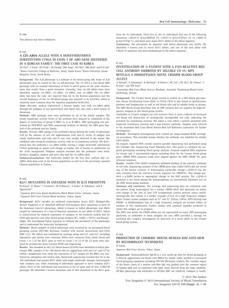
P-160
This abstract has been withdrawn.
P-161
A CISCIS-AB04AB04 ALLELE WITH A NONSYNONYMOUSSUBSTITUTION C796A IN EXON 7 OF ABO GENE IDENTIFIEDIN A KOREAN FAMILY: THE FIRST CASE IN KOREAJS Choi1, J Yoon1, HJ Youk1, JH Chang2, MA Jang1, JH Choi1, MH Nam1 and CS Lim1
1Korea University College of Medicine, Seoul, South-Korea 2Korea University Ansan
Hospital, Seoul, South-Korea
Background: The A2B phenotype is a subtype of the blood group AB. Some of A2B
phenotypes may be caused by the cis-AB genotype. The cis-AB is a rare blood ABO
genotype with an unusual inheritance of both A and B genes on the same chromo-
some that results from a point mutation. Currently, four cis-AB alleles have been
identified, namely cis-AB01, cis-AB02, cis-AB03, and cis-AB04. The cis-AB01
allele has been the only one reported thus far in the Korean population and the
overall frequency of the cis-AB blood group was reported to be 0.0354%, which is
relatively more common than the Japanese population (0.0012%).
Aims: Recently, authors experienced a Korean family case with cis-AB04 allele
through the pedigree of two generations and report this case with a brief review of
literatures.
Methods: ABO serologic tests were performed on all of the family samples. The
serum transferase activity levels of the proband were assayed by estimation of the
degree of conversion of group O RBCs to A or B RBCs. ABO genotyping was per-
formed by PCR-RFLP and DNA sequencing analysis to identify the ABO gene hap-
loid of the proband and her family.
Results: Routine ABO typing of the proband’s blood showed the result of phenotype
A2B by the absence of red cell agglutination with anti-A1 lectin. H antigen was
nearly undetectable and only anti-A was very weakly detected in the serum. She
exhibited only strong B-transferase activity with no A-transferase activity. We deter-
mined that she harbored a cis-AB04 allelle, having a single nucleotide substitution
C796A predicting an amino acid change at residue 266 of leucine to methionine on
the A102 background. Pedigree analysis revealed that the proband’s twin sister,
brothers, daughter, nephew and niece also had the cis-AB04 allele.
Summary/Conclusions: Our molecular studies for the first time confirm that cis-
AB04 allele does exist in the Korean population as well as in the previously reported
Chinese population in Taiwan.
P-162
KLF1 MUTATIONS IN JAPANESE WITH IN (LU) PHENOTYPEM Kawai1, K Obara1, T Enomoto1, M Uchikawa1, S Inaba1, K Nakajima1 and K
Ogasawara2
1Japanese Red Cross Kanto-Koshinetsu Block Blood Center, Saitama, Japan2Japanese Red Cross Central Blood Institute, Tokyo, Japan
Background: KLF1 encodes an erythroid transcription factor, KLF1 (Kr€uppel-like
factor). Singleton et al. identified different heterozygous KLF1 mutations in most of
the dominant Lu(a-b-) phenotype, which is known as In(Lu) phenotype, was likely
caused by inheritance of a loss-of-function mutations in one allele of KLF1. In(Lu)
is characterized by reduced expression of antigens in the Lutheran system and for
CD44 glycoprotein and other blood group antigens (P1, AnWj, i, CD151 and Knops).
Aims: We investigated In(Lu) Japanese to estimate the prevalence of the phenotype
and to understand the molecular background.
Methods: Blood samples of In(Lu) phenotype were screened by an automated blood
grouping system (PK7300; Beckman Coulter) with murine monoclonal anti-CD44
(CBC-121). The In(Lu) was confirmed by serology using anti-Lua, anti-Lub, including
adsorption and elution tests. Genomic DNAs were extracted from whole blood and
exons 1 to 3 of the KLF1 gene as well as exons 1 to 15 of the LU gene were ana-
lyzed by polymerase chain reaction (PCR) and sequencing.
Results: One-hundred in 481,322 blood donors (0.02%) were identified as In(Lu) phe-
notype. RBC samples of the 100 donors did not agglutinate with anti-Lua and Lub by
indirect antiglobulin tests while the expression of Lub antigen on the RBCs was con-
firmed by adsorption and elution tests. Nucleotide sequencing revealed that 92 in the
100 individuals had mutant KLF1 allele with single nucleotide changes, heterozygote
with common one, while remaining 8 individuals appeared to have common KLF1
alleles. None of the individuals had mutations in the LU gene and all were LUB/LUB
genotype. We identified 7 known mutations and 22 new mutations in the KLF1 gene
from the 92 individuals. Forty-five in the 92 individuals had one of the following
mutations, c.862A>G (p.Lys288Glu) 19, c.947G>A (p.Cys316Tyr) 15, or c.968C>G(p.Ser323Trp) 11, and those were major KLF1 alleles in the In(Lu) Japanese.
Conclusions: The prevalence in Japanese with In(Lu) phenotype was 0.02%. We
identified 7 known and 22 novel KLF1 alleles, and one of the new allele with
c.862A>G mutation was most predominant in the In(Lu) Japanese.
P-163
INVESTIGATION OF A PATIENT WITH A PAN-REACTIVE REDCELL ANTIBODY INHIBITED BY SOLUBLE CD 55: MPSREVEALS A HOMOZYGOUS NOVEL CROMER BLOOD GROUPALLELEA Hyland1, E Schoeman1, K McGrath1, B Wilson1, HC Lin2, CW Wu2, RL Flower1, T
Powley1 and YW Liew1
1Australian Red Cross Blood Service, Brisbane, Australia 2Kaohsiung Blood Center,
Kaohsiung, Taiwan
Background: The Cromer blood group system is carried on a GPI-linked glycopro-
tein, Decay Accelerating Factor (DAF or CD55). CD55 is also found on granulocytes,
platelets and lymphocytes as well as red blood cells and in soluble forms in plasma.
The ISBT Blood Group Data Base lists 10 SNP variants that are negative for high fre-
quency antigens in this blood group system.
Antibodies to Cromer antigens are rare however there is some evidence of increased
red blood cell destruction of serologically incompatible red cells, indicating the
potential for transfusion reaction. We report a case where a patient presented with
suspected transfusion reaction and a pan-reactive antibody. Samples were referred
to the Australian Red Cross Blood Service Red Cell Reference Laboratory for further
investigation.
Methods: Serological investigations were carried out using standard RCRL serologi-
cal procedures. This included testing whether the antibody was inhibited by soluble
CD55 (DAF).
On request, targeted DNA exomic massive parallel sequencing was performed using
the TruSight One Sequencing Panel (Illumina Inc). This panel is validated for tar-
geted genotyping including blood group analysis. Analysis targeted DNA sequences
for exonic and 30 untranslated regions including those for the Cromer blood group
gene, CROM. DNA sequence reads were aligned against the ISBT CROM *01 gene
reference sequence.
Results: Soluble CD55 (DAF) completely inhibited binding of the patient’s antibody
to red cells. Sequencing analysis of the CROM gene (also called CD55) did not reveal
any of the known variants. A homozygous nucleotide change c.203G>A was the
only variation from the reference exomic sequence for CROM*01. This change pre-
dicts a p.S68N (serine to asparagine) change in the DAF protein. The c.203G>Amutation is not listed among the polymorphisms on international databases nor on
the ISBT blood group database.
Summary and conclusion: The serology and sequencing data are consistent with
the patient being homozygous for a variant CROM allele that generates an amino
acid change in the first of four CCP (complement control protein) repeat motifs.
Serologically this variant is a further example of loss of a high frequency antigen.
Other Cromer system antigens such as Tcb and Tcc (52Leu, 52Pro), WES (82Arg) and
CROM: -6 (Ile80Asn)(also loss of a high frequency antigen) are located within 16
residues of this substitution. Further studies with synthetic peptides predicted to
mimic this antigen are in progress.
The SNPs that define the CROM alleles are not represented on many SNP genotyping
platforms, as antibodies to these antigens are rare. MPS provided a strategy for
resolving this complex investigation by detection of a novel allele in the Cromer
blood group system.
P-164
PRODUCTION OF CHIMERIC MOUSE-HUMAN IGM ANTI-MURBY RECOMBINANT TECHNIQUESR Tobita
Japanese Red Cross Society, Tokyo, Japan
Background: Immunoglobulin (Ig) M is a very useful Ig class for blood grouping as
it directly agglutinates red blood cells (RBCs) by simple saline method or automated
blood grouping equipment including PK7300 (Beckman coulter). The variable region
of the heavy chain of a murine anti-Mur IgG was grafted onto the constant region
of human IgM and co-expressed with light chain derived from the same antibody.
GP.Mur phenotype and antibodies to GP.Mur RBC are relatively common is South-
© 2015 The AuthorVox Sanguinis © 2015 International Society of Blood Transfusion
Vox Sanguinis (2015) 109 (Suppl. 2), 1–96
Red Cell Immunology: Molecular 75
East Asia. Antibodies to GP.Mur RBC have been responsible for immediate and
delayed hemolytic transfusion reactions, and severe hemolytic disease of the new-
born. It is recommended that a GP.Mur RBC are included in antibody screening pan-
els in South-East Asia.
Aim: To produce chimeric mouse-human anti-Mur for use in the clinical laboratory
and automated blood grouping equipment.
Methods: The full-length heavy- and light-chain domains was amplified from cDNA
of a single hybridoma clone using the polymerase chain reaction (PCR) with
immunoglobulin specific primers. The heavy chain variable domain was amplified
by PCR, and was the expression vector ligation together human IgM constant
domain fragment. Likewise, the light chain variable domain was amplified by PCR,
were ligation into an expression vector with human kappa constant domain frag-
ment. The expression vectors of the heavy- and light chain were restriction enzyme
treatment to ligation as one of the expression vectors. The construct was transfected
CHO cell line to express of the antibodies.
Results: The heavy and light chain variable domains from the murine anti-Mur IgG
were cloned using PCR amplification and inserted into an expression vector with
human IgM constant domain. The chimeric mouse-human IgM anti-Mur directly
agglutinated 4 + with the Mur+ RBCs (GP.Mur, GP.Bun and GP.Kip) in saline at
room temperature, resembling the specificity of the original murine antibody. The
chimeric anti-Mur also determined clearly Mur+ or Mur- RBCs by PK7300.
Conclusions: Mouse-human chimeric IgM from a murine IgG monoclonal anti-Mur
has been successfully engineered for use in the clinical laboratory, and PK7300 for
large number of blood samples.
P-165
HIGHER LEVEL OF MUR ANTIGEN EXPRESSION ON THE REDCELLS OF GP.MUR HOMOZYGOTES THAN THAT OFHETEROZYGOTESL Wei, Y Ji, Z Wang, Z Shan, J Wen and G Luo
Guangzhou Blood Center, Guangzhou, China
Background: GP.Mur was one of GP (B-A-B) hybrid glycophorins in MNS blood
group system. It was reported to be the most common hybrid glycophorin in Southeast
Asia, with a frequency of 7.3% in Taiwan, 8.0% in Thailand, and 6.3% in Hongkong. In
our previous study, the frequency of heterozygous GP.Mur was about 9%, and
homozygous GP.Mur was very rare with an inferred frequency of about 0.2% in
Guangzhou. The comparison of the level of antigens expressed on the red cells between
the homozygous and heterozygous GP.Mur has not been investigated. In this study,
Mur antigen expression was detected by flow cytometry in the individuals with
GP.Mur homozygotes and heterzygotes to explore whether any differences exist.
Aim: To compare the level of Mur antigen expressed on the surface of the red cells
with homozygous and heterozygous GP.Mur.
Methods: The whole blood samples of 528 volunteer blood donors from Guangzhou
blood center were collected. Monoclonal anti-Mur was used to detect Mur antigen
expressed on the red cells of each donor. Genomic DNAs of Mur positive donors
were extracted, and then, GYPB pseudoexon 3 and part of adjacent intron 2 and 3
was sequenced by direct sequencing. Flow cytometry was performed to detect Mur
antigen expression on the red cells of GP.Mur homozygotes and heterozygotes.
Results: In total, fifty-one Mur positive samples were identified with a frequency of
9.7% (51/528) by serological testing. Further sequencing of GYPB pseudoexon 3 and
adjacent introns in these donors showed heterozygous GYP.Mur allele existed in 48
donors, homozygous GYP.Mur allele in 2 donors, and a novel heterozygous GYP.-
Bun allele in one donor. Flow cytometry results revealed higher Mur antigens
expression on the red cells of GP.Mur homozygotes than heterozygotes.
Conclusions: GP.Mur was the main hybrid glycophorin in Guangzhou with a high
frequency of 9.5% (50/528). Since homozygous GP.Mur individual expressed higher
level of Mur antigens, whether GP.Mur homozygotes could more easily stimulate the
recipient to produce the alloantibodies than GP.Mur heterozygotes during incompati-
ble transfusion should be further investigated.
P-166
THE DISTRIBUTION OF THE RED CELLS STA (STONES)ANTIGEN IN CHINESE BLOOD DONORS FROM GUANGZHOUGP Luo, Z Wang, YL Ji, L Wei and JZ Wen
Guangzhou Blood Center, Guangzhou, China
Background: Sta is a low-frequency antigen in MNS blood group system. It could
be expressed by the variant hybrid glycophorins, which usually resulted from the
hybrid GYP (B-A) gene with unequal crossing over at the intron 3 of GYPA and
GYPB. So far, six different Sta related alleles (type A to F) have been identified, dif-
fering in the location of crossing-over points in the intron 3. And the frequency of
Sta is 1.0–5.2% in Taiwan, 6% in Japanese. But there is no report about the Sta fre-
quency of Chinese in Mainland of China.
Aim: To study the distribution of Sta in Chinese blood donors from Guangzhou and
clarify the molecule background.
Methods: A PCR-based method was used to detect Sta in 390 Chinese blood donors
from Guangzhou Blood Center. Upstream primer was GYPB-specific and downstream
primer was GYPA-specific. Several DNA samples with sequence-confirmed Sta allele
detected previously were used as positive control. Then, the samples with Sta specific
PCR product were analyzed by direct sequencing to clarify the allele types. The extra
two presumed Sta(+) samples were also included.
Results: Five donors with Sta specific PCR product were identified in 390 (5/390,
1.3%) donors tested. Combined with the two extra suspected Sta(+) samples, the PCR
product of a total of 7 samples were analyzed by direct sequencing. Sequencing
results revealed 5 of them with heterozygous Sta type B allele and the other two
Samples with heterozygous Sta type E allele.
Conclusions: The frequency of Sta in China South Han from Guangzhou is around
1.3%, which is similar with Taiwanese, higher than Europeans. The distribution of
anti-Sta in Chinese population is still needed for further investigation in the future.
P-167
DEVELOPMENT OF REAL-TIME PCR AND MELTING CURVEANALYSIS FOR THE RAPID DETECTION OF DEL PHENOTYPEYJ Hong1, H Kim2, D Cho3, KU Park1, J Song1 and KS Han2
1Seoul National University Bundang Hospital, Seongnam, South-Korea 2Seoul
National University College of Medicine, Seoul, South-Korea 3Samsung Medical
Center, Seoul, South-Korea
Background: The Rh blood group D antigen is clinically important since it is asso-
ciated with transfusion reactions in the alloimmunized individuals with RhD. DEL, a
type of variant RhD, is hard to ascertain in routine blood bank, since it expresses
extremely low level of D antigens. However, it is capable of alloimmunizing a
patient when transfused to RhD-negative individuals. Thus, the detection of DEL in
donors with serologically RhD-negatives is crucial in transfusion medicine.
Aims: In this study, we developed real-time PCR and melting curve analysis for the
rapid detection of DEL phenotype.
Methods: Total 325 RhD-negative individuals were involved in this study. RhD neg-
ativity were confirmed with minimum two different reagents: BIOSCOT Anti-D IgM/
IgG blend (Merck Millipore, Darmstadt, Germany), Anti-D Bioclone (Ortho Clinical
Diagnostics, Raritan, NJ, USA), or NOVACLONE Anti-D IgM + IgG (Immucor, Nor-
cross, GA, USA). We designed primers and probes targeting DEL-specific single
nucleotide polymorphism (SNP) in exon9 and the DEL variants were analyzed by
melting temperature. The SNP at RHD 1222T>C and 1227G>A were confirmed by
direct DNA sequencing on exon 9 of the RHD gene.
Results: Among 325 D-negative individuals, 244 individuals (75.1%) were true RHD
deletion given that amplification of exon 9 was failed in real-time PCR and melting
curve analysis. Of remaining 81 individuals, 56 (17.2%) showed distinguishing melt-
ing temperature from that of D deletion as follows: Fifty three RHD 1227G>A speci-
men had specific dual melting temperature of 55.24°C and 62.10°C in average; three
RHD 1222T>C had single melting temperature at 61.94°C in average. Twenty-five
specimens were revealed to have RHD genes other than DEL.
Conclusions: In this study, we developed rapid DEL typing methods by real-time
PCR and melting curve analysis. The targeted SNP were relatively common in Asian.
This method would be helpful in screening donors with serologically RhD-negatives
and in prevention of alloimmunization of RhD individuals from DEL.
P-168
DETECTION OF DEL PHENOTYPE USING ADSORPTIONELUTION TECHNIQUE AND SEQUENCE SPECIFIC PRIMER-POLYMERASE CHAIN REACTION (SSP-PCR)S Ramli, E Kamarudin, M Bahari, NEI Maswan, D Nordin and A Che Nor
MARA University of Technology (UiTM), Puncak Alam, Malaysia
Background: DEL phenotype is the most weakly expressed D antigen which only
detected by Adsorption-Elution technique. It is capable to induce anti D immuniza-
tion in Rhesus (Rh) negative recipient. Currently in Asia, RHD1227A is a useful
genetic marker to identify DEL phenotype.
© 2015 The AuthorVox Sanguinis © 2015 International Society of Blood TransfusionVox Sanguinis (2015) 109 (Suppl. 2), 1–96
76 Posters
Aims: The objective of this study was to detect DEL phenotype in Rh negative
blood using Adsorption-Elution technique and Sequence Specific Primer-Polymerase
Chain Reaction (SSP-PCR).
Method: A total number of 43 serologically Rh negative blood samples collected
from National Blood Centre, Malaysia were tested for Rh phenotyping, undergo three
different adsorption-elution technique which are by chloroform, freeze – thaw and
heat elution technique followed by detection of RHD1227A allele by SSP-PCR.
Results: Rh-phenotype identified were ccee with 79.07%, Ccee with 13.95%, 4.65%
of ccEe phenotype and CCee phenotype with only 2.33%. Out of 43 samples, only
one sample was identified as DEL phenotype carrying RHD1227A allele when tested
using SSP-PCR but none was identified from Adsorption-Elution test.
Conclusion: RHD1227A analysis able to provide an accurate laboratory screening
protocol for DEL detection in Rh negative population. As this study was a prelimi-
nary study, it is recommended, larger sample size and statistically the specificity and
sensitivity can be determined between for RHD1227A detection as compared with
the traditional adsorption/elution technique.
P-169
EXTENDED RED CELL ANTIGEN TYPED REGULAR BLOODDONORS FOR THALASSEMIC PATIENTSS Kulkarni, B Choudhary, H Gogri and K Ghosh
National Institute of Immunohaematology, Mumbai, India
Background: Thalassemic patients are at a risk of alloimmunization after multiple
RBC transfusions because of disparity between donor and recipient antigen profile.
The presence of RBC alloantibodies further complicates transfusion therapy. As a
result, the two most common problems in blood bank pertain to finding the correct
blood group status of multitransfused patients and issuing appropriate antigen nega-
tive blood for transfusion.
In multi-transfused patients hemagglutination fails to determine the patients pheno-
type due to donor derived erythrocytes from previous transfusions. DNA techniques
are now being used for blood group antigen typing. Molecular genotyping for clini-
cally significant blood group antigens in thalassemics and extended blood group
phenotyping (other than ABO & Rh) in regular donors, will facilitate the procure-
ment of antigen matched blood in patients who have not produced antibodies as
well as in identification of appropriate antigen negative RBCs for transfusion in
alloimmunised patients.
Matching for the critical antigens of Rh, Kell, Kidd and Duffy blood group systems
has been shown in literature to dramatically minimize alloimmunization. However,
there is no universal agreement on the most appropriate strategy on selection of
RBC units for multitransfused patients. Many transfusion services provide antigen
matched donor RBCs for C, E and K antigens and in few countries the matching is
extended to Kidd and Duffy blood group systems.
Aim: To create a database of extensively typed donors for clinically significant and
common blood group antigens of Rh, Kidd, Kell, Duffy systems for multitransfused
thalassemic patients.
Methods: Blood samples from seventy multitransfused beta-thalassemia patients
were tested for common antigens or genes of Rh, Kell, Kidd and Duffy blood group
system by hemagglutination and PCR techniques. Two hundred and forty five ‘O’
group regular blood donors were also phenotyped for the above mentioned antigens.
Results: Based on partial matching of donors and patients for Rh antigens (C, c, E,
e), we identified 97 R1R1 donors for 37 thalassemic patients, 66 R1r donors for 15
patients, 25 donors R1R2 for 11 patients, five R0r donors for four patients and 35 rr
donors for three patients. A total of 153 regular donors matching perfectly for D, C,
c, E, e, Fya, Fyb, K, k, Jka, Jkb antigens were identified for 70 thalassemic patients.
For eleven alloimmunised patients 100 regular donors matching perfectly for above
antigens were identified.
Conclusion: Blood group genotyping enabled determination of correct antigen pro-
file where serology failed. Database of regular blood donors phenotyped for common
antigens of Rh, Duffy, Kell, Kidd blood group systems was created which will help
in providing extended antigen matched RBCs for thalassemic patients. This will
thereby improve the quality and effectiveness of transfusion therapy.
5.3 Platelet Immunology
P-170
This abstract has been withdrawn.
P-171
EVALUATION OF THE MANAGEMENT WITH CROSSMATCH-COMPATIBLE PLATELET FOR REFRACTORY PATIENTS ANDPREVALENCE OF ANTIBODIES DEVELOPED IN THESEPATIENTSJ Wang, J Deng, X Xu, W Xia, Y Shao, H Ding, Y Chen, J Liu, D Chen and X Ye
Guangzhou Blood Center, Guangzhou, China
Background: Alloantibodies against platelets could induce platelet transfusion
refractoriness in patients receiving intensive platelet transfusions. The use of cross-
match-compatible platelets improves posttransfusion corrected count increments
(CCIs) in these patients.
Aims: Few studies have addressed the efficacy of crossmatch-compatible platlet
transfusions and the development of the anbitibodies during the period.The aim of
this study was to determine the effectiveness of the management with crossmatch-
compatible platelet for refractory patients and the prevalence of the antibodies
developed in these patients.
Materials and methods: Medical records of patients who were refractory to random
single-donor apheresis platelet units between January 2014 and December 2014 were
reviewed. Enzyme-linked immunosorbent assay (ELISA) was used to detect anti-HLA
and anti-HPA antibodies in patients’ serum. The platelet crossmatch assays were per-
formed to select the compatible platelet units (PLTs) with a commercial solid-phase
adherence kit.
Results: 204 patients were enrolled. Antibodies with HLA and/or HPA were detected
in 125 (61.27%) patients, including 83 (66.4%) with only HLA antibodies, 8 (6.4%)
with only HPA antibodies, 34 (27.2%) with both HLA and HPA antibodies. A total of
550 crossmatched PLTs were administered to those antibody-positvie patients with a
mean �standard deviation CCI of 8000 � 6500 (n = 310 units with adequate 24-h
posttransfusion counts), a significant improvement over random PLTs (P < 0.001).
The platelet-specific antibodies were developed in 11 patients, 4 patients with anti-
body-negative transformed to antibody-positive, the other 7 patients had detected
with HLA and/or HPA antibodies in initial ELISA assay, but appeared to demonstrate
decreasing alloimmunization over successive assays. Our data do not shown any evi-
dence for increasing alloimmunization in response to repeated transfusions of cross-
matched PLTs.
Conclusions: HLA and/or HPA alloimmunization is an important factor to cause
platelet transfusion refractoriness. For most patients, crossmatch-compatible PLTs
offers an effective and rapid first-line approach to management of PLT transfusion
refractoriness and demonstrates no trend toward progressive alloimmunization.
P-172
HUMAN PLATELET ANTIGEN (HPA) GENOTYPE AND ANTI-PLATELET ANTIBODY DETERMINATION IN APLASTICANEMIA PATIENTE Arwanih1, NK Ritchie2 and SM Asmarinah1
1FKUI, Jakarta, Indonesia 2Jakarta Blood Center, Jakarta, Indonesia
Background: Aplastic anemia is characterized by peripheral blood pancytopenia and
bone marrow hypocellularity due to absence of a clonal hematological process. There-
fore, patient needs transfusion support routinely. The incidence is approximately four
per million in Asia population including Indonesia. Transfusion of HPA incompatible
donor platelets may initiate the production of alloantibodies against polymorphic gly-
coprotein structure residing on donor’s platelet, leading to failure to respond to plate-
let transfusion therapy (known as platelet transfusion refractoriness) in aplastic
anemia patients. In contrast, the specificity of alloantibodies in aplastic anemia patient
is unknown. Therefore, it is important to determine the specificity of alloantibodies
and the allele of HPA in aplastic anemia patient for the management of this disease.
Aims: Determination of anti-platelet antibody and the allele of Human Platelet
Antigen in aplastic anemia patients.
Methods: DNA derived from 29 aplastic anemia patients in Cipto Mangunkusumo
Hospital-Jakarta was genotyped for HPA-1 to HPA-6 and HPA-15 alleles by using
of polymerase chain reaction – sequence specific primer (PCR-SSP) method. Anti-
platelet antibody in serum sample was screened by whole platelet ELISA method.
The specificity of positive samples from whole platelet ELISA method was deter-
mined by Monoclonal Antibody Immobilization of Platelet Antigen (MAIPA)
method.
Results: The gene frequencies of the rare allelic variants HPA-1b, -2b, and -6b were
0.04, 0.06, and 0.04, respectively. However, HPA- 4b and -5b were not found in this
cohort, indicating that the risks of alloimmunization caused by incompatibility of
these HPA systems are very low. The gene frequencies of HPA-3b and -15b were
0.51 and 0.44, this indicate that the risks of alloimmunization caused by incompati-
© 2015 The AuthorVox Sanguinis © 2015 International Society of Blood Transfusion
Vox Sanguinis (2015) 109 (Suppl. 2), 1–96
Platelet Immunology 77

bility of these HPA systems are extremely high. Sixteen sera from 29 aplastic ane-
mia patients have positive reaction with ELISA method. This shows that approxi-
mately 55% of total sample had been exposed by incompatible platelet antigen from
donors in platelet transfusion. Using MAIPA assay, we found 7 sera have anti-b-2microglobulin antibody (HLA Class I), 2 sera have anti-GPIIb/IIIa antibody (HPA-3),
and 1 serum has anti-CD109 antibody (HPA-15).
Conclusions: The blood center should supply HPA-genotyped platelet for multi
transfusion patients such as aplastic anemia, that could result in HPA alloantibody
generation. The HPA-genotyping should be focused on HPA-3 and HPA-15.
P-173
HUMAN PLATELET ANTIGEN DIVERSITY IN PENINSULARMALAYSIAWS Wan Ubaidillah, N Hanim Kamis, SM Manaf, Z Zulkafli and EH Atan
Universiti Sains Malaysia, Kubang Kerian, Malaysia
Background: Human genetic structure in Peninsular Malaysia is complex and com-
prises several groups of highly differentiated genetic lineages; e.g. Negrito, Senoi,
Proto-Malays, Chinese and Indians. We have screened several genetic markers in
these people including human platelet antigen (HPA) reported here.
Aims: The major aims of our study were to compile allelic data for HPA-1 to -6
and -15 systems in Malay subethnic and Orang Asli individuals in Peninsular
Malaysia, use the data for studying origins and evaluate their influence in transfu-
sion and gestation.
Methods: We have typed seven HPA loci using combination of sequence specific
primer and sequence based typing techniques in 358 volunteers representing 11
subethnic/subgroups of Malays and Orang Asli currently living in Peninsular Malay-
sia. These study groups include five Malay subethnic groups (Banjar, Bugis, Champa,
Jawa and Kelantan) and three Orang Asli groups (Negrito, Senoi and Proto-Malays).
We have also included HPA data from the previous study on Malays, Chinese and
Indians in our statistical analyses.
Results: Homozygous a/a was the most predominant genotype for HPA-1,-2,-4,-5
and -6 in Malays and Orang Asli. Except for Batek, Kensiu and Lanoh, the other
studied population groups/subgroup were observed to have HPA 15a/a as the most
common genotype. Tests for Hardy-Weinberg equilibrium were only significant in
Che Wong for HPA-15. In general, HPA data collected for Malays and Orang Asli
are similar to their putative ancestors (Taiwan aboriginals, Africans and Austro-Asi-
atic populations) and these genetic relationships were illustrated using principal
coordinate plot.
Conclusion: The present study has successfully genotyped HPA-1 to -6 and -15 loci
in Malay subethnic and Orang Asli individuals in Peninsular Malaysia. The results
showed high genetic diversity in Peninsular Malaysia and this is related to indepen-
dent waves of immigrations in pre-historic (i.e. Negrito, Senoi and Proto-Malays) and
historic (Malay subethnic groups, Chinese, Indians) times into this region. The gene-
pools of the settled Peninsular people were then further shaped by various evolution-
ary processes such as repeated founder effects, natural selection and admixture.
P-174
ANALYSIS OF ANTI-HPA-1A AND ANTI-NAKA ANTIBODIESON ENDOTHELIAL FUNCTIONS: IMPACT ON THEPATHOMECHANISM OF FOETAL/NEONATAL ALLOIMMUNETHROMBOCYTOPENIAH Wihadmadyatami1, X Xu2, B Bayat3 and S Santoso3
1Faculty of Veterinary Medicine Universitas Gadjah Mada, Yogyakarta, Indonesia2Institute of Blood Transfusion, Guangzhou Blood Centre, Guangzhou, Guangdong,
China 3Institut for Clinical Immunology & Transfusion Medicine Justus Liebig
University, Giessen, Germany
Background: Foetal/neonatal alloimmune thrombocytopenia (FNAIT) is bleeding
disorder due to the destruction of foetal platelets by maternal alloantibodies bound
to paternal human platelet antigen (HPA) expressed on foetal platelets. The majority
of FNAIT cases in Caucasians are caused by anti-HPA-1a antibodies reacted with the
Leu33 isoform of the b3 integrin (CD 61). In contrast, recent data indicate that anti-
Naka antibodies developed in CD36 deficient mothers represent the most frequent
isoantibodies responsible for FNAIT in Asian populations. The frequency of CD36
deficient individuals among Asian populations ranges between 1–7%. Besides on pla-
telets, CD61 is also expressed on endothelial cells, known as vitronectin receptor
(avb3). CD36 is widely distributed, found in different blood cells including platelets,
erythrocytes, monocytes and also on endothelial cells. However, little is known about
the impact of anti-HPA-1a and anti-Naka antibodies on endothelial function(s).
Aim: In this study, we compared the capability of anti-HPA-1a and anti-Naka anti-
bodies derived from mothers with severe FNAIT to induce endothelial dysfunction
in vitro.
Methods: The influence of antibodies on monolayer integrity, apoptosis and angio-
genesis of Human Umbilical Vein Endothelial Cells (HUVEC) was evaluated by per-
meability assay (albumin-FITC), apoptosis assay (caspase 3/7) and angiogenesis
assay (tube formation). Mammalian cell lines expressing recombinant CD61 integrin
(avb3) or CD36 were produced to isolate anti-HPA-1a and anti-Naka antibodies from
the serum samples.
Results: Prior to functional analysis, binding of both antibody specificities was con-
firmed by antigen capture assay using HUVEC as target cells. Functional analysis
showed that both anti-HPA-1a as well as anti-Naka antibodies significantly
enhanced albumin-FITC penetrance through HUVEC monolayer. When anti-HPA-1a
antibodies sera were tested with HPA-1a positive HUVEC, endothelial apoptosis was
significantly observed. This result could be confirmed by absorption of these sera
with avb3 transfected cells. After absorption, IgG antibodies remained in serum sam-
ples (absorbates) did not trigger endothelial apoptosis. Surprisingly, no apoptosis
was observed when endothelial cells were treated with anti-Naka antibodies isolated
by the same approach. Similar phenomenon was observed by the angiogenesis assay.
Only anti-HPA-1a antibodies, but not anti-Naka antibodies were able to inhibit the
tube formation of HUVEC.
Conclusions: Although anti-HPA-1a and anti-Naka antibodies bound endothelial
cells, both antibody specificities differ from each other in respect to their function.
This result indicates that other mechanism, unlike anti-HPA-1a, may participate on
the mechanism of severe bleeding and hydrops and shown by some FNAIT cases
associated with anti-Naka antibodies.
P-175
THE FREQUENCY OF CD36 NEGATIVE INDIVIDUALS AMONGDIFFERENT SOUTH EAST ASIAN POPULATIONSY Fu1, N Tsuno1, Y Ye2 and S Santoso1
1ISBT Platelet Immunology Working Party (PIWP), Guangzhou, China 2Guangzhou
Blood Center, Guangzhou, China
Background: CD36 is a highly glycosylated protein (known as GPIV) and is
expressed widely in different cells such as platelets, monocytes, macrophages,
endothelial and epithelial cells. Two types of CD36 deficiency have been identified:
type I lacking CD36 surface expression on platelets and monocytes, and type II lack-
ing CD36 on platelets only. Several mutations underlying type I CD36 deficiency
have been described. Currently, immunization against CD36 (development anti-Naka
isoantibodies) associated with immune mediated thrombocyte- penia (platelet trans-
fusion refractoriness (PTR) and fetal neonatal alloimmune thrombocytopenia) has
been mainly reported in Japanese and Chinese populations, but little is known about
the clinical impact of CD36 among other South East Asian populations platelet
transfusion refractoriness
Aim: To determine the frequency of CD36 negative individuals among South East
Asian populations, multinational study including different South East countries and
Australia has been design by PIWP and conducted by 10 laboratories in Asia (see
Table 1)
Methods: All laboratories were requested to screen platelet samples from healthy
blood donors (n = 200) for CD36 deficient using flow cytometry analysis, and were
asked to analyze thereafter the molecular bases underlying the CD36 deficient by
nucleotide sequencing analysis. Fluorescence labelled monoclonal antibody against
CD36 and flow cytometry protocol was provided by the PIWP.
Results: Among the 10 labs, 8 labs provided data derived from more than 100 indi-
viduals. Two labs submitted data from less than 100 individuals. Most laboratories
analyzed CD36 expression on platelets only (Table 1), but not on platelets and
monocytes. Therefore, accuracy differentiation between type I and type cannot be
concluded from this study. The frequency of CD36 negative individuals varied
between 0% (Australia) until 7.92% (Japan) (Table 1). Overall, the frequency of
CD36-negative among South East Asia populations was 2.59% (36 out of 1391).
Although nucleotide sequencing platform has been provided by the Guangzhou
Blood Center, only 6 labs have provided DNA samples for mutation analysis. Seven-
teen samples (3 Type I, 7 Type II and 7 unknown Type). Among Type I individuals
(n = 3), two carry heterozygous mutations (exon4 C268T and exon13 1228–1239delATTGTGCCTATT; exon5 329–330 delAC and exon8 T737A), one homozygous
mutation (exon4 C268T). Among type II (n = 7), three exhibit point mutations (ex-
on12 A1163T and exon12 A1163T). No mutation was found in 4 other subjects. In
the unknown type cohort (n = 7), no mutation was found in two subjects. Five indi-
© 2015 The AuthorVox Sanguinis © 2015 International Society of Blood TransfusionVox Sanguinis (2015) 109 (Suppl. 2), 1–96
78 Posters
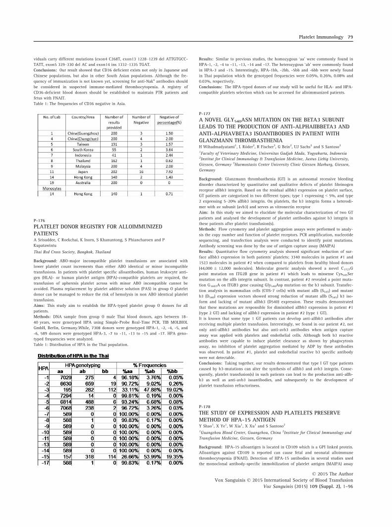
viduals carry different mutations (exon4 C268T, exon13 1228–1239 del ATTGTGCC-
TATT, exon5 329–330 del AC and exon14 ins 1332–1335 TGAT.
Conclusions: Our result showed that CD36 deficient exists not only in Japanese and
Chinese populations, but also in other South Asian populations. Although the fre-
quency of immunization is not known yet, screening for anti-Naka antibodies should
be considered in suspected immune-mediated thrombocytopenia. A registry of
CD36-deficient blood donors should be established to maintain PTR patients and
fetus with FNAIT.
Table 1: The frequencies of CD36 negative in Asia.
P-176
PLATELET DONOR REGISTRY FOR ALLOIMMUNIZEDPATIENTSA Srisuddee, C Roekchai, K Inorn, S Khanuntong, S Phiancharoen and P
Kupatawintu
Thai Red Cross Society, Bangkok, Thailand
Background: ABO-major incompatible platelet transfusions are associated with
lower platelet count increments than either ABO identical or minor incompatible
transfusions. In patients with platelet specific alloantibodies, human leukocyte anti-
gen (HLA)- or human platelet antigen (HPA)-compatible platelets are required, the
transfusion of apheresis platelet across with minor ABO incompatible cannot be
avoided. Plasma replacement by platelet additive solution (PAS) in group O platelet
donor can be managed to reduce the risk of hemolysis in non ABO identical platelet
transfusion.
Aims: This study aim to establish the HPA-typed platelet group O donors for all
patients.
Methods: DNA sample from group O male Thai blood donors, ages between 18–40 years, were genotyped HPA using Simple-Probe Real-Time PCR, TIB MOLBIOL
GmbH, Berlin, Germany.While, 7308 donors were genotyped HPA-1, -2, -4, -5, and
-6, 589 donors were genotyped HPA-3, -7 to -11, -13 to -15 and -17. HPA geno-
typed frequencies were analyzed.
Table 1: Distribution of HPA in the Thai population.
Results: Similar to previous studies, the homozygous ‘aa’ were commonly found in
HPA-1, -2, -4 to -11, -13, -14 and -17. The heterozygous ‘ab’ were commonly found
in HPA-3 and -15. Interestingly, HPA-1bb, -2bb, -5bb and -6bb were newly found
in Thai population which the genotyped frequencies were 0.05%, 0.26%, 0.08% and
0.03%, respectively.
Conclusions: The HPA-typed donors of our study will be useful for HLA- and HPA-
compatible platelets selection which can be accessed for alloimmunized patients.
P-177
A NOVEL GLY540ASN MUTATION ON THE BETA3 SUBUNITLEADS TO THE PRODUCTION OF ANTI-ALPHAIIBBETA3 ANDANTI-ALPHAVBETA3 ISOANTIBODIES IN PATIENT WITHGLANZMANN THROMBASTHENIAH Wihadmadyatami1, L R€oder2, R Fischer3, G Bein2, UJ Sachs2 and S Santoso2
1Faculty of Veterinary Medicine, Universitas Gadjah Mada, Yogyakarta, Indonesia2Institut for Clinical Immunology & Transfusion Medicine, Justus Liebig University,
Giessen, Germany 3Haemostasis Center University Clinic Giessen Marburg, Giessen,
Germany
Background: Glanzmann thrombasthenia (GT) is an autosomal recessive bleeding
disorder characterized by quantitative and qualitative defects of platelet fibrinogen
receptor aIIbb3 integrin. Based on the residual aIIbb3 expression on platelet surface,
GT patients are categorized in two different types; type 1 expressing < 5%, and type
2 expressing 5–20% aIIbb3 integrin. On platelets, the b3 integrin forms a heterodi-
mer with av subunit (avb3) and serves as vitronectin receptor
Aim: In this study we aimed to elucidate the molecular characterization of two GT
patients and analysed the development of platelet antibodies against b3 integrin in
these patients after platelet transfusion(s).
Methods: Flow cytometry and platelet aggregation assays were performed to analy-
sis the copy number and function of platelet receptors. PCR amplification, nucleotide
sequencing, and transfection analysis were conducted to identify point mutations.
Antibody screening was done by the use of antigen capture assay (MAIPA)
Results: Quantitative flow cytometry analysis showed significant reduction of sur-
face aIIbb3 expression in both patients’ platelets; 3340 molecules in patient #1 and
1523 molecules in patient #2 when compared to platelets from healthy blood donors
(44,000 � 12,000 molecules). Molecular genetic analysis showed a novel C1727G
point mutation on ITG2B gene in patient #1 which leads to missense Cys545Ser
mutation on the aIIb integrin subunit. In contrast, patient #2 revealed a point muta-
tion G1697A on ITGB3 gene causing Gly540Asp mutation on the b3 subunit. Transfec-
tion analysis in mammalian cells (COS-7 cells) with mutant aIIb (S545) and mutant
b3 (D540) expression vectors showed strong reduction of mutant aIIb (S545) b3 iso-
form and lacking of mutant aIIbb3 (D540) expression. These results demonstrated
that these mutations are responsible for diminished aIIbb3 expression in patient #1
(type 2 GT) and lacking of aIIbb3 expression in patient #2 (type 1 GT).
It is known that some type 1 GT patients can develop anti-aIIbb3 antibodies after
receiving multiple platelet transfusion. Interestingly, we found in our patient #2, not
only anti-aIIbb3 antibodies but also anti-avb3 antibodies when antigen capture
assay was applied with platelets and endothelial cells. Although both b3 reactive
antibodies were capable to induce platelet clearance as shown by phagocytosis
assay, no inhibition of platelet aggregation mediated by ADP by these antibodies
was observed. In patient #1, platelet and endothelial reactive b3 specific antibody
were not detectable.
Conclusions: Taking together, our results demonstrated that type I GT type patients
caused by b3-mutations can alter the synthesis of aIIbb3 and avb3 integrin. Conse-
quently, platelet transfusion(s) in such patients can lead to the production anti-aIIb-
b3 as well as anti-avb3 isoantibodies, and subsequently to the development of
platelet transfusion refractoriness.
P-178
THE STUDY OF EXPRESSION AND PLATELETS PRESERVEMETHOD OF HPA-15 ANTIGENY Shao1, X Ye1, W Xia1, X Xu1 and S Santoso2
1Guangzhou Blood Center, Guangzhou, China 2Institute for Clinical Immunology and
Transfusion Medicine, Giessen, Germany
Background: HPA-15 alloantigen is located in CD109 which is a GPI linked protein.
Alloantigen against CD109 is reported can cause fetal and neonatal alloimmune
thrombocytopenia (FNAIT). Detection of HPA-15 antibodies in several studies used
the monoclonal antibody-specific immobilization of platelet antigen (MAIPA) assay
© 2015 The AuthorVox Sanguinis © 2015 International Society of Blood Transfusion
Vox Sanguinis (2015) 109 (Suppl. 2), 1–96
Platelet Immunology 79
which related to the level of HPA-15 expression. But detection of HPA-15 antibodies
is difficult because of the low and instability of CD109 expression. Analysis of the
HPA-15 expression and study of the platelets preserve methods may contribute to
the detection of HPA-15 antibodies.
Aims: Determination and analysis the HPA-15 antigen expression on platelet. Ana-
lyze the HPA-15 genotypes frequencies in South China donors. Find a better plate-
lets preserving method which can sustain the CD109 expression.
Methods: A total of 109 healthy blood donors were collected in Guangzhou Blood
Center, China. The HPA-15 expression was determined on the platelets by flow
cytometry. The HPA-15 genotypes were also determined by PCR technology.
Another 76 healthy blood donors were divided into two groups. Platelets in Group 1
were suspended in PBS-BSA and stored in 4�C. Platelets in Group 2 were stored in
an orbital shaker in 22�C. HPA-15 expression of the two groups was determined by
flow cytometry on days 0, 1, 2, 3, 7, 14.
Results: In the 109 donors, the allele frequencies of HPA-15a and HPA-15b are
0.495 and 0.505. The MFI of HPA-15 expression ranged from 2.44 to 20.4. In the
two compared groups, the linear mixed model was used to analyse the data. Accord-
ing to different determination time point, the MFI of the two groups have statistical
difference (P = 0.001) during the storage. Although the HPA-15 MFI of the two
groups both decline during this period, the HPA-15 expression of group 2 is better
than group 1.
Conclusions: The expression of CD109 on platelet is low and varies among different
individuals. The CD109 expression decreases during storage, especially in 4�C.
P-179
THE INFLUENCE OF LEUKODEPLETION OF PLATELETCONCENTRATES ON CD62P EXPRESSION AND CORRECTEDCOUNT INCREMENTT Triyono1, Z Rofinda1 and U Sukorini2
1Medical Faculty of Andalas University, Yogyakarta, Indonesia 2Medical Faculty of
Gadjah Mada University, Yogyakarta, Indonesia
Background: Platelet concentrate (PC) is a blood component which is needed to be
transfused to patient with thrombocytopenia including the patient with hematology
malignancies. Platelet concentrates contain contaminant leukocytes that can lead
side effects and platelet activation that can reduce the efficacy of platelet transfu-
sion. For minimazing the contaminant leukocytes, leukodepletion by filtration might
be performed. Platelet activation can be assessed by CD62P expression, whereas the
efficacy of platelet transfusion can be assessed by corrected count increment (CCI).
Leukodepletion was not yet applied universally in Indonesia.
Aims: To analyze the CD62P expression between non-leukodepleted and leukode-
pleted PC groups, and also to analyze the CCI between group of patients which were
transfused with non-leukodepleted and leukodepleted PCs.
Method: The study was performed into two groups of patients with hematology
malignancies in Estella Children Ward of Dr.Sardjito Hospital, Yogyakarta Indonesia
i.e. 24 patients were transfused with non-leukodepleted and 24 patients were trans-
fused with leukodepleted PRP-derived PCs. The platelet expression of CD62P in PC
products was measured by flowcytometri, and the 1 h post-transfusion corrected
count increment was counted by CCI formula. The differences of CD62P expression
and CCI between the two groups were analyzed by using Mann-Whitney Test with
statistical significance of P < 0.05.
Results: From PC products, it was found that the CD62P expression in non-leukode-
pleted groups were higher with median of 34.4% (16.8–94.4%) compared to the
leukodepleted groups with median of 21.7% (6.2–34.0%).There was a statistical sig-
nificantly differences of the CD62P expression betweentwo groups (P = 0.00). It was
found that in the group of patients which were transfused with non-leukodepleted
PCs, the median of CCI was 18.8 9 109/l (2.4–94.8 9 109/l). The median of CCI in
the group of patients which was transfused with leukodepleted PCs was 14.7 9 109/l
(2.4–124 9 109/l). There was no statistically significant differences of CCI between
the two groups (P = 0.42).
Conclusion: The platelet CD62P expression in leukodepleted were statistically sig-
nificant lower than in non-leukodepleted PCs. But there was no statistically signifi-
cant CCI differences between the group of patients which were transfused with non-
leukodepleted and with leukodepleted PCs.
P-180
This abstract has been withdrawn.
P-181
RELEVANT ROLE OF TISSUE FACTOR IN VON WILLEBRANDFACTOR-DEPENDENT THROMBUS FORMATION UNDER FLOW:INVOLVEMENT OF INFLAMMATORY RESPONSES BYLEUKOCYTES IN I INTRA-THROMBUS FIBRIN GENERATIONY Matsunari, H Matsui, M Kawaguchi and M Sugimoto
Nara Medical University, Kashihara, Japan
Background: von Willebrand factor (VWF) plays a pivotal role in platelet thrombo-
genesis at sites of injured vessel walls under high shear flow conditions. Such plate-
let thrombus formation is concurrently reinforced by fibrin generation initiated by
tissue factor (TF). However, little is known about the functional link between VWF
and TF in flow-dependent thrombus formation.
Aims: To address this issue, we have studied the relevant role of TF on von Wille-
brand factor (VWF)-dependent thrombus formation by use of an in vitro perfusion
chamber system.
Methods: Recombinant human TF were co-coated with VWF onto a glass plate to
prepare ‘surface-immobilized VWF/TF complex’. Recalcified citrated whole blood
was perfused over a surface-immobilized VWF/TF complex. In some experiments,
neutrophil Cathepsin G (12 μg/ml) was added to sample blood. Platelet adhesion/ag-
gregation were evaluated by the surface coverage of thrombi. The mural thrombi
formed on VWF-surface was double-stained with fluorescently labeled anti-fibrin
and anti-fibrinogen antibodies. Fibrin generation was evaluated as a ratio of fibrin
relative to fibrinogen fluorescence within mural thrombi.
Results: Surface-immobilized TF significantly augmented flow-dependent fibrin
generation as a function of increasing surface density of TF under both low and
high shear rate conditions. In addition, immobilized TF significantly up-regulated
VWF-dependent platelet adhesion and aggregation under high shear rate conditions.
Cathepsin G significantly increased intra-thrombus fibrin generation on immobilized
VWF-TF complex under both high and low shear rate conditions.
Summary/Conclusions: Surface-immobilized TF and VWF could build mural
thrombi in a concerted manner under high shear rate conditions. The present experi-
mental approach revealed that Cathepsin G enhanced the TF-dependent fibrin gener-
ation, suggesting the critical involvement of inflammatory responses by leukocytes
on thrombus formation under whole blood flow conditions.
5.4 Granulocyte Immunology
P-182
GENERATION OF CYTOKINES IN RED BLOOD CELL PRODUCTSDURING STORAGE: A COMPARISON OF NON- AND LEUKO-REDUCED RED BLOOD CELL COMPONENTST Lee1, H Lin1, CC Chang1, LF Chang2 and FY Chu1
1FarEastern Memorial Hospital, New Taipei City, Taiwan 2Asia-Med Medical
Reference Lab, Taipei City, Taiwan
Background: Leukocyte reduction before storage effectively reduced the generation
of white blood cell (WBC)-derived cytokines. However, it was still unknown whether
there was accumulation of bioactive mediators including non-leukocyte-derived
cytokines, such as platelet-specific proinflammatory factors, in non-leukocyte-re-
duced RBC components during storage.
Aims: The aim of the present study was to investigate the cytokine accumulation,
including MIP-1a, RANTES, VEGF, TGF-b1, TGF-b2 and TGF-b3, in the pre-storage
leuko-reduced RBC (Pre-LPR) products and standard non-leuko-reduced RBC (S-RBC)
products during a 42-day storage.
Methods: Fifteen units of pre-LPR and S-RBC were included in the study and stored
at 4°C. Samples were acquired using aseptic technique from each unit on the 4th and
42th day of storage. Supernatant was collected by centrifugation at 10,000 9 g for
10 min and then stored at �70°C until analysis. The concentration of cytokines was
determined by the bead-based Luminex Bioplex system according to the manufac-
turer’s instructions. The accumulation of various cytokines for within-sample correla-
tions across designated time points of storage was assessed in Pre-LPR and S-RBC.
Results: The cytokine levels of RANTES, VEGF, TGF-b1, TGF-b2 and TGF-b3 were
markedly increased in S-RBC from the 4th day to 42th day, but not in Pre-LPR. The
post-storage cytokine concentrations were significantly increased in comparison with
pre-storage cytokine levels of RANTES (P < 0.001), VEGF (P < 0.001), TGF-b1(P < 0.001), TGF-b2 (P = 0.012) and TGF-b3 (P < 0.001) in S-RBC, but there was no
significant change during storage in Pre-LPR. MIP-1a decreased significantly during
storage in Pre-LPR (P < 0.001), but not in S-RBC (P = 0.76).
Conclusions: Our results indicated that leuko-reduction in RBC products before stor-
age effectively prevented the cytokine generation, including RANTES, VEGF, TGF-
© 2015 The AuthorVox Sanguinis © 2015 International Society of Blood TransfusionVox Sanguinis (2015) 109 (Suppl. 2), 1–96
80 Posters
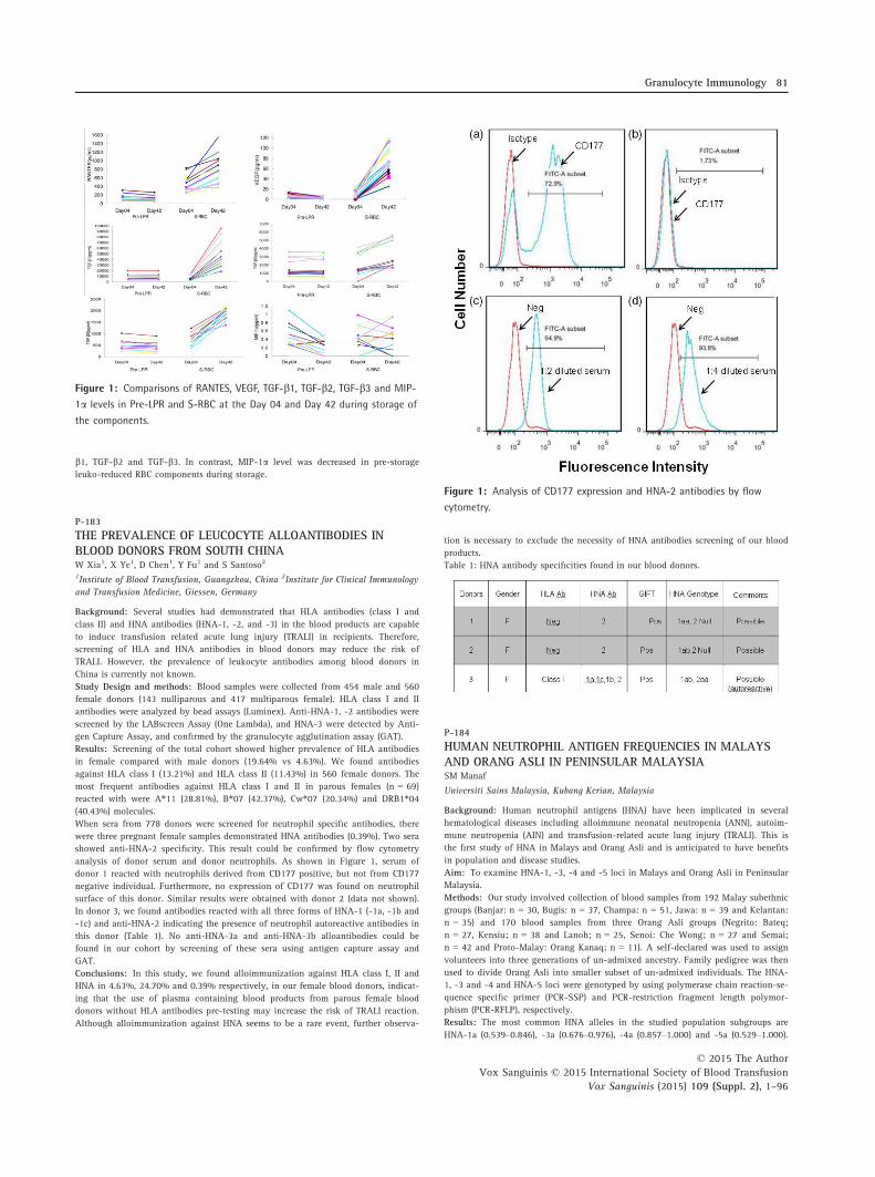
b1, TGF-b2 and TGF-b3. In contrast, MIP-1a level was decreased in pre-storage
leuko-reduced RBC components during storage.
P-183
THE PREVALENCE OF LEUCOCYTE ALLOANTIBODIES INBLOOD DONORS FROM SOUTH CHINAW Xia1, X Ye1, D Chen1, Y Fu1 and S Santoso2
1Institute of Blood Transfusion, Guangzhou, China 2Institute for Clinical Immunology
and Transfusion Medicine, Giessen, Germany
Background: Several studies had demonstrated that HLA antibodies (class I and
class II) and HNA antibodies (HNA-1, -2, and -3) in the blood products are capable
to induce transfusion related acute lung injury (TRALI) in recipients. Therefore,
screening of HLA and HNA antibodies in blood donors may reduce the risk of
TRALI. However, the prevalence of leukocyte antibodies among blood donors in
China is currently not known.
Study Design and methods: Blood samples were collected from 454 male and 560
female donors (143 nulliparous and 417 multiparous female). HLA class I and II
antibodies were analyzed by bead assays (Luminex). Anti-HNA-1, -2 antibodies were
screened by the LABscreen Assay (One Lambda), and HNA-3 were detected by Anti-
gen Capture Assay, and confirmed by the granulocyte agglutination assay (GAT).
Results: Screening of the total cohort showed higher prevalence of HLA antibodies
in female compared with male donors (19.64% vs 4.63%). We found antibodies
against HLA class I (13.21%) and HLA class II (11.43%) in 560 female donors. The
most frequent antibodies against HLA class I and II in parous females (n = 69)
reacted with were A*11 (28.81%), B*07 (42.37%), Cw*07 (20.34%) and DRB1*04(40.43%) molecules.
When sera from 778 donors were screened for neutrophil specific antibodies, there
were three pregnant female samples demonstrated HNA antibodies (0.39%). Two sera
showed anti-HNA-2 specificity. This result could be confirmed by flow cytometry
analysis of donor serum and donor neutrophils. As shown in Figure 1, serum of
donor 1 reacted with neutrophils derived from CD177 positive, but not from CD177
negative individual. Furthermore, no expression of CD177 was found on neutrophil
surface of this donor. Similar results were obtained with donor 2 (data not shown).
In donor 3, we found antibodies reacted with all three forms of HNA-1 (-1a, -1b and
-1c) and anti-HNA-2 indicating the presence of neutrophil autoreactive antibodies in
this donor (Table 1). No anti-HNA-3a and anti-HNA-3b alloantibodies could be
found in our cohort by screening of these sera using antigen capture assay and
GAT.
Conclusions: In this study, we found alloimmunization against HLA class I, II and
HNA in 4.63%, 24.70% and 0.39% respectively, in our female blood donors, indicat-
ing that the use of plasma containing blood products from parous female blood
donors without HLA antibodies pre-testing may increase the risk of TRALI reaction.
Although alloimmunization against HNA seems to be a rare event, further observa-
tion is necessary to exclude the necessity of HNA antibodies screening of our blood
products.
Table 1: HNA antibody specificities found in our blood donors.
P-184
HUMAN NEUTROPHIL ANTIGEN FREQUENCIES IN MALAYSAND ORANG ASLI IN PENINSULAR MALAYSIASM Manaf
Universiti Sains Malaysia, Kubang Kerian, Malaysia
Background: Human neutrophil antigens (HNA) have been implicated in several
hematological diseases including alloimmune neonatal neutropenia (ANN), autoim-
mune neutropenia (AIN) and transfusion-related acute lung injury (TRALI). This is
the first study of HNA in Malays and Orang Asli and is anticipated to have benefits
in population and disease studies.
Aim: To examine HNA-1, -3, -4 and -5 loci in Malays and Orang Asli in Peninsular
Malaysia.
Methods: Our study involved collection of blood samples from 192 Malay subethnic
groups (Banjar: n = 30, Bugis: n = 37, Champa: n = 51, Jawa: n = 39 and Kelantan:
n = 35) and 170 blood samples from three Orang Asli groups (Negrito: Bateq;
n = 27, Kensiu; n = 38 and Lanoh; n = 25, Senoi: Che Wong; n = 27 and Semai;
n = 42 and Proto-Malay: Orang Kanaq; n = 11). A self-declared was used to assign
volunteers into three generations of un-admixed ancestry. Family pedigree was then
used to divide Orang Asli into smaller subset of un-admixed individuals. The HNA-
1, -3 and -4 and HNA-5 loci were genotyped by using polymerase chain reaction-se-
quence specific primer (PCR-SSP) and PCR-restriction fragment length polymor-
phism (PCR-RFLP), respectively.
Results: The most common HNA alleles in the studied population subgroups are
HNA-1a (0.539–0.846), -3a (0.676–0.976), -4a (0.857–1.000) and -5a (0.529–1.000).
Figure 1: Analysis of CD177 expression and HNA-2 antibodies by flow
cytometry.
Figure 1: Comparisons of RANTES, VEGF, TGF-b1, TGF-b2, TGF-b3 and MIP-
1a levels in Pre-LPR and S-RBC at the Day 04 and Day 42 during storage of
the components.
© 2015 The AuthorVox Sanguinis © 2015 International Society of Blood Transfusion
Vox Sanguinis (2015) 109 (Suppl. 2), 1–96
Granulocyte Immunology 81
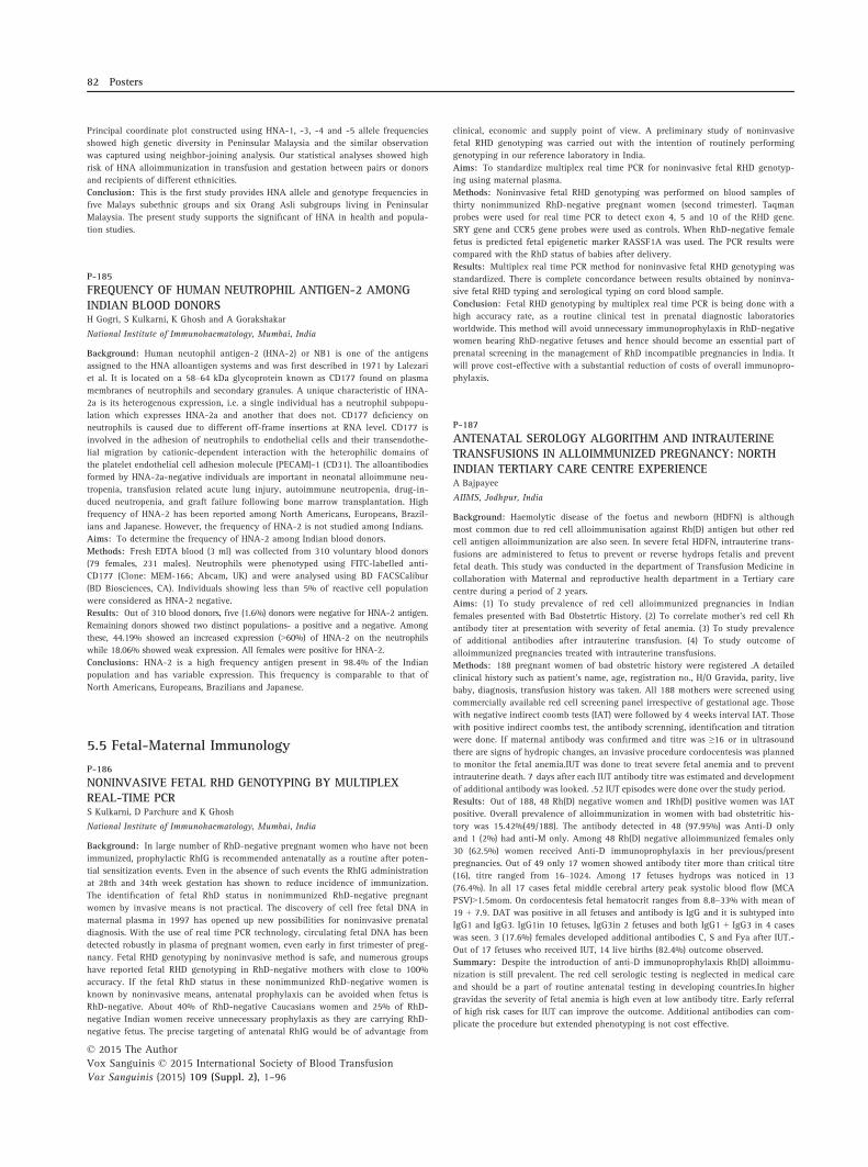
Principal coordinate plot constructed using HNA-1, -3, -4 and -5 allele frequencies
showed high genetic diversity in Peninsular Malaysia and the similar observation
was captured using neighbor-joining analysis. Our statistical analyses showed high
risk of HNA alloimmunization in transfusion and gestation between pairs or donors
and recipients of different ethnicities.
Conclusion: This is the first study provides HNA allele and genotype frequencies in
five Malays subethnic groups and six Orang Asli subgroups living in Peninsular
Malaysia. The present study supports the significant of HNA in health and popula-
tion studies.
P-185
FREQUENCY OF HUMAN NEUTROPHIL ANTIGEN-2 AMONGINDIAN BLOOD DONORSH Gogri, S Kulkarni, K Ghosh and A Gorakshakar
National Institute of Immunohaematology, Mumbai, India
Background: Human neutophil antigen-2 (HNA-2) or NB1 is one of the antigens
assigned to the HNA alloantigen systems and was first described in 1971 by Lalezari
et al. It is located on a 58–64 kDa glycoprotein known as CD177 found on plasma
membranes of neutrophils and secondary granules. A unique characteristic of HNA-
2a is its heterogenous expression, i.e. a single individual has a neutrophil subpopu-
lation which expresses HNA-2a and another that does not. CD177 deficiency on
neutrophils is caused due to different off-frame insertions at RNA level. CD177 is
involved in the adhesion of neutrophils to endothelial cells and their transendothe-
lial migration by cationic-dependent interaction with the heterophilic domains of
the platelet endothelial cell adhesion molecule (PECAM)-1 (CD31). The alloantibodies
formed by HNA-2a-negative individuals are important in neonatal alloimmune neu-
tropenia, transfusion related acute lung injury, autoimmune neutropenia, drug-in-
duced neutropenia, and graft failure following bone marrow transplantation. High
frequency of HNA-2 has been reported among North Americans, Europeans, Brazil-
ians and Japanese. However, the frequency of HNA-2 is not studied among Indians.
Aims: To determine the frequency of HNA-2 among Indian blood donors.
Methods: Fresh EDTA blood (3 ml) was collected from 310 voluntary blood donors
(79 females, 231 males). Neutrophils were phenotyped using FITC-labelled anti-
CD177 (Clone: MEM-166; Abcam, UK) and were analysed using BD FACSCalibur
(BD Biosciences, CA). Individuals showing less than 5% of reactive cell population
were considered as HNA-2 negative.
Results: Out of 310 blood donors, five (1.6%) donors were negative for HNA-2 antigen.
Remaining donors showed two distinct populations- a positive and a negative. Among
these, 44.19% showed an increased expression (>60%) of HNA-2 on the neutrophils
while 18.06% showed weak expression. All females were positive for HNA-2.
Conclusions: HNA-2 is a high frequency antigen present in 98.4% of the Indian
population and has variable expression. This frequency is comparable to that of
North Americans, Europeans, Brazilians and Japanese.
5.5 Fetal-Maternal Immunology
P-186
NONINVASIVE FETAL RHD GENOTYPING BY MULTIPLEXREAL-TIME PCRS Kulkarni, D Parchure and K Ghosh
National Institute of Immunohaematology, Mumbai, India
Background: In large number of RhD-negative pregnant women who have not been
immunized, prophylactic RhIG is recommended antenatally as a routine after poten-
tial sensitization events. Even in the absence of such events the RhIG administration
at 28th and 34th week gestation has shown to reduce incidence of immunization.
The identification of fetal RhD status in nonimmunized RhD-negative pregnant
women by invasive means is not practical. The discovery of cell free fetal DNA in
maternal plasma in 1997 has opened up new possibilities for noninvasive prenatal
diagnosis. With the use of real time PCR technology, circulating fetal DNA has been
detected robustly in plasma of pregnant women, even early in first trimester of preg-
nancy. Fetal RHD genotyping by noninvasive method is safe, and numerous groups
have reported fetal RHD genotyping in RhD-negative mothers with close to 100%
accuracy. If the fetal RhD status in these nonimmunized RhD-negative women is
known by noninvasive means, antenatal prophylaxis can be avoided when fetus is
RhD-negative. About 40% of RhD-negative Caucasians women and 25% of RhD-
negative Indian women receive unnecessary prophylaxis as they are carrying RhD-
negative fetus. The precise targeting of antenatal RhIG would be of advantage from
clinical, economic and supply point of view. A preliminary study of noninvasive
fetal RHD genotyping was carried out with the intention of routinely performing
genotyping in our reference laboratory in India.
Aims: To standardize multiplex real time PCR for noninvasive fetal RHD genotyp-
ing using maternal plasma.
Methods: Noninvasive fetal RHD genotyping was performed on blood samples of
thirty nonimmunized RhD-negative pregnant women (second trimester). Taqman
probes were used for real time PCR to detect exon 4, 5 and 10 of the RHD gene.
SRY gene and CCR5 gene probes were used as controls. When RhD-negative female
fetus is predicted fetal epigenetic marker RASSF1A was used. The PCR results were
compared with the RhD status of babies after delivery.
Results: Multiplex real time PCR method for noninvasive fetal RHD genotyping was
standardized. There is complete concordance between results obtained by noninva-
sive fetal RHD typing and serological typing on cord blood sample.
Conclusion: Fetal RHD genotyping by multiplex real time PCR is being done with a
high accuracy rate, as a routine clinical test in prenatal diagnostic laboratories
worldwide. This method will avoid unnecessary immunoprophylaxis in RhD-negative
women bearing RhD-negative fetuses and hence should become an essential part of
prenatal screening in the management of RhD incompatible pregnancies in India. It
will prove cost-effective with a substantial reduction of costs of overall immunopro-
phylaxis.
P-187
ANTENATAL SEROLOGY ALGORITHM AND INTRAUTERINETRANSFUSIONS IN ALLOIMMUNIZED PREGNANCY: NORTHINDIAN TERTIARY CARE CENTRE EXPERIENCEA Bajpayee
AIIMS, Jodhpur, India
Background: Haemolytic disease of the foetus and newborn (HDFN) is although
most common due to red cell alloimmunisation against Rh(D) antigen but other red
cell antigen alloimmunization are also seen. In severe fetal HDFN, intrauterine trans-
fusions are administered to fetus to prevent or reverse hydrops fetalis and prevent
fetal death. This study was conducted in the department of Transfusion Medicine in
collaboration with Maternal and reproductive health department in a Tertiary care
centre during a period of 2 years.
Aims: (1) To study prevalence of red cell alloimmunized pregnancies in Indian
females presented with Bad Obstetrtic History. (2) To correlate mother’s red cell Rh
antibody titer at presentation with severity of fetal anemia. (3) To study prevalence
of additional antibodies after intrauterine transfusion. (4) To study outcome of
alloimmunized pregnancies treated with intrauterine transfusions.
Methods: 188 pregnant women of bad obstetric history were registered .A detailed
clinical history such as patient’s name, age, registration no., H/O Gravida, parity, live
baby, diagnosis, transfusion history was taken. All 188 mothers were screened using
commercially available red cell screening panel irrespective of gestational age. Those
with negative indirect coomb tests (IAT) were followed by 4 weeks interval IAT. Those
with positive indirect coombs test, the antibody screnning, identification and titration
were done. If maternal antibody was confirmed and titre was ≥16 or in ultrasound
there are signs of hydropic changes, an invasive procedure cordocentesis was planned
to monitor the fetal anemia.IUT was done to treat severe fetal anemia and to prevent
intrauterine death. 7 days after each IUT antibody titre was estimated and development
of additional antibody was looked. .52 IUT episodes were done over the study period.
Results: Out of 188, 48 Rh(D) negative women and 1Rh(D) positive women was IAT
positive. Overall prevalence of alloimmunization in women with bad obstetritic his-
tory was 15.42%(49/188). The antibody detected in 48 (97.95%) was Anti-D only
and 1 (2%) had anti-M only. Among 48 Rh(D) negative alloimmunized females only
30 (62.5%) women received Anti-D immunoprophylaxis in her previous/present
pregnancies. Out of 49 only 17 women showed antibody titer more than critical titre
(16), titre ranged from 16–1024. Among 17 fetuses hydrops was noticed in 13
(76.4%). In all 17 cases fetal middle cerebral artery peak systolic blood flow (MCA
PSV)>1.5mom. On cordocentesis fetal hematocrit ranges from 8.8–33% with mean of
19 + 7.9. DAT was positive in all fetuses and antibody is IgG and it is subtyped into
IgG1 and IgG3. IgG1in 10 fetuses, IgG3in 2 fetuses and both IgG1 + IgG3 in 4 cases
was seen. 3 (17.6%) females developed additional antibodies C, S and Fya after IUT.-
Out of 17 fetuses who received IUT, 14 live births (82.4%) outcome observed.
Summary: Despite the introduction of anti-D immunoprophylaxis Rh(D) alloimmu-
nization is still prevalent. The red cell serologic testing is neglected in medical care
and should be a part of routine antenatal testing in developing countries.In higher
gravidas the severity of fetal anemia is high even at low antibody titre. Early referral
of high risk cases for IUT can improve the outcome. Additional antibodies can com-
plicate the procedure but extended phenotyping is not cost effective.
© 2015 The AuthorVox Sanguinis © 2015 International Society of Blood TransfusionVox Sanguinis (2015) 109 (Suppl. 2), 1–96
82 Posters
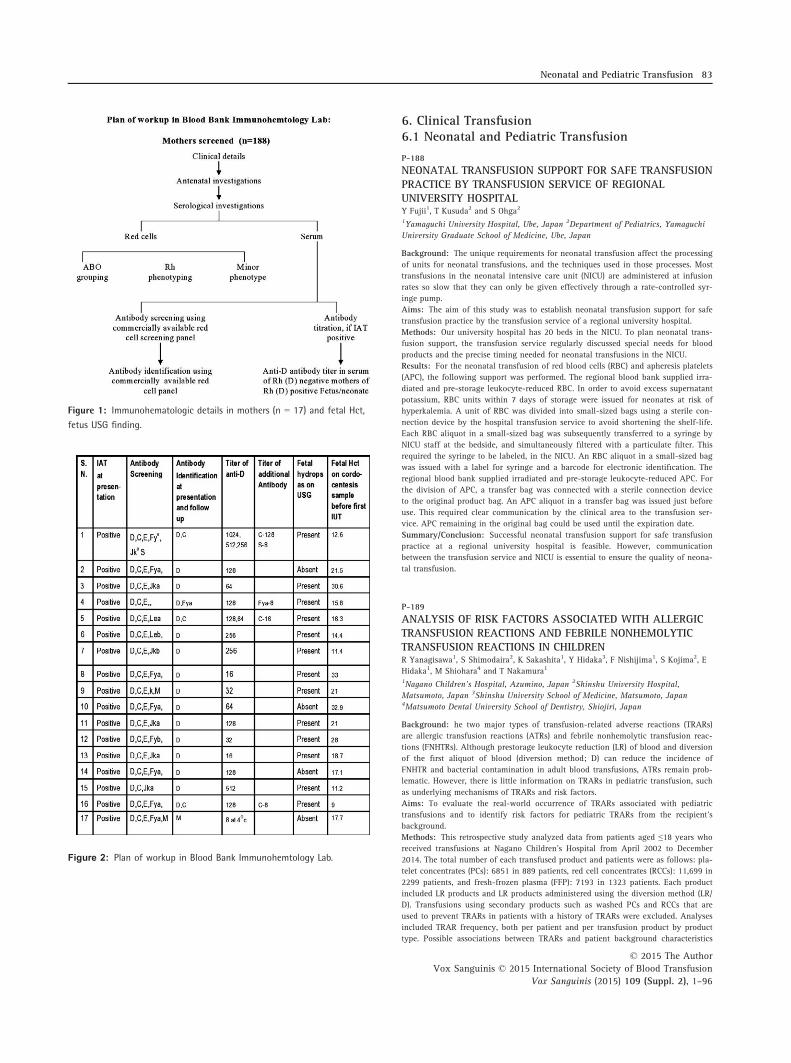
6. Clinical Transfusion6.1 Neonatal and Pediatric Transfusion
P-188
NEONATAL TRANSFUSION SUPPORT FOR SAFE TRANSFUSIONPRACTICE BY TRANSFUSION SERVICE OF REGIONALUNIVERSITY HOSPITALY Fujii1, T Kusuda2 and S Ohga2
1Yamaguchi University Hospital, Ube, Japan 2Department of Pediatrics, Yamaguchi
University Graduate School of Medicine, Ube, Japan
Background: The unique requirements for neonatal transfusion affect the processing
of units for neonatal transfusions, and the techniques used in those processes. Most
transfusions in the neonatal intensive care unit (NICU) are administered at infusion
rates so slow that they can only be given effectively through a rate-controlled syr-
inge pump.
Aims: The aim of this study was to establish neonatal transfusion support for safe
transfusion practice by the transfusion service of a regional university hospital.
Methods: Our university hospital has 20 beds in the NICU. To plan neonatal trans-
fusion support, the transfusion service regularly discussed special needs for blood
products and the precise timing needed for neonatal transfusions in the NICU.
Results: For the neonatal transfusion of red blood cells (RBC) and apheresis platelets
(APC), the following support was performed. The regional blood bank supplied irra-
diated and pre-storage leukocyte-reduced RBC. In order to avoid excess supernatant
potassium, RBC units within 7 days of storage were issued for neonates at risk of
hyperkalemia. A unit of RBC was divided into small-sized bags using a sterile con-
nection device by the hospital transfusion service to avoid shortening the shelf-life.
Each RBC aliquot in a small-sized bag was subsequently transferred to a syringe by
NICU staff at the bedside, and simultaneously filtered with a particulate filter. This
required the syringe to be labeled, in the NICU. An RBC aliquot in a small-sized bag
was issued with a label for syringe and a barcode for electronic identification. The
regional blood bank supplied irradiated and pre-storage leukocyte-reduced APC. For
the division of APC, a transfer bag was connected with a sterile connection device
to the original product bag. An APC aliquot in a transfer bag was issued just before
use. This required clear communication by the clinical area to the transfusion ser-
vice. APC remaining in the original bag could be used until the expiration date.
Summary/Conclusion: Successful neonatal transfusion support for safe transfusion
practice at a regional university hospital is feasible. However, communication
between the transfusion service and NICU is essential to ensure the quality of neona-
tal transfusion.
P-189
ANALYSIS OF RISK FACTORS ASSOCIATED WITH ALLERGICTRANSFUSION REACTIONS AND FEBRILE NONHEMOLYTICTRANSFUSION REACTIONS IN CHILDRENR Yanagisawa1, S Shimodaira2, K Sakashita1, Y Hidaka3, F Nishijima1, S Kojima2, E
Hidaka1, M Shiohara4 and T Nakamura1
1Nagano Children’s Hospital, Azumino, Japan 2Shinshu University Hospital,
Matsumoto, Japan 3Shinshu University School of Medicine, Matsumoto, Japan4Matsumoto Dental University School of Dentistry, Shiojiri, Japan
Background: he two major types of transfusion-related adverse reactions (TRARs)
are allergic transfusion reactions (ATRs) and febrile nonhemolytic transfusion reac-
tions (FNHTRs). Although prestorage leukocyte reduction (LR) of blood and diversion
of the first aliquot of blood (diversion method; D) can reduce the incidence of
FNHTR and bacterial contamination in adult blood transfusions, ATRs remain prob-
lematic. However, there is little information on TRARs in pediatric transfusion, such
as underlying mechanisms of TRARs and risk factors.
Aims: To evaluate the real-world occurrence of TRARs associated with pediatric
transfusions and to identify risk factors for pediatric TRARs from the recipient’s
background.
Methods: This retrospective study analyzed data from patients aged ≤18 years who
received transfusions at Nagano Children’s Hospital from April 2002 to December
2014. The total number of each transfused product and patients were as follows: pla-
telet concentrates (PCs): 6851 in 889 patients, red cell concentrates (RCCs): 11,699 in
2299 patients, and fresh-frozen plasma (FFP): 7193 in 1323 patients. Each product
included LR products and LR products administered using the diversion method (LR/
D). Transfusions using secondary products such as washed PCs and RCCs that are
used to prevent TRARs in patients with a history of TRARs were excluded. Analyses
included TRAR frequency, both per patient and per transfusion product by product
type. Possible associations between TRARs and patient background characteristics
Figure 1: Immunohematologic details in mothers (n = 17) and fetal Hct,
fetus USG finding.
Figure 2: Plan of workup in Blood Bank Immunohemtology Lab.
© 2015 The AuthorVox Sanguinis © 2015 International Society of Blood Transfusion
Vox Sanguinis (2015) 109 (Suppl. 2), 1–96
Neonatal and Pediatric Transfusion 83
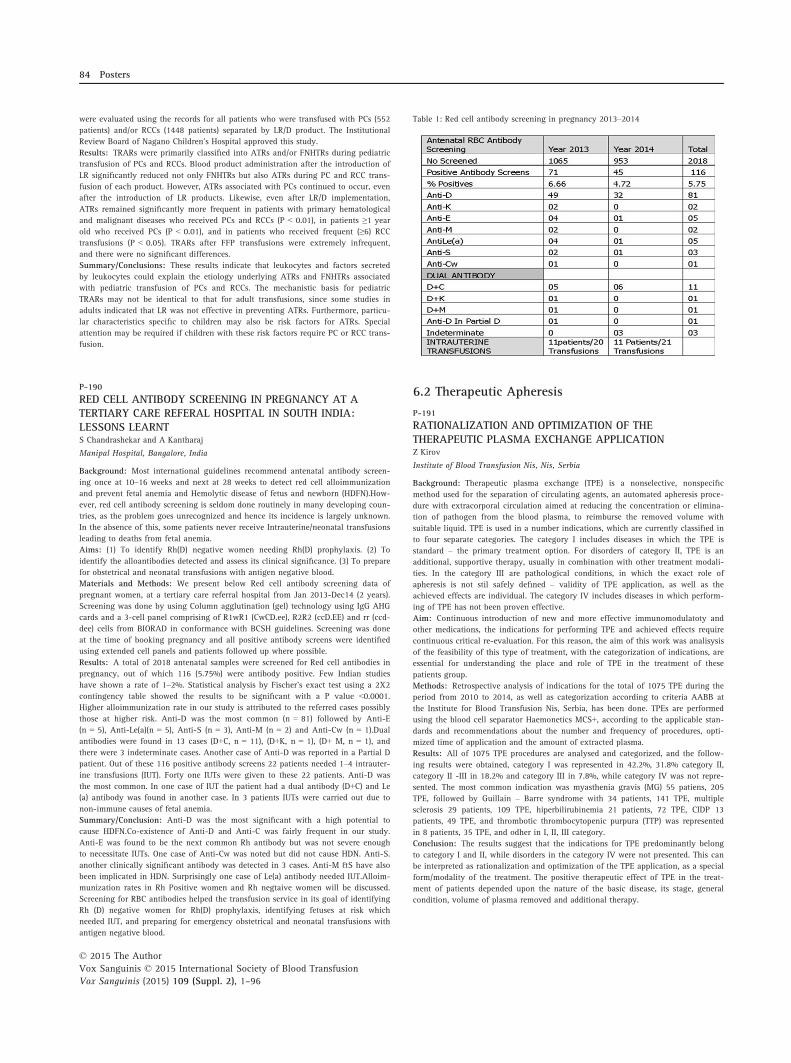
were evaluated using the records for all patients who were transfused with PCs (552
patients) and/or RCCs (1448 patients) separated by LR/D product. The Institutional
Review Board of Nagano Children’s Hospital approved this study.
Results: TRARs were primarily classified into ATRs and/or FNHTRs during pediatric
transfusion of PCs and RCCs. Blood product administration after the introduction of
LR significantly reduced not only FNHTRs but also ATRs during PC and RCC trans-
fusion of each product. However, ATRs associated with PCs continued to occur, even
after the introduction of LR products. Likewise, even after LR/D implementation,
ATRs remained significantly more frequent in patients with primary hematological
and malignant diseases who received PCs and RCCs (P < 0.01), in patients ≥1 year
old who received PCs (P < 0.01), and in patients who received frequent (≥6) RCC
transfusions (P < 0.05). TRARs after FFP transfusions were extremely infrequent,
and there were no significant differences.
Summary/Conclusions: These results indicate that leukocytes and factors secreted
by leukocytes could explain the etiology underlying ATRs and FNHTRs associated
with pediatric transfusion of PCs and RCCs. The mechanistic basis for pediatric
TRARs may not be identical to that for adult transfusions, since some studies in
adults indicated that LR was not effective in preventing ATRs. Furthermore, particu-
lar characteristics specific to children may also be risk factors for ATRs. Special
attention may be required if children with these risk factors require PC or RCC trans-
fusion.
P-190
RED CELL ANTIBODY SCREENING IN PREGNANCY AT ATERTIARY CARE REFERAL HOSPITAL IN SOUTH INDIA:LESSONS LEARNTS Chandrashekar and A Kantharaj
Manipal Hospital, Bangalore, India
Background: Most international guidelines recommend antenatal antibody screen-
ing once at 10–16 weeks and next at 28 weeks to detect red cell alloimmunization
and prevent fetal anemia and Hemolytic disease of fetus and newborn (HDFN).How-
ever, red cell antibody screening is seldom done routinely in many developing coun-
tries, as the problem goes unrecognized and hence its incidence is largely unknown.
In the absence of this, some patients never receive Intrauterine/neonatal transfusions
leading to deaths from fetal anemia.
Aims: (1) To identify Rh(D) negative women needing Rh(D) prophylaxis. (2) To
identify the alloantibodies detected and assess its clinical significance. (3) To prepare
for obstetrical and neonatal transfusions with antigen negative blood.
Materials and Methods: We present below Red cell antibody screening data of
pregnant women, at a tertiary care referral hospital from Jan 2013-Dec14 (2 years).
Screening was done by using Column agglutination (gel) technology using IgG AHG
cards and a 3-cell panel comprising of R1wR1 (CwCD.ee), R2R2 (ccD.EE) and rr (ccd-
dee) cells from BIORAD in conformance with BCSH guidelines. Screening was done
at the time of booking pregnancy and all positive antibody screens were identified
using extended cell panels and patients followed up where possible.
Results: A total of 2018 antenatal samples were screened for Red cell antibodies in
pregnancy, out of which 116 (5.75%) were antibody positive. Few Indian studies
have shown a rate of 1–2%. Statistical analysis by Fischer’s exact test using a 2X2
contingency table showed the results to be significant with a P value <0.0001.Higher alloimmunization rate in our study is attributed to the referred cases possibly
those at higher risk. Anti-D was the most common (n = 81) followed by Anti-E
(n = 5), Anti-Le(a)(n = 5), Anti-S (n = 3), Anti-M (n = 2) and Anti-Cw (n = 1).Dual
antibodies were found in 13 cases (D+C, n = 11), (D+K, n = 1), (D+ M, n = 1), and
there were 3 indeterminate cases. Another case of Anti-D was reported in a Partial D
patient. Out of these 116 positive antibody screens 22 patients needed 1–4 intrauter-
ine transfusions (IUT). Forty one IUTs were given to these 22 patients. Anti-D was
the most common. In one case of IUT the patient had a dual antibody (D+C) and Le
(a) antibody was found in another case. In 3 patients IUTs were carried out due to
non-immune causes of fetal anemia.
Summary/Conclusion: Anti-D was the most significant with a high potential to
cause HDFN.Co-existence of Anti-D and Anti-C was fairly frequent in our study.
Anti-E was found to be the next common Rh antibody but was not severe enough
to necessitate IUTs. One case of Anti-Cw was noted but did not cause HDN. Anti-S.
another clinically significant antibody was detected in 3 cases. Anti-M &S have also
been implicated in HDN. Surprisingly one case of Le(a) antibody needed IUT.Alloim-
munization rates in Rh Positive women and Rh negtaive women will be discussed.
Screening for RBC antibodies helped the transfusion service in its goal of identifying
Rh (D) negative women for Rh(D) prophylaxis, identifying fetuses at risk which
needed IUT, and preparing for emergency obstetrical and neonatal transfusions with
antigen negative blood.
Table 1: Red cell antibody screening in pregnancy 2013–2014
6.2 Therapeutic Apheresis
P-191
RATIONALIZATION AND OPTIMIZATION OF THETHERAPEUTIC PLASMA EXCHANGE APPLICATIONZ Kirov
Institute of Blood Transfusion Nis, Nis, Serbia
Background: Therapeutic plasma exchange (TPE) is a nonselective, nonspecific
method used for the separation of circulating agents, an automated apheresis proce-
dure with extracorporal circulation aimed at reducing the concentration or elimina-
tion of pathogen from the blood plasma, to reimburse the removed volume with
suitable liquid. TPE is used in a number indications, which are currently classified in
to four separate categories. The category I includes diseases in which the TPE is
standard – the primary treatment option. For disorders of category II, TPE is an
additional, supportive therapy, usually in combination with other treatment modali-
ties. In the category III are pathological conditions, in which the exact role of
apheresis is not stil safely defined – validity of TPE application, as well as the
achieved effects are individual. The category IV includes diseases in which perform-
ing of TPE has not been proven effective.
Aim: Continuous introduction of new and more effective immunomodulatoty and
other medications, the indications for performing TPE and achieved effects require
continuous critical re-evaluation. For this reason, the aim of this work was analisysis
of the feasibility of this type of treatment, with the categorization of indications, are
essential for understanding the place and role of TPE in the treatment of these
patients group.
Methods: Retrospective analysis of indications for the total of 1075 TPE during the
period from 2010 to 2014, as well as categorization according to criteria AABB at
the Institute for Blood Transfusion Nis, Serbia, has been done. TPEs are performed
using the blood cell separator Haemonetics MCS+, according to the applicable stan-
dards and recommendations about the number and frequency of procedures, opti-
mized time of application and the amount of extracted plasma.
Results: All of 1075 TPE procedures are analysed and categorized, and the follow-
ing results were obtained, category I was represented in 42.2%, 31.8% category II,
category II -III in 18.2% and category III in 7.8%, while category IV was not repre-
sented. The most common indication was myasthenia gravis (MG) 55 patiens, 205
TPE, followed by Guillain – Barre syndrome with 34 patients, 141 TPE, multiple
sclerosis 29 patients, 109 TPE, hiperbilirubinemia 21 patients, 72 TPE, CIDP 13
patients, 49 TPE, and thrombotic thrombocytopenic purpura (TTP) was represented
in 8 patients, 35 TPE, and odher in I, II, III category.
Conclusion: The results suggest that the indications for TPE predominantly belong
to category I and II, while disorders in the category IV were not presented. This can
be interpreted as rationalization and optimization of the TPE application, as a special
form/modality of the treatment. The positive therapeutic effect of TPE in the treat-
ment of patients depended upon the nature of the basic disease, its stage, general
condition, volume of plasma removed and additional therapy.
© 2015 The AuthorVox Sanguinis © 2015 International Society of Blood TransfusionVox Sanguinis (2015) 109 (Suppl. 2), 1–96
84 Posters
P-192
THERAPEUTIC PLASMA EXCHANGE: REVIEW OF THERESULTSR Sood, G Aroskar, S Kumar and V Kumar
Saket City Hospital, New Delhi, India
Background: Therapeutic plasma-exchange (TPE) is used as primary as well as
adjunctive therapy in treatment of several category of diseases (1,2). Technological
innovations and the adoption of evidence-based indications in the field of medicine
have led to the establishment of guidelines on the clinical indications for the use of
therapeutic apheresis (3).
Aim: Review of the outcome of the consecutive TPE procedures performed on vari-
ous categories of patients.
Methods: Retrospective analysis and evaluation of the TPE procedures performed at
our centre was done for a time duration of July 2013–2015 June.
Results: A total of 71 TPE procedures were performed in patients (2 males and 8fe-
males, with mean age of 57.2 � 5 years). Three of the 10 patients had Guillain-
Barr�e syndrome (GBS), 1 patient had anti glomerular basement membrane CKD dis-
ease (Good pasteurs Syndrome), 2 patients had Wegeners Granulomatosis (Systemic
Vasculitis cANCA positive), 2 had Myasthenia Gravis, 1 Chronic Inflammatory
Demyelinating Polyneuropathy (CIDP) and 1 patient was given TPE treatment for
SLE. Out of the total, 9 patients belonged to indication category 1 and one patient
belonged to indication category 111.Fresh frozen plasma (FFP) was used as replace-
ment fluid. Complete response (CR) was achieved on 8 patients (80%) in of the cases.
Complications were detected in none of the TPE procedures. Adverse events (AEs),
were seen in 5.6% of all procedures. None of the patients died from any complica-
tion or adverse event. AE occurred as three as Grade-I (mild -no treatment required
and procedure continued) and one patient as Grade-III (severe- treatment required,
procedure had to be finished). The most common AE were nausea/vomiting,
hypotension, pruritus and abdominal pain.
Conclusions: TPE is effectively and safely carried out for various diseases. (1) Med-
linePlus. ‘Goodpasture syndrome’. U.S. National Library of Medicine. Retrieved 7
April 2013. (2) Jump up ^ Yazdi, MF; Baghianimoghadam, M; Nazmiyeh, H; Ahmad-
abadi, AD; Adabi, MA (2012). ‘Response to plasmapheresis in myasthenia gravis
patients: 22 cases report’. Romanian journal of internal medicine = Revue roumaine
de medecine interne 50 (3): 245–7. PMID 23330293. (3) McLeod BC. Therapeutic
apheresis: history, clinical application, and lingering uncertainties. Transfusion.
2010;50:1413–26.
P-193
A CASE SERIES OF THREE PATIENTS UNDERGOINGTHERAPEUTIC LEUKAPHERESIS IN A TERTIARY CAREHOSPITALB Kakkar, RN Makroo, M Chowdhry, A Bhatia and UK Thakur
Indraprastha Apollo Hospitals, New Delhi, India
Introduction: Hyperleukocytosis is a condition characterised by increase in the total
leukocyte count (TLC) or blast count greater than 1,00,000/cumm. It is usually seen
in 5–18% of patients with AML depending on the leukemic subtype. Leukostasis is
characterised by abnormal intravascular leukocyte aggregation and clumping and is
associated with high early mortality in these patients. As leukostasis can compromise
the functioning of respiratory system and central nervous system. Leucoreduction is
achieved either by leucapheresis or chemotherapy. Therapeutic leukapheresis is usu-
ally initiated as soon as clinical signs of leukostasis are seen.
Case report: We present here case reports of 3 patients who underwent therapeutic
leukoapheresis at our centre. Leukapheresis was performed using Haemonetics MCS+(USA) cell separator using central venous access. ACD was used as anticoagulant in
a ratio of 1:12 with blood.
Case 1: A 25 year old male, admitted with history of sore throat and fever with
chills and rigors for 1 month, petechial haemorrhages for 20 days and unexplained
weight loss. On examination, patient had subconjunctival haemorrhage, petechiae,
hepatosplenomegaly and bilateral submandibular and cervical lympadenopathy.
After detailed laboratory workup and bone marrow biopsy along with flow cytom-
etry, he was diagnosed as biphenotypic acute leukemia (myeloid/Tcell). On day 4
of admission, patient experienced acute blast crisis (97%) and emergency therapeu-
tic leukapheresis was planned with pre procedure TLC of 3,21,200/ll. A TLC fall of
51.4% was seen post procedure to 1,65,200/ll. No adverse events or procedure
related complications were reported. Post procedure the patient improved consider-
ably. He was weaned of assisted ventilator support and his TLC counts stablilised.
Case 2: A 28 year old male, diagnosed case of chronic myeloid leukemia on treat-
ment since 2007. He came with relapsed disease with blast crisis (96%). His TLC was
1,02,100/ll) for which two procedures were performed on consecutive days. There
was considerable fall in total leucocyte count to 78,800/ ll (77% reduction) at the
end of two procedures. He experienced mild symptoms of citrate toxicity during the
first procedure, which was managed effectively with calcium infusion. Marked
improvement in symptoms was seen and patient went into complete remission.
Case 3: This was a 69 year old diabetic, hypertensive male recently diagnosed with
myeloproliferative syndrome with myelofibrosis; JAK-2 positive. His pre-procedure
TLC was 87,100/ll. There was a significant fall in TLC to 54,200/ll (62%) after one
procedure. There were no procedure related complications. Patient improced consid-
erably and was discharged. However, he was readmitted later with urological prob-
lems and later succumbed to his illness.
Conclusion: It is evident from the cases presented above that Therapeutic leukapa-
pheresis is a safe and effective approach for rapidly reducing WBC counts in patients
with blast crisis or any other haematological malignancies and can be a lifesaving
modality in patients at risk of leukostasis.
6.3 Evidence Based Transfusion Medicine Practice
P-194
OPTIMIZING RED CELL EXCHANGE PROCEDURES FOR SICKLECELL PATIENTSE Himchak, A Bonzon-Adelson, J Uehlinger, K Mohandas, R Walsh and D Wu
Montefiore Medical Center, Bronx, United States of America
In patients with sickle cell disease (SCD), transfusion therapies have been shown to
reduce the risk of recurrent strokes and prevent initial strokes in patients with
abnormal cerebral blood flow. Current guidelines recommend a monthly transfusion
protocol using manual or automated red cell exchange (RCE) or simple transfusion.
The goal of therapy in the pediatric population is to maintain the patient’s HbS
below 30% at the time of the next transfusion in at-risk patients. It is not clear in
any given patient how much to transfuse or exchange to reach these goals.
Immediate post transfusion HbS (post-HbS) can be calculated with relative certainty
with the number of red blood cell (RBC) units, pre-transfusion parameters like HbS,
hematocrit, and patient weight. Our goal is to find a post-HbS following which our
patients will remain below 30% HbS until their next transfusion.
We uploaded a cohort of 18 sickle cell patients receiving long-term RCE into our
institution’s patented clinical information system search tool called Clinical Looking
Glass. We extracted HbS results from these patients and categorized them into pre-
transfusion HbS (pre-HbS), post-HbS, and follow up pre-transfusion HbS (f/u HbS)
22–40 days later (i.e. the pre-HbS at the next visit). Each set of data was considered
a patient event and categorized by post-HbS of <10%, 10–15%, 15–20%, 20–25%,
25–30%, and 30 + %. Follow-up data was analyzed to identify if fHbS was above or
below the 30% level. Linear regression analysis was used to evaluate the correlation
between post-HbS and HbS.
A total of 218 sets of data were identified from 2000–2015. Linear regression analy-
sis revealed a weak correlation (R2 = 0.369) between post-HbS and fHbS. No particu-
lar level of post-HbS guarantees maintenance of HbS <30%. In the post-HbS <10%category (n = 8), 62.5% of patients had fHbS <30% while the 10–15% category
(n = 16) had 19% of patients with HbS <30%. Applying a Fischer’s Exact test sug-
gests a possible difference in these categories (P = 0.06) which may be statistically
significant with a larger cohort. Few patients with post-HbS >15% had acceptable
fHbS results (5 of 194 patient events). For each individual patient, monthly HbS val-
ues varied markedly over the course of years without a discernible pattern.
This project highlights the difficulty of maintaining low HbS in SCD patients espe-
cially over many years. The cutoff post-HbS level will likely lie around 10%, in
order to maintain most of the patients’ f/u HbS <30%. With several of our patients
developing alloantibodies to very high frequency or multiple antigens, we try to
maintain an appropriate fHbS while minimizing exposure to donor antigens. Further
studies may be done to correlate HbS to clinically significant events like strokes and
possible underlying clinical factors.
P-195
A CLINICAL AUDIT OF RED CELL TRANSFUSION IN ELECTIVETOTAL HIP REPLACEMENT SURGERYYG Peters1, GM Davison2, G Bellairs1 and A Bird1
1Western Province Blood Transfusion Service, Cape Town, South Africa 2Cape
Peninsula University of Technology, Cape Town, South Africa
Background: Previous international studies have documented a marked variation in
transfusion practice for Total Hip Replacement (THR) surgery. This is despite wide-
spread dissemination of clinical guidelines for the use of blood products. The cost
© 2015 The AuthorVox Sanguinis © 2015 International Society of Blood Transfusion
Vox Sanguinis (2015) 109 (Suppl. 2), 1–96
Evidence Based Transfusion Medicine Practice 85
and potential wastage of blood products as well as concerns regarding patient care
and outcomes are important drivers of optimal blood management.
Aim: The aim of this study was to audit red cell product utilisation for THR surg-
eries at two tertiary referral hospitals in the Western Cape, South Africa.
Methods: A retrospective clinical audit was conducted at two tertiary hospitals in
the Western Cape. The audit involved patient folder review and the review of com-
puter records of the respective blood banks. The folders of two hundred and seven
consecutive patients undergoing elective THR surgery from January 2013-December
2013 from each hospital were reviewed. Baseline data relating to age, gender, clini-
cal observations, indications for surgery, pre and post operative haemoglobin values,
comorbidities, length of hospital stay and transfusion history was recorded.
Data was then uploaded from individual data collection sheets onto IBM SPSS ver-
sion 22 statistical package for analysis.
Results: The two institutions were coded Hospital Green (HG) and Hospital Yellow
(HY). Of the two hundred and seven patients who had THR surgery, eighty seven
were excluded from the analysis, leaving one hundred and twenty eligible for
review, which were equally split between the two hospitals. The transfusion rate at
HY (41.6%) was significantly higher than that at HG (10%). The mean post-operative
haemoglobin (Hb) in the transfused patients at HG was 8.3 g/dl, compared to HY
with a mean of 9.1 g/dl. Females had a significantly higher transfusion rate (33%)
than males (15%) (P < 0.05) and the mean age of transfused patients was signifi-
cantly greater than untransfused patients (P < 0.005). Although patients with co-
morbidities had a higher transfusion rate than those without, this did not reach sta-
tistical significance. One hundred and thirteen (94%) patients had a blood order from
the blood bank, the vast majority, one hundred and two (85%), were group and
screen requests of which twenty nine (24%) were converted to a full crossmatch.
Conclusions: The transfusion rates for the two hospitals for elective THR were sig-
nificantly different although both are well within the wide ranges documented in
the literature. Although there are no bench marks, several studies support a restric-
tive transfusion regimen strategy with a guideline ‘trigger’ Hb level of 7–8 g/dl. In
this study HG had a transfusion rate of 10% with a mean post operative Hb of
8.3 g/dl in the transfused population, compared to 9.1 g/dl at HY which had a trans-
fusion rate of 41.6%. This suggests that a benchmark of 20% would not be unrea-
sonable and together with rational blood ordering policies (group and screen rather
than a full crossmatch) would lead to significant savings with no extra risk to the
patient.
P-196
RE-EVALUATION OF MAXIMUM SURGICAL BLOOD ORDERSCHEDULE (MSBOS) IN A SECONDARY CARE HOSPITAL INKOREAMJ Kim and ACdr Oh
Seonam University College of Medicine, Goyang, South-Korea
Background: Effective usage of RBC product can be achieved by monitoring maxi-
mum surgical blood order schedule (MSBOS). Our previous MSBOS was set for 220
type of surgery and its range was from type and screen (T&S) to 4 units. Re-evalua-
tion of MSBOS is essential for transfusion because surgical technique and need for
transfusion can be changed. We reset MSBOS by reviewing of surgical blood order
data from 2011 to 2014.
Aims: We planned to reset MSBOS by reviewing of surgical blood order data from
2011 to 2014 to use RBC product effectively and feedback to surgeons.
Methods: Number of each surgery and RBC product ordered from 2011 to 2014
were reviewed. Exclusion criteria were local anesthetic surgery, emergency surgery,
transfused within 24 h before surgery, surgery for more than 2 organs and less than
5 cases per 3 years. Mean C/T ratio, mean unit, median unit, minimum unit, maxi-
mum unit and SD of each surgery and each surgeon were calculated. MOBOS was
decided as a mean of blood unit for each surgery.
Results: Total number of included surgery was 2087 (total number: 3403, excluded
number: 1316) and kinds of the surgery were 51. The lesser kind of surgery and lar-
ger amount of RBC units than previous one was included in MSBOS (from T&S to
15 units). T&S for 10 types (19.6%), 1 unit for 10 types (19.6%), 2 units for 12 types
(23.5%), 3 units for 12 types (23.5%), 4 units for 2 types (3.9%), 5 units for 1 type
(2.0%), 6 units for 1 type (2.0%), 7 units for 1 type (2.0%), 8 units for 1 type (2.0%)
and 15 units for 1 type (2.0%) of surgery were calculated. Mean C/T ratio, mean
unit, median unit, minimum unit, maximum unit and SD of each surgeon were 1.9,
1.6, 1, 0, 37, and 2.8, respectively.
Conclusions: Our current MSBOS was higher than previous one and other articles
reported. And amount of RBC units of each surgeon were highly variable. Regular
re-evaluation of MSBOS is mandatory to reflect the progression of surgical technol-
ogy. We will give surgeons information of MSBOS for each surgery and suggest the
adequate number of blood component units.
P-197
This abstract has been withdrawn.
P-198
EFFICACY OF PLATELET CONCENTRATES PREPARED BYRANDOM DONOR BUFFY COAT AND SINGLE DONORAPHERESIS IN ACUTE LEUKEMIA AND CHEMOTHERAPYINDUCED PANCYTOPENIAAK Jindal1, A Kaur1, R Kumar1 and JS Sekhon2
1Dayanand Medical College and Hospital, Ludhiana, India 2Fortis Hospital,
Ludhiana, India
Background: Since discovery of platelets, there has been continuous and acceler-
ated progress in understanding of platelet functions and their utilization. In Oncol-
ogy, platelet transfusions are indicated for prophylactically and/or therapeutically.
Despite our improving knowledge about the beneficial effect of platelet concentrates,
there has been limited data that can predict their therapeutic efficacy quantitatively.
The need for this study arose to fill this void, to assess the therapeutic efficacy of
leuko-reduced platelet concentrates made by buffy coats and single donor apheresis
in thrombocytopenic patients of Acute Leukemia and Chemotherapy Induced Pancy-
topenia
Aims: To study the post transfusion therapeutic efficacy of Buffy Coat Random
Donor Platelet Concentrates (RDP) and Single Donor Apheresis Platelet concentrates
(SDP) in patients of Acute Leukemia and Chemotherapy Induced Pancytopenia.
Methods: Thrombocytopenic patients from oncology ward were included as per the
inclusion criteria. Platelet count of the platelet concentrates to be transfused was
done prior to transfusion. Post transfusion efficacy of platelets in the patients
(in vivo efficacy) was assessed by Corrected Count Increment (CCI) [units: platelets
m2/ll 9 1011, hereafter implied for the sake of clarity] and Percentage Recovery
(PR) at 1 h and 24 h. If the CCI at 1 h and 24 h is < 7500 platelets m2/ll and <4500 platelets m2/ll and PPR at 1 h and 24 h < 30% and <20% respectively, it indi-
cates platelet refractoriness. Data was analyzed using Student t-test on SPSS Version
21.0.
Results: Out of 61 patients, who had comparable pre-transfusion platelet count; 48
received RDP and 13 received SDP. In patients receiving RDP, the mean platelet
increment at 1 h and 24 h was 22784.8 � 1166.2 9 1011 platelets and
16835.4 � 8859.9 9 1011 platelets, respectively. The Mean CCI at 1 Hour and 24 h
was 10727.1 � 5495.3 and 7915.1 � 4201.1 respectively. The Mean PR at 1 h and
24 h was 24.0 � 12.2% and 17.7 � 9.3% respectively. In patients receiving SDP,
the mean platelet increment at 1 h and 24 h was 48619.0 � 17214.8 9 1011 plate-
lets and 41952.4 � 16758.5 9 1011 platelets respectively. The Mean CCI at 1 h and
24 h was 18104.5 � 6375.5 and 15602.6 � 6229.1 respectively. The Mean PR at
1 h and 24 h was 48.4 � 17.2% and 41.7 � 16.9% respectively. Platelet refractori-
ness was seen in 15 (24.5%) of cases out of which 13 (86.6%) were seen in patients
receiving RDP and 2 (13.3%) in patients receiving SDP. Statistically significant
(P = 0.001) higher platelet increment, CCI and PR was observed with SDP than RDP
and less refractoriness was seen with SDP transfusion.
Summary/Conclusions: Transfusing SDP is more efficient method to increase plate-
let count in oncologic thrombocytopenic patients and is associated with longer
in vivo survival of platelets.
P-199
BLOOD PRODUCT USAGE IN ADULT PATIENTS WITHMASSIVE BLEEDINGB Zhou, A Zhou and Y Wu
Bloodworks Northwest, Seattle, United States of America
Background: Patients with massive bleeding often require large number of blood
product transfusion. These requirements often put an extra burden on the hospital
transfusion service inventory as well as the local community blood resource. Under-
standing the usage pattern of blood products in these patients can help blood bank
and blood service personnel estimate the needs and prepare for these events, which
are often unexpected.
Aims: To identify usage pattern of blood products in patients with massive bleed-
ing.
Methods: Retrospective review of transfusion data from 08/01/2013 to 07/31/2014
was performed. Patients with massive bleeding were defined as patients who used 10
or more units of RBC over a 24 h period. Data included blood product usage of mul-
© 2015 The AuthorVox Sanguinis © 2015 International Society of Blood TransfusionVox Sanguinis (2015) 109 (Suppl. 2), 1–96
86 Posters
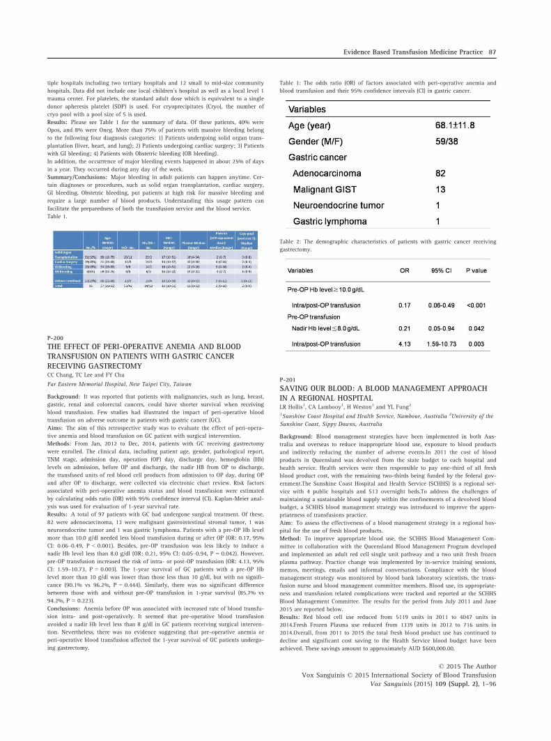
tiple hospitals including two tertiary hospitals and 12 small to mid-size community
hospitals. Data did not include one local children’s hospital as well as a local level 1
trauma center. For platelets, the standard adult dose which is equivalent to a single
donor apheresis platelet (SDP) is used. For cryoprecipitates (Cryo), the number of
cryo pool with a pool size of 5 is used.
Results: Please see Table 1 for the summary of data. Of these patients, 40% were
Opos, and 8% were Oneg. More than 75% of patients with massive bleeding belong
to the following four diagnosis categories: 1) Patients undergoing solid organ trans-
plantation (liver, heart, and lung); 2) Patients undergoing cardiac surgery; 3) Patients
with GI bleeding; 4) Patients with Obstetric bleeding (OB bleeding).
In addition, the occurrence of major bleeding events happened in about 25% of days
in a year. They occurred during any day of the week.
Summary/Conclusions: Major bleeding in adult patients can happen anytime. Cer-
tain diagnoses or procedures, such as solid organ transplantation, cardiac surgery,
GI bleeding, Obstetric bleeding, put patients at high risk for massive bleeding and
require a large number of blood products. Understanding this usage pattern can
facilitate the preparedness of both the transfusion service and the blood service.
Table 1.
P-200
THE EFFECT OF PERI-OPERATIVE ANEMIA AND BLOODTRANSFUSION ON PATIENTS WITH GASTRIC CANCERRECEIVING GASTRECTOMYCC Chang, TC Lee and FY Chu
Far Eastern Memorial Hospital, New Taipei City, Taiwan
Background: It was reported that patients with malignancies, such as lung, breast,
gastric, renal and colorectal cancers, could have shorter survival when receiving
blood transfusion. Few studies had illustrated the impact of peri-operative blood
transfusion on adverse outcome in patients with gastric cancer (GC).
Aims: The aim of this retrospective study was to evaluate the effect of peri-opera-
tive anemia and blood transfusion on GC patient with surgical intervention.
Methods: From Jan, 2012 to Dec, 2014, patients with GC receiving gastrectomy
were enrolled. The clinical data, including patient age, gender, pathological report,
TNM stage, admission day, operation (OP) day, discharge day, hemoglobin (Hb)
levels on admission, before OP and discharge, the nadir HB from OP to discharge,
the transfused units of red blood cell products from admission to OP day, during OP
and after OP to discharge, were collected via electronic chart review. Risk factors
associated with peri-operative anemia status and blood transfusion were estimated
by calculating odds ratio (OR) with 95% confidence interval (CI). Kaplan-Meier anal-
ysis was used for evaluation of 1-year survival rate.
Results: A total of 97 patients with GC had undergone surgical treatment. Of these,
82 were adenocarcinoma, 13 were malignant gastrointestinal stromal tumor, 1 was
neuroendocrine tumor and 1 was gastric lymphoma. Patients with a pre-OP Hb level
more than 10.0 g/dl needed less blood transfusion during or after OP (OR: 0.17, 95%
CI: 0.06–0.49, P < 0.001). Besides, pre-OP transfusion was less likely to induce a
nadir Hb level less than 8.0 g/dl (OR: 0.21, 95% CI: 0.05–0.94, P = 0.042). However,
pre-OP transfusion increased the risk of intra- or post-OP transfusion (OR: 4.13, 95%
CI: 1.59–10.73, P = 0.003). The 1-year survival of GC patients with a pre-OP Hb
level more than 10 g/dl was lower than those less than 10 g/dl, but with no signifi-
cance (90.1% vs 96.2%, P = 0.444). Similarly, there was no significant difference
between those with and without pre-OP transfusion in 1-year survival (85.7% vs
94.2%, P = 0.223).
Conclusions: Anemia before OP was associated with increased rate of blood transfu-
sion intra- and post-operatively. It seemed that pre-operative blood transfusion
avoided a nadir Hb level less than 8 g/dl in GC patients receiving surgical interven-
tion. Nevertheless, there was no evidence suggesting that pre-operative anemia or
peri-operative blood transfusion affected the 1-year survival of GC patients undergo-
ing gastrectomy.
Table 1: The odds ratio (OR) of factors associated with peri-operative anemia and
blood transfusion and their 95% confidence intervals (CI) in gastric cancer.
Table 2: The demographic characteristics of patients with gastric cancer receiving
gastrectomy.
P-201
SAVING OUR BLOOD: A BLOOD MANAGEMENT APPROACHIN A REGIONAL HOSPITALLR Hollis1, CA Lambooy1, H Weston1 and YL Fung2
1Sunshine Coast Hospital and Health Service, Nambour, Australia 2University of the
Sunshine Coast, Sippy Downs, Australia
Background: Blood management strategies have been implemented in both Aus-
tralia and overseas to reduce inappropriate blood use, exposure to blood products
and indirectly reducing the number of adverse events.In 2011 the cost of blood
products in Queensland was devolved from the state budget to each hospital and
health service. Health services were then responsible to pay one-third of all fresh
blood product cost, with the remaining two-thirds being funded by the federal gov-
ernment.The Sunshine Coast Hospital and Health Service (SCHHS) is a regional ser-
vice with 4 public hospitals and 513 overnight beds.To address the challenges of
maintaining a sustainable blood supply within the confinements of a devolved blood
budget, a SCHHS blood management strategy was introduced to improve the appro-
priateness of transfusions practice.
Aim: To assess the effectiveness of a blood management strategy in a regional hos-
pital for the use of fresh blood products.
Method: To improve appropriate blood use, the SCHHS Blood Management Com-
mittee in collaboration with the Queensland Blood Management Program developed
and implemented an adult red cell single unit pathway and a two unit fresh frozen
plasma pathway. Practice change was implemented by in-service training sessions,
memos, meetings, emails and informal conversations. Compliance with the blood
management strategy was monitored by blood bank laboratory scientists, the trans-
fusion nurse and blood management committee members. Blood use, its appropriate-
ness and transfusion related complications were tracked and reported at the SCHHS
Blood Management Committee. The results for the period from July 2011 and June
2015 are reported below.
Results: Red blood cell use reduced from 5119 units in 2011 to 4047 units in
2014.Fresh Frozen Plasma use reduced from 1339 units in 2012 to 716 units in
2014.Overall, from 2011 to 2015 the total fresh blood product use has continued to
decline and significant cost saving to the Health Service blood budget have been
achieved. These savings amount to approximately AUD $600,000.00.
© 2015 The AuthorVox Sanguinis © 2015 International Society of Blood Transfusion
Vox Sanguinis (2015) 109 (Suppl. 2), 1–96
Evidence Based Transfusion Medicine Practice 87
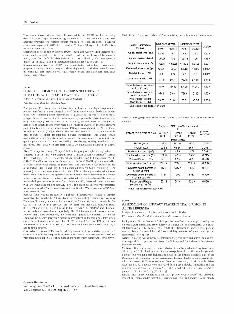
Transfusion related adverse events documented in the SCHHS incident reporting
database (PRIME CI) have reduced significantly in alignment with the blood man-
agement strategies and reduced patient exposure to blood products. 48 adverse
events were reported in 2013, 28 reported in 2014, and 21 reported in 2015, this is
an overall reduction of 56%.
Comparison of blood use by activity (WAU – Weighted Activity Unit) indicates that
even though hospital activity is increasing, blood use has decreased by approxi-
mately 30%. Current SCHHS data indicates the cost of blood by WAU was approxi-
mately $11 in 2012/13 and has reduced to approximately $7 in 2014/15.
Summary/Conclusion: The SCHHS data demonstrates that a blood management
program including simple strategies such as single unit transfusions, complimented
by promotion and education can significantly reduce blood use and transfusion
related complications.
P-202
CLINICAL EFFICACY OF ‘O’ GROUP SINGLE DONORPLATELETS WITH PLATELET ADDITIVE SOLUTIONP Jain, A Tendulkar, A Gupta, J Sanal and A Navkudkar
Tata Memorial Hospital, Mumbai, India
Background: This study was conducted in a tertiary care oncology setup wherein
platelet transfusions are an integral part of the supportive care. Guidelines recom-
mend ABO-identical platelet transfusions to patients as opposed to non-identical
groups. However, maintaining an inventory of group specific platelet concentrates
(PC) is challenging. Also as a prelude to this study, we performed the titres (anti A,
anti B) in ‘O’ group donors which were high (≥128) in 57.19% of donors. Hence, we
studied the feasibility of preparing group ‘O’ Single donor platelets (SDP) with Plate-
let additive solution (PAS) to obtain safer low titre units and to overcome the prob-
lems related to minor incompatible platelet transfusions. This would ensure
availability of group O units during emergency. The units produced were tested for
quality parameters with respect to viability, morphology, sterility, metabolism and
activation. These units were then transfused to the patients and analyzed for clinical
efficacy.
Aim: To study the clinical efficacy of PAS added group O single donor platelets.
Methods: SDP (n = 50) were prepared from O group donors on Amicus� (version
3.2, Fenwal Inc., USA) cell separator which provides 3 log leukodepletion. PAS III
(SSP+TM, MacoPharma, Moveaux, France) in a ratio 70:30 (PASIII: plasma) was added
at source under sterile conditions (study arm). The units after being studied on day
of collection (day 0) and day 4, and compared with 50 SDP’s containing 100%
plasma (control arm) were transfused to the adult inpatients presenting with throm-
bocytopenia. The study was approved by institutional ethics committee and written
informed consent from the patients was obtained prior to transfusion. The parame-
ters studied post transfusion were Count Increment (CI), corrected count increments
(CCI) and Percentage platelet recovery (PPR). The statistical analysis was performed
using one way ANOVA for parametric data and Kruskal-Wallis one way ANOVA for
non-parametric data.
Results: There was no statistically significant difference with respect to baseline
parameters like weight, height and body surface area of the patients in two arms.
The mean CI in study and control arm was 30,900/ll and 31,500/ll respectively. TheCCI at 1 h and at 24 h amongst the two arms was not significantly different
(P = 0.839, and P = 0.536), with mean CCI at 1 h being 11,970/ll/m2 and 13,525/ll/m2 for study and control arm respectively. The PPR for study and control arms was
33.74% and 36.6% respectively and were not significantly different (P = 0.885).
There was no adverse reaction reported in any patient in the two arms. Intra-group
comparison of study arm showed that CI, CCI at 1 and 24 h and PPR at 1 h were
not significantly different when group O SDP’s with PAS were transfused to A, B
and O group patients.
Conclusion: O group SDPs can be safely prepared with an additive solution and
show clinical efficacy comparable to units with 100% plasma. Patients are benefitted
with these units, especially during platelet shortages which require ABO switchovers.
Table 1: Inter-Group comparison of Clinical efficacy in study arm and control arm.
Table 2: Intra-group comparison of Study arm SDP’s issued to A, B and O group
patients.
P-203
ASSESSMENT OF EFFICACY PLATELET TRANSFUSION INACUTE LEUKEMIAI Frigaa, H Mahnaoui, B Rached, A Amireche and H Ouelaa
CHU Annaba /Faculty of Medecine of Annaba, Annaba, Algeria
Background: The evaluation of yield platelet transfusion is a way of testing the
various factors influencing the efficiency of transfusion.The effectiveness of a plate-
let transfusion can be variable as a result of differences in platelet dose, platelet
source, platelet donor-recipient ABO compatibility, duration of platelet storage and
characterstic of recipient .
Aims: This study was designed to determine the prevalence and assess the risk fac-
tors responsible for platelet transfusion inefficiency and fractoriness in hemato-on-
cological patients.
Methods: This is a prospective study, during 6 months, evaluating the transfusion
efficiency of 111 blood platelet transfusionsperformed in 64 thrombocytopenic
patients followed for acute leukemia admitted in the hemato-oncology unit of the
Department of H�ematology at our universitary hospital. Single-donor apheresis pla-
telet concentrates (APC) were collected from our community blood center by Trima
Accel system. All patients were monitored during each platelet transfusion and the
response was assessed by estimating CCI at 1 h and 24 h. The average weight of
patients is 68.15 � 16.87 kg [39–125 kg].
Results: Half of the patients have an initial platelet count <10x109 Plt/l .Bleeding
evaluation comprehended petechiae, haematomas, nose and mouse bleeds, bloody
© 2015 The AuthorVox Sanguinis © 2015 International Society of Blood TransfusionVox Sanguinis (2015) 109 (Suppl. 2), 1–96
88 Posters
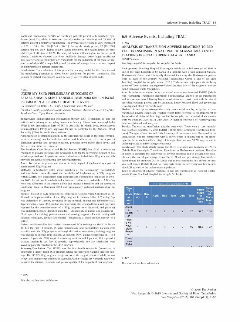
stools and haematuria. 62.50% of transfused patients present a hemorrhagic syn-
drome (level 02), while 24.66% are clinically stable (no bleeding) and 79.86% of
patients present a history of transfusion. The average platelet dose of APC transfused
is 5.26 � 1.38 9 1011 Plt [2.5–8 9 1011 ]. During the study period, 27 (19, 28%)
patients did not show desired platelet count increment. The results found an early
platelet yield effective of 80.71. The study of factors influencing on ineffective yield
platelet transfusion showed that fever, antibiotic therapy, hemorrhage, insufficient
dose platelet and splenomegaly are responsible for the reduction of the yield of pla-
telet transfusion.ABO compatibility, and duration of storage have a modest impact
on posttransfusion platelet increments.
Conclusions: The evaluation of this parameter is a very useful for the clinician and
the transfusing physician to adopt better conditions for platelet transfusion. The
number of platelet transfusions could be safely lowered after clinical audit.
P-204
UNDER MY SKIN: PRELIMINARY OUTCOMES OFESTABLISHING A SUBCUTANEOUS IMMUNOGLOBULIN (SCIG)PROGRAM IN A REGIONAL HEALTH SERVICECA Lambooy1, LR Hollis1, YL Fung2, K Morwood1 and H Weston1
1Sunshine Coast Hospital and Health Service, Nambour, Australia 2University of the
Sunshine Coast, Sippy Downs, Australia
Background: Immunoglobulin replacement therapy (IRT) is standard of care for
patients with primary or secondary immune deficiency. Intravenous immunoglobulin
(IVIg) is the predominant form of IRT in Australia. In March 2013, subcutaneous
immunoglobulin (SCIg) was approved for use in Australia by the National Blood
Authority (NBA) for use in these patients.
Administration of immunoglobulin by the subcutaneous route in the home environ-
ment is advantageous for both the patient and health service. It reduces patient
admission episodes and adverse reactions, produces more stable blood levels and
thus decreases infective episodes.
The Sunshine Coast Hospital and Health Service (SCHHS) has faced a continuing
challenge to provide sufficient day procedure beds for the increasing number of day
infusions procedures. As patients can be trained to self-administer SCIg at home, this
provided an avenue of reducing day bed requirements.
Aims: To review the process and assess the early impact of implementing a patient
administered SCIg Program.
Methods: In September 2013, senior members of the immunology, haematology
and transfusion teams discussed the possibility of implementing a SCIg program
within SCHHS. Key stakeholders were identified and consultation took place in Octo-
ber 2013. A cost benefit analysis and a literature review were undertaken. A Briefing
Note was submitted to the Patient Safety and Quality Committee and the Executive
Leadership Team in December 2013 and subsequently endorsed implementing the
program.
Results: Rollout of SCIg program:The Transfusion Clinical Nurse Consultant co-or-
dinated the implementation of the SCIg program in January 2014. A Training Day
was undertaken in January involving 20 key medical, nursing and laboratory staff.
Representatives from SCIg product manufacturers also attended.Issues and processes
required for the commencement of a SCIg program were discussed, and planning
was undertaken. Issues identified included: – Availability of pumps and equipment –Clinic space for training, patient review and nursing support – Patient training (self
infusion techniques, product knowledge) – Dispensing a blood product directly to a
patient
Patient recruitment:The first patient commenced SCIg training on the 17th March
2014.In the first 12 months, 35 adult immunology and haematology patients were
recruited onto the SCIg program. Although the patient competency training program
was planned to include four sessions, 25 patients (71%) gained competency in 1 to 3
sessions. 9 patients (26%) required 4 training sessions and 1 patient (3%) required 5
training sessions.In the first 12 months, approximately 455 day admissions were
saved by patients enrolled in the SCIg program.
Summary/Conclusion: The SCHHS was the first health service in Queensland to
implement a home based SCIg program which has generated valuable day bed sav-
ings. The SCHHS SCIg program has grown to be the largest cohort of adult haema-
tology and immunology patients in Australia.Further studies are currently underway
to assess the clinical, economic and patient quality of life impacts of this program.
P-205
This abstract has been withdrawn.
6.5 Adverse Events, Including TRALI
P-206
ANALYSIS OF TRANSFUSION ADVERSE REACTIONS TO REDCELL TRANSFUSION IN NATIONAL THALASSAEMIA CENTERTEACHING HOSPITAL KURUNEGALA SRI LANKABGSRRBandara
Teaching Hospital Kurunegala, Kurunegala, Sri Lanka
Back ground: Teaching Hospital Kurunegala which has a bed strength of 1991 is
one of the main hospitals in Sri Lanka. It a hospital with a well equipped National
Thalassaemia Center which is totally dedicated for caring the Thalassaemic patiens
from all parts of the country. National Thalassaemic Center is one of the units
Teaching Hospital Kurunegala, where 1032 b Thalassaemia major patients are being
managed.These patients are registered since the first day of the diagnosis and are
being managed whole throughout.
Aim: In order to minimise the occurance of adverse reactions and FNHTR (Febrile
Non Haemolytic Transfusion Reactions) a retrospective analysis of all transfusions
and adverse reactions following blood transfusions were carried out with the aim of
providing optimum patient care by promoting Leuco Reduced Blood and pre storage
leucodepleted blood for transfusions
Method: A descriptive retrospective study was carried out by analysing all post
transfusion adverse events and reaction report forms received to the Department of
Transfusion Medicine of Teaching Hospital Kurunegala, over a period of six months
from 01 February 2015 to 31 July 2015. A detailed collection of Haemovigilence
data was gathered and analyzed.
Results: The total no transfusion episodes were 6738. There were 31 post transfu-
sion reactions reported, 15 were FNHTR (Febrile Non Haemolytic Transfusion Reac-
tions). The type of reaction and their frequency of occurrence were illustrated in the
table.FNHTR was the commonest with a 48.4% which is mainly due to the leuco-
cytes in the whole blood.(Percentage of Allergic Reaction was 38.7% may be due to
under reporting of minor allergic reactions.)
Conclusion: This study clearly shows that there is an increased tendency of FNHTR
(Febrile Non Haemolytic Transfusion Reactions) in Thalassaemic patients. Therefore
in order to minimize the occurrence of adverse reactions and to provide best possi-
ble care the use of pre storage leucoreduced Blood and pre storage leucodepleted
blood should be promoted .At Sri Lanka due to cost constraints It’s difficult to pro-
vide LDB (Leuco Depletd Blood) for every patient.But we are trying our best to pro-
vide LDB at least to the thalassaemic population.
Table 1: Analysis of adverse reactions to red cell transfosions in National Thalas-
saemia Center Teachind Hospital Kurunegala Sri Lanka
P-207
This abstract has been withdrawn.
© 2015 The AuthorVox Sanguinis © 2015 International Society of Blood Transfusion
Vox Sanguinis (2015) 109 (Suppl. 2), 1–96
Adverse Events, Including TRALI 89
6.6 Haemovigilance and Transfusion Safety
P-208
THE PROGRESS OF THE IMPLEMENTATION OF HOSPITALTRANSFUSION REACTION REPORTING AND INVESTIGATIONSYSTEMFA Said
Indonesia Army Central Hospital, Jakarta Pusat, Indonesia
Background: One of the hospital blood bank duties is to report and investigate
adverse event related to transfusion, as stated in the Indonesian Goverments Regula-
tions No. 7 year 2011 concerning blood transfusion services. However, there is no
established national guidelines to conduct it, such as how to assess, classify the
symptoms, and standardize the reporting forms. Although there is a monthly report
to the Indonesian Red Cross Society, but until now there is no institusion appointed
by the local authorities to perform an audit on the transfusion reactions reports.
Nevertheless, since January 2012, the hospital blood bank has developed an acute
transfusion reaction reporting and investigation system to meet the function of
haemovigilance. Here we reported the 2012–2014 experience of implementation.
Aim: To review the reporting and investigation system of the acute blood transfu-
sion adverse reactions in the Indonesia Army Central Hospital- Gatot Soebroto for
better health services and patient safety.
Methods: Since January 2012 we have implemented the blood transfusion related
adverse effect reporting and investigating system using a paper-based reporting form
with check-box format containing 11 signs and symptoms of transfusion reactions.
Because of the requirement of the Joint Commission on International accreditation
(JCI) to maintain the quality of patient safety by strict monitoring and evaluation
during blood administration, in October 2013, the reporting form was revised and
redesigned to be a blood transfusion monitoring and evaluation check-box form.
Attached to it is a notification form to be completed when adverse reactions are
found. Based on the above, the blood bank carried out an investigation and exami-
nation into the complete immunohematology tests.
Result: All of the transfusion reaction data from 2012–2014 were collected and
analyzed. In 2012, there were 12 cases of transfusion reaction out of 17,806 blood
bags (0.067%). In 2013, the reported cases showed an increase to 33 cases out of
19,290 blood bags (0.171%). In 2014, when transfusion monitoring and evaluation
form was fully implemented, and the reported cases rose to 48 cases out of 16,791
blood bags (0.286%). A total there were 93 cases out of 53,887 blood bags, and
based on the signs and symptomps, classified accordingly: urticaria 33 (0.061%),
febrile reactions 42 (0.078%), dyspnea 14 (0.026%), hypotension 2 (0.004%) and
hemolytic reaction 1 (0.002%). All of the cases were investigated with immunohema-
tology confirmation examinations, It was found that there was 1 minor incompati-
bility and 1 mayor incompatibility, the rest was all compatible. It was discovered
that there was 1 near-miss case and 1 error case because of wrong blood grouping
due to blood discrepancies. There was no fatal incident in these cases.
Conclusion: The transfusion adverse reaction reporting rates vary, and may be
under reported. With the better designed monitoring and evaluation form, there has
been a significant increase in the reporting of suspected transfusion reaction cases.
Although in this 3 years span no fatal incidents have been reported, the transfusion
reaction rates especially error incidents should be minimize by practising haemovigi-
lance program.
P-209
MONITORING NEAR MISS EVENTS IN A HOSPITAL BLOODBANKMS Soomro and FK Mahar
Aga Khan University Hospital, Karachi, Pakistan
Background: Near miss errors related to sample identification are the preventable
errors that may have fatal consequences if remain undetected, resulting in the deter-
mination of a wrong blood group or transfusion of an incorrect component.
Aims: This study was done with aim to identify the Near miss events in our trans-
fusion medicine setup in order to identify the sources and prevent them from hap-
pening again.
Methods: It’s a retrospective descriptive study, which was carried out at the section
of hematology (Blood bank), with a purpose to identify the near miss events mainly
pertaining to errors in sample identification. A total number the requests forms
along with the blood sample for grouping and cross matching, from January 2013
till December 2014 were included in the study. Data was analyzed retrospectively
and sample identification errors were identified on the grounds of
when notified by the on call doctor/ staff nurse who has performed the sampling.
when noticed by laboratory staff by delta checking with previous results.
Results: During these two years a total of 75,157 request slips were received in
blood bank for arranging various blood products and 44 sample identification errors
were identified, out of which 0.4% were in 2013 (26 in 63,670 bags) and 0.3% were
in 2014 (18 in 59,690 bags) per 1000 units of various blood components issued.
Out of these 44 identification errors, 42 (95.4%) were labeled as Wrong blood in
tube (WBIT), which is further sub classified as WBIT due to miscollection (sample
was labeled correctly but blood was collected from the wrong patient) which was
found to be the most commonest error 26 out of 44 (59%); and WBIT due to misla-
beling of samples (sample was collected from the right patient but was labeled
incorrectly with medical record number/ name of another person) was found to be
16 out of 44 (36.3%). While 2 (4.55%) of these errors were made by blood bank staff
by issuing units on wrong blood unit releasing slips.
Most of these events, 30 out of 44 (68%) happened on floor/wards. 2 out of 44
(4.5%) were reported from ICU (intensive care unit), 8 out of 44 (18%) were reported
from emergency room (ER), 1 (2.27%) of them was reported from operating room
(OR), another case of misidentification happened in labor room.
All of the samples in high critical areas are taken by staff nurses which makes 12
(27.2%) of total sample identification errors. Out of total errors 30 were reported
from floor/ward, 29 (65.9%) were done by residents only one (2.27%) was carried
out by trained phlebotomist.
Conclusion: In our study we observed that unlike usual most of the errors were
reported from floor/ward not from high throughput areas like ER (emergency room)
and ICU (intensive care unit) and most of these errors were carried out by the on call
doctors (intern/resident). Most of these errors occurred due to mal practice of label-
ing the samples after collection and away from patient’s bed side.
P-210
OVERCOMING THE CHALLENGES IN ESTABLISHING ASUCCESSFUL HAEMOVIGILANCE PROGRAMME IN ACOUNTRY IN THE ASIAN REGIONK Gonsalkorale
National Blood Transfusion Service of Sri Lanka, Ratmalana, Sri Lanka
Haemovigilance, a dire necessity to provide safe and quality transfusions was a
known but a novel concept to the National Blood Transfusion Service (NBTS) of Sri
Lanka in the year 2008. The need was well understood by the administration and
the encouragement given to initiate the programme is praiseworthy. The unique
structure of the NBTS was the greatest advantage in commencing the programme.
NBTS is an island wide, centrally coordinated, non-profitable government service
which comes under the ministry of health and is the sole supplier of blood and
blood products to the entire patient population in the state sector and majority in
the private sector. It consists of the National Blood Centre (NBC) which is the main
administrative centre and 95 Hospital Based Blood Banks (HBBs) which are grouped
into 17 clusters taking into consideration the proximity and the accessibility to each
other. Each cluster is headed by a Consultant or a Medical officer In Charge (MOIC)
who acts as the administrative officer of the said cluster.
All cluster heads were initially invited to the NBC for a familiarization programme
on haemovigilance. Reporting forms, Instruction leaflets were distributed and pre-
sentations were conducted. Despite this meeting the initial reporting of adverse
events was poor. The thoughts and attitudes of the Sri Lankans with their long his-
tory and the culture heritage are quite different to the western world. Yet they would
understand and accept an evolving situation, probably due to the colonial rule that
prevailed in the bygone era.
The next strategy was to meet the staff of each HBB during the regular Hospital
Transfusion Committee (HTC) meetings and to explain the importance of the system
and the blame free nature of the programme.
Fright and doubt still prevailed. Telephone number of the In-charge consultant of
the haemovigilance unit was given to all HBBs so that they could contact her
throughout the day. This was a major breakthrough. The staff would often clear their
minds by phoning and gradually regular reporting became a norm.
Near miss events and delayed reactions are still under reported. The occasional hesi-
tancy of reporting a mishap in the blood bank still prevails and refresher pro-
grammes are a must.
Lectures are now given regularly to all newly recruited medical officers and nursing
officers. Undergraduates and the intern medical officers are given lectures on
haemovigilance. A review programme was held this year which paved the way to
clarify many doubts and queries.
The system is now a familiar concept and reporting of transfusion related adverse
events (recipient haemovigilance) have become a routine procedure. The first annual
haemovigilance report was published in 2010. Donor haemovigilance was com-
menced two years later and this was included in the annual report of 2012. With a
© 2015 The AuthorVox Sanguinis © 2015 International Society of Blood TransfusionVox Sanguinis (2015) 109 (Suppl. 2), 1–96
90 Posters
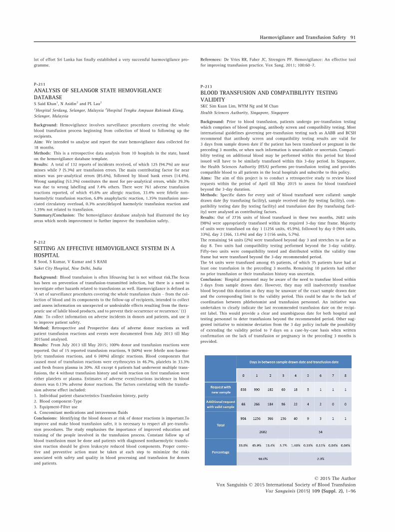
lot of effort Sri Lanka has finally established a very successful haemovigilance pro-
gramme.
P-211
ANALYSIS OF SELANGOR STATE HEMOVIGILANCEDATABASES Said Khan1, N Asidin2 and PL Lau2
1Hospital Serdang, Selangor, Malaysia 2Hospital Tengku Ampuan Rahimah Klang,
Selangor, Malaysia
Background: Hemovigilance involves surveillance procedures covering the whole
blood transfusion process beginning from collection of blood to following up the
recipients.
Aim: We intended to analyse and report the state hemovigilance data collected for
18 months.
Methods: This is a retrospective data analysis from 10 hospitals in the state, based
on the hemovigilance database template.
Results: A total of 132 reports of incidents received, of which 125 (94.7%) are near
misses while 7 (5.3%) are transfusion errors. The main contributing factor for near
misses was pre-analytical errors (85.6%), followed by blood bank errors (14.4%).
Wrong sampling (53.3%) constitutes the most for pre-analytical errors, while 39.3%
was due to wrong labelling and 7.4% others. There were 761 adverse transfusion
reactions reported, of which 45.6% are allergic reaction, 33.4% were febrile non-
haemolytic transfusion reaction, 6.8% anaphylactic reaction, 1.35% transfusion asso-
ciated circulatory overload, 0.3% acute/delayed haemolytic transfusion reaction and
1.35% not related to transfusion.
Summary/Conclusion: The hemovigilance database analysis had illustrated the key
areas which needs improvement to further improve the transfusion safety.
P-212
SETTING AN EFFECTIVE HEMOVIGILANCE SYSTEM IN AHOSPITALR Sood, S Kumar, V Kumar and S RANI
Saket City Hospital, New Delhi, India
Background: Blood transfusion is often lifesaving but is not without risk.The focus
has been on prevention of transfusion-transmitted infection, but there is a need to
investigate other hazards related to transfusions as well. Haemovigilance is defined as
‘A set of surveillance procedures covering the whole transfusion chain – from the col-
lection of blood and its components to the follow-up of recipients, intended to collect
and assess information on unexpected or undesirable effects resulting from the thera-
peutic use of labile blood products, and to prevent their occurrence or recurrence.’ (1)
Aim: To collect information on adverse incidents in donors and patients, and use it
to improve patient safety.
Method: Retrospective and Prospective data of adverse donor reactions as well
patient transfusion reactions and events were documented from July 2013 till May
2015and analysed.
Results: From July 2013 till May 2015; 100% donor and transfusion reactions were
reported. Out of 15 reported transfusion reactions, 9 (60%) were febrile non-haemo-
lytic transfusion reactions, and 6 (40%) allergic reactions. Blood components that
caused most of transfusion reactions were erythrocytes in 46.7%, platelets in 33.3%
and fresh frozen plasma in 20%. All except 4 patients had underwent multiple trans-
fusions, the 4 without transfusion history and with reaction on first transfusion were
either platelets or plasma. Estimates of adverse event/reactions incidence in blood
donors was 0.13% adverse donor reactions. The factors correlating with the transfu-
sion adverse effect included:
1. Individual patient characteristics-Transfusion history, parity
2. Blood component-Type
3. Equipment-Filter use
4. Concomitant medications and intravenous fluids
Conclusions: Identifying the blood donors at risk of donor reactions is important.To
improve and make blood transfusion safer, it is necessary to respect all pre-transfu-
sion procedures. The study emphasises the importance of improved education and
training of the people involved in the transfusion process. Constant follow up of
blood transfusion must be done and patients with diagnosed nonhaemolytic transfu-
sion reaction should be given leukocyte reduced blood components. Proper correc-
tive and preventive action must be taken at each step to minimize the risks
associated with safety and quality in blood processing and transfusion for donors
and patients.
References: De Vries RR, Faber JC, Strengers PF. Hemovigilance: An effective tool
for improving transfusion practice. Vox Sang. 2011; 100:60–7.
P-213
BLOOD TRANSFUSION AND COMPATIBILIYTY TESTINGVALIDITYSKC Sim Kuan Lim, WYM Ng and M Chan
Health Sciences Authority, Singapore, Singapore
Background: Prior to blood transfusion, patients undergo pre-transfusion testing
which comprises of blood grouping, antibody screen and compatibility testing. Most
international guidelines governing pre-transfusion testing such as AABB and BCSH
recommend that antibody screen and compatibility testing results are valid for
3 days from sample drawn date if the patient has been transfused or pregnant in the
preceding 3 months, or when such information is unavailable or uncertain. Compati-
bility testing on additional blood may be performed within this period but blood
issued will have to be similarly transfused within this 3-day period. In Singapore,
the Health Sciences Authority (HSA) performs pre-transfusion testing and provides
compatible blood to all patients in the local hospitals and subscribe to this policy.
Aims: The aim of this project is to conduct a retrospective study to review blood
requests within the period of April till May 2015 to assess for blood transfused
beyond the 3-day duration.
Methods: Specific dates for every unit of blood transfused were collated: sample
drawn date (by transfusing facility), sample received date (by testing facility), com-
patibility testing date (by testing facility) and transfusion date (by transfusing facil-
ity) were analysed as contributing factors.
Results: Out of 2736 units of blood transfused in these two months, 2682 units
(98%) were appropriately transfused within the required 3-day time frame. Majority
of units were transfused on day 1 (1256 units, 45.9%), followed by day 0 (904 units,
33%), day 2 (366, 13.4%) and day 3 (156 units, 5.7%).
The remaining 54 units (2%) were transfused beyond day 3 and stretches to as far as
day 8. Two units had compatibility testing performed beyond the 3-day validity.
Fifty-two units were compatibility tested and distributed within the validity time
frame but were transfused beyond the 3-day recommended period.
The 54 units were transfused among 45 patients, of which 35 patients have had at
least one transfusion in the preceding 3 months. Remaining 10 patients had either
no prior transfusion or their transfusion history was uncertain.
Conclusion: Hospital personnel may be aware of the need to transfuse blood within
3 days from sample drawn date. However, they may still inadvertently transfuse
blood beyond this duration as they may be unaware of the exact sample drawn date
and the corresponding limit to the validity period. This could be due to the lack of
coordination between phlebotomist and transfusion personnel. An initiative was
undertaken to clearly indicate the last recommended transfusion date on the recipi-
ent label. This would provide a clear and unambiguous date for both hospital and
testing personnel to deter transfusions beyond the recommended period. Other sug-
gested initiative to minimise deviation from the 3 day policy include the possibility
of extending the validity period to 7 days on a case-by-case basis when written
confirmation on the lack of transfusion or pregnancy in the preceding 3 months is
provided.
© 2015 The AuthorVox Sanguinis © 2015 International Society of Blood Transfusion
Vox Sanguinis (2015) 109 (Suppl. 2), 1–96
Haemovigilance and Transfusion Safety 91
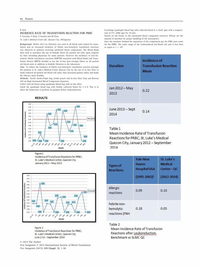
P-214
INCIDENCE RATE OF TRANSFUSION REACTION FOR PRBCE Torculas, E Datu, S Saceda and M Chua
St. Luke’s Medical Center-QC, Quezon City, Philippines
Background: Before 2013 no filtration was used in all blood units issued for trans-
fusion and an increased incidence of febrile non-hemolytic transfusion reactions
was observed in patients receiving unfiltered blood components. The Blood Bank
then tried to introduce the use of bedside filters for packed red cells, upon requests
by their attending physician for some patients. However the incidence of non-he-
molytic febrile transfusion reactions (NHFTR) remained until Blood Bank and Trans-
fusion Service (BBTS) decided to use the in-line (pre-storage) filters on all packed
red blood units in addition to bedside filtration in the laboratory.
Aim: To reduce the incidence of febrile non-hemolytic transfusion reaction amongst
the patients at St. Luke’s Medical Center Quezon City by the use of in line filter to
leuko-reduced all packed red blood cell units, thus increased patient safety and make
the efficacy more feasible.
Methods: Sterile DockerTriple bag w/side pouch and In-line filter (Top and Bottom
450 ml Bag) Automated Blood Component Separator
Collect 450 ml blood using quadruple blood bag with in-line filter.
Stand the quadruple blood bag with freshly collected blood for 2–4 h. This is to
allow the leukocytes to perform its purpose before leukoreduction.
Centrifuge quadruple blood bag with collected blood in a ‘hard’ spin with a tempera-
ture of 4°C, 4000 rpm for 10 min.
Switch on the Power to the automated blood component extractor. (Please see the
manual of machine for proper handling of the instrument.)
Once the machine finished the separation of the components get the WBC post count
for the PRBC. The value range of the Leukoreduced red blood cell unit is less than
or equal to 1 9 10⁶.
© 2015 The AuthorVox Sanguinis © 2015 International Society of Blood TransfusionVox Sanguinis (2015) 109 (Suppl. 2), 1–96
92 Posters
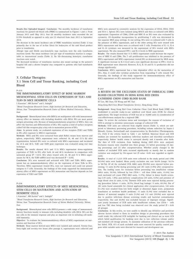
Results (See Uploaded Images): Conclusion: The monthly incidence of transfusion
reactions for packed red blood cells (PRBC) is summarized in Figures 1 and 2. From
January 2012 until May 2013, four (4) monthly incidence rates exceeded the set
0.35% threshold as opposed to only one (1) observed from June 2013 to September
2014.
The decrease in the mean incidence rate of PRBC transfusion reaction (Table 1) was
primarily due to the use of in-line filters for leukocytes of the said blood product
prior to transfusion.
Allergic type and febrile non-hemolytic type reactions were the only transfusion
reactions noted. The mean incidence rate per type of transfusion reaction is compa-
rable to international results (Table 2). No life-threatening hemolytic transfusion
reactions were noted.
The decreased incidence of transfusion reaction also meant savings in the patient’s
hospital bill and a shorter hospital stay compared to patients who had transfusion
reactions.
7. Cellular Therapies7.1 Stem Cell and Tissue Banking, including CordBlood
P-215
THE IMMUNOMODULATORY EFFECT OF BONE MARROWMESENCHYMAL STEM CELLS ON EXPRESSION OF TLR3 ANDTLR9 IN MICE DENDRITIC CELLSL Kasraian1, MH Karimi2 and L Sadeghi2
1Blood Transfusion Research Centre, High Institute for Research and Education,
Shiraz, Iran 2Transplantation Reaserch Center of Shiraz Medical University, Shiraz,
Iran
Background: Mesenchymal stem cells (MSCs) are multipotent cells with immunomod-
ulatory effect on immune cells including dendritic cells (DCs). DCs are most potent
antigen presenting cells. Because of immunomodulatory properties of MSCs, they can
affect the function and differentiation of DCs. DCs express a kind of surface receptors
called Toll-like receptors play a key role in maturation process of DCs.
Aims: In present study, we evaluated expression of two receptors (TLR3 and TLR9)
in DCs after exposure to MSCs’s supernatant.
Methods: MSCs and DCs were extracted from adult Balb/c mouse bone marrow and
spleen, respectively. MSCs supernatant were collected 24, 48 and 72 h (h) after
trypsinization. Isolated DCs were co-cultured with MSCs supernatant and incubated
for 24 h and 48 h. TLR3 and TLR9 gene expression was evaluated using real time
technique.
Results: The results showed 48 h and 72 h MSCs supernatant down-regulation
expression of TLR3 in DCs after both 24 and 48 h incubation in comparison with
untreated group (P < 0.01). DCs when treated with 24, 48 and 72 h MSCs super-
natant for 48 h, the TLR9 mRNA level was decreased (P < 0.05).
Conclusion: DCs were matured and activated with TLR3 and TLR9. MSCs super-
natant has an immunomodulatory effect on the expression of these TLRs in DCs.
Therefore, MSCs supernatant treated DCs, were not matured and could not present
antigens to T cells. Generally, the findings of this study supported the immunomod-
ulatory effect of MSCs supernatant on DCs maturation and function depends on low
expression of TLR3 and TLR9
P-216
IMMUNOMODULATORY EFFECTS OF MICE MESENCHYMALSTEM CELLS ON MATURATION AND ACTIVATION OFDENDRITIC CELLSL Kasraian1, MH Karimi2 and L Sadeghi2
1Blood Transfusion Research Centre, High Institute for Research and Education,
Shiraz, Iran 2Transplantation Reaserch Center of Shiraz Medical University, Shiraz,
Iran
Background: Mesenchymal stem cells (MSCs) possess a wide range of immunomod-
ulatory functions mostly in immune cells including dendritic cells (DCs). DCs are the
key cells in the immune response and play an important role in initiating cell-medi-
ated immunity.
Objective: To evaluate the immunomodulatory effects of MSCs supernatant on mat-
uration and function of DCs.
Methods: Bone marrow derived mice MSCs were isolated and cultured. Twenty-four,
forty-eight and seventy-two hours after passage 6, supernatants were collected and
MSCs were assessed by cytometric analysis for the expression of CD34, CD44, CD45
and SCA-1. Splenic DCs were isolated using MACS and then co-cultured with MSCs
supernatant. Expression of CD86, CD40 and MHC-II on DCs were also evaluated by
cytometry. H3-thymidine incorporation by proliferating T cells was determined in
two separate MLR assay settings. In one setting, DCs were co-cultured with T cells in
the presence of MSCs supernatant, and in the other setting DCs were treated with
MSCs supernatant and then were co-cultured with T cells. Production of IL-12, IL-6
and IL-10 cytokines was measured in the supernatant of DCs treated with MSCs
supernatant. We also measured IFN-c and IL-4 levels in MLR supernatant.
Results: The results showed that 72 h MSCs supernatant could decrease the expres-
sion of MHC-II and CD86. The T cell proliferation was inhibited in the presence of
MSCs supernatant and MSCs supernatant treated DCs as demonstrated by MLR assay.
A significant increase in IL-4 level and a non significant decrease in IFN-c level in
MLR supernatant were observed. However, IL-6, IL-10 and IL-12 production did not
change significantly.
Conclusion: MSCs supernatant has a time dependent effect on the maturation of
DCs. Also, it could alter cytokine production from responding T cells toward Th2.
Generally, the findings of this study supported the immunomodulatory effect of
MSCs supernatant on DCs maturation and function
P-217
A REVIEW ON THE EXCLUSION STATUS OF UMBILICAL CORDBLOOD COLLECTIONS IN HONG KONG RED CROSSCATHERINE CHOW CORD BLOOD BANKKY Lee, NK Chan, CK Wong and WC Tsoi
Hong Kong Red Cross Blood Transfusion Service, Hong Kong, China
Background: Hong Kong Red Cross Catherine Chow Cord Blood Bank (CBB) was
established to provide ready-to-use umbilical cord blood units (UCB) for clinical
applications. The target inventory of UCB was set at 10,000 units in consideration of
cost-effectiveness analysis for a regional CBB.
Aims: To review the exclusion status and investigate the reasons of exclusion of
UCB in attempt to minimise unnecessary wastage of collected UCB.
Methods: UCB units are collected from eligible donors (Table 1), which are sub-
jected to laboratory processing procedure, including volume reduction by Sepax-2
(Biosafe, Eysins, Switzerland) and cryopreservation by BioArchive (Thermogenesis,
CA, USA) if the criteria listed in Table 1 are fulfilled. Maternal blood and UCB
residues are screened for mandatory infectious markers and microbial surveillance
respectively. Babies’ health status is surveyed shortly and 6 months after birth.
The data of UCB exclusion from banking during 2007–2014 were analysed.
Exclusion reasons were classified into three groups: (1) before processing; (2) dur-
ing processing; and (3) after cryopreservation. Whether yearly changes in the
number of excluded UCB units occurred significantly or merely due to random
variation was analysed by Chi-square test (P-value <0.05 was regarded as signifi-
cant).
Results: A total of 13,325 UCB units were collected in the study period and 5786
(43.4%) units were banked. Mean yearly exclusion rate was 56.6% (range: 54.1%
to 58.7%). Of all the excluded UCB, 6882 units (95.9%) were rejected before pro-
cessing, 13 units (0.2%) during processing and 281 units (3.9%) after cryopreserva-
tion. The leading cause for exclusion before processing was insufficient volume
(4852 units, 70.5%), followed by low CD34 + cell dose (1064 units, 15.5%), low
total nucleated cell count (TNC) (802 units, 11.7%), failure in donor health screen-
ing (129 units, 1.9%), post-delivery complications (29 units, 0.4%) and presence of
large blood clots (6 units, 0.1%). Thirteen UCB units were rejected during process-
ing (procedure failure: 4 units, 30.7%; blood bag leakage: 9 units, 69.3%). Out of
281 units found unsuitable for clinical application after cryopreservation, 144 units
(51.2%) were resulted from low birth weight or abnormal Apgar score, postpartum
transfusion or newborn health problems; 15 units (5.3%) were excluded due to
absence of maternal samples; 9 units (3.2%) and 112 unit (39.9%) were rejected
due to positive infectious disease screening results and bacterial contamination
respectively. One unit (0.4%) was excluded because of improper storage. Signifi-
cant yearly increment of UCB units with insufficient volume, low CD34 + cells
and low TNC dose being excluded was observed over the study period (P < 0.05
all groups).
Conclusion: In this review, we were unable to identify any definitive and persistent
adverse factors related to flaws in workflow design or processing procedures that
could render the collected UCB ineligible for banking and clinical use as most UCB
which failed qualification for banking were due to low collection volume and/or
sub-standard cell dosage present. To maximise utilization of the precious collected,
but failed, UCB, 473 of such excluded units were reserved for quality control pur-
pose while suitable units were directed for research and development use.
© 2015 The AuthorVox Sanguinis © 2015 International Society of Blood Transfusion
Vox Sanguinis (2015) 109 (Suppl. 2), 1–96
Stem Cell and Tissue Banking, including Cord Blood 93
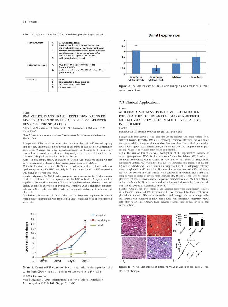
Table 1: Acceptance criteria for UCB to be collected/processed/cryopreserved.
P-218
DNA METHYL TRANSFERASE 1 EXPRESSION DURING EXVIVO EXPANSION OF UMBILICAL CORD BLOOD-DERIVEDHEMATOPOIETIC STEM CELLSA Oodi1, M Ahmadnejad1, N Amirizadeh1, M Nikougoftar1, R Mehrasa1 and M
Khorshidfar1
1Blood Transfusion Research Center, High Institute for Research and Education,
Tehran, Iran
Background: HSCs reside in the ex-vivo expansion by their self-renewal capacity
and also they differentiate into a myriad of cell types, as well as the regeneration of
stem cells. Whereas the DNA methyltransferase1 is thought to be principally
involved in the maintenance of pre-existing methylation, the role of Dnmt1 in prim-
itive stem cells pool preservation also is not known.
Aims: In this study, mRNA expression of Dnmt1 was evaluated during CB-HSC
ex vivo expansion with and without mesenchymal stem cells (MSCs).
Methods: Ex vivo cultures of CB-HSCs were performed in three culture conditions:
cytokine, cytokine with MSCs and only MSCs for 7 days. Dnmt1 mRNA expression
was evaluated by real time -PCR.
Results: Maximum CB-CD34+ cells expansion was observed in day 7 of expansion
in all three cultures. Ex vivo expansion of CB-CD34+ cells after 7 days resulted in,
significant decreased expression of Dnmt1 in cytokine culture, whereas in two co-
culture conditions expression of Dnmt1 was increased. Also a significant difference
between CD34+ cells and CD34- cells of co-culture system with cytokine was
observed.
Conclusions: Expression of Dnmt1 that are an important regulator in normal
hematopoietic regeneration was increased in CD34+ expanded cells on mesenchymal
stem cells.
7.3 Clinical Applications
P-219
AUTOPHAGY SUPPRESSION IMPROVES REGENERATIONPOTENTIALITIES OF HUMAN BONE MARROW-DERIVEDMESENCHYMAL STEM CELLS IN ACUTE LIVER FAILURE-INDUCED MICEF Amiri
Iranian Blood Transfusion Organization (IBTO), Tehran, Iran
Background: Mesenchymal stem cells (MSCs) are isolated and characterized from
different tissues. Recently, MSCs are receiving increased attention for cell-based
therapy especially in regenerative medicine. However, their low survival rate restricts
their clinical applications. Interestingly, it is hypothesized that autophagy might play
an important role in cellular homeostasis and survival.
Aims: The aim of this study was investigation of the regenerative capacity of
autophagy-suppressed MSCs for the treatment of acute liver failure (ALF) in mice.
Methods: Authophagy was suppressed in bone marrow derived-MSCs using shRNA
suppressive vectors. ALF was induced in mice by intraperitoneal injection of 1.5 ml/
Kg carbon tetrachloride. MSCs which are suppressed in their autophagy pathway
were transplanted to afflicted mice. The mice that received normal MSCs and those
that did not receive any cells (sham) were considered as control. Blood and liver
samples were collected at several time intervals (24, 48 and 72 hrs) after the trans-
plantation of MSCs. Liver enzymes, aspartate aminotransferase (AST) and alanine
aminotransferase (ALT), were evaluated with biochemical methods. Liver necrosis
was also assayed using histological analysis.
Results: After 24 hrs, liver enzymes and necrosis score were significantly reduced
in autophagy-suppressed MSCs-transplanted mice compared to those that trans-
planted with normal MSCs and sham (with no cell therapy). Normal histology with-
out necrosis was observed in mice transplanted with autophagy-suppressed MSCs
cells after 72 hrs. Interestingly, liver enzymes reached their normal levels in this
period of time.
Figure 1: Dnmt1 mRNA expression fold change ratio: In the expanded cells
to the fresh CD34 + cells at the three culture conditions (P < 0.05).
Figure 2: The fold increase of CD34+ cells during 7-days expansion in three
culture conditions.
Figure 1: Therapeutic effects of different MSCs in ALF-induced mice 24 hrs
after cell therapy.
© 2015 The AuthorVox Sanguinis © 2015 International Society of Blood TransfusionVox Sanguinis (2015) 109 (Suppl. 2), 1–96
94 Posters

Conclusion: Autophagy suppression in MSCs might be suggested as a new strategy
to improve of cell therapy in ALF. This modulation ameliorates MSCs liver regenera-
tion potentials due to paracrine effects.
P-220
CHANGE OF RBC DEFORMABILITY DURING HEMATOPOIETICSTEM CELL TRANSPLANTATIONJM Lee, KH Suh and YT Lim
Yeungnam University Hospital, Daegu, South-Korea
Background: The red blood cell (RBC) deformability test is the measurement of the
ability of the RBCs to adapt their shape to the flow conditions. The major determi-
nants of the deformability include cell shape, composition of the cell membrane and
its cytoskeleton, and internal viscosity (mean cell hemoglobin concentration). The
contribution of the erythrocyte membrane to the deformability is primarily regulated
by the composition and arrangement of its structural constituents. In cancer patient,
chemotherapy and hematopoietic stem transplantation (HSCT) change bone marrow
microenvironment, these may affect the RBC production and deformability.
Aim: We evaluate the change of RBC deformability during HSCT.
Method: Blood samples were obtained from each patients who underwent HSCT.
Red cell deformability was measured with a microfluidic ektacytometer (RheoScan-
D, RheoMeditech, Seoul, Korea). All analyses were completed within 24 h after blood
collection. The elongation index (EI) of erythrocytes is defined as (L-W)/(L + W),
where L and W are the major and minor axes of the ellipse, respectively.
Result: Eleven children (3 acute lymphoblastic leukemia, 5 acute myeloid leukemia,
1 aplastic anemia, 1 Ewing sarcoma, 1 retinoblastoma) were enrolled in this study.
The EI of RBCs were decreased to D + 5 from D-day and slowly increased from
D + 5 to D + 30 after hematopoietic stem cell transplantation.
Conclusion: The deformabilites of RBCs were slowly increased during hematopoietic
stem cell transplantation. RBC deformability may reflect bone marrow microenviron-
ment of patient during HSCT. Further study about correlation with RBC deformabil-
ity and prognosis of HSCT is needed.
P-221
INVESTIGATION OF ERYTHROCYTE DEFORMABILITY OFCHILDREN WITH VARIOUS HEMATOLOGIC DISEASEJM Lee, YT Lim, KH Suh and YK Kim
Yeungnam University Hospital, Daegu, South-Korea
Background: Erythrocyte deformability is expressed as the elongation index (EI) at
a shear stress of 3 Pa. The EI of erythrocyte is defined as (L-W)/(L + W), where L
and W are the major and minor axes of the ellipse, at a shear stress of 3 Pa.
Aim: We aimed to investigate the effects of hereditary hemolytic anemia (HHA),
iron deficiency anemia (IDA), immune thrombocytic purpura (ITP) and aplastic ane-
mia (AA) on hemorrheologic parameters, especially erythrocyte deformability.
Methods: Thirty-one HHA patients (age 15.3 � 14.7), 16 IDA patients (age
9.6 � 5.7), 16 ITP patients (age 4.8 � 5.4) and 6 aplastic anemia patients (age
15.3 � 8.0) and 14 healthy controls (age 11.7 � 12.7) were enrolled. Erythrocyte
deformability were estimated by ectacytometer (RheoScan-D300; Rheomeditech, Seoul,
Korea). Differences between HA, IDA, ITP, AA and healthy controls were compared.
Result: The EI value of IDA and AA were significantly decreased compared with
normal control, but those of ITP and HHA were similar to normal control. But when
shear stress were greater than 4 Pa, EI value of HHA were significantly decreased
compared with normal control.
Conclusion: This study shows different RBC deformability in various hematologic
disease. Diagnostic and prognostic significance of RBC deformability is needed.
P-222
A SH-SY5Y DIFFERENTIATED HUMAN NEUROBLASTOMACELL MODEL TO EVALUATE THE POTENTIALNEUROPROTECTIVE ROLE OF PLATELET NEUROTROPHINSCT Chang1, ML Chou2 and T Burnouf1
1Institute of Biomedical Materials and Tissue Engineering, Taipei, Taiwan 2Institute
of Medical Sciences, Taipei, Taiwan
Background: Progressive neurodegenerative diseases, such as Alzheimer’s disease
(AD), Parkinson’s disease (PD), or amyotrophic lateral sclerosis (ALS), are the leading
cause of disability in the elderly. No therapies exist yet to delay their onset or
reverse symptoms. Neurotrophins are potential drug candidates since they activate
neuronal signaling pathways and modulate neurons survival. The SH-SY5Y human
neuroblastoma cells have already been used to study neuroprotection induced by
neurotoxins, since, using different inducers, they can be differentiated into various
adult neuronal phenotypes, including dopaminergic neurons. Platelet lysates (PL)
prepared from human platelet concentrates (PCs) may be a valuable physiological
and balanced source of native neurotrophic growth factors (GFs) such as PDGF,
BDNF, and VEGF for ‘disease-modifying strategy’ against PD, ALS and other neu-
rodegenerative disorders.
Aims: The primary objectives of this study were to determine the doses of 1-
Methyl-4-phenyl-pyridinium ion (MPP+) neurotoxin inducing neurotoxicity in a dif-
ferentiated SH-SY5Y cellular model. The second goal was to perform preliminary
evaluations of the capacity of a PL to exert neuroprotective effects in this in vitro
model.
Methods: SH-SY5Y neuroblastoma cells (ATCC � CRL-2266) were maintained in
T75 culture flasks with a 1:1 mixture of Ham’s F12 and Dulbecco Modified Eagle
Medium (DMEM) supplemented with 10% fetal bovine serum (FBS) and 1% peni-
cillin. Cells were maintained in a humidified 5% CO2 atmosphere at 37°C. Differenti-ation was triggered by 1 mM retinoic acid (RA) plus 0.5% of FBS during 2 and
4 days in Advanced DMEM/F12 medium or DMEM/F12 medium. On day 7, the med-
ium was replaced by differentiation medium without FBS or RA. Viable cells
(2 9 104/well in Advanced DMEM/F12 and 5 9 104/well in DMEM/F12 medium)
were seeded into 24-well plates. Differentiated SH-SY5Y cells were treated with dif-
ferent dosage of MPP+ (0.01–10 mM) for 48 h. After incubation, cell viability was
determined by 3-(4,5-dimethylthiazol-2-yl)-2,5-diphenyltetrazolium bromide (MTT)
assay and the absorbance was measured at 540 nm using a microplate spectropho-
tometer. PLs rich in neurotrophins were prepared from therapeutic-grade human pla-
telet concentrates (PCs) and pre-evaluated for their neuroprotective potential.
Results: MTT assays revealed that differentiated SH-SY5Y viability was reduced to
50–60% in Advanced DMEM/F12 medium and around 80% in DMEM/F12 medium
when using 0.3 mM MPP+, which was the MPP+ dose selected for further experi-
ments. Preliminary evaluation using 0.1–5% PPL to test the viability of differentiated
SH-SY5Y cells exposed to MPP+, showed that PL improved cell viability. The highest
level of protection (above ca. 70%) was achieved with 1% PPL when cells were
grown in Advanced DMEM/F12 medium, and with 0.5% PPL (ca. 90–95%) when
using DMEM/F12 medium.
Conclusions: MPP+ neurotoxin can be used to induce toxicity in a differentiated
SH-SY5Y human neuroblastoma cell model. This model can be of value to study the
potential neuroprotective effects of platelet growth factors in neurodegenerative dis-
orders, like PD or ALS.
8. Clinical Immunogenetics8.1 HLA in transfusion medicine
P-223
USING TWO SOFTWARE TOOLS TO DEFINE HLA CLASS IIMMUNOGENIC EPITOPES OF ANTIBODIES PRODUCED INALLOIMMUNIZED THROMBOCYTOPENIC PATIENTSSC Pai1, JW Chen1 and SC Lo2
1Taiwan Blood Services Foundation, Taipei, Taiwan 2National Taiwan University
Hospital, Taipei, Taiwan
Background: Human immune responses involve recognition of peptides presented by
HLA class II molecules. In literature, the recipient HLA-DR phenotype has been sug-
Figure 2: Therapeutic effects of different MSCs in ALF-induced mice 72 hrs
after cell therapy.
© 2015 The AuthorVox Sanguinis © 2015 International Society of Blood Transfusion
Vox Sanguinis (2015) 109 (Suppl. 2), 1–96
HLA in transfusion medicine 95
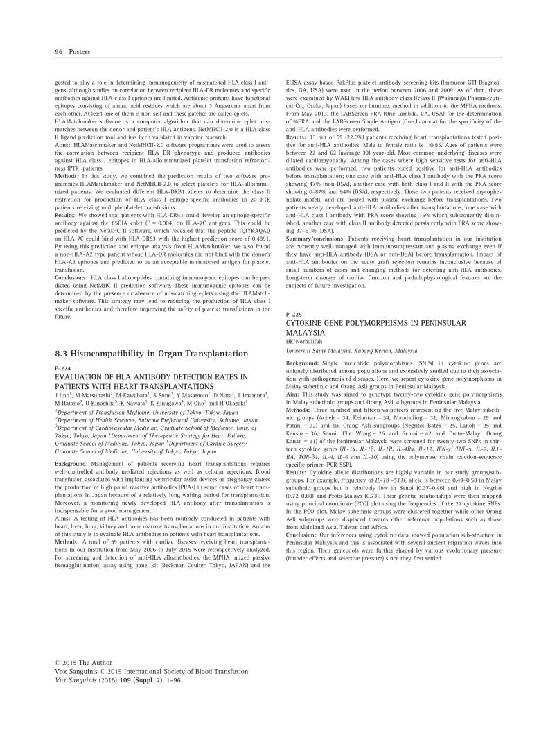
gested to play a role in determining immunogenicity of mismatched HLA class I anti-
gens, although studies on correlation between recipient HLA-DR molecules and specific
antibodies against HLA class I epitopes are limited. Antigenic proteins have functional
epitopes consisting of amino acid residues which are about 3 �Angstroms apart from
each other. At least one of them is non-self and these patches are called eplets.
HLAMatchmaker software is a computer algorithm that can determine eplet mis-
matches between the donor and patient’s HLA antigens. NetMHCII-2.0 is a HLA class
II ligand prediction tool and has been validated in vaccine research.
Aims: HLAMatchmaker and NetMHCII-2.0 software programmes were used to assess
the correlation between recipient HLA DR phenotype and produced antibodies
against HLA class I epitopes in HLA-alloimmunized platelet transfusion refractori-
ness (PTR) patients.
Methods: In this study, we combined the prediction results of two software pro-
grammes HLAMatchmaker and NetMHCII-2.0 to select platelets for HLA-alloimmu-
nized patients. We evaluated different HLA-DRB1 alleles to determine the class II
restriction for production of HLA class I epitope-specific antibodies in 20 PTR
patients receiving multiple platelet transfusions.
Results: We showed that patients with HLA-DR53 could develop an epitope-specific
antibody against the 65QIA eplet (P = 0.004) on HLA-7C antigens. This could be
predicted by the NetMHC II software, which revealed that the peptide TQIYKAQAQ
on HLA-7C could bind with HLA-DR53 with the highest prediction score of 0.4891.
By using this prediction and epitope analysis from HLAMatchmaker, we also found
a non-HLA-A2 type patient whose HLA-DR molecules did not bind with the donor’s
HLA-A2 epitopes and predicted to be an acceptable mismatched antigen for platelet
transfusion.
Conclusions: HLA class I allopeptides containing immunogenic epitopes can be pre-
dicted using NetMHC II prediction software. These immunogenic epitopes can be
determined by the presence or absence of mismatching eplets using the HLAMatch-
maker software. This strategy may lead to reducing the production of HLA class I
specific antibodies and therefore improving the safety of platelet transfusions in the
future.
8.3 Histocompatibility in Organ Transplantation
P-224
EVALUATION OF HLA ANTIBODY DETECTION RATES INPATIENTS WITH HEART TRANSPLANTATIONSJ Iino1, M Matsuhashi2, M Kawabata1, S Sone1, Y Masamoto1, D Nitta3, T Imamura4,
M Hatano3, O Kinoshita5, K Nawata5, K Kinugawa4, M Ono5 and H Okazaki1
1Department of Transfusion Medicine, University of Tokyo, Tokyo, Japan2Department of Health Sciences, Saitama Prefectural University, Saitama, Japan3Department of Cardiovascular Medicine, Graduate School of Medicine, Univ. of
Tokyo, Tokyo, Japan 4Department of Therapeutic Strategy for Heart Failure,
Graduate School of Medicine, Tokyo, Japan 5Department of Cardiac Surgery,
Graduate School of Medicine, University of Tokyo, Tokyo, Japan
Background: Management of patients receiving heart transplantations requires
well-controlled antibody mediated rejections as well as cellular rejections. Blood
transfusion associated with implanting ventricular assist devices or pregnancy causes
the production of high panel reactive antibodies (PRAs) in some cases of heart trans-
plantations in Japan because of a relatively long waiting period for transplantation.
Moreover, a monitoring newly developed HLA antibody after transplantation is
indispensable for a good management.
Aims: A testing of HLA antibodies has been routinely conducted in patients with
heart, liver, lung, kidney and bone marrow transplantations in our institution. An aim
of this study is to evaluate HLA antibodies in patients with heart transplantations.
Methods: A total of 59 patients with cardiac diseases receiving heart transplanta-
tions in our institution from May 2006 to July 2015 were retrospectively analyzed.
For screening and detection of anti-HLA alloantibodies, the MPHA (mixed passive
hemagglutination) assay using panel kit (Beckman Coulter, Tokyo, JAPAN) and the
ELISA assay-based PakPlus platelet antibody screening kits (Immucor GTI Diagnos-
tics, GA, USA) were used in the period between 2006 and 2009. As of then, these
were examined by WAKFlow HLA antibody class I/class II (Wakunaga Pharmaceuti-
cal Co., Osaka, Japan) based on Luminex method in addition to the MPHA methods.
From May 2013, the LABScreen PRA (One Lambda, CA, USA) for the determination
of %PRA and the LABScreen Single Antigen (One Lambda) for the specificity of the
anti-HLA antibodies were performed.
Results: 13 out of 59 (22.0%) patients receiving heart transplantations tested posi-
tive for anti-HLA antibodies. Male to female ratio is 1:0.85. Ages of patients were
between 22 and 62 (average 39) year-old. Most common underlying diseases were
dilated cardiomyopathy. Among the cases where high sensitive tests for anti-HLA
antibodies were performed, two patients tested positive for anti-HLA antibodies
before transplantation; one case with anti-HLA class I antibody with the PRA score
showing 47% (non-DSA), another case with both class I and II with the PRA score
showing 0–87% and 94% (DSA), respectively. These two patients received mycophe-
nolate mofetil and are treated with plasma exchange before transplantations. Two
patients newly developed anti-HLA antibodies after transplantations; one case with
anti-HLA class I antibody with PRA score showing 15% which subsequently dimin-
ished, another case with class II antibody detected persistently with PRA score show-
ing 37–51% (DSA).
Summary/conclusions: Patients receiving heart transplantation in our institution
are currently well-managed with immunosuppressant and plasma exchange even if
they have anti-HLA antibody (DSA or non-DSA) before transplantation. Impact of
anti-HLA antibodies on the acute graft rejection remains inconclusive because of
small numbers of cases and changing methods for detecting anti-HLA antibodies.
Long-term changes of cardiac function and patholophysiological features are the
subjects of future investigation.
P-225
CYTOKINE GENE POLYMORPHISMS IN PENINSULARMALAYSIAHK Norhalifah
Universiti Sains Malaysia, Kubang Kerian, Malaysia
Background: Single nucleotide polymorphisms (SNPs) in cytokine genes are
uniquely distributed among populations and extensively studied due to their associa-
tion with pathogenesis of diseases. Here, we report cytokine gene polymorphisms in
Malay subethnic and Orang Asli groups in Peninsular Malaysia.
Aim: This study was aimed to genotype twenty-two cytokine gene polymorphisms
in Malay subethnic groups and Orang Asli subgroups in Peninsular Malaysia.
Methods: Three hundred and fifteen volunteers representing the five Malay subeth-
nic groups (Acheh = 34, Kelantan = 34, Mandailing = 31, Minangkabau = 29 and
Patani = 22) and six Orang Asli subgroups (Negrito: Batek = 25, Lanoh = 25 and
Kensiu = 36, Senoi: Che Wong = 26 and Semai = 42 and Proto-Malay: Orang
Kanaq = 11) of the Peninsular Malaysia were screened for twenty-two SNPs in thir-
teen cytokine genes (IL-1a, IL-1b, IL-1R, IL-4Ra, IL-12, IFN-ƴ, TNF-a, IL-2, IL1-RA, TGF-b1, IL-4, IL-6 and IL-10) using the polymerase chain reaction-sequence
specific primer (PCR-SSP).
Results: Cytokine allelic distributions are highly variable in our study groups/sub-
groups. For example, frequency of IL-1b -511C allele is between 0.49–0.58 in Malay
subethnic groups but is relatively low in Senoi (0.37–0.46) and high in Negrito
(0.72–0.88) and Proto-Malays (0.73). Their genetic relationships were then mapped
using principal coordinate (PCO) plot using the frequencies of the 22 cytokine SNPs.
In the PCO plot, Malay subethnic groups were clustered together while other Orang
Asli subgroups were displaced towards other reference populations such as those
from Mainland Asia, Taiwan and Africa.
Conclusion: Our inferences using cytokine data showed population sub-structure in
Peninsular Malaysia and this is associated with several ancient migration waves into
this region. Their genepools were further shaped by various evolutionary pressure
(founder effects and selective pressure) since they first settled.
© 2015 The AuthorVox Sanguinis © 2015 International Society of Blood TransfusionVox Sanguinis (2015) 109 (Suppl. 2), 1–96
96 Posters


