Embed Size (px)
Citation preview
ARTICLE IN PRESS
FOODHYDROCOLLOIDS
0268-005X/$ - s
doi:10.1016/j.fo
�CorrespondPretoria, Lynw
Tel.: +2712 42
E-mail addr
(M. Naushad E
Food Hydrocolloids 21 (2007) 1245–1255
www.elsevier.com/locate/foodhyd
In situ tensile deformation of zein films with plasticizersand filler materials
M. Naushad Emmambuxa,b,�, Mats Stadinga,b
aStructure and Material Design, The Swedish Institute for Food and Biotechnology, Box 5401, SE 402 29 Gothenburg, SwedenbDepartment of Material Science and Engineering, Chalmers University of Technology, Goteborg, Sweden
Received 26 January 2006; accepted 29 September 2006
Abstract
Material deformation is a dynamic process. Visualisation of this deformation can help to understand the local deformation and
fracture behaviour. Zein (the prolamin protein from maize) films with different amount of plasticizers (0–25%) and different filler
materials (maize oil, Dimodans, Vestosints, at 25% (w/w) to protein) were deformed under tension and observed at micron scale in real
time by a confocal laser scanning microscope (CLSM). The addition of plasticizers increased strain and decreased stress of zein films. At
low level of plasticizers (6.25% and 12%), zein films deformed and fracture through micro-crack formation and propagation normal the
tensile axis. At high Plasticization, only micro-pores were observed during tensile deformation. The filler material oil and Dimodans
increased, but Vestosints decreased tensile strain in comparison to the control. This shows that the fracture dynamic is affected by the
filler materials and is indeed observed by the CLSM. Analysis of local strain by Fluospheress as particle tracking showed a good linear
correlation with the tensile strain of the plasticized zein films. The local strains of filler materials and zein matrix in the films were
different from the overall tensile strain. The combination of CLSM with a fluospheress as particle tracking is a good method to study
local deformation in biomaterials to understand the deformation and fracture behaviour of biomaterials.
r 2006 Elsevier Ltd. All rights reserved.
Keywords: Biomaterial; Zein films; Tensile deformation and fracture; CLSM; Local deformation
1. Introduction
Biomaterials are widely used in food, pharmaceuticaland medical industries. The strength of biomaterials isgenerally assessed by mechanical deformation test topredict their performance, especially in highly stressedconditions. Deformation of biomaterials like gels, biopo-lymer films and food materials, is a dynamic mechanicalprocess. During deformation, load (or stress) is transferredthrough the material and eventually lead to fracture, failureand energy dissipation. The types of deformation andfracture depend on the fracture process, the structure andthe properties of the material (Lillford, 2001; Stanley,
ee front matter r 2006 Elsevier Ltd. All rights reserved.
odhyd.2006.09.013
ing author. Department of Food Science, University of
ood Road, Pretoria 0002, South Africa.
0 5865; fax:+2712 420 2839.
esses: [email protected]
mmambux), [email protected] (M. Stading).
1994). In conventional studies, fracture behaviour has beeninvestigated by microscopy before and after the deforma-tion process. This hindered the information about themicrostructural change during the dynamic deformationprocess. Deformation is also studied in bulk to give stress(deforming force) and strain (deformability) behaviour ofmaterials. Stress and strain of the material is often assumedto be uniform and details about the local deformationwithin the sample are left unknown. However, thisassumption has proven untrue for the deformation oftendon as a biomaterial (Defrate, Van der Ven, Boyer,Gill, & Li, 2006).Microrheology is a new approach to look into the local
deformation of biomaterials. It generally uses rheologicaltechniques in combination with microscopy or lightscattering to study deformation and flow of materials inreal time at micrometre length scale. Local deformation atmicrometre scale was obtained for (1) aggregation, colli-sion, particle coalescence and break up of gelatin–dextran

ARTICLE IN PRESSM. Naushad Emmambux, M. Stading / Food Hydrocolloids 21 (2007) 1245–12551246
emulsion by a combination of confocal laser-scanningmicroscopy (CLSM) and continuous shear cell (Nicolaset al., 2003) (2) a ‘pseudo-yielding’ fracture behaviour as aresults of debonding of particle/matrix interface in agelatin–maltodextrin composite gel system as observed byCLSM with a tensile cell (Plucknett et al., 2000) (3) stressdistribution in tissue, crack propagation and cell walldistortion of fresh carrots by environmental scanningmicroscopy (Thiel & Donald, 1998). Thus, real-timemeasurement techniques provide the possibility to deter-mine local deformation and microstructural changes ofbiomaterials during the deformation process.
This paper describes the local deformation of zein filmsas a biomaterial with different amount of plasticizers anddifferent filler materials in real time with a CLSM and amicrotensile stage. Zein is the prolamin protein of maize.Zein films and coatings have received considerable atten-tion as biomaterial coatings and films (Lawton, 2002).These coatings and films can be both edible andbiodegradable. They can be an attractive environmentfriendly alternative to plastic packaging made from non-renewable resources. However, only a few edible coatingsand films are used in commercial application because oftheir imitations in performance compared to syntheticpackaging. Performance of edible coating and films willdepend on the tensile, oxygen and water vapour barrierproperties and at times nutritional properties if hydro-colloids films are used as sausage casings. Performance inrelation to tensile properties also depends on the plastici-zers and surrounding relative humidity (RH). For exampleaddition of plasticizer and conditioning at high RH(greater than 50%) increase the strain, but decrease thetensile stress (Lawton, 2004). Therefore visualisation ofdeformation process in real time with CLSM zein filmswith plasticizers and fillers can provide the possibility toelucidate deformation mechanism and help to design filmswith better properties for end use.
2. Experimental
2.1. Materials
Zein protein was purchased from Sigma-Aldrich Chemie(Diesenhofen, Germany) and defatted three times sequen-tially with n-hexane for 1 h (Emmambux & Taylor, 2003).After each extraction, the n-hexane was filtered out. Thelast residue was washed with n-hexane, filtered and thedried in ventilated oven at 25 1C for 24 h to remove anyresidual n-hexane. Glycerol (Analars, BDH lab suppliesPoole, England) and polyethylene glycol (PEG) 400(Merck, Honenbrunn, Germany) were the plasticizingagents. Fluospheress (Molecular probes, Leiden, Nether-lands) were the particle tracking in the experiments. Thefluospheress are sulphate microspheres at 2% solid, 2 mmin diameter and yellow green fluorescent at 505/515 nm.Commercial kitchen maize oil (Zeta, Stockholm, Sweden),Dimodans distilled monoglycerides (Danisco, Grinsted,
Denmark) and Vestosints 1111 particles (Degussa, Gote-borg, Sweden) were the filler materials. The Dimodans
consists of a mixture of mono and di-glycerides of fattyacid. The Vestosints 1111 is a polyamide 12 powder and isabout 90 mm diameter, density of 1.016 g/cm3, and a ballindentation hardness of 90N/mm2.
2.2. Methods
2.2.1. Preparation of zein films
Zein films were produced by casting for both theexperiment with different amount of plasticizers and fillermaterials.
Experiment with different amount of plasticizers: Zeinpowder was weighed in a conical flask, followed bydifferent amount of glycerol and PEG (1:1) mix, lacticacid (12.5% w/w of zein) and aqueous ethanol (70% w/w inwater). The protein concentration was 16% (w/w) withrespect to aqueous ethanol. The amount of a glycerol andPEG mix were 0%, 6.25%, 12%, 18.75% and 25% (w/w)zein. The flask was heated at 70 1C for 10min undervigorous stirring. After dissolution 2 ml of the fluospheres
was pipetted into 10 g of film forming solution of pH 3.Then, aliquots (2 g) were poured into plastic petridishes(9 cm diameter), gently swirled, placed in a ventilated ovenat 50 1C for 4 h to evaporate the solvent and produce a freestanding films. After drying, the films were conditioned forat least 48 h at 22 1C and 5075% relative humidity (RH).
Experiment with different filler materials: The fillermaterials oil, Dimodans and Vestosints were added at25% (w/w of zein) to make films of two-phase system. Thefilms forming procedure was the same as described abovewith the following changes depending on the fillermaterials. A mixture of glycerol, PEG and lactic acid(1:1:1) at 40% (w/w of zein) was added. The oil wasvigorous mixed in aqueous ethanol with a magnetic stirrer,then heated at 75 1C for 10min. The aqueous ethanol andoil mix was then added with the film forming ingredients,and further heated for another 10min at 70 1C. Dimodans
was first dissolved in ethanol and then added to the filmforming ingredients. Vestosints was added before pouringthe film solution (pH 3) into the petridishes. Two ml of thefluospheres, 0.25ml of 0.01% (w/w) Texas Reds sulpfonylchloride (Molecular probes, Leiden, Netherlands) and0.25ml of 0.01% (w/w) Nile Red (Polysciences Inc.,Washington, PA, USA) were also pipetted into 10 g offilm forming solution before film casting.
2.2.2. Tensile deformation in real time with CLSM
Film strips, 2mm wide were cut with a scalpel, measuredfor thickness with a micrometre and placed in between thegrips of a microtest material testing modules (Deben,Suffolk, UK) with a 2N load cell and an initial gripseparation of 10mm. The microtest material-testingmodule was then placed under an air objective of CLSMLeica TCS SP2 (leica Ltd., Heidelberg, Germany).

ARTICLE IN PRESS
0
5
10
15
20
25
0 5 10 15 20Strain (%)
Stre
ss (
MPa
)
unplasticized
plasticized 6.25 %
plasticized 12.5 %
plasticized 18.75 %
plasticized 25 %
Fig. 1. Effect of plasticizers (plasticizers were glycerol and polyethylene
glycol 400 in equal amount at 0%, 6.25%, 12.5%, 18.75% and 25% (w/w)
to zein) on the tensile properties of zein film.
M. Naushad Emmambux, M. Stading / Food Hydrocolloids 21 (2007) 1245–1255 1247
The settings of the CLSM were different for the differentexperiments.
Experiment with different amount of plasticizers: Thetensile deformation of the plasticized films was performedat a crosshead speed of 0.1mm/min while observing withthe CLSM. The light sources of the CLSM were Ar laserusing lex ¼ 488 nm to detect the fluospheress from emittedsignal at 500–540 nm. The HeNe light source at 594 nm wasused in the reflection mode. An air objective with amagnification of 10� (Numerical aperture ¼ 0.3) and adigital zoom 2� were used. The scanning was bi-directional with a speed of 400Hz and two scans wereaveraged out to give an image of 512� 256 pixels every1.015 s as a real-time series.
Experiment with different filler materials: The tensiledeformation of the films with different filler materials wasperformed at a crosshead speed of 0.2mm/min whileobserving with the CLSM. The light sources of the CLSMwere Ar laser using lex ¼ 488 nm to detect the fluospheress
and Nile red stained materials from emitted signal at500–525 and 550–585 nm, respectively. The HeNe laserlight source lex ¼ 594 nm was used to detect Nile redstained zein from emitted signal at 610–640 nm. Theobjective and scanning speed and images were the sameexcept that images were taken every 4 s.
2.2.3. Dynamic mechanical analysis (DMA)
DMA of the unplasticized, plasticized zein films, andfilms with filler materials were performed in a Rheometricsolid analyser, RSA-II (Rheometrics Scientific, Piscataway,NJ, USA). The films strips were 10mm long and 2mmwide. A frequency sweep test from 0.1 to 15Hz wasperformed in tension at different strain level up to 50%.The temperature during the test was 23 1C. The films weresubjected to a sinusoidal strain on top of the staticdeformation, the sum of these being non-destructive. Themagnitude and the resulting sinusoidal stress at specificfrequency were recorded during the measurements. Thephase angle d was calculated from storage modulus (E0)and loss modulus (E00) where tan d ¼ E0=E00.
2.2.4. Image analysis
Images obtained from the CLSM or the CLSMmicrographs were selected and analysed with an imageanalysis software Image J 1.32j (Wayne Rasband, NationalInstitutes of Health, USA, http://rsb.info.nih.gov/ij/). Thefluospheress were processed as particles in the softwareand the centre of gravity of selected fluospheress werecalculated in terms of XY pixel position in the image. Thedistance between two selected fluospheress parallel to thetensile axis were then calculated using the pixel position.These selected fluospheress were followed at differentoverall tensile strain (calculated from grip to grip separa-tion of microtest material testing modules) during thetensile deformation. The change in the distance betweenthe selected fluospheress were taken as the local strain forthe zein films.
The films with different filler materials were consideredas a two-phase systems with zein protein as the matrix, andthe oil, phase-separated Dimodans and Vestosints as thefiller materials. It was not possible to calculate the localstrain of the filler materials using the fluospheress as theywere hardly located in this phase. The local straindeformation of the filler materials were calculated aschanges in the longitudinal distance parallel to the tensileaxis using the above-mentioned image analysis software.The fluospheress were used to calculate the local matrixstrain. The calculation method was as mentioned above.
2.2.5. Statistical analysis
The relationship between overall tensile strain (calcu-lated by grip to grip separation) and the local strains(calculated by image analysis) for both experiments wasstatistically analysed. The outliers were first excluded fromthe residual plot analysis. Then the correlation coefficientswere calculated and linear regression analysis was per-formed between the two set of data by Statistica 6.1(StatSoft Inc., USA). Overall tensile results from at least 4samples were used and at least 4 local strains werecalculated from each CLSM micrograph.
3. Results and discussion
3.1. In situ tensile deformation of plasticized zein films
The stress of zein films without the addition ofplastcicizer mix increased with an increase in strain (Fig.1). The average stress at break (sb, stress is force/crosssectional area), strain at break (eb) and Young’s Modulusof elasticity (E, calculated stress/strain from the linearregion of the stress–strain graph) were, respectively19.4MPa, 0.97% and 2.1GPa. The stress–strain curve ofzein films has an elastic region, without showing a yieldpoint and a transition to ductile or plastic region. Thisclearly demonstrates that zein films without glycerol andPEG were brittle. The stress–strain behaviour of zein filmswith a mixture of glycerol and PEG mix shows an elastic

ARTICLE IN PRESSM. Naushad Emmambux, M. Stading / Food Hydrocolloids 21 (2007) 1245–12551248
region, a yield point and ductile region (Fig. 1). The yieldstress (sy), sb, eb, E were, respectively 14.9, 16MPa, 2.5%and 1.4GPa for zein films with glycerol and PEG mix at6.25%. An increased in level of glycerol and PEG mix to25% decreased sy to 3.6MPa, sb to 4.2MPa, and E to0.12GPa, but increased the eb to 16.5%. The stress–straincurve of zein films with glycerol and PEG mix showed atypical behaviour of Plasticization, i.e., an increase in strainand a decrease in stress and Young’s modulus (Park,Brunn, Weller, Vergano, & Testin, 1994; Parris & Coffin,1997). Plasticizers also increased the tensile strain anddecreased the tensile stress of starch-methyl cellulosehydrocolloid films (Arvanitoyannis & Biliaderis, 1999).
Freestanding zein films without addition of plasticizerswere brittle, but a mixture of glycerol and PEG asplasticizers increased the strain and decrease the stress.Similar results were reported for zein films (Park et al.,1994; Parris & Coffin, 1997), and kafirin films (Van Eck,2004), a similar prolamin protein to zein (DeRose et al.,1989). Glycerol and PEG are also hygroscopic and canabsorb water during conditioning at 50% RH. The watercontent of glycerol and PEG plasticized films at 30% (w/w)were less than 10% when conditioned at 50% RH (Lawton2004). This increase the tensile strain as water can also actas a plasticizer (Arvanitoyannis & Biliaderis, 1999; Lawton,2004). Many theories have been proposed to explain theplasticizer action (Sears & Darby, 1982). However theexact mechanism is not clear. Plasticizers like glycerol andPEG can possibly form numerous hydrogen bonds with thezein polypeptide chain because of the carbonyl of proteinand hydroxyl groups of polyol type plasticizers. H-bondhas been reported to occur between polyol plasticizers andcellulose (Xiao, Zhang, Lu, & Zhang, 2003). Thus theplasticizer can interpose between polypeptide chains andthus can probably decrease polymer–polymer interactionto decrease stress and film brittleness.
Fig. 2 shows the CLSM micrographs of zein films withand without the plastcicizers at different levels. The zeinfilms without plasticizer have some imperfections seen asblackish spots in the micrographs. These imperfectionscould be micro-pores of about 5–10 mm in diameter, whichreflected the laser light differently in comparison to the zeinprotein matrix. Micro-pores like pinholes have beenobserved by scanning electron microscopy in cast zeinfilms (Lai & Padua, 1997). Imperfections in syntheticmaterials (Kinloch & Young, 1983) and biomaterials(Zioupos, 1998) can be sites for micro-crack formationand propagation that lead to fracture. The reflection modeof the laser HeNe at 594 nm was used instead of theemission mode because in preliminary experiments of thiswork, the micro-pores were more visible in the reflectionmode.
Zein films without plasticizer were brittle (Fig. 1). TheCLSM micrographs (Fig. 2) of these films from unstrainedto 0.2%, 0.5% and 0.8% strain did not show any evidentmicrostructural changes at the scanned position. Duringtensile deformation, zein films with added plastcicizers at
6.25% and 12.5% plasticizers formed micro-cracks. Themicro-cracks (Fig. 2B) formed between 1% and 2% strain.The micro-cracks formation is followed and is encircled inFig. 3. The micro-crack seemed to originate from micro-pore. These micro-pores increase in size with increase instrain from 1.05% to 1.25% until it cracks in oppositedirection normal to the tensile axis. With further increase intensile strain, the micro-crack propagates normal to thetensile axis. These micro-cracks can join together and formbigger cracks to break or rupture the zein films (Fig. 4A).The microstructural changes during tensile deformation
in terms of micro-cracks formation and propagation weredifferent for zein films with different amount of plasticizers.At 2% and 3% strain of zein films with 6.25% plasticizers,the micro-cracks were thin, long and normal to the tensileaxis (Fig. 2B). However, at 2% strain of zein films with12.5% plasticizers, only the increased size of the micro-pore or the formation of some small micro-cracks could beobserved (Fig. 2C). At 3% and 5% strain of deformed zeinfilms plasticized at 12.5%, the observed micro-cracks werethick, short, and normal to the tensile axis in comparisonto the micro-cracks of zein films plasticized at 6.25%. Thisindicates that increasing Plasticization can delay theformation and propagation of micro-cracks during tensiledeformation. The effects of Plasticization on the micro-structural changes during tensile deformation of zein filmscan be further observed Fig. 2(D and E). The CLSMmicrographs of zein films plasticized at 18.75% and 25%show few cracks or no cracks at different strain level duringtensile deformation. Only occurrence of micro-pores can beinterpreted from the CLSM micrographs. It seems thatthese micro-pores can increase in size and join together torupture the films (Fig. 4B).The dynamic mechanical analysis (DMA) of the zein
films without plasticizers had a phase angle between 3.41and 4.61 at different strains and different frequencies(Table 1). Plasticization of zein films increased phase angle.The highest phase angle of about 27.91 was recorded, a 4–5fold increase, for the zein films Plasticized 25%, at 1%strain and 0.15Hz. These data showed that increasePlasticization lead a more visco-elastic zein films. Thevisco-elasticity of plasticized zein films can be related to thefracture dynamic as observed by the microstructuralchanges and the tensile graph. Plasticized zein films aretougher because they are highly visco-elastic materials.They can dissipate energy into the material instead of theatmosphere to reduce micro-cracks during deformation(Kinloch & Young, 1983) and to produce a ductile material(Kinloch & Young, 1983; Zioupos, 1998). Thus moreenergy is required to fracture plasticized zein filmscompared to unplasticized zein films. The fracture energyof zein films calculated from the stress–strain data (Fig. 1),showed that average fracture energy were 1.3, 3.1, 5.4, 5.5and 6.9 Jm�2 for the unplasticized zein films and plasti-cized at 6.25%, 12.5%, 18.75%, and 25%, respectively.Fracture mechanics of synthetic polymers are charac-
terised by crazing and shear yielding (Kinloch & Young,

ARTICLE IN PRESS
Fig. 2. Selected confocal laser-scanning microscopy micrographs of unplasticized (A) and plasticized (B, C, D and E, respectively at 6.25%, 12.5%,
18.75% and 25% (w/w) to zein) zein films at different strain level. Particles of 2 mm are the fluospheress and the background is film matrix. The bar is
150mm. MC is microcracks and MP is micropores.
M. Naushad Emmambux, M. Stading / Food Hydrocolloids 21 (2007) 1245–1255 1249
1983). A craze is a lenticular and perpendicular to thetensile axis, and occurs during brittle fracture. Craze cangrow in real cracks during deformation to cause fracture(Marissen, 2000). Shear yielding allows the formation of
shear bands, which tear apart. It is not perpendicular totensile axis and occurs in ductile fracture. Occurrence ofmicro-cracks in brittle fracture of zein films plasticized at6.25% and 12.5% suggests the crazing phenomena.

ARTICLE IN PRESS
Fig. 3. Selected confocal laser-scanning microscopy micrographs showing
the crack initiation of the zein film plasticized at 6.25%. A crack initiation
process is encircled and followed at different tensile strain. The numbers at
the left of the micrographs are % tensile strain. Particles of 2mm are the
fluospheress and the background is film matrix. The bar is 150mm.
M. Naushad Emmambux, M. Stading / Food Hydrocolloids 21 (2007) 1245–12551250
Increase in micro-pore size along the tensile axis andabsence (or reduced) of micro-cracks in the ductile fracturesuggest the phenomena of shear yielding or tearing of zeinfilms plasticized at 18.75% and 25%.
3.2. In situ tensile deformation of zein films with different
filler materials
The different filler materials show different stress–straincurve during tensile deformation (Fig. 5). The fillermaterials used during this experiment have differentrheological behaviour and thus may affect the tensiledeformation differently. Corn oil has a liquid-like beha-viour, Dimodans behaves more like a visco-elasticmaterial and has a waxy like texture, and Vestosints
particles are solid. The zein films with and without fillermaterials show an elastic and plastic region of thestress–strain graphs (Fig. 5). There was a decrease in sy,sb and E for films with oil and increase eb compared to zeinfilms without filler materials. The eb of zein films withDimodans increased whereas the eb of zein films withVestosints decreased in comparison to films with no fillers.The E of non-filled, Dimodans and Vestosints as fillers, inzein films were similar. The stress increased after the yieldpoint for the Dimodans and Vestosints as fillers in zeinfilms, showing a pseudo-yielding behaviour. This pseudo-yielding behaviour was also found for gelatin–maltodextringel and has been associated with the disruption ordissociation of maltodextrin particle from gelatin matrix(Plucknett et al., 2000).The CLSM micrographs of films without any filler
material (Fig. 6A) before tensile deformation had similarmicrostructure with the unstrained plasticized zein films(Fig. 4). They contained micro-pores, as stated already.During deformation, the films did not produce any micro-cracks, but produced enlarged micro-pores. Similar to thezein films plasticized with 18.75% and 25% from abovesection, the micro-pores increased in size and number athigher strain level. It seems that the micro-pores can jointogether to rupture the zein films (Fig. 4B).The films with oil as filler materials had an emulsion type
of structure with oil droplets as spherical particlesdispersed in protein matrix (Fig. 6B). The oil dropletswere about 30–40 mm in diameter or less. Theses films wererecorded to have an average thickness of about 50 mm.Thus, it can be suggested that the protein matrix above andbelow the oil droplets will be a thin layer less than 10 mm.Oil droplets can also be at the surface of the films. Duringtensile deformation, above 5% strain, the shape of the oildroplets changed from a spherical to ellipsoidal duringdeformation along the tensile axis. Micro-pores were alsoformed in the oil droplets close to the oil–matrixinterphase. The micro-pores grew in size and becameellipsoidal from spherical along the tensile axis. A scanacross the film showed the micro-pores to be very smallholes across the film. Thus it seems that during tensiledeformation, the thin protein matrix film above and belowoil droplets can easily rupture to formed micro-poresacross the films. In addition the oil droplets at the surfaceof the film can also act as micro-pores. The micro-poresgrew during tensile deformation to form small holes acrossthe films and they joined together to rupture the films(Fig. 7A). Vigorous mixing produces a two-phase film withfat droplet dispersed in the protein phase. However, asystem with real homoginisation produce smaller fatdroplets dispersed in protein phase. This type of filmincreased the strain compared to less homogenised (Anker,Berntsen, hermansson, & Stading, 2002), suggesting thatreal homoginisation can have different fracture dynamics.Dimodans dissolved in ethanol during film-making
process. During film drying, Dimodans can phaseseparated from the protein to form ellipsoidal structures.

ARTICLE IN PRESS
Fig. 4. Selected confocal laser-scanning microscopy micrographs showing the Fracture dynamics of the zein film plasticized at (A) 6.25% and (B) 25%.
The arrows indicate fracture direction of the film. Particles of 2mm are the fluospheress and the background is film matrix. The bar is 150mm.
Table 1
Phase anglea from dynamic mechanical analysis of unplasticized,
plasticized zein films at different strain % and frequencies
Treatments Strain (%) Frequency (Hz)
0.15 1.5 15.0
Unplasticized 0.1 (0.09) 3.7 (0.59) 3.4 (0.22) 3.7 (0.18)
1.1 (0.07) 4.6 (1.27) 4.2 (0.01) 4.0 (0.02)
Plasticized 6.25% 0.2 (0.16) 4.9 (2.40) 4.5 (1.64) 4.7 (1.93)
1.6 (0.15) 5.6 (0.69) 5.3 (0.40) 5.1 (0.61)
2.9 (0.11) 7.4 (0.93) 6.1 (0.78) 5.9 (0.88)
Plasticized 12.5% 0.1 (0.01) 14.3 (0.7) 11.1 (0.62) 9.7 (0.50)
2.6 (0.07) 14.6 (1.04) 11.0 (0.58) 9.5 (0.64)
4.1 (0.20) 14.7 (0.59) 11.0 (0.77) 9.4 (0.56)
Plastcized 18.75% 0.2 (0.18) 22.5 (4.65) 15.8 (2.11) 13.0 (1.47)
3.5 (0.49) 22.5 (5.15) 16.0 (6.55) 12.7 (2.57)
11.3 (0.66) 24.0 (6.15) 16.2 (3.76) 12.9 (2.44)
Plasticized 25% 1.0 (0.59) 27.9 (1.49) 20.9 (1.44) 15.1 (1.95)
8.8 (0.38) 26.7 (4.25) 19.2 (2.02) 14.6 (1.34)
15.4 (2.56) 25.7 (3.96) 19.1 (1.98) 14.6 (0.86)
aAverage and standard deviation in bracket.
0
1
2
3
4
0 20 40 60 80 100
Strain (%)
Stre
ss (
MPa
)
Non-filler
Oil
Dimodan®Vestosint®
Fig. 5. Effect of oil, Dimodans and Vestosints added at 25% (w/w of
zein) as filler materials on the tensile properties of zein film.
M. Naushad Emmambux, M. Stading / Food Hydrocolloids 21 (2007) 1245–1255 1251
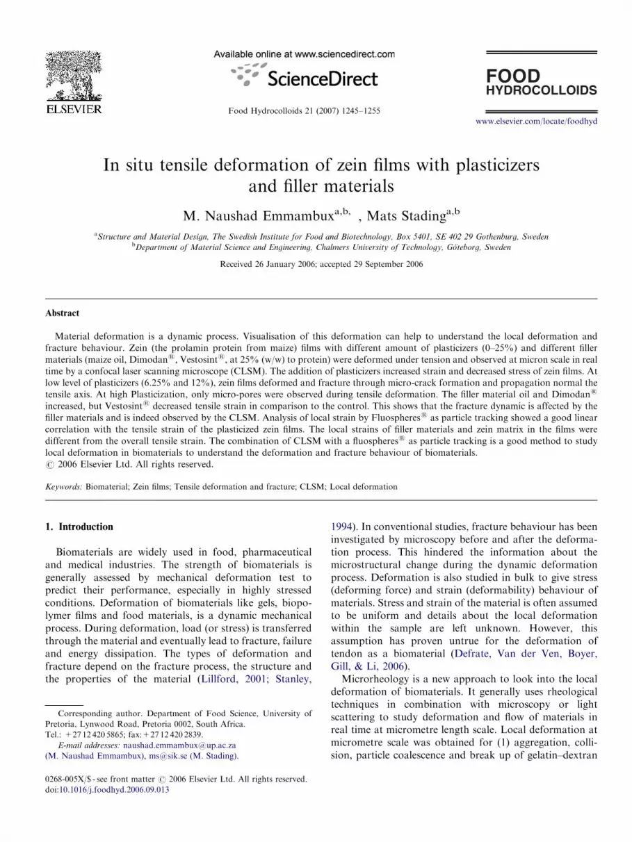
The phase-separated Dimodans were seen as platelet likestructure surrounded by protein phase under the CLSM(Fig. 6C, undeformed films). The phase-separated Dimo-dans was of different sizes up to about 150 mm in diameter.During tensile deformation, micro-pores occurred in boththe zein matrix and phase-separated Dimodans and at theinterphase. The phase-separated Dimodans tends to havea more elongated shape parallel to the tensile axis duringdeformation. The micro-pores grow bigger and jointogether to rupture the films (Fig. 7B). The micro-poresbetween the phase-separated Dimodans and the protein
matrix suggests the possible debonding between the phases-separated Dimodans and the protein matrix. The debond-ing is supported by the pseudo-yielding relationship of thestress–strain graph (Fig. 5). The pseudo yielding may occuras a result of debonding between the two phases asreported for debonding between gelatin and maltodextrin(Plucknett et al., 2000). Dimodans can associate with zeinmolecules by weak hydrophobic interaction and hydrogenbonds. This is because Dimodans as distilled monoglycer-ides has hydroxyl groups and zein has carbonyl groups toform H-bonds. They can also interact by hydrophobicinteraction as they both have hydrophobic ends (Damo-daran, 1996).Vestosints is the particles of about 90 mm in diameter
within the protein matrix in the film (Fig. 6D). TheVestosints was added after film dissolution, but before filmcasting. The Vestosints was small protrusion on the filmsurfaces as the pouring solution produced films of about50 mm thick. During tensile deformation micro-pores were

ARTICLE IN PRESS
Fig. 6. Selected confocal laser-scanning microscopy micrographs of zein films without any filler material (A), with oil (B), Dimodans (C) and Vestosints
(D). The fluospheress are particles of 2mm. The filler materials oil, Dimodans and Vestosints is surrounded by zein protein. P is pores across the film.
The bar is 150mm.
M. Naushad Emmambux, M. Stading / Food Hydrocolloids 21 (2007) 1245–12551252
observed in the protein matrix. The micro-pores increasedin number and seemed to increase in size during deforma-tion. Micro-pores are also seen between the protein andVestosints interface. These micro-pores showed theseparation of Vestosints and the protein matrix duringtensile deformation. The separation seemed to grow firstnormal to the tensile axis like a micro-crack and then alongthe tensile axis. This separation can be regarded asdebonding and it is reflected in the pseudo-yieldingresponse of the Vestosints stress–strain graph. Vestosints
did not seem to be affected by tensile deformation probablybecause zein matrix was a visco-elastic and Vestosints wasa solid material. The micro-pores in the protein–Vesto-sints interface grew in size during tensile deformation and
joined together with other growing micro-pores to rupturethe films (Fig. 7C). The deformation mechanism asobserved by CLSM for zein films with filler materialsVestosints is similar to the mechanism explained byPlucknett et al. (2000) for gelatin/maltodextrin gel withgelatin as continuous phase. The gelatin/maltodextrin gel isa phase-separated mixed biopolymer composite. Thegelatin continuous phase was visco-elastic compared withthe maltodextrin to be more brittle. During deformationvoids were formed at the interphase region between gelatinand maltodextrin, grew around the maltodextrin, thenjoined together to fracture the composite gel. Similarly,fracture by debonding of calcium carbonate as fillermaterials from the main matrix of polypropylene has been
ARTICLE IN PRESS
Fig. 7. Selected confocal laser-scanning microscopy micrographs showing the fracture dynamics of zein film with oil (A), Dimodans (B) and Vestosints
(C) as filler materials. The fluospheress are particles of 2 mm. The filler materials oil, Dimodans and Vestosints is surrounded by zein protein. Arrows
show fracture. The bar is 150mm.
Table 2
Phase anglea from dynamic mechanical analysis of zein films with different
filler materials at different strain percentage and frequencies
Treatments Strain (%) Frequency (Hz)
0.15 1.5 15.0
No filler materials 0.1 (0.02) 18.5 (1.22) 13.9 (1.07) 11.7 (1.27)
5.2 (0.23) 18.8 (1.98) 13.9 (1.02) 11.2 (0.81)
10.3 (0.69) 21.0 (1.62) 14.7 (0.88) 11.8 (0.62)
Oil filler materials 0.3 (0.13) 16.8 (2.5) 12.4 (1.34) 10.2 (1.26)
2.9 (0.82) 17.0 (0.65) 13.0 (1.05) 10.6 (0.666)
11.5 (1.06) 18.3 (1.32) 13.3 (0.35) 10.8 (0.28)
Dimodans filler
materials
0.2 (0.10) 15.0 (0.88) 11.5 (0.62) 10.2 (0.51)
5.4 (0.13) 15.5 (0.35) 12.0 (0.31) 9.9 (0.20)
11.0 (0.12) 18.0 (0.59) 13.3 (0.24) 10.8 (0.31)
Vestosints filler
materials
0.3 (0.27) 14.5 (1.75) 11.4 (1.19) 10.72 (0.81)
5.5 (1.3) 15.7 (1.2) 12.5 (0.61) 10.12 (0.32)
10.9 (0.83) 18.24 (0.83) 13.7 (0.64) 10.81 (0.24)
aAverage and standard deviation in bracket.
M. Naushad Emmambux, M. Stading / Food Hydrocolloids 21 (2007) 1245–1255 1253
reported by debonding off the filler from the matrix(Zebarjad, Tahani, & Sajjadi, 2004).
The phase angles from the dynamic mechanical analysisat different strain level at a specific frequency were similarbetween the films with different filler materials (Table 2).This indicates that films with different filler materials didnot seem to greatly change the visco-elastic properties ofzein films as determined by small deformation. It can bespeculated that DMA could not pick up the differences asshown by the CLSM probably because DMA occurs insmall deformations. These small deformations probablyresult in responses in the protein matrix rather than thefiller materials or the interphase region.
3.3. Overall tensile strain versus local strain
The relationship between the tensile stain (grip to gripseparation from tensile stage) and local strain (particletracking) showed a significant (Po0.001) linear correlationfor plasticized zein films (Fig. 8). The correlation coeffi-cients were 0.79, 0.93, 0.98, 0.98 and 0.97; and the slopes of

ARTICLE IN PRESS
0
0.25
0.5
0.75
1
1.25
1.5
0 0.25 0.5 0.75 1 1.25
Tensile strain (%)
loca
l str
ain
(%)
0
5
10
15
20
25
30
35
0 10 20 30
Tensile strain (%)
Loc
al s
trai
n (%
)
(a) (b)
Fig. 8. The relationship between the overall tensile strain and local strain of (a) unplasticized , plasticized zein films at (b) 25%.
-500
50100150200250300
0 10 20 30 40 50 60
Tensile strain (%)
Loc
al s
trai
n (%
)
-10
0
10
20
30
40
0 10 20 30 40
Tensile strain (%)
Loc
al s
trai
n (%
)
-20
0
20
40
60
0 10 20 30 40 50 60
Tensile strain (%)
0
10
20
30
40
50
0 10 20 30 40 50
Tensile strain (%)
Loc
al s
trai
n (%
)L
ocal
str
ain
(%)
(a) (b)
(c) (d)
Fig. 9. The relationship between the overall tensile strain and the stain of zein matrix and the filler materials of the films. (a) No filler materials, (b) oil as
filler materials, (c)Dimodans as filler materials, and (d) Vestosints as filler materials. The big open diamond, triangle, circle and square is the zein matrix
strain; the small open diamond, triangle, circle and square is the strain of filler material.
M. Naushad Emmambux, M. Stading / Food Hydrocolloids 21 (2007) 1245–12551254
the calculated linear regression line were 0.80, 1.27, 0.88,1.25 and 1.10 for the unplasticized and plasticized films at6.25%, 12.5%, 18.75% and 25%, respectively. As the slopewere close to 1, the use of fluospheress as particle trackingis a good estimate to calculate the local deformation ofbiobased materials like films. However, Fig. 8 also shows ahigh standard deviation of local strains for one tensilestrain. This may show that local deformation at micronscale within a piece of film is not uniform.
The correlation coefficient and the calculated slope oflinear regression line for the films without filler materialswere 0.97 and 0.78, respectively (Fig. 9). The linearregression slope for the oil as filler material was 0.69 forthe matrix and 2.01 for the oil phase. The slope for thematrix was 0.32 and the Dimodans was 0.51 for the zeinfilm with Dimodans as fillers. There was no linearrelationship between the Vestosints strain and the overalltensile strain of zein films with this filler material. However,the matrix of the zein film with Vestosints fillers showed alinear response with a linear regression slope of 0.90.Negative strains were obtained for the filler materialsbecause scanned position during deformation changed,
thus here overall trend should be noted rather thanindividual values. The regression analysis clearly showedthat local strains of the different phases of zein films withdifferent filler materials are not the same as the overalltensile strain. The deformability depends on the fillermaterials. The strength of the filler material may play a rolein the deformability.Local strains in comparison to overall tensile strain have
been reported for tendons as biomaterials. Defrate et al.(2006) found the local strain of achille tendons ascalculated by image analysis were lower than overall tensilestrain. He suggested this to non-homogeneity of thetendons. Similarly, there was a poor linear correlationbetween the overall tensile strain when compared to the cellnucleus strain and cell matrix strain of tendons (Arnoczky,Lavagnino, Whallon, & Hoonjan, 2002). There was also apoor linear correlation between the locals train of cellnucleus and cell matrix of the tendons. The local strainswere calculated by images analysis of CLSM micrographsat specific strain during tensile deformation. The cellnucleus and cell matrix of the tendon can be consideredas two-phase systems and has analogy to the zein films with

ARTICLE IN PRESSM. Naushad Emmambux, M. Stading / Food Hydrocolloids 21 (2007) 1245–1255 1255
different fillers. This suggests that local strains of two ormore phase systems like zein films with different fillermaterials are not representative of the overall tensile strain.
3.4. Conclusions
CLSM is a good technique to investigate local deforma-tion of biomaterials like zein films. It is possible to calculatethe local strains of plasticized zein films, and zein films withdifferent fillers with particle tracking such as fluospheres.The local strains showed good linear correlation withplasticized zein film as compared to different local strainsfor the films with different filler materials. It can also beconcluded from the CLSM micrographs that plasticizerscan delay crack formation and propagation to increasetensile strains of zein films. Plasticized zein films aretougher and seem to fracture through shear yielding andtearing, compared to crazing and micro-cracking forunplasticized or low plasticized zein films. Microstructuralchanges during tensile deformation of zein films alsodepends the type of filler materials.
Acknowledgement
M. Naushad Emmambux thanks M. Petersson, P.Olofsson and N. Loren for initial help in using theConfocal laser-scanning microscope at SIK. This workform part of the postdoctoral research work by M.N.Emmambux, sponsored by the Swedish Lift programme.
References
Anker, M., Berntsen, J., Hermansson, A. M., & Stading, M. (2002).
Improved water vapor barrier of whey protein films by addition of an
acetylated monoglycerides. Innovative Food Science and Emerging
Technologies, 3, 81–92.
Arnoczky, S. P., Lavagnino, M., Whallon, J. H., & Hoonjan, A. (2002). In
situ cell nucleus deformation in tendons under tensile load; a
morphological analysis using confocal laser microscopy. Journal of
Orthopaedic Research, 20, 29–35.
Arvanitoyannis, I., & Biliaderis, C. G. (1999). Physical properties of
polyol-plastcized edible blends made of methyl cellulose and soluble
starch. Carbohydrate Polymers, 38, 47–58.
Damodaran, S. (1996). Amino acids, peptides and proteins. In O. R.
Fennema (Ed.), Food chemistry (pp. 321–430). New York: Marcek
Dekker.
Defrate, L. E., Van der Ven, A., Boyer, P. J., Gill, T. M., & Li, G. (2006).
The measurement of the variation in the surface strains of Achilles
tendons grafts using imaging techniques. Journal of Biomechanics, 39,
399–405.
DeRose, R. T., Ma, D. P., Kwon, I. S., Hasnain, S. E., Klassy, R. C., &
Hall, T. C. (1989). Characterization of the kafirin gene family from
sorghum reveals extensive homology with zein from maize. Plant
Molecular Biology, 12, 245–256.
Emmambux, N. M., & Taylor, J. R. N. (2003). Sorghum kafirin
interaction with various phenolic compounds. Journal of the Science
of Food and Agriculture, 83, 402–407.
Kinloch, A. J., & Young, R. J. (1983). Fracture behaviour in polymers.
London: Applied Science Publishers.
Lai, H. M., & Padua, G. W. (1997). Properties and microstructure of
plastcicized zein films. Cereal Chemistry, 74, 771–775.
Lawton, J. W. (2002). Zein: A history of processing and use. Cereal
Chemistry, 79, 1–18.
Lawton, J. W. (2004). Palsticizers for zein, their effect on tensile properties
and water absorption of zein films. Cereal Chemistry, 81, 1–5.
Lillford, P. J. (2001). Mechanisms of fracture in foods. Journal of Texture
Studies, 32, 397–417.
Marissen, R. (2000). Craze growth mechanics. Polymer, 41, 1119–1129.
Nicolas, Y., Paques, M., Van den Ende, D., Dhont, J. K. G., Van
Polanen, R. C., Knaebel, A., et al. (2003). Microrheology: new
methods to approach the functional properties of food. Food
Hydrocolloids, 17, 907–913.
Park, H. L., Brunn, J. M., Weller, C. L., Vergano, P. G., & Testin, R. F.
(1994). Water vapour permeability and mechanical properties of grain
protein-based films as affected by mixtures of polyethylene glycol and
glycerine plasticizers. Transaction of the American Society of Agricul-
tural Engineers, 37, 1281–1285.
Parris, N., & Coffin, D. R. (1997). Composition factors affecting the water
vapour permeability and tensile properties of hydrophilic zein films.
Journal of Agricultural and Food Chemistry, 45, 1596–1599.
Plucknett, K. P., Pomfret, S. J., Normand, V., Ferdinando, D., Veerman,
C., Frith, W. J., et al. (2000). Dynamic experimentation on the
confocal laser scanning microscope: application to soft-solid, compo-
site food materials. Journal of Microscopy, 201, 279–290.
Sears, J. K., & Darby, J. R. (1982). The technology of plasticizers.
New York: Wiley Interscience.
Stanley, D. W. (1994). Understanding the materials used in foods-food
materials science. Food Research International, 27, 135–144.
Thiel, B. L., & Donald, A. M. (1998). In situ mechnical testing of fully
hydrated carrots (Daucus carota) in the environment SEM. Annals of
Botany, 82, 727–733.
Van Eck, H. M. (2004). Plasticization of kafirin films. MSc dissertation,
University of Pretoria, Pretoria.
Xiao, C., Zhang, Z., Zhang, J., Lu, Y., & Zhang, L. (2003). Properties of
regenerated cellulose films plastcicized with a-monoglycerides. Journal
of Applied Polymer Science, 89, 3500–3505.
Zebarjad, S. M., Tahani, M., & Sajjadi, S. A. (2004). Influence of filler
particles on deformation and fracture mechanism of isotactic
polypropylene. Journal of Material Property and Technology,
155–156, 1459–1464.
Zioupos, P. (1998). Recent developments in the study of failure of solid
biomaterials and bone: ‘Fracture’ and ‘pre-fracture’ toughness.
Material Science and Engineering, C6, 33–40.


