Embed Size (px)
Citation preview

Dermal Filler Injection: A Novel Approach for Limiting InfarctExpansion
Liam P. Ryan, MD, Kanji Matsuzaki, MD, PhD, Mio Noma, MD, Benjamin M. Jackson, MD,Thomas J. Eperjesi, BS, Theodore J. Plappert, CVT, Martin G. St. John-Sutton, MBBS,Joseph H. Gorman III, MD, and Robert C. Gorman, MDDepartments of Surgery and Medicine, and the Gorman Cardiovascular Research Group,University of Pennsylvania, Philadelphia, Pennsylvania
AbstractBackground—Early infarct expansion after coronary occlusion compromises contractilefunction in perfused myocardial regions and promotes adverse long-term left ventricular (LV)remodeling. We hypothesized that injection of a tissue-expanding dermal filler material into amyocardial infarction (MI) would attenuate infarct expansion and limit LV remodeling.
Methods—Fifteen sheep were subjected to an anteroapical MI involving approximately 20% ofthe LV followed by the injection of 1.3 mL of a calcium hydroxyapatite–based dermal filler intothe infarct. Real-time three-dimensional echocardiography was performed at baseline, 30 minutesafter MI, and 15 minutes after injection to assess infarct expansion. Sixteen additional sheep weresubjected to the same infarction and followed echocardiographically and hemodynamically for 4weeks after MI to assess chronic remodeling. Eight animals had injection with dermal filler asdescribed above immediately after MI, and 8 animals were injected with an equal amount of salinesolution.
Results—All animals exhibited infarct expansion soon after coronary occlusion. The regionalejection fraction of the apex became negative after infarction, consistent with systolic dyskinesia.Injection of the dermal filler converted the apical wall motion from dyskinetic to akinetic andresulted immediately in significant decreases in global, regional, and segmental LV volumes.Chronically, relative to saline control, dermal filler injection significantly reduced LV end-systolicvolume (62.2 ± 3.6 mL versus 44.5 ± 3.9 mL; p < 0.05) and improved global ejection fraction(0.295 ± 0.016 versus 0.373 ± 0.017; p < 0.05) at 4 weeks after infarction.
Conclusions—Injection of an acellular dermal filler into an MI immediately after coronaryocclusion reduces early infarct expansion and limits chronic LV remodeling.
Left ventricular (LV) remodeling caused by a myocardial infarction (MI) is now responsiblefor almost 70% of 5 million cases of heart failure in the United States [1]. Early infarctexpansion, or stretching, has been associated with adverse remodeling and a poor long-termprognosis [2–4]. In previous experimental studies using sonomicrometry [5],echocardiography [6,7], and magnetic resonance imaging [8–10], infarct expansion has beenshown to result in stretching and decreased contractile function in the adjacent normallyperfused borderzone myocardium. Additionally, although the perfused but hypocontractilemyocardium is initially limited to the region immediately adjacent to the infarct, the process
© 2009 by The Society of Thoracic Surgeons Published by Elsevier IncAddress correspondence to Dr Robert C. Gorman, Gorman Cardiovascular Research Group, Glenolden Research Laboratory,University of Pennsylvania, 500 S Ridgeway Ave, Glenolden, PA 19036; [email protected] For information about ordering reprints, please [email protected]
NIH Public AccessAuthor ManuscriptAnn Thorac Surg. Author manuscript; available in PMC 2011 January 14.
Published in final edited form as:Ann Thorac Surg. 2009 January ; 87(1): 148–155. doi:10.1016/j.athoracsur.2008.09.028.
NIH
-PA Author Manuscript
NIH
-PA Author Manuscript
NIH
-PA Author Manuscript

extends with time to involve myocardium progressively more remote from the infarctedregion [5,11].
We have demonstrated that ventricular restraint early after MI limits long-term globalventricular remodeling in animal infarction models [10,12–17]. Although these studies haveestablished the prevention of early infarct expansion as a potentially important therapeuticgoal, it is unlikely that the surgical placement of restraining devices early after MI will gainwidespread acceptance and application. We hypothesized that a similar therapeutic benefitcould be achieved by less invasive means. Therefore, as an initial step, we tested thehypothesis that intramyocardial injection of an acellular dermal filler material currentlyavailable for cosmetic facial procedures can reduce infarct expansion, limit globalremodeling, and improve contractile function within nonischemic territories when injectedearly after MI. The material used in this study is a viscous suspension that could potentiallybe delivered by existing percutaneous catheter-based technologies [18].
Material and MethodsBiocompatible Dermal Filler
Radiesse (Bioform Medical Inc, San Mateo, CA) is a viscous (gel-like consistency)biocompatible dermal and soft tissue filler of calcium hydroxyapatite microspheressuspended in an aqueous gel carrier of water, glycerin, and carboxymethylcellulose. Onceinjected, fibroblasts grow on and around the microspheres, replacing the carrier over time[19]. Radiesse has been studied in diverse applications including radiopaque tumor marking,bladder neck augmentation, vocal cord injection, and cosmetic facial-volume augmentation[20–24].
Assessment of Early Infarct ExpansionSURGICAL PROTOCOL—The study protocol was reviewed and approved by theUniversity of Pennsylvania School of Medicine Institutional Animal Care and UseCommittee. In compliance with guidelines for humane care (National Institutes of HealthPublication No. 85-23, revised 1996), 15 adult male sheep (35–40 kg) were anesthetized; theelectrocardiogram, arterial blood pressure, LV pressure, and pulmonary artery pressure weremonitored throughout the procedure. A left thoracotomy was performed, and baselineechocardiographic data were acquired. Suture ligatures were placed around the left anteriordescending artery and its second diagonal branch 40% of the distance from the apex to thebase of the heart. Occlusion of these arteries at these locations reproducibly results in amoderately sized infarction involving slightly more than 20% of the LV mass at theanteroapex [25]. Echocardiographic image acquisition was then repeated 30 minutes afterinfarction.
Forty-five minutes after infarction, 1.3 mL of Radiesse was injected at 20 uniformly spacedpoints within the ischemic territory, in each case to a depth of approximately 2 mm (Fig 1).Echocardiographic image acquisition was again repeated 15 minutes after injection.
ECHOCARDIOGRAPHIC PROTOCOL—Transapical epicardial real-time three-dimensional echocardiography was performed through a left thoracotomy immediatelybefore MI, 30 minutes after MI, and 15 minutes after Radiesse injection in all subjects. Ineach case, electrocardiogram-gated full-volume images were acquired by a single,experienced operator using a Sonos 7500 (Philips Medical Systems, Andover, MA) platformequipped with a 2- to 4-MHz phased-array probe and an X4 matrix-array handheldtransducer. Each full-volume data set was exported to a dedicated workstation (DellOptiplex GX 270, Dell Inc, Round Rock, TX) for image manipulation and analysis.
Ryan et al. Page 2
Ann Thorac Surg. Author manuscript; available in PMC 2011 January 14.
NIH
-PA Author Manuscript
NIH
-PA Author Manuscript
NIH
-PA Author Manuscript

IMAGE ANALYSIS—Image analysis was performed using QLab 3D AdvancedQuantification Software (Philips Medical Systems, Andover, MA). For each data set, LVendocardial contours were manually traced in both end-diastolic and end-systolic frames.The endocardial contours of the remaining frames were traced in sequence by means ofautomated contour detection. The resulting four-dimensional LV model was thenautomatically segmented in accordance with the American Society of Echocardiography 17-segment model (Fig 2). The global and segmental volume-time curves were then exported asExcel CSV (Microsoft Corp, Redmond, WA) files for further analysis.
DATA ANALYSIS—Global end-diastolic and end-systolic volumes (EDV, ESV), whichwere defined as the maximum and minimum LV cavity volumes, were extracted from theglobal volume-time curve for each data set at each observation interval. Global ejectionfraction (EF) was defined as [(EDV — ESV) / EDV].
For each data set, the cardiac cycle was normalized to a length of 800 ms (systole = 500 ms,diastole = 300 ms), where end-diastole and end-systole were defined as the time pointscorresponding to the maximum and minimum global LV volumes, respectively. A shape-preserving interpolant was then fit to the time-normalized global time-volume curve and toeach of the 17 time-normalized segmental volume-time curves within a given data set, afterwhich interpolated volumes were extracted at 1-ms intervals from the global curve and fromeach segmental curve. For each data set, interpolated volumes from segments 1 through 6were merged into a single basal regional volume-time curve. Interpolated volumes fromsegments 7 through 12 were merged into a single midventricular regional volume-timecurve, whereas those from segments 13 through 17 were merged into a single apical regionalvolume-time curve. Basal, midventricular, and apical regional EDV, ESV, and EF were thencalculated for each data set from these regional volume-time curves. Fifteen subject hybridvolume-time curves were then constructed for the LV cavity and for each of the threedefined LV regions at baseline, after infarction, and after injection.
To clearly characterize the effect of Radiesse injection on infarct expansion, EDV, ESV, andEF were also calculated for each of the five segments comprising the apical region–apicalanterior (segment 13), apical septal (segment 14), apical inferior (segment 15), apical lateral(segment 16), and the apical cap (segment 17). In all animals segment 17 was containedentirely within the infarct whereas the other segments (13–16) that contributed to the apicalregion contained both infarcted and perfused myocardium to variable degrees. Fifteensubject hybrid volume-time curves were then constructed for each of the five apicalsegments at baseline, after infarction, and after injection.
Statistical comparison among individual values at baseline, 30 minutes after infarction, and15 minutes after Radiesse injection were made with a Student’s t test for pairedobservations. All statistical analysis was performed using SPSS (Statistical Package for theSocial Sciences, SPSS Inc, Chicago, Ill). The level of significance selected for all variableswas p < 0.05. Probability values were computed with reference to baseline values afterinfarction and with reference to postinfarction values after injection. All data are reported asmean ± standard error of the mean.
Assessment of Late Left Ventricular RemodelingA separate cohort of 16 sheep was used to assess the effect of Radiesse injection on longer-term LV remodeling. All animals were subjected to the anteroapical infarction describedabove. In 8 animals 1.3 mL of Radiesse was injected into the infarct region as describedabove 1 hour after infarction. Eight additional animals were injected with 1.3 mL ofphysiologic saline solution and acted as a control group. Three-dimensionalechocardiography was performed before infarction, 1 hour after infarction (just before
Ryan et al. Page 3
Ann Thorac Surg. Author manuscript; available in PMC 2011 January 14.
NIH
-PA Author Manuscript
NIH
-PA Author Manuscript
NIH
-PA Author Manuscript

injection), 1 hour after injection, and again at 4 weeks after infarction. Global LV end-diastolic and end-systolic volumes as well as EF were measured to assess the extent of LVremodeling. A pulmonary artery catheter was placed to allow for the measurement ofcardiac output by means of thermodilution as well as central venous pressure. A high-fidelity pressure transducer was also placed in the LV to assess LV end-diastolic pressure.Hemodynamic data were recorded immediately before the echocardiographic studies.
Animals were sacrificed after the 4-week follow-up data was obtained. The infarct thicknesswas measured with a digital micrometer. The infarct tissue was fixed and stained withMason trichrome.
STATISTICS—Measurements of long-term remodeling and hemodynamic function arereported as the mean ± standard error of the mean. Analysis of variance was used for allcomparisons between groups, and repeated measures analysis of variance was used for allcomparisons within groups. Individual post-hoc comparisons were performed using theTukey HSD test. All analyses were completed using SPSS Version 11.0 (SPSS, Inc,Chicago, IL). Statistically significant differences were established at p < 0.05.
ResultsAssessment of Early Infarct Expansion
GLOBAL REMODELING—Global EDV and ESV increased from 47.1 ± 3.0 mL and 23.7± 1.6 mL at baseline to 64.8 ± 3.0 mL (p < 0.001) and 39.9 ± 1.8 mL (p < 0.001) afterinfarction and then decreased to 58.6 ± 2.8 mL (p < 0.001, relative to post-MI) and 33.4 ±1.7 mL (p < 0.001, relative to post-MI) after injection (Fig 3A). Ejection fraction decreasedfrom 0.498 ± 0.015 at baseline to 0.382 ± 0.011 after infarction (p < 0.001) and thenincreased to 0.427 ± 0.018 after injection (p ± 0.05, relative to post-MI; Fig 3B).
REGIONAL REMODELING—Apical regional EDV and ESV increased from 8.5 ± 0.6mL and 3.6 ± 0.3 mL at baseline to 12.4 ± 0.6 mL (p < 0.001) and 12.5 ± 0.5 mL (p < 0.001)after MI and then decreased to 10.6 ± 0.5 mL (p < 0.05) and 9.9 ± 0.5 mL (p < 0.001) afterinjection. Apical regional EF decreased from 0.573 ± 0.031 to −0.028 ± 0.042 (p < 0.001)and then increased to 0.053 ± 0.043 (p = 0.12) after injection.
Midventricular EDV and ESV increased from 16.6 ± 1.1 mL and 7.0 ± 0.6 mL at baseline to22.9 ± 1.4 mL (p < 0.001) and 11.8 ± 0.8 mL (p < 0.001) after MI and then decreased to21.8 ± 1.2 mL (p > 0.334) and 10.1 ± 0.7 mL (p < 0.05) after injection, whereasmidventricular EF decreased from 0.585 ± 0.020 to 0.471 ± 0.040 (p < 0.05) and thenincreased to 0.537 ± 0.022 (p = 0.11) after injection.
Basal EDV and ESV increased from 21.5 ± 1.3 mL and 11.8 ± 0.7 mL at baseline to 27.0 ±1.7 mL (p < 0.001) and 14.7 ± 0.9 mL (p < 0.001) after MI and then decreased to 25.5 ± 1.3mL (p > 0.206) and 12.8 ± 0.9 mL (p < 0.05) after injection, whereas basal EF increasedfrom 0.439 ± 0.026 to 0.447 ± 0.027 (p > 0.06) and then to 0.497 ± 0.027 (p < 0.05). Thesevolume and EF data are summarized in Figure 4.
APICAL SEGMENTAL REMODELING—Apical anterior EDV and ESV increased from1.6 ± 0.1 mL and 0.5 ± 0.1 mL at baseline to 2.2 ± 0.2 mL (p < 0.05) and 1.8 ± 0.2 mL (p <0.001) after MI and then decreased to 1.9 ± 0.1 mL (p = 0.14) and 1.5 ± 0.1 mL (p = 0.13)after injection, whereas EF decreased from 0.631 ± 0.040 to 0.028 ± 0.193 (p < 0.05) andthen increased to 0.202 ± 0.049 (p = 0.37). Apical septal EDV and ESV increased from 1.7± 0.1 mL and 0.8 ± 0.1 mL at baseline to 2.3 ± 0.2 mL (p < 0.05) and 2.3 ± 0.2 mL (p <0.001) after MI and then decreased to 2.2 ± 0.1 mL (p = 0.56) and 2.0 ± 0.1 mL (p = 0.11)
Ryan et al. Page 4
Ann Thorac Surg. Author manuscript; available in PMC 2011 January 14.
NIH
-PA Author Manuscript
NIH
-PA Author Manuscript
NIH
-PA Author Manuscript

after injection, whereas EF decreased from 0.557 ± 0.031 to −0.110 ± 0.134 (p < 0.001) andthen increased to 0.035 ± 0.062 (p = 0.30). Apical inferior EDV and ESV increased from 1.8± 0.1 mL and 0.7 ± 0.1 mL at baseline to 2.7 ± 0.2 mL (p < 0.05) and 2.6 ± 0.1 mL (p <0.001) after MI and then decreased to 2.2 ± 0.2 mL (p < 0.05) and 1.9 ± 0.1 mL (p < 0.05)after injection, whereas EF decreased from 0.593 ± 0.051 to −0.046 ± 0.084 (p < 0.001) andthen increased to 0.058 ± 0.076 (p = 0.21). Apical lateral EDV and ESV increased from 1.3± 0.2 mL and 0.5 ± 0.1 mL at baseline to 2.2 ± 0.3 mL (p < 0.05) and 2.2 ± 0.1 mL (p <0.001) after MI and then decreased to 1.7 ± 0.1 mL (p = 0.07) and 1.6 ± 0.2 mL (p < 0.001)after injection, whereas EF decreased from 0.599 ± 0.060 to −0.158 ± 0.157 (p < 0.001) andthen increased to 0.070 ± 0.088 (p = 0.12). Apical cap (segment 17) EDV and ESVincreased from 2.1 ± 0.2 mL and 1.1 ± 0.1 mL at baseline to 3.0 ± 0.2 mL (p < 0.001) and3.6 ± 0.2 mL (p < 0.001) after MI and then decreased to 2.7 ± 0.1 mL (p < 0.08) and 2.8 ±0.1 mL (p < 0.001) after Radiesse injection, whereas EF decreased from 0.465 ± 0.047 to−0.608 ± 0.488 (p < 0.05) and then increased to −0.092 ± 0.048 (p = 0.31). These volumeand EF data are summarized in Figure 5.
APICAL SEGMENTAL VOLUME-TIME PLOTS—Each of the five apical segmentalvolume-time curves (Fig 6) is plotted for each observation interval on common axes. Allvolumes are plotted as the mean ± standard error of the mean for the 15-animal cohort.Regional volumes increased throughout the cardiac cycle for each of the five apicalsegments after infarction; paradoxical systolic bulging was most pronounced within theapical cap segment. Segmental volumes decreased throughout the cardiac cycle for each ofthe five apical segments after injection. Additionally, paradoxical systolic bulging wasgreatly reduced within the apical cap segment after injection, indicating a transition fromapical dyskinesia to akinesia.
Assessment of Late RemodelingGLOBAL REMODELING—In the control group, global LV ESV increased from 23.5 ±2.2 mL at baseline to 60.7 ± 3.6 mL 4 weeks after infarction. In the Radiesse-treated group,ESV increased from 23.6 ± 1.6 mL at baseline to 47.0 ± 3.9 mL 4 weeks after infarction (p <0.05 versus control). Global LV EDV increased from 48.1 ± 3.7 mL at baseline to 85.8 ± 3.7mL 4 weeks after infarction in the control group. In the Radiesse-treated group, LV EDVincreased from 47.1 ± 3.0 mL at baseline to 74.9 ± 2.9 mL 4 weeks after infarction (p = 0.05versus control). Ejection fraction 4 weeks after MI in the Radiesse-treated group (0.373 ±0.027) was significantly higher than in the control group (0.295 ± 0.016; p = 0.05).Remodeling and hemodynamic data are presented in Table 1.
POSTMORTEM MEASUREMENTS—Infarct thickness in Radiesse-treated animals wassignificantly greater than in control animals (4.5 ± 0.2 mm versus 2.7 ± 0.3 mm; p = 0.001).Histologically the untreated infarcts demonstrated acellular collagenous scar formation.Radiesse-treated infarcts contained hydroxyapatite microspheres surrounded by a densecellular infiltrate. The increased thickness of the treated infarcts seemed to be related to thehydroxyapatite microspheres and associated cellular response as well as to an increase incollagen production within the scar (Fig 7).
CommentAll animals in this study were subjected to a moderately sized transmural anteroapical MIthat resulted in immediate expansion of the infarct region, global LV dilatation, andimpaired global LV function. A regional analysis of remodeling using real-time three-dimensional echocardiography demonstrated that all regions (apical, midventricular, andbasilar) contributed to the global increase in LV volume early after MI. The regional EF at
Ryan et al. Page 5
Ann Thorac Surg. Author manuscript; available in PMC 2011 January 14.
NIH
-PA Author Manuscript
NIH
-PA Author Manuscript
NIH
-PA Author Manuscript

the LV apex became highly negative immediately after infarction, indicating systolicdyskinesia. The EF of the normally perfused midventricular region was also significantlydecreased relative to baseline, whereas the basilar regional EF remained unchanged afterinfarction. These early global and regional responses to MI are similar to those that we havepreviously documented with sonomicrometry and two-dimensional echocardiography in thisinfarct model [5].
Injection of 1.3 mL of Radiesse immediately stabilized the infarcted LV territory, effectivelyconverted apical dyskinesia to apical akinesia, and resulted in significant decreases inglobal, regional, and segmental LV volumes. Injection of the filler material was alsoassociated with a significant improvement in global EF. It is important to note that theobserved improvement in global EF was attributable to improved regional EF in both themidventricular and basilar portions of the ventricle as well as attenuation of apicaldyskinesia.
The longer-term arm of this experiment demonstrated that early limitation of infarctexpansion by Radiesse injection significantly attenuated LV remodeling at 4 weeks after MIand improved global function when compared with saline-treated control animals. Thesebeneficial effects on remodeling likely resulted from thickening or stiffening of the infarct,which acted to normalize regional myocardial stress distribution both in the infarcted andnormally perfused regions of the LV. Increased stress in the normally perfused border zonemyocardium has been proposed as a likely mechanism for the myopathic process that isinherent to the postinfarction remodeling process [5,11].
Our group has studied the impact of early postinfarction LV restraint on subsequent LVremodeling for nearly a decade [13]. We have extensively explored the use of surgicalmeshes of varying sizes and mechanical properties to effect this therapeutic goal [10,12–14].Most recently we have reported the ability of the CorCap Cardiac Support Device (AcornCardiovascular, St. Paul, MN) to limit early infarct expansion [10,26,27]. These experimentshave consistently demonstrated that mechanical restraint applied early after MI preservesLV function and substantially attenuates long-term LV remodeling. Although our previouswork with external restraining devices has been encouraging, the clinical utility of thisapproach seems limited given the requirement for early post-MI general anesthesia andthoracic surgery. This study represents our initial effort to develop a technique forattenuating postinfarction remodeling that may be implemented by means of existingcatheter-based technology.
Preclinical studies reporting dramatic effects of gene therapy, growth factor administration,and cell therapy in the context of ischemic heart disease have stimulated the rapiddevelopment of sophisticated catheter-based delivery systems [18]. In fact, the developmentof these percutaneous technologies has outpaced the refinement and regulatory approval ofmost, if not all, “biologic” therapies for heart disease. It is likely that these same cathetertechnologies could be applied to the transendocardial delivery of acellular, biologically inertmaterials designed primarily to affect regional mechanical (not biologic) changes within thedamaged heart early after MI.
The number of patients who could benefit from this type of intervention is potentially quitelarge. Although reperfusion therapy for acute MI is likely the best method to limit infarctexpansion [28,29], only about 50% to 60% of the 1.1 million MIs that occur annually in theUnited States receive this treatment [30] and it is ineffective in restoring microvascularperfusion in nearly one third [31,32] of patients. A catheter-based technique that couldeffectively limit infarct expansion early after MI would be beneficial to the large number ofpatients who are not offered or do not respond to reperfusion therapy.
Ryan et al. Page 6
Ann Thorac Surg. Author manuscript; available in PMC 2011 January 14.
NIH
-PA Author Manuscript
NIH
-PA Author Manuscript
NIH
-PA Author Manuscript

The current study demonstrates the efficacy of a clinically available, acellular dermal fillerin limiting infarct expansion when injected directly into the infarct region immediately afterMI. Although these early results are encouraging, the durability of this approach and itseffect on long-term remodeling both remain to be demonstrated. Furthermore, both optimaldosing and the delivery technique require further refinement. These caveats notwithstanding, it seems likely that a material can be engineered with the optimal mechanical,rheologic, and degradation variables to provide long-term protection from prolonged infarctexpansion and associated adverse LV remodeling.
AcknowledgmentsThis research was supported by National Institutes of Health grants HL63954 (RCG), HL71137 (RCG), andHL76560 (JHG), by an American Heart Association Post-Doctoral Fellowship 0625455U (LPR), and by individualEstablished Investigator Awards from the American Heart Association (RCG, JHG).
References1. Gheorghiade M, Bonow RO. Chronic heart failure in the United States: a manifestation of coronary
artery disease. Circulation 1998;97:282–289. [PubMed: 9462531]2. Erlebacher JA, Weiss JL, Weisfeldt ML, Bulkley BH. Early dilation of the infarcted segment in
acute transmural myocardial infarction: role of infarct expansion in acute left ventricularenlargement. J Am Coll Cardiol 1984;4:201–208. [PubMed: 6234343]
3. Eaton LW, Weiss JL, Bulkley BH, Garrison JB, Weisfeldt ML. Regional cardiac dilatation afteracute myocardial infarction. N Engl J Med 1979;300:57–62. [PubMed: 758578]
4. Weisman HF, Healy B. Myocardial infarct expansion, infarct extension, and reinfarction:pathophysiologic concepts. Prog Cardiovasc Dis 1987;30:73–110. [PubMed: 2888158]
5. Jackson BM, Gorman JH 3rd, Moainie S, et al. Extension of borderzone myocardium inpostinfarction dilated cardiomyopathy. J Am Coll Cardiol 2002;40:1160–1167. [PubMed:12354444]
6. Lima JA, Becker LC, Melin JA, et al. Impaired thickening of nonischemic myocardium during acuteregional ischemia in the dog. Circulation 1985;71:1048–1059. [PubMed: 3986975]
7. Jackson BM, Gorman JH 3rd, Salgo IS, et al. Border zone geometry increases wall stress aftermyocardial infarction: contrast echocardiographic assessment. Am J Physiol 2003;284:H475–H479.
8. Kramer CM, Lima JA, Reichek N, et al. Regional differences in function within noninfarctedmyocardium during left ventricular remodeling. Circulation 1993;88:1279–1288. [PubMed:8353890]
9. Epstein FH, Yang Z, Gilson WD, Berr SS, Kramer CM, French BA. MR tagging early aftermyocardial infarction in mice demonstrates contractile dysfunction in adjacent and remote regions.Magn Reson Med 2002;48:399–403. [PubMed: 12210951]
10. Pilla JJ, Blom AS, Gorman JH 3rd, et al. Early post infarction ventricular restraint improvesborderzone wall thickening dynamics during remodeling. Ann Thorac Surg 2005;80:2257–2262.[PubMed: 16305885]
11. Ratcliffe MB. Non ischemic infarct expansion. J Am Coll Cardiol 2002;40:1168–1171.12. Enomoto Y, Gorman JH 3rd, Moainie SL, et al. Early ventricular restraint after myocardial
infarction: the extent of the wrap determines the outcome of remodeling. Ann Thorac Surg2005;79:881–887. [PubMed: 15734399]
13. Kelley ST, Malekan R, Jackson BM, Gorman JH 3rd, et al. Restraining infarct expansion preservesleft ventricular geometry and function after acute anteroapical infarction. Circulation1999;99:135–142. [PubMed: 9884390]
14. Moainie SL, Guy TS, Gorman JH Jr, et al. Infarct restraint attenuates ischemic mitral regurgitationfollowing posterolateral infarction. Ann Thorac Surg 2002;74:444–449. [PubMed: 12173827]
15. Dai W, Wold LE, Dow JS, Kloner RA. Thickening of the infarcted wall by collagen injectionimproves left ventricular function in rats: a novel approach to preserve cardiac function aftermyocardial infarction. J Am Coll Cardiol 2005;46:714–719. [PubMed: 16098441]
Ryan et al. Page 7
Ann Thorac Surg. Author manuscript; available in PMC 2011 January 14.
NIH
-PA Author Manuscript
NIH
-PA Author Manuscript
NIH
-PA Author Manuscript

16. Cheng A, Nguyen TC, Malinowski M, et al. Passive ventricular constraint prevents transmuralshear strain progression in left ventricle remodeling. Circulation 2006;114 Suppl 1:I-79–I-86.[PubMed: 16820650]
17. Blom AS, Pilla JJ, Gorman RC, et al. Infarct size reduction and attenuation of global leftventricular remodeling with the CorCap™ Cardiac Support Device following acute myocardialinfarction in sheep. Heart Fail Rev 2005;10:125–139. [PubMed: 16258720]
18. Sherman W, Martens TP, Viles-Gonzalez JF, Siminiak T. Catheter-based delivery of cells to theheart. Nat Clin Pract Cardiovasc Med 2006;3 Suppl 1:S57–S64. [PubMed: 16501633]
19. Shimizu SI. Subcutaneous tissue response in rats to injection of fine particles of synthetichydroxyapatite ceramic. Biomed Res 1988;9:95–111.
20. Tzikas TL. Evaluation of the Radiance FN soft tissue filler for facial soft tissue augmentation.Arch Facial Plast Surg 2004;6:234–239. [PubMed: 15262717]
21. Kanchwala SK, Holloway L, Lisa Bucky LP. Reliable soft tissue augmentation: a clinicalcomparison of injectable soft-tissue fillers for facial-volume augmentation. Ann Plast Surg2005;55:30–35. [PubMed: 15985788]
22. Hobar PC, Pantaloni M, Byrd HS. Porous hydroxyapatite granules for alloplastic enhancement ofthe facial region. Clin Plast Surg 2000;27:557–569. [PubMed: 11039889]
23. Havlik RJ. Hydroxyapatite. Plast Reconstr Surg 2002;110:1176–1179. [PubMed: 12198436]24. Drobeck HP, Rothstein SS, Gumaer KI, Sherer AD, Slighter RD. Histologic observation of soft
tissue responses to implanted, multifaceted particles and discs of hydroxylapatite. J OralMaxillofac Surg 1984;42:143–149. [PubMed: 6321707]
25. Markovitz LJ, Savage EB, Ratcliffe MB, et al. Large animal model of left ventricular aneurysm.Ann Thorac Surg 1989;48:838–845. [PubMed: 2596920]
26. Blom AS, Mukherjee R, Pilla JJ, et al. A cardiac support device modifies left ventricular geometryand myocardial structure after myocardial infarction. Circulation 2005;112:1274–1283. [PubMed:16129812]
27. Blom A, Pilla J, Arkles J, et al. Ventricular restraint prevents infarct expansion and improvesborderzone function after myocardial infarction: a study using magnetic resonance imaging, three-dimensional surface modeling, and myocardial tagging. Ann Thorac Surg 2007;84:2004–2010.[PubMed: 18036925]
28. Force T, Kemper A, Leavitt M, Parisi AF. Acute reduction in functional infarct expansion with latecoronary reperfusion: assessment with quantitative two-dimensional echocardiography. J Am CollCardiol 1988;11:192–200. [PubMed: 3335697]
29. Hochman JS, Choo H. Limitation of myocardial infarct expansion by reperfusion independent ofmyocardial salvage. Circulation 1987;75:299–306. [PubMed: 3791612]
30. Fox KAA, Steg PG, Eagle KA, et al. Decline in rates of death and heart failure in acute coronarysyndromes, 1999–2006. JAMA 2007;297:892–900.
31. Bolognese L, Neskovic AN, Parodi G, et al. Left ventricular remodeling after primary coronaryangioplasty: patterns of left ventricular dilation and long-term prognostic implications. Circulation2002;106:2351–2357. [PubMed: 12403666]
32. Bolognese L, Carrabba N, Parodi G, et al. Impact of microvascular dysfunction on left ventricularremodeling and long-term clinical outcome after primary coronary angioplasty for acutemyocardial infarction. Circulation 2004;109:1121–1126. [PubMed: 14967718]
Ryan et al. Page 8
Ann Thorac Surg. Author manuscript; available in PMC 2011 January 14.
NIH
-PA Author Manuscript
NIH
-PA Author Manuscript
NIH
-PA Author Manuscript
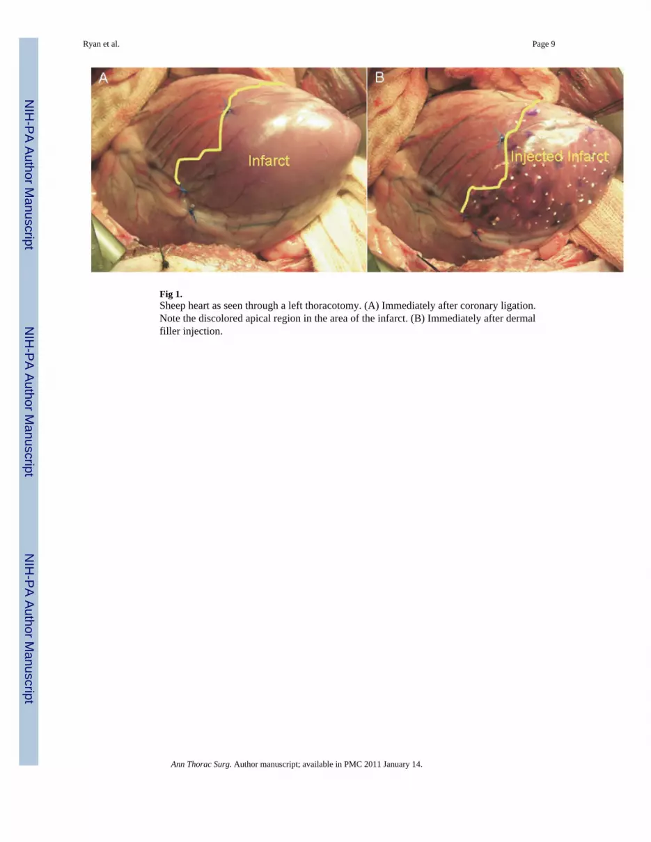
Fig 1.Sheep heart as seen through a left thoracotomy. (A) Immediately after coronary ligation.Note the discolored apical region in the area of the infarct. (B) Immediately after dermalfiller injection.
Ryan et al. Page 9
Ann Thorac Surg. Author manuscript; available in PMC 2011 January 14.
NIH
-PA Author Manuscript
NIH
-PA Author Manuscript
NIH
-PA Author Manuscript

Fig 2.American Society of Echocardiography 17-segment left ventricle model.
Ryan et al. Page 10
Ann Thorac Surg. Author manuscript; available in PMC 2011 January 14.
NIH
-PA Author Manuscript
NIH
-PA Author Manuscript
NIH
-PA Author Manuscript

Fig 3.Left ventricular global end-diastolic volumes (EDV) and end-systolic volumes (ESV) atbaseline (medium gray bars), 30 minutes after infarction (Post-MI; dark gray bars), and 15minutes after injection (Post-Injection; light gray bars) are plotted on common axes (A). Thecorresponding ejection fractions are illustrated in B. Data are presented as mean ± standarderror of the mean. *p < 0.05 with respect to the corresponding value at baseline; †p < 0.05with respect to the corresponding value after infarction.
Ryan et al. Page 11
Ann Thorac Surg. Author manuscript; available in PMC 2011 January 14.
NIH
-PA Author Manuscript
NIH
-PA Author Manuscript
NIH
-PA Author Manuscript

Fig 4.Regional end-diastolic volume (EDV; A), end-systolic volume (ESV; B), and ejectionfraction (C) for each of the three left ventricular regions are presented at baseline (mediumgray bars), 30 minutes after infarction (Post-MI; dark gray bars), and 15 minutes afterinjection (Post-Injection; light gray bars). Data are presented as mean ± standard error of themean. *p < 0.05 with respect to the corresponding value at baseline; †p < 0.05 with respectto the corresponding value after infarction.
Ryan et al. Page 12
Ann Thorac Surg. Author manuscript; available in PMC 2011 January 14.
NIH
-PA Author Manuscript
NIH
-PA Author Manuscript
NIH
-PA Author Manuscript

Fig 5.Apical segmental end-diastolic volume (EDV; A), end-systolic volume (ESV; B), andejection fraction (C) for each of the five left ventricular segments comprising the apicalregion are presented at baseline (medium gray bars), 30 minutes after infarction (Post-MI;dark gray bars), and 15 minutes after Radiesse injection (Post-Injection; light gray bars).Data are presented as mean ± standard error of the mean. *p < 0.05 with respect to thecorresponding value at baseline; †p < 0.05 with respect to the corresponding value afterinfarction.
Ryan et al. Page 13
Ann Thorac Surg. Author manuscript; available in PMC 2011 January 14.
NIH
-PA Author Manuscript
NIH
-PA Author Manuscript
NIH
-PA Author Manuscript

Fig 6.Apical anterior (A), apical septal (B), apical inferior (C), apical lateral (D), and apical cap(E) segmental volumes have been plotted as functions of normalized time at baseline (blacklines), 30 minutes after infarction (Post-MI; red lines), and 15 minutes after Radiesseinjection (Post-Injection; blue lines) for the 15-subject cohort on common axes. Data arepresented as mean (solid line) ± standard error of the mean (dotted lines). End-diastoleoccurs at 0 ms and 800 ms, whereas end-systole occurs at 500 ms. Note in E that the apicalcap segment is profoundly dyskinetic after infarction but becomes virtually akinetic afterinjection.
Ryan et al. Page 14
Ann Thorac Surg. Author manuscript; available in PMC 2011 January 14.
NIH
-PA Author Manuscript
NIH
-PA Author Manuscript
NIH
-PA Author Manuscript

Fig 7.Mason trichrome stain of infarct tissue at ×2 and ×20 from untreated (A, C) 4-week-oldovine infarcts and infarcts of the same age injected with 1.3 mL of Radiesse (B, D).Untreated infarcts are thin and relatively acellular with occasional islands of residualvacuolated myocytes. Radiesse-treated infarcts are thicker with more extensive collagendeposition. The calcium hydroxyapatite microspheres are apparent. The carrier agent hasbeen replaced by a dense cellular infiltrate.
Ryan et al. Page 15
Ann Thorac Surg. Author manuscript; available in PMC 2011 January 14.
NIH
-PA Author Manuscript
NIH
-PA Author Manuscript
NIH
-PA Author Manuscript

NIH
-PA Author Manuscript
NIH
-PA Author Manuscript
NIH
-PA Author Manuscript
Ryan et al. Page 16
Tabl
e 1
Chr
onic
Rem
odel
ing
and
Hem
odyn
amic
Dat
a
Pre-
MI
Post
-MI
Afte
r In
ject
ion
4 W
eeks
Pos
t-MI
Var
iabl
eC
ontr
olR
adie
sse
Con
trol
Rad
iess
eC
ontr
olR
adie
sse
Con
trol
Rad
iess
e
LVES
V (m
L)23
.5 ±
2.2
23.7
± 1
.638
.9 ±
2.4
a39
.9 ±
1.8
a39
.3 ±
2.6
a33
.4 ±
1.7
a,b
61.2
± 3
.6a
44.5
± 3
.9a,
b
LVED
V (m
L)48
.1 ±
3.7
47.1
± 3
.066
.1 ±
3.4
a64
.8 ±
3.0
a65
.9 ±
3.5
a58
.6 ±
2.8
a85
.7 ±
3.7
a70
.7 ±
3.9
a
Ejec
tion
frac
tion
0.51
7 ±
0.01
70.
498
± 0.
015
0.41
0 ±
0.01
9a0.
382
± 0.
011a
0.40
3 ±
0.01
9a0.
427
± 0.
018a
0.29
5 ±
0.01
6a0.
373
± 0.
017a
,b
Car
diac
out
put (
L/m
in)
4.6
± 0.
24.
9 ±
0.4
2.7
± 0.
5a2.
6 ±
0.5a
2.6
± 0.
4a3.
1 ±
0.2a
2.8
± 0.
3a3.
9 ±
0.3a
,b
LVED
P (m
m H
g)5
± 2
4 ±
1.5
19 ±
620
± 7
19 ±
616
± 5
12 ±
49
± 3
CV
P (m
m H
g)13
± 3
12 ±
214
± 2
13 ±
415
± 3
12 ±
211
± 2
10 ±
1
a p <
0.05
ver
sus P
re-M
I.
b p <
0.05
ver
sus C
ontro
l.
CV
P =
cent
ral v
enou
s pre
ssur
e; L
VED
P =
left
vent
ricul
ar e
nd-d
iast
olic
pre
ssur
e; L
VED
V =
left
vent
ricul
ar e
nd-d
iast
olic
vol
ume;
LV
ESV
= le
ft ve
ntric
ular
end
-sys
tolic
vol
ume;
MI =
myo
card
ial i
nfar
ctio
n.
Ann Thorac Surg. Author manuscript; available in PMC 2011 January 14.


