Embed Size (px)
Citation preview
Cell Host & Microbe
Article
IgA Response to Symbiotic Bacteriaas a Mediator of Gut HomeostasisDaniel A. Peterson,1,2 Nathan P. McNulty,2 Janaki L. Guruge,2 and Jeffrey I. Gordon2,*1Department of Pathology and Immunology2Center for Genome Sciences
Washington University School of Medicine, St. Louis, MO, 63108 USA*Correspondence: [email protected]
DOI 10.1016/j.chom.2007.09.013
SUMMARY
Colonization of germ-free mice with a normalgut microbiota elicits bacteria-specific IgA anti-body responses. The effects of these responseson microbial and host biology remain poorlydefined. Therefore, we developed a gnotobioticmouse model where the microbiota is reducedto one bacterial species, and the antibody rep-ertoire to a single, monoclonal IgA against thebacterium’s capsular polysaccharide. Bacter-oides thetaiotaomicron was introduced intogerm-free wild-type, immunodeficient Rag1�/�,or Rag1�/� mice harboring IgA-producing hy-bridoma cells. Without IgA, B. thetaiotaomicronelicits a more robust innate immune responseand reacts to this response by inducing genesthat metabolize host oxidative products. IgA re-duces intestinal proinflammatory signaling andbacterial epitope expression, thereby balancingsuppression of the oxidative burst with theantibody’s negative impact on bacterial fitness.These results underscore the adaptive immunesystem’s critical role in establishing a sustain-able host-microbial relationship. Immunoselec-tion of bacterial epitope expression may con-tribute to the remarkable strain-level diversityin this ecosystem.
INTRODUCTION
Our adult bodies are home to trillions of microbes, produc-
ing a ‘‘supraorganism’’ whose microbial cell population
exceeds the number of human cells by an estimated order
of magnitude. Our gut contains the vast majority of our
bacterial and archaeal partners (Ley et al., 2006a). Over
90% of the bacterial phylogenetic types (phylotypes) be-
long to just two divisions (superkingdoms)—the Bacteroi-
detes and the Firmicutes. Each of us appears to harbor
a distinctive collection of species- and strain-level phylo-
types belonging to these two dominant bacterial divisions.
The presence of relatively few bacterial and archaeal
divisions in this environmental ecosystem indicates that
328 Cell Host & Microbe 2, 328–339, November 2007 ª2007 Els
strong selective pressures operate to shape the commu-
nity (Ley et al., 2006a).
Deciphering how an individual’s immune system and
microbiota coevolve should help provide answers to
a number of intriguing questions. How is our microbiota
selected? How does it manifest compositional diversity
and functional stability? How does it adapt to changes in
our lifestyles? How do perturbations in microbial ecology
contribute to certain pathologic states such as infectious
diarrheas, inflammatory bowel diseases, and metabolic
abnormalities, for example (Frank et al., 2007; Ley et al.,
2006b; Turnbaugh et al., 2006)? How can the represen-
tation of components of our microbiota be intentionally
manipulated for therapeutic benefit?
Changes in gut microbial ecology have been docu-
mented in mice with immune deficiencies (Fagarasan
et al., 2002; Suzuki et al., 2004). Colonization of germ-
free mice with members of the normal gut microbiota elicits
bacteria-specific IgA responses (Shroff et al., 1995). In the
few reports where the specificities of these antibodies
have been determined, their effects on microbial and
host biology have not been described (Macpherson
et al., 2000). However, when immunodeficient Rag1�/� or
SCID mice, which lack a functional adaptive immune sys-
tem, are colonized with single or multiple species of bacte-
ria, they display a more robust innate immune response
than their immunocompetent wild-type counterparts (Keil-
baugh et al., 2005; Cash et al., 2006). This indicates that in
normal mice the adaptive immune response plays a critical
role in minimizing activation of the innate immune system
by the gut microbiota. In models of T cell-mediated colitis
produced either by adoptive transfer of CD45RBhigh T cells
into SCID recipients or by gene knockouts, immunoglobu-
lin (Ig) impacts mucosal inflammation (Gerth et al., 2004;
Kanai et al., 2006). Moreover, serum responses to micro-
bial antigens are associated with inflammatory bowel dis-
ease in mice and humans (Landers et al., 2002), as are de-
fects in the innate and adaptive immune system (Cobb
et al., 2006; Izcue et al., 2006).
Based on these observations, we developed a gnotobi-
otic mouse model to define mechanisms by which an IgA
response plays a key role in establishing and maintaining
a noninflammatory host-microbial relationship. In this
model, the diversity of the gut microbiota is reduced to
a single prominent bacterial member of the human distal
intestinal ecosystem, and the repertoire of the adaptive
evier Inc.

Cell Host & Microbe
IgA and Host-Bacterial Symbiosis in the Gut
Figure 1. Characterization of the Epitope
Recognized by the 225.4 Monoclonal IgA
Antibody
(A) ELISA reveals that the antibody reacts only
with the type strain (VPI-5482) and not with any
other B. thetaiotaomicron isolates or four other
sequenced human gut-associated Bacter-
oides species. Colors indicate the degree of
phylogenetic relatedness to B. thetaiotaomi-
cron VPI-5482 (defined by percent 16S rRNA
nucleotide sequence identity using ARB
[http://www.arb-home.de/]).
(B) Expression of the 225.4 epitope in fecal
pellets obtained from germ-free wild-type B6
mice colonized for the indicated periods of
time. ELISAs were performed using biotiny-
lated 225.4.
(C) ELISA assays of transposon mutants with
diminished or absent 225.4 reactivity (B. the-
taiotaomicron [BT] gene name given at site of
transposon insertion).
(D) Reactivity of 225.4 mAb with isogenic B.
thetaiotaomicron strains containing pGERM
disruptions of CPS locus expression. Mutants
were all grown in TYG medium to stationary
phase: aliquots, diluted in bicarbonate buffer,
were used to coat ELISA plates. 225.4 recog-
nized all but the CPS4 mutant.
(E) Dose-response ELISA of a preparation of
the 225.4 epitope purified from B. thetaiotao-
micron using the hot water-phenol method
typically employed to isolate capsular polysac-
charides. Prior to the assay, lipopolysaccha-
ride (LPS) was removed by ultracentrifugation.
immune system is reduced to one naturally primed Ig
directed against an identified capsular polysaccharide
epitope expressed by the bacterium in vivo.
RESULTS
We chose B. thetaiotaomicron as a model symbiont for our
experiments. It possesses a large arsenal of glycoside
hydrolases for breaking down dietary polysaccharides
that our own human proteome is ill-equipped to process
(Sonnenburg et al., 2005) and efficiently colonizes the
intestines of adult germ-free C57Bl/6J mice. Twenty-four
hours after gavage with 108 CFU of the sequenced
B. thetaiotaomicron type strain, VPI-5482, bacteria
achieve a density in the small intestine and cecum that
does not change significantly over the ensuing 6 months
(Figure S1). ELISA of serum levels of IgG subtypes, IgM
and IgA 1 day, 2 days, 4 days, 7 days, 14 days, 28 days,
and 6 months after gavage of germ-free recipients dis-
closed that the IgG2a subtype exhibited the greatest rela-
tive increase (75-fold), while IgG1 changed %1.5-fold
(Figure S2), consistent with a CD4 T-helper 1 (Th1) envi-
ronment (Mazmanian et al., 2005). These observations
led us to predict that B. thetaiotaomicron reactive B cells
would be present within the intestinal wall at the 14 day
Cell Host &
time point and could be captured by hybridoma fusion,
thereby allowing us to immortalize a single naturally
primed, bacterial epitope-specific IgA response (see
Experimental Procedures for additional details).
B. thetaiotaomicron-primed IgA-producing hybridomas
were identified (see Experimental Procedures), including
one that produced a mAb, named 225.4, specific for the
sequenced VPI-5482 type strain. ELISA disclosed that
this mAb does not react with bacteria closely related to
B. thetaiotaomicron, or with any of 1200 bacterial colonies
recovered from the ceca of conventionally raised B6 mice
and feces of healthy human donors (Figure 1A and data
not shown). Using biotinylated 225.4 mAb, we tested fecal
pellets for expression of the epitope and found that it was
produced by the bacterium in vivo starting from day one of
colonization (Figure 1B).
With a finished genome sequence available for B. the-
taiotaomicron VPI-5482 (Xu et al., 2003), we were able to
identify genes required for generation of the 225.4
epitope. A library of 4600 transposon (Tn4351) mutants
was generated (see Experimental Procedures for details)
and screened by ELISA for colonies that had lost their
225.4 reactivity (Figure 1C). Sixty-five percent of the
225.4-negative mutants contained inserts in the capsular
polysaccharide synthesis 4 (CPS4) locus; three of the
Microbe 2, 328–339, November 2007 ª2007 Elsevier Inc. 329

Cell Host & Microbe
IgA and Host-Bacterial Symbiosis in the Gut
Table 1. Tn4351 Insertion Sites Associated with Markedly Diminished or Absent 225.4 Epitope Expression inB. thetaiotaomicron
Insertion Site Gene Gene Description Insertion Site Gene Gene Description
/ 100 base pairs upstream
of BT1358
/ BT4576 hypothetical protein
BT1358 UpxZ homolog BT4575 hypothetical protein
BT1357 UpxY homolog
/ BT1356 polysialic acid transport protein
kpsD precursor
/ BT2642 conserved hypothetical
protein
/ BT1355 polysaccharide biosynthesis proteinchain length determinator
BT2643 conserved hypotheticalprotein
BT1354 flippase BT2644 DNA topoisomerase I
/ BT1353 glycosyltransferase (GT 2)
BT1352 glycosyltransferase (GT 4)
BT1351 glucose-1-phosphatecytidylyltransferase
/ BT2952 SusC homolog
/ BT1350 CDP-glucose 4,6-dehydratase BT2951 SusD homolog
BT1349 dTDP-4-dehydrorhamnose3,5-epimerase
BT2950 hypothetical protein
BT1348 CDP-abequose synthase BT2949 a-1,6-mannanase
BT1347 glycosyltransferase (GT 2) BT2948 a-1,2-mannosidase
*/ BT1346 capA domain protein
BT1345 glycosyltransferase / BT3775 mannosyltransferase
*/ BT1344 glycosyltransferase (GT 4)
BT1343 capsule biosynthesis protein / BT1035 b hexaminidase
BT1342 UDP-glucuronic epimerase BT1034 putative signal transducer
BT1341 UDP-glucose 6-dehydrogenase BT1033 hypothetical protein
BT1340 lipopolysaccharide biosynthesis
glycosyltransferase (GT 2)
BT1032 a-1,2-mannosidase
BT1339 undecaprenyl-phosphateacetylglucosaminyltransferase
BT1338 DTDP-4-dehydrorhamnose
3,5-epimerase
/ BT0664 ABC transporter
substrate-binding protein
BT0665 hypothetical protein
BT0666 hypothetical protein
BT0667 signal peptidase I
BT0668 glutathione synthetase
Arrows indicate the gene that contains the transposon insertion. Downstream genes in known or predicted operons are also listed
(Westover et al., 2005).
* Indicates two unique mutants were identified in this gene. Glycosyltransferases (GT) are classified based on their assignment to
families in the Carbohydrate-Active Enzymes (CAZy) database (http://www.cazy.org/).
other six loci are known or predicted to be related to var-
ious aspects of carbohydrate metabolism (Table 1).
To confirm the CPS4 dependence of the 225.4 epitope,
we disrupted all eight of the bacterium’s CPS loci using
a suicide vector. Each of the resulting isogenic strains har-
bored a polar insertion in the first gene of each locus.
When analyzed by ELISA, only the CPS4 disruption was
associated with loss of epitope (Figure 1D). Hot water-
330 Cell Host & Microbe 2, 328–339, November 2007 ª2007 Els
phenol extraction of wild-type bacteria recovered after
overnight growth in rich TYG medium revealed that
225.4 reactivity was retained in the water phase of the
extract (Figure 1E). In addition, the epitope was resistant
to digestion with DNase, RNase, and proteases, but sus-
ceptible to acid hydrolysis (data not shown). Thus, a com-
bination of biochemical and genetic data are consistent
with the notion that 225.4 recognizes a surface capsular
evier Inc.
Cell Host & Microbe
IgA and Host-Bacterial Symbiosis in the Gut
carbohydrate epitope whose production is directed
by the CPS4 locus and influenced by additional genes
distributed throughout the genome. The exquisite strain
specificity of the antibody is reminiscent of strain-specific
responses to encapsulated pathogens such as S. pneu-
moniae (Coughlin et al., 1998). While it is unclear how
well the 225.4 specificity is represented in the normal
anti-B. thetaiotaomicron repertoire, our results indicate
that it is clearly a member of a naturally primed immune
response.
The next question we addressed was the impact of this
specific antisymbiont IgA in vivo. Therefore, germ-free
Rag1�/� mice, which lack mature T and B cells, were
injected with 225.4-producing hybridoma cells under their
dorsal skin, creating a hybridoma ‘‘backpack’’ mouse
where the only Ig in the serum and gut lumen was 225.4
(Michetti et al., 1992). We waited 10 days before coloniz-
ing these backpack animals with wild-type B. thetaiotao-
micron: this decision was based on an analysis of the
time course of rise of serum 225.4 mAb levels in Rag1�/�
backpack controls (Figure S3A). Variations in levels of
225.4 IgA in the sera of different animals at this time point
were linked to differences in the growth of their backpack
hybridomas and correlated with levels of antibody ex-
creted into their intestinal lumen (r2 = 0.94 based on ELISA
of fecal pellets; n = 12 mice; Figure S3B). This feature al-
lowed us to conduct a dose-response study of the effects
of the capsular epitope-specific IgA on the host-bacterial
relationship.
The first in vivo effect of the 225.4 antibody response
that we measured was on expression of its own epitope.
Ten days after colonization of germ-free Rag1�/� mice
with implanted backpacks, and Rag1�/� controls with
no backpacks (n = 4–16/group, 2 replicate experiments),
animals were sacrificed, and hot water-phenol extracts
of their cecal contents were prepared. ELISA revealed
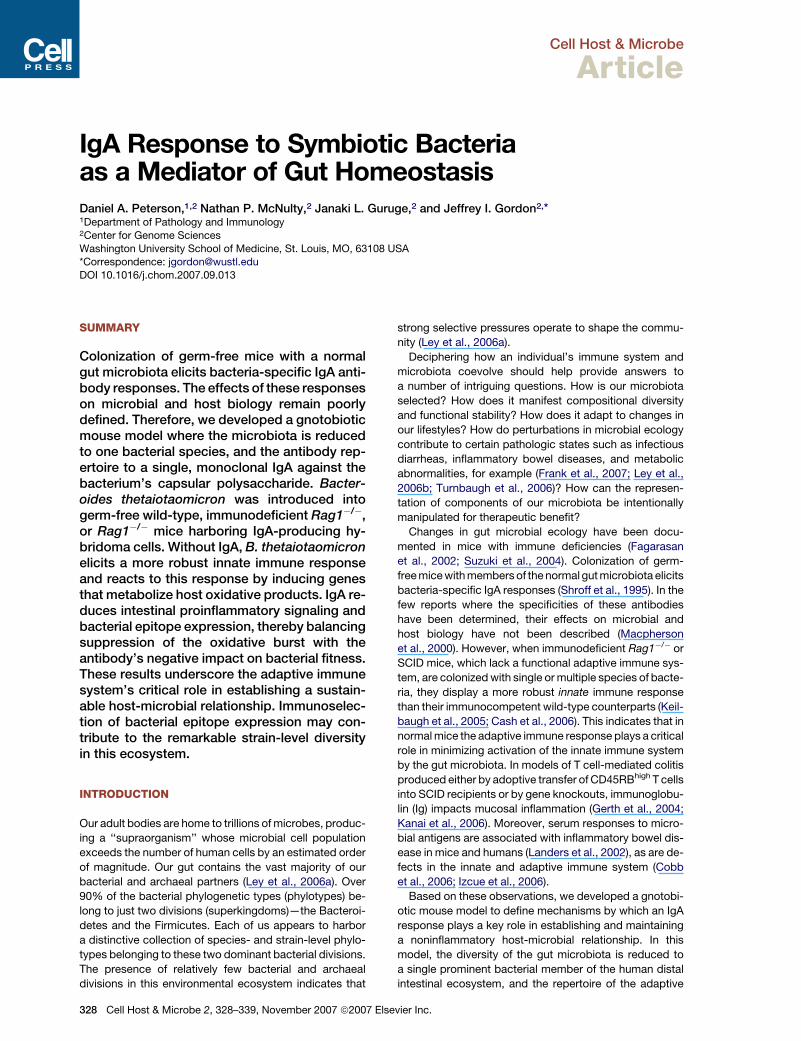
that (1) levels of the 225.4 antigen were significantly de-
creased in the ceca of Rag1�/� backpack mice even
though there were no significant differences in bacterial
density between the two groups of animals (p < 0.01; Fig-
ures 2A and 2B) and (2) there was an inverse relationship
between antibody levels and luminal levels of its epitope:
i.e., lowest levels of epitope were documented in mice
with the greatest concentration of 225.4 IgA (r2 = 0.73;
Figure 2C). qRT-PCR assays of RNA prepared from cecal
contents confirmed significantly decreased expression of
the bacterial CPS4 locus in the presence of the 225.4
antibody (p < 0.05; Figure 2D).
The second effect of the antibody response we mea-
sured was its impact on bacterial fitness. Colony-forming
unit (CFU) and ELISA assays of cecal contents, harvested
from gnotobiotic Rag1�/� mice with and without back-
packs 10 days after cocolonization with equivalent num-
bers of the isogenic wild-type and DCPS4 mutant strains,
established that, in the absence of any 225.4 antibody, the
representation of the mutant in this habitat was 100- to
1000-fold lower than wild-type (Figures 3A–3C). The
basis of the fitness defect produced by DCPS4 remains
unknown.
Cell Host
The ratio of wild-type to isogenic DCPS4 mutant cells
was shifted by an order of magnitude in favor of the mutant
in mice with high levels of cecal 225.4 antibody, reflecting
a reduction in the dominance of the wild-type bacterium
(the representation of the wild-type strain decreased
from 99.9% to 99% of the population; Figure 3C). In mice
with <5 mg (0.4–2.3 mg) of 225.4 antibody per ml of cecal
contents, B. thetaiotaomicron 225.4 epitope expression
was still significantly reduced compared to Rag1�/� ani-
mals without backpacks (Figure 3D), but bacterial fitness
was not detectably reduced as judged by the ratio of
wild-type to DCPS4 mutant CFUs in their ceca (Figure 3C).
We next examined the in vivo effect of the 225.4 anti-
body on global bacterial gene expression. Whole-genome
transcriptional profiling with custom B. thetaiotaomicron
GeneChips containing probe sets that cover 98.6% of
the bacterium’s 4779 protein-coding genes (Sonnenburg
et al., 2005) was used to define the impact of this engi-
neered antisymbiont immune response on bacterial phys-
iology in vivo. We compared wild-type B. thetaiotaomicron
Figure 2. Impact of 225.4 IgA In Vivo
Adult germ-free Rag1�/� mice, with or without 225.4 mAb-producing
backpack hybridomas, were colonized for 10 days with wild-type B.
thetaiotaomicron.
(A) Concentration of 225.4 epitope in cecal contents (see Experimental
Procedures).
(B) Density of colonization of the ceca of gnotobiotic mice.
(C) Correlation between 225.4 epitope levels and 225.4 IgA levels in
cecal contents.
(D) qRT-PCR assays of RNA prepared from cecal contents. Expression
of BT1348, a member of the CPS4 locus, was used as a biomarker for
locus expression. Data were normalized to 16S rRNA (for each sample)
and expressed as fold difference of each mouse compared to a B.
thetaioatomicron-colonized wild-type C57Bl/6J (B6) reference control
(DDCT method). *p < 0.05; **p < 0.01.
& Microbe 2, 328–339, November 2007 ª2007 Elsevier Inc. 331
Cell Host & Microbe
IgA and Host-Bacterial Symbiosis in the Gut
recovered from the cecum after a 10 day colonization of
Rag1�/� mice without backpacks (reference control, no
antibody response), with bacteria harvested from Rag1�/�
backpack animals (monoclonal antibody response to
a single surface capsular epitope) and wild-type B6 mice
minus backpacks (polyclonal antibody response to multi-
ple B. thetaiotaomicron epitopes) (n = 4 mice/group; each
cecal sample analyzed separately with its own bacterial
GeneChip) (see Figure S4 for the results of unsupervised
hierarchical clustering of GeneChip data sets and Table
S1 for an annotated list of differentially expressed genes).
Notably, in the absence of an immune response (i.e.,
Rag1�/� mice without backpacks), there was significantly
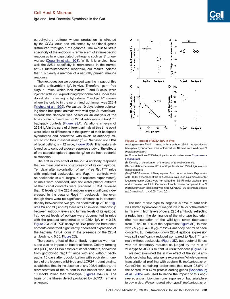
Figure 3. The Impact of Naturally Primed 225.4 Antibody onCompetition In Vivo
Germ-free Rag1�/� mice, with or without 225.4-producing hybridoma
backpacks, were colonized with a 1:1 mixture of the B. thetaiotaomi-
cron type strain (VPI-5482) and an isogenic CPS4 null mutant
(DCPS4; erythromycin-resistant). Cecal contents were harvested
10 days later, and the representation of the two strains was defined
by plating on medium with and without erythromycin.
(A) CFU of wild-type strain in Rag1�/� animals, Rag1�/� mice plus
backpack (range of IgA levels, 5.75–175 mg/ml cecal contents;
geometric mean, 18.6 mg/ml), or ‘‘backpack-low’’ animals (cecal
IgA, 0.4–2.3 mg/ml; geometric mean, 0.9 mg/ml). n.s., no significant
difference.
(B) The less competitive DCPS4 mutant demonstrates a 10-fold in-
crease in density in backpack mice with cecal IgA > 5 mg/ml compared
to Rag1�/� controls or Rag1�/� ‘‘backpack-low’’ animals.
(C) Ratio of wild-type to DCPS4 bacterial CFU/ml of cecal contents in
Rag1�/�, Rag1�/� backpack, or Rag1�/� ‘‘backpack-low’’ mice. The
competitive advantage of wild-type B. thetaiotaomicron decreases
nearly 10-fold in backpack mice with cecal IgA > 5 mg/ml.
(D) 225.4 epitope expression the ceca of Rag1�/� mice without back-
packs, and Rag1�/�mice with high and low levels of cecal IgA. Levels
of epitope are significantly decreased in backpack mice. Wild-type B6
mice (polyclonal IgA control) have the same levels of wild-type and
DCPS4 CFU, and the same 225.4 epitope levels, as Rag1�/� controls
(p > 0.05; data not shown). **p < 0.01 ***p < 0.001 according to
Student’s t test or ANOVA, as appropriate.
332 Cell Host & Microbe 2, 328–339, November 2007 ª2007 Els
higher expression of (1) an operon (BT1414-1418) that
encodes nitrite reductase, (2) another gene (BT0687) in-
volved in nitric oxide metabolism (Rodionov et al., 2005),
and (3) an operon encoding subunits of cytochrome D ubiq-
uinol oxidase (implicated in aerotolerance) (Figure 4A).
Inducible nitric oxide synthase (iNOS) is a prototypic
member of the host’s oxidative response pathway, and
nitric oxide is an evolutionarily conserved component of
the innate immune response (Davidson et al., 2004).
qRT-PCR assays of RNA prepared from the distal small
intestines of mice belonging to the three different groups
established that iNOS expression was, on average, 5-
fold higher in Rag1�/�mice without backpacks compared
to Rag1�/�mice with backpacks, and 23-fold higher com-
pared to wild-type B6 animals (p < 0.01; Figure 4B). More-
over, in Rag1�/� mice with and without backpack tumors
there was a direct and significant correlation between
levels of expression of BT1417 (nitrite reductase locus
member) and iNOS (r2 = 0.75; Figures 4C and 4D).
These results indicate that, in the absence of a secreted
IgA, B. thetaiotaomicron elicits a more robust oxidative
response in the host and adapts by inducing bacterial
genes involved in the metabolism of products of the host
response. They provide evidence that a quiescent rela-
tionship between B. thetaiotaomicron and its host is pred-
icated on the Ig(A) response and are consistent with
a model where an iterative set of adaptations involving
symbiont and host results in a coevolved homeostasis.
In both Rag1�/� and wild-type gnotobiotic mice, possess-
ing CPS4 gives the bacteria a competitive advantage over
isogenic strains that lack it. There appears to be an ‘‘opti-
mal’’ level of anti-CPS response: at high levels of 225.4
IgA, CPS4-associated epitope expression is decreased,
but not extinguished (Figures 2A–2C), and innate cell acti-
vation is diminished; however, if epitope levels decrease
too much under immune pressure B. thetaiotaomicron
may elicit a more robust innate immune response (as sug-
gested by Figure 4E).
The final effect of the antisymbiont antibody response
examined was its impact on intestinal gene expression.
Transcriptional profiling of distal small intestinal RNA
prepared from the same mice as those described
above revealed that, in addition to iNOS, wild-type
B. thetaiotaomicron-colonized Rag1�/� animals without
backpacks exhibited a marked upregulation of other genes
involved in innate inflammatory responses when com-
pared to colonized Rag1�/� mice with backpacks or
wild-type B6 animals (n = 3–5/group; see Figure S5 and Ta-
ble S2 for gene lists). Ingenuity Pathway Analysis (IPA) soft-
ware was subsequently used to organize these regulated
genes, as well as genes that are expressed but not regu-
lated, into known interaction networks. A large network
composed of complement components, phospholipase
A2 group 2A (PLA2G2A), regenerating islet-derived 1-
b (Reg1-b), interleukin-1 receptor-antagonist (IL1-RN),
and iNOS (with concomitant decreases in expression of ar-
ginase 2, a competitive iNOS inhibitor) is shown in Figure 5
and Tables S3 and S4. Downstream signaling pathways
are also upregulated in Rag1�/� mice without backpacks
evier Inc.

Cell Host & Microbe
IgA and Host-Bacterial Symbiosis in the Gut
Figure 4. Functional Genomics Analysis
of the Effects of 225.4 IgA on Bacterial
and Host Gene Expression
(A) B. thetaiotaomicron GeneChip analysis of
bacterial genes, known or predicted to be in-
volved in oxidative stress responses, that are
modulated by the presence of an adaptive im-
mune response (i.e., whose expression was
significantly different when comparing Rag1�/�
to backpack or B6 mice). Standard deviations
above (increasing red) or below (increasing
green) the mean level of expression (black) of
a gene across all animals are indicated; *p <
0.05 according to Student’s t test. Pathways
for nitric oxide (NO) metabolism are shown.
(B–E) qRT-PCR analysis of mouse iNOS
(NOS2a) and bacterial nitrite reductase
(BT1417) expression in vivo. The relative ex-
pression in each sample was compared to
a reference mouse in the wild-type B6 control
group (indicated by open circle). (B) RNA iso-
lated from the distal small intestines of mice
colonized for 10 days with wild-type B. thetaio-
taomicron. Rag1�/� mice without backpacks
have significantly higher levels of iNOS expres-
sion than wild-type B6 controls. Backpack
Rag1�/� mice (range of cecal IgA, 9–153 mg/
ml; geometric mean, 38 mg/ml) display a signif-
icant decrease in iNOS expression compared
to Rag1�/� controls. (C) BT1417, a gene in a
nitrite reductase operon, demonstrates a
similar pattern of downregulation by 225.4
IgA in vivo. (D) Direct relationship between
BT1417 and iNOS expression. (E) In-
verse relationship between cecal 225.4 epi-
tope levels and iNOS expression. *p < 0.05;
**p < 0.01.
compared to controls, including signal transducer and
activator of transcription-3 (STAT3) and STAT6, and
interferon regulatory factor-8. In addition, other known
markers of innate immune cell activation not in this
network are affected, such as chemokine CX3CL1 (Frac-
talkine), nuclear factor kB (NFkB), and Reg3-g. Since
Rag1�/�mice lack B and T lymphocytes, these transcrip-
tional profiles reflect activation of genes in intestinal
epithelial cells (including members of the Paneth lineage),
natural killer cells, and/or macrophages/dendritic cells
(DC) that are present in these animals.
Two observations emphasize that the impact of the
engineered 225.4 IgA response on the host-microbial rela-
tionship is the result of recognition of a bacterial surface
epitope, rather than a unique property of the CPS4 epitope
per se. Colonization of germ-free Rag1�/� mice without
backpacks (no antibody response) and wild-type B6
mice with the DCPS4 strain produced the same effects
on iNOS expression as the wild-type strain (Figure 4 and
Figure S6). Moreover, in the wild-type B6 mice, where
225.4 specificity is only one component of the adaptive
polyclonal IgA response to colonization, iNOS but not
225.4 expression was suppressed, and there was no
significant impact the ratio of wild-type to DCPS4 mutant
cells in the cecum (Figure S6).
Cell Host
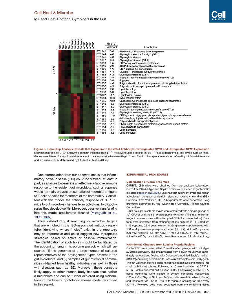
We found that the antisymbiont IgA response not only
suppresses CPS4 expression; it also induces expression
of another other capsular locus (CPS5; Figure 6), which,
in turn, could initiate another round of host immune re-
sponses. Studies of another gut Bacteroides, B. fragilis
(Krinos et al., 2001), have provided important insights
about the complex genetic regulation of CPS locus ex-
pression through inversion of upstream, locus-associated
promoters. We did not find any evidence of inversion in the
proximal promoter of CPS4 but cannot rule out that con-
trol of CPS4 expression could be dependent on invertible
elements positioned elsewhere in the B. thetaiotaomicron
genome. Our findings provide an additional view of this
regulation, one where anti-capsular antibodies modulate
expression of loci producing their cognate epitopes. Gut
symbionts exhibit an enormous capacity for surface vari-
ation, not only in response to immune pressure, but also
as a result of other forces including nutrient availability
(Sonnenburg et al., 2005; Bjursell et al., 2006). The ge-
nomes of prominent human gut Bacteroides species in-
cluding B. thetaiotaomicron, B. fragilis, B. vulgatus, and
B. distasonis have revealed that CPS loci are prominent
sites of variation, in part due to lateral gene transfer (Xu
et al., 2007). Moreover, comparisons of the B. thetaiotao-
micron type strain VPI-5482 and two additional human
& Microbe 2, 328–339, November 2007 ª2007 Elsevier Inc. 333

Cell Host & Microbe
IgA and Host-Bacterial Symbiosis in the Gut
gut-derived strains we have sequenced indicate that the
CPS4 locus is highly polymorphic. Successful gut symbi-
onts like B. thetaiotaomicron possess large collections of
paralogous outer member proteins involved in nutrient
sensing and acquisition. Thus, it seems logical to postu-
late that, at least from the bacterial perspective, CPS epi-
topes would be preferred ‘‘bait’’ for the adaptive immune
system in order to minimize immune recognition of key
nutrient-related proteins.
DISCUSSION
Our study supports the notion that the role for antibody in
the gut is to mediate tolerance. Tolerance in the gut may
Figure 5. Ingenuity Pathway Analysis of Host Genes Differen-
tially Expressed in B. thetaiotaomicron-Colonized Rag1�/�
Mice without Backpacks Compared with Rag1�/� Backpack
and Wild-Type B6 Animals
Mouse GeneChip analysis of the distal small intestines of animals sac-
rificed 10 days after colonization. In Rag1�/�mice without an antisym-
biont IgA response, there is augmented induction of iNOS (NOS2a) and
IFNgR2 (implies the presence of IFN-g). Complement components, IL-
18 and IL-13-RA/IL-4-R, IL-1RN, PLA2G2A, and Reg1B genes are also
induced. Genes downregulated in either wild-type B6 or Rag1�/�
backpack animals compared to Rag1�/�mice are highlighted in green,
while upregulated genes are in red. Full gene names, fold differences in
expression between the different groups of mice, and p values are pro-
vided in Table S2 and Figure S5. Solid lines indicate a direct interaction
between the gene products, while dashed lines indicate an indirect
interaction. Genes in bold/italic were significantly changed in the
comparison between Rag1�/� and Rag1�/� backpack or wild-type
B6 mice. Genes highlighted by gray were called present (expressed)
in the GeneChip data set, but their levels of expression were not signif-
icantly different between the groups of animals. Genes in white (open)
shapes were not in the GeneChip data set but are implicated as being
in the network by the Ingenuity Pathway Analysis (IPA) software.
334 Cell Host & Microbe 2, 328–339, November 2007 ª2007 E
be viewed as a quiescent homeostasis predicated on im-
mune recognition of members of its microbiota. Bacteria
that elicit excessive levels of IgA may suffer a greater com-
petitive disadvantage than those that elicit low to moder-
ate amounts. This requires that long-term residents mod-
ulate their immunodominant determinants continually,
likely providing one explanation for the extraordinary level
of strain-level diversity observed in the gut ecosystem. Al-
though relatively few human gut-associated Bacteroides
genomes have been sequenced, those that have reveal
a large representation of genes involved in the generation
of surface carbohydrates (Xu et al., 2003, 2007; Kuwahara
et al., 2004; Sonnenburg et al., 2005; Cerdeno-Tarraga
et al., 2005; Krinos et al., 2001; Fletcher et al., 2007).
Our observations are consistent with the adaptive immune
system being a driver of diversification of these surface
structures, with the beneficial outcome being promotion
of a noninflammatory relationship between gut symbiont
and host.
The innate immune response to bacteria, including the
generation of NO, is highly conserved in invertebrates
and vertebrates (Davidson et al., 2004). A key evolved
role of the adaptive immune system in vertebrates may
be to accommodate more complex microbial communi-
ties, even at the added risk of susceptibility to colonization
with pathogens, and/or autoimmunity (Hedrick, 2004;
McFall-Ngai, 2007). Our results support this concept and
provide evidence that a major role for the adaptive
immune system is to maintain ‘‘connection’’ with the gut
microbiota by selectively generating immune responses
to bacteria that stimulate the innate system. This arrange-
ment allows the host to detect new bacterial phylotypes,
and to ignore the presence of those that it has previously
encountered (memory). The result would be support of
greater diversity without sacrificing the essential protec-
tive role of the innate immune system in maintaining the
mucosal barrier.
There is an emerging model of mucosal immunity where
differentiation of B cells to IgA-producing plasma cells is
locally induced in the lamina propria of the small intestine
by local factors (Fagarasan et al., 2001): e.g., it appears
that T cell-independent class switching in the gut is in-
duced by expression of costimulatory molecules such as
CD40 ligand, APRIL, BlyS, and TGF-b by epithelial and
DCs (Litinskiy et al., 2002; Macpherson and Uhr, 2004;
He et al., 2007). The result of locally controlled IgA produc-
tion is to create a simple feedback loop that bypasses the
systemic immune system: i.e., where DCs, and epithelial
cells drive IgA production independent of T cells until suf-
ficient levels of IgA block microbial stimulation. This model
could be tested in the future by creating a quasi-monoclo-
nal (gnotobiotic) mouse with a T cell-deficient genetic
background that expresses the 225.4 antibody as a B
cell receptor knocked into the immunoglobulin gene locus
(Cascalho et al., 1996): colonization of this mouse with
B. thetaiotaomicron would reveal how efficiently IgA class
switching occurs in a highly defined system, and the
degree to which IgA production is antigen specific and T
cell dependent.
lsevier Inc.
Cell Host & Microbe
IgA and Host-Bacterial Symbiosis in the Gut
Figure 6. GeneChip Analysis Reveals that Exposure to the 225.4 Antibody Downregulates CPS4 and Upregulates CPS5 Expression
Expression profile for CPS4 and CPS5 genes in the ceca of Rag1�/�mice without backpacks, in Rag1�/� backpack animals, and in wild-type B6 mice.
Genes were filtered for significant differences in their expression between Rag1�/� and Rag1�/� backpack animals as defined by >1.5-fold difference
and a p value < 0.05 (determined by Student’s t test in dChip).
One extrapolation from our observations is that inflam-
matory bowel disease (IBD) could be viewed, at least in
part, as a failure to generate an effective adaptive immune
response to the resident gut microbiota: such a response
would normally prevent presentation of microbial antigens
to T cells specific for members of the community. Consis-
tent with this model, the antibody response of TCRa�/�
mice to gut microbes changes from polyclonal to oligoclo-
nal as they develop colitis. Moreover, passive transfer of Ig
into this model ameliorates disease (Mizoguchi et al.,
1996, 1997).
Thus, instead of just searching for microbial targets
that are enriched in the IBD-associated antibody reper-
toire, identifying where ‘‘holes’’ exist in the repertoire
may be informative and could suggest new therapeutic
strategies based on active or passive immunization.
The identification of such holes should be facilitated by
the upcoming human microbiome project, which will se-
quence (1) the genomes of a large number of cultured
representatives of the phylogenetic types present in the
gut microbiota, and (2) samples of gut microbial commu-
nities obtained from healthy individuals as well as those
with diseases such as IBD. Moreover, these concepts
likely apply to other human body habitats that harbor
a microbiota and can be further explored using elabora-
tions of the type of gnotobiotic mouse model described
in this report.
Cell Host &
EXPERIMENTAL PROCEDURES
Colonization of Germ-Free Mice
C57Bl/6J (B6) mice were obtained from the Jackson Laboratory.
Germ-free B6 wild-type and Rag1�/�mice were housed in gnotobiotic
isolators (Hooper et al., 2002) under a strict 12 hr light cycle and fed an
autoclaved, polysaccharide-rich, standard rodent chow diet (B&K
Universal, East Yorkshire, UK). All experiments were performed using
protocols approved by the Washington University Animal Studies
Committee.
Six- to eight-week-old males were colonized with a single gavage of
108 CFU of wild-type B. thetaiotaomicron strain VPI-5482, and/or an
isogenic mutant strain with a disrupted CPS4 locus (see below). Bac-
teria were harvested from stationary phase cultures in TYG medium
(1% tryptone, 0.5% yeast extract, 0.2% glucose) supplemented with
100 mM potassium phosphate buffer (pH 7.2), 4.1 mM cysteine,
200 mM histidine, 6.8 mM CaCl2, 140 nM FeSO4, 81 mM MgSO4,
4.8mM NaHCO3, 1.4mM NaCl, 1.9mM hematin, and5.8mM vitaminK3.
Hybridomas Obtained from Lamina Propria Fusions
Gnotobiotic mice were killed 2 weeks after gavage with wild-type
B. thetaiotaomicron. The small intestine from a given mouse was imme-
diately removed and flushed with Dulbecco’s modified Eagle’s medium
(DMEM) containingpenicillin (100units/ml) and streptomycin (100 mg/ml).
The gut was then opened along its cephalocaudal axis and minced into
small (�2–5 mm) pieces. Following a 15 min incubation at 37�C in
50 ml Hank’s buffered salt solution (HBSS) containing 5 mM EDTA,
tissue fragments were placed in DMEM containing collagenase
(100 units/ml; Sigma, St. Louis, MO) and dispase (0.5 units/ml; Fisher)
and incubated for 2 hr at 37�C, with vigorous shaking for 30 s every
30 min. Released cells were separated from the remaining tissue
Microbe 2, 328–339, November 2007 ª2007 Elsevier Inc. 335
Cell Host & Microbe
IgA and Host-Bacterial Symbiosis in the Gut
fragments by sedimentation on the bench-top for 1 min, and the resulting
cell suspension (supernatant fraction) was filtered through a Nytex filter
(70 mm diameter pore; BD Biosciences, Bedford, MA). Cells in the filtrate
were then combined with the myeloma fusion partner (P3X63.Ag8); the
combined population was washed three times in serum-free DMEM
medium, and the cells were fused by adding PEG1500 (50% w/v; Roche,
Mannheim, Germany; Kohler and Milstein, 1975). The ratio of IgA-, IgM-,
and IgG2b-producing hybridomas was 7:11:14 (n = 61 colonies scored
representing three animals; note that control experiments using intestinal
lamina propria lymphocytes recovered from age-matched, convention-
ally raised wild-type B6 animals produced hybridomas with a similar
isotype distribution).
B. thetaiotaomicron ELISAs
All ELISAs were performed using standard protocols (Velazquez et al.,
2001) in 96-well plates (Nunc-Maxisorb, Nalge-Nunc, Rochester, NY).
Bacterial strains suspended in PBS, lysates prepared from strains,
capsular polysaccharides purified from strains, or extracts of cecal
contents harvested from colonized gnotobiotic mice were assayed
by ELISA (see following paragraphs for descriptions of how samples
were prepared).
Samples to be assayed were diluted in sodium bicarbonate coating
buffer (15 mM sodium carbonate, 35 mM sodium bicarbonate, and
3 mM sodium azide [pH 8.5]). All steps were performed either at 4�C
overnight, or room temperature for 2 hr. Following addition of superna-
tants from hybridomas (�1–5 mg/IgA/well), goat horseradish peroxi-
dase (HRP)-conjugated anti-mouse IgA (Southern Biotech; 1:1000 di-
lution, 50 ml/well) was added. All ELISAs were developed using ABTS
[2,20-azino-bis(3-ethylbenzthiazoline-6-sulfonic acid)] (1 mM; Roche)
in citrate buffer (100 mM citric acid, 50 mM sodium phosphate [pH
4.2]) containing 0.03% H2O2. Reactions were read in an ELISA plate
reader (ThermoMax, Molecular Devices) at O.D. 405 nm.
Bacterial Lysates
Lysates were generated by suspending 1010 CFU of bacteria in 0.5 ml
of sterile PBS and sonicating the mixture in a Misonix XL-2020 sonica-
tor (10 min at a setting of ‘‘10’’). This mixture was then diluted 1:1000 in
bicarbonate buffer, and a 50 ml aliquot was added to each well of an
ELISA plate. Lysates contained capsular antigens, membrane and
cell wall antigens, plus intracellular antigens and were therefore used
for initial screening of hybridoma fusions.
Capsular Polysaccharides
Capsular polysaccharides were isolated from B. thetaiotaomicron VPI-
5482 cells, harvested at the stationary phase of growth in TYG me-
dium, using the hot water-phenol method (Jann et al., 1965). Briefly,
bacteria were extracted for 2 hr in phenol:water (1:1) at 65�C with
constant stirring. Phases were separated by gravity, and the top phase
was dialyzed for 3 days at 4�C against tap water. The dialyzed material
was subsequently lyophilized and suspended in sterile PBS (1 mg/ml).
LPS was removed by ultracentrifugation (65,000 3 g for 2 hr at 4�C).
Cecal Contents
Extractions were carried at 65�C in 1.5 ml eppendorf tubes containing
0.5 ml of a 1:100 dilution of cecal contents in PBS, and 0.5 ml phenol.
Samples were vortexed for 30 s, every 30 min over a 2 hr period. Fol-
lowing centrifugation at 20,000 3 g for 30 min at 4�C, the upper phase
was recovered, serial dilutions (in bicarbonate buffer) were made, and
ELISA was performed. To estimate the amount of 225.4 epitope pres-
ent in cecal contents, ELISA reactivity was standardized to a curve
constructed from serial dilutions of B. thetaiotaomicron (grown in
TYG to stationary phase) into bicarbonate buffer. This stationary cul-
ture was arbitrarily designated as having 1 3 109 units per ml (the
equivalent of CFU under these conditions). Therefore, in TYG-grown
B. thetaiotaomicron, one 225.4 epitope unit is the equivalent to one
colony-forming unit.
336 Cell Host & Microbe 2, 328–339, November 2007 ª2007 E
Isotype ELISA
ELISA plates were coated overnight at 4�C with goat anti-mouse Ig
(Southern Biotech; reacts with heavy and light chains), then washed
and blocked with 1% BSA-PBS (30 min at room temperature). Serial
dilutions of serum samples, obtained from B. thetaiotaomicron-
colonized mice at the time of their sacrifice, were added to the 96-
well plates (n = 3 biological replicates/per time point), followed by
HRP-conjugated, isotype-specific, secondary antibodies (1:1000
dilution in PBS-BSA; Southern Biotech). Fold change at each time
point following colonization was calculated as the isotype level of
each mouse relative to the mean of the germ-free control group.
Transposon Mutagenesis
Mutagenesis was performed using the donor plasmid pEP4351 and
B. thetaiotaomicron VPI-5482 (Salyers et al., 2000; Shoemaker et al.,
1986). Mutants were isolated based on their growth on Brain Heart
Infusion-sheep red blood cell agar plates containing erythromycin
(10 mg ml; to select for transposon-containing B. thetaiotaomicron)
and 600 mg/ml gentamicin (to select against any persistent E. coli). In-
dividual colonies were picked into 96-well plates and grown overnight
in TYG medium in anaerobic jars. Plates were split: one replicate was
frozen in 25% glycerol for later studies; the other plate was frozen for
subsequent ELISA screening with the 225.4 mAb.
Sites of transposon insertion in 225.4 antibody-negative colonies
were identified using an arbitrarily primed polymerase chain reaction
(AP-PCR). The AP-PCR protocol consists of a nested PCR with the fol-
lowing primers: Round 1, S3794 (50-ATCAGTATGCTTTGTGTGTG) and
either AR7 [50-GGCCACGCGTCGACTAGTAC(N)10GTAAT] or AR8 [50-
GGCCACGCGTCGACTAGTAC(N)10GATGC]; Round 2, ISF (50-TCG
GTTATATGTTTGCTCATCTGC) and AR2 (50-GGCCACGCGTCGACTA
GTAC) (7). The product of the second round of PCR was purified (QIA-
quick PCR Purification Kit; QIAGEN) and then sequenced from the
right arm of the transposon into the chromosomal DNA using primer
IS4908S (50-ATCCATTCAGAGTGAGAGAAAG). The results were com-
pared to the genome sequence of B. thetaiotaomicron VPI-5481
(GenBank accession number NC_004663) and Tn4351 (accession
number M17124): sequences aligning to both were considered to be
a positive hit.
pGERM-Directed Mutagenesis of CPS Loci
The first gene in every CPS locus was targeted using the pGERM
suicide vector (Xu et al., 2003; Sonnenburg et al., 2005). Mutagenesis
was performed using protocols described by Hooper et al. (1999). The
site of insertion of pGERM was verified by PCR (see Table S5 for a list
of primers), and by sequencing.
Backpack Experiments
Hybridomas were grown to 50%–75% confluency in DMEM and
washed three times in pyrogen-free saline. Cells were introduced
into the gnotobiotic isolators in a way that preserved sterility (see
Crawford and Gordon, 2005 for details) and then injected subcutane-
ously into 6-to 8-week-old male B6 Rag1�/� recipients (2 3 106 cells/
mouse; n = 16–20 mice/experiment; three independent experiments).
Ten days after implantation, each mouse was colonized with a sin-
gle gavage of 108 CFU of B. thetaiotaomicron (with or without the
DCPS4 isogenic mutant). Mice were then sacrificed 10 days following
colonization, and cecal contents were recovered. The small intestine
was rapidly divided into 16 equal length segments, and segments 9–
12 and 14 were snap frozen in liquid nitrogen for subsequent isolation
of total cellular RNA (see below).
The Impact of Anti-B. thetaiotaomicron on Bacterial RNA
Expression In Vivo
Cecal contents were flash frozen in liquid nitrogen immediately after
their harvest from each mouse in each treatment group (n = 4 mice/
group; selected based on cecal IgA levels contents [range of cecal
IgA, 9–153 mg/ml; geometric mean, 38 mg/ml]) and stored at �80�C
until use. An aliquot (�200 mg) was thawed in 2–3 volumes of
lsevier Inc.
Cell Host & Microbe
IgA and Host-Bacterial Symbiosis in the Gut
RNAProtect (QIAGEN) and centrifuged (3000 3 g for 10 min), and
500 ml of 200 mM NaCl/20 mM EDTA was added to the resulting pellet,
together with 200 ml of 20% SDS and 500 ml phenol:chloroform:isoamyl
alcohol (125:24:1; pH 4.5; Ambion). Acid-washed silica beads (Sigma;
212–300 mm diameter; 250 mg) were introduced, and bacteria were
lysed (Mini-Beadbeater; Biospec; ‘‘high’’ setting for 5 min at room tem-
perature). Following centrifugation (13,000 3 g for 3 min at 4�C), the
sample was re-extracted in phenol:chloroform:isoamyl alcohol, pre-
cipitated with 60 ml of 3 M sodium acetate (pH 5.2) plus 600 ml cold iso-
propanol, and RNA was purified (RNEasy kit; QIAGEN; note that resid-
ual genomic DNA was subsequently removed by treatment with
DNAfree; Ambion). The relative proportion of bacterial versus host
RNA in each preparation was defined by qRT-PCR, using primers di-
rected at B. thetaiotaomicron 16S rRNA (forward, 50-GGTAGTCCACA
CAGTAAACGATGAA; reverse, 50-CCCGTCAATTCCTTTGAGTTTC)
and mouse 18S rRNA (forward, 50-CATTCGAACGTCTGCCCTATC; re-
verse, 50-CCTGTGCCTTCCTTGGA). The results revealed that R90%
of the recovered rRNA in each preparation was bacterial. GeneChip
targets were prepared as described (Sonnenburg et al., 2005). The
cDNA product was isolated (QiaQuick Spin columns; QIAGEN), frag-
mented (DNase-I; Amersham Biosciences), and biotinylated (Enzo-
BioArray Terminal Labeling Kit). Standard Affymetrix protocols were
used for hybridization of the cDNA targets to each B. thetaiotaomicron
GeneChip (Sonnenburg et al., 2005, 2006).
GeneChip data were analyzed as follows. After normalization,
model-based expression values were generated (PM-MM model). Sig-
nals from spike-in control transcripts and oligo-B2 (Affymetrix) were
used to assess the quality of the target preparation and target hybrid-
ization, respectively. Comparisons were performed on specified Gen-
eChip data sets to identify genes up- or downregulated in the experi-
mental group (E) relative to the baseline group (B). Criteria used to
determine significant changes in expression were guided by the
median false discovery rate of 50 permutations in which samples
were randomly shuffled between groups. The following criteria were
routinely used to identify significantly upregulated genes while main-
taining an empirical false discovery rate of less than 0.05: (1) E/B >
1.5; (2) E-B R 100; (3) E = B p % 0.05; and (4) called ‘‘present’’
in R75% of ‘‘E’’ GeneChips.
Profiling Intestinal Gene Expression
RNA prepared from small intestinal segments 9–12 (treated as a single
domain) and segment 14 from each mouse in each treatment group
were combined proportionally (4:1) for each mouse. Targets were then
prepared and hybridized to Affymetrix Mouse Genome Moe430_2
GeneChips using standard protocols (Affymetrix). GeneChip data
sets were analyzed using DNA-chip analyzer (dChip). Genes up- or
downregulated in Rag1�/� mice with backpack hybridomas or wild-
type B6 animals (E) relative to Rag1�/� mice without backpacks (B)
were identified using the following criteria: (1) E/B > 1.2; (2) called
‘‘present’’ in at least two of three ‘‘E’’ GeneChips; and (3) E = B p < 0.05.
Real-Time Quantitative RT-PCR
Aliquots (2 ng) of the intestinal RNAs described above were reverse
transcribed in 20 ml reactions containing Superscript II RT (200 units;
Invitrogen) and oligo-dT15 (50 ng/ml; Roche). An aliquot (2 ml) containing
the cDNA product was then added to 25 ml qRT-PCR reaction mix-
tures together with Sybr-green reagent (12.5 ml; ABgene, Rochester,
NY) and primers specific for iNOS (50-CAGCTGGGCTGTACAAACC
TT; 50-CATTGGAAGTGAAGCGTTTCG; final concentration, 900 nM).
Assays were performed in triplicate using a Mx3000P QPCR System
instrument (Stratagene). Transcript levels within samples were normal-
ized to ribosomal protein L32 mRNA (50-CCTCTGGTGAAGCCCAA
GATC; 50-TCTGGGTTTCCGCCAGTTT), and fold changes were deter-
mined for each colonized mouse referenced against the lowest value
within the set (a wild-type B6 mouse) (DDCT method) (Sonnenburg
et al., 2006; Stappenbeck et al., 2002).
RNA prepared from cecal contents was also subjected to qRT-PCR
analysis of BT1348 gene expression. In this case, the reverse tran-
Cell Host &
scription reaction mix contained 75 ng of random hexamer primers
(Invitrogen). Transcript levels were normalized to 16S rRNA expression
(see Table S5 for primers).
GeneChip data were submitted to Gene Expression Omnibus (GEO)
under accession numbers GSE9018 and GSE9019.
Supplemental Data
The Supplemental Data include six supplemental figures and five sup-
plemental tables and can be found with this article online at http://
www.cellhostandmicrobe.com/cgi/content/full/2/5/328/DC1/.
ACKNOWLEDGMENTS
We thank David O’Donnell and Maria Karlsson for invaluable help with
husbandry of gnotobiotic mice, Sabrina Wagoner for expert technical
assistance, Jennifer Gill and Abigail Salyers for contributions to trans-
poson mutagenesis of B. thetaiotaomicron, and Emil Unanue and
members of the Gordon lab for many helpful discussions. This work
was supported by grants from the NIH (DK30292, T32HD07409,
T32CA09547).
Received: June 19, 2007
Revised: August 17, 2007
Accepted: September 19, 2007
Published: November 14, 2007
REFERENCES
Bjursell, M.K., Martens, E.C., and Gordon, J.I. (2006). Functional geno-
mic and metabolic studies of the adaptations of a prominent adult
human gut symbiont, Bacteroides thetaiotaomicron, to the suckling
period. J. Biol. Chem. 281, 36269–36279.
Cascalho, M., Ma, A., Lee, S., Masat, L., and Wabl, M. (1996). A quasi-
monoclonal mouse. Science 272, 1649–1652.
Cash, H.L., Whitham, C.V., Behrendt, C.L., and Hooper, L.V. (2006).
Symbiotic bacteria direct expression of an intestinal bactericidal lectin.
Science 313, 1126–1130.
Cerdeno-Tarraga, A.M., Patrick, S., Crossman, L.C., Blakely, G.,
Abratt, V., Lennard, N., Poxton, I., Duerden, B., Harris, B., Quail,
M.A., et al. (2005). Extensive DNA inversions in the B. fragilis genome
control variable gene expression. Science 307, 1463–1465.
Cobb, B.S., Hertweck, A., Smith, J., O’Connor, E., Graf, D., Cook, T.,
Smale, S.T., Sakaguchi, S., Livesey, F.J., Fisher, A.G., and
Merkenschlager, M. (2006). A role for Dicer in immune regulation.
J. Exp. Med. 203, 2519–2527.
Coughlin, R.T., White, A.C., Anderson, C.A., Carlone, G.M., Klein, D.L.,
and Treanor, J. (1998). Characterization of pneumococcal specific
antibodies in healthy unvaccinated adults. Vaccine 16, 1761–1767.
Crawford, P.A., and Gordon, J.I. (2005). Microbial regulation of
intestinal radiosensitivity. Proc. Natl. Acad. Sci. USA 102, 13254–
13259.
Davidson, S.K., Koropatnick, T.A., Kossmehl, R., Sycuro, L., and
McFall-Ngai, M.J. (2004). NO means ‘yes’ in the squid-vibrio symbio-
sis: Nitric oxide (NO) during the initial stages of a beneficial association.
Cell. Microbiol. 6, 1139–1151.
Fagarasan, S., Kinoshita, K., Muramatsu, M., Ikuta, K., and Honjo, T.
(2001). In situ class switching and differentiation to IgA-producing cells
in the gut lamina propria. Nature 413, 639–643.
Fagarasan, S., Muramatsu, M., Suzuki, K., Nagaoka, H., Hiai, H., and
Honjo, T. (2002). Critical roles of activation-induced cytidine deami-
nase in the homeostasis of gut flora. Science 298, 1424–1427.
Fletcher, C.M., Coyne, M.J., Bentley, D.L., Villa, O.F., and Comstock,
L.E. (2007). Phase-variable expression of a family of glycoproteins
Microbe 2, 328–339, November 2007 ª2007 Elsevier Inc. 337
Cell Host & Microbe
IgA and Host-Bacterial Symbiosis in the Gut
imparts a dynamic surface to a symbiont in its human intestinal eco-
system. Proc. Natl. Acad. Sci. USA 104, 2413–2418.
Frank, D.N., St. Amand, A.L., Feldman, R.A., Boedeker, E.C., Harpaz,
N., and Pace, N.R. (2007). Molecular-phylogenetic characterization of
microbial community imbalances in human inflammatory bowel dis-
eases. Proc. Natl. Acad. Sci. USA 104, 13780–13785.
Gerth, A.J., Lin, L., Neurath, M.F., Glimcher, L.H., and Peng, S.L.
(2004). An innate cell-mediated, murine ulcerative colitis-like syn-
drome in the absence of nuclear factor of activated T cells. Gastroen-
terology 126, 1115–1121.
He, B., Xu, W., Santini, P.A., Polydorides, A.D., Chiu, A., Estrella, J.,
Shan, M., Chadburn, A., Villanacci, V., Plebani, A., et al. (2007). Intes-
tinal bacteria trigger T cell-independent immunoglobulin A(2) class
switching by inducing epithelial-cell secretion of the cytokine APRIL.
Immunity 26, 812–826.
Hedrick, S.M. (2004). The acquired immune system: A vantage from
beneath. Immunity 21, 607–615.
Hooper, L.V., Xu, J., Falk, P.G., Midtvedt, T., and Gordon, J.I. (1999). A
molecular sensor that allows a gut commensal to control its nutrient
foundation in a competitive ecosystem. Proc. Natl. Acad. Sci. USA
96, 9833–9838.
Hooper, L.V., Mills, J.C., Roth, K.R., Stappenbeck, T.S., Wong, M.H.,
and Gordon, J.I. (2002). Combining gnotobiotic mouse models with
functional genomics to define the impact of the microflora on host
physiology. Molecular Cellular Microbiol. 31, 559.
Izcue, A., Coombes, J.L., and Powrie, F. (2006). Regulatory T cells sup-
press systemic and mucosal immune activation to control intestinal
inflammation. Immunol. Rev. 212, 256–271.
Jann, K., Jann, B., Orskov, F., Orskov, I., and Westphal, O. (1965). Im-
munochemical studies of K antigens from Escherichia coli. II. K antigen
from E. coli 08:K42(A):H-. Biochem. Z. 342, 1–22.
Kanai, T., Kawamura, T., Dohi, T., Makita, S., Nemoto, Y., Totsuka, T.,
and Watanabe, M. (2006). TH1/TH2-mediated colitis induced by adop-
tive transfer of CD4+CD45RBhigh T lymphocytes into nude mice. In-
flamm. Bowel Dis. 12, 89–99.
Keilbaugh, S.A., Shin, M.E., Banchereau, R.F., McVay, L.D., Boyko, N.,
Artis, D., Cebra, J.J., and Wu, G.D. (2005). Activation of RegIIIbeta/
gamma and interferon gamma expression in the intestinal tract of
SCID mice: An innate response to bacterial colonisation of the gut.
Gut 54, 623–629.
Kohler, G., and Milstein, C. (1975). Continuous cultures of fused cells
secreting antibody of predefined specificity. Nature 256, 495–497.
Krinos, C.M., Coyne, M.J., Weinacht, K.G., Tzianabos, A.O., Kasper,
D.L., and Comstock, L.E. (2001). Extensive surface diversity of a com-
mensal microorganism by multiple DNA inversions. Nature 414, 555–
558.
Kuwahara, T., Yamashita, A., Hirakawa, H., Nakayama, H., Toh, H.,
Okada, N., Kuhara, S., Hattori, M., Hayashi, T., and Ohnishi, Y.
(2004). Genomic analysis of Bacteroides fragilis reveals extensive
DNA inversions regulating cell surface adaptation. Proc. Natl. Acad.
Sci. USA 101, 14919–14924.
Landers, C.J., Cohavy, O., Misra, R., Yang, H., Lin, Y.C., Braun, J., and
Targan, S.R. (2002). Selected loss of tolerance evidenced by Crohn’s
disease-associated immune responses to auto- and microbial anti-
gens. Gastroenterology 123, 689–699.
Ley, R.E., Peterson, D.A., and Gordon, J.I. (2006a). Ecological and
evolutionary forces shaping microbial diversity in the human intestine.
Cell 124, 837–848.
Ley, R.E., Turnbaugh, P.J., Klein, S., and Gordon, J.I. (2006b). Micro-
bial ecology: Human gut microbes associated with obesity. Nature
444, 1022–1023.
Litinskiy, M.B., Nardelli, B., Hilbert, D.M., He, B., Schaffer, A., Casali,
P., and Cerutti, A. (2002). DCs induce CD40-independent immuno-
338 Cell Host & Microbe 2, 328–339, November 2007 ª2007 El
globulin class switching through BLyS and APRIL. Nat. Immunol. 3,
822–829.
Macpherson, A.J., Gatto, D., Sainsbury, E., Harriman, G.R., Hengart-
ner, H., and Zinkernagel, R.M. (2000). A primitive T cell-independent
mechanism of intestinal mucosal IgA responses to commensal bacte-
ria. Science 288, 2222–2226.
Macpherson, A.J., and Uhr, T. (2004). Induction of protective IgA by
intestinal dendritic cells carrying commensal bacteria. Science 303,
1662–1665.
Mazmanian, S.K., Liu, C.H., Tzianabos, A.O., and Kasper, D.L. (2005).
An immunomodulatory molecule of symbiotic bacteria directs matura-
tion of the host immune system. Cell 122, 107–118.
McFall-Ngai, M. (2007). Adaptive immunity: Care for the community.
Nature 445, 153.
Michetti, P., Mahan, M.J., Slauch, J.M., Mekalanos, J.J., and Neutra,
M.R. (1992). Monoclonal secretory immunoglobulin A protects mice
against oral challenge with the invasive pathogen Salmonella typhimu-
rium. Infect. Immun. 60, 1786–1792.
Mizoguchi, A., Mizoguchi, E., Tonoegawa, S., and Bhan, A.K. (1996).
Alteration of a polyclonal to an oligoclonal immune response to cecal
aerobic bacteril antigens in TCR alpha mutant mice with inflammatory
bowel disease. Int. Immunol. 8, 1387–1394.
Mizoguchi, A., Mizoguchi, E., Smith, R.N., Preffer, F.I., and Bhan, A.K.
(1997). Suppressive role of B cells in chronic colitis of T cell receptor
alpha mutant mice. J. Exp. Med. 186, 1749–1756.
Rodionov, D.A., Dubchak, I.L., Arkin, A.P., Alm, E.J., and Gelfand, M.S.
(2005). Dissimilatory metabolism of nitrogen oxides in bacteria: Com-
parative reconstruction of transcriptional networks. PLoS Comput.
Biol. 1, e55.
Salyers, A.A., Bonheyo, G., and Shoemaker, N.B. (2000). Starting
a new genetic system: Lessons from bacteroides. Methods 20, 35–
46.
Shoemaker, N.B., Getty, C., Gardner, J.F., and Salyers, A.A. (1986).
Tn4351 transposes in Bacteroides spp. and mediates the integration
of plasmid R751 into the Bacteroides chromosome. J. Bacteriol.
165, 929–936.
Shroff, K.E., Meslin, K., and Cebra, J.J. (1995). Commensal enteric
bacteria engender a self-limiting humoral mucosal immune response
while permanently colonizing the gut. Infect. Immun. 63, 3904–3913.
Sonnenburg, J.L., Xu, J., Leip, D.D., Chen, C.H., Westover, B.P.,
Weatherford, J., Buhler, J.D., and Gordon, J.I. (2005). Glycan foraging
in vivo by an intestine-adapted bacterial symbiont. Science 307, 1955–
1959.
Sonnenburg, J.L., Chen, C.T., and Gordon, J.I. (2006). Genomic and
metabolic studies of the impact of probiotics on a model gut symbiont
and host. PLoS Biol. 4, e413.
Stappenbeck, T.S., Hooper, L.V., Manchester, J.K., Wong, M.H., and
Gordon, J.I. (2002). Laser capture microdissection of mouse intestine:
Characterizing mRNA and protein expression, and profiling intermedi-
ary metabolism in specified cell populations. Methods Enzymol. 356,
167–196.
Suzuki, K., Meek, B., Doi, Y., Muramatsu, M., Chiba, T., Honjo, T., and
Fagarasan, S. (2004). Aberrant expansion of segmented filamentous
bacteria in IgA-deficient gut. Proc. Natl. Acad. Sci. USA 101, 1981–
1986.
Turnbaugh, P.J., Ley, R.E., Mahowald, M.A., Magrini, V., Mardis, E.R.,
and Gordon, J.I. (2006). An obesity-associated gut microbiome with
increased capacity for energy harvest. Nature 444, 1027–1031.
Velazquez, C., DiPaolo, R., and Unanue, E.R. (2001). Quantitation of
lysozyme peptides bound to class II MHC molecules indicates very
large differences in levels of presentation. J. Immunol. 166, 5488–
5494.
sevier Inc.

Cell Host & Microbe
IgA and Host-Bacterial Symbiosis in the Gut
Westover, B.P., Buhler, J.D., Sonnenburg, J.L., and Gordon, J.I.
(2005). Operon prediction without a training set. Bioinformatics 21,
880–888.
Xu, J., Bjursell, M.K., Himrod, J., Deng, S., Carmichael, L.K., Chiang,
H.C., Hooper, L.V., and Gordon, J.I. (2003). A genomic view of the hu-
man-Bacteroides thetaiotaomicron symbiosis. Science 299, 2074–2076.
Xu, J., Mahowald, M.A., Ley, R.E., Lozupone, C.A., Hamady, M., Mar-
tens, E.C., Henrissat, B., Coutinho, P.M., Minx, P., Latreille, P., et al.
Cell Host &
(2007). Evolution of symbiotic bacteria in the distal human intestine.
PLoS Biol. 5, e156.
Accession Numbers
GeneChip data sets are available in the Gene Expression Omnibus re-
pository under the accession numbers GSE9018, GSE9019, and
GSE6504.
Microbe 2, 328–339, November 2007 ª2007 Elsevier Inc. 339