Embed Size (px)
Citation preview

Br J clin Pharmac 1994; 37: 597-604
Identification of human liver cytochrome P450 isoformsmediating secondary omeprazole metabolism
TOMMY ANDERSSON" 2, JOHN 0. MINERS', MAURICE E. VERONESE' & DONALD J. BIRKETT''Department of Clinical Pharmacology, Flinders Medical Centre, Bedford Park, South Australia 5042, Australia and2Clinical Pharmacology, Astra Hassle AB, S-431 83 M6lndal, Sweden
1 The in vitro metabolism of omeprazole was studied in human liver microsomes inorder to define the secondary metabolic pathways and identify the cytochromeP450 (CYP) isoforms responsible for the formation of the secondary metabolitesof omeprazole.
2 The major secondary omeprazole metabolite was the hydroxysulphone, which was
formed during incubation with both hydroxyomeprazole and omeprazole sulphone.A second metabolite, tentatively identified as pyridine-N-oxide omeprazole sul-phone, was also formed during incubation with omeprazole sulphone. The forma-tion kinetics of these two metabolites from omeprazole sulphone were biphasicsuggesting the involvement of multiple CYP isoforms in each case. In contrast, theformation kinetics of hydroxysulphone from hydroxyomeprazole were linear.
3 Inhibition studies, performed with omeprazole sulphone as substrate at concentra-tions at which the high affinity activities predominated, with a series of isoformselective inhibitors as well as with an anti-CYP2C3 antibody suggested a dominantrole of S-mephenytoin hydroxylase in the formation of hydroxysulphone fromomeprazole sulphone. By contrast, CYP3A activities were predominant in the for-mation of hydroxysulphone from hydroxyomeprazole as well as in the formationof pyridine-N-oxide omeprazole sulphone from omeprazole sulphone.
Keywords omeprazole human microsomal metabolism kinetics CYP isoforms
Introduction
Omeprazole binds to the gastric proton pump, H+/K+-ATPase, in the parietal cell secretory membraneresulting in suppression of gastric acid secretion[1-3]. Results from studies on the healing of pepticulcers and reflux oesophagitis have shown thatomeprazole heals these acid related diseasessignificantly more rapidly than histamine H2-receptorblockers [4-6].
Omeprazole is extensively and rapidly (half-lifeusually < 1 h) metabolised by the liver, and the majormetabolites found in plasma are hydroxyomeprazoleand omeprazole sulphone [7]. Eighty per cent of agiven dose is excreted in urine as metabolites,primarily hydroxyomeprazole and its correspondingcarboxylic acid, and the rest is found in faeces asa result of biliary secretion [8-10]. Omeprazolesulphone is, like omeprazole itself, metabolisedextensively to secondary metabolites since thismetabolite was not detected in urine in significant
amounts. Previous in vivo studies, which indicatedthat the formation of hydroxyomeprazole is mediatedby S-mephenytoin hydroxylase [7, 11-14], haverecently found support in an in vitro study performedin this laboratory [15]. Apart from showing thathydroxyomeprazole is formed mainly by S-mepheny-toin hydroxylase we have also demonstrated thatthe formation of omeprazole sulphone is mediatedalmost exclusively by a cytochrome P450 (CYP) 3Aisoform(s). Previous in vivo studies have alsoindicated that the further metabolism of omeprazolesulphone is mediated by S-mephenytoin hydroxylasewhile that of hydroxyomeprazole is not [7, 8]. It wasshown further in our -recent study that the mainsecondary metabolite formed in vitro from bothhydroxyomeprazole and omeprazole sulphone ishydroxysulphone [15].The present study was performed in order to
characterise the secondary metabolism of omeprazole
Correspondence: Dr Tommy Andersson, Clinical Pharmacology, Astra Hassle AB, S-431 83 M61ndal, Sweden
597

598 T. Andersson et al.
in human liver microsomes. This report presentsresults on the formation kinetics of secondarymetabolites and also investigates which CYP iso-forms perform the further transformation of the mainomeprazole metabolites, hydroxyomeprazole andomeprazole sulphone. Results from effects ofchemical and antibody inhibitors selective for variousCYP isoforms are presented'.
Methods
Chemicals
Omeprazole (5-methoxy-2[[(4-methoxy-3,5-dimethyl-2-pyridinyl)-methyl] sulphinyl] -1H -benzimidazole),omeprazole sulphone (5-methoxy-2-[[(4-methoxy-3,5-dimethyl-2-pyridinyl)methyl] sulphonyl]- 1 H-benzimidazole), hydroxyomeprazole (5-methoxy-2-[[(4-methoxy-3-methyl-5-hydroxymethyl-2-pyridinyl)-methyl] sulphinyl] -1 H-benzimidazole),H2 15/02 or 5-O-desmethylomeprazole (5-hydroxy-2-[ [(4-methoxy-3 ,5 -dimethyl-2-pyridinyl)-methyl] sulphinyl]-1H-benzimidazole), H 195/77 orhydroxysulphone (5-methoxy-2-[[(4-methoxy-3-methyl-5-hydroxymethyl-2-pyridinyl)methyl] sulphonyl]- 1 H-benzimidazole), (Figure 1), and the assay internalstandard (4,6-dimethyl-2-[[(4-methoxy-2-pyridinyl)methyl]sulphinyl]-1H-benzimidazole) were obtainedfrom Astra Hassle AB (Molndal, Sweden). Coumarin,diethyldithiocarbamate and troleandomycin were pur-chased from Sigma Chemical Co (St Louis, MO).Other drugs were obtained from the followingsources: ct-naphthoflavone from Aldrich Chemical Co(Milwaukee, WI), sulphaphenazole from Ciba-GeigyAust (Sydney, Australia), R,S-mephenytoin fromSandoz Ltd (Basel, Switzerland) and quinidine fromBurroughs Wellcome Aust (Sydney, Australia).Furafylline was a kind gift from Dr Rodolpho Gasser,Hoffmann La Roche (Basel, Switzerland).NADP+, glucose 6-phosphate and glucose 6-phos-
phate dehydrogenase were purchased from SigmaChemical Co (St Louis, MO). All other reagents andsolvents were of analytical grade.
Liver samples
Human liver samples were obtained from renal trans-plant donors and relevant details of the donors oflivers used in the present study (H8 and H9) havebeen published elsewhere [16]. Liver samples werestored at -18° C until used. Microsomes were pre-pared by differential centrifugation as previouslydescribed [17] and microsomal protein concentrationwas measured by the procedure of Lowry et al. [18]using crystalline bovine serum albumin as standard.Ethical approval was obtained from the FlindersMedical Centre Committee on Clinical Investigationto use these livers for drug metabolism studies.
'For information on cytochrome P450 nomenclature, see
Nebert et al., DNA 1993; 12: 1-5 1.
Measurement of hydroxysulphone and pyridine-N-oxide omeprazole in human liver microsomes
The method used for determination of hydroxysul-phone and another secondary metabolite, tentativelyidentified as pyridine-N-oxide omeprazole sulphone(see Results), was similar to the method used foranalysis of hydroxyomeprazole and omeprazolesulphone [19], except that in the present study theincubation time was set to 30 min instead of 15 min.The formation of hydroxysulphone from both hydr-oxyomeprazole and omeprazole sulphone was linearup to at least 60 min. The formation of pyridine-N-oxide omeprazole sulphone from omeprazolesulphone was also linear up to at least 60 min incuba-tion time.
Briefly, reaction mixtures contained human livermicrosomes 1 mg (experiments on protein depen-dence showed linearity up to 1.5 mg), hydroxy-omeprazole (0.5-200 rM) or omeprazole sulphone(0.5-200 rmM), and NADPH-generating system in afinal volume of 1.0 ml 0.1 M potassium dihydrogenphosphate buffer (pH 7.4). After 30 min incuba-tion at 370 C the samples were extracted withdichloromethane:butanol (99:1) and the organicphase was evaporated to dryness under nitrogen.The residue was reconstituted in the h.p.l.c.mobile phase (dichloromethane:5% NH40H/MeOH:2-propanol (191:8:1)) and an aliquot injected ontothe chromatograph. Using a Supersphere SI-60column (4 gm particle size, 125 x 4 mm ; E. Merck,Darmstadt, Germany) and an Aquapore Silica guardcolumn (7 gm particle size, 15 x 3 mm; Brownleelaboratories, CA) and a mobile phase flow rate1.5 ml min-1, retention times for omeprazolesulphone, hydroxyomeprazole, 5-O-desmethylomepra-zole, hydroxysulphone, pyridine-N-oxide omeprazolesulphone and internal standard were 2, 13.5, 11, 9, 4and 3.3 min, respectively (see also ref. 19). The stan-dard curve for hydroxysulphone was linear over theconcentration range studied (0.5- 4.0 ,UM).
After incubation with hydroxyomeprazole in theabsence of NADPH generating system a small peakcorresponding to the hydroxysulphone was seen. Theamount found (0-15% of that obtained in the pres-ence of NADPH generating system) of hydroxysul-phone was dependent on substrate concentration andwas not detected after direct injection of hydroxy-omeprazole; under these conditions the compoundformed probably arises from degradation of substrateduring incubation or extraction. The amounts ofhydroxysulphone formed in the absence of NADPHgenerating system were measured at different concen-trations of hydroxyomeprazole and after constructionof a standard curve in each experiment a correctionwas applied. Neither pyridine-N-oxide omeprazolesulphone nor hydroxysulphone were formed duringincubation with omeprazole sulphone in the absenceof NADPH generating system.
Kinetics of secondary omeprazole metabolism
Ten different concentrations (0.5-200 ,UM) of hydroxy-omeprazole or omeprazole sulphone were used in the

Omeprazole secondary metabolism in human microsomes
OCH,
H,C CH, 0
NyN QH
OCH,
IV v
S-MPH CYP3A(CYP2D6) OCH,
H,C CH, O
ai 8sN > OCH,
S-MPH CYP3A(CYP3A) (S-MFH)
OCH,
HOH,C CH, o
cIiJ</8CHN,OCH,NN
/1
CYP3A
HOH,C
H,C C.OH, O
a° NN OCH,
S-MPH1
CH,
,OCH,
VI
1CYP3A
OCH,
H,C CH, O
8 HN OCH,I oI0 (
VIII
Figure 1 Proposed scheme for omeprazole metabolism in humans. Structures: I = omeprazole, II = hydroxyomeprazole, III =omeprazole sulphone, IV = 5-0-desmethylomeprazole, V = 3-hydroxyomeprazole, VI = hydroxysulphone, VII = pyridine-N-oxide omeprazole sulphone, VIII = carboxyomeprazole. (S-MPH = S-mephenytoin hydroxylase).
kinetic experiments. Two different livers (H8 and H9)were studied and apparent Km and Vmax values for theformation of hydroxysulphone were determined.These parameters were initially estimated fromgraphical analysis of Eadie-Hofstee plots to providefirst estimates for MK Model, an extended leastsquares modelling programme [20]. The apparent Km
value for the formation of the other secondarymetabolite, tentatively identified as pyridine-N-oxideomeprazole sulphone, was estimated using peakheight ratios (PHR) as an index of reaction velocity.
Inhibition experiments
The effects of various CYP isoform-specificinhibitors or substrates (i.e. compounds acting as
competitive inhibitors) on the formation of hydroxy-sulphone (from hydroxyomeprazole) and on the for-mation of hydroxysulphone and pyridine-N-oxideomeprazole sulphone (from omeprazole sulphone)were studied. The isoform selective inhibitors oralternative substrates were x-naphthoflavone (CYPlAinhibition at low concentrations and normally CYP3A activation at higher concentration [16]),furafylline (CYP1A2; [21]), coumarin (CYP2A6; [22,23]), sulphaphenazole (CYP2C9/10; [24]), R,S-mephenytoin (S-mephenytoin hydroxylase; [25]),quinidine (CYP2D6; [26, 27]), diethyldithiocarba-mate (CYP2E1; [28]), and troleandomycin (CYP3A;[29]). The putative inhibitors were studied at a con-
centration chosen to be selective for the respectiveCYP isoforms on the basis of published IC50, K1 or
Km values. Except for diethyldithiocarbamate, whichwas dissolved in water, inhibitors were dissolved inDMSO with a volume of 5 ,ul added to the incubationmixture. In all cases, inhibited activities were com-
pared with activities of control incubations containing5 gl (0.5%) DMSO or, 5 gl water as appropriate.Troleandomycin was preincubated with microsomesand NADPH generating system for 10 min before thereaction was started by addition of the hydroxy-omeprazole or omeprazole sulphone [29]. Otherwise,there was no preincubation with inhibitor and incuba-tions were started by adding NADPH generating sys-tem. Two different livers (H8 and H9) were
investigated for all inhibitors. The concentration ofomeprazole sulphone utilised in the inhibition experi-ments was 10 ,UM to allow assessment of the highaffinity activity, while that of hydroxyomeprazolewas 100 ,UM (see Results). These concentrations were
used also in the antibody inhibition experiments (seebelow).
Immunoinhibition experiments with an anti-rabbitCYP2C3 antibody [30] were carried out by preincu-bating antibody with microsomes at room tempera-ture for 15 min, before initiating reactions by theaddition of substrate and NADPH generating system.The total amount of protein (in the form of antibody)added to each incubation was kept constant by theaddition of preimmune antibody, and the proportion
599
OCH, OH
H,C CH, a
( Sy1QACSHNIOCH,
OCH,
HOOC CH, O
V8IINI OCH,
HN

600 T. Andersson et al.
of antibody to microsomal content in reactionmixtures was 15:1. The effect of the antibody on theprimary metabolism of omeprazole using the same
conditions was also studied (i.e. the effect of the anti-body on the formation of hydroxyomeprazole,omeprazole sulphone and 5-O-desmethylomeprazole).This latter experiment was performed in order toverify further the findings reported in our earlierstudy [15]. The omeprazole concentration was 5 giMin this experiment.
In a complementary study omeprazole sulphone(10-120 gM) was tested as a potential inhibitor ofthe formation of hydroxyomeprazole as well as on
the formation of 5-O-desmethylomeprazole. Thesubstrate (omeprazole) concentration was 5 gM alsoin this experiment.
Km and Vmax values are presented in Table 1. The Kmfor hydroxysulphone formation from hydroxyomepra-zole was approximately 100 ,UM for both livers. Themean Km values for the high and low affinity compo-
nent of hydroxysulphone formation from omeprazolesulphone was 7.6 gM and 427 ,UM, respectively.Corresponding values for formation of pyridine-N-oxide omeprazole sulphone were 40 ,UM and 620 ,UM.The Michaelis-Menten expression for a two-
enzyme system with the values (Table 1) forhydroxysulphone formation from omeprazole sul-phone was used to derive an omeprazole sulphoneconcentration (10 giM) that would result primarily in a
contribution of the high affinity activity to total reac-
tion velocity in further experiments. Values were 87%
a6Or
Results
Microsomes from both livers converted hydroxy-omeprazole to hydroxysulphone and convertedomeprazole sulphone to hydroxysulphone and anothermetabolite. Aliquots of this second metabolite were
collected for mass spectrometric analysis performedat Astra Hassle AB (Molndal, Sweden); its structurewas determined tentatively as pyridine-N-oxideomeprazole sulphone (Weidolf et al., unpublishedresults).
Eadie-Hofstee plots for the formation of hydroxy-sulphone from both hydroxyomeprazole and omepra-zole sulphone in liver H8 (representative of bothlivers) are presented in Figure 2. The plot for the for-mation of hydroxysulphone from hydroxyomeprazolewas linear, while that for hydroxysulphone fromomeprazole sulphone was biphasic. This latter findingindicates that the formation of this metabolite fromomeprazole sulphone could be mediated by multipleCYP isoforms. The Eadie-Hofstee plot for the forma-tion of pyridine-N-oxide omeprazole sulphone fromomeprazole sulphone was also biphasic. In the case
of hydroxysulphone and pyridine-N-oxide omeprazolesulphone formation from omeprazole sulphone, thedata were best fitted by the Michaelis-Menten expres-sion for a two-enzyme system, whereas the data forhydroxysulphone formation from hydroxyomeprazolewere best fitted by a one-enzyme system. The derived
404
20
0-E
I
QE
-5
0
a)
m
m
m
a2
0.2 0.4 0.6Velocity (pmol min ' mg )
Hydroxyomeprazole (>.M)
0.8
2 4 6Velocity (pmol min -'mg )
Omeprazole sulphone (>1M)
8
Figure 2 Eadie-Hofstee plots for formation of hydroxy-sulphone from hydroxyomeprazole (a) and from omeprazolesulphone (b) by microsomes from liver H8. Points are
experimentally determined values while solid lines are thecomputer generated curves of best fit.
Table 1 Computer derived Michaelis-Menten parameters for formation of hydroxysulphone fromhydroxyomeprazole and for the formation of hydroxysulphone and pyridine-N-oxide omeprazolesulphone from omeprazole sulphone in human liver microsomes from two livers (H8 and H9)
Hydroxysulphone from Hydroxysulphone from Pyridine-N-oxide omeprazoleLiver hydroxyomeprazole omeprazole sulphone sulphonefrom omeprazole sulphonenumber Km Vm Kml Km2 Vmar] Vmax2 Km I Km2
H8 142 0.09 7.8 341 0.05 0.15 27 660H9 120 0.11 7.3 513 0.09 0.30 52 579
Mean 131 0.10 7.6 427 0.07 0.22 40 620
Note: Km expressed as gM and Vmax expressed as nmol mg-1 min . Vmax values for formation ofpyridine-N-oxide omeprazole sulphone could not be determined as activity was assessed only by peakheight ratios (see Methods).
M
Onieplrazole secondary metabolisnm in humnaii nmiclrosonmes 601
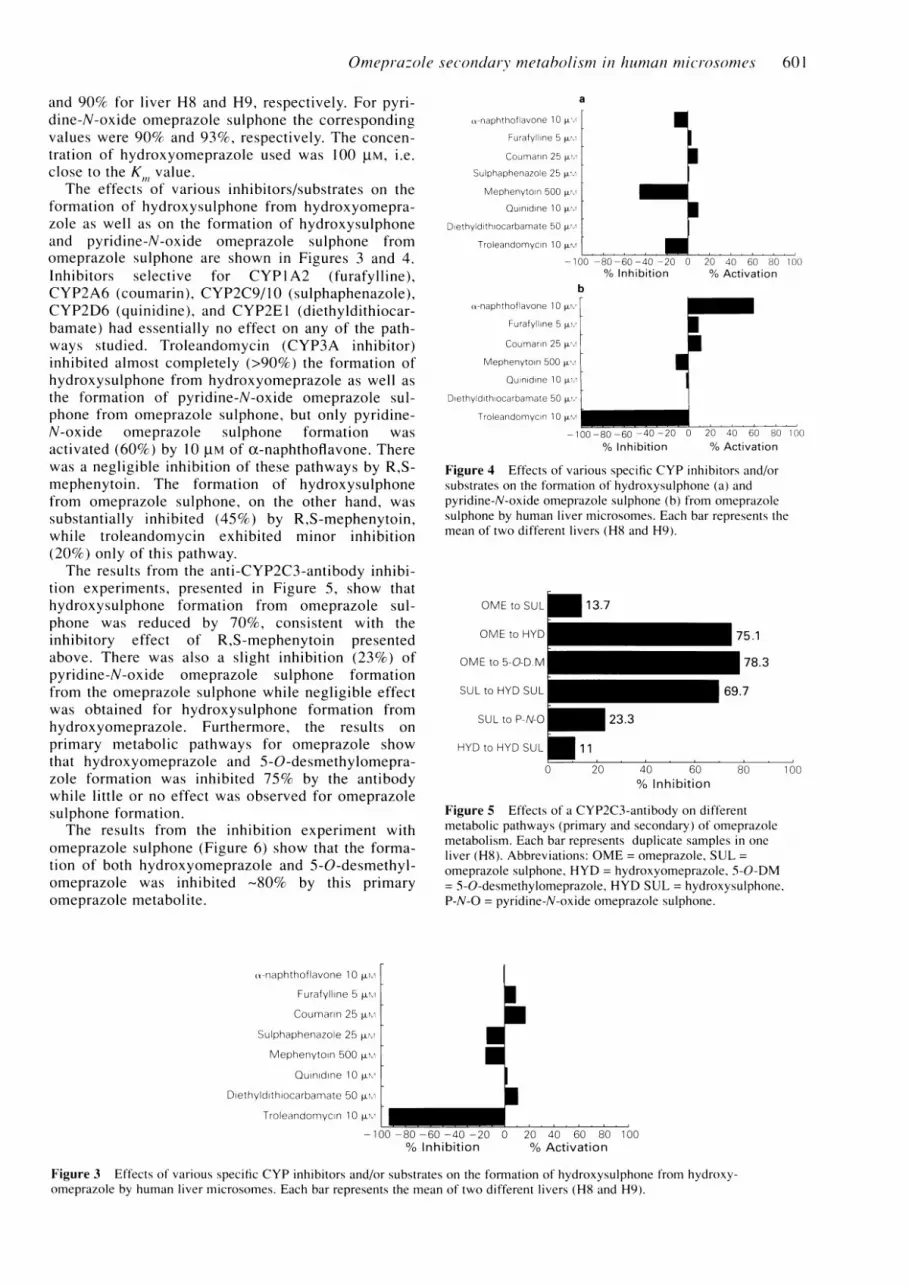
and 90% for liver H8 and H9, respectively. For pyri-dine-N-oxide omeprazole sulphone the correspondingvalues were 90% and 93%, respectively. The concen-tration of hydroxyomeprazole used was 100 tM, i.e.close to the K value.
The effects of various inhibitors/substrates on theformation of hydroxysulphone from hydroxyomepra-zole as well as on the formation of hydroxysulphoneand pyridine-N-oxide omeprazole sulphone fromomeprazole sulphone are shown in Figures 3 and 4.Inhibitors selective for CYP 1 A2 (furafylline),CYP2A6 (coumarin), CYP2C9/10 (sulphaphenazole),CYP2D6 (quinidine), and CYP2E1 (diethyldithiocar-bamate) had essentially no effect on any of the path-ways studied. Troleandomycin (CYP3A inhibitor)inhibited almost completely (>90%) the formation ofhydroxysulphone from hydroxyomeprazole as well asthe formation of pyridine-N-oxide omeprazole sul-phone from omeprazole sulphone, but only pyridine-N-oxide omeprazole sulphone formation wasactivated (60%) by 10 plM of ct-naphthoflavone. Therewas a negligible inhibition of these pathways by R,S-mephenytoin. The formation of hydroxysulphonefrom omeprazole sulphone, on the other hand, wassubstantially inhibited (45%) by R,S-mephenytoin,while troleandomycin exhibited minor inhibition(20%) only of this pathway.The results from the anti-CYP2C3-antibody inhibi-
tion experiments, presented in Figure 5, show thathydroxysulphone formation from omeprazole sul-phone was reduced by 70%, consistent with theinhibitory effect of R,S-mephenytoin presentedabove. There was also a slight inhibition (23%) ofpyridine-N-oxide omeprazole sulphone formationfrom the omeprazole sulphone while negligible effectwas obtained for hydroxysulphone formation fromhydroxyomeprazole. Furthermore, the results onprimary metabolic pathways for omeprazole showthat hydroxyomeprazole and 5-O-desmethylomepra-zole formation was inhibited 75% by the antibodywhile little or no effect was observed for omeprazolesulphone formation.The results from the inhibition experiment with
omeprazole sulphone (Figure 6) show that the forma-tion of both hydroxyomeprazole and 5-O-desmethyl-omeprazole was inhibited -80% by this primaryomeprazole metabolite.
U-naphthoflavone 10 p.l
Furafylline 5 iN
Coumarin 25 p.m
Sulphaphenazole 25 p.
Mephenytoin 500 p.m
Quinidine 10 p.c
Diethyldithiocarbamate 50 p.
Troleandomycin 1 0 p.I
-naphthoflavone 10 pLv
Furafylline 5 wm
Coumarin 25 [m
Sulphaphenazole 25 L
Mephenytoin 500
Quinidine 10 vLm
Diethyld thlocarbamate 50 vL.
Troleandomycin 1 0 p.M
x-naphthof lavone 1 0 p
Furafylline 5 p.
Coumarin 25 p.
Mephenytoin 500 p.
Quinidine 1u0 p.
Diethyldithlocarbamate 50 pL.
Troleandomycin 10 p.m
I
U
I
I
o0 -80 -60 -40 -20 0 20 40 60 80 100% Inhibition % Activation
b
-100 -80 -60 -40 -20 0 20 40 60 80 100% Inhibition % Activation
Figure 4 Effects of various specific CYP inhibitors and/orsubstrates on the formation of hydroxysulphone (a) andpyridine-N-oxide omeprazole sulphone (b) from omeprazolesulphone by human liver microsomes. Each bar represents themean of two different livers (H8 and H9).
OME to SUL
OME to HYD
OME to 5-0-D M
SUL to HYD SUL
SULto P-N-O
HYD to HYD SUL
13.7
I- 75.1
178.369.7
- 23.3
f 1
0 20 40 60% Inhibition
80 100
Figure 5 Effects of a CYP2C3-antibody on differentmetabolic pathways (primary and secondary) of omeprazolemetabolism. Each bar represents duplicate samples in oneliver (H8). Abbreviations: OME = omeprazole, SUL =
omeprazole sulphone. HYD = hydroxyomeprazole, 5-0-DM= 5-0-desmethylomeprazole, HYD SUL = hydroxysulphone,P-N-O = pyridine-N-oxide omeprazole sulphone.
UU
U
I- 100 -80 -60 -40 -20 0 20 40 60 80 100
% Inhibition % Activation
Figure 3 Effects of various specific CYP inhibitors and/or substrates on the formation of hydroxysulphone from hydroxy-omeprazole by human liver microsomes. Each bar represents the mean of two different livers (H8 and H9).
7

602 T. Andersson et al.
-_
0
0
20
o
Omeprazole sulphone concentration (>LM)
Figure 6 Effects of omeprazole sulphone on the formationof hydroxyomeprazole (0) and 5-0-desmethylomeprazole (0)from omeprazole by human microsomes in one liver (H8).
Discussion
Secondary metabolic pathways are common in vivobut they are not usually seen at saturating substrateconcentrations in vitro. In the case of omeprazole, thesulphone is not seen in urine but the hydroxysulphonedoes appear. Interpretation of sequential metabolicformation clearances in vivo is complex as both for-mation and elimination mechanisms are occurring atthe same time. It is, therefore, hard to delineateenvironmental and genetic influences. This has beenclarified in the current instance by mapping the meta-bolic pathways to the particular CYPs involved.It confirms the suggestions from in vivo data that theclearance of the sulphone may also be linked to theS-mephenytoin polymorphism. While the inhibitionof the H+/K+-ATPase is probably irreversible, theeffect of the drug will be related to the area under itsconcentration-time curve in plasma. It is, therefore,relevant to have some indication of the pharmaco-kinetic and environmental influences that will effectits clearance in vivo.An updated proposed scheme for both the primary
and secondary metabolism of omeprazole is presentedin Figure 1. The data are based on results from thepresent study together with previously reportedresults [15]. The primary metabolism of omeprazolehas been discussed extensively in a previous paper
[15], except that the structure of 3-hydroxyomepra-zole (V) at that stage was still unknown and thiscompound was referred to simply as metabolite X.Mass spectrometry experiments on collected aliquotsof metabolite X, performed at Astra Hassle AB,revealed the structure of this metabolite (Weidolf etal., unpublished results). However, this discussionwill deal mainly with the secondary metabolism ofomeprazole. Pyridine-N-oxide omeprazole sulphone,one of the two secondary metabolites presented inthis study, was also identified tentatively by mass
spectrometry performed by Dr Lars Weidolf (Weidolfet al., unpublished results).
This study shows that hydroxysulphone is the mainsecondary omeprazole metabolite formed duringincubation with both hydroxyomeprazole andomeprazole sulphone. Following incubation withomeprazole sulphone a second metabolite, tentatively
identified as pyridine-N-oxide omeprazole sulphone,was detected and measured. The kinetics of hydroxy-sulphone formation from hydroxyomeprazole werelinear while the hydroxysulphone formation fromomeprazole sulphone exhibited biphasic kinetics, thelatter finding suggesting involvement of multipleCYP isoforms. Biphasic kinetics were observed alsofor the formation of pyridine-N-oxide omeprazolesulphone.The lack of effect on the secondary metabolic path-
ways by inhibitors and/or substrates specific forCYP1A2, CYP2A6, CYP2C9/10, CYP2D6 andCYP2E1 indicates strongly that none of these iso-forms is involved in the various reactions. The com-plete inhibition of the formation of hydroxysulphonefrom hydroxyomeprazole and of the formation ofpyridine-N-oxide omeprazole sulphone from omepra-zole sulphone by troleandomycin shows clearly thatthese secondary metabolic pathways are mainlyCYP3A mediated reactions. However, of these tworeactions, only the formation of pyridine-N-oxideomeprazole sulphone was accompanied by an activa-tion by oc-naphthoflavone. Alpha-naphthoflavone isnormally activator of CYP3A mediated reactionsbut exceptions, like that above, have previouslybeen reported for other substrates [31]. By contrast,the substantial inhibition by R,S-mephenytoin ofhydroxysulphone formation from omeprazole sul-phone indicates that this reaction is S-mephenytoinhydroxylase mediated as previously suggested on thebasis of in vivo experiments [7, 8]. The weak inhibi-tion by troleandomycin of this pathway suggests thatCYP3A could be involved as a minor component inthis metabolic transformation.The results from the CYP2C3 antibody experi-
ments show excellent concordance with previouslyreported inhibitory data on the primary metabolism ofomeprazole, i.e. a major role for S-mephenytoinhydroxylase in the formation of hydroxyomeprazoleand 5-O-desmethylomeprazole from omeprazole [15].Furthermore, the antibody experiments are in goodagreement with the other inhibitory data presented inthis report as regards the formation of secondarymetabolites. S-mephenytoin hydroxylase is the domi-nant (70%) enzyme responsible for the formation ofhydroxysulphone from omeprazole sulphone. Theinhibition of this pathway by R,S-mephenytoin (45%)and troleandomycin (20%) was not complete. There-fore, a contribution from another isoform to thistransformation cannot be excluded. However, inhibi-tion by R,S-mephenytoin is typically weak and, sincethe inhibitory effect of the CYP2C3 antibodyprobably affords a better measure of the contributionof the S-mephenytoin hydroxylase, we find it morelikely that only two isoforms are involved in thistransformation. A negligible influence of the CYP2C3antibody was observed on the two othersecondary metabolic steps.
In the previous paper we showed that formation ofhydroxyomeprazole and 5-O-desmethylomeprazolefrom omeprazole were mediated mainly by the S-mephenytoin hydroxylase and that the apparent Kmvalues for these reactions were in the range 3-16 ,UM.Omeprazole sulphone would be expected to inhibit

Omeprazole secondary metabolism in human microsomes 603
these reactions as data in this paper suggest that theformation of hydroxysulphone from omeprazole sul-phone is mediated by the same enzyme. This wasconfirmed as shown by the data in Figure 6. Further,with omeprazole used at a substrate concentration5 gM, equal to its apparent Km, it can be calculatedthat the IC50 for an inhibition is 2K.. From Figure 6 itcan be estimated that IC50 is about 20 gM giving a Kiof about 10 gM which is consistent with the highaffinity Km for omeprazole sulphone (see Table 1),assuming competitive inhibition.
Variation in metabolism between different livers
[15] is possible, but the results obtained using ourtwo livers were similar. Thus we conclude that theformation of hydroxysulphone from omeprazolesulphone is mainly mediated by S-mephenytoinhydroxylase while the formation of the same metabo-lite from hydroxyomeprazole is mainly CYP3A medi-ated. Pyridine-N-oxide omeprazole sulphone for-mation from omeprazole sulphone also seems to be aCYP3A mediated reaction.
This study was supported financially by the AustralianNational Health and Medical Research Council.
References
1 Fellenius E, Berglindh T, Sachs G, Olbe L, Elander B,Sjostrand SE, Wallmark B. Substituted benzimidazolesinhibit gastric acid secretion by blocking (H+ + K+)ATPase. Nature 1981; 290: 159-161.
2 Wallmark B, Lorentzon P, Larsson H. The mechanismof action of omeprazole-a survey of its inhibitoryactions in vitro. Scand J Gastroenterol 1985; 20(Suppl. 108): 37-51.
3 Lind T, Cederberg C, Ekenved G, Haglund U, Olbe L.Effect of omeprazole-a gastric proton pump inhibitor-on pentagastrin stimulated acid secretion in man. Gut1983; 24: 270-276.
4 Bardhan KD, Bianchi-Porro G, Bose K, Daly M,Hinchliffe RF, Jonsson E, Lazzaroni M, Naesdal J,Rikner L, Walan A. A comparison of two differentdoses of omeprazole versus ranitidine in treatment ofduodenal ulcer. J clin Gastroenterol 1986; 8: 408-413.
5 Walan A, Bader JP, Classen M, Lamers CBHW, PiperDW, Rutgersson K, Eriksson S. Effect of omeprazoleand ranitidine on ulcer healing and relapse rates inpatients with benign gastric ulcer. New Engl J Med1989; 320: 69-75.
6 Klinkenberg-Knol EC, Jansen JMBJ, Festen HPM,Meuwissen SGM, Lamers CBHW. Double-blind multi-centre comparison of omeprazole and ranitidine in thetreatment of reflux oesophagitis. Lancet 1987; i:349-351.
7 Andersson T. Pharmacokinetics of omeprazole in man:with special reference to single and repeated adminis-tration, drug interactions and polymorphic metabolism.Thesis, 1991, University of Goteborg, Sweden.
8 Regardh CG, Andersson T, Lagerstrom PO, LundborgP, Skanberg I. The pharmacokinetics of omeprazole inhumans-a study of single intravenous and oral doses.Ther Drug Monit 1990; 12: 163-172.
9 Renberg L, Simonsson R, Hoffman KJ. Identification oftwo main urinary metabolites of [14C] omeprazole inhumans. Drug Metab Dispos 1989; 17: 69-76.
10 Lind T, Anderson T, Skanberg I, Olbe L. Biliaryexcretion of intravenous [14C] omeprazole in humans.Clin Pharmac Ther 1987; 42: 504-508.
11 Andersson T, Cederberg C, Edvardsson G, HeggelundA, Lundborg P. Effect of omeprazole treatment ondiazepam plasma levels in slow versus normal rapidmetabolizers of omeprazole. Clin Pharmac Ther 1990;47: 79-85.
12 Andersson T, RegArdh CG, Dahl-Puustinen ML,Bertilsson L. Slow omeprazole metabolizers are alsopoor S-mephenytoin hydroxylators. Ther Drug Monit1990; 12:415-416.
13 Andersson T, Regardh CG, Lou YC, Zhang Y, Dahl
ML, Bertilsson L. Polymorphic hydroxylation of S-mephenytoin and omeprazole metabolism in Caucasianand Chinese subjects. Pharmacogenetics 1992; 2:25-31.
14 Sohn DR, Kobayashi K, Chiba K, Lee KH, Shin SG,Ishizaki T. Disposition kinetics and metabolism ofomeprazole in extensive and poor metabolizers of S-mephenytoin 4-hydroxylation recruited from an orientalpopulation. J Pharmac exp Ther 1992; 262: 1195-1202.
15 Andersson T, Miners J-O, Tassaneeyakul W, Tassa-neeyakul W, Veronese ME, Meyer UA, Birkett DJ.Identification of human liver cytochrome P450 isoformsmediating omeprazole metabolism. Br J clin Pharmac1993; 36: 521-530.
16 McManus ME, Burgess WM, Veronese ME, Huggett A,Quattrochi LC, Tukey RH. Metabolism of 2-acetyl-aminofluorene and benzo(a)pyrene and activation offood-derived heterocyclic amine mutagens by humancytochromes P450. Cancer Res 1990; 50: 3367-3376.
17 Robson RA, Matthews AP, Miners JO, McManus ME,Meyer UA, Hall PM, Birkett DJ. Characterisation oftheophylline metabolism by human liver microsomes.Br J clin Pharmac 1987; 24: 293-300.
18 Lowry OH, Rosebrough NJ, Farr AL, Randall RJ.Protein measurement with the Folin phenol reagent. Jbiol Chem 1951; 193: 265-275.
19 Andersson T, Lagerstrom PO, Miners JO, VeroneseME, Weidolf L, Birkett DJ. High performance liquidchromatographic assay for human liver microsomalomeprazole metabolism. J Chromatogr 1993; 619:291-297.
20 Holford NHG. Mk Model: a modelling tool for micro-computers. Pharmacokinetic evaluation and comparisonwith standard computer programmes. Clin exp Phar-macPhysiol 1985; 9 (Suppl.): 95.
21 Sesardic D, Boobis AR, Murray BP, Murray S, SeguraJ, de-la-Torre R, Davies DS. Furafylline is a potent andselective inhibitor of cytochrome P4501A2 in man. BrJ clin Pharmac 1990; 29: 651-663.
22 Yamano S, Tatsuno J, Gonzalez FJ. The CYP2A3 geneproduct catalyzes coumarin 7-hydroxylation in humanliver microsomes. Biochemistry 1990; 29: 1322-1329.
23 Yun CH, Shimada T, Guengerich FP. Purification andcharacterization of human microsomal cytochromeP450 2A6. Mol Pharmac 1991; 40: 679-685.
24 Veronese ME, Doecke CJ, Mackenzie PI, McManusME, Miners JO, Rees DL, Gasser R, Meyer UA, BirkettDJ. Site-directed mutation studies of human livercytochrome P450 isoenzymes in the CYP2C subfamily.Biochem J 1993; 289: 533-538.
25 Kupfer A, Preisig R. Pharmacogenetics of mepheny-

604 T. Andersson et al.
toin: a new drug hydroxylation polymorphism in man.Eur J clin Pharmac 1984; 26: 753-759.
26 Guengerich FP, Muller-Enoch D, Blair IA. Oxidationof quinidine by human liver cytochrome P-450. MolPharmac 1986; 30: 287-295.
27 Inaba T, Jurima M, Mahon WA, Kalow W. In vitroinhibition studies of two isozymes of human livercytochrome P-450. Mephenytoin p-hydroxylase andsparteine monooxygenase. Drug Metab Dispos 1985;13: 443-448.
28 Guengerich FP, Kim DH, Iwasaki M. Role of humancytochrome P-45011E1 in the oxidation of many lowmolecular weight cancer suspects. Chem Res Toxicol1991; 4: 168-179.
29 Pessayre D, Tinel M, Larrey D, Cobert B, Funck-BrentanoC, Babany G. Inactivation of cytochrome P-450 by a
troleandomycin metabolite. Protective role of glutathione.J Pharmac exp Ther 1983; 224: 685-691.
30 Doecke CJ, Veronese ME, Pond SM, Miners JO,Birkett DJ, Sansom LN, McManus ME. Relationshipbetween phenytoin and tolbutamide hydroxylations inhuman liver microsomes. Br J clin Pharmac 1991; 31:125-130.
31 Raney KD, Shimada T, Kim DH, Groopman JD,Harris TM, Guengerich FP. Oxidation of aflatoxins andsterigmatocystin by human liver microsomes: significanceof aflatoxin Ql as a detoxication product of aflatoxin B1.Chem Res Toxicol 1992; 5: 202-2 10.
(Received 21 October 1993,accepted 22 February 1994)


