Embed Size (px)
Citation preview
BioMed CentralMolecular Cancer
ss
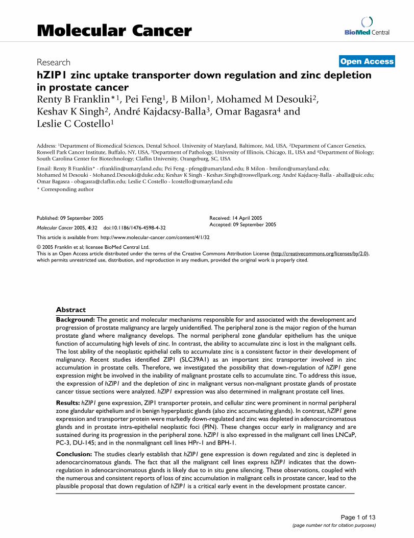
Open AcceResearchhZIP1 zinc uptake transporter down regulation and zinc depletion in prostate cancerRenty B Franklin*1, Pei Feng1, B Milon1, Mohamed M Desouki2, Keshav K Singh2, André Kajdacsy-Balla3, Omar Bagasra4 and Leslie C Costello1Address: 1Department of Biomedical Sciences, Dental School. University of Maryland, Baltimore, Md, USA, 2Department of Cancer Genetics, Roswell Park Cancer Institute, Buffalo, NY, USA, 3Department of Pathology, University of Illinois, Chicago, IL, USA and 4Department of Biology; South Carolina Center for Biotechnology; Claflin University, Orangeburg, SC, USA
Email: Renty B Franklin* - [email protected]; Pei Feng - [email protected]; B Milon - [email protected]; Mohamed M Desouki - [email protected]; Keshav K Singh - [email protected]; André Kajdacsy-Balla - [email protected]; Omar Bagasra - [email protected]; Leslie C Costello - [email protected]
* Corresponding author
prostate cancerzincZIP1 zinc transportercitrateZIP1 gene expression
AbstractBackground: The genetic and molecular mechanisms responsible for and associated with the development andprogression of prostate malignancy are largely unidentified. The peripheral zone is the major region of the humanprostate gland where malignancy develops. The normal peripheral zone glandular epithelium has the uniquefunction of accumulating high levels of zinc. In contrast, the ability to accumulate zinc is lost in the malignant cells.The lost ability of the neoplastic epithelial cells to accumulate zinc is a consistent factor in their development ofmalignancy. Recent studies identified ZIP1 (SLC39A1) as an important zinc transporter involved in zincaccumulation in prostate cells. Therefore, we investigated the possibility that down-regulation of hZIP1 geneexpression might be involved in the inability of malignant prostate cells to accumulate zinc. To address this issue,the expression of hZIP1 and the depletion of zinc in malignant versus non-malignant prostate glands of prostatecancer tissue sections were analyzed. hZIP1 expression was also determined in malignant prostate cell lines.
Results: hZIP1 gene expression, ZIP1 transporter protein, and cellular zinc were prominent in normal peripheralzone glandular epithelium and in benign hyperplastic glands (also zinc accumulating glands). In contrast, hZIP1 geneexpression and transporter protein were markedly down-regulated and zinc was depleted in adenocarcinomatousglands and in prostate intra-epithelial neoplastic foci (PIN). These changes occur early in malignancy and aresustained during its progression in the peripheral zone. hZIP1 is also expressed in the malignant cell lines LNCaP,PC-3, DU-145; and in the nonmalignant cell lines HPr-1 and BPH-1.
Conclusion: The studies clearly establish that hZIP1 gene expression is down regulated and zinc is depleted inadenocarcinomatous glands. The fact that all the malignant cell lines express hZIP1 indicates that the down-regulation in adenocarcinomatous glands is likely due to in situ gene silencing. These observations, coupled withthe numerous and consistent reports of loss of zinc accumulation in malignant cells in prostate cancer, lead to theplausible proposal that down regulation of hZIP1 is a critical early event in the development prostate cancer.
Published: 09 September 2005
Molecular Cancer 2005, 4:32 doi:10.1186/1476-4598-4-32
Received: 14 April 2005Accepted: 09 September 2005
This article is available from: http://www.molecular-cancer.com/content/4/1/32
© 2005 Franklin et al; licensee BioMed Central Ltd. This is an Open Access article distributed under the terms of the Creative Commons Attribution License (http://creativecommons.org/licenses/by/2.0), which permits unrestricted use, distribution, and reproduction in any medium, provided the original work is properly cited.
Page 1 of 13(page number not for citation purposes)

Molecular Cancer 2005, 4:32 http://www.molecular-cancer.com/content/4/1/32
BackgroundDespite the extensive clinical and experimental studiesover the recent decades, the pathogenesis of prostate can-cer remains unknown. The genetic and molecular mecha-nisms responsible for and associated with thedevelopment of malignant prostate cells and their pro-gression are largely unidentified [for reviews see [1,2]].The major site for the development of prostate malig-nancy is the peripheral zone, which comprises about 70%of the prostate gland. It is well established that the normalperipheral zone has the function of accumulatingextremely high zinc levels that are 3–10-fold greater thanfound in other soft tissues [3]. This capability resides inthe highly specialized glandular secretory epithelial cellsof the peripheral zone, which we characterize as "zinc-accumulating" cells. In contrast, the malignant prostatecells that develop in the peripheral zone do not containthe high zinc levels that characterize the normal secretoryepithelial cells. Repeated studies consistently show thatthe zinc levels of malignant prostate tissue are 62–75%lower than the normal prostate tissue [4-8]. Measure-ments of pure malignant tissue in the absence of normalglandular epithelium would reveal even lower zinc levelsthat would approximate the levels found in other soft tis-sues. This consistency persists in different reports by dif-ferent investigators employing different populations andtissue samples and involving various stages of malig-nancy. The studies of Zaichick et al [9] and Vartsky et al[10] further reveal the critically important relationshipthat, in individual analyses, malignant prostate tissuenever exhibits high zinc levels. In addition, Habib [11]reported that the decrease in zinc occurs early in malig-nancy. These persistent results, and the additional corrob-orating evidence presented below, firmly establish thatthe unique zinc-accumulating capability of the normalperipheral zone secretory epithelial cells is lost in the neo-plastic transformation to malignant cells; and that zinc-accumulating malignant cells do not exist in situ in pros-tate cancer. For extensive presentations of the relation-ships of zinc in normal prostate and prostate cancer, werefer the reader to our recent reviews [12-14].
Established clinical and experimental evidence providesthe basis for our concept that zinc accumulation preventsthe malignant activities of the neoplastic prostate cell; andthat impaired zinc accumulation is an essential require-ment for the manifestation of prostate malignancy. If suchis the case, one should expect that the zinc-accumulatingprocess that characterizes the normal glandular epithe-lium is absent or defective in the malignant cells. Untilrecently, no information had been available regarding themechanism(s) of zinc accumulation in prostate cells.Recent studies [15-17] have established that the zincuptake transporter, ZIP1, is important in the uptake andaccumulation of zinc by prostate cells. Up-regulation of
ZIP1 in prostate cells increases zinc accumulation; and,correspondingly, down-regulation of ZIP1 decreases zincaccumulation in prostate cells. In addition, Rishi et al [18]reported that ZIP1 (and ZIP2) expression in peripheralzone glandular epithelium of black males is down regu-lated when compared to its expression in white males;which coincides with the race-associated higher incidenceof prostate cancer in African-Americans. These relation-ships suggested that the decrease in zinc in malignantprostate glands might be due to the down regulation ofZIP1 expression. In this report we show, for the first time,the down regulation of hZIP1 gene expression, the loss ofZIP1 transporter protein and the depletion of zinc that isevident in malignant prostate glands. The evidence pre-sented supports the likelihood that down regulation ofZIP1 gene expression in the neoplastic prostate cell is anessential step in the development of prostate malignancy.The studies were conducted independently at three differ-ent institutions, which strengthens the validity of thesecorroborating results.
ResultsThe studies presented in this report were conducted at theUniversity of Maryland (UMaryland study), the RoswellPark Cancer Institute (Roswell Park Study), and ClaflinUniversity (ClaflinU study). Therefore the results will bepresented as provided by each separate and independentstudy, followed by the discussion of the evidence and sup-porting basis for the genetic/metabolic concept of the roleof zinc in prostate malignancy.
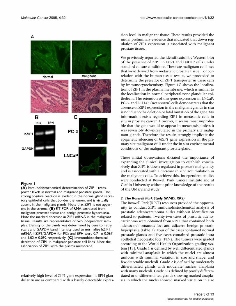
The UMaryland Study (RBF, PF, BM, LCC)Earlier studies [15-17] demonstrated that ZIP1 isexpressed in malignant prostate cell lines (PC-3 andLNCaP cells); and that this zinc uptake transporter func-tions in the uptake and cellular accumulation of zinc. Thiscaused us to initiate preliminary studies to determine ifZIP1 gene expression and/or the level of the transporterprotein might be down-regulated in malignant prostateglands in comparison to the expression in normal pros-tate glandular epithelium. Paraffin mounted serial sec-tions of human prostate tissue were used for ZIP1immumohistochemistry staining. Hematoxylin and eosinstaining was used for pathologic evaluation of normalglands and adencarcinomatous foci. Figure 1A reveals themembrane-associated immunohistochemical identifica-tion of ZIP1 in the normal peripheral zone glandular epi-thelium. In contrast, the malignant glands wereessentially devoid of demonstrable membrane-associatedZIP1. It is also apparent that ZIP1 is confined to glandularepithelium and is not demonstrable in the stromal tissue.Figure 1B presents RT-PCR analysis of ZIP1 expression intissue extracts of malignant tissue versus benign hyper-plastic (BPH) glands; which, like normal peripheral zone,are zinc-accumulating glands. The results demonstrate a
Page 2 of 13(page number not for citation purposes)
Molecular Cancer 2005, 4:32 http://www.molecular-cancer.com/content/4/1/32
relatively high level of ZIP1 gene expression in BPH glan-dular tissue as compared with a barely detectable expres-
sion level in malignant tissue. These results provided theinitial preliminary evidence that indicated that down reg-ulation of ZIP1 expression is associated with malignantprostate tissue.
We previously reported the identification by Western blotof the presence of ZIP1 in PC-3 and LNCaP cells understandard culture conditions. These are malignant cell linesthat were derived from metastatic prostate tissue. For cor-relation with the human tissue results, we proceeded todetermine the presence of ZIP1 transporter in these cellsby immunocytochemistry. Figure 1C shows the localiza-tion of ZIP1 in the plasma membrane; which is similar tothe localization in normal peripheral zone glandular epi-thelium. The retention of this gene expression in LNCaP,PC-3, and DU145 (not shown) cells demonstrates that theabsence of ZIP1 expression in the malignant glands in situis not due to the deletion or fatal mutation of the gene. Noinformation exists regarding ZIP1 in metastatic cells insitu in prostate cancer. However, it seems most improba-ble that the gene would re-appear in metastasis, unless itwas reversibly down-regulated in the primary site malig-nant glands. Therefore the results strongly implicate theepigenetic silencing of hZIP1 gene expression in the pri-mary site malignant cells under the in situ environmentalconditions of the malignant prostate gland.
These initial observations dictated the importance ofexpanding the clinical investigation to establish conclu-sively that ZIP1 is down regulated in prostate malignancyand is associated with a decrease in zinc accumulation inthe malignant cells. To achieve this, independent studieswere conducted at Roswell Park Cancer Institute and atClaflin University without prior knowledge of the resultsof the UMaryland study.
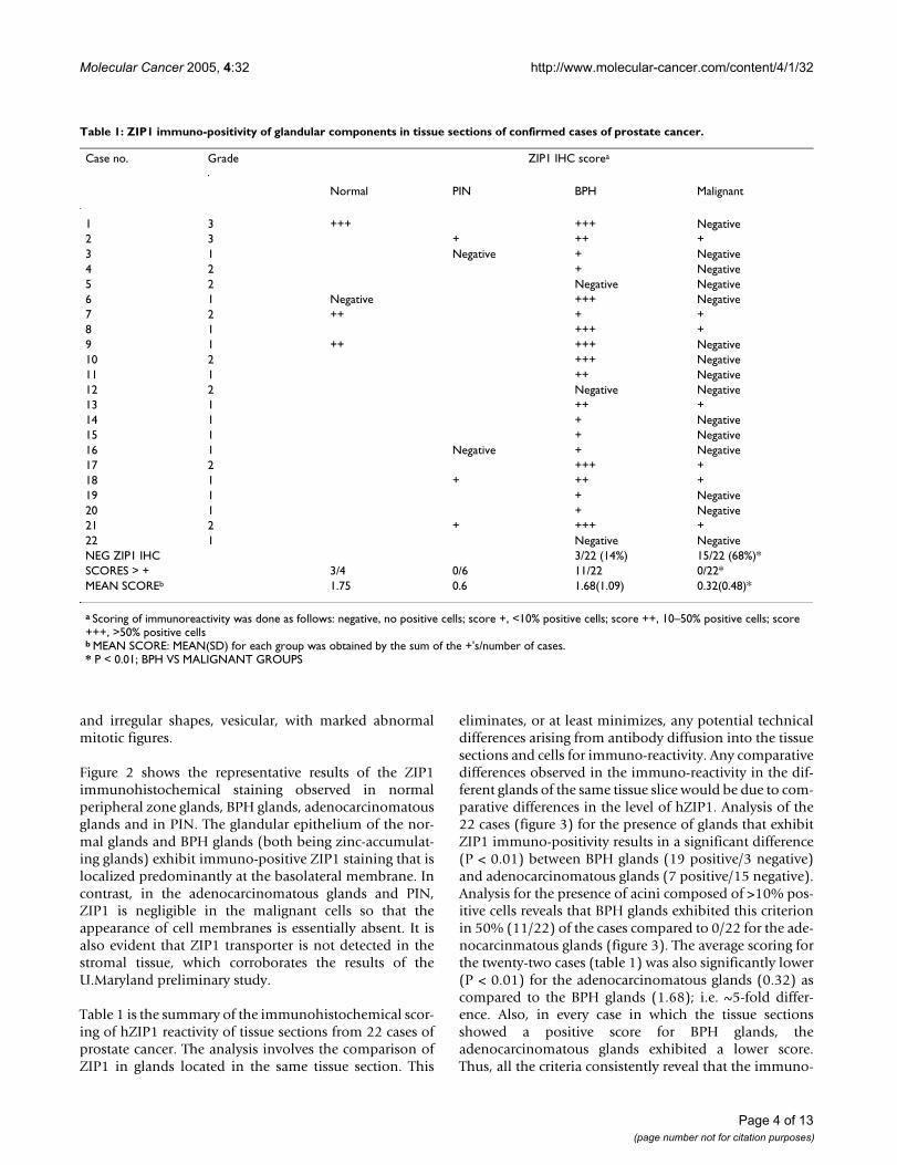
2. The Roswell Park Study (MMD, KKS)The Roswell Park (RPCI) resources provided the opportu-nity to conduct ZIP1 immunohistochemical analysis ofprostatic adenocarcinoma slides without identificationrelated to patients. Twenty-two cases of prostatic adeno-carcinoma were obtained from RPCI that contained bothadenocarcinomatous foci and adjacent benign prostatichyperplasia (table 1). Four of the cases contained normalprostatic glands and five cases contained prostatic intraepithelial neoplastic foci (PIN). The tumors were gradedaccording to the World Health Organization grading sys-tem [19]. Grade 1 is defined by well differentiated glandswith minimal anaplasia in which the nuclei are almostuniform with minimal variation in size and shape, andfew detectable nucleoli. Grade 2 is defined by moderatelydifferentiated glands with moderate nuclear anaplasiawith many nucleoli. Grade 3 is defined by poorly differen-tiated or undifferentiated glands showing marked anapla-sia in which the nuclei showed marked variation in size
(A) Immunohistochemical determination of ZIP 1 trans-porter levels in normal and malignant prostate glandsFigure 1(A) Immunohistochemical determination of ZIP 1 trans-porter levels in normal and malignant prostate glands. The strong positive reaction is evident in the normal gland secre-tory epithelial cells that border the lumen, and is virtually absent in the malignant glands. Note that ZIP1 is not appar-ent in the stroma. (B) RT-PCR of RNA extracted from malignant prostate tissue and benign prostatic hyperplasia. Note the marked decrease in ZIP1 mRNA in the malignant tissue. Results are representative of two independent sam-ples. Density of the bands was determined by densitometry scans and GAPDH band intensity used to normalize hZIP1 mRNA. hZIP1/GAPDH for PCa and BPH were 0.71 ± 0.067 and 1.02 ± 0.092 respectively. (C) Immunohistochemical detection of ZIP1 in malignant prostate cell lines. Note the association of ZIP1 with the plasma membrane.
Page 3 of 13(page number not for citation purposes)
Molecular Cancer 2005, 4:32 http://www.molecular-cancer.com/content/4/1/32
and irregular shapes, vesicular, with marked abnormalmitotic figures.
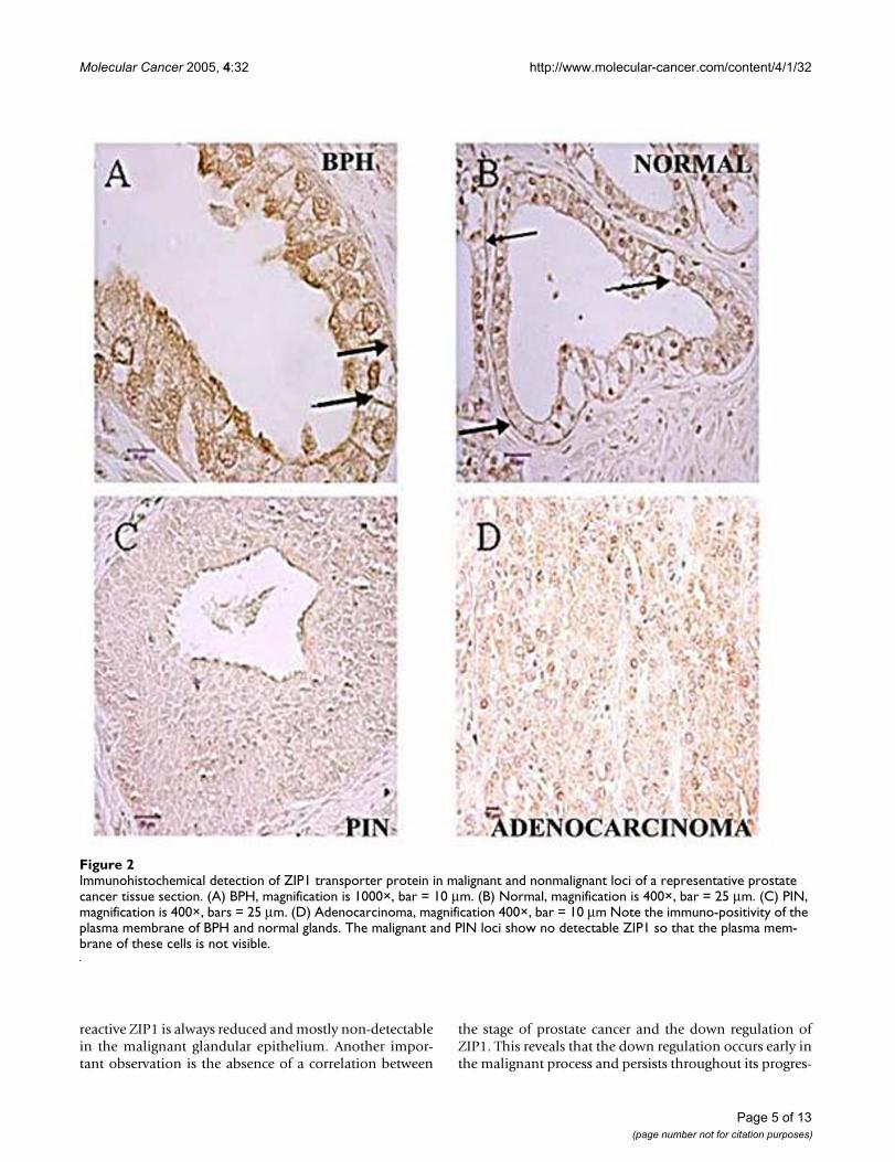
Figure 2 shows the representative results of the ZIP1immunohistochemical staining observed in normalperipheral zone glands, BPH glands, adenocarcinomatousglands and in PIN. The glandular epithelium of the nor-mal glands and BPH glands (both being zinc-accumulat-ing glands) exhibit immuno-positive ZIP1 staining that islocalized predominantly at the basolateral membrane. Incontrast, in the adenocarcinomatous glands and PIN,ZIP1 is negligible in the malignant cells so that theappearance of cell membranes is essentially absent. It isalso evident that ZIP1 transporter is not detected in thestromal tissue, which corroborates the results of theU.Maryland preliminary study.
Table 1 is the summary of the immunohistochemical scor-ing of hZIP1 reactivity of tissue sections from 22 cases ofprostate cancer. The analysis involves the comparison ofZIP1 in glands located in the same tissue section. This
eliminates, or at least minimizes, any potential technicaldifferences arising from antibody diffusion into the tissuesections and cells for immuno-reactivity. Any comparativedifferences observed in the immuno-reactivity in the dif-ferent glands of the same tissue slice would be due to com-parative differences in the level of hZIP1. Analysis of the22 cases (figure 3) for the presence of glands that exhibitZIP1 immuno-positivity results in a significant difference(P < 0.01) between BPH glands (19 positive/3 negative)and adenocarcinomatous glands (7 positive/15 negative).Analysis for the presence of acini composed of >10% pos-itive cells reveals that BPH glands exhibited this criterionin 50% (11/22) of the cases compared to 0/22 for the ade-nocarcinmatous glands (figure 3). The average scoring forthe twenty-two cases (table 1) was also significantly lower(P < 0.01) for the adenocarcinomatous glands (0.32) ascompared to the BPH glands (1.68); i.e. ~5-fold differ-ence. Also, in every case in which the tissue sectionsshowed a positive score for BPH glands, theadenocarcinomatous glands exhibited a lower score.Thus, all the criteria consistently reveal that the immuno-
Table 1: ZIP1 immuno-positivity of glandular components in tissue sections of confirmed cases of prostate cancer.
Case no. Grade ZIP1 IHC scorea
Normal PIN BPH Malignant
1 3 +++ +++ Negative2 3 + ++ +3 1 Negative + Negative4 2 + Negative5 2 Negative Negative6 1 Negative +++ Negative7 2 ++ + +8 1 +++ +9 1 ++ +++ Negative10 2 +++ Negative11 1 ++ Negative12 2 Negative Negative13 1 ++ +14 1 + Negative15 1 + Negative16 1 Negative + Negative17 2 +++ +18 1 + ++ +19 1 + Negative20 1 + Negative21 2 + +++ +22 1 Negative NegativeNEG ZIP1 IHC 3/22 (14%) 15/22 (68%)*SCORES > + 3/4 0/6 11/22 0/22*MEAN SCOREb 1.75 0.6 1.68(1.09) 0.32(0.48)*
a Scoring of immunoreactivity was done as follows: negative, no positive cells; score +, <10% positive cells; score ++, 10–50% positive cells; score +++, >50% positive cellsb MEAN SCORE: MEAN(SD) for each group was obtained by the sum of the +'s/number of cases.* P < 0.01; BPH VS MALIGNANT GROUPS
Page 4 of 13(page number not for citation purposes)
Molecular Cancer 2005, 4:32 http://www.molecular-cancer.com/content/4/1/32
reactive ZIP1 is always reduced and mostly non-detectablein the malignant glandular epithelium. Another impor-tant observation is the absence of a correlation between
the stage of prostate cancer and the down regulation ofZIP1. This reveals that the down regulation occurs early inthe malignant process and persists throughout its progres-
Immunohistochemical detection of ZIP1 transporter protein in malignant and nonmalignant loci of a representative prostate cancer tissue sectionFigure 2Immunohistochemical detection of ZIP1 transporter protein in malignant and nonmalignant loci of a representative prostate cancer tissue section. (A) BPH, magnification is 1000×, bar = 10 µm. (B) Normal, magnification is 400×, bar = 25 µm. (C) PIN, magnification is 400×, bars = 25 µm. (D) Adenocarcinoma, magnification 400×, bar = 10 µm Note the immuno-positivity of the plasma membrane of BPH and normal glands. The malignant and PIN loci show no detectable ZIP1 so that the plasma mem-brane of these cells is not visible.
Page 5 of 13(page number not for citation purposes)
Molecular Cancer 2005, 4:32 http://www.molecular-cancer.com/content/4/1/32
sion in the primary site; which is consistent with the earlychanges in zinc levels.
As would be expected, the presence of normal peripheralzone glands in the malignant tissue sections is minimal,and insufficient for statistical analysis. However in threeof the 4 cases, the normal glands exhibited the expectedhigher ZIP1 expression than the adenocarcinomatousglands, and gave results that were similar to BPH; both ofwhich are zinc accumulating glands. In one case the nor-mal gland was negative for ZIP1, which, seemingly, is ananomaly. However an important point needs to be con-sidered. It is consistent with existing evidence (discussedbelow) that these metabolic changes occur before theappearance of histopathological evidence of malignantcells. Therefore, this "anomaly" might be due to changesthat occur in a "premalignant" neoplastic condition thatwas histologically identified as "normal". Furthermore, inall five cases with PIN, the glands were either negative or+ (none was ++ or +++), which mimics the profile of theadenocarcinomatous glands. It is striking that the com-bined PIN and adenocarcinoma glands showed noinstance of ZIP1 positive cells that exceeded 10%. Thiscould be supportive of a malignant relationship betweenPIN and adenocarcinoma; but further studies with
additional PIN and normal peripheral zone glands areneeded. Nevertheless, the Roswell Park study clearlyestablishes a consistent down-regulation of hZIP1 trans-porter in malignant prostate glands that corroborates andextends the results of the U.Maryland study, and is furthercorroborated by the following ClaflinU study.
In a parallel study (unpublished information, to be pre-sented in a separate report), the tissue sections were alsoassayed for the immunohistochemical identification ofm-aconitase. m-Aconitase was prevalent and unchangedin BPH, malignant, PIN and normal glands. Thus thedown regulation of ZIP1 is specific. Moreover, the differ-ences in citrate levels in malignant versus non malignantglands is not due to altered levels of m-aconitase. This re-emphasizes the role of altered zinc and ZIP1 in the meta-bolic transformation associated with prostate malignancy.
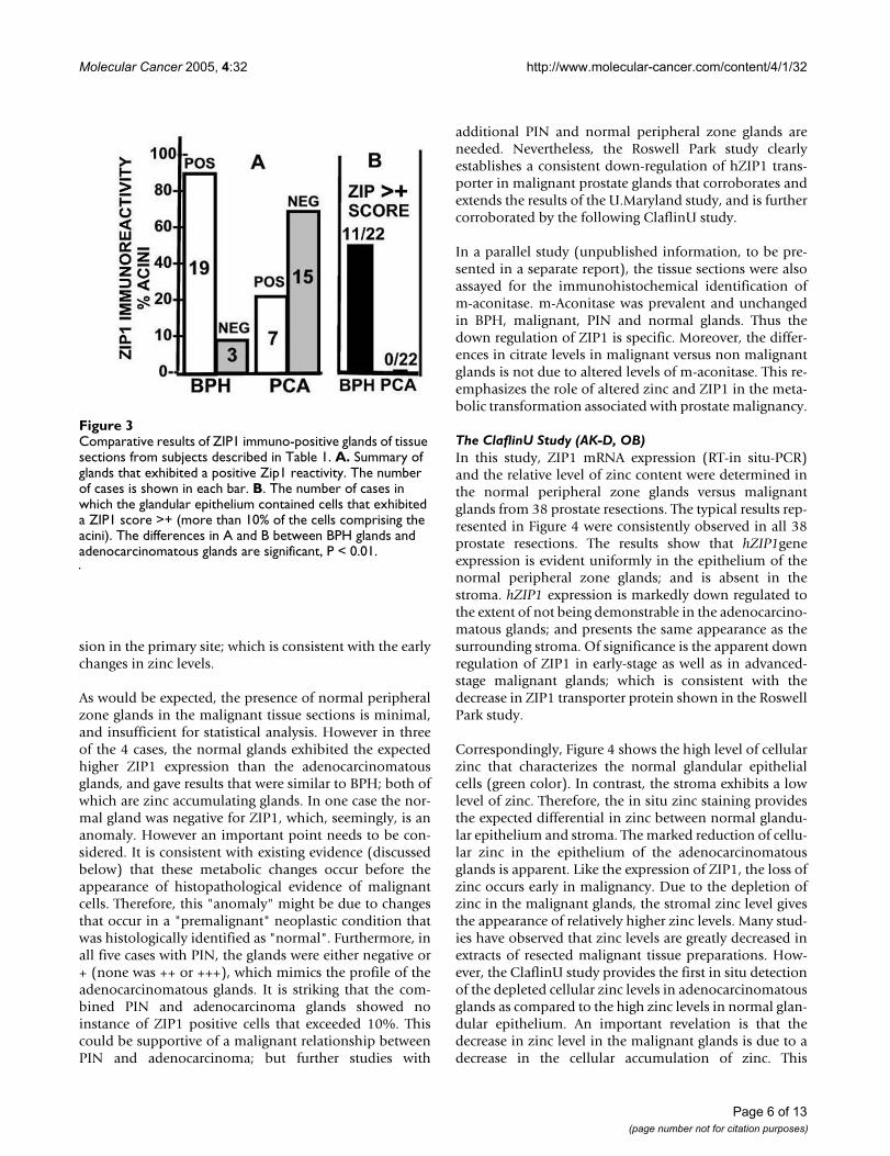
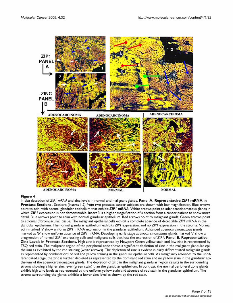
The ClaflinU Study (AK-D, OB)In this study, ZIP1 mRNA expression (RT-in situ-PCR)and the relative level of zinc content were determined inthe normal peripheral zone glands versus malignantglands from 38 prostate resections. The typical results rep-resented in Figure 4 were consistently observed in all 38prostate resections. The results show that hZIP1geneexpression is evident uniformly in the epithelium of thenormal peripheral zone glands; and is absent in thestroma. hZIP1 expression is markedly down regulated tothe extent of not being demonstrable in the adenocarcino-matous glands; and presents the same appearance as thesurrounding stroma. Of significance is the apparent downregulation of ZIP1 in early-stage as well as in advanced-stage malignant glands; which is consistent with thedecrease in ZIP1 transporter protein shown in the RoswellPark study.
Correspondingly, Figure 4 shows the high level of cellularzinc that characterizes the normal glandular epithelialcells (green color). In contrast, the stroma exhibits a lowlevel of zinc. Therefore, the in situ zinc staining providesthe expected differential in zinc between normal glandu-lar epithelium and stroma. The marked reduction of cellu-lar zinc in the epithelium of the adenocarcinomatousglands is apparent. Like the expression of ZIP1, the loss ofzinc occurs early in malignancy. Due to the depletion ofzinc in the malignant glands, the stromal zinc level givesthe appearance of relatively higher zinc levels. Many stud-ies have observed that zinc levels are greatly decreased inextracts of resected malignant tissue preparations. How-ever, the ClaflinU study provides the first in situ detectionof the depleted cellular zinc levels in adenocarcinomatousglands as compared to the high zinc levels in normal glan-dular epithelium. An important revelation is that thedecrease in zinc level in the malignant glands is due to adecrease in the cellular accumulation of zinc. This
Comparative results of ZIP1 immuno-positive glands of tissue sections from subjects described in Table 1Figure 3Comparative results of ZIP1 immuno-positive glands of tissue sections from subjects described in Table 1. A. Summary of glands that exhibited a positive Zip1 reactivity. The number of cases is shown in each bar. B. The number of cases in which the glandular epithelium contained cells that exhibited a ZIP1 score >+ (more than 10% of the cells comprising the acini). The differences in A and B between BPH glands and adenocarcinomatous glands are significant, P < 0.01.
Page 6 of 13(page number not for citation purposes)
Molecular Cancer 2005, 4:32 http://www.molecular-cancer.com/content/4/1/32
In situ detection of ZIP1 mRNA and zinc levels in normal and malignant glandsFigure 4In situ detection of ZIP1 mRNA and zinc levels in normal and malignant glands. Panel A. Representative ZIP1 mRNA in Prostate Sections . Sections (inserts 1,2) from two prostate cancer subjects are shown with low magnification. Blue arrows point to acini with normal glandular epithelium that exhibit ZIP1 mRNA. White arrows point to adenocarcinomatous glands in which ZIP1 expression is not demonstrable. Insert 3 is a higher magnification of a section from a cancer patient to show more detail. Blue arrows point to acini with normal glandular epithelium. Red arrows point to malignant glands. Green arrows point to stromal (fibromuscular) tissue. The malignant epithelial cells exhibit a complete absence of detectable ZIP1 mRNA in the glandular epithelium. The normal glandular epithelium exhibits ZIP1 expression; and no ZIP1 expression in the stroma. Normal acini marked 'a' show uniform ZIP1 mRNA expression in the glandular epithelium. Advanced adenocarcinomatous glands marked as 'b" show uniform absence of ZIP1 mRNA. Developing early stage adenocarcinomatous glands marked 'c' show a progression of normal ZIP1 expressing cells and malignant cells that lost the expression of ZIP1. Panel B. Representative Zinc Levels in Prostate Sections. High zinc is represented by Newport Green yellow stain and low zinc is represented by TSQ red stain. The malignant region of the peripheral zone shows a significant depletion of zinc in the malignant glandular epi-thelium as exhibited by the red staining (white arrows). The depletion of zinc is evident in early differentiated malignant glands as represented by combinations of red and yellow staining in the glandular epithelial cells. As malignancy advances to the undif-ferentiated stage, the zinc is further depleted as represented by the dominant red stain and no yellow stain in the glandular epi-thelium of the adenocarcinomatous glands. The depletion of zinc in the malignant glandular region results in the surrounding stroma showing a higher zinc level (green stain) than the glandular epithelium. In contrast, the normal peripheral zone glands exhibit high zinc levels as represented by the uniform yellow stain and absence of red stain in the glandular epithelium. The stroma surrounding the glands exhibits a lower zinc level as shown by the red stain.
Page 7 of 13(page number not for citation purposes)
Molecular Cancer 2005, 4:32 http://www.molecular-cancer.com/content/4/1/32
establishes that the decrease in intracellular zinc, and notimpaired secretion of zinc into the lumen (prostaticfluid), is principally responsible for the decrease in malig-nant tissue zinc level. Thus, the results of the ClaflinUstudy are consistent with and corroborate the RoswellPark study and the preliminary results of the UMarylandstudy.
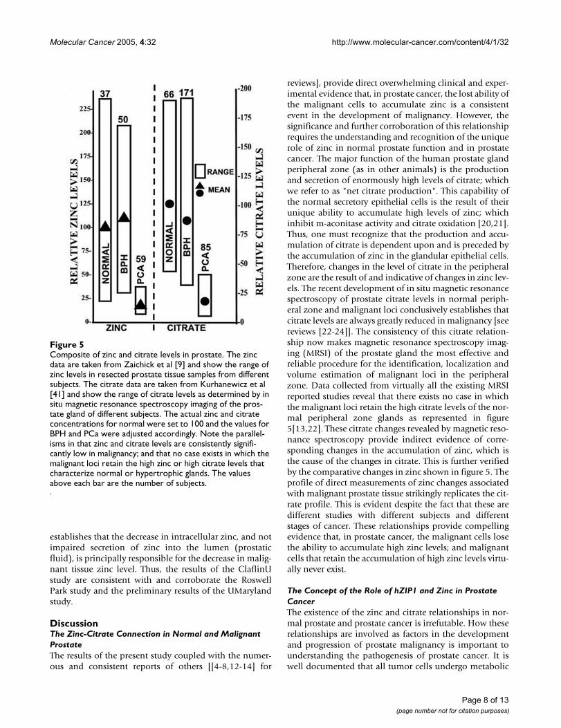
DiscussionThe Zinc-Citrate Connection in Normal and Malignant ProstateThe results of the present study coupled with the numer-ous and consistent reports of others [[4-8,12-14] for
reviews], provide direct overwhelming clinical and exper-imental evidence that, in prostate cancer, the lost ability ofthe malignant cells to accumulate zinc is a consistentevent in the development of malignancy. However, thesignificance and further corroboration of this relationshiprequires the understanding and recognition of the uniquerole of zinc in normal prostate function and in prostatecancer. The major function of the human prostate glandperipheral zone (as in other animals) is the productionand secretion of enormously high levels of citrate; whichwe refer to as "net citrate production". This capability ofthe normal secretory epithelial cells is the result of theirunique ability to accumulate high levels of zinc; whichinhibit m-aconitase activity and citrate oxidation [20,21].Thus, one must recognize that the production and accu-mulation of citrate is dependent upon and is preceded bythe accumulation of zinc in the glandular epithelial cells.Therefore, changes in the level of citrate in the peripheralzone are the result of and indicative of changes in zinc lev-els. The recent development of in situ magnetic resonancespectroscopy of prostate citrate levels in normal periph-eral zone and malignant loci conclusively establishes thatcitrate levels are always greatly reduced in malignancy [seereviews [22-24]]. The consistency of this citrate relation-ship now makes magnetic resonance spectroscopy imag-ing (MRSI) of the prostate gland the most effective andreliable procedure for the identification, localization andvolume estimation of malignant loci in the peripheralzone. Data collected from virtually all the existing MRSIreported studies reveal that there exists no case in whichthe malignant loci retain the high citrate levels of the nor-mal peripheral zone glands as represented in figure5[13,22]. These citrate changes revealed by magnetic reso-nance spectroscopy provide indirect evidence of corre-sponding changes in the accumulation of zinc, which isthe cause of the changes in citrate. This is further verifiedby the comparative changes in zinc shown in figure 5. Theprofile of direct measurements of zinc changes associatedwith malignant prostate tissue strikingly replicates the cit-rate profile. This is evident despite the fact that these aredifferent studies with different subjects and differentstages of cancer. These relationships provide compellingevidence that, in prostate cancer, the malignant cells losethe ability to accumulate high zinc levels; and malignantcells that retain the accumulation of high zinc levels virtu-ally never exist.
The Concept of the Role of hZIP1 and Zinc in Prostate CancerThe existence of the zinc and citrate relationships in nor-mal prostate and prostate cancer is irrefutable. How theserelationships are involved as factors in the developmentand progression of prostate malignancy is important tounderstanding the pathogenesis of prostate cancer. It iswell documented that all tumor cells undergo metabolic
Composite of zinc and citrate levels in prostateFigure 5Composite of zinc and citrate levels in prostate. The zinc data are taken from Zaichick et al [9] and show the range of zinc levels in resected prostate tissue samples from different subjects. The citrate data are taken from Kurhanewicz et al [41] and show the range of citrate levels as determined by in situ magnetic resonance spectroscopy imaging of the pros-tate gland of different subjects. The actual zinc and citrate concentrations for normal were set to 100 and the values for BPH and PCa were adjusted accordingly. Note the parallel-isms in that zinc and citrate levels are consistently signifi-cantly low in malignancy; and that no case exists in which the malignant loci retain the high zinc or high citrate levels that characterize normal or hypertrophic glands. The values above each bar are the number of subjects.
Page 8 of 13(page number not for citation purposes)

Molecular Cancer 2005, 4:32 http://www.molecular-cancer.com/content/4/1/32
transformations that are essential for their malignantexistence (25, 26 for reviews). It is important to empha-size that these metabolic transformations are not thecause of malignancy. Malignancy requires the genetictransformation of a sane cell to a neoplastic cell that isendowed with the potential capability of malignancy. Themetabolic transformation is essential for the neoplasticcells to manifest their malignant capabilities.
The accumulation of zinc in normal prostate glandularepithelial cells results in two important effects; a meta-bolic effect and a proliferative effect. Its metabolic effect isthe inhibition of citrate oxidation that is essential for theprostate function of production and secretion of high lev-els of citrate [20,21]; and its inhibition of terminal oxida-tion [27]. This has a bioenergetic cost in that theinhibition of citrate oxidation results in a ~60% loss ofATP production that would arise from complete glucoseoxidation. Consequently, zinc-accumulating citrate-pro-ducing cells (normal peripheral zone epithelial cells) areenergy-inefficient cells. A second effect of zinc is its inhi-bition of prostate cell proliferation. This effect resultsfrom zinc induction of apoptosis in prostate cells [28-31].These are the consequences imposed upon highly special-ized zinc-accumulating citrate-producing cells (i.e. nor-
mal peripheral zone secretory epithelial cells) in order toachieve their unique function of net citrate production.
Malignant prostate cells do not exist for the specializedfunction of citrate production and secretion. They mustreplace the metabolic pathways associated with net citrateproduction with metabolic relationships that are suitablefor their malignant existence. That the malignant prostatecells in situ never exist as zinc-accumulating, citrate-pro-ducing cells is evidence of the incompatibility of the highzinc accumulation and net citrate production for theirexistence. Their metabolic transformation to energy-effi-cient citrate-oxidizing cells that have lost the ability toaccumulate zinc provides their metabolic/bioenergeticrequirements of malignancy. Also, the apoptotic influenceof zinc is eliminated, which permits the proliferation ofthe malignant cells. However, the evidence presentedherein clearly establishes hZIP1 down regulation in theprimary in situ site and further suggests that this is theexplanation for the consistently observed decrease in zinclevels in prostate cancer.
This concept is represented in figure 6. The occurrence ofthis metabolic transformation is dependent upon theability of the normal epithelial cells and the inability ofthe malignant cells to accumulate zinc. The present
The integrated role of ZIP1, zinc, and citrate metabolism in the pathogenesis of prostate malignancyFigure 6The integrated role of ZIP1, zinc, and citrate metabolism in the pathogenesis of prostate malignancy. The normal glandular epi-thelial cell expresses ZIP1 that permits zinc accumulation, which inhibits citrate oxidation and terminal respiration. Citrate accumulates and coupled ATP production is reduced. A genetic transformation results in a neoplastic cell with potential malig-nant capability. ZIP1 expression is silenced by epigenetic factors which eliminate Zip1 transporter and accumulation of zinc in the premalignant cell. The level of cellular zinc decreases which removes the inhibitory effects on citrate oxidation and terminal oxidation. The Krebs cycle is functional and coupled ATP production is increased. The malignant cell is metabolically and bioenergetically capable of manifesting its malignant potential. Additionally, the growth inhibitory effect of zinc is removed, which allows growth and progression of the malignant cell.
Page 9 of 13(page number not for citation purposes)
Molecular Cancer 2005, 4:32 http://www.molecular-cancer.com/content/4/1/32
studies establish that hZIP1 is down-regulated in the ade-nocarcinomatous glands. This is consistent with thedown-regulation of hZIP1 gene expression in the African-American male population, which exhibits a higher inci-dence of prostate cancer [18]. The functional importanceof hZIP1 in the accumulation of zinc in prostate cells hasbeen established [15-17]. Over-expression of hZIP1results in increased accumulation of zinc which leads toinhibition of cell proliferation; whereas cells with down-regulation of hZIP1 have decreased cellular zinc levels andincreased proliferation. Also the accumulation of zinc inthe malignant prostate cells in culture and in vivo [31]results in increased citrate levels.
Consequently, consistent clinical and experimental evi-dence strongly implicate the down-regulation of hZIP1 inthe lost ability of the malignant cells to accumulate zinc.The existence of hZIP1 insures that prostate cells will accu-mulate zinc. If ZIP1 is not down regulated in the neoplas-tic cell, zinc accumulation and its metabolic/energetic andapoptotic effects will prevail; and the neoplastic cell willremain in a pre-malignant dormant state and/or will die.In this concept (figure 6), prostate malignancy requirestwo essential transformations; the genetic transformationto a neoplastic cell with potential malignant capability;and the metabolic transformation to an energy-efficientcitrate-producing cell that has lost the ability to accumu-late zinc. These relationships provide a plausible explana-tion and expectation for the apparent absence of theidentification of malignant prostate glands that exhibithigh zinc and high citrate levels.
The present studies raise two related important issues thatwe will be investigating: what is the cause of the down reg-ulation of ZIP1? ; do the ZIP1 and zinc changes persist inthe metastatic cells in situ? No information currentlyexists regarding the latter issue. The fact that hZIP1 isexpressed in prostate cancer cell lines (that were estab-lished from metastatic lesions) suggests that down regula-tion of hZIP1 is a reversible phenomenon that occurs inthe primary site in situ. This is suggestive of an epigeneticeffect imposed by the interaction of the neoplastic cellsand their in situ environment. In this case, the in vitroconditions of the cultured cells would eliminate the insitu factor(s) associated with the suppression of hZIP1expression; thus permitting its re-expression. Moreover,the re-expression in the culture cells results in functionalhZIP1 that manifests zinc uptake ; so that a fatal mutationis not involved. It is notable that SLC5A8, a gene thatencodes a monocarboxylic acid transporter protein, hasbeen reported to be a tumor suppressor gene in colon can-cer [32-35] and other cancers [36,37]. The silencing ofthat gene occurs by hypermethylation and is a commonand early event in human colon cancer. Similarly, it isplausible to propose that hZIP1 is a candidate tumor sup-
pressor gene in prostate cancer. It will be important todetermine the in situ conditions and mechanism that ini-tiates the silencing of ZIP1 gene expression; which willthen provide an understanding of the etiology of prostatemalignancy.
The focus of this report on ZIP1 is not to imply that otherzinc transport processes might not be involved in thealtered accumulation of zinc. Rishi et al [18] demon-strated that ZIP1 and also ZIP2 are expressed in humanprostate glandular epithelium. An increase in export ofzinc could also decrease zinc accumulation by "true"malignant cells. However no information currently existsconcerning the functional role of zinc exporters in pros-tate cells. Beck et al [38] reported that ZnT-4 wasdecreased in peripheral zone malignant tissue when com-pared to normal peripheral zone tissue samples. ZnT-4 isassociated with the sequestering of cytosolic zinc intoorganelles, and is not involved as a plasma membranezinc exporter. Moreover, a decrease in ZnT-4 would not beassociated with a decrease in cellular zinc level, even as asecretory process. They also reported that ZnT-1 expres-sion was unchanged in malignant versus normal periph-eral zone. ZnT-1 does function as a plasma membrane-associated zinc exporter in some cells and possibly inprostate cells. Hasumi et al [39] reported that ZnT-1expression was significantly lower in malignant prostatetissue samples when compared to BPH samples, whichled them to conclude that ZnT-1 was not likely to be asso-ciated with the decreased zinc accumulation in the malig-nant cells. Consequently, a possible role of alteredexpression of zinc exporters in the genetic/metabolictransformation of the malignant cells in situ is not evi-dent, but more research is required regarding this issue.We are now investigating the possible involvement ofother zinc transporters in prostate malignancy.
ConclusionThe present studies, conducted independently in threeinstitutions, collectively establish the presence of hZIP1gene expression, the presence of membrane-associatedhZIP1 transporter protein, and the accumulation of cellu-lar zinc in the normal peripheral zone glandular epithe-lium and in benign hyperplastic glandular epithelium.The studies reveal that hZIP1 gene expression is down-reg-ulated and hZIP1 transporter protein is depleted in aden-ocarcinomatous glands in prostate cancer.Correspondingly, the cellular level of zinc is also depleted.These effects occur in early and late stages of malignantdevelopment of the peripheral zone. hZIP1 expression isevident in the malignant prostate cell lines in culture. Thisleads to the likelihood that the lost expression in the ade-nocarcinomatous glands is due to an epigenetic silencingof hZIP1 that occurs in the in situ environment of theperipheral zone. When coupled with the voluminous
Page 10 of 13(page number not for citation purposes)
Molecular Cancer 2005, 4:32 http://www.molecular-cancer.com/content/4/1/32
clinical and experimental evidence, it becomes irrefutablethat the development of malignancy in prostate cancerinvolves an essential metabolic transformation thatresults in the lost ability of malignant cells to accumulatezinc. Conversely, as long as the capability of high zincaccumulation exists, the neoplastic cells cannot manifesttheir malignant potential. Consequently, the expressionof hZIP1 that sustains zinc accumulation in prostate cellswill prevent the malignant activities and proliferation ofthe neoplastic cells. This provides a compelling basis forthe proposal that hZIP1 down regulation is necessary fortumor progression and could be a tumor suppressor genein prostate cancer. Consideration of all the clinical andexperimental evidence leads to the concept that zinc andcitrate-related metabolism play an important role in thepathogenesis and progression of prostate malignancy.
Methods1. U.Maryland StudyImmunohistochemistry of Human Tissue SectionsParaffin mounted serial sections of human prostate tissuewas used for hZIP1 immunohistochemistry staining.Hematoxylin and eosin staining was used for identifica-tion of normal and adenocarcinomatous glands. Forimmunohistochemistry, slides were dewaxed by incuba-tion in xylene and then rehydrated. Non-specific bindingof antibody was blocked by incubation in BlokHen (AvesLabs, Inc.) solution. The slides were washed with PBS,incubated in hZIP1 antibody solution, washed again, andincubated with fluorescein-labeled secondary antibodysolution; and then washed and mounted with anti-fadefluorescent medium (Molecular Probes). For controlstaining, adjacent serial sections were stained as describedabove except that the antibody-depleted and preimmunepreparation were used instead of antihZIP1 antibody
Immunocytochemistry of Prostate CellsPC-3 and LNCaP cells were plated on cover slips. Thecover slips were washed with PBS, and the cells fixed inparaformaldehyde solution. The cells were permeabilizedby incubation in 0.2% NP-40 solution, washed in PBS,and stained by the procedure described above forimmunohistochemistry.
RT-PCR of Human Tissue mRNAhZIP and GAPDH cDNA were synthesized from totalmRNA isolated from human prostate tissue using 1.0 ugof total RNA, reverse transcriptase and random primers(TaqMan7 reagents, Perkin Elmer). hZIP1 and GAPDHfragments were amplified from the cDNA using 1.0 µMforward and reverse primers and 35 cycles. These condi-tions were shown to be in the quantitative detection rangebased on the concentration of template DNA. The clonedcDNA for hZIP1 was used as the template DNA in controlreactions to determine the specificity of the PCR reactions.
The RT-PCR products were analyzed by agarose gelelectrophoresis with ethidium bromide staining and pho-tographed under UV light. No products were detectedwithout reverse transcriptase. The primers for hZIP1 were5'-TCAGAGCCTCCAGTGCCTGT-3' and 5'-GCAGCAG-GTCCAGGAGACAA-3'
2. The Roswell Park StudyImmunohistochemistry of Human Tissue SectionsEmbedded prostatic adenocarcinoma slides that con-tained both benign prostatic hyperplasia (BPH) and ade-nocarcinomatous foci were obtained from Roswell ParkCancer Institute. Normal glands and intra epithelial neo-plastic foci (PIN) were seen in a few cases. Immunohisto-chemistry with anti-hZIP1 antibody was performed bystandard protocol [40]. Briefly, the slides were deparaffin-ized. Antigen retrieval was done by heating in 10 mMsodium citrate buffer (pH 6.0) at 98°C, incubated in 1%hydrogen peroxide (H2O2), blocked with 5% BlokHenwith avidin D, incubated with ZIP1 antibody in 5%BlokHen with biotin (Vector Laboratories) at 4°C overnight followed by incubation with Horseradish peroxi-dase-labeled goat anti chicken IgY secondary antibody ina dilution of 1:200 (AvesLabs, Tigard, Oregon). Color wasdeveloped by incubating slides with DAB kit (Vector Lab-oratories) followed by Hematoxylin counterstaining. Sec-tions were examined with light E600 Nikon microscope.Pictures were taken with Spot advanced soft ware (ver-sion. 4.0.1). The appearance of membrane-associatedhZIP1 immuno-positivity of the glandular epithelial cellswas used for scoring as previously described [40]. Thescores employed were; negative, no positive cells; + <10%positive cells; ++ 10–50% positive cells; +++ > 50% posi-tive cells. The mean scores between groups were analyzedby the Student's t-Test.
3. The ClaflinU. StudyRT-in situ-PCR of Human Tissue SectionsFresh frozen sections from 38 post-prostatectomy of menwith clinical histories of prostate cancer were processedfor zinc content analyses and RT-in situ-PCR. RT-in situ-PCR of the frozen sections was performed as described indetail by Rishi et al [18]. To preserve the intensity of thehybridized probes, the tissues were not counter-stained.Parallel hematoxylin and eosin-stained slides were used toidentify various histologic cell types in the tissue sections.Microscopic examination usually reveals cytoplasmicstaining for mRNA versus nuclear staining for DNA. Cellenumeration was performed on coded slides by at leasttwo pathologists.
Determination of Intracellular Zinc ContentThe relative intracellular zinc content in situ was deter-mined by utilizing fresh frozen tissues. For this purposethe cells must be biochemically active. The relative
Page 11 of 13(page number not for citation purposes)
Molecular Cancer 2005, 4:32 http://www.molecular-cancer.com/content/4/1/32
concentrations of zinc in various cell types of the prostatictissues were determined according to the manufacturer'sinstructions (Molecular Probes, Inc., Eugene, Oregon,USA). Briefly, the frozen tissues were incubated withequal molar concentrations of two zinc-indicator dyes;Newport Green (NPG), and TSQ. The frozen tissues wereincubated in 20 ul/section of the zinc indicator cocktailover night and washed in PBS, gently, without disturbingthe tissues. The slides were heat-fixed for 10 sec at 104°Cto immobilize the signals. These slides were mountedwith solution containing 50% glycerol in PBS andobserved under a fluorescent microscope. TSQ has a highaffinity for zinc (Kd~10 nM) and a detection limit of ~0.1nM. The ZN-TSQ positive cells stain red. NPG has moder-ate zinc-binding affinity (Kd ~1 µM). The ZN-NPG posi-tive cells appear yellowish green. Together, TSQ and NPGprovide a relative difference in zinc concentrations in var-ious cell types of the prostate. TSQ has about 2–3-loghigher affinity for zinc than NPG, but has detection limitof about 3-log lower than NPG. Therefore, the cells thatcontain very low concentrations of intracellular zincappear red and the ones with higher concentrationsappear green. The cells with no detectable Zn2+ willappear black or dark blue.
Authors' contributionsUmaryland Study: RBF and LCC conceived and directedthe study, wrote the Umaryland studies, wrote the finalmanuscript. BM performed ZIP immunohistochemicalstudy. PF provided malignant cells and performed West-ern blots. Roswell Park Study: KS directed the study. MDobtained and conducted analyses of prostate cancer slides.KS and MD wrote the Roswell Park studies. ClaflinUStudy: AK-D provided human tissue samples, performedhistopathology, made the diagnosis and cataloged the tis-sues. OB performed the in situ RT-PCR on slides, devel-oped the in situ zinc method, wrote the ClaflinU studies
AcknowledgementsThe UMaryland study was supported by NIH grants CA 79903 and CA 71207 (RBF and LCC). The Roswell Park study was supported by grants from the National Institutes of Health RO1-097714 and Elsa Pardee Foun-dation (KKS). The ClaflinU study was supported by DOD grant DAMD 17-02-1-0233 (OB).
References1. Ostrander EA, Stanford JL: Genetics of prostate cancer: Too
many loci; too few genes. Am J Hum Genet 2000, 67:1367-1375.2. Porkka KP, Visakorpi T: Molecular mechanisms of prostate
cancer. European Urol 2004, 45:683-691.3. Costello LC, Franklin RB: The novel role of zinc in the interme-
diary metabolism of prostate epithelial cells and its implica-tions in prostate malignancy. Prostate 1998, 38:285-296.
4. Dhar NK, Goel TC, Dube PC, Chowdhury AR, Kar AB: Distribu-tion and concentration of zinc in the subcellular fractions ofbenign hyperplastic and malignant neoplastic humanprostate. Exp Mol Pathol 1973, 19:139-142.
5. Gonic P, Oberleas D, Knechtges T, Prasad AS: Atomic absorptiondetermination of zinc in the prostate. Invest Urol 1969,6:345-347.
6. Gyorkey F, Min K-W, Huff JA, Gyorkey P: Zinc and magnesium inhuman prostate gland: Normal, hyperplastic, and neoplastic.Cancer Res 1967, 27:1349-1353.
7. Ogunlewe JO, Osegbe DN: Zinc and cadmium concentrationsin indigenous blacks with normal, hypertrophic, and malig-nant prostate. Cancer 1989, 63:1388-1392.
8. Feustal A, Wennrich R, Steiniger D, Klauss P: Zinc and cadmiumconcentration in prostatic carcinoma of different histologi-cal grading in comparison to normal prostate tissue andadenofibromyomatosis (BPH). Urol Res 1982, 10:301-303.
9. Zaichick VY, Sviridova TV, Zaichick SV: Zinc in the human pros-tate gland: normal, hyperplastic, cancerous. Int Urol Nephr1997, 29:565-574.
10. Vartsky D, Shilstein S, Bercovich A, Huszar M, Breskin A, Chechik R,Korotinsky S, Malnick SD, Moriel E: Prostatic zinc and prostatespecific antigen: an experimental evaluation of their com-bined diagnostic value. J Urol 2003, 170:2258-62.
11. Habib FK, Mason MK, Smith PH, Stitch SR: Cancer of the prostate:Early diagnosis by zinc and hormone analysis. Br J Cancer 1979,39:700-704.
12. Costello LC, Franklin RB: The intermediary metabolism of theprostate: A key to understanding the pathogenesis and pro-gression of prostate malignancy. Oncology 2000, 59:269-282.
13. Costello LC, Feng P, Milon B, Tan M, Franklin RB: The Role of Zincin the Pathogenesis and Treatment of Prostate Cancer: Crit-ical Issues to Resolve. Prostate Canc Prostate Dis 2004, 7:111-117.
14. Franklin RB, Milon B, Feng P, Costello LC: Zinc and zinc transport-ers in normal prostate function and the pathogenesis of pros-tate cancer. Frontiers in Biosciences 2005, 10:2230-2239.
15. Costello LC, Liu Y, Zou J, Franklin RB: Evidence for a zinc uptaketransporter in human prostate cancer cells which is regu-lated by prolactin and testosterone. J Biol Chem 1999,274:17499-17504.
16. Franklin RB, Ma J, Zou JB, Kukoyi B, Feng P, Costello LC: hZIP1 is amajor zinc uptake transporter for the accumulation of zincin prostate cells. J Inorg Biochem 2003, 96:435-442.
17. Gaither AL, Eide DJ: Functional expression of the human hZip2zinc transporter. J Biol Chem 2000, 275:5560-5564.
18. Rishi I, Baidouri H, Abbasi JA, Kajdacsy-Balla A, Pestaner JP, Skacel M,Tubbs R, Bagasra O: Prostate cancer in African-American menis associated with down-regulation of zinc transporters. AppImmunohistochem Mol Morph 2003, 11:253-260.
19. Mostofi FK (Fathollah Keshvar), Sesterhenn I, Davis CJ, Sobin LH:Histological typing of prostate tumours. 2nd edition. Berlin;New York : Springer; 2002:115.
20. Costello LC, Liu Y, Franklin RB, Kennedy MC: Zinc inhibition ofmitochondrial aconitase and its importance in citratemetabolism of prostate epithelial cells. J Biol Chem 1997,272:28875-28881.
21. Costello LC, Franklin RB, Liu Y, Kennedy MC: Zinc causes a shifttoward citrate at equilibrium of the m-aconitase reaction ofprostate mitochondria. J Inorg Biochem 2000, 78:161-5.
22. Costello LC, Franklin RB, Kurhanewicz J: The metabolic diagnosisof prostate by magnetic resonance spectroscopy. In Encyclo-pedia of Cancer Volume 3. 2nd edition. Elsevier Science; 2002:167-177.
23. Costello LC, Franklin RB, Narayan P: Citrate in the diagnosis ofprostate cancer. Prostate 1999, 38:237-245.
24. Kurhanewicz J, Swanson MG, Nelson SJ, Vigneron DB: Combinedmagnetic resonance imaging and spectroscopic imagingapproach to molecular imaging of prostate cancer. J MagneticReson Imag 2002, 16:451-463.
25. Costello LC, Feng P, Franklin RB: Mitochondrial Function, Zinc,and Intermediary Metabolism Relationships in Normal Pros-tate and Prostate Cancer. Mitochondrion 2005, 5:143-153.
26. Costello LC, Franklin RB: "Why Do Tumor Cells Glycolyze?":From Glycolysis Through Citrate To Lipogenesis. Mol CellBiochem 2005 in press.
27. Costello LC, Guan Z, Kukoyi B, Feng P, Franklin RB: Terminal oxi-dation and the effects of zinc in prostate versus livermitochondria. Mitochond 2004, 4:331-338.
28. Liang JY, Liu YY, Zou J, Franklin RB, Costello LC, Feng P: Inhibitoryeffect of zinc on human prostatic carcinoma cell growth.Prostate 1999, 40:200-207.
29. Feng P, Liang JY, Li TL, Guan ZX, Zou J, Franklin RB, Costello LC:Zinc induces mitochondria apoptogenesis in prostate cells.Mol Urol 2000, 4:31-35.
Page 12 of 13(page number not for citation purposes)
Molecular Cancer 2005, 4:32 http://www.molecular-cancer.com/content/4/1/32
Publish with BioMed Central and every scientist can read your work free of charge
"BioMed Central will be the most significant development for disseminating the results of biomedical research in our lifetime."
Sir Paul Nurse, Cancer Research UK
Your research papers will be:
available free of charge to the entire biomedical community
peer reviewed and published immediately upon acceptance
cited in PubMed and archived on PubMed Central
yours — you keep the copyright
Submit your manuscript here:http://www.biomedcentral.com/info/publishing_adv.asp
BioMedcentral
30. Feng P, Li TL, Guan ZX, Franklin RB, Costello LC: Direct effect ofzinc on mitochondrial apoptogenesis in prostate cells. Pros-tate 2002, 52:311-318.
31. Feng P, Li TL, Guan ZX, Franklin RB, Costello LC: Effect of zinc onprostatic tumorigenicity in nude mice. Ann N Y Acad Sci 2003,1010:316-320.
32. Li H, Myeroff L, Smiraglia D, Romero MF, Pretlow TP, Kasturi L, Lut-terbaugh J, Rerko RM, Casey G, Issa JP, Willis J, Willson JK, Plass C,Markowitz SD: SLC5A8, a sodium transporter, is a tumor sup-pressor gene silenced by methylation in human colon aber-rant crypt foci and cancers. Pro of the National Acad of Sci (USA)2003, 100:8412-8417.
33. Coady MJ, Chang M-H, Charron FM, Consuelo Plata, Bernadette Wal-lendorff, Jerome Sah Frank, Sanford Markowitz D, Michael Romero F,Jean-Yves Lapointe: The human tumour suppressor geneSLC5A8 expresses a Na+-monocarboxylate cotransporter. JPhysiol (Lond) 2004, 557:719-731.
34. Ganapathy V, Gopal E, Miyauchi S, Prasad PD: Biological functionsof SLC5A8, a candidate tumour suppressor. Biochemical SocietyTransactions 2005, 33(Pt 1):237-240.
35. Miyauchi S, Gopal E, Fei YJ, Ganapathy V: Functional identificationof SLC5A8, a tumor suppressor down-regulated in coloncancer, as a Na(+)-coupled transporter for short-chain fattyacids. Journal of Biological Chemistry 2004, 279:13293-6.
36. Ueno M, Toyota M, Akino K, Suzuki H, Kusano M, Satoh A, Mita H,Sasaki Y, Nojima M, Yanagihara K, Hinoda Y, Tokino T, Imai K: Aber-rant methylation and histone deacetylation associated withsilencing of SLC5A8 in gastric cancer. Tumour Biology 2004,253:134-40.
37. Porra V, Ferraro-Peyret C, Durand C, Selmi-Ruby S, Giroud H,Berger-Dutrieux N, Decaussin M, Peix JL, Bournaud C, Orgiazzi J,Borson-Chazot F, Dante R, Rousset B: Silencing of the tumor sup-pressor gene SLC5A8 is associated with BRAF mutations inclassical papillary thyroid carcinomas. Journal of Clinical Endo-crinology & Metabolism 2005, 905:3028-35.
38. Beck FW, Prasad AS, Butler CE, Sakr WA, Kucuk O, Sarkar FH: Dif-ferential expression of hZnT-4 in human prostate tissues.Prostate 2004, 58:374-381.
39. Hasumi M, Suzuki K, Matsui H, Koike H, Ito K, Yamanaka H: Regula-tion of metallothionein and zinc transporter expression inhuman prostate cancer cells and tissues. Cancer Letters 2003,200:187-95.
40. Desouki MM, Rowan BG: SRC kinase and mitogen-activatedprotein kinases in the progression from normal to malignantendometrium. Clin Cancer Res 2004, 10:546-55.
41. Kurhanewicz J, Vigneron DB, Hricak H, Narayan P, Carroll P, NelsonSJ: Three dimensional hydrogen-1 MR spectroscopic imagingof the in situ human prostate with (0.24-0.7-cm3) high spatialresolution. Radiology 1996, 198:795-805.
Page 13 of 13(page number not for citation purposes)