Embed Size (px)
Citation preview
University of Westminster Eprints http://eprints.wmin.ac.uk Human anti-anthrax protective antigen neutralizing monoclonal antibodies derived from donors vaccinated with anthrax vaccine adsorbed. Ritsuko Sawada-Hirai1, Ivy Jiang1, Fei Wang1, Shu Man Sun1, Rebecca Nedellec1, Paul Ruther1, Alejandro Alvarez1,2, Diane Millis1, Philip Morrow1, Angray Kang1,* * Angray Kang now works in the School of Biosciences, University of Westminster 1 Avanir Pharmaceuticals Inc, Antibody Technology, 11388 Sorrento Valley Rd, San Diego, California 92121, USA
2 2Current affiliation Acadia Pharmaceuticals Inc, 3911 Sorrento Valley Rd, San Diego, California 92121, USA
This is an electronic version of an article published in Journal of Immune Based Therapies and Vaccines, 2 (5), May 2004. Journal of Immune Based Therapies and Vaccines is available online at: http://www.jibtherapies.com/content/2/1/5 The Eprints service at the University of Westminster aims to make the research output of the University available to a wider audience. Copyright and Moral Rights remain with the authors and/or copyright owners. Users are permitted to download and/or print one copy for non-commercial private study or research. Further distribution and any use of material from within this archive for profit-making enterprises or for commercial gain is strictly forbidden. Whilst further distribution of specific materials from within this archive is forbidden, you may freely distribute the URL of the University of Westminster Eprints (http://eprints.wmin.ac.uk). In case of abuse or copyright appearing without permission e-mail [email protected].
BioMed Central
Journal of Immune Based Therapies and Vaccines
ss
Open AcceOriginal researchHuman anti-anthrax protective antigen neutralizing monoclonal antibodies derived from donors vaccinated with anthrax vaccine adsorbedRitsuko Sawada-Hirai1, Ivy Jiang1, Fei Wang1, Shu Man Sun1, Rebecca Nedellec1, Paul Ruther1, Alejandro Alvarez1,2, Diane Millis1, Phillip R Morrow1 and Angray S Kang*1Address: 1Avanir Pharmaceuticals Inc, Antibody Technology, 11388 Sorrento Valley Rd, San Diego, California 92121, USA and 2Current affiliation Acadia Pharmaceuticals Inc, 3911 Sorrento Valley Rd, San Diego, California 92121, USA
Email: Ritsuko Sawada-Hirai - [email protected]; Ivy Jiang - [email protected]; Fei Wang - [email protected]; Shu Man Sun - [email protected]; Rebecca Nedellec - [email protected]; Paul Ruther - [email protected]; Alejandro Alvarez - [email protected]; Diane Millis - [email protected]; Phillip R Morrow - [email protected]; Angray S Kang* - [email protected]
* Corresponding author
AbstractBackground: Potent anthrax toxin neutralizing human monoclonal antibodies were generated from peripheralblood lymphocytes obtained from Anthrax Vaccine Adsorbed (AVA) immune donors. The anti-anthrax toxinhuman monoclonal antibodies were evaluated for neutralization of anthrax lethal toxin in vivo in the Fisher 344rat bolus toxin challenge model.
Methods: Human peripheral blood lymphocytes from AVA immunized donors were engrafted into severecombined immunodeficient (SCID) mice. Vaccination with anthrax protective antigen and lethal factor produceda significant increase in antigen specific human IgG in the mouse serum. The antibody producing lymphocytes wereimmortalized by hybridoma formation. The genes encoding the protective antibodies were rescued and stable celllines expressing full-length human immunoglobulin were established. The antibodies were characterized by; (1)surface plasmon resonance; (2) inhibition of toxin in an in vitro mouse macrophage cell line protection assay and(3) in vivo in a Fischer 344 bolus lethal toxin challenge model.
Results: The range of antibodies generated were diverse with evidence of extensive hyper mutation, and all wereof very high affinity for PA83~1 × 10⁻¹⁰-11M. Moreover all the antibodies were potent inhibitors of anthrax lethaltoxin in vitro. A single IV dose of AVP-21D9 or AVP-22G12 was found to confer full protection with as little as0.5× (AVP-21D9) and 1× (AVP-22G12) molar equivalence relative to the anthrax toxin in the rat challengeprophylaxis model.
Conclusion: Here we describe a powerful technology to capture the recall antibody response to AVAvaccination and provide detailed molecular characterization of the protective human monoclonal antibodies. AVP-21D9, AVP-22G12 and AVP-1C6 protect rats from anthrax lethal toxin at low dose. Aglycosylated versions ofthe most potent antibodies are also protective in vivo, suggesting that lethal toxin neutralization is not Fc effectormediated. The protective effect of AVP-21D9 persists for at least one week in rats. These potent fully human anti-PA toxin-neutralizing antibodies are attractive candidates for prophylaxis and/or treatment against Anthrax ClassA bioterrorism toxins.
Published: 12 May 2004
Journal of Immune Based Therapies and Vaccines 2004, 2:5
Received: 29 January 2004Accepted: 12 May 2004
This article is available from: http://www.jibtherapies.com/content/2/1/5
© 2004 Sawada-Hirai et al; licensee BioMed Central Ltd. This is an Open Access article: verbatim copying and redistribution of this article are permitted in all media for any purpose, provided this notice is preserved along with the article's original URL.
Page 1 of 15(page number not for citation purposes)
Journal of Immune Based Therapies and Vaccines 2004, 2 http://www.jibtherapies.com/content/2/1/5
BackgroundUnlike diphtheria, tetanus and botulinum, anthrax infec-tion manifests itself due to toxin mediated immune dys-function, which permits the anthrax bacteria to evadeimmune surveillance and thus disseminate throughoutthe body and reach extremely high levels. Very high levelsof toxins produced later in the infection may also facilitatesubsequent rapid onset of death due to massive organ fail-ure. Hence inhibiting anthrax toxins early may change thecourse of infection and may allow a vigorous immuneresponse against the bacteria and the toxins; in essencepassive immunity against the toxins may facilitate activeimmunity in a natural exposure. Anthrax toxin, whichconsists of three polypeptides protective antigen (PA, 83kDa), lethal factor (LF, 90 kDa) and edema factor (EF, 89kDa), is a major virulence factor of Bacillus anthracis. TheLF and EF components are enzymes that are carried intothe cell by PA. The combination of PA and LF forms lethaltoxin [1-3]. Anthrax toxin enters cells via a receptor-medi-ated endocytosis [4,5]. PA binds to the receptor and isprocessed (PA, 63 kDa), which forms a heptameric ringthat delivers the EF or LF to the cytosol. The path leadingfrom PA binding to cells via TEM-8 [5] or CMG2 [6], furinprocessing, heptamer formation, LF or EF binding to hep-tamer, or the translocation of EF/LF to the cytosol pro-vides multiple sites for molecular intervention.
The PA plays an elaborate yet critical role in virulence andhas been the main target for immune disruption of theanthrax toxins. The role of the PA component in the vac-cine was established soon after the discovery of the toxin[7]. In the 1880's it had been demonstrated that inocula-tion of animals with attenuated strains of B. anthracis ledto protection [8]. An improved unencapsulated avirulentvariant of B. anthracis was developed in the late 1930's forveterinary use [9,10]. The observation that exudates fromanthrax lesions could provide protection in laboratoryanimals [11] led to the evaluation of filtrates of culture ofB. anthracis as vaccines [12]. The current licensed anthraxvaccine developed more than half a century ago is basedon B. anthracis culture filtrate [13], utilizes B. anthracisstrains that produce more PA under certain growth condi-tions [14,15]. The standard immunization schedule withthis crude PA preparation with aluminium hydroxide,involves 3 subcutaneous injections at 0, 2 and 4 weeks,and 3 booster at 6, 12 and 18 months, and it is suggestedthat an annual booster is required to maintain immunity.
In the event of an intentional or inadvertent exposure toB. anthracis aerosolized spore [16], immediate immunityis required. This may be attained by passive immuniza-tion. Passive immunity against B. anthracis has been dem-onstrated with polyclonal antibodies in laboratoryanimals [17,18]. More recently several groups have dem-onstrated passive efficacy of recombinant antibody frag-
ments derived and optimized by phage display directedagainst PA in Fisher 344 rats challenged with lethal toxin[19,20]. Neutralizing anthrax toxins immediately mayallow the immune system to recognizes components ofthe B. anthracis bacteria and mount an appropriateresponse and significantly alter the course of infection.Secondly, toxin neutralization may also prevent death.
A passive immunization approach would provide imme-diate immunity, which would complement antibiotictherapy.
Here we describe the generation of a panel of potenthuman monoclonal antibodies derived from anthrax vac-cine adsorbed immune donors. Protection against anthraxtoxin challenge in an in vitro cell culture assay correlateswell with affinity, with the highest affinity antibody AVP-21D9 (Kd = 82 pM) exhibiting the most potent toxin inhi-bition. Moreover, we report a panel of fully human anti-bodies generated from AVA vaccinated donors usingXenerex™ technology protect Fisher 344 rats from anthraxintoxication in vivo.
MethodsSelection of donorsWe have an Institutional Review Board (IRB) approvedprotocol to obtain units of blood from donors at AvanirPharmaceuticals. Volunteer donors informed consentwere obtained. Volunteers serum obtained at the time ofblood collection by venipuncture from anthrax-vacci-nated donors were pre-screened against a panel of anti-gens (including components of the anthrax exotoxin PAand LF) in an ELISA for both IgG and IgM. An internal cal-ibrator was incorporated into each assay consisting of acontrol antiserum containing both IgG and IgM anti-teta-nus toxoid. The IgG and IgM titres were compared acrossassays performed on different days, thereby permittingmore robust comparisons of the entire donor panel.
Engraftment of SCID mice with human PBMC from pre-selected AVA immune donorsPeripheral blood mononuclear cells were separated fromwhole blood of AVA immune donors by density gradientusing Histopaque (Sigma, St Louis, MO). Twelve-week-old SCID/bg mice were each engrafted (via intraperitoneal ip injection) with 2.5 × 10⁷ human PBMC.They were treated with 0.5 ml of conditioned media,which contains 0.2 mg of the anti-CD8 monoclonal anti-body. After 2 hours, the mice were immunized (ip) withthe recombinant PA and LF (List Laboratories Inc, Camp-bell, CA) 2 µg each adsorbed to Alum (Imject®, Pierce,Rockford, IL) and subsequently boosted (ip) 8 and 28days later. Mice were inoculated with 0.5 ml of EBVobtained from B95-8 cells spent conditioned culturemedium at day 15 following engraftment. Test bleeds
Page 2 of 15(page number not for citation purposes)
Journal of Immune Based Therapies and Vaccines 2004, 2 http://www.jibtherapies.com/content/2/1/5
were obtained from the orbital sinus, on days 15 and 30.Two consecutive iv and ip boosts with the appropriate tox-ins were administered (typically, 5 µg each on day 35 andday 36; both in saline) prior to harvesting cells for fusionon day 37. The total IgG and specific anti-PA IgG assayscombined with potency in the RAW 264.7 cell bioassay(as described below) were determined.
Generation of human hybridomasSplenocytes, peritoneal washes, as well as lymphoblastoidcell line (LCL) tumors transformed by EBV, were har-vested on day 37 from those mice showing positive testbleeds in PA specific ELISA and the appropriate bioassay.Human hybridomas were generated from these in sepa-rate fusions using a mouse myeloma cell line P3X/63Ag8.653 [21] with PEG-1500 (Sigma, St Louis, MO).Double selection against the EBV transformed LCL andthe un-fused fusion partner was carried out using a com-bination of HAT and Ouabain.
Variable region IgH and IgL cDNA cloning and expressionTotal RNA was prepared from hybridoma cells using RNe-asy Mini Kit (Qiagen, Valencia, CA). Mixture of VH and VLcDNAs were synthesized and amplified in a same tubeusing One-Step RT-PCR Kit (Qiagen, Valencia, CA).Cycling parameters were 50°C for 35 min, 95°C for 15min, 35 cycles of 94°C for 30 sec, 52°C for 20 sec and72°C for 1 min 15 sec, and 72°C for 5 min.
Primers used for RT-PCR were:
For VHγForward
a. CVH2 TGCCAGRTCACCTTGARGGAG
b. CVH3 TGCSARGTGCAGCTGKTGGAG
c. CVH4 TGCCAGSTGCAGCTRCAGSAG
d. CVH6 TGCCAGGTACAGCTGCAGCAG
e. CVH1257 TGCCAGGTGCAGCTGGTGSARTC
Reverse (located at 5' of CH1 region)
a. CγII GCCAGGGGGAAGACSGATG
For VLκForward
a. VK1F GACATCCRGDTGACCCAGTCTCC
b. VK36F GAAATTGTRWTGACRCAGTCTCC
c. VK2346F GATRTTGTGMTGACBCAGWCTCC
d. VK5F GAAACGACACTCACGCAGTCTC
Reverse (located in constant region)
a. Ck543 GTTTCTCGTAGTCTGCTTTGCTCA
For VLλForward
a. VL1 CAGTCTGTGYTGACGCAGCCGCC
b. VL2 CAGTCTGYYCTGAYTCAGCCT
c. VL3 TCCTATGAGCTGAYRCAGCYACC
d. VL1459 CAGCCTGTGCTGACTCARYC
e. VL78 CAGDCTGTGGTGACYCAGGAGCC
f. VL6 AATTTTATGCTGACTCAGCCCC
Reverse (located in constant region)
a. CL2 AGCTCCTCAGAGGAGGGYGG
The RT-PCR was followed by nested PCR with High Fidel-ity Platinum PCR Mix (Invitrogen, Carlsbad, CA). An aliq-uot (1 µl)of RT-PCR products were used for VHγ, VLκ orVLλ specific cDNA amplification in a separate tube. At thesame time restriction enzyme sites were introduced atboth ends. Cycling parameters were 1 cycle of 94°C for 2min, 60°C for 30 sec and 68°C for 45 sec, 35 cycles of94°C for 40 sec, 54°C for 25 sec and 68°C for 45 sec, and68°C for 5 min.
Each specific PCR product was separately purified,digested with restriction enzymes, and subcloned intoappropriate mammalian full-length Ig expression vectorsas described below.
Primers for nested PCR were:
For VH γForward (adding BsrGI site at 5' end)
a. BsrGIVHF2 AAAATGTACAGTGCCAGRTCACCTT-GARGGAG
b. BsrGIVHF3 AAAATGTACAGTGCSARGTGCAGCTGKT-GGAG
c. BsrGIVHF4 AAAATGTACAGTGCCAGST-GCAGCTRCAGSAG
Page 3 of 15(page number not for citation purposes)
Journal of Immune Based Therapies and Vaccines 2004, 2 http://www.jibtherapies.com/content/2/1/5
d. BsrGIVHF6 AAAATGTACAGTGCCAGGTACAGCT-GCAGCAG
e. BsrGIVHF1257 AAAATGTACAGTGCCAGGTGCAGCT-GGTGSARTC
Reverse (including native ApaI site)
a. CγER GACSGATGGGCCCTTGGTGGA
VHγPCR products were digested with BsrG I and Apa I andligated into pEEG1.1 vector that is linearlized by Spl I andApa, I double digestion.
For VLκForward (adding AgeI site, Cys and Asp at 5'end)
a. AgeIVK1F TTTTACCGGTGTGACATCCRGDTGAC-CCAGTCTCC
b. AgeIVK36F TTTTACCGGTGTGAAATTGTRWT-GACRCAGTCTCC
c. AgeIVK2346F TTTTACCGGTGTGATRTTGTGMTGACB-CAGWCTCC
d. AgeIVK5F TTTTACCGGTGTGAAACGACACTCACG-CAGTCTC
Reverse (adding SplI site, located between FR4 and 5' ofconstsnt region)
a. SplKFR4R12 TTTCGTACGTTTGAYYTCCASCTTGGTC-CCYTG
b. SplKFR4R3 TTTCGTACGTTTSAKATCCACTTTGGTC-CCAGG
c. SplKFR4R4 TTTCGTACGTTTGATCTCCACCTTGGTC-CCTCC
d. SplKFR4R5 TTTCGTACGTTTAATCTCCAGTCGTGTC-CCTTG
VLκPCR products were digested with Age I and Spl I andligated into pEEK1.1 vector linearized by Xma I and Spl Idouble digestion.
For VLλForward (adding ApaI site at 5' end)
a. ApaIVL1 ATATGGGCCCAGTCTGTGYTGACG-CAGCCGCC
b. ApaIVL2 ATATGGGCCCAGTCTGYYCTGAYTCAGCCT
c. ApaIVL3 ATATGGGCCCAGTATGAGCTGAYRCAGCY-ACC
d. ApaIVL1459 ATATGGGCCCAGCCTGTGCTGACT-CARYC
e. ApaIVL78 ATATGGGCCCAGDCTGTGGTGACYCAG-GAGCC
f. ApaIVL6 ATATGGGCCCAGTTTTATGCTGACT-CAGCCCC
Reverse (adding Avr II site, located between FR4 and 5' ofconstant region)
a. AvrIIVL1IR TTTCCTAGGACGGTGACCTTGGTCCCAGT
b. AvrIIVL237IR TTTCCTAGGACGGTCAGCTTGGTSC-CTCCKCCG
c. AvrIIVL6IR TTTCCTAGGACGGTCACCTTGGTGCCACT
d. AvrIIVLmixIR TTTCCTAGGACGGTCARCTKGGTBC-CTCC
VLλPCR products were digested with Apa I and Avr II andligated into pEELg vector linearized by Apa I and Avr IIdouble digestion.
The positive clones were identified after transient co-transfection by determining expression in the superna-tants by indirect ELISA on PA coated plates. CHO K1 cellswere transfected with different combinations of IgG andIgK cDNAs using Lipofectamine-2000 (Invitrogen,Carlsbad, CA). The supernatants were harvested 48 – 72 hafter transfection. Multiple positive clones weresequenced with the ABI 3700 automatic sequencer(Applied Biosystems, Foster City, CA) and analyzed withSequencher v4.1.4 software (GeneCodes, Ann Arbor, MI).
Stable cell line establishmentIg heavy chain or light chain expression vector were dou-ble digested with Not I and Sal I, and then both fragmentswere ligated to form a double gene expression vector.CHO-K1 cells in 6 well-plate were transfected with thedouble gene expression vector using Lipofectamine 2000(Invitrogen, Carlsbad, CA). After 24 hrs, transfected cellswere transferred to 10 cm dish with selection medium (D.MEM supplemented with 10% dialyzed FBS, 50 µM L-methionine sulphoximine (MSX), penicillin/streptomy-cin, GS supplement). Two weeks later MSX resistant trans-fectants were isolated and expanded. High anti-PAantibody producing clones were selected by measuringthe antibody levels in supernatants in a PA specific ELISA
Page 4 of 15(page number not for citation purposes)
Journal of Immune Based Therapies and Vaccines 2004, 2 http://www.jibtherapies.com/content/2/1/5
assay. MSX concentration was increased from 50 to 100µM to enhance the antibody productivity.
Antigen specific ELISAThe presence of antibody to anthrax toxin components inhuman sera, engrafted SCID mouse sera, supernatants ofhybridomas or transiently transfected CHO-K1 cells weredetermined by ELISA. Briefly, flat bottom microtiter plates(Nunc F96 Maxisorp, Rochester, NY) were coated with theappropriate component of the Bacillus anthracis tripartiteexotoxin, such as PA or LF, diluted sera was added to thewells for one hour at room temperature. Plates werewashed and secondary antibody, goat anti-human IgG-HRP, Fcγ specific or goat anti-human IgM-HRP, Fcµ spe-cific antibody were added and incubated for one hour atroom temperature. After another wash step, a substratesolution containing OPD (O-phenylenediamine dihydro-chloride) in citrate buffer was added. After 15 minutes, 3N HCl was added to stop the reaction and plates were readon a microplate reader at 490 nm.
Human Ig/κ/λ quantification by ELISAFlat bottom microtiter plates (Nunc F96 Maxisorp) werecoated overnight at 4°C with 50 µl of goat anti-humanIgG, Fcγ specific antibody, at 1 µg/ml in PBS. Plates werewashed four times with PBS-0.1% Tween 20. Meanwhile,in a separate preparation plate, dilutions of standards (induplicate) and unknowns were prepared in 100 µl volumeof PBS with 1 mg/ml BSA. A purified monoclonal humanIgG1/κ or λ protein was used as the standard and a differ-ent IgG1/κ or λ protein serves as an internal calibrator forcomparison. Diluted test samples (50 µl) were transferredto the wells of the assay plate and incubated for one hourat room temperature. Plates were washed as before and 50µl of the detecting antibody, goat anti-human kappa orlambda-HRP was added, incubated for one hour at roomtemperature, and developed as described above.
RAW 264.7 cell line in vitro bioassayThe presence of neutralizing (protective) antibody toanthrax toxin components in human sera, engrafted SCIDmouse sera, supernatants of hybridomas or transientlytransfected CHO-K1 cells were determined using an invitro protection bioassay with the mouse macrophageRAW 264.7 target cell line [22]. Briefly, recombinant PA(100 ng/ml) and LF (50 ng/ml) were pre-incubated with arange of dilutions of each sample for 30 minutes at 37°Cin a working volume of 100 ul of DMEM medium supple-mented with 10% fetal calf serum. This 100 ul volume wassubsequently transferred into a 96 well flat bottom tissueculture plate containing 1 × 10⁴ RAW 264.7 cells/well in100 ul of the same medium. The culture plate was incu-bated for 3 hours at 37°C. The lysed cells were removedby washing. The remaining cells were assayed using theCytoTox Assay96 kit (Promega Corp., WI) following the
manufacturer protocol. Briefly, the cells are lysed, an aliq-uot added to a substrate mix and the lactate dehydroge-nase activity determined spectrophometrically at 490 nm.This assay was used to approximate the IC50 of antibodiesin conferring protection against lethal toxin.
Binding affinity determinationsBiaCore: Affinity constants were determined using theprincipal of surface plasmon resonance (SPR) with aBiaCore 3000 (BiaCore Inc.). Affinity purified goat anti-human IgG (Jackson ImmunoResearch) was conjugatedto two flow cells of the CM5 chip according to manufac-turer's instructions. An optimal concentration of an anti-body preparation was first introduced into one of the twoflowcells, and was captured by the anti-human IgG. Next,a defined concentration of antigen was introduced intoboth flow cells for a defined period of time, using the flowcell without antibody as a reference signal. As antigenbound to the captured antibody of interest, there was achange in the SPR signal, which was proportional to theamount of antigen bound. After a defined period of time,antigen solution was replaced with buffer, and the disso-ciation of the antigen from the antibody was then meas-ured, again by the SPR signal. Curve-fitting softwareprovided by BiaCore generated estimates of the associa-tion and dissociation rates from which affinities werecalculated.
Fischer rat in vivo anthrax toxin neutralizationThe in vivo anthrax toxin neutralization experiments wereperformed basically as described by Ivins [23]. MaleFisher 344 rats with jugular vein catheters weighingbetween 200–250 g were purchased from Charles RiverLaboratories (Wilmington, MA). Human anti-anthrax PAIgG monoclonal antibodies AVP-21D9, AVP-22G12, AVP-1C6, AVP-21D9.1 and AVP22G12.1 were produced fromrecombinant CHO cell lines adapted for growth in serumfree media. The human IgG monoclonal antibodies werepurified by affinity chromatography on HiTrap Protein A,dialyzed against PBS pH7.4 and filter sterilized. Rats wereanaesthetized in an Isofluorane (Abbott, Il) EZ-anesthesiachamber (Euthanex Corp, PA) following manufacturersguidelines. The antibody was administered via the cathe-ter in 0.2 ml PBS/0.1%BSA pH 7.4 and at 5 minutes, 17hours or a week later lethal toxin (PA 20 µg / LF 4 µg in0.2 ml PBS 0.1%BSA pH 7.4/ 200 g rat) was administeredvia the same route. Five animals were used in each testgroup and four animals in each control IgG (Sigma, StLouis, MO). Test and control experiments were carried outat the same time using the same batch of reconstituted PAand LF toxins (List Laboratories). Animals were moni-tored for discomfort and time of death versus survival, asassessed on the basis of cessation of breathing and heart-beat. Rats were maintained under anesthesia for 5 hr postexposure to lethal toxin or until death to minimize
Page 5 of 15(page number not for citation purposes)
Journal of Immune Based Therapies and Vaccines 2004, 2 http://www.jibtherapies.com/content/2/1/5
discomfort. Rats that survived were monitored for 24hours and then euthanized by carbon dioxide asphyxia-tion. All experimental protocols involving animals werereviewed and approved by the Avanir PharmaceuticalsInstitutional Animal Care and Use Committee (SanDiego, CA).
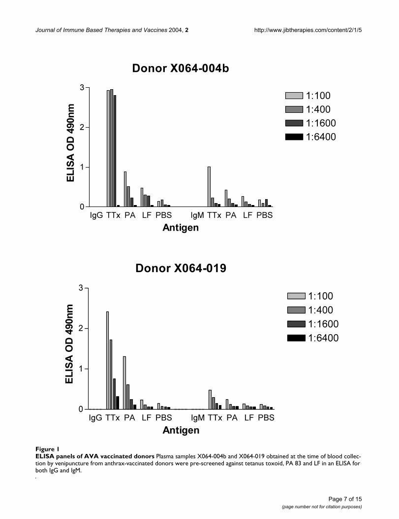
ResultsDonor screeningDonors sera (X064-004b and X064-019) were screenedfor IgG and IgM against tetanus toxoid, PA and LF byELISA. Figure 1 shows that both donors had significantIgG responses to tetanus toxoid and some albeit low levelsof specific IgG antibody against PA and LF.
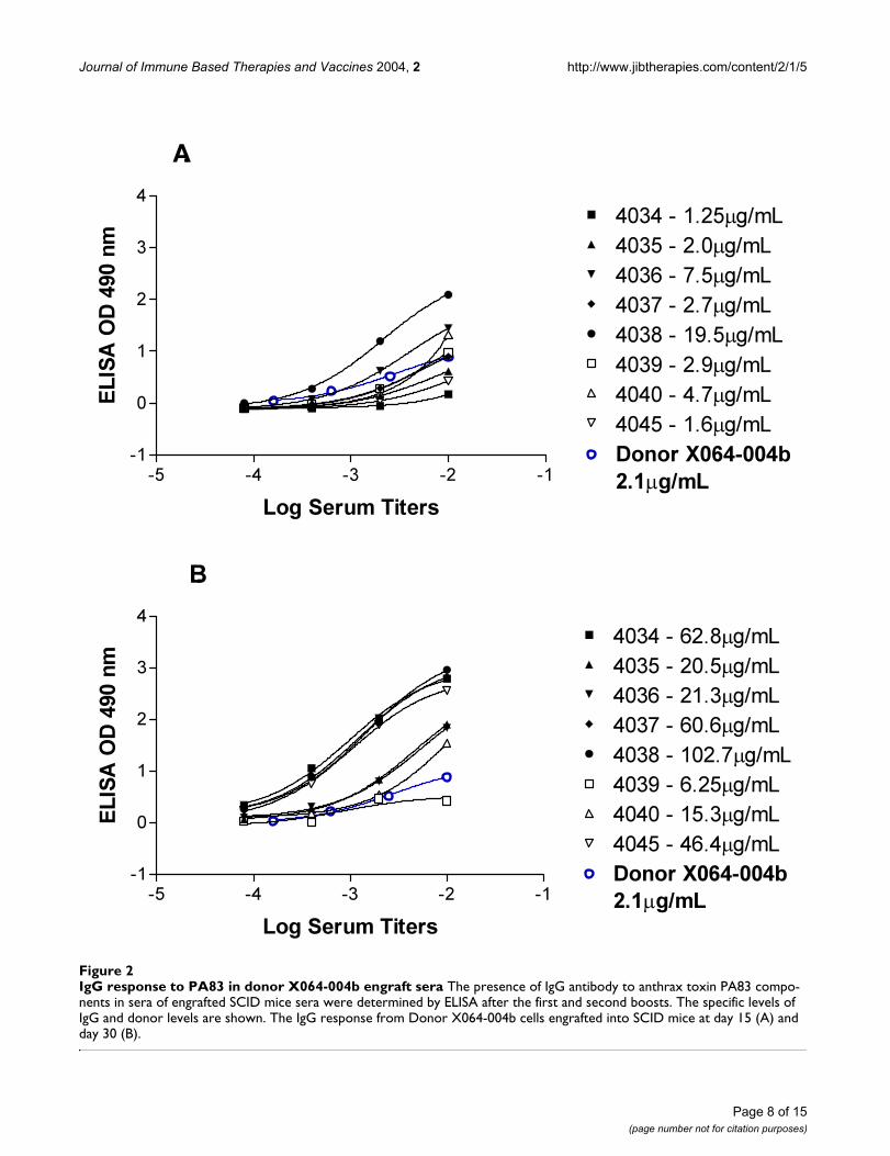
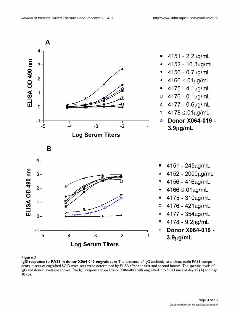
Chimeric engraft screeningThe PBLs from donor X064-004b and X064-019 wereengrafted into mice designated X040-042 and X040-043respectively. After boosting, sera from engrafted mice werescreened for human IgG against PA. In figure 2A the initialbleed after the first boost is plotted alongside the X064-004b donor sera. One engraft had an anti-PA IgG levelthat is 9 × higher than the donor sera. Moreover in figure2B, the second bleed from the engrafted mice, a range of8–30 fold increase in specific anti-PA IgG is observed. Thisincrease in specific IgG over time in the engrafted mice iseven more pronounced in the second engraft using cellsfrom donor X064-019 as shown in figure 3A and 3B. Theincrease in specific anti-PA IgG in the second bleed ismore than 500 fold relative to specific anti-PA in thedonor sera.
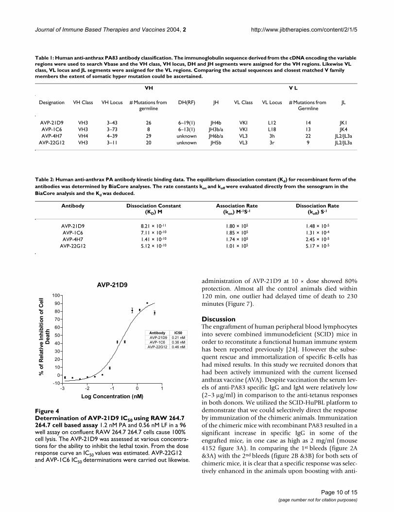
Immunoglobulin sequence analysisFollowing fusion, hybridoma cells producing humananti-anthrax PA IgG were selected and the cDNA encodingthe immunoglobulin variable regions were rescued andsequenced. The cDNA templates were used to establishstable CHO K1 cell lines producing antibodies. Four neu-tralizing anti-PA antibodies were discovered by thisapproach. The VH families were represented by the VH3,VH1 and VH4. Likewise VK 1 and VL 3 represented the VLfamilies. Both VH and VL regions contained evidence ofhyper mutation away from the germline. The Table 1 liststhe antibodies isolated by this approach and the D and Jregions are assigned where possible using DNAPLOT inVbase.
Kinetics of bindingThe equilibrium dissociation constants (Kd) for recom-binant form of the antibodies were determined byBiaCore analysis. The rate constants kon and koff were eval-uated directly from the sensogram in the BiaCore analysisand the Kd was deduced. The results are summarized inTable 2. One striking feature of all the protective antibod-ies isolated by the Xenerex Technology™ is the very slow
off rate, which contribute to the very high affinities 8.21 ×10⁻¹¹M to 7.11 × 10⁻¹⁰M. The slow off rate may confer sig-nificant physiological advantages for toxin neutralizationin vivo.
In vitro lethal toxin inhibitionAll the antibodies were initially selected based on bindingto PA83 and secondly on inhibition of lethal toxin in aRaw 264.7 cell based in vitro assay. Only clones exhibitingtoxin neutralization in a qualitative assay were developedfurther. The Raw 264.7 cell assay was adapted to comparethe various antibodies for potency of toxin inhibition. Infigure 4 a typical antibody dose response curve is recon-structed to provide an estimate of the IC50 for AVP-21D9,AVP-22G12 and AVP-1C6. Again the inhibitory potencyranking of all the selected antibodies were reflecting thesame ranking observed for the binding to PA83.
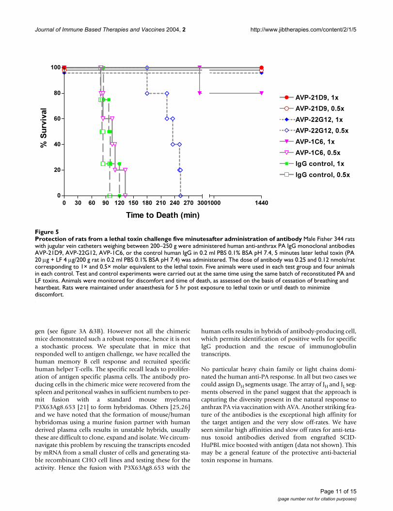
Effect of anti-anthrax PA antibodies on protection of rats from lethal toxin challengeFigure 5 illustrates the protection profile of the three anti-bodies AVP-21D9, 22G12 and 1C6 in the rat model at twodoses 0.5× and 1× molar ratios relative to toxin challenge.AVP-21D9 protected rats at 0.5× and no deaths wereobserved in the 5 hr following toxin administration, like-wise AVP-22G12 at 1× also showed complete protection.However with AVP22G12 at 0.5× the time to death wasprolonged to 255 min. The administrations of lethal toxin5 min after the infusion of 0.5× or 1× control human IgGresulted in time to death of 85–120 min. AVP-1C6 at 1×conferred 80% protection and at 0.5× were not protective.
Effect of antibody glycosylation on anti-anthrax PA antibodies on protection of rats from lethal toxin challengeSite directed mutagenesis (N297Q) was used to removethe N-glycosylation site in the Fc region. These aglyco-sylated antibodies were designated as AVP-21D9.1 andAVP-22G12.1 and compared to the glycosylated counter-parts in the rat toxin challenge prophylaxis model. Asdescribed earlier, antibody was intravenously adminis-tered 5 minutes prior to the lethal toxin (PA/LF) chal-lenge. Both AVP-21D9 and AVP-21D9.1 fully protectedrats against anthrax toxin with 0.5× molar excess relativeto PA toxin, whilst AVP-22G12.1 was slightly less potentthan the parent molecule at 1× as shown in figure 6.
Duration of AVP-21D9 antibody mediated protection of rats from lethal toxin challengeTo investigate the duration of the protection afforded by afully human antibody in Fisher rats AVP-21D9 was intra-venously administered 17 hours or 1 week prior to thelethal toxin (PA/LF) challenge. A single administration ofAVP-21D9 at 1× protected 100% when challenged 17hours later. Over the extended period of time
Page 6 of 15(page number not for citation purposes)
Journal of Immune Based Therapies and Vaccines 2004, 2 http://www.jibtherapies.com/content/2/1/5
ELISA panels of AVA vaccinated donorsFigure 1ELISA panels of AVA vaccinated donors Plasma samples X064-004b and X064-019 obtained at the time of blood collec-tion by venipuncture from anthrax-vaccinated donors were pre-screened against tetanus toxoid, PA 83 and LF in an ELISA for both IgG and IgM.
Donor X064-004b
IgG TTx PA LF PBS IgM TTx PA LF PBS0
1
2
31:100
1:400
1:1600
1:6400
Antigen
EL
ISA
OD
490n
m
Donor X064-019
IgG TTx PA LF PBS IgM TTx PA LF PBS0
1
2
31:100
1:400
1:1600
1:6400
Antigen
EL
ISA
OD
490n
m
Page 7 of 15(page number not for citation purposes)
Journal of Immune Based Therapies and Vaccines 2004, 2 http://www.jibtherapies.com/content/2/1/5
IgG response to PA83 in donor X064-004b engraft seraFigure 2IgG response to PA83 in donor X064-004b engraft sera The presence of IgG antibody to anthrax toxin PA83 compo-nents in sera of engrafted SCID mice sera were determined by ELISA after the first and second boosts. The specific levels of IgG and donor levels are shown. The IgG response from Donor X064-004b cells engrafted into SCID mice at day 15 (A) and day 30 (B).
A.
-5 -4 -3 -2 -1
-1
0
1
2
3
4
4034 - 1.25µg/mL
4035 - 2.0µg/mL
4036 - 7.5µg/mL
4037 - 2.7µg/mL
4038 - 19.5µg/mL
4039 - 2.9µg/mL
4040 - 4.7µg/mL
4045 - 1.6µg/mL
Donor X064-004b
2.1µg/mL
Log Serum Titers
EL
ISA
OD
490 n
m
B.
-5 -4 -3 -2 -1
-1
0
1
2
3
4
4034 - 62.8µg/mL
4035 - 20.5µg/mL
4036 - 21.3µg/mL
4037 - 60.6µg/mL
4038 - 102.7µg/mL
4039 - 6.25µg/mL
4040 - 15.3µg/mL
4045 - 46.4µg/mL
Donor X064-004b
2.1µg/mL
Log Serum Titers
EL
ISA
OD
490 n
m
Page 8 of 15(page number not for citation purposes)
Journal of Immune Based Therapies and Vaccines 2004, 2 http://www.jibtherapies.com/content/2/1/5
IgG response to PA83 in donor X064-043 engraft seraFigure 3IgG response to PA83 in donor X064-043 engraft sera The presence of IgG antibody to anthrax toxin PA83 compo-nents in sera of engrafted SCID mice sera were determined by ELISA after the first and second boosts. The specific levels of IgG and donor levels are shown. The IgG response from Donor X064-043 cells engrafted into SCID mice at day 15 (A) and day 30 (B).
A
-5 -4 -3 -2 -1
-1
0
1
2
3
4
4151 - 2.2µg/mL
4152 - 16.3µg/mL
4156 - 0.7µg/mL
4166 ≤.01µg/mL
4175 - 4.1µg/mL
4176 - 0.1µg/mL
4177 - 0.6µg/mL
4178 ≤.01µg/mL
Donor X064-019 -
3.9µg/mLLog Serum Titers
EL
ISA
OD
490 n
m
B.
-5 -4 -3 -2 -1
-1
0
1
2
3
4
4151 - 245µg/mL
4152 - 2000µg/mL
4156 - 416µg/mL
4166 ≤.01µg/mL
4175 - 310µg/mL
4176 - 421µg/mL
4177 - 354µg/mL
4178 - 9.2µg/mL
Donor X064-019 -
3.9µg/mLLog Serum Titers
EL
ISA
OD
490 n
m
Page 9 of 15(page number not for citation purposes)
Journal of Immune Based Therapies and Vaccines 2004, 2 http://www.jibtherapies.com/content/2/1/5
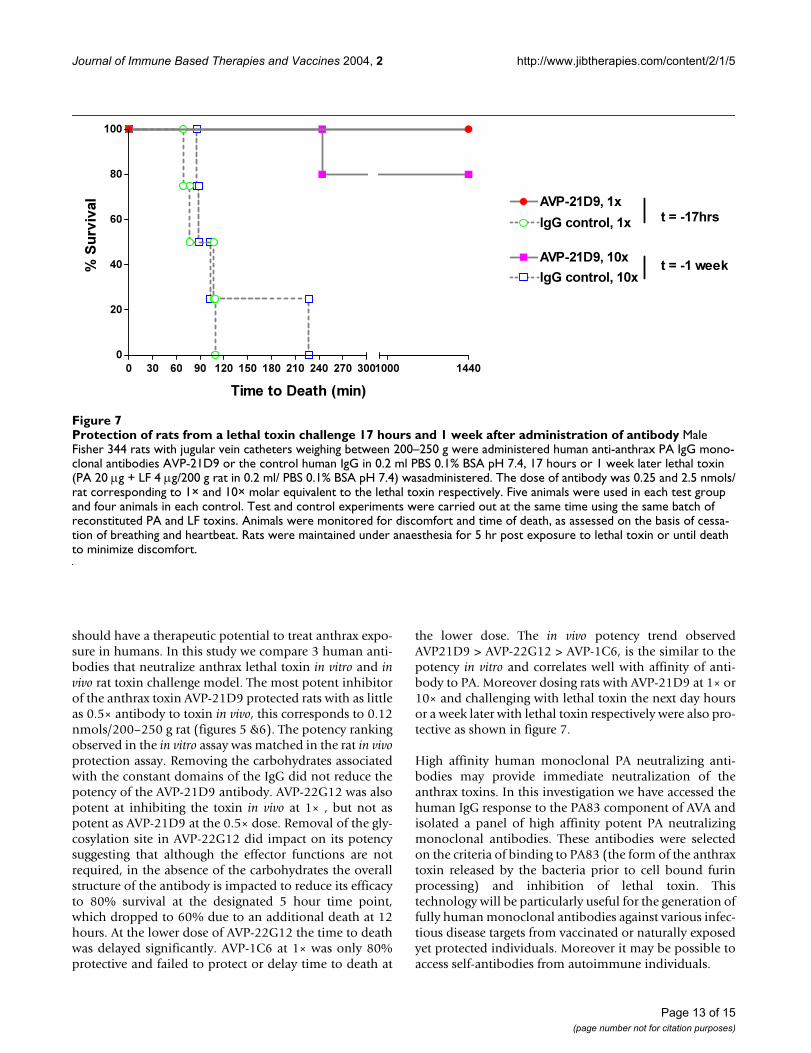
administration of AVP-21D9 at 10 × dose showed 80%protection. Almost all the control animals died within120 min, one outlier had delayed time of death to 230minutes (Figure 7).
DiscussionThe engraftment of human peripheral blood lymphocytesinto severe combined immunodeficient (SCID) mice inorder to reconstitute a functional human immune systemhas been reported previously [24]. However the subse-quent rescue and immortalization of specific B-cells hashad mixed results. In this study we recruited donors thathad been actively immunized with the current licensedanthrax vaccine (AVA). Despite vaccination the serum lev-els of anti-PA83 specific IgG and IgM were relatively low(2–3 µg/ml) in comparison to the anti-tetanus responsesin both donors. We utilized the SCID-HuPBL platform todemonstrate that we could selectively direct the responseby immunization of the chimeric animals. Immunizationof the chimeric mice with recombinant PA83 resulted in asignificant increase in specific IgG in some of theengrafted mice, in one case as high as 2 mg/ml (mouse4152 figure 3A). In comparing the 1st bleeds (figure 2A&3A) with the 2nd bleeds (figure 2B &3B) for both sets ofchimeric mice, it is clear that a specific response was selec-tively enhanced in the animals upon boosting with anti-
Table 1: Human anti-anthrax PA83 antibody classification. The immunoglobulin sequence derived from the cDNA encoding the variable regions were used to search Vbase and the VH class, VH locus, DH and JH segments were assigned for the VH regions. Likewise VL class, VL locus and JL segments were assigned for the VL regions. Comparing the actual sequences and closest matched V family members the extent of somatic hyper mutation could be ascertained.
VH V L
Designation VH Class VH Locus # Mutations from germline
DH(RF) JH VL Class VL Locus # Mutations from Germline
JL
AVP-21D9 VH3 3–43 26 6–19(1) JH4b VKI L12 14 JK1AVP-1C6 VH3 3–73 8 6–13(1) JH3b/a VKI L18 13 JK4AVP-4H7 VH4 4–39 29 unknown JH6b/a VL3 3h 22 JL2/JL3a
AVP-22G12 VH3 3–11 20 unknown JH5b VL3 3r 9 JL2/JL3a
Table 2: Human anti-anthrax PA antibody kinetic binding data. The equilibrium dissociation constant (Kd) for recombinant form of the antibodies was determined by BiaCore analyses. The rate constants kon and koff were evaluated directly from the sensogram in the BiaCore analysis and the Kd was deduced.
Antibody Dissociation Constant(KD) M
Association Rate(kon) M-1S-1
Dissociation Rate(koff) S-1
AVP-21D9 8.21 × 10⁻¹¹ 1.80 × 10⁵ 1.48 × 10⁻⁵
AVP-1C6 7.11 × 10⁻¹⁰ 1.85 × 10⁵ 1.31 × 10⁻⁴
AVP-4H7 1.41 × 10⁻¹⁰ 1.74 × 10⁵ 2.45 × 10⁻⁵
AVP-22G12 5.12 × 10⁻¹⁰ 1.01 × 10⁵ 5.17 × 10⁻⁵
Determination of AVP-21D9 IC50 using RAW 264.7 264.7 cell based assayFigure 4Determination of AVP-21D9 IC50 using RAW 264.7 264.7 cell based assay 1.2 nM PA and 0.56 nM LF in a 96 well assay on confluent RAW 264.7 264.7 cells cause 100% cell lysis. The AVP-21D9 was assessed at various concentra-tions for the ability to inhibit the lethal toxin. From the dose response curve an IC50 values was estimated. AVP-22G12 and AVP-1C6 IC50 determinations were carried out likewise.
AVP-21D9
-3 -2 -1 0 1
-10
0
10
20
30
40
50
60
70
80
90
100
Log Concentration (nM)
% o
f R
ela
tive I
nh
ibit
ion
of
Cell
Death Antibody IC50
AVP-21D9 0.21 nM
AVP-1C6 0.36 nM
AVP-22G12 0.46 nM
Page 10 of 15(page number not for citation purposes)
Journal of Immune Based Therapies and Vaccines 2004, 2 http://www.jibtherapies.com/content/2/1/5
gen (see figure 3A &3B). However not all the chimericmice demonstrated such a robust response, hence it is nota stochastic process. We speculate that in mice thatresponded well to antigen challenge, we have recalled thehuman memory B cell response and recruited specifichuman helper T-cells. The specific recall leads to prolifer-ation of antigen specific plasma cells. The antibody pro-ducing cells in the chimeric mice were recovered from thespleen and peritoneal washes in sufficient numbers to per-mit fusion with a standard mouse myelomaP3X63Ag8.653 [21] to form hybridomas. Others [25,26]and we have noted that the formation of mouse/humanhybridomas using a murine fusion partner with humanderived plasma cells results in unstable hybrids, usuallythese are difficult to clone, expand and isolate. We circum-navigate this problem by rescuing the transcripts encodedby mRNA from a small cluster of cells and generating sta-ble recombinant CHO cell lines and testing these for theactivity. Hence the fusion with P3X63Ag8.653 with the
human cells results in hybrids of antibody-producing cell,which permits identification of positive wells for specificIgG production and the rescue of immunoglobulintranscripts.
No particular heavy chain family or light chains domi-nated the human anti-PA response. In all but two cases wecould assign DH segments usage. The array of JH and JL seg-ments observed in the panel suggest that the approach iscapturing the diversity present in the natural response toanthrax PA via vaccination with AVA. Another striking fea-ture of the antibodies is the exceptional high affinity forthe target antigen and the very slow off-rates. We haveseen similar high affinities and slow off rates for anti-teta-nus toxoid antibodies derived from engrafted SCID-HuPBL mice boosted with antigen (data not shown). Thismay be a general feature of the protective anti-bacterialtoxin response in humans.
Protection of rats from a lethal toxin challenge five minutesafter administration of antibodyFigure 5Protection of rats from a lethal toxin challenge five minutesafter administration of antibody Male Fisher 344 rats with jugular vein catheters weighing between 200–250 g were administered human anti-anthrax PA IgG monoclonal antibodies AVP-21D9, AVP-22G12, AVP-1C6, or the control human IgG in 0.2 ml PBS 0.1% BSA pH 7.4, 5 minutes later lethal toxin (PA 20 µg + LF 4 µg/200 g rat in 0.2 ml PBS 0.1% BSA pH 7.4) was administered. The dose of antibody was 0.25 and 0.12 nmols/rat corresponding to 1× and 0.5× molar equivalent to the lethal toxin. Five animals were used in each test group and four animals in each control. Test and control experiments were carried out at the same time using the same batch of reconstituted PA and LF toxins. Animals were monitored for discomfort and time of death, as assessed on the basis of cessation of breathing and heartbeat. Rats were maintained under anaesthesia for 5 hr post exposure to lethal toxin or until death to minimize discomfort.
0 30 60 90 120 150 180 210 240 270 300
0
20
40
60
80
100
AVP-21D9, 1x
AVP-21D9, 0.5x
AVP-22G12, 1x
AVP-22G12, 0.5x
AVP-1C6, 1x
AVP-1C6, 0.5x
IgG control, 1x
IgG control, 0.5x
1000 1440
Time to Death (min)
% S
urviv
al
Page 11 of 15(page number not for citation purposes)

Journal of Immune Based Therapies and Vaccines 2004, 2 http://www.jibtherapies.com/content/2/1/5
Currently in the event of an inadvertent Bacillus anthracisspore exposure two preventative measures can be taken. Ifthe risk can be assessed well in advance, vaccination canbe employed. In the event of near term or immediate postexposure antibiotic such as Cipro may be effective.Anthrax Vaccine Adsorbed (AVA) is the only licensedhuman anthrax vaccine in the United States. The vaccineis known to contain a mixture of cell products includingPA, LF and EF, however the exact amounts are unknown[27]. The immunization schedule consists of three subcu-taneous injections at 0, 2 and 4 weeks and booster vacci-nation at 6, 12 and 18 months and it is suggested thatannual boost may be required to maintain immunity.Mass vaccination in the event of anthrax spore release isan unlikely scenario. First, the time taken for effectivenessof such vaccination based on AVA or various rPA mole-cules in development may be too short, weeks as opposedto minutes. The utilization of antibiotic can inhibitbacterial growth and spread, and may prevent some of the
symptoms, but the administration needs to be timely andpreferably as a prophylactic, even as such, the toxinsreleased during the early stages of an infection may impairthe immune system to cause lasting damage. Ideally, acombination of approaches that inhibit anthrax bacteriaand toxins is desirable.
Human antibodies are safe and well tolerated for a rangeof therapeutic indications and are logical choices for animmediate anthrax therapeutic or prophylactic inhumans. Passive protection against anthrax toxins in ratsand anthrax infection in guinea pigs has been demon-strated for murine monoclonal antibodies [28,29] andpolyclonal antibodies [17], respectively. Recently ahuman monoclonal antibody against PA has demon-strated efficacy in protecting rats challenged with lethaltoxin [20]. The antibody was fully protective at 0.3 nmol/250 g rat. It is expected that exceptionally high affinityhuman monoclonal antibodies against the anthrax toxin
Protection of rats from a lethal toxin challenge by aglycosylated antibodyFigure 6Protection of rats from a lethal toxin challenge by aglycosylated antibody Male Fisher 344 rats with jugular vein catheters weighing between 200–250 g were administered human anti-anthrax PA IgG monoclonal antibodies AVP-21D9, AVP-22G12, the aglycosylated forms AVP-21D9.1 and AVP22G12.1 or the control human IgG in 0.2 ml PBS 0.1% BSA pH 7.4, 5 minutes later lethal toxin (PA 20 µg / LF 4 µg in 0.2 ml/200 g rat PBS 0.1% BSA pH 7.4) was administered. The dose of antibody was 0.25 and 0.12 nmols/rat corresponding to 1× and 0.5× molar equivalent to the lethal toxin. Five animals were used in each test group and four animals in each control. Test and control experiments were carried out at the same time using the same batch of reconstituted PA and LF toxins. Animals were monitored for discomfort and time of death, as assessed on the basis of cessation of breathing and heartbeat. Rats were maintained under anaesthesia for 5 hr post exposure to lethal toxin or until death to minimize discomfort.
0 30 60 90 120 150 180 210 240 270 300
0
20
40
60
80
100
AVP-21D9, 0.5x
AVP-21D9.1, 0.5x
IgG control, 0.5x
1000 1440
AVP-22G12, 1x
AVP-22G12.1, 1x
IgG control, 1x
Time to Death (min)
% S
urviv
al
Page 12 of 15(page number not for citation purposes)
Journal of Immune Based Therapies and Vaccines 2004, 2 http://www.jibtherapies.com/content/2/1/5
should have a therapeutic potential to treat anthrax expo-sure in humans. In this study we compare 3 human anti-bodies that neutralize anthrax lethal toxin in vitro and invivo rat toxin challenge model. The most potent inhibitorof the anthrax toxin AVP-21D9 protected rats with as littleas 0.5× antibody to toxin in vivo, this corresponds to 0.12nmols/200–250 g rat (figures 5 &6). The potency rankingobserved in the in vitro assay was matched in the rat in vivoprotection assay. Removing the carbohydrates associatedwith the constant domains of the IgG did not reduce thepotency of the AVP-21D9 antibody. AVP-22G12 was alsopotent at inhibiting the toxin in vivo at 1× , but not aspotent as AVP-21D9 at the 0.5× dose. Removal of the gly-cosylation site in AVP-22G12 did impact on its potencysuggesting that although the effector functions are notrequired, in the absence of the carbohydrates the overallstructure of the antibody is impacted to reduce its efficacyto 80% survival at the designated 5 hour time point,which dropped to 60% due to an additional death at 12hours. At the lower dose of AVP-22G12 the time to deathwas delayed significantly. AVP-1C6 at 1× was only 80%protective and failed to protect or delay time to death at
the lower dose. The in vivo potency trend observedAVP21D9 > AVP-22G12 > AVP-1C6, is the similar to thepotency in vitro and correlates well with affinity of anti-body to PA. Moreover dosing rats with AVP-21D9 at 1× or10× and challenging with lethal toxin the next day hoursor a week later with lethal toxin respectively were also pro-tective as shown in figure 7.
High affinity human monoclonal PA neutralizing anti-bodies may provide immediate neutralization of theanthrax toxins. In this investigation we have accessed thehuman IgG response to the PA83 component of AVA andisolated a panel of high affinity potent PA neutralizingmonoclonal antibodies. These antibodies were selectedon the criteria of binding to PA83 (the form of the anthraxtoxin released by the bacteria prior to cell bound furinprocessing) and inhibition of lethal toxin. Thistechnology will be particularly useful for the generation offully human monoclonal antibodies against various infec-tious disease targets from vaccinated or naturally exposedyet protected individuals. Moreover it may be possible toaccess self-antibodies from autoimmune individuals.
Protection of rats from a lethal toxin challenge 17 hours and 1 week after administration of antibodyFigure 7Protection of rats from a lethal toxin challenge 17 hours and 1 week after administration of antibody Male Fisher 344 rats with jugular vein catheters weighing between 200–250 g were administered human anti-anthrax PA IgG mono-clonal antibodies AVP-21D9 or the control human IgG in 0.2 ml PBS 0.1% BSA pH 7.4, 17 hours or 1 week later lethal toxin (PA 20 µg + LF 4 µg/200 g rat in 0.2 ml/ PBS 0.1% BSA pH 7.4) wasadministered. The dose of antibody was 0.25 and 2.5 nmols/rat corresponding to 1× and 10× molar equivalent to the lethal toxin respectively. Five animals were used in each test group and four animals in each control. Test and control experiments were carried out at the same time using the same batch of reconstituted PA and LF toxins. Animals were monitored for discomfort and time of death, as assessed on the basis of cessa-tion of breathing and heartbeat. Rats were maintained under anaesthesia for 5 hr post exposure to lethal toxin or until death to minimize discomfort.
0 30 60 90 120 150 180 210 240 270 300
0
20
40
60
80
100
AVP-21D9, 1x
IgG control, 1x
AVP-21D9, 10x
IgG control, 10x
1000 1440
t = -17hrs
t = -1 week
Time to Death (min)
% S
urviv
al
Page 13 of 15(page number not for citation purposes)
Journal of Immune Based Therapies and Vaccines 2004, 2 http://www.jibtherapies.com/content/2/1/5
ConclusionsWe have successfully engrafted human PBL's fromanthrax-vaccinated donors into SCID/bg mice and dem-onstrated that a specific recall response can be selectivelyenhanced by immunization with PA83. Moreover, wehave shown that the cells producing the antibodies can beisolated, the transcript mRNA encoding the desiredantibodies can be readily recovered and stable recom-binant cell lines producing human monoclonal antibod-ies can be generated. The human monoclonal antibodiesgenerated are of very high affinity for PA83 and neutralizelethal toxin in an in vitro cell based assay. Vaccination withAnthrax Vaccine Adsorbed can induce the production of arange of protective antibodies. Here we show that most ofthe human anti-anthrax toxin antibodies selected by theXenerex technology™ are potent inhibitors of the lethaltoxin in vivo. The three parental antibodies and the twoaglycosylated forms described may be therapeutically use-ful against anthrax infection and in the passive protectionof high risk individuals. In particular the two most potentanthrax toxin-neutralizing antibody AVP-21D9 and AVP-22G12 were completely effective at a dose correspondingto 0.12 nmols/rat and 0.25 nmols/rat respectively.
Competing interestsNone declared.
Authors' contributionsIJ carried out cell engrafting immunization and cell recov-ery/fusion. RS-H carried out Ig cloning, expression andcoordinated rat studies. FW carried out vector construc-tion, IgG engineering and purification. SMS was responsi-ble for fusion/cell culture and antibody production. RNcarried out PBL preparation, antibody expression, in vitroassay and in vivo studies. Paul Ruther was responsible fordonor/mice sera screening, IgG quantification, affinitydeterminations. DM and AA were responsible for SCIDmouse facility and carried out the rat studies. PRM† wasProject initiator. ASK Project leader. All authors read andapproved the final manuscript
†Phillip R. Morrow deceased.
AcknowledgementsWe acknowledge the support of Center for Commercialization of Advanced Technology (CCAT) 52109A/7805 to PRM and National Institute of Health, National Institute of Allergy and Infectious Diseases for funding SBIR R43 AI052901-01A1 to PRM and SBIR R43 AI058458-01 to ASK.
References1. Leppla SH: Anthrax toxin edema factor: a bacterial adenylate
cyclase that increases cyclic AMP concentrations of eukary-otic cells. Proc Natl Acad Sci U S A 1982, 79:3162-3166.
2. Duesbery NS, Webb CP, Leppla SH, Gordon VM, Klimpel KR, Cope-land TD, Ahn NG, Oskarsson MK, Fukasawa K, Paull KD, VandeWoude GF: Proteolytic inactivation of MAP-kinase-kinase byanthrax lethal factor. Science 1998, 280:734-737.
3. Vitale G, Pellizzari R, Recchi C, Napolitani G, Mock M, MontecuccoC: Anthrax lethal factor cleaves the N-terminus of MAPKKsand induces tyrosine/threonine phosphorylation of MAPKs incultured macrophages. Biochem Biophys Res Commun 1998,248:706-711.
4. Bradley KA, Mogridge J, Jonah G, Rainey A, Batty S, Young JA: Bind-ing of anthrax toxin to its receptor is similar to alphaintegrin-ligand interactions. J Biol Chem 2003, 278:49342-7.
5. Bradley KA, Mogridge J, Mourez M, Collier RJ, Young JA: Identifica-tion of the cellular receptor for anthrax toxin. Nature 2001,414:225-229.
6. Scobie HM, Rainey GJ, Bradley KA, Young JA: Human capillarymorphogenesis protein 2 functions as an anthrax toxinreceptor. Proc Natl Acad Sci U S A 2003, 100:5170-5174.
7. Smith H, Keppie J: Observations on experimental anthrax;demonstration of a specific lethal factor produced in vivo byBacillus anthracis. Nature 1954, 173:869-870.
8. Greenfield WS: Lectures on some recent investigations intopathology of infective and contagious diseases. Lecture III.Part I. Anthrax and anthracoid diseases. Lancet 1880,1:865-867.
9. Sterne M: The use of anthrac vaccines prepared from aviru-lent (unencapsulated) variants of Bacillus anthracis. Onderste-poort J Vet Sci An Ind 1939, 13:307-312.
10. Sterne M: The immunization of laboratory animals againstanthrax. J S Afr Vet Med Assoc 1942, 13:53-57.
11. Salsbery CE: Anthrax aggressin. J Am Vet MEd Assoc 1926,68:755-757.
12. Gladstone GP: Immunity to anthrax: protective antigenpresent in cell-free culture filtrates. Br J Exp Pathol 1946,27:394-418.
13. Wright GG, Green, TW, Kanode, RG Jr: Studies on immunity inanthrax.V. Immunizing activity of alum-precipitated protec-tive antigen. J Immunol 1954, 73:387-391.
14. Puziss M, Manning LC, Lynch JW, Barclaye, Abelow I, Wright GG:Large-scale production of protective antigen of Bacillusanthracis in anaerobic cultures. Appl Microbiol 1963, 11:330-334.
15. Puziss M, Wright GG: Studies on immunity in anthrax. X. Gel-adsorbed protective antigen for immunization of man. JBacteriol 1963, 85:230-236.
16. Wein LM, Craft DL, Kaplan EH: Emergency response to ananthrax attack. Proc Natl Acad Sci U S A 2003, 100:4346-4351.
17. Little SF, Ivins BE, Fellows PF, Friedlander AM: Passive protectionby polyclonal antibodies against Bacillus anthracis infectionin guinea pigs. Infect Immun 1997, 65:5171-5175.
18. Kobiler D, Gozes Y, Rosenberg H, Marcus D, Reuveny S, Altboum Z:Efficiency of protection of guinea pigs against infection withBacillus anthracis spores by passive immunization. InfectImmun 2002, 70:544-560.
19. Maynard JA, Maassen CB, Leppla SH, Brasky K, Patterson JL, IversonBL, Georgiou G: Protection against anthrax toxin by recom-binant antibody fragments correlates with antigen affinity.Nat Biotechnol 2002, 20:597-601.
20. Wild MA, Xin H, Maruyama T, Nolan MJ, Calveley PM, Malone JD,Wallace MR, Bowdish KS: Human antibodies from immunizeddonors are protective against anthrax toxin in vivo. NatBiotechnol 2003, 21:1305-1306.
21. Kearney JF, Radbruch A, Liesegang B, Rajewsky K: A new mousemyeloma cell line that has lost immunoglobulin expressionbut permits the construction of antibody-secreting hybridcell lines. J Immunol 1979, 123:1548-1550.
22. Hanna PC, Acosta D, Collier RJ: On the role of macrophages inanthrax. Proc Natl Acad Sci U S A 1993, 90:10198-10201.
23. Ivins BE, Ristroph JD, Nelson GO: Influence of body weight onresponse of Fischer 344 rats to anthrax lethal toxin. Appl Envi-ron Microbiol 1989, 55:2098-2100.
24. Mosier DE, Gulizia RJ, Baird SM, Wilson DB: Transfer of a func-tional human immune system to mice with severe combinedimmunodeficiency. Nature 1988, 335:256-259.
25. Alkan SS, Mestel F, Jiricka J, Blaser K: Estimation of heterokaryonformation and hybridoma growth in murine and human cellfusions. Hybridoma 1987, 6:371-379.
26. Kozbor D, Dexter D, Roder JC: A comparative analysis of thephenotypic characteristics of available fusion partners forthe construction of human hybridomas. Hybridoma 1983,2:7-16.
Page 14 of 15(page number not for citation purposes)
Journal of Immune Based Therapies and Vaccines 2004, 2 http://www.jibtherapies.com/content/2/1/5
Publish with BioMed Central and every scientist can read your work free of charge
"BioMed Central will be the most significant development for disseminating the results of biomedical research in our lifetime."
Sir Paul Nurse, Cancer Research UK
Your research papers will be:
available free of charge to the entire biomedical community
peer reviewed and published immediately upon acceptance
cited in PubMed and archived on PubMed Central
yours — you keep the copyright
Submit your manuscript here:http://www.biomedcentral.com/info/publishing_adv.asp
BioMedcentral
27. Turnbull PC, Broster MG, Carman JA, Manchee RJ, Melling J: Devel-opment of antibodies to protective antigen and lethal factorcomponents of anthrax toxin in humans and guinea pigs andtheir relevance to protective immunity. Infect Immun 1986,52:356-363.
28. Little SF, Leppla SH, Cora E: Production and characterization ofmonoclonal antibodies to the protective antigen componentof Bacillus anthracis toxin. Infect Immun 1988, 56:1807-1813.
29. Little SF, Leppla SH, Friedlander AM: Production and characteri-zation of monoclonal antibodies against the lethal factorcomponent of Bacillus anthracis lethal toxin. Infect Immun1990, 58:1606-1613.
Page 15 of 15(page number not for citation purposes)


![[Vaccination coverage in children and adolescents in Mexico: vaccinated, under vaccinated and non vaccinated]](https://img.dokumen.tips/doc/110x75/634b125dc27aad9758027926/vaccination-coverage-in-children-and-adolescents-in-mexico-vaccinated-under-vaccinated.jpg)


