Embed Size (px)
Citation preview

Behavioral/Systems/Cognitive
How Silent Is Silent Reading? Intracerebral Evidence forTop-Down Activation of Temporal Voice Areas duringReading
Marcela Perrone-Bertolotti,1,2 Jan Kujala,1,2 Juan R. Vidal,1,2 Carlos M. Hamame,3 Tomas Ossandon,4 Olivier Bertrand,1,2
Lorella Minotti,5 Philippe Kahane,5 Karim Jerbi,1,2 and Jean-Philippe Lachaux1,2
1INSERM U1028-CNRS UMR5292, Brain Dynamics and Cognition Team, Lyon Neuroscience Research Center, F-69500 Lyon-Bron, France; 2UniversityClaude Bernard, F-69000 Lyon, France; 3Laboratoire de Psychologie Cognitive, CNRS (UMR 6146), Aix-Marseille Universite 13003 Marseille Cedex, 3France; 4Departamento de Psiquiatría, Facultad de Medicina y Centro Interdisciplinario de Neurociencia, Pontificia Universidad Catolica de Chile, CL-8330024 Santiago, Chile; and 5CHU Grenoble and Department of Neurology, INSERM U704, F-38043 Grenoble, France
As you might experience it while reading this sentence, silent reading often involves an imagery speech component: we can hear our own“inner voice” pronouncing words mentally. Recent functional magnetic resonance imaging studies have associated that component withincreased metabolic activity in the auditory cortex, including voice-selective areas. It remains to be determined, however, whether thisactivation arises automatically from early bottom-up visual inputs or whether it depends on late top-down control processes modulatedby task demands. To answer this question, we collaborated with four epileptic human patients recorded with intracranial electrodes in theauditory cortex for therapeutic purposes, and measured high-frequency (50 –150 Hz) “gamma” activity as a proxy of population levelspiking activity. Temporal voice-selective areas (TVAs) were identified with an auditory localizer task and monitored as participantsviewed words flashed on screen. We compared neural responses depending on whether words were attended or ignored and found asignificant increase of neural activity in response to words, strongly enhanced by attention. In one of the patients, we could record thatresponse at 800 ms in TVAs, but also at 700 ms in the primary auditory cortex and at 300 ms in the ventral occipital temporal cortex.Furthermore, single-trial analysis revealed a considerable jitter between activation peaks in visual and auditory cortices. Altogether, ourresults demonstrate that the multimodal mental experience of reading is in fact a heterogeneous complex of asynchronous neuralresponses, and that auditory and visual modalities often process distinct temporal frames of our environment at the same time.
IntroductionWhen children learn to read, they learn to associate written sym-bols with spoken sounds, until the association is so well trainedthat it occurs effortlessly. Around that time, they become able toread silently. Basic introspection suggests that adults continue topronounce written text covertly using auditory verbal imagery (AVI;Jancke and Shah, 2004). This intuition has been confirmed by severalpsychophysics experiments (Alexander and Nygaard, 2008).
Until recently, AVI had received little interest from experi-mental psychology and cognitive neuroscience (Kurby et al.,2009; Yao et al., 2011). Yet, it is one of the most striking examplesof cross talk between sensory modalities, as written words produce a
vivid auditory experience almost effortlessly. It is therefore a privi-leged situation to understand multisensory integration and its un-derlying functional connectivity. It also offers a unique window intothe actual reading experience of “what it is like” to read. AVI is one ofits most salient components and should be included in any neuro-cognitive model of reading. Finally, few would contest that most ofour waking time is spent talking to ourselves covertly, and somediseases, like schizophrenia or depression, are often characterized bya lack of control over AVI (Linden et al., 2011). Silent reading is anefficient way to elicit AVI experimentally with precise control over itstiming, and understand its neural bases.
The neurophysiological investigation of AVI has been focused onthe auditory cortex, because that region is active during several formsof auditory imagery, including hallucinations (Lennox et al., 1999),silent lip reading (Calvert et al., 1997), and music imagery (Halpernand Zatorre, 1999). One might therefore assume that if silent readinggenerates AVI, it should activate the auditory cortex concurrently.This hypothesis was recently validated with functional magnetic res-onance imaging (fMRI) by Yao et al. (2011), who showed a strongactivation of auditory voice-selective areas, or temporal voice areas(TVAs; Belin et al., 2000).
One remaining question, unresolved by fMRI, is whether TVAactivation is mostly early and bottom-up, or controlled by late
Received June 24, 2012; revised Sept. 13, 2012; accepted Oct. 8, 2012.Author contributions: J.-P.L. designed research; M.P.-B., J.R.V., C.M.H., T.O., and J.-P.L. performed research;
M.P.-B., K.J., J.R.V., J.K., and J.-P.L. analyzed data; M.P.-B., K.J., J.R.V., C.M.H., O.B., L.M., P.K., J.K., and J.-P.L. wrotethe paper.
This work was supported by the Paulo Foundation, the KAUTE Foundation, and the TES Foundation. We thank allpatients for their participation; the staff of the Grenoble Neurological Hospital epilepsy unit; and Dominique Hoff-mann, Patricia Boschetti, Carole Chatelard, Veronique Dorlin, Crhystelle Mosca, and Luca de Palma for their support.
Correspondence should be addressed to Marcela Perrone-Bertolotti, INSERM U1028-CNRS UMR5292, BrainDynamics and Cognition Team, Lyon Neuroscience Research Center, F-69500 Lyon-Bron, France. E-mail ad-dress: marcela. [email protected].
DOI:10.1523/JNEUROSCI.2982-12.2012Copyright © 2012 the authors 0270-6474/12/3217554-09$15.00/0
17554 • The Journal of Neuroscience, December 5, 2012 • 32(49):17554 –17562

top-down processes (Alexander and Nygaard, 2008) and there-fore modulated by attention or cognitive strategy. We addressedthis question with direct intracerebral electroencephalographic(EEG) recordings of TVA in epileptic patients. We analyzed high-frequency “gamma” activity (HFA), between 50 and 150 Hz, be-cause it is now considered as a reliable physiological marker ofneuronal activity at the population level, and a general index ofcortical processing during cognition (Lachaux et al., 2012).Lower frequencies were not considered in this study, either be-cause of their less precise association with fine cognitive processes(� and �; Jerbi et al., 2009; Crone et al., 2011) or simply becausetheir frequency range was incompatible with the timing of theexperimental design (theta and delta).We found that the neuralresponse to written words in TVA comes between 400 and 800 msafter the visual response, with a highly variable delay, and isstrongly modulated by attention. We therefore demonstrate thatthe rich and seemingly coherent audiovisual experience of read-ing arises from a heterogeneous amalgam of asynchronous neuralresponses.
Materials and MethodsParticipants. Intracranial recordings were obtained from four neurosur-gical patients with intractable epilepsy (three female, mean age: 28.75�/� 10.04 years) at the Epilepsy Department of the Grenoble Neurolog-ical Hospital (Grenoble, France). All patients were stereotacticallyimplanted with multilead EEG depth electrodes. All electrode data ex-hibiting pathological waveforms were discarded from the present study.This was achieved in collaboration with the medical staff and was basedon visual inspection of the recordings and by systematically excludingdata from any electrode site that was a posteriori found to be locatedwithin the seizure onset zone. All participants provided written informedconsent, and the experimental procedures were approved by the Institu-tional Review Board and by the National French Science EthicalCommittee.
Electrode implantation. Eleven to 15 semirigid, multilead electrodeswere stereotactically implanted in each patient. The stereotactic electro-encephalography (SEEG) electrodes used have a diameter of 0.8 mm and,depending on the target structure, consist of 10 –15 contact leads 2 mmwide and 1.5 mm apart (DIXI Medical Instruments). All electrode con-tacts were identified on a postimplantation MRI showing the electrodes,and coregistered on a pre-implantation MRI. MNI and Talairach) coor-dinates were computed using the SPM (http://www.fil.ion.ucl.ac.uk/spm/) toolbox (see Table 1).
Intracranial recordings. Intracranial recordings were conducted using avideo-SEEG monitoring system (Micromed), which allowed the simul-taneous data recording from 128 depth EEG electrode sites. The datawere bandpass filtered on-line from 0.1 to 200 Hz and sampled at 512 Hzin all patients. At the time of acquisition data are recorded using a refer-ence electrode located in white matter, and each electrode trace is subse-quently re-referenced with respect to its direct neighbor (bipolarderivations). This bipolar montage has a number of advantages overcommon referencing. It helps eliminate signal artifacts common to adja-cent electrode contacts (such as the 50 Hz mains artifact or distant phys-iological artifacts) and achieves a high local specificity by canceling outeffects of distant sources that spread equally to both adjacent sitesthrough volume conduction. The spatial resolution achieved by the bi-
polar SEEG is on the order of 3 mm (Kahane et al., 2003; Lachaux et al.,2003; Jerbi et al., 2009). Both spatial resolution and spatial samplingachieved with SEEG differ slightly from that obtained with subdural gridelectrocorticography (Jerbi et al., 2009).
Time–frequency analysis, gamma power, and envelope computations.The frequency band of interest, between 50 and 150 Hz, was defined frompreliminary time-frequency analysis of the SEEG data using wavelets(Tallon-Baudry et al., 1997) (performed with in-house software packagefor electrophysiological signal analysis) (Aguera et al., 2011) and fromprevious studies by our group (Jerbi et al., 2009).
Raw data were transformed into gamma-band amplitude (GBA) timeseries with the following procedure (Ossandon et al., 2011). Step 1: contin-uous SEEG signals were first bandpass filtered in multiple successive 10 Hzwide frequency bands (e.g., 10 bands from 50–60 Hz to 140–150 Hz) usinga zero phase shift noncausal finite impulse filter with 0.5 Hz roll-off. Step 2:next, for each bandpass filtered signal we computed the envelope using stan-dard Hilbert transform (Le Van Quyen, 2001). The obtained envelope isdownsampled to a sampling rate of 64 Hz (i.e., one time sample every 15,625ms). Step 3: for each band this envelope signal (i.e., time-varying amplitude)was divided by its mean across the entire recording session and multiplied by100. This yields instantaneous envelope values expressed in percentage (%)of the mean. Step 4: finally, the envelope signals computed for each consec-utive frequency band (the 10 bands of 10 Hz intervals between 50 and 150Hz) were averaged together to provide one single time series (the GBA)across the entire session. By construction, the mean value of that time seriesacross the recording session is equal to 100. Note that computing the Hilbertenvelopes in 10 Hz sub-bands and normalizing them individually beforeaveraging over the broadband interval allows us to account for a bias towardthe lower frequencies of the interval that would otherwise occur due to the 1/fdrop-off in amplitude.
Auditory functional localizer. During the auditory localizer task(AUDI), participants listened to sounds of several categories with theinstruction that three of them would be presented again at the end of thetask, together with three novel sounds and that they should be able todetect previously played items. There were three speech and speech-likecategories, including sentences told by a computerized voice in a lan-guage familiar to the participant (French) or unfamiliar (Suomi), andreversed speech, originally in French (the same sentences as the “French”category, played backwards). These categories were compared withnonspeech-like human sounds (coughing and yawning), music, environ-mental sounds, and animal sounds. In the text, these categories will bereferred to as French, Suomi, Revers, Cough, Yawn, Music, Envir, andAnimal. Some of the sounds (Cough, Yawn, Envir, and Animal) wereconcatenations of shorter stimuli of the functional localizer of the TVAs(Belin et al., 2000; Pernet et al., 2007).
Participants were instructed to close their eyes while listening to threesounds of each category, with a duration of 12 s each, along with three12 s intervals with no stimulation, serving as a baseline (Silence). Con-secutive sounds were separated by a 3 s silent interval. The sequence waspseudorandom, to ensure that two sounds of the same category did notfollow each other. Sound sequences were delivered binaurally via ear-phones via the Presentation stimulus delivery software (NeurobehavioralSystems). All acoustic signals had the same intensity, quantified by theirroot mean square, set at a comfortable sound level.
Reading task. The reading task (Read) was adapted from Nobre et al.(1998). In each block of the experiment, participants were presented withtwo intermixed stories, shown word by word at a rapid rate. One of thestories was written in gray (on a black screen) and the other in white.Consecutive words with the same color formed a meaningful and simpleshort story in French. Participants were instructed to read the gray storyto report it at the end of the block, while ignoring the white one. Eachblock comprised 400 words, with 200 gray words (Attend condition) and200 white words (Ignore condition) for the two stories. The time se-quence of colors within the 400 words series was randomized, so thatparticipants could not predict whether the subsequent word was to beattended or not; however, the randomization was constrained to forbidseries of more than three consecutive words with the same color. Afterthe block, participants were asked questions about the attended text,which could not have been answered from general knowledge. The ex-
Table 1. Talairach coordinates of sites of interest
Patient Site X Y Z
P1 T8 58 0 �12P2 T7 57 �16 �6P3 U�9 �60 �15 4P4 T8 61 �11 �5P2 U2 37 �28 8P2 F�2 �25 �56 �8
Coordinates are expressed in millimeters in the Talairach coordinate system (Talairach and Tournoux, 1988).
Perrone-Bertolotti et al. • Top-Down of Temporal Voice Areas during Reading J. Neurosci., December 5, 2012 • 32(49):17554 –17562 • 17555

perimental procedure took place in patients’hospital rooms. Stimuli were presented to theparticipants on a 17 inch computer screen at a20 cm viewing distance and the average wordsubtended 28 of visual angle. Words appearedsingly for 100 ms every 700 ms.
Statistical analysis. Statistical analyses wereperformed on high-frequency activity time se-ries, HFA, computed as above. In the AUDItask, for each sound (12 s duration), we se-lected 20 nonoverlapping 500 ms windowscovering latencies between 1500 ms after stim-ulus onset to 500 ms before stimulus offset(windows within the first 1500 ms were ex-cluded to avoid the transient response to thestimulus). The mean HFA was measured ineach window to provide 20 values per stim-ulus, and a total of 60 values per category(since there were three sounds for each cate-gory). We used a nonparametric Kruskal–Wallis test to detect an effect of stimuluscategory on those values (60 HFA values � 8categories, that is, excluding the Silence cat-egory). We used a Bonferroni procedure tocorrect for multiple comparisons. In theRead task the effect of silent reading on TVAsites was evaluated by comparing HFA acrossconsecutive, nonoverlapping 100 ms win-dows covering a 1 s interval following wordpresentation (Kruskal–Wallis test, correctedfor multiple comparisons with a Bonferroniprocedure). This comparison was done sep-arately for attended and ignored words. We reasoned that if a TVA siteresponds to word presentation, then a significant HFA modulation
should be observed when the participant is processing that stimulus,
that is, within the first second following stimulus onset. Anotherapproach would have been to compare HFA before and after stimulusonset, as most studies usually do, but the interstimulus interval wastoo short to define a neutral baseline before stimulus onset.
Figure 1. Anatomical location of recording sites. Black crosshairs indicate site location on coronal and axial views of individual MRI.
Figure 2. Selectivity to speech in the auditory cortex. Bar plots show the mean HFA (�SEM) measured while participantslistened to sounds of several categories (averaged across 60 nonoverlapping 500 ms windows during sounds, see Materials andMethods). HFA is expressed in percentage of the average amplitude measured across the entire recording session. Values with acolored asterisk are significant higher than values indicated by a bracket of the same color (Kruskal–Wallis, post hoc pairwisecomparison). Note that in P2 and P4 the French sentence did not elicit a response significantly stronger than to animal sounds, butthe sites were nevertheless included in the study because of the strong response to other speech-like stimuli (Revers and Suomi).
17556 • J. Neurosci., December 5, 2012 • 32(49):17554 –17562 Perrone-Bertolotti et al. • Top-Down of Temporal Voice Areas during Reading

Directed interactions. To quantify how attentional modulation affects theamount of influence brain regions exert on each other, Granger Causality(Granger, 1980; Geweke, 1982) was estimated between selected recordingsites over the visual and auditory cortices. The analysis was conducted usinga MATLAB Toolbox developed by Seth (2010). In the analysis, GrangerCausality was computed across trials separately in the Attend and Ignoreconditions; the computations were performed using partially overlapping200 sample-long windows, with the window centers spanning the rangefrom 100 ms prestimuli to 700 ms poststimuli. Each time window was thendetrended, the ensemble mean was subtracted, and first-order differencingwas applied. The stationarity of the processed data was tested by examiningthe autocorrelation functions in each time window for both conditions andrecording sites (Seth, 2010); no violations of covariance stationarity were
detected. As neither the Akaike (1974) nor Bayes-ian information (Seth, 2005) criterion yieldedconclusively an optimal model order, the lowestorder that led to 80% of the data to be captured bythe autoregressive model was used. Subsequently,the Granger Causality terms were computed, andthe significance of the difference of influenceterms was determined using permutation testing(200 permutations).
ResultsBehavioral dataIn both tasks, behavioral data analysis wasmostly qualitative, simply to ensure thatparticipants had understood tasks in-structions. It was the case for all patients.In particular, after the Reading task, de-briefing sessions clearly indicated thatthey had read the target story, captured itsglobal meaning, and were able to tell thecorrect sequence of events.
Selective responses to speech in theauditory cortexThe auditory localizer task revealed fourvoice-selective sites, one in each patient (Fig.1). All sites were characterized by a strongerincrease of HFA (50–150 Hz) after speech-like stimuli than after other stimulus catego-ries such as music or environmental sounds
(Kruskal–Wallis, corrected p � 0.05; Fig. 2). In each patient, voice-selective sites were located in the superior temporal gyrus, one in theleft hemisphere (P1) and three in the right hemisphere (P2, P3, andP4). All of them were within the TVAs defined by Pernet et al. (2007)(Fig. 3). Interestingly, the strongest responses were not obtained forFrench sentences, but for meaningless speech stimuli, Revers andSuomi, as already reported by Lachaux et al. (2007).
Responses to written words in TVAsIn three of the four TVA sites (in P1, P2, and P4), word pre-sentation induced a significant HFA modulation in the Attend
Figure 3. Correspondence between recording sites and TVAs. For each site, blue crosshairs indicate its normalized MNI coordinates projected upon the single-subject MRI. Red overlay shows theprobabilistic map of TVA, as defined by Belin et al. (2000). (TVA map available for download at http://vnl.psy.gla.ac.uk/resources.php.)
Figure 4. Response to written words in TVAs. For each site, plots display HFA modulations (50 –150 Hz) averaged across trialsin the two attention conditions (�SEM): Attend (blue) and Ignore (red). Amplitude is expressed in percentage of the averageamplitude measured across the entire recording session. Shaded areas indicate time windows during which HFA is significantlyhigher in the Attend condition. Words were presented at 0 ms for 100 ms. Note that in that dataset, the probability for thepreceding word to be attended or ignored is the same (50%).
Perrone-Bertolotti et al. • Top-Down of Temporal Voice Areas during Reading J. Neurosci., December 5, 2012 • 32(49):17554 –17562 • 17557

condition (Kruskal–Wallis, corrected p � 0.05; Fig. 4). In the re-maining patient (P3), the modulation was significant only in theIgnore condition. However, the response to attended words wasstronger than to ignored words in all sites (Attend � Ignore;Kruskal–-Wallis, corrected p � 0.05), in at least one 100 ms window.The effect of attention was late and variable in time, starting 400 msafter word presentation at the earliest (P1 and P2), but later than 500ms in the remaining sites (P3 and P4). This effect of attention oc-curred independently of participants’ reading skills (see Table 2);although P1 and P2 had different reading skills (better for P1 thanP2), the effect of attention was observed in the same temporal win-dow for both patients.
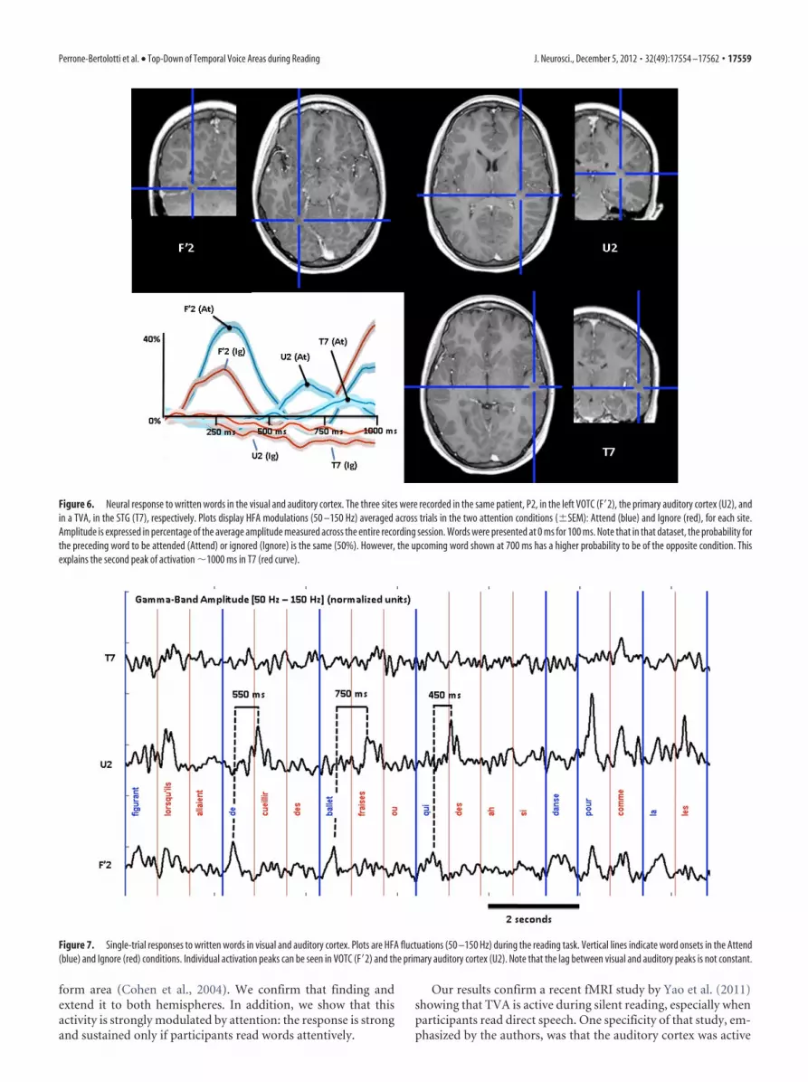
A wavering sequence of activation from visual toauditory cortexIn one patient (P2), two additional electrodes were located in theprimary auditory cortex (Fig. 5) and in the ventral occipital tem-poral cortex (VOTC). This provided a unique opportunity tocompare the timing of neural responses to written words acrossvisual and auditory regions. As shown in Figure 6, there was aclear propagation peaking first in the left VOTC (F�2, 300 ms),then in the right primary auditory cortex (U2, 700 ms), and fi-nally in TVA (T7, 800 ms). This is a clear indication that auditoryand visual areas do not react conjointly to provide a multimodalrepresentation of the stimulus. That “representation” is in fact aheterogeneous collection of asynchronous activities, with nobinding mechanism to assemble them. Further, it is also clear thatprimary auditory areas also participate in the auditory represen-tation of the written word, and sometimes before TVA, which
means that the auditory representation itself is not homoge-neous, even within a single modality in the auditory cortex. Inaddition, single-trial display of visual and auditory responses re-vealed that the lag between visual and auditory responses is farfrom constant (Fig. 7). In this figure, the response of the VTOC(F�2) and primary auditory cortex (U2) can be seen for severalconsecutive attended words, with a clear time separation varyingbetween 450 and 750 ms. To quantify this qualitative observation,we measured the peak latency of the response to individual wordsin the attend condition, in F�2 and U2. We selected only trials with aclearly visible response in each of the two sites (i.e., an excellentsignal-to-noise ratio), as defined by a peak value exceeding the meanof the signal by at least two SDs (the mean and SD of HFA over theentire experimental session for that channel). Thirty-six trials wereselected, and the mean peak latencies were, respectively, 324 ms inF�2 (SD � 63 ms) and 681 ms in U2 (SD, 164 ms), significantly laterin the auditory cortex than in the visual cortex (sign test; p � 10–9).When considering all visible peaks to increase the number of trials(not necessarily visible in both U2 and F’2 in the same trial), wefound similar latencies: 333 ms in F�2 (SD, 100 ms; 83 trials) and 674ms in U2 (SD, 174 ms; 87 trials). Mean latency in T7 was 651 ms (SD,219 ms; 54 trials); however, there were not enough common peaks inT7 and U2 or F�2 to perform a meaningful comparison of theirlatencies (�20 peaks).
There was also a clear difference between the Attend and Ig-nore conditions in the directed interactions between the VOTCand primary auditory cortex (Fig. 8). For the Attend condition,there was significantly (p � 0.01) more influence from VOTC toprimary auditory cortex than vice versa in two consecutive timewindows, centered at 400 and 500 ms, whereas no dominantdirection of influence was observed in the Ignore condition.
Finally, it is also clear from Figures 6 and 7 that the response ofthe auditory cortex often occurs during the display of the nextword, which means that auditory and visual areas are active si-multaneously while processing different temporal frames of theparticipant’s environment. We tested for a possible interferencebetween those responses by computing the correlation betweenGBA in T7 and F�2 at the peak latency in F�2, between 200 and 400ms. Since the response peak in T7 to the previous stimulus occursaround that latency, we reasoned that both processes might com-pete for resources and interfere. Accordingly, we found that av-erage GBA values over the 200 ms:400 ms time window for F�2and T7 are negatively correlated (Spearman r � �0.1887, p �0.00014). However, one might argue that Attend stimuli areslightly more often preceded by an Ignore stimulus, and thereforethat strong F�2 responses should be often simultaneous with aweak T7 response (to the preceding stimulus). We corrected forthat possible bias by considering only Attend stimuli preceded byan Attend stimulus and found that the correlation was in factmore negative (r � �0.2167); however, it failed to reach signifi-cance level because of a smaller number of trials (p � 0.06).
DiscussionWe used direct electrophysiological recordings from the auditorycortex to show that silent reading activates voice-selective regionsof the auditory cortex, or TVAs. The neural response involveshigh-frequency activity, already shown to be exquisitely sensitiveto auditory stimuli, including speech (Crone et al., 2001; Towle etal., 2008; Chang et al., 2010). This result is reminiscent of anearlier study by our group, reporting that a region in the leftanterior superior temporal gyrus (STG) was active during a silentreading task (Mainy et al., 2008), in line with an earlier suggestionthat the left STG might comprise an auditory analog of the word
Table 2. Demographic (sex, age, manual lateralization, index of manuallateralization, laterality index of language) and verbal neuropsychologicalperformance (verbal comprehension index, WAIS-IIII, and reading scores assessedby means of Stroop test) data of patients P1–P4
Patient Sex Age ML EIML LIL ICV Reading (Stroop)
P1 F 39 R 100% LH 107 (68 ct) 44.38 s (�0.73 ET)P2 F 16 R NA NA 77 (6 ct) 70 s (�2 ET)P3 H 34 R 90% LH 114 (82 ct) 37.33 s (�0.44 ET)P4 F 26 R 100% LH 96 (39 ct) 61 s (�0.27 ET)
Manual lateralization, ML; R, right-handed; LH, left-handed; Edinburgh Handedness Inventory (Oldfield, 1971),EIMI; laterality index of language lateralization, LIL; verbal comprehension index, ICV; information not available, NA.
Figure 5. Response to sounds in the primary auditory cortex. Bar plots show the mean HFA(�SEM) measured while participants listened to sounds of several categories (averaged across60 nonoverlapping 500 ms windows during sounds, see Materials and Methods). Values with acolored asterisk are significant higher than values indicated by a bracket of the same color(Kruskal–Wallis, post hoc pairwise comparison). HFA is expressed in percentage of the averageamplitude measured across the entire recording session. Note that speech-like and nonspeech-like sounds elicit equally strong responses.
17558 • J. Neurosci., December 5, 2012 • 32(49):17554 –17562 Perrone-Bertolotti et al. • Top-Down of Temporal Voice Areas during Reading
form area (Cohen et al., 2004). We confirm that finding andextend it to both hemispheres. In addition, we show that thisactivity is strongly modulated by attention: the response is strongand sustained only if participants read words attentively.
Our results confirm a recent fMRI study by Yao et al. (2011)showing that TVA is active during silent reading, especially whenparticipants read direct speech. One specificity of that study, em-phasized by the authors, was that the auditory cortex was active
Figure 6. Neural response to written words in the visual and auditory cortex. The three sites were recorded in the same patient, P2, in the left VOTC (F�2), the primary auditory cortex (U2), andin a TVA, in the STG (T7), respectively. Plots display HFA modulations (50 –150 Hz) averaged across trials in the two attention conditions (�SEM): Attend (blue) and Ignore (red), for each site.Amplitude is expressed in percentage of the average amplitude measured across the entire recording session. Words were presented at 0 ms for 100 ms. Note that in that dataset, the probability forthe preceding word to be attended (Attend) or ignored (Ignore) is the same (50%). However, the upcoming word shown at 700 ms has a higher probability to be of the opposite condition. Thisexplains the second peak of activation 1000 ms in T7 (red curve).
Figure 7. Single-trial responses to written words in visual and auditory cortex. Plots are HFA fluctuations (50 –150 Hz) during the reading task. Vertical lines indicate word onsets in the Attend(blue) and Ignore (red) conditions. Individual activation peaks can be seen in VOTC (F�2) and the primary auditory cortex (U2). Note that the lag between visual and auditory peaks is not constant.
Perrone-Bertolotti et al. • Top-Down of Temporal Voice Areas during Reading J. Neurosci., December 5, 2012 • 32(49):17554 –17562 • 17559

even though participants were not explicitly encouraged to useauditory imagery. Understandably, most studies so far have usedtask designs emphasizing covert pronunciation, such as rhymeand phoneme detection tasks (Cousin et al., 2007, Khateb et al.,2007; Perrone-Bertolotti et al., 2011) to maximize the imagerycomponent. Our task did not emphasize mental pronunciationand therefore confirms the conclusion of Yao et al. (2011): read-ing spontaneously elicits auditory processing in the absence ofany auditory stimulation (Linden et al., 2011), which might indi-cate that readers spontaneously use auditory images.
Possible role of the inner voice during readingIn that sense, our study also contradicts a recent claim that read-ers are unlikely to experience an auditory image of a written text,unless the text corresponds to sentences spoken by people whosevoice is familiar (Kurby et al., 2009). We show that participantsproduce inner voice even when reading narrative with no identi-fied speaker. Our results are more compatible with an alternativeinterpretation of that group, that inner voice processes are also,and in fact mostly, modulated by attention. This modulation wasalso suggested by Alexander and Nygaard (2008) to explain whyreaders tend to use auditory imagery more often when readingdifficult texts.
In line with this interpretation, recent studies have proposedthat reading might rely more on phonological processes as textsbecome more difficult to read. Phonological activation would bemore active as linguistic complexity increases, or in nonprofi-cient readers (Jobard et al., 2011), and triggered by top-downattentional processes. This suggestion is compatible with our ob-servation that the strongest TVA activation was found in patientP2, who was not a proficient reader. It would also explain whyAVI is active while reading sentences, but not while reading iso-lated words (Price, 2000; Jobard et al., 2007): AVI might facilitatethe processing of prosodic information and the active integra-tion of consecutive words into a sentence, using verbal work-ing memory.
Indeed, the superior temporal sulcus and superior temporalgyrus (STG), which support AVI, play an important role in sev-eral phonological processes, such as the integration of letters intospeech sounds (Vigneau et al., 2006; van Atteveldt et al., 2009),the identification of phonological word forms (Shalom andPoeppel, 2008), and the analysis of speech sounds (Hickok, 2009;Grimaldi, 2012). Furthermore, the STG has been shown to reactspecifically to readable stimuli, i.e., to words and pseudowords,
but not to, e.g., consonant strings (Vartiainen et al., 2011). TVAmight then interact with frontal regions to support the phono-logical component of verbal working memory during reading,through articulatory rehearsal processes (Baddeley and Logie,1992; Yvert et al., 2012). Those processes would allow an updateand refresh of the phonological material stored in working mem-ory (Baddeley and Logie, 1992; Smith et al., 1995). Auditory ver-bal Imagery would thus facilitate verbal working memory, assuggested by several authors (Smith et al., 1995; Sato et al., 2004),to process the sentence as a whole, and not as a collection ofunrelated pieces. Simultaneously, the activation of phonologicalrepresentations in TVA would produce the vivid experience ofthe inner voice (Abramson and Goldinger, 1997).
How automatic is the inner voice during sentence reading?Our results directly relate to a long-standing debate as to whetherexpert readers automatically access phonological representationswhen reading, since it is difficult to think of a phonological rep-resentation that would not include an auditory imagery compo-nent (Alexander and Nygaard, 2008). In the three most proficientreaders (P1, P3, and P4), unattended words triggered the sameinitial activation peak as attended words 300 ms, followed by afast decline, which might suggest an automatic activation of TVAin expert readers. This assumption is further supported by a re-cent event-related potential study (Froyen et al., 2009) showingthat early and automatic letter–speech sound processing onlyoccurs in advanced readers (Blomert and Froyen, 2010). Oneneural possible explanation is that learning to read mightstrengthen the connectivity between visual and auditory areas(Booth et al., 2008; Richardson et al., 2011) based on Hebbianplasticity: both regions would be repeatedly coactivated becauseof repeated associations between visual and auditory inputs dur-ing the learning period (the written word and the auditory per-cept of one’s own voice while reading overtly). With practice, thisconnectivity would allow for a direct activation of the auditorycortex by visual inputs through the visual cortex, in the absence ofovert speech, very much like an automatic stimulus-responseassociation.
However, we also show that if it is the case, this connectivitycan still be modulated by top-down control. Sustained innervoice activation is not an automatic process occurring systemat-ically in response to any written word. It is clearly enhanced whenparticipants read attentively (to understand and memorize sen-tences) and minimized when words are not processed attentively.Provided that high-frequency neural activity can be measuredwith sufficient specificity, as in patient P2, for instance, it caneven be proposed as an on-line measure of attention: the engage-ment of attention during reading can be followed in time on aword-by-word basis (as well as covert speech, consequently). Oneremaining question is whether the reduced response to unat-tended words results from active inhibition or from the absenceof phasic activation. This second interpretation is supported byseveral intracranial EEG studies showing that high-frequency ac-tivity in sensory cortices comprise two consecutive components:a transient peak determined by the stimulus, followed by a sus-tained component determined by task condition, that is, whetherthe stimulus contains task-relevant information (Jung et al.,2008; Ossandon et al., 2011; Bastin et al., 2012). This interpreta-tion is supported also by the findings of the present study, show-ing that directed interactions from the visual to auditory cortexare significantly stronger than vice versa only during attentivereading. Therefore, words might trigger an automatic TVA re-
Figure 8. Directed interactions between VOTC and the primary auditory cortex. Differencebetween the Granger Causality terms quantifying the directed interactions from VOTC to pri-mary auditory cortex and vice versa for the Attend (black) and Ignore (gray) condition; positivevalues indicate more influence from VOTC to primary auditory cortex. Time windows showingsignificant ( p � 0.01) differences of influence are marked by shaded area. Time windowsshowing significant ( p � 0.01) differences of influence are shaded in gray (indicating signifi-cance in the Attend condition).
17560 • J. Neurosci., December 5, 2012 • 32(49):17554 –17562 Perrone-Bertolotti et al. • Top-Down of Temporal Voice Areas during Reading

sponse (at least in proficient readers), which is sustained by top-down attentional processes when the word is task-relevant.
Early auditory cortex first, TVA secondJancke and Shah (2004) had shown that in a population trained toimagine syllables in response to flicker lights, AVI generates bi-lateral hemodynamic responses in TVA. However, they also sug-gested that during auditory imagery, the auditory systemactivates in reverse order, from higher order to lower order areas,and that the primary auditory cortex is not active (Bunzeck et al.,2005, for nonverbal complex stimuli). Our results qualify thesetwo conclusions: we argue that the extent to which auditory areasactivate during AVI depends on the strategy used by the subject.In patient P2, the primary auditory cortex was clearly active dur-ing AVI, and, most importantly, before higher order voice-selective areas. However, this sequence was driven by a visualstimulus in our case, and we cannot exclude that the primaryauditory cortex might be driven by the visual cortex (Booth et al.,2008); in fact, our Granger Causality results support this assump-tion. Still, one intriguing finding of our study is that the activa-tion of the auditory cortex can follow the response of the visualcortex by �500 ms, which means that in natural reading condi-tions paced by an average of four saccades per second, the audi-tory cortex might react with a lag of two saccades, that is, twowords in most instances.
What remains to be determined is whether the brief activationof the auditory cortex after unattended words is sufficient to leadto a conscious experience of an inner voice. Indeed, showing thata voice-selective region of the auditory cortex is active is notundisputable evidence that the reader actually hears a voice. Theconscious experience might arise only for longer durations, forinstance, or when that activation broadcasts to a wider brainnetwork (Baars, 2002). This will remain the main limitation of allstudies in that field, until new experiments reveal the neural cor-relates of auditory consciousness.
ReferencesAbramson M, Goldinger SD (1997) What the reader’s eye tells the mind’s
ear: silent reading activates inner speech. Percept Psychophys 59:1059 –1068. CrossRef Medline
Aguera P, Jerbi K, Caclin A, Bertrand O (2011) ELAN: a software packagefor analysis and visualization of MEG, EEG, and LFP signals. ComputIntell Neurosci 158970.
Akaike H (1974) A new look at the statistical model identification. IEEETrans Autom Control 19:716 –723. CrossRef
Alexander JD, Nygaard LC (2008) Reading voices and hearing text: talker-specific auditory imagery in reading. J Exp Psychol Hum Percept Perform34:446 – 459. CrossRef Medline
Baars BJ (2002) The conscious access hypothesis: origins and recent evi-dence. Trends Cogn Sci 6:47–52. CrossRef Medline
Baddeley AD, Logie R (1992) Auditory imagery and working memory. In:Auditory imagery (Reisberg D, ed.), pp 179 –197. Hillsdale, NJ: LawrenceErlbaum.
Bastin J, Committeri G, Kahane P, Galati G, Minotti L, Lachaux JP,Berthoz A (2012) Timing of posterior parahippocampal gyrus activ-ity reveals multiple scene processing stages. Hum Brain Mapp. Ad-vance online publication. Retrieved Jan 30, 2012. doi:10.1002/hbm.21515. CrossRef
Belin P, Zatorre RJ, Lafaille P, Ahad P, Pike B (2000) Voice-selective areas inhuman auditory cortex. Nature 403:309 –312. CrossRef Medline
Blomert L, Froyen D (2010) Multi-sensory learning and learning to read. IntJ Psychophysiol 77:195–204. CrossRef Medline
Booth JR, Mehdiratta N, Burman DD, Bitan T (2008) Developmentalincreases in effective connectivity to brain regions involved in phono-logical processing during tasks with orthographic demands. Brain Res1189:78 – 89. CrossRef Medline
Bunzeck N, Wuestenberg T, Lutz K, Heinze HJ, Jancke L (2005) Scanning
silence: mental imagery of complex sounds. Neuroimage 26:1119 –1127.CrossRef Medline
Calvert GA, Bullmore ET, Brammer MJ, Campbell R, Williams SCR,McGuire PK (1997) Activation of auditory cortex during silent lipread-ing. Science 276:593–596. CrossRef Medline
Chang EF, Rieger JW, Johnson K, Berger MS, Barbaro NM, Knight RT(2010) Categorical speech representation in human superior temporalgyrus. Nat Neurosci 13:1428 –1432. CrossRef Medline
Cohen L, Jobert A, Le Bihan D, Dehaene S (2004) Distinct unimodal andmultimodal regions for word processing in the left temporal cortex. Neu-roimage 23:1256 –1270. CrossRef Medline
Cousin E, Peyrin C, Pichat C, Lamalle L, Le Bas JF, Baciu M (2007) Func-tional MRI approach for assessing hemispheric predominance of regionsactivated by a phonological and a semantic task. Eur J Radiol 63:274 –285.CrossRef Medline
Crone NE, Boatman D, Gordon B, Hao L (2001) Induced electrocortico-graphic gamma activity during auditory perception. Clin Neurophysiol112:565–582. CrossRef Medline
Crone NE, Korzeniewska A, Franaszczuk PJ (2011) Cortical gamma re-sponses: searching high and low. Int J Psychophysiol 79:9 –15. CrossRefMedline
Froyen DJ, Bonte ML, van Atteveldt N, Blomert L (2009) The long road toautomation: neurocognitive development of letter-speech sound process-ing. J Cogn Neurosci 21:567–580. CrossRef Medline
Geweke J (1982) Measurement of linear dependence and feedback betweenmultiple time series. J Am Stat Assoc 77:304 –313. CrossRef
Granger CWJ (1980) Testing for causality: a personal viewpoint. J Econ DynControl 2:329 –352. CrossRef
Grimaldi M (2012) Toward a neural theory of language: old issues and newperspectives. J Neurolinguistics 25:304 –327. CrossRef
Halpern AR, Zatorre RJ (1999) When that tune runs through your head: aPET investigation of auditory imagery for familiar melodies. Cereb Cor-tex 9:697–704. CrossRef Medline
Hickok G (2009) The functional neuroanatomy of language. Phys Life Rev6:121–143. CrossRef Medline
Jancke L, Shah NJ (2004) ‘Hearing’ syllables by ‘seeing’ visual stimuli. EurJ Neurosci 19:2603–2608. CrossRef Medline
Jerbi K, Ossandon T, Hamame, CM, Senova S, Dalal SS, Jung J (2009) Task-related gamma-band dynamics from an intracerebral perspective: reviewand implications for surface EEG and MEG. Hum Brain Mapp 30:1758 –1771. CrossRef Medline
Jobard G, Vigneau M, Simon G, Tzourio-Mazoyer N (2011) The weight ofskill: interindividual variability of reading related brain activation pat-terns in fluent readers. J Neurolinguistics 24:113–132. CrossRef
Jung J, Mainy N, Kahane P, Minotti L, Hoffmann D, Bertrand O, Lachaux JP(2008) The neural bases of attentive reading. Hum Brain Mapp 29:1193–1206. CrossRef Medline
Kahane P, Minotti L, Hoffmann D, Lachaux J-P, Ryvlin P (2003) InvasiveEEG in the definition of the seizure onset zone: depth electrodes. In:Handbook of Clinical Neurophysiology, Vol 3 (Rosenow F, Luders HO,eds) pp 109 –133. Amsterdam: Elsevier.
Khateb A, Pegna AJ, Landis T, Michel CM, Brunet D, Seghier ML, Annoni JM(2007) Rhyme processing in the brain: an ERP mapping study. Int J Psy-chophysiol 63:240 –250. CrossRef Medline
Kurby CA, Magliano JP, Rapp DN (2009) Those voices in your head: acti-vation of auditory images during reading. Cognition 112:457– 461.CrossRef Medline
Lachaux JP, Rudrauf D, Kahane P (2003) Intracranial EEG and humanbrain mapping. J Physiol (Paris) 97:613– 628. CrossRef
Lachaux JP, Jerbi K, Bertrand O, Minotti L, Hoffmann D, Schoendorff B,Kahane P (2007) A blueprint for real-time functional mapping via hu-man intracranial recordings. PLoS One 2:e1094. CrossRef Medline
Lachaux JP, Axmacher N, Mormann F, Halgren E, Crone NE (2012) High-frequency neural activity and human cognition: past, present and possiblefuture of intracranial EEG research. Prog Neurobiol 98:279 –301.CrossRef Medline
Le Van Quyen M, Foucher J, Lachaux JP, Rodriguez E, Lutz A, Martinerie J,Varela FJ (2001) Comparison of Hilbert transform and wavelet methodsfor the analysis of neuronal synchrony. J Neurosci Methods 111:83–98.CrossRef Medline
Lennox, BR, Bert, S, Park, G, Jones, PB, Morris, PG (1999) Spatial and tem-
Perrone-Bertolotti et al. • Top-Down of Temporal Voice Areas during Reading J. Neurosci., December 5, 2012 • 32(49):17554 –17562 • 17561

poral mapping of neural activity associated with auditory hallucinations.Lancet 353.
Linden DE, Thornton K, Kuswanto CN, Johnston SJ, van de Ven V, JacksonMC (2011) The brain’s voices: comparing nonclinical auditory hallucina-tions and imagery. Cereb Cortex 21:330 –337. CrossRef Medline
Mainy N, Jung J, Baciu M, Kahane P, Schoendorff B, Minotti L, Hoffmann D,Bertrand O, Lachaux JP (2008) Cortical dynamics of word recognition.Hum Brain Mapp 29:1215–1230. CrossRef Medline
Nobre AC, Allison T, McCarthy G (1998) Modulation of human extrastriatevisual processing by selective attention to colours and words. Brain 121:1357–1368. CrossRef Medline
Oldfield RC (1971) The assessment and analysis of handedness: the Edin-burgh inventory. Neuropsychologia 9:97–113. CrossRef Medline
Ossandon T, Jerbi K, Vidal JR, Bayle DJ, Henaff MA, Jung J (2011) Transientsuppression of broadband gamma power in the default-mode network iscorrelated with task complexity and subject performance. J Neurosci 31:14521–14530. CrossRef Medline
Pernet C, Charest I, Belizaire G, Zatorre RJ, Belin P (2007) The temporalvoice areas: spatial characterization and variability. 13th InternationalConference on Functional Mapping of the Human Brain, Chicago, IL.Neuroimage 36 [Supp 1].
Perrone-Bertolotti M, Pichat C, Le Bas JF, Baciu A, Baciu M (2011) Func-tional MRI evidence for modulation of cerebral activity by grapheme-to-phoneme conversion in French, and by the variable of gender.J Neurolinguistics 24:507–520. CrossRef
Price C (2000) The anatomy of language: contributions from functional neu-roimaging. J Anat 197:335–359. CrossRef Medline
Richardson FM, Seghier ML, Leff AP, Thomas MS, Price CJ (2011) Multipleroutes from occipital to temporal cortices during reading. J Neurosci31:8239 – 8247. CrossRef Medline
Sato M, Baciu M, Loevenbruck H, Schwartz JL, Cathiard MA, Segebarth C,Abry C (2004) Multistable representation of speech forms: a functionalMRI study of verbal transformation. Neuroimage 23:1143–1151.CrossRef Medline
Seth AK (2005) Causal connectivity of evolved neural networks during be-havior. Network 16:35–54. CrossRef Medline
Seth AK (2010) A MATLAB toolbox for Granger causal connectivity analy-sis. J Neurosci Methods 186:262–273. CrossRef Medline
Shalom DB, Poeppel D (2008) Functional anatomic models of language:assembling the pieces. Neuroscientist 14:119 –127. Medline
Smith JD, Wilson M, Reisberg D (1995) The role of subvocalization in au-ditory imagery. Neuropsychologia 33:1433–1454. CrossRef Medline
Talairach J, Tournoux P (1988) Co-Planar stereotaxic atlas of the humanbrain: 3-dimensional proportional system: an approach to cerebral imag-ing. New York: Thieme.
Tallon-Baudry C, Bertrand O, Delpuech C, Permier J (1997) Oscillatorygamma-band (30 –70 Hz) activity induced by a visual search task in hu-mans. J Neurosci 17:722–734. Medline
Towle VL, Yoon HA, Castelle M, Edgar JC, Biassou NM, Frim DM, SpireJP, Kohrman MH (2008) ECoG gamma activity during a languagetask: differentiating expressive and receptive speech areas. Brain 131:2013–2027. CrossRef Medline
van Atteveldt N, Roebroeck A, Goebel R (2009) Interaction of speech andscript in human auditory cortex: insights from neuro-imaging and effec-tive connectivity. Hear Res 258:152–164. CrossRef
Vartiainen J, Liljestrom M, Koskinen M, Renvall H, Salmelin R (2011)Functional magnetic resonance imaging blood oxygenation level-dependent signal and magnetoencephalography evoked responses yielddifferent neural functionality in reading. J Neurosci 31:1048 –1058.CrossRef Medline
Vigneau M, Beaucousin V, Hervé PY, Duffau H, Crivello F, Houdé O, et al.(2006) Meta-analyzing left hemisphere language areas: Phonology, se-mantics, and sentence processing. Neuroimage 30:1414 –1432. CrossRefMedline
Yao B, Belin P, Scheepers C (2011) Silent reading of direct versus indirectspeech activates voice-selective areas in the auditory cortex. J Cogn Neu-rosci 23:3146 –3152. CrossRef Medline
Yvert G, Perrone-Bertolotti M, Baciu M, David O (2012) Dynamic causalmodeling of spatiotemporal integration of phonological and semanticprocesses: an electroencephalographic study. J Neurosci 32:4297– 4306.CrossRef Medline
17562 • J. Neurosci., December 5, 2012 • 32(49):17554 –17562 Perrone-Bertolotti et al. • Top-Down of Temporal Voice Areas during Reading


