Embed Size (px)
Citation preview
UvA-DARE is a service provided by the library of the University of Amsterdam (https://dare.uva.nl)
UvA-DARE (Digital Academic Repository)
Hiding in plain sight: Leptospirosis and rickettsial diseaseUnmasking two causes of severe imported infectionsde Vries, S.G.
Publication date2020Document VersionFinal published versionLicenseOther
Link to publication
Citation for published version (APA):de Vries, S. G. (2020). Hiding in plain sight: Leptospirosis and rickettsial disease: Unmaskingtwo causes of severe imported infections.
General rightsIt is not permitted to download or to forward/distribute the text or part of it without the consent of the author(s)and/or copyright holder(s), other than for strictly personal, individual use, unless the work is under an opencontent license (like Creative Commons).
Disclaimer/Complaints regulationsIf you believe that digital publication of certain material infringes any of your rights or (privacy) interests, pleaselet the Library know, stating your reasons. In case of a legitimate complaint, the Library will make the materialinaccessible and/or remove it from the website. Please Ask the Library: https://uba.uva.nl/en/contact, or a letterto: Library of the University of Amsterdam, Secretariat, Singel 425, 1012 WP Amsterdam, The Netherlands. Youwill be contacted as soon as possible.
Download date:23 Jul 2022
Hiding in plain sight: leptospirosis and rickettsial disease. Unmasking two causes of severe
imported infections.
Sophia Geertruda de Vries
Hiding in plain sight: leptospirosis and rickettsial disease
Unmasking two causes of severe imported infections
Hiding in plain sight: leptospirosis and rickettsial disease
SOPHIA DE VRIES
SOP
HIA
DE
VR
IES


Hiding in plain sight: leptospirosis and rickettsial disease. Unmasking two causes of severe
imported infections.
Sophia Geertruda de Vries

Copyright © 2020, S.G. de Vries, Amsterdam, the Netherlands
All rights are reserved. No part of this thesis may be reproduced, stored, or transmitted in any
form or by any means without the prior permission of the author.
ISBN 978-94-6361-479-5
Layout and printing by Optima Grafische Communicatie (www.ogc.nl)
Printing of this thesis was financially supported by the Amsterdam UMC, location AMC,
University of Amsterdam, and the SBOH, employer of GP trainees
Hiding in plain sight: leptospirosis and rickettsial disease.
Unmasking two causes of severe imported infections.
ACADEMISCH PROEFSCHRIFT
ter verkrijging van de graad van doctor
aan de Universiteit van Amsterdam
op gezag van de Rector Magnificus
prof. dr. ir. K.I.J. Maex
en overstaan van een door het College voor Promoties ingestelde commissie,
in het openbaar te verdedigen in de Agnietenkapel
op donderdag 3 december 2020, te 10.00 uur
door Sophia Geertruda de Vries
geboren te Amsterdam
Promotiecommissie
Promotor: Prof. dr. M.P. Grobusch AMC - UvA
Copromotor: Dr. A. Goorhuis AMC - UvA
Overige leden: Prof. dr. M. Van Vugt AMC - UvA
Prof. Dr. W.J. Wiersinga AMC - UvA
Prof. Dr. J.W.R. Hovius AMC- UvA
Prof. Dr. M.A. van Agtmael Vrije Universiteit Amsterdam
Prof. Dr. C. Schultsz AMC - UvA
Dr. P.J.J. van Genderen Travel Clinic Erasmus MC
Faculteit der Geneeskunde

Alle wriemeldiertjes
alle wiebeldiertjes
alle kruip- en kriebeldiertjes
zitten verstopt in het hoge gras.
Ik zou maar op mijn tenen lopen
als ik jou was.
- Joke van Leeuwen

Contents
Chapter 1. General introduction and outline of this thesis
Part 1: Leptospirosis
Chapter 2. Leptospirosis in Sub-Saharan Africa: a systematic review
International Journal of Infectious Diseases 2014; 28; 47-64
Chapter 3. Travel-related leptospirosis in the Netherlands 2009-2016:
An epidemiological report and case series
Travel Medicine and Infectious Disease 2018; 24: 44-50
Chapter 4. Leptospirosis among Returned Travelers:
a GeoSentinel Site Survey and Multicenter Analysis – 1997-2016
American Journal of Tropical Medicine and Hygiene 2018; 99: 127-
135
Chapter 5. Nucleic acid and antigen detection tests for leptospirosis
Cochrane Database of Systematic Reviews 2019; 8: CD011871
Part 2: Rickettsial disease
Chapter 6. Under-diagnosis of rickettsial disease in clinical practice:
A systematic review
Travel Medicine and Infectious Disease 2018; 26: 7-15
Chapter 7. Searching and finding the hidden treasure:
Rickettsial disease among Dutch international travelers
– a retrospective analysis
Clinical Infectious Diseases 2020 Jan 30
Part 3: Epilogue
Chapter 8. Summary and general discussion
Chapter 9. Nederlandse samenvatting
Addendum
List of publications
PhD Portfolio
Curriculum vitae
Dankwoord
9
29
79
97
123
191
217
237
247
257
259
263
265
267


Chapter 1General introduction

11
General introduction
Leptospirosis
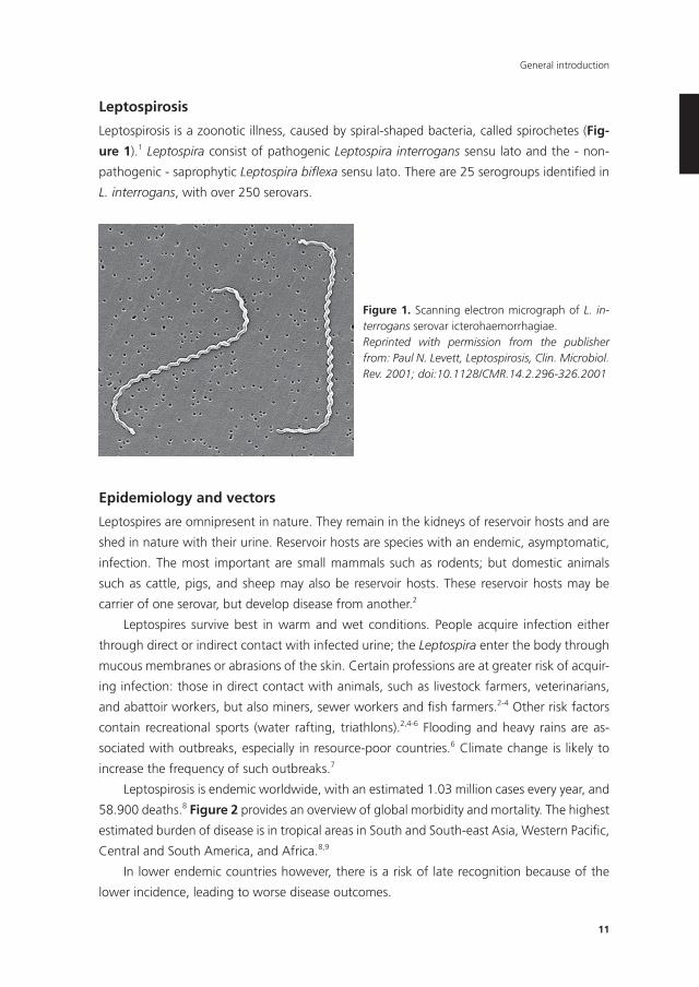
Leptospirosis is a zoonotic illness, caused by spiral-shaped bacteria, called spirochetes (Fig-
ure 1).1 Leptospira consist of pathogenic Leptospira interrogans sensu lato and the - non-
pathogenic - saprophytic Leptospira biflexa sensu lato. There are 25 serogroups identified in
L. interrogans, with over 250 serovars.
Epidemiology and vectors
Leptospires are omnipresent in nature. They remain in the kidneys of reservoir hosts and are
shed in nature with their urine. Reservoir hosts are species with an endemic, asymptomatic,
infection. The most important are small mammals such as rodents; but domestic animals
such as cattle, pigs, and sheep may also be reservoir hosts. These reservoir hosts may be
carrier of one serovar, but develop disease from another.2
Leptospires survive best in warm and wet conditions. People acquire infection either
through direct or indirect contact with infected urine; the Leptospira enter the body through
mucous membranes or abrasions of the skin. Certain professions are at greater risk of acquir-
ing infection: those in direct contact with animals, such as livestock farmers, veterinarians,
and abattoir workers, but also miners, sewer workers and fish farmers.2-4 Other risk factors
contain recreational sports (water rafting, triathlons).2,4-6 Flooding and heavy rains are as-
sociated with outbreaks, especially in resource-poor countries.6 Climate change is likely to
increase the frequency of such outbreaks.7
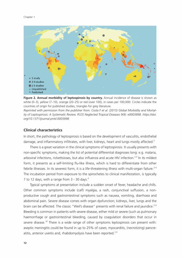
Leptospirosis is endemic worldwide, with an estimated 1.03 million cases every year, and
58.900 deaths.8 Figure 2 provides an overview of global morbidity and mortality. The highest
estimated burden of disease is in tropical areas in South and South-east Asia, Western Pacific,
Central and South America, and Africa.8,9
In lower endemic countries however, there is a risk of late recognition because of the
lower incidence, leading to worse disease outcomes.
Figure 1. Scanning electron micrograph of L. in-terrogans serovar icterohaemorrhagiae.Reprinted with permission from the publisher from: Paul N. Levett, Leptospirosis, Clin. Microbiol. Rev. 2001; doi:10.1128/CMR.14.2.296-326.2001
Chapter 1
12
Clinical characteristics
In short, the pathology of leptospirosis is based on the development of vasculitis, endothelial
damage, and inflammatory infiltrates, with liver, kidneys, heart and lungs mostly affected.2
There is a great variation in the clinical symptoms of leptospirosis. It usually presents with
non-specific symptoms, making the list of potential differential diagnoses long: e.g. malaria,
arboviral infections, rickettsioses, but also influenza and acute HIV infection.2,3 In its mildest
form, it presents as a self-limiting flu-like illness, which is hard to differentiate from other
febrile illnesses. In its severest form, it is a life-threatening illness with multi-organ failure.2-4
The incubation period from exposure to the spirochetes to clinical manifestation, is typically
7 to 12 days, with a range from 3 - 30 days.4
Typical symptoms at presentation include a sudden onset of fever, headache and chills.
Other common symptoms include (calf) myalgia, a rash, conjunctival suffusion, a non-
productive cough and gastrointestinal symptoms such as nausea, vomiting, diarrhoea and
abdominal pain. Severe disease comes with organ dysfunction; kidneys, liver, lungs and the
brain can be affected. The classic “Weil’s disease” presents with renal failure and jaundice.2-4
Bleeding is common in patients with severe disease, either mild or severe (such as pulmonary
haemorrhage or gastrointestinal bleeding, caused by coagulation disorders that occur in
severe disease.10 There is a wide range of other symptoms leptospirosis can present with:
aseptic meningitis could be found in up to 25% of cases; myocarditis, (necrotizing) pancre-
atitis, anterior uveitis and, rhabdomyolysis have been reported.2,3
Figure 2. Annual morbidity of leptospirosis by country. Annual incidence of disease is shown as white (0–3), yellow (7–10), orange (20–25) or red (over 100), in cases per 100,000. Circles indicate the countries of origin for published studies, triangles for grey literature.Reprinted with permission from the publisher from: Costa F et al. (2015) Global Morbidity and Mortal-ity of Leptospirosis: A Systematic Review. PLOS Neglected Tropical Diseases 9(9): e0003898. https://doi.org/10.1371/journal.pntd.0003898
13
General introduction
Diagnostics
Diagnosis of leptospirosis has long depended on serological assays; however, over the past
couple of years, molecular detection tests have gained ground.
The microscopic agglutination test (MAT) is the reference standard, often combined
with an immunoglobulin M (IgM) enzyme-linked immunosorbent assay (ELISA), and culture.
MAT detects agglutinating antibodies in serum of the patient, and requires testing of paired
samples, with the second sample taken preferably at least 5-7 days after onset of the disease,
when antibody production commences.1,2 When performed ‘according to the books’, it has
a high specificity; confirming the diagnosis. However, sensitivity is limited; thus, a negative
test cannot safely rule out the illness.11 The reported sensitivity varies among studies,11-13
and increases when MAT is combined with IgM ELISA.12 Moreover, MAT is a cumbersome
method and confined to specialised centres, as it requires the maintenance of a full panel
of live leptospires, and well-trained laboratory technicians.1 In addition, antibodies can be
detected in the blood after 5-7 days of illness,2 rendering an early diagnosis with serological
methods impossible.
A positive culture confirms the diagnosis as definite. Leptospires circulate in the blood
in the first week of the illness (leptospiraemia); so samples should be taken swiftly.2 Cultures
can be obtained from blood, cerebrospinal fluid (CSF) and urine; the duration of excretion of
leptospires in the urine lasts longer than the leptospiraemia. Cultures should incubate up to
13 weeks, and require weekly examination by dark-field microscopy.1,2
Molecular detection techniques are suited to diagnose the illness in the early stages,
when leptospiraemia is present. Polymerase chain reaction (PCR) and its variants are com-
monly used, and can be applied on blood, CSF, urine, and aqueous humour. In blood, the
diagnostic accuracy is highest when the test is performed the first ten days of illness; after
that period the leptospiraemia declines.1 The earlier the test is applied, the higher the sen-
sitivity.14 Molecular tests on urine get positive at a later stage: ten to fourteen days after
onset of symptoms.15 However, the true diagnostic accuracy of molecular tests is not yet
elucidated, as there is a big variation between published studies.
Prevention and treatment
Socio-economic factors, sanitation and risky behaviours are consistently associated with an
elevated risk for leptospirosis.6 Floods are a major risk factor, and likely to increase with
climate change; governments should prepare for this.6,7 Rodent control is key in prevention
of leptospirosis.16 Other preventive strategies for leptospirosis in humans mainly encompass
reducing risk behaviour, such as prolonged exposure to water for recreational or occupational
activities, and animal (e.g., cattle, rats) contact.
For people in rural areas, who are already at higher risk, this means, for example, not
walking barefoot and covering wounds. Open sewers contribute to the risk, and should
be avoided.6 People working with animals should use appropriate protective methods. In
Chapter 1
14
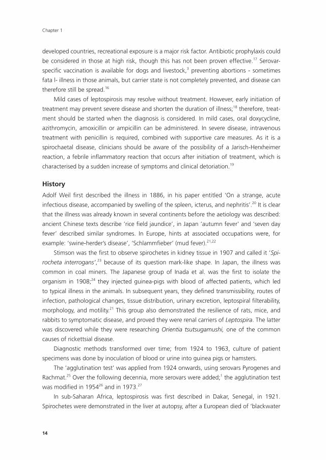
developed countries, recreational exposure is a major risk factor. Antibiotic prophylaxis could
be considered in those at high risk, though this has not been proven effective.17 Serovar-
specific vaccination is available for dogs and livestock,3 preventing abortions - sometimes
fata l- illness in those animals, but carrier state is not completely prevented, and disease can
therefore still be spread.16
Mild cases of leptospirosis may resolve without treatment. However, early initiation of
treatment may prevent severe disease and shorten the duration of illness;18 therefore, treat-
ment should be started when the diagnosis is considered. In mild cases, oral doxycycline,
azithromycin, amoxicillin or ampicillin can be administered. In severe disease, intravenous
treatment with penicillin is required, combined with supportive care measures. As it is a
spirochaetal disease, clinicians should be aware of the possibility of a Jarisch-Herxheimer
reaction, a febrile inflammatory reaction that occurs after initiation of treatment, which is
characterised by a sudden increase of symptoms and clinical detoriation.19
History
Adolf Weil first described the illness in 1886, in his paper entitled ‘On a strange, acute
infectious disease, accompanied by swelling of the spleen, icterus, and nephritis’.20 It is clear
that the illness was already known in several continents before the aetiology was described:
ancient Chinese texts describe ‘rice field jaundice’, in Japan ‘autumn fever’ and ‘seven day
fever’ described similar syndromes. In Europe, hints at associated occupations were, for
example: ‘swine-herder’s disease’, ‘Schlammfieber’ (mud fever).21,22
Stimson was the first to observe spirochetes in kidney tissue in 1907 and called it ‘Spi-
rocheta interrogans’,23 because of its question mark-like shape. In Japan, the illness was
common in coal miners. The Japanese group of Inada et al. was the first to isolate the
organism in 1908;24 they injected guinea-pigs with blood of affected patients, which led
to typical illness in the animals. In subsequent years, they defined transmissibility, routes of
infection, pathological changes, tissue distribution, urinary excretion, leptospiral filterability,
morphology, and motility.21 This group also demonstrated the resilience of rats, mice, and
rabbits to symptomatic disease, and proved they were renal carriers of Leptospira. The latter
was discovered while they were researching Orientia tsutsugamushi, one of the common
causes of rickettsial disease.
Diagnostic methods transformed over time; from 1924 to 1963, culture of patient
specimens was done by inoculation of blood or urine into guinea pigs or hamsters.
The ‘agglutination test’ was applied from 1924 onwards, using serovars Pyrogenes and
Rachmat.25 Over the following decennia, more serovars were added;1 the agglutination test
was modified in 195426 and in 1973.27
In sub-Saharan Africa, leptospirosis was first described in Dakar, Senegal, in 1921.
Spirochetes were demonstrated in the liver at autopsy, after a European died of ‘blackwater
15
General introduction
fever’ (haemoglobinuria),28 an entity at that time more commonly described in connection
with quinine therapy of severe malaria.
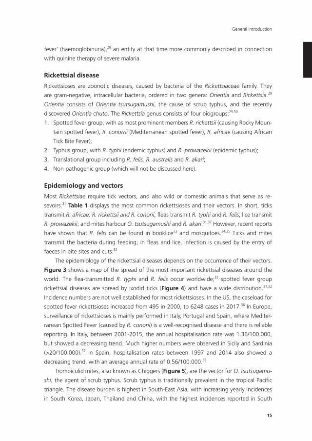
Rickettsial disease
Rickettsioses are zoonotic diseases, caused by bacteria of the Rickettsiaceae family. They
are gram-negative, intracellular bacteria, ordered in two genera: Orientia and Rickettsia.29
Orientia consists of Orientia tsutsugamushi, the cause of scrub typhus, and the recently
discovered Orientia chuto. The Rickettsia genus consists of four biogroups:29,30
1. Spotted fever group, with as most prominent members R. rickettsii (causing Rocky Moun-
tain spotted fever), R. conorrii (Mediterranean spotted fever), R. africae (causing African
Tick Bite Fever);
2. Typhus group, with R. typhi (endemic typhus) and R. prowazekii (epidemic typhus);
3. Translational group including R. felis, R. australis and R. akari;
4. Non-pathogenic group (which will not be discussed here).
Epidemiology and vectors
Most Rickettsiae require tick vectors, and also wild or domestic animals that serve as re-
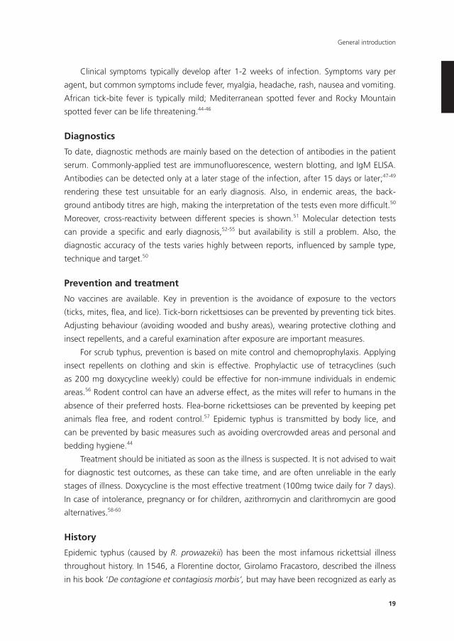
sevoirs.31 Table 1 displays the most common rickettsioses and their vectors. In short, ticks
transmit R. africae, R. rickettsii and R. conorii; fleas transmit R. typhi and R. felis; lice transmit
R. prowazekii; and mites harbour O. tsutsugamushi and R. akari.31,32 However, recent reports
have shown that R. felis can be found in booklice33 and mosquitoes.34,35 Ticks and mites
transmit the bacteria during feeding; in fleas and lice, infection is caused by the entry of
faeces in bite sites and cuts.32
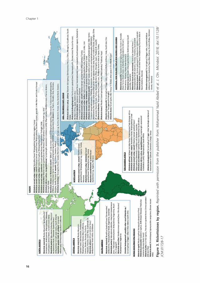
The epidemiology of the rickettsial diseases depends on the occurrence of their vectors.
Figure 3 shows a map of the spread of the most important rickettsial diseases around the
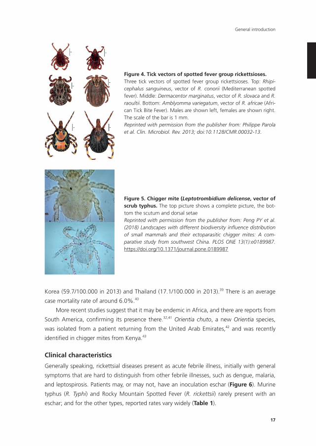
world. The flea-transmitted R. typhi and R. felis occur worldwide;32 spotted fever group
rickettsial diseases are spread by ixodid ticks (Figure 4) and have a wide distribution.31,32
Incidence numbers are not well established for most rickettsioses. In the US, the caseload for
spotted fever rickettsioses increased from 495 in 2000, to 6248 cases in 2017.36 In Europe,
surveillance of rickettsioses is mainly performed in Italy, Portugal and Spain, where Mediter-
ranean Spotted Fever (caused by R. conorii) is a well-recognised disease and there is reliable
reporting. In Italy, between 2001-2015, the annual hospitalisation rate was 1.36/100.000,
but showed a decreasing trend. Much higher numbers were observed in Sicily and Sardinia
(>20/100.000).37 In Spain, hospitalisation rates between 1997 and 2014 also showed a
decreasing trend, with an average annual rate of 0.56/100.000.38
Trombiculid mites, also known as Chiggers (Figure 5), are the vector for O. tsutsugamu-
shi, the agent of scrub typhus. Scrub typhus is traditionally prevalent in the tropical Pacific
triangle. The disease burden is highest in South-East Asia, with increasing yearly incidences
in South Korea, Japan, Thailand and China, with the highest incidences reported in South
Chapter 1
16
Fig
ure
3.
Ric
kett
sio
ses
by
reg
ion
. Re
prin
ted
with
per
mis
sion
fro
m t
he p
ublis
her
from
: M
oham
mad
Yaz
id A
bdad
et
al.
J. C
lin.
Mic
robi
ol.
2018
; do
i:10.
1128
/JC
M.0
1728
-17
17
General introduction
Korea (59.7/100.000 in 2013) and Thailand (17.1/100.000 in 2013).39 There is an average
case mortality rate of around 6.0%.40
More recent studies suggest that it may be endemic in Africa, and there are reports from
South America, confirming its presence there.32,41 Orientia chuto, a new Orientia species,
was isolated from a patient returning from the United Arab Emirates,42 and was recently
identified in chigger mites from Kenya.43
Clinical characteristics
Generally speaking, rickettsial diseases present as acute febrile illness, initially with general
symptoms that are hard to distinguish from other febrile illnesses, such as dengue, malaria,
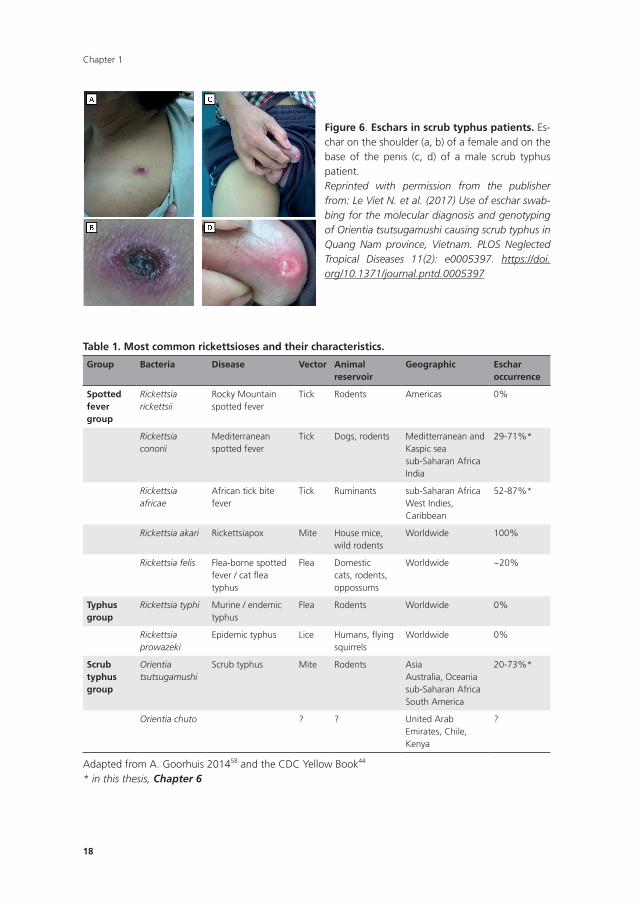
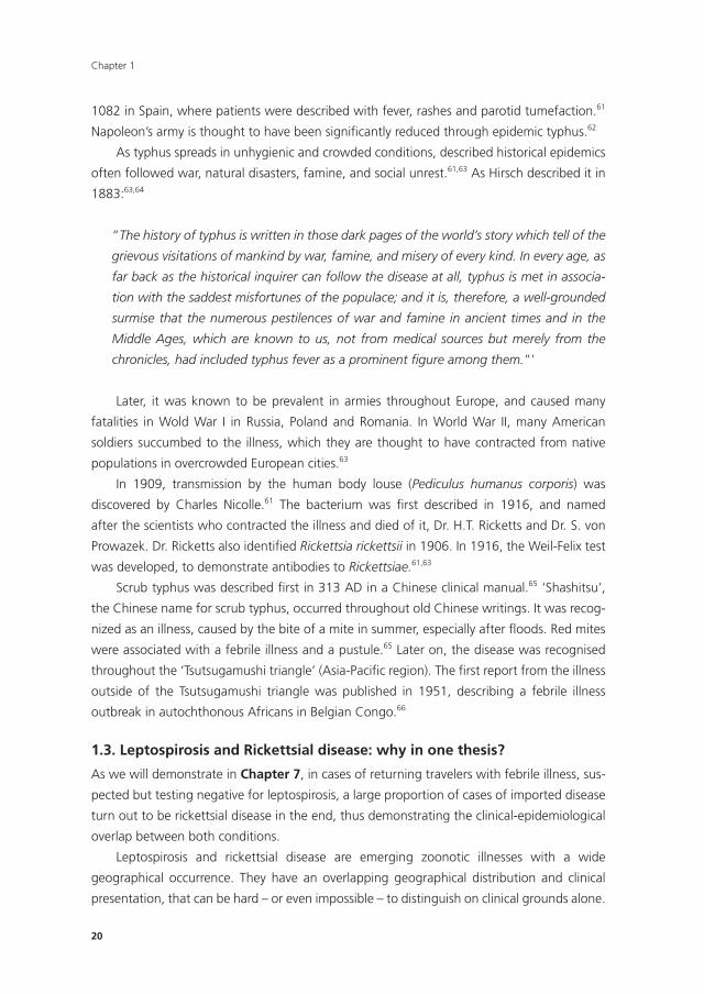
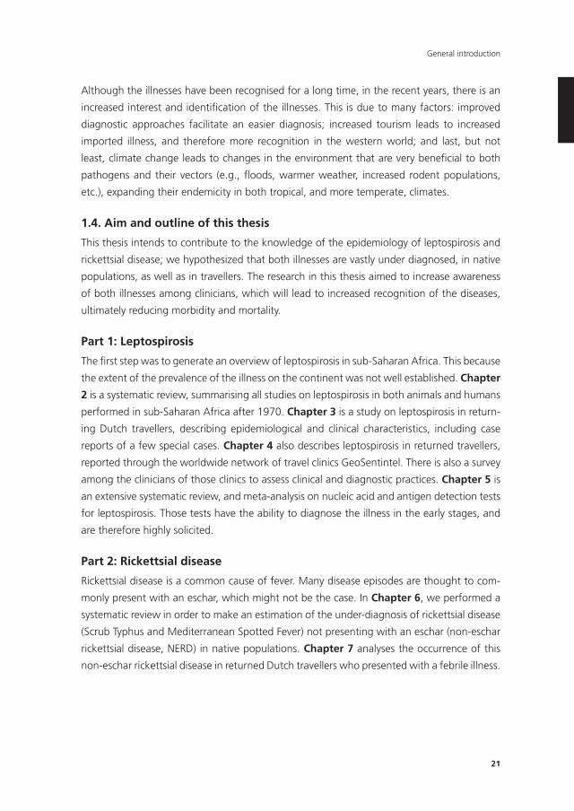
and leptospirosis. Patients may, or may not, have an inoculation eschar (Figure 6). Murine
typhus (R. Typhi) and Rocky Mountain Spotted Fever (R. rickettsii) rarely present with an
eschar; and for the other types, reported rates vary widely (Table 1).
Figure 4. Tick vectors of spotted fever group rickettsioses.Three tick vectors of spotted fever group rickettsioses. Top: Rhipi-cephalus sanguineus, vector of R. conorii (Mediterranean spotted fever). Middle: Dermacentor marginatus, vector of R. slovaca and R. raoultii. Bottom: Amblyomma variegatum, vector of R. africae (Afri-can Tick Bite Fever). Males are shown left, females are shown right. The scale of the bar is 1 mm.Reprinted with permission from the publisher from: Philippe Parola et al. Clin. Microbiol. Rev. 2013; doi:10.1128/CMR.00032-13.
Figure 5. Chigger mite (Leptotrombidium delicense, vector of scrub typhus. The top picture shows a complete picture, the bot-tom the scutum and dorsal setaeReprinted with permission from the publisher from: Peng PY et al. (2018) Landscapes with different biodiversity influence distribution of small mammals and their ectoparasitic chigger mites: A com-parative study from southwest China. PLOS ONE 13(1):e0189987. https://doi.org/10.1371/journal.pone.0189987
Chapter 1
18
Figure 6. Eschars in scrub typhus patients. Es-char on the shoulder (a, b) of a female and on the base of the penis (c, d) of a male scrub typhus patient.Reprinted with permission from the publisher from: Le Viet N. et al. (2017) Use of eschar swab-bing for the molecular diagnosis and genotyping of Orientia tsutsugamushi causing scrub typhus in Quang Nam province, Vietnam. PLOS Neglected Tropical Diseases 11(2): e0005397. https://doi.org/10.1371/journal.pntd.0005397
Table 1. Most common rickettsioses and their characteristics.
Group Bacteria Disease Vector Animal reservoir
Geographic Eschar occurrence
Spotted fever group
Rickettsia rickettsii
Rocky Mountain spotted fever
Tick Rodents Americas 0%
Rickettsia conorii
Mediterranean spotted fever
Tick Dogs, rodents Meditterranean and Kaspic seasub-Saharan AfricaIndia
29-71%*
Rickettsia africae
African tick bite fever
Tick Ruminants sub-Saharan AfricaWest Indies, Caribbean
52-87%*
Rickettsia akari Rickettsiapox Mite House mice, wild rodents
Worldwide 100%
Rickettsia felis Flea-borne spotted fever / cat flea typhus
Flea Domestic cats, rodents, oppossums
Worldwide ~20%
Typhus group
Rickettsia typhi Murine / endemic typhus
Flea Rodents Worldwide 0%
Rickettsia prowazeki
Epidemic typhus Lice Humans, flying squirrels
Worldwide 0%
Scrub typhus group
Orientia tsutsugamushi
Scrub typhus Mite Rodents AsiaAustralia, Oceaniasub-Saharan AfricaSouth America
20-73%*
Orientia chuto ? ? United Arab Emirates, Chile, Kenya
?
Adapted from A. Goorhuis 201458 and the CDC Yellow Book44
* in this thesis, Chapter 6
19
General introduction
Clinical symptoms typically develop after 1-2 weeks of infection. Symptoms vary per
agent, but common symptoms include fever, myalgia, headache, rash, nausea and vomiting.
African tick-bite fever is typically mild; Mediterranean spotted fever and Rocky Mountain
spotted fever can be life threatening.44-46
Diagnostics
To date, diagnostic methods are mainly based on the detection of antibodies in the patient
serum. Commonly-applied test are immunofluorescence, western blotting, and IgM ELISA.
Antibodies can be detected only at a later stage of the infection, after 15 days or later;47-49
rendering these test unsuitable for an early diagnosis. Also, in endemic areas, the back-
ground antibody titres are high, making the interpretation of the tests even more difficult.50
Moreover, cross-reactivity between different species is shown.51 Molecular detection tests
can provide a specific and early diagnosis,52-55 but availability is still a problem. Also, the
diagnostic accuracy of the tests varies highly between reports, influenced by sample type,
technique and target.50
Prevention and treatment
No vaccines are available. Key in prevention is the avoidance of exposure to the vectors
(ticks, mites, flea, and lice). Tick-born rickettsioses can be prevented by preventing tick bites.
Adjusting behaviour (avoiding wooded and bushy areas), wearing protective clothing and
insect repellents, and a careful examination after exposure are important measures.
For scrub typhus, prevention is based on mite control and chemoprophylaxis. Applying
insect repellents on clothing and skin is effective. Prophylactic use of tetracyclines (such
as 200 mg doxycycline weekly) could be effective for non-immune individuals in endemic
areas.56 Rodent control can have an adverse effect, as the mites will refer to humans in the
absence of their preferred hosts. Flea-borne rickettsioses can be prevented by keeping pet
animals flea free, and rodent control.57 Epidemic typhus is transmitted by body lice, and
can be prevented by basic measures such as avoiding overcrowded areas and personal and
bedding hygiene.44
Treatment should be initiated as soon as the illness is suspected. It is not advised to wait
for diagnostic test outcomes, as these can take time, and are often unreliable in the early
stages of illness. Doxycycline is the most effective treatment (100mg twice daily for 7 days).
In case of intolerance, pregnancy or for children, azithromycin and clarithromycin are good
alternatives.58-60
History
Epidemic typhus (caused by R. prowazekii) has been the most infamous rickettsial illness
throughout history. In 1546, a Florentine doctor, Girolamo Fracastoro, described the illness
in his book ‘De contagione et contagiosis morbis’, but may have been recognized as early as
Chapter 1
20
1082 in Spain, where patients were described with fever, rashes and parotid tumefaction.61
Napoleon’s army is thought to have been significantly reduced through epidemic typhus.62
As typhus spreads in unhygienic and crowded conditions, described historical epidemics
often followed war, natural disasters, famine, and social unrest.61,63 As Hirsch described it in
1883:63,64
“The history of typhus is written in those dark pages of the world’s story which tell of the
grievous visitations of mankind by war, famine, and misery of every kind. In every age, as
far back as the historical inquirer can follow the disease at all, typhus is met in associa-
tion with the saddest misfortunes of the populace; and it is, therefore, a well-grounded
surmise that the numerous pestilences of war and famine in ancient times and in the
Middle Ages, which are known to us, not from medical sources but merely from the
chronicles, had included typhus fever as a prominent figure among them.”’
Later, it was known to be prevalent in armies throughout Europe, and caused many
fatalities in Wold War I in Russia, Poland and Romania. In World War II, many American
soldiers succumbed to the illness, which they are thought to have contracted from native
populations in overcrowded European cities.63
In 1909, transmission by the human body louse (Pediculus humanus corporis) was
discovered by Charles Nicolle.61 The bacterium was first described in 1916, and named
after the scientists who contracted the illness and died of it, Dr. H.T. Ricketts and Dr. S. von
Prowazek. Dr. Ricketts also identified Rickettsia rickettsii in 1906. In 1916, the Weil-Felix test
was developed, to demonstrate antibodies to Rickettsiae.61,63
Scrub typhus was described first in 313 AD in a Chinese clinical manual.65 ‘Shashitsu’,
the Chinese name for scrub typhus, occurred throughout old Chinese writings. It was recog-
nized as an illness, caused by the bite of a mite in summer, especially after floods. Red mites
were associated with a febrile illness and a pustule.65 Later on, the disease was recognised
throughout the ‘Tsutsugamushi triangle’ (Asia-Pacific region). The first report from the illness
outside of the Tsutsugamushi triangle was published in 1951, describing a febrile illness
outbreak in autochthonous Africans in Belgian Congo.66
1.3. Leptospirosis and Rickettsial disease: why in one thesis?
As we will demonstrate in Chapter 7, in cases of returning travelers with febrile illness, sus-
pected but testing negative for leptospirosis, a large proportion of cases of imported disease
turn out to be rickettsial disease in the end, thus demonstrating the clinical-epidemiological
overlap between both conditions.
Leptospirosis and rickettsial disease are emerging zoonotic illnesses with a wide
geographical occurrence. They have an overlapping geographical distribution and clinical
presentation, that can be hard – or even impossible – to distinguish on clinical grounds alone.
21
General introduction
Although the illnesses have been recognised for a long time, in the recent years, there is an
increased interest and identification of the illnesses. This is due to many factors: improved
diagnostic approaches facilitate an easier diagnosis; increased tourism leads to increased
imported illness, and therefore more recognition in the western world; and last, but not
least, climate change leads to changes in the environment that are very beneficial to both
pathogens and their vectors (e.g., floods, warmer weather, increased rodent populations,
etc.), expanding their endemicity in both tropical, and more temperate, climates.
1.4. Aim and outline of this thesis
This thesis intends to contribute to the knowledge of the epidemiology of leptospirosis and
rickettsial disease; we hypothesized that both illnesses are vastly under diagnosed, in native
populations, as well as in travellers. The research in this thesis aimed to increase awareness
of both illnesses among clinicians, which will lead to increased recognition of the diseases,
ultimately reducing morbidity and mortality.
Part 1: Leptospirosis
The first step was to generate an overview of leptospirosis in sub-Saharan Africa. This because
the extent of the prevalence of the illness on the continent was not well established. Chapter
2 is a systematic review, summarising all studies on leptospirosis in both animals and humans
performed in sub-Saharan Africa after 1970. Chapter 3 is a study on leptospirosis in return-
ing Dutch travellers, describing epidemiological and clinical characteristics, including case
reports of a few special cases. Chapter 4 also describes leptospirosis in returned travellers,
reported through the worldwide network of travel clinics GeoSentintel. There is also a survey
among the clinicians of those clinics to assess clinical and diagnostic practices. Chapter 5 is
an extensive systematic review, and meta-analysis on nucleic acid and antigen detection tests
for leptospirosis. Those tests have the ability to diagnose the illness in the early stages, and
are therefore highly solicited.
Part 2: Rickettsial disease
Rickettsial disease is a common cause of fever. Many disease episodes are thought to com-
monly present with an eschar, which might not be the case. In Chapter 6, we performed a
systematic review in order to make an estimation of the under-diagnosis of rickettsial disease
(Scrub Typhus and Mediterranean Spotted Fever) not presenting with an eschar (non-eschar
rickettsial disease, NERD) in native populations. Chapter 7 analyses the occurrence of this
non-eschar rickettsial disease in returned Dutch travellers who presented with a febrile illness.
Chapter 1
22
REFEREnCES
1. World Health Organization. Human leptospirosis: guidance for diagnosis, surveillance and con-
trol. 2003.
2. Levett PN. Leptospirosis. Clin Microbiol Rev 2001; 14(2): 296-326.
3. Bharti AR, Nally JE, Ricaldi JN, et al. Leptospirosis: a zoonotic disease of global importance. Lancet
Infect Dis 2003; 3(12): 757-71.
4. Haake DA, Levett PN. Leptospirosis in humans. Curr Top Microbiol Immunol 2015; 387: 65-97.
5. Gundacker ND, Rolfe RJ, Rodriguez JM. Infections associated with adventure travel: A systematic
review. Travel Med Infect Dis 2017; 16: 3-10.
6. Mwachui MA, Crump L, Hartskeerl R, Zinsstag J, Hattendorf J. Environmental and Behavioural
Determinants of Leptospirosis Transmission: A Systematic Review. PLoS Negl Trop Dis 2015; 9(9):
e0003843.
7. Lau CL, Smythe LD, Craig SB, Weinstein P. Climate change, flooding, urbanisation and leptospi-
rosis: fuelling the fire? Trans R Soc Trop Med Hyg 2010; 104(10): 631-8.
8. Costa F, Hagan JE, Calcagno J, et al. Global Morbidity and Mortality of Leptospirosis: A Systematic
Review. PLOS Neglected Tropical Diseases 2015; 9(9): e0003898.
9. Torgerson PR, Hagan JE, Costa F, et al. Global Burden of Leptospirosis: Estimated in Terms of
Disability Adjusted Life Years. PLOS Neglected Tropical Diseases 2015; 9(10): e0004122.
10. Wagenaar JF, Goris MG, Partiningrum DL, et al. Coagulation disorders in patients with severe
leptospirosis are associated with severe bleeding and mortality. Trop Med Int Health 2010; 15(2):
152-9.
11. Limmathurotsakul D, Turner EL, Wuthiekanun V, et al. Fool’s gold: Why imperfect reference tests
are undermining the evaluation of novel diagnostics: a reevaluation of 5 diagnostic tests for
leptospirosis. Clin Infect Dis 2012; 55(3): 322-31.
12. Goris M, Leeflang M, Boer K, et al. Establishment of valid laboratory case definition for human
leptospirosis. J Bacteriol Parasitol 2012; 3(132): 2.
13. Goris MG, Boer KR, Bouman‐Strijker M, Hartskeerl R, Lucas C, Leeflang MM. Serological labora-
tory tests for diagnosis of human leptospirosis in patients presenting with clinical symptoms.
Cochrane Database of Systematic Reviews 2011; (11).
14. Ahmed A, Engelberts MF, Boer KR, Ahmed N, Hartskeerl RA. Development and validation of a
real-time PCR for detection of pathogenic leptospira species in clinical materials. PLoS One 2009;
4(9): e7093.
15. Picardeau M, Bertherat E, Jancloes M, Skouloudis AN, Durski K, Hartskeerl RA. Rapid tests for
diagnosis of leptospirosis: current tools and emerging technologies. Diagn Microbiol Infect Dis
2014; 78(1): 1-8.
16. Hartskeerl RA, Collares-Pereira M, Ellis WA. Emergence, control and re-emerging leptospirosis:
dynamics of infection in the changing world. Clin Microbiol Infect 2011; 17(4): 494-501.
17. Brett‐Major DM, Lipnick RJ. Antibiotic prophylaxis for leptospirosis. Cochrane Database of Sys-
tematic Reviews 2009; (3).
18. Katz AR, Ansdell VE, Effler PV, Middleton CR, Sasaki DM. Assessment of the clinical presentation
and treatment of 353 cases of laboratory-confirmed leptospirosis in Hawaii, 1974-1998. Clin
Infect Dis 2001; 33(11): 1834-41.
19. Guerrier G, D’Ortenzio E. The Jarisch-Herxheimer reaction in leptospirosis: a systematic review.
PLoS One 2013; 8(3): e59266.
23
General introduction
20. Weil A. Über eine eigenthümliche, mit Milztumor, Icterus und Nephritis einhergehende, acute
Infektionskrankheit. Deutsches Archiv für Klinische 1886; 39: 209-32.
21. Adler B. History of leptospirosis and leptospira. Curr Top Microbiol Immunol 2015; 387: 1-9.
22. Farr RW. Leptospirosis. Clin Infect Dis 1995; 21(1): 1-6; quiz 7-8.
23. Stimson AM. Note on an Organism Found in Yellow-Fever Tissue. Public Health Reports (1896-
1970) 1907; 22(18): 541-.
24. Inada R, Ito Y. A report of the discovery of the causal organism (a new species of spirocheta) of
Weil’s disease”. Tokyo Ijishinshi 1915: 351–60.
25. Martin L, Pettit A. Sero-diagnosis of the icterohaemorrhagic spirochetosis. Bull Mem Soc Med
Hop Paris 1918; 42: 672-5.
26. Wolff J. The laboratory diagnosis of leptospirosis. In: Dalldorf G, editor American lectures in tests
and techniques Springfield (IL): Charles C Thomas; 1954.
27. Cole JR, Jr., Sulzer CR, Pursell AR. Improved microtechnique for the leptospiral microscopic ag-
glutination test. Appl Microbiol 1973; 25(6): 976-80.
28. Noc F, Nogue M. Les spirochetoses humaines a Dakar (Senegal). Bulletin de la Societe de Patholo-
gie exotique, 1921; (14): 460
29. Merhej V, Raoult D. Rickettsial evolution in the light of comparative genomics. Biol Rev Camb
Philos Soc 2011; 86(2): 379-405.
30. Murray GG, Weinert LA, Rhule EL, Welch JJ. The Phylogeny of Rickettsia Using Different Evolu-
tionary Signatures: How Tree-Like is Bacterial Evolution? Syst Biol 2016; 65(2): 265-79.
31. Parola P, Paddock CD, Socolovschi C, et al. Update on Tick-Borne Rickettsioses around the World:
a Geographic Approach. Clinical Microbiology Reviews 2013; 26(4): 657-702.
32. Abdad MY, Abou Abdallah R, Fournier P-E, Stenos J, Vasoo S. A Concise Review of the Epidemiol-
ogy and Diagnostics of Rickettsioses: Rickettsia and Orientia spp. Journal of Clinical Microbiology
2018; 56(8): e01728-17.
33. Behar A, McCormick LJ, Perlman SJ. Rickettsia felis infection in a common household insect pest,
Liposcelis bostrychophila (Psocoptera: Liposcelidae). Appl Environ Microbiol 2010; 76(7): 2280-5.
34. Socolovschi C, Pages F, Ndiath MO, Ratmanov P, Raoult D. Rickettsia species in African Anopheles
mosquitoes. PLoS One 2012; 7(10): e48254.
35. Socolovschi C, Pages F, Raoult D. Rickettsia felis in Aedes albopictus mosquitoes, Libreville,
Gabon. Emerg Infect Dis 2012; 18(10): 1687-9.
36. CDC. Rocky Mountain Spotted Fever - Epidemiology and Statistics. https://www.cdc.gov/rmsf/
stats/index.html (accessed 6 March 2020).
37. Gomez-Barroso D, Vescio MF, Bella A, et al. Mediterranean spotted fever rickettsiosis in Italy,
2001-2015: Spatio-temporal distribution based on hospitalization records. Ticks Tick Borne Dis
2019; 10(1): 43-50.
38. Herrador Z, Fernandez-Martinez A, Gomez-Barroso D, et al. Mediterranean spotted fever in
Spain, 1997-2014: Epidemiological situation based on hospitalization records. PLoS One 2017;
12(3): e0174745.
39. Bonell A, Lubell Y, Newton PN, Crump JA, Paris DH. Estimating the burden of scrub typhus: A
systematic review. PLoS Negl Trop Dis 2017; 11(9): e0005838.
40. Taylor AJ, Paris DH, Newton PN. A Systematic Review of Mortality from Untreated Scrub Typhus
(Orientia tsutsugamushi). PLoS Negl Trop Dis 2015; 9(8): e0003971.
41. Weitzel T, Dittrich S, Lopez J, et al. Endemic Scrub Typhus in South America. N Engl J Med 2016;
375(10): 954-61.
Chapter 1
24
42. Izzard L, Fuller A, Blacksell SD, et al. Isolation of a novel Orientia species (O. chuto sp. nov.) from
a patient infected in Dubai. J Clin Microbiol 2010; 48(12): 4404-9.
43. Masakhwe C, Linsuwanon P, Kimita G, et al. Identification and Characterization of Orientia
chuto in Trombiculid Chigger Mites Collected from Wild Rodents in Kenya. J Clin Microbiol 2018;
56(12).
44. CDC. Yellow Book. https://wwwnc.cdc.gov/travel/yellowbook/2020/travel-related-infectious-
diseases/rickettsial-including-spotted-fever-and-typhus-fever-rickettsioses-scrub-typhus-anaplas-
mosis-and-ehr.
45. Kirkland KB, Wilkinson WE, Sexton DJ. Therapeutic delay and mortality in cases of Rocky Moun-
tain spotted fever. Clin Infect Dis 1995; 20(5): 1118-21.
46. Angerami RN, Camara M, Pacola MR, et al. Features of Brazilian spotted fever in two different
endemic areas in Brazil. Ticks Tick Borne Dis 2012; 3(5-6): 346-8.
47. Blacksell SD, Bryant NJ, Paris DH, Doust JA, Sakoda Y, Day NP. Scrub typhus serologic testing
with the indirect immunofluorescence method as a diagnostic gold standard: a lack of consensus
leads to a lot of confusion. Clin Infect Dis 2007; 44(3): 391-401.
48. Dumler JS, Taylor JP, Walker DH. Clinical and laboratory features of murine typhus in south Texas,
1980 through 1987. Jama 1991; 266(10): 1365-70.
49. Fournier PE, Jensenius M, Laferl H, Vene S, Raoult D. Kinetics of antibody responses in Rickettsia
africae and Rickettsia conorii infections. Clin Diagn Lab Immunol 2002; 9(2): 324-8.
50. Paris DH, Dumler JS. State of the art of diagnosis of rickettsial diseases: the use of blood speci-
mens for diagnosis of scrub typhus, spotted fever group rickettsiosis, and murine typhus. Curr
Opin Infect Dis 2016; 29(5): 433-9.
51. Raoult D, Dasch GA. Immunoblot cross-reactions among Rickettsia, Proteus spp. and Legionella
spp. in patients with Mediterranean spotted fever. FEMS Immunol Med Microbiol 1995; 11(1):
13-8.
52. Paris DH, Blacksell SD, Stenos J, et al. Real-time multiplex PCR assay for detection and differentia-
tion of rickettsiae and orientiae. Trans R Soc Trop Med Hyg 2008; 102(2): 186-93.
53. Giulieri S, Jaton K, Cometta A, Trellu LT, Greub G. Development of a duplex real-time PCR for the
detection of Rickettsia spp. and typhus group rickettsia in clinical samples. FEMS Immunol Med
Microbiol 2012; 64(1): 92-7.
54. Papp S, Rauch J, Kuehl S, Richardt U, Keller C, Osterloh A. Comparative evaluation of two
Rickettsia typhi-specific quantitative real-time PCRs for research and diagnostic purposes. Med
Microbiol Immunol 2017; 206(1): 41-51.
55. Watthanaworawit W, Turner P, Turner C, et al. A prospective evaluation of real-time PCR assays
for the detection of Orientia tsutsugamushi and Rickettsia spp. for early diagnosis of rickettsial
infections during the acute phase of undifferentiated febrile illness. Am J Trop Med Hyg 2013;
89(2): 308-10.
56. Andrew R, Bonnin JM, Williams S. Tick typhus in North Queensland. Med J Aust 1946; 2: 253-8.
57. CDC. Typhus Fevers. 2019. https://www.cdc.gov/typhus/index.html (accessed 22 April 2020).
58. Goorhuis A. [Rickettsioses]. Ned Tijdschr Geneeskd 2014; 158: A7603.
59. Rolain JM, Maurin M, Vestris G, Raoult D. In vitro susceptibilities of 27 rickettsiae to 13 antimi-
crobials. Antimicrobial agents and chemotherapy 1998; 42(7): 1537-41.
60. Rolain JM, Stuhl L, Maurin M, Raoult D. Evaluation of antibiotic susceptibilities of three rickettsial
species including Rickettsia felis by a quantitative PCR DNA assay. Antimicrob Agents Chemother
2002; 46(9): 2747-51.
61. Angelakis E, Bechah Y, Raoult D. The History of Epidemic Typhus. Microbiol Spectr 2016; 4(4).

25
General introduction
62. Raoult D, Dutour O, Houhamdi L, et al. Evidence for louse-transmitted diseases in soldiers of
Napoleon’s Grand Army in Vilnius. J Infect Dis 2006; 193(1): 112-20.
63. Snyder JC. TYPHUS FEVER IN THE SECOND WORLD WAR. Calif Med 1947; 66(1): 3-10.
64. Hirsch A, Creighton C. Handbook of geographical and historical pathology. London: The New
Sydenham Society; 1883.
65. Richards AL, Jiang J. Scrub Typhus: Historic Perspective and Current Status of the Worldwide
Presence of Orientia Species. Trop Med Infect Dis 2020; 5(2).
66. Giroud P. The rickettsioses in Equatorial Africa. Bulletin of the World Health Organization 1951;
4(4): 535-46.


PART 1Leptospirosis

Chapter 2Leptospirosis in Sub-Saharan Africa: a
systematic review
Sophia G. de Vries, Benjamin J. Visser, Ingeborg M. Nagel, Marga G.A. Goris, Rudy A. Hartskeerl, Martin P. Grobusch
International Journal of Infectious Diseases 2014; 28: 47-64.
Chapter 2
30
ABSTRACT
Background
Leptospirosis is an emerging zoonotic infection worldwide, possibly due to climate change
and demographic shifts. It is regarded as endemic in sub-Saharan Africa; however, for most
countries scarce epidemiological data, if any, exists. The primary objective was to describe
the prevalence of leptospirosis in countries in sub-Saharan Africa, and to develop options for
prevention and control in the future.
Methods
A systematic review to determine the prevalence of leptospirosis in sub-Saharan Africa was
conducted, following PRISMA guidelines. Medline/PubMed, Embase, The Cochrane Library,
Web of Science, Biosis Previews, the African Index Medicus, AJOL and Google Scholar were
searched.
Results
Information about the prevalence and incidence of leptospirosis in humans is available, but
remains scarce for many countries. Data is unavailable or outdated for many countries, par-
ticularly in Central Africa. Most data is available from animals, probably due to the economic
losses caused by leptospirosis in livestock. In humans, leptospirosis is an important cause of
febrile illness in sub-Saharan Africa. It concerns numerous serogroups, harboured by many
different animal carriers.
Discussion
A wide variety of data was identified. Prevalence rates vary throughout the continent and
more research, especially in humans, is needed to reliably gauge the extent of the problem.
Preventive measures need to be reconsidered to control outbreaks in the future.
REGISTRATIOn nUMBER PROSPERO
CRD42013006545.
Article Highlights
• Leptospirosisconstitutesaneglectedtropicaldiseaseinsub-SaharanAfrica.
• Itisprobablethatthecombinationofclimatechange,increasedriskofflooding,popula-
tion growth, and urbanisation will lead to an increase in the burden of leptospirosis in
sub-Saharan Africa.
• Withanever-growingproportionoffebrilediseaseepisodesbeingrecognizedasnon-
malarial, leptospirosis moves into the light as an important differential diagnosis to
malaria.

31
Leptospirosis in Sub-Saharan Africa: a systematic review
• Moreresearchhasbeenconductedinanimalscomparedtohumans,probablybecauseof
the larger economic losses caused by leptospirosis in livestock (e.g. abortion) compared
to economic losses due to human morbidity and mortality.
• Future leptospirosis research should be a collaborative effort of multiple disciplines,
mainly human and animal medicine, to attain the optimal health for individuals and
animals.
Chapter 2
32
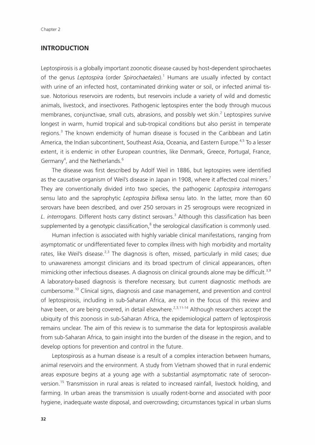
InTRODUCTIOn
Leptospirosis is a globally important zoonotic disease caused by host-dependent spirochaetes
of the genus Leptospira (order Spirochaetales).1 Humans are usually infected by contact
with urine of an infected host, contaminated drinking water or soil, or infected animal tis-
sue. Notorious reservoirs are rodents, but reservoirs include a variety of wild and domestic
animals, livestock, and insectivores. Pathogenic leptospires enter the body through mucous
membranes, conjunctivae, small cuts, abrasions, and possibly wet skin.2 Leptospires survive
longest in warm, humid tropical and sub-tropical conditions but also persist in temperate
regions.3 The known endemicity of human disease is focused in the Caribbean and Latin
America, the Indian subcontinent, Southeast Asia, Oceania, and Eastern Europe.4,5 To a lesser
extent, it is endemic in other European countries, like Denmark, Greece, Portugal, France,
Germany4, and the Netherlands.6
The disease was first described by Adolf Weil in 1886, but leptospires were identified
as the causative organism of Weil’s disease in Japan in 1908, where it affected coal miners.7
They are conventionally divided into two species, the pathogenic Leptospira interrogans
sensu lato and the saprophytic Leptospira biflexa sensu lato. In the latter, more than 60
serovars have been described, and over 250 serovars in 25 serogroups were recognized in
L. interrogans. Different hosts carry distinct serovars.3 Although this classification has been
supplemented by a genotypic classification,8 the serological classification is commonly used.
Human infection is associated with highly variable clinical manifestations, ranging from
asymptomatic or undifferentiated fever to complex illness with high morbidity and mortality
rates, like Weil’s disease.2,3 The diagnosis is often, missed, particularly in mild cases; due
to unawareness amongst clinicians and its broad spectrum of clinical appearances, often
mimicking other infectious diseases. A diagnosis on clinical grounds alone may be difficult.3,9
A laboratory-based diagnosis is therefore necessary, but current diagnostic methods are
cumbersome.10 Clinical signs, diagnosis and case management, and prevention and control
of leptospirosis, including in sub-Saharan Africa, are not in the focus of this review and
have been, or are being covered, in detail elsewhere.2,3,11-14 Although researchers accept the
ubiquity of this zoonosis in sub-Saharan Africa, the epidemiological pattern of leptospirosis
remains unclear. The aim of this review is to summarise the data for leptospirosis available
from sub-Saharan Africa, to gain insight into the burden of the disease in the region, and to
develop options for prevention and control in the future.
Leptospirosis as a human disease is a result of a complex interaction between humans,
animal reservoirs and the environment. A study from Vietnam showed that in rural endemic
areas exposure begins at a young age with a substantial asymptomatic rate of serocon-
version.15 Transmission in rural areas is related to increased rainfall, livestock holding, and
farming. In urban areas the transmission is usually rodent-borne and associated with poor
hygiene, inadequate waste disposal, and overcrowding; circumstances typical in urban slums
33
Leptospirosis in Sub-Saharan Africa: a systematic review
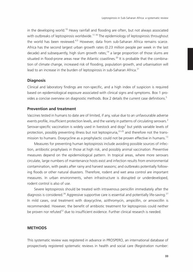
in the developing world.16 Heavy rainfall and flooding are often, but not always associated
with outbreaks of leptospirosis worldwide.17,18 The epidemiology of leptospirosis throughout
the world has been reviewed.4,5 However, data from sub-Saharan Africa remains scarce.
Africa has the second largest urban growth rates (0.23 million people per week in the last
decade) and subsequently, high slum growth rates;19 a large proportion of those slums are
situated in flood-prone areas near the Atlantic coastlines.20 It is probable that the combina-
tion of climate change, increased risk of flooding, population growth, and urbanisation will
lead to an increase in the burden of leptospirosis in sub-Saharan Africa.21
Diagnosis
Clinical and laboratory findings are non-specific, and a high index of suspicion is required
based on epidemiological exposure associated with clinical signs and symptoms. Box 1 pro-
vides a concise overview on diagnostic methods. Box 2 details the current case definitions.5
Prevention and treatment
Vaccines tested in humans to date are of limited, if any, value due to an unfavourable adverse
events profile, insufficient protection levels, and the variety in patterns of circulating serovars.2
Serovar-specific vaccination is widely used in livestock and dogs2 but yields variable levels of
protection, possibly preventing illness but not leptospiruria,22-25 and therefore not the trans-
mission to humans. Doxycycline as a prophylactic could not be proven effective in humans.13
Measures for preventing human leptospirosis include avoiding possible sources of infec-
tion, antibiotic prophylaxis in those at high risk, and possibly animal vaccination. Preventive
measures depend on the epidemiological pattern. In tropical areas, where more serovars
circulate, large numbers of maintenance hosts exist and infection results from environmental
contamination, with peaks after rainy and harvest seasons; and outbreaks potentially follow-
ing floods or other natural disasters. Therefore, rodent and wet area control are important
measures. In urban environments, when infrastructure is disrupted or underdeveloped,
rodent control is also of use.
Severe leptospirosis should be treated with intravenous penicillin immediately after the
diagnosis is considered.26 Aggressive supportive care is essential and potentially life-saving.27
In mild cases, oral treatment with doxycycline, azithromycin, ampicillin, or amoxicillin is
recommended. However, the benefit of antibiotic treatment for leptospirosis could neither
be proven nor refuted12 due to insufficient evidence. Further clinical research is needed.
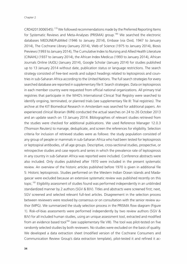
METHODS
This systematic review was registered in advance in PROSPERO, an international database of
prospectively registered systematic reviews in health and social care (Registration number:
Chapter 2
34
CRD42013006545).179 We followed recommendations made by the Preferred Reporting Items
for Systematic Reviews and Meta-Analyses (PRISMA) group.180 We searched the electronic
databases MEDLINE/PubMed (1946 to January 2014), Embase (via Ovid, 1947 to January
2014), The Cochrane Library (January 2014), Web of Science (1975 to January 2014), Biosis
Previews (1993 to January 2014), The Cumulative Index to Nursing and Allied Health Literature
(CINAHL) (1937 to January 2014), the African Index Medicus (1993 to January 2014), African
Journals Online (AJOL) (January 2014), Google Scholar (January 2014) for studies published
up to 13 January 2014 without date, publication status or language restrictions. The search
strategy consisted of free-text words and subject headings related to leptospirosis and coun-
tries in sub-Saharan Africa according to the United Nations. The full search strategies for every
searched database are reported in supplementary file II: Search strategies. Data on leptospirosis
in each member country were requested from official national organizations. All primary trial
registries that participate in the WHO’s International Clinical Trial Registry were searched to
identify ongoing, terminated, or planned trials (see supplementary file III: Trial registries). The
archive at the KIT Biomedical Research in Amsterdam was searched for additional papers. An
experienced clinical librarian (IMN) conducted the actual searches on 24 to 26 October 2013
and an update search on 13 January 2014. Bibliographies of relevant studies retrieved from
the studies were checked for additional publications. We used Reference Manager 12.0.3
(Thomson Reuters) to manage, deduplicate, and screen the references for eligibility. Selection
criteria for inclusion of retrieved studies were as follows: the study population consisted of
any group of people or mammals in sub-Saharan Africa who had been tested for leptospirosis
or leptospiral antibodies, of all age groups. Descriptive, cross-sectional studies, prospective, or
retrospective studies and case reports and series in which the prevalence rate of leptospirosis
in any country in sub-Saharan Africa was reported were included. Conference abstracts were
also included. Only studies published after 1970 were included in the present systematic
review. An overview of the historic articles published before 1970 is given in additional file
5: Historic leptospirosis. Studies performed on the Western Indian Ocean islands and Mada-
gascar were excluded because an extensive systematic review was published recently on this
topic.181 Eligibility assessment of studies found was performed independently in an unblinded
standardized manner by 2 authors (SGV & BJV). Titles and abstracts were screened first; next,
SGV screened and selected relevant full-text articles. Disagreement in the selection process
between reviewers were resolved by consensus or on consultation with the senior review au-
thor (MPG). We summarized the study selection process in the PRISMA flow diagram (Figure
1). Risk-of-bias assessments were performed independently by two review authors (SGV &
BJV) for all included human studies, using an unique assessment tool, extracted and modified
from an evidence based tool182 (see supplementary file VIII). The tool was pilot-tested on five
randomly selected studies by both reviewers. No studies were excluded on the basis of quality.
We developed a data extraction sheet (modified version of the Cochrane Consumers and
Communication Review Group’s data extraction template), pilot-tested it and refined it ac-
35
Leptospirosis in Sub-Saharan Africa: a systematic review
cordingly. SGV extracted the following study characteristics: first author, year of publication,
PubMed ID if available, language, study site & setting, study design, characteristics of trial
participants, objectives / measure of primary outcome, target population and selection crite-
ria, total enrolment, attrition rate (if applicable), sample size, diagnostic methods and cut-off
values and, if applicable, prevalence of leptospiral antibodies, leptospiral serovars/serogroups/
strains, characteristics of leptospirosis cases, risk factors, seasonal influences, demograph-
ics, co-infections, treatment, and mortality. Leptospiral serovars and strains were placed in
serogroups according to the ‘‘Leptospira Library’’ from KIT Biomedical Research.183 95% con-
fidence intervals (CI) of prevalence rates were calculated using the modified Wald method.
The following equations were used. p’= (S+0.5z2)/(n+z2) (S= numerator, n= denominator,
z= 1.96, as for a 95% confidence interval) and concomitantly to compute the margin of
error of the CI: W= z H((p’(1-p’))/(n+z2)). Data was plotted in forest plots using Prism version
6.0 (GraphPad Software, Inc., CA, USA). The primary outcome in the present review is the
prevalence of leptospirosis in countries in sub-Saharan Africa. Secondary outcomes include
risk factors, circulating serogroups, serovars and/or strains, clinical manifestations, treatment,
prevention measures, seasonal influences, and mortality. Extracted data was double checked
by BJV for all the included articles (n=140) using the original records. Regional and national
WHO offices were contacted for additional data on leptospirosis in the region, but this did
not result in additional data. We did not contact authors for further information or confirm
the accuracy of information included in our review with the original researchers, since for the
majority of papers adequate contact information was missing. A meta-analysis could not be
performed due to the clinical heterogeneity, and the non-uniformity of the diagnostic tools
and case definitions. We did not investigate publication bias.
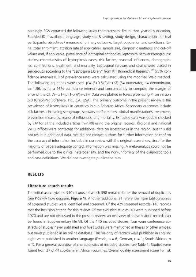
RESULTS
Literature search results
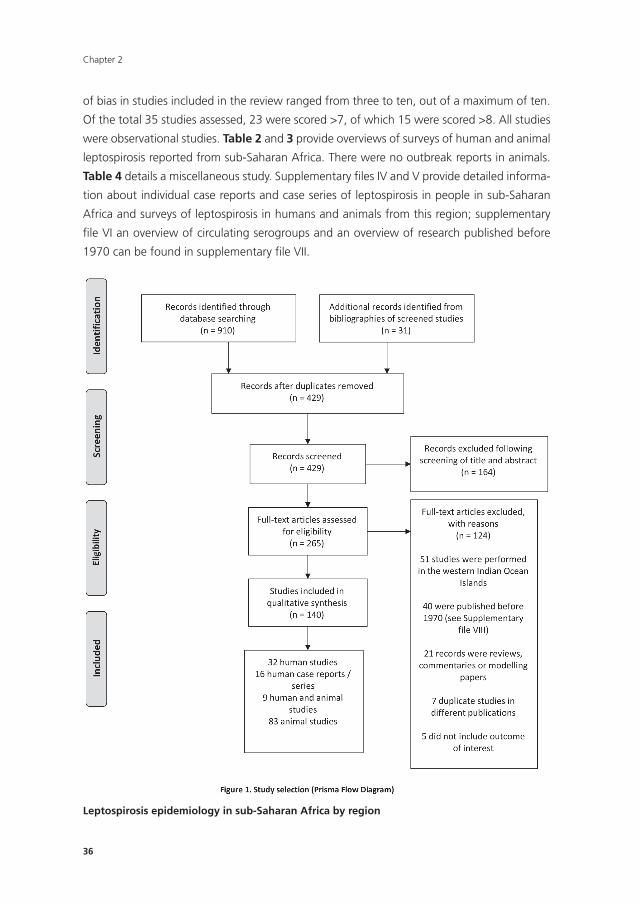
The initial search yielded 910 records, of which 398 remained after the removal of duplicates
(see PRISMA flow diagram, Figure 1). Another additional 31 references from bibliographies
of screened studies were identified and screened. Of the 429 screened records, 140 records
met the inclusion criteria for this review. Of the excluded studies, 40 were published before
1970 and are not discussed in the present review; an overview of these historic records can
be found in Supplementary file VII. Of the 140 included studies, four were conference ab-
stracts of studies never published and five studies were mentioned in theses or other articles,
but never published in an online database. The majority of records were published in English:
eight were published in another language (French, n = 6; German, n = 1; South African, n
= 1). For a general overview of characteristics of included studies, see Table 1. Studies were
found from 27 of 44 sub-Saharan African countries. Overall quality assessment scores for risk
Chapter 2
36
of bias in studies included in the review ranged from three to ten, out of a maximum of ten.
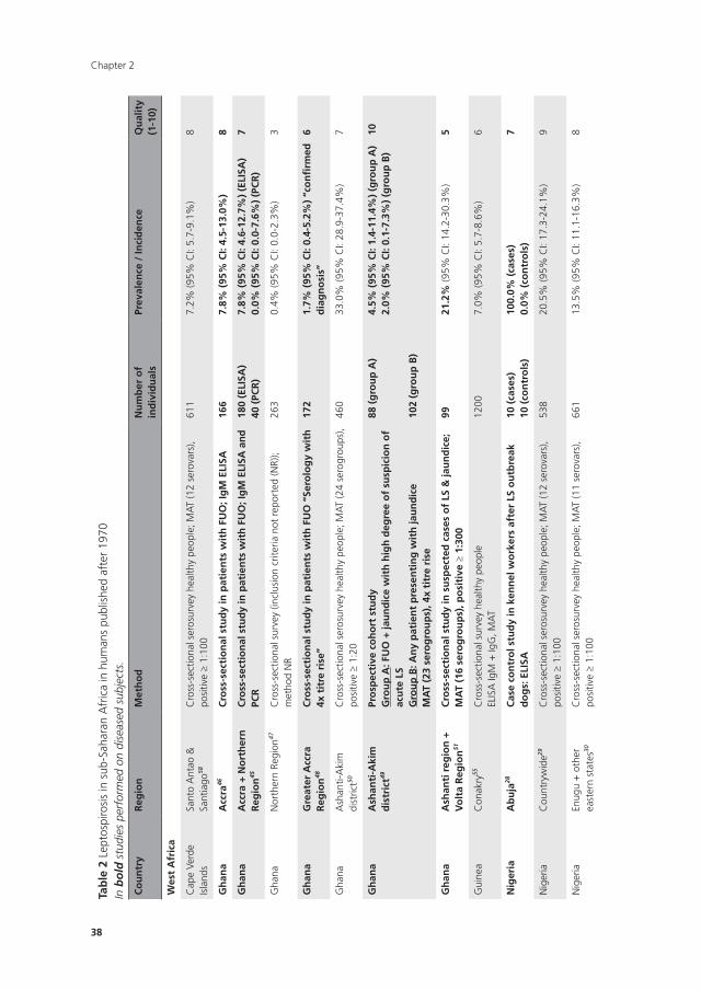
Of the total 35 studies assessed, 23 were scored >7, of which 15 were scored >8. All studies
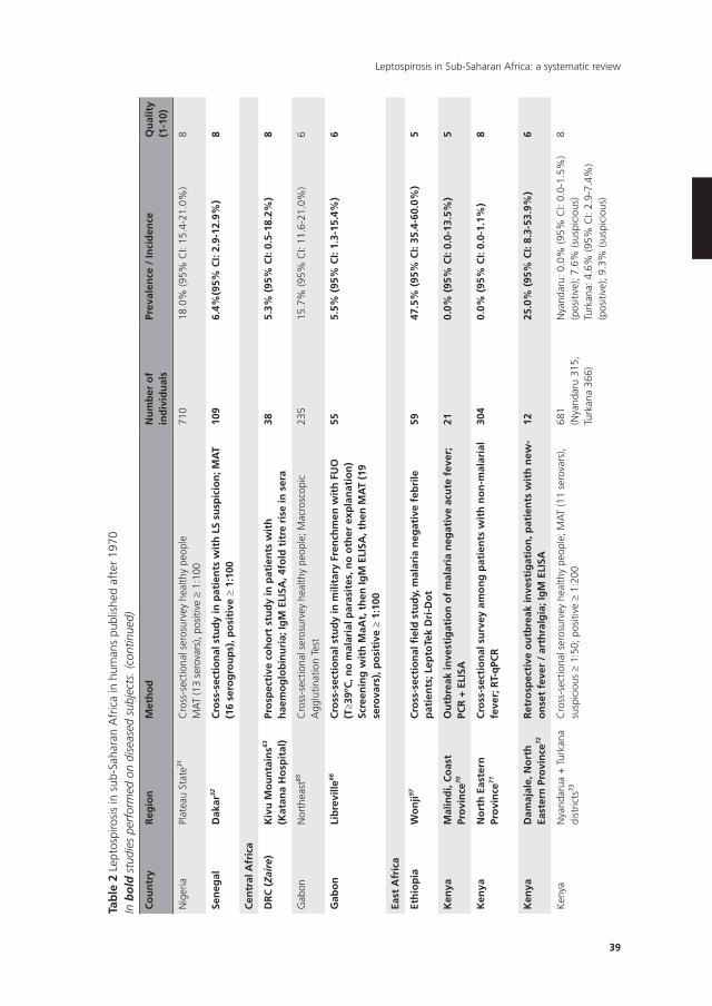
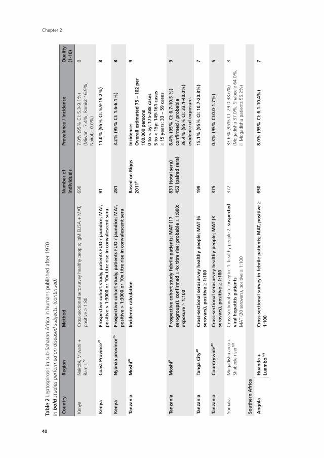
were observational studies. Table 2 and 3 provide overviews of surveys of human and animal
leptospirosis reported from sub-Saharan Africa. There were no outbreak reports in animals.
Table 4 details a miscellaneous study. Supplementary files IV and V provide detailed informa-
tion about individual case reports and case series of leptospirosis in people in sub-Saharan
Africa and surveys of leptospirosis in humans and animals from this region; supplementary
file VI an overview of circulating serogroups and an overview of research published before
1970 can be found in supplementary file VII.
Leptospirosis epidemiology in sub-Saharan Africa by region
37
Leptospirosis in Sub-Saharan Africa: a systematic review
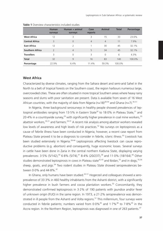
West Africa
Characterized by diverse climates, ranging from the Sahara desert and semi-arid Sahel in the
North to a belt of tropical forests on the Southern coast, the region harbours numerous large,
overcrowded cities. These are often situated in more tropical Southern areas where heavy rainy
seasons and slums with poor sanitation are present. Data is available from seven of 16 West
African countries; with the majority of data from Nigeria (n=18)28-44 and Ghana (n=7).45-51
In Nigeria, three background serosurveys in healthy people showed prevalences of lep-
tospiral antibodies ranging from 13·5% in Eastern State30 to 18·0% in Plateau State,31 and
20·4% in a countrywide survey,29 with significantly higher prevalence in coal mine workers,30
abattoir workers,30,31 and farmers.29-31 A recent risk analysis among abattoir workers revealed
low levels of awareness and high levels of risk practices.52 No surveys on leptospirosis as a
cause of febrile illness have been conducted in Nigeria; however, a recent case report from
Plateau State proved it to be a diagnosis to consider in febrile, icteric illness.53 Livestock has
been studied extensively in Nigeria.28-44 Leptospirosis affecting livestock can cause repro-
ductive problems (e.g. abortion) and consequently, huge economic losses. Several surveys
in cattle have been done in Zaria in the central northern Kaduna State, displaying varying
prevalences: 3·5% (5/142),40 6·8% (5/74),41 8·4% (20/237),38 and 11·0% (18/164).40 Other
studies demonstrated leptospirosis in cows in Plateau state42,43 and Ibidan,33 and in dogs,35,36
sheep, goats, and pigs.34 Two rodent studies in Plateau State showed seroprevalences be-
tween 0·0% and 44·8%.41
In Ghana, only humans have been studied.45-51 Hogerzeil and colleagues showed a sero-
prevalence of 33·3% in 460 healthy inhabitants from the Ashanti district, with a significantly
higher prevalence in bush farmers and cocoa plantation workers.50 Concomitantly, they
demonstrated confirmed leptospirosis in 3·2% of 190 patients with jaundice and/or fever
of unknown origin (FUO) in the same region. In 1973, a 21·2% seroprevalence was demon-
strated in ill people from the Ashanti and Volta regions.51 This millennium, four surveys were
conducted in febrile patients; numbers varied from 0·0%45 and 1·7%48 to 7·8%45 in the
Accra region. In the Northern Region, leptospirosis was diagnosed in one of 263 patients.47
Table 1 Overview characteristics included studies
Human surveys
Human + animal surveys
Case reports
Animal Total Percentage
West Africa 12 3 3 15 33 23.6%
Central Africa 3 0 4 4 11 7.9%
East Africa 12 2 1 30 45 32.1%
Southern Africa 2 4 5 34 45 32.1%
Travellers 3 0 3 0 6 4.3%
Total 32 9 16 83 140 100.0%
Percentage 22.9% 6.4% 11.4% 59.3% 100.0%
Chapter 2
38
Tab
le 2
Lep
tosp
irosi
s in
sub
-Sah
aran
Afr
ica
in h
uman
s pu
blis
hed
afte
r 19
70In
bo
ld s
tudi
es p
erfo
rmed
on
dise
ased
sub
ject
s.
Co
un
try
Reg
ion
Met
ho
dn
um
ber
of
ind
ivid
ual
sPr
eval
ence
/ In
cid
ence
Qu
alit
y(1
-10)
Wes
t A
fric
a
Cap
e Ve
rde
Isla
nds
Sant
o A
ntao
&
Sant
iago
58
Cro
ss-s
ectio
nal s
eros
urve
y he
alth
y pe
ople
; MA
T (1
2 se
rova
rs),
posi
tive ≥
1:10
061
17.
2% (9
5% C
I: 5.
7-9.
1%)
8
Gh
ana
Acc
ra46
Cro
ss-s
ecti
on
al s
tud
y in
pat
ien
ts w
ith
FU
O; I
gM
ELI
SA16
67.
8% (
95%
CI:
4.5-
13.0
%)
8
Gh
ana
Acc
ra +
no
rth
ern
R
egio
n45
Cro
ss-s
ecti
on
al s
tud
y in
pat
ien
ts w
ith
FU
O; I
gM
ELI
SA a
nd
PC
R18
0 (E
LISA
)40
(PC
R)
7.8%
(95
% C
I: 4.
6-12
.7%
) (E
LISA
)0.
0% (
95%
CI:
0.0-
7.6%
) (P
CR
)7
Gha
naN
orth
ern
Regi
on47
Cro
ss-s
ectio
nal s
urve
y (in
clus
ion
crite
ria n
ot r
epor
ted
(NR)
); m
etho
d N
R26
30.
4% (9
5% C
I: 0.
0-2.
3%)
3
Gh
ana
Gre
ater
Acc
ra
Reg
ion
48
Cro
ss-s
ecti
on
al s
tud
y in
pat
ien
ts w
ith
FU
O “
Sero
log
y w
ith
4x
tit
re r
ise”
172
1.7%
(95
% C
I: 0.
4-5.
2%)
“co
nfi
rmed
d
iag
no
sis”
6
Gha
naA
shan
ti-A
kim
di
stric
t50
Cro
ss-s
ectio
nal s
eros
urve
y he
alth
y pe
ople
; MA
T (2
4 se
rogr
oups
), po
sitiv
e ≥
1:20
460
33.0
% (9
5% C
I: 28
.9-3
7.4%
)7
Gh
ana
Ash
anti
-Aki
m
dis
tric
t49
Pro
spec
tive
co
ho
rt s
tud
yG
rou
p A
: FU
O +
jau
nd
ice
wit
h h
igh
deg
ree
of
susp
icio
n o
f ac
ute
LS
Gro
up
B: A
ny
pat
ien
t p
rese
nti
ng
wit
h ja
un
dic
eM
AT
(23
sero
gro
up
s), 4
x ti
tre
rise
88 (
gro
up
A)
102
(gro
up
B)
4.5%
(95
% C
I: 1.
4-11
.4%
) (g
rou
p A
)2.
0% (
95%
CI:
0.1-
7.3%
) (g
rou
p B
)10
Gh
ana
Ash
anti
reg
ion
+
Vo
lta
Reg
ion
51
Cro
ss-s
ecti
on
al s
tud
y in
su
spec
ted
cas
es o
f LS
& ja
un
dic
e;
MA
T (1
6 se
rog
rou
ps)
, po
siti
ve ≥
1:3
0099
21.2
% (9
5% C
I: 14
.2-3
0.3%
)5
Gui
nea
Con
akry
55C
ross
-sec
tiona
l sur
vey
heal
thy
peop
leEL
ISA
IgM
+ Ig
G, M
AT
1200
7.0%
(95%
CI:
5.7-
8.6%
)6
nig
eria
Ab
uja
28C
ase
con
tro
l stu
dy
in k
enn
el w
ork
ers
afte
r LS
ou
tbre
ak
do
gs:
ELI
SA10
(ca
ses)
10 (
con
tro
ls)
100.
0% (
case
s)0.
0% (
con
tro
ls)
7
Nig
eria
Cou
ntry
wid
e29C
ross
-sec
tiona
l ser
osur
vey
heal
thy
peop
le; M
AT
(12
sero
vars
), po
sitiv
e ≥
1:10
053
820
.5%
(95%
CI:
17.3
-24.
1%)
9
Nig
eria
Enug
u +
oth
er
east
ern
stat
es30
Cro
ss-s
ectio
nal s
eros
urve
y he
alth
y pe
ople
; MA
T (1
1 se
rova
rs),
posi
tive ≥
1:10
066
113
.5%
(95%
CI:
11.1
-16.
3%)
8
39
Leptospirosis in Sub-Saharan Africa: a systematic review
Tab
le 2
Lep
tosp
irosi
s in
sub
-Sah
aran
Afr
ica
in h
uman
s pu
blis
hed
afte
r 19
70In
bo
ld s
tudi
es p
erfo
rmed
on
dise
ased
sub
ject
s. (c
ontin
ued)
Co
un
try
Reg
ion
Met
ho
dn
um
ber
of
ind
ivid
ual
sPr
eval
ence
/ In
cid
ence
Qu
alit
y(1
-10)
Nig
eria
Plat
eau
Stat
e31C
ross
-sec
tiona
l ser
osur
vey
heal
thy
peop
leM
AT
(13
sero
vars
), po
sitiv
e ≥
1:10
071
018
.0%
(95%
CI:
15.4
-21.
0%)
8
Sen
egal
Dak
ar32
Cro
ss-s
ecti
on
al s
tud
y in
pat
ien
ts w
ith
LS
susp
icio
n; M
AT
(16
sero
gro
up
s), p
osi
tive
≥ 1
:100
109
6.4%
(95%
CI:
2.9-
12.9
%)
8
Cen
tral
Afr
ica
DR
C (
Zair
e)K
ivu
Mo
un
tain
s63
(Kat
ana
Ho
spit
al)
Pro
spec
tive
co
ho
rt s
tud
y in
pat
ien
ts w
ith
h
aem
og
lob
inu
ria;
IgM
ELI
SA, 4
fold
tit
re r
ise
in s
era
385.
3% (
95%
CI:
0.5-
18.2
%)
8
Gab
onN
orth
east
65C
ross
-sec
tiona
l ser
osur
vey
heal
thy
peop
le; M
acro
scop
ic
Agg
lutin
atio
n Te
st23
515
.7%
(95%
CI:
11.6
-21.
0%)
6
Gab
on
Lib
revi
lle66
Cro
ss-s
ecti
on
al s
tud
y in
mili
tary
Fre
nch
men
wit
h F
UO
(T≥3
9ºC
, no
mal
aria
l par
asit
es, n
o o
ther
exp
lan
atio
n)
Scre
enin
g w
ith
MaA
t, t
hen
IgM
ELI
SA, t
hen
MA
T (1
9 se
rova
rs),
po
siti
ve ≥
1:1
00
555.
5% (
95%
CI:
1.3-
15.4
%)
6
East
Afr
ica
Eth
iop
iaW
on
ji97C
ross
-sec
tio
nal
fiel
d s
tud
y, m
alar
ia n
egat
ive
feb
rile
p
atie
nts
; Lep
toTe
k D
ri-D
ot
5947
.5%
(95
% C
I: 35
.4-6
0.0%
)5
Ken
yaM
alin
di,
Co
ast
Pro
vin
ce70
Ou
tbre
ak in
vest
igat
ion
of
mal
aria
neg
ativ
e ac
ute
fev
er;
PCR
+ E
LISA
210.
0% (
95%
CI:
0.0-
13.5
%)
5
Ken
yan
ort
h E
aste
rn
Pro
vin
ce71
Cro
ss-s
ecti
on
al s
urv
ey a
mo
ng
pat
ien
ts w
ith
no
n-m
alar
ial
feve
r; R
T-q
PCR
304
0.0%
(95
% C
I: 0.
0-1.
1%)
8
Ken
yaD
amaj
ale,
no
rth
Ea
ster
n P
rovi
nce
72
Ret
rosp
ecti
ve o
utb
reak
inve
stig
atio
n, p
atie
nts
wit
h n
ew-
on
set
feve
r /
arth
ralg
ia; I
gM
ELI
SA12
25.0
% (
95%
CI:
8.3-
53.9
%)
6
Ken
yaN
yand
arua
+ T
urka
na
dist
ricts
73
Cro
ss-s
ectio
nal s
eros
urve
y he
alth
y pe
ople
; MA
T (1
1 se
rova
rs),
susp
icio
us ≥
1:5
0; p
ositi
ve ≥
1:2
0068
1(N
yand
aru
315,
Tu
rkan
a 36
6)
Nya
ndar
u: 0
.0%
(95%
CI:
0.0-
1.5%
) (p
ositi
ve);
7.6%
(sus
pici
ous)
Turk
ana:
4.6
% (9
5% C
I: 2.
9-7.
4%)
(pos
itive
); 9.
3% (s
uspi
ciou
s)
8
Chapter 2
40
Tab
le 2
Lep
tosp
irosi
s in
sub
-Sah
aran
Afr
ica
in h
uman
s pu
blis
hed
afte
r 19
70In
bo
ld s
tudi
es p
erfo
rmed
on
dise
ased
sub
ject
s. (c
ontin
ued)
Co
un
try
Reg
ion
Met
ho
dn
um
ber
of
ind
ivid
ual
sPr
eval
ence
/ In
cid
ence
Qu
alit
y(1
-10)
Ken
yaN
airo
bi, M
iwan
i +
Ram
isi74
Cro
ss-s
ectio
nal s
eros
urve
y he
alth
y pe
ople
; IgM
ELI
SA +
MA
T,
posi
tive ≥
1:80
690
7.0%
(95%
CI:
5.3-
9.1%
)(M
iwan
i: 7.
4%, R
amis
i: 16
.9%
, N
airo
bi: 0
.0%
)
8
Ken
yaC
oas
t Pr
ovi
nce
76Pr
osp
ecti
ve c
oh
ort
stu
dy,
pat
ien
ts F
UO
/ ja
un
dic
e; M
AT,
p
osi
tive
> 1
:300
0 o
r 10
x ti
tre
rise
in c
on
vale
scen
t se
ra91
11.0
% (
95%
CI:
5.9-
19.2
%)
8
Ken
yan
yan
za p
rovi
nce
75Pr
osp
ecti
ve c
oh
ort
stu
dy,
pat
ien
ts F
UO
/ ja
un
dic
e; M
AT,
p
osi
tive
> 1
:300
0 o
r 10
x ti
tre
rise
in c
on
vale
scen
t se
ra28
13.
2% (
95%
CI:
1.6-
6.1%
)8
Tan
zan
iaM
osh
i87In
cid
ence
cal
cula
tio
nB
ased
on
Big
gs
2011
9
Inci
den
ce:
Ove
rall
esti
mat
ed 7
5 –
102
per
10
0.00
0 p
erso
ns
0 to
< 5
y: 1
75-2
88 c
ases
5 to
< 1
5y: 1
49-1
61 c
ases
≥ 15
yea
rs: 3
3 –
59 c
ases
9
Tan
zan
iaM
osh
i9Pr
osp
ecti
ve c
oh
ort
stu
dy
feb
rile
pat
ien
ts; M
AT
(17
sero
gro
up
s), c
on
firm
ed ≥
4x
titr
e ri
se: p
rob
able
≥ 1
:800
: ex
po
sure
≥ 1
:100
831
(to
tal s
era)
453
(pai
red
ser
a)8.
4% (
95%
CI:
6.7-
10.5
%)
con
firm
ed /
pro
bab
le36
.4%
(95
% C
I: 33
.1-4
0.0%
) ev
iden
ce o
f ex
po
sure
.
9
Tan
zan
iaTa
ng
a C
ity88
Cro
ss-s
ecti
on
al s
ero
surv
ey h
ealt
hy
peo
ple
; MA
T (6
se
rova
rs),
po
siti
ve ≥
1:1
6019
915
.1%
(95
% C
I: 10
.7-2
0.8%
)7
Tan
zan
iaC
ou
ntr
ywid
e89C
ross
-sec
tio
nal
ser
osu
rvey
hea
lth
y p
eop
le; M
AT
(3
sero
vars
), p
osi
tive
≥ 1
:160
375
0.3%
(95
% C
I:0.0
-1.7
%)
5
Som
alia
Mog
adis
hu a
rea
+
Shab
eele
riv
er10
1
Cro
ss-s
ectio
nal s
eros
urve
y in
: 1. h
ealth
y pe
ople
2. s
usp
ecte
d
vira
l hep
atit
is p
atie
nts
MA
T (2
0 se
rova
rs),
posi
tive ≥
1:10
0
372
33.6
% (9
5% C
I: 29
.0-3
8.6%
)(M
ogad
ishu
37.
0%, S
habe
ele
64.0
%,
ill M
ogad
ishu
pat
ient
s 56
.2%
)
8
Sou
ther
n A
fric
a
An
go
laH
uan
da
+
Luam
bo
144
Cro
ss-s
ecti
on
al s
urv
ey in
feb
rile
pat
ien
ts; M
AT,
po
siti
ve ≥
1:
100
650
8.0%
(95
% C
I: 6.
1-10
.4%
)7
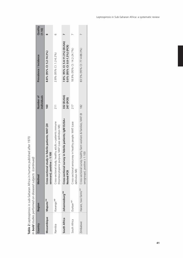
41
Leptospirosis in Sub-Saharan Africa: a systematic review
Tab
le 2
Lep
tosp
irosi
s in
sub
-Sah
aran
Afr
ica
in h
uman
s pu
blis
hed
afte
r 19
70In
bo
ld s
tudi
es p
erfo
rmed
on
dise
ased
sub
ject
s. (c
ontin
ued)
Co
un
try
Reg
ion
Met
ho
dn
um
ber
of
ind
ivid
ual
sPr
eval
ence
/ In
cid
ence
Qu
alit
y(1
-10)
Mo
zam
biq
ue
Map
uto
150
Cro
ss-s
ecti
on
al s
tud
y in
feb
rile
pat
ien
ts; M
AT
(20
sero
vars
), p
osi
tive
≥ 1
:100
160
8.8%
(95
% C
I: 5.
2-14
.3%
)8
Nam
ibia
Kav
ango
160
Cro
ss-s
ectio
nal s
tudy
, hea
lthy
adul
ts +
pat
ient
s im
mun
e th
rom
bocy
tope
nic
purp
ura;
MA
T (c
ase
defin
ition
NR)
211
2.8%
(95%
CI:
1.2-
6.2%
)6
Sou
th A
fric
aJo
han
nes
bu
rg10
6C
ross
-sec
tio
nal
su
rvey
in f
ebri
le p
atie
nts
; Ig
M E
LISA
+
nes
ted
PC
R33
2 (E
LISA
)24
7 (P
CR
)7.
8% (9
5% C
I: 5.
4-11
.3%
) (E
LISA
)0.
0% (
95%
CI:
0.0-
1.3%
) (P
CR
)7
Sout
h A
fric
aD
urba
n107
Cro
ss-s
ectio
nal s
eros
urve
y in
hea
lthy
peop
le: M
AT
(cas
e de
finiti
on N
R)21
718
.9%
(95%
CI:
14.2
-24.
7%)
7
Zim
babw
eH
arar
e, t
wo
farm
s121
Cro
ss-s
ectio
nal s
urve
y he
alth
y fa
rm w
orke
rs &
fam
ilies
; MA
T (8
se
rogr
oups
), po
sitiv
e ≥
1:10
018
283
.5%
(95%
CI:
77.4
-88.
3%)
7
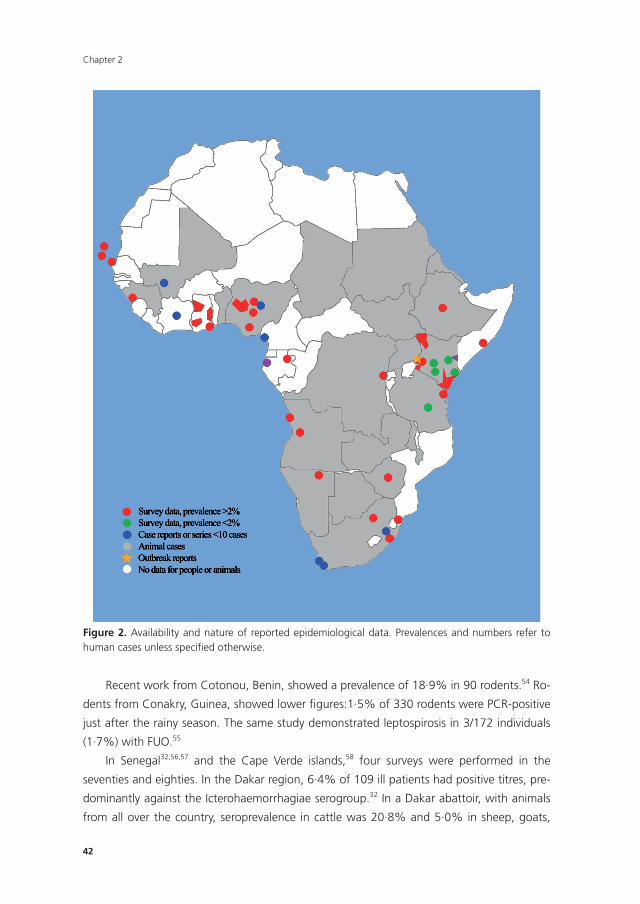
Chapter 2
42
Figure 2. Availability and nature of reported epidemiological data. Prevalences and numbers refer to human cases unless specified otherwise.
Recent work from Cotonou, Benin, showed a prevalence of 18·9% in 90 rodents.54 Ro-
dents from Conakry, Guinea, showed lower figures:1·5% of 330 rodents were PCR-positive
just after the rainy season. The same study demonstrated leptospirosis in 3/172 individuals
(1·7%) with FUO.55
In Senegal32,56,57 and the Cape Verde islands,58 four surveys were performed in the
seventies and eighties. In the Dakar region, 6·4% of 109 ill patients had positive titres, pre-
dominantly against the Icterohaemorrhagiae serogroup.32 In a Dakar abattoir, with animals
from all over the country, seroprevalence in cattle was 20·8% and 5·0% in sheep, goats,
43
Leptospirosis in Sub-Saharan Africa: a systematic review
and pigs.57 On Santo Antão and Santiago, a serosurvey showed a prevalence of 7·2% in 611
healthy inhabitants, and lower rates in cattle, donkeys, sheep and goats.58-60
No data were retrievable from other West African countries, except for Mali. A Bamako
survey showed 44·9% of 642 dairy cows to be seropositive.61 No human data exist from Mali
except for a case report.62
Central Africa
Data from the Central African region is very limited. Even though van Riel and colleagues
performed extensive research on the disease in the Democratic Republic of Congo from
1939 onwards (see Supplementary file VII), no data was published after the country’s in-
dependence in 1960. In 1995, a survey was performed in the Katana hospital in the Kivu
region, before the arrival of longstanding, violent conflict. In a prospective cohort study on
the aetiology of haemoglobinuria63, the diagnosis of acute leptospirosis was established in
one of 65 patients. This is probably an underestimation since the study was not designed
to detect leptospirosis. Subsequently, a serosurvey in 61 antelopes from the Ituri rainforest
showed leptospiral antibodies in 26·2%.64
Most Central African data comes from Gabon where in the 1990s, two surveys were
performed in people.65,66 In five villages in the northeast, a seroprevalence of 15·7% in
healthy people was demonstrated.65 No risk factors could be determined; members from the
Bakoule tribe, traditionally fisherman, were found to have a higher risk of being seropositive.
All villages were located in a gold panning area; this was not found to be a risk factor. Of
52 French military personnel presenting with FUO in Libreville, two had high single antibody
titres against pathogenic serovars.66 They had been dispatched in the field and exposed to
freshwater. Recently, a case series was published of four men from poorer areas in Libreville
with proven acute leptospirosis.67
Scolamaccia and colleagues68 designed a prevalence model for cattle from the Adamwa
region in Cameroon, adjusting for the diagnostic test performance and study design. Based
on banked sera of 1.377 cows from 146 herds, they estimated the region-wide animal
prevalence to be 30·4%, and 93·0% of herds to be positive.
The only other report originating from this region is a case report of a military dog from
Chad with leptospirosis-induced renal insufficiency.69
East Africa
Most epidemiological data on leptospirosis in East Africa originates from Kenya70-86 and
Tanzania.9,87-89 Since the fifties, Kenya has been key in establishing knowledge on the epide-
miology of leptospirosis on the African continent. Since 1997, Tanzania, has evolved as the
focus of many leptospirosis studies.
The only well established incidence calculation for leptospirosis in sub-Saharan Africa
was done in 2013, for the rural and urban area of Moshi, in the Kilimanjaro Region of Tan-
Chapter 2
44
zania.87 Based on a prospective cohort study,9 the overall estimated incidence was 75–102
per 100.000 persons per year, with the highest incidence in children under five, i.e. 175–288
cases per 100.000 per year (table 2). This is probably an underestimation because it only
represents hospitalized cases. The higher incidence in younger children can be explained by
their lower admission threshold.87 In the aforementioned study9 among febrile adults and
children, 8·4% (70/831) serologically tested participants met the definition of confirmed
or (highly) probable leptospirosis; another 277 patients (36·4%) exhibited evidence of ex-
posure. Patients with confirmed or probable leptospirosis were older than those without
(P<0·001), and serological evidence of exposure increased with age (P<0·001). In adults,
thrombocytopenia was associated with confirmed (OR 3·5, P=0·005) or probable disease (OR
2·2, P=0·017). In children, lymphadenopathy was associated with leptospirosis (25·9% vs.
9·2%; P=0·14). The study demonstrates clinicians’ unawareness of leptospirosis: provisional
diagnoses in confirmed and probable cases were mainly malaria and pneumonia; leptospiro-
sis was never included into the differential diagnosis. 40% of confirmed cases suffered from
a co-infection; HIV (15%), rickettsiosis, brucellosis, and community acquired blood stream
infections (12·5% each) were most prevalent. No association between HIV and leptospirosis
could be demonstrated, in contrast to the later re-analysis of this study9, where leptospirosis
was found to be less prevalent in a group of HIV infected patients (OR 0·43, P=0·019).90 The
only risk factor identified was living in a rural area (OR 3·4, P<0·001). No distinguished link
was found between chest X-ray abnormalities and leptospirosis.91
In the Tanga region, bordered by the Indian Ocean and Kilimanjaro mountains, a 2009
survey showed a seroprevalence of 15·1% among 199 healthy inhabitants; milking of cows
constituted a risk (OR 3·44, P<0·001).88 Serosurveys in cattle demonstrated prevalences vary-
ing from 11·0%,92 and 30·0%93 in larger surveys, with upto 51·0% in a smaller abattoir
survey.94 A countrywide serosurvey among healthy blood donors (n=375) showed a low
prevalence of 0·3%;89 however, the sera were only screened against three serovars.
First reports of leptospirosis in Kenya date from 1944 in cattle. De Geus and colleagues
later conducted 2 studies in Coast76 and Nyanza provinces,75 in adults and children with
FUO or jaundice. Applying strict case definitions, 11·0% from 91 patients from Coast and
3·2% of 281 patients from Nyanza province were found to suffer from acute leptospirosis. A
significant higher prevalence in males, and in hospitals close to sugar-estates was found.95,96
In 1987, 7·4% of 353 healthy people in Nyanza province, and 16.9% of 130 in Coast prov-
ince were found to have leptospiral antibodies;74 recent outbreak investigation of malaria-
negative fever in 21 patients in Malindi in Coast province could not prove leptospirosis.70
In Kibera, a densely populated part of Nairobi with poor sanitary conditions, leptospiral
DNA was recently demonstrated in 41/224 (18·3%) of rodents.77 Human data available from
Nairobi is from 1987; none of 207 healthy subjects had leptospiral antibodies.74 Recent stud-
ies in patients with FUO in North East province did not yield positive Leptospira PCRs;71 yet
no serological tests were performed. However, a case series after an outbreak of acute febrile
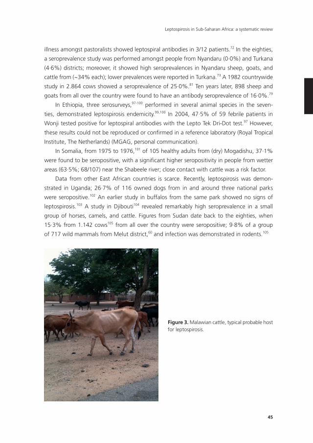
45
Leptospirosis in Sub-Saharan Africa: a systematic review
illness amongst pastoralists showed leptospiral antibodies in 3/12 patients.72 In the eighties,
a seroprevalence study was performed amongst people from Nyandaru (0·0%) and Turkana
(4·6%) districts; moreover, it showed high seroprevalences in Nyandaru sheep, goats, and
cattle from (~34% each); lower prevalences were reported in Turkana.73 A 1982 countrywide
study in 2.864 cows showed a seroprevalence of 25·0%.81 Ten years later, 898 sheep and
goats from all over the country were found to have an antibody seroprevalence of 16·0%.79
In Ethiopia, three serosurveys,97-100 performed in several animal species in the seven-
ties, demonstrated leptospirosis endemicity.99,100 In 2004, 47·5% of 59 febrile patients in
Wonji tested positive for leptospiral antibodies with the Lepto Tek Dri-Dot test.97 However,
these results could not be reproduced or confirmed in a reference laboratory (Royal Tropical
Institute, The Netherlands) (MGAG, personal communication).
In Somalia, from 1975 to 1976,101 of 105 healthy adults from (dry) Mogadishu, 37·1%
were found to be seropositive, with a significant higher seropositivity in people from wetter
areas (63·5%; 68/107) near the Shabeele river; close contact with cattle was a risk factor.
Data from other East African countries is scarce. Recently, leptospirosis was demon-
strated in Uganda; 26·7% of 116 owned dogs from in and around three national parks
were seropositive.102 An earlier study in buffalos from the same park showed no signs of
leptospirosis.103 A study in Djibouti104 revealed remarkably high seroprevalence in a small
group of horses, camels, and cattle. Figures from Sudan date back to the eighties, when
15·3% from 1.142 cows105 from all over the country were seropositive; 9·8% of a group
of 717 wild mammals from Melut district,60 and infection was demonstrated in rodents.105
Figure 3. Malawian cattle, typical probable host for leptospirosis.
Chapter 2
46
Southern Africa
Forty-six studies from Southern Africa were identified (see table 1). Most studies came from
South Africa (n=15)80,106-120 and Zimbabwe (n=17).121-136 No data was found from Swaziland
and Lesotho.
Feresu and colleagues established knowledge on leptospirosis in Zimbabwe. In 1995/6,
they demonstrated that 83·5% of 182 workers and their families from two farms in and
around Harare carried leptospiral antibodies in one of the samples after following them
for 1·5 years; they could not identify any seasonal influences.121 Of 437 rodents from the
same farms, 62·5% were seropositive.121 In the early 80s, 26·8% of a cattle herd (n=2382)
were seropositive,127 and later leptospires were isolated in 10.4% of 480 cattle in a Harare
abattoir.129 These isolates revealed several new serovars and strains.130-134,136 In Harare, rural
dogs (n=250) were found to have a positive ELISA in 15·6%.122 Similar numbers in dogs
were found in 1979.137 Zimbabwean wildlife has been studied: in rhinoceroses numbers
varied from 63·0% (38/60)125 to 4·9% in a group of 102.124 Buffalos, elands, wildebeests,
and zebras were also found to carry antibodies.124 No other human data from Zimbabwe was
retrieved except two case reports.138,139
South Africa, human data is relatively scarce: two surveys were published.107,140 In 2010,
7·8% of 332 febrile patients from Johannesburg were seropositive;140 a survey in Durban
among healthy volunteers showed a seroprevalence of 18·9%, with a significantly higher
percentage among people from 18-22 years.107 The latter study found seropositivity in 22 of
221 rodents trapped in the same area, and 14·5% of 69 kidneys to be PCR positive. Other
sporadic data on human leptospirosis dates from the eighties.141-143
Concerning livestock, a survey in 33 districts of KwaZulu-Natal proved 19·4% of 2.021
cows to be seropositive.109 Other surveys showed numbers varying from 3·0% (of 860 cows)
in Mpumalanga Province114 to 52·4% in Transvaal.116 Cows and pigs showed high prevalence
rates after abortion-clusters in pigs in farms in Mpumalanga, Gauteng Province and Free
State.110 Earlier outbreak investigation in pigs showed similarly high rates.120 An abattoir sero-
survey demonstrated a 22·2% prevalence in 5.041 pigs from several regions.111 In Transvaal,
1.3% of 152 sheep had antibodies.116
Seropositivity was shown in rhinoceroses (n=182) from four National Parks (26·4%);80
buffaloes from Kruger National Park (7/406);114 in a small group of wild animals from North-
ern Natal (12%);115 and in 1978 in 8·0% of 50 vervet monkeys.119
In South African dogs, rates were equally varied: 86.5% in 37 ill dogs in the early seven-
ties;120 1.5% (6/400) in healthy dogs in Pretoria;112 a recent study in four coastal regions
demonstrated a seroprevalence of 4·7% in 530 dogs. Stray dogs were 4·4 times more likely
to have antibodies compared to owned dogs (7·6% vs. 1·9%, P=0·0017), with dogs in
KwaZulu Natal and Eastern Cape more likely to be positive.108
In Angola, in a group of 650 febrile patients, 8·0% were probable cases of leptospirosis.
13·0% of 77 rodents showed PCR positive kidney samples.144 Zambian rodents, recently
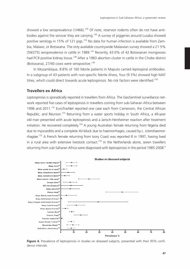
47
Leptospirosis in Sub-Saharan Africa: a systematic review
showed a low seroprevalence (1/466).145 Of note, reservoir rodents often do not have anti-
bodies against the serovar they are carrying.146 A survey of piggeries around Lusaka showed
positive serology in 15% of 121 pigs.126 No data for human infection is available from Zam-
bia, Malawi, or Botswana. The only available countrywide Malawian survey showed a 21·5%
(59/275) seroprevalence in cattle in 1989.147 Recently, 43·0% of 42 Botswanan mongooses
had PCR positive kidney tissue.148 After a 1983 abortion cluster in cattle in the Chobe district
(Botswana), 27/40 cows were seropositive.149
In Mozambique, 8·8% of 160 febrile patients in Maputo carried leptospiral antibodies.
In a subgroup of 43 patients with non-specifi c febrile illness, four (9·3%) showed high MAT
titres, which could direct towards acute leptospirosis. No risk factors were identifi ed.150
Travellers ex Africa
Leptospirosis is sporadically reported in travellers from Africa. The GeoSentinel surveillance net-
work reported fi ve cases of leptospirosis in travellers coming from sub-Saharan Africa between
1996 and 2011.152 EuroTravNet reported one case each from Cameroon, the Central African
Republic, and Réunion.153 Returning from a water sports holiday in South Africa, a 49-year
old man presented with acute leptospirosis and a Jarisch-Herxheimer reaction after treatment
initiation. He recovered completely.154 A young Australian female returning from Nigeria died
due to myocarditis and a complete AV-block due to haemorrhages, caused by L. icterohaemor-
rhagiae.155 A French female returning from Ivory Coast was reported ill in 1997, having lived
in a rural area with extensive livestock contact.156 In the Netherlands alone, seven travellers
returning from sub-Saharan Africa were diagnosed with leptospirosis in the period 1985-2008.6
Figure 4. Prevalence of leptospirosis in studies on diseased subjects, presented with their 95% confi -dence intervals.
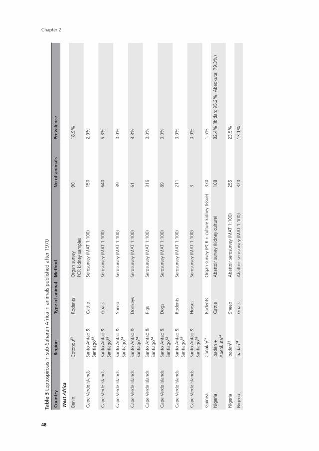
Chapter 2
48
Tab
le 3
Lep
tosp
irosi
s in
sub
-Sah
aran
Afr
ica
in a
nim
als
publ
ishe
d af
ter
1970
Co
un
try
Reg
ion
Typ
e o
f an
imal
Met
ho
dn
o o
f an
imal
sPr
eval
ence
Wes
t A
fric
a
Beni
nC
oton
ou54
Rode
nts
Org
an s
urve
yPC
R ki
dney
sam
ples
9018
.9%
Cap
e Ve
rde
Isla
nds
Sant
o A
ntao
&
Sant
iago
58
Cat
tleSe
rosu
rvey
(MA
T 1:
100)
150
2.0%
Cap
e Ve
rde
Isla
nds
Sant
o A
ntao
&
Sant
iago
58
Goa
tsSe
rosu
rvey
(MA
T 1:
100)
640
5.3%
Cap
e Ve
rde
Isla
nds
Sant
o A
ntao
&
Sant
iago
58
Shee
pSe
rosu
rvey
(MA
T 1:
100)
390.
0%
Cap
e Ve
rde
Isla
nds
Sant
o A
ntao
&
Sant
iago
58
Don
keys
Sero
surv
ey (M
AT
1:10
0)61
3.3%
Cap
e Ve
rde
Isla
nds
Sant
o A
ntao
&
Sant
iago
58
Pigs
Sero
surv
ey (M
AT
1:10
0)31
60.
0%
Cap
e Ve
rde
Isla
nds
Sant
o A
ntao
&
Sant
iago
58
Dog
sSe
rosu
rvey
(MA
T 1:
100)
890.
0%
Cap
e Ve
rde
Isla
nds
Sant
o A
ntao
&
Sant
iago
58
Rode
nts
Sero
surv
ey (M
AT
1:10
0)21
10.
0%
Cap
e Ve
rde
Isla
nds
Sant
o A
ntao
&
Sant
iago
58
Hor
ses
Sero
surv
ey (M
AT
1:10
0)3
0.0%
Gui
nea
Con
akry
55Ro
dent
sO
rgan
sur
vey
(PC
R +
cul
ture
kid
ney
tissu
e)33
01.
5%
Nig
eria
Ibad
an +
A
beok
uta33
Cat
tleA
batt
oir
surv
ey (k
idne
y cu
lture
)10
882
.4%
(Ibi
dan:
95.
2%, A
beok
uta:
79.
3%)
Nig
eria
Ibad
an34
Shee
pA
batt
oir
sero
surv
ey (M
AT
1:10
0)25
523
.5%
Nig
eria
Ibad
an34
Goa
tsA
batt
oir
sero
surv
ey (M
AT
1:10
0)32
013
.1%
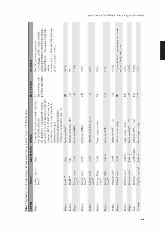
49
Leptospirosis in Sub-Saharan Africa: a systematic review
Tab
le 3
Lep
tosp
irosi
s in
sub
-Sah
aran
Afr
ica
in a
nim
als
publ
ishe
d af
ter
1970
(con
tinue
d)
Co
un
try
Reg
ion
Typ
e o
f an
imal
Met
ho
dn
o o
f an
imal
sPr
eval
ence
Nig
eria
Ibad
an, L
agos
+
Ilorin
35
Dog
sPr
ospe
ctiv
e se
rolo
gica
l sur
vey
in s
ick
dogs
w
ith p
ositi
ve L
S se
rolo
gyM
AT
posi
tive ≥
1:10
0 (v
acci
nal s
erov
ars)
+
a 4-
foul
d ris
e in
10-
14 d
ays
or a
sin
gle
titre
be
twee
n 1:
800
to 1
:320
0 (n
on-v
acci
nal
sero
vars
). Va
ccin
al s
erov
ars:
Can
icol
a &
Ic
tero
haem
orrh
agia
eTr
eatm
ent:
am
pici
llin/
amox
icill
in/
enro
floxa
cin,
reh
ydra
tion,
diu
retic
s.
52 (98%
wer
e (5
1/52
) pr
imar
ily v
acci
nate
d)
100.
0% (s
ee in
clus
ion
crite
ria)
- C
linic
al s
igns
: sud
den
illne
ss, a
nore
xia,
vo
miti
ng, d
epre
ssio
n, d
ehyd
ratio
n, o
ligur
ia,
subn
orm
al r
ecta
l tem
p, a
nuria
, rno
meg
aly,
ur
emia
- U
rinal
ysis
: occ
ult
bloo
d (6
1.5%
), bi
lirub
in
(51.
9%),
prot
ein
(11.
5%)
Nig
eria
Ibad
an36
Dog
sSe
rosu
rvey
(MA
T)N
R16
.7%
Nig
eria
Ibad
an36
Pigs
Aba
ttoi
r se
rosu
rvey
(MA
T)42
3N
R
Nig
eria
Kad
una
stat
e,
Zaria
40
Cat
tleSe
rosu
rvey
(ELI
SA)
164
11.0
%
Nig
eria
Kad
una
stat
e,
Zaria
38
Cat
tleSe
rosu
rvey
(ELI
SA)
237
8.4%
Nig
eria
Kad
una
stat
e,
Zaria
39
Cat
tleA
batt
oir
sero
surv
ey (E
LISA
)14
23.
5%
Nig
eria
Kad
una
Stat
e,
Zaria
41
Cat
tleO
rgan
sur
vey
(cul
ture
)74
6.8%
Nig
eria
Kad
una
Stat
e,
Zaria
41
Rode
nts
Sero
surv
ey (N
R)25
24.
0%
Nig
eria
Plat
eau
Stat
e42C
attle
Sero
surv
ey (M
AT
1:10
0)15
3714
.4%
Nig
eria
Plat
eau
Stat
e43C
attle
Isol
atio
n fr
om k
idne
yN
R5
stra
ins
isol
ated
, 1 n
ew s
erov
ar a
nd s
trai
n:
sero
var
Nig
eria
, str
ain
Vom
Nig
eria
Plat
eau
Stat
e44Ro
dent
sO
rgan
sur
vey
(cul
ture
)50
30.
0%
Nig
eria
Plat
eau
Stat
e44Ro
dent
sSe
rosu
rvey
(MA
T 1:
100)
105
44.8
%
Mal
iBa
mak
o61C
attle
(dai
ry)
Sero
surv
ey (M
AT
1:10
0)64
244
.9%
Sene
gal
Cap
-Ver
t re
gion
56Ro
dent
sSe
ro-+
org
an s
urve
y17
00.
0%
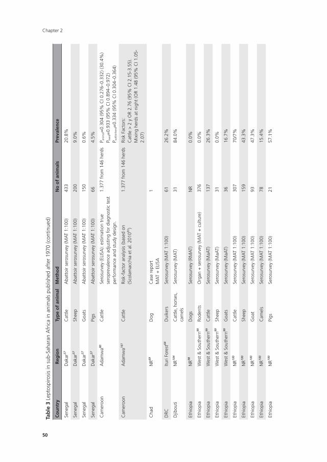
Chapter 2
50
Tab
le 3
Lep
tosp
irosi
s in
sub
-Sah
aran
Afr
ica
in a
nim
als
publ
ishe
d af
ter
1970
(con
tinue
d)
Co
un
try
Reg
ion
Typ
e o
f an
imal
Met
ho
dn
o o
f an
imal
sPr
eval
ence
Sene
gal
Dak
ar57
Cat
tleA
batt
oir
sero
surv
ey (M
AT
1:10
0)43
320
.8%
Sene
gal
Dak
ar57
Shee
pA
batt
oir
sero
surv
ey (M
AT
1:10
0)20
09.
0%
Sene
gal
Dak
ar57
Goa
tsA
batt
oir
sero
surv
ey (M
AT
1:10
0)15
00.
6%
Sene
gal
Dak
ar57
Pigs
Aba
ttoi
r se
rosu
rvey
(MA
T 1:
100)
664.
5%
Cam
eroo
nA
dam
wa80
Cat
tleSe
rosu
rvey
(ELI
SA):
estim
atio
n tr
ue
sero
prev
alen
ce a
djus
ting
for
diag
nost
ic t
est
perf
orm
ance
and
stu
dy d
esig
n.
1.37
7 fr
om 1
46 h
erds
P ani
mal=
0.30
4 (9
5% C
I 0.2
76–0
.332
) (30
.4%
)P h
erd=
0.93
3 (9
5% C
I 0.8
94–0
.972
)P w
ithin
herd=
0.33
4 (9
5% C
I 0.3
04–0
.364
)
Cam
eroo
nA
dam
wa16
1C
attle
Risk
-fac
tor
anal
ysis
(bas
ed o
n (S
cola
mac
chia
et
al. 2
01080
)1.
377
from
146
her
dsRi
sk F
acto
rs:
Cat
tle >
2 y
OR
2.76
(95%
CI 2
.15-
3.55
). M
ixin
g he
rds
at n
ight
(OR
1.48
(95%
CI 1
.05-
2.07
)
Cha
dN
R69D
ogC
ase
repo
rtM
AT
+ E
LISA
1
DRC
Ituri
Fore
st64
Dui
kers
Sero
surv
ey (M
AT
1:10
0)61
26.2
%
Djib
outi
NR10
4C
attle
, hor
ses,
ca
mel
sSe
rosu
rvey
(MA
T)31
84.0
%
Ethi
opia
NR98
Dog
sSe
rosu
rvey
(RM
AT)
NR
0.0%
Ethi
opia
Wes
t &
Sou
ther
n99Ro
dent
sO
rgan
+ s
eros
urve
y (M
AT
+ c
ultu
re)
376
0.0%
Ethi
opia
Wes
t &
Sou
ther
n99C
attle
Sero
surv
ey (M
aAT)
137
26.3
%
Ethi
opia
Wes
t &
Sou
ther
n99Sh
eep
Sero
surv
ey (M
aAT)
310.
0%
Ethi
opia
Wes
t &
Sou
ther
n99G
oats
Sero
surv
ey (M
aAT)
3616
.7%
Ethi
opia
NR10
0C
attle
Sero
surv
ey (M
AT
1:10
0)30
770
/7%
Ethi
opia
NR10
0Sh
eep
Sero
surv
ey (M
AT
1:10
0)15
943
.3%
Ethi
opia
NR10
0G
oat
Sero
surv
ey (M
AT
1:10
0)93
47.3
%
Ethi
opia
NR10
0C
amel
sSe
rosu
rvey
(MA
T 1:
100)
7815
.4%
Ethi
opia
NR10
0Pi
gsSe
rosu
rvey
(MA
T 1:
100)
2157
.1%
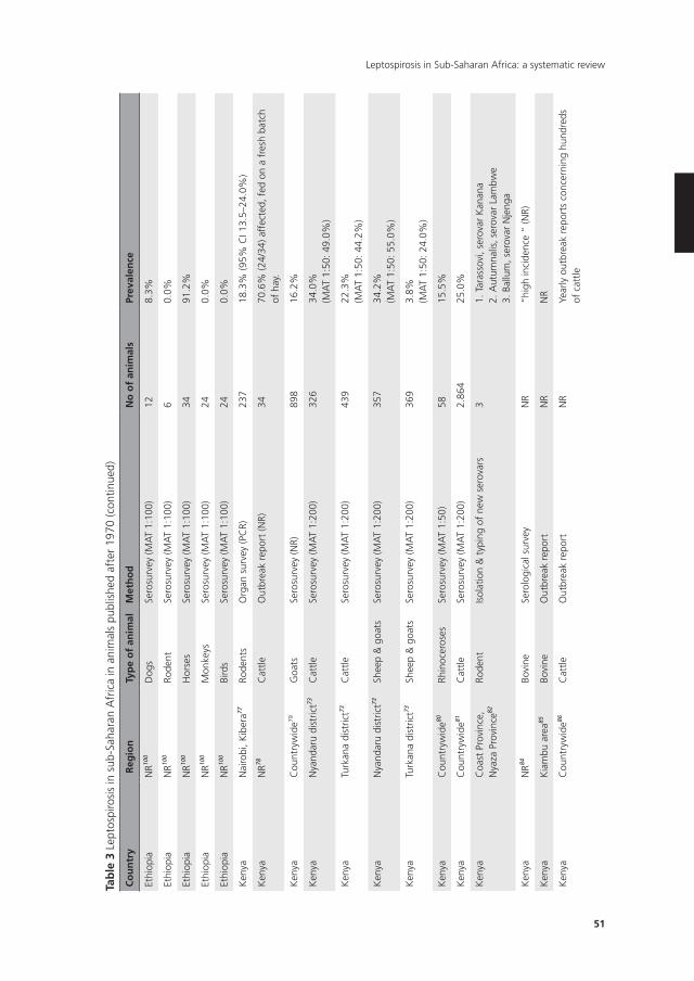
51
Leptospirosis in Sub-Saharan Africa: a systematic review
Tab
le 3
Lep
tosp
irosi
s in
sub
-Sah
aran
Afr
ica
in a
nim
als
publ
ishe
d af
ter
1970
(con
tinue
d)
Co
un
try
Reg
ion
Typ
e o
f an
imal
Met
ho
dn
o o
f an
imal
sPr
eval
ence
Ethi
opia
NR10
0D
ogs
Sero
surv
ey (M
AT
1:10
0)12
8.3%
Ethi
opia
NR10
0Ro
dent
Sero
surv
ey (M
AT
1:10
0)6
0.0%
Ethi
opia
NR10
0H
orse
sSe
rosu
rvey
(MA
T 1:
100)
3491
.2%
Ethi
opia
NR10
0M
onke
ysSe
rosu
rvey
(MA
T 1:
100)
240.
0%
Ethi
opia
NR10
0Bi
rds
Sero
surv
ey (M
AT
1:10
0)24
0.0%
Ken
yaN
airo
bi, K
iber
a77Ro
dent
sO
rgan
sur
vey
(PC
R)23
718
.3%
(95%
CI 1
3.5–
24.0
%)
Ken
yaN
R78C
attle
Out
brea
k re
port
(NR)
3470
.6%
(24/
34) a
ffec
ted,
fed
on
a fr
esh
batc
h of
hay
.
Ken
yaC
ount
ryw
ide79
Goa
tsSe
rosu
rvey
(NR)
898
16.2
%
Ken
yaN
yand
aru
dist
rict73
Cat
tleSe
rosu
rvey
(MA
T 1:
200)
326
34.0
%(M
AT
1:50
: 49.
0%)
Ken
yaTu
rkan
a di
stric
t73C
attle
Sero
surv
ey (M
AT
1:20
0)43
922
.3%
(MA
T 1:
50: 4
4.2%
)
Ken
yaN
yand
aru
dist
rict73
Shee
p &
goa
tsSe
rosu
rvey
(MA
T 1:
200)
357
34.2
%(M
AT
1:50
: 55.
0%)
Ken
yaTu
rkan
a di
stric
t73Sh
eep
& g
oats
Sero
surv
ey (M
AT
1:20
0)36
93.
8%(M
AT
1:50
: 24.
0%)
Ken
yaC
ount
ryw
ide80
Rhin
ocer
oses
Sero
surv
ey (M
AT
1:50
)58
15.5
%
Ken
yaC
ount
ryw
ide81
Cat
tleSe
rosu
rvey
(MA
T 1:
200)
2.86
425
.0%
Ken
yaC
oast
Pro
vinc
e,
Nya
za P
rovi
nce82
Rode
ntIs
olat
ion
& t
ypin
g of
new
ser
ovar
s3
1. T
aras
sovi
, ser
ovar
Kan
ana
2. A
utum
nalis
, ser
ovar
Lam
bwe
3. B
allu
m, s
erov
ar N
jeng
a
Ken
yaN
R84Bo
vine
Sero
logi
cal s
urve
yN
R“h
igh
inci
denc
e “
(NR)
Ken
yaK
iam
bu a
rea85
Bovi
neO
utbr
eak
repo
rtN
RN
R
Ken
yaC
ount
ryw
ide86
Cat
tleO
utbr
eak
repo
rtN
RYe
arly
out
brea
k re
port
s co
ncer
ning
hun
dred
s of
cat
tle
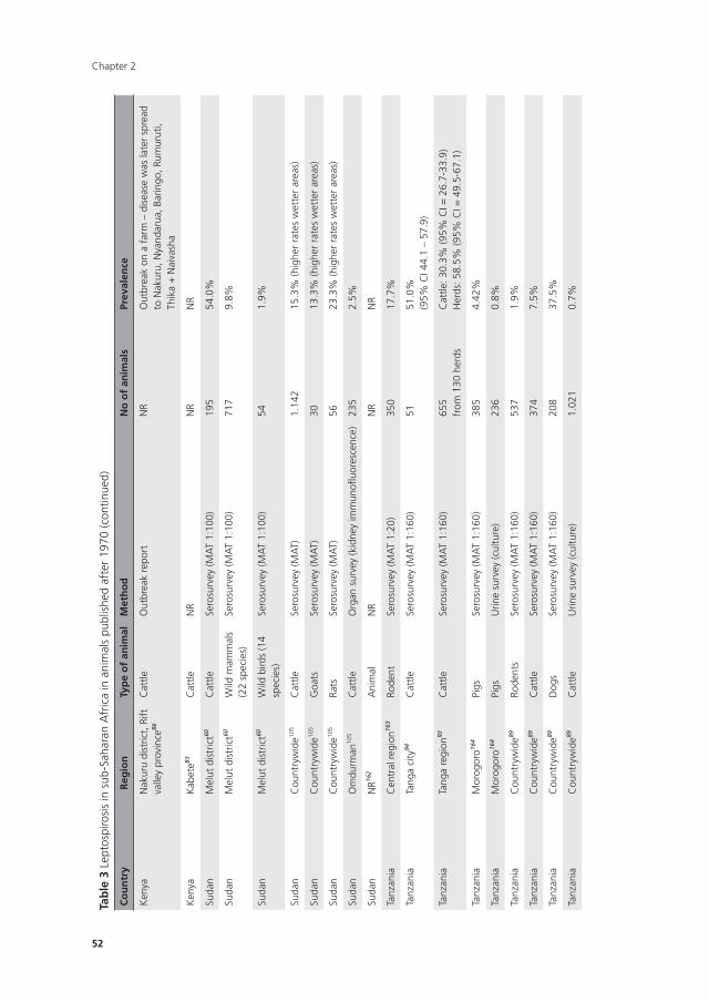
Chapter 2
52
Tab
le 3
Lep
tosp
irosi
s in
sub
-Sah
aran
Afr
ica
in a
nim
als
publ
ishe
d af
ter
1970
(con
tinue
d)
Co
un
try
Reg
ion
Typ
e o
f an
imal
Met
ho
dn
o o
f an
imal
sPr
eval
ence
Ken
yaN
akur
u di
stric
t, R
ift
valle
y pr
ovin
ce86
Cat
tleO
utbr
eak
repo
rtN
RO
utbr
eak
on a
far
m –
dis
ease
was
late
r sp
read
to
Nak
uru,
Nya
ndar
ua, B
arin
go, R
umur
uti,
Thik
a +
Nai
vash
a
Ken
yaK
abet
e83C
attle
NR
NR
NR
Suda
nM
elut
dis
tric
t60C
attle
Sero
surv
ey (M
AT
1:10
0)19
554
.0%
Suda
nM
elut
dis
tric
t60W
ild m
amm
als
(22
spec
ies)
Sero
surv
ey (M
AT
1:10
0)71
79.
8%
Suda
nM
elut
dis
tric
t60W
ild b
irds
(14
spec
ies)
Sero
surv
ey (M
AT
1:10
0)54
1.9%
Suda
nC
ount
ryw
ide10
5C
attle
Sero
surv
ey (M
AT)
1.14
215
.3%
(hig
her
rate
s w
ette
r ar
eas)
Suda
nC
ount
ryw
ide10
5G
oats
Sero
surv
ey (M
AT)
3013
.3%
(hig
her
rate
s w
ette
r ar
eas)
Suda
nC
ount
ryw
ide10
5Ra
tsSe
rosu
rvey
(MA
T)56
23.3
% (h
ighe
r ra
tes
wet
ter
area
s)
Suda
nO
mdu
rman
105
Cat
tleO
rgan
sur
vey
(kid
ney
imm
unofl
uore
scen
ce)
235
2.5%
Suda
nN
R162
Ani
mal
NR
NR
NR
Tanz
ania
Cen
tral
reg
ion16
3Ro
dent
Sero
surv
ey (M
AT
1:20
)35
017
.7%
Tanz
ania
Tang
a ci
ty94
Cat
tleSe
rosu
rvey
(MA
T 1:
160)
5151
.0%
(95%
CI 4
4.1
– 57
.9)
Tanz
ania
Tang
a re
gion
93C
attle
Sero
surv
ey (M
AT
1:16
0)65
5fr
om 1
30 h
erds
Cat
tle: 3
0.3%
(95%
CI =
26.
7-33
.9)
Her
ds: 5
8.5%
(95%
CI =
49.
5-67
.1)
Tanz
ania
Mor
ogor
o164
Pigs
Sero
surv
ey (M
AT
1:16
0)38
54.
42%
Tanz
ania
Mor
ogor
o164
Pigs
Urin
e su
rvey
(cul
ture
)23
60.
8%
Tanz
ania
Cou
ntry
wid
e89Ro
dent
sSe
rosu
rvey
(MA
T 1:
160)
537
1.9%
Tanz
ania
Cou
ntry
wid
e89C
attle
Sero
surv
ey (M
AT
1:16
0)37
47.
5%
Tanz
ania
Cou
ntry
wid
e89D
ogs
Sero
surv
ey (M
AT
1:16
0)20
837
.5%
Tanz
ania
Cou
ntry
wid
e89C
attle
Urin
e su
rvey
(cul
ture
)1.
021
0.7%
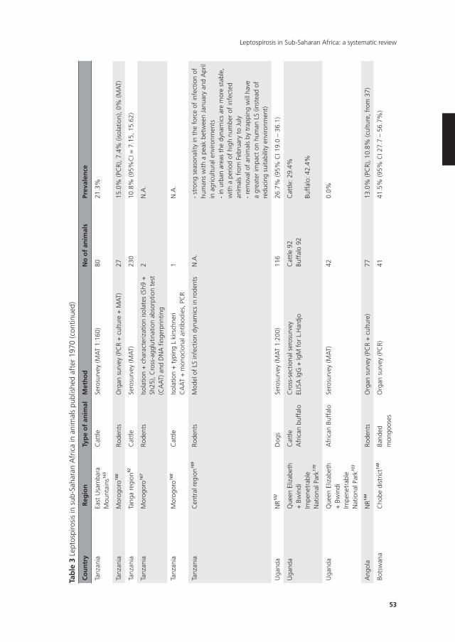
53
Leptospirosis in Sub-Saharan Africa: a systematic review
Tab
le 3
Lep
tosp
irosi
s in
sub
-Sah
aran
Afr
ica
in a
nim
als
publ
ishe
d af
ter
1970
(con
tinue
d)
Co
un
try
Reg
ion
Typ
e o
f an
imal
Met
ho
dn
o o
f an
imal
sPr
eval
ence
Tanz
ania
East
Usa
mba
ra
Mou
ntai
ns16
5
Cat
tleSe
rosu
rvey
(MA
T 1:
160)
8021
.3%
Tanz
ania
Mor
ogor
o166
Rode
nts
Org
an s
urve
y (P
CR
+ c
ultu
re +
MA
T)27
15.0
% (P
CR)
, 7.4
% (i
sola
tion)
, 0%
(MA
T)
Tanz
ania
Tang
a re
gion
92C
attle
Sero
surv
ey (M
AT)
230
10.8
% (9
5%C
I = 7
.15,
15.
62)
Tanz
ania
Mor
ogor
o167
Rode
nts
Isol
atio
n +
cha
ract
eriz
atio
n is
olat
es (S
h9 +
Sh
25),
Cro
ss-a
gglu
tinat
ion
abso
rptio
n te
st
(CA
AT)
and
DN
A fi
nger
prin
ting
2N
.A.
Tanz
ania
Mor
ogor
o168
Cat
tleIs
olat
ion
+ t
ypin
g L
kirs
chne
riC
AA
T +
mon
oclo
nal a
ntib
odie
s, P
CR
1N
.A.
Tanz
ania
Cen
tral
reg
ion16
9Ro
dent
sM
odel
of
LS in
fect
ion
dyna
mic
s in
rod
ents
N.A
.-
stro
ng s
easo
nalit
y in
the
for
ce o
f in
fect
ion
of
hum
ans
with
a p
eak
betw
een
Janu
ary
and
Apr
il in
agr
icul
tura
l env
ironm
ents
- in
urb
an a
reas
the
dyn
amic
s ar
e m
ore
stab
le,
with
a p
erio
d of
hig
h nu
mbe
r of
infe
cted
an
imal
s fr
om F
ebru
ary
to J
uly
- re
mov
al o
f an
imal
s by
tra
ppin
g w
ill h
ave
a gr
eate
r im
pact
on
hum
an L
S (in
stea
d of
re
duci
ng s
uita
bilit
y en
viro
nmen
t)
Uga
nda
NR10
2D
ogs
Sero
surv
ey (M
AT
1:20
0)11
626
.7%
(95%
CI 1
9.0
– 36
.1)
Uga
nda
Que
en E
lizab
eth
+ B
win
di
Impe
netr
able
N
atio
nal P
ark17
0
Cat
tleA
fric
an b
uffa
loC
ross
-sec
tiona
l ser
osur
vey
ELIS
A Ig
G +
IgM
for
L.H
ardj
oC
attle
92
Buff
alo
92C
attle
: 29.
4%
Buff
alo:
42.
4%
Uga
nda
Que
en E
lizab
eth
+ B
win
di
Impe
netr
able
N
atio
nal P
ark10
3
Afr
ican
Buf
falo
Sero
surv
ey (M
AT)
420.
0%
Ang
ola
NR14
4Ro
dent
sO
rgan
sur
vey
(PC
R +
cul
ture
)77
13.0
% (P
CR)
, 10.
8% (c
ultu
re, f
rom
37)
Bots
wan
aC
hobe
dis
tric
t148
Band
ed
mon
goos
esO
rgan
sur
vey
(PC
R)41
41.5
% (9
5% C
I 27.
7 –
56.7
%)
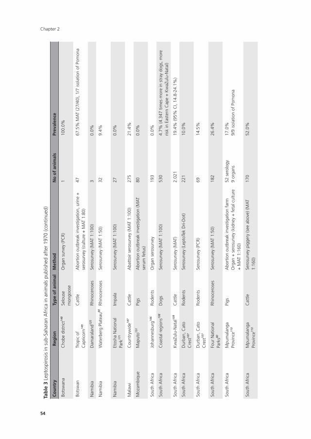
Chapter 2
54
Tab
le 3
Lep
tosp
irosi
s in
sub
-Sah
aran
Afr
ica
in a
nim
als
publ
ishe
d af
ter
1970
(con
tinue
d)
Co
un
try
Reg
ion
Typ
e o
f an
imal
Met
ho
dn
o o
f an
imal
sPr
eval
ence
Bots
wan
aC
hobe
dis
tric
t148
Selo
use
mon
goos
eO
rgan
sur
vey
(PC
R)1
100.
0%
Bots
wan
Trop
ic o
f C
apric
orn14
9
Cat
tleA
bort
ion
outb
reak
inve
stig
atio
n, u
rine
+
sero
surv
ey (c
ultu
re +
MA
T 1:
80)
4767
.5%
MA
T (2
7/40
), 1/
7 is
olat
ion
of P
omon
a
Nam
ibia
Dam
aral
and12
5Rh
inoc
eros
esSe
rosu
rvey
(MA
T 1:
100)
30.
0%
Nam
ibia
Wat
erbe
rg P
late
au80
Rhin
ocer
oses
Sero
surv
ey (M
AT
1:50
)32
9.4%
Nam
ibia
Etos
ha N
atio
nal
Park
171
Impa
laSe
rosu
rvey
(MA
T 1:
100)
270.
0%
Mal
awi
Cou
ntry
wid
e147
Cat
tleA
batt
oir
sero
surv
ey (M
AT
1:10
0)27
521
.4%
Moz
ambi
que
Map
uto15
1Pi
gsA
bort
ion
outb
reak
inve
stig
atio
n (M
AT
seru
m f
etus
)80
0.0%
Sout
h A
fric
aJo
hann
esbu
rg10
6Ro
dent
sO
rgan
ser
osur
vey
193
0.0%
Sout
h A
fric
aC
oast
al r
egio
ns10
8D
ogs
Sero
surv
ey (M
AT
1:10
0)53
04.
7% (4
.347
tim
es m
ore
in s
tray
dog
s, m
ore
risk
in E
aste
rn C
ape
+ K
waZ
ulu-
Nat
al)
Sout
h A
fric
aK
waZ
ulu-
Nat
al10
9C
attle
Sero
surv
ey (M
AT)
2.02
119
.4%
(95%
CI,
14.8
-24.
1%)
Sout
h A
fric
aD
urba
n, C
ato
Cre
st10
7
Rode
nts
Sero
surv
ey (L
epto
Tek
Dri-
Dot
)22
110
.0%
Sout
h A
fric
aD
urba
n, C
ato
Cre
st10
7
Rode
nts
Sero
surv
ey (P
CR)
6914
.5%
Sout
h A
fric
aFo
ur N
atio
nal
Park
s80
Rhin
ocer
oses
Sero
surv
ey (M
AT
1:50
)18
226
.4%
Sout
h A
fric
aM
pum
alan
ga
Prov
ince
110
Pigs
Abo
rtio
n ou
tbre
ak in
vest
igat
ion
farm
Org
an +
ser
osur
vey
(kid
ney
+ f
etal
cul
ture
+
MA
T 1:
160)
52 s
erol
ogy
9 or
gans
17.0
%9/
9 is
olat
ion
of P
omon
a
Sout
h A
fric
aM
pum
alan
ga
Prov
ince
110
Cat
tleSe
rosu
rvey
pig
gery
(see
abo
ve) (
MA
T 1:
160)
170
52.0
%
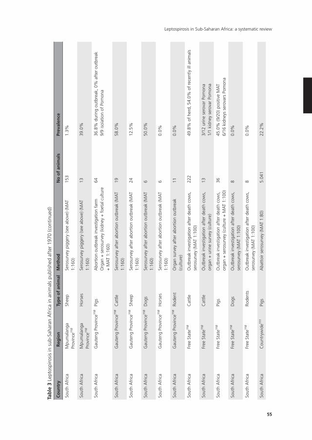
55
Leptospirosis in Sub-Saharan Africa: a systematic review
Tab
le 3
Lep
tosp
irosi
s in
sub
-Sah
aran
Afr
ica
in a
nim
als
publ
ishe
d af
ter
1970
(con
tinue
d)
Co
un
try
Reg
ion
Typ
e o
f an
imal
Met
ho
dn
o o
f an
imal
sPr
eval
ence
Sout
h A
fric
aM
pum
alan
ga
Prov
ince
110
Shee
pSe
rosu
rvey
pig
gery
(see
abo
ve) (
MA
T 1:
160)
153
1.3%
Sout
h A
fric
aM
pum
alan
ga
Prov
ince
110
Hor
ses
Sero
surv
ey p
igge
ry (s
ee a
bove
) (M
AT
1:16
0)13
39.0
%
Sout
h A
fric
aG
aute
ng P
rovi
nce11
0Pi
gsA
bort
ion
outb
reak
inve
stig
atio
n fa
rmO
rgan
+ s
eros
urve
y (k
idne
y +
foe
tal c
ultu
re
+ M
AT
1:16
0)
6436
.8%
dur
ing
outb
reak
, 0%
aft
er o
utbr
eak
9/9
isol
atio
n of
Pom
ona
Sout
h A
fric
aG
aute
ng P
rovi
nce11
0C
attle
Sero
surv
ey a
fter
abo
rtio
n ou
tbre
ak (M
AT
1:16
0)19
58.0
%
Sout
h A
fric
aG
aute
ng P
rovi
nce11
0Sh
eep
Sero
surv
ey a
fter
abo
rtio
n ou
tbre
ak (M
AT
1:16
0)24
12.5
%
Sout
h A
fric
aG
aute
ng P
rovi
nce11
0D
ogs
Sero
surv
ey a
fter
abo
rtio
n ou
tbre
ak (M
AT
1:16
0)6
50.0
%
Sout
h A
fric
aG
aute
ng P
rovi
nce11
0H
orse
sSe
rosu
rvey
aft
er a
bort
ion
outb
reak
(MA
T 1:
160)
60.
0%
Sout
h A
fric
aG
aute
ng P
rovi
nce11
0Ro
dent
Org
an s
urve
y af
ter
abor
tion
outb
reak
(c
ultu
re)
110.
0%
Sout
h A
fric
aFr
ee S
tate
110
Cat
tleO
utbr
eak
inve
stig
atio
n af
ter
deat
h co
ws,
se
rosu
rvey
(MA
T 1:
100)
222
49.8
% o
f he
rd, 5
4.0%
of
rece
ntly
ill a
nim
als
Sout
h A
fric
aFr
ee S
tate
110
Cat
tleO
utbr
eak
inve
stig
atio
n af
ter
deat
h co
ws,
or
gan
+ u
rine
surv
ey (c
ultu
re)
133/
12 u
rine
sero
var
Pom
ona
1/1
kidn
ey s
erov
ar P
omon
a
Sout
h A
fric
aFr
ee S
tate
110
Pigs
Out
brea
k in
vest
igat
ion
afte
r de
ath
cow
s,
orga
n +
ser
osur
vey
(cul
ture
+ M
AT
1:10
0)36
45.0
% (9
/20)
pos
itive
MA
T6/
16 k
idne
ys s
erov
ars
Pom
ona
Sout
h A
fric
aFr
ee S
tate
110
Dog
sO
utbr
eak
inve
stig
atio
n af
ter
deat
h co
ws,
se
rosu
rvey
(MA
T 1:
100)
80.
0%
Sout
h A
fric
aFr
ee S
tate
110
Rode
nts
Out
brea
k in
vest
igat
ion
afte
r de
ath
cow
s,
sero
surv
ey (M
AT
1:10
0)8
0.0%
Sout
h A
fric
aC
ount
ryw
ide11
1Pi
gsA
batt
oir
sero
surv
ey (M
AT
1:80
)5.
041
22.2
%
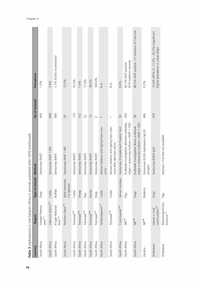
Chapter 2
56
Tab
le 3
Lep
tosp
irosi
s in
sub
-Sah
aran
Afr
ica
in a
nim
als
publ
ishe
d af
ter
1970
(con
tinue
d)
Co
un
try
Reg
ion
Typ
e o
f an
imal
Met
ho
dn
o o
f an
imal
sPr
eval
ence
Sout
h A
fric
aG
reat
er P
reto
ria
area
112
Dog
sSe
rosu
rvey
(MA
T)40
01.
5%
Sout
h A
fric
aVo
lksr
ust
dist
rict11
3C
attle
Sero
surv
ey (M
AT
1:80
)86
03.
0%
Sout
h A
fric
aK
ruge
r N
atio
nal
Park
114
Buff
alo
Sero
surv
ey (M
AT)
406
1.7%
(6.6
% in
conc
lusi
ve)
Sout
h A
fric
aN
orth
ern
Nat
al11
5W
ild m
amm
als
(12
spec
ies)
Sero
surv
ey (M
AT
1:40
)50
12.0
%
Sout
h A
fric
aTr
ansv
aal11
6C
attle
Sero
surv
ey (M
AT)
170
52.0
%
Sout
h A
fric
aTr
ansv
aal11
6Sh
eep
Sero
surv
ey (M
AT)
152
1.0%
Sout
h A
fric
aTr
ansv
aal11
6Pi
gsSe
rosu
rvey
(MA
T)52
17.0
%
Sout
h A
fric
aTr
ansv
aal11
6H
orse
sSe
rosu
rvey
(MA
T)13
38.0
%
Sout
h A
fric
aTr
ansv
aal11
6D
ogs
Sero
surv
ey (M
AT)
210
0.0%
Sout
h A
fric
aO
nder
step
oort
117
Cat
tleSe
rova
r is
olat
ion
and
typi
ng f
rom
cow
-ur
ine
1N
.A.
Sout
h A
fric
aTr
ansv
aal11
8C
attle
Sero
var
isol
atio
n an
d ty
ping
fro
m c
ow-
urin
e af
ter
abor
tion
stor
m1
N.A
.
Sout
h A
fric
aC
ount
ryw
ide11
9Ve
rvet
mon
keys
Sero
surv
ey (C
ompl
emen
t Fi
xatio
n Te
st)
508.
0%
Sout
h A
fric
aN
R120
Pigs
Out
brea
k in
vest
igat
ion
abor
tion
outb
reak
, or
gan
+ s
eros
urve
y (c
ultu
re +
MA
T 1:
100)
250
62.1
% M
AT
posi
tive
6/10
isol
atio
n ca
nico
la
Sout
h A
fric
aN
R120
Dog
sO
utbr
eak
inve
stig
atio
n ill
ness
out
brea
k,
orga
n +
ser
osur
vey
(cul
ture
+ M
AT
1:10
0)38
86.5
% M
AT
posi
tive,
1/1
isol
atio
n of
Can
icol
a
Zam
bia
NR14
5Ro
dent
sSe
rosu
rvey
(ELI
SA L
epto
spira
rLi
pL32
an
tigen
)46
60.
2%
Zim
babw
eH
arar
e +
rur
al
com
mun
ities
122
Dog
sSe
rosu
rvey
(ELI
SA Ig
G)
250
15.6
% (9
5% C
I: 11
.0%
- 2
0.2%
) (si
gnifi
cant
hi
gher
pre
vale
nce
in u
rban
dog
s)
Zim
babw
eM
asho
nala
nd E
ast
Prov
ince
123
Pigs
Abs
trac
t +
ful
l tex
t no
t av
aila
ble
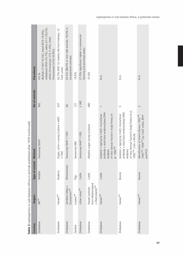
57
Leptospirosis in Sub-Saharan Africa: a systematic review
Tab
le 3
Lep
tosp
irosi
s in
sub
-Sah
aran
Afr
ica
in a
nim
als
publ
ishe
d af
ter
1970
(con
tinue
d)
Co
un
try
Reg
ion
Typ
e o
f an
imal
Met
ho
dn
o o
f an
imal
sPr
eval
ence
Zim
babw
eN
R124
Wild
life
Sero
surv
ey (M
AT)
992
4.6
%(B
uffa
lo 2
1/34
7 (6
.0%
), el
and
8/74
(10.
8%),
wild
ebee
st 9
/54
(16.
7%),
zebr
a 2/
11 (1
8.2%
), w
hite
rhi
noce
rose
s 1/
72 (1
.4%
), bl
ack
rhin
ocer
oses
5/1
02 (4
.9%
))
Zim
babw
eH
arar
e121
Rode
nts
Org
an, u
rine
+ s
urve
y (c
ultu
re +
MA
T 1:
100)
437
62.5
% M
AT,
52
isol
ates
(40
from
kid
neys
, 12
from
urin
e)
Zim
babw
eZa
mbe
zi V
alle
y +
Es
carp
men
t125
Rhin
ocer
oses
Sero
surv
ey (M
AT
1:10
0)60
63.0
% (8
8.0%
in r
iver
sid
e an
imal
s, 1
8.0%
in
esca
rpm
ents
ani
mal
s)
Zam
bia
Lusa
ka12
6Pi
gsSe
rosu
rvey
(NR)
121
14.9
%
Zim
babw
eU
rban
are
as12
7C
attle
Sero
surv
ey (M
AT
1:10
0)2.
382
27.0
% (s
igni
fican
t hi
gher
in c
omm
erci
al
farm
ing
and
purc
hase
are
as)
Zim
babw
eH
arar
e (a
nim
als
from
Mas
hona
land
+
Man
ical
and)
129
Cat
tleA
batt
oir
orga
n su
rvey
(cul
ture
)48
010
.4%
Zim
babw
eH
arar
e130
Cat
tleIs
olat
ion
+ t
ypin
g by
CA
AT,
mon
oclo
nal
antib
ody
+ r
estr
ictio
n en
donu
clea
se D
NA
an
alys
esof
new
Ser
ovar
fou
nd in
stu
dy F
eres
u et
al
. 199
2129
1N
.A.
Zim
babw
eH
arar
e131
Bovi
neIs
olat
ion
+ t
ypin
g by
CA
AT,
mon
oclo
nal
antib
ody
+ r
estr
ictio
n en
donu
clea
se D
NA
an
alys
esof
new
Ser
ovar
fou
nd in
stu
dy F
eres
u et
al.
1992
129 . C
AA
+ R
FLPA
5N
.A.
Zim
babw
eH
arar
e132
Bovi
neId
entifi
catio
n of
str
ains
(Fer
esu
1992
129 ,
1993
130 , 1
99413
1 ) by
CA
AT
mA
bs, R
FLP
and
PCR
2N
.A.
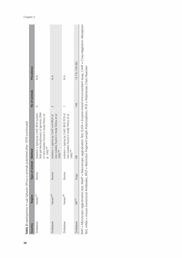
Chapter 2
58
Tab
le 3
Lep
tosp
irosi
s in
sub
-Sah
aran
Afr
ica
in a
nim
als
publ
ishe
d af
ter
1970
(con
tinue
d)
Co
un
try
Reg
ion
Typ
e o
f an
imal
Met
ho
dn
o o
f an
imal
sPr
eval
ence
Zim
babw
eH
arar
e133
Bovi
neIs
olat
ion
+ t
ypin
g by
CA
AT,
RFL
P, p
ulse
d-fie
ld g
el e
lect
roph
ores
is o
f ge
nom
ic D
NA
of n
ew is
olat
es f
ound
in s
tudy
Fer
esu
et
al. 1
99212
9
4N
.A.
Zim
babw
eH
arar
e134
Bovi
neIs
olat
ion
+ t
ypin
g by
CA
AT
and
RFLP
of
new
isol
ates
fou
nd in
stu
dy F
eres
u et
al.
1992
129
2N
.A.
Zim
babw
eH
arar
e136
Bovi
neIs
olat
ion
+ t
ypin
g by
CA
AT,
RFL
P, P
CR
of
new
isol
ates
fou
nd in
stu
dy F
eres
u et
al.
1992
129
1N
.A.
Zim
babw
eN
R137
Dog
sN
R14
613
.7%
(19/
146)
MA
T =
Mic
rosc
opic
agg
lutin
atio
n te
st, M
aAT
= M
acro
scop
ic a
gglu
tinat
ion
Test
, ELI
SA =
Enz
yme-
linke
d im
mun
osor
bent
ass
ay, C
AA
T =
Cro
ss-A
gglu
tinin
Abs
orpt
ion
Test
, mA
bs =
mou
se m
onoc
lona
l Ant
ibod
ies,
RFL
P =
Res
tric
tion
Frag
men
t Le
ngth
Pol
ymor
phis
m, P
CR
= P
olym
eras
e C
hain
Rea
ctio
n
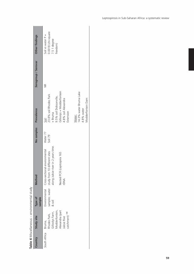
59
Leptospirosis in Sub-Saharan Africa: a systematic review
Tab
le 4
Mis
cella
neou
s –
envi
ronm
enta
l stu
dy
Co
un
try
Stu
dy
site
Typ
e o
f sa
mp
leM
eth
od
no
sam
ple
sPr
eval
ence
Sero
gro
up
/ S
ero
var
Oth
er fi
nd
ing
s
Sout
h A
fric
aBr
uma,
Rh
odes
Par
k,
Gill
ooly
s Fa
rm,
Mod
derf
onte
in,
Ale
xand
ra (p
art
Juks
ie R
ive
catc
hmen
t) 10
6
Envi
ronm
enta
l sa
mpl
es: w
ater
&
soi
l
Cro
ss-s
ectio
nal e
nviro
nmen
tal
stud
y fr
om 1
0 di
ffer
ent
site
s al
ong
Juks
ie r
iver
in 2
yea
rs t
ime.
Nes
ted
PCR
(Lep
tosp
ira 1
6S
rDN
A
Wat
er 7
7So
il 79
Soil:
23.8
% s
oil R
hode
s Pa
rk
+ B
rum
a9.
5% s
oil D
obso
nvill
e,
Gill
ooly
s +
Mod
derf
onte
in4.
8% s
oil A
lexa
ndra
Ex
tens
ion
Wat
er:
14.3
% w
ater
Bru
ma
Lake
4.8%
wat
er
Mod
derf
onte
in D
am
NR
Soil
vs w
ater
P =
0.
0018
(chi
-squ
are
7.5
1 de
gree
fr
eedo
m)
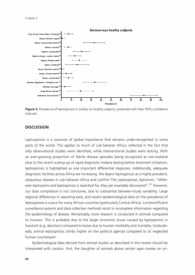
Chapter 2
60
Figure 5. Prevalence of leptospirosis in studies on healthy subjects, presented with their 95% confi dence intervals.
DISCUSSIOn
Leptospirosis is a zoonosis of global importance that remains under-recognized in some
parts of the world. This applies to much of sub-Saharan Africa, refl ected in the fact that
only observational studies were identifi ed, while interventional studies were lacking. With
an ever-growing proportion of febrile disease episodes being recognized as non-malarial
(due to the recent scaling-up of rapid diagnostic malaria testing before treatment initiation),
leptospirosis is highlighted as one important differential diagnosis. Additionally, adequate
diagnostic facilities across Africa are increasing. We depict leptospirosis as a highly prevalent,
ubiquitous disease in sub-Saharan Africa and confi rm The Leptospirosis Aphorism, “Wher-
ever leptospires and leptospirosis is searched for, they are invariably discovered”.157 However,
our data compilation is not conclusive, due to substantial between-study variability. Large
regional differences in reporting exist, and recent epidemiological data on the prevalence of
leptospirosis is scarce for many African countries (particularly Central Africa). Limited effi cient
surveillance systems and data collection methods result in incomplete information regarding
the epidemiology of disease. Remarkably, more research is conducted in animals compared
to humans. This is probably due to the larger economic losses caused by leptospirosis in
livestock (e.g. abortion) compared to losses due to human morbidity and mortality. Undoubt-
edly, animal leptospirosis climbs higher on the political agenda compared to its neglected
human counterpart.
Epidemiological data derived from animal studies as described in this review should be
interpreted with caution. First, the slaughter of animals above certain ages creates an un-
61
Leptospirosis in Sub-Saharan Africa: a systematic review
avoidable selection bias in livestock surveys (often abattoirs). This gives a distorted prevalence
because higher seroprevalence of leptospirosis is related to older host ages. Second, preva-
lence and incidence of leptospirosis fluctuates; there is usually more leptospirosis during, and
just after the rainy and harvest season. Often, studies did not report the season and time
frame of the study and are therefore not easily comparable to other studies. Finally, large
proportions of reservoir hosts have no antibodies against the serovar they carry. Thus, while
seroprevalence studies in healthy animal populations indicate levels of local infection expo-
sure, these are less suited to define their role as infection reservoirs. Therefore, assumptions
that the seroprevalence in animals corresponds with leptospirosis prevalence or incidence in
humans cannot easily be made. Animals with low seroprevalence may be a substantial cause
of infection in humans; high seroprevalence may indicate exposure pressure from different
animal populations, hence a high infection risk in humans as well.
In human studies, two types of survey are distinguished; studies establishing leptospiro-
sis prevalence as a cause of (mostly febrile) illness and background seroprevalence studies.
The background studies showed variable, but generally high seroprevalences, which were
elevated in risk groups. This does not necessarily mean a high disease burden; asymptomatic
seroconversion in young children in rural areas occurs,15 which may apply to most lepto-
spiral infections in general. Patient surveys show varying data. An equivalent exposure level
to leptospirosis does not necessarily result in the same prevalence of clinical disease, possibly
due to different immunity patterns and circulating serovars. Variations in inclusion criteria,
case definitions, and diagnostic tools make outcome data comparison between different
studies and regions challenging. In some of the original articles, the terms fever of unknown
origin (FUO) or pyrexia of unknown origin (PUO) have been used. With some degree of un-
certainty as to whether these classifications would match with the precise definition of what
constitutes FUO according to the current accepted definition, we decided to refer to those
conditions consistently as ‘undifferentiated febrile disease’ (UFD) in order to avoid confusion.
Further studies are required to elucidate the exact epidemiology of leptospirosis in sub-
Saharan Africa. Even in regions where leptospirosis research has been conducted, such as
East Africa, researchers believe that it remains under-diagnosed because of lack of awareness,
facilities, and surveillance systems. Many questions remain unanswered, and new questions
continue to arise (see Panel). Moreover, countries are confronted with larger scale epidemics
(HIV, tuberculosis, malaria), which consume already limited resources. Thus, leptospirosis can
be considered a neglected tropical disease in sub-Saharan Africa. Knowledge of the hu-
man leptospirosis burden has not yet translated into increased or strengthened control and
prevention programmes, nor is it regularly considered routinely as an important differential
diagnosis in non-malarial fevers.
In most countries, an adult predominance in clinical disease has been noted, which is not
reflected across all clinical surveys. The only incidence calculation, from Tanzania, showed a
higher (serological) incidence in younger age groups,87 which can only partly be explained
Chapter 2
62
by a lower hospital admission threshold for children. Immunological and genetic host factors
may play a particular role in the development of active disease. However, the same study
showed that patients with confirmed or probable leptospirosis were older than those without
leptospirosis;9 or there was no difference in age groups at all.158 Almost certainly, exposure
to leptospirosis starts at a young age and infection often remains unnoticed or undiagnosed.
The findings of this review are subject to methodological limitations. Heterogeneity be-
tween studies was high. Few studies reported sufficient outcome data to allow for population
comparisons. Novel diagnostic tests have shed more light on the prevalence of leptospirosis
and require further investigation. Although we assessed the quality of included studies, we
were unable to control for differences in study design and quality of microbiological data. For
many surveys, the prevalence of leptospirosis was not the primary outcome, making a formal
meta-analysis potentially misleading. Unpublished data was included in this review (confer-
ence abstracts), although controversy on this policy exists.159 We minimized bias by searching
clinical trial registries for ongoing or unpublished studies. Whilst no restrictions were placed
on the publication language, the focus of the majority of the search engines (Medline/
PubMed being the primary source of studies) is predominantly English. Other unpublished
data were not included; therefore, our findings may have a publication bias towards studies
that report high levels of concordance.
COnCLUSIOn
Leptospirosis is an important zoonosis in sub-Saharan Africa. The combination of climate
change, massive urbanisation, and the interdependence of humans and animals may be criti-
cal for the burden of leptospirosis on Africa’s people. The nature of leptospirosis highlights
the need for a fresh approach to control and prevention, incorporating the links between
veterinary and human medicine. Future leptospirosis research should be a collaborative effort
between the fields of human and animal medicine, to attain optimal health outcomes.
PAneL: PRESEnT KnOWLEDGE, UnAnSWERED QUESTIOnS AnD RESEARCH GAPS
Leptospirosis is common in sub-Saharan Africa, as shown by numerous observational studies
Which populations are most at risk? To what extent are leptospirosis cases misclassified as
malaria? Do more infections occur in poorly sanitized urban settings or in rural areas as
a result of occupational or environmental exposure? How prevalent is the disease in the
Central African region?
63
Leptospirosis in Sub-Saharan Africa: a systematic review
The increasing urbanisation and climate change are thought to be risk factors for future outbreaks of leptospirosis in sub-Saharan Africa
What public health or control measures should be taken? Will livestock vaccination, sanita-
tion measures and rodent control be enough to prevent larger outbreaks?
Data on the risk of acquiring leptospirosis related to age vary and remain unclear
Which age-groups are most at risk? Is disease vastly under recognized in children due to their
milder symptoms? What is the influence of acquired immunity?
Antibiotic treatment is recommended but there are no clear cut clinical trials to prove or refute the use of antibiotical treatment for leptospirosis,
An almost certain cause of some of the inconclusive studies is that patients in resource-poor
countries arrive for treatment with advanced disease: antibiotics are then unlikely to be of
benefit because organ damage has already occurred. How can this problem be avoided in
future clinical trials?
Contributors
MPG, SGV, and BJV conceived the paper. SGV wrote the first draft. BJV double-checked the
data. IMN conducted the searches and contributed to the methods section. All authors con-
tributed to the concept of the paper and all have been involved in drafting and revising the
manuscript; all have agreed to be fully accountable for all aspects of this work. All authors
read and approved the final manuscript.
Acknowledgments
We gratefully acknowledge Ramzi Amri (Academic Medical Center, University of Amsterdam,
The Netherlands) and Gerben Molenaar (Freelance professional post-production and anima-
tion) for their help designing the graphic. We also thank Samuel T. Greene (International
Institute for Environment and Development) for his contribution in English language editing
of the manuscript.
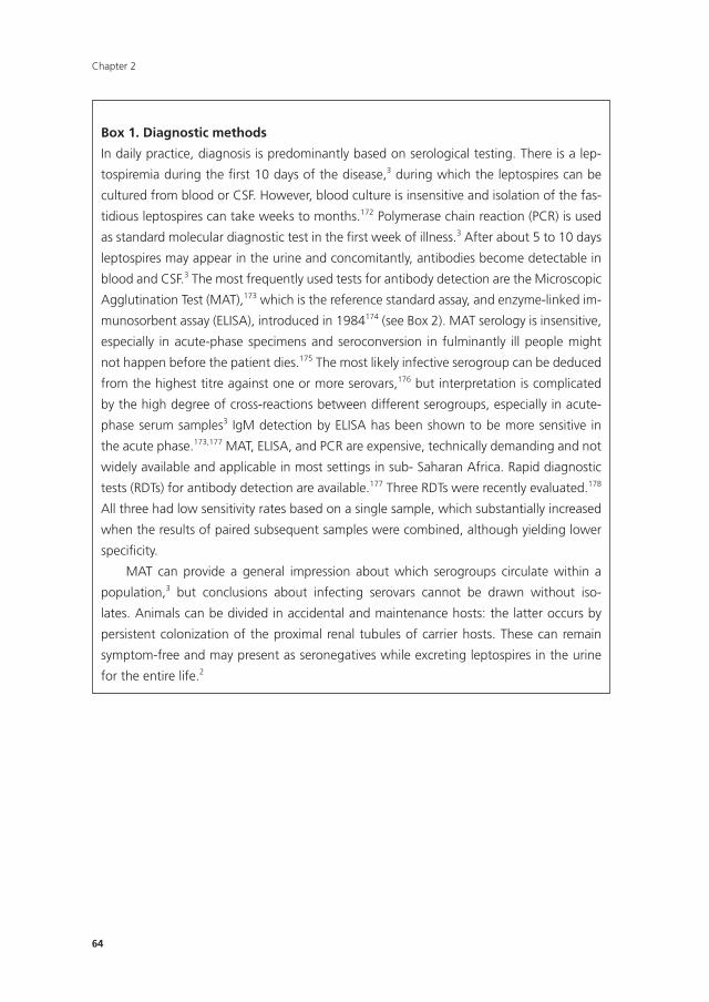
Chapter 2
64
Box 1. Diagnostic methods
In daily practice, diagnosis is predominantly based on serological testing. There is a lep-
tospiremia during the first 10 days of the disease,3 during which the leptospires can be
cultured from blood or CSF. However, blood culture is insensitive and isolation of the fas-
tidious leptospires can take weeks to months.172 Polymerase chain reaction (PCR) is used
as standard molecular diagnostic test in the first week of illness.3 After about 5 to 10 days
leptospires may appear in the urine and concomitantly, antibodies become detectable in
blood and CSF.3 The most frequently used tests for antibody detection are the Microscopic
Agglutination Test (MAT),173 which is the reference standard assay, and enzyme-linked im-
munosorbent assay (ELISA), introduced in 1984174 (see Box 2). MAT serology is insensitive,
especially in acute-phase specimens and seroconversion in fulminantly ill people might
not happen before the patient dies.175 The most likely infective serogroup can be deduced
from the highest titre against one or more serovars,176 but interpretation is complicated
by the high degree of cross-reactions between different serogroups, especially in acute-
phase serum samples3 IgM detection by ELISA has been shown to be more sensitive in
the acute phase.173,177 MAT, ELISA, and PCR are expensive, technically demanding and not
widely available and applicable in most settings in sub- Saharan Africa. Rapid diagnostic
tests (RDTs) for antibody detection are available.177 Three RDTs were recently evaluated.178
All three had low sensitivity rates based on a single sample, which substantially increased
when the results of paired subsequent samples were combined, although yielding lower
specificity.
MAT can provide a general impression about which serogroups circulate within a
population,3 but conclusions about infecting serovars cannot be drawn without iso-
lates. Animals can be divided in accidental and maintenance hosts: the latter occurs by
persistent colonization of the proximal renal tubules of carrier hosts. These can remain
symptom-free and may present as seronegatives while excreting leptospires in the urine
for the entire life.2
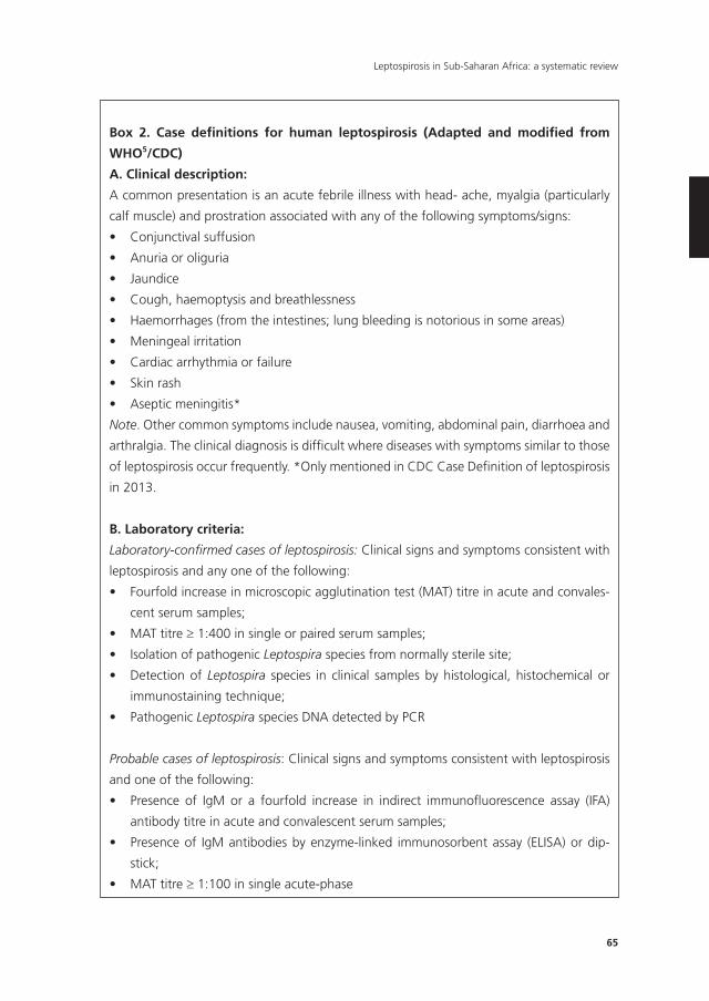
65
Leptospirosis in Sub-Saharan Africa: a systematic review
Box 2. Case definitions for human leptospirosis (Adapted and modified from
WHO5/CDC)
A. Clinical description:
A common presentation is an acute febrile illness with head- ache, myalgia (particularly
calf muscle) and prostration associated with any of the following symptoms/signs:
• Conjunctivalsuffusion
• Anuriaoroliguria
• Jaundice
• Cough,haemoptysisandbreathlessness
• Haemorrhages(fromtheintestines;lungbleedingisnotoriousinsomeareas)
• Meningealirritation
• Cardiacarrhythmiaorfailure
• Skinrash
• Asepticmeningitis*
Note. Other common symptoms include nausea, vomiting, abdominal pain, diarrhoea and
arthralgia. The clinical diagnosis is difficult where diseases with symptoms similar to those
of leptospirosis occur frequently. *Only mentioned in CDC Case Definition of leptospirosis
in 2013.
B. Laboratory criteria:
Laboratory-confirmed cases of leptospirosis: Clinical signs and symptoms consistent with
leptospirosis and any one of the following:
• Fourfoldincreaseinmicroscopicagglutinationtest(MAT)titreinacuteandconvales-
cent serum samples;
• MATtitre≥ 1:400 in single or paired serum samples;
• IsolationofpathogenicLeptospira species from normally sterile site;
• DetectionofLeptospira species in clinical samples by histological, histochemical or
immunostaining technique;
• PathogenicLeptospira species DNA detected by PCR
Probable cases of leptospirosis: Clinical signs and symptoms consistent with leptospirosis
and one of the following:
• Presence of IgM or a fourfold increase in indirect immunofluorescence assay (IFA)
antibody titre in acute and convalescent serum samples;
• Presenceof IgMantibodiesbyenzyme-linked immunosorbentassay (ELISA)ordip-
stick;
• MATtitre≥ 1:100 in single acute-phase
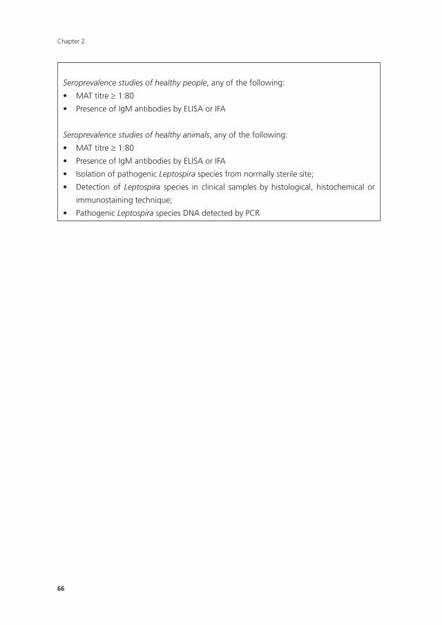
Chapter 2
66
Seroprevalence studies of healthy people, any of the following:
• MATtitre≥ 1:80
• PresenceofIgMantibodiesbyELISAorIFA
Seroprevalence studies of healthy animals, any of the following:
• MATtitre≥ 1:80
• PresenceofIgMantibodiesbyELISAorIFA
• IsolationofpathogenicLeptospira species from normally sterile site;
• DetectionofLeptospira species in clinical samples by histological, histochemical or
immunostaining technique;
• PathogenicLeptospira species DNA detected by PCR
67
Leptospirosis in Sub-Saharan Africa: a systematic review
REFEREnCES
1. Farr RW. Leptospirosis. Clin Infect Dis 1995;21:1-6; Quiz 7-8.
2. Bharti AR, Nally JE, Ricaldi JN, et al. Leptospirosis: a zoonotic disease of global importance. Lancet
Infect Dis 2003;3:757-71.
3. Levett PN. Leptospirosis. Clin Microbiol Rev 2001;14:296-326.
4. Pappas G, Papadimitriou P, Siozopoulou V, Christou L, Akritidis N. The globalization of leptospi-
rosis: worldwide incidence trends. Int J Infect Dis 2008;12:351-7.
5. World Health Organization. Report of the Second Meeting of the Leptospirosis Burden Epide-
miology Reference Group. 2011. Available at http://www.paho.org/hq/index.php?option=com_
docman&task=doc_view&gid=19163&Itemid.(Accessed 20 May 2014)
6. Goris MG, Boer KR, Duarte TA, Kliffen SJ, Hartskeerl RA. Human leptospirosis trends, the Nether-
lands, 1925-2008. Emerg Infect Dis 2013;19:371-8.
7. Inada R, Ito Y. A report of the discovery of the causal organism (a new species of spirocheta) of
Weil’s disease”. Tokyo Ijishinshi 1915:351–60.
8. Brenner DJ, Kaufmann AF, Sulzer KR, Steigerwalt AG, Rogers FC, Weyant RS. Further determina-
tion of DNA relatedness between serogroups and serovars in the family Leptospiraceae with a
proposal for Leptospira alexanderi sp. nov. and four new Leptospira genomospecies. Int J Syst
Bacteriol 1999;49:839-58.
9. Biggs HM, Bui DM, Galloway RL, et al. Leptospirosis among hospitalized febrile patients in
northern Tanzania. Am J Trop Med Hyg 2011;85:275-81.
10. Poeppl W, Orola MJ, Herkner H, et al. High prevalence of antibodies against Leptospira spp. in
male Austrian adults: a cross-sectional survey, April to June 2009. Euro Surveill 2013;18.
11. Goris MG, Boer KR, Bouman-Strijker M, Hartskeerl R, Lucas C, Leeflang MM. Serological labora-
tory tests for diagnosis of human leptospirosis in patients presenting with clinical symptoms
(Protocol). Cochrane Database Syst Rev 2011:Art. No.: CD009406.
12. Brett-Major DM, Coldren R. Antibiotics for leptospirosis. Cochrane Database Syst Rev
2012;2:Cd008264.
13. Brett-Major DM, Lipnick RJ. Antibiotic prophylaxis for leptospirosis. Cochrane Database Syst Rev
2009:Cd007342.
14. Helmerhorst HJ, van Tol EN, Tuinman PR, et al. Severe pulmonary manifestation of leptospirosis.
The Netherl J Med 2012;70:215-21.
15. Thai KT, Nga TT, Phuong HL, et al. Seroepidemiology and serological follow-up of anti-leptospiral
IgG in children in Southern Vietnam. Acta Trop 2008;106:128-31.
16. Maciel EA, de Carvalho AL, Nascimento SF, et al. Household transmission of leptospira infection
in urban slum communities. PLoS Negl Trop Dis 2008;2:e154.
17. Gaynor K, Katz AR, Park SY, Nakata M, Clark TA, Effler PV. Leptospirosis on Oahu: an outbreak
associated with flooding of a university campus. Am J Trop Med Hyg 2007;76:882-5.
18. Kawaguchi L, Sengkeopraseuth B, Tsuyuoka R, et al. Seroprevalence of leptospirosis and risk
factor analysis in flood-prone rural areas in Lao PDR. Am J Trop Med Hyg 2008;78:957-61.
19. Habitat U. State of the World’s Cities 2012/2013: Prosperity of Cities: Routledge; 2013.
20. Nicholls RJ, Hoozemans FM, Marchand M. Increasing flood risk and wetland losses due to global
sea-level rise: regional and global analyses. Global Environm Change 1999;9:S69-S87.
21. Lau CL, Smythe LD, Craig SB, Weinstein P. Climate change, flooding, urbanisation and leptospi-
rosis: fuelling the fire? Trans R Soc Trop Med Hyg 2010;104:631-8.
Chapter 2
68
22. Rinehart CL, Zimmerman AD, Buterbaugh RE, Jolie RA, Chase CC. Efficacy of vaccination of cattle
with the Leptospira interrogans serovar hardjo type hardjoprajitno component of a pentavalent
Leptospira bacterin against experimental challenge with Leptospira borgpetersenii serovar hardjo
type hardjo-bovis. Amer J Vet Res 2012;73:735-40.
23. Sykes JE, Hartmann K, Lunn KF, Moore GE, Stoddard RA, Goldstein RE. 2010 ACVIM small animal
consensus statement on leptospirosis: diagnosis, epidemiology, treatment, and prevention. J Vet
Intern Med 2011;25:1-13.
24. Andre-Fontaine G, Branger C, Gray AW, Klaasen HL. Comparison of the efficacy of three com-
mercial bacterins in preventing canine leptospirosis. Vet Rec 2003;153:165-9.
25. Hancock GA, Wilks CR, Kotiw M, Allen JD. The long term efficacy of a hardjo-pomona vaccine
in preventing leptospiruria in cattle exposed to natural challenge with Leptospira interrogans
serovar hardjo. Aust Vet J 1984;61:54-6.
26. Edwards CN, Nicholson GD, Hassell TA, Everard CO, Callender J. Penicillin therapy in icteric
leptospirosis. Am J Trop Med Hyg 1988;39:388-90.
27. Wiwanitkit V. Comparison between blood exchange and classical therapy for acute renal failure
in Weil’s disease: appraisal on Thai reports. Nephrol 2006;11:481.
28. Awosanya EJ, Nguku P, Oyemakinde A, Omobowale O. Factors associated with probable cluster
of Leptospirosis among kennel workers in Abuja, Nigeria. Pan Afr Med J 2013.
29. Agunloye CA, Alabi FO, Odemuyiwa SO, Olaleye OD. Leptospirosis in Nigerians: A seroepidemio-
logical survey. Ind Vet J 2001;78:371-5.
30. Onyemelukwe NF. A serological survey for leptospirosis in the Enugu area of eastern Nigeria
among people at occupational risk. J Trop Med Hyg 1993;96:301-4.
31. Ezeh AO, Adesiyun AA, Addo PB, Ellis WA, Makinde AA, Bello CS. Serological and cultural
examination for human leptospirosis in Plateau State, Nigeria. Cent Afr J Med 1991;37:11-5.
32. Sankale M, Sow AM, Ruscher H, Sarrat H. [Leptospirosis in a Dakar hospital: results of a new
survey]. Afr J Med Med Sci 1976;5:213-20.
33. Jagun AT, Ajayi OL, Ilugbo MO, Olugasa BO, Kafer J, Schobesberger H. Isolation and prevalence
of pathogenic Leptospira interrogans in slaughtered cattle in two abattoirs in southwestern
Nigeria. Tribun EU 2011. p. 235-7.
34. Agunloye CA. Leptospiral agglutinating antibodies in sheep and goats in south-west Nigeria. Isr
J Vet Med 2002;57:28-30.
35. Okewole EA, Ayoola MO. Seroprevalence of leptospiral serovars other than Canicola and Ictero-
haemorrhagiae in dogs in the Southwestern Nigeria. Vet Arc 2009;79:87-96.
36. Agunloye CA, Ajuwape ATP, Nottidge HO. Comparative study of the prevalence of leptospirosis
in vaccinated and unvaccinated dogs in Ibadan, Nigeria. Trop Vet 2002;20:22-6.
37. Agunloye CA. Agglutinating antibodies to leptospires in slaughter pigs in Ibadan, Nigeria. Tropi-
cal Veterinarian 2001;19:188-90.
38. Ngbede EO, Raji MA, Kwanashie CN, Okolocha EC. Serosurvey of Leptospira spp serovar Hardjo
in cattle from Zaria, Nigeria. Med Vet 2013;164:85-9.
39. Ngbede EO, Raji MA, Kwanashie CN, Okolocha EC, Gugong VT, Hambolu SE. Serological preva-
lence of leptospirosis in cattle slaughtered in the Zango abattoir in Zaria, Kaduna State, Nigeria.
Vet Ital 2012;48:179-84.
40. Ngbede EO, Raji MA, Kwanashie CN, et al. Leptospirosis among zebu cattle in farms in kaduna
state, nigeria. Asian Pacif J Trop Dis 2012;2:367-9.
41. Diallo AA, Dennis SM. Bacteriological survey of leptospirosis in Zaria, Nigeria. Trop Geogr
Med1982;34:29-34.
69
Leptospirosis in Sub-Saharan Africa: a systematic review
42. Ezeh AO, Addo PB, Adesiyun AA, Bello CS, Makinde AA. Serological prevalence of bovine lepto-
spirosis in Plateau State, Nigeria. Elev Med VetPays Trop 1990;42:505-8.
43. Ezeh AO, Kmety E, Ellis WA, Addo PB, Adesiyun AA. A new leptospiral serovar in the Pyrogenes
serogroup isolated in Nigeria. Rev Sci Tech 1990;9:1195-6.
44. Ezeh AO, Addo PB, Adesiyun AA, Makinde AA, Bello CS. Serological and bacteriological exami-
nation of rodents for leptospirosis in Plateau State, Nigeria. Bull Anim Prod Afr 1990;38:359-60.
45. Tagoe JA, Puplampu N, Nimo-Paintsil SC, et al. Leptospirosis in acute febrile patients in Ghana:
Diagnosis by culture, serology and polymerase chain reaction. Amer J Trop Med Hyg 2011;Con-
ference: 60th ASTMH 2011 Philadelphia, PA United States. Conference Publication:328-9.
46. Tagoe JA, Puplampu N, Odoom SC, et al. Serosurvey of leptospirosis among patients with acute
febrile illness in Accra. Amer J Trop Med Hyg 2010;Conference: 59th ASTMH 2010 Atlanta, GA
United States. Conference Publication:306.
47. Bonney JH. Molecular characterization of hemorrhagic fever viruses circulating in Northern
Ghana. Amer J Trop Med Hyg 2011;Conference: 60th ASTMH 2011 Philadelphia, PA United
States. Conference Publication:126.
48. Kronmann KC, Pimentel G, Puplampu N, et al. Laboratory confirmed diagnoses of acute febrile
Illness in Ghana. Amer J Trop Med Hyg 2009;Conference: 58th ASTMH Washington, DE United
States. Conference Publication:221-2.
49. Hogerzeil HV, De GA, Terpstra WJ, Korver H, Ligthart GS. Leptospirosis in rural Ghana: Part 2,
Current leptospirosis. Trop Geogr Med 1986;38:408-14.
50. Hogerzeil HV, Terpstra WJ, De GA, Korver H. Leptospirosis in rural Ghana. Trop Geogr Med
1986;38:162-6.
51. Kinebuchi H, Afoakwa SN. Leptospirosis in Ghana. Ghana Med J 1973;12:190-3.
52. Ngbede EO, Raji MA, Kwanashie CN, et al. Risk practices and awareness of leptospirosis in an
abattoir in northwestern Nigeria. Scient J Vet Adv 2012;1:65-9.
53. Isa SE, Onyedibe KI, Okolo MO, et al. A 21-year-old student with fever and profound jaundice.
PLoS Negl Trop Dis 2014;8:e2534.
54. Houemenou G, Ahmed A, Libois R, Hartskeerl RA. Leptospira spp. Prevalence in Small Mammal
Populations in Cotonou, Benin. ISRN Epidemiol 2013;2013:8.
55. Zimmermann S, ter Meulen A, Fichet-Calvet E, et al. Seroprevalence and reservoirs of leptospiro-
sis in Conakry (Guinea). Int J Med Microbiol 2007;297:147-8.
56. Sarrat H, Doutre MP, Ruscher H. [On the epidemiology of leptospirosis in the region of Cap-Vert
(Sénégal)]. Bull Soc Med Afr Noire Lang Fr 1973;18:236-9.
57. Riel J, Baylet R, Rioche M. Enquetes seroepidemiologique sur les leptospiroses des animaux
d’elevage au Sénégal. VIIes Journ Med Dakar 1973;February.
58. Sebek Z, Miorini I, Brosch R, et al. A survey of leptospirological studies carried out on the Cape
Verde Islands. Geogr Med Suppl 1989;5:153-60.
59. Sebek Z, Sixl W, Reinthaler F, et al. Results of serological examination for leptospirosis of do-
mestic and wild animals in the Upper Nile province (Sudan). J Hyg Epidemiol Microbiol Immunol
1989;33:337-45.
60. Sebek Z, Sixl W, Reinthaler F, et al. Leptospirosis in the Melut district--upper Nile province (south
Sudan)-an overview. Geogr Med Suppl 1989;5:161-78.
61. Niang M, Will LA, Kane M, Diallo AA, Hussain M. Seroprevalence of Leptospiral Antibodies
Among Dairy-Cattle Kept in Communal Corrals in Periurban Areas of Bamako, Mali, West-Africa.
Prev Vet Med 1994;18:259-65.
Chapter 2
70
62. Mailloux CH, Mailloux M, Giraudeau P, Gilot Y, Saint-Andre P. [First case of confirmed leptospiro-
sis in Mali]. Bull Soc Pathol Exot Filiales 1974;67:498-503.
63. Delacollette C, Taelman H, Wery M. An etiologic study of hemoglobinuria and blackwater fever
in the Kivu Mountains, Zaire. Ann Soc Belg Med Trop 1995;75:51-63.
64. Karesh WB, Hart JA, Hart TB, et al. Health evaluation of five sympatric duiker species (Cephalo-
phus spp). J Zoo Wildl Med 1995;26:485-502.
65. Bertherat E, Renaut A, Nabias R, Dubreuil G, Georges-Courbot MC. Leptospirosis and Ebola virus
infection in five gold-panning villages in northeastern Gabon. Am J Trop Med Hyg 1999;60:610-
5.
66. Perret JL, Velasque L, Morillon M, Martet G. [Leptospirosis: a cause of pseudo-malarial fever in
Gabon]. Bull Soc Pathol Exot 1996;89:217-9.
67. Magne MC, Ondounda M, Mbethe LG, Mounguengui D, Nzenze JR. Leptospirosis in Libreville
(Gabon): Four cases. Med Sante Trop 2013 23(3):347-50.
68. Scolamacchia F, Handel IG, Fevre EM, Morgan KL, Tanya VN, Bronsvoort BM. Serological pat-
terns of brucellosis, leptospirosis and Q fever in Bos indicus cattle in Cameroon. PLoS One
2010;5:e8623.
69. Roqueplo C, Aviat F, Lamour T, Marie JL, Davoust B, Andre-Fontaine G. A case of canine leptospi-
rosis in Chad. Point Vet 2008;39:65.
70. Wurapa EK, Kambi J, Lumbaso S, Oluoch D, Abdirizak M, Batonjo G. Investigation of a sus-
pected outbreak of acute febrile illness in Malindi, Kenya in December 2010. Am J Trop Med
Hyg 2011;Conference: 60th ASTMH 2011 Philadelphia, PA United States. Conference Publica-
tion:387.
71. Njoroge RN, Wurapa EK, Waitumbi JN, Breiman RF, Kariuki NM. The etiology of acute febrile
illness in patients presenting to Garissa Provincial Hospital in Northeastern Province, Kenya. Am J
Trop Med Hyg 2011;Conference: 60th ASTMH 2011 Philadelphia, PA United States. Conference
Publication:136.
72. Ari MD, Guracha A, Fadeel MA, et al. Challenges of establishing the correct diagnosis of out-
breaks of acute febrile illnesses in Africa: the case of a likely Brucella outbreak among nomadic
pastoralists, northeast Kenya, March-July 2005. Am J Trop Med Hyg 2011;85:909-12.
73. Macharia SM. A comparative sero-epidemiological survey for the prevalence of leptospira
antibodies in domestic animals and man in Nyandarua and Turkana districts of Kenya. Thesis
Univeristy of Nairobi 1989. Available at: http://erepository.uonbi.ac.ke/handle/11295/14490 (ac-
cessed 20 May 2014)
74. Terpstra WJ, Njenga R, Korver H, Ligthart GS. ELISA for the detection of leptospirosis in Kenya.
East Afr Med J 1987;64:49-54.
75. De GA, Wolff JW, Timmer VE. Clinical leptospirosis in Kenya (II): A field study in Nyanza Province.
East Afr Med J 1977;54:125-32.
76. De GA, Wolff JW, Timmer VE. Clinical leptospirosis in Kenya (1): a clinical study in Kwale District,
Cost Province. East Afr Med J 1977;54:115-24.
77. Halliday JE, Knobel DL, Allan KJ, et al. Urban Leptospirosis in Africa: A Cross-Sectional Survey of
Leptospira Infection in Rodents in the Kibera Urban Settlement, Nairobi, Kenya. Am J Trop Med
Hyg 2013 89(6):1095-102.
78. Mule CM, Macharia SM, Mbuthia PG. An outbreak of bovine leptospirosis due to leptospira
hardjo and leptospira pomona in a zero-grazing dairy herd in Kenya. Thesis, University of Nairobi
,1994. Available at: http://erepository.uonbi.ac.ke/handle/11295/14419 (accessed 20 May 2014)
71
Leptospirosis in Sub-Saharan Africa: a systematic review
79. Wanyangu SW, Angolio A, Wamwayi HM. Further serological evidence or caprine leptospirosis in
Kenya. East Afr Agricult Forest J 1993;59.
80. Fischer-Tenhagen C, Hamblin C, Quandt S, Frolich K. Serosurvey for selected infectious disease
agents in free-ranging black and white rhinoceros in Africa. J Wildl Dis 2000;36:316-23.
81. D’Souza CF. Occurrence of bovine leptospirosis in Kenya. Publisher unknown. 1983.
82. Dikken H, Timmer VE, Njenga R. Three new leptospiral serovars from Kenya. Trop Geogr Med
1981;33:343-6.
83. Gossler R, Hunermund G. [Serological studies on cattle in catchment area of Kabete (Kenia).
2. Determination of antibodies against Mycobacterium paratuberculosis, Brucella, Salmonella,
Pasteurella multocida, Listeria and Leptospira]. Berl Munch Tierarztl Wochenschr 1973;86:267-
70.
84. Stirling J, Lhermette P. A serological survey for leptospirosis antibodies in Kenya. Royal Veterinary
College Africa Team Final Report 1980:30-40.
85. Tabel H, Losos G. Report of an outbreak of bovine leptospirosis in Kenya due to Leptospira
Grippotyphosa. Bull Anim Hlth Prod Afr 1979;27:61-4.
86. Anon. Annual report. Republic of Kenya Economic Survey. Central Bureau of Statistics. Ministry
of Livestock Development, Department of Veterinary Services, Kenya. 1991.
87. Biggs HM, Hertz JT, Munishi OM, et al. Estimating leptospirosis incidence using hospital-based
surveillance and a population-based health care utilization survey in Tanzania. PLoS Negl Trop
Dis2013:e2589.
88. Schoonman L, Swai ES. Risk factors associated with the seroprevalence of leptospirosis, amongst
at-risk groups in and around Tanga city, Tanzania. Ann Trop Med Parasitol 2009;103:711-8.
89. Machangu RS, Mgode G, Mpanduji D. Leptospirosis in animals and humans in selected areas of
Tanzania. Belg J Zoo 1997;127:97-104.
90. Biggs HM, Galloway RL, Bui DM, Morrissey AB, Maro VP, Crump JA. Leptospirosis and human
immunodeficiency virus co-infection among febrile inpatients in northern Tanzania. Vector Borne
Zoonotic Dis 2013;13:572-80.
91. Fiorillo SP, Diefenthal HC, Goodman PC, et al. Chest radiography for predicting the cause of
febrile illness among inpatients in Moshi, Tanzania. Clin Radiol 2013;68:1039-46.
92. Swai ES, Schoonman L, Machang’u R. Prevalence and factors associated with of bovine leptospi-
rosis in small scale dairy farms in Tanga region, Tanzania. Bull Anim Prod Afr 2005;53:51-9.
93. Schoonman L, Swai ES. Herd- and animal-level risk factors for bovine leptospirosis in Tanga
region of Tanzania. Trop Anim Health Prod 2010;42:1565-72.
94. Swai E, Schoonman L. A survey of zoonotic diseases in trade cattle slaughtered at Tanga city
abattoir: a cause of public health concern. Asian Pac J Trop Biomed 2012;2:55-60.
95. De Geus A, Oomen LJ. Medical research in Kenya. IV. Tropical medicine and pathology. Trop
Geogr Med 1977;29:S31-S5.
96. De Geus A, Wolff JW, Timmer VEA. Clinical Leptospirosis in Kenya .2. Field-Study in Nyanza
Province. East African Medic J 1977;54:125-48.
97. Yimer E, Koopman S, Messele T, et al. Human leptospirosis, in Ethiopia: a pilot study in Wonji.
Ethiop J Health Dev 2004;18:48-51.
98. Ryu E. An international survey of leptospiral agglutinin of dogs by RMAT. Int J Zoonoses
1976;3:33-60.
99. Obeck DK, Birhanu GM. Leptospirosis Survey of Rodents and Domestic Animals in Ethiopia. 1976
1976.
Chapter 2
72
100. Moch RW, Ebner EE, Barsoum LS, Botros BA. Leptospirosis in Ethiopia: a serological survey in
domestic and wild animals. J Trop Med Hyg 1975;78:38-42.
101. Cacciapuoti B, Nuti M, Pinto A, Sabrie AM. Human leptospirosis in Somalia: a serological survey.
Trans R Soc Trop Med Hyg 1982;76:178-82.
102. Millan J, Chirife AD, Kalema-Zikusoka G, et al. Serosurvey of dogs for human, livestock, and
wildlife pathogens, Uganda. Emerg Infect Dis 2013;19:680-2.
103. Kalema-Zikusoka G, Bengis RG, Michel AL, Woodford MH. A preliminary investigation of tuber-
culosis and other diseases in African buffalo (Syncerus caffer) in Queen Elizabeth National Park,
Uganda. Onderstepoort J Vet Res 2005;72:145-51.
104. Roqueplo C, Davoust B, Mulot B, Lafrance B, Kodjo A. [Serological study of leptospirosis in
equids, camelids and bovids from Djibouti]. Med Trop (Mars) 2011;71:517-8.
105. Wali Ae. Epizotiological studies on leptospirosis in the Sudan with especial emphasis to bovine
leptospirosis. Sudan J Vet Res 1980;2:27-31.
106. Saif AN. The detection of Burkholderia spp. and pathogenic Leptospira spp. in South Africa.
Thesis, University of Witwaterstrand, Johannesburg, South Africa,2013. Available at: http://
wiredspace.wits.ac.za/handle/10539/12547 (accessed 20 May 2014)
107. Taylor PJ, Arntzen L, Hayter M, Iles M, Frean J, Belmain S. Understanding and managing sanitary
risks due to rodent zoonoses in an African city: beyond the Boston Model. Integr Zool 2008;3:38-
50.
108. Roach JM, van VM, Picard JA. A serological survey of antibodies to Leptospira species in dogs in
South Africa. J S Afr Vet Assoc 2010;81:156-9.
109. Hesterberg UW, Bagnall R, Bosch B, Perrett K, Horner R, Gummow B. A serological survey of
leptospirosis in cattle of rural communities in the province of KwaZulu-Natal, South Africa. J S
Afr Vet Assoc 2009;80:45-9.
110. Gummow B, Myburgh JG, Thompson PN, van der Lugt JJ, Spencer BT. Three case studies involv-
ing Leptospira interrogans serovar pomona infection in mixed farming units. J S Afr Vet Assoc
1999;70:29-34.
111. Potts AD, Lotter C, Robinson JT. Serological prevalence of leptospiral antibodies in pigs in South
Africa. Onderstepoort J Vet Res 1995;62:281-4.
112. Myburgh JG, Posnett SJ, Lawrence JV. Serological survey for canine leptospirosis in the Pretoria
area. J S Afr Vet Assoc 1993;64:37-8.
113. Myburgh JG, Otto QT. Serological survey for bovine leptospirosis in the Volksrust district. J S Afr
Vet Assoc 1990;61:172-3.
114. Myburgh JG, Bengis RG, Bester CJ, Chaparro F. Serological reactions to Leptospira species in
buffalo (Syncerus caffer) from the Kruger National Park. Onderstepoort J Vet Res 1990;57:281-2.
115. Hunter P, Flamand JR, Myburgh J, van der Merwe SM. Serological reactions to Leptospira species
in game animals of northern Natal. Onderstepoort J Vet Res 1988;55:191-2.
116. De Lange JF, Gummow B, Turner GV, Redman AR. The isolation of Leptospira interrogans serovar
pomona and related serological findings associated with a mixed farming unit in the Transvaal.
Onderstepoort J Vet Res 1987;54:119-21.
117. Te Brugge LA, Dreyer T. Leptospira interrogans serovar hardjo associated with bovine abortion in
South Africa. Onderstepoort J Vet Res 1985;52:51-2.
118. Herr S, Riley AE, Neser JA, Roux D, De Lange JF. Leptospira interrogans serovar pomona associated
with abortion in cattle: isolation methods and laboratory animal histopathology. Onderstepoort
J Vet Res 1982;49:57-62.
73
Leptospirosis in Sub-Saharan Africa: a systematic review
119. Kaschula VR, Van Dellen AF, de V, V. Some infectious diseases of wild vervet monkeys (Cercopi-
thecus aethiops pygerythrus) in South Africa. J S Afr Vet Assoc 1978;49:223-7.
120. van Rensburg WJ. [Isolation of Leptospira canicola in swine and dogs in South Africa]. J S Afr Vet
Assoc 1973;44:435-6.
121. Dalu JM, Feresu SB. Domestic rodents as reservoirs of pathogenic leptospira on two City of Harare
farms: Preliminary results of bacteriological and serological studies. Belgian J Zoo 1997;127:105-
12.
122. Dhliwayo S, Matope G, Marabini L, Dutlow K, Pfukenyi DM. Seroprevalence of leptospirosis in
dogs in urban Harare and selected rural communities in Zimbabwe. Onderstepoort J Vet Res
2012;79:E1-E6.
123. Mavenyengwa M, Keller E, Munyombwe T. Seroprevalence of leptospiral antibodies in com-
mercial pigs in the Mashonaland east province of Zimbabwe. Zimbabwe Vet J 2004;30:85-92.
124. Anderson EC, Rowe LW. The prevalence of antibody to the viruses of bovine virus diarrhoea,
bovine herpes virus 1, rift valley fever, ephemeral fever and bluetongue and to Leptospira sp in
free-ranging wildlife in Zimbabwe. Epidemiol Infect 1998;121:441-9.
125. Jessup DA, Miller RE, Bolin CA, Kock MD, Morkel P. Retrospective Evaluation of Leptospirosis in
Free-Ranging and Captive Black Rhinoceroses (Diceros-Bicornis) by Microscopic Agglutination
Titers and Fluorescent-Antibody Testing. J Zoo Wildl Med 1992;23:401-8.
126. Stafford K, Stafford Y, Paton D, Gamble P. Antibodies to some swine diseases in commercial
piggeries in Central Zambia. Rev Elev Med Vet Pays Trop 1992;45:229-30.
127. Feresu SB. Serological survey of leptospiral antibodies in cattle in Zimbabwe. Trop Anim Health
Prod 1987;19:209-14.
128. Feresu SB. Leptospirosis in cattle in Zimbabwe. AMBIO 1990;19:394-6.
129. Feresu SB. Isolation of Leptospira interrogans from kidneys of Zimbabwe beef cattle. Vet Rec
1992;130:446-8.
130. Feresu SB, Bolin CA, Korver H. A new leptospiral serovar in the Icterohaemorrhagiae serogroup
isolated from an ox in Zimbabwe. Int J Syst Bacteriol 1993;43:179-82.
131. Feresu SB, Bolin CA, Korver H, Terpstra WJ. Classification of leptospires of the pyrogenes sero-
group isolated from cattle in Zimbabwe by cross-agglutinin absorption and restriction fragment
length polymorphism analysis. Int J Syst Bacteriol 1994;44:541-6.
132. Feresu SB, Bolin CA, Korver H, van de Kemp H. Identification of leptospires of the Pomona and
Grippotyphosa serogroups isolated from cattle in Zimbabwe. Res Vet Sci 1995;59:92-4.
133. Feresu SB, Korver H, Riquelme N, Baranton G, Bolin CA. Two new leptospiral serovars in the
Hebdomadis serogroup isolated from Zimbabwe cattle. Int J Syst Bacteriol 1996;46:694-8.
134. Feresu SB, Bolin CA, Korver H. A new leptospiral serovar, ngavi, in the Tarassovi serogroup
isolated from Zimbabwe oxen. Int J Syst Bacteriol 1998;48 Pt 1:207-13.
135. Feresu SB, Steigerwalt AG, Brenner DJ. DNA relatedness of Leptospira strains isolated from beef
cattle in Zimbabwe. Int J Syst Bacteriol 1999;49 Pt 3:1111-7.
136. Feresu SB, Ann BC, van de Kemp H, Korver H. Identification of a serogroup bataviae Leptospira
strain isolated from an ox in Zimbabwe. Zentralbl Bakteriol 1999;289:19-29.
137. Banks PM, Pigott CM. An investigation of leptospirosis of dogs in Bulawayo. Rhodesia Veterinary
Journal 1979;10:17-9.
138. Mossop RT. A case report. Leptospirosis. Cent Afr J Med 1974;20:12-4.
139. Wiles WA. Leptospirosis (Weil’s disease) in Zimbabwe Rhodesia--a proven case. Cent Afr J Med
1979;25:261-2.
Chapter 2
74
140. Saif A, Frean J, Rossouw J, Trataris AN. Leptospirosis in South Africa. Onderstepoort J Vet Res
2012;79:E1.
141. Seedat YK, Hallett AF, Vythilingum S. Letter: Weil’s disease in Natal. S Afr Med J 1974;48:2012.
142. Maze SS, Kirsch RE. Leptospirosis experience at Groote Schuur hospital, 1969-1979. South Afr
Med J 1981;59:33-6.
143. Turner JS, Willcox PA. Respiratory failure in leptospirosis. Quart J Med 1989;72:841-7.
144. Fortes-Gabriel E, Carreira T, Vieira ML. First isolates of Leptospira interrogans from rodents and
leptospirosis seroprevalence of human patients with malaria symptoms in Luanda and Huambo
provinces (Angola). Trop Med Intern Health 2013;18:77-.
145. Nakamura I, Hang’ombe BM, Sawa H, et al. Cross-reactivity of secondary antibodies against
African rodents and application for sero-surveillance. J Vet Med Sci 2013;75:819-25.
146. Priya CG, Hoogendijk KT, Berg M, et al. Field rats form a major infection source of leptospirosis
in and around Madurai, India. J Postgrad Med 2007;53:236-40.
147. Myburgh JG, Staley GP, van der Merwe SM. Serological evidence of bovine leptospirosis in
Malawi. Onderstepoort J Vet Res 1989;56:285-6.
148. Jobbins SE, Sanderson CE, Alexander KA. Leptospira interrogans at the Human-Wildlife Interface
in Northern Botswana: A Newly Identified Public Health Threat. Zoo Publ Health 2013.
149. Herr S, Winnen GM. The first isolation of Leptospira interrogans serovar pomona from cattle in
Botswana. J S Afr Vet Assoc 1983;54:83-4.
150. Collares-Pereira M, Gomes AC, Prassad M, et al. Preliminary survey of Leptospirosis and Lyme
disease amongst febrile patients attending community hospital ambulatory care in Maputo,
Mozambique. Cent Afr J Med 1997;43:234-8.
151. Rivera E, Concha C, Braganca M, Gunnarsson A, Karlsson KA. Acute outbreak of porcine parvo-
virus infection in Mozambique. Trop Anim Health Prod 1995;27:217-20.
152. Jensenius M, Han PV, Schlagenhauf P, et al. Acute and potentially life-threatening tropical dis-
eases in western travelers--a GeoSentinel multicenter study, 1996-2011. Am J Trop Med Hyg
2013;88:397-404.
153. Field V, Gautret P, Schlagenhauf P, et al. Travel and migration associated infectious diseases
morbidity in Europe, 2008. BMC Infect Dis 2010;10:330.
154. Pimenta D, Democratis J. Risky behaviour: a rare complication of an uncommon disease in a
returning traveller. BMJ Case Rep 2013;2013.
155. Caignault JR, Staat P, Poncet B, Ferry T, Bonnefoy E, Kirkorian G. [Ictero-haemorrhagic leptospi-
rosis with a cardiac presentation in a patient returning from an endemic zone]. Arch Mal Coeur
Vaiss 2006;99:259-61.
156. Monsuez JJ, Kidouche R, Le Gueno B, Postic D. Leptospirosis presenting as haemorrhagic fever in
visitor to Africa. Lancet 1997;349:254-5.
157. Torten M, Marshall R, Beran G, Steel J. Handbook of zoonosis. Section A: Bacterial, Rickettsial,
Chlamydial, and Mycotic: CRC Press; 1994.
158. De Geus A, Kranendonk O, Bohlander HJ. Clinical leptospirosis in Kwale District, Coast Province,
Kenya. East Afr Med J 1969;46:491-6.
159. Cook DJ, Guyatt GH, Ryan G, et al. Should unpublished data be included in meta-analyses?
Current convictions and controversies. JAMA 1993;269:2749-53.
160. Wessels G, Hesseling PB, Cooper RC. Q-fever, OX19, OX2 and leptospirosis antibodies in patients
with onyalai and in negroid, bushman and white inhabitants of Kavango, Namibia. TransR Soc
Trop Med Hyg 1986;80:847-8.
75
Leptospirosis in Sub-Saharan Africa: a systematic review
161. Mazeri S, Scolamacchia F, Handel IG, Morgan KL, Tanya VN, Bronsvoort BM. Risk factor analysis
for antibodies to Brucella, Leptospira and C. burnetii among cattle in the Adamawa Region of
Cameroon: a cross-sectional study. Trop Anim Health Prod 2013;45:617-23.
162. Shigidi MT. Animal leptospirosis in the Sudan. Br VetJ 1974;130:528-31.
163. Katakweba AAS, Mulungu LS, Eiseb SJ, et al. Prevalence of haemoparasites, leptospires and
coccobacilli with potential for human infection in the blood of rodents and shrews from selected
localities in Tanzania, Namibia and Swaziland. Afric Zoo 2012;47:119-27.
164. Kessy MJ, Machang’u RS, Swai ES. A microbiological and serological study of leptospirosis among
pigs in the Morogoro municipality, Tanzania. Trop Anim Health Prod 2010;42:523-30.
165. Karimuribo ED, Swai ES, Kyakaisho PK. Investigation of a syndrome characterised by passage
of red urine in smallholder dairy cattle in East Usambara Mountains, Tanzania. J S Afr Vet Assoc
2008;79:89-94.
166. Mgode GF, Mhamphi G, Katakweba A, et al. Pcr detection of Leptospira DNA in rodents and
insectivores from Tanzania. Belgian J Zoo 2005;135:17-9.
167. Machang’u RS, Mgode GF, Assenga J, et al. Serological and molecular characterization of lep-
tospira serovar Kenya from captive African giant pouched rats (Cricetomys gambianus) from
Morogoro Tanzania. FEMS Immunol Med Microbiol 2004;41:117-21.
168. Mgode GF, Machang’u RS, Goris MG, Engelbert M, Sondij S, Hartskeerl RA. New Leptospira se-
rovar Sokoine of serogroup Icterohaemorrhagiae from cattle in Tanzania. Int J Syst Evol Microbiol
2006;56:593-7.
169. Holt J, Davis S, Leirs H. A model of Leptospirosis infection in an African rodent to determine risk
to humans: seasonal fluctuations and the impact of rodent control. Acta Trop 2006;99:218-25.
170. Atherstone C, Picozzi K, Kalema-Zikusoka G. Seroprevalence of Leptospira Hardjo in Cattle and
African Buffalos in Southwestern Uganda. Am J Trop Med Hyg 2014 Feb;90(2):288-90
171. Karesh WB, Rothstein A, Green W, et al. Health evaluation of black-faced impala (Aepyceros
melampus petersi) using blood chemistry and serology. J Zoo Wildl Med 1997;28:361-7.
172. Hartskeerl RA, Collares-Pereira M, Ellis WA. Emergence, control and re- emerging leptospirosis:
dynamics of infection in the changing world. Clin Microbiol Infect 2011;17:494–501.
173. Goris MG, Hartskeerl RA. Leptospirosis serodiagnosis by the microscopic agglutination test. Curr
Protoc Microbiol 2014;32. Unit 12E.5.
174. Terpstra WJ, Ligthart GS, Schoone GJ. ELISA for the detection of specific IgM and IgG in human
leptospirosis. J Gen Microbiol 1985;131:377–85.
175. Cumberland P, Everard CO, Levett PN. Assessment of the efficacy of an IgM- ELISA and mi-
croscopic agglutination test (MAT) in the diagnosis of acute leptospirosis. Am J Trop Med Hyg
1999;61:731–4.
176. Levett PN. Usefulness of serologic analysis as a predictor of the infecting serovar in patients
with severe leptospirosis. Clin Infect Dis 2003;36:447–52. 177. Goris M, Leeflang M, Boer K,
Goeijenbier M, Van Gorp E, Wagenaar J, Hartskeerl R. Establishment of valid laboratory case
definition for human leptospirosis. J Bacteriol Parasitol 2012;3:2.
178. Picardeau M, Bertherat E, Jancloes M, Skouloudis AN, Durski K, Hartskeerl RA.
Rapid tests for diagnosis of leptospirosis: current tools and emerging technologies. Diagn Microbiol
Infect Dis 2014;78:1–8.
179. Goris MG, Leeflang MM, Loden M, Wagenaar JF, Klatser PR, Hartskeerl RA, Boer KR. Prospective
evaluation of three rapid diagnostic tests for diagnosis of human leptospirosis. PLoS Negl Trop Dis
2013;7:e2290.

Chapter 2
76
180. Liberati A, Altman DG, Tetzlaff J, Mulrow C, Gotzsche PC, Ioannidis JP, et al. The PRISMA state-
ment for reporting systematic reviews and meta-analyses of studies that evaluate healthcare
interventions: explanation and elaboration. BMJ 2009;339:b2700.
181. Desvars A, Michault A, Bourhy P. Leptospirosis in the western Indian Ocean islands: what is
known so far? Vet Res 2013;44:80.
182. Hoy D, Brooks P, Woolf A, Blyth F, March L, Bain C, et al. Assessing risk of bias in prevalence
studies: modification of an existing tool and evidence of inter-rater agreement. J Clin Epidemiol
2012;65:934–9.
183. Royal Tropical Institute. Leptospira library. KIT Biomedical Research; 2014.
77
Leptospirosis in Sub-Saharan Africa: a systematic review
SUPPLEMEnTARy MATERIALS
The supplementary materials provide details on the systematic review methodology, extensive
tables, historic leptospirosis in sub-Saharan Africa, and risk of bias assessments. This data can
be found, in the online version, at http://dx.doi.org/10.1016/j.ijid.2014.06.013
Supplementary Material I: ProtocolSupplementary Material II: Search StrategySupplementary Material III: Trial registriesSupplementary Material IV: Table human leptospirosis after 1970 – extended versionSupplementary Material V: Table animal leptospirosis after 1970 – extended version.Supplementary Material VI: Trends in Leptospirosis SerogroupsSupplementary Material VII: Historic leptospirosis before 1970Supplementary Material VIII: Assessment Risk of Bias.


Chapter 3Travel-related leptospirosis in
the Netherlands 2009-2016: An epidemiological report and case series
Sophia G. de Vries, Maud M.I. Bekedam, Benjamin J. Visser, Cornelis Stijnis, Pieter P.A.M. van Thiel, Michèle van Vugt, Abraham Goorhuis, Jiri F.P. Wagenaar,
Martin P. Grobusch, Marga G.A. Goris
Travel Medicine and Infectious Disease 2018; 24: 44-50
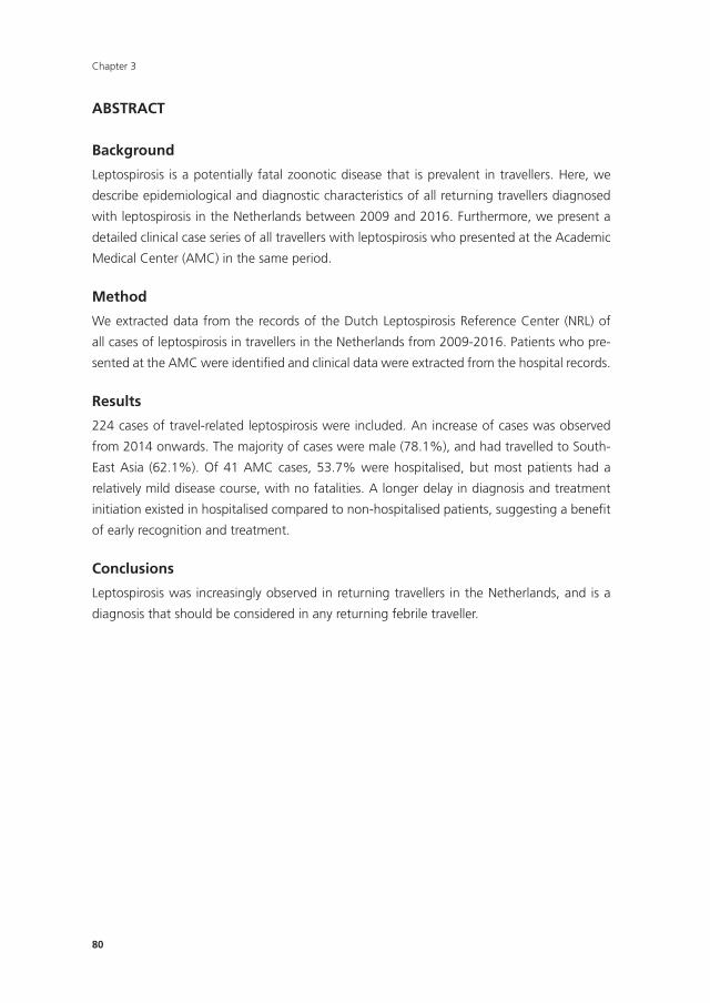
Chapter 3
80
ABSTRACT
Background
Leptospirosis is a potentially fatal zoonotic disease that is prevalent in travellers. Here, we
describe epidemiological and diagnostic characteristics of all returning travellers diagnosed
with leptospirosis in the Netherlands between 2009 and 2016. Furthermore, we present a
detailed clinical case series of all travellers with leptospirosis who presented at the Academic
Medical Center (AMC) in the same period.
Method
We extracted data from the records of the Dutch Leptospirosis Reference Center (NRL) of
all cases of leptospirosis in travellers in the Netherlands from 2009-2016. Patients who pre-
sented at the AMC were identified and clinical data were extracted from the hospital records.
Results
224 cases of travel-related leptospirosis were included. An increase of cases was observed
from 2014 onwards. The majority of cases were male (78.1%), and had travelled to South-
East Asia (62.1%). Of 41 AMC cases, 53.7% were hospitalised, but most patients had a
relatively mild disease course, with no fatalities. A longer delay in diagnosis and treatment
initiation existed in hospitalised compared to non-hospitalised patients, suggesting a benefit
of early recognition and treatment.
Conclusions
Leptospirosis was increasingly observed in returning travellers in the Netherlands, and is a
diagnosis that should be considered in any returning febrile traveller.
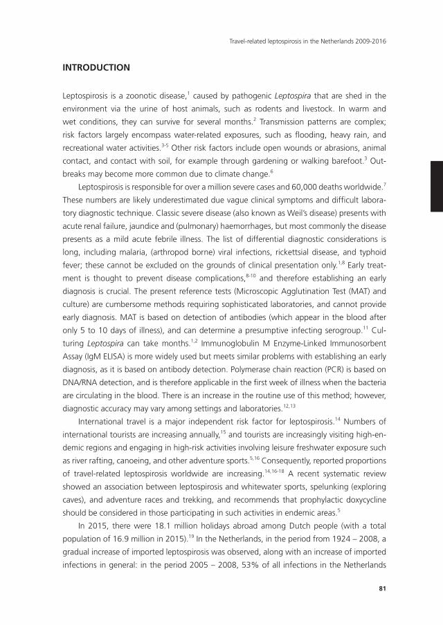
81
Travel-related leptospirosis in the Netherlands 2009-2016
InTRODUCTIOn
Leptospirosis is a zoonotic disease,1 caused by pathogenic Leptospira that are shed in the
environment via the urine of host animals, such as rodents and livestock. In warm and
wet conditions, they can survive for several months.2 Transmission patterns are complex;
risk factors largely encompass water-related exposures, such as flooding, heavy rain, and
recreational water activities.3-5 Other risk factors include open wounds or abrasions, animal
contact, and contact with soil, for example through gardening or walking barefoot.3 Out-
breaks may become more common due to climate change.6
Leptospirosis is responsible for over a million severe cases and 60,000 deaths worldwide.7
These numbers are likely underestimated due vague clinical symptoms and difficult labora-
tory diagnostic technique. Classic severe disease (also known as Weil’s disease) presents with
acute renal failure, jaundice and (pulmonary) haemorrhages, but most commonly the disease
presents as a mild acute febrile illness. The list of differential diagnostic considerations is
long, including malaria, (arthropod borne) viral infections, rickettsial disease, and typhoid
fever; these cannot be excluded on the grounds of clinical presentation only.1,8 Early treat-
ment is thought to prevent disease complications,8-10 and therefore establishing an early
diagnosis is crucial. The present reference tests (Microscopic Agglutination Test (MAT) and
culture) are cumbersome methods requiring sophisticated laboratories, and cannot provide
early diagnosis. MAT is based on detection of antibodies (which appear in the blood after
only 5 to 10 days of illness), and can determine a presumptive infecting serogroup.11 Cul-
turing Leptospira can take months.1,2 Immunoglobulin M Enzyme-Linked Immunosorbent
Assay (IgM ELISA) is more widely used but meets similar problems with establishing an early
diagnosis, as it is based on antibody detection. Polymerase chain reaction (PCR) is based on
DNA/RNA detection, and is therefore applicable in the first week of illness when the bacteria
are circulating in the blood. There is an increase in the routine use of this method; however,
diagnostic accuracy may vary among settings and laboratories.12,13
International travel is a major independent risk factor for leptospirosis.14 Numbers of
international tourists are increasing annually,15 and tourists are increasingly visiting high-en-
demic regions and engaging in high-risk activities involving leisure freshwater exposure such
as river rafting, canoeing, and other adventure sports.5,16 Consequently, reported proportions
of travel-related leptospirosis worldwide are increasing.14,16-18 A recent systematic review
showed an association between leptospirosis and whitewater sports, spelunking (exploring
caves), and adventure races and trekking, and recommends that prophylactic doxycycline
should be considered in those participating in such activities in endemic areas.5
In 2015, there were 18.1 million holidays abroad among Dutch people (with a total
population of 16.9 million in 2015).19 In the Netherlands, in the period from 1924 – 2008, a
gradual increase of imported leptospirosis was observed, along with an increase of imported
infections in general: in the period 2005 – 2008, 53% of all infections in the Netherlands
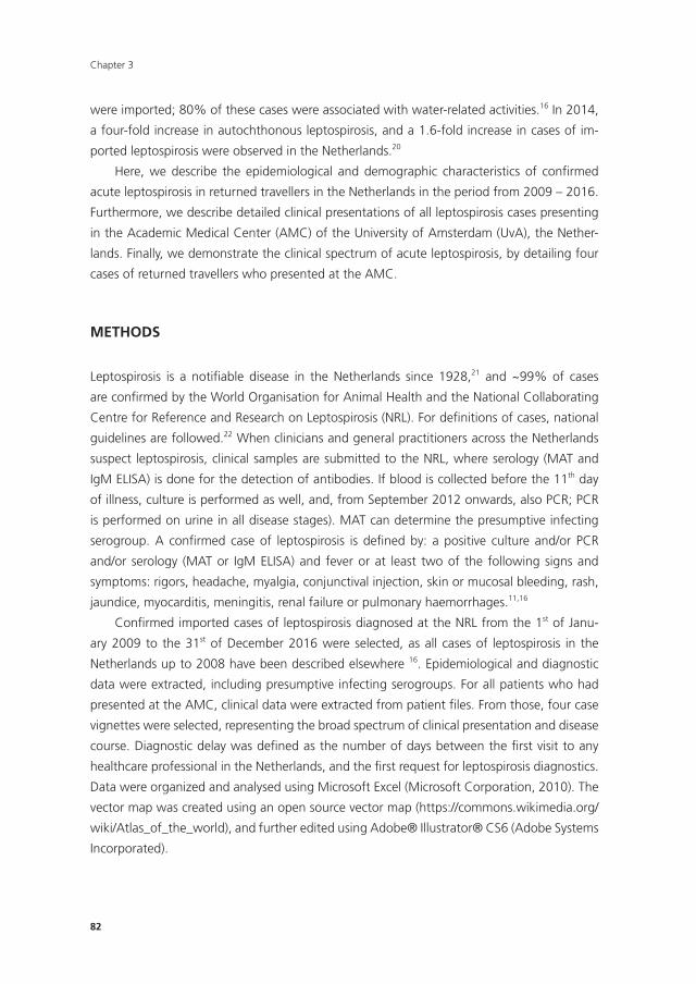
Chapter 3
82
were imported; 80% of these cases were associated with water-related activities.16 In 2014,
a four-fold increase in autochthonous leptospirosis, and a 1.6-fold increase in cases of im-
ported leptospirosis were observed in the Netherlands.20
Here, we describe the epidemiological and demographic characteristics of confirmed
acute leptospirosis in returned travellers in the Netherlands in the period from 2009 – 2016.
Furthermore, we describe detailed clinical presentations of all leptospirosis cases presenting
in the Academic Medical Center (AMC) of the University of Amsterdam (UvA), the Nether-
lands. Finally, we demonstrate the clinical spectrum of acute leptospirosis, by detailing four
cases of returned travellers who presented at the AMC.
METHODS
Leptospirosis is a notifiable disease in the Netherlands since 1928,21 and ~99% of cases
are confirmed by the World Organisation for Animal Health and the National Collaborating
Centre for Reference and Research on Leptospirosis (NRL). For definitions of cases, national
guidelines are followed.22 When clinicians and general practitioners across the Netherlands
suspect leptospirosis, clinical samples are submitted to the NRL, where serology (MAT and
IgM ELISA) is done for the detection of antibodies. If blood is collected before the 11th day
of illness, culture is performed as well, and, from September 2012 onwards, also PCR; PCR
is performed on urine in all disease stages). MAT can determine the presumptive infecting
serogroup. A confirmed case of leptospirosis is defined by: a positive culture and/or PCR
and/or serology (MAT or IgM ELISA) and fever or at least two of the following signs and
symptoms: rigors, headache, myalgia, conjunctival injection, skin or mucosal bleeding, rash,
jaundice, myocarditis, meningitis, renal failure or pulmonary haemorrhages.11,16
Confirmed imported cases of leptospirosis diagnosed at the NRL from the 1st of Janu-
ary 2009 to the 31st of December 2016 were selected, as all cases of leptospirosis in the
Netherlands up to 2008 have been described elsewhere 16. Epidemiological and diagnostic
data were extracted, including presumptive infecting serogroups. For all patients who had
presented at the AMC, clinical data were extracted from patient files. From those, four case
vignettes were selected, representing the broad spectrum of clinical presentation and disease
course. Diagnostic delay was defined as the number of days between the first visit to any
healthcare professional in the Netherlands, and the first request for leptospirosis diagnostics.
Data were organized and analysed using Microsoft Excel (Microsoft Corporation, 2010). The
vector map was created using an open source vector map (https://commons.wikimedia.org/
wiki/Atlas_of_the_world), and further edited using Adobe® Illustrator® CS6 (Adobe Systems
Incorporated).
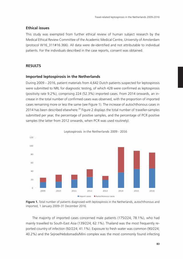
83
Travel-related leptospirosis in the Netherlands 2009-2016
Ethical issues
This study was exempted from further ethical review of human subject research by the
Medical Ethical Review Committee of the Academic Medical Centre, University of Amsterdam
(protocol W16_311#16.366). All data were de-identifi ed and not attributable to individual
patients. For the individuals described in the case reports, consent was obtained.
RESULTS
Imported leptospirosis in the netherlands
During 2009 – 2016, patient materials from 4,642 Dutch patients suspected for leptospirosis
were submitted to NRL for diagnostic testing, of which 428 were confi rmed as leptospirosis
(positivity rate 9.2%), comprising 224 (52.3%) imported cases. From 2014 onwards, an in-
crease in the total number of confi rmed cases was observed, with the proportion of imported
cases remaining more or less the same (see Figure 1). The increase of autochthonous cases in
2014 has been described elsewhere.20 Figure 2 displays the total number of traveller-samples
submitted per year, the percentage of positive samples, and the percentage of PCR positive
samples (the latter from 2012 onwards, when PCR was used routinely).
The majority of imported cases concerned male patients (175/224; 78.1%), who had
mainly travelled to South-East Asia (139/224; 62.1%). Thailand was the most frequently re-
ported country of infection (92/224; 41.1%). Exposure to fresh water was common (90/224;
40.2%) and the Sejroe/Hebdomadis/Mini complex was the most commonly found infecting
0
20
40
60
80
100
120
2009 2010 2011 2012 2013 2014 2015 2016
Leptospirosis intheNetherlands 2009- 2016
Importcases Autochtonouscases
Figure 1. Total number of patients diagnosed with leptospirosis in the Netherlands, autochthonous and imported, 1 January 2009–31 December 2016.
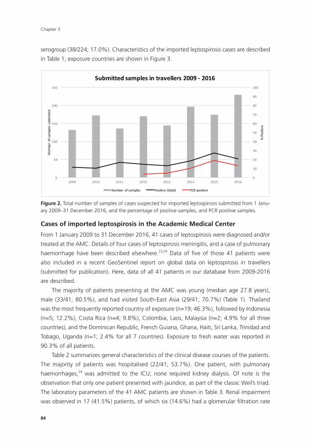
Chapter 3
84
serogroup (38/224; 17.0%). Characteristics of the imported leptospirosis cases are described
in Table 1; exposure countries are shown in Figure 3.
83
Figure 2. Total number of samples of cases suspected for imported leptospirosis submitted from 1 January 2009–31 December 2016, and the percentage of positive samples, and PCR positive samples.
Cases of imported leptospirosis in the Academic Medical Center
From 1 January 2009 to 31 December 2016, 41 cases of leptospirosis were diagnosed and/or
treated at the AMC. Details of four cases of leptospirosis meningitis, and a case of pulmonary
haemorrhage have been described elsewhere.23,24 Data of five of those 41 patients were also
included in a recent GeoSentinel report on global data on leptospirosis in travellers
(submitted for publication). Here, data of all 41 patients in our database from 2009-2016 are
described.
The majority of patients presenting at the AMC was young (median age 27.8 years), male
(33/41; 80.5%), and had visited South-East Asia (29/41; 70.7%) (Table 1). Thailand was the
most frequently reported country of exposure (n=19; 46.3%), followed by Indonesia (n=5;
12.2%), Costa Rica (n=4; 9.8%), Colombia, Laos, Malaysia (n=2; 4.9% for all three countries),
and the Dominican Republic, French Guiana, Ghana, Haiti, Sri Lanka, Trinidad and Tobago,
Uganda (n=1; 2.4% for all 7 countries). Exposure to fresh water was reported in 90.3% of all
patients.
0
10
20
30
40
50
60
70
80
90
100
0
50
100
150
200
250
2009 2010 2011 2012 2013 2014 2015 2016
%Positive
NUmberofsa
mplessubmitted
Submittedsamplesintravellers2009- 2016
Number ofsamples Positive(total) PCRpositive
Figure 2. Total number of samples of cases suspected for imported leptospirosis submitted from 1 Janu-ary 2009–31 December 2016, and the percentage of positive samples, and PCR positive samples.
Cases of imported leptospirosis in the Academic Medical Center
From 1 January 2009 to 31 December 2016, 41 cases of leptospirosis were diagnosed and/or
treated at the AMC. Details of four cases of leptospirosis meningitis, and a case of pulmonary
haemorrhage have been described elsewhere.23,24 Data of fi ve of those 41 patients were
also included in a recent GeoSentinel report on global data on leptospirosis in travellers
(submitted for publication). Here, data of all 41 patients in our database from 2009-2016
are described.
The majority of patients presenting at the AMC was young (median age 27.8 years),
male (33/41; 80.5%), and had visited South-East Asia (29/41; 70.7%) (Table 1). Thailand
was the most frequently reported country of exposure (n=19; 46.3%), followed by Indonesia
(n=5; 12.2%), Costa Rica (n=4; 9.8%), Colombia, Laos, Malaysia (n=2; 4.9% for all three
countries), and the Dominican Republic, French Guiana, Ghana, Haiti, Sri Lanka, Trinidad and
Tobago, Uganda (n=1; 2.4% for all 7 countries). Exposure to fresh water was reported in
90.3% of all patients.
Table 2 summarizes general characteristics of the clinical disease courses of the patients.
The majority of patients was hospitalised (22/41; 53.7%). One patient, with pulmonary
haemorrhages,24 was admitted to the ICU; none required kidney dialysis. Of note is the
observation that only one patient presented with jaundice, as part of the classic Weil’s triad.
The laboratory parameters of the 41 AMC patients are shown in Table 3. Renal impairment
was observed in 17 (41.5%) patients, of which six (14.6%) had a glomerular fi ltration rate
85
Travel-related leptospirosis in the Netherlands 2009-2016
85
92 p
atie
nts
10 -
20 p
atie
nts
< 5
patie
nts
Fig
ure
3. E
xpos
ure
coun
trie
s am
ong
impo
rted
cas
es o
f co
nfi r
med
lept
ospi
rosi
s in
the
Net
herla
nds,
1 J
anua
ry 2
009–
31 D
ecem
ber
2016
(n=
222)
*†.
(For
inte
rpre
ta-
tiono
f th
e re
fere
nces
to
colo
ur in
thi
s fi g
ure
lege
nd, t
he r
eade
r is
ref
erre
d to
the
web
ver
sion
of
this
art
icle
.)*
Incl
udes
onl
y pa
tient
s fo
r w
hom
cou
ntry
of
expo
sure
dat
a w
ere
avai
labl
e.†P
urpl
e re
pres
ents
92
patie
nts
expo
sed
in T
haila
nd. D
ark
blue
repr
esen
ts c
ount
ries
with
10–
20 e
xpos
ed p
atie
nts
(19
for
Mal
aysi
a; 1
6 fo
r In
done
sia;
13
for
Cub
a; 1
2fo
r C
osta
Ric
a; a
nd 1
0 fo
r Fr
ance
). Li
ght
blue
rep
rese
nts
coun
trie
s w
ith<
5 pa
tient
s (4
eac
h fo
r Be
lgiu
m, C
olom
bia,
Lao
s, S
urin
ame,
and
Vie
tnam
; 3 e
ach
for
Ger
man
y, P
anam
a, a
nd S
ri La
nka;
2 e
ach
for
Bulg
aria
, Cam
bodi
a, D
omin
ican
Rep
ublic
, Jam
aica
, Phi
lippi
nes,
Indi
a, N
epal
, Sou
th A
fric
a; a
nd 1
eac
h fo
r A
fgha
nist
an,
Braz
il, E
cuad
or, F
renc
h G
uian
a, G
hana
, Hai
ti, L
uxem
bour
g, M
adag
asca
r, M
aurit
ius,
Mex
ico,
Sie
rra
Leon
e, S
lova
kia,
Slo
veni
a, T
rinid
ad &
Tob
ago,
and
Uga
nda)
.
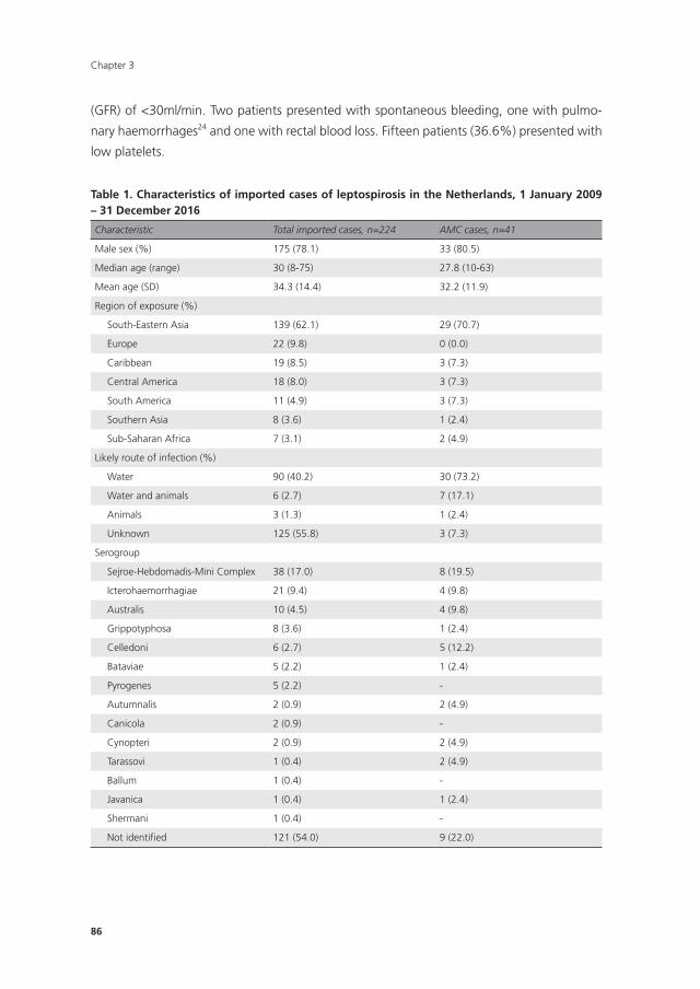
Chapter 3
86
(GFR) of <30ml/min. Two patients presented with spontaneous bleeding, one with pulmo-
nary haemorrhages24 and one with rectal blood loss. Fifteen patients (36.6%) presented with
low platelets.
Table 1. Characteristics of imported cases of leptospirosis in the netherlands, 1 January 2009 – 31 December 2016
Characteristic Total imported cases, n=224 AMC cases, n=41
Male sex (%) 175 (78.1) 33 (80.5)
Median age (range) 30 (8-75) 27.8 (10-63)
Mean age (SD) 34.3 (14.4) 32.2 (11.9)
Region of exposure (%)
South-Eastern Asia 139 (62.1) 29 (70.7)
Europe 22 (9.8) 0 (0.0)
Caribbean 19 (8.5) 3 (7.3)
Central America 18 (8.0) 3 (7.3)
South America 11 (4.9) 3 (7.3)
Southern Asia 8 (3.6) 1 (2.4)
Sub-Saharan Africa 7 (3.1) 2 (4.9)
Likely route of infection (%)
Water 90 (40.2) 30 (73.2)
Water and animals 6 (2.7) 7 (17.1)
Animals 3 (1.3) 1 (2.4)
Unknown 125 (55.8) 3 (7.3)
Serogroup
Sejroe-Hebdomadis-Mini Complex 38 (17.0) 8 (19.5)
Icterohaemorrhagiae 21 (9.4) 4 (9.8)
Australis 10 (4.5) 4 (9.8)
Grippotyphosa 8 (3.6) 1 (2.4)
Celledoni 6 (2.7) 5 (12.2)
Bataviae 5 (2.2) 1 (2.4)
Pyrogenes 5 (2.2) -
Autumnalis 2 (0.9) 2 (4.9)
Canicola 2 (0.9) -
Cynopteri 2 (0.9) 2 (4.9)
Tarassovi 1 (0.4) 2 (4.9)
Ballum 1 (0.4) -
Javanica 1 (0.4) 1 (2.4)
Shermani 1 (0.4) -
Not identified 121 (54.0) 9 (22.0)
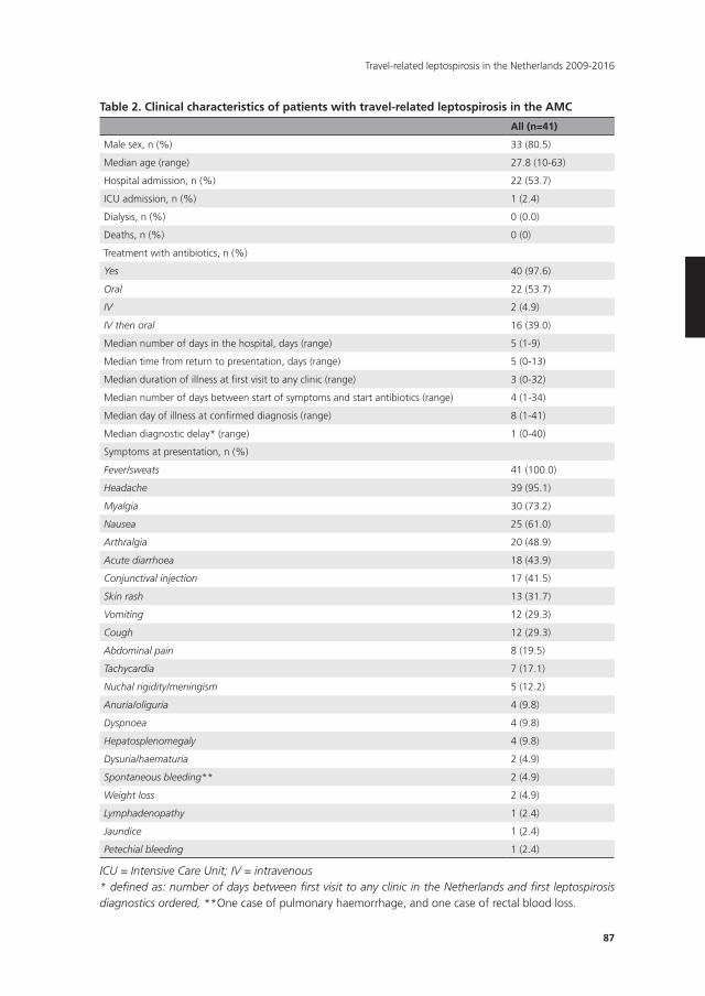
87
Travel-related leptospirosis in the Netherlands 2009-2016
Table 2. Clinical characteristics of patients with travel-related leptospirosis in the AMC
All (n=41)
Male sex, n (%) 33 (80.5)
Median age (range) 27.8 (10-63)
Hospital admission, n (%) 22 (53.7)
ICU admission, n (%) 1 (2.4)
Dialysis, n (%) 0 (0.0)
Deaths, n (%) 0 (0)
Treatment with antibiotics, n (%)
Yes 40 (97.6)
Oral 22 (53.7)
IV 2 (4.9)
IV then oral 16 (39.0)
Median number of days in the hospital, days (range) 5 (1-9)
Median time from return to presentation, days (range) 5 (0-13)
Median duration of illness at first visit to any clinic (range) 3 (0-32)
Median number of days between start of symptoms and start antibiotics (range) 4 (1-34)
Median day of illness at confirmed diagnosis (range) 8 (1-41)
Median diagnostic delay* (range) 1 (0-40)
Symptoms at presentation, n (%)
Fever/sweats 41 (100.0)
Headache 39 (95.1)
Myalgia 30 (73.2)
Nausea 25 (61.0)
Arthralgia 20 (48.9)
Acute diarrhoea 18 (43.9)
Conjunctival injection 17 (41.5)
Skin rash 13 (31.7)
Vomiting 12 (29.3)
Cough 12 (29.3)
Abdominal pain 8 (19.5)
Tachycardia 7 (17.1)
Nuchal rigidity/meningism 5 (12.2)
Anuria/oliguria 4 (9.8)
Dyspnoea 4 (9.8)
Hepatosplenomegaly 4 (9.8)
Dysuria/haematuria 2 (4.9)
Spontaneous bleeding** 2 (4.9)
Weight loss 2 (4.9)
Lymphadenopathy 1 (2.4)
Jaundice 1 (2.4)
Petechial bleeding 1 (2.4)
ICU = Intensive Care Unit; IV = intravenous* defined as: number of days between first visit to any clinic in the Netherlands and first leptospirosis diagnostics ordered, **One case of pulmonary haemorrhage, and one case of rectal blood loss.
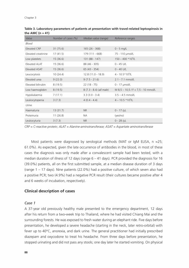
Chapter 3
88
Most patients were diagnosed by serological methods (MAT or IgM ELISA, n =25;
61.0%). As expected, given the late occurrence of antibodies in the blood, in most of these
cases the diagnosis was only made after a convalescent sample had been tested, with a
median duration of illness of 12 days (range 6 – 41 days). PCR provided the diagnosis for 16
(39.0%) patients, all on the first submitted sample, at a median disease duration of 3 days
(range 1 – 17 days). Nine patients (22.0%) had a positive culture, of which seven also had
a positive PCR; two (4.9%) had a negative PCR result (their cultures became positive after 4
and 6 weeks of incubation, respectively).
Clinical description of cases
Case 1
A 37-year old previously healthy male presented to the emergency department, 12 days
after his return from a two-week trip to Thailand, where he had visited Chiang Mai and the
surrounding forests. He was exposed to fresh water during an elephant ride. Five days before
presentation, he developed a severe headache (starting in the neck, later retro-orbital) with
fever up to 40°C, anorexia, and dark urine. The general practitioner had initially prescribed
diazepam and oxycodone to treat his headache. From three days before presentation, he
stopped urinating and did not pass any stools; one day later he started vomiting. On physical
Table 3. Laboratory parameters of patients at presentation with travel-related leptospirosis in the AMC (n = 41)
Value Number of cases (%) Median value (range) Reference ranges
Blood
Elevated CRP 31 (75.6) 165 (26 - 368) 0 - 5 mg/L
Elevated creatinine 17 (41.5) 179 (111 - 668) 75 - 110 μmol/L
Low platelets 15 (36.6) 131 (88 - 147) 150 – 400 *109/L
Elevated ALAT 15 (36.6) 88 (46 - 305) 0 - 45 U/L
Elevated ASAT 15 (36.6) 65 (43 - 354) 0 - 40 U/L
Leucocytosis 10 (24.4) 12.8 (11.0 - 18.9) 4 - 10.5*109/L
Elevated urea 9 (22.0) 9 (7.5 - 21.6) 2.1 - 7.1 mmol/L
Elevated bilirubin 8 (19.5) 22 (18 - 75) 0 - 17 μmol/L
Low haemoglobin 8 (19.5) 8 (7.3 - 8.4) (all male) M 8,5 – 10,5 / F < 7,5 - 10 mmol/L
Hypokalaemia 7 (17.1) 3.3 (3.0 - 3.4) 3.5 – 4.5 mmol/L
Leukocytopenia 3 (7.3) 4 (0.4 - 4.4) 4 – 10.5 *109/L
Urine
Haematuria 13 (31.7) NR 0 - 17 /μL
Proteinuria 11 (26.8) NA (yes/no)
Leukocyturia 3 (7.3) NR 0 - 28 /μL
CRP = C-reactive protein; ALAT = Alanine-aminotransferase; ASAT = Aspartate aminotransferase
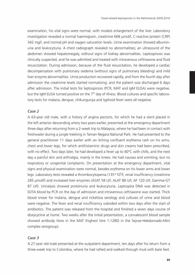
89
Travel-related leptospirosis in the Netherlands 2009-2016
examination, his vital signs were normal, with modest enlargement of the liver. Laboratory
investigation revealed a normal haemogram, creatinine 668 μmol/l, C-reactive protein (CRP)
342 mg/l, and normal pH and oxygen saturation levels. Urine examination showed albumin-
uria and leukocyturia. A chest radiograph revealed no abnormalities; an ultrasound of the
abdomen showed hepatomegaly, without signs of kidney abnormalities. Leptospirosis was
clinically suspected, and he was admitted and treated with intravenous ceftriaxone and fluid
resuscitation. During admission, because of the fluid resuscitation, he developed a cardiac
decompensation with pulmonary oedema (without signs of pulmonary bleeding) and mild
liver enzyme abnormalities. Urine production recovered rapidly, and from the fourth day after
admission the creatinine levels started normalizing, and the patient was discharged 6 days
after admission. The initial tests for leptospirosis (PCR, MAT and IgM ELISA) were negative,
but the IgM ELISA turned positive on the 7th day of illness. Blood cultures and specific labora-
tory tests for malaria, dengue, chikungunya and typhoid fever were all negative.
Case 2
A 63-year old male, with a history of angina pectoris, for which he had a stent placed in
the left anterior descending artery two years earlier, presented at the emergency department
three days after returning from a 2-week trip to Malaysia, where he had been in contact with
freshwater during a jungle trekking in Taman Negara National Park. He had presented to the
general practitioner 11 days earlier with an itching confluent erythema rash on his arms,
chest and lower legs, for which antihistaminic drugs and skin creams had been prescribed,
with no effect. Two days later, he had developed a fever up to 40°C with chills, and the next
day a painful skin and arthralgia, mainly in the knees. He had nausea and vomiting, but no
respiratory or urogenital complaints. On presentation at the emergency department, vital
signs and physical examination were normal, besides erythema on his lower arms and lower
legs. Laboratory tests revealed a thrombocytopenia (135*109/l, renal insufficiency (creatinine
285 μmol/l) and increased liver enzymes (ASAT 58 U/l; ALAT 88 U/l; AF 120 U/l; Gamma-GT
87 U/l). Urinalysis showed proteinuria and leukocyturia. Leptospira DNA was detected in
EDTA blood by PCR on the day of admission and intravenous ceftriaxone was started. Thick
blood smear for malaria, dengue and rickettsia serology and cultures of urine and blood
were negative. The fever and renal insufficiency subsided within two days after the start of
antibiotics. The patient was released from the hospital and finished a seven days course of
doxycycline at home. Two weeks after the initial presentation, a convalescent blood sample
showed antibody titres in the MAT (highest titre 1:1280) in the Sejroe-Hebdomadis-Mini
complex serogroup).
Case 3
A 27-year old male presented at the outpatient department, ten days after his return from a
three-week trip to Colombia, where he had rafted and walked through mud with bare feet.
Chapter 3
90
Seven days before presentation, he developed fever up to 39.7°C, with chills and arthralgia
in the ankles, knees and lower back, and myalgia in the calves during the first two days of
disease. Two days later, he became nauseated and vomited, and developed watery diarrhoea
one day later. The next day, he developed pain in both testes. He finally presented at the
AMC two days later. On physical examination, there was a conjunctival injection, and a light
exanthema on the abdomen and lower back, and both testes were swollen and painful.
Laboratory tests revealed no abnormalities other than an increased CRP (196 mg/L). The thick
blood smear for malaria, stool and urine cultures, and serology for syphilis and mumps were
all negative. As the patient started to feel better, no treatment was initiated. Two days later,
at follow-up visit he had improved clinically. In the meantime, the PCR for leptospiral DNA on
serum became positive. As complaints of orchitis continued, doxycyline was administered for
seven days, with good result. Two weeks later, a follow-up sample showed a more than four-
fold increase in MAT titre against strains in the Sejroe-Hebdomadis-Mini complex serogroup.
Case 4
A 31-year old male was admitted with fever, headache, nausea, and malaise. Symptoms had
started the during a 2.5-week journey to Costa Rica, and at presentation, symptoms had
existed for five days. In Costa Rica, there had been floods after heavy rainfall, and he had
been exposed to prolonged water contact because the hotel was flooded. He presented with
retro-orbital headache, photophobia, myalgia in the calves of his legs, nausea, vomiting, and
rectal blood loss. Physical examination showed red, injected conjunctivae, mild jaundice, a
tachycardia of 102 bpm, fever (39.9°C), and a normal blood pressure. Laboratory investi-
gations revealed a haemoglobin of 7.3 mmol/l; thrombocytes of 107*109/l, elevated liver
enzymes (bilirubin 75 μmol/l, ASAT 111 U/l; ALAT 104 U/l; AF 180 U/l; gamma-GT 98U/l), and
a normal renal function. Leptospirosis, typhoid fever, or an arthropod borne viral infection
were suspected, and the patient was admitted and treated with intravenous ceftriaxone.
The PCR for Leptospira was positive, after which treatment was continued with oral doxycy-
cline. The patient improved substantially, but after two days he developed a second-degree
atrioventricular block, for which he was observed for 48 hours in the cardiac observation
unit. The atrioventricular block disappeared spontaneously, but an incomplete right bundle
branch block remained, for which he still receives cardiologic follow-up. Despite initial slow
resolution of general fatigue, he had recovered one month after discharge.
DISCUSSIOn
During the time period 2009 – 2016, leptospirosis was increasingly observed in the Neth-
erlands among returned travellers. In addition, a marked increase of the total number of
leptospirosis cases, including autochthonous infections, was observed from 2014 onwards.
91
Travel-related leptospirosis in the Netherlands 2009-2016
The increase in autochthonous cases in 2014 was thought to be due to a warm winter,
followed by the warmest year in centuries.20 This trend continued over 2015 and 2016,
possibly for the same reason. For the imported cases however, the explanation is less obvi-
ous. The number of Dutch tourists travelling abroad and their destinations have been more
or less stable since 2008.19,25 Possibly, travellers are increasingly participating in high-risk
activities, such as rafting and jungle trekking. Another explanation could be that physicians
in the Netherlands have become more aware of leptospirosis, and thus request diagnostic
tests more often. This is supported by the fact that the NRL has received increasing numbers
of samples over the past years. Additionally, PCR was implemented in September 2012 in
the NRL. PCR can identify leptospirosis cases in the early disease stages, which would have
needed a convalescent sample for diagnosis in the period before September 2012, which
is often not submitted to the laboratory. However, an increase of positivity rate was only
observed from 2014 onwards. This increase was mainly attributable to PCR positive cases.
The majority of cases comprised relatively young males, who had travelled to Southeast
Asia, consistent with other reports on leptospirosis in travellers.16,26,27 Thailand, Malaysia,
and Indonesia were the most frequently reported countries of exposure. Remarkably, Europe
was the second most common region of exposure, with France contributing ten cases, and
nine more cases from Belgium, Bulgaria, Luxembourg, Slovakia, and Slovenia. France is the
number one destination for Dutch holidays, which could explain the relatively high number
of cases. It does indicate however, that leptospirosis should be considered in all travellers
presenting with a febrile illness, independent of the region they have visited, in particular also
because delayed diagnosis leads to serious complications, shown in our cases.
Clinicians are usually well aware of the risk of leptospirosis when a typical exposure
history is present, such as floods and contact with freshwater. In travellers, clear exposure
histories have been reported,26,28 but in the general population, the mechanism of infection
often remains uncertain.3 In our data, exposure histories were not known in almost 56% of
all travel-related leptospirosis cases in the Netherlands; among the patients that presented
at the AMC, where leptospirosis is a frequently diagnosed travel related disease, a clear
exposure history was registered in more than 90% of the patients. It is possible that the
lower percentage in the national group is due to incompleteness of the data, or due to
unfamiliarity with the disease among physicians who rarely encounter leptospirosis.
It is likely that the cases reported here merely represent the more severe cases, as mild
cases are more likely to remain unrecognized.1,29 It has been described that the disease pre-
sentation in Dutch imported cases is less severe than in autochthonous cases, which has been
postulated to be associated with a lower number of imported infections with serovars from
the Icterohaemorrhagiae serogroup, linked to severe disease.16,20 Indeed, the most common
infecting serogroup in imported cases was the Sejroe-Hebdomadis-Mini complex (17% of
cases), for which milder disease courses have been described,16 whereas only around 10% of
the described cases were infected with the Icterohaemorrhagiae serogroup.
Chapter 3
92
We did not collect detailed clinical data of the overall group of 224 travellers with
leptospirosis in the Netherlands. We do report, however, detailed data on a subset of 41
cases (18.3% of all confirmed cases of leptospirosis in the Netherlands) who were diagnosed
and treated in the AMC. Of note, no single patient of those 41 succumbed to leptospirosis.
The diagnostic delay in this group was generally short, with most patients being tested for
leptospirosis relatively quickly (median of one day).
The AMC is a tertiary hospital with a specialized travel clinic (the Center of Tropical
Medicine and Travel Medicine), and the Leptospirosis Reference Center is located at the
premises building, which likely explains the relatively short delay, and possibly the relatively
high caseload at the AMC. Another explanation is that there is a low threshold for consid-
eration of the disease. Diagnostic delay most frequently occurred before presentation at the
AMC. In addition, several patients had received inadequate treatments before presentation,
such as very short courses of oral antibiotics.
None of the patients presented with a classic Weil’s syndrome. Hospital admission was
required in only 22 cases (53.7%), which is lower than in the previous report.16 There was
only one ICU admission,24 and no need for renal replacement therapy in any patient. All
patients survived. Further symptoms at presentation were similar to those described in other
case series,16,30 except for lower rates of jaundice.
Case #4, a young male, developed a second-degree atrioventricular block during admis-
sion, which resolved spontaneously, but a right bundle-branch block remained. Electrocar-
diographic alterations have been described in case series on leptospirosis, with ventricular
repolarization disorders, atrial fibrillation and first-degree atrioventricular blocks most com-
mon, also in younger patients.31,32 Different theories on the aetiology of cardiac involvement
in leptospirosis have been postulated.31-33 In severe and fatal cases of leptospirosis, myocar-
dial involvement has been described.33 The more commonly observed electrocardiographic
abnormalities could be an effect of the leptospiraemia, or a general occurrence in febrile
disease, also through metabolic and electrolytic disturbances.
Case #3 developed an orchitis four days after the acute febrile episode started. Orchitis
has been described as a complication of leptospirosis in the older literature.34-38 Most of those
cases had been infected with the Ballum serogroup, contracted from laboratory34-36 or pet
mice,37 and developed the orchitis at a later stage in the disease, after about 10 to 20 days.
Our patient was PCR positive and later developed high MAT titres against serovars from the
Sejroe-Hebdomadis-Mini complex serogroup. A similar case has been described in a 25-year
old dairy farm worker who presented with a fever and epididymitis, who was also found to
be infected with a serovar from the Sejroe-Hebdomadis-Mini complex serogroup.38
In the group of 41 AMC patients, the diagnosis of leptospirosis was most often obtained
through serology, and for most patients, only the follow up sample was positive (median 12
days after onset of symptoms). These patients were either tested too late to perform PCR,
or the PCR was negative. In confirmed cases with a negative PCR, the test was performed
93
Travel-related leptospirosis in the Netherlands 2009-2016
later in the illness compared to the confirmed cases with a positive PCR, with a median of
5 days after the onset of symptoms in PCR negatives vs. a median of 2 days in PCR positive
cases. The latter suggests that the PCR is more sensitive in the earliest stages of the disease,
which is in line with previous studies.12,39 These findings show that it is important to request
diagnostic testing as early as possible in the disease course; when the time-frame for PCR is
missed, it can take over a week for the serology to become positive. However, a negative PCR
does not rule out the disease.
It is likely that leptospirosis is often missed, because many returning travellers with an
acute febrile illness are treated with antibiotics empirically. If they improve, a diagnosis is not
always sought. However, because of the potentially severe disease course when diagnosis is
delayed or missed, there is an urgent need for an easy-to-use and simple diagnostic test in
the acute phase of the disease.
Limitations to this study are that, even though ~99% of all leptospirosis cases in the
Netherlands are diagnosed in the NRL, the data presented here may be incomplete. Clinical
data were only available in the subset of patients who had presented at the AMC. Data on
the serogroups were mostly based on the MAT, which can only determine a presumptive
infecting serogroup.11 Furthermore, the cases presented in detail here, were diagnosed and
treated at the AMC, which is a tertiary hospital with a specialized travel clinic, which possibly
resulted in selection bias; this could may implicate that the clinical picture of diagnosed
leptospirosis patients the total population is not accurately reflected.
We conclude that leptospirosis is an increasing and likely underestimated cause of febrile
illness and hospitalisation in returned travellers in the Netherlands. The disease has different
and often surprising clinical manifestations in travellers. Mild outcome is associated with
early diagnosis after the start of symptoms. Therefore, diagnostic testing should therefore be
performed with a low threshold of suspicion in any febrile returning traveller.
Chapter 3
94
REFEREnCES
1. World Health Organization. Human leptospirosis: guidance for diagnosis, surveillance and con-
trol. 2003.
2. Levett PN. Leptospirosis. Clin Microbiol Rev 2001; 14(2): 296-326.
3. Mwachui MA, Crump L, Hartskeerl R, Zinsstag J, Hattendorf J. Environmental and Behavioural
Determinants of Leptospirosis Transmission: A Systematic Review. PLoS Negl Trop Dis 2015; 9(9):
e0003843.
4. Monahan AM, Miller IS, Nally JE. Leptospirosis: risks during recreational activities. J Appl Micro-
biol 2009; 107(3): 707-16.
5. Gundacker ND, Rolfe RJ, Rodriguez JM. Infections associated with adventure travel: A systematic
review. Travel Med Infect Dis 2017; 16: 3-10.
6. Lau CL, Smythe LD, Craig SB, Weinstein P. Climate change, flooding, urbanisation and leptospi-
rosis: fuelling the fire? Transactions of the Royal Society of Tropical Medicine and Hygiene 2010;
104(10): 631-8.
7. Costa F, Hagan JE, Calcagno J, et al. Global Morbidity and Mortality of Leptospirosis: A Systematic
Review. PLoS Negl Trop Dis 2015; 9(9): e0003898.
8. Bharti AR, Nally JE, Ricaldi JN, et al. Leptospirosis: a zoonotic disease of global importance. Lancet
Infect Dis 2003; 3(12): 757-71.
9. Sharp TM, Rivera Garcia B, Perez-Padilla J, et al. Early Indicators of Fatal Leptospirosis during the
2010 Epidemic in Puerto Rico. PLoS Negl Trop Dis 2016; 10(2): e0004482.
10. Amilasan AS, Ujiie M, Suzuki M, et al. Outbreak of leptospirosis after flood, the Philippines, 2009.
Emerg Infect Dis 2012; 18(1): 91-4.
11. Goris M, Leeflang M, Boer K, et al. Establishment of valid laboratory case definition for human
leptospirosis. J Bacteriol Parasitol 2012; 3(132): 2.
12. Ahmed A, Engelberts MF, Boer KR, Ahmed N, Hartskeerl RA. Development and validation of a
real-time PCR for detection of pathogenic leptospira species in clinical materials. PLoS One 2009;
4(9): e7093.
13. Thaipadungpanit J, Chierakul W, Wuthiekanun V, et al. Diagnostic accuracy of real-time PCR
assays targeting 16S rRNA and lipL32 genes for human leptospirosis in Thailand: a case-control
study. PLoS One 2011; 6(1): e16236.
14. Lau C, Smythe L, Weinstein P. Leptospirosis: an emerging disease in travellers. Travel Med Infect
Dis 2010; 8(1): 33-9.
15. UNWTO Tourism Highlights 2016. 2016. http://www.e-unwto.org/doi/
book/10.18111/9789284418145.
16. Goris MG, Boer KR, Duarte TA, Kliffen SJ, Hartskeerl RA. Human leptospirosis trends, the Nether-
lands, 1925-2008. Emerg Infect Dis 2013; 19(3): 371-8.
17. Jensenius M, Han PV, Schlagenhauf P, et al. Acute and potentially life-threatening tropical dis-
eases in western travelers--a GeoSentinel multicenter study, 1996-2011. Am J Trop Med Hyg
2013; 88(2): 397-404.
18. Jansen A, Schoneberg I, Frank C, Alpers K, Schneider T, Stark K. Leptospirosis in Germany, 1962-
2003. Emerg Infect Dis 2005; 11(7): 1048-54.
19. Trendrapport toerisme, recreatie en vrije tijd 2016. The Hague: Centraal Bureau voor de Statistiek,
2016.
20. Pijnacker R, Goris MG, Te Wierik MJ, et al. Marked increase in leptospirosis infections in humans
and dogs in the Netherlands, 2014. Euro Surveill 2016; 21(17).
95
Travel-related leptospirosis in the Netherlands 2009-2016
21. van Vliet J. History of notification [in Dutch]. Tijdschrift voor Infectieziekten 2009; 4: 51-60.
22. National Coordinating Body for Infectious Diseases (LCI). LCI-richtlijn Leptospirose [Guideline for
Leptospirosis]. Bilthoven: RIVM; 2011. Dutch.
23. van Samkar A, van de Beek D, Stijnis C, Goris M, Brouwer MC. Suspected leptospiral meningitis
in adults: report of four cases and review of the literature. Neth J Med 2015; 73(10): 464-70.
24. Helmerhorst HJ, van Tol EN, Tuinman PR, et al. Severe pulmonary manifestation of leptospirosis.
Neth J Med 2012; 70(5): 215-21.
25. Centraal Bureau voor de Statistiek. Lange vakanties buitenland; bestemmingen naar vakantiek-
enmerken. 2017. http://statline.cbs.nl/Statweb/publication/?DM=SLNL&PA=71086NED&D1=a
&D2=39-41&D3=a&HDR=T&STB=G1,G2&VW=T (accessed 26 July 2017 2017).
26. van de Werve C, Perignon A, Jaureguiberry S, Bricaire F, Bourhy P, Caumes E. Travel-related
leptospirosis: a series of 15 imported cases. J Travel Med 2013; 20(4): 228-31.
27. Forbes AE, Zochowski WJ, Dubrey SW, Sivaprakasam V. Leptospirosis and Weil’s disease in the
UK. Qjm 2012; 105(12): 1151-62.
28. Leshem E, Segal G, Barnea A, et al. Travel-related leptospirosis in Israel: a nationwide study. Am
J Trop Med Hyg 2010; 82(3): 459-63.
29. Hartskeerl RA, Collares-Pereira M, Ellis WA. Emergence, control and re-emerging leptospirosis:
dynamics of infection in the changing world. Clin Microbiol Infect 2011; 17(4): 494-501.
30. Katz AR, Buchholz AE, Hinson K, Park SY, Effler PV. Leptospirosis in Hawaii, USA, 1999-2008.
Emerg Infect Dis 2011; 17(2): 221-6.
31. Skerk V, Markotic A, Puljiz I, et al. Electrocardiographic changes in hospitalized patients with
leptospirosis over a 10-year period. Med Sci Monit 2011; 17(7): Cr369-75.
32. Sacramento E, Lopes AA, Costa E, Passos OL, Costa YA, Matos ED. Electrocardiographic altera-
tions in patients hospitalized with leptospirosis in the Brazilian city of Salvador. Arq Bras Cardiol
2002; 78(3): 267-70.
33. Navinan MR, Rajapakse S. Cardiac involvement in leptospirosis. Trans R Soc Trop Med Hyg 2012;
106(9): 515-20.
34. Kappeler R, Barandun S, Luthi H, Wiesmann E. [On a laboratory Leptospira ballum infection;
orchitis as a complication]. Schweiz Med Wochenschr 1961; 91: 810-2.
35. Stoenner HG, Maclean D. Leptospirosis (ballum) contracted from Swiss albino mice. AMA Arch
Intern Med 1958; 101(3): 606-10.
36. Boak RA, Linscott WD, Bodfish RE. A Case of Leptospirosis Ballum in California. California Medi-
cine 1960; 93(3): 163-5.
37. Friedmann CT, Spiegel EL, Aaron E, McIntyre R. Leptospirosis ballum contracted from pet mice.
California Medicine 1973; 118(6): 51-2.
38. Hoghton M, Price P. Leptospirosis hardjo epididymitis. British Medical Journal 1986; 292(6514):
174.
39. Céspedes M, Tapia R, Balda L, Gonzalez D, Peralta C, Condori P. Estandarización y validación
de una prueba de PCR para el diagnóstico precoz de Leptospirosis humana. Revista Peruana de
Medicina Experimental y Salud Pública 2007; 24(1): 20-6.
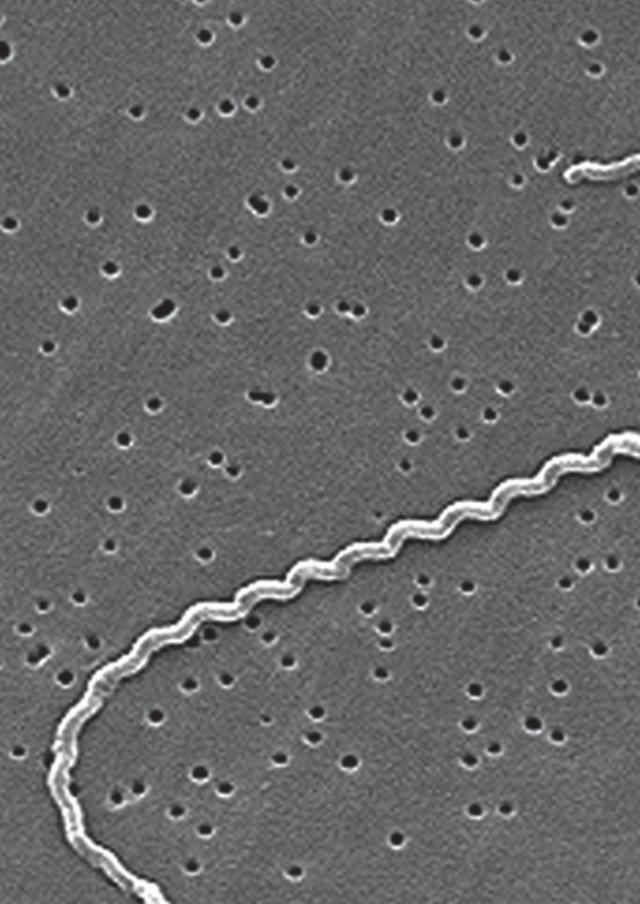
Chapter 4Leptospirosis among Returned Travelers: a GeoSentinel Site Survey and Multicenter
Analysis – 1997-2016
Sophia G. de Vries; Benjamin J. Visser; Rhett J. Stoney; Jiri F.P. Wagenaar; Emmanuel Bottieau; Lin H. Chen; Annelies Wilder-Smith, Mary Wilson;
Christophe Rapp; Karin Leder; Eric Caumes; Eli Schwartz, Noreen A. Hynes; Abraham Goorhuis; Douglas H. Esposito; Davidson H. Hamer; Martin P.
Grobusch, for the GeoSentinel Surveillance Network¶
American Journal of Tropical Medicine and Hygiene 2018; 99: 127-135
Chapter 4
98
ABSTRACT
Leptospirosis is a potentially fatal emerging zoonosis with worldwide distribution and a broad
range of clinical presentations and exposure risks. It typically affects vulnerable populations
in (sub)tropical countries, but is increasingly reported in travelers as well. Diagnostic methods
are cumbersome and require further improvement. Here, we describe leptospirosis among
travelers presenting to the GeoSentinel Global Surveillance Network.
We performed a descriptive analysis of leptospirosis cases reported in GeoSentinel from
January 1997 through December 2016. We included 180 travelers with leptospirosis (mostly
male; 74%; mostly tourists; 81%). The most frequent region of infection was Southeast Asia
(52%); the most common source countries were Thailand (n=52), Costa Rica (n=13), Indo-
nesia and Laos (n=11 each). Fifty-nine percent were hospitalized; one fatality was reported.
We also distributed a supplemental survey to GeoSentinel sites to assess clinical and
diagnostic practices. Of 56 GeoSentinel sites, three-quarters responded to the survey. Lep-
tospirosis was reported to have been most frequently considered in febrile travelers with
hepatic and renal abnormalities and a history of freshwater exposure. Serology was the most
commonly used diagnostic method, although convalescent samples were reported to have
been collected infrequently.
Within GeoSentinel, leptospirosis was diagnosed mostly among international tourists
and caused serious illness. Clinical suspicion and diagnostic workup among surveyed Ge-
oSentinel clinicians were mainly triggered by a classical presentation and exposure history,
possibly resulting in under-diagnosis. Suboptimal usage of available diagnostic methods may
have resulted in additional missed, or misdiagnosed, cases.
99
Leptospirosis among Returned Travelers
InTRODUCTIOn
Leptospirosis is an emerging zoonotic disease, causing more than a million severe cases
worldwide and around 60,000 deaths annually.1 The disease-causing spirochetes of the
Leptospira genus2 display complex transmission patterns.3 A wide range of infecting serovars
exist in a broad range of host animals, most notoriously in rats.2 Well-known major risk
factors are water-associated, such as recreational water activities; water exposure following
floods and heavy rain; contact with animal or animal urine such as rodents and livestock; and
poor sanitation.3 Less common risk factors include exposure through open skin wounds and
soil contact,3 which might occur while gardening4, 5 or walking barefoot.3 Moreover, leptospi-
rosis has emerged as an important problem in urban slums in the developing world, where
rat-borne transmission increasingly triggers outbreaks.6, 7, 8, 9, 10, 11 Most cases are sporadic;
however, outbreaks do occur, and may be increasingly frequent due to climate change.12
Clinical manifestations can vary from a mild self-limiting infection to life-threatening ill-
ness. However, patients seeking care typically present with a mild acute febrile illness including
chills, headache (often with retro-orbital pain), conjunctival suffusion, photophobia, myalgia,
abdominal pain, nausea, vomiting, and sometimes transient exanthema.2, 13 About 10% of
patients progress to severe disease, including Weil’s disease, characterized by jaundice, acute
kidney failure, or (pulmonary) hemorrhage.2, 13, 14, 15, 16 Pulmonary hemorrhage can also be
a stand-alone manifestation. Aseptic meningitis may be seen in up to 25% of leptospirosis
cases.13 Cardiac involvement is likely more common than recognized.13 The list of differential
diagnoses for the evaluating clinician to consider is long, encompassing, among others,
malaria, arboviral infections, rickettsial diseases, and typhoid fever,13, 17 and misdiagnosis
is common.6, 18, 19, 20 Early recognition and treatment may be essential to improve patient
outcomes13, 21, 22 and minimize hospitalization costs.
Establishing an early, rapid and accurate diagnosis remains a complex matter. Widely
used serological tests that detect antibodies, including the microscopic agglutination test
(MAT, the current reference test), and immunoglobulin M enzyme-linked immunosorbent
assay (IgM ELISA), are not suitable for early diagnosis, as antibodies only appear in the blood
about 5 to 10 days after onset of symptoms.2 Leptospirae can be cultured from blood in the
early stages of disease, but this process can take weeks or months.2, 17 Molecular detection
tests, such as polymerase chain reaction (PCR), are increasingly used for routine diagnosis
of the disease in the first week of illness,23, 24, 25 but are not yet available in all countries.26, 27
Commercially available multiplex panels, testing for multiple pathogens, could be a more
accessible alternative.28 For early presentations, PCR is recommended; if possible in combina-
tion with culture. For later presentations, MAT and IgM ELISA are suggested. Convalescent
samples should be collected, and a combination of the above tests is always preferable.24
The disease burden of leptospirosis is highest in resource-poor, tropical countries; the
highest incidence rates occur in Oceania, Southeast Asia, the Caribbean, and East Africa.1, 29
Chapter 4
100
Notably, international travel to these regions now constitutes a major independent risk factor
for acquisition of leptospirosis.30, 31 In 2015, almost 1.2 billion international tourist arrivals
were recorded worldwide, with an ever-growing number of travelers visiting tropical and
subtropical regions.32 With travelers being increasingly engaged in high-risk recreational
activities, such as white-water rafting,33 growing numbers of travelers with leptospirosis
returning from tropical countries have been reported.4, 31, 33, 34, 35, 36, 37 In a GeoSentinel analy-
sis (1996-2011) of acute and potentially life-threatening tropical disease among 3,666 ill
travelers from higher-resource areas, leptospirosis was the fourth most common diagnosis.38
Here, we describe the epidemiology of leptospirosis among travelers reported to the
GeoSentinel Surveillance Network since its inception more than 20 years ago (January 1997
until 31 December 2016). Additionally, we report our analysis of the reported diagnostic
approaches utilized by current GeoSentinel Surveillance Network members, through a
supplemental survey among GeoSentinel sites.
METHODS
Data source – GeoSentinel surveillance system
GeoSentinel is a global surveillance network designed to monitor travel-related morbidity. It
was established in 1995, with systematic data collection beginning in 1997.39, 40 Currently,
70 GeoSentinel travel and tropical medicine clinics, located in 31 countries on six continents,
contribute anonymous clinician-based surveillance information on ill travelers with a focus on
infections acquired during travel (see http://www.istm.org/geosentinel for an up-to-date site
distribution map and other information). In brief, for inclusion in the GeoSentinel database,
patients must have crossed an international border within 10 years of presentation, and
sought medical care from a GeoSentinel site for a presumed travel-associated condition.
Standard data collection forms capture patient demographic characteristics, detailed recent
travel itinerary, countries visited within 5 years, reason for recent travel, symptom-based
grouping by affected organ system, and whether there was a reported encounter with a
health care provider before travel.40 Final diagnoses are assigned by the attending clinician
and chosen from a list of standard diagnosis codes, guided by GeoSentinel diagnostic defini-
tions. Each patient may have more than one travel-related diagnosis, and each diagnosis is
reported as confirmed, probable, or suspected, based on the strength of the diagnosis.40 All
GeoSentinel sites use the best reference diagnostic methods available in their own country.
Clinical treatments and outcomes are not routinely reported.
GeoSentinel’s data-collection protocol is for public health surveillance, and has received
a determination of non-research by a CDC human subjects’ advisor.
101
Leptospirosis among Returned Travelers
GeoSentinel surveillance data inclusion criteria and definitions
GeoSentinel records for patients with a post-travel diagnosis of ‘confirmed’ leptospirosis and
a clinic visit date from 1 January 1997 to 31 December 2016 were included. GeoSentinel de-
fined criteria for ‘confirmed’ leptospirosis diagnosis state: ‘a compatible clinical history (e.g.
fever with associated symptoms) plus positive microscopy, culture, histopathology, nucleic
acid amplification test, antigen detection, or seroconversion with a ≥4x titer rise on serology.’
Furthermore, only patients presenting within 30 days after return from travel were included,
based on the incubation period of leptospirosis of 2-30 days (average 7-10 days).17 Patients
seen during travel, missing travel itinerary data, or having a non-ascertainable region of
exposure were excluded. The reason for travel was stratified into four categories: tourism,
business, visiting friends and relatives (VFR), and other (combining small numbers of foreign
aid and missionary workers or military deployment).
Data source – Supplemental GeoSentinel network survey
A 21-question multiple-choice survey (Supplementary Material 1) aimed to assess clinical and
diagnostic practices among the GeoSentinel sites. After piloting among selected sites, the
survey was distributed by email between December 2015 and March 2016 to 56 GeoSentinel
sites active at the time of the survey, excluding the site of survey origin (Amsterdam). Re-
minders to complete the survey were sent twice via separate emails. Each site was permitted
one response. In case of multiple responses, incomplete surveys or the last submitted survey
were discarded.
Data management and analysis
A descriptive analysis of data from the GeoSentinel surveillance system, including demo-
graphics, travel characteristics, and symptoms, was performed. Simple frequency statistics
were calculated for categorical variables. Analysis of symptoms only included data from
October 2015 onward, the point where revised methods for collection of symptom data
were implemented. Data from before this date are known to be incomplete. Data on the
method of diagnosis have been only been captured in GeoSentinel since October 2015;
similar data are not available for patients seen prior to that time. Data were analyzed us-
ing Microsoft Excel (2010) and SAS Enterprise Guide v5.1 (SAS Institute, Cary, NC). The
GeoSentinel network survey was distributed in Survey Monkey® (www.surveymonkey.com).
Results were directly exported to, and descriptively analyzed in, Microsoft Excel (2010). The
vector map was created using an open source vector map (https://commons.wikimedia.org/
wiki/Atlas_of_the_world), and further edited using Adobe® Illustrator® CS6 (Adobe Systems
Incorporated).
Further statistical analyses of GeoSentinel data, which is not population-based, are not
appropriate or advised. Such methods are limited by the biases that are introduced by each
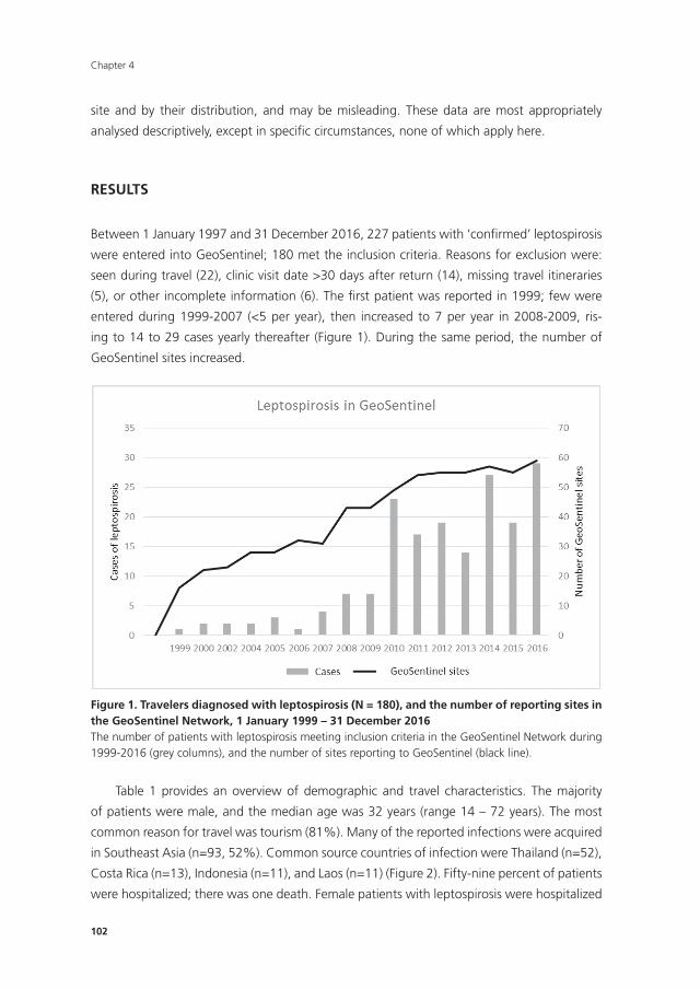
Chapter 4
102
site and by their distribution, and may be misleading. These data are most appropriately
analysed descriptively, except in specifi c circumstances, none of which apply here.
RESULTS
Between 1 January 1997 and 31 December 2016, 227 patients with ‘confi rmed’ leptospirosis
were entered into GeoSentinel; 180 met the inclusion criteria. Reasons for exclusion were:
seen during travel (22), clinic visit date >30 days after return (14), missing travel itineraries
(5), or other incomplete information (6). The fi rst patient was reported in 1999; few were
entered during 1999-2007 (<5 per year), then increased to 7 per year in 2008-2009, ris-
ing to 14 to 29 cases yearly thereafter (Figure 1). During the same period, the number of
GeoSentinel sites increased.
105
RESULTS
Between 1 January 1997 and 31 December 2016, 227 patients with ‘confirmed’ leptospirosis
were entered into GeoSentinel; 180 met the inclusion criteria. Reasons for exclusion were:
seen during travel (22), clinic visit date >30 days after return (14), missing travel itineraries
(5), or other incomplete information (6). The first patient was reported in 1999; few were
entered during 1999-2007 (<5 per year), then increased to 7 per year in 2008-2009, rising to
14 to 29 cases yearly thereafter (Figure 1). During the same period, the number of
GeoSentinel sites increased.
Figure 1. Travelers diagnosed with leptospirosis (N = 180), and the number of reporting sites in the GeoSentinel Network, 1 January 1999 – 31 December 2016 The number of patients with leptospirosis meeting inclusion criteria in the GeoSentinel Network during 1999-2016 (grey columns), and the number of sites reporting to GeoSentinel (black line).
Table 1 provides an overview of demographic and travel characteristics. The majority of
patients were male, and the median age was 32 years (range 14 – 72 years). The most
common reason for travel was tourism (81%). Many of the reported infections were acquired
in Southeast Asia (n=93, 52%). Common source countries of infection were Thailand (n=52),
Costa Rica (n=13), Indonesia (n=11), and Laos (n=11) (Figure 2). Fifty-nine percent of patients
were hospitalized; there was one death. Female patients with leptospirosis were hospitalized
less frequently than male patients, and travelers to Southeast Asia were hospitalized slightly
Figure 1. Travelers diagnosed with leptospirosis (n = 180), and the number of reporting sites in the GeoSentinel network, 1 January 1999 – 31 December 2016The number of patients with leptospirosis meeting inclusion criteria in the GeoSentinel Network during 1999-2016 (grey columns), and the number of sites reporting to GeoSentinel (black line).
Table 1 provides an overview of demographic and travel characteristics. The majority
of patients were male, and the median age was 32 years (range 14 – 72 years). The most
common reason for travel was tourism (81%). Many of the reported infections were acquired
in Southeast Asia (n=93, 52%). Common source countries of infection were Thailand (n=52),
Costa Rica (n=13), Indonesia (n=11), and Laos (n=11) (Figure 2). Fifty-nine percent of patients
were hospitalized; there was one death. Female patients with leptospirosis were hospitalized
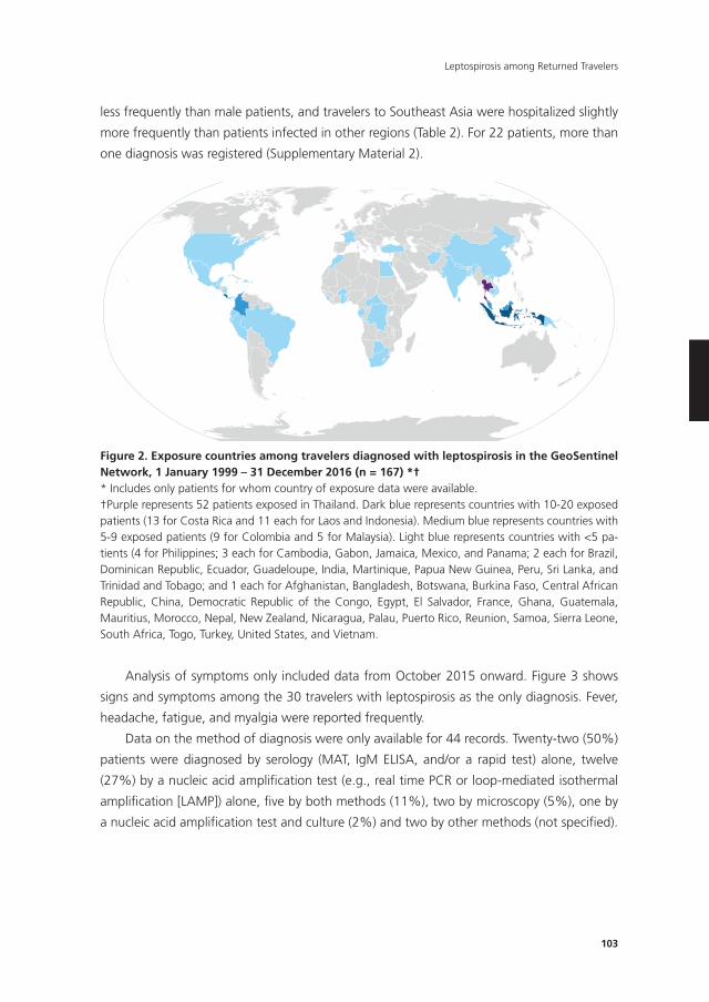
103
Leptospirosis among Returned Travelers
less frequently than male patients, and travelers to Southeast Asia were hospitalized slightly
more frequently than patients infected in other regions (Table 2). For 22 patients, more than
one diagnosis was registered (Supplementary Material 2).
106
more frequently than patients infected in other regions (Table 2). For 22 patients, more than
one diagnosis was registered (Supplementary Material 2).
Figure 2. Exposure countries among travelers diagnosed with leptospirosis in the GeoSentinel Network, 1 January 1999 – 31 December 2016 (n = 167) *† * Includes only patients for whom country of exposure data were available. †Purple represents 52 patients exposed in Thailand. Dark blue represents countries with 10-20 exposed patients (13 for Costa Rica and 11 each for Laos and Indonesia). Medium blue represents countries with 5-9 exposed patients (9 for Colombia and 5 for Malaysia). Light blue represents countries with <5 patients (4 for Philippines; 3 each for Cambodia, Gabon, Jamaica, Mexico, and Panama; 2 each for Brazil, Dominican Republic, Ecuador, Guadeloupe, India, Martinique, Papua New Guinea, Peru, Sri Lanka, and Trinidad and Tobago; and 1 each for Afghanistan, Bangladesh, Botswana, Burkina Faso, Central African Republic, China, Democratic Republic of the Congo, Egypt, El Salvador, France, Ghana, Guatemala, Mauritius, Morocco, Nepal, New Zealand, Nicaragua, Palau, Puerto Rico, Reunion, Samoa, Sierra Leone, South Africa, Togo, Turkey, United States, and Vietnam.
Analysis of symptoms only included data from October 2015 onward. Figure 3 shows signs
and symptoms among the 30 travelers with leptospirosis as the only diagnosis. Fever,
headache, fatigue, and myalgia were reported frequently.
Figure 2. Exposure countries among travelers diagnosed with leptospirosis in the GeoSentinel network, 1 January 1999 – 31 December 2016 (n = 167) *†* Includes only patients for whom country of exposure data were available.†Purple represents 52 patients exposed in Thailand. Dark blue represents countries with 10-20 exposed patients (13 for Costa Rica and 11 each for Laos and Indonesia). Medium blue represents countries with 5-9 exposed patients (9 for Colombia and 5 for Malaysia). Light blue represents countries with <5 pa-tients (4 for Philippines; 3 each for Cambodia, Gabon, Jamaica, Mexico, and Panama; 2 each for Brazil, Dominican Republic, Ecuador, Guadeloupe, India, Martinique, Papua New Guinea, Peru, Sri Lanka, and Trinidad and Tobago; and 1 each for Afghanistan, Bangladesh, Botswana, Burkina Faso, Central African Republic, China, Democratic Republic of the Congo, Egypt, El Salvador, France, Ghana, Guatemala, Mauritius, Morocco, Nepal, New Zealand, Nicaragua, Palau, Puerto Rico, Reunion, Samoa, Sierra Leone, South Africa, Togo, Turkey, United States, and Vietnam.
Analysis of symptoms only included data from October 2015 onward. Figure 3 shows
signs and symptoms among the 30 travelers with leptospirosis as the only diagnosis. Fever,
headache, fatigue, and myalgia were reported frequently.
Data on the method of diagnosis were only available for 44 records. Twenty-two (50%)
patients were diagnosed by serology (MAT, IgM ELISA, and/or a rapid test) alone, twelve
(27%) by a nucleic acid amplification test (e.g., real time PCR or loop-mediated isothermal
amplification [LAMP]) alone, five by both methods (11%), two by microscopy (5%), one by
a nucleic acid amplification test and culture (2%) and two by other methods (not specified).
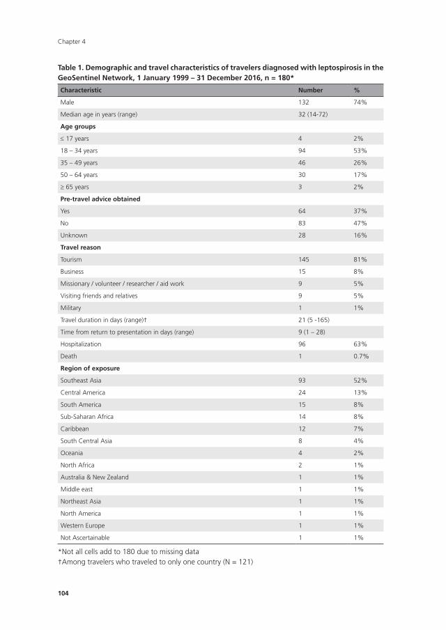
Chapter 4
104
Table 1. Demographic and travel characteristics of travelers diagnosed with leptospirosis in the GeoSentinel network, 1 January 1999 – 31 December 2016, n = 180*
Characteristic number %
Male 132 74%
Median age in years (range) 32 (14-72)
Age groups
≤ 17 years 4 2%
18 – 34 years 94 53%
35 – 49 years 46 26%
50 – 64 years 30 17%
≥ 65 years 3 2%
Pre-travel advice obtained
Yes 64 37%
No 83 47%
Unknown 28 16%
Travel reason
Tourism 145 81%
Business 15 8%
Missionary / volunteer / researcher / aid work 9 5%
Visiting friends and relatives 9 5%
Military 1 1%
Travel duration in days (range)† 21 (5 -165)
Time from return to presentation in days (range) 9 (1 – 28)
Hospitalization 96 63%
Death 1 0.7%
Region of exposure
Southeast Asia 93 52%
Central America 24 13%
South America 15 8%
Sub-Saharan Africa 14 8%
Caribbean 12 7%
South Central Asia 8 4%
Oceania 4 2%
North Africa 2 1%
Australia & New Zealand 1 1%
Middle east 1 1%
Northeast Asia 1 1%
North America 1 1%
Western Europe 1 1%
Not Ascertainable 1 1%
*Not all cells add to 180 due to missing data†Among travelers who traveled to only one country (N = 121)
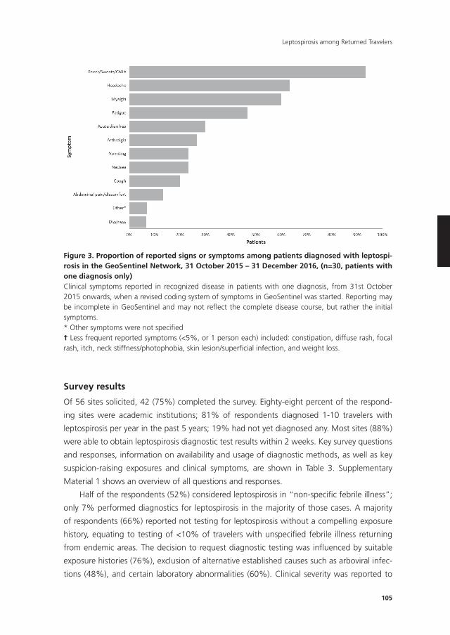
105
Leptospirosis among Returned Travelers
107
Figure 3. Proportion of reported signs or symptoms among patients diagnosed with leptospirosis in the GeoSentinel Network, 31 October 2015 – 31 December 2016, (n=30, patients with one diagnosis only) Clinical symptoms reported in recognized disease in patients with one diagnosis, from 31st October 2015 onwards, when a revised coding system of symptoms in GeoSentinel was started. Reporting may be incomplete in GeoSentinel and may not reflect the complete disease course, but rather the initial symptoms. * Other symptoms were not specified
† Less frequent reported symptoms (<5%, or 1 person each) included: constipation, diffuse rash, focal rash, itch, neck stiffness/photophobia, skin lesion/superficial infection, and weight loss.
Data on the method of diagnosis were only available for 44 records. Twenty-two (50%)
patients were diagnosed by serology (MAT, IgM ELISA, and/or a rapid test) alone, twelve
(27%) by a nucleic acid amplification test (e.g., real time PCR or loop-mediated isothermal
amplification [LAMP]) alone, five by both methods (11%), two by microscopy (5%), one by a
nucleic acid amplification test and culture (2%) and two by other methods (not specified).
Survey results
Of 56 sites solicited, 42 (75%) completed the survey. Eighty-eight percent of the responding
sites were academic institutions; 81% of respondents diagnosed 1-10 travelers with
leptospirosis per year in the past 5 years; 19% had not yet diagnosed any. Most sites (88%)
were able to obtain leptospirosis diagnostic test results within 2 weeks. Key survey questions
and responses, information on availability and usage of diagnostic methods, as well as key
Figure 3. Proportion of reported signs or symptoms among patients diagnosed with leptospi-rosis in the GeoSentinel network, 31 October 2015 – 31 December 2016, (n=30, patients with one diagnosis only)Clinical symptoms reported in recognized disease in patients with one diagnosis, from 31st October 2015 onwards, when a revised coding system of symptoms in GeoSentinel was started. Reporting may be incomplete in GeoSentinel and may not refl ect the complete disease course, but rather the initial symptoms.* Other symptoms were not specifi ed† Less frequent reported symptoms (<5%, or 1 person each) included: constipation, diffuse rash, focal rash, itch, neck stiffness/photophobia, skin lesion/superfi cial infection, and weight loss.
Survey results
Of 56 sites solicited, 42 (75%) completed the survey. Eighty-eight percent of the respond-
ing sites were academic institutions; 81% of respondents diagnosed 1-10 travelers with
leptospirosis per year in the past 5 years; 19% had not yet diagnosed any. Most sites (88%)
were able to obtain leptospirosis diagnostic test results within 2 weeks. Key survey questions
and responses, information on availability and usage of diagnostic methods, as well as key
suspicion-raising exposures and clinical symptoms, are shown in Table 3. Supplementary
Material 1 shows an overview of all questions and responses.
Half of the respondents (52%) considered leptospirosis in “non-specifi c febrile illness”;
only 7% performed diagnostics for leptospirosis in the majority of those cases. A majority
of respondents (66%) reported not testing for leptospirosis without a compelling exposure
history, equating to testing of <10% of travelers with unspecifi ed febrile illness returning
from endemic areas. The decision to request diagnostic testing was infl uenced by suitable
exposure histories (76%), exclusion of alternative established causes such as arboviral infec-
tions (48%), and certain laboratory abnormalities (60%). Clinical severity was reported to
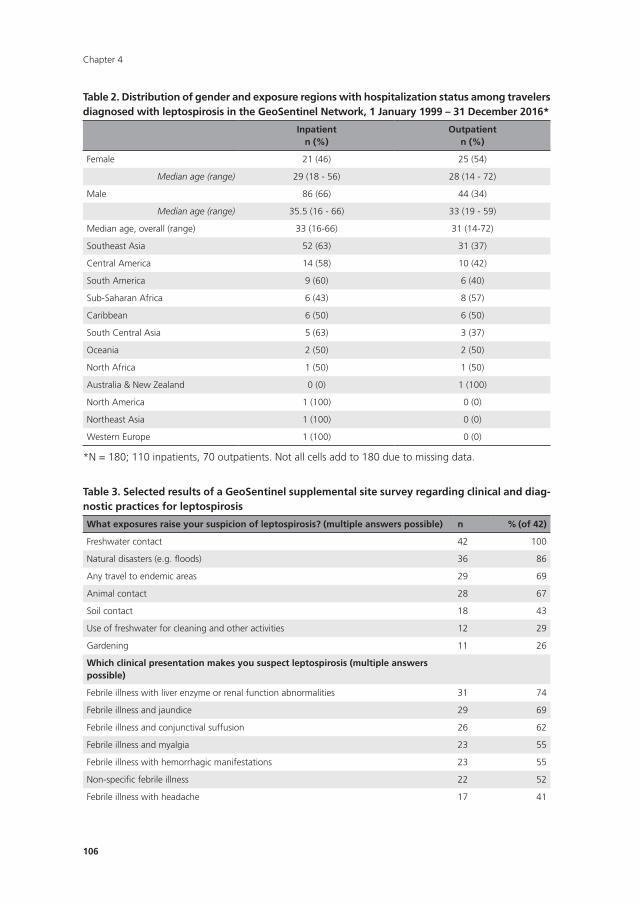
Chapter 4
106
Table 2. Distribution of gender and exposure regions with hospitalization status among travelers diagnosed with leptospirosis in the GeoSentinel network, 1 January 1999 – 31 December 2016*
Inpatientn (%)
Outpatientn (%)
Female 21 (46) 25 (54)
Median age (range) 29 (18 - 56) 28 (14 - 72)
Male 86 (66) 44 (34)
Median age (range) 35.5 (16 - 66) 33 (19 - 59)
Median age, overall (range) 33 (16-66) 31 (14-72)
Southeast Asia 52 (63) 31 (37)
Central America 14 (58) 10 (42)
South America 9 (60) 6 (40)
Sub-Saharan Africa 6 (43) 8 (57)
Caribbean 6 (50) 6 (50)
South Central Asia 5 (63) 3 (37)
Oceania 2 (50) 2 (50)
North Africa 1 (50) 1 (50)
Australia & New Zealand 0 (0) 1 (100)
North America 1 (100) 0 (0)
Northeast Asia 1 (100) 0 (0)
Western Europe 1 (100) 0 (0)
*N = 180; 110 inpatients, 70 outpatients. Not all cells add to 180 due to missing data.
Table 3. Selected results of a GeoSentinel supplemental site survey regarding clinical and diag-nostic practices for leptospirosis
What exposures raise your suspicion of leptospirosis? (multiple answers possible) n % (of 42)
Freshwater contact 42 100
Natural disasters (e.g. floods) 36 86
Any travel to endemic areas 29 69
Animal contact 28 67
Soil contact 18 43
Use of freshwater for cleaning and other activities 12 29
Gardening 11 26
Which clinical presentation makes you suspect leptospirosis (multiple answers possible)
Febrile illness with liver enzyme or renal function abnormalities 31 74
Febrile illness and jaundice 29 69
Febrile illness and conjunctival suffusion 26 62
Febrile illness and myalgia 23 55
Febrile illness with hemorrhagic manifestations 23 55
Non-specific febrile illness 22 52
Febrile illness with headache 17 41
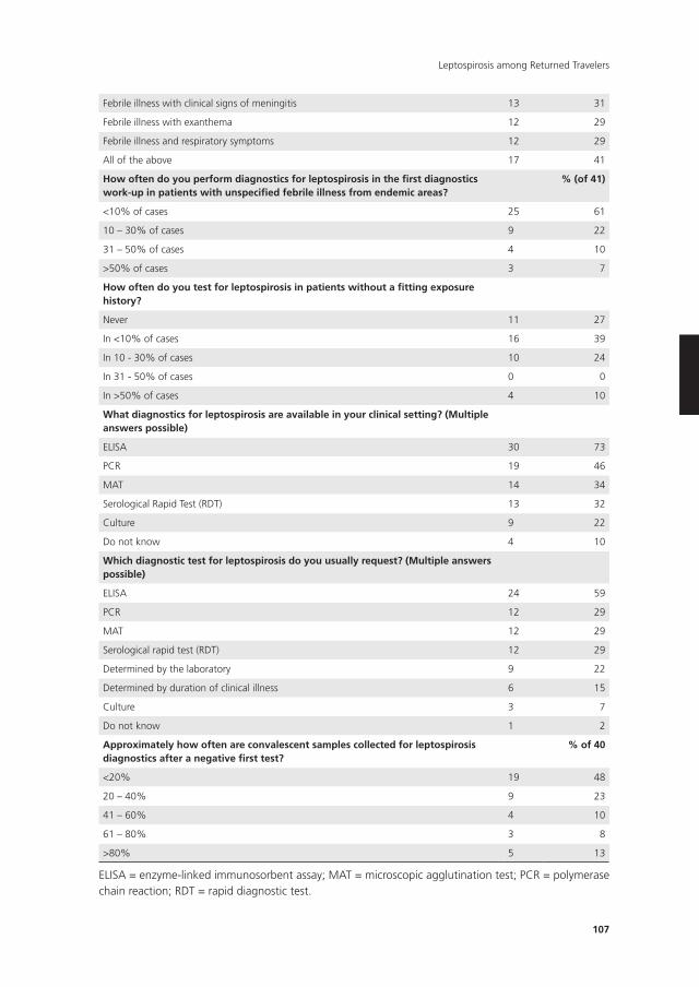
107
Leptospirosis among Returned Travelers
Febrile illness with clinical signs of meningitis 13 31
Febrile illness with exanthema 12 29
Febrile illness and respiratory symptoms 12 29
All of the above 17 41
How often do you perform diagnostics for leptospirosis in the first diagnostics work-up in patients with unspecified febrile illness from endemic areas?
% (of 41)
<10% of cases 25 61
10 – 30% of cases 9 22
31 – 50% of cases 4 10
>50% of cases 3 7
How often do you test for leptospirosis in patients without a fitting exposure history?
Never 11 27
In <10% of cases 16 39
In 10 - 30% of cases 10 24
In 31 - 50% of cases 0 0
In >50% of cases 4 10
What diagnostics for leptospirosis are available in your clinical setting? (Multiple answers possible)
ELISA 30 73
PCR 19 46
MAT 14 34
Serological Rapid Test (RDT) 13 32
Culture 9 22
Do not know 4 10
Which diagnostic test for leptospirosis do you usually request? (Multiple answers possible)
ELISA 24 59
PCR 12 29
MAT 12 29
Serological rapid test (RDT) 12 29
Determined by the laboratory 9 22
Determined by duration of clinical illness 6 15
Culture 3 7
Do not know 1 2
Approximately how often are convalescent samples collected for leptospirosis diagnostics after a negative first test?
% of 40
<20% 19 48
20 – 40% 9 23
41 – 60% 4 10
61 – 80% 3 8
>80% 5 13
ELISA = enzyme-linked immunosorbent assay; MAT = microscopic agglutination test; PCR = polymerase chain reaction; RDT = rapid diagnostic test.
Chapter 4
108
have no influence on testing by 69% of respondents. Most sites (63%) continued to search
for a definitive diagnosis (in >50% of patients), even when the patient was improving after
empiric antibiotic therapy. Sixty percent of respondents would prescribe empiric doxycycline
after exclusion of key differential diagnoses such as malaria, typhoid fever, and arboviral
infections (Supplementary Material 1).
DISCUSSIOn
GeoSentinel surveillance data
Leptospirosis was an infrequent diagnosis among febrile returning travelers in GeoSentinel,
but was identified mostly among those who had traveled for tourism purposes. With increas-
ing popularity of tourism in tropical areas,32 including active, environmental exposures like
rafting, canoeing, and triathlons, the risk of leptospirosis acquisition among travelers may
be increasing, as illustrated in previous case reports.37, 41, 42 Known high-risk areas, such as
Southeast Asia and Central America, were the most common regions of exposure in our
database. The top countries where leptospirosis was acquired, Thailand, Costa Rica and Laos,
are not necessarily known to be most endemic,1, 43 but rather have higher numbers of travel-
ers who are potentially engaged in freshwater recreational activities. The epidemiology of
leptospirosis worldwide1, 43 should be considered when evaluating the ill traveler presenting
with fever.
Increased risk of leptospirosis has recently been observed in more temperate regions,34, 44, 45
possibly influenced by climate change or different types of risk activities.12, 45 Case series
have reported significant proportions of travelers infected within Europe as well.4, 33, 35, 46 In
the Netherlands, 318 imported cases of leptospirosis were diagnosed between 1925 and
2008; 134 (42.1%) were acquired in Asia and 132 (41.5%) in Europe. France is a major
tourist destination, with one of the highest reported leptospirosis incidences in Europe.47
Because travelers returning ill from developed temperate-zone countries may be less likely to
visit GeoSentinel clinics, patients with leptospirosis acquired in those regions may be under-
represented here.
An overall increase in patients with leptospirosis reported within the GeoSentinel net-
work was observed over time; however, this was likely associated with an increase in the
number of sites reporting in the network in the same period. Alternatively, it is possible that
clinicians have become more aware of the illness, and that the wider introduction of PCR
provided an option for more accurate diagnosis.
Only one fatality was reported to GeoSentinel, despite the potential risk of mortality
associated with leptospirosis;1 however, the GeoSentinel surveillance approach may not
capture deaths efficiently.38
109
Leptospirosis among Returned Travelers
Most patients in this series were in the 18- to 49-year age group and reported non-
specific symptoms, consistent with other published reports.33, 46, 48 Fever was present in
almost all patients; neurologic and respiratory symptoms were present in lower proportions
than found in other reports,13 but similar to a small case series in travelers.37 It is also pos-
sible that the disease presentation was different in these travelers than is typically seen in
leptospirosis in endemic areas. Other possibilities are that leptospirosis was not recognized in
patients presenting with those rarer reported symptoms, that they were treated with empiric
doxycycline, or that patients with these more severe forms of disease presented elsewhere. A
prospective study would be needed to better address these uncertainties.
Most patients were male, although the proportion of female patients (26%) was higher
than in some other large case series (reported proportions of <10%).33, 37, 46, 48 A higher
proportion of the reported hospitalized cases were male compared with female patients,
akin to other reports.33, 49 Some possible factors that can contribute to male predominance
in leptospirosis include a greater tendency to participate in high-risk outdoor activities, later
presentation for medical evaluation, androgenic steroids and other biologic factors,49, 50 or a
higher infectious inoculum. The relatively high percentage of female patients in our analysis
could signal a shift in behavior of female travelers, increasing their risk of contracting the
disease. Female travelers should be considered equally at risk as those who are male with
respect to leisure exposure, and leptospirosis should be routinely considered when evaluating
the febrile returned female traveler.
Site survey
Leptospirosis is an infection that is frequently under-diagnosed, especially in mild disease
presentations.17 In our survey among GeoSentinel clinicians, all reported seeing relatively
few leptospirosis patients each year (<10). The majority of respondents (75% of all sites)
worked at an academic institution, where leptospirosis diagnostics are expected to be readily
available. Molecular detection techniques are the cornerstone of diagnosis of leptospirosis
in the acute phase,26 but were available in only half of the clinics, and were actually used
in less than a third of the clinics. In addition, convalescent samples were reported to have
been collected in a minority of cases after a negative acute serology, despite the fact that
antibodies only appear in the blood 5-7 days or sometimes after 10 days or longer following
disease onset.2 Possibly, patients with mild disease improved on treatment and abandoned
follow-up, forgoing further testing.
Half of the clinicians considered leptospirosis in a case of “non-specified febrile illness”,
but very few actually performed diagnostics for it. Few clinicians reported testing patients
without a suggestive exposure history. Classical exposures, such as freshwater contact and
floods, were well known among clinicians. In the general population the infection mecha-
nism often remains unclear, and often there is no classical exposure history.3 Data on travelers
are scarce, but the case series that exist do report clear exposure histories.35, 37 Nevertheless,
Chapter 4
110
pre-selecting patients based only on a well-defined exposure history may result in under-
diagnosis of leptospirosis among patients without a clear-cut exposure, especially when they
returned from highly endemic areas.
Although the classic Weil’s triad of fever, jaundice, renal failure, with or without ac-
companying hemorrhagic features, was well recognized, other severe disease manifestations
were less known among GeoSentinel Surveillance Network respondents. Aseptic meningitis
can occur in up to 25% of cases,13 but this manifestation was recognized by relatively few
respondents (31%). Pulmonary hemorrhage is an important but under-recognized form of
the disease, and outbreaks with only this manifestation have been described.14, 16 Almost half
of the respondents did not include leptospirosis in the differential diagnosis of the febrile
traveler presenting with hemorrhagic disease manifestations in the absence of the classic
Weil’s triad. In this analysis of returned travelers, the lesser-known disease manifestations
were not frequently reported, possibly due to the nature of symptom data collection in
GeoSentinel, which combines specific symptoms into broad systemic categories.40
The results of our survey on clinical practices in specialized travel medicine settings
suggest that the number of leptospirosis cases in travelers reported in the GeoSentinel
Surveillance system is likely to be an underestimate. This may be due to suboptimal access to
diagnostic testing at many sites, especially in the setting where acute disease is encountered.
Furthermore, we found that the diagnosis of leptospirosis may not be routinely considered
when clear-cut exposure histories are absent and when some rather typical clinical features
are missing. Based on our site survey, there is a need for improved awareness among clini-
cians about the spectrum of exposures, clinical presentations, and diagnostic considerations.
Strengths and limitations
In addition to those already discussed, the GeoSentinel surveillance data have several other
limitations and may not be generalizable to the traveler population as a whole.38, 51, 52, 53
GeoSentinel reporting may be biased towards capture of more complicated or more severe
disease, since most GeoSentinel sites are also academic institutions. Overall, hospitalization
may be underestimated in GeoSentinel, but there are some sites that almost exclusively
capture data from the inpatient setting. Therefore, the high proportion of hospitalizations
should be interpreted carefully. Furthermore, leptospirosis may have been missed among
travelers presenting with atypical exposures or symptoms, or presentations of milder, self-
limited disease, and may have had an influence on the results. GeoSentinel preferentially
captures travelers returning from tropical regions due to the nature of the clinics that make
up the network; so, travelers with disease acquired in temperate regions are likely under-
represented. GeoSentinel criteria for diagnosing patients with leptospirosis may differ from
the definitions used by national reference laboratories and other reporting systems, limiting
direct comparison with data from those sources. Despite these limitations, this is one of the
111
Leptospirosis among Returned Travelers
largest series of leptospirosis in travelers published to date and provides valuable information
about the epidemiology of leptospirosis in international travelers.
A limitation of the network survey is that the answers were captured from clinicians spe-
cialized in travel medicine and may not be representative of all clinical practices in a particular
institution. Furthermore, the data are self-reported and may therefore not be reflective of
actual practice. Some questions allowed multiple answers, leading to respondents choosing
incompatible answers, making the interpretation more complex. However, the relatively high
response rate of 75% of GeoSentinel sites in our view accounts for a representative survey
among our global network of mostly academic travel clinics.
Conclusions
Leptospirosis may be an infrequently encountered cause of substantial morbidity among
international travelers that may not be clinically suspected. Although leptospirosis was
most frequently diagnosed among persons visiting highly endemic countries, it may occur
elsewhere and warrants broader consideration in the differential of the ill traveler. Given
the laboratory diagnostic challenges and non-specific presentation of many clinically evi-
dent cases, the burden of reported disease is likely underestimated, as supported by other
published data.1, 18, 19, 20 We recommend enhancement of awareness about leptospirosis and
heightened clinical suspicion when evaluating the ill traveler. Laboratories need to have up-to-
date diagnostic methods available; molecular detection techniques are key to early diagnosis,
which is helpful for the early initiation of treatment that may substantially reduce morbidity
and improve outcomes. Empiric treatment in cases of high suspicion is recommended. Efforts
to improve knowledge among clinicians regarding (the often unclear) exposures and clinical
presentations are needed.
Disclaimer
The findings and conclusions in this report are those of the authors and do not necessarily
represent the official position of the Centers for Disease Control and Prevention.
Acknowledgements
We thank Jodi Metzgar, Kayce Maisel, and Sarah Mosley for their administrative support.
Chapter 4
112
REFEREnCES
1. Costa F, Hagan JE, Calcagno J, Kane M, Torgerson P, Martinez-Silveira MS, Stein C, Abela-Ridder
B, Ko AI, 2015. Global Morbidity and Mortality of Leptospirosis: A Systematic Review. PLoS Negl
Trop Dis 9: e0003898.
2. Levett PN, 2001. Leptospirosis. Clin Microbiol Rev 14: 296-326.
3. Mwachui MA, Crump L, Hartskeerl R, Zinsstag J, Hattendorf J, 2015. Environmental and Behav-
ioural Determinants of Leptospirosis Transmission: A Systematic Review. PLoS Negl Trop Dis 9:
e0003843.
4. Jansen A, Schoneberg I, Frank C, Alpers K, Schneider T, Stark K, 2005. Leptospirosis in Germany,
1962-2003. Emerging Infectious Diseases 11: 1048-1054.
5. Desai S, van Treeck U, Lierz M, Espelage W, Zota L, Sarbu A, Czerwinski M, Sadkowska-Todys
M, Avdicova M, Reetz J, Luge E, Guerra B, Nockler K, Jansen A, 2009. Resurgence of field fever
in a temperate country: an epidemic of leptospirosis among seasonal strawberry harvesters in
Germany in 2007. Clin Infect Dis 48: 691-7.
6. Flannery B, Pereira MM, Velloso LdF, Carvalho CdC, De Codes LG, Orrico GdS, Dourado CM, Riley
LW, Reis MG, Ko AI, 2001. Referral pattern of leptospirosis cases during a large urban epidemic
of dengue. Am J Trop Med Hyg 65: 657-63.
7. Ko AI, Galvao Reis M, Ribeiro Dourado CM, Johnson WD, Jr., Riley LW, 1999. Urban epidemic of
severe leptospirosis in Brazil. Salvador Leptospirosis Study Group. Lancet 354: 820-5.
8. Costa F, Ribeiro GS, Felzemburgh RD, Santos N, Reis RB, Santos AC, Fraga DB, Araujo WN,
Santana C, Childs JE, Reis MG, Ko AI, 2014. Influence of household rat infestation on leptospira
transmission in the urban slum environment. PLoS Negl Trop Dis 8: e3338.
9. Costa F, Wunder EA, Jr., De Oliveira D, Bisht V, Rodrigues G, Reis MG, Ko AI, Begon M, Childs JE,
2015. Patterns in Leptospira Shedding in Norway Rats (Rattus norvegicus) from Brazilian Slum
Communities at High Risk of Disease Transmission. PLoS Negl Trop Dis 9: e0003819.
10. Felzemburgh RD, Ribeiro GS, Costa F, Reis RB, Hagan JE, Melendez AX, Fraga D, Santana FS,
Mohr S, dos Santos BL, Silva AQ, Santos AC, Ravines RR, Tassinari WS, Carvalho MS, Reis MG,
Ko AI, 2014. Prospective study of leptospirosis transmission in an urban slum community: role of
poor environment in repeated exposures to the Leptospira agent. PLoS Negl Trop Dis 8: e2927.
11. Hagan JE, Moraga P, Costa F, Capian N, Ribeiro GS, Wunder EA, Felzemburgh RDM, Reis RB, Nery
N, Santana FS, Fraga D, dos Santos BL, Santos AC, Queiroz A, Tassinari W, Carvalho MS, Reis
MG, Diggle PJ, Ko AI, 2016. Spatiotemporal Determinants of Urban Leptospirosis Transmission:
Four-Year Prospective Cohort Study of Slum Residents in Brazil. PLoS Neglected Tropical Diseases
10: e0004275.
12. Lau CL, Smythe LD, Craig SB, Weinstein P, 2010. Climate change, flooding, urbanisation and
leptospirosis: fuelling the fire? Transactions of the Royal Society of Tropical Medicine and Hygiene
104: 631-638.
13. Bharti AR, Nally JE, Ricaldi JN, Matthias MA, Diaz MM, Lovett MA, Levett PN, Gilman RH, Willig
MR, Gotuzzo E, Vinetz JM, 2003. Leptospirosis: a zoonotic disease of global importance. Lancet
Infect Dis 3: 757-71.
14. Zaki SR, Shieh WJ, 1996. Leptospirosis associated with outbreak of acute febrile illness and
pulmonary haemorrhage, Nicaragua, 1995. The Epidemic Working Group at Ministry of Health
in Nicaragua. Lancet 347: 535-6.
15. Helmerhorst HJ, van Tol EN, Tuinman PR, de Vries PJ, Hartskeerl RA, Grobusch MP, Hovius JW,
2012. Severe pulmonary manifestation of leptospirosis. Neth J Med 70: 215-21.
113
Leptospirosis among Returned Travelers
16. Segura ER, Ganoza CA, Campos K, Ricaldi JN, Torres S, Silva H, Cespedes MJ, Matthias MA,
Swancutt MA, Lopez Linan R, Gotuzzo E, Guerra H, Gilman RH, Vinetz JM, 2005. Clinical spec-
trum of pulmonary involvement in leptospirosis in a region of endemicity, with quantification of
leptospiral burden. Clin Infect Dis 40: 343-51.
17. 2003. World Health Organization. Human leptospirosis: guidance for diagnosis, surveillance and
control.
18. Suppiah J, Chan SY, Ng MW, Khaw YS, Ching SM, Mat-Nor LA, Ahmad-Najimudin NA, Chee HY,
2017. Clinical predictors of dengue fever co-infected with leptospirosis among patients admitted
for dengue fever - a pilot study. J Biomed Sci 24: 40.
19. Ribeiro P, Bhatt N, Ali S, Monteiro V, da Silva E, Balassiano IT, Aquino C, de Deus N, Guiliche O,
Muianga AF, Alho PJ, Lara Evaristo V, Manhica I, Medeiros M, Gudo ES, 2017. Seroepidemiology
of leptospirosis among febrile patients in a rapidly growing suburban slum and a flood-vulnerable
rural district in Mozambique, 2012-2014: Implications for the management of fever. Int J Infect
Dis 64: 50-57.
20. Reller ME, Wunder EA, Jr., Miles JJ, Flom JE, Mayorga O, Woods CW, Ko AI, Dumler JS, Matute
AJ, 2014. Unsuspected Leptospirosis Is a Cause of Acute Febrile Illness in Nicaragua. PLOS Ne-
glected Tropical Diseases 8: e2941.
21. Sharp TM, Rivera Garcia B, Perez-Padilla J, Galloway RL, Guerra M, Ryff KR, Haberling D, Ra-
makrishnan S, Shadomy S, Blau D, Tomashek KM, Bower WA, 2016. Early Indicators of Fatal
Leptospirosis during the 2010 Epidemic in Puerto Rico. PLoS Negl Trop Dis 10: e0004482.
22. Amilasan AS, Ujiie M, Suzuki M, Salva E, Belo MC, Koizumi N, Yoshimatsu K, Schmidt WP, Marte
S, Dimaano EM, Villarama JB, Ariyoshi K, 2012. Outbreak of leptospirosis after flood, the Philip-
pines, 2009. Emerg Infect Dis 18: 91-4.
23. Ahmed A, Engelberts MF, Boer KR, Ahmed N, Hartskeerl RA, 2009. Development and validation
of a real-time PCR for detection of pathogenic leptospira species in clinical materials. PLoS One
4: e7093.
24. Goris M, Leeflang M, Boer K, Goeijenbier M, Van Gorp E, Wagenaar J, Hartskeerl R, 2012.
Establishment of valid laboratory case definition for human leptospirosis. J Bacteriol Parasitol 3:
2.
25. Thaipadungpanit J, Chierakul W, Wuthiekanun V, Limmathurotsakul D, Amornchai P, Boonslip
S, Smythe LD, Limpaiboon R, Hoffmaster AR, Day NP, Peacock SJ, 2011. Diagnostic accuracy of
real-time PCR assays targeting 16S rRNA and lipL32 genes for human leptospirosis in Thailand: a
case-control study. PLoS One 6: e16236.
26. Waggoner JJ, Pinsky BA, 2016. Molecular diagnostics for human leptospirosis. Curr Opin Infect
Dis 29: 440-5.
27. Yang B, de Vries SG, Visser BJ, Nagel IM, Goris M, Leeflang MMG, Grobusch MP, Hartskeerl RA,
2015. Molecular and antigen detection tests for leptospirosis (protocol). Cochrane Database Syst
Rev.
28. Waggoner JJ, Abeynayake J, Balassiano I, Lefterova M, Sahoo MK, Liu Y, Vital-Brazil JM, Gresh
L, Balmaseda A, Harris E, Banaei N, Pinsky BA, 2014. Multiplex nucleic acid amplification test for
diagnosis of dengue fever, malaria, and leptospirosis. J Clin Microbiol 52: 2011-8.
29. de Vries SG, Visser BJ, Nagel IM, Goris MG, Hartskeerl RA, Grobusch MP, 2014. Leptospirosis in
Sub-Saharan Africa: a systematic review. Int J Infect Dis 28: 47-64.
30. Bandara M, Ananda M, Wickramage K, Berger E, Agampodi S, 2014. Globalization of leptospi-
rosis through travel and migration. Global Health 10: 61.
Chapter 4
114
31. Lau C, Smythe L, Weinstein P, 2010. Leptospirosis: an emerging disease in travellers. Travel Med
Infect Dis 8: 33-9.
32. 2016. UNWTO Tourism Highlights 2016. Available at: http://www.e-unwto.org/doi/
book/10.18111/9789284418145. Accessed.
33. Goris MG, Boer KR, Duarte TA, Kliffen SJ, Hartskeerl RA, 2013. Human leptospirosis trends, the
Netherlands, 1925-2008. Emerg Infect Dis 19: 371-8.
34. Pijnacker R, Goris MG, Te Wierik MJ, Broens EM, van der Giessen JW, de Rosa M, Wagenaar JA,
Hartskeerl RA, Notermans DW, Maassen K, Schimmer B, 2016. Marked increase in leptospirosis
infections in humans and dogs in the Netherlands, 2014. Euro Surveill 21.
35. Leshem E, Segal G, Barnea A, Yitzhaki S, Ostfeld I, Pitlik S, Schwartz E, 2010. Travel-related
leptospirosis in Israel: a nationwide study. Am J Trop Med Hyg 82: 459-63.
36. Christen JR, Savini H, Pierrou C, Boisnault G, Fournier PE, Kraemer P, Simon F, 2015. Two Cases of
Leptospirosis in French Travelers Returning From Koh Samui, Thailand. J Travel Med 22: 419-21.
37. van de Werve C, Perignon A, Jaureguiberry S, Bricaire F, Bourhy P, Caumes E, 2013. Travel-related
leptospirosis: a series of 15 imported cases. J Travel Med 20: 228-31.
38. Jensenius M, Han PV, Schlagenhauf P, Schwartz E, Parola P, Castelli F, von SF, Loutan L, Leder K,
Freedman DO, 2013. Acute and potentially life-threatening tropical diseases in western travelers-
-a GeoSentinel multicenter study, 1996-2011. Am J Trop Med Hyg 88: 397-404.
39. Freedman DO, Kozarsky PE, Weld LH, Cetron MS, 1999. GeoSentinel: the global emerging infec-
tions sentinel network of the International Society of Travel Medicine. J Travel Med 6: 94-8.
40. Harvey K, Esposito DH, Han P, Kozarsky P, Freedman DO, Plier DA, Sotir MJ, 2013. Surveillance
for travel-related disease--GeoSentinel Surveillance System, United States, 1997-2011. MMWR
Surveill Summ 62: 1-23.
41. Agampodi SB, Karunarathna D, Jayathilala N, Rathnayaka H, Agampodi TC, Karunanayaka L,
2014. Outbreak of leptospirosis after white-water rafting: sign of a shift from rural to recreational
leptospirosis in Sri Lanka? Epidemiol Infect 142: 843-6.
42. Pages F, Larrieu S, Simoes J, Lenabat P, Kurtkowiak B, Guernier V, Le Minter G, Lagadec E, Gomard
Y, Michault A, Jaffar-Bandjee MC, Dellagi K, Picardeau M, Tortosa P, Filleul L, 2016. Investigation
of a leptospirosis outbreak in triathlon participants, Reunion Island, 2013. Epidemiol Infect 144:
661-9.
43. Torgerson PR, Hagan JE, Costa F, Calcagno J, Kane M, Martinez-Silveira MS, Goris MGA, Stein
C, Ko AI, Abela-Ridder B, 2015. Global Burden of Leptospirosis: Estimated in Terms of Disability
Adjusted Life Years. PLOS Neglected Tropical Diseases 9: e0004122.
44. 2014. European Centre for Disease Prevention and Control. Annual epidemiological report 2014
– food- and waterborne diseases and zoonoses. Stockholm: ECDC.
45. Dupouey J, Faucher B, Edouard S, Richet H, Kodjo A, Drancourt M, Davoust B, 2014. Human
leptospirosis: an emerging risk in Europe? Comp Immunol Microbiol Infect Dis 37: 77-83.
46. Forbes AE, Zochowski WJ, Dubrey SW, Sivaprakasam V, 2012. Leptospirosis and Weil’s disease in
the UK. Qjm 105: 1151-62.
47. 2014. European Centre for Disease Prevention and Control. Annual epidemiological report 2014
– food- and waterborne diseases and zoonoses. Stockholm: ECDC.
48. Katz AR, Buchholz AE, Hinson K, Park SY, Effler PV, 2011. Leptospirosis in Hawaii, USA, 1999-
2008. Emerg Infect Dis 17: 221-6.
49. Jansen A, Stark K, Schneider T, Schoneberg I, 2007. Sex differences in clinical leptospirosis in
Germany: 1997-2005. Clin Infect Dis 44: e69-72.

115
Leptospirosis among Returned Travelers
50. Hubacek JA, Stuber F, Frohlich D, Book M, Wetegrove S, Ritter M, Rothe G, Schmitz G, 2001.
Gene variants of the bactericidal/permeability increasing protein and lipopolysaccharide binding
protein in sepsis patients: gender-specific genetic predisposition to sepsis. Crit Care Med 29:
557-61.
51. Jensenius M, Davis X, von Sonnenburg F, Schwartz E, Keystone JS, Leder K, Lopez-Velez R, Cau-
mes E, Cramer JP, Chen L, Parola P, 2009. Multicenter GeoSentinel analysis of rickettsial diseases
in international travelers, 1996-2008. Emerg Infect Dis 15: 1791-8.
52. Freedman DO, Weld LH, Kozarsky PE, Fisk T, Robins R, von Sonnenburg F, Keystone JS, Pandey
P, Cetron MS, 2006. Spectrum of disease and relation to place of exposure among ill returned
travelers. N Engl J Med 354: 119-30.
53. Wilson ME, Weld LH, Boggild A, Keystone JS, Kain KC, von Sonnenburg F, Schwartz E, 2007.
Fever in returned travelers: results from the GeoSentinel Surveillance Network. Clin Infect Dis 44:
1560-8.
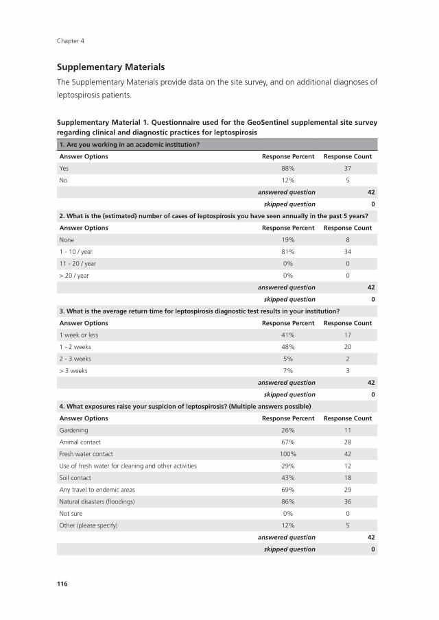
Chapter 4
116
Supplementary Materials
The Supplementary Materials provide data on the site survey, and on additional diagnoses of
leptospirosis patients.
Supplementary Material 1. Questionnaire used for the GeoSentinel supplemental site survey regarding clinical and diagnostic practices for leptospirosis
1. Are you working in an academic institution?
Answer Options Response Percent Response Count
Yes 88% 37
No 12% 5
answered question 42
skipped question 0
2. What is the (estimated) number of cases of leptospirosis you have seen annually in the past 5 years?
Answer Options Response Percent Response Count
None 19% 8
1 - 10 / year 81% 34
11 - 20 / year 0% 0
> 20 / year 0% 0
answered question 42
skipped question 0
3. What is the average return time for leptospirosis diagnostic test results in your institution?
Answer Options Response Percent Response Count
1 week or less 41% 17
1 - 2 weeks 48% 20
2 - 3 weeks 5% 2
> 3 weeks 7% 3
answered question 42
skipped question 0
4. What exposures raise your suspicion of leptospirosis? (Multiple answers possible)
Answer Options Response Percent Response Count
Gardening 26% 11
Animal contact 67% 28
Fresh water contact 100% 42
Use of fresh water for cleaning and other activities 29% 12
Soil contact 43% 18
Any travel to endemic areas 69% 29
Natural disasters (floodings) 86% 36
Not sure 0% 0
Other (please specify) 12% 5
answered question 42
skipped question 0
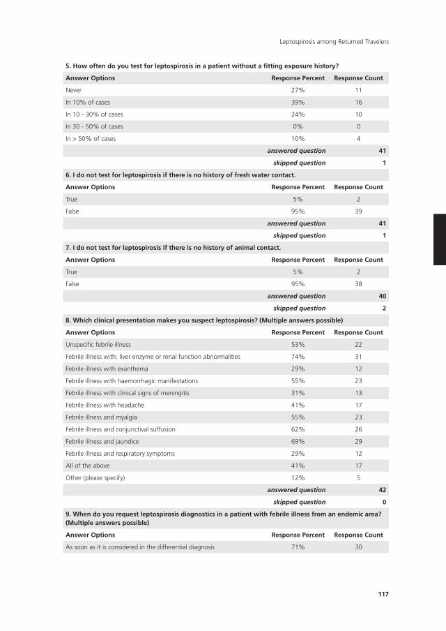
117
Leptospirosis among Returned Travelers
5. How often do you test for leptospirosis in a patient without a fitting exposure history?
Answer Options Response Percent Response Count
Never 27% 11
In 10% of cases 39% 16
In 10 - 30% of cases 24% 10
In 30 - 50% of cases 0% 0
In > 50% of cases 10% 4
answered question 41
skipped question 1
6. I do not test for leptospirosis if there is no history of fresh water contact.
Answer Options Response Percent Response Count
True 5% 2
False 95% 39
answered question 41
skipped question 1
7. I do not test for leptospirosis if there is no history of animal contact.
Answer Options Response Percent Response Count
True 5% 2
False 95% 38
answered question 40
skipped question 2
8. Which clinical presentation makes you suspect leptospirosis? (Multiple answers possible)
Answer Options Response Percent Response Count
Unspecific febrile illness 53% 22
Febrile illness with: liver enzyme or renal function abnormalities 74% 31
Febrile illness with exanthema 29% 12
Febrile illness with haemorrhagic manifestations 55% 23
Febrile illness with clinical signs of meningitis 31% 13
Febrile illness with headache 41% 17
Febrile illness and myalgia 55% 23
Febrile illness and conjunctival suffusion 62% 26
Febrile illness and jaundice 69% 29
Febrile illness and respiratory symptoms 29% 12
All of the above 41% 17
Other (please specify) 12% 5
answered question 42
skipped question 0
9. When do you request leptospirosis diagnostics in a patient with febrile illness from an endemic area? (Multiple answers possible)
Answer Options Response Percent Response Count
As soon as it is considered in the differential diagnosis 71% 30
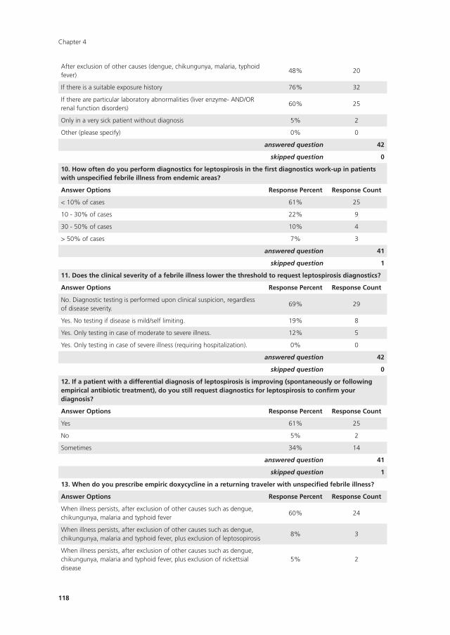
Chapter 4
118
After exclusion of other causes (dengue, chikungunya, malaria, typhoid fever)
48% 20
If there is a suitable exposure history 76% 32
If there are particular laboratory abnormalities (liver enzyme- AND/OR renal function disorders)
60% 25
Only in a very sick patient without diagnosis 5% 2
Other (please specify) 0% 0
answered question 42
skipped question 0
10. How often do you perform diagnostics for leptospirosis in the first diagnostics work-up in patients with unspecified febrile illness from endemic areas?
Answer Options Response Percent Response Count
< 10% of cases 61% 25
10 - 30% of cases 22% 9
30 - 50% of cases 10% 4
> 50% of cases 7% 3
answered question 41
skipped question 1
11. Does the clinical severity of a febrile illness lower the threshold to request leptospirosis diagnostics?
Answer Options Response Percent Response Count
No. Diagnostic testing is performed upon clinical suspicion, regardless of disease severity.
69% 29
Yes. No testing if disease is mild/self limiting. 19% 8
Yes. Only testing in case of moderate to severe illness. 12% 5
Yes. Only testing in case of severe illness (requiring hospitalization). 0% 0
answered question 42
skipped question 0
12. If a patient with a differential diagnosis of leptospirosis is improving (spontaneously or following empirical antibiotic treatment), do you still request diagnostics for leptospirosis to confirm your diagnosis?
Answer Options Response Percent Response Count
Yes 61% 25
No 5% 2
Sometimes 34% 14
answered question 41
skipped question 1
13. When do you prescribe empiric doxycycline in a returning traveler with unspecified febrile illness?
Answer Options Response Percent Response Count
When illness persists, after exclusion of other causes such as dengue, chikungunya, malaria and typhoid fever
60% 24
When illness persists, after exclusion of other causes such as dengue, chikungunya, malaria and typhoid fever, plus exclusion of leptosopirosis
8% 3
When illness persists, after exclusion of other causes such as dengue, chikungunya, malaria and typhoid fever, plus exclusion of rickettsial disease
5% 2
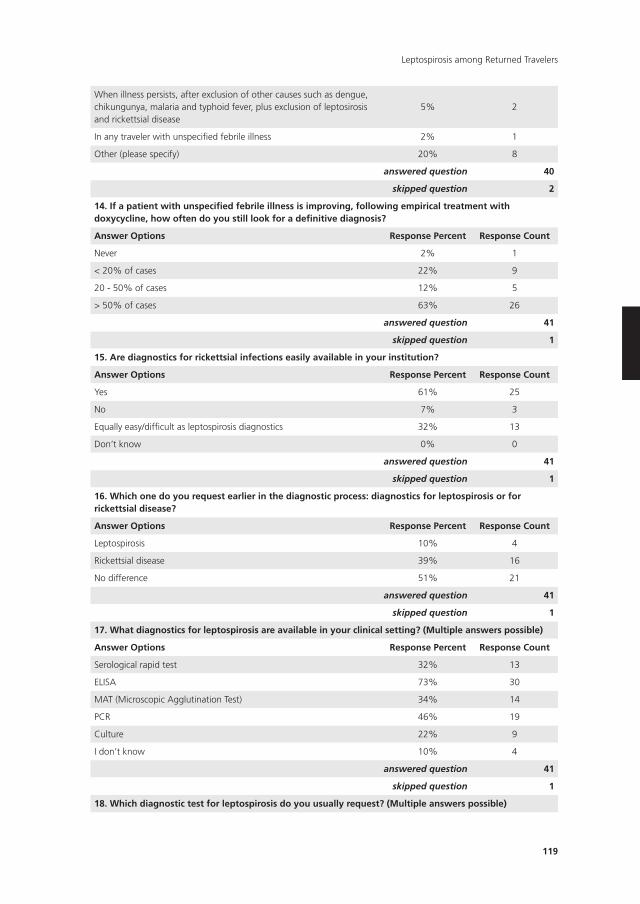
119
Leptospirosis among Returned Travelers
When illness persists, after exclusion of other causes such as dengue, chikungunya, malaria and typhoid fever, plus exclusion of leptosirosis and rickettsial disease
5% 2
In any traveler with unspecified febrile illness 2% 1
Other (please specify) 20% 8
answered question 40
skipped question 2
14. If a patient with unspecified febrile illness is improving, following empirical treatment with doxycycline, how often do you still look for a definitive diagnosis?
Answer Options Response Percent Response Count
Never 2% 1
< 20% of cases 22% 9
20 - 50% of cases 12% 5
> 50% of cases 63% 26
answered question 41
skipped question 1
15. Are diagnostics for rickettsial infections easily available in your institution?
Answer Options Response Percent Response Count
Yes 61% 25
No 7% 3
Equally easy/difficult as leptospirosis diagnostics 32% 13
Don’t know 0% 0
answered question 41
skipped question 1
16. Which one do you request earlier in the diagnostic process: diagnostics for leptospirosis or for rickettsial disease?
Answer Options Response Percent Response Count
Leptospirosis 10% 4
Rickettsial disease 39% 16
No difference 51% 21
answered question 41
skipped question 1
17. What diagnostics for leptospirosis are available in your clinical setting? (Multiple answers possible)
Answer Options Response Percent Response Count
Serological rapid test 32% 13
ELISA 73% 30
MAT (Microscopic Agglutination Test) 34% 14
PCR 46% 19
Culture 22% 9
I don’t know 10% 4
answered question 41
skipped question 1
18. Which diagnostic test for leptospirosis do you usually request? (Multiple answers possible)
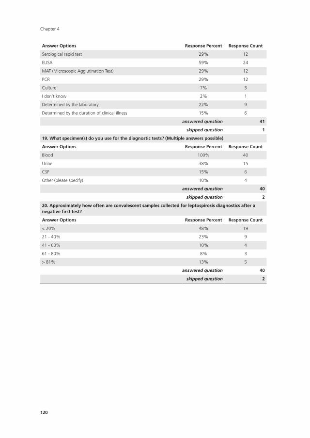
Chapter 4
120
Answer Options Response Percent Response Count
Serological rapid test 29% 12
ELISA 59% 24
MAT (Microscopic Agglutination Test) 29% 12
PCR 29% 12
Culture 7% 3
I don’t know 2% 1
Determined by the laboratory 22% 9
Determined by the duration of clinical illness 15% 6
answered question 41
skipped question 1
19. What specimen(s) do you use for the diagnostic tests? (Multiple answers possible)
Answer Options Response Percent Response Count
Blood 100% 40
Urine 38% 15
CSF 15% 6
Other (please specify) 10% 4
answered question 40
skipped question 2
20. Approximately how often are convalescent samples collected for leptospirosis diagnostics after a negative first test?
Answer Options Response Percent Response Count
< 20% 48% 19
21 - 40% 23% 9
41 - 60% 10% 4
61 - 80% 8% 3
> 81% 13% 5
answered question 40
skipped question 2
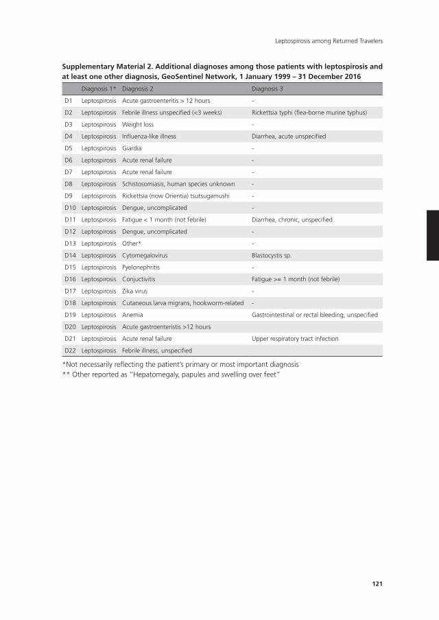
121
Leptospirosis among Returned Travelers
Supplementary Material 2. Additional diagnoses among those patients with leptospirosis and at least one other diagnosis, GeoSentinel network, 1 January 1999 – 31 December 2016
Diagnosis 1* Diagnosis 2 Diagnosis 3
D1 Leptospirosis Acute gastroenteritis > 12 hours -
D2 Leptospirosis Febrile illness unspecified (<3 weeks) Rickettsia typhi (flea-borne murine typhus)
D3 Leptospirosis Weight loss -
D4 Leptospirosis Influenza-like illness Diarrhea, acute unspecified
D5 Leptospirosis Giardia -
D6 Leptospirosis Acute renal failure -
D7 Leptospirosis Acute renal failure -
D8 Leptospirosis Schistosomiasis, human species unknown -
D9 Leptospirosis Rickettsia (now Orientia) tsutsugamushi -
D10 Leptospirosis Dengue, uncomplicated -
D11 Leptospirosis Fatigue < 1 month (not febrile) Diarrhea, chronic, unspecified
D12 Leptospirosis Dengue, uncomplicated -
D13 Leptospirosis Other* -
D14 Leptospirosis Cytomegalovirus Blastocystis sp.
D15 Leptospirosis Pyelonephritis -
D16 Leptospirosis Conjuctivitis Fatigue >= 1 month (not febrile)
D17 Leptospirosis Zika virus -
D18 Leptospirosis Cutaneous larva migrans, hookworm-related -
D19 Leptospirosis Anemia Gastrointestinal or rectal bleeding, unspecified
D20 Leptospirosis Acute gastroenteristis >12 hours
D21 Leptospirosis Acute renal failure Upper respiratory tract infection
D22 Leptospirosis Febrile illness, unspecified
*Not necessarily reflecting the patient’s primary or most important diagnosis** Other reported as “Hepatomegaly, papules and swelling over feet”

Chapter 5Nucleic acid and antigen detection tests
for leptospirosis
Bada Yang, Sophia G. de Vries, Ahmed Ahmed, Benjamin J. Visser, Ingeborg M. Nagel, René Spijker, Martin P. Grobusch, Rudy A. Hartskeerl, Marga G.A. Goris,
Marika M.G. Leefl ang
Cochrane Database of Systematic Reviews 2019, Issue 8. Art. No.: CD011871. DOI:
10.1002/14651858.CD011871.pub2.
Chapter 5
124
ABSTRACT
Background
Early diagnosis of leptospirosis may contribute to the effectiveness of antimicrobial therapy
and early outbreak recognition. Nucleic acid and antigen detection tests have the potential
for early diagnosis of leptospirosis. With this systematic review, we assessed the sensitivity
and specificity of nucleic acid and antigen detection tests.
Objectives
To determine the diagnostic test accuracy of nucleic acid and antigen detection tests for the
diagnosis of human symptomatic leptospirosis.
Search methods
We searched electronic databases including MEDLINE, Embase, the Cochrane Library, and
regional databases from inception to 6 July 2018. We did not apply restrictions to language
or time of publication.
Selection criteria
We included diagnostic cross-sectional studies and case-control studies of tests that made
use of nucleic acid and antigen detection methods in people suspected of systemic leptospi-
rosis. As reference standards, we considered the microscopic agglutination test alone (which
detects antibodies against leptospirosis) or in a composite reference standard with culturing
or other serological tests. Studies were excluded when the controls were healthy individuals
or when there were insufficient data to calculate sensitivity and specificity.
Data collection and analysis
At least two review authors independently extracted data from each study. We used the
revised Quality Assessment of Diagnostic Accuracy Studies tool (QUADAS-2) to assess risk of
bias. We calculated study-specific values for sensitivity and specificity with 95% confidence
intervals (CI) and pooled the results in a meta-analysis when appropriate. We used the bivari-
ate model for index tests with one positivity threshold, and we used the hierarchical summary
receiver operating characteristic model for index tests with multiple positivity thresholds.
As possible sources of heterogeneity, we explored: timing of index test, disease prevalence,
blood sample type, primers or target genes, and the real-time polymerase chain reaction
(PCR) visualisation method. These were added as covariates to the meta-regression models.
Main results
We included 41 studies evaluating nine index tests (conventional PCR (in short: PCR),
real-time PCR, nested PCR, PCR performed twice, loop-mediated isothermal amplification,
125
Nucleic acid and antigen detection tests for leptospirosis
enzyme-linked immunosorbent assay (ELISA), dot-ELISA, immunochromatography-based
lateral flow assay, and dipstick assay) with 5981 participants (1834 with and 4147 without
leptospirosis). Methodological quality criteria were often not reported, and the risk of bias
of the reference standard was generally considered high. The applicability of findings was
limited by the frequent use of frozen samples. We conducted meta-analyses for the PCR and
the real-time PCR on blood products.
The pooled sensitivity of the PCR was 70% (95% CI 37% to 90%) and the pooled
specificity was 95% (95% CI 75% to 99%). When studies with a high risk of bias in the
reference standard domain were excluded, the pooled sensitivity was 87% (95% CI 44% to
98%) and the pooled specificity was 97% (95% CI 60% to 100%). For the real-time PCR,
we estimated a summary receiver operating characteristic curve. To illustrate, a point on the
curve with 85% specificity had a sensitivity of 49% (95% CI 30% to 68%). Likewise, at 90%
specificity, sensitivity was 40% (95% CI 24% to 59%) and at 95% specificity, sensitivity was
29% (95% CI 15% to 49%). The median specificity of real-time PCR on blood products was
92%. We did not formally compare the diagnostic test accuracy of PCR and real-time PCR, as
direct comparison studies were lacking. Three of 15 studies analysing PCR on blood products
reported the timing of sample collection in the studies included in the meta-analyses (range
1 to 7 days postonset of symptoms), and nine out of 16 studies analysing real-time PCR on
blood products (range 1 to 19 days postonset of symptoms). In PCR studies, specificity was
lower in settings with high leptospirosis prevalence. Other investigations of heterogeneity
did not identify statistically significant associations. Two studies suggested that PCR and
real-time PCR may be more sensitive on blood samples collected early in the disease stage.
Results of other index tests were described narratively.
Authors’ conclusions
The validity of review findings are limited and should be interpreted with caution. There is
a substantial between-study variability in the accuracy of PCR and real-time PCR, as well as
a substantial variability in the prevalence of leptospirosis. Consequently, the position of PCR
and real-time PCR in the clinical pathway depends on regional considerations such as disease
prevalence, factors that are likely to influence accuracy, and downstream consequences of
test results. There is insufficient evidence to conclude which of the nucleic acid and antigen
detection tests is the most accurate. There is preliminary evidence that PCR and real-time PCR
are more sensitive on blood samples collected early in the disease stage, but this needs to be
confirmed in future studies.
Chapter 5
126
PLAIn LAnGUAGE SUMMARy
How accurate are nucleic and antigen detection tests in diagnosing leptospirosis?
What was studied in this review?
Leptospirosis is an infectious disease, caused by bacteria called Leptospira that can be found
in soil, freshwater, or in the infected urine of certain animals. It is mainly a problem in humid,
tropical countries in Southeast Asia, and Central and South America, but it can also occur in
temperate regions.
Leptospirosis causes fever and headache, and in some cases kidney, lung, or heart
problems. Often, the symptoms are not unique for the disease, which makes it difficult to
diagnose, and is therefore frequently missed.
Laboratory tests confirm diagnosis. These tests are based on demonstration of the pres-
ence of Leptospira, its DNA, or antibodies against Leptospira. Nucleic acid and antigen detec-
tion tests, such as conventional polymerase chain reaction (PCR) and real-time PCR, identify
the bacterium or its DNA directly in blood or urine. Nucleic acid and antigen detection tests
may detect Leptospira better in the early days of an infection, so that people can be treated
earlier with antibiotics – resulting in better outcomes – and can provide useful information
in outbreak situations. In outbreak situations, nucleic acid and antigen detection tests could
serve as early warning systems.
What was the aim of this review?
The aim was to assess how well nucleic acid and antigen tests perform in detecting leptospi-
rosis. In other words, to assess how many mistakes these tests make by either missing people
with leptospirosis or misidentifying people without leptospirosis (healthy people or people
with another disease).
What were the main results in this review?
The review included information from 41 studies with 5981 participants. We identified nine
nucleic acid and antigen detection tests, of which PCR and real-time PCR were most often
investigated.
An important finding was that the accuracy of both PCR and real-time PCR varied
strongly between studies. We presented average accuracies for both tests, but there was
great uncertainty around these averages. PCR often correctly identified people without
leptospirosis (averaging 95 in 100 people), but frequently missed people with leptospirosis
(averaging 30 in 100 people). The accuracy of the real-time PCR depended on the cut-
off value for a positive test result. At a cut-off value where real-time PCR often correctly
identified people without leptospirosis (averaging 95 in 100 people), it also frequently missed
127
Nucleic acid and antigen detection tests for leptospirosis
people with leptospirosis (averaging 71 in 100 people). If a person tests positive or negative
for PCR or real-time PCR, the chance of the person actually having the disease depends on
whether the suspicion of leptospirosis in that person was already high before taking the test.
So, when interpreting the results of any of these tests, one must consider the strength of
suspicion of leptospirosis in an individual, and how often leptospirosis occurs in the setting
in which the test will be used.
It was uncertain whether PCR or real-time PCR performed better in detecting leptospiro-
sis, since studies directly comparing these two tests were lacking. The results of other nucleic
and antigen detection tests are described in the main text of the review.
How reliable were the results of the studies in this review?
Not all studies were conducted according to the highest scientific standards. This means that
the results of some studies may have been overestimated or underestimated. Furthermore,
the tests used to verify whether a person truly had leptospirosis or not (called the reference
standard) may not accurately distinguish people with or without leptospirosis. For these
reasons, more high-quality studies are needed to confirm the reliability of these results.
Who do the results of this review apply to?
The results may apply to people who may have leptospirosis. However, the performance of
the PCR and real-time PCR vary considerably among studies and it is yet unclear what causes
this difference in performances. It is probable that the test performs better or worse depend-
ing on how prevalent leptospirosis is in the region, and depending on the time between the
onset of symptoms and time of testing. Therefore, it is difficult to generalise the results of
this review to all settings.
How up-to-date is this review?
The review authors searched for and used studies published up to 6 July 2018.
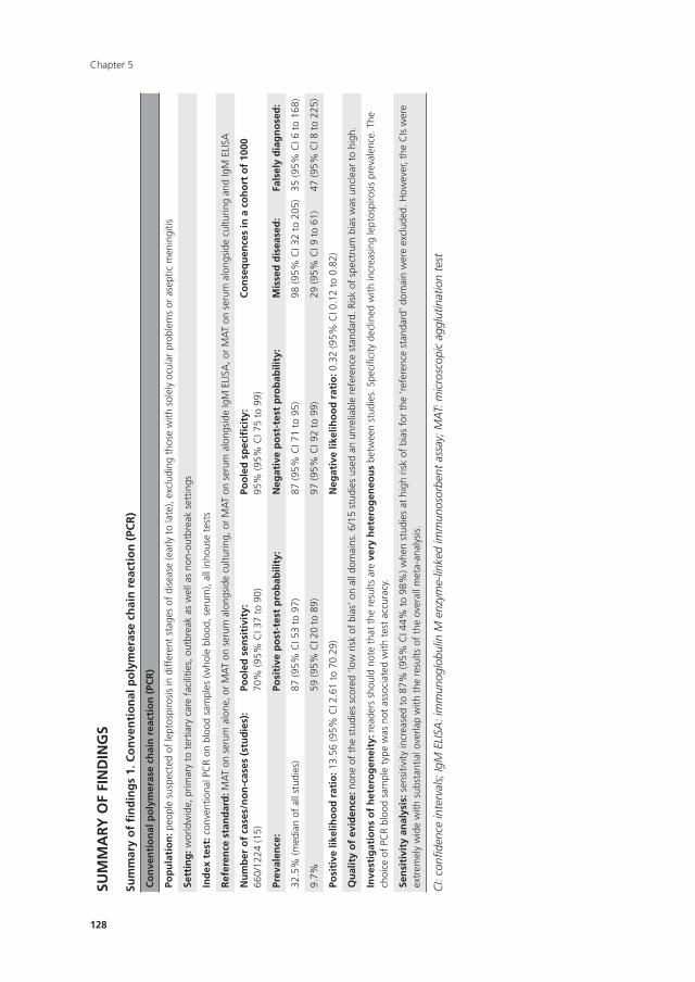
Chapter 5
128
SUM
MA
Ry O
F FI
nD
InG
S
Sum
mar
y o
f fi
nd
ing
s 1.
Co
nve
nti
on
al p
oly
mer
ase
chai
n r
eact
ion
(PC
R)
Co
nve
nti
on
al p
oly
mer
ase
chai
n r
eact
ion
(PC
R)
Pop
ula
tio
n: p
eopl
e su
spec
ted
of le
ptos
piro
sis
in d
iffer
ent
stag
es o
f di
seas
e (e
arly
to
late
), ex
clud
ing
thos
e w
ith s
olel
y oc
ular
pro
blem
s or
ase
ptic
men
ingi
tis
Sett
ing
: wor
ldw
ide,
prim
ary
to t
ertia
ry c
are
faci
litie
s, o
utbr
eak
as w
ell a
s no
n-ou
tbre
ak s
ettin
gs
Ind
ex t
est:
con
vent
iona
l PC
R on
blo
od s
ampl
es (w
hole
blo
od, s
erum
), al
l inh
ouse
tes
ts
Ref
eren
ce s
tan
dar
d: M
AT
on s
erum
alo
ne, o
r M
AT
on s
erum
alo
ngsi
de c
ultu
ring,
or
MA
T on
ser
um a
long
side
IgM
ELI
SA, o
r M
AT
on s
erum
alo
ngsi
de c
ultu
ring
and
IgM
ELI
SA
nu
mb
er o
f ca
ses/
no
n-c
ases
(st
ud
ies)
:66
0/12
24 (1
5)Po
ole
d s
ensi
tivi
ty:
70%
(95%
CI 3
7 to
90)
Poo
led
sp
ecifi
city
:95
% (9
5% C
I 75
to 9
9)C
on
seq
uen
ces
in a
co
ho
rt o
f 10
00
Prev
alen
ce:
Posi
tive
po
st-t
est
pro
bab
ility
:n
egat
ive
po
st-t
est
pro
bab
ility
:M
isse
d d
isea
sed
:Fa
lsel
y d
iag
no
sed
:
32.5
% (m
edia
n of
all
stud
ies)
87 (9
5% C
I 53
to 9
7)87
(95%
CI 7
1 to
95)
98 (9
5% C
I 32
to 2
05)
35 (9
5% C
I 6 t
o 16
8)
9.7%
59 (9
5% C
I 20
to 8
9)97
(95%
CI 9
2 to
99)
29 (9
5% C
I 9 t
o 61
)47
(95%
CI 8
to
225)
Posi
tive
like
liho
od
rat
io: 1
3.56
(95%
CI 2
.61
to 7
0.29
)n
egat
ive
likel
iho
od
rat
io: 0
.32
(95%
CI 0
.12
to 0
.82)
Qu
alit
y o
f ev
iden
ce: n
one
of t
he s
tudi
es s
core
d ‘lo
w r
isk
of b
ias’
on
all d
omai
ns. 6
/15
stud
ies
used
an
unre
liabl
e re
fere
nce
stan
dard
. Ris
k of
spe
ctru
m b
ias
was
unc
lear
to
high
.
Inve
stig
atio
ns
of
het
ero
gen
eity
: rea
ders
sho
uld
note
tha
t th
e re
sults
are
ver
y h
eter
og
eneo
us
betw
een
stud
ies.
Spe
cific
ity d
eclin
ed w
ith in
crea
sing
lept
ospi
rosi
s pr
eval
ence
. The
ch
oice
of
PCR
bloo
d sa
mpl
e ty
pe w
as n
ot a
ssoc
iate
d w
ith t
est
accu
racy
.
Sen
siti
vity
an
alys
is: s
ensi
tivity
incr
ease
d to
87%
(95%
CI 4
4% t
o 98
%) w
hen
stud
ies
at h
igh
risk
of b
ias
for
the
‘ref
eren
ce s
tand
ard’
dom
ain
wer
e ex
clud
ed. H
owev
er, t
he C
Is w
ere
extr
emel
y w
ide
with
sub
stan
tial o
verla
p w
ith t
he r
esul
ts o
f th
e ov
eral
l met
a-an
alys
is.
CI:
confi
denc
e in
terv
als;
IgM
ELI
SA: i
mm
unog
lobu
lin M
enz
yme-
linke
d im
mun
osor
bent
ass
ay; M
AT:
mic
rosc
opic
agg
lutin
atio
n te
st
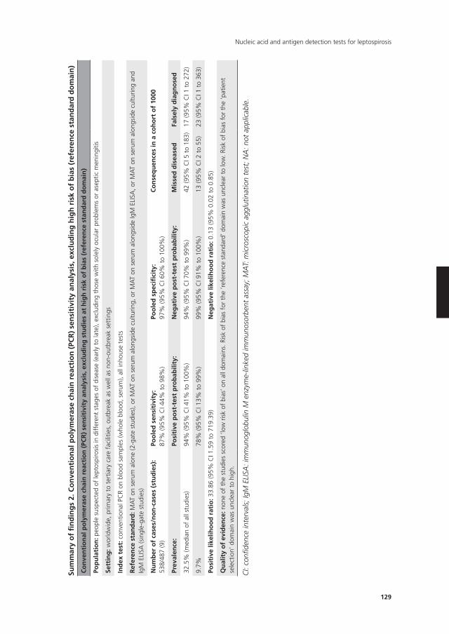
129
Nucleic acid and antigen detection tests for leptospirosis
Sum
mar
y o
f fi
nd
ing
s 2.
Co
nve
nti
on
al p
oly
mer
ase
chai
n r
eact
ion
(PC
R)
sen
siti
vity
an
alys
is, e
xclu
din
g h
igh
ris
k o
f b
ias
(ref
eren
ce s
tan
dar
d d
om
ain
)
Co
nve
nti
on
al p
oly
mer
ase
chai
n r
eact
ion
(PC
R)
sen
siti
vity
an
alys
is, e
xclu
din
g s
tud
ies
at h
igh
ris
k o
f b
ias
(ref
eren
ce s
tan
dar
d d
om
ain
)
Pop
ula
tio
n: p
eopl
e su
spec
ted
of le
ptos
piro
sis
in d
iffer
ent
stag
es o
f di
seas
e (e
arly
to
late
), ex
clud
ing
thos
e w
ith s
olel
y oc
ular
pro
blem
s or
ase
ptic
men
ingi
tis
Sett
ing
: wor
ldw
ide,
prim
ary
to t
ertia
ry c
are
faci
litie
s, o
utbr
eak
as w
ell a
s no
n-ou
tbre
ak s
ettin
gs
Ind
ex t
est:
con
vent
iona
l PC
R on
blo
od s
ampl
es (w
hole
blo
od, s
erum
), al
l inh
ouse
tes
ts
Ref
eren
ce s
tan
dar
d: M
AT
on s
erum
alo
ne (2
-gat
e st
udie
s), o
r M
AT
on s
erum
alo
ngsi
de c
ultu
ring,
or
MA
T on
ser
um a
long
side
IgM
ELI
SA, o
r M
AT
on s
erum
alo
ngsi
de c
ultu
ring
and
IgM
ELI
SA (s
ingl
e-ga
te s
tudi
es)
nu
mb
er o
f ca
ses/
no
n-c
ases
(st
ud
ies)
:53
8/48
7 (9
)Po
ole
d s
ensi
tivi
ty:
87%
(95%
CI 4
4% t
o 98
%)
Poo
led
sp
ecifi
city
:97
% (9
5% C
I 60%
to
100%
)C
on
seq
uen
ces
in a
co
ho
rt o
f 10
00
Prev
alen
ce:
Posi
tive
po
st-t
est
pro
bab
ility
:n
egat
ive
po
st-t
est
pro
bab
ility
:M
isse
d d
isea
sed
Fals
ely
dia
gn
ose
d
32.5
% (m
edia
n of
all
stud
ies)
94%
(95%
CI 4
1% t
o 10
0%)
94%
(95%
CI 7
0% t
o 99
%)
42 (9
5% C
I 5 t
o 18
3)17
(95%
CI 1
to
272)
9.7%
78%
(95%
CI 1
3% t
o 99
%)
99%
(95%
CI 9
1% t
o 10
0%)
13 (9
5% C
I 2 t
o 55
)23
(95%
CI 1
to
363)
Posi
tive
like
liho
od
rat
io: 3
3.86
(95%
CI 1
.59
to 7
19.3
9)n
egat
ive
likel
iho
od
rat
io: 0
.13
(95%
0.0
2 to
0.8
5)
Qu
alit
y o
f ev
iden
ce: n
one
of t
he s
tudi
es s
core
d ‘lo
w r
isk
of b
ias’
on
all d
omai
ns. R
isk
of b
ias
for
the
‘ref
eren
ce s
tand
ard’
dom
ain
was
unc
lear
to
low
. Ris
k of
bia
s fo
r th
e ‘p
atie
nt
sele
ctio
n’ d
omai
n w
as u
ncle
ar t
o hi
gh.
CI:
confi
denc
e in
terv
als;
IgM
ELI
SA: i
mm
unog
lobu
lin M
enz
yme-
linke
d im
mun
osor
bent
ass
ay; M
AT:
mic
rosc
opic
agg
lutin
atio
n te
st; N
A: n
ot a
pplic
able
.
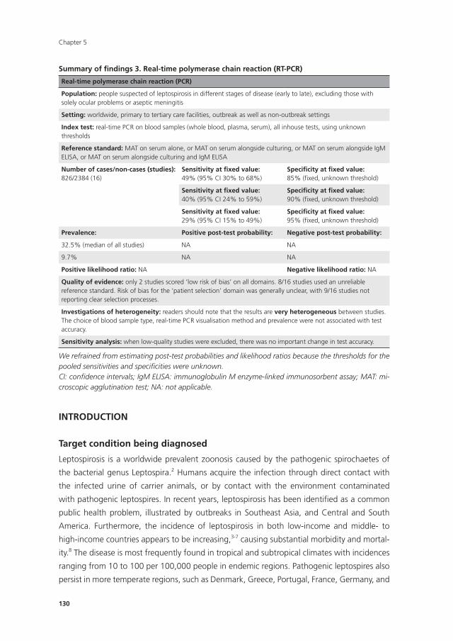
Chapter 5
130
InTRODUCTIOn
Target condition being diagnosed
Leptospirosis is a worldwide prevalent zoonosis caused by the pathogenic spirochaetes of
the bacterial genus Leptospira.2 Humans acquire the infection through direct contact with
the infected urine of carrier animals, or by contact with the environment contaminated
with pathogenic leptospires. In recent years, leptospirosis has been identified as a common
public health problem, illustrated by outbreaks in Southeast Asia, and Central and South
America. Furthermore, the incidence of leptospirosis in both low-income and middle- to
high-income countries appears to be increasing,3-7 causing substantial morbidity and mortal-
ity.8 The disease is most frequently found in tropical and subtropical climates with incidences
ranging from 10 to 100 per 100,000 people in endemic regions. Pathogenic leptospires also
persist in more temperate regions, such as Denmark, Greece, Portugal, France, Germany, and
Summary of findings 3. Real-time polymerase chain reaction (RT-PCR)
Real-time polymerase chain reaction (PCR)
Population: people suspected of leptospirosis in different stages of disease (early to late), excluding those with solely ocular problems or aseptic meningitis
Setting: worldwide, primary to tertiary care facilities, outbreak as well as non-outbreak settings
Index test: real-time PCR on blood samples (whole blood, plasma, serum), all inhouse tests, using unknown thresholds
Reference standard: MAT on serum alone, or MAT on serum alongside culturing, or MAT on serum alongside IgM ELISA, or MAT on serum alongside culturing and IgM ELISA
number of cases/non-cases (studies):826/2384 (16)
Sensitivity at fixed value:49% (95% CI 30% to 68%)
Specificity at fixed value:85% (fixed, unknown threshold)
Sensitivity at fixed value:40% (95% CI 24% to 59%)
Specificity at fixed value:90% (fixed, unknown threshold)
Sensitivity at fixed value:29% (95% CI 15% to 49%)
Specificity at fixed value:95% (fixed, unknown threshold)
Prevalence: Positive post-test probability: negative post-test probability:
32.5% (median of all studies) NA NA
9.7% NA NA
Positive likelihood ratio: NA negative likelihood ratio: NA
Quality of evidence: only 2 studies scored ‘low risk of bias’ on all domains. 8/16 studies used an unreliable reference standard. Risk of bias for the ‘patient selection’ domain was generally unclear, with 9/16 studies not reporting clear selection processes.
Investigations of heterogeneity: readers should note that the results are very heterogeneous between studies. The choice of blood sample type, real-time PCR visualisation method and prevalence were not associated with test accuracy.
Sensitivity analysis: when low-quality studies were excluded, there was no important change in test accuracy.
We refrained from estimating post-test probabilities and likelihood ratios because the thresholds for the pooled sensitivities and specificities were unknown.CI: confidence intervals; IgM ELISA: immunoglobulin M enzyme-linked immunosorbent assay; MAT: mi-croscopic agglutination test; NA: not applicable.
131
Nucleic acid and antigen detection tests for leptospirosis
the Netherlands, where it is an important cause of illness in returning travellers.9,10 Factors
contributing to higher levels of prevalence are local agricultural practices, close proximity
to mammalian reservoirs, poor sanitation, soil contact, and high rainfall.11 Flooding associ-
ated with heavy seasonal rainfall and natural disasters may increase incidence to epidemic
proportions, to more than 100 per 100,000 people.12 It is thought that the emergence of
leptospirosis is aggravated by global climate change, increasing contact between humans
and wild animal populations, and the exponential expansion of urban slums.13,14
The clinical manifestations of leptospirosis are diverse; symptoms range from a mild
undifferentiated fever syndrome including myalgia and headaches, to the severest form that
may involve renal failure and jaundice (classically known as Weil’s disease), and other com-
plications such as pulmonary haemorrhages, aseptic meningitis, and myocarditis.15 Fatality
rates for severe forms range from 5% to 50%.12,13 The non-specific clinical presentation of
leptospirosis makes it challenging to distinguish from infections such as malaria, dengue,
influenza, hepatitis, and yellow fever.15 Consequently, laboratory tests are essential to con-
firm the diagnosis. These tests are based on either demonstration of leptospires, antibodies
against leptospires, or their DNA.
The current reference standard for the diagnosis of leptospirosis is based on antibody
detection by the microscopic agglutination test (MAT), with or without culture. Since anti-
Leptospira antibodies appear only in the later stage of the disease, MAT and other serological
tests, such as the immunoglobulin M (IgM) enzyme-linked immunosorbent assay (ELISA), are
impractical in establishing an early diagnosis.16 In addition, the culture of leptospires does
not contribute to an early diagnosis, due to their slow growth.12 Nucleic acid tests, such as
the polymerase chain reaction (PCR) and antigen detection tests, can detect leptospiral DNA
or antigens directly in blood in the first days of the disease and are thus capable of yielding
an early diagnosis.12 This type of early detection test may facilitate early outbreak warnings
and make the administration of early microbial treatment possible. Additionally, leptospires
appear in the urine after a few days, on which nucleic acid detection methods can be applied
as well.12 Early administration of treatment is generally considered to improve a person’s
outcome compared to treatment at a later disease stage, although more studies are needed
to confirm this.17
Index test(s)
This review evaluated nucleic acid and antigen detection tests for pathogenic leptospires.
Commonly used nucleic acid tests for the diagnosis of human leptospirosis are the PCR, its
variants, and isothermal amplification tests such as loop-mediated isothermal amplification
(LAMP). Nucleic acid tests can be used to test blood, cerebrospinal fluid (CSF), aqueous
humour, and urine samples. Other antigen detection tests include ELISA and fluorescent
antibody testing (FAT) for the detection of Leptospira antigens, silver staining, and immuno-
histochemistry.
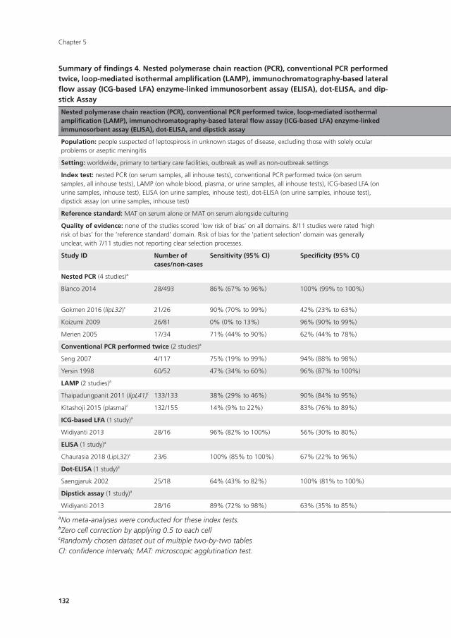
Chapter 5
132
Summary of findings 4. nested polymerase chain reaction (PCR), conventional PCR performed twice, loop-mediated isothermal amplification (LAMP), immunochromatography-based lateral flow assay (ICG-based LFA) enzyme-linked immunosorbent assay (ELISA), dot-ELISA, and dip-stick Assay
nested polymerase chain reaction (PCR), conventional PCR performed twice, loop-mediated isothermal amplification (LAMP), immunochromatography-based lateral flow assay (ICG-based LFA) enzyme-linked immunosorbent assay (ELISA), dot-ELISA, and dipstick assay
Population: people suspected of leptospirosis in unknown stages of disease, excluding those with solely ocular problems or aseptic meningitis
Setting: worldwide, primary to tertiary care facilities, outbreak as well as non-outbreak settings
Index test: nested PCR (on serum samples, all inhouse tests), conventional PCR performed twice (on serum samples, all inhouse tests), LAMP (on whole blood, plasma, or urine samples, all inhouse tests), ICG-based LFA (on urine samples, inhouse test), ELISA (on urine samples, inhouse test), dot-ELISA (on urine samples, inhouse test), dipstick assay (on urine samples, inhouse test)
Reference standard: MAT on serum alone or MAT on serum alongside culturing
Quality of evidence: none of the studies scored ‘low risk of bias’ on all domains. 8/11 studies were rated ‘high risk of bias’ for the ‘reference standard’ domain. Risk of bias for the ‘patient selection’ domain was generally unclear, with 7/11 studies not reporting clear selection processes.
Study ID number ofcases/non-cases
Sensitivity (95% CI) Specificity (95% CI) Positive post-testprobability (95% CI)
negative post-testprobability (95% CI)
Positive likelihoodratio (95% CI)
negative likelihoodratio (95% CI):
nested PCR (4 studies)a
Blanco 2014 28/493 86% (67% to 96%) 100% (99% to 100%) 100% (100% to 100%) 99% (98% to 100%) 834.69 (52.05 to 13,384.42)b
0.14 (0.06 to 0.35)
Gokmen 2016 (lipL32)c 21/26 90% (70% to 99%) 42% (23% to 63%) 56% (47% to 64%) 85% (58% to 96%) 1.57 (1.10 to 2.24) 0.23 (0.06 to 0.91)
Koizumi 2009 26/81 0% (0% to 13%) 96% (90% to 99%) 13% (0% to 70%)b 75% (74% to 76%) 0.43 (0.02 to 8.13)b 1.04 (1.00 to 1.08)
Merien 2005 17/34 71% (44% to 90%) 62% (44% to 78%) 48% (35% to 61%) 81% (66% to 90%) 1.85 (1.09 to 3.12) 0.48 (0.22 to 1.04)
Conventional PCR performed twice (2 studies)a
Seng 2007 4/117 75% (19% to 99%) 94% (88% to 98%) 30% (15% to 52%) 99% (95% to 100%) 12.54 (5.02 to 31.28) 0.27 (0.05 to 1.45)
Yersin 1998 60/52 47% (34% to 60%) 96% (87% to 100%) 93% (78% to 98%) 61% (55% to 67%) 12.13 (3.04 to 48.50) 0.55 (0.44 to 0.71)
LAMP (2 studies)a
Thaipadungpanit 2011 (lipL41)c 133/133 38% (29% to 46%) 90% (84% to 95%) 79% (69% to 87%) 59% (56% to 63%) 3.85 (2.20 to 6.74) 0.69 (0.60 to 0.80)
Kitashoji 2015 (plasma)c 132/155 14% (9% to 22%) 83% (76% to 89%) 42% (30% to 56%) 53% (51% to 56%) 0.86 (0.50 to 1.48) 1.03 (0.93 to 1.14)
ICG-based LFA (1 study)a
Widiyanti 2013 28/16 96% (82% to 100%) 56% (30% to 80%) 79% (69% to 87%) 90% (56% to 98%) 2.20 (1.26 to 3.86) 0.06 (0.01 to 0.46)
ELISA (1 study)a
Chaurasia 2018 (LipL32)c 23/6 100% (85% to 100%) 67% (22% to 96%) 92% (79% to 97%) 90% (38% to 100%)b 3.00 (0.97 to 9.30) 0.03 (0.00 to 0.58)**
Dot-ELISA (1 study)a
Saengjaruk 2002 25/18 64% (43% to 82%) 100% (81% to 100%) 97% (73% to 100%)b 67% (54% to 77%) 24.12 (1.54 to 377.45)b 0.36 (0.21 to 0.61)
Dipstick assay (1 study)a
Widiyanti 2013 28/16 89% (72% to 98%) 63% (35% to 85%) 81% (69% to 89%) 77% (52% to 91%) 2.38 (1.25 to 4.54) 0.17 (0.06 to 0.53)
aNo meta-analyses were conducted for these index tests.bZero cell correction by applying 0.5 to each cellcRandomly chosen dataset out of multiple two-by-two tablesCI: confidence intervals; MAT: microscopic agglutination test.
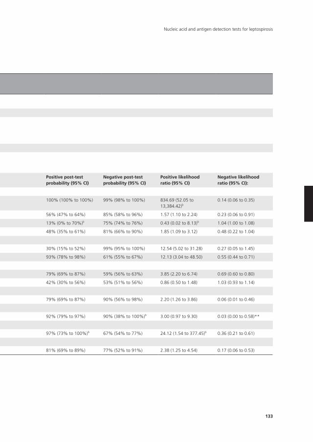
133
Nucleic acid and antigen detection tests for leptospirosis
Summary of findings 4. nested polymerase chain reaction (PCR), conventional PCR performed twice, loop-mediated isothermal amplification (LAMP), immunochromatography-based lateral flow assay (ICG-based LFA) enzyme-linked immunosorbent assay (ELISA), dot-ELISA, and dip-stick Assay
nested polymerase chain reaction (PCR), conventional PCR performed twice, loop-mediated isothermal amplification (LAMP), immunochromatography-based lateral flow assay (ICG-based LFA) enzyme-linked immunosorbent assay (ELISA), dot-ELISA, and dipstick assay
Population: people suspected of leptospirosis in unknown stages of disease, excluding those with solely ocular problems or aseptic meningitis
Setting: worldwide, primary to tertiary care facilities, outbreak as well as non-outbreak settings
Index test: nested PCR (on serum samples, all inhouse tests), conventional PCR performed twice (on serum samples, all inhouse tests), LAMP (on whole blood, plasma, or urine samples, all inhouse tests), ICG-based LFA (on urine samples, inhouse test), ELISA (on urine samples, inhouse test), dot-ELISA (on urine samples, inhouse test), dipstick assay (on urine samples, inhouse test)
Reference standard: MAT on serum alone or MAT on serum alongside culturing
Quality of evidence: none of the studies scored ‘low risk of bias’ on all domains. 8/11 studies were rated ‘high risk of bias’ for the ‘reference standard’ domain. Risk of bias for the ‘patient selection’ domain was generally unclear, with 7/11 studies not reporting clear selection processes.
Study ID number ofcases/non-cases
Sensitivity (95% CI) Specificity (95% CI) Positive post-testprobability (95% CI)
negative post-testprobability (95% CI)
Positive likelihoodratio (95% CI)
negative likelihoodratio (95% CI):
nested PCR (4 studies)a
Blanco 2014 28/493 86% (67% to 96%) 100% (99% to 100%) 100% (100% to 100%) 99% (98% to 100%) 834.69 (52.05 to 13,384.42)b
0.14 (0.06 to 0.35)
Gokmen 2016 (lipL32)c 21/26 90% (70% to 99%) 42% (23% to 63%) 56% (47% to 64%) 85% (58% to 96%) 1.57 (1.10 to 2.24) 0.23 (0.06 to 0.91)
Koizumi 2009 26/81 0% (0% to 13%) 96% (90% to 99%) 13% (0% to 70%)b 75% (74% to 76%) 0.43 (0.02 to 8.13)b 1.04 (1.00 to 1.08)
Merien 2005 17/34 71% (44% to 90%) 62% (44% to 78%) 48% (35% to 61%) 81% (66% to 90%) 1.85 (1.09 to 3.12) 0.48 (0.22 to 1.04)
Conventional PCR performed twice (2 studies)a
Seng 2007 4/117 75% (19% to 99%) 94% (88% to 98%) 30% (15% to 52%) 99% (95% to 100%) 12.54 (5.02 to 31.28) 0.27 (0.05 to 1.45)
Yersin 1998 60/52 47% (34% to 60%) 96% (87% to 100%) 93% (78% to 98%) 61% (55% to 67%) 12.13 (3.04 to 48.50) 0.55 (0.44 to 0.71)
LAMP (2 studies)a
Thaipadungpanit 2011 (lipL41)c 133/133 38% (29% to 46%) 90% (84% to 95%) 79% (69% to 87%) 59% (56% to 63%) 3.85 (2.20 to 6.74) 0.69 (0.60 to 0.80)
Kitashoji 2015 (plasma)c 132/155 14% (9% to 22%) 83% (76% to 89%) 42% (30% to 56%) 53% (51% to 56%) 0.86 (0.50 to 1.48) 1.03 (0.93 to 1.14)
ICG-based LFA (1 study)a
Widiyanti 2013 28/16 96% (82% to 100%) 56% (30% to 80%) 79% (69% to 87%) 90% (56% to 98%) 2.20 (1.26 to 3.86) 0.06 (0.01 to 0.46)
ELISA (1 study)a
Chaurasia 2018 (LipL32)c 23/6 100% (85% to 100%) 67% (22% to 96%) 92% (79% to 97%) 90% (38% to 100%)b 3.00 (0.97 to 9.30) 0.03 (0.00 to 0.58)**
Dot-ELISA (1 study)a
Saengjaruk 2002 25/18 64% (43% to 82%) 100% (81% to 100%) 97% (73% to 100%)b 67% (54% to 77%) 24.12 (1.54 to 377.45)b 0.36 (0.21 to 0.61)
Dipstick assay (1 study)a
Widiyanti 2013 28/16 89% (72% to 98%) 63% (35% to 85%) 81% (69% to 89%) 77% (52% to 91%) 2.38 (1.25 to 4.54) 0.17 (0.06 to 0.53)
aNo meta-analyses were conducted for these index tests.bZero cell correction by applying 0.5 to each cellcRandomly chosen dataset out of multiple two-by-two tablesCI: confidence intervals; MAT: microscopic agglutination test.
Chapter 5
134
Substantial variation can be expected between laboratories on how the index tests are
performed with regard to the timing of sample collection and threshold values. The timing
of sample collection may greatly affect the test’s accuracy, as leptospires are known to (dis)
appear in different sample types as the disease progresses. For example, it is recommended
that nucleic acid and antigen detection tests are performed on blood between one and 10
days postonset (DPO) of symptoms, as leptospiraemia declines rapidly until below detection
after 10 DPO.12 Tests performed on blood samples collected after 10 DPO may lead to false-
negative findings. Tests in urine are expected to be positive after 10 to 14 DPO.16
Reference standard
MAT is the most widely used serological test for leptospirosis. It is considered to be the refer-
ence standard, often used in combination with other serological tests (such as IgM ELISA),
and with or without culture of leptospires from blood or urine.
MAT is considered an imperfect reference standard. It has a high diagnostic specificity, as
the observation of seroconversion or a titre rise confirms current leptospirosis, but a negative
MAT does not rule out the possibility of leptospirosis. Limmathurotsakul and colleagues used
a Bayesian latent class analysis (LCA) to estimate the accuracy of MAT, which was 49.8%
sensitive and 98.8% specific.18 The LCA assumes that there is no reference standard, and
estimates disease prevalence by taking the results of multiple tests into account.19
In another study to estimate the accuracy of MAT, Goris and colleagues selected culture-
positive people as being infected (proof of leptospirosis) and people with other known
diseases and unknown disease as controls, and performed MAT on both groups.1 In this
study, the sensitivity of MAT was estimated at 81.7% and specificity of MAT was estimated
at 100%.
Using a reference standard with low sensitivity to compare against the index test may
result in biased estimates of specificity. However, when the case definition in the Goris 2012
study was changed to include people who were IgM ELISA positive, the sensitivity increased
to 93.3% without sacrificing the specificity.1 This indicates that combining multiple tests with
high specificity as a composite reference standard can yield increased sensitivity. Therefore,
we decided to include studies with only MAT as the reference standard, and studies that
used other serological tests, or culturing, or both, alongside MAT as the reference standard.
Variability in MAT performance between laboratories exists and may affect test accuracy.
MAT requires a panel of live Leptospira serovars (group of micro-organisms characterised
by specific set of antigens) that occur in the region, supplemented with a panel of glob-
ally standardised serovars when people present with a travelling history.1 Determining and
maintaining such panels are major, but essential, tasks; inadequate panels may lead to false-
negative results. The timing of sample collection may also influence sensitivity or specificity;
antibodies are usually detectable from five to seven DPO onwards. MAT-case definitions may
vary between laboratories; a four-fold rise in titre in paired sera or seroconversion is indicative
135
Nucleic acid and antigen detection tests for leptospirosis
of current infection, but some laboratories may use a high titre in a single serum sample (se-
ropositivity) as a case definition for people who do not return for follow-up. Seropositivity is
not necessarily evidential of a current infection, since antibodies may persist after a previous
infection, or cross-reactivity with other diseases may occur (such as legionellosis, hepatitis,
and autoimmune diseases).12 Therefore, the desirable cut-off titres for the single-sample MAT
are higher in regions where leptospirosis and similar infectious diseases are highly prevalent.
Leptospires can be cultured from blood, CSF, dialysate fluid, and (postmortem) tissue,
often within 10 DPO. Culture of urine is useful after 10 DPO. Leptospires are slow-growing,
fastidious bacteria. Cultures have to be maintained for at least four months before being
regarded negative. Culturing provides evidence for leptospirosis but lacks sensitivity and does
not contribute to an early diagnosis. The sensitivity of culture is estimated not to exceed 23%,
according to an analysis of people with leptospirosis from 1925 to 2008 in the Netherlands.20
Clinical pathway
Figure 1 shows a diagnostic pathway, as suggested by Goris and colleagues.1 A person with
symptoms compatible with leptospirosis (such as fever, headaches, myalgia, conjunctival ef-
fusion, and vomiting) is evaluated for likelihood by assessing risk factors, and consequently
classified as an ‘early presentation’ (DPO 10 or fewer) or a ‘late presentation’ (DPO greater
than 10). Real-time PCR is recommended as the test of choice for early presentations as it
can detect leptospiral DNA in blood. Blood culture is conducted alongside real-time PCR to
confirm leptospirosis as well as to provide insight in locally occurring serovars. MAT and IgM
ELISA are recommended for later presentations since antibodies are expected to appear in
serum after five to seven DPO. The person is considered to have leptospirosis if any of the
test results is positive.
If a person tests positive with either an antibody or an antigen test, this person will be
treated with antibiotics. If the tests return negative, then the recommendation is to test again
in two weeks’ time. However, if the person is very ill, clinicians will in some cases decide to
treat with antibiotics anyway.
Rationale
The aim of this systematic review was to assess the diagnostic test accuracy of nucleic acid
and antigen detection tests for human symptomatic leptospirosis. A similar diagnostic ac-
curacy review on serology tests (antibody detection tests) for leptospirosis is being conducted
by Goris and colleagues.21
Nucleic acid and antigen detection tests may serve several purposes based on their
ability for early detection. First, and most important, an accurate test in the early stage of
the disease may improve patient outcomes by facilitating timely administration of effective
antibiotics. Although the limited available evidence presented by the latest Cochrane Review
on antimicrobial therapy was inconclusive,17 one study reported a shortened duration of
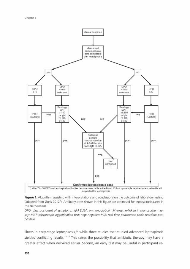
Chapter 5
136
illness in early-stage leptospirosis,22 while three studies that studied advanced leptospirosis
yielded confl icting results.23-25 This raises the possibility that antibiotic therapy may have a
greater effect when delivered earlier. Second, an early test may be useful in participant re-
144
weeks' time. However, if the person is very ill, clinicians will in some cases decide to treat with
antibiotics anyway.
Figure 1. Algorithm, assisting with interpretations and conclusions on the outcome of laboratory testing (adapted from Goris 20121). Antibody titres shown in this figure are optimised for leptospirosis cases in the Netherlands. DPO: days postonset of symptoms; IgM ELISA: immunoglobulin M enzyme-linked immunosorbent assay; MAT: microscopic agglutination test; neg: negative; PCR: real-time polymerase chain reaction; pos: positive.
Figure 1. Algorithm, assisting with interpretations and conclusions on the outcome of laboratory testing (adapted from Goris 20121). Antibody titres shown in this fi gure are optimised for leptospirosis cases in the Netherlands.DPO: days postonset of symptoms; IgM ELISA: immunoglobulin M enzyme-linked immunosorbent as-say; MAT: microscopic agglutination test; neg: negative; PCR: real-time polymerase chain reaction; pos: positive.
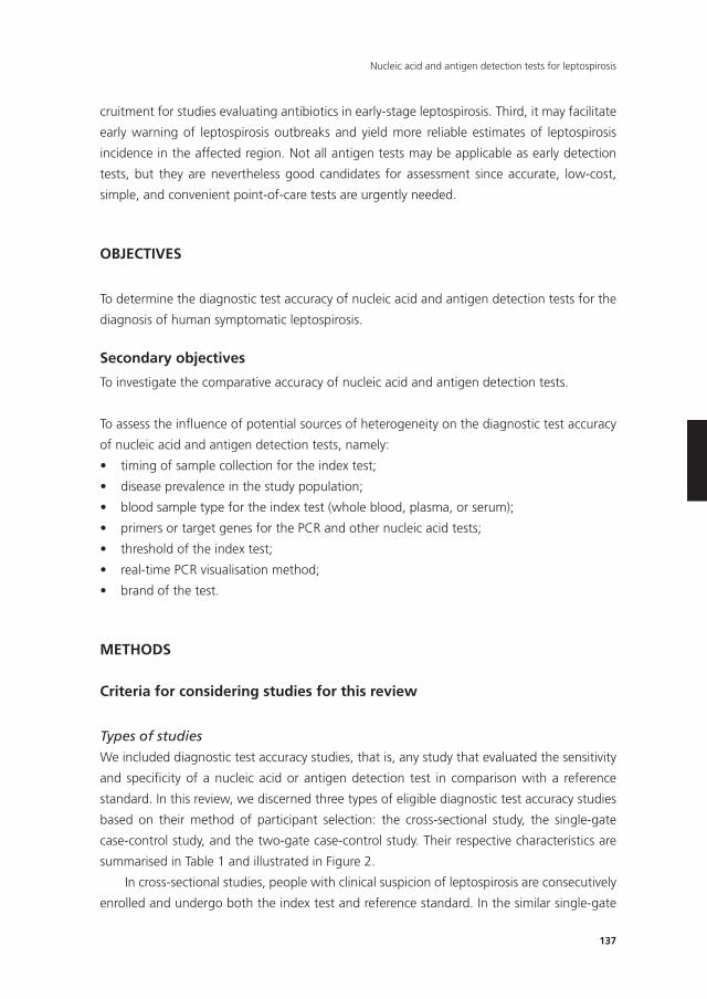
137
Nucleic acid and antigen detection tests for leptospirosis
cruitment for studies evaluating antibiotics in early-stage leptospirosis. Third, it may facilitate
early warning of leptospirosis outbreaks and yield more reliable estimates of leptospirosis
incidence in the affected region. Not all antigen tests may be applicable as early detection
tests, but they are nevertheless good candidates for assessment since accurate, low-cost,
simple, and convenient point-of-care tests are urgently needed.
OBJECTIVES
To determine the diagnostic test accuracy of nucleic acid and antigen detection tests for the
diagnosis of human symptomatic leptospirosis.
Secondary objectives
To investigate the comparative accuracy of nucleic acid and antigen detection tests.
To assess the influence of potential sources of heterogeneity on the diagnostic test accuracy
of nucleic acid and antigen detection tests, namely:
• timingofsamplecollectionfortheindextest;
• diseaseprevalenceinthestudypopulation;
• bloodsampletypefortheindextest(wholeblood,plasma,orserum);
• primersortargetgenesforthePCRandothernucleicacidtests;
• thresholdoftheindextest;
• real-timePCRvisualisationmethod;
• brandofthetest.
METHODS
Criteria for considering studies for this review
Types of studies
We included diagnostic test accuracy studies, that is, any study that evaluated the sensitivity
and specificity of a nucleic acid or antigen detection test in comparison with a reference
standard. In this review, we discerned three types of eligible diagnostic test accuracy studies
based on their method of participant selection: the cross-sectional study, the single-gate
case-control study, and the two-gate case-control study. Their respective characteristics are
summarised in Table 1 and illustrated in Figure 2.
In cross-sectional studies, people with clinical suspicion of leptospirosis are consecutively
enrolled and undergo both the index test and reference standard. In the similar single-gate
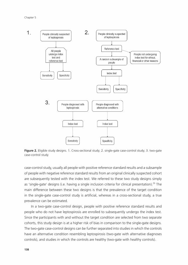
Chapter 5
138
case-control study, usually all people with positive reference standard results and a subsample
of people with negative reference standard results from an original clinically suspected cohort
are subsequently tested with the index test. We referred to these two study designs simply
as ‘single-gate’ designs (i.e. having a single inclusion criteria for clinical presentation).26 The
main difference between these two designs is that the prevalence of the target condition
in the single-gate case-control study is artifi cial, whereas in a cross-sectional study, a true
prevalence can be estimated.
In a two-gate case-control design, people with positive reference standard results and
people who do not have leptospirosis are enrolled to subsequently undergo the index test.
Since the participants with and without the target condition are selected from two separate
cohorts, this study design is at a higher risk of bias in comparison to the single-gate designs.
The two-gate case-control designs can be further separated into studies in which the controls
have an alternative condition resembling leptospirosis (two-gate with alternative diagnoses
controls), and studies in which the controls are healthy (two-gate with healthy controls).
147
METHODS
Criteria for considering studies for this review
Types of studies
We included diagnostic test accuracy studies, that is, any study that evaluated the sensitivity
and specificity of a nucleic acid or antigen detection test in comparison with a reference
standard. In this review, we discerned three types of eligible diagnostic test accuracy studies
based on their method of participant selection: the cross-sectional study, the single-gate
case-control study, and the two-gate case-control study. Their respective characteristics are
summarised in Table 1 and illustrated in Figure 2.
Figure 2. Eligible study designs. 1. Cross-sectional study; 2. single-gate case-control study; 3. two-gate case-control study
Figure 2. Eligible study designs. 1. Cross-sectional study; 2. single-gate case-control study; 3. two-gate case-control study
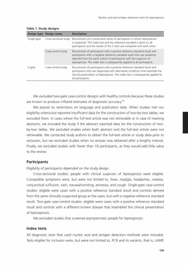
139
Nucleic acid and antigen detection tests for leptospirosis
We excluded two-gate case-control designs with healthy controls because these studies
are known to produce inflated estimates of diagnostic accuracy.26
We placed no restrictions on language and publication date. When studies met our
eligibility criteria but reported insufficient data for the construction of two-by-two tables, we
excluded them. In cases where the full-text article was not retrievable or in case of meeting
abstracts, we included the study if the abstract reported data for the construction of two-
by-two tables. We excluded studies when both abstract and the full-text article were not
retrievable. We contacted study authors to obtain the full-text article or study data prior to
exclusion, but we excluded studies when no answer was obtained after a lengthy interval.
Finally, we excluded studies with fewer than 10 participants, as they would add little value
to the review.
Participants
Eligibility of participants depended on the study design.
Cross-sectional studies: people with clinical suspicion of leptospirosis were eligible.
Compatible symptoms were, but were not limited to, fever, myalgia, headaches, malaise,
conjunctival suffusion, rash, nausea/vomiting, anorexia, and cough. Single-gate case-control
studies: eligible were cases with a positive reference standard result and controls derived
from the same clinically suspected group as the cases, but with a negative reference standard
result. Two-gate case-control studies: eligible were cases with a positive reference standard
result and controls with a different known disease that resembled the clinical presentation
of leptospirosis.
We excluded studies that screened asymptomatic people for leptospirosis.
Index tests
All diagnostic tests that used nucleic acid and antigen detection methods were included.
Tests eligible for inclusion were, but were not limited to, PCR and its variants, that is, LAMP,
Table 1. Study designs
Design type Design name Description
Single-gate Cross-sectional study Recruitment of a consecutive series of participants in whom leptospirosis is suspected. The index test and the reference standard is done on all participants and the results of the 2 tests are compared with each other.
Case-control study Recruitment of participants with a positive reference standard result and participants with a negative reference standard result who are randomly selected from the same cohort of participants with the suspicion of leptospirosis. The index test is subsequently applied to all participants.
2-gate Case-control study Recruitment of participants with a positive reference standard result and participants who are diagnosed with alternative conditions that resemble the clinical presentation of leptospirosis. The index test is subsequently applied to all participants.
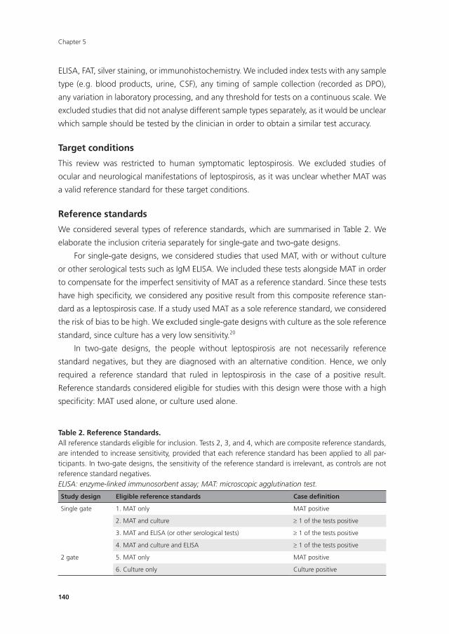
Chapter 5
140
ELISA, FAT, silver staining, or immunohistochemistry. We included index tests with any sample
type (e.g. blood products, urine, CSF), any timing of sample collection (recorded as DPO),
any variation in laboratory processing, and any threshold for tests on a continuous scale. We
excluded studies that did not analyse different sample types separately, as it would be unclear
which sample should be tested by the clinician in order to obtain a similar test accuracy.
Target conditions
This review was restricted to human symptomatic leptospirosis. We excluded studies of
ocular and neurological manifestations of leptospirosis, as it was unclear whether MAT was
a valid reference standard for these target conditions.
Reference standards
We considered several types of reference standards, which are summarised in Table 2. We
elaborate the inclusion criteria separately for single-gate and two-gate designs.
For single-gate designs, we considered studies that used MAT, with or without culture
or other serological tests such as IgM ELISA. We included these tests alongside MAT in order
to compensate for the imperfect sensitivity of MAT as a reference standard. Since these tests
have high specificity, we considered any positive result from this composite reference stan-
dard as a leptospirosis case. If a study used MAT as a sole reference standard, we considered
the risk of bias to be high. We excluded single-gate designs with culture as the sole reference
standard, since culture has a very low sensitivity.20
In two-gate designs, the people without leptospirosis are not necessarily reference
standard negatives, but they are diagnosed with an alternative condition. Hence, we only
required a reference standard that ruled in leptospirosis in the case of a positive result.
Reference standards considered eligible for studies with this design were those with a high
specificity: MAT used alone, or culture used alone.
Table 2. Reference Standards.All reference standards eligible for inclusion. Tests 2, 3, and 4, which are composite reference standards, are intended to increase sensitivity, provided that each reference standard has been applied to all par-ticipants. In two-gate designs, the sensitivity of the reference standard is irrelevant, as controls are not reference standard negatives.ELISA: enzyme-linked immunosorbent assay; MAT: microscopic agglutination test.
Study design Eligible reference standards Case definition
Single gate 1. MAT only MAT positive
2. MAT and culture ≥ 1 of the tests positive
3. MAT and ELISA (or other serological tests) ≥ 1 of the tests positive
4. MAT and culture and ELISA ≥ 1 of the tests positive
2 gate 5. MAT only MAT positive
6. Culture only Culture positive
141
Nucleic acid and antigen detection tests for leptospirosis
In order to avoid incorporation bias (the reference standard uses or incorporates the
index test), we excluded studies which contained a nucleic acid or antigen detection test in
the reference standard.
Search methods for identification of studies
Electronic searches
We searched the following 16 electronic databases: the Cochrane Library (6 July 2018),
MEDLINE Ovid (1946 to 6 July 2018), Embase Ovid (1974 to 6 July 2018), Web of Science
(1975 to 6 July 2018), CINAHL (1937 to 6 July 2018), BIOSIS Previews (1993 to 8 February
2015 due to terminated institutional subscription), PubMed (for publications not yet included
in MEDLINE; 1946 to 8 February 2015), Google Scholar, African Index Medicus (1993 to 6
July 2018), African Journals Online (from inception to 8 February 2015), LILACS (Literature
in the Health Sciences in Latin America and the Caribbean, 1982 to 6 July 2018), KoreaMed
(from inception to 8 February 2015), IMSEAR (Index Medicus for the South-East Asian Re-
gion, from inception to 6 July 2018), IMEMR (Index Medicus for the Eastern Mediterranean
Region, from inception to 8 February 2015), WPRIM (Western Pacific Region Index Medicus,
from inception to 6 July 2018), and IndMed (from inception to 8 February 2015). For each
database, we identified subject headings or free-text terms and synonyms (or both) related
to: leptospirosis, antigen, nucleic acids, PCR, LAMP, hybridisation, immunohistochemistry,
silver staining, and dot blot. Appendix 1 shows the search strategies for each database.
Searching other resources
Additionally, we scanned the reference lists of included articles and we searched the World
Health Organization’s (WHO) International Clinical Trial Registry Platform (www.who.int/
ictrp) for ongoing or unpublished trials.
Data collection and analysis
Selection of studies
Two review authors (BY, MG) independently screened the titles and abstracts of all records,
and excluded records with no relevance to the review question (first sift). We retrieved the
full-text of the remaining records, and three review authors (BY, MG, SdV) independently
checked the full-text articles for eligibility, using a full-text assessment checklist, with each
record being assessed by at least two review authors (second sift). Studies that were excluded
during data extraction, excluded meeting abstracts and studies with irretrievable full-texts,
are listed in the Characteristics of excluded studies tables. We resolved disagreements be-
tween review authors by consensus or by consulting a senior author (ML).
Chapter 5
142
Data extraction and management
From each study, two out of three review authors (BY, MG, SdV) independently extracted
data by using a specially designed data collection form. The data collection form contained
the following items.
• StudyID.
• Studydesign.
• Studyregion.
• Regionalprevalence.
• Participants:selectionmethods,sexandagedistribution,symptoms,riskfactors.
• Indextests:thresholdvalues,timingofsamplecollection(definedasDPOofsymptoms
where 1 DPO was 0 to 24 hours after onset of symptoms); type of sample.
• Referencestandards:thresholdvalues,timingofsamplecollection.
• Two-by-twocontingencytableforsensitivityandspecificitycalculations.
Each of the three review authors first piloted the form on two included studies to check for
applicability. We resolved discrepancies between the authors by discussion and consensus.
We contacted study authors for missing information.
Assessment of methodological quality
We assessed the quality of included studies using the revised Quality Assessment of Diag-
nostic Accuracy Studies (QUADAS-2) tool.27 The QUADAS-2 tool helps quality assessment by
assessment of risk of bias and applicability of results across four domains: participant selec-
tion, index test, reference standard, and flow and timing. We custom-tailored QUADAS-2 to
the needs of our review by adding additional signalling questions where needed. We also
piloted the tool on two included studies and refined it accordingly. See Appendix 2 for the
signalling questions and review-specific guidance.
Statistical analysis and data synthesis
We arranged results from each study in two-by-two contingency tables in which we compared
people with confirmed leptospirosis (as defined by a positive MAT or other serological test or
culture result) and people without leptospirosis (none of the reference standard tests were
positive, or people having another disease than leptospirosis) to the binary test results from
the index tests. From these tables, we calculated sensitivity and specificity for each study. As
previously described, we excluded studies reporting insufficient data for the construction of
two-by-two tables.
Some studies reported two thresholds for MAT, where the higher threshold was consid-
ered ‘confirmed leptospirosis’, and the lower threshold was considered ‘probable leptospiro-
sis’. In these studies, we chose the higher threshold dataset for the primary analysis. This was
143
Nucleic acid and antigen detection tests for leptospirosis
because we considered the specificity of the reference standard to be more important than
its sensitivity. The lower threshold dataset was analysed in a sensitivity analysis.
We presented individual study results graphically by plotting estimates of sensitivity and
specificity in forest plots and the summary receiver operating characteristic (SROC) space. We
conducted a random-effects meta-analysis using the bivariate model to estimate summary
values for sensitivity and specificity when little variation in threshold values was presumed.
If studies used multiple thresholds for the index test, we constructed a SROC curve using
the hierarchical summary receiver operating characteristic (HSROC) model. All analyses were
done in SAS 9.4 (Cary Inc.).
We separately described studies that reported head-to-head comparisons of index tests
(or index test characteristics) in the same study population, but did not perform meta-analyses
to formally compare these index tests due to the lack of a sufficient number of studies.
Investigations of heterogeneity
We assessed heterogeneity initially by visually inspecting the forest plots and the ROC plot.
The following covariates were investigated as potential sources of heterogeneity.
• Timingofsamplecollectionfortheindextest.Weplannedtoanalysethisbasedonhow
study authors reported the timing: as a continuous variable using medians or means, or
as a categorical variable using timing intervals (e.g. 1 DPO to 4 DPO versus 5 DPO to 10
DPO).
• Prevalenceinthestudypopulation(continuousvariable).Thiswascomputedusingtwo-
by-two table data from cross-sectional studies. If a case-control study reported prevalence
data of the original cohort, we also used these data.
• Bloodsampletypefortheindextest(categoricalvariable;wholeblood,plasma,orse-
rum).
• PrimersortargetgenesforthePCRandothernucleicacidtests (categoricalvariable).
Since two PCRs with the same target gene could use different primers, we also specified
the original reference of the technique.
• Thresholdoftheindextest,ifapplicable(continuousvariable;e.g.thresholdcycles(Ct)
for the real-time PCR).
• Real-timePCRvisualisationmethod(categoricalvariable;TaqManprobe;orSYBRgreen).
• Brandofthetest,ifapplicable(categoricalvariable).
Sensitivity analyses
To examine the robustness of the results to the decisions we made in the review process, we
conducted analyses with the following alternative decisions.
• Exclusionofstudieswithonlyabstracts.
• Exclusionofstudieswithhighriskofbiasforthe'patients'domain.
• Exclusionofstudieswithhighriskofbiasforthe'referencestandard'domain.
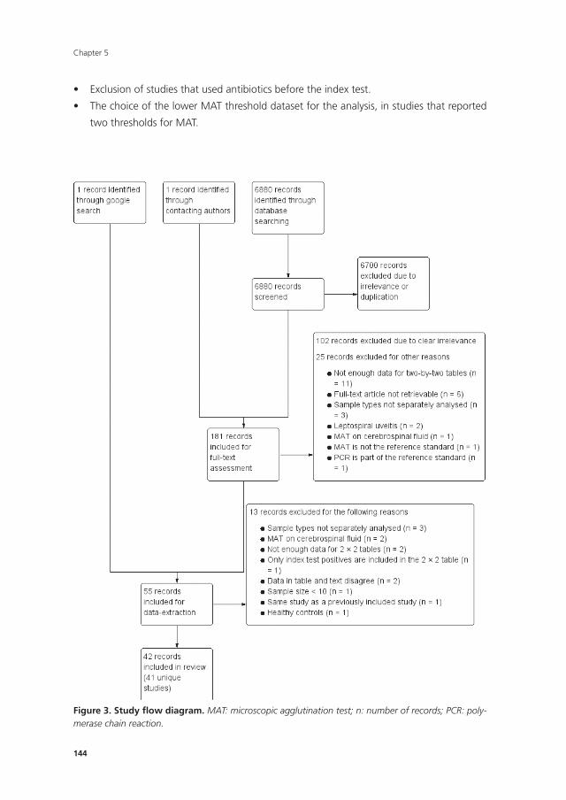
Chapter 5
144
• Exclusionofstudiesthatusedantibioticsbeforetheindextest.
• ThechoiceofthelowerMATthresholddatasetfortheanalysis,instudiesthatreported
two thresholds for MAT.
156
Figure 3. Study flow diagram. MAT: microscopic agglutination test; n: number of records; PCR: polymerase chain reaction. Description of studies
Included studies
The Characteristics of included studies table and Table 3 give an overview of all included
studies. Forty-one studies included 5981 participants, of whom 1834 were classified as having
Figure 3. Study fl ow diagram. MAT: microscopic agglutination test; n: number of records; PCR: poly-merase chain reaction.
145
Nucleic acid and antigen detection tests for leptospirosis
RESULTS
Results of the search
We conducted the final electronic search on 6 July 2018 and identified 6880 records (see Fig-
ure 3). After title and abstract screening, and after inclusion of one additional record, which
we identified by contacting one of the authors (Destura 2007), we included 181 records
for full-text assessment. We excluded 127 records; 102 records due to clear irrelevance and
25 records for other reasons (see Characteristics of excluded studies table, Supplementary
Materials). At this stage, we identified and included one full-text publication of a meeting
abstract (Denipitiya 2016).28
We included the remaining 55 records for data collection, of which 13 were excluded
for various reasons.
The review included 42 records,28-69 corresponding to 41 unique studies. We regarded
four publications as two studies because they included the same population (Thaipadungpa-
nit 201158 and Sonthayanon 201156 are grouped under Thaipadungpanit 2011; Waggoner
201461 and Waggoner 201462 are grouped under Waggoner 2014). We considered one
publication as two studies because two different populations were included (Villumsen 2012
BC; Villumsen 2012 U).60 Searching the WHO International Clinical Trial Registry Platform
yielded no relevant records.
Description of studies
Included studies
The Characteristics of included studies table and Table 3 give an overview of all included
studies. Forty-one studies included 5981 participants, of whom 1834 were classified as hav-
ing leptospirosis, and 4147 as not having leptospirosis. Thirty studies were cross-sectional,
five were single-gate case-control, four were two-gate case-control studies, and study design
was dubious in two studies (Zhang 1992;69 Gravekamp 199344), with Zhang 1992 being most
likely either a cross-sectional or single-gate case-control study, but not a two-gate study.
The index tests evaluated were conventional PCR (henceforth PCR; 17 studies), real-time
PCR (18 studies), nested PCR (four studies), PCR performed twice (performed twice on each
participant at different DPO and regarded as positive if at least one result was positive;
two studies), LAMP (two studies), ELISA (one study), dot-ELISA (one study), immunochro-
matography-based lateral flow assay (ICG-based LFA; one study), and dipstick assay (one
study) (see Table 4). Five studies directly compared tests in the same population: PCR versus
real-time PCR (Vanasco 201659), PCR versus nested PCR (Blanco 201435), nested PCR versus
real-time PCR (Merien 200547), real-time PCR versus LAMP (Thaipadungpanit 201156,58), and
ICG-based IFA versus dipstick assay versus PCR (Widiyanti 201365).
We observed high heterogeneity regarding the characteristics of the participants,
the execution of the index tests, and the choice of the reference standards. Most of the
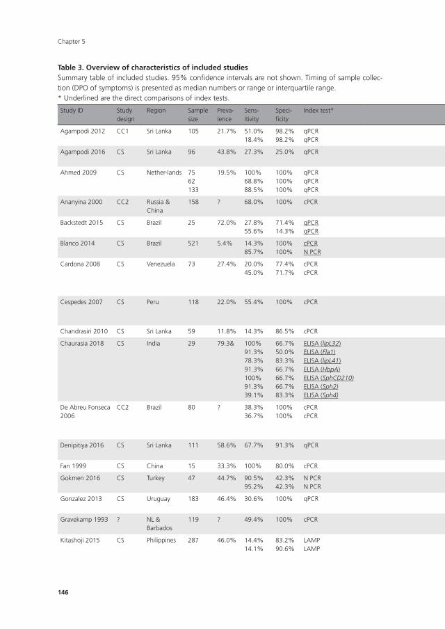
Chapter 5
146
Table 3. Overview of characteristics of included studiesSummary table of included studies. 95% confidence intervals are not shown. Timing of sample collec-tion (DPO of symptoms) is presented as median numbers or range or interquartile range.* Underlined are the direct comparisons of index tests.
Study ID Study design
Region Sample size
Preva-lence
Sens-itivity
Speci-ficity
Index test* Original reference of index test method
Target gene/primer*
Threshold Timing (DPO)*
Sample type* Reference test Sample for MAT
Agampodi 2012 CC1 Sri Lanka 105 21.7% 51.0%18.4%
98.2%98.2%
qPCRqPCR
Smythe 2002Smythe 2002
rrsrrs
??
1-101-10
Whole bloodSerum
MAT Paired only
Agampodi 2016 CS Sri Lanka 96 43.8% 27.3% 25.0% qPCR Smythe 2002 rrs ? 3-7 Blood/serum MAT Single + paired
Ahmed 2009 CS Nether-lands 7562133
19.5% 100%68.8%88.5%
100%100%100%
qPCRqPCRqPCR
Ahmed 2009Ahmed 2009Ahmed 2009
secYsecYsecY
Ct 35Ct 35Ct 35
1-45-101-10
Blood/serumBlood/serumBlood/serum
MAT OR IgM ELISA OR Culture
Single + Paired
Ananyina 2000 CC2 Russia & China
158 ? 68.0% 100% cPCR Gravekamp 1993 G1/G2 and B64-I/B64-II primers
NA ? Serum MAT Single + Paired
Backstedt 2015 CS Brazil 25 72.0% 27.8%55.6%
71.4%14.3%
qPCRqPCR
Stoddard 2009Backstedt 2015
lipL32rrs
??
??
Whole bloodWhole blood
MAT OR Culture
Single + Paired
Blanco 2014 CS Brazil 521 5.4% 14.3%85.7%
100%100%
cPCRN PCR
Merien 1992Merien 1992
rrsrrs
NANA
??
SerumSerum
MAT Single + Paired
Cardona 2008 CS Venezuela 73 27.4% 20.0%45.0%
77.4%71.7%
cPCRcPCR
Gravekamp 1993Gravekamp 1993
G1/G2 and B64-I/B64-II primersG1/G2 and B64-I/B64-II primers
NANA
??
SerumUrine
MAT Single + Paired
Cespedes 2007 CS Peru 118 22.0% 55.4% 100% cPCR Merien 1992 rrs NA 1-7 Whole blood MAT OR IgM ELISA OR Culture
Single + Paired
Chandrasiri 2010 CS Sri Lanka 59 11.8% 14.3% 86.5% cPCR ? G1/G2 primers NA ? Whole blood MAT Single
Chaurasia 2018 CS India 29 79.3& 100%91.3%78.3%91.3%100%91.3%39.1%
66.7%50.0%83.3%66.7%66.7%66.7%83.3%
ELISA (lipL32)ELISA (Fla1)ELISA (lipL41)ELISA (HbpA)ELISA (SphCD210)ELISA (Sph2)ELISA (Sph4)
Chaurasia 2018Chaurasia 2018Chaurasia 2018Chaurasia 2018Chaurasia 2018Chaurasia 2018Chaurasia 2018
NANANANANANANA
???????
???????
UrineUrineUrineUrineUrineUrineUrine
MAT Single
De Abreu Fonseca 2006
CC2 Brazil 80 ? 38.3%36.7%
100%100%
cPCRcPCR
Gravekamp 1993/Kee 1994Gravekamp 1993/Kee 1994
G1/G2 and LP1/LP2 primersG1/G2 and LP1/LP2 primers
NANA
??
Whole bloodUrine
MAT OR Culture
Single + Paired
Denipitiya 2016 CS Sri Lanka 111 58.6% 67.7% 91.3% qPCR Ahmed 2009 secY 35 Ct 1-5 Whole blood MAT Single + paired
Fan 1999 CS China 15 33.3% 100% 80.0% cPCR Fan 1999 rrs NA ? Serum MAT ?
Gokmen 2016 CS Turkey 47 44.7% 90.5%95.2%
42.3%42.3%
N PCRN PCR
Bomfim 2008Merien 1992
lipL32rrs
NANA
??
SerumSerum
MAT Single
Gonzalez 2013 CS Uruguay 183 46.4% 30.6% 100% qPCR Stoddard 2009 and Bourhahy 2001
lipL32 ? ? Serum MAT Paired only
Gravekamp 1993 ? NL & Barbados
119 ? 49.4% 100% cPCR Gravekamp 1993 G1/G2 and LP1/LP2 primers
NA ? Serum MAT OR IgM ELISA
?
Kitashoji 2015 CS Philippines 287 46.0% 14.4%14.1%
83.2%90.6%
LAMPLAMP
Koizumi 2012Koizumi 2012
rrsrrs
NANA
6.5?
PlasmaUrine
MAT Single + Paired
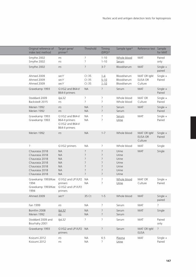
147
Nucleic acid and antigen detection tests for leptospirosis
Table 3. Overview of characteristics of included studiesSummary table of included studies. 95% confidence intervals are not shown. Timing of sample collec-tion (DPO of symptoms) is presented as median numbers or range or interquartile range.* Underlined are the direct comparisons of index tests.
Study ID Study design
Region Sample size
Preva-lence
Sens-itivity
Speci-ficity
Index test* Original reference of index test method
Target gene/primer*
Threshold Timing (DPO)*
Sample type* Reference test Sample for MAT
Agampodi 2012 CC1 Sri Lanka 105 21.7% 51.0%18.4%
98.2%98.2%
qPCRqPCR
Smythe 2002Smythe 2002
rrsrrs
??
1-101-10
Whole bloodSerum
MAT Paired only
Agampodi 2016 CS Sri Lanka 96 43.8% 27.3% 25.0% qPCR Smythe 2002 rrs ? 3-7 Blood/serum MAT Single + paired
Ahmed 2009 CS Nether-lands 7562133
19.5% 100%68.8%88.5%
100%100%100%
qPCRqPCRqPCR
Ahmed 2009Ahmed 2009Ahmed 2009
secYsecYsecY
Ct 35Ct 35Ct 35
1-45-101-10
Blood/serumBlood/serumBlood/serum
MAT OR IgM ELISA OR Culture
Single + Paired
Ananyina 2000 CC2 Russia & China
158 ? 68.0% 100% cPCR Gravekamp 1993 G1/G2 and B64-I/B64-II primers
NA ? Serum MAT Single + Paired
Backstedt 2015 CS Brazil 25 72.0% 27.8%55.6%
71.4%14.3%
qPCRqPCR
Stoddard 2009Backstedt 2015
lipL32rrs
??
??
Whole bloodWhole blood
MAT OR Culture
Single + Paired
Blanco 2014 CS Brazil 521 5.4% 14.3%85.7%
100%100%
cPCRN PCR
Merien 1992Merien 1992
rrsrrs
NANA
??
SerumSerum
MAT Single + Paired
Cardona 2008 CS Venezuela 73 27.4% 20.0%45.0%
77.4%71.7%
cPCRcPCR
Gravekamp 1993Gravekamp 1993
G1/G2 and B64-I/B64-II primersG1/G2 and B64-I/B64-II primers
NANA
??
SerumUrine
MAT Single + Paired
Cespedes 2007 CS Peru 118 22.0% 55.4% 100% cPCR Merien 1992 rrs NA 1-7 Whole blood MAT OR IgM ELISA OR Culture
Single + Paired
Chandrasiri 2010 CS Sri Lanka 59 11.8% 14.3% 86.5% cPCR ? G1/G2 primers NA ? Whole blood MAT Single
Chaurasia 2018 CS India 29 79.3& 100%91.3%78.3%91.3%100%91.3%39.1%
66.7%50.0%83.3%66.7%66.7%66.7%83.3%
ELISA (lipL32)ELISA (Fla1)ELISA (lipL41)ELISA (HbpA)ELISA (SphCD210)ELISA (Sph2)ELISA (Sph4)
Chaurasia 2018Chaurasia 2018Chaurasia 2018Chaurasia 2018Chaurasia 2018Chaurasia 2018Chaurasia 2018
NANANANANANANA
???????
???????
UrineUrineUrineUrineUrineUrineUrine
MAT Single
De Abreu Fonseca 2006
CC2 Brazil 80 ? 38.3%36.7%
100%100%
cPCRcPCR
Gravekamp 1993/Kee 1994Gravekamp 1993/Kee 1994
G1/G2 and LP1/LP2 primersG1/G2 and LP1/LP2 primers
NANA
??
Whole bloodUrine
MAT OR Culture
Single + Paired
Denipitiya 2016 CS Sri Lanka 111 58.6% 67.7% 91.3% qPCR Ahmed 2009 secY 35 Ct 1-5 Whole blood MAT Single + paired
Fan 1999 CS China 15 33.3% 100% 80.0% cPCR Fan 1999 rrs NA ? Serum MAT ?
Gokmen 2016 CS Turkey 47 44.7% 90.5%95.2%
42.3%42.3%
N PCRN PCR
Bomfim 2008Merien 1992
lipL32rrs
NANA
??
SerumSerum
MAT Single
Gonzalez 2013 CS Uruguay 183 46.4% 30.6% 100% qPCR Stoddard 2009 and Bourhahy 2001
lipL32 ? ? Serum MAT Paired only
Gravekamp 1993 ? NL & Barbados
119 ? 49.4% 100% cPCR Gravekamp 1993 G1/G2 and LP1/LP2 primers
NA ? Serum MAT OR IgM ELISA
?
Kitashoji 2015 CS Philippines 287 46.0% 14.4%14.1%
83.2%90.6%
LAMPLAMP
Koizumi 2012Koizumi 2012
rrsrrs
NANA
6.5?
PlasmaUrine
MAT Single + Paired
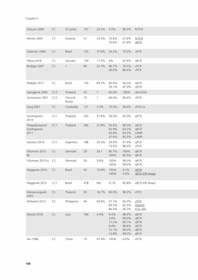
Chapter 5
148
Koizumi 2009 CS Sri Lanka 107 24.3% 0.0% 96.3% N PCR Kawabata 2001/mod:Koizumi 2008
flaB NA 7 Serum MAT Single only
Merien 2005 CS Oceania 51 33.3% 70.6%70.6%
61.8%61.8%
N PCRqPCR
Merien 1992Merien 2005
rrsLFB1-F/LFB1-R primers
NA?
55
SerumSerum
MAT Single + Paired
Ooteman 2006 CS Brazil 125 37.6% 36.2% 70.5% cPCR Gravekamp 1993 G1/G2 primers NA ? Serum MAT Single + Paired
Pakoa 2018 CS Vanuatu 130 11.5% 0% 97.4% qPCR Stoddard 2009 lipL32 ? ? Serum MAT Single
Riediger 2007 CS ? 66 22.7% 46.7%40.0%
76.5%80.4%
cPCRcPCR
Gravekamp 1993Gravekamp 1993
G1/G2 and B64-I/B64-II primersG1/G2 and B64-I/B64-II primers
NANA
??
Whole bloodUrine
MAT Single + Paired
Riediger 2017 CS Brazil 150 84.7% 60.6%29.1%
56.2%87.0%
qPCRqPCR
Stoddard 2009Stoddard 2009
lipL32lipL32
40 Ct40 Ct
??
Whole bloodSerum
MAT OR culture
Single + paired
Saengjaruk 2002 CC2 Thailand 43 ? 64.0% 100% dot-ELISA Saengjaruk 2002 NA NA 5 Urine Culture NA
Samsonova 1997 CC2 China & Russia
75 ? 66.0% 96.4% cPCR Gravekamp 1993 G1/G2 and B64-I/B64-II primers
NA ? Serum MAT ?
Seng 2007 CS Cambodia 121 3.3% 75.0% 94.0% cPCR 2x ? rrl (23S) NA (1) 14, (2) 35
Serum MAT OR Culture
Single + Paired
Sonthayanon 2013
CC1 Thailand 250 31.8% 59.0% 92.0% qPCR Slack 2007 rrs ? ? Whole blood MAT OR Culture
Single + Paired
Thaipadunpanit/ Sonthayanon 2011
CC1 Thailand 266 31.8% 55.6%42.9%43.6%37.6%
89.5%93.2%83.5%90.2%
qPCRqPCRLAMPLAMP
Slack 2007Stoddard 2009Sonthayanon 2011Lin 2009
rrslipL32rrslipL41
??NANA
5555
Whole bloodWhole bloodWhole bloodWhole blood
MAT OR Culture
Paired only
Vanasco 2016 CC1 Argentina 188 35.5% 29.9%13.4%
81.0%88.4%
qPCRcPCR
Stoddard 2009Stoddard 2009
lipL32lipL32
40 CtNA
55
Serum/bloodSerum/blood
MAT OR IgM ELISA
Single + paired
Villumsen 2012-BC
CS Denmark 29 24.1 85.7%100%
100%95.5%
qPCRqPCR
Villumsen 2012Smythe 2002
lipL32rrs
??
??
Blood cultureBlood culture
MAT Single + paired
Villumsen 2012-U CS Denmark 54 5.6% 100%100%
98.0%98.0%
qPCRqPCR
Villumsen 2012Smythe 2002
lipL32rrs
??
??
UrineUrine
MAT Single + paired
Waggoner 2014 CS Brazil 55 10.9% 100%100%
4.1%0.0%
qPCRqPCR (UFI Assay)
Waggoner 2014Waggoner 2014
rrsrrs
45 Ct45 Ct
8Range 1-19
Plasma/serumPlasma/serum
MAT Single only
Waggoner 2015 CC1 Brazil 478 NA 9.1% 92.8% qPCR (UFI Assay) Waggoner 2014 rrs 45 Ct ? Serum MAT Single only
Wangroongsarb 2005
CS Thailand 93 16.1% 80.0% 96.2% cPCR ? / Kawabata 2001 rrs / flaB NA ? Whole blood MAT OR Culture
Paired
Widiyanti 2013 CS Philippines 44 63.6% 57.1%89.3%96.4%
56.3%62.5%56.3%
cPCRDipstickICG- LFA
Kawabata 2001Widyanti 2013Widiyanti 2013
flaBNANA
NANANA
5.55.55.5
UrineUrineUrine
MAT Single only
Woods 2018 CS Laos 766 4.4% 9.4%3.0%17.2%9.4%12.1%13.8%
98.5%99.0%90.1%98.8%99.0%99.0%
qPCRqPCRqPCRqPCRqPCRqPCR
Slack 2007Slack 2007Slack 2007Woods 2018Woods 2018Woods 2018
rrsrrsrrsrrs/lipL32rrs/lipL32rrs/lipL32
40 Ct40 Ct40 Ct45 Ct45 Ct45 Ct
555555
SerumBuffy coatUrineSerumBuffy coatUrine
MAT OR Culture
Single + paired
Wu 1996 CS China 19 47.4% 100% 0.0% cPCR Wu 1993 rrs NA ? Serum MAT OR Culture
?
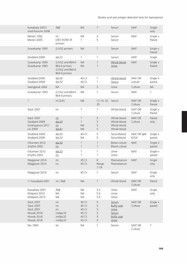
149
Nucleic acid and antigen detection tests for leptospirosis
Koizumi 2009 CS Sri Lanka 107 24.3% 0.0% 96.3% N PCR Kawabata 2001/mod:Koizumi 2008
flaB NA 7 Serum MAT Single only
Merien 2005 CS Oceania 51 33.3% 70.6%70.6%
61.8%61.8%
N PCRqPCR
Merien 1992Merien 2005
rrsLFB1-F/LFB1-R primers
NA?
55
SerumSerum
MAT Single + Paired
Ooteman 2006 CS Brazil 125 37.6% 36.2% 70.5% cPCR Gravekamp 1993 G1/G2 primers NA ? Serum MAT Single + Paired
Pakoa 2018 CS Vanuatu 130 11.5% 0% 97.4% qPCR Stoddard 2009 lipL32 ? ? Serum MAT Single
Riediger 2007 CS ? 66 22.7% 46.7%40.0%
76.5%80.4%
cPCRcPCR
Gravekamp 1993Gravekamp 1993
G1/G2 and B64-I/B64-II primersG1/G2 and B64-I/B64-II primers
NANA
??
Whole bloodUrine
MAT Single + Paired
Riediger 2017 CS Brazil 150 84.7% 60.6%29.1%
56.2%87.0%
qPCRqPCR
Stoddard 2009Stoddard 2009
lipL32lipL32
40 Ct40 Ct
??
Whole bloodSerum
MAT OR culture
Single + paired
Saengjaruk 2002 CC2 Thailand 43 ? 64.0% 100% dot-ELISA Saengjaruk 2002 NA NA 5 Urine Culture NA
Samsonova 1997 CC2 China & Russia
75 ? 66.0% 96.4% cPCR Gravekamp 1993 G1/G2 and B64-I/B64-II primers
NA ? Serum MAT ?
Seng 2007 CS Cambodia 121 3.3% 75.0% 94.0% cPCR 2x ? rrl (23S) NA (1) 14, (2) 35
Serum MAT OR Culture
Single + Paired
Sonthayanon 2013
CC1 Thailand 250 31.8% 59.0% 92.0% qPCR Slack 2007 rrs ? ? Whole blood MAT OR Culture
Single + Paired
Thaipadunpanit/ Sonthayanon 2011
CC1 Thailand 266 31.8% 55.6%42.9%43.6%37.6%
89.5%93.2%83.5%90.2%
qPCRqPCRLAMPLAMP
Slack 2007Stoddard 2009Sonthayanon 2011Lin 2009
rrslipL32rrslipL41
??NANA
5555
Whole bloodWhole bloodWhole bloodWhole blood
MAT OR Culture
Paired only
Vanasco 2016 CC1 Argentina 188 35.5% 29.9%13.4%
81.0%88.4%
qPCRcPCR
Stoddard 2009Stoddard 2009
lipL32lipL32
40 CtNA
55
Serum/bloodSerum/blood
MAT OR IgM ELISA
Single + paired
Villumsen 2012-BC
CS Denmark 29 24.1 85.7%100%
100%95.5%
qPCRqPCR
Villumsen 2012Smythe 2002
lipL32rrs
??
??
Blood cultureBlood culture
MAT Single + paired
Villumsen 2012-U CS Denmark 54 5.6% 100%100%
98.0%98.0%
qPCRqPCR
Villumsen 2012Smythe 2002
lipL32rrs
??
??
UrineUrine
MAT Single + paired
Waggoner 2014 CS Brazil 55 10.9% 100%100%
4.1%0.0%
qPCRqPCR (UFI Assay)
Waggoner 2014Waggoner 2014
rrsrrs
45 Ct45 Ct
8Range 1-19
Plasma/serumPlasma/serum
MAT Single only
Waggoner 2015 CC1 Brazil 478 NA 9.1% 92.8% qPCR (UFI Assay) Waggoner 2014 rrs 45 Ct ? Serum MAT Single only
Wangroongsarb 2005
CS Thailand 93 16.1% 80.0% 96.2% cPCR ? / Kawabata 2001 rrs / flaB NA ? Whole blood MAT OR Culture
Paired
Widiyanti 2013 CS Philippines 44 63.6% 57.1%89.3%96.4%
56.3%62.5%56.3%
cPCRDipstickICG- LFA
Kawabata 2001Widyanti 2013Widiyanti 2013
flaBNANA
NANANA
5.55.55.5
UrineUrineUrine
MAT Single only
Woods 2018 CS Laos 766 4.4% 9.4%3.0%17.2%9.4%12.1%13.8%
98.5%99.0%90.1%98.8%99.0%99.0%
qPCRqPCRqPCRqPCRqPCRqPCR
Slack 2007Slack 2007Slack 2007Woods 2018Woods 2018Woods 2018
rrsrrsrrsrrs/lipL32rrs/lipL32rrs/lipL32
40 Ct40 Ct40 Ct45 Ct45 Ct45 Ct
555555
SerumBuffy coatUrineSerumBuffy coatUrine
MAT OR Culture
Single + paired
Wu 1996 CS China 19 47.4% 100% 0.0% cPCR Wu 1993 rrs NA ? Serum MAT OR Culture
?
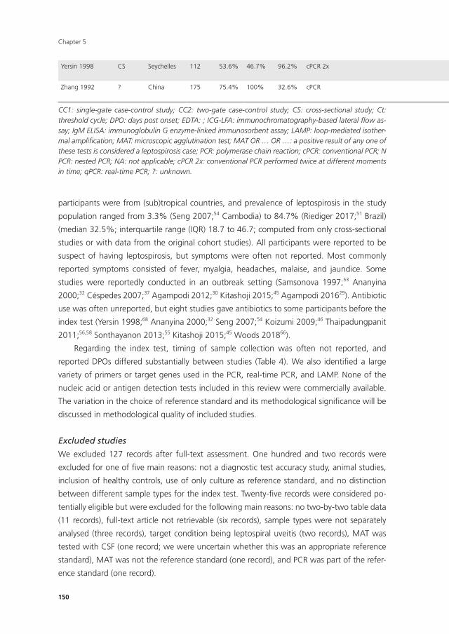
Chapter 5
150
participants were from (sub)tropical countries, and prevalence of leptospirosis in the study
population ranged from 3.3% (Seng 2007;54 Cambodia) to 84.7% (Riediger 2017;51 Brazil)
(median 32.5%; interquartile range (IQR) 18.7 to 46.7; computed from only cross-sectional
studies or with data from the original cohort studies). All participants were reported to be
suspect of having leptospirosis, but symptoms were often not reported. Most commonly
reported symptoms consisted of fever, myalgia, headaches, malaise, and jaundice. Some
studies were reportedly conducted in an outbreak setting (Samsonova 1997;53 Ananyina
2000;32 Céspedes 2007;37 Agampodi 2012;30 Kitashoji 2015;45 Agampodi 201629). Antibiotic
use was often unreported, but eight studies gave antibiotics to some participants before the
index test (Yersin 1998;68 Ananyina 2000;32 Seng 2007;54 Koizumi 2009;46 Thaipadungpanit
2011;56,58 Sonthayanon 2013;55 Kitashoji 2015;45 Woods 201866).
Regarding the index test, timing of sample collection was often not reported, and
reported DPOs differed substantially between studies (Table 4). We also identified a large
variety of primers or target genes used in the PCR, real-time PCR, and LAMP. None of the
nucleic acid or antigen detection tests included in this review were commercially available.
The variation in the choice of reference standard and its methodological significance will be
discussed in methodological quality of included studies.
Excluded studies
We excluded 127 records after full-text assessment. One hundred and two records were
excluded for one of five main reasons: not a diagnostic test accuracy study, animal studies,
inclusion of healthy controls, use of only culture as reference standard, and no distinction
between different sample types for the index test. Twenty-five records were considered po-
tentially eligible but were excluded for the following main reasons: no two-by-two table data
(11 records), full-text article not retrievable (six records), sample types were not separately
analysed (three records), target condition being leptospiral uveitis (two records), MAT was
tested with CSF (one record; we were uncertain whether this was an appropriate reference
standard), MAT was not the reference standard (one record), and PCR was part of the refer-
ence standard (one record).
Yersin 1998 CS Seychelles 112 53.6% 46.7% 96.2% cPCR 2x Merien 1995 rrs NA (1) 3.9-4.5(2) ≥14
Serum MAT Paired only
Zhang 1992 ? China 175 75.4% 100% 32.6% cPCR Zhang 1992 rrl (23S) NA 1-5 Serum MAT OR Culture
Paired only
CC1: single-gate case-control study; CC2: two-gate case-control study; CS: cross-sectional study; Ct: threshold cycle; DPO: days post onset; EDTA: ; ICG-LFA: immunochromatography-based lateral flow as-say; IgM ELISA: immunoglobulin G enzyme-linked immunosorbent assay; LAMP: loop-mediated isother-mal amplification; MAT: microscopic agglutination test; MAT OR … OR …: a positive result of any one of these tests is considered a leptospirosis case; PCR: polymerase chain reaction; cPCR: conventional PCR; N PCR: nested PCR; NA: not applicable; cPCR 2x: conventional PCR performed twice at different moments in time; qPCR: real-time PCR; ?: unknown.
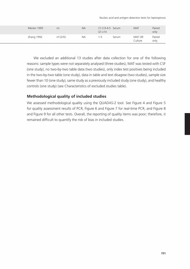
151
Nucleic acid and antigen detection tests for leptospirosis
We excluded an additional 13 studies after data collection for one of the following
reasons: sample types were not separately analysed (three studies), MAT was tested with CSF
(one study), no two-by-two table data (two studies), only index test positives being included
in the two-by-two table (one study), data in table and text disagree (two studies), sample size
fewer than 10 (one study), same study as a previously included study (one study), and healthy
controls (one study) (see Characteristics of excluded studies table).
Methodological quality of included studies
We assessed methodological quality using the QUADAS-2 tool. See Figure 4 and Figure 5
for quality assessment results of PCR, Figure 6 and Figure 7 for real-time PCR, and Figure 8
and Figure 9 for all other tests. Overall, the reporting of quality items was poor; therefore, it
remained difficult to quantify the risk of bias in included studies.
Yersin 1998 CS Seychelles 112 53.6% 46.7% 96.2% cPCR 2x Merien 1995 rrs NA (1) 3.9-4.5(2) ≥14
Serum MAT Paired only
Zhang 1992 ? China 175 75.4% 100% 32.6% cPCR Zhang 1992 rrl (23S) NA 1-5 Serum MAT OR Culture
Paired only
CC1: single-gate case-control study; CC2: two-gate case-control study; CS: cross-sectional study; Ct: threshold cycle; DPO: days post onset; EDTA: ; ICG-LFA: immunochromatography-based lateral flow as-say; IgM ELISA: immunoglobulin G enzyme-linked immunosorbent assay; LAMP: loop-mediated isother-mal amplification; MAT: microscopic agglutination test; MAT OR … OR …: a positive result of any one of these tests is considered a leptospirosis case; PCR: polymerase chain reaction; cPCR: conventional PCR; N PCR: nested PCR; NA: not applicable; cPCR 2x: conventional PCR performed twice at different moments in time; qPCR: real-time PCR; ?: unknown.
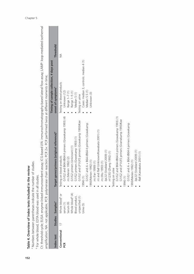
Chapter 5
152
Tab
le 4
. Ove
rvie
w o
f in
dex
tes
ts in
clu
ded
in t
he
revi
ewa N
umbe
rs b
etw
een
pare
nthe
ses
indi
cate
the
num
ber
of s
tudi
es.
b For
who
le b
lood
, ED
TA b
lood
was
use
d in
all
stud
ies.
Ct:
thre
shol
d cy
cle;
ELI
SA: e
nzym
e-lin
ked
imm
unos
orbe
nt a
ssay
; IC
G-b
ased
LFA
: im
mun
ochr
omat
ogra
phy-
base
d la
tera
l flow
ass
ay; L
AM
P: lo
op-m
edia
ted
isot
herm
al
ampl
ifica
tion;
NA
: not
app
licab
le; P
CR:
pol
ymer
ase
chai
n re
actio
n; P
CR
2×: P
CR
perf
orm
ed t
wic
e at
diff
eren
t m
omen
ts in
tim
e;
Ind
ex t
est
Stu
die
sSa
mp
lesa
Targ
et g
enes
/pri
mer
s (o
rig
inal
ref
eren
ce)a
Tim
ing
of
sam
ple
co
llect
ion
, # d
ays
po
st
on
set
of
sym
pto
msa
Thre
sho
ld
Co
nve
nti
on
al
PCR
17W
hole
blo
odb o
r se
rum
(1)
Seru
m (9
)W
hole
blo
odb (4
)Bl
ood
prod
uct,
un
spec
ified
(1)
Urin
e (5
)
Test
ing
on b
lood
pro
duct
s•
G1/
G2
and
B64-
I/B64
-IIp
rimer
s(G
rave
kam
p19
93)(
4)•
rrs
(Mer
ien
1992
) (2)
•G
1/G
2pr
imer
s(U
nkno
wn)
(1)
•G
1/G
2pr
imer
s(G
rave
kam
p19
93)(
1)•
G1/
G2
and
LP1/
LP2
prim
ers
(Gra
veka
mp
1993
/Kee
19
94) (
1)•
G1/
G1
and
A+
B64
-I/B6
4-II
prim
ers
(Gra
veka
mp
1993
/Mer
ien
1992
) (1)
•rr
s (F
an 1
999)
(1)
•rr
s an
d fla
B (U
nkno
wn/
Kaw
abat
a 20
01) (
1)•
rrs
(Wu
1993
) (1)
•lip
L32
(Sto
ddar
d 20
09) (
1)•
rrl (
23S)
(Zha
ng 1
992)
(1)
Test
ing
on u
rine
•G
1/G
2an
dB6
4-I/B
64-II
prim
ers
(Gra
veka
mp
1993
)(1)
•G
1/G
2an
dLP
1/LP
2pr
imer
s(G
ram
ekam
p19
93/K
ee
1994
) (1)
•G
1/G
1an
dA
+B
64-I/
B64-
IIpr
imer
s(G
rave
kam
p19
93/M
erie
n 19
92) (
1)•
lipL3
2 (S
todd
ard
2009
) (1)
•fla
B (K
awab
ata
2001
) (1)
Test
ing
on b
lood
pro
duct
s•
Unk
now
n(1
2)•
Rang
e1–
7(1
)•
Rang
e1–
5(1
)•
Med
ian
5(1
)Te
stin
g on
urin
e•
Cas
es:m
edia
n3,
con
trol
s:m
edia
n4
(1)
•M
edia
n5.
5(1
)•
Unk
now
n(3
)
NA
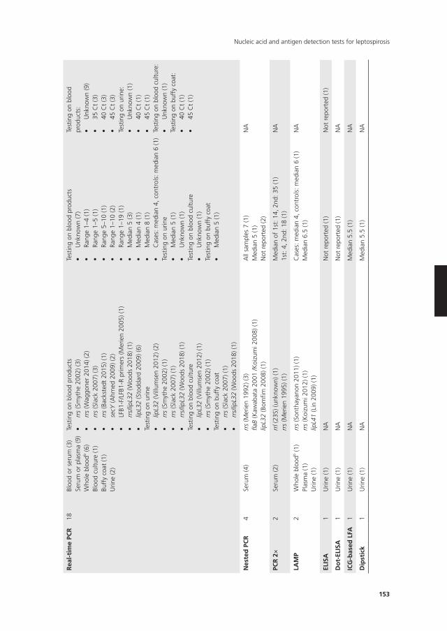
153
Nucleic acid and antigen detection tests for leptospirosis
Rea
l-ti
me
PCR
18Bl
ood
or s
erum
(3)
Seru
m o
r pl
asm
a (9
)W
hole
blo
odb (6
)Bl
ood
cultu
re (1
)Bu
ffy
coat
(1)
Urin
e (2
)
Test
ing
on b
lood
pro
duct
s•
rrs
(Sm
ythe
200
2) (3
)•
rrs
(Wag
gone
r 20
14) (
2)•
rrs
(Sla
ck 2
007)
(3)
•rr
s (B
acks
tedt
201
5) (1
)•
secY
(Ahm
ed 2
009)
(2)
•LF
B1-F
/LFB
1-R
prim
ers
(Mer
ien
2005
)(1)
•rr
s/lip
L32
(Woo
ds 2
018)
(1)
•lip
L32
(Sto
ddar
d 20
09) (
6)Te
stin
g on
urin
e•
lipL3
2 (V
illum
sen
2012
) (2)
•rr
s (S
myt
he 2
002)
(1)
•rr
s (S
lack
200
7) (1
)•
rrs/
lipL3
2 (W
oods
201
8) (1
)Te
stin
g on
blo
od c
ultu
re•
lipL3
2 (V
illum
sen
2012
) (1)
•rr
s (S
myt
he 2
002)
(1)
Test
ing
on b
uffy
coa
t•
rrs
(Sla
ck 2
007)
(1)
•rr
s/lip
L32
(Woo
ds 2
018)
(1)
Test
ing
on b
lood
pro
duct
s•
Unk
now
n(7
)•
Rang
e1–
4(1
)•
Rang
e1–
5(1
)•
Rang
e5–
10(1
)•
Rang
e1–
10(2
)•
Rang
e1–
19(1
)•
Med
ian
5(3
)•
Med
ian
4(1
)•
Med
ian
8(1
)•
Cas
es:m
edia
n4,
con
trol
s:m
edia
n6
(1)
Test
ing
on u
rine
•M
edia
n5
(1)
•U
nkno
wn
(1)
Test
ing
on b
lood
cul
ture
•U
nkno
wn
(1)
Test
ing
on b
uffy
coa
t•
Med
ian
5(1
)
Test
ing
on b
lood
pr
oduc
ts:
•U
nkno
wn
(9)
•35
Ct
(3)
•40
Ct
(3)
•45
Ct
(3)
Test
ing
on u
rine:
•U
nkno
wn
(1)
•40
Ct
(1)
•45
Ct
(1)
Test
ing
on b
lood
cul
ture
:•
Unk
now
n(1
)Te
stin
g on
buf
fy c
oat:
•40
Ct
(1)
•45
Ct
(1)
nes
ted
PC
R4
Seru
m (4
)rr
s (M
erie
n 19
92) (
3)fla
B (K
awab
ata
2001
/Koi
zum
i 200
8) (1
)lip
L32
(Bom
fim 2
008)
(1)
All
sam
ples
7 (1
)M
edia
n 5
(1)
Not
rep
orte
d (2
)
NA
PCR
2×
2Se
rum
(2)
rrl (
23S)
(unk
now
n) (1
)rr
s (M
erie
n 19
95) (
1)M
edia
n of
1st
: 14,
2nd
: 35
(1)
1st:
4, 2
nd: 1
8 (1
)N
A
LAM
P2
Who
le b
lood
b (1)
Plas
ma
(1)
Urin
e (1
)
rrs
(Son
thay
anon
201
1) (1
)rr
s (K
oizu
mi 2
012)
(1)
lipL4
1 (L
in 2
009)
(1)
Cas
es: m
edia
n 4,
con
trol
s: m
edia
n 6
(1)
Med
ian
6.5
(1)
NA
ELIS
A1
Urin
e (1
)N
AN
ot r
epor
ted
(1)
Not
rep
orte
d (1
)
Do
t-EL
ISA
1U
rine
(1)
NA
Not
rep
orte
d (1
)N
A
ICG
-bas
ed L
FA1
Urin
e (1
)N
AM
edia
n 5.
5 (1
)N
A
Dip
stic
k1
Urin
e (1
)N
AM
edia
n 5.
5 (1
)N
A
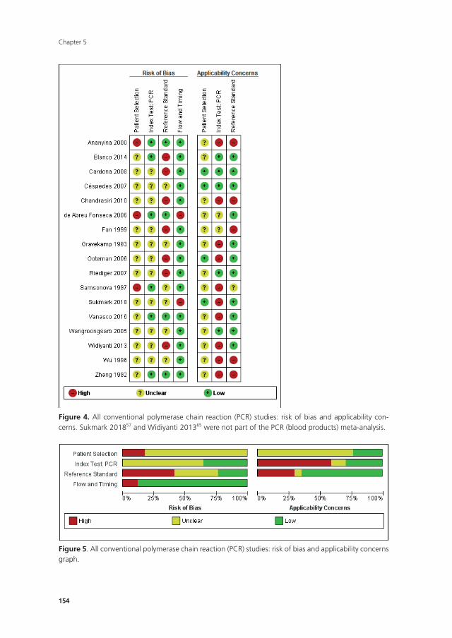
Chapter 5
154
159
Figure 5. All conventional polymerase chain reaction (PCR) studies: risk of bias and applicability concerns graph.
Figure 4. All conventional polymerase chain reaction (PCR) studies: risk of bias and applicability concerns. Sukmark 201857 and Widiyanti 201365 were not part of the PCR (blood products) meta-analysis.
Figure 4. All conventional polymerase chain reaction (PCR) studies: risk of bias and applicability con-cerns. Sukmark 201857 and Widiyanti 201365 were not part of the PCR (blood products) meta-analysis.
159
Figure 5. All conventional polymerase chain reaction (PCR) studies: risk of bias and applicability concerns graph.
Figure 4. All conventional polymerase chain reaction (PCR) studies: risk of bias and applicability concerns. Sukmark 201857 and Widiyanti 201365 were not part of the PCR (blood products) meta-analysis.
Figure 5. All conventional polymerase chain reaction (PCR) studies: risk of bias and applicability concerns graph.
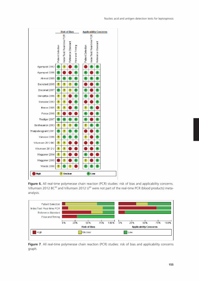
155
Nucleic acid and antigen detection tests for leptospirosis
160
Figure 7. All real-time polymerase chain reaction (PCR) studies: risk of bias and applicability concerns graph.
Figure 6. All real-time polymerase chain reaction (PCR) studies: risk of bias and applicability concerns. Villumsen 2012 BC60 and Villumsen 2012 U60 were not part of the real-time PCR (blood products) meta-analysis.
Figure 6. All real-time polymerase chain reaction (PCR) studies: risk of bias and applicability concerns. Villumsen 2012 BC60 and Villumsen 2012 U60 were not part of the real-time PCR (blood products) meta-analysis.
160
Figure 7. All real-time polymerase chain reaction (PCR) studies: risk of bias and applicability concerns graph.
Figure 6. All real-time polymerase chain reaction (PCR) studies: risk of bias and applicability concerns. Villumsen 2012 BC60 and Villumsen 2012 U60 were not part of the real-time PCR (blood products) meta-analysis.
Figure 7. All real-time polymerase chain reaction (PCR) studies: risk of bias and applicability concerns graph.
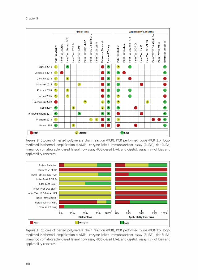
Chapter 5
156
161
Figure 8. Studies of nested polymerase chain reaction (PCR), PCR performed twice (PCR 2x), loop-mediated isothermal amplification (LAMP), enzyme-linked immunosorbent assay (ELISA), dot-ELISA, immunochromatography-based lateral flow assay (ICG-based LFA), and dipstick assay: risk of bias and applicability concerns.
Figure 9. Studies of nested polymerase chain reaction (PCR), PCR performed twice (PCR 2x), loop-mediated isothermal amplification (LAMP), enzyme-linked immunosorbent assay (ELISA), dot-ELISA, immunochromatography-based lateral flow assay (ICG-based LFA), and dipstick assay: risk of bias and applicability concerns.
Figure 8. Studies of nested polymerase chain reaction (PCR), PCR performed twice (PCR 2x), loop-mediated isothermal amplification (LAMP), enzyme-linked immunosorbent assay (ELISA), dot-ELISA, immunochromatography-based lateral flow assay (ICG-based LFA), and dipstick assay: risk of bias and applicability concerns.
161
Figure 8. Studies of nested polymerase chain reaction (PCR), PCR performed twice (PCR 2x), loop-mediated isothermal amplification (LAMP), enzyme-linked immunosorbent assay (ELISA), dot-ELISA, immunochromatography-based lateral flow assay (ICG-based LFA), and dipstick assay: risk of bias and applicability concerns.
Figure 9. Studies of nested polymerase chain reaction (PCR), PCR performed twice (PCR 2x), loop-mediated isothermal amplification (LAMP), enzyme-linked immunosorbent assay (ELISA), dot-ELISA, immunochromatography-based lateral flow assay (ICG-based LFA), and dipstick assay: risk of bias and applicability concerns.
Figure 9. Studies of nested polymerase chain reaction (PCR), PCR performed twice (PCR 2x), loop-mediated isothermal amplification (LAMP), enzyme-linked immunosorbent assay (ELISA), dot-ELISA, immunochromatography-based lateral flow assay (ICG-based LFA), and dipstick assay: risk of bias and applicability concerns.
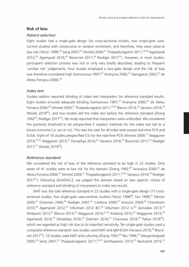
157
Nucleic acid and antigen detection tests for leptospirosis
Risk of bias
Patient selection
Eight studies had a single-gate design (six cross-sectional studies, two single-gate case-
control studies) with consecutive or random enrolment, and therefore, they were rated at
low risk (Yersin 1998;68 Seng 2007;54 Ahmed 2009;31 Thaipadungpanit 2011;56,58 Agampodi
2012;29 Agampodi 2016;30 Biscornet 2017;34 Riediger 201751). However, in most studies,
participant selection process was not or only very briefly described, leading to frequent
‘unclear risk’ judgements. Four studies employed a two-gate design and the risk of bias
was therefore considered high (Samsonova 1997;53 Ananyina 2000;32 Saengjaruk 2002;52 de
Abreu Fonseca 2006).40
Index test
Studies seldom reported blinding of index test interpreters for reference standard results.
Eight studies ensured adequate blinding (Samsonova 1997;53 Ananyina 2000;32 de Abreu
Fonseca 2006;40 Ahmed 2009;31 Thaipadungpanit 2011;56,58 Blanco 2014;35 Vanasco 2016;59
Woods 201866), and two studies did the index test before the reference standard (Zhang
199269; Riediger 201751). No study reported that interpreters were unblinded. We considered
the positivity threshold to be prespecified if readout methods for the index test led to a
binary outcome (i.e. yes or no). This was the case for all index tests except real-time PCR and
ELISA. Eight of 18 studies prespecified Cts for the real-time PCR (Ahmed 2009;31 Waggoner
2014;61,62 Waggoner 2015;63 Denipitiya 2016;28 Vanasco 2016;59 Biscornet 2017;34 Riediger
2017;51 Woods 201866).
Reference standard
We considered the risk of bias of the reference standard to be high in 22 studies. Only
seven of 41 studies were at low risk for this domain (Zhang 1992;69 Ananyina 2000;32 de
Abreu Fonseca 2006;40 Ahmed 2009;31 Thaipadungpanit 2011;56,58 Vanasco 2016;59 Riediger
201751). Following QUADAS-2, we judged this domain based on two aspects: choice of
reference standard and blinding of interpreters to index test results.
MAT was the sole reference standard in 22 studies with a single-gate design (17 cross-
sectional studies, five single-gate case-controls studies) (Yersin 199868; Fan 1999;41 Merien
2005;47 Ooteman 2006;48 Riediger 2007;50 Cardona 2008;36 Koizumi 2009;46 Chandrasiri
2010;38 Agampodi 2012;30 Villumsen 2012 BC;60 Villumsen 2012 U;60 Gonzalez 2013;43
Widiyanti 2013;65 Blanco 2014;35 Waggoner 2014;61,62 Kitashoji 2015;45 Waggoner 2015;63
Agampodi 2016;29 Denipitiya 2016;28 Gokmen 2016;42 Chaurasia 2018;39 Pakoa 201849),
which we regarded as high risk due to its imperfect sensitivity. Ten single-gate studies used a
composite reference standard: two studies used MAT and IgM ELISA (Vanasco 2016;59 Biscor-
net 201734), 10 studies used MAT and culturing (Zhang 1992;69 Wu 1996;67 Wangroongsarb
2005;64 Seng 2007;54 Thaipadungpanit 2011;56,58 Sonthayanon 2013;55 Backstedt 2015;33
Chapter 5
158
Riediger 2017;51 Sukmark 2018;57 Woods 201866), and two studies used all three (Céspedes
2007;37 Ahmed 200931).
As an additional criterion, we required MAT to include paired samples for the judgement
‘low risk’. All but one study (Wu 199667) with a composite reference standard fulfilled this
criterion. When MAT alone or culture alone was used as the reference standard in two-gate
designs, risk of bias was considered low.
Two studies reported blinding of the reference standard interpreters, in which the
blinding was adequate (Ahmed 2009;31 Riediger 201751). We also considered blinding to
be adequate in nine studies in which the reference standard was done before the index test
(Samsonova 1997;53 Ananyina 2000;32 de Abreu Fonseca 2006;40 Thaipadungpanit 2011;56,58
Villumsen 2012 BC;60 Villumsen 2012 U;60 Waggoner 2014;61,62 Waggoner 2015;63 Vanasco
201659).
Flow and timing
Risk of bias for flow and timing was low for 32 studies and unclear for one study (Riediger
2007). Eight studies were considered high risk, as they did not include all patients in the
analysis, with reasons varying from decisions by clinicians not to request MAT to exclusion
based on inadequate urine samples (Yersin 1998;68 de Abreu Fonseca 2006;40 Villumsen
2012 BC;60 Villumsen 2012 U;60 Waggoner 2014;61,62 Pakoa 2018;49 Sukmark 2018;57 Woods
201866). All two-gate studies did not apply the same reference standards for cases and con-
trols (Samsonova 1997;53 Ananyina 2000;32 Saengjaruk 2002;52 de Abreu Fonseca 200640).
However, we did not consider this as differential verification bias, as differential verification
bias implies that the choice of reference standard depended on the result of the index test,
which was not the case in these studies.
Concerns regarding applicability of results to clinical practice
Concerns regarding the representativeness of the patient population
As studies were largely heterogeneous in their population, our standard for a representative
patient population was low. We considered the patient population to be representative if pa-
tients with both single and paired samples were included, and if the patient characteristics did
not differ significantly from the expected recipients of the test in practice (e.g. not all patients
were female, or not all had severe renal failure). However, 17 studies did not provide suf-
ficient description regarding patient selection methods or characteristics, leading to frequent
‘unclear concern’ judgements (Zhang 1992;69 Gravekamp 1993;44 Wu 1996;67 Samsonova
1997;53 Fan 1999;41 Ananyina 2000;32 Saengjaruk 2002;52 Merien 2005;47 Wangroongsarb
2005;64 de Abreu Fonseca 2006;40 Riediger 2007;50 Chandrasiri 2010;38 Widiyanti 2013;65
Blanco 2014;35 Waggoner 2015;63 Gokmen 2016;42 Vanasco 201659). We had high concerns
for four studies that excluded patients with only a single blood sample instead of paired
samples (Yersin 1998;68 Thaipadungpanit 2011;56,58 Agampodi 2012;30 Gonzalez 201343).
159
Nucleic acid and antigen detection tests for leptospirosis
Although verification by MAT is more accurate with paired samples, excluding patients with
single samples may not reflect a representative clinical population, as they may have been
patients with a severe disease course who did not survive until the second blood sampling.
We also had high concerns for one study that included only patients with a strong suspicion
for advanced severe leptospirosis (Backstedt 201533), and two studies that excluded patients
that had used antibiotics (Villumsen 2012 BC;60 Villumsen 2012 U60).
Concerns regarding the representativeness and reproducibility of the index test
Defining representativeness was difficult for the index test, since all of the included tests were
inhouse tests. Since we assumed that only fresh patient samples would be used for testing in
clinical practice, we defined the concern as being high when the studies used frozen samples.
This was the case for 22 studies (Gravekamp 1993;44 Samsonova 1997;53 Yersin 1998;68
Ooteman 2006;48 Seng 2007;54 Thaipadungpanit 2011;56,58 Agampodi 2012;30 Villumsen
2012 BC;60 Villumsen 2012 U;60 Gonzalez 2013;43 Widiyanti 2013;65 Waggoner 2014;61,62
Backstedt 2015;33 Kitashoji 2015;45 Waggoner 2015;63 Agampodi 2016;29 Denipitiya 2016;28
Gokmen 2016;42 Vanasco 2016;59 Biscornet 2017;34 Riediger 2017;51 Sukmark 201857). We
also had concerns regarding applicability in one study, which added salt buffer to patient
samples (Wu 199667). Five studies failed to provide detailed descriptions of the execution of
the index test, leading us to have high concern whether repetition would be possible (Zhang
1992;69 Ananyina 2000;32 Chandrasiri 2010;38 Chaurasia 2018;39 Pakoa 201849).
Concerns regarding the reproducibility of the reference standard
We also applied ‘high concern’ judgements for the reference standard when studies failed
to provide detailed description of the execution of the reference standard (Zhang 1992;69
Wu 1996;67 Fan 1999;41 Ananyina 2000;32 Merien 2005;47 Chandrasiri 2010;38 Waggoner
2014;61,62 Kitashoji 2015;45 Gokmen 201642).
FInDInGS
Conventional polymerase chain reaction
Seventeen studies reported test accuracy data for the PCR. Fifteen studied PCR on blood
products (serum: nine studies: whole blood (ethylenediaminetetraacetic acid (EDTA)): four
studies; blood or serum: one study; unspecified blood product: one study) and of them, three
also studied PCR on urine. Two studies included exclusively urine samples. The sensitivity of
PCR on blood products ranged from 13% to 100%, and the specificity from 0% to 100%
(see Figure 10). The 12 studies analysing PCR on blood products did not report the timing of
sample collection for the index test (further referred to as DPO). Three studies did report the
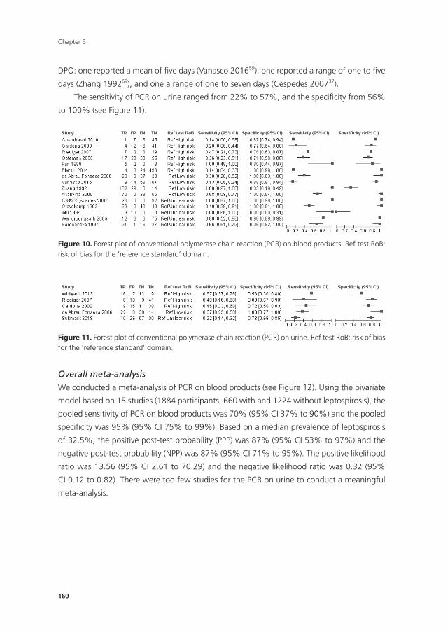
Chapter 5
160
DPO: one reported a mean of five days (Vanasco 201659), one reported a range of one to five
days (Zhang 199269), and one a range of one to seven days (Céspedes 200737).
The sensitivity of PCR on urine ranged from 22% to 57%, and the specificity from 56%
to 100% (see Figure 11).
166
FINDINGS
Conventional polymerase chain reaction
Seventeen studies reported test accuracy data for the PCR. Fifteen studied PCR on blood
products (serum: nine studies: whole blood (ethylenediaminetetraacetic acid (EDTA)): four
studies; blood or serum: one study; unspecified blood product: one study) and of them, three
also studied PCR on urine. Two studies included exclusively urine samples. The sensitivity of
PCR on blood products ranged from 13% to 100%, and the specificity from 0% to 100% (see
Figure 10). The 12 studies analysing PCR on blood products did not report the timing of sample
collection for the index test (further referred to as DPO). Three studies did report the DPO:
one reported a mean of five days (Vanasco 201659), one reported a range of one to five days
(Zhang 199269), and one a range of one to seven days (Céspedes 200737).
The sensitivity of PCR on urine ranged from 22% to 57%, and the specificity from 56% to 100%
(see Figure 11).
Figure 10. Forest plot of conventional polymerase chain reaction (PCR) on blood products. Ref test RoB: risk of bias for the 'reference standard' domain.
Figure 11. Forest plot of conventional polymerase chain reaction (PCR) on urine. Ref test RoB: risk of bias for the 'reference standard' domain.
Figure 10. Forest plot of conventional polymerase chain reaction (PCR) on blood products. Ref test RoB: risk of bias for the ‘reference standard’ domain.
166
FINDINGS
Conventional polymerase chain reaction
Seventeen studies reported test accuracy data for the PCR. Fifteen studied PCR on blood
products (serum: nine studies: whole blood (ethylenediaminetetraacetic acid (EDTA)): four
studies; blood or serum: one study; unspecified blood product: one study) and of them, three
also studied PCR on urine. Two studies included exclusively urine samples. The sensitivity of
PCR on blood products ranged from 13% to 100%, and the specificity from 0% to 100% (see
Figure 10). The 12 studies analysing PCR on blood products did not report the timing of sample
collection for the index test (further referred to as DPO). Three studies did report the DPO:
one reported a mean of five days (Vanasco 201659), one reported a range of one to five days
(Zhang 199269), and one a range of one to seven days (Céspedes 200737).
The sensitivity of PCR on urine ranged from 22% to 57%, and the specificity from 56% to 100%
(see Figure 11).
Figure 10. Forest plot of conventional polymerase chain reaction (PCR) on blood products. Ref test RoB: risk of bias for the 'reference standard' domain.
Figure 11. Forest plot of conventional polymerase chain reaction (PCR) on urine. Ref test RoB: risk of bias for the 'reference standard' domain.
Figure 11. Forest plot of conventional polymerase chain reaction (PCR) on urine. Ref test RoB: risk of bias for the ‘reference standard’ domain.
Overall meta-analysis
We conducted a meta-analysis of PCR on blood products (see Figure 12). Using the bivariate
model based on 15 studies (1884 participants, 660 with and 1224 without leptospirosis), the
pooled sensitivity of PCR on blood products was 70% (95% CI 37% to 90%) and the pooled
specificity was 95% (95% CI 75% to 99%). Based on a median prevalence of leptospirosis
of 32.5%, the positive post-test probability (PPP) was 87% (95% CI 53% to 97%) and the
negative post-test probability (NPP) was 87% (95% CI 71% to 95%). The positive likelihood
ratio was 13.56 (95% CI 2.61 to 70.29) and the negative likelihood ratio was 0.32 (95%
CI 0.12 to 0.82). There were too few studies for the PCR on urine to conduct a meaningful
meta-analysis.

161
Nucleic acid and antigen detection tests for leptospirosis
167
Overall meta-analysis
We conducted a meta-analysis of PCR on blood products (see Figure 12). Using the bivariate
model based on 15 studies (1884 participants, 660 with and 1224 without leptospirosis), the
pooled sensitivity of PCR on blood products was 70% (95% CI 37% to 90%) and the pooled
specificity was 95% (95% CI 75% to 99%). Based on a median prevalence of leptospirosis of
32.5%, the positive post-test probability (PPP) was 87% (95% CI 53% to 97%) and the negative
post-test probability (NPP) was 87% (95% CI 71% to 95%). The positive likelihood ratio was
13.56 (95% CI 2.61 to 70.29) and the negative likelihood ratio was 0.32 (95% CI 0.12 to 0.82).
There were too few studies for the PCR on urine to conduct a meaningful meta-analysis.
Figure 12. Summary ROC plot for conventional polymerase chain reaction (PCR) on blood products. Figure 12. Summary ROC plot for conventional polymerase chain reaction (PCR) on blood products.Transparent dots indicate the test accuracy of the individual studies included in the analysis; the black dot indicates the pooled test accuracy. The ellipse around the pooled test accuracy is the 95% confidence region. The size of the transparent dots represents the sample size, with the vertical diameter represent-ing the number of cases and horizontal diameter representing the number of non-cases.
Investigations of heterogeneity
We assessed heterogeneity only for PCR on blood products. We planned to investigate the
following sources of heterogeneity: timing of sample collection, prevalence, blood sample
type, target gene/primer, and brand of test.
• Timingofsamplecollection:timingofsamplecollectionwasusuallyunreported(12stud-
ies) and the subgroups were too small to investigate heterogeneity.
• Prevalence: we investigated whether prevalence of leptospirosis was associated with
test accuracy. Studies with a higher prevalence of leptospirosis had a significantly lower
specificity (P = 0.0004). Prevalence was not associated with sensitivity (P = 0.2).
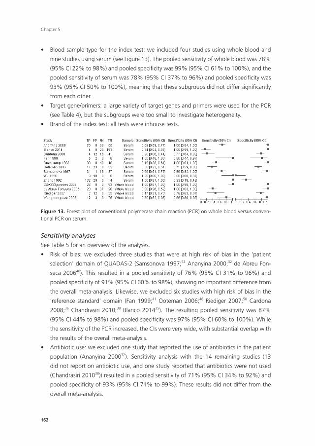
Chapter 5
162
• Bloodsampletypefortheindextest:weincludedfourstudiesusingwholebloodand
nine studies using serum (see Figure 13). The pooled sensitivity of whole blood was 78%
(95% CI 22% to 98%) and pooled specificity was 99% (95% CI 61% to 100%), and the
pooled sensitivity of serum was 78% (95% CI 37% to 96%) and pooled specificity was
93% (95% CI 50% to 100%), meaning that these subgroups did not differ significantly
from each other.
• Targetgene/primers:alargevarietyoftargetgenesandprimerswereusedforthePCR
(see Table 4), but the subgroups were too small to investigate heterogeneity.
• Brandoftheindextest:alltestswereinhousetests.
169
Figure 13. Forest plot of conventional polymerase chain reaction (PCR) on whole blood versus conventional PCR on serum.
Sensitivity analyses
See Table 5 for an overview of the analyses.
¥ Risk of bias: we excluded three studies that were at high risk of bias in the 'patient
selection' domain of QUADAS-2 (Samsonova 1997;53 Ananyina 2000;32 de Abreu
Fonseca 200640). This resulted in a pooled sensitivity of 76% (95% CI 31% to 96%) and
pooled specificity of 91% (95% CI 60% to 98%), showing no important difference from
the overall meta-analysis. Likewise, we excluded six studies with high risk of bias in
the 'reference standard' domain (Fan 1999;41 Ooteman 2006;48 Riediger 2007;50
Cardona 2008;36 Chandrasiri 2010;38 Blanco 201435). The resulting pooled sensitivity
was 87% (95% CI 44% to 98%) and pooled specificity was 97% (95% CI 60% to 100%).
While the sensitivity of the PCR increased, the CIs were very wide, with substantial
overlap with the results of the overall meta-analysis.
¥ Antibiotic use: we excluded one study that reported the use of antibiotics in the
patient population (Ananyina 200032). Sensitivity analysis with the 14 remaining
studies (13 did not report on antibiotic use, and one study reported that antibiotics
were not used (Chandrasiri 201038)) resulted in a pooled sensitivity of 71% (95% CI
34% to 92%) and pooled specificity of 93% (95% CI 71% to 99%). These results did not
differ from the overall meta-analysis.
¥ Lower MAT threshold: two studies reported each two threshold values for the MAT
(Ooteman 2006;48 Cardona 200836). For the overall analyses, we selected the higher
Figure 13. Forest plot of conventional polymerase chain reaction (PCR) on whole blood versus conven-tional PCR on serum.
Sensitivity analyses
See Table 5 for an overview of the analyses.
• Risk of bias: we excluded three studies that were at high risk of bias in the 'patient
selection'domainofQUADAS-2(Samsonova1997;53 Ananyina 2000;32 de Abreu Fon-
seca 200640). This resulted in a pooled sensitivity of 76% (95% CI 31% to 96%) and
pooled specificity of 91% (95% CI 60% to 98%), showing no important difference from
the overall meta-analysis. Likewise, we excluded six studies with high risk of bias in the
‘reference standard’ domain (Fan 1999;41 Ooteman 2006;48 Riediger 2007;50 Cardona
2008;36 Chandrasiri 2010;38 Blanco 201435). The resulting pooled sensitivity was 87%
(95% CI 44% to 98%) and pooled specificity was 97% (95% CI 60% to 100%). While
the sensitivity of the PCR increased, the CIs were very wide, with substantial overlap with
the results of the overall meta-analysis.
• Antibioticuse:weexcludedonestudythatreportedtheuseofantibioticsinthepatient
population (Ananyina 200032). Sensitivity analysis with the 14 remaining studies (13
did not report on antibiotic use, and one study reported that antibiotics were not used
(Chandrasiri 201038)) resulted in a pooled sensitivity of 71% (95% CI 34% to 92%) and
pooled specificity of 93% (95% CI 71% to 99%). These results did not differ from the
overall meta-analysis.
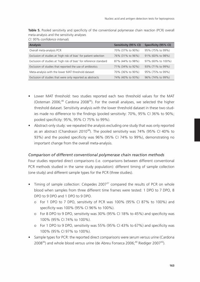
163
Nucleic acid and antigen detection tests for leptospirosis
• Lower MAT threshold: two studies reported each two threshold values for the MAT
(Ooteman 2006;48 Cardona 200836). For the overall analyses, we selected the higher
threshold dataset. Sensitivity analysis with the lower threshold dataset in these two stud-
ies made no difference to the findings (pooled sensitivity: 70%, 95% CI 36% to 90%;
pooled specificity: 95%, 95% CI 75% to 99%).
• Abstract-onlystudy:werepeatedtheanalysisexcludingonestudythatwasonlyreported
as an abstract (Chandrasiri 201038). The pooled sensitivity was 74% (95% CI 40% to
93%) and the pooled specificity was 96% (95% CI 74% to 99%), demonstrating no
important change from the overall meta-analysis.
Comparison of different conventional polymerase chain reaction methods
Four studies reported direct comparisons (i.e. comparisons between different conventional
PCR methods studied in the same study population): different timing of sample collection
(one study) and different sample types for the PCR (three studies).
• Timingof samplecollection:Céspedes200737 compared the results of PCR on whole
blood when samples from three different time frames were tested: 1 DPO to 7 DPO, 8
DPO to 9 DPO and 1 DPO to 9 DPO.
o For 1 DPO to 7 DPO, sensitivity of PCR was 100% (95% CI 87% to 100%) and
specificity was 100% (95% CI 96% to 100%).
o For 8 DPO to 9 DPO, sensitivity was 30% (95% CI 18% to 45%) and specificity was
100% (95% CI 74% to 100%).
o For 1 DPO to 9 DPO, sensitivity was 55% (95% CI 43% to 67%) and specificity was
100% (95% CI 97% to 100%).
• SampletypesforPCR:thereporteddirectcomparisonswereserumversusurine(Cardona
200836) and whole blood versus urine (de Abreu Fonseca 2006;40 Riediger 200750).
Table 5. Pooled sensitivity and specificity of the conventional polymerase chain reaction (PCR) overall meta-analysis and the sensitivity analyses CI: 95% confidence intervals
Analysis Sensitivity (95% CI) Specificity (95% CI)
Overall meta-analysis PCR 70% (37% to 90%) 95% (75% to 99%)
Exclusion of studies at ‘high risk of bias’ for patient selection 76% (31% to 96%) 91% (60% to 98%)
Exclusion of studies at ‘high risk of bias’ for reference standard 87% (44% to 98%) 97% (60% to 100%)
Exclusion of studies that reported the use of antibiotics 71% (34% to 92%) 93% (71% to 99%)
Meta-analysis with the lower MAT threshold dataset 70% (36% to 90%) 95% (75% to 99%)
Exclusion of studies that were only reported as abstracts 74% (40% to 93%) 96% (74% to 99%)
Chapter 5
164
o In Cardona 2008,36 the sensitivity of serum PCR was 20% (95% CI 6% to 44%) and
specificity was 77% (95% CI 64% to 88%), and sensitivity of urine PCR was 45%
(95% CI 23% to 68%) and specificity was 72% (95% CI 58% to 83%).
o In de Abreu Fonseca 2006,40 the sensitivity of whole blood PCR was 38% (95% CI
26% to 52%) and specificity was 100% (95% CI 83% to 100%), and sensitivity of
urine PCR was 37% (95% CI 25% to 40%) and specificity was 100% (95% CI 77%
to 100%).
o In Riediger 2007,50 the sensitivity of whole blood PCR was 47% (95% CI 21% to
73%) and specificity was 76% (95% CI 63% to 87%), and sensitivity of urine PCR
was 40% (95% CI 16% to 68%) and specificity was 80% (95% CI 67% to 90%).
Real-time polymerase chain reaction
Eighteen studies assessed the accuracy of the real-time PCR. Sixteen studies used blood
products as sample type: serum or plasma (nine studies), whole blood (six studies), serum
or whole blood (three studies), blood culture samples (one study), and buffy coat samples
(one study). Two studies used urine samples. Six studies each reported two sets of data:
Agampodi 201230 and Riediger 201751 reported data for whole blood and serum, while
Backstedt 2015,33 Thaipadungpanit 2011,56,58 and Woods 201866 reported data for two real-
time PCRs, each using a different target gene (rrs, lipL32, or rrs/lipL32). Waggoner 201461,62
reported data for a monoplex and multiplex (detecting also dengue and malaria) real-time
PCR. Ahmed 200931 reported three sets of data, each evaluating the test at different DPOs
(1 DPO to 4 DPO, 5 DPO to 10 DPO, and 1 DPO to 10 DPO). Because we considered 1 DPO
to 10 DPO to be the most representative time of sample collection, we included the dataset
of 1 DPO to 10 DPO in the meta-analysis. Seven studies of real-time PCR on blood products
did not report the DPO (Gonzalez 2013;43 Sonthayanon 2013;55 Backstedt 2015;33 Waggoner
2015;63 Biscornet 2017;34 Riediger 2017;51 Pakoa 201849). The other studies all reported
DPOs of the index test under 10 days.
The sensitivity of real-time PCR on blood products ranged from 0% to 100%, and the
specificity ranged from 0% to 100% (Figure 14).
Two studies assessed real-time PCR on urine (Villumsen 2012 U;60 Woods 201866). One
study assessed real-time PCR on blood culture samples (Villumsen 2012 BC60), and one study
assessed real-time PCR on buffy coat samples (Woods 201866). Due to the lack of enough
studies for urine, blood culture, and buffy coat real-time PCR, we did not perform a meta-
analysis. The sensitivities and specificities of these PCRs are displayed in Table 6.
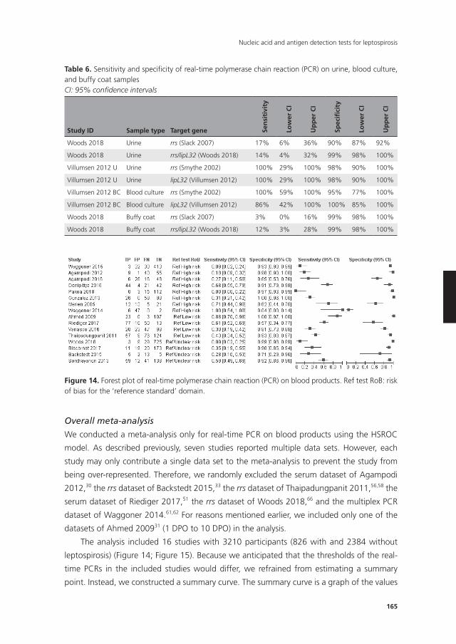
165
Nucleic acid and antigen detection tests for leptospirosis
172
studies for urine, blood culture, and buffy coat real-time PCR, we did not perform a meta-
analysis. The sensitivities and specificities of these PCRs are displayed in Table 6.
Figure 14. Forest plot of real-time polymerase chain reaction (PCR) on blood products. Ref test RoB: risk of bias for the 'reference standard' domain. Overall meta-analysis
We conducted a meta-analysis only for real-time PCR on blood products using the HSROC
model. As described previously, seven studies reported multiple data sets. However, each
study may only contribute a single data set to the meta-analysis to prevent the study from
being over-represented. Therefore, we randomly excluded the serum dataset of Agampodi
2012,30 the rrs dataset of Backstedt 2015,33 the rrs dataset of Thaipadungpanit 2011,56,58 the
serum dataset of Riediger 2017,51 the rrs dataset of Woods 2018,66 and the multiplex PCR
dataset of Waggoner 2014.61,62 For reasons mentioned earlier, we included only one of the
datasets of Ahmed 200931 (1 DPO to 10 DPO) in the analysis.
The analysis included 16 studies with 3210 participants (826 with and 2384 without
leptospirosis) (Figure 14; Figure 15). Because we anticipated that the thresholds of the real-
time PCRs in the included studies would differ, we refrained from estimating a summary
point. Instead, we constructed a summary curve. The summary curve is a graph of the values
of sensitivity and specificity that are obtained by varying the threshold across all possible
values. To illustrate, we estimated the accuracy for three fixed specificity values of 85%, 90%
and 95%. At 85% specificity, pooled sensitivity was 49% (95% CI 30% to 68%); at 90%
specificity, pooled sensitivity was 40% (95% CI 24% to 59%); and at 95% specificity, pooled
Figure 14. Forest plot of real-time polymerase chain reaction (PCR) on blood products. Ref test RoB: risk of bias for the ‘reference standard’ domain.
Overall meta-analysis
We conducted a meta-analysis only for real-time PCR on blood products using the HSROC
model. As described previously, seven studies reported multiple data sets. However, each
study may only contribute a single data set to the meta-analysis to prevent the study from
being over-represented. Therefore, we randomly excluded the serum dataset of Agampodi
2012,30 the rrs dataset of Backstedt 2015,33 the rrs dataset of Thaipadungpanit 2011,56,58 the
serum dataset of Riediger 2017,51 the rrs dataset of Woods 2018,66 and the multiplex PCR
dataset of Waggoner 2014.61,62 For reasons mentioned earlier, we included only one of the
datasets of Ahmed 200931 (1 DPO to 10 DPO) in the analysis.
The analysis included 16 studies with 3210 participants (826 with and 2384 without
leptospirosis) (Figure 14; Figure 15). Because we anticipated that the thresholds of the real-
time PCRs in the included studies would differ, we refrained from estimating a summary
point. Instead, we constructed a summary curve. The summary curve is a graph of the values
Table 6. Sensitivity and specificity of real-time polymerase chain reaction (PCR) on urine, blood culture, and buffy coat samplesCI: 95% confidence intervals
Study ID Sample type Target gene Sen
siti
vity
Low
er C
I
Up
per
CI
Spec
ifici
ty
Low
er C
I
Up
per
CI
Woods 2018 Urine rrs (Slack 2007) 17% 6% 36% 90% 87% 92%
Woods 2018 Urine rrs/lipL32 (Woods 2018) 14% 4% 32% 99% 98% 100%
Villumsen 2012 U Urine rrs (Smythe 2002) 100% 29% 100% 98% 90% 100%
Villumsen 2012 U Urine lipL32 (Villumsen 2012) 100% 29% 100% 98% 90% 100%
Villumsen 2012 BC Blood culture rrs (Smythe 2002) 100% 59% 100% 95% 77% 100%
Villumsen 2012 BC Blood culture lipL32 (Villumsen 2012) 86% 42% 100% 100% 85% 100%
Woods 2018 Buffy coat rrs (Slack 2007) 3% 0% 16% 99% 98% 100%
Woods 2018 Buffy coat rrs/lipL32 (Woods 2018) 12% 3% 28% 99% 98% 100%
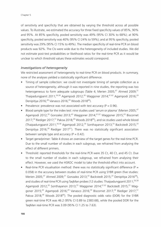
Chapter 5
166
of sensitivity and specificity that are obtained by varying the threshold across all possible
values. To illustrate, we estimated the accuracy for three fixed specificity values of 85%, 90%
and 95%. At 85% specificity, pooled sensitivity was 49% (95% CI 30% to 68%); at 90%
specificity, pooled sensitivity was 40% (95% CI 24% to 59%); and at 95% specificity, pooled
sensitivity was 29% (95% CI 15% to 49%). The median specificity of real-time PCR on blood
products was 92%. The CIs were wide due to the heterogeneity of included studies. We did
not estimate post-test probabilities or likelihood ratios for the real-time PCR as it would be
unclear to which threshold values these estimates would correspond.
Investigations of heterogeneity
We restricted assessment of heterogeneity to real-time PCR on blood products. In summary,
none of the analyses yielded a statistically significant difference.
• Timingofsamplecollection:wecouldnotinvestigatetimingofsamplecollectionasa
source of heterogeneity; although it was reported in nine studies, the reporting was too
heterogeneous to form adequate subgroups (Table 4; Merien 2005;47 Ahmed 2009;31
Thaipadungpanit 2011;56,58 Agampodi 2012;30 Waggoner 2014;61,62 Agampodi 2016;29
Denipitiya 2016;28 Vanasco 2016;59 Woods 201866).
• Prevalence:prevalencewasnotassociatedwithtestaccuracy(P=0.96).
• Bloodsampletypefortheindextest:ninestudiesused'serumorplasma'(Merien2005;47
Agampodi 2012;30 Gonzalez 2013;43 Waggoner 2014;61,62 Waggoner 2015;63 Biscornet
2017;34 Riediger 2017;51 Pakoa 2018;49 Woods 201866), and six studies used whole blood
(Thaipadungpanit 2011;56,58 Agampodi 2012;30 Sonthayanon 2013;55 Backstedt 2015;33
Denipitiya 2016;28 Riediger 201751). There was no statistically significant association
between sample type and accuracy (P = 0.42).
• Targetgene/primer:Table 4 shows an overview of the target genes for the real-time PCR.
Due to the small number of studies in each subgroup, we refrained from analysing the
effect of different primers.
• Threshold:reportedthresholdsforthereal-timePCRwere35Ct,40Ct,and45Ct.Due
to the small number of studies in each subgroup, we refrained from analysing their
effect. However, we used the HSROC model to take the threshold effect into account.
• Real-timePCRvisualisationmethod:therewasnostatisticallysignificantdifference(P=
0.058) in the accuracy between studies of real-time PCR using SYBR green (five studies:
Merien 2005;47 Ahmed 2009;31 Gonzalez 2013;43 Backstedt 2015;33 Denipitiya 201628),
and studies of real-time PCR using TaqMan probes (12 studies: Thaipadungpanit 2011;56,58
Agampodi 2012;30 Sonthayanon 2013;55 Waggoner 2014;61,62 Backstedt 2015;33 Wag-
goner 2015;63 Agampodi 2016;29 Vanasco 2016;59 Biscornet 2017;34 Riediger 2017;51
Pakoa 2018;49 Woods 201866). The pooled diagnostic odds ratio (DOR) for the SYBR
green real-time PCR was 46.2 (95% CI 0.89 to 2383.68), while the pooled DOR for the
TaqMan real-time PCR was 3.09 (95% CI 1.25 to 7.63).
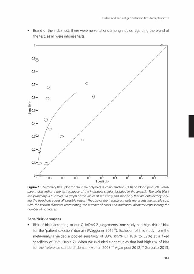
167
Nucleic acid and antigen detection tests for leptospirosis
• Brandoftheindextest:therewerenovariationsamongstudiesregardingthebrandof
the test, as all were inhouse tests.
174
Thaipadungpanit 2011;56,58 Agampodi 2012;30 Sonthayanon 2013;55 Waggoner
2014;61,62 Backstedt 2015;33 Waggoner 2015;63 Agampodi 2016;29 Vanasco 2016;59
Biscornet 2017;34 Riediger 2017;51 Pakoa 2018;49 Woods 201866). The pooled
diagnostic odds ratio (DOR) for the SYBR green real-time PCR was 46.2 (95% CI 0.89 to
2383.68), while the pooled DOR for the TaqMan real-time PCR was 3.09 (95% CI 1.25
to 7.63).
¥ Brand of the index test: there were no variations among studies regarding the brand
of the test, as all were inhouse tests.
Figure 15. Summary ROC plot for real-time polymerase chain reaction (PCR) on blood products. Transparent dots indicate the test accuracy of the individual studies included in the Figure 15. Summary ROC plot for real-time polymerase chain reaction (PCR) on blood products. Trans-parent dots indicate the test accuracy of the individual studies included in the analysis. The solid black line (summary ROC curve) is a graph of the values of sensitivity and specificity that are obtained by vary-ing the threshold across all possible values. The size of the transparent dots represents the sample size, with the vertical diameter representing the number of cases and horizontal diameter representing the number of non-cases.
Sensitivity analyses
• Riskofbias:accordingtoourQUADAS-2judgements,onestudyhadhighriskofbias
forthe'patientselection'domain(Waggoner201563). Exclusion of this study from the
meta-analysis yielded a pooled sensitivity of 33% (95% CI 18% to 52%) at a fixed
specificity of 95% (Table 7). When we excluded eight studies that had high risk of bias
for the ‘reference standard’ domain (Merien 2005;47 Agampodi 2012;30 Gonzalez 2013;
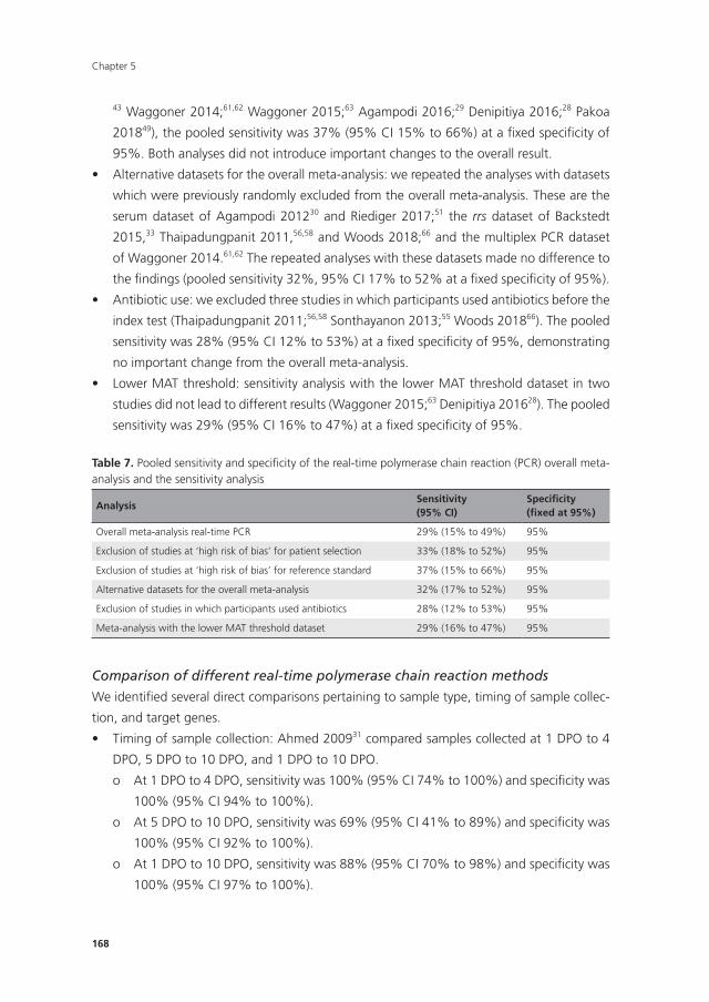
Chapter 5
168
43 Waggoner 2014;61,62 Waggoner 2015;63 Agampodi 2016;29 Denipitiya 2016;28 Pakoa
201849), the pooled sensitivity was 37% (95% CI 15% to 66%) at a fixed specificity of
95%. Both analyses did not introduce important changes to the overall result.
• Alternativedatasetsfortheoverallmeta-analysis:werepeatedtheanalyseswithdatasets
which were previously randomly excluded from the overall meta-analysis. These are the
serum dataset of Agampodi 201230 and Riediger 2017;51 the rrs dataset of Backstedt
2015,33 Thaipadungpanit 2011,56,58 and Woods 2018;66 and the multiplex PCR dataset
of Waggoner 2014.61,62 The repeated analyses with these datasets made no difference to
the findings (pooled sensitivity 32%, 95% CI 17% to 52% at a fixed specificity of 95%).
• Antibioticuse:weexcludedthreestudiesinwhichparticipantsusedantibioticsbeforethe
index test (Thaipadungpanit 2011;56,58 Sonthayanon 2013;55 Woods 201866). The pooled
sensitivity was 28% (95% CI 12% to 53%) at a fixed specificity of 95%, demonstrating
no important change from the overall meta-analysis.
• LowerMATthreshold:sensitivityanalysiswiththelowerMATthresholddatasetintwo
studies did not lead to different results (Waggoner 2015;63 Denipitiya 201628). The pooled
sensitivity was 29% (95% CI 16% to 47%) at a fixed specificity of 95%.
Comparison of different real-time polymerase chain reaction methods
We identified several direct comparisons pertaining to sample type, timing of sample collec-
tion, and target genes.
• Timingofsamplecollection:Ahmed200931 compared samples collected at 1 DPO to 4
DPO, 5 DPO to 10 DPO, and 1 DPO to 10 DPO.
o At 1 DPO to 4 DPO, sensitivity was 100% (95% CI 74% to 100%) and specificity was
100% (95% CI 94% to 100%).
o At 5 DPO to 10 DPO, sensitivity was 69% (95% CI 41% to 89%) and specificity was
100% (95% CI 92% to 100%).
o At 1 DPO to 10 DPO, sensitivity was 88% (95% CI 70% to 98%) and specificity was
100% (95% CI 97% to 100%).
Table 7. Pooled sensitivity and specificity of the real-time polymerase chain reaction (PCR) overall meta-analysis and the sensitivity analysis
AnalysisSensitivity(95% CI)
Specificity(fixed at 95%)
Overall meta-analysis real-time PCR 29% (15% to 49%) 95%
Exclusion of studies at ‘high risk of bias’ for patient selection 33% (18% to 52%) 95%
Exclusion of studies at ‘high risk of bias’ for reference standard 37% (15% to 66%) 95%
Alternative datasets for the overall meta-analysis 32% (17% to 52%) 95%
Exclusion of studies in which participants used antibiotics 28% (12% to 53%) 95%
Meta-analysis with the lower MAT threshold dataset 29% (16% to 47%) 95%
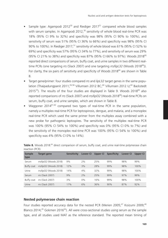
169
Nucleic acid and antigen detection tests for leptospirosis
• Sample type: Agampodi 201230 and Riediger 201751 compared whole blood samples
with serum samples. In Agampodi 2012,30 sensitivity of whole blood real-time PCR was
18% (95% CI 9% to 32%) and specificity was 98% (95% CI 90% to 100%), and
sensitivity of serum was 51% (95% CI 36% to 66%) and specificity was 98% (95% CI
90% to 100%). In Riediger 2017,51 sensitivity of whole blood was 61% (95% CI 52% to
69%) and specificity was 57% (95% CI 34% to 77%), and sensitivity of serum was 29%
(95% CI 21% to 38%) and specificity was 87% (95% CI 66% to 97%). Woods 201866
reported direct comparisons of serum, buffy coat, and urine samples in two different real-
time PCRs (one targeting rrs (Slack 2007) and one targeting rrs/lipL32 (Woods 201866)).
For clarity, the six pairs of sensitivity and specificity of Woods 201866 are shown in Table
8.
• Targetgene/primer:fourstudiescomparedrrs and lipL32 target genes in the same popu-
lation (Thaipadungpanit 2011;56,58 Villumsen 2012 BC;60 Villumsen 2012 U;60 Backstedt
201533). The results of the four studies are displayed in Table 9. Woods 201866 also
reported comparisons of rrs (Slack 2007) and rrs/lipl32 (Woods 201866) real-time PCRs on
serum, buffy coat, and urine samples, which are shown in Table 8.
• Waggoner 201461,62 compared two types of real-time PCR in the same population,
namely a multiplex real-time PCR for leptospirosis, dengue, and malaria, and a monoplex
real-time PCR which used the same primer from the multiplex assay combined with a
new probe for pathogenic leptospires. The sensitivity of the multiplex real-time PCR
was 100% (95% CI 54% to 100%) and specificity was 0% (95% CI 0% to 7%) and
the sensitivity of the monoplex real-time PCR was 100% (95% CI 54% to 100%) and
specificity was 4% (95% CI 0% to 14%).
nested polymerase chain reaction
Four studies reported accuracy data for the nested PCR (Merien 2005;47 Koizumi 2009;46
Blanco 2014;35 Gokmen 201642). All were cross-sectional studies using serum as the sample
type, and all studies used MAT as the reference standard. The reported mean timing of
Table 8. Woods 2018:66 direct comparison of serum, buffy coat, and urine real-time polymerase chain reaction (PCR)
Sampletype
Target gene Sensitivity Lower CI Upper CI Specificity Lower CI Upper CI
Serum rrs/lipl32 (Woods 2018) 9% 2% 25% 99% 98% 99%
Buffy coat rrs/lipl32 (Woods 2018) 12% 3% 28% 99% 98% 100%
Urine rrs/lipl32 (Woods 2018) 14% 4% 32% 99% 98% 100%
Serum rrs (Slack 2007) 9% 2% 25% 99% 97% 99%
Buffy coat rrs (Slack 2007) 3% 0% 16% 99% 98% 100%
Urine rrs (Slack 2007) 17% 6% 36% 90% 87% 92%
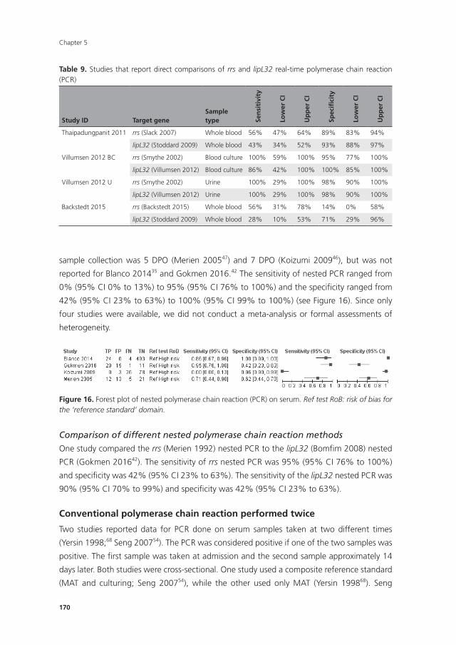
Chapter 5
170
sample collection was 5 DPO (Merien 200547) and 7 DPO (Koizumi 200946), but was not
reported for Blanco 201435 and Gokmen 2016.42 The sensitivity of nested PCR ranged from
0% (95% CI 0% to 13%) to 95% (95% CI 76% to 100%) and the specificity ranged from
42% (95% CI 23% to 63%) to 100% (95% CI 99% to 100%) (see Figure 16). Since only
four studies were available, we did not conduct a meta-analysis or formal assessments of
heterogeneity.
177
multiplex real-time PCR was 100% (95% CI 54% to 100%) and specificity was 0% (95%
CI 0% to 7%) and the sensitivity of the monoplex real-time PCR was 100% (95% CI 54%
to 100%) and specificity was 4% (95% CI 0% to 14%).
Nested polymerase chain reaction
Four studies reported accuracy data for the nested PCR (Merien 2005;47 Koizumi 2009;46
Blanco 2014;35 Gokmen 201642). All were cross-sectional studies using serum as the sample
type, and all studies used MAT as the reference standard. The reported mean timing of
sample collection was 5 DPO (Merien 200547) and 7 DPO (Koizumi 200946), but was not
reported for Blanco 201435 and Gokmen 2016.42 The sensitivity of nested PCR ranged from
0% (95% CI 0% to 13%) to 95% (95% CI 76% to 100%) and the specificity ranged from 42%
(95% CI 23% to 63%) to 100% (95% CI 99% to 100%) (see Figure 16). Since only four studies
were available, we did not conduct a meta-analysis or formal assessments of heterogeneity.
Figure 16. Forest plot of nested polymerase chain reaction (PCR) on serum. Ref test RoB: risk of bias for the 'reference standard' domain.
Comparison of different nested polymerase chain reaction methods
One study compared the rrs (Merien 1992) nested PCR to the lipL32 (Bomfim 2008) nested
PCR (Gokmen 201642). The sensitivity of rrs nested PCR was 95% (95% CI 76% to 100%) and
specificity was 42% (95% CI 23% to 63%). The sensitivity of the lipL32 nested PCR was 90%
(95% CI 70% to 99%) and specificity was 42% (95% CI 23% to 63%).
Conventional polymerase chain reaction performed twice
Figure 16. Forest plot of nested polymerase chain reaction (PCR) on serum. Ref test RoB: risk of bias for the ‘reference standard’ domain.
Comparison of different nested polymerase chain reaction methods
One study compared the rrs (Merien 1992) nested PCR to the lipL32 (Bomfim 2008) nested
PCR (Gokmen 201642). The sensitivity of rrs nested PCR was 95% (95% CI 76% to 100%)
and specificity was 42% (95% CI 23% to 63%). The sensitivity of the lipL32 nested PCR was
90% (95% CI 70% to 99%) and specificity was 42% (95% CI 23% to 63%).
Conventional polymerase chain reaction performed twice
Two studies reported data for PCR done on serum samples taken at two different times
(Yersin 1998;68 Seng 200754). The PCR was considered positive if one of the two samples was
positive. The first sample was taken at admission and the second sample approximately 14
days later. Both studies were cross-sectional. One study used a composite reference standard
(MAT and culturing; Seng 200754), while the other used only MAT (Yersin 199868). Seng
Table 9. Studies that report direct comparisons of rrs and lipL32 real-time polymerase chain reaction (PCR)
Study ID Target geneSample type Se
nsi
tivi
ty
Low
er C
I
Up
per
CI
Spec
ifici
ty
Low
er C
I
Up
per
CI
Thaipadungpanit 2011 rrs (Slack 2007) Whole blood 56% 47% 64% 89% 83% 94%
lipL32 (Stoddard 2009) Whole blood 43% 34% 52% 93% 88% 97%
Villumsen 2012 BC rrs (Smythe 2002) Blood culture 100% 59% 100% 95% 77% 100%
lipL32 (Villumsen 2012) Blood culture 86% 42% 100% 100% 85% 100%
Villumsen 2012 U rrs (Smythe 2002) Urine 100% 29% 100% 98% 90% 100%
lipL32 (Villumsen 2012) Urine 100% 29% 100% 98% 90% 100%
Backstedt 2015 rrs (Backstedt 2015) Whole blood 56% 31% 78% 14% 0% 58%
lipL32 (Stoddard 2009) Whole blood 28% 10% 53% 71% 29% 96%
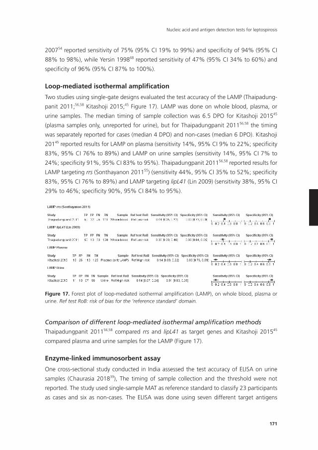
171
Nucleic acid and antigen detection tests for leptospirosis
200754 reported sensitivity of 75% (95% CI 19% to 99%) and specificity of 94% (95% CI
88% to 98%), while Yersin 199868 reported sensitivity of 47% (95% CI 34% to 60%) and
specificity of 96% (95% CI 87% to 100%).
Loop-mediated isothermal amplification
Two studies using single-gate designs evaluated the test accuracy of the LAMP (Thaipadung-
panit 2011;56,58 Kitashoji 2015;45 Figure 17). LAMP was done on whole blood, plasma, or
urine samples. The median timing of sample collection was 6.5 DPO for Kitashoji 201545
(plasma samples only, unreported for urine), but for Thaipadungpanit 201156,58 the timing
was separately reported for cases (median 4 DPO) and non-cases (median 6 DPO). Kitashoji
20145 reported results for LAMP on plasma (sensitivity 14%, 95% CI 9% to 22%; specificity
83%, 95% CI 76% to 89%) and LAMP on urine samples (sensitivity 14%, 95% CI 7% to
24%; specificity 91%, 95% CI 83% to 95%). Thaipadungpanit 201156,58 reported results for
LAMP targeting rrs (Sonthayanon 201155) (sensitivity 44%, 95% CI 35% to 52%; specificity
83%, 95% CI 76% to 89%) and LAMP targeting lipL41 (Lin 2009) (sensitivity 38%, 95% CI
29% to 46%; specificity 90%, 95% CI 84% to 95%).
179
Figure 17. Forest plot of loop-mediated isothermal amplification (LAMP), on whole blood, plasma or urine. Ref test RoB: risk of bias for the 'reference standard' domain. Comparison of different loop-mediated isothermal amplification methods
Thaipadungpanit 201156,58 compared rrs and lipL41 as target genes and Kitashoji 201545
compared plasma and urine samples for the LAMP (Figure 17).
Enzyme-linked immunosorbent assay
One cross-sectional study conducted in India assessed the test accuracy of ELISA on urine
samples (Chaurasia 201839), The timing of sample collection and the threshold were not
reported. The study used single-sample MAT as reference standard to classify 23 participants
as cases and six as non-cases. The ELISA was done using seven different target antigens
(LipL32, Fla1, LipL41, HbpA, SphCD210, Sph2, and Sph4). For clarity, the sensitivities and
specificities of these tests are shown in Table 10.
Dot-enzyme-linked immunosorbent assay
One two-gate case-control study evaluated the test accuracy of a monoclonal antibody-based
dot-ELISA for the detection of leptospiral antigens in urine samples (Saengjaruk 200252). The
Figure 17. Forest plot of loop-mediated isothermal amplification (LAMP), on whole blood, plasma or urine. Ref test RoB: risk of bias for the ‘reference standard’ domain.
Comparison of different loop-mediated isothermal amplification methods
Thaipadungpanit 201156,58 compared rrs and lipL41 as target genes and Kitashoji 201545
compared plasma and urine samples for the LAMP (Figure 17).
Enzyme-linked immunosorbent assay
One cross-sectional study conducted in India assessed the test accuracy of ELISA on urine
samples (Chaurasia 201839), The timing of sample collection and the threshold were not
reported. The study used single-sample MAT as reference standard to classify 23 participants
as cases and six as non-cases. The ELISA was done using seven different target antigens
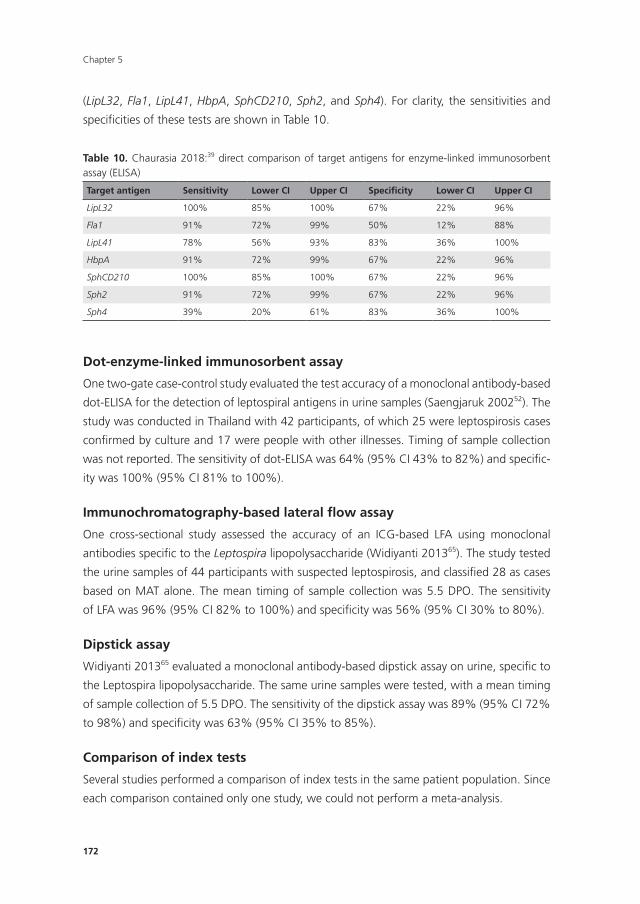
Chapter 5
172
(LipL32, Fla1, LipL41, HbpA, SphCD210, Sph2, and Sph4). For clarity, the sensitivities and
specificities of these tests are shown in Table 10.
Dot-enzyme-linked immunosorbent assay
One two-gate case-control study evaluated the test accuracy of a monoclonal antibody-based
dot-ELISA for the detection of leptospiral antigens in urine samples (Saengjaruk 200252). The
study was conducted in Thailand with 42 participants, of which 25 were leptospirosis cases
confirmed by culture and 17 were people with other illnesses. Timing of sample collection
was not reported. The sensitivity of dot-ELISA was 64% (95% CI 43% to 82%) and specific-
ity was 100% (95% CI 81% to 100%).
Immunochromatography-based lateral flow assay
One cross-sectional study assessed the accuracy of an ICG-based LFA using monoclonal
antibodies specific to the Leptospira lipopolysaccharide (Widiyanti 201365). The study tested
the urine samples of 44 participants with suspected leptospirosis, and classified 28 as cases
based on MAT alone. The mean timing of sample collection was 5.5 DPO. The sensitivity
of LFA was 96% (95% CI 82% to 100%) and specificity was 56% (95% CI 30% to 80%).
Dipstick assay
Widiyanti 201365 evaluated a monoclonal antibody-based dipstick assay on urine, specific to
the Leptospira lipopolysaccharide. The same urine samples were tested, with a mean timing
of sample collection of 5.5 DPO. The sensitivity of the dipstick assay was 89% (95% CI 72%
to 98%) and specificity was 63% (95% CI 35% to 85%).
Comparison of index tests
Several studies performed a comparison of index tests in the same patient population. Since
each comparison contained only one study, we could not perform a meta-analysis.
Table 10. Chaurasia 2018:39 direct comparison of target antigens for enzyme-linked immunosorbent assay (ELISA)
Target antigen Sensitivity Lower CI Upper CI Specificity Lower CI Upper CI
LipL32 100% 85% 100% 67% 22% 96%
Fla1 91% 72% 99% 50% 12% 88%
LipL41 78% 56% 93% 83% 36% 100%
HbpA 91% 72% 99% 67% 22% 96%
SphCD210 100% 85% 100% 67% 22% 96%
Sph2 91% 72% 99% 67% 22% 96%
Sph4 39% 20% 61% 83% 36% 100%
173
Nucleic acid and antigen detection tests for leptospirosis
• PCRversusreal-timePCR:Vanasco201659 reported a comparison of PCR and real-time
PCR, both performed on whole blood or serum samples. PCR had a sensitivity of 13%
(95% CI 6% to 24%) and a specificity of 88% (95% CI 81% to 94%). Real-time PCR
had a sensitivity of 30% (95% CI 19% to 42%) and a specificity of 81% (95% CI 73%
to 88%).
• PCRversusnestedPCR:Blanco201435 reported a comparison of PCR and nested PCR.
PCR had a sensitivity of 14% (95% CI 4% to 33%) and a specificity of 100% (95%
CI 99% to 100%). Nested PCR had a sensitivity of 86% (95% CI 67% to 96%) and a
specificity of 100% (95% CI 99% to 100%).
• Real-timePCRversusnestedPCR:Merien200547 compared real-time PCR with nested
PCR. The study reported identical results for the two tests: sensitivity was 71% (95% CI
44% to 90%) and specificity was 62% (95% CI 44% to 78%).
• PCRversusICG-basedIFAversusdipstickassay:Widiyanti201365 reported a comparison
between PCR, ICG-based IFA, and dipstick assay, with all three tests performed on urine
samples.
o For PCR on urine, sensitivity was 57% (95% CI 37% to 76%) and specificity was
56% (95% CI 30% to 80%).
o For ICG-based IFA, sensitivity was 96% (95% CI 82% to 100%) and specificity was
56% (95% CI 30% to 80%).
o For dipstick assay, sensitivity was 89% (95% CI 72% to 98%) and specificity was
63% (95% CI 35% to 85%).
• Real-time PCR versus LAMP: Thaipadungpanit 201156,58 reported a comparison of rrs
(Slack 2007) and lipL32 (Stoddard 2009) real-time PCR and rrs (Sonthayanon 201155) and
lipL41 (Lin 2009) LAMP.
o For real-time PCR targeting rrs, sensitivity was 56% (95% CI 47% to 64%) and
specificity was 89% (95% CI 83% to 94%).
o For real-time PCR targeting lipL32, sensitivity was 43% (95% CI 34% to 52%) and
specificity was 93% (95% CI 88% to 97%).
o For LAMP targeting rrs, sensitivity was 44% (95% CI 35% to 52%) and specificity
was 83% (95% CI 76% to 89%).
o For LAMP targeting lipL41, sensitivity was 38% (95% CI 29% to 46%) and specificity
was 90% (95% CI 84% to 95%).
DISCUSSIOn
Summary of main results
In this systematic review, we summarised the diagnostic test accuracy of nucleic acid and
antigen detection tests for human symptomatic leptospirosis, verified by (a combination of)
Chapter 5
174
currently established tests – MAT, culture, and IgM ELISA. We identified 41 studies in the
literature evaluating nine index tests, of which conventional PCR and real-time PCR were
the most frequently evaluated tests. While we have performed a meta-analysis for PCR and
real-time PCR on blood products, individual study results suggested very high between-study
heterogeneity (PCR sensitivity ranging from 13% to 100% and PCR specificity from 0% to
100%). Therefore readers should interpret the meta-analytic result as a weighted mean of all
the heterogeneous settings in which the index tests were evaluated, rather than an estimate
that is applicable across settings. Summary of findings table 1, Summary of findings table 2,
Summary of findings table 3, and Summary of findings table 4 give an overview of the most
important findings.
Interpretation of the conventional polymerase chain reaction meta-analysis
From a meta-analysis of 15 studies evaluating PCR on blood products collected during vari-
ous stages of disease, the pooled sensitivity was 70% (95% CI 37% to 90%) and the pooled
specificity was 95% (95% CI 75% to 99%). This means that in a hypothetical cohort of 1000
people, with a prevalence of 32.5% (325 diseased), 98 (95% CI 32 to 205) cases would
be missed by the PCR and 35 (95% CI 6 to 168) non-diseased people would be incorrectly
diagnosed with leptospirosis. If the prior probability of an individual to have leptospirosis is
32.5%, the PPP was 87% (95% CI 53% to 97%) and the NPP was 87% (95% CI 71% to
95%). If the PCR would be used in a setting such as in the Netherlands where the prevalence
is lower (9.7% in 2016; Collaborating Centre for Reference and Research on Leptospirosis in
Amsterdam, unpublished data), 97/1000 people would have leptospirosis. In such a cohort,
29 (95% CI 9 to 61) diseased people would be missed and 47 (95% CI 8 to 225) non-
diseased people would be incorrectly diagnosed with leptospirosis. If the prior probability of
leptospirosis in an individual is 9.7%, the PPP is 59% (95% CI 20% to 89%) and the NPP is
97% (95% CI 92% to 99%). See Summary of findings table 1.
When the PCR meta-analysis was repeated without the studies at high risk of bias for the
‘reference standard’ domain (i.e. only including studies with composite reference standards
or two-gate studies with MAT as reference standard, based on nine studies), the pooled
sensitivity was 87% (95% CI 44% to 98%), and the pooled specificity was 97% (95% CI
60% to 100%). See Summary of findings table 2. This means that in a cohort of 1000 people
with 325 leptospirosis patients, 42 (95% CI 5 to 183) cases would be missed and 17 (95%
CI 1 to 272) non-diseased people would be incorrectly diagnosed with leptospirosis. In an
individual with 32.5% prior probability of leptospirosis, the PPP is 94% (95% CI 41% to
100%) and the NPP is 94% (95% CI 70% to 99%). If the prevalence is 9.7%, 13 (95% CI
2 to 55) diseased people would be missed and 23 (95% CI 1 to 363) non-diseased people
would be incorrectly diagnosed with leptospirosis. If an individual has 9.7% prior probability
175
Nucleic acid and antigen detection tests for leptospirosis
of leptospirosis, the PPP is 78% (95% CI 13% to 99%), and the NPP is 99% (95% CI 91%
to 100%).
A repeated meta-analysis of PCR without studies at high risk of bias for the ‘patients’
domain did not lead to important changes in our results.
Interpretation of the real-time polymerase chain reaction meta-analysis
For the real-time PCR, we estimated an SROC curve instead of a summary sensitivity and
specificity, since we expected the positivity threshold (which was often not reported) to vary
between studies. The median specificity of real-time PCR on blood products was 92%. For
illustrative purposes, if we were to select a point on the curve with 95% specificity, the
pooled sensitivity would be 29% (95% CI 15% to 49%) at an unknown threshold (Summary
of findings table 3). Translating these numbers to a cohort of 1000 people of whom 325
are diseased, would mean that 230 (95% CI 167 to 276) diseased people would be missed
and 34 non-diseased people would be incorrectly diagnosed with leptospirosis. Again, in
a setting with a prevalence of 9.7% this would imply that 69 (95% CI 50 to 82) diseased
people would be missed and 45 non-diseased people would be incorrectly diagnosed with
leptospirosis. We did not provide PPP and NPP for real-time PCR, as threshold values needed
to produce these estimates were unknown.
When we excluded real-time PCR studies at high risk of bias for the ‘patients’ domain or
the ‘reference standard’ domain, there were no important changes in the pooled estimate.
The position of conventional polymerase chain reaction and real-time polymerase chain reaction in the clinical pathway
Based on the properties of PCR and real-time PCR, we examined the possible role of these
tests in the diagnostic pathway for leptospirosis. Leptospirosis is a potentially life-threatening
disease, meaning that efforts should be undertaken to minimise false-negative results.
In the clinic, while most patients with suspicion of a severe infection are likely to receive
broad-spectrum antibiotics, a false-negative result impedes the optimisation of the antibiotic
therapy and assessment of the prognosis. In an outbreak setting, a missed case of leptospiro-
sis will delay outbreak response and facilitate further dissemination of disease.
For patients presenting in the early disease stage, PCR and real-time PCR on blood
products are preferable as first-line tests in the clinical pathway based on their ability for
early detection. However, whether additional testing is needed to verify a positive or negative
test result depends on test accuracy and the prevalence of leptospirosis. While PCR-based
methods have been described as sensitive tests in the literature,16,70,71 our results show
that the sensitivity of PCR and real-time PCR vary greatly between studies, with the CI for
PCR sensitivity ranging from 37% to 90%. This can partly be explained by differences in
methodological quality, but it is also likely that there is true heterogeneity, such as differ-
ences in timing of sample collection. Furthermore, the reliability of a positive or negative test
Chapter 5
176
result depends on the prevalence. In our review, prevalence of leptospirosis varied greatly
between studies (range 3% to 85%). Consequently, whether PCR-based methods can be
used alone, or together with other follow-up tests depends on regional considerations such
as prevalence, factors that are likely to influence accuracy (e.g. timing of sampling), and
downstream consequences of a positive or negative result. For example, in settings with
a high prevalence, PCR and real-time PCR may not have a high enough negative post-test
probability to confidently rule out leptospirosis. In this case, additional testing to verify nega-
tive results should be considered.
Comparison between index tests
This review did not find enough evidence to formally compare the diagnostic accuracy of
included tests. Direct comparison studies (where two or more index tests are evaluated in
the same patient population) are needed to draw valid conclusions about the differences in
diagnostic test accuracy between tests, but such studies were lacking. Although the results
of the meta-analyses seem to imply that real-time PCR has a lower sensitivity than PCR, this is
not a valid comparison. The meta-analysis results are composed of mostly single-test studies
and any differences between real-time PCR and PCR could arise from other reasons than
the differences in the tests themselves, such as differences in study design or spectrum of
disease. Therefore, any comparison between the meta-analytic results of PCR and real-time
PCR must be interpreted with caution.
Heterogeneity of included studies
Substantial heterogeneity, as demonstrated by the wide CIs, complicated the interpretation
of our findings.
An important covariate, timing of sample collection, could not be explored in hetero-
geneity analysis. Three of 15 studies assessing PCR on blood products reported the timing
of sample collection (range 1 DPO to 7 DPO of symptoms), and in 9/16 studies assessing
real-time PCR on blood products (range 1 to 19 DPO). However, the subgroups were either
too small or the reporting of the timing variable was too heterogeneous for analysis. Two
studies comparing test accuracy in patients who presented early with test accuracy in pa-
tients who presented later appear to support the hypothesis that the sensitivity of PCR and
real-time PCR is greater in the first few days of illness (Céspedes 2007;37 Ahmed 200931).
This is consistent with the current pathophysiological understanding of leptospirosis, that
leptospiraemia declines rapidly and becomes undetectable after 10 DPO.12 However, this
could also be caused by patients with a higher bacterial load presenting earlier to the clinic
due to a more severe clinical presentation than those with a lower bacterial load. Moreover,
one study reported that the sensitivity of real-time PCR was not associated with timing of
sample collection in patients presenting with fewer than 10 DPO (P = 0.33) (Agampodi
177
Nucleic acid and antigen detection tests for leptospirosis
201230). Ultimately, more studies are needed to confirm the association between timing and
test accuracy.
In the case of PCR, statistical heterogeneity may be partly explained by the prevalence
of leptospirosis in the study population. Specificity was inversely correlated with prevalence
(P = 0.0004). A number of explanations for this association are possible. The prevalence of
alternative diagnoses may be higher in places where leptospirosis prevalence is high, causing
false-positive results on the PCR. The PCR may be detecting lower levels of infections that
occur more frequently in high-prevalence settings, that are missed by MAT, and, therefore,
recorded as false-positive results. The laboratories in high-prevalence settings may be less
well-equipped and more often contaminated, and, therefore, allow more false-positive
results. We did not examine the inverse correlation between prevalence and sensitivity or
specificity for real-time PCR studies, since the HSROC model by default examines the associa-
tion between prevalence and accuracy (alpha parameter) instead of sensitivity and specificity.
Other covariates that could possibly influence test accuracy, such as real-time PCR
threshold and specific target genes or primers used in PCR-based methods, have not been
ruled out as possible explanations for the significant heterogeneity. It is theoretically possible
that the heterogeneity in sensitivity of PCR-based methods could be explained by differences
in Leptospira species, as a primer may not able to detect a particular species. However, this
is not very probable as it is usual practice to account for all existing species when developing
PCR or its variants (unless a new species emerges). Since all index tests were inhouse tests,
there may be other potential sources of heterogeneity (e.g. use of different laboratory equip-
ment or protocols) that cannot be measured reliably or be reported in sufficient detail. For
this reason, readers should be cautious when applying summary estimates of test accuracy
in their own clinical settings.
Risk of bias
A major point of attention in our review was the use of MAT as reference standard, which
is considered to have an imperfect sensitivity. If the reference standard is not sensitive, the
specificity of the index test is likely to be underestimated. It is furthermore not inconceivable
that some index tests, for example, PCR-based methods, may be more sensitive than MAT
alone. We aimed to address this problem by including composite reference standards and
rating the risk of bias as high when MAT was used as the sole reference standard, and when
single samples were used (instead of paired). However, only a minority of studies used an-
other reference standard alongside the MAT, leading to a ‘high risk of bias’ judgement in the
majority of studies for this domain. In the case of the PCR, sensitivity increased when studies
at ‘high risk of bias’ for the reference standard were excluded, but specificity was unchanged
(Table 5). Other covariates that may be of importance, such as the cut-off value for MAT, the
use of adequate regional panels for MAT, and the differences between composite reference
standards, were not taken into account in our review to avoid excessive complexity.
Chapter 5
178
Another issue is the inclusion of four two-gate case-control studies (Samsonova 1997;53
Ananyina 2000;32 Saengjaruk 2002;52 de Abreu Fonseca 200640). Cases and controls in these
studies are selected separately and do not reflect the spectrum of disease in the clinical popu-
lation.26 Another concern in these studies is the possibility of coinfections of leptospirosis
and another infectious disease. In two-gate designs, since controls are not MAT negatives
but people with a condition resembling leptospirosis, coinfections with leptospirosis may be
present. Treating these people as controls may underestimate the specificity of the index test.
These studies have been excluded in the abovementioned sensitivity analyses as they were
considered ‘high risk’ for patient selection (Table 5; Table 7).
Blinding of the index test result to the reference standard result interpreters (or vice
versa) was largely unreported. Interpreters were blinded because either blinding methods
were used, or by the virtue of their study design (e.g. blinding of the index test interpreter
was not needed when the index test was done first). In the index test, eight studies reported
to have used a form of blinding in their methods. However, we noted that only one study
reported explicit methods for blinding (Samsonova 199753). It was unclear if the remaining
studies used proper blinding methods. Considering other possible biases in our review (Figure
4; Figure 6; Figure 8), readers are advised to weigh the results against the quality of evidence.
Other index tests
For other tests included in our review (nested PCR, PCR performed twice, LAMP, ELISA,
dot-ELISA, ICG-based LFA, and dipstick assay), we could not conduct meta-analyses or inves-
tigations of heterogeneity due to the small number of studies.
Strengths and weaknesses of the review
The strength of our review lies in the fact that we used an extensive search strategy including
16 national and regional databases, without any limitations on languages and without using
search filters or keywords containing terms related to diagnostic accuracy. We also contacted
authors for full-text articles in case the studies did not report complete data for the construc-
tion of two-by-two contingency tables. Furthermore, we aimed to include all nucleic acid
tests and antigen detection tests that we could find in the literature. And lastly, we included
studies that used MAT with convalescent samples as a reference standard, as well as studies
that used MAT with a single, acute sample. This is a strength, because it provides a good
reflection of the day-to-day reality in clinics and laboratories, but at the same time, it is one
of the major limitations of our review. It is known that antibodies appear in the blood only
after several days to weeks.72 Thus a serological test, such as the MAT, is not applicable for
diagnosis of leptospirosis in the early stages. Inclusion of studies using MAT only on acute
samples could have led to false-negative results by the reference standard. This lack of a
perfect reference standard implies that the test accuracy of nucleic acid and antigen tests
179
Nucleic acid and antigen detection tests for leptospirosis
presented here simply reflects the extent of agreement between the index test and MAT, and
not necessarily the true test accuracy of the index tests.
Other limitations of our review are as follows. First, studies that potentially satisfy our
inclusion criteria could not be included due to lack of clarity or inconsistencies in the full-text
article. Second, due to poor reporting in primary studies, many aspects of the index test and
methodological quality remain unclear and limit the potential to generalise our findings.
Third, as we have discussed previously, we could not explain the substantial heterogeneity in
study results due to the lack of statistical power. Although we have conducted meta-analyses
for PCR and real-time PCR, it is debatable to what extent the pooled results are applicable to
clinical practice, since numerous unexplored covariates are likely to have contributed to the
pooled sensitivities and specificities.
Applicability of findings to the review question
We identified some concerns regarding the applicability of the results to our review question
when the used research methods differed significantly from clinical practice (Figure 4; Figure
6; Figure 8).
Regarding the selection of participants, four studies excluded participants due to
unavailability of convalescent samples for MAT (Yersin 1998;68 Thaipadungpanit 2011;56,58
Agampodi 2012;30 Gonzalez 201343). Although this is a reasonable decision, since verification
by MAT is more accurate with paired acute and convalescent samples, it does not necessarily
reflect the patient population in clinical practice, as participants with a fatal course of disease
were likely to be excluded from the study as a result. In 19 studies, the patient selection
method, inclusion criteria, and characteristics (including the timing of sample collection)
were not well reported. These studies stated that the participants were ‘clinically suspected
for leptospirosis’ without stating which signs, symptoms, and risk factors the participants had
that made them clinically suspected. Furthermore, with baseline characteristics not reported,
it was not possible to determine whether a particular age group or sex was over-represented
in the study.
For the applicability of the index test and the reference standard, we did not only consider
whether the method of testing differed from clinical practice, but also whether the execution
of the test was reported in such detail that the test could be reproduced in full elsewhere.
In four studies, we found that this was not the case for the index test (Ananyina 2000;32
Chandrasiri 2010;38 Chaurasia 2018;39 Pakoa 201849), and in five studies, the procedure
(including the cut-off value) for the MAT was not reported, or there was a reference to an
irretrievable study (Samsonova 1997;53 Ananyina 2000;32 Merien 2005;47 Chandrasiri 2010;38
Waggoner 201461,62). At least 11 studies used frozen samples for the index test rather than
fresh samples. We were uncertain if this could have influenced the test accuracy, so we
considered the concern regarding applicability to be high.
Chapter 5
180
AUTHORS’ COnCLUSIOnS
Implications for practice
The validity of review findings are limited by the poor reporting of methodological quality
items and the use of suboptimal reference standards. We conclude that there is substan-
tial between-study variability in the accuracy of conventional polymerase chain reaction
(PCR) and real-time PCR, as well as substantial variability in the prevalence of leptospirosis.
Consequently, the position of conventional PCR and real-time PCR in the clinical pathway
depends on regional considerations such as prevalence, factors that are likely to influence
accuracy (such as timing of sampling), and downstream consequences of test results. There
is insufficient evidence to conclude which of the nucleic acid and antigen detection tests
are the most accurate in the early stage of leptospirosis. There is preliminary evidence that
conventional PCR and real-time PCR are more sensitive on blood samples collected early in
the disease stage, but this needs to be confirmed in future studies. Evidence regarding other
index tests was very limited.
Implications for research
Our review demonstrates that while there is a wealth of publications on new nucleic acid
and antigen detection tests, there is a marked scarcity on well-designed, well-performed,
and well-reported validations of such tests. More high-quality studies are needed with larger
samples sizes, especially a larger group of cases to estimate sensitivity more precisely. Future
investigators should follow the reporting guidelines of STARD (Standards for the Reporting of
Diagnostic Accuracy Studies), to allow the assessment of potential biases in the study, as well
as the assessment of the clinical value of the estimated test accuracy. Single-gate designs,
such as the cross-sectional study, with consecutive enrolment have our recommendation
above two-gate designs because of their lower risk of spectrum bias. The choice of reference
standard should not only include MAT, but also culture, and if possible immunoglobulin M
enzyme-linked immunosorbent assay (IgM ELISA), to minimise false-negative results from
occurring. The emphasis should be on paired sampling, to show a possible rise in antibody
titres. In order to compare and select the best performing tests, multiple index tests should
be evaluated on the same participants so that direct comparison of their accuracy is possible.
Last, we encourage future investigators to explore the effects of varying times of sample
collection on test accuracy as a potential source of heterogeneity.
Acknowledgements
Cochrane Review Group funding acknowledgement: the Danish State is the largest single
funder of The Cochrane Hepato-Biliary Group through its investment in The Copenhagen
Trial Unit, Centre for Clinical Intervention Research, Rigshospitalet, Copenhagen University
Hospital, Denmark. Disclaimer: the views and opinions expressed in this review are those of

181
Nucleic acid and antigen detection tests for leptospirosis
the authors and do not necessarily reflect those of the Danish State or The Copenhagen Trial
Unit.
We would like to thank Drs Agostino Colli, Deputy Co-ordinating editor, and Dimitrinka
Nikolova, Managing editor, of the Cochrane Hepato-Biliary Group, and Cochrane’s Diagnos-
tic Test Accuracy Review team, for reviewing our protocol and review, and giving us valuable
recommendations. We also thank Dr Patrick Bossuyt for his advice on the interpretation of
diagnostic test accuracy. Last, we thank the authors of included studies for their correspon-
dence and sending us valuable study data.
Chapter 5
182
REFEREnCES
1. Goris M, Leeflang M, Boer K, et al. Establishment of valid laboratory case definition for human
leptospirosis. J Bacteriol Parasitol 2012; 3(132): 2.
2. Farr RW. Leptospirosis. Clinical Infectious Diseases 1995; 21(1): 1-8.
3. Pappas G, Papadimitriou P, Siozopoulou V, Christou L, Akritidis N. The globalization of leptospi-
rosis: worldwide incidence trends. Int J Infect Dis 2008; 12(4): 351-7.
4. Vijayachari P, Sugunan AP, Shriram AN. Leptospirosis: an emerging global public health problem.
J Biosci 2008; 33(4): 557-69.
5. Pijnacker R, Goris MG, Te Wierik MJ, et al. Marked increase in leptospirosis infections in humans
and dogs in the Netherlands, 2014. Euro Surveill 2016; 21(17).
6. Duarte JL, Giatti LL. Incidência da leptospirose em uma capital da Amazônia Ocidental brasileira e
sua relação com a variabilidade climática e ambiental, entre os anos de 2008 e 2013. Epidemio-
logia e Serviços de Saúde 2019; 28.
7. Warnasekara J, Koralegedara I, Agampodi S. Estimating the burden of leptospirosis in Sri Lanka;
a systematic review. BMC Infect Dis 2019; 19(1): 119.
8. Costa F, Hagan JE, Calcagno J, et al. Global Morbidity and Mortality of Leptospirosis: A Systematic
Review. PLoS Negl Trop Dis 2015; 9(9): e0003898.
9. Lau C, Smythe L, Weinstein P. Leptospirosis: An emerging disease in travellers. Travel Medicine
and Infectious Disease 2010; 8(1): 33-9.
10. Jensenius M, Han PV, Schlagenhauf P, et al. Acute and potentially life-threatening tropical dis-
eases in western travelers--a GeoSentinel multicenter study, 1996-2011. Am J Trop Med Hyg
2013; 88(2): 397-404.
11. Mwachui MA, Crump L, Hartskeerl R, Zinsstag J, Hattendorf J. Environmental and Behavioural
Determinants of Leptospirosis Transmission: A Systematic Review. PLoS Negl Trop Dis 2015; 9(9):
e0003843.
12. World Health Organization. Human leptospirosis: guidance for diagnosis, surveillance and con-
trol. 2003.
13. McBride AJ, Athanazio DA, Reis MG, Ko AI. Leptospirosis. Curr Opin Infect Dis 2005; 18(5):
376-86.
14. Guerra MA. Leptospirosis: Public health perspectives. Biologicals 2013; 41(5): 295-7.
15. Bharti AR, Nally JE, Ricaldi JN, et al. Leptospirosis: a zoonotic disease of global importance. Lancet
Infect Dis 2003; 3(12): 757-71.
16. Picardeau M, Bertherat E, Jancloes M, Skouloudis AN, Durski K, Hartskeerl RA. Rapid tests for
diagnosis of leptospirosis: current tools and emerging technologies. Diagn Microbiol Infect Dis
2014; 78(1): 1-8.
17. Brett-Major DM, Coldren R. Antibiotics for leptospirosis. Cochrane Database Syst Rev 2012; 2:
Cd008264.
18. Limmathurotsakul D, Turner EL, Wuthiekanun V, et al. Fool’s gold: Why imperfect reference tests
are undermining the evaluation of novel diagnostics: a reevaluation of 5 diagnostic tests for
leptospirosis. Clinical Infectious Diseases 2012; 55(3): 322-31.
19. Rutjes AW, Reitsma JB, Coomarasamy A, Khan KS, Bossuyt PM. Evaluation of diagnostic tests
when there is no gold standard. A review of methods. Health Technol Assess 2007; 11(50): iii,
ix-51.
20. Goris MG, Boer KR, Duarte TA, Kliffen SJ, Hartskeerl RA. Human leptospirosis trends, the Nether-
lands, 1925-2008. Emerg Infect Dis 2013; 19(3): 371-8.
183
Nucleic acid and antigen detection tests for leptospirosis
21. Goris MG, Boer KR, Bouman-Strijker M, Hartskeerl R, Lucas C, Leeflang MM. Serological labora-
tory tests for diagnosis of human leptospirosis in patients presenting with clinical symptoms
(Protocol). . Cochrane Database of Systematic Reviews 2011; (Issue 11): Art. No.: CD009406.
22. McClain JB, Ballou WR, Harrison SM, Steinweg DL. Doxycycline therapy for leptospirosis. Ann
Intern Med 1984; 100(5): 696-8.
23. Edwards CN, Nicholson GD, Hassell TA, Everard CO, Callender J. Penicillin therapy in icteric
leptospirosis. Am J Trop Med Hyg 1988; 39(4): 388-90.
24. Watt G, Padre LP, Tuazon ML, et al. Placebo-controlled trial of intravenous penicillin for severe
and late leptospirosis. Lancet 1988; 1(8583): 433-5.
25. Costa E, Lopes AA, Sacramento E, et al. Penicillin at the late stage of leptospirosis: a randomized
controlled trial. Rev Inst Med Trop Sao Paulo 2003; 45(3): 141-5.
26. Rutjes AW, Reitsma JB, Vandenbroucke JP, Glas AS, Bossuyt PM. Case-control and two-gate
designs in diagnostic accuracy studies. Clin Chem 2005; 51(8): 1335-41.
27. Whiting PF, Rutjes AW, Westwood ME, et al. QUADAS-2: a revised tool for the quality assessment
of diagnostic accuracy studies. Ann Intern Med 2011; 155(8): 529-36.
28. Denipitiya DTH, Jiffrey AM, Abeyewickreme W, et al. Application of a real time polymerase chain
reaction (PCR) assay for the early diagnosis of human leptospirosis in Sri Lanka. Biologicals 2016;
44(6): 497-502.
29. Agampodi SB, Dahanayaka NJ, Nockler K, Anne MS, Vinetz JM. Redefining Gold Standard
Testing for Diagnosing Leptospirosis: Further Evidence from a Well-Characterized, Flood-Related
Outbreak in Sri Lanka. American Journal of Tropical Medicine & Hygiene 2016; 95(3): 531-6.
30. Agampodi SB, Matthias MA, Moreno AC, Vinetz JM. Utility of quantitative polymerase chain
reaction in leptospirosis diagnosis: association of level of leptospiremia and clinical manifesta-
tions in Sri Lanka. Clinical Infectious Diseases 2012; 54(9): 1249-55.
31. Ahmed A, Engelberts MFM, Boer KR, Ahmed N, Hartskeerl RA. Development and validation of
a real-time PCR for detection of pathogenic Leptospira species in clinical materials. PLoS ONE
2009; 4(9).
32. Ananyina IV, Samsonova AP, Lebedev VV, Petrov EM, Esipov EN. Gene diagnostics of acute persis-
tent Leptospira infection. [Russian]. Zhurnal mikrobiologii, epidemiologii, i immunobiologii 2000;
(4 Suppl): 23-6.
33. Backstedt BT, Buyuktanir O, Lindow J, et al. Efficient Detection of Pathogenic Leptospires Using
16S Ribosomal RNA. PLoS ONE [Electronic Resource] 2015; 10(6): e0128913.
34. Biscornet L, Dellagi K, Pagès F, et al. Human leptospirosis in Seychelles: A prospective study
confirms the heavy burden of the disease but suggests that rats are not the main reservoir. PLoS
neglected tropical diseases 2017; 11(8): e0005831-e.
35. Blanco RM, Romero EC. Evaluation of nested polymerase chain reaction for the early detection of
Leptospira spp. DNA in serum samples from patients with leptospirosis. Diagnostic Microbiology
& Infectious Disease 2014; 78(4): 343-6.
36. Cardona E MN, Moros V RM, López L EA, Pérez C JL, Hernández RC. Diagnóstico de leptospirosis
mediante la PCR en pacientes con síndrome febril icterohemorrágico PCR leptospirosis diagnosis
in patients with ictohaemorrhagic fever syndrome. Rev Soc Venez Microbiol 2008; 28(1): 24-30.
37. Céspedes Z M, Tapia L R, Balda J L, Gonzalez Q D, Peralta C, Condori P. Estandarización y
validación de una prueba de PCR para el diagnóstico precoz de Leptospirosis humana. Revista
Peruana de Medicina Experimental y Salud Publica 2007; 24: 20-6.
Chapter 5
184
38. Chandrasiri P, Wahala P, Ramesh R, Kulathunge K. Detection of Leptospira by PCR and compari-
son of different methods for early diagnosis of leptospirosis. Clinical Microbiology and Infection
2010; 16: S394.
39. Chaurasia R, Thresiamma KC, Eapen CK, Zachariah BJ, Paul R, Sritharan M. Pathogen-specific
leptospiral proteins in urine of patients with febrile illness aids in differential diagnosis of lepto-
spirosis from dengue. Eur J Clin Microbiol Infect Dis 2018; 37(3): 423-33.
40. de Abreu Fonseca C, Teixeira de Freitas VL, Calo Romero E, et al. Polymerase chain reaction in
comparison with serological tests for early diagnosis of human leptospirosis. Tropical Medicine &
International Health 2006; 11(11): 1699-707.
41. Fan Q, Cao D, Liu J. Rapid detection of Leptospira DNA by polymerase chain reaction. encnki-
comcn/Article_en/CJFDTOTAL-ZRSZ902025htm 1999 (accessed 3 August 2017) 1999.
42. Gokmen TG, Soyal A, Kalayci Y, Onlen C. Comparison of 16S rRNA-PCR-RFLP, LipL32-PCR and
OmpL1-PCR methods in the diagnosis of leptospirosis.; 2016.
43. Gonzalez S, Geymonat JP, Hernandez E, Marques JM, Schelotto F, Varela G. Usefulness of real-
time PCR assay targeting lipL32 gene for diagnosis of human leptospirosis in Uruguay. Journal of
Infection in Developing Countries 2013; 7(12): 941-5.
44. Gravekamp C, Van de Kemp H, Franzen M, et al. Detection of seven species of pathogenic
leptospires by PCR using two sets of primers. Journal of General Microbiology 1993; 139(8):
1691-700.
45. Kitashoji E, Koizumi N, Lacuesta TL, et al. Diagnostic Accuracy of Recombinant Immunoglobulin-
like Protein A-Based IgM ELISA for the Early Diagnosis of Leptospirosis in the Philippines. PLoS
Neglected Tropical Diseases 2015; 9(6): e0003879.
46. Koizumi N, Gamage CD, Muto M, et al. Serological and genetic analysis of leptospirosis in pa-
tients with acute febrile illness in kandy, sri lanka. Japanese Journal of Infectious Diseases 2009;
62(6): 474-5.
47. Merien F, Portnoi D, Bourhy P, Charavay F, Berlioz-Arthaud A, Baranton G. A rapid and quantita-
tive method for the detection of Leptospira species in human leptospirosis. FEMS Microbiology
Letters 2005; 249(1): 139-47.
48. Ooteman MC, Vago AR, Koury MC. Evaluation of MAT, IgM ELISA and PCR methods for the
diagnosis of human leptospirosis. Journal of Microbiological Methods 2006; 65(2): 247-57.
49. Pakoa JG, Soupe-Gilbert ME, Girault D, et al. High incidence of leptospirosis in an observational
study of hospital outpatients in Vanuatu highlights the need for improved awareness and diag-
nostic capacities. PLoS Negl Trop Dis 2018; 12(6): e0006564.
50. Riediger IN, Moreirab SD, Skrabaa I, et al. Use of Urine and Blood Samples for PCR Diagnosis of
Human Leptospirosis [Dissertation]. 2007.
51. Riediger IN, Stoddard RA, Ribeiro GS, et al. Rapid, actionable diagnosis of urban epidemic lepto-
spirosis using a pathogenic Leptospira lipL32-based real-time PCR assay. PLoS Negl Trop Dis 2017;
11(9): e0005940.
52. Saengjaruk P, Chaicumpa W, Watt G, et al. Diagnosis of human leptospirosis by monoclonal
antibody-based antigen detection in urine. Journal of Clinical Microbiology 2002; 40(2): 480-9.
53. Samsonova AP, Chzhun-Fu L, Petrov EM, et al. Development of polymerase chain reaction-based
test systems for detecting leptospira in polytypical leptospirosis foci. [Russian]. Molekuliarnaia
genetika, mikrobiologiia i virusologiia 1997; (4): 15-9.
54. Seng H, Sok T, Tangkanakul W, et al. Leptospirosis in Takeo Province, Kingdom of Cambodia,
2003. Journal of the Medical Association of Thailand 2007; 90(3): 546-51.
185
Nucleic acid and antigen detection tests for leptospirosis
55. Sonthayanon P, Chierakul W, Wuthiekanun V, et al. Molecular confirmation of co-infection by
pathogenic Leptospira spp. and Orientia tsutsugamushi in patients with acute febrile illness in
Thailand. American Journal of Tropical Medicine & Hygiene 2013; 89(4): 797-9.
56. Sonthayanon P, Chierakul W, Wuthiekanun V, et al. Accuracy of loop-mediated isothermal ampli-
fication for diagnosis of human leptospirosis in Thailand. American Journal of Tropical Medicine
& Hygiene 2011; 84(4): 614-20.
57. Sukmark T, Lumlertgul N, Peerapornratana S, et al. Thai-Lepto-on-admission probability (THAI-
LEPTO) score as an early tool for initial diagnosis of leptospirosis: Result from Thai-Lepto AKI
study group. PLoS Negl Trop Dis 2018; 12(3): e0006319.
58. Thaipadungpanit J, Chierakul W, Wuthiekanun V, et al. Diagnostic accuracy of real-time PCR
assays targeting 16S rRNA and lipl32 genes for human leptospirosis in Thailand: A case-control
study. PLoS ONE 2011; 6(1).
59. Vanasco NB, Jacob P, Landolt N, et al. Diagnostic accuracy of an IgM enzyme-linked immuno-
sorbent assay and comparison with 2 polymerase chain reactions for early diagnosis of human
leptospirosis. Diagnostic Microbiology & Infectious Disease 2016; 84(4): 292-7.
60. Villumsen S, Pedersen R, Borre MB, Ahrens P, Jensen JS, Krogfelt KA. Novel TaqMan PCR for
detection of Leptospira species in urine and blood: pit-falls of in silico validation. Journal of
Microbiological Methods 2012; 91(1): 184-90.
61. Waggoner JJ, Abeynayake J, Balassiano I, et al. A Multiplex Nucleic Acid Amplification Test for the
Diagnosis of Dengue, Malaria, and Leptospirosis. Journal of Clinical Microbiology 2014; 52(6):
2011-8.
62. Waggoner JJ, Balassiano I, Abeynayake J, et al. Sensitive real-time PCR detection of pathogenic
Leptospira spp. and a comparison of nucleic acid amplification methods for the diagnosis of
leptospirosis. PLoS One 2014; 9(11): e112356.
63. Waggoner JJ, Balassiano I, Mohamed-Hadley A, Vital-Brazil JM, Sahoo MK, Pinsky BA. Reverse-
Transcriptase PCR Detection of Leptospira: Absence of Agreement with Single-Specimen Micro-
scopic Agglutination Testing. PloS one 2015; 10(7): e0132988.
64. Wangroongsarb P, Yasaeng S, Petkanchanapong W, et al. Applicability of polymerase chain reac-
tion to diagnosis of leptospirosis. Journal of Tropical Medicine and Parasitology 2005; 28(2):
43-7.
65. Widiyanti D, Koizumi N, Fukui T, et al. Development of immunochromatography-based methods
for detection of leptospiral lipopolysaccharide antigen in urine. Clinical and Vaccine Immunology
2013; 20(5): 683-90.
66. Woods K, Nic-Fhogartaigh C, Arnold C, et al. A comparison of two molecular methods for
diagnosing leptospirosis from three different sample types in patients presenting with fever in
Laos. Clinical Microbiology and Infection 2018; 24(9): 1017.e1-.e7.
67. Weihua W, Shengfu L, Baomin D. Rapid treatment and PCR detection of serum samples from
patients with leptospirosis. wwwrsghbcn/CN/abstract/article_19932shtml 1996 (accessed 3
August 2017) 1996.
68. Yersin C, Bovet P, Merien F, Wong T, Panowsky J, Perolat P. Human leptospirosis in the Seychelles
(Indian Ocean): a population-based study. American Journal of Tropical Medicine & Hygiene
1998; 59(6): 933-40.
69. Zhang Y, Dai B. Detection of leptospiral DNA in the serum of 175 patients with early leptospirosis
by polymerase chain reaction. Hua Xi Yi Ke da Xue Xue Bao [Journal of West China University of
Medical Sciences] 1992; 23: 256-60.

Chapter 5
186
70. Budihal SV, Perwez K. Leptospirosis diagnosis: competancy of various laboratory tests. Journal of
Clinical and Diagnostic Research JCDR 2014; 8(1): 199-202.
71. Hall C, Lambourne J. The challenges of diagnosing leptospirosis. J Travel Med 2014; 21(2): 139-
40.
72. Levett PN. Leptospirosis. Clin Microbiol Rev 2001; 14(2): 296-326.
187
Nucleic acid and antigen detection tests for leptospirosis
SUPPLEMEnTARy MATERIALS
Supplementary materials can be accessed at
https://www.cochranelibrary.com/cdsr/doi/10.1002/14651858.CD011871.pub2/full
The supplementary materials not printed here include:
Characteristics of included studies (p41-133)
Characteristics of excluded studies (p133-135)
Data tables by test (p135-139)
Appendix 1 Search strategies (p153-159)
Appendix 2. QUADAS-2 review-specific guidance (p159-161)
Appendix 3. Differences between protocol and review (p162-163)


PART 2Rickettsioses


Chapter 6Under-diagnosis of rickettsial disease in
clinical practice: a systematic review
Sophia G de Vries*, Louise E van Eekeren*, Jiri F P Wagenaar, René Spijker, Martin P Grobusch, Abraham Goorhuis
*authors contributed equally
Travel Medicine and Infectious Disease 2018; 26: 7-15
Chapter 6
192
ABSTRACT
Background
Rickettsial diseases present as acute febrile illnesses, sometimes with inoculation eschars.
Methods
We performed a systematic review of studies published between 1997 and 2017 to assess the
underestimation of non-eschar rickettsial disease (NERD) relative to eschar rickettsial disease
(ERD), as a cause of acute fever in patients with rickettsial diseases that commonly present
with eschar(s): scrub typhus (ST), Mediterranean spotted fever (MSF), and African tick-bite
fever. We compared ERD/NERD ratios according to study design: ‘complete approach’ stud-
ies, with testing performed in all patients with ‘unspecified febrile illness’; versus ‘clinical
judgement’ studies, with testing performed if patients presented with specific symptoms.
Results
In ‘complete approach’ studies, ERD/NERD ratios were significantly lower, suggesting a
considerable under-diagnosis of NERD in ‘clinical judgement’ studies. Based on these results,
we estimate that the diagnosis of rickettsial disease was missed in 66.5% of patients with ST,
and in 57.9% of patients with MSF.
Conclusions
Study design influences the reported eschar rates in ST and MSF significantly. NERD is likely
to be a vastly underdiagnosed entity, and clinicians should consider and test for the disease
more often.
PROSPERO registration number
CRD 42016053348
193
Under-diagnosis of rickettsial disease in clinical practice
InTRODUCTIOn
Rickettsial diseases (RD) present as acute febrile illness. They are caused by intra-cellular,
gram-negative bacteria of the Rickettsiales order,1 and are typically transmitted by various
arthropods (e.g. ticks, mites, fleas and lice).2 Clinical manifestations of rickettsial diseases
range from mild, self-limiting to severe or fatal illness3 and cause substantial mortality and
morbidity worldwide.4-11 Early diagnosis, facilitating early antibiotic treatment, reduces
morbidity and mortality.12 However, this may be cumbersome, as it may be clinically difficult
to differentiate rickettsial diseases from other acute febrile illnesses. Moreover, rickettsial
diseases are largely endemic in low-resource areas that lack diagnostic capacity. Additionally,
available standard tests have a low sensitivity, especially in the early stages of disease.13,14
Conversely, diagnosis can be straightforward, when a patient presents with one or more
inoculation eschars (‘tache noire’), which are typical painless necrotic skin lesions that may
develop at the site of the vector bite in some, but not all cases of several rickettsial diseases.10
Traditionally, pathogens causing disease in humans are divided in three groups: the
‘Spotted Fever Group’ (SFG rickettsial diseases), ‘Typhus Group’ and the ‘Scrub Typhus
Group’. However, increasing numbers of pathogenic Rickettsia spp. are being identified,1
and advances in molecular taxonomic methods have led to an ongoing reorganization of
the classification,10,15,16 which is difficult to be meaningfully disentangled by the practising
clinician. From a clinical perspective, rickettsial diseases could be divided in eschar-rickettsial
disease (‘ERD’) and non-eschar-rickettsial disease (‘NERD’). Commonly diagnosed rickettsial
diseases that can clinically present as either ERD or NERD are: Scrub typhus (ST, caused by
Orientia tsutsugamushi), Mediterranean spotted fever (MSF, caused by Rickettsia conorii),
and African tick-bite fever (ATBF, caused by Rickettsia africae).
Scrub typhus is transmitted by larval trombiculid mites, and mainly occurs in Asia and
the Pacific islands, although recent reports suggest a wider geographical distribution.8,11,17 In
Southeastern Asia, where the disease is most common in rural areas, there are an estimated
1 million cases per year,6 contributing to a substantial part of hospital admissions for acute
undifferentiated fever.18 Average mortality is around 12.7%, varying across regions.8
Mediterranean Spotted Fever is transmitted by Rhipicephalus sanguineus (sensu lato)
ticks, and is endemic in the Mediterranean areas of Europe and North Africa, but has also
been reported in sub-Saharan Africa.10 Most cases occur in the warmer months, and in
recent years, more fatal cases of MSF (‘malignant MSF’) have been reported.19
African Tick Bite Fever, transmitted by various ticks, is endemic in sub-Saharan Africa
and the eastern Caribbean.20 Reports of illness in the indigenous population are scarce; it is
thought that infection usually occurs at a young age, taking a mild course.20 The disease is
common in travellers, often occurring after a safari trip in southern Africa, presenting as a
relatively mild, acute febrile illness.21
Chapter 6
194
Among returned travellers, rickettsial diseases are considered as amongst the most
common causes of fever apart from malaria, typhoid fever, leptospirosis and arboviral infec-
tions.5,22
Among patients with unspecified febrile illness, we suspect a vast under-diagnosis of
rickettsial diseases, especially in the absence of an inoculation eschar (‘NERD’), because other
symptoms, such as a skin rash, are unspecific. The frequency in which eschars are reported in
these diseases varies.23 Theories to explain this variation, such as geography and associated
strains,24-26 have been postulated, but have not been proven so far.6
We hypothesize that a large portion of the reported variation in eschar occurrence is
caused by study design. For example, in studies evaluating rickettsial diseases (among others)
in all patients with febrile illness, higher proportions of ‘NERD’ would be found compared to
studies that only evaluate rickettsial diseases on the basis of clinical presentation, in which
the proportion of ‘ERD’ would be overestimated, due to missed diagnoses of NERD. This
would result in different ERD/NERD ratios depending on the diagnostic approach of rickettsial
disease.
We hypothesize that this ratio will be closest to reality in studies in which diagnostic test-
ing was performed independent of clinical presentation, thus in all patients presenting with
unspecified febrile illness. However, the ERD/NERD ratio will be higher in studies in which
diagnostic testing was performed according to clinical judgment, in patients presenting with
symptoms compatible with rickettsial disease. A difference in ERD/NERD ratios depending on
study design would enable us to quantify the extent of under-diagnosis of NERD.
This hypothesis is not applicable to rickettsial diseases that never or rarely present with
an eschar, such as murine typhus (caused by Rickettsia typhi), Rocky Mountain spotted fever
(caused by Rickettsia rickettsii), or to diseases caused by Rickettsia-like organisms such as
Coxiella spp. and Bartonella spp. Neither does the hypothesis apply to other diseases that
can present with eschar-like skin lesions (e.g., leishmaniasis, tularaemia, melioidosis, etc.).
Objectives of this study were (i) to assess whether the ERD/NERD ratio reported in studies
varies depending on the diagnostic approach in rickettsial diseases that can present as either
ERD or NERD, using Scrub Typhus, Mediterranean Spotted Fever, and African Tick Bite Fever
as examples; and (ii), using the variation in ERD/NERD ratios, to determine to what extent
NERD is an underestimated cause of acute febrile illness in endemic areas.
METHODS
A review protocol was developed and registered in the PROSPERO database, an international
database of prospectively registered systematic reviews in health and social care (registration
number: CRD 42016053348). Recommendations made by the Preferred Reporting Items for
195
Under-diagnosis of rickettsial disease in clinical practice
Systematic Reviews and Meta-Analyses (PRISMA) group were followed in the conduct and
reporting of this review.27
Search strategy and selection criteria
Medline, Embase, and Global Health Library were searched, applying the search strategies
provided in Appendix 1. Records published in the past 20 years, from the 1st of January 1997
onwards, were included in the search. The last search was performed on 24 March 2017.
Additionally, included studies and systematic reviews covering related topics were checked
for relevant references.
Inclusion criteria were: (i) studies evaluating ATBF, MSF and/or ST in symptomatic patients;
ii) evaluation of the presence of an eschar in patients with ATBF, MSF, or ST; iii) diagnosis of
ATBF, MSF or ST is made according to laboratory diagnostics in the majority of patients.
Exclusion criteria were: (i) inappropriate study design (reviews, conference abstracts, and
case series reporting on less than ten cases); (ii) studies in which patients were included
based only on eschar presence; and (iii) studies in which the ERD/NERD ratio could not be
calculated; for example, in cases of doubtful eschar presence (co-infections with other tick-
borne diseases, unspecified SFG rickettsial diseases). A diagnosis of ‘SFG rickettsial disease’
was a reason for exclusion, as differentiation between R. conorii conorii, R. africae and other
species of the SFG rickettsial diseases was impossible. In case the same study population was
described in multiple reports, or evidence towards this existed, only the article evaluating the
largest population sample was included.
Identified citations were imported to a Covidence database (VERITAS Health Innovation
Ltd, www.covidence.org), and duplicate records were removed. Titles and abstracts were
screened by two authors (LvE and SdV) independently. Disagreement was resolved by discus-
sion. Full texts of included citations were retrieved; irretrievable articles – those not available
online, from the university library, or through contacting authors – were excluded. The full
texts of selected articles were screened by two independent authors (LvE and SdV), using an
inclusion checklist. Disagreement was resolved by discussion.
Data extraction and data analysis
We extracted data using data extraction forms in Microsoft Word (Microsoft Corporation
2010). The form was piloted independently by two authors for two studies (SdV and LvE),
after which the forms were refined accordingly. Data were extracted by one reviewer (LvE or
SdV), and checked by a second reviewer (SdV or LvE); disagreements were resolved by discus-
sion. For each study, the following data items were extracted: place and period in which
the study was conducted; study objective(s); selection methods, including in- and exclusion
criteria; laboratory diagnostic methods; number of patients assessed for eschar presence;
percentage of patients presenting with eschar(s); mean or median age; and sex ratio. Within
the population of patients diagnosed with RD in each study, we assessed the proportion of
Chapter 6
196
patients that presented with one or more eschars. To define the sub-region wherein patients
acquired the rickettsial disease, the United Nations geo-scheme was applied.28 For the patient
selection method, the studies were classified according to two scenarios: ‘clinical judgement’
or ‘complete approach’. In studies with a selection approach according to the ‘complete
approach’ scenario, diagnostics were performed independent of clinical presentation, in all
patients presenting with ‘unspecified febrile illness’. Studies were labelled as having a ‘clinical
judgement’ selection approach, if diagnostics were performed according to clinical judge-
ment, or if multiple pre-specified symptoms were applied as inclusion criteria, apart from
fever. If the selection method was not clearly described, it was marked as ‘unclear’, ‘clinical
judgement/unclear’, or ‘complete approach/unclear’, if the study was leaning towards either
of the scenarios. A second reviewer reviewed all studies and classified the patient selection
method independently. A final decision was made by consensus and, if necessary, the last
author (AG) was consulted. If a study compared multiple groups, data were merged into one
imaginary group to receive a larger sample. If this was not possible, data from the group with
the largest sample of patients were extracted.
Assessment of risk of bias in individual studies
Studies were assessed for quality and risk of bias using a quality assessment tool composed
of items of the case series quality assessment tools by NICE29 and the Joanna Briggs Insti-
tute30 (Appendix 2). Two authors (LvE and SdV) assessed quality independently for all studies.
Disagreements were resolved by discussion. Based on the quality assessment, we assigned
each study a rating: high quality (++), medium quality (+), or low quality (–).
Statistical analysis
Statistical analyses were performed applying the Statistical Package for the Social Sciences
(SPSS, version 24.0, IBM, Chicago, IL, USA). Continuous variables were compared between
the ‘clinical judgement’ and ‘complete approach’ groups using the unpaired t-test. Simple
linear regression analyses were performed to assess whether continuous data were signifi-
cantly associated. A chi-square test was performed to assess whether categorical data were
significantly associated. The continuous data are expressed as mean ± standard deviation
and all calculated values are weighted for the number of patients unless otherwise specified.
P-values less than 0.05 were considered to be statistically significant.
For ‘unclear’, a worst/best case scenario analysis was performed, in which studies were
subjected to the ‘clinical judgement’ group in the first analysis, and to the ‘complete ap-
proach’ group in a second analysis.
The estimation of the under diagnosis of NERD (missed eschar percentages) was calcu-
lated according to formulas depicted in Appendix 3.
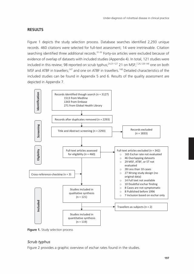
197
Under-diagnosis of rickettsial disease in clinical practice
RESULTS
Figure 1 depicts the study selection process. Database searches identifi ed 2,293 unique
records. 460 citations were selected for full-text assessment; 14 were irretrievable. Citation
searching identifi ed three additional records.31-33 Forty-six articles were excluded because of
evidence of overlap of datasets with included studies (Appendix 4). In total, 121 studies were
included in this review; 98 reported on scrub typhus;24,31-127 21 on MSF;7,26,128-146 one on both
MSF and ATBF in travellers;147 and one on ATBF in travellers.148 Detailed characteristics of the
included studies can be found in Appendix 5 and 6. Results of the quality assessment are
depicted in Appendix 7.
218
RESULTS
Figure 1 depicts the study selection process. Database searches identified 2,293 unique records.
460 citations were selected for full-text assessment; 14 were irretrievable. Citation searching
identified three additional records.31-33 Forty-six articles were excluded because of evidence of
overlap of datasets with included studies (Appendix 4). In total, 121 studies were included in this
review; 98 reported on scrub typhus;24,31-127 21 on MSF;7,26,128-146 one on both MSF and ATBF in
travellers;147 and one on ATBF in travellers.148 Detailed characteristics of the included studies can
be found in Appendix 5 and 6. Results of the quality assessment are depicted in Appendix 7.
Records Identified though search (n = 3127) 1513 from Medline 1343 from Embase 271 from Global Health Library
Scre
enin
g In
clude
d El
igib
ility
Id
entif
icatio
n
Cross-reference-checking (n = 3)
Records after duplicates removed (n = 2293)
Records excluded (n = 1833)
Full-text articles assessed for eligibility (n = 460)
(n = 462) (n = 461)
Full-text articles excluded (n = 342) o 165 Eschar rate not evaluated o 46 Overlapping datasets o 29 MSF, ATBF, or ST not
evaluated o 28 Less than 10 cases o 27 Wrong study design (no
original data) o 14 Full text not available o 10 Doubtful eschar finding o 8 Cases are not symptomatic o 8 Published before 1996 o 7 Inclusion based on eschar only
Studies included in qualitative synthesis
(n = 121)
(n = 118)
Studies included in quantitative synthesis
(n = 119)
Travellers as subjects (n = 2)
Title and Abstract screening (n = 2293)
Figure 1. Study selection process
Scrub typhus
Figure 2 provides a graphic overview of eschar rates found in the studies.
Chapter 6
198
Eschar presence was evaluated in a total number of 20,142 scrub typhus pa-
tients.24,31-127 The mean overall percentage of eschar presence was 64.3% (standard
deviation (SD) 25.4). Studies were conducted in India (n=39),31,32,44-80 South Korea
(n=19),83-101 Thailand (n=13),33,115-126 China (n=10),24,35-43 Taiwan (n=9),106-114 Japan
(n=2),81,82 Laos (n=2),102,103 Bangladesh (n=1),34 Malaysia (n=1),104 Sri Lanka (n=1),105 and
Vietnam (n=1).127 Among these 98 studies, 30 were assessed as having been performed
according the ‘complete approach’ scenario,31-33,44-50,52-59,76,102,106,107,115-122 and four were
classified as complete approach/unclear’.35,60,61,104 Of these 34 ‘complete approach’ or
‘complete approach/unclear’ studies, eight were of high quality;44,45,52,55,76,102,119,121 22
of medium quality;32,33,35,46-50,56-61,106,107,115-118,120,122 and four of low quality.31,53,54,104 Forty
studies were classified as ‘clinical judgement’,24,37-42,51,70-75,77-80,82,87-101,105,114,124-127 and 24 as
‘clinical judgement/unclear’.34,36,43,62-69,81,83-86,103,108-113,123 Of these 64 ‘clinical judgement’ or
‘clinical judgement/unclear’ studies, none was of high quality, 35 were of medium qual-
ity,24,34,37,38,40-42,51,62,71,72,75,77,78,80,81,87,89-94,98,99,103,105,108-110,112,114,124,126,127 and 29 were of low
quality.36,39,43,63-70,73,74,79,82-86,88,95-97,100,101,111,113,123,125 ‘Complete approach’ and ‘complete
approach/unclear’ studies had a significantly higher quality than ‘clinical judgement’ and
‘clinical judgement/unclear studies’ (p < 0.001).
Baseline characteristics on age and sex are presented in Table 1. In the ‘complete ap-
proach’ group, the mean or median age was lower compared to ‘clinical judgement’ studies
(38.1 ± 7.6 years vs. 47.2 ± 18.2 years, p < 0.001), and there were fewer males (47.4% ±
13.1% vs 48.7% ± 10.3%, p < 0.001).
Mean percentages of eschar presence (Table 1) were significantly different between the
‘complete approach’ and ‘clinical judgement’ studies (23.6% ± 20.1% vs. 70.4% ± 19.8%, p
< 0.001). As stated in our hypothesis, we assumed that the ERD/NERD ratio in the ‘complete
approach’-group is closest to the actual ratio. Using the calculations detailed in Appendix 3,
we estimated that a diagnosis of ST was missed in 34,744 patients in the ‘clinical judgement’
group, which represents 87.0% (34,744/39,930) of the patients with NERD and 66.5%
(34,744/52,263) of the total group of patients with ST, as shown in Figure 3.
When all unclear studies (both ‘clinical judgement/unclear’ and ‘complete approach/
unclear’) were assigned to the ‘clinical judgement’ studies (‘best case scenario’), the eschar
rates remained significantly lower in complete approach studies compared to ‘clinical judge-
ment’ studies (‘clinical judgement’ + ‘clinical judgement/unclear’ + ‘complete approach/
unclear’: mean = 70.0 ± 20.3 (17,655 patients) vs. ‘complete approach’: mean = 23.8 ± 20.3
(2,487 patients); p < 0.001). The same applied to the ‘worst case scenario’, when all unclear
studies were assigned to the ‘complete approach’ group (‘clinical judgement’: mean = 66.8
± 22.5 (8,035 patients) vs. ‘complete approach’ + ‘complete approach/unclear’ + ‘clinical
judgement/unclear’: mean = 62.6 ± 27.0 (12,107 patients); p < 0.001).
Figure 4 provides an overview of reported mean eschar rates per region.
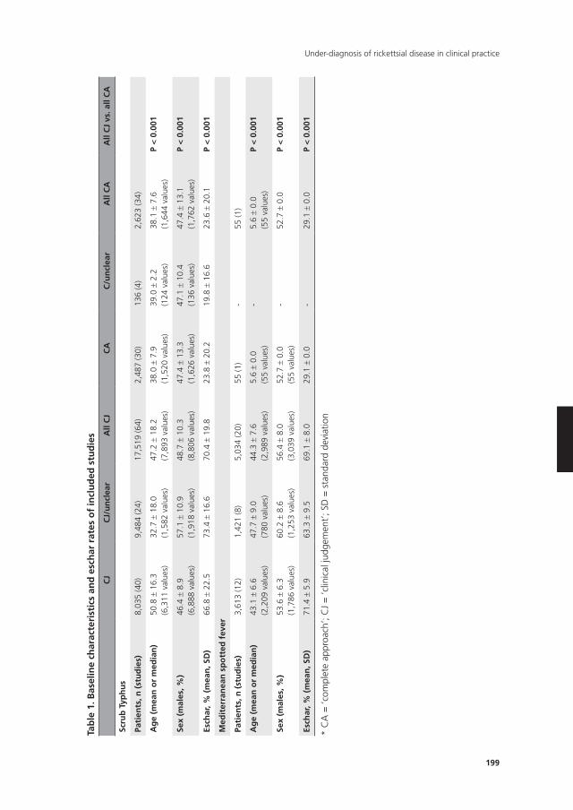
199
Under-diagnosis of rickettsial disease in clinical practice
Tab
le 1
. Bas
elin
e ch
arac
teri
stic
s an
d e
sch
ar r
ates
of
incl
ud
ed s
tud
ies
CJ
CJ/
un
clea
rA
ll C
JC
AC
/un
clea
rA
ll C
AA
ll C
J vs
. all
CA
Scru
b T
yph
us
Pati
ents
, n (
stu
die
s)8,
035
(40)
9,48
4 (2
4)17
,519
(64)
2,48
7 (3
0)13
6 (4
)2,
623
(34)
Ag
e (m
ean
or
med
ian
)50
.8 ±
16.
3(6
,311
val
ues)
32.7
± 1
8.0
(1,5
82 v
alue
s)47
.2 ±
18.
2(7
,893
val
ues)
38.0
± 7
.9(1
,520
val
ues)
39.0
± 2
.2(1
24 v
alue
s)38
.1 ±
7.6
(1,6
44 v
alue
s)P
< 0
.001
Sex
(mal
es, %
)46
.4 ±
8.9
(6,8
88 v
alue
s)57
.1 ±
10.
9(1
,918
val
ues)
48.7
± 1
0.3
(8,8
06 v
alue
s)47
.4 ±
13.
3(1
,626
val
ues)
47.1
± 1
0.4
(136
val
ues)
47.4
± 1
3.1
(1,7
62 v
alue
s)P
< 0
.001
Esch
ar, %
(m
ean
, SD
)66
.8 ±
22.
573
.4 ±
16.
670
.4 ±
19.
823
.8 ±
20.
219
.8 ±
16.
623
.6 ±
20.
1P
< 0
.001
Med
iter
ran
ean
sp
ott
ed f
ever
Pati
ents
, n (
stu
die
s)3,
613
(12)
1,42
1 (8
)5,
034
(20)
55 (1
)-
55 (1
)
Ag
e (m
ean
or
med
ian
)43
.1 ±
6.6
(2,2
09 v
alue
s)47
.7 ±
9.0
(780
val
ues)
44.3
± 7
.6(2
,989
val
ues)
5.6 ±
0.0
(55
valu
es)
-5.
6 ±
0.0
(55
valu
es)
P <
0.0
01
Sex
(mal
es, %
)53
.6 ±
6.3
(1,7
86 v
alue
s)60
.2 ±
8.6
(1,2
53 v
alue
s)56
.4 ±
8.0
(3,0
39 v
alue
s)52
.7 ±
0.0
(55
valu
es)
-52
.7 ±
0.0
P <
0.0
01
Esch
ar, %
(m
ean
, SD
)71
.4 ±
5.9
63.3
± 9
.569
.1 ±
8.0
29.1
± 0
.0-
29.1
± 0
.0P
< 0
.001
* C
A =
‘com
plet
e ap
proa
ch’;
CJ
= ‘c
linic
al ju
dgem
ent’
; SD
= s
tand
ard
devi
atio
n
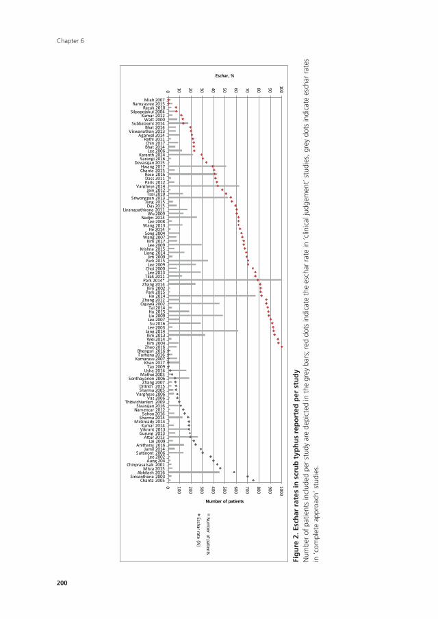
Chapter 6
200
Figure 2. Eschar rates in scrub typhus reported per study Num
ber of patients included per study are depicted in the grey bars; red dots indicate the eschar rate in 'clinical judgement' studies,
grey dots indicate eschar rates in 'complete approach' studies.
0 100
200
300
400
500
600
700
800
900
1000
0
10 20 30 40 50 60 70 80 90
100
Miah2007Ramyasree2015
Razak2010Silpapojakul2004
Kumar2012Watt2000
Subbalaxmi2014Bhat2014
Viswanathan2013Agarwal2014
Rathi2011Chin2017Bhat2014
Lee2006Karanth2014Sarangi2016
Devarajan2015Hwang2017Chanta2015
Rose2016Dass2011Paris2012
Varghese2014Jain2012Tsai2010
Sriwongpan2013Jung2015Das2015
Liyanapathirana2011Wu2009
Nadjm2014Lee2008
Wang2013He2014
Song2004Wang2007
Kim2017Lee2009
Krishna2015Liang2014
Jim2009Park2015Lee2009
Choi2000Lee2013
Tilak2011Park2014*Zhang2014
Kim2002Park2015
Ho2014Zhang2012
Ogawa2002Tai2014Hu2015Liu2009Lee2007Su2016
Lee2003Jang2014Kim2013Wei2014Kim2004
Zhao2016Bhengsri 2016Farhana2016
Kamarasu2007Khan2017
Tay2009Usha2014
Mathai2003Sonthayanon2006
Zhang2007Dittrich 2015Sharma2005
Varghese2006Vaz2006
Thitivichianlert 2009Sivarajan2016
Narvencar2012Sahoo2016
Sharma2014McGready2014
Kumar2014Vikrant2013Gurung 2013
Attur2013Lai2009
Anitharaj 2016Jamil2014
Suttinont 2006Lee2002Aung204
Chinprasatsak2001Misra2015
Abhilash2016Sirisanthana2003
Chanta2005
Numberofpatients
Eschar,%
Num
berofpatients
Escharrate(%)
Fig
ure
2. E
sch
ar r
ates
in s
cru
b t
yph
us
rep
ort
ed p
er s
tud
yN
umbe
r of
pat
ient
s in
clud
ed p
er s
tudy
are
dep
icte
d in
the
gre
y ba
rs; r
ed d
ots
indi
cate
the
esc
har
rate
in ‘c
linic
al ju
dgem
ent’
stu
dies
, gre
y do
ts in
dica
te e
scha
r ra
tes
in ‘c
ompl
ete
appr
oach
’ stu
dies
.
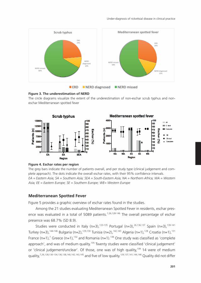
201
Under-diagnosis of rickettsial disease in clinical practice
Mediterranean Spotted Fever
Figure 5 provides a graphic overview of eschar rates found in the studies.
Among the 21 studies evaluating Mediterranean Spotted Fever in residents, eschar pres-
ence was evaluated in a total of 5089 patients.7,26,128-146 The overall percentage of eschar
presence was 68.7% (SD 8.9).
Studies were conducted in Italy (n=3),133-135 Portugal (n=3),26,136,137 Spain (n=3),139-141
Turkey (n=3),144-146 Bulgaria (n=2),129,130 Tunisia (n=2),142,143 Algeria (n=1),128 Croatia (n=1),131
France (n=1),7 Greece (n=1),132 and Romania (n=1).138 One study was classifi ed as ‘complete
approach’, and was of medium quality.135 Twenty studies were classifi ed ‘clinical judgement’
or ‘clinical judgement/unclear’. Of those, one was of high quality,139 14 were of medium
quality,7,26,128,130-134,136,138,140,142,143,145 and fi ve of low quality.129,137,141,144,146 Quality did not differ
Figure 3. The underestimation of NERD The circle diagrams visualize the extent of the underestimation of non-eschar scrub typhus and non-eschar Mediterranean spotted fever
Figure 4. Eschar rates per region The grey bars indicate the number of patients overall, and per study type (clinical judgement and complete approach). The dots indicate the overall eschar rates, with their 95% confidence intervals. EA = Eastern Asia; SA = Southern Asia; SEA = South-Eastern Asia; NA = Northern Africa; WA = Western Asia; EE = Eastern Europe; SE = Southern Europe; WE= Western Europe
ERD24%
NERD diagnosed
10%NERD missed
66%
Scrub typhus
ERD29%
NERD diagnosed13%
NERD missed58%
Mediterranean spotted fever
Figure 3. The underestimation of nERDThe circle diagrams visualize the extent of the underestimation of non-eschar scrub typhus and non-eschar Mediterranean spotted fever
Figure 3. The underestimation of NERD The circle diagrams visualize the extent of the underestimation of non-eschar scrub typhus and non-eschar Mediterranean spotted fever
Figure 4. Eschar rates per region The grey bars indicate the number of patients overall, and per study type (clinical judgement and complete approach). The dots indicate the overall eschar rates, with their 95% confidence intervals. EA = Eastern Asia; SA = Southern Asia; SEA = South-Eastern Asia; NA = Northern Africa; WA = Western Asia; EE = Eastern Europe; SE = Southern Europe; WE= Western Europe
ERD24%
NERD diagnosed
10%NERD missed
66%
Scrub typhus
ERD29%
NERD diagnosed13%
NERD missed58%
Mediterranean spotted fever
Figure 4. Eschar rates per regionThe grey bars indicate the number of patients overall, and per study type (clinical judgement and com-plete approach). The dots indicate the overall eschar rates, with their 95% confi dence intervals.EA = Eastern Asia; SA = Southern Asia; SEA = South-Eastern Asia; NA = Northern Africa; WA = Western Asia; EE = Eastern Europe; SE = Southern Europe; WE= Western Europe
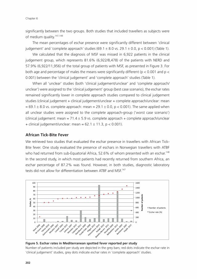
Chapter 6
202
signifi cantly between the two groups. Both studies that included travellers as subjects were
of medium quality.147,148
The mean percentages of eschar presence were signifi cantly different between ‘clinical
judgement’ and ‘complete approach’ studies (69.1 ± 8.0 vs. 29.1 ± 0.0, p < 0.001) (Table 1).
We calculated that the diagnosis of MSF was missed in 6,922 patients in the clinical
judgement group, which represents 81.6% (6,922/8,478) of the patients with NERD and
57.9% (6,922/11,956) of the total group of patients with MSF, as presented in Figure 3. For
both age and percentage of males the means were signifi cantly different (p < 0.001 and p <
0.001) between the ‘clinical judgement’ and ‘complete approach’ studies (Table 1).
When all ‘unclear’ studies (both ‘clinical judgement/unclear’ and ‘complete approach/
unclear’) were assigned to the ‘clinical judgement’ group (best case scenario), the eschar rates
remained signifi cantly lower in complete approach studies compared to clinical judgement
studies (clinical judgement + clinical judgement/unclear + complete approach/unclear: mean
= 69.1 ± 8.0 vs. complete approach: mean = 29.1 ± 0.0, p < 0.001). The same applied when
all unclear studies were assigned to the complete approach-group (‘worst case scenario’)
(clinical judgement: mean = 71.4 ± 5.9 vs. complete approach + complete approach/unclear
+ clinical judgement/unclear: mean = 62.1 ± 11.3, p < 0.001).
African Tick-Bite Fever
We retrieved two studies that evaluated the eschar presence in travellers with African Tick-
Bite fever. One study evaluated the presence of eschars in Norwegian travellers with ATBF
who had returned from sub-Equatorial Africa, 52.6% of whom presented with an eschar.148
In the second study, in which most patients had recently returned from southern Africa, an
eschar percentage of 87.2% was found. However, in both studies, diagnostic laboratory
tests did not allow for differentiation between ATBF and MSF.147
Figure 5. Eschar rates in Mediterranean spotted fever reported per study Number of patients included per study are depicted in the grey bars; red dots indicate the eschar rate in 'clinical judgement' studies, grey dots indicate eschar rates in 'complete approach' studies.
0
200
400
600
800
1000
1200
1400
1600
0
10
20
30
40
50
60
70
80
90
100
Num
bero
fpatients
Eschar,%
Number ofpatients
Escharrate(%)
Figure 5. Eschar rates in Mediterranean spotted fever reported per studyNumber of patients included per study are depicted in the grey bars; red dots indicate the eschar rate in ‘clinical judgement’ studies, grey dots indicate eschar rates in ‘complete approach’ studies.
203
Under-diagnosis of rickettsial disease in clinical practice
DISCUSSIOn
Rickettsial diseases, and especially non-eschar forms, are an underestimated cause of febrile
illness in endemic areas. In this study, we hypothesised that the ERD/NERD ratio in ‘complete
approach’ studies was closest to reality. Ratios in ‘clinical judgement’ and ‘complete ap-
proach’ studies differed significantly. For scrub typhus, we estimated that among the studies
included here, the diagnosis was missed in 66.5% of all patients. In MSF, this was up to
57.9%.
An expected underestimation of these rickettsial diseases has been mentioned
before,128,145,149 but no systematic evaluation and informed estimation of the scale of this
problem had been performed to date.
For scrub typhus, overall percentages of eschar prevalence in patients (64.3%) are in line
with a previously published review.150 This is, however, likely to constitute an over-estimation,
because the largest population included in this review came from ‘clinical judgement’ stud-
ies, which we have shown to be have significantly higher ERD/NERD ratios than ‘complete
approach’ studies. Theories on eschar occurrence relating to geography and associated
strains6,24-26 are neither supported, nor can they be refuted by this review. Although strains
of O. tsutsugamushi were not taken into account (neither were they frequently investigated)
within countries, eschar prevalence rates showed considerable variance, often depending on
the study design (‘complete approach’ vs. ‘clinical judgement’). Countries with more ‘com-
plete approach’ studies, such as India and Thailand, showed lower average eschar rates, as
expected. High average eschar rates were found in countries with no, few, or small ‘complete
approach’ studies, such as South Korea, China, and Taiwan. For example, within India (39
studies, of which 20 with a ‘complete approach’ study design), reported eschar rates ranged
from 0%47,49,50,65 to 76.7%,79 independent of geography (in this case provinces). In Taiwan,
average eschar rates were high (62.5%), with a range of 22.5%106 up to 91.3%,109 but few
patients came from studies with a ‘complete approach’ design. Overall, eschar rates reported
in Eastern Asia were higher than in South-Eastern and Southern Asia. We suspect this is due
to the lower number of studies with an ‘complete approach’ study design in these regions;
among ‘complete approach’ studies, reported eschar rates were similar across all regions.
That notwithstanding, this hypothesis should be confirmed by studies including all patients
presenting with febrile illness, comprising O. tsutsugamushi strain genotyping as well. It is
also possible that pre-existing immunity among inhabitants of endemic areas, would lead
to a lower prevalence of eschars,151,152 but no longitudinal studies on this subject have been
conducted to date.
For Mediterranean Spotted Fever, an overall eschar percentage of 68.7% was found,
similar to other reports.10 Only one study with a ‘complete approach’ study design was
identified (with an eschar prevalence of 29.1% among 55 patients),135 making our findings
for this disease less robust.
Chapter 6
204
In MSF, the R. conorii conorii, Malish strain causes eschars in similar percentages, whereas
for the Israeli Spotted Fever (ISF) strain (R. conorii subsp. israelensis) much lower eschar rates
are reported, around 38%.10,26 The ISF strain is also believed to be more virulent, with higher
fatality rates.10 It is possible that the patients in the ‘complete approach’ study included in
this review135 suffered from ISF instead of MSF, explaining the low eschar rates. But given the
facts that most clinicians do not have access to sophisticated diagnostic facilities to be able
to differentiate between strains, and that the diseases are endemic in the same geographic
areas,10 this is of less importance for the clinical practice. The extent of R. conorii and its
subspecies as a cause of NERD in endemic areas should be studied further.
For both diseases, significant differences were observed in age and sex between ‘com-
plete approach’ and ‘clinical judgement’ studies. Patients included in complete approach
studies were younger and there were fewer males. This is most likely due to heterogeneity,
but is remarkable either way. It is reported that in areas where the disease is hyper-endemic,
the percentage of patients presenting with an eschar is lower due to partial immunity ac-
quired by prior exposure 153. Thus, since most of the studies included here were performed in
highly endemic areas, we expected to find lower eschar rates coming with higher age. Yet,
we found the opposite, supporting our hypothesis that study design was the major factor
influencing reported eschar rates.
Few studies on African tick bite fever were identified; of which only two were conducted
in travellers.147,148. Both had a ‘complete approach’ study design. Therefore, we could con-
clude that schars are highly prevalent in travellers infected with R. africae; however, with a
total population of 116 patients, evidence is moderate at best.
It has been postulated that, especially in international travellers, rickettsial disease is
underreported in the published literature.154 This was stressed by our finding that only a
minority of the studies that we included reported on this disease in travellers. However, with
an ever-growing number of tourists to areas highly endemic for rickettsial diseases,155 and
mosquitos being identified as new potential transmitting vectors,156 this disease group is of
high importance in travellers. Rickettsial disease, and especially NERD should be considered in
every returning traveller with acute febrile illness. Travellers may be more likely than residents
to present with an eschar due to a lack of acquired immunity, as was supported by the find-
ings of the included studies reporting on travellers.147,148 However, the proportion of patients
with a missed diagnosis of NERD could potentially be even greater in returned travellers
compared to residents, due to lack of awareness among physicians in non-endemic areas.
The burden of rickettsial diseases as a cause of febrile illness in travellers should be studied
more extensively in prospective, ‘complete approach’ designs.
Strengths of this review are that the recommendations made by PRISMA27 were fol-
lowed. A comprehensive literature search was performed by an experienced librarian, and all
steps in data extraction, assessment, and analysis were performed by two reviewers. Studies
were included irrespective of language, in an attempt to reduce publication bias.
205
Under-diagnosis of rickettsial disease in clinical practice
Limitations of the study are that some Korean and Chinese articles were not retrievable,
possibly introducing a bias towards studies in English from different countries. Heterogene-
ity between the included studies was high. Studies with different designs were included.
Different microbiological methods were used, varying in reliability and quality, and thus in
diagnostic accuracy, which may have influenced our results. However, the net effect is pos-
sibly mitigated by the fact these differences were distributed over both ‘complete approach’
and ‘clinical judgement’ studies, reducing the influence on the ERD/NERD ratios. In order to
account for components of heterogeneity for which we could not control, we performed
quality assessments for all studies instead. Also, bacterial strains as a cause of variance
in reported eschar rates were not taken into account: firstly, most included studies used
serological methods, thus we could not differentiate between strains; but most importantly,
as clinicians, we tried to approach the problem from a clinical perspective. In daily clinical
practice, genotyping is not readily available and as a result has little consequences for further
medical management.
We did not analyse skin rashes in this review, because this is a prevalent finding in both
ERD and NERD, and many other causes of febrile illness. Presence or absence of a skin rash
does therefore not contribute to the estimation of under-diagnosis of rickettsial disease. On
the contrary, ERD/NERD ratios between the two study designs would have been more similar,
or even equal, should rickettsial disease have been considered more often in febrile patients
presenting with a skin rash, but without eschar.
We analysed studies performed in all sub-regions together. Across different geographic
locations, variation in circulating strains and immunity in local populations is to be expected,
both influencing eschar occurrence. In addition, differences in skin colour of the included
patients could partially explain a lower frequency of observed eschars in some regions, as es-
chars may be more difficult to diagnose in patients with a darker skin.68,69 In warm and moist
areas, eschars can be missed due to detachment of the crust, resulting in an ulcerous lesion.
Differences in overall eschar rates between sub-regions were substantial, but if only studies
applying ‘complete approach’ selection criteria were included, eschar rates were similar. This
underpins our hypothesis that study design could be the most important explanation for the
variation in reported eschar rates.
The strongest evidence for the under-diagnosis of NERD was found among patients with
ST. Current surveillance guidelines for scrub typhus149 emphasize the presence of eschars for
diagnosis of the disease, as this is an important diagnostic clue. The absence of an eschar
does not rule out scrub typhus. We argue that the importance of NERD recognition should
be stressed in guidelines, also because the absence of an eschar in ST is associated with
increased severity and mortality of the disease.96,157
For MSF, more studies in patients with febrile illness should be conducted to investigate
our hypothesis, but here we also suspect a vast underestimation of the NERD and recom-
mend clinicians in endemic areas to consider the disease with a low threshold.
Chapter 6
206
The underestimation of rickettsial disease is caused by the fact that out of the total
spectrum of clinical presentation (ERD/NERD combined), NERD is the presentation that is
most often missed. In scrub typhus for example, the total underestimation (NERD/ERD com-
bined) was 66.5%, but the underestimation of NERD alone was 87.0%. This latter higher
percentage could also apply for rickettsial diseases in which an eschar is rarely or never seen,
such as murine typhus, caused by Rickettsia typhi and Rocky Mountain Spotted Fever, caused
by Rickettsia rickettsii. This was supported by a recent study from Croatia,154 in which a
strong discrepancy was observed between the prevalence of anti-rickettsial antibodies and
the substantially lower number of reported cases of clinical disease. This discrepancy was
most evident for murine typhus (in comparison to Mediterranean spotted fever).
We recommend that clinicians working in endemic areas, or treating travellers returning
from endemic areas, should consider rickettsial disease, and especially NERD, with a low
threshold of suspicion. The most-commonly available serologic diagnostic methods, have a
poor sensitivity in the early phase of the disease, and to establish the diagnosis, a consecutive
sample is often required. Pending diagnosis, and to prevent treatment delay, empirical anti-
biotic therapy should therefore be considered, when rickettsial disease is suspected clinically.
In conclusion, this review indicates that rickettsial disease, a potentially fatal but treat-
able disease, should be considered in all patients presenting with acute febrile illness in, and
returning from, endemic areas, irrespective of eschar presence (NERD). For future research,
more ‘complete approach’ studies investigating causes of febrile illness should be performed
in all geographic areas, because local clinicians need to have a good knowledge of the
local epidemiology. The under-diagnosis of rickettsial disease in returned travellers with
fever should also be studied. And last but not least, current surveillance standards should
be revised, in order to increase awareness of NERD among clinicians. A clinical suspicion of
the possibility of NERD and the initiation of appropriate laboratory diagnostic tests and early
(empirical) antibiotic treatment should be facilitated in a broader population, to prevent
unnecessary morbidity and mortality.
207
Under-diagnosis of rickettsial disease in clinical practice
REFEREnCES
1. Merhej V, Raoult D. Rickettsial evolution in the light of comparative genomics. Biol Rev Camb
Philos Soc 2011; 86(2): 379-405.
2. Parola P, Davoust B, Raoult D. Tick- and flea-borne rickettsial emerging zoonoses. Vet Res 2005;
36(3): 469-92.
3. Botelho-Nevers E, Raoult D. Host, pathogen and treatment-related prognostic factors in rickett-
sioses. Eur J Clin Microbiol Infect Dis 2011; 30(10): 1139-50.
4. Jensenius M, Davis X, von Sonnenburg F, et al. Multicenter GeoSentinel analysis of rickettsial
diseases in international travelers, 1996-2008. Emerg Infect Dis 2009; 15(11): 1791-8.
5. Jensenius M, Han PV, Schlagenhauf P, et al. Acute and potentially life-threatening tropical dis-
eases in western travelers--a GeoSentinel multicenter study, 1996-2011. Am J Trop Med Hyg
2013; 88(2): 397-404.
6. Paris DH, Shelite TR, Day NP, Walker DH. Unresolved problems related to scrub typhus: a seriously
neglected life-threatening disease. Am J Trop Med Hyg 2013; 89(2): 301-7.
7. Botelho-Nevers E, Rovery C, Richet H, Raoult D. Analysis of risk factors for malignant Medi-
terranean spotted fever indicates that fluoroquinolone treatment has a deleterious effect. J
Antimicrob Chemother 2011; 66(8): 1821-30.
8. Taylor AJ, Paris DH, Newton PN. A Systematic Review of Mortality from Untreated Scrub Typhus
(Orientia tsutsugamushi). PLoS Negl Trop Dis 2015; 9(8): e0003971.
9. Fang LQ, Liu K, Li XL, et al. Emerging tick-borne infections in mainland China: an increasing
public health threat. Lancet Infect Dis 2015; 15(12): 1467-79.
10. Parola P, Paddock CD, Socolovschi C, et al. Update on tick-borne rickettsioses around the world:
a geographic approach. Clin Microbiol Rev 2013; 26(4): 657-702.
11. Bonell A, Lubell Y, Newton PN, Crump JA, Paris DH. Estimating the burden of scrub typhus: A
systematic review. PLoS Negl Trop Dis 2017; 11(9): e0005838.
12. Botelho-Nevers E, Socolovschi C, Raoult D, Parola P. Treatment of Rickettsia spp. infections: a
review. Expert Rev Anti Infect Ther 2012; 10(12): 1425-37.
13. Brouqui P, Bacellar F, Baranton G, et al. Guidelines for the diagnosis of tick-borne bacterial
diseases in Europe. Clin Microbiol Infect 2004; 10(12): 1108-32.
14. Blacksell SD, Bryant NJ, Paris DH, Doust JA, Sakoda Y, Day NP. Scrub typhus serologic testing
with the indirect immunofluorescence method as a diagnostic gold standard: a lack of consensus
leads to a lot of confusion. Clin Infect Dis 2007; 44(3): 391-401.
15. Dumler JS, Barbet AF, Bekker CP, et al. Reorganization of genera in the families Rickettsiaceae
and Anaplasmataceae in the order Rickettsiales: unification of some species of Ehrlichia with
Anaplasma, Cowdria with Ehrlichia and Ehrlichia with Neorickettsia, descriptions of six new spe-
cies combinations and designation of Ehrlichia equi and ‘HGE agent’ as subjective synonyms of
Ehrlichia phagocytophila. Int J Syst Evol Microbiol 2001; 51(Pt 6): 2145-65.
16. Parola P, Raoult D. Tropical rickettsioses. Clin Dermatol 2006; 24(3): 191-200.
17. Weitzel T, Dittrich S, Lopez J, et al. Endemic Scrub Typhus in South America. N Engl J Med 2016;
375(10): 954-61.
18. Mayxay M, Castonguay-Vanier J, Chansamouth V, et al. Causes of non-malarial fever in Laos: a
prospective study. Lancet Glob Health 2013; 1(1): e46-54.
19. Baltadzhiev I, Popivanova N, Zaprianov Z. Malignant forms of Mediterranean spotted fever: risk
factors for fatal outcomes. Braz J Infect Dis 2016; 20(5): 511-2.
Chapter 6
208
20. Jensenius M, Fournier PE, Kelly P, Myrvang B, Raoult D. African tick bite fever. Lancet Infect Dis
2003; 3(9): 557-64.
21. Jensenius M, Fournier PE, Vene S, et al. African tick bite fever in travelers to rural sub-Equatorial
Africa. 2003; 36(11): 1411-7.
22. Wilson ME, Weld LH, Boggild A, et al. Fever in returned travelers: results from the GeoSentinel
Surveillance Network. Clin Infect Dis 2007; 44(12): 1560-8.
23. Delord M, Socolovschi C, Parola P. Rickettsioses and Q fever in travelers (2004-2013). Travel Med
Infect Dis 2014; 12(5): 443-58.
24. Zhang M, Zhao ZT, Wang XJ, Li Z, Ding L, Ding SJ. Scrub typhus: surveillance, clinical profile and
diagnostic issues in Shandong, China. Am J Trop Med Hyg 2012; 87(6): 1099-104.
25. Kim DM, Yun NR, Neupane GP, et al. Differences in clinical features according to Boryoung and
Karp genotypes of Orientia tsutsugamushi. PLoS One 2011; 6(8): e22731.
26. Sousa R, França A, Dória Nòbrega S, et al. Host- and microbe-related risk factors for and patho-
physiology of fatal Rickettsia conorii infection in Portuguese patients. J Infect Dis 2008; 198(4):
576-85.
27. Moher D, Liberati A, Tetzlaff J, Altman DG. Preferred reporting items for systematic reviews and
meta-analyses: the PRISMA statement. BMJ 2009; 339: b2535.
28. United Nations Statistics Division - Standard Country and Area Codes Classifications (M49).
http://unstats.un.org/unsd/methods/m49/m49regin.htm (accessed 15 May 2017.
29. NIH. Quality Assessment Tool for Case Series Studies. https://www.nhlbi.nih.gov/health-pro/
guidelines/in-develop/cardiovascular-risk-reduction/tools/case_series (accessed 6 May 2017.
30. The Joanna Briggs Institute Critical Appraisal tools for use in JBI Systematic Reviews - Checklist for
Case Series. http://joannabriggs.org/research/critical-appraisal-tools.html (accessed 6 May 2017.
31. Mathai E RJ, Verghese GM, et al. Outbreak of scrub typhus in southern India during the cooler
months. Ann N Y Acad Sci 2003; 990: 359-64.
32. Gurung S PJ BP. Outbreak of scrub typhus in the North East Himalayan region-Sikkim: an emerg-
ing threat. Indian J Med Microbiol 2013; 31: 72-4.
33. Suttinont C LK, Niwatayakul K, et al. Causes of acute, undifferentiated, febrile illness in rural
Thailand: results of a prospective observational study. Ann Trop Med Parasitol 2006; 100(4):
363-70.
34. Miah MT, Rahman S, Sarker CN, Khan GK, Barman TK. Study on 40 cases of rickettsia. My-
mensingh Med J 2007; 16(1): 85-8.
35. Zhang L, Jin Z, Xia S, et al. Follow-up analysis on the epidemic strains of Orientia tsutsugamushi
in the first outbreak of scrub typhus in Henan Province, China. Southeast Asian J Trop Med Public
Health 2007; 38(3): 482-6.
36. He S, Ge L, Jin Y, Huang A. Clinical analysis of scrub typhus-associated hemophagocytic syn-
drome. Zhonghua Er Ke Za Zhi 2014; 52(9): 683-7.
37. Hu J, Tan Z, Ren D, et al. Clinical characteristics and risk factors of an outbreak with scrub
typhus in previously unrecognized areas, Jiangsu province, China 2013. PloS One 2015; 10(5):
e0125999.
38. Lee N, Ip M, Wong B, et al. Risk factors associated with life-threatening rickettsial infections. Am
J Trop Med Hyg 2008; 78(6): 973-8.
39. Liang XJ, Huang SM, Li JP, et al. Hepatic impairment induced by scrub typhus is associated with
new onset of renal dysfunction. Clin Lab 2014; 60(1): 63-8.
40. Liu YX, Feng D, Suo JJ, et al. Clinical characteristics of the autumn-winter type scrub typhus cases
in south of Shandong province, northern China. BMC Infect Dis 2009; 9: 82.
209
Under-diagnosis of rickettsial disease in clinical practice
41. Wei Y, Luo L, Jing Q, et al. A city park as a potential epidemic site of scrub typhus: a case-control
study of an outbreak in Guangzhou, China. Parasit Vectors 2014; 7: 513.
42. Zhang L, Zhao Z, Bi Z, et al. Risk factors associated with severe scrub typhus in Shandong,
northern China. Int J Infect Dis 2014; 29: 203-7.
43. Zhao M, Wang T, Yuan X, Du W, Lin M, Shen Y. Comparison of minocycline and azithromycin
for the treatment of mild scrub typhus in northern China. Int J Antimicrob Agents 2016; 48(3):
317-20.
44. Abhilash K, Jeevan J, Mitra S, et al. Acute undifferentiated febrile illness in patients presenting to
a Tertiary Care Hospital in South India: Clinical spectrum and outcome. J Glob Infect Dis 2016;
8(4): 147-54.
45. Anitharaj V, Stephen S, Pradeep J, et al. Serological diagnosis of acute scrub typhus in southern
India: Evaluation of InBios scrub typhus detect IgM rapid test and comparison with other sero-
logical tests. J Clin Diagn Res 2016; 10(11): DC07-DC10.
46. Attur RP, Kuppasamy S, Bairy M, et al. Acute kidney injury in scrub typhus. Clin Exp Nephrol 2013;
17(5): 725-9.
47. Farhana A, Bali N, Kanth F, Farooq R, Haq I, Shah P. Serological evidence of Scrub Typhus among
cases of PUO in the Kashmir valley- A hospital based study. J Clin Diagn Res 2016; 10(5): DC24-
DC6.
48. Jamil M, Lyngrah KG, Lyngdoh M, Hussain M. Clinical Manifestations and Complications of Scrub
Typhus : A Hospital Based Study from North Eastern India. J Assoc Physicians India 2014; 62(12):
19-23.
49. Kamarasu K, Malathi M, Rajagopal V, Subramani K, Jagadeeshramasamy D, Mathai E. Serological
evidence for wide distribution of spotted fevers & typhus fever in Tamil Nadu. Indian J Med Res
2007; 126(2): 128-30.
50. Khan SA, Bora T, Laskar B, Khan AM, Dutta P. Scrub typhus leading to acute encephalitis syn-
drome, Assam, India. Emerg Infect Dis 2017; 23(1): 148-50.
51. Kumar M, Krishnamurthy S, Delhikumar CG, Narayanan P, Biswal N, Srinivasan S. Scrub typhus in
children at a tertiary hospital in southern India: clinical profile and complications. J Infect Public
Health 2012; 5(1): 82-8.
52. Narvencar KP, Rodrigues S, Nevrekar RP, et al. Scrub typhus in patients reporting with acute
febrile illness at a tertiary health care institution in Goa. Indian J Med Res 2012; 136(6): 1020-4.
53. Sharma A, Mahajan S, Gupta ML, Kanga A, Sharma V. Investigation of an outbreak of scrub
typhus in the himalayan region of India. Jpn J Infect Dis 2005; 58(4): 208-10.
54. Sharma R, Krishna VP, Manjunath, et al. Analysis of Two Outbreaks of Scrub Typhus in Rajasthan:
A Clinico-epidemiological Study. J Assoc Physicians India 2014; 62(12): 24-9.
55. Sivarajan S, Shivalli S, Bhuyan D, Mawlong M, Barman R. Clinical and paraclinical profile, and
predictors of outcome in 90 cases of scrub typhus, Meghalaya, India. Infect Dis Poverty 2016;
5(1): 91.
56. Usha K, Kumar E, Kalawat U, Siddhartha Kumar B, Chaudhury A, Sai Gopal DVR. Seropreva-
lence of scrub typhus among febrile patients: A preliminary study. Asian J Pharm Clin Res 2014;
7(SUPPL. 1): 19-21.
57. Vaz LS, Gupta NK. Outbreak of scrub typhus in Jammu - A report. Med J Armed Forces India
2006; 62(4): 342-3.
58. Varghese GM, Abraham OC, Mathai D, et al. Scrub typhus among hospitalised patients with
febrile illness in South India: magnitude and clinical predictors. J Infect 2006; 52(1): 56-60.
Chapter 6
210
59. Vikrant S, Dheer SK, Parashar A, et al. Scrub typhus associated acute kidney injury--a study from
a tertiary care hospital from western Himalayan State of India. Ren Fail 2013; 35(10): 1338-43.
60. Misra UK, Kalita J, Mani VE. Neurological manifestations of scrub typhus. J Neurol Neurosurg
Psychiatry 2015; 86(7): 761-6.
61. Sahoo JN, Gurjar M, Harde Y. Acute respiratory failure in scrub typhus patients. Indian J Crit Care
Med 2016; 20(8): 480-4.
62. Das D, Das B, Roy AD, Singh TSK. Common Infectious Etiologies of Acute Febrile Illness in a
Remote Geographical Location: Could Scrub Typhus be the Most Common Cause? Br J Med Med
Res 2015; 10(10): 1-10.
63. Devarajan V, Nambi PS, Gopalakrishnan R, Murali A. Scrub Typhus in Hospitalized Children. Infect
Dis Clin Pract 2015; 23(2): 89-90.
64. Krishna MR, Vasuki B, Nagaraju K. Scrub typhus: audit of an outbreak. Indian J Pediatr 2015;
82(6): 537-40.
65. Ramyasree A, Kalawat U, Rani ND, Chaudhury A. Seroprevalence of Scrub typhus at a tertiary
care hospital in Andhra Pradesh. Indian J Med Microbiol 2015; 33(1): 68-72.
66. Razak A, Sathyanarayanan V, Prabhu M, Sangar M, Balasubramanian R. Scrub typhus in Southern
India: are we doing enough? Trop Doct 2010; 40(3): 149-51.
67. Rose W, Rajan RJ, Punnen A, Ghosh U. Distribution of eschar in pediatric scrub typhus. J Trop
Pediatr 2016; 62(5): 415-20.
68. Subbalaxmi MV, Madisetty MK, Prasad AK, et al. Outbreak of scrub typhus in Andhra Pradesh--
experience at a tertiary care hospital. J Assoc Physicians India 2014; 62(6): 490-6.
69. Viswanathan S, Muthu V, Iqbal N, Remalayam B, George T. Scrub typhus meningitis in South
India--a retrospective study. PloS One 2013; 8(6): e66595.
70. Agarwal VK, Reddy GKM, Krishna MR, Ramareddy G, Saroj P, Bandaru VCSS. Predictors of scrub
typhus: A study from a tertiary care center. Asian Pac J Trop Dis 2014; 4(S2): S666-S73.
71. Bhat NK, Dhar M, Mittal G, et al. Scrub typhus in children at a tertiary hospital in north India:
Clinical profile and complications. Iran J Pediatr 2014; 24(4): 387-92.
72. Bhat NK, Dhar M, Mittal G, Chandra H, Rawat A, Chandar V. Scrub typhus: A common rickettsial
disease emerging in a new geographical region of north India. J Pediatric Infect Dis 2014; 9(2):
93-9.
73. Dass R, Deka NM, Duwarah SG, et al. Characteristics of pediatric scrub typhus during an outbreak
in the North Eastern region of India: peculiarities in clinical presentation, laboratory findings and
complications. Indian J Pediatr 2011; 78(11): 1365-70.
74. Jain N, Jain V. Study on clinico -laboratory profile of children with scrub typhus. J Nepal Paediatr
Soc 2012; 32(2): 187-92.
75. Karanth SS, Marupudi KC, Sama VPR, Gupta A, Ballal K, Kamath A. Predictors of severity of scrub
typhus in the Indian subcontinent. Asian Pac J Trop Dis 2014; 4: S674-S8.
76. Kumar V, Kumar V, Yadav AK, et al. Scrub typhus is an under-recognized cause of acute febrile
illness with acute kidney injury in India. PLoS Negl Trop Dis 2014; 8(1): e2605.
77. Rathi NB, Rathi AN, Goodman MH, Aghai ZH. Rickettsial diseases in central India: proposed
clinical scoring system for early detection of spotted fever. Indian Pediatr 2011; 48(11): 867-72.
78. Sarangi R, Pradhan S, Debata NC, Mahapatra S. Clinical profile of scrub typhus in children treated
in a tertiary care hospital in eastern India. Pediatr Pol 2016; 91(4): 308-11.
79. Tilak R, Kunwar R, Wankhade UB, Tilak VW. Emergence of Schoengastiella ligula as the vector
of scrub typhus outbreak in Darjeeling: has Leptotrombidium deliense been replaced? Indian J
Public Health 2011; 55(2): 92-9.
211
Under-diagnosis of rickettsial disease in clinical practice
80. Varghese GM, Trowbridge P, Janardhanan J, et al. Clinical profile and improving mortality trend
of scrub typhus in South India. Int J Infect Dis 2014; 23: 39-43.
81. Tai K, Iwasaki H, Ikegaya S, et al. Significantly higher cytokine and chemokine levels in patients
with Japanese spotted fever than in those with Tsutsugamushi disease. J Clin Microbiol 2014;
52(6): 1938-46.
82. Ogawa M, Hagiwara T, Kishimoto T, et al. Scrub typhus in Japan: epidemiology and clinical
features of cases reported in 1998. Am J Trop Med Hyg 2002; 67(2): 162-5.
83. Choi YH, Kim SJ, Lee JY, Pai HJ, Lee KY, Lee YS. Scrub typhus: radiological and clinical findings.
Clin Radiol 2000; 55(2): 140-4.
84. Kim HS, Jo KH, Kang SG, Cha MJ, Hwang HH. A clinical research of tsutsugamushi disease
occurred in and around Jeoung-up in the autumn of 2000. J Korean Acad Fam Med 2002; 23(1):
87-95.
85. Lee JS, Kang JH, Cho BK, Yu BY. Factors Affecting Disease Duration in Patients with Tsutsugamu-
shi Disease. Korean Acad Fam Med 2007; 28(10): 774-81.
86. Park JH, Kim SJ, Youn SK, Park K, Gwack J. Epidemiology of scrub typhus and the eschars pat-
terns in South Korea from 2008 to 2012. Jpn J Infect Dis 2014; 67(6): 458-63.
87. Chin JY, Kang KW, Moon KM, Kim J, Choi YJ. Predictors of acute myocarditis in complicated
scrub typhus: an endemic province in the Republic of Korea. Korean J Intern Med 2017: 23.
88. Ho YH, Park KC, Jang YT. A comparison of clinical manifestations of patients with tsutsugamushi
disease between children and adults. [Korean]. Korean J Pediatr Infect Dis 2014; 21(2): 104-13.
89. Hwang K, Jang HN, Lee TW, et al. Incidence, risk factors and clinical outcomes of acute kidney
injury associated with scrub typhus: a retrospective study of 510 consecutive patients in South
Korea (2001-2013). BMJ Open 2017; 7(3): e013882.
90. Jang MO, Kim JE, Kim UJ, et al. Differences in the clinical presentation and the frequency of
complications between elderly and non-elderly scrub typhus patients. Arch Gerontol Geriatr
2014; 58(2): 196-200.
91. Jung HC, Chon SB, Oh WS, Lee DH, Lee HJ. Etiologies of acute undifferentiated fever and clinical
prediction of scrub typhus in a non-tropical endemic area. Am J Trop Med Hyg 2015; 92(2):
256-61.
92. Kim YS, Yun HJ, Shim SK, Koo SH, Kim SY, Kim S. A comparative trial of a single dose of azithro-
mycin versus doxycycline for the treatment of mild scrub typhus. Clin Infect Dis 2004; 39(9):
1329-35.
93. Kim DM, Chung JH, Yun NR, et al. Scrub typhus meningitis or meningoencephalitis. Am J Trop
Med Hyg 2013; 89(6): 1206-11.
94. Kim KY, Song JS, Park EH, Jin GY. Scrub typhus: radiological and clinical findings in abdominopel-
vic involvement. Jpn J Radiol 2017; 35(3): 101-8.
95. Lee KY, Lee HS, Hong JH, Hur JK, Whang KT. Roxithromycin treatment of scrub typhus (tsutsuga-
mushi disease) in children. Pediatr Infect Dis J 2003; 22(2): 130-3.
96. Lee CS, Hwang JH, Lee HB, Kwon KS. Risk factors leading to fatal outcome in scrub typhus
patients. Am J Trop Med Hyg 2009; 81(3): 484-8.
97. Lee CS, Min IS, Hwang JH, Kwon KS, Lee HB. Clinical significance of hypoalbuminemia in out-
come of patients with scrub typhus. BMC Infect Dis 2009; 10: 216.
98. Lee JH, Song HY, Lee JM, Cho JH. Optimal cutoff value of serum adenosine deaminase activity for
diagnosing acute scrub typhus. Jpn J Infect Dis 2013; 66(3): 232-4.
99. Park SW, Ha NY, Ryu B, et al. Urbanization of scrub typhus disease in South Korea. PLoS Negl Trop
Dis 2015; 9(5): e0003814.
Chapter 6
212
100. Park MJ, Lee HS, Shim SG, Kim SH. Scrub typhus associated hepatic dysfunction and abdominal
CT findings. Pak J Med Sci 2015; 31(2): 295-9.
101. Song SW, Kim KT, Ku YM, et al. Clinical role of interstitial pneumonia in patients with scrub
typhus: a possible marker of disease severity. J Korean Med Sci 2004; 19(5): 668-73.
102. Dittrich S, Rattanavong S, Lee SJ, et al. Orientia, rickettsia, and leptospira pathogens as causes of
CNS infections in Laos: a prospective study. Lancet Glob Health 2015; 3(2): e104-12.
103. Paris DH, Chansamouth V, Nawtaisong P, et al. Coagulation and inflammation in scrub typhus
and murine typhus--a prospective comparative study from Laos. Eur J Clin Microbiol Infect Dis
2012; 18(12): 1221-8.
104. Tay ST, Rohani MY, Ho TM, Devi S. Isolation and PCR detection of rickettsiae from clinical and
rodent samples in Malaysia. Southeast Asian J Trop Med Public Health 2009.
105. Liyanapathirana VC, Thevanesam V. Seroepidemiology of rickettsioses in Sri Lanka: a patient
based study. BMC Infect Dis 2011; 11: 328.
106. Lai CH, Huang CK, Weng HC, et al. The difference in clinical characteristics between acute Q
fever and scrub typhus in southern Taiwan. Int J Infect Dis 2009; 13(3): 387-93.
107. Lee HC, Ko WC, Lee HL, Chen HY. Clinical manifestations and complications of rickettsiosis in
southern Taiwan. J Formos Med Assoc 2002; 101(6): 385-92.
108. Jim WT, Chiu NC, Chan WT, et al. Clinical manifestations, laboratory findings and complications
of pediatric scrub typhus in eastern Taiwan. Pediatr Neonatol 2009; 50(3): 96-101.
109. Su TH, Liu CJ, Shu PY, et al. Associated factors and clinical implications of serum aminotransferase
elevation in scrub typhus. J Microbiol Immunol Infect 2016; 49(6): 941-6.
110. Tsai CC, Lay CJ, Wang CL, Ho YH, Wang LS, Chen LK. Levofloxacin versus tetracycline antibiotics
for the treatment of scrub typhus. Int J Infect Dis 2010; 14(1): e62-7.
111. Wang CC, Liu SF, Liu JW, Chung YH, Su MC, Lin MC. Acute respiratory distress syndrome in scrub
typhus. Am J Trop Med Hyg 2007; 76(6): 1148-52.
112. Wang YC, Chen PC, Lee KF, Wu YC, Chiu CH. Scrub typhus cases in a teaching hospital in
Penghu, Taiwan, 2006-2010. Vector Borne Zoonotic Dis 2013; 13(3): 154-9.
113. Wu KM, Wu ZW, Peng GQ, Wu JL, Lee SY. Radiologic pulmonary findings, clinical manifesta-
tions and serious complications in scrub typhus: experiences from a teaching hospital in eastern
taiwan. Int J Gerontol 2009; 3(4): 223-32.
114. Lee YS, Wang PH, Tseng SJ, Ko CF, Teng HJ. Epidemiology of scrub typhus in eastern Taiwan,
2000-2004. Jpn J Infect Dis 2006; 59(4): 235-8.
115. Aung T, Supanaranond W, Phumiratanaprapin W, Phonrat B, Chinprasatsak S, Ratanajaratroj N.
Gastrointestinal manifestations of septic patients with scrub typhus in Maharat Nakhon Ratcha-
sima Hospital. Southeast Asian J Trop Med Public Health 2004; 35(4): 845-51.
116. Bhengsri S, Baggett HC, Edouard S, et al. Sennetsu Neorickettsiosis, Spotted Fever Group, and
Typhus Group Rickettsioses in Three Provinces in Thailand. Am J Trop Med Hyg 2016; 95(1): 43-9.
117. Chanta C, Chanta S. Clinical study of 20 children with scrub typhus at Chiang Rai Regional
Hospital. J Med Assoc Thai 2005; 88(12): 1867-72.
118. Chinprasatsak S, Wilairatana P, Looareesuwan S, et al. Evaluation of a newly developed dipstick
test for the rapid diagnosis of scrub typhus in febrile patients. Southeast Asian J Trop Med Public
Health 2001; 32(1): 132-6.
119. McGready R, Prakash JA, Benjamin SJ, et al. Pregnancy outcome in relation to treatment of
murine typhus and scrub typhus infection: a fever cohort and a case series analysis. PLoS Negl
Trop Dis 2014; 8(11): e3327.
213
Under-diagnosis of rickettsial disease in clinical practice
120. Sirisanthana V, Puthanakit T, Sirisanthana T. Epidemiologic, clinical and laboratory features of
scrub typhus in thirty Thai children. Pediatr Infect Dis J 2003; 22(4): 341-5.
121. Sonthayanon P, Chierakul W, Wuthiekanun V, et al. Rapid diagnosis of scrub typhus in rural
Thailand using polymerase chain reaction. Am J Trop Med Hyg 2006; 75(6): 1099-102.
122. Thitivichianlert S, Panichkul S, Bodhidatta D, et al. Incidence of rickettsial infection in patients
with acute fever in provincial Thai army hospitals. J Med Assoc Thai 2009; 92 Suppl 1: S39-46.
123. Silpapojakul K, Varachit B, Silpapojakul K. Paediatric scrub typhus in Thailand: a study of 73
confirmed cases. Trans R Soc Trop Med Hyg 2004; 98(6): 354-9.
124. Chanta C, Phloenchaiwanit P. Randomized controlled trial of azithromycin versus doxycycline or
chloramphenicol for treatment of uncomplicated pediatric scrub typhus. J Med Assoc Thai 2015;
98(8): 756-60.
125. Sriwongpan P, Krittigamas P, Kantipong P, Kunyanone N, Patumanond J, Namwongprom S. Clini-
cal indicators for severe prognosis of scrub typhus. Risk Manag Healthc Policy 2013; 6: 43-9.
126. Watt G, Kantipong P, Jongsakul K, Watcharapichat P, Phulsuksombati D, Strickman D. Doxycy-
cline and rifampicin for mild scrub-typhus infections in northern Thailand: a randomised trial.
Lancet 2000; 356(9235): 1057-61.
127. Nadjm B, Thuy PT, Trang VD, Ha LD, Kinh NV, Wertheim HF. Scrub typhus in the northern prov-
inces of Vietnam: An observational study of admissions to a national referral hospital. Trans R Soc
Trop Med Hyg 2014; 108(11): 739-40.
128. Mouffok N, Parola P, Lepidi H, Raoult D. Mediterranean spotted fever in Algeria--new trends. Int
J Infect Dis 2009; 13(2): 227-35.
129. Baltadzhiev IG, Popivanova NI. Some epidemiological features of the Mediterranean spotted
fever re-emerging in Bulgaria. Folia Med (Plovdiv) 2012; 54(1): 36-43.
130. Pishmisheva M, Stoycheva M, Vatev N, Semerdjieva M. Mediterranean spotted fever in children
in the Pazardjik region, South Bulgaria. Pediatr Infect Dis J 2014; 33(5): 542-4.
131. Punda-Polic V, Luksic B, Capkun V. Epidemiological features of Mediterranean spotted fever,
murine typhus, and Q fever in Split-Dalmatia County (Croatia), 1982-2002. Epidemiol Infect
2008; 136(7): 972-9.
132. Germanakis A, Psaroulaki A, Gikas A, Tselentis Y. Mediterranean spotted fever in crete, Greece:
clinical and therapeutic data of 15 consecutive patients. Ann N Y Acad Sci 2006; 1078: 263-9.
133. Colomba C, Saporito L, Siracusa L, Giammanco G, Bonura S, Titone L. [Mediterranean spotted
fever in paediatric and adult patients: two clinical aspects of the same disease]. Infez Med 2011;
19(4): 248-53.
134. Micalizzi A, La Spada E, Corsale S, et al. Abnormal liver function in Mediterranean spotted fever.
[Italian]. Infez Med 2007; 15(2): 105-10.
135. Vitaliti G, Falsaperla R, Lubrano R, et al. Incidence of Mediterranean spotted fever in Sicilian
children: a clinical-epidemiological observational retrospective study from 1987 to 2010. Int J
Infect Dis 2015; 31: 35-40.
136. Crespo P, Seixas D, Marques N, Oliveira J, da Cunha S, Melico-Silvestre A. Mediterranean spotted
fever: case series of 24 years (1989-2012). Springerplus 2015; 4: 272.
137. Meireles M, Magalhaes R, Guimas A. [Mediterranean Spotted Fever: Retrospective Review of
Hospitalized Cases and Predictive Factors of Severe Disease]. Acta Med Port 2015; 28(5): 624-31.
138. Pitigoi D, Olaru ID, Badescu D, Rafila A, Arama V, Hristea A. Mediterranean spotted fever in
southeastern Romania. Biomed Res Int 2013; (395806).
139. Antón E, Font B, Muñoz T, Sanfeliu I, Segura F. Clinical and laboratory characteristics of 144
patients with mediterranean spotted fever. Eur J Clin Microbiol Infect Dis 2003; 22(2): 126-8.
Chapter 6
214
140. Bartolome J, Lorente S, Hernandez-Perez N, Martinez-Alfaro E, Marin-Ors A, Crespo MD. [Clinical
and epidemiological study of spotted fever group rickettsiosis in Albacete, Spain]. Enferm Infecc
Microbiol Clin 2005; 23(4): 194-6.
141. Jufresa J, Alegre J, Surinach JM, et al. [Study of 86 cases of Mediterranean boutonneuse fever
hospitalized at a university hospital]. An Med Interna 1997; 14(7): 328-31.
142. Letaïef A, Souissi J, Trabelsi H, Ghannem H, Jemni L. Evaluation of clinical diagnosis scores for
Boutonneuse fever. Ann N Y Acad Sci 2003; 990: 327-30.
143. Romdhane FB, Loussaief C, Toumi A, et al. Mediterranean spotted fever: A report of 200 cases in
Tunisia. Clin Microbiol Infect 2009; 15(SUPPL. 2): 209-10.
144. Mert A, Ozaras R, Tabak F, Bilir M, Ozturk R. Mediterranean spotted fever: a review of fifteen
cases. J Dermatol 2006; 33(2): 103-7.
145. Kuloglu F, Rolain JM, Akata F, Eroglu C, Celik AD, Parola P. Mediterranean spotted fever in the
Trakya region of Turkey. Ticks Tick Borne Dis 2012; 3(5-6): 298-304.
146. Yilmaz E, Akalin H, Mistik R, et al. Mediterranean spotted fever: Retrospective evaluation of 16
cases. [Turkish]. Balkan Med J 2010; 27(2): 167-71.
147. Jelinek T, Loscher T. Clinical features and epidemiology of tick typhus in travelers. J Travel Med
2001; 8(2): 57-9.
148. Jensenius M, Fournier PE, Vene S, et al. African tick bite fever in travelers to rural sub-Equatorial
Africa. Clin Infect Dis 2003; 36(11): 1411-7.
149. World Health Organization. WHO Recommended Surveillance Standards, Second edition. http://
www.who.int/csr/resources/publications/surveillance/WHO_CDS_CSR_ISR_99_2_EN/en/ (ac-
cessed 15 May 2017.
150. Rajapakse S, Weeratunga P, Sivayoganathan S, Fernando SD. Clinical manifestations of scrub
typhus. Trans R Soc Trop Med Hyg 2017; 111(2): 43-54.
151. Smadel JE, Ley HL, Jr., Diercks FH, Traub R. Immunity in scrub typhus: resistance to induced
reinfection. AMA Arch Pathol 1950; 50(6): 847-61.
152. Kitaoka M, Asanuma K, Otsuji J. Transmission of Rickettsia orientalis to man by Leptotrombidium
akamushi at a scrub typhus endemic area in Akita Prefecture, Japan. Am J Trop Med Hyg 1974;
23(5): 993-9.
153. Silpapojakul K. Scrub typhus in the Western Pacific region. Ann Acad Med Singapore 1997;
26(6): 794-800.
154. Dzelalija B, Punda-Polic V, Medic A, Dobec M. Rickettsiae and rickettsial diseases in Croatia:
Implications for travel medicine. Travel Med Infect Dis 2016; 14(5): 436-43.
155. UNWTO Tourism Highlights 2016. 2016. http://www.e-unwto.org/doi/
book/10.18111/9789284418145 (accessed 15 May 2017.
156. Parola P. Rickettsioses and travel: Be alert to new vectors including mosquitoes. Travel Med Infect
Dis 2016; 14(5): 433.
157. Kim DM, Kim SW, Choi SH, Yun NR. Clinical and laboratory findings associated with severe scrub
typhus. BMC Infect Dis 2010; 10: 108.
215
Under-diagnosis of rickettsial disease in clinical practice
SUPPLEMEnTARy MATERIALS
The supplementary materials provide details on the search strategy, quality assessment tool,
calculation logarithms, excluded studies, and extensive tables with study characteristics and
quality assessment forms. This data can be found, in the online version, at https://www.
sciencedirect.com/science/article/pii/S1477893918300176?via%3Dihub#appsec1
Appendix 1 Search strategy
Appendix 2 Quality Assessment Tool
Appendix 3 Underestimation Calculation Logarithms
Appendix 4 Excluded studies due to overlapping datasets
Appendix 5 Characteristics of included studies
Appendix 6 Data Extraction Forms
Appendix 7 Quality Assessment Forms


Chapter 7Searching and fi nding the hidden
treasure: Rickettsial disease among Dutch international travelers – a retrospective
analysis
Sophia G de Vries, Louise E van Eekeren, Hans van der Linden, Benjamin J. VIsser, Martin P Grobusch, Jiri F P Wagenaar, Marga G.A. Goris , Abraham
Goorhuis
Clinical Infectious Diseases 2020 Jan 30 (epub ahead of print)
Chapter 7
218
ABSTRACT
Background
Rickettsial disease (RD) is a prevalent and underestimated cause of febrile illness worldwide,
especially in the absence of an inoculation eschar. We attempted to quantify this under
estimation at our clinic, by investigating past cases of febrile illness in travelers who had
tested negative for leptospirosis, a disease that can initially present similar to non-eschar RD,
and which we routinely consider when other important causes of unspecified febrile illness
have tested negative.
Methods
We performed a retrospective analysis in febrile returned travelers from Asia, Africa or the
Americas between 2010-2017, who had tested negative for leptospirosis. Serologic immu-
nofluorescence assays were performed for Orientia tsutsugamushi (scrub typhus), Typhus
Group (TG) and Spotted Fever Group (SFG) RD. We performed a chart review of all patients
who tested positive. In case of a fitting medical history, cases were deemed either confirmed
(based on convalescent serology) or suspected (based on single serology).
Results
Among 97 patients, convalescent serology was available in 16 (16.5%) patients, and a
single serology in 81 (83.5%) patients. RD was the likely diagnosis in 8/16 (50.0%) patients
with convalescent serology, and in 8/81 (9.9%) with single serology. Of the 16 confirmed/
suspected cases, 11 (69%) had been missed and 7 (44%) had not received adequate empiric
antibiotic therapy.
Conclusions
This study shows that non-eschar RD is an important and poorly recognized cause of illness
in travelers, even in a specialized travel clinic. A lower threshold to test and treat for RD is
warranted in returning travelers with febrile illness.
219
Searching and finding the hidden treasure
InTRODUCTIOn
Rickettsial diseases (RD) are zoonotic infections, transmitted to humans by predominantly
arthropod vectors,1 although leeches and mosquitoes have also been described as vec-
tors.2,3 The disease may be mild to life-threatening,4 especially when treatment is delayed.5,6
Substantial morbidity is reported worldwide in autochthonous populations, as well as in
travelers.7-15 RD generally presents as an indifferent acute febrile illness, with non-specific
accompanying symptoms such as nausea, vomiting, lymphadenopathy, headache, skin rash,
and sometimes an inoculation eschar. The prevalence of the latter varies widely per specific
RD:16 from 0% in patients with murine typhus (cause by R. typhi), to 30-90% in patients
with African tick bite fever (caused by R. africae).11 Clinically, the symptomatology of RD is
often similar to other acute febrile illnesses such as malaria, dengue fever, and leptospirosis,17
especially if an eschar is absent at presentation.
The disease is caused by intracellular bacteria of the Rickettsiaceae family, ordered into
two genera: Orientia (consisting of Orientia tsutsugamushi, causing scrub typhus) and Rick-
ettsia.18 The Rickettsia genus is divided in four biogroups: (1) the spotted fever group (SFG),
which, among others, includes R. conorii (causing Mediterranean spotted fever), R. africae
(causing African Tick Bite Fever) and, R. rickettsii (causing Rocky Mountain spotted fever);
(2) the typhus group (TG), which comprises R. typhi and R. prowazekii, causing endemic and
epidemic typhus, respectively; (3) a translational group, including R. felis, R. australis and R.
akari; and (4), a non-pathogenic group.18,19 Rickettsial organisms have been identified on all
continents, except Antarctica.20 R. typhi and R. felis and are distributed globally; SFG RD has
been reported on all continents; Scrub typhus (cause by O. tsutsugamushi) is traditionally
prevalent in the tropical Pacific triangle, but there are recent reports from South America and
sub-Saharan Africa.20
Currently, the cornerstone of diagnosis is still the indirect detection of Rickettsia-specific
antibodies in patient sera by serologic methods, such as immunofluorescence or western
blotting. Because antibodies are detected at a later stage after infection, typically 15 days
or more,21-23 these methods have limited clinical impact in the acute stage of disease, when
most initial diagnostic testing is done 5. Additionally, there is cross-reactivity between spe-
cies.24 For a specific diagnosis in the acute phase of illness, molecular detection methods
are preferred,25-28 but these are not widely available. Also, reported diagnostic accuracy of
the different tests varies considerably, also based on the specimen type (e.g., whole blood,
serum), and reference tests are suboptimal, with differences in applied techniques, and
targets.29
Because of the unspecific clinical presentation of RD, and difficulties in laboratory di-
agnostics in the early phase of disease, patients may be un- or misdiagnosed. In a previous
study based on reported literature, we estimated that the diagnosis of RD was missed in
66.5% of patients with scrub typhus (ST), and in 57.9% of patients with Mediterranean
Chapter 7
220
spotted fever (MSF) in autochthonous populations.16 However, these percentages applied to
patients who presented with or without an inoculation eschar. Among patients in whom an
inoculation eschar was absent, RD was missed in 87.0% of patients with ST and 81.6% of
patients with MSF.
In travelers, this proportion could even be higher due a low index of suspicion by phy-
sicians in areas that are not endemic for the disease. This underestimation is of growing
concern, given the expansion of international travel to endemic regions such as Asia and
Africa, resulting in increased numbers of imported infections such as RD.30
We hypothesize that in the absence of an inoculation eschar, the diagnosis of RD is
missed in a substantial proportion of returned travelers presenting with acute febrile illness.
Our hospital houses the Dutch Leptospirosis Reference Center (NRL), which means that test-
ing for leptospirosis can be easily performed upon clinical suspicion. The disease is usually
considered when diagnostic routine testing for other important causes of unspecified febrile
illness turns out negative (i.e. malaria, typhoid fever, dengue, chikungunya and zika virus
infection), even in the absence of evident exposure to fresh water, as this is often difficult
to ascertain in retrospect. Therefore, and because leptospirosis and RD can have clinical
similarities at initial presentation, we hypothesized that missed diagnoses of RD would likely
be found among patients who had presented with unspecified febrile illness and who had
tested negative for leptospirosis. Finding these missed diagnoses would provide us a rough
indication of the under diagnosis of non-eschar RD at our travel clinic. In this study, we
retrospectively assessed sera of a group of leptospirosis-negative returned travelers for the
presence of antibodies to SFG and TG rickettsioses, and O. tsutsugamushi.
METHODS
This retrospective cohort study was performed as a collaboration of the NRL and the Center
for Tropical and Travel Medicine, both part of the Amsterdam UMC.
We selected samples from adult (≥18 years) travelers, in whom leptospirosis had been
clinically suspected, but had tested negative. All had presented at the Center of Tropical
Medicine and Travel Medicine of the Amsterdam UMC between January 2010 and July 2017,
and had recently returned from Africa, the Americas, or Asia, and had an available stored
serum sample.
Laboratory diagnostics
Diagnostic tests were performed in December 2015 and June 2017 at the NRL. Serum samples
had been stored at -20°C. If available, convalescent samples were tested. All samples were
tested with several immunofluorescence assays (IFA). Two different kits were used:
221
Searching and finding the hidden treasure
1) The "Rickettsia Screen IFA Antibody Kit”, IgG and IgM (Fuller Laboratories, Fullerton,
California, USA), using R. conorii and R. typhi substrate antigens. A positive result was
defined as a titer ≥1:128 (IgG) and ≥1:64 (IgM), a ≥4-fold titer-rise between acute and
convalescent samples, or seroconversion.
2) The "Orientia tsutsugamushi IFA Antibody Kit”, IgG and IgM, (Fuller Laboratories, Ful-
lerton, California, USA), using the Boryong, Gilliam, Karp and Kato antigen strains of O.
tsutsugamushi. A positive result was defined as a titer ≥1:128 (IgG) and ≥1:64 (IgM), a
≥4-fold titer-rise between acute and convalescent samples, or seroconversion.
Cut-off titers were determined based on the low prevalence of RD in the research popu-
lation, as the occurrence of autochthonous infections in the Netherlands is rare.31 The IFAs
were performed by two trained individuals (SdV and HvdL). In case of positivity or doubt,
both interpreted all sample results independently. For a subset of samples, further dilutions
were prepared once the sample was positive.
Chart review
The medical records of all patients who tested positive for RD were reviewed. Epidemiological
and clinical data were extracted, including travel history, reason for travel, tick exposure
during travel, whether or not the differential diagnosis had included RD, whether or not
the patient had initially been tested for RD, the final clinical diagnosis, whether or not the
patient had received treatment with anti-rickettsial drugs, and the follow-up. Countries of
exposure were grouped. Tetracyclines, macrolides and fluoroquinolones were considered as
effective treatments for RD. Finally, all clinical data of patients with positive laboratory tests
were reviewed by two clinicians (SDV and AG), to assess whether RD was indeed the most
likely diagnosis.
Case definitions
A “laboratory-confirmed case” was defined as a ≥4-fold titer-increase, or seroconversion in
convalescent samples.
A “laboratory-suspected case” was defined as an IFA positive single serum sample, with
the earlier mentioned cut-off titers.
A “definitive-confirmed case” was defined as a “laboratory confirmed case” in combi-
nation with a compatible clinical course and no other likely or confirmed diagnosis.
A “definitive-suspected case” was defined as a “laboratory suspected case” in combina-
tion with a compatible clinical course and no other likely or confirmed diagnosis.
Laboratory- and definitive-confirmed and suspected cases were categorized in four groups:
Spotted Fever Group (SFG) rickettsiosis, Typhus Group (TG) RD, indeterminate RD (either SFG
or TG, but IFA could not differentiate between the two), and scrub typhus.
Chapter 7
222
Analysis
Data were anonymized, organized and analyzed using Microsoft Excel (Microsoft Corpora-
tion, 2010). Data were de-identified and not attributable to individual patients. For numerical
variables with a normal distribution, including age and laboratory values, mean and standard
deviation was calculated. For numerical variables with a non-normal distribution, including
variables about the disease course, median and interquartile range was calculated.
RESULTS
Figure 1 provides the study flow and main results. In short, 97 patients met the inclusion
criteria, of whom 16 (16.5%) had a convalescent sample available and 81 (83.5%) only a
single sample. In total, 32 (33%) patients tested IFA-positive: 10/16 (62.5%) of patients with
a convalescent sample (laboratory-confirmed cases), and 22/81 (27.2%) of patients with a
single sample (laboratory-suspected cases).
Chart consolidation
Of the 32 patients who were IFA-positive (10 laboratory-confirmed and 22 laboratory-
suspected), medical data were extracted. After chart review, 2/10 laboratory-confirmed cases
were excluded, resulting in 8/16 (50%) definitive-confirmed cases among patients with a
convalescent sample, which is 8/97 (8.2%) definitive-confirmed cases in the whole cohort.
The two excluded cases comprised immunocompetent patients; one with a PCR-proven
shigellosis, and one with PCR-proven Epstein Barr virus infection.
Of the 22 laboratory-suspected cases, 14 were excluded, resulting in 8/81 (9.9%)
definitive-suspected cases among patients with a single sample, which is 8/97 (8.2%)
definitive-suspected cases in the whole cohort. The 14 excluded patients comprised four
with a dengue infection (two PCR-confirmed, two with positive IgM and dubious IgG); one
with acute hepatitis A infection (anti-HAV IgM-positive); one with-PCR proven influenza
B infection; one with a blood smear-positive Plasmodium falciparum malaria; one with a
streptococcal infection complicated by glomerulonephritis; one with a recent (IgM positive)
EBV infection; one with a lobar pneumonia; one with a bacterial cellulitis of the leg; one with
an auto-immune mediated encephalitis, one with a cerebral and retinal vasculitis (although
the latter could have been due to RD), and one with relapsing fevers.
In total, we thus identified 16/97 (16.5%) patients with either definitive-confirmed RD
(8 patients) or definitive-suspected RD (8 patients).
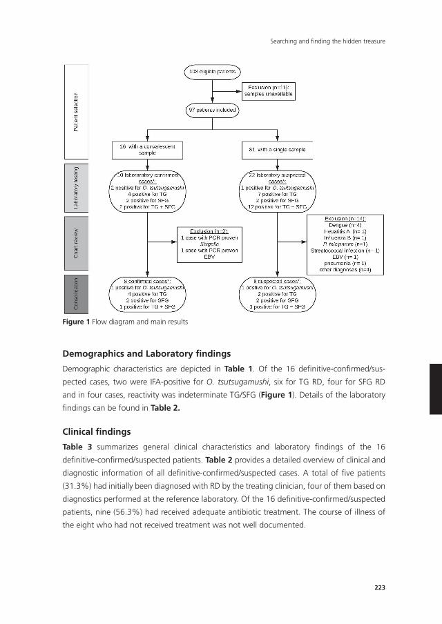
223
Searching and finding the hidden treasure
250
Figure 1 Flow diagram and main results
Figure 1 Flow diagram and main results
Demographics and Laboratory findings
Demographic characteristics are depicted in Table 1. Of the 16 definitive-confirmed/sus-
pected cases, two were IFA-positive for O. tsutsugamushi, six for TG RD, four for SFG RD
and in four cases, reactivity was indeterminate TG/SFG (Figure 1). Details of the laboratory
findings can be found in Table 2.
Clinical findings
Table 3 summarizes general clinical characteristics and laboratory findings of the 16
definitive-confirmed/suspected patients. Table 2 provides a detailed overview of clinical and
diagnostic information of all definitive-confirmed/suspected cases. A total of five patients
(31.3%) had initially been diagnosed with RD by the treating clinician, four of them based on
diagnostics performed at the reference laboratory. Of the 16 definitive-confirmed/suspected
patients, nine (56.3%) had received adequate antibiotic treatment. The course of illness of
the eight who had not received treatment was not well documented.
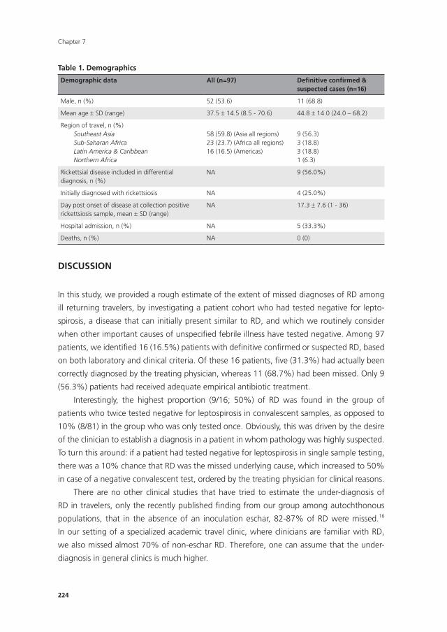
Chapter 7
224
DISCUSSIOn
In this study, we provided a rough estimate of the extent of missed diagnoses of RD among
ill returning travelers, by investigating a patient cohort who had tested negative for lepto-
spirosis, a disease that can initially present similar to RD, and which we routinely consider
when other important causes of unspecified febrile illness have tested negative. Among 97
patients, we identified 16 (16.5%) patients with definitive confirmed or suspected RD, based
on both laboratory and clinical criteria. Of these 16 patients, five (31.3%) had actually been
correctly diagnosed by the treating physician, whereas 11 (68.7%) had been missed. Only 9
(56.3%) patients had received adequate empirical antibiotic treatment.
Interestingly, the highest proportion (9/16; 50%) of RD was found in the group of
patients who twice tested negative for leptospirosis in convalescent samples, as opposed to
10% (8/81) in the group who was only tested once. Obviously, this was driven by the desire
of the clinician to establish a diagnosis in a patient in whom pathology was highly suspected.
To turn this around: if a patient had tested negative for leptospirosis in single sample testing,
there was a 10% chance that RD was the missed underlying cause, which increased to 50%
in case of a negative convalescent test, ordered by the treating physician for clinical reasons.
There are no other clinical studies that have tried to estimate the under-diagnosis of
RD in travelers, only the recently published finding from our group among autochthonous
populations, that in the absence of an inoculation eschar, 82-87% of RD were missed.16
In our setting of a specialized academic travel clinic, where clinicians are familiar with RD,
we also missed almost 70% of non-eschar RD. Therefore, one can assume that the under-
diagnosis in general clinics is much higher.
Table 1. Demographics
Demographic data All (n=97) Definitive confirmed & suspected cases (n=16)
Male, n (%) 52 (53.6) 11 (68.8)
Mean age ± SD (range) 37.5 ± 14.5 (8.5 - 70.6) 44.8 ± 14.0 (24.0 – 68.2)
Region of travel, n (%) Southeast Asia Sub-Saharan Africa Latin America & Caribbean Northern Africa
58 (59.8) (Asia all regions)23 (23.7) (Africa all regions)16 (16.5) (Americas)
9 (56.3)3 (18.8)3 (18.8)1 (6.3)
Rickettsial disease included in differential diagnosis, n (%)
NA 9 (56.0%)
Initially diagnosed with rickettsiosis NA 4 (25.0%)
Day post onset of disease at collection positive rickettsiosis sample, mean ± SD (range)
NA 17.3 ± 7.6 (1 - 36)
Hospital admission, n (%) NA 5 (33.3%)
Deaths, n (%) NA 0 (0)
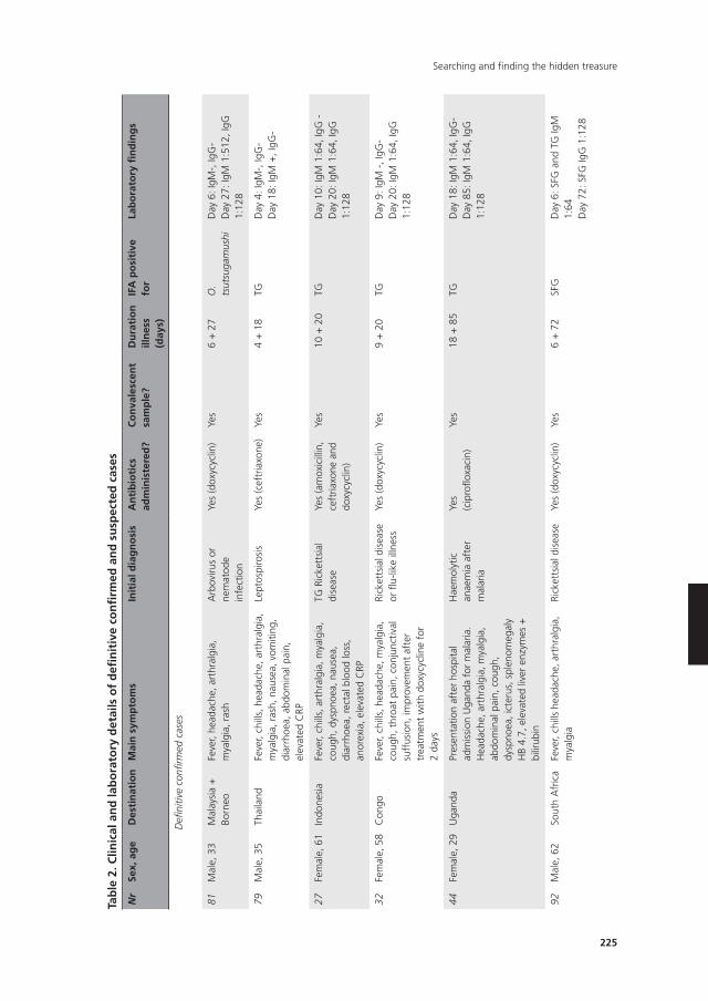
225
Searching and finding the hidden treasure
Tab
le 2
. Clin
ical
an
d la
bo
rato
ry d
etai
ls o
f d
efin
itiv
e co
nfi
rmed
an
d s
usp
ecte
d c
ases
nr
Sex,
ag
eD
esti
nat
ion
Mai
n s
ymp
tom
sIn
itia
l dia
gn
osi
sA
nti
bio
tics
ad
min
iste
red
?C
on
vale
scen
t sa
mp
le?
Du
rati
on
ill
nes
s (d
ays)
IFA
po
siti
ve
for
Lab
ora
tory
fin
din
gs
Defi
nitiv
e co
nfirm
ed c
ases
81M
ale,
33
Mal
aysi
a +
Bo
rneo
Feve
r, he
adac
he, a
rthr
algi
a,
mya
lgia
, ras
hA
rbov
irus
or
nem
atod
e in
fect
ion
Yes
(dox
ycyc
lin)
Yes
6 +
27
O.
tsut
suga
mus
hiD
ay 6
: IgM
-, Ig
G-
Day
27:
IgM
1:5
12, I
gG
1:12
8
79M
ale,
35
Thai
land
Feve
r, ch
ills,
hea
dach
e, a
rthr
algi
a,
mya
lgia
, ras
h, n
ause
a, v
omiti
ng,
diar
rhoe
a, a
bdom
inal
pai
n,
elev
ated
CRP
Lept
ospi
rosi
sYe
s (c
eftr
iaxo
ne)
Yes
4 +
18
TGD
ay 4
: IgM
-, Ig
G-
Day
18:
IgM
+, I
gG-
27Fe
mal
e, 6
1In
done
sia
Feve
r, ch
ills,
art
hral
gia,
mya
lgia
, co
ugh,
dys
pnoe
a, n
ause
a,
diar
rhoe
a, r
ecta
l blo
od lo
ss,
anor
exia
, ele
vate
d C
RP
TG R
icke
ttsi
al
dise
ase
Yes
(am
oxic
illin
, ce
ftria
xone
and
do
xycy
clin
)
Yes
10 +
20
TGD
ay 1
0: Ig
M 1
:64,
IgG
-D
ay 2
0: Ig
M 1
:64,
IgG
1:
128
32Fe
mal
e, 5
8C
ongo
Feve
r, ch
ills,
hea
dach
e, m
yalg
ia,
coug
h, t
hroa
t pa
in, c
onju
nctiv
al
suff
usio
n, im
prov
emen
t af
ter
trea
tmen
t w
ith d
oxyc
yclin
e fo
r 2
days
Rick
etts
ial d
isea
se
or fl
u-lik
e ill
ness
Yes
(dox
ycyc
lin)
Yes
9 +
20
TGD
ay 9
: IgM
-, I
gG-
Day
20:
IgM
1:6
4, Ig
G
1:12
8
44Fe
mal
e, 2
9U
gand
aPr
esen
tatio
n af
ter
hosp
ital
adm
issi
on U
gand
a fo
r m
alar
ia.
Hea
dach
e, a
rthr
algi
a, m
yalg
ia,
abdo
min
al p
ain,
cou
gh,
dysp
noea
, ict
erus
, spl
enom
egal
y H
B 4.
7, e
leva
ted
liver
enz
ymes
+
bilir
ubin
Hae
mol
ytic
an
aem
ia a
fter
m
alar
ia
Yes
(cip
roflo
xaci
n)Ye
s18
+ 8
5TG
Day
18:
IgM
1:6
4, Ig
G-
Day
85:
IgM
1:6
4, Ig
G
1:12
8
92M
ale,
62
Sout
h A
fric
aFe
ver,
chill
s he
adac
he, a
rthr
algi
a,
mya
lgia
Rick
etts
ial d
isea
seYe
s (d
oxyc
yclin
)Ye
s6
+ 7
2SF
GD
ay 6
: SFG
and
TG
IgM
1:
64D
ay 7
2: S
FG Ig
G 1
:128
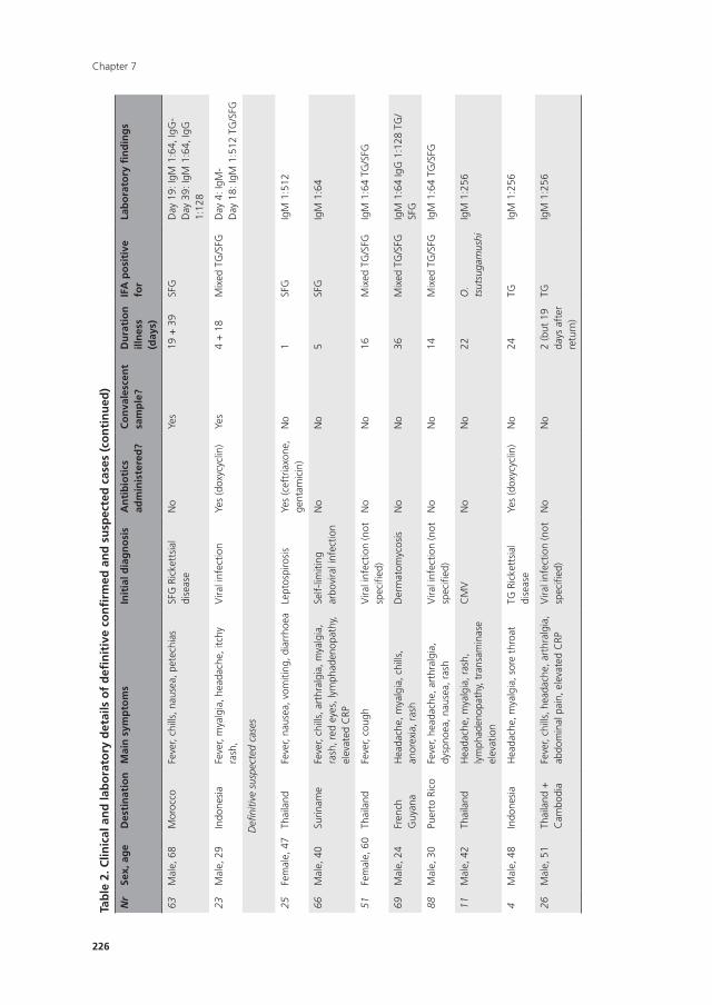
Chapter 7
226
Tab
le 2
. Clin
ical
an
d la
bo
rato
ry d
etai
ls o
f d
efin
itiv
e co
nfi
rmed
an
d s
usp
ecte
d c
ases
(co
nti
nu
ed)
nr
Sex,
ag
eD
esti
nat
ion
Mai
n s
ymp
tom
sIn
itia
l dia
gn
osi
sA
nti
bio
tics
ad
min
iste
red
?C
on
vale
scen
t sa
mp
le?
Du
rati
on
ill
nes
s (d
ays)
IFA
po
siti
ve
for
Lab
ora
tory
fin
din
gs
63M
ale,
68
Mor
occo
Feve
r, ch
ills,
nau
sea,
pet
echi
asSF
G R
icke
ttsi
al
dise
ase
No
Yes
19 +
39
SFG
Day
19:
IgM
1:6
4, Ig
G-
Day
39:
IgM
1:6
4, Ig
G
1:12
8
23M
ale,
29
Indo
nesi
aFe
ver,
mya
lgia
, hea
dach
e, it
chy
rash
,V
iral i
nfec
tion
Yes
(dox
ycyc
lin)
Yes
4 +
18
Mix
ed T
G/S
FGD
ay 4
: IgM
-D
ay 1
8: Ig
M 1
:512
TG
/SFG
Defi
nitiv
e su
spec
ted
case
s
25Fe
mal
e, 4
7Th
aila
ndFe
ver,
naus
ea, v
omiti
ng, d
iarr
hoea
Lept
ospi
rosi
sYe
s (c
eftr
iaxo
ne,
gent
amic
in)
No
1SF
GIg
M 1
:512
66M
ale,
40
Surin
ame
Feve
r, ch
ills,
art
hral
gia,
mya
lgia
, ra
sh, r
ed e
yes,
lym
phad
enop
athy
, el
evat
ed C
RP
Self-
limiti
ng
arbo
vira
l inf
ectio
nN
oN
o5
SFG
IgM
1:6
4
51Fe
mal
e, 6
0Th
aila
ndFe
ver,
coug
hV
iral i
nfec
tion
(not
sp
ecifi
ed)
No
No
16M
ixed
TG
/SFG
IgM
1:6
4 TG
/SFG
69M
ale,
24
Fren
ch
Guy
ana
Hea
dach
e, m
yalg
ia, c
hills
, an
orex
ia, r
ash
Der
mat
omyc
osis
No
No
36M
ixed
TG
/SFG
IgM
1:6
4 Ig
G 1
:128
TG
/SF
G
88M
ale,
30
Puer
to R
ico
Feve
r, he
adac
he, a
rthr
algi
a,
dysp
noea
, nau
sea,
ras
hV
iral i
nfec
tion
(not
sp
ecifi
ed)
No
No
14M
ixed
TG
/SFG
IgM
1:6
4 TG
/SFG
11M
ale,
42
Thai
land
Hea
dach
e, m
yalg
ia, r
ash,
ly
mph
aden
opat
hy, t
rans
amin
ase
elev
atio
n
CM
VN
oN
o22
O.
tsut
suga
mus
hiIg
M 1
:256
4M
ale,
48
Indo
nesi
aH
eada
che,
mya
lgia
, sor
e th
roat
TG R
icke
ttsi
al
dise
ase
Yes
(dox
ycyc
lin)
No
24TG
IgM
1:2
56
26M
ale,
51
Thai
land
+
Cam
bodi
aFe
ver,
chill
s, h
eada
che,
art
hral
gia,
ab
dom
inal
pai
n, e
leva
ted
CRP
Vira
l inf
ectio
n (n
ot
spec
ified
)N
oN
o2
(but
19
days
aft
er
retu
rn)
TGIg
M 1
:256
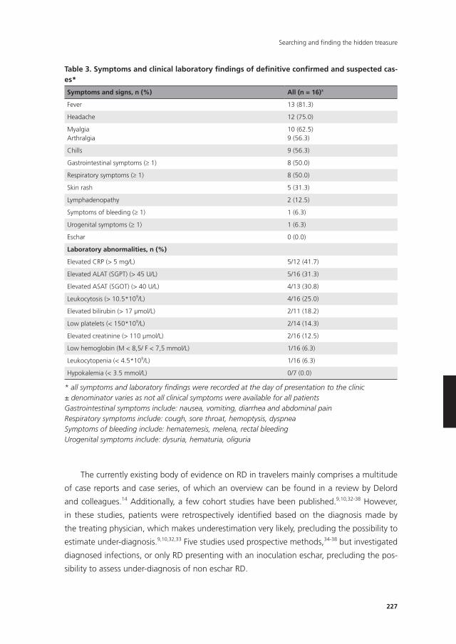
227
Searching and finding the hidden treasure
The currently existing body of evidence on RD in travelers mainly comprises a multitude
of case reports and case series, of which an overview can be found in a review by Delord
and colleagues.14 Additionally, a few cohort studies have been published.9,10,32-38 However,
in these studies, patients were retrospectively identified based on the diagnosis made by
the treating physician, which makes underestimation very likely, precluding the possibility to
estimate under-diagnosis.9,10,32,33 Five studies used prospective methods,34-38 but investigated
diagnosed infections, or only RD presenting with an inoculation eschar, precluding the pos-
sibility to assess under-diagnosis of non eschar RD.
Table 3. Symptoms and clinical laboratory findings of definitive confirmed and suspected cas-es*
Symptoms and signs, n (%) All (n = 16)±
Fever 13 (81.3)
Headache 12 (75.0)
MyalgiaArthralgia
10 (62.5)9 (56.3)
Chills 9 (56.3)
Gastrointestinal symptoms (≥ 1) 8 (50.0)
Respiratory symptoms (≥ 1) 8 (50.0)
Skin rash 5 (31.3)
Lymphadenopathy 2 (12.5)
Symptoms of bleeding (≥ 1) 1 (6.3)
Urogenital symptoms (≥ 1) 1 (6.3)
Eschar 0 (0.0)
Laboratory abnormalities, n (%)
Elevated CRP (> 5 mg/L) 5/12 (41.7)
Elevated ALAT (SGPT) (> 45 U/L) 5/16 (31.3)
Elevated ASAT (SGOT) (> 40 U/L) 4/13 (30.8)
Leukocytosis (> 10.5*109/L) 4/16 (25.0)
Elevated bilirubin (> 17 μmol/L) 2/11 (18.2)
Low platelets (< 150*109/L) 2/14 (14.3)
Elevated creatinine (> 110 μmol/L) 2/16 (12.5)
Low hemoglobin (M < 8,5/ F < 7,5 mmol/L) 1/16 (6.3)
Leukocytopenia (< 4.5*109/L) 1/16 (6.3)
Hypokalemia (< 3.5 mmol/L) 0/7 (0.0)
* all symptoms and laboratory findings were recorded at the day of presentation to the clinic± denominator varies as not all clinical symptoms were available for all patientsGastrointestinal symptoms include: nausea, vomiting, diarrhea and abdominal painRespiratory symptoms include: cough, sore throat, hemoptysis, dyspneaSymptoms of bleeding include: hematemesis, melena, rectal bleedingUrogenital symptoms include: dysuria, hematuria, oliguria

Chapter 7
228
The results presented here should be interpreted with caution, as there are several limita-
tions.
Firstly, all patients had presented to a specialized travel clinic in an academic medical
center, with a lower than average threshold of suspicion for RD.
Secondly, the group of patients in our study is not representative for the overall group
of travelers with fever. Because we were interested in under diagnosis of RD, and studied
a specific subset of patients who had tested negative for leptospirosis, we “missed” the
typical presentations of RD who had presented with an eschar. These patients are readily
diagnosed at our clinic based on the clinical presentation, precluding the need for further
diagnostic testing for leptospirosis or other diseases. The fact that the diagnostic process
for leptospirosis had been initiated, typically implies that more common causes of fever had
already been excluded (e.g., malaria, dengue, chikungunya, Zika virus infection, common
bacterial infections). Thus, we studied a selected group of patients with a higher a-priori
likelihood of less common illnesses, such as non-eschar-RD. For this study however, this was
intentional, because we expected to find missed cases of non-eschar RD in this population.
Obviously, an important criterion to test for leptospirosis is exposure to fresh water, which
means that we missed additional cases of non-eschar RD among patients who were never
tested for leptospirosis because they were not exposed to fresh water. It is possible that this
population was tested for RD more frequently.
Thirdly, important limitations apply to the laboratory methods. The diagnostic process
for RD is changing rapidly.29 Whereas many reference laboratories are still working with IFA
or the micro-immunofluorescence assay (MIF) as reference standards,20 molecular detection
methods are gaining popularity,29 as they can diagnose the illness in its early stage. Be-
cause of restrictions in the type and quality of samples available for this study, we only used
serology-based methods. It is known that there are many limitations to IFA in general: (i) poor
sensitivity in the acute phase of illness (and thus limited diagnostic value of single samples);
(ii) high variation and lack of consensus in cut-off limits; (iii) inter-reader heterogeneity; (iv)
cross-reactivity of IgM with other species and antibody persistence beyond the acute phase
of illness.20,23,29,39 All these limitations apply to this study. For the majority of patients, only
a single sample was available. Therefore, dynamics in antibody titers could not be assessed;
resulting in unconfirmed or even missed diagnoses of RD. Also, due to material constraints,
not all samples underwent further diluting; presented dilutions could have been higher for
some samples. Almost certainly, some positive IgM titers were based on cross-reactivity, or
on previous infections. Although the latter is less likely in the Dutch population, co-infections
with tick-borne Rickettsiae have been described in the Netherlands.40 Remarkably, we ob-
served cross-reactivity between SFG and TG groups in a considerable number of samples. It is
possible that this has been caused by R. felis infections, a rickettsial illness that has been on
the rise globally in the past years.41
Finally, the retrospective nature of this study introduced limitations by itself.
229
Searching and finding the hidden treasure
For example, the clinical information was extracted from patient files, and was often
incomplete. Also, though not expected,42 long-term freezing could have affected the quality
of the serum samples.
The most important message from this study is that even in a specialized travel clinic,
where clinicians are familiar with the diagnosis of RD, this diagnosis is still missed in a sub-
stantial proportion of patients, especially when an inoculation eschar is absent. In retrospect,
in our study, 68.7% of the confirmed/suspected RD cases had been missed and 43.7%
did not receive adequate (empiric) antibiotic therapy. Although no deaths occurred in this
small group of patients, the hospitalization rate was high (33.3%), which emphasizes the
importance of timely recognition and treatment of this disease. In a non-specialized clini-
cal setting, the proportion of missed diagnoses of RD will probably be higher, as we also
estimated earlier 16.
There is a dire need for properly conducted prospective studies among febrile travelers,
in order to reach a credible estimation of the burden of this disease as an imported cause
of febrile illness. A lower threshold to test for RD by clinicians is justified, and RD should be
included in the testing algorithm of febrile illnesses.
Chapter 7
230
REFEREnCES
1. Parola P, Davoust B, Raoult D. Tick- and flea-borne rickettsial emerging zoonoses. Vet Res 2005;
36(3): 469-92.
2. Dieme C, Bechah Y, Socolovschi C, et al. Transmission potential of Rickettsia felis infection by
Anopheles gambiae mosquitoes. Proc Natl Acad Sci U S A 2015; 112(26): 8088-93.
3. Slesak G, Inthalath S, Dittrich S, Paris DH, Newton PN. Leeches as further potential vectors for
rickettsial infections. Proceedings of the National Academy of Sciences of the United States of
America 2015; 112(48): E6593-E4.
4. Botelho-Nevers E, Raoult D. Host, pathogen and treatment-related prognostic factors in rickett-
sioses. Eur J Clin Microbiol Infect Dis 2011; 30(10): 1139-50.
5. La Scola B, Raoult D. Laboratory diagnosis of rickettsioses: current approaches to diagnosis of old
and new rickettsial diseases. J Clin Microbiol 1997; 35(11): 2715-27.
6. Botelho-Nevers E, Socolovschi C, Raoult D, Parola P. Treatment of Rickettsia spp. infections: a
review. Expert Rev Anti Infect Ther 2012; 10(12): 1425-37.
7. Taylor AJ, Paris DH, Newton PN. A Systematic Review of Mortality from Untreated Scrub Typhus
(Orientia tsutsugamushi). PLoS Negl Trop Dis 2015; 9(8): e0003971.
8. Botelho-Nevers E, Rovery C, Richet H, Raoult D. Analysis of risk factors for malignant Medi-
terranean spotted fever indicates that fluoroquinolone treatment has a deleterious effect. J
Antimicrob Chemother 2011; 66(8): 1821-30.
9. Jensenius M, Davis X, von Sonnenburg F, et al. Multicenter GeoSentinel analysis of rickettsial
diseases in international travelers, 1996-2008. Emerg Infect Dis 2009; 15(11): 1791-8.
10. Jensenius M, Han PV, Schlagenhauf P, et al. Acute and potentially life-threatening tropical dis-
eases in western travelers--a GeoSentinel multicenter study, 1996-2011. Am J Trop Med Hyg
2013; 88(2): 397-404.
11. Parola P, Paddock CD, Socolovschi C, et al. Update on tick-borne rickettsioses around the world:
a geographic approach. Clin Microbiol Rev 2013; 26(4): 657-702.
12. Fang LQ, Liu K, Li XL, et al. Emerging tick-borne infections in mainland China: an increasing
public health threat. Lancet Infect Dis 2015; 15(12): 1467-79.
13. Eldin C, Parola P. Update on Tick-Borne Bacterial Diseases in Travelers. Curr Infect Dis Rep 2018;
20(7): 17.
14. Delord M, Socolovschi C, Parola P. Rickettsioses and Q fever in travelers (2004-2013). Travel Med
Infect Dis 2014; 12(5): 443-58.
15. Schlagenhauf P, Weld L, Goorhuis A, et al. Travel-associated infection presenting in Europe
(2008-12): an analysis of EuroTravNet longitudinal, surveillance data, and evaluation of the effect
of the pre-travel consultation. Lancet Infect Dis 2015; 15(1): 55-64.
16. van Eekeren LE, de Vries SG, Wagenaar JFP, Spijker R, Grobusch MP, Goorhuis A. Under-diagnosis
of rickettsial disease in clinical practice: A systematic review. Travel Med Infect Dis 2018.
17. Raeber PA, Winteler S, Paget J. Fever in the returned traveller: remember rickettsial diseases.
Lancet 1994; 344(8918): 331.
18. Merhej V, Raoult D. Rickettsial evolution in the light of comparative genomics. Biol Rev Camb
Philos Soc 2011; 86(2): 379-405.
19. Murray GG, Weinert LA, Rhule EL, Welch JJ. The Phylogeny of Rickettsia Using Different Evolu-
tionary Signatures: How Tree-Like is Bacterial Evolution? Syst Biol 2016; 65(2): 265-79.
20. Abdad MY, Abou Abdallah R, Fournier PE, Stenos J, Vasoo S. A Concise Review of the Epidemiol-
ogy and Diagnostics of Rickettsioses: Rickettsia and Orientia spp. J Clin Microbiol 2018; 56(8).
231
Searching and finding the hidden treasure
21. Dumler JS, Taylor JP, Walker DH. Clinical and laboratory features of murine typhus in south Texas,
1980 through 1987. Jama 1991; 266(10): 1365-70.
22. Fournier PE, Jensenius M, Laferl H, Vene S, Raoult D. Kinetics of antibody responses in Rickettsia
africae and Rickettsia conorii infections. Clin Diagn Lab Immunol 2002; 9(2): 324-8.
23. Blacksell SD, Bryant NJ, Paris DH, Doust JA, Sakoda Y, Day NP. Scrub typhus serologic testing
with the indirect immunofluorescence method as a diagnostic gold standard: a lack of consensus
leads to a lot of confusion. Clin Infect Dis 2007; 44(3): 391-401.
24. Raoult D, Dasch GA. Immunoblot cross-reactions among Rickettsia, Proteus spp. and Legionella
spp. in patients with Mediterranean spotted fever. FEMS Immunol Med Microbiol 1995; 11(1):
13-8.
25. Giulieri S, Jaton K, Cometta A, Trellu LT, Greub G. Development of a duplex real-time PCR for the
detection of Rickettsia spp. and typhus group rickettsia in clinical samples. FEMS Immunol Med
Microbiol 2012; 64(1): 92-7.
26. Papp S, Rauch J, Kuehl S, Richardt U, Keller C, Osterloh A. Comparative evaluation of two
Rickettsia typhi-specific quantitative real-time PCRs for research and diagnostic purposes. Med
Microbiol Immunol 2017; 206(1): 41-51.
27. Paris DH, Blacksell SD, Stenos J, et al. Real-time multiplex PCR assay for detection and differentia-
tion of rickettsiae and orientiae. Trans R Soc Trop Med Hyg 2008; 102(2): 186-93.
28. Watthanaworawit W, Turner P, Turner C, et al. A prospective evaluation of real-time PCR assays
for the detection of Orientia tsutsugamushi and Rickettsia spp. for early diagnosis of rickettsial
infections during the acute phase of undifferentiated febrile illness. Am J Trop Med Hyg 2013;
89(2): 308-10.
29. Paris DH, Dumler JS. State of the art of diagnosis of rickettsial diseases: the use of blood speci-
mens for diagnosis of scrub typhus, spotted fever group rickettsiosis, and murine typhus. Curr
Opin Infect Dis 2016; 29(5): 433-9.
30. Jensenius M, Fournier PE, Raoult D. Rickettsioses and the international traveler. Clin Infect Dis
2004; 39(10): 1493-9.
31. Rijksinstituut voor Volksgezondheid en Milieu - Rickettsia spp. https://www.rivm.nl/wilde-
knaagdieren-en-zo-nosen/ziekteverwekkers/rickettsia-spp-rickettsiose (accessed 6 May 2019).
32. Herbinger KH, Hanus I, Felbinger TW, et al. Elevated Values of Clinically Relevant Transferases
Induced by Imported Infectious Diseases: A Controlled Cross-Sectional Study of 14,559 Diseased
German Travelers Returning from the Tropics and Subtropics. Am J Trop Med Hyg 2016; 95(2):
481-7.
33. Herbinger KH, Hanus I, Schunk M, et al. Elevated Values of C-Reactive Protein Induced by
Imported Infectious Diseases: A Controlled Cross-Sectional Study of 11,079 Diseased German
Travelers Returning from the Tropics and Subtropics. Am J Trop Med Hyg 2016; 95(4): 938-44.
34. Parola P, Soula G, Gazin P, Foucault C, Delmont J, Brouqui P. Fever in travelers returning from
tropical areas: prospective observational study of 613 cases hospitalised in Marseilles, France,
1999-2003. Travel Med Infect Dis 2006; 4(2): 61-70.
35. Jensenius M, Hoel T, Raoult D, et al. Seroepidemiology of Rickettsia africae infection in Norwe-
gian travellers to rural Africa. Scand J Infect Dis 2002; 34(2): 93-6.
36. Jensenius M, Fournier PE, Vene S, et al. African tick bite fever in travelers to rural sub-Equatorial
Africa. Clin Infect Dis 2003; 36(11): 1411-7.
37. Morand A, Angelakis E, Ben Chaabane M, Parola P, Raoult D, Gautret P. Seek and Find! PCR
analyses of skin infections in West-European travelers returning from abroad with an eschar.
Travel Med Infect Dis 2018; 26: 32-6.
Chapter 7
232
38. Beltrame A, Angheben A, Casolari S, et al. Imported rickettsioses in Italy. Travel Med Infect Dis
2012; 10(4): 201-4.
39. Phetsouvanh R, Thojaikong T, Phoumin P, et al. Inter- and intra-operator variability in the reading
of indirect immunofluorescence assays for the serological diagnosis of scrub typhus and murine
typhus. Am J Trop Med Hyg 2013; 88(5): 932-6.
40. Koetsveld J, Tijsse-Klasen E, Herremans T, Hovius JW, Sprong H. Serological and molecular evi-
dence for spotted fever group Rickettsia and Borrelia burgdorferi sensu lato co-infections in The
Netherlands. Ticks Tick Borne Dis 2016; 7(2): 371-7.
41. Angelakis E, Mediannikov O, Parola P, Raoult D. Rickettsia felis: The Complex Journey of an
Emergent Human Pathogen. Trends Parasitol 2016; 32(7): 554-64.
42. Dard C, Bailly S, Drouet T, Fricker-Hidalgo H, Brenier-Pinchart MP, Pelloux H. Long-term sera
storage does not significantly modify the interpretation of toxoplasmosis serologies. J Microbiol
Methods 2017; 134: 38-45.


PART 3Epilogue


Chapter 8Summary and general discussion

239
Summary and general discussion
Over the past years, the extent and burden of leptospirosis and rickettsial disease has in-
creasingly been elucidated,1-4 in both autochthonous as well as travelling populations. This
has been due to an increased research interest, improved diagnostic techniques, and also
new epidemiological methods to calculate disease burden. Also, climate change, with its
accompanying increasing temperatures and wetter weather, contributes to an increase in
caseload. That notwithstanding, the exact epidemiology and extent of both illnesses seems
to be vastly underestimated worldwide.
SUMMARy
In Part 1, we focused on leptospirosis. Chapter 2 focused on summarising all studies on lep-
tospirosis known for the sub-Saharan African region, as the epidemiological situation on the
continent was unclear. We found that most data was known on the occurrence of the illness
in animals on the continent; probably due to the larger economic impact of the illness when
livestock gets infected. Many different animals can carry Leptospira in their renal tubules,
and shed them with urine. They can be asymptomatic (‘maintenance hosts’), or develop
symptoms. In cattle and pigs, infection can cause considerable reproductive problems, such
as abortions and reproductive failure.5 In animals, the disease seemed to be widespread on
the continent; suggesting plenty of potential infection risk for humans. Data in humans is
scarce, however, especially for the Central African region. Seroprevalence studies in healthy
subjects showed exposure to the pathogen in many populations throughout the continent;
in some regions, it was also found to be a frequent cause of febrile illness. Having said that,
there were only a few well-performed studies in febrile patients, and the amount of studies
that was done was very small for such a large continent. Therefore, the data presented in this
chapter probably only represent the tip of the iceberg (or the ears of the hippopotamus, in
more tropical terms), and the true burden of the illness on the continent is likely much higher.
As the leptospirosis adage goes:
“Wherever leptospires and leptospirosis is searched for, they are invariably discovered.”6
In Chapter 3 and 4, we studied cases of ‘imported’, or ‘travel-related’ leptospirosis. In our
study of leptospirosis in the Netherlands (Chapter 3) from 2009 to 2016, we observed an
increase of cases from 2014 onwards, both autochthonous and imported. The increase
in autochthonous cases was thought to be mainly due to warm weather,7 increasing the
chances of survival of leptospires. The reason for the increase of imported cases, however,
was multifactorial. An increased number of samples had been submitted to the National Col-
laborating Centre for Reference and Research on Leptospirosis (NRL); this could have been
an indication of travellers engaging in more high-risk activities (e.g. jungle trekking, rafting),
Chapter 8
240
or that clinicians considered the diagnosis more often. The positivity rate also increased
from 2014 onwards; this could have been influenced by the introduction of polymerase
chain reaction (PCR) as a standard test from September 2012 onwards at the NRL. PCR can
detect leptospirosis in the early stages of the illness, unlike the widely-used serology-based
methods, that require convalescent samples, or samples taken in a later stage of the illness.
Of the 224 imported cases, the majority had travelled to Southeast Asia, the traditional
‘leptospirosis-hotspot’; only 7 (3.1%) had travelled to sub-Saharan Africa. Most patients
were male and relatively young; all characteristics that are also described in other studies
on the subject. We studied extensive clinical characteristics of 41 patients with imported
leptospirosis that had presented to the AMC. More than half of them (53.7%) had to be
hospital-admitted, indicating the severity of the illness; and it took on average four days from
symptom onset until they received antibiotics. We highlighted three cases that demonstrated
the broad spectrum of clinical presentations, and the importance of a swift diagnosis.
In Chapter 4, we analysed confirmed leptospirosis cases that were reported to the
GeoSentinel Surveillance Network, a network monitoring travel-related morbidity through
travel and tropical medicine clinics. We analysed 180 confirmed cases of travel-related lep-
tospirosis that were reported between 1999 and 2016. They showed similar characteristics
as the Dutch cases; the majority was young, male, and had travelled to Southeast Asia.
We also conducted a survey about leptospirosis diagnostic and therapeutic practices among
GeoSentinel clinicians, most of which working in academic institutions. Remarkably, clini-
cians did often not consider the illness in the absence of the classic exposure history or clinical
symptoms, even though it is known that these might often be absent. Also, the applied
diagnostic methods were sub-optimal. These findings imply that even in highly specialised
travel clinics, there could be a considerable under-estimation of cases.
The diagnostic methods for leptospirosis are complicated. For long, diagnosis depended
on the Microscopic Agglutination Test (MAT) and culture, two laborious and highly special-
ized techniques, not suited to establish a diagnosis swiftly. Over the past decades, molecular
detection tests, such as PCR, have gained terrain in the diagnosis of many infectious diseases.
PCR can demonstrate the illness before the antibodies are formed, thus in the early stages.
In Chapter 5, we performed a systematic review and meta-analysis on the diagnostic ac-
curacy of nucleic acid and antigen detection tests for the diagnosis of acute leptospirosis.
We identified 41 studies, and were able to do a meta-analysis for PCR and real-time PCR.
There was, however, a very high heterogeneity between the studies; reports were often of
poor methodological quality and study settings and disease prevalence varied widely. The
interpretation of the review and analyses are therefore not straightforward. Also, we used
the MAT as a reference standard, which has an imperfect sensitivity, and therefore likely
underestimates the specificity of the index tests. We tried to address this problem by using
“composite reference standards”, MAT combined with IgM ELISA and/or culture. MAT alone
as a reference standard was considered at high risk of bias. For PCR, we included 15 studies.
241
Summary and general discussion
The pooled sensitivity was 70% (95% CI 37% to 90%) and the pooled specificity was 95%
(95% CI 75% to 99%). This improved to a pooled sensitivity of 87% (95% CI 44% to 98%),
and a pooled specificity of 97% (95% CI 60% to 100%) when studies using only MAT as
a reference test were removed. For real-time PCR, an SROC curve was calculated, as the
positivity threshold varied between studies. The median specificity was 92%. When 95%
specificity was selected on the curve, the pooled sensitivity was 29% (95% CI 15% to 49%).
Because of the high variability between studies, it is hard to draw firm conclusions about the
accuracy of these tests, and their position in the testing pathway of leptospirosis.
In Part 2, we focused on the underestimation of rickettsial disease. In Chapter 6 we
made an estimation of the under-estimation of rickettsial disease in native populations. We
performed a systematic review, including studies reporting on Mediterranean Spotted Fever,
scrub typhus and African Tick Bite Fever, as these are rickettsial disease that an present with
an eschar, but certainly also without. The reported rates of presentation with an eschar varies
between studies.8 We hypothesised that this variation in reported eschar rates is caused by
the design of the studies. Many studies seem to include patients based on ‘clinical suspicion’,
which will automatically lead to higher reported rates of eschar prevalence, as clinicians
often associate these illnesses with eschars. We expected to find higher rates of non-eschar
rickettsial disease (NERD) in studies including all patients with febrile illness. In this way, we
could make an estimation of the under-reporting of NERD. We included 121 studies, of
which the majority (98) reported on scrub typhus, 21 reported on Mediterranean Spotted
Fever and two on African Tick Bite Fever. We were able to confirm our hypothesis: in scrub
typhus, NERD was missed in 66% of patients. In Mediterranean Spotted Fever, this applied
to 58% of patients. For African Tick Bite Fever, an estimate could not be provided, due to the
limited number of studies found. All studies were performed in autochthonous populations,
with consequently local clinicians that are aware of the local epidemiology. We therefore
think this is an important finding; and earlier suspicion of the disease, and with that earlier
initiation of treatment, could reduce morbidity and even mortality.
In Chapter 7, we aimed to test the hypothesis outlined above in returned travellers with
febrile illness. We retrospectively performed serological tests for Spotted Fever Group rickett-
sioses, typhus group rickettsioses and scrub typhus in returned travellers who had presented
to the AMC with an unspecified febrile illness, and were suspected of leptospirosis, but had
tested negative. We hypothesised that rickettsial diseases were missed frequently in travellers
returning with fever, but without an eschar. We included 97 patients who had returned from
Asia, Africa or the Americas, of which16 were confirmed and suspected cases, 11 (69%)
had been missed, and 7 (44%) had not received empiric antibiotics. This study certainly had
limitations, however, the important message is, that even in a highly specialised academic
travel clinic the diagnosis of non-eschar rickettsial disease is missed frequently, and it should
be included in standard testing algorithms.
Chapter 8
242
Future perspectives
Leptospirosis and rickettsioses are under-estimated causes of febrile illness with substantial
morbidity and mortality worldwide. This thesis gives an overview of the current epidemiologi-
cal concerns of both diseases.
For leptospirosis, we conclude that it is an entity prevalent in sub-Saharan Africa (Chap-
ter 2), although the true scale of the problem is largely unknown due to a lack of regional
epidemiological studies. For long, all febrile illness was attributed to malaria, but in the
past decade, the over-diagnosis problem has emerged.9,10 It is of vital importance that more
surveys on the causes of febrile illness will be performed, as has increasingly been done the
past years.11-14 Only a better understanding of causes of illness can improve local clinical
practice and subsequent patients outcomes, and enhance local governments to take ad-
equate preventive measures. For rickettsial diseases, despite being a more widely recognised
disease entity worldwide, the problem of lack of awareness at a local level is also a problem,
as we demonstrated in Chapter 6. As febrile illnesses remain a highly prevalent challenge
for clinicians worldwide, it is important that in the research on the causes of it, an open view
is always retained. Traditional ideas of presentations of diseases may turn out wrong; with
“non-eschar rickettsial disease” being a telling example. The downside is, that cause-of-fever
research is expensive and resource-consuming, and funding not always available in a world
directed by pharmaceutical giants. Smart algorithms for prospective studies to bypass this
problem should be designed.
Not only clinicians in tropical parts of the world struggle with the cause-of-fever co-
nundrum, as we demonstrated in Chapters 3, 4 and 7. Travel medicine is a specialised part
of medicine, in which physicians deal with non-local, imported pathogens on a daily basis.
In the Netherlands, the first step for a patient is usually the general practitioner, who often
has very little knowledge on foreign illnesses and epidemiology. This can cause considerable
delay in treatment, and worse outcomes for the individual patient (Chapter 3). However,
even highly-specialised travel-medicine physicians can struggle to recognise the cause of
febrile illness in their patients (Chapter 4 and Chapter 7), especially when the “big causes”,
such as malaria and dengue fever, have been excluded. Broad prospective studies on causes
of fever in travellers should be conducted.
Part of the problem of the under-estimation of leptospirosis and rickettsioses, are the
complicated, laborious and poorly available diagnostic tests. Molecular detection test, such
as (real-time) PCR, hold a promise in the swift and timely diagnosis of both illnesses, as the
traditional antibody detection tests fail diagnosis in the early stages of illness. In Chapter
5, we demonstrated that the basis of these tests for leptospirosis is not yet very solid, as
too little well-performed research exists. Robust methodological studies on the diagnostic
accuracy of these tests should be performed.
In conclusion, this thesis showed that leptospirosis and rickettsioses are underestimated
causes of febrile illness worldwide, of which the true extend and epidemiological patterns

243
Summary and general discussion
still have to be elucidated. To reach this goal, improved knowledge on the value of the
available diagnostic tests is key.
Chapter 8
244
REFEREnCES
1. Costa F, Hagan JE, Calcagno J, et al. Global Morbidity and Mortality of Leptospirosis: A Systematic
Review. PLOS Neglected Tropical Diseases 2015; 9(9): e0003898.
2. Torgerson PR, Hagan JE, Costa F, et al. Global Burden of Leptospirosis: Estimated in Terms of
Disability Adjusted Life Years. PLOS Neglected Tropical Diseases 2015; 9(10): e0004122.
3. Bonell A, Lubell Y, Newton PN, Crump JA, Paris DH. Estimating the burden of scrub typhus: A
systematic review. PLoS Negl Trop Dis 2017; 11(9): e0005838.
4. Taylor AJ, Paris DH, Newton PN. A Systematic Review of Mortality from Untreated Scrub Typhus
(Orientia tsutsugamushi). PLoS Negl Trop Dis 2015; 9(8): e0003971.
5. Levett PN. Leptospirosis. Clin Microbiol Rev 2001; 14(2): 296-326.
6. Torten M. MR, Beran G., Steel J. Handbook of zoonosis. Section A: bacterial, rickettsial, chla-
mydial, and mycotic: CRC Press; 1994.
7. Pijnacker R, Goris MG, Te Wierik MJ, et al. Marked increase in leptospirosis infections in humans
and dogs in the Netherlands, 2014. Euro Surveill 2016; 21(17).
8. Delord M, Socolovschi C, Parola P. Rickettsioses and Q fever in travelers (2004-2013). Travel Med
Infect Dis 2014; 12(5): 443-58.
9. Reyburn H, Mbatia R, Drakeley C, et al. Overdiagnosis of malaria in patients with severe febrile
illness in Tanzania: a prospective study. Bmj 2004; 329(7476): 1212.
10. Carugati M, Kilonzo KG, Crump JA. Fever, bacterial zoonoses, and One Health in sub-Saharan
Africa. Clin Med (Lond) 2019; 19(5): 375-80.
11. Biggs HM, Bui DM, Galloway RL, et al. Leptospirosis among hospitalized febrile patients in
northern Tanzania. Am J Trop Med Hyg 2011; 85(2): 275-81.
12. Fernandes JF, Held J, Dorn M, et al. Causes of fever in Gabonese children: a cross-sectional
hospital-based study. Sci Rep 2020; 10(1): 2080.
13. Abe H, Ushijima Y, Loembe MM, et al. Re-emergence of dengue virus serotype 3 infections in
Gabon in 2016-2017, and evidence for the risk of repeated dengue virus infections. Int J Infect
Dis 2020; 91: 129-36.
14. Reddy EA, Shaw AV, Crump JA. Community-acquired bloodstream infections in Africa: a system-
atic review and meta-analysis. Lancet Infect Dis 2010; 10(6): 417-32.


Chapter 9Nederlandse samenvatting

249
Nederlandse samenvatting
In de afgelopen jaren is steeds meer duidelijkheid ontstaan over de omvang en de ziektelast
van leptospirose en rickettsiosen,1-4 bij zowel autochtone als reizende bevolkingsgroepen.
Dit dankzij toegenomen belangstelling voor onderzoek, verbeterde diagnostische technieken
en nieuwe epidemiologische methoden om de ziektelast te berekenen. Ook draagt klimaat-
verandering, met de daarbij behorende stijgende temperaturen en natter weer, bij aan een
toename van de ziektebelasting. Desalniettemin lijkt de exacte epidemiologie en omvang van
beide ziekten wereldwijd enorm onderschat.
SAMEnVATTInG
In Deel 1 hebben we ons gericht op leptospirose. In Hoofdstuk 2 zijn alle onderzoeken naar
leptospirose die bekend zijn voor Afrika ten zuiden van de Sahara samengevat, aangezien er
veel onduidelijkheid van over de epidemiologische situatie op het continent bestaat. We ont-
dekten dat de meeste gegevens bekend waren over het voorkomen van de ziekte bij dieren
op het continent; waarschijnlijk vanwege de grote economische impact wanneer vee wordt
besmet. Veel verschillende dieren kunnen Leptospira in hun niertubuli dragen en ze met
urine uitscheiden. Ze kunnen asymptomatisch zijn (“onderhoudsgastheren”) of symptomen
ontwikkelen. Bij runderen en varkens kan infectie aanzienlijke reproductieve problemen
veroorzaken, zoals abortus en infertiliteit.5 Bij dieren bleek de ziekte wijdverbreid op het con-
tinent; dit suggereert veel potentieel infectierisico voor mensen. Gegevens bij mensen waren
echter schaars, vooral voor de Centraal-Afrikaanse regio. Seroprevalentie onderzoeken bij
gezonde proefpersonen toonden blootstelling aan de ziekteverwekker bij veel populaties
over het hele continent; in sommige regio’s bleek het ook een veelvoorkomende oorzaak
van koortsende ziekte te zijn. Er waren echter maar een paar goed uitgevoerde onderzoeken
bij patiënten met koorts, en het aantal uitgevoerde onderzoeken was erg klein voor zo’n
groot continent. Daarom vertegenwoordigen de gegevens in dit hoofdstuk waarschijnlijk
slechts het topje van de ijsberg (of de oren van het nijlpaard, in meer tropische termen), en
is de werkelijke ziektelast op het continent waarschijnlijk veel hoger. Zoals het leptospirose
gezegde luidt:
“Waar naar leptospieren en leptospirose wordt gezocht, worden ze ontdekt.’6
In Hoofdstuk 3 en 4 hebben we gevallen van “geïmporteerde” of “reis-gerelateerde”
leptospirose bestudeerd. In ons onderzoek naar leptospirose in Nederland van 2009 tot
2016 (Hoofdstuk 3) constateerden we een toename van het aantal gevallen vanaf 2014,
zowel autochtoon als geïmporteerd. Aangenomen wordt dat de toename van autochtone
gevallen voornamelijk te wijten was aan warm weer,7 waardoor de overlevingskansen van
leptospieren toenamen. De reden voor de toename van geïmporteerde gevallen was echter
Chapter 9
250
multifactorieel. Een groter aantal monsters was ingediend bij het Nationaal Referentielabo-
ratorium voor Leptospirosen (NRL); dit zou een aanwijzing kunnen zijn dat reizigers meer
risicovolle activiteiten ondernamen (bv. trektochten door de jungle en raften), of dat clinici
de diagnose vaker overwogen. Vanaf 2014 is ook het percentage positieve monsters ge-
stegen; dit kan zijn beïnvloed door de introductie van polymerasekettingreactie (PCR) als
standaardtest vanaf september 2012 bij het NRL. PCR kan leptospirose in de vroege stadia
van de ziekte detecteren, in tegenstelling tot de veelgebruikte serologische methoden,
waarvoor monsters nodig zijn die in een later stadium van de ziekte worden afgenomen.
Van de 224 geïmporteerde gevallen was de meerderheid naar Zuidoost-Azië gereisd, de
traditionele “leptospirose-hotspot”; slechts 7 (3,1%) waren in Sub-Sahara Afrika geweest.
De meeste patiënten waren mannelijk en relatief jong; allemaal kenmerken die ook worden
beschreven in andere studies over het onderwerp. We bestudeerden de klinische kenmerken
van 41 patiënten met geïmporteerde leptospirose die in het AMC waren behandeld. Meer
dan de helft van hen (53,7%) moest in het ziekenhuis worden opgenomen, wat de ernst van
de ziekte aangeeft; en het duurde gemiddeld vier dagen vanaf het begin van de symptomen
totdat ze antibiotica kregen. We belichtten drie gevallen die het brede klinische spectrum van
de ziekte en het belang van een snelle diagnose aantoonden.
In Hoofdstuk 4 analyseerden we bevestigde gevallen van leptospirose die werden
gemeld aan het GeoSentinel Surveillance Network, een netwerk dat reis-gerelateerde
morbiditeit bewaakt via reis- en tropische geneeskunde-klinieken. We analyseerden 180
bevestigde gevallen van reis-gerelateerde leptospirose die werden gemeld tussen 1999 en
2016. Ze vertoonden vergelijkbare kenmerken met de Nederlandse gevallen; de meerderheid
van de patiënten was jong, man, en naar Zuidoost-Azië gereisd. Ook hielden we een enquête
onder de clinici van GeoSentinel, waarvan de meeste in academische instellingen werken,
over de diagnostische en therapeutische praktijken van leptospirose. Opmerkelijk genoeg
overwogen de clinici de ziekte vaak niet zonder de klassieke blootstellingsgeschiedenis of
klinische symptomen, hoewel bekend is dat deze vaak afwezig zijn. Ook waren de toege-
paste diagnostische methoden niet optimaal. Deze bevindingen impliceren dat zelfs in zeer
gespecialiseerde reisklinieken, de ziekte aanzienlijk zou kunnen worden onderschat.
De diagnostische methoden voor leptospirose zijn ingewikkeld. De diagnostiek was lange
tijd gebaseerd op de microscopische agglutinatietest (MAT) en kweek, twee bewerkelijke en
zeer gespecialiseerde technieken, die niet geschikt zijn om snel een diagnose te stellen. In
de afgelopen decennia hebben moleculaire detectietests, zoals PCR, terrein gewonnen bij de
diagnostiek van veel infectieziekten. PCR kan de ziekte aantonen voordat de antilichamen
worden gevormd, dus in een vroege stadium van de ziekte. In Hoofdstuk 5 hebben we een
systematische review en meta-analyse uitgevoerd over de diagnostische nauwkeurigheid van
nucleïnezuur- en antigeendetectietests voor de diagnose van leptospirose. We identificeerden
41 onderzoeken en konden een meta-analyse uitvoeren voor PCR en real-time PCR. Er was
echter een zeer hoge heterogeniteit tussen de onderzoeken; de onderzoeksrapporten waren
251
Nederlandse samenvatting
vaak van slechte methodologische kwaliteit en de studieomgevingen en de prevalentie van
ziekte liepen sterk uiteen. De interpretatie van de beoordeling en analyses is daarom niet een-
voudig. Daarnaast hebben we de MAT gebruikt als referentiestandaard, die een onvolmaakte
sensitiviteit heeft en daarom waarschijnlijk de specificiteit van de indextests onderschat. We
hebben geprobeerd dit probleem aan te pakken door gebruik te maken van “samengestelde
referentiestandaarden”, MAT gecombineerd met IgM ELISA en / of kweek. Als alleen MAT als
referentiestandaard werd gebruikt, beschouwden we dit als een hoog risico op vertekening
van resultaten (“bias”). Voor PCR hebben we 15 onderzoeken geïncludeerd in onze studie.
De gepoolde sensitiviteit was 70% (95% betrouwbaarheidsinterval (BI) 37% tot 90%) en
de gepoolde specificiteit was 95% (95% BI 75% tot 99%). Dit verbeterde tot een gepoolde
sensitiviteit van 87% (95% BI 44% tot 98%) en een gepoolde specificiteit van 97% (95% BI
60% tot 100%) wanneer studies die alleen MAT als referentietest gebruikten werden verwij-
derd. Voor real-time PCR werd een SROC-curve berekend, aangezien de positiviteits-drempel
tussen studies varieerde. De mediane specificiteit was 92%. Wanneer 95% specificiteit op
de curve werd geselecteerd, was de gepoolde sensitiviteit 29% (95% BI 15% tot 49%).
Vanwege de grote variabiliteit tussen studies, is het moeilijk om harde conclusies te trekken
over de nauwkeurigheid van deze tests en hun positie in het testtraject van leptospirose.
In Deel 2 hebben we ons gericht op de onderschatting van rickettsiosen. In Hoofdstuk
6 hebben we de onderschatting van rickettsiosen bij autochtone populaties berekend, door
een systematische review te doen over studies die rapporteren over “Mediterranean Spotted
Fever”, scrubtyfus en “African Tick Bite Fever” (Afrikaanse tekenbeetkoorts), omdat deze
rickettsiosen zich zowel met, als zonder eschar presenteren. De gerapporteerde percentages
van presentatie met een eschar variëert tussen studies.8 We veronderstelden dat deze variatie
in gerapporteerde eschar-percentages wordt veroorzaakt door de opzet van de studies.
Veel studies includeren patiënten op basis van ‘klinische verdenking’, wat automatisch zal
leiden tot hogere gerapporteerde percentages van eschar-prevalentie, aangezien clinici deze
ziekten vaak associëren met eschars. We verwachtten hogere percentages van niet-eschar-
rickettsiosen (NERD) te vinden in onderzoeken die alle patiënten met koorts includeerden.
Op deze manier konden we een inschatting maken van de onderrapportage van NERD. We
hebben 121 onderzoeken geïncludeerd, waarvan de meerderheid (98) rapporteerde over
scrubtyfus, 21 over Mediterranean Spotted Fever en twee over African Tick Bite Fever. Onze
hypothese werd bevestigd: bij scrubtyfus werd NERD bij 66% van de patiënten gemist. Bij
Mediterranean Spotted Fever gold dit voor 58% van de patiënten. Voor African Tick Bite
Fever kon vanwege het beperkte aantal gevonden onderzoeken geen schatting worden
gemaakt. Alle onderzoeken zijn uitgevoerd in autochtone populaties, met lokale clinici die
op de hoogte zijn van de lokale epidemiologie. We denken daarom dat dit een belangrijke
bevinding is; eerder vermoeden van de ziekte, en daarmee een eerdere start van de behan-
deling, zou de morbiditeit en zelfs de mortaliteit kunnen verminderen.
Chapter 9
252
In Hoofdstuk 7 hebben we de hierboven geschetste hypothese getest bij terugkerende
reizigers met koorts. We voerden retrospectief serologische tests uit voor Spotted Fever
Group-rickettsiosen, tyfusgroep-rickettsiosen en scrubtyfus bij teruggekeerde reizigers die
zich bij het AMC hadden gepresenteerd met aspecifieke koorts en verdacht werden van
leptospirose, maar hierop negatief waren getest. We veronderstelden dat rickettsiosen vaak
werden gemist bij reizigers die terugkeerden met koorts, maar zonder eschar. We namen
97 patiënten op die waren teruggekeerd uit Azië, Afrika of Amerika. We ontdekten 16
bevestigde en vermoedelijke gevallen, waarvan 11 (69%) waren gemist en 7 (44%) geen
empirische antibiotica hadden gekregen. Deze studie had beperkingen, maar de belangrijke
boodschap is dat zelfs in een zeer gespecialiseerde academische reiskliniek de diagnose van
niet-eschar rickettsiosen (NERD) vaak wordt gemist, en een plaats zou moeten krijgen in de
standaard testalgoritmen.
Toekomstperspectieven en conclusie
Leptospirose en rickettsiosen zijn onderschatte oorzaken van met koorts gepaard gaande
ziekten, met aanzienlijke morbiditeit en mortaliteit wereldwijd. Dit proefschrift geeft een
overzicht van de huidige epidemiologische stand van zaken rondom beide ziekten.
Voor leptospirose concluderen we dat het een entiteit is die voorkomt in Afrika ten
zuiden van de Sahara (Hoofdstuk 2), hoewel de ware omvang van het probleem grotend-
eels onbekend blijft vanwege een gebrek aan goede regionale epidemiologische studies.
Lange tijd werd alle koortsende ziekte toegeschreven aan malaria, maar in het afgelopen
decennium is het probleem van de over-diagnostiek naar voren gekomen.9,10 Het is van
cruciaal belang dat er meer onderzoeken worden uitgevoerd naar de oorzaken van met
koorts gepaard gaande ziekten, zoals de afgelopen jaren steeds vaker is gedaan.11-14 Alleen
een beter begrip van de oorzaken van koorts kan de lokale klinische praktijk en de klinische
uitkomst van patiënten verbeteren, en de lokale overheden ertoe aanzetten om adequate
preventieve maatregelen te nemen. Voor rickettsiosen, een over het algemeen wereldwijd
beter erkende ziekte-entiteit, is het gebrek aan bewustzijn op lokaal niveau ook een probl-
eem, zoals we in Hoofdstuk 6 hebben aangetoond. Aangezien koortsende ziekten een veel
voorkomende uitdaging blijven voor clinici over de hele wereld, is het belangrijk dat in het
onderzoek naar de oorzaken ervan, men dit met open vizier blijft doen. Traditionele ideeën
over ziektepresentaties kunnen verkeerd uitpakken; met “niet-eschar rickettsiose” (NERD)
als sprekend voorbeeld. Het nadeel is dat onderzoek naar de oorzaken van koorts kostbaar
en intensief is, en de financiering niet altijd beschikbaar is in een wereld geleid door de
farmaceutische industrie. Er zijn slimme algoritmen nodig voor prospectieve studies om dit
probleem te omzeilen.
Niet alleen clinici in tropische delen van de wereld worstelen met de oorzaak van koorts,
zoals we lieten zien in de Hoofdstukken 3, 4 en 7. Reisgeneeskunde is een gespecialiseerd
onderdeel van de geneeskunde, waarbij artsen dagelijks te maken hebben met niet-lokale,
253
Nederlandse samenvatting
geïmporteerde pathogenen. In Nederland is de eerste stap voor een patiënt is meestal de
huisarts, die vaak weinig kennis over buitenlandse ziekten en epidemiologie heeft. Dit kan
een aanzienlijke vertraging in de behandeling veroorzaken en slechtere uitkomsten voor
de individuele patiënt (Hoofdstuk 3). Maar zelfs zeer gespecialiseerde artsen in de reisge-
neeskunde kunnen moeite hebben om de oorzaak van met koorts gepaard gaande ziekten
bij hun patiënten te herkennen (Hoofdstuk 4 en Hoofdstuk 7), vooral wanneer de “grote
oorzaken”, zoals malaria en knokkelkoorts, zijn uitgesloten. Er moeten brede prospectieve
onderzoeken naar de oorzaken van koorts bij reizigers worden uitgevoerd.
Een deel van het probleem van de onderschatting van leptospirose en rickettsiosen
zijn de gecompliceerde, bewerkelijke en slecht beschikbare diagnostische tests. Moleculaire
detectietests, zoals (real-time) PCR, kunnen beide ziekten snel en tijdig diagnosticeren, waar
de traditionele antilichaamdetectietests de diagnose in de vroege stadia van de ziekte niet
kunnen stellen. In Hoofdstuk 5 hebben we aangetoond dat de basis van deze tests voor
leptospirose nog niet erg solide is, omdat er te weinig goed uitgevoerd onderzoek bestaat.
Er moeten robuuste methodologische onderzoeken naar de diagnostische nauwkeurigheid
van deze tests worden uitgevoerd.
Concluderend toonde dit proefschrift aan dat leptospirose en rickettsiosen wereldwijd
onderschatte oorzaken zijn van koortsende ziekte, waarvan de ware omvang en epidemi-
ologische patronen nog moeten worden opgehelderd. Om dit doel te bereiken is verbeterde
kennis over de waarde van de beschikbare diagnostische tests essentieel.

Chapter 9
254
REFEREnTIES
1. Costa F, Hagan JE, Calcagno J, et al. Global Morbidity and Mortality of Leptospirosis: A Systematic
Review. PLOS Neglected Tropical Diseases 2015; 9(9): e0003898.
2. Torgerson PR, Hagan JE, Costa F, et al. Global Burden of Leptospirosis: Estimated in Terms of
Disability Adjusted Life Years. PLOS Neglected Tropical Diseases 2015; 9(10): e0004122.
3. Bonell A, Lubell Y, Newton PN, Crump JA, Paris DH. Estimating the burden of scrub typhus: A
systematic review. PLoS Negl Trop Dis 2017; 11(9): e0005838.
4. Taylor AJ, Paris DH, Newton PN. A Systematic Review of Mortality from Untreated Scrub Typhus
(Orientia tsutsugamushi). PLoS Negl Trop Dis 2015; 9(8): e0003971.
5. Levett PN. Leptospirosis. Clin Microbiol Rev 2001; 14(2): 296-326.
6. Torten M. MR, Beran G., Steel J. Handbook of zoonosis. Section A: bacterial, rickettsial, chla-
mydial, and mycotic: CRC Press; 1994.
7. Pijnacker R, Goris MG, Te Wierik MJ, et al. Marked increase in leptospirosis infections in humans
and dogs in the Netherlands, 2014. Euro Surveill 2016; 21(17).
8. Delord M, Socolovschi C, Parola P. Rickettsioses and Q fever in travelers (2004-2013). Travel Med
Infect Dis 2014; 12(5): 443-58.
9. Reyburn H, Mbatia R, Drakeley C, et al. Overdiagnosis of malaria in patients with severe febrile
illness in Tanzania: a prospective study. Bmj 2004; 329(7476): 1212.
10. Carugati M, Kilonzo KG, Crump JA. Fever, bacterial zoonoses, and One Health in sub-Saharan
Africa. Clin Med (Lond) 2019; 19(5): 375-80.
11. Biggs HM, Bui DM, Galloway RL, et al. Leptospirosis among hospitalized febrile patients in
northern Tanzania. Am J Trop Med Hyg 2011; 85(2): 275-81.
12. Fernandes JF, Held J, Dorn M, et al. Causes of fever in Gabonese children: a cross-sectional
hospital-based study. Sci Rep 2020; 10(1): 2080.
13. Abe H, Ushijima Y, Loembe MM, et al. Re-emergence of dengue virus serotype 3 infections in
Gabon in 2016-2017, and evidence for the risk of repeated dengue virus infections. Int J Infect
Dis 2020; 91: 129-36.
14. Reddy EA, Shaw AV, Crump JA. Community-acquired bloodstream infections in Africa: a system-
atic review and meta-analysis. Lancet Infect Dis 2010; 10(6): 417-32.


ADDEnDUMList of publications
PortfolioCurriculum VitaeWord of thanks

259
Publications included in this thesis
PUBLICATIOnS InCLUDED In THIS THESIS
de Vries SG, Visser BJ, Nagel IM, Goris MG, Hartskeerl RA, Grobusch MP. Leptospirosis in
Sub-Saharan Africa: a systematic review. International Journal of Infectious Diseases 2014;
28: 47-64.
de Vries SG, Bekedam MMI, Visser BJ, Stijnis C., van Thiel P.P.A.M., van Vugt M., Goorhuis
A, Wagenaar JFP, Grobusch MP, Goris MG. Travel related leptospirosis in the Netherlands
2009-2016. Travel Med Infect Dis. 2018 Jul - Aug;24:44-50.
de Vries SG, Visser BJ, Stoney RJ, Wagenaar JFP, Bottieau E, Chen LH, Wilder-Smith A, Wil-
son M, Rapp C, Leder K, Caumes E, Schwartz E, Hynes NA, Goorhuis A, Esposito DH, Hamer
DH, Grobusch MP. Leptospirosis among travelers: A GeoSentinel Site Survey and Multicenter
Analysis 1995 - 2016. Am J Trop Med Hyg. 2018 Jul;99(1):127-135
Yang B, de Vries SG, Ahmed A, Visser BJ, Nagel IM, Spijker R, Grobusch MP, Hartskeerl
RA, Goris MGA, Leeflang MMG. Nucleic acid and antigen detection tests for leptospiro-
sis. Cochrane Database of Systematic Reviews 2019, Issue 8. Art. No.: CD011871. DOI:
10.1002/14651858.CD011871.pub2.
de Vries SG, van Eekeren LE, Wagenaar JFP, Spijker R, Grobusch MP, Goorhuis A. Under-
diagnosis of rickettsial disease in clinical practice: A systematic review. Travel Medicine and
Infectious Disease 2018; 26: 7-15
de Vries SG, van Eekeren LE, van der Linden H, Visser BJ, Grobusch MP, Wagenaar JFP, Goris
MGA, Goorhuis A. Searching and finding the hidden treasure: Rickettsial disease among
Dutch international travelers – a retrospective analysis. Clinical Infectious Diseases 2020 Jan
30. doi: 10.1093/cid/ciaa091 [epub ahead of print]
PUBLICATIOnS nOT InCLUDED In THIS THESIS
de Vries SG*, Adegnika AA*, Zinsou JF, Honkpehedji J, Agobe JC, Bache EB, Massinga-
Loembe M, Hounkpatin AB, Rhett CM, Voudounou K, Yazdanbakhsh M, Hotez PJ, Botazzi
ME, Bethony JM, Grobusch MP, Diemert DJ. Randomized, controlled, phase 1 trial of co-
administered hookworm vaccine candidates Na-GST-1 and Na-APR-1 in Gabonese adults.
Lancet Infectious Diseases, 2020 Sep 11;S1473-3099(20)30288-7. doi: 10.1016/S1473-
3099(20)30288-7. Online ahead of print.
ADDENDUM
260
Abe H, Ushijima Y, Bikangui R, Ondo GN, Zadeh VR, Pemba CM, Mpingabo PI, Igasaki Y,
de Vries SG, Grobusch MP, Loembe MM, Agnandji ST, Lell B, Yasuda J. First evidence for
continuous circulation of hepatitis A virus subgenotype IIA in Central Africa. Journal of Viral
Hepatits 2020 Jun 21. doi: 10.1111/jvh.13348 (ePub ahead of print)
Abe H, Ushijima Y, Bikangui R, Loembe MM, Agnandji ST, de Vries SG, Grobusch MP, Lell
B, Yasuda J. Ongoing evolution of hepatitis B virus during viremia in patients with febrile in
Central Africa. Journal of Medical Virology 2020 Feb;92(2):251-256.
Abe H, Ushijima Y, Loembe MM, Bikangui R, Nguema-Ondo G, Mpingabo PI, Zadeh VR,
Pemba CM, Kurosaki Y, Igasaki Y, de Vries SG, Grobusch MP, Agnandji ST, Lell B, Yasuda J.
Re-emergence of dengue virus serotype 3 infections in Gabon in 2016-2017, and evidence
for the risk of repeated dengue virus infections. International Journal of Infectious Diseases
2020 Feb;91:129-136.
Petrus N., de Vries S.G. Ondansetron voor kinderen met gastro-enteritis. Huisarts en Weten-
schap 62, 61 (2019). https://doi.org/10.1007/s12445-019-0365-7
Heuvelings CC, Greve PF, de Vries SG, Visser BJ, Bélard S, Janssen S, Cremers AL, Spijker R,
Shaw B, Hill RA, Zumla A, Sandgren A, van der Werf M, Grobusch MP. Effectiveness of service
models and organisational structures supporting tuberculosis identification and management
in hard-to-reach populations: A systematic review. BMJ Open. 2018 Sep 8;8(9):e019642
de Vries SG, Cremers AL, Heuvelings CC, Greve PF, Visser BJ, Bélard S, Janssen S, Spijker R,
Shaw B, Hill RA, Zumla A, van der Werf M, Sandgren A, Grobusch MP. Barriers and facilitators
to the uptake of tuberculosis diagnostic and treatment services by hard-to-reach populations
in countries of low and medium tuberculosis incidence: a systematic review of qualitative
literature. Lancet Infectious Diseases, 2017 May;17(5):e128-e143.
Heuvelings CC, de Vries SG, Greve PF, Visser BJ, Bélard S, Janssen S, Cremers AL, Spijker
R, Shaw B, Hill RA, Zumla A, Sandgren A, van der Werf M, Grobusch MP. Effectiveness of
interventions aiming at identifying and managing tuberculosis among hard-to-reach popula-
tions in countries of low and medium tuberculosis incidence: A systematic review. Lancet
Infectious Diseases, 2017 May;17(5):e144-e158.
Heuvelings CC, de Vries SG, Grobusch MP. Tackling TB in low-incidence countries: improv-
ing diagnosis and management in vulnerable populations. International Journal of Infectious
Diseases 2017;56:77-80.
261
Publications included in this thesis
Visser BJ, de Vries SG, Vingerling R, Gritter M, Kroon D, Aguilar LC, Kraan RBJ, Wieten
RW, Danion F, Sjouke B, Adegnika AA, Agnandji ST, Kremsner PG, Hänscheid T, Mens PF,
van Vugt M, Grobusch MP. Serum Lipids and Lipoproteins During Uncomplicated Malaria:
A Cohort Study in Lambaréné, Gabon. American Journal of Tropical Medicine and Hygiene.
2017 May;96(5):1205-1214
Visser BJ, de Vries SG, Bache EB, Meerveld-Gerrits J, Kroon D, Boersma J, Agnandji ST, van
Vugt M, Grobusch MP. The diagnostic accuracy of the hand-held Raman spectrometer for the
identification of anti-malarial drugs. Malaria Journal. 2016 Mar 15;15(1):160.
Heuvelings C.C., de Vries S.G., Greve P.F., Visser B.J., Cremers A.L., Janssen S., Bélard S.,
Spijker R., Grobusch M.P. The report of four systematic reviews on interventions for tubercu-
losis identification and management in hard-to-reach and vulnerable populations - an update
and extension of the 4 evidence reviews by NICE. 2015. Publication served as template for
drafting the “Guidance on tuberculosis control in vulnerable and hard-to-reach populations”
by the European Center of Disease Control (ECDC) - Publication Reference: OJ/02/05/2014-
PROC/2014/014

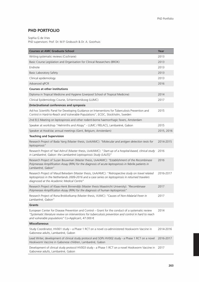
263
PhD Portfolio
PHD PORTFOLIO
Sophia G de VriesPhD supervisors: Prof. Dr. M.P. Grobusch & Dr. A. Goorhuis
Courses at AMC Graduate School year
Writing systematic reviews (Cochrane) 2013
Basic Course Legislation and Organisation for Clinical Researchers (BROK) 2013
Endnote 2013
Basic Laboratory Safety 2013
Clinical epidemiology 2013
Advanced qPCR 2016
Courses at other institutions
Diploma in Tropical Medicine and Hygiene (Liverpool School of Tropical Medicine) 2014
Clinical Epidemiology Course, Schiermonnikoog (LUMC) 2017
(Inter)national conferences and symposia
Ad-hoc Scientific Panel for Developing Guidance on Interventions for Tuberculosis Prevention and Control in Hard-to-Reach and Vulnerable Populations“, ECDC, Stockholm, Sweden
2015
2nd ELS Meeting on leptospirosis and other rodent-borne haemorrhagic fevers, Amsterdam
Speaker at workshop “Helminths and Atopy” - LUMC / RELACS, Lambaréné, Gabon 2015
Speaker at HookVac annual meetings (Gent, Belgium; Amsterdam) 2015, 2016
Teaching and Supervision
Research Project of Bada Yang (Master thesis, UvA/AMC): “Molecular and antigen detection tests for leptospirosis”
2014-2015
Research Project of Yael Ashruf (Master thesis, UvA/AMC): “Start-up of a hospital-based, clinical study in Lambaréné, Gabon: the Lambaréné Leptospirosis Study (LALES)”
2016
Research Project of Suzan Bouwman (Master thesis, UvA/AMC): “Establishment of the Recombinase Polymerase Amplification Assay (RPA) for the diagnosis of acute leptospirosis in febrile patients in Lambaréné, Gabon”
2016
Research Project of Maud Bekedam (Master thesis, UvA/AMC): “Retrospective study on travel related leptospirosis in the Netherlands 2009-2016 and a case series on leptospirosis in returned travelers diagnosed at the Academic Medical Centre”
2016-2017
Research Project of Klaas-Henk Binnendijk (Master thesis Maastricht University): “Recombinase Polymerase Amplification Assay (RPA) for the diagnosis of human leptospirosis”
2017
Research Project of Rona Brokkelkamp (Master thesis, VUMC): “Causes of Non-Malarial Fever in Lambaréné, Gabon”
2017
Grants
European Center for Disease Prevention and Control – Grant for the conduct of a systematic review “Systematic literature review on interventions for tuberculosis prevention and control in hard to reach and vulnerable populations” Co-Applicant, 47.000 €
2014
Miscellaneous
Study Coordinator, HV001 study – a Phase 1 RCT on a novel co-administered Hookworm Vaccine in Gabonese adults, Lambaréné, Gabon
2014-2016
Lead Writer, development of clinical study protocol and SOPs HV002 study - a Phase 1 RCT on a novel Hookworm Vaccine in Gabonese children, Lambaréné, Gabon
2016-2017
Development of clinical study protocol HV003 study - a Phase 1 RCT on a novel Hookworm Vaccine in Gabonese adults, Lambaréné, Gabon
2017

265
Curriculum Vitae
CURRICULUM VITAE
Sophia de Vries was born in Amsterdam on the 5th of November, 1985. She attended the
“Sint Antonius” primary school in the Nieuwmarkt area, and obtained her pre-university
education at the Vossius Gymnasium in 2004.
She had wanted to become a doctor for long, but had always had a great interest in the
African continent. In 2005 she went to Ghana to work in an orphanage and for the Guinea
Worm Eradication Program. She also travelled through Burkina Faso and Mali and visited
Timbuctoo. In the meantime, she cooperated in the founding, coordination and fundraising
of an NGO for young women empowerment (providing vocational training and means to
start up own businesses), in Tamale, Ghana. She went back to Ghana several times in the
years after.
In September 2005, she started the study Medicine at the Academic Medical Center (AMC),
University of Amsterdam (UvA) and graduated in May 2013. During the clinical rotations
she developed and interest in internal medicine and infectious diseases, and did the elective
“developing countries” at the St. Lukes Hospital in Malosa, Malawi. After graduating, she
went back to Malawi to volunteer for a project supporting and monitoring health needs of
disabled children in rural and urban communities in Northern Malawi.
In October 2013, she started the PhD trajectory under supervision of Prof. Martin Grobusch.
To prepare for the work in Lambaréné, Gabon, she obtained the Diploma of Tropical Medi-
cine and Hygiene (DTM&H) at the Liverpool School of Tropical Medicine in 2014. She went
to Gabon later in 2014, where she coordinated a Randomized, controlled phase 1 study on
a novel Hookworm vaccine. In the mean time she worked on several other clinical research
projects, and systematic reviews.
During her work in Gabon, she realised that she has a broad clinical interest and enjoyed
working in a generalist way. Since September 2017, she is being trained as a general practi-
tioner at the AMC.

267
Dankwoord
DAnKWOORD / WORD OF THAnKS
In m’n eentje had ik deze klus nooit kunnen klaren. Hoe langer ik erover nadenk, hoe meer
mensen ik moet bedanken. Zeven jaar is dan ook een hele lange tijd.
Allereerst, Martin, Professor Grobusch, mijn promotor, en mijn steun door dik en dun. Jij
gaf me de geweldige kans om in het prachtige Gabon te werken. Vanaf de dag dat ik je
ontmoette stak je me aan met je onuitputtelijke enthousiasme en toewijding. Je was er altijd:
niet alleen bij wetenschappelijke problemen, maar ook als het persoonlijke leven vastliep was
je tot grote hulp. Zonder jou was ik, en mijn gezin (!), niet waar het nu is.
Mijn co-promotor Bram Goorhuis - het brein. Toen ik was vastgelopen, kwamen jouw crea-
tieve en slimme ideeën als redding! Ik ken weinig mensen die zo slim en vindingrijk zijn. Dank
je wel, zonder jou was het zeker niet gelukt.
De leden van de commissie: Prof. dr. M. Van Vugt, Prof. Dr. W.J. Wiersinga, Prof. Dr. J.W.R.
Hovius, Prof. Dr. M.A. van Agtmael, Prof. Dr. C. Schultsz, en Dr. P.J.J. van Genderen. Dank
voor jullie bereidheid dit proefschrift te beoordelen.
Jelle Visser, ook al zo’n slimme en vindingrijke man, met ook een onuitputtelijke werklust en
enthousiasme. Je hebt me ontzettend veel geleerd, geholpen, gesteund. En nee, niet alleen
over systematic reviews. Al denk ik dat het wel jouw schuld is dat ik er daar nu zes (!) van op
mijn naam heb staan!
Marga Goris, de grote leptospirose kenner. Dank voor al je hoognodige hulp en uitleg, en
het meedenken. En voor de vele uurtjes aan noeste arbeid die we in het Cochrane review
hebben gestopt!
En de andere collega’s van het Referentielaboratorium Leptospirose, Hans van der Lin-
den, Ahmed Ahmed, Rudy Hartskeerl en Jiri Wagenaar, voor het delen van jullie kennis en
kunde, en Hans in het bijzonder voor de vele uren die je uittrok om mij wegwijs te maken in
het lab (geen gemakkelijke klus..).
Alle collega’s van het tropencentrum, en in het bijzonder Michele van Vugt, Pieter van Thiel
en Cees Stijnis, voor de altijd inspirerende klinische en wetenschappelijke discussies.
Bada Yang, zonder jou was Hoofdstuk 5, het Cochrane review, er nooit geweest. Wat een
bevalling was dat (een echte is er niets bij!!) ‐ ik ben blij dat je je carrière in de wetenschap
voortzet! En Mariska Leeflang, ook jij was cruciaal voor het succes van het Cochrane review.

ADDENDUM
268
Louise van Eekeren, ook een student, maar wat voor een! Wat was het leuk met je samen te
werken, en wat ben ik blij dat je je wetenschappelijke carrière in de infectieziekten voortzet!
De studenten in Gabon: Yaël Ashruf, Suzan Bouwman, Maud Bekedam, Klaas-Henk Binnen-
dijk, en Rona Brokkelkamp. Dankzij jullie inzet in de niet altijd gemakkelijke omstandigheden
in Gabon hebben we veel voor elkaar gekregen, dank jullie wel!
Lotje Heuvelings, ons meesterwerk is dan wel geen onderdeel van dit boekje, maar de vele
uren die wij hebben gezweet op het ECDC Tuberculose project zal ik nooit vergeten. Evenmin
als jouw tomeloze energie en uithoudingsvermogen, als ik de handdoek eigenlijk in de ring
had willen gooien.
All my colleagues in Gabon, and in particular the three J‐s- Josiane Honkpehedji, Jeannot
Zinsou and Jean-Claude Dejon Agobé, for our intense collaboration. Doctor Edoa Ronald,
for being the best office-mate and always cheering me up. Kafui Vodonou, for being a good
friend when I really needed one. Professor Akim Ayola Adegnika, for always keeping us sharp
on Monday mornings. All the field workers, and especially Gaspard, for endlessly chasing our
patients when they were nowhere to be found. And of course the institute of CERMEL, for
introducing me to my love and father of my children, Emmanuel Bache.
Tine Sibbing en Monique Gortzak, voor al jullie ondersteuning van alle administratieve klus-
sen, visa, en andere zaken die tijdens dit traject zich voordeden!
The colleagues from GeoSentinel, and especially Douglas Esposito and Davidson Hamer, for
bringing the project to the finish line. It seemed like an easy project, but we spent so many
hours in getting that paper published!!
René Spijker en Ingeborg Nagel, de PubMed/zoek-toppers van het AMC. Ik ben jullie zeer
erkentelijk voor de vele (her)zoekacties en het meedenken!
David Diemert, the Hookvac project is not part of this thesis, but I want to thank you for
your always calm and bright leadership in often difficult times. You were and are a true
inspiration!
Al mijn vrienden en vriendinnen, die ik tegenwoordig veel te weinig spreek, dank voor alle
late uurtjes (waar er tegenwoordig veel te weinig van zijn) en avonturen die we samen
beleefden.

269
Dankwoord
And my friends at the DTM&H course in Liverpool! For all the (late night) adventures we had,
explorations in the lab, study groups.. Rajni and Ellie, my favourite mean girls, I hope we can
see each other again soon. And Shuvo, you left us way too soon. You were a great inspiration
and good friend, and I wish we’d had more time together on this planet.
Mijn paranimfen: Emma de Vries en Renee Verburg. Emma, mijn zus en steun en toeverlaat.
Zonder jou had dit proefschrift niet bestaan; had ik wellicht zelf niet bestaan! Ik kan me geen
leven zonder jou voorstellen. En Renee, we zijn al vriendinnen sinds de 1e klas van het Vossius
(22 jaar alweer); we kennen elkaar door dik en dun. Ik kon me niemand beter wensen om
me bij te staan dan jullie twee!
Mama en Franklin, dank voor jullie onvoorwaardelijke steun. Mama, jij leerde me altijd vol te
houden en iets te maken van mijn leven; je steunde me in al mijn keuzes, en stond ook voor
me klaar als het toch niet de juiste keuzes bleken te zijn. Zonder jou was ik nooit gekomen
waar ik nu ben. Franklin, je bent de beste stiefvader die ik me had kunnen wensen. En
bovenal, zonder de vele dagen die jullie voor Ella zorgden (en de continue morele steun) was
dit proefschrift nooit en te nimmer afgekomen!
Papa, zonder jou was ik nooit dokter geworden. Jouw liefde voor het vak heb ik van kleins af
aan meegekregen, en dat heeft het verlangen ook te “helen” in mij aangewakkerd. Je bent
er nu niet meer, maar ik weet zeker dat je beretrots bent, waar je ook bent. En lieve Evelien,
dank voor alle avonturen die we altijd beleefden, en de liefde voor de natuur die je op mij
overbracht. Zonder jouw geweldige zorg voor papa had ik nooit naar Gabon kunnen gaan,
en al mijn andere dromen kunnen nastreven.
En niet te vergeten, mijn gezin! Emmanuel, you started as my colleague and my guidance
on Clinical Trials. I had no clue what to do – you really saved me and the Hoovac trial! And
then you became my lover. We had great times and difficult times, but we belong together
and I love you more every day. You gave me the two most precious things in my life: Ella and
Benjamin. Ella, kleine draak, dankzij jou duurde dit proces véél langer dan gepland, maar dat
was voor een hele goede rede! Je bent het liefste, grappigste, slimste en knapste kind. En
Benjamin, nog zo klein, maar al zo‐n grote verdienste: mijn zwangerschapsverlof van jou gaf
me de kans dit proefschrift EINDELIJK af te maken! Je bent de liefste, schattigste en knapste
baby, en ik zie nu al dat je net zo’n klein lief monstertje als je zus zult worden.
En natuurlijk dank aan alle andere mensen die ik hier niet heb genoemd, maar die toch een
bijdrage hebben geleverd aan dit proefschrift, of aan mijn welzijn de afgelopen zeven jaar.

Hiding in plain sight: leptospirosis and rickettsial disease. Unmasking two causes of severe
imported infections.
Sophia Geertruda de Vries
Hiding in plain sight: leptospirosis and rickettsial disease
Unmasking two causes of severe imported infections
Hiding in plain sight: leptospirosis and rickettsial disease
SOPHIA DE VRIES
SOP
HIA
DE
VR
IES


![[Prevalence of leptospirosis in adults in Chile, 2003]](https://img.dokumen.tips/doc/110x75/634915fd7442d262850f5b56/prevalence-of-leptospirosis-in-adults-in-chile-2003.jpg)


