Embed Size (px)
Citation preview
Copyright 2003 by the Genetics Society of America
Hedgehog Signaling in the Drosophila Eye and Head: An Analysisof the Effects of Different patched Trans-heterozygotes
Chloe Thomas and Philip W. Ingham1
MRC Intercellular Signalling Group, Centre for Developmental Genetics, University of Sheffield,Sheffield S10 2TN, United Kingdom
Manuscript received June 12, 2003Accepted for publication August 20, 2003
ABSTRACTCharacterization of different alleles of the Hedgehog receptor patched (ptc) indicates that they can be
grouped into several classes. Most mutations result in complete loss of Ptc function. However, missensemutations located within the putative sterol-sensing domain (SSD) or C terminus of ptc encode antimorphicproteins that are unable to repress Smo activity and inhibit wild-type Ptc from doing so, but retain theability to bind and sequester Hh. Analysis of the eye and head phenotypes of Drosophila melanogaster invarious ptc/ptc tuf1 heteroallelic combinations shows that these two classes of ptc allele can be easily distin-guished by their eye phenotype, but not by their head phenotype. Adult eye size is inversely correlatedwith head vertex size, suggesting an alteration of cell fate within the eye-antennal disc. A balance betweenexcess cell division and cell death in the mutant eye discs may also contribute to final eye size. In addition,contrary to results reported recently, the role of Hh signaling in the Drosophila head vertex appears tobe primarily in patterning rather than in proliferation, with Ptc and Smo having opposing effects onformation of medial structures.
THE Hedgehog (Hh) signaling pathway is necessary ptc have been isolated, and these can be grouped into threemain classes. Gain-of-function alleles are characterized byfor the growth and patterning of many tissue typesmissense mutations in either of the large extracellularduring the development of both vertebrates and inverte-loops and are unable to bind and sequester Hh, re-brates (reviewed in McMahon et al. 2003). The Hh ligandsulting in constitutive inhibition of Smo activity (Mul-is received by the transmembrane receptor Patched (Ptc),lor and Guerrero 2000; Vegh and Basler 2003). Anti-a negative regulator of the pathway (Ingham et al. 1991),morphic alleles correspond to missense mutations inwhich normally inhibits the activity of Smoothenedthe SSD or C terminus of the protein and have the(Smo; Hooper 1994; Alcedo et al. 1996). In the pres-ability to sequester Hh but cannot interact with Smoence of Hh, this inhibition is released and the signal is(Martın et al. 2001; Strutt et al. 2001). In contrast,transduced through downstream components, ultimatelythe loss-of-function alleles cannot interact with eitherleading to stabilization and activation of the transcrip-Hh or Smo and include among them the deletion alleletion factor Cubitus interruptus (reviewed in InghamptcG12, all truncation mutations, and some missense mu-and McMahon 2001).tations in the extracellular loops. Thus far, analysis ofPtc is important not only for repressing Smo, but alsoptc alleles has been largely confined to the wing andfor sequestering the Hh protein and thus preventingembryo. However, Hh signaling is required for pat-signaling in inappropriate cells (Chen and Struhlterning and growth in many other Drosophila tissues,1996). Ptc is predicted to have two large extracellularincluding the adult eye and head.loops and 12 transmembrane domains (Nakano et al.
The Drosophila compound eye is composed of �8001989). The second through sixth of these comprise aommatidia and develops from the larval eye-antennalputative sterol-sensing domain (SSD), which possessesimaginal disc (Ready et al. 1976). During early thirdsequence similarity to those in other proteins such asinstar, neuronal differentiation occurs in a wave known3-hydroxy-3-methyl-glutaryl coenzyme A reductase, Nie-as the morphogenetic furrow (MF), sweeping from themann-Pick C1, and SREBP cleavage-activating proteinposterior margin toward the anterior of the eye over a(Chin et al. 1984; Hua et al. 1996; Carstea et al. 1997;period of �2 days (Ready et al. 1976; Tomlinson 1985,Loftus et al. 1997).1988; Tomlinson and Ready 1987). Furrow progressionMany alleles corresponding to mutations throughoutis driven by Hh, which is produced in the developingphotoreceptors and moves anteriorly to activate signal-ing in undifferentiated cells (Heberlein et al. 1993; Ma
1Corresponding author: MRC Intercellular Signalling Group, Centre et al. 1993).for Developmental Genetics, University of Sheffield, Firth Court, West-
Hh signaling is known to induce the expression ofern Bank, Sheffield S10 2TN, United Kingdom.E-mail: [email protected] at least two different signals to mediate photoreceptor
Genetics 165: 1915–1928 (December 2003)
1916 C. Thomas and P. W. Ingham
differentiation. The long-range TGF� signaling mole- Skarmeta et al. 1996; McNeill et al. 1997; Cavodeassi etal. 1999; Lee and Treisman 2001). In turn, IRO-C restrictscule Decapentaplegic (Dpp) is thought to be responsi-
ble for inducing a “preproneural” state in cells ahead the production of Fringe (Fng) to the ventral compart-ment (Cho and Choi 1998; Dominguez and de Celisof the furrow (Greenwood and Struhl 1999), charac-
terized by the upregulation of proneural repressor 1998). Fng is a glycosyltransferase that modulates the abil-ity of cells to respond to Notch ligands (Panin et al. 1997;genes such as hairy (h) and extramacrochaetae (emc), to-
gether with cell cycle arrest and synchronization (Hors- Munro and Freeman 2000). This results in N signalingoccurring specifically at the equator where it is essentialfield et al. 1998). A second signal is believed to act at
short range to induce the expression of atonal (ato), for growth of the eye disc and upregulation of Hh at theposterior margin prior to furrow initiation (Papayan-a proneural gene directly required for photoreceptor
differentiation (Jarman et al. 1994). It is unclear exactly nopoulos et al. 1998; Cavodeassi et al. 1999).During analysis of trans-heterozygotes carrying thehow ato is induced, but data implicate the Notch (N)
and/or Raf pathways in mediating this process (Green- ptctuf1 allele in combination with various alleles of ptc(Strutt et al. 2001), we noted that the severity of thewood and Struhl 1999; Baonza and Freeman 2001).
In addition to its role in furrow progression, upregula- eye phenotype appeared to be inversely related to thatof the wing phenotype. To investigate this phenomenontion of Hh at the posterior margin of the eye-antennal
disc during early third instar is necessary for the furrow further, we have studied the eyes and heads of mutantadults in some detail. The eye phenotype differs dramat-to initiate (Dominguez and Hafen 1997; Borod and
Heberlein 1998). Evidence suggests that Hh directly ically between trans-heterozygotes carrying loss-of-func-tion and antimorphic ptc alleles. However, the headinduces the expression of both dpp and the gene encod-
ing an eye-specific nuclear protein known as Eyes absent vertex phenotype, while varying in severity between dif-ferent loss-of-function heteroallelic combinations, is not(Eya; Pappu et al. 2003). Dpp and Eya interact in a
complex manner with a series of other nuclear proteins, intrinsically different in those carrying antimorphic al-leles. Our results suggest that Hh signaling primarilyincluding Eyeless (Ey), Eyegone (Eyg), Sine oculis (So),
and Dachshund (Dac), to induce furrow initiation (Bon- controls patterning in the head vertex, rather than pro-liferation as has been recently suggested (Shyamalaini et al. 1997; Chen et al. 1997; Pignoni et al. 1997;
Hazelett et al. 1998; Chen et al. 1999; Curtiss and and Bhat 2002). However, in the presumptive eye, Hhappears to be involved in regulating cell division and cellMlodzik 2000). This process is opposed by Wingless
(Wg), which inhibits furrow initiation at the anterior of death in addition to influencing cell fate specification.the eye disc (Ma and Moses 1995; Treisman and Rubin1995). Antagonism between Dpp and Wg plays an ear-
MATERIALS AND METHODSlier role in defining the regions of the disc that corre-spond to the eye field vs. the head vertex (Royet and Drosophila stocks: A selection of ptc alleles, as described inFinkelstein 1997). In early second instar wg is expressed Strutt et al. (2001), was chosen for analysis. ptc 34, ptc S2, andthroughout the eye primordium, becoming refined to ptc 13 are characterized by missense mutations in the SSD or
C terminus of ptc and act in an antimorphic way. ptcG12, ptc 9,the presumptive head domain due to Hh-dependent dppptc 14, ptc15, ptc 16, ptc17, ptc 37, and ptc47 are loss-of-function alleles.expression in the posterior. The patterning of the vertexThe ptc H84lacZ enhancer trap is also a loss-of-function allele. Theitself also requires Hh signaling, which is necessary formild regulatory mutations ptc tuf1 (Hidalgo 1989; Capdevila
specification of the medial ocelli and ocellar cuticle, et al. 1994) and ptc GAL4 (Speicher et al. 1994) were also utilized.whereas Wg signaling specifies more lateral structures For analysis of adult phenotypes, controlled crosses were set(Royet and Finkelstein 1996). Recent work suggests up in egg-laying cages and embryos were collected on apple-
juice/agar plates. To prevent overcrowding, 50 hatched larvaethat Ptc and Smo cooperate to promote cell prolifera-were transferred to each vial of fly food and allowed to maturetion in the head, rather than opposing each other asto adulthood at 25�. For easy analysis of ptc/ptc tuf1 trans-hetero-in other tissues (Shyamala and Bhat 2002). According zygote larvae, ptc alleles were crossed to a Sco/Cyo, actGFP line
to these authors, loss of either gene causes a reduction to generate ptc/Cyo, actGFP stocks. For analysis of dpplacZin the size of the head capsule, although whether this expression, a ptc tuf1, dpplacZ/Cyo recombinant line was utilized.
A yw; P(w�)69DD1-12/TM3 (mirr B1-12lacZ) line was donated byis due to a loss of Hh-specified medial vertex structuresD. Strutt (Choi et al. 1996). ptc tuf1ltd was crossed to mirr B1-12lacZor some other part of the head is unclear.to generate ptc tuf1ltd, mirr B1-12lacZ/Cyo recombinants. TheseIn addition to its roles in anteroposterior patterning, were then crossed directly to each ptc allele for analysis of
Hh is also involved in the establishment of the dorsoven- mutant phenotypes. The hh1 allele (Mohler 1988) was do-tral (DV) organizing center, known as the equator, dur- nated by D. Strutt. Several ptc alleles (ptc13, ptc14, ptc16, ptc 34,
ptc 37, ptc47, and ptc tuf1) were recombined into a hh1 backgrounding early second instar (Baker 1978; Campos-Ortegato create ptc/Cyo; hh1 stocks. ptc tuf1, dpplacZ/Cyo was also recom-and Waitz 1978). High levels of Hh, Wg, and otherbined into the hh1 background to create a ptc tuf1, dpplacZ/Cyo,unknown molecules, present in the dorsal half of the eyehh1 stock.
disc, activate the expression of the dorsal selector genes Generation of clones: For making ptc clones, FRT42D, ptcS2/araucan (ara), caupolican (caup), and mirror (mirr), other- Cyo and FRT42D, ptc IIW sha/Cyo were crossed to a y, hsFLP;
FRT42D P(ry�,y�)/Cyo line donated by D. Strutt. For makingwise known as the Iroquois complex (IRO-C; Gomez-
1917Hh Signaling in the Drosophila Eye and Head
smo clones, smoD16 Sco, FRT40A/Cyo and smoQ14, FRT40A/Cyo werecrossed to a y, hsFLP; P(y�, hs-CD2)2L-1, FRT40A/Cyo line donatedby D. Strutt. Larvae were heat-shocked at 37� for 2 hr eachduring early second and early third instar and allowed todevelop to adulthood.
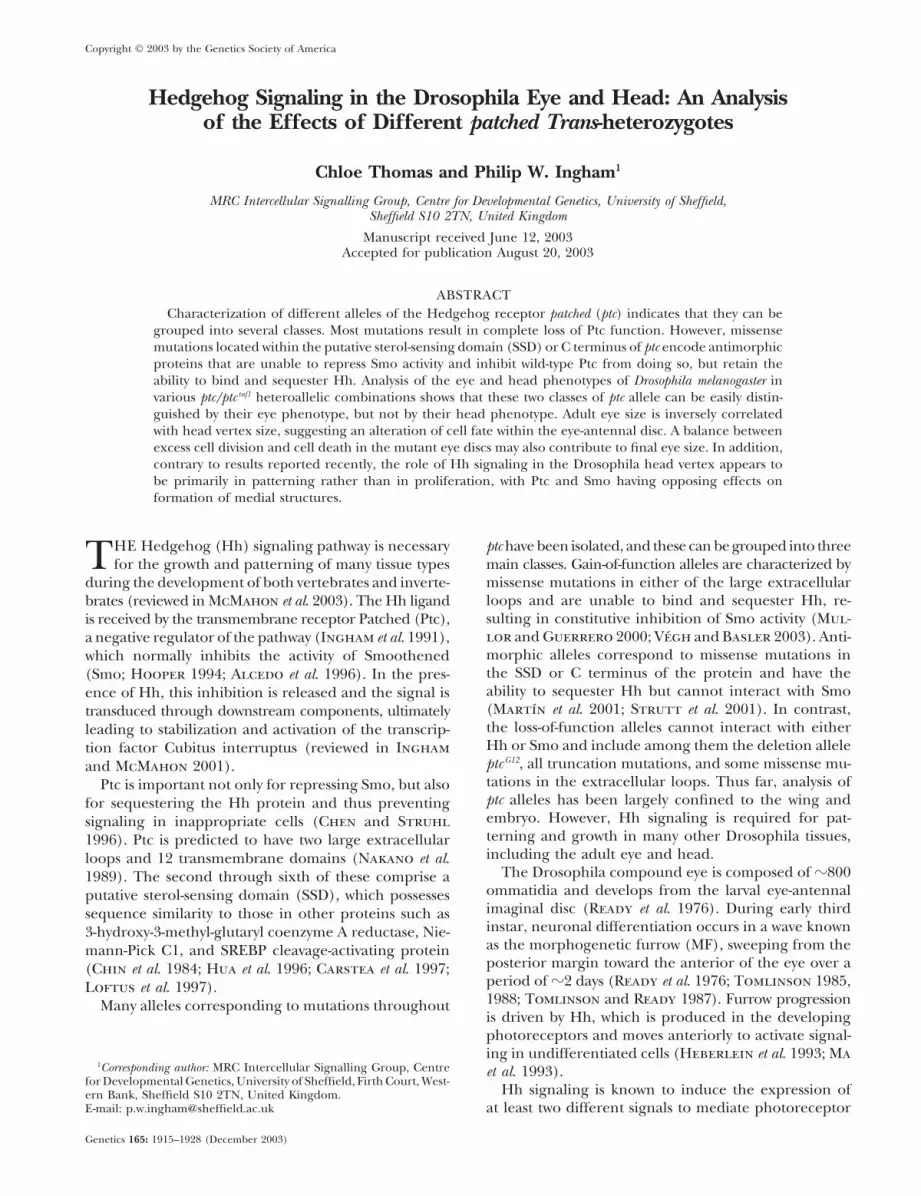
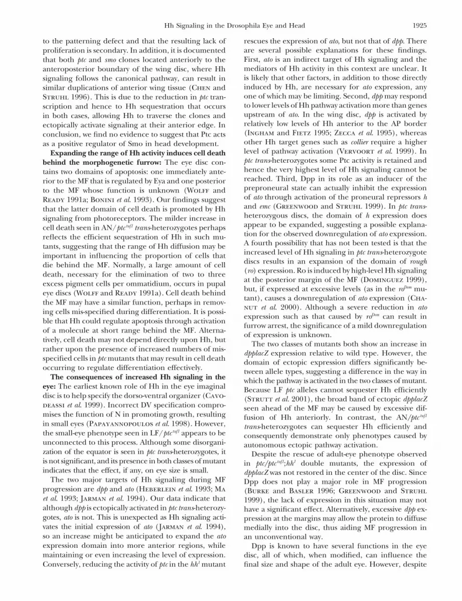
Immunohistochemistry: Imaginal discs dissected from 48 hror late third instar larvae were fixed using standard protocols.Discs were blocked in 1% BSA and incubated overnight at 4�with primary antibodies. After extensive washing, discs wereincubated for 2 hr with fluorescent secondary antibodies atroom temperature (Jackson Laboratories, West Grove, PA,and Molecular Probes, Eugene, OR). Where necessary, a Cy3-conjugated antiphalloidin or a Cy5-conjugated HRP second-ary, donated by D. Strutt, were used to visualize photorecep-tors. After further washing, discs were mounted in glyceroland observed with a Leica SP confocal microscope. Primaryantibodies were rabbit polyclonal antiatonal ( Jarman et al.1994), donated by A. Jarman; mouse monoclonal antiwingless4D4 (Brook and Cohen 1996); mouse polyclonal antihairy(Carroll and Whyte 1989), donated by K. Howard; rabbitpolyclonal anti-phospho-histone-H3 Ser10 from Upstate Bio- Figure 1.—The adult-eye phenotype of wild type and atechnology (Lake Placid, NY; Hendzel et al. 1997), donated selection of ptc trans-heterozygous flies shown by scanningby P. Rashbass; and rabbit polyclonal anti-�-galactosidase electron microscopy. In A–F, anterior is to the left and dorsal(Cappel). Acridine orange and �-galactosidase staining were is up. (A) The wild-type eye displays an ordered array of �800carried out using standard protocols. Samples stained with ommatidia and a consistent oval shape. (B–F) All ptc trans-�-galactosidase were observed on a Zeiss Axioplan 2 microscope. heterozygous eyes have a rough appearance: (B) ptcG12/ptctuf1,All images were manipulated using Photoshop software. (C) ptc15/ptctuf1, (D) ptc 37/ptctuf1, (E) ptc 34/ptctuf1, and (F) ptcS2/
Electron microscopy: Adult eyes were dissected and dehy- ptctuf1. Eyes of mutants carrying loss-of-function ptc alleles indrated through increasing concentrations of ethanol (25, 50, trans to ptctuf1 are generally reduced in size (B–D plus ptc17/75, 90, and 100%), before being critical point dried and ptctuf1, ptc47/ptctuf1, ptc16/ptctuf1, ptc9/ptctuf1, and ptc14/ptctuf1, notmounted on electron microscope specimen-holding stubs. shown), while mutants carrying antimorphic ptc alleles in transSamples were coated in gold using an Edwards sputter coater to ptctuf1 have enlarged eyes compared to wild type (E and Fand then analyzed on a Phillips PSEM 501B scanning electron plus ptc13/ptctuf1, not shown).microscope.
heterozygotes in that the eye is elongated and thereappears to be a reduction of surrounding head tissue inRESULTSsome cases (Figure 1B). In contrast, AN/ptctuf1 mutants
Eye phenotype can distinguish between different ptc show a marked increase in eye size (Figure 1, E and F),heteroallelic combinations: Lethal ptc alleles can be which is consistent within allele class.grouped into several classes, including those that have Expansion and mispatterning of the head vertex in ptcan antimorphic (AN) effect and those that show a loss- trans-heterozygotes is caused by excess Hh diffusion ratherof-function (LF) phenotype (Strutt et al. 2001). To than by ectopic pathway activation: The eye-antennal discanalyze the role of ptc in the development of the Dro- gives rise to the adult eye, antenna, and the head vertexsophila eye, the phenotypes of adult flies heteroallelic (Haynie and Bryant 1986). As Hh signaling is knownfor various ptc alleles in trans to the ptc tuf1 allele were to be important in patterning the vertex and, in particu-observed. ptc tuf1 is a regulatory mutation that results in lar, the ocelli (Royet and Finkelstein 1996), we investi-a reduced quantity of wild-type protein being produced gated whether ptc mutants cause head defects in an allele-(Hidalgo 1989; Capdevila et al. 1994). specific manner. In addition to ptc tuf1, the ptcGAL4 allele was
The wild-type eye displays an ordered array of �800 analyzed as it has been recently reported that some ptcGAL4
ommatidia and a consistent oval shape (Figure 1A). The trans-heterozygous combinations can cause a small headmajority of ptc/ptc tuf1 eyes have disorganized ommatidia phenotype (Shyamala and Bhat 2002). Like ptc tuf1,leading to a rough appearance (Figure 1, B–F). In addi- ptcGAL4 is homozygous viable, with a mild adult wing andtion, most trans-heterozygotes show changes in eye scutellar bristle phenotype due to the insertion of a Pshape and size, characteristic of allele class. LF/ptc tuf1 element containing the GAL4 gene into the regulatoryadults generally have reduced, rounded eyes, although sequence of ptc (Speicher et al. 1994).the degree of severity can vary both within and (more In wild-type flies, the head vertex is symmetrical andstrongly) between genotypes. For example, eyes of trans- composed of three distinct domains (Figure 2A). Theheterozygotes carrying the mild allele ptc15 (Figure 1C) three ocelli are centrally located in the ocellar cuticle,are almost wild type in appearance, whereas the mis- which exhibits a characteristic pattern of bristles. Thesense mutation ptc 37 causes an extremely small eye in orbital cuticle is the most lateral region and is alsotrans to ptc tuf1 (Figure 1D). The ptcG12/ptc tuf1 mutant phe- covered in bristles, while the bare-ridged tissue of the
frons lies between these two domains.notype differs slightly from that of the other LF trans-

1918 C. Thomas and P. W. Ingham
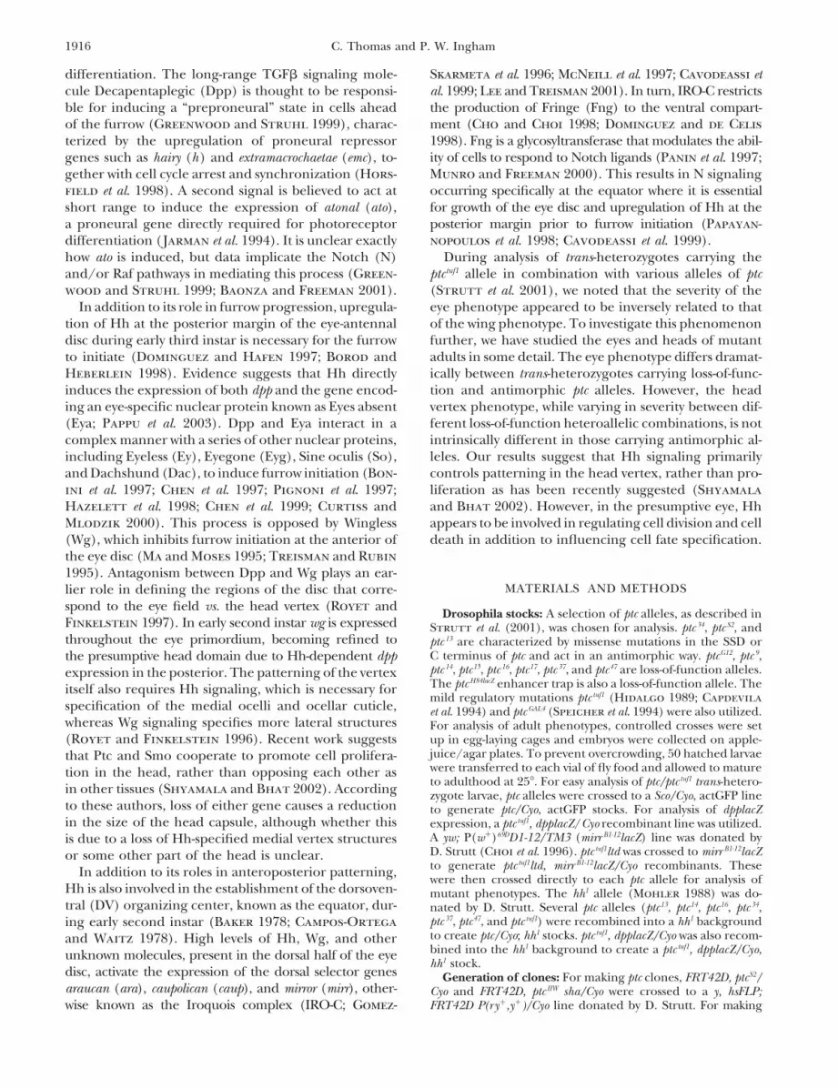
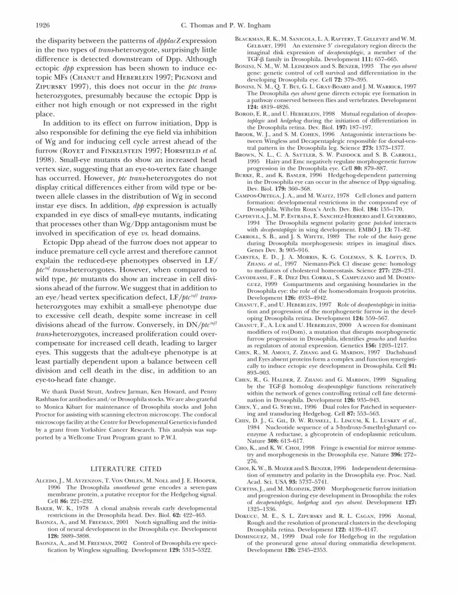
Figure 2.—The conse-quences of removing ptcand smo activity in the headvertex. In all panels, ante-rior is up. (A) Wild-type ver-tex showing the three ocelli(oc) situated medially in atriangular pattern. Adjacentto these lie the two ocellarbristles (ocb) and the twopostvertical bristles (pvb) inaddition to several micro-chaetae known as interocel-lar bristles (ioc). The frons(fr) is composed of ridgedtissue, lateral to the ocellarcuticle. In the most lateralregions lies the orbital cuti-cle containing three orbitalbristles (orb) on each sidein addition to microchae-tae. (B–L) Head vertex pat-terning is disrupted in ptctrans-heterozygotes. AN trans-heterozygotes exhibit a rela-
tively mild head vertex defect: (B) ptc34/ptctuf1, (C) ptc34/ptcGAL4, (D) ptc13/ptctuf1, and (E) ptc13/ptcGAL4. LF trans-heterozygous headsexhibit a range of severity depending upon the allele: (F) ptcG12/ptctuf1, (G) ptcG12/ptcGAL4, (H) ptc37/ptctuf1, and (I) ptc37/ptcGAL4. ( J)Magnification of ptc13/ptctuf1 in D, showing ectopic bristles adjacent to eye. (K) Magnification of ptc37/ptctuf1 in S, showing ectopicocelli. (L) Magnification of ptc37/ptctuf1, showing an outgrowth of tissue covered in bristles and an ectopic ocellus on the anteriorleft side of the vertex. (M–T) The consequences of inducing clones in the head vertex lacking ptc or smo function. Clones aremarked with yellow, which causes reduced pigmentation of bristles. Due to the limited number of bristles in the head vertex, itis difficult to correctly ascertain clone boundaries. Heads from flies containing clones of (M and N) ptc16, (O and P) ptcS2, (Qand R) smoQ14, and (S and T) smoD16 are shown. (M) The medial ocellus is duplicated due to the presence of a clone in thecentral region. (N) A clone has been magnified, showing an ectopic ocellus adjacent to a yellow bristle. (O) An outgrowth ofthe eye adjacent to an ectopic ocellus indicates a clone spanning the eye and head. In P, another clone has been magnified,showing ectopic ocelli and bristles in the frons. (Q–T) smo clones often lack bristles, making it difficult to assess the exact positionof the clone using the yellow marker. (Q) The left lateral ocellus is almost completely missing (magnified in R); presumably theclone to the left extends into this region. This clone also causes a duplication of the orbital cuticle, presumably due to activationof Hh signaling and hence a more medial fate (frons) on its lateral side. In T, a portion of S has been magnified showingincomplete fusion of the two halves of the medial ocellus.
Mutants were assessed for the overall size and shape sess ectopic ocelli in anterior lateral regions (e.g., Figure2H, magnified in K)—areas in which eye fate wouldof the entire head, in addition to the vertex phenotype
(Table 1; Figure 2, A–L). Contrary to the results pre- normally be specified, indicating the activation of high-level Hh signaling. These may be accompanied by out-sented by Shyamala and Bhat (2002), a severely small
head phenotype is rarely observed; rather, many hetero- growths of tissue, either covered in or devoid of bristles(Figure 2L). AN/ptc tuf1 trans-heterozygotes do not ap-allelic combinations show an increase in the size of the
vertex (e.g., Figure 2, F and H). This is particularly pear to differ significantly from LF/ptc tuf1 with respectto head vertex phenotype. The vertex tends to be closerapparent in mutants with reduced eyes, suggesting a
defect in the specification of eye tissue vs. head tissue. to wild type in size, with excess bristles in anterior re-gions and occasionally some ectopic frons or a smallSome of the ptcGAL4 trans-heterozygotes, however, do show
a slight reduction in head vertex size, although this may outgrowth of tissue (Figure 2, B, D, and J). In this respectthe AN alleles behave as mild LF alleles such as ptc15 (seenot be significant (Table 1). In addition, occasionally
one or more antennae are missing, which can give the Table 1) and do not exhibit an antimorphic phenotype.This suggests that the head defect in the LF/ptc tuf1 mu-appearance of a reduced head, particularly in combina-
tion with a small-eye phenotype (e.g., ptcG12/ptc tuf1, Figure tants is caused predominantly by Hh diffusing fartherthan normal to specify ocellar cuticle fate in competent2F). It is important to note that there is a variability of
at least 5–10% in head vertex size within heteroallelic regions, rather than by ectopic pathway activation (seediscussion).combinations and even among wild type.
All trans-heterozygotes exhibit some degree of mispat- In general, ptc/ptcGAL4 trans-heterozygotes exhibit milderhead, eye, and wing phenotypes than ptc/ptc tuf1 combina-terning in the vertex. The most extreme examples pos-
1919Hh Signaling in the Drosophila Eye and Head
TABLE 1
A summary of head and eye defects in ptc trans-heterozygotes
Head vertex size % with % with % missingAppearance (% relative to % with major head minor head one or two
Genotype of eye wild type)a ectopic ocelli defectb defectc antennae
13/tuf1 Enlarged 102 0 100 0 013/gal4 Wild type 98 0 0 50 034/tuf1 Enlarged 117 0 63 37 034/gal4 Wild type 94 0 0 9 0S2/tuf1 Enlarged 125 6 44 56 0S2/gal4 Wild type 110 0 0 6 047/tuf1 Severely reduced 157 56 100 0 3347/gal4 Mildly reduced 102 0 8 20 437/tuf1 Severely reduced 155 50 100 0 2737/gal4 Mildly reduced 120 2 2 36 017/tuf1 Severely reduced 148 2.5 100 0 917/gal4 Mildly reduced 122 0 20 13 1316/tuf1 Severely reduced 140 18 100 0 3016/gal4 Mildly reduced 105 0 6 6 09/tuf1 Severely reduced 146 10 100 0 509/gal4 Wild type 108 0 0 29 4lacz/tuf1 Mildly reduced 145 0 90 10 0lacz/gal4 Wild type 107 0 0 13 0G12/tuf1 Severely reduced 136 0 100 0 18G12/gal4 Wild type 106 0 0 0 014/tuf1 Mildly reduced 121 0 0 30 014/gal4 Wild type 108 0 0 0 015/tuf1 Mildly reduced 117 0 0 30 015/gal4 Wild type 105 0 0 3 0gal4/tuf1 Wild type 99 0 0 5 0
a The head vertex area of at least 10 flies of wild type and each ptc heteroallelic combination was measuredusing Improvision Openlab software and then an average was taken for each genotype.
b A major head defect is defined as a mutant exhibiting tissue outgrowths, large numbers of ectopic bristles,or many missing bristles.
c A minor head defect is defined as a mutant exhibiting one or two missing, extra, or misplaced bristles.
tions do, often appearing almost wild type in both head more lateral regions, ectopic ocelli often form, togetherwith ocellar and postvertical bristles (Figure 2, N–P),size and patterning. An exception to this is ptc13/ptcGAL4
(Figure 2E), which exhibits ocelli that are larger and although if the clone is small then the phenotype is lessdramatic. This effect is similar to that caused by clonescloser to one another—a phenotype not observed in
any other mutant. ectopically expressing hh (Royet and Finkelstein 1996)appearing to be slightly milder in the case of ptc S2 clones.Clones of ptc and smo cause opposite patterning de-
fects in the head vertex: Shyamala and Bhat (2002) The presence of ectopic ocelli is often accompanied byan outgrowth in the adjacent eye tissue, suggesting thathave also reported that clones of cells homozygous for
ptc or smo loss-of-function alleles cause a small head pheno- the clone spans both the eye and head domains (Figure2O). Very rarely (�1%) there does appear to be a severetype, implying that both Ptc and Smo act to drive cell
proliferation in the head. The results presented above reduction in head size if the clone covers the entirevertex (not shown), due to the lack of almost all fronsdo not support this hypothesis since they indicate that
head vertex size is generally increased in mutants with and orbital cuticle.In contrast, clones lacking wild-type smo activity havea reduced level of Ptc activity. To investigate this discrep-
ancy further, marked clones of cells expressing various the most effect when they are located within the ocellarcuticle (Figure 2, Q–T). In this case the ocelli are oftenmutant alleles of ptc and smo were generated (Figure 2,
M–T). smaller in size or even absent. Occasionally the medialocellus is split into two (Figure 2, S and T), indicatingIn general, ptc clones located in medial regions of
the head vertex lead to a duplication of macrochaetae a defect in the fusion of the two half-ocelli during mor-phogenesis. No obvious change in eye size was noted.and an increase in size of the ocelli, occasionally causing
the medial ocellus to be split into two (Figure 2M). In Again, infrequently the vertex can be reduced in size if
1920 C. Thomas and P. W. Ingham
covered by a large clone. However, the disruption ofpatterning is conspicuously different from that causedby a ptc loss-of-function clone, making it unlikely that Ptcand Smo act in concert to promote growth in the head,as previously suggested (Shyamala and Bhat 2002).
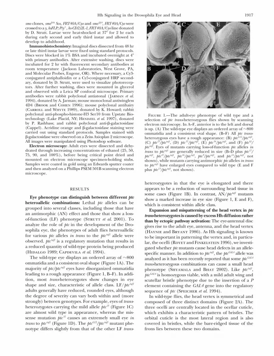
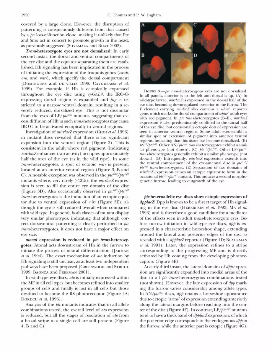
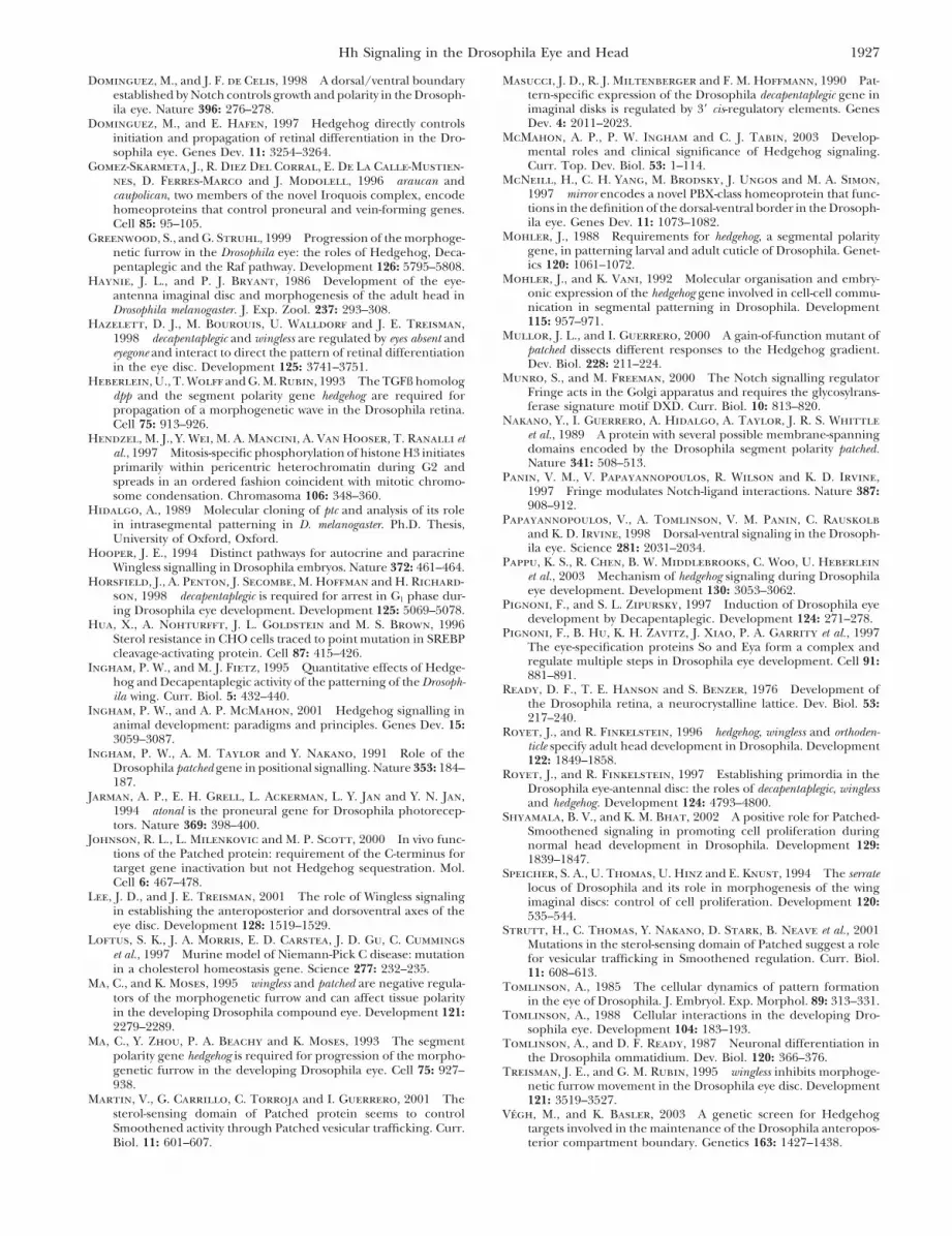
Trans-heterozygote eyes are not dorsalized: In earlysecond instar, the dorsal and ventral compartments ofthe eye disc and the equator separating them are estab-lished. Hh signaling has been implicated in the processof initiating the expression of the Iroquois genes (caup,ara, and mirr), which specify the dorsal compartment(Dominguez and de Celis 1998; Cavodeassi et al.1999). For example, if Hh is ectopically expressed Figure 3.—ptc trans-heterozygous eyes are not dorsalized.throughout the eye disc using ey-GAL4, the IRO-C- In all panels, anterior is to the left and dorsal is up. (A) Inexpressing dorsal region is expanded and fng is re- wild-type larvae, mirrlacZ is expressed in the dorsal half of the
eye disc, becoming downregulated posterior to the furrow. Thestricted to a narrow ventral domain, resulting in a se-P element carrying mirrlacZ also contains a white� reporterverely reduced, dorsalized eye. This is not dissimilargene, which marks the dorsal compartment of white� adult eyesfrom the eyes of LF/ptc tuf1 mutants, suggesting that ex- with red pigment. In ptc trans-heterozygotes (B–E), mirrlacZ
cess diffusion of Hh in such trans-heterozygotes may cause expression is also predominantly confined to the dorsal halfIRO-C to be activated ectopically in ventral regions. of the eye disc, but occasionally ectopic dots of expression are
seen in anterior ventral regions. Some adult eyes exhibit aInvestigation of mirrlacZ expression (Choi et al. 1996)similar spot or extension of pigment into anterior ventralin mutant discs revealed that there is no significantregions, indicating that this tissue has become dorsalized. (B)expansion into the ventral region (Figure 3). This is ptc13/ptctuf1. Other AN/ptctuf1 trans-heterozygotes exhibit a simi-
consistent in the adult where red pigment (indicating lar phenotype (not shown). (C) ptc17/ptctuf1. Other LF/ptctuf1
mirrlacZ enhancer trap activity) occupies approximately trans-heterozygotes generally exhibit a similar phenotype (notshown). (D) Infrequently, mirrlacZ expression extends intohalf the area of the eye (as in the wild type). In somethe ventral compartment of the eye-antennal disc in ptcG12/trans-heterozygotes, a spot of ectopic mirr is present,ptctuf1 trans-heterozygotes. (E) Separation of two domains oflocated at an anterior ventral region (Figure 3, B and mirrlacZ expression causes an ectopic equator to form in the
C). A notable exception was observed in the ptcG12/ptc tuf1occasional ptcG12/ptctuf1 mutant. This induces a second morpho-
mutants where, very rarely (�2%), the mirrlacZ expres- genetic furrow, leading to outgrowth of the eye.sion is seen to fill the entire eye domain of the disc(Figure 3D). Also occasionally observed in ptcG12/ptc tuf1
trans-heterozygotes is the induction of an ectopic equa- ptc heteroallelic eye discs show ectopic expression ofdpplacZ: Dpp is known to be a direct target of Hh signal-tor due to ventral expression of mirr (Figure 3E), al-
though the eye is still reduced overall when compared ing in the eye disc (Heberlein et al. 1993; Ma et al.1993) and is therefore a good candidate for a mediatorwith wild type. In general, both classes of mutant display
very similar phenotypes, indicating that although cor- of the effects seen in adult trans-heterozygote eyes. Be-fore furrow initiation in wild-type eye discs, dpp is ex-rect dorsoventral patterning is clearly perturbed in ptc
trans-heterozygotes, it does not have a major effect on pressed in a characteristic horseshoe shape, extendingaround the lateral and posterior edges of the disc aseye size.
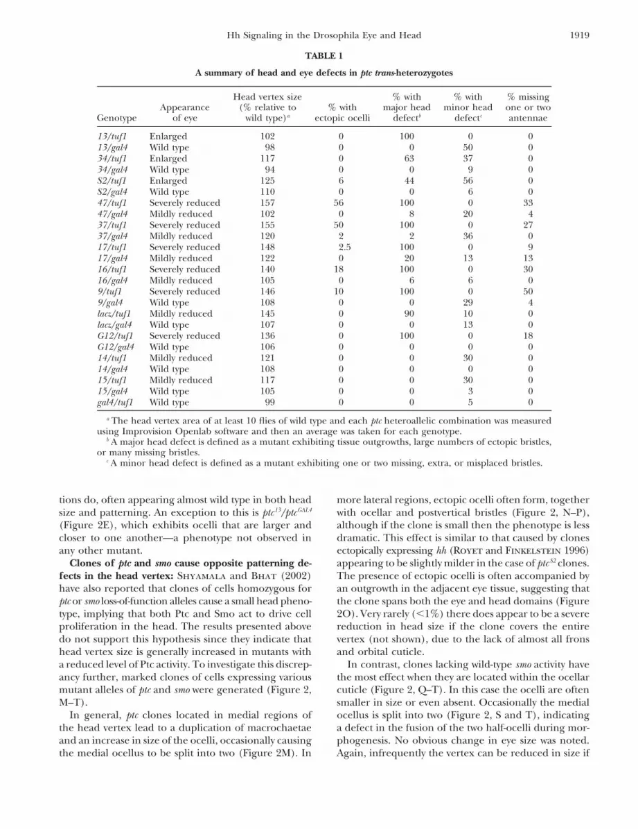
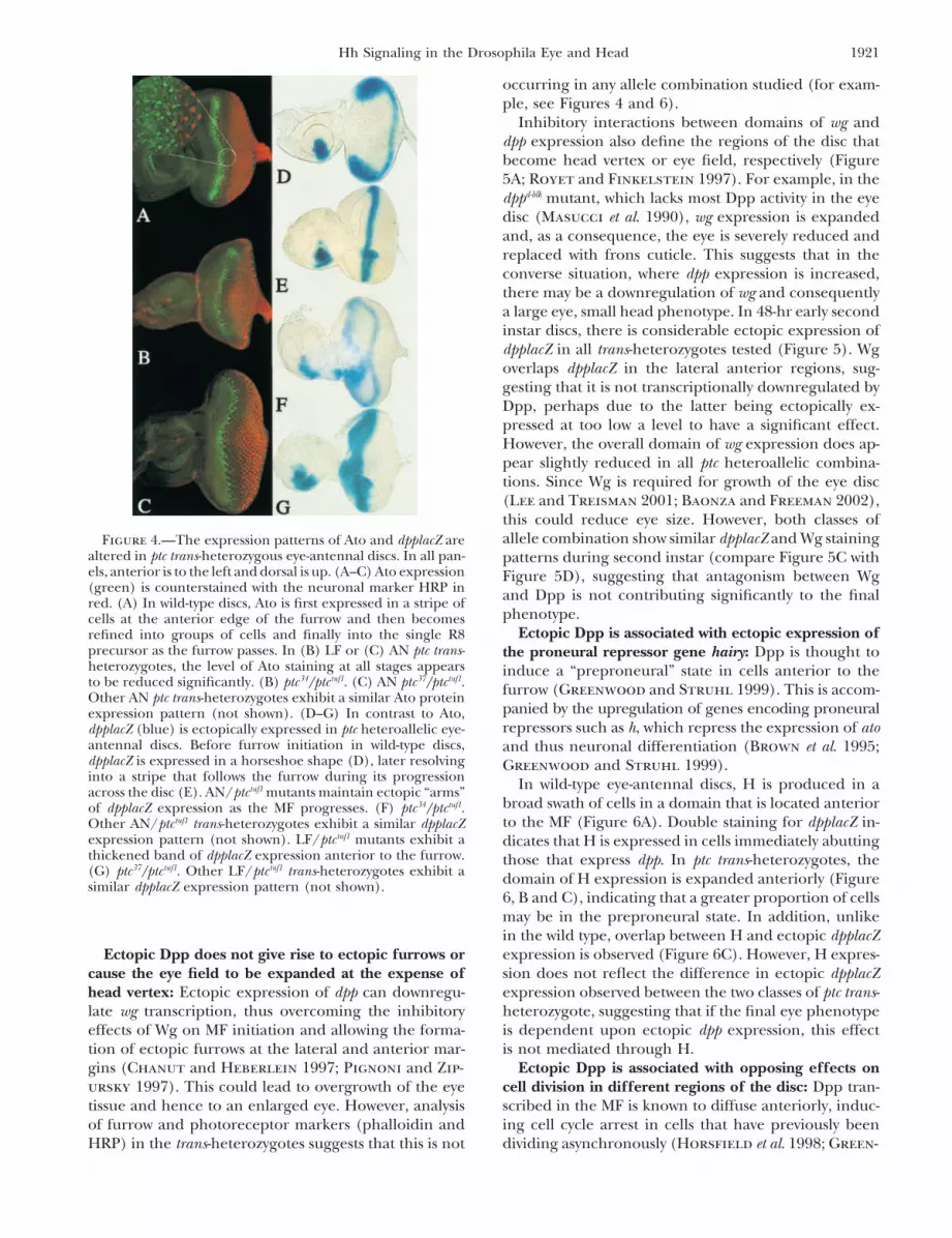
atonal expression is reduced in ptc trans -heterozy- revealed with a dpplacZ reporter (Figure 4D; Blackmanet al. 1991). Later, the expression refines to a stripegotes: Atonal acts downstream of Hh in the furrow to
initiate the process of neural differentiation (Jarman corresponding to the progressing MF and is directlyactivated by Hh coming from the developing photore-et al. 1994). The exact mechanism of ato induction by
Hh signaling is still unclear, as at least two independent ceptors (Figure 4E).At early third instar, the lateral domains of dpp expres-pathways have been proposed (Greenwood and Struhl
1999; Baonza and Freeman 2001). sion are significantly expanded into medial areas of thedisc in all ptc trans-heterozygous combinations testedIn wild-type eye discs, ato is initially expressed within
the MF in all cell types, but becomes refined into smaller (not shown). However, the late expression of dpp mark-ing the furrow varies considerably among allele types.groups of cells and finally is lost in all cells but those
destined to become the R8 photoreceptor (Figure 4A; In AN/ptc tuf1 discs, dpp retains a horseshoe appearancedue to ectopic “arms” of expression extending anteriorlyDokucu et al. 1996).
Analysis of the ptc mutants indicates that in all allele along the lateral margins before reaching into the cen-ter of the disc (Figure 4F). In contrast, LF/ptc tuf1 mutantscombinations tested, the overall level of ato expression
is reduced, but all the stages of resolution of ato from tend to have a thick band of dpplacZ expression, of whichthe posterior edge corresponds to the endogenous dpp ina broad stripe to a single cell are still present (Figure
4, B and C). the furrow, while the anterior part is ectopic (Figure 4G).
1921Hh Signaling in the Drosophila Eye and Head
occurring in any allele combination studied (for exam-ple, see Figures 4 and 6).
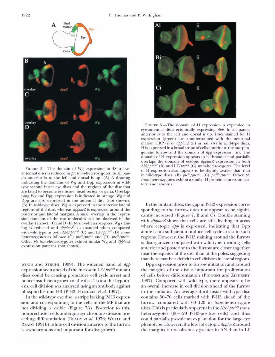
Inhibitory interactions between domains of wg anddpp expression also define the regions of the disc thatbecome head vertex or eye field, respectively (Figure5A; Royet and Finkelstein 1997). For example, in thedppd-blk mutant, which lacks most Dpp activity in the eyedisc (Masucci et al. 1990), wg expression is expandedand, as a consequence, the eye is severely reduced andreplaced with frons cuticle. This suggests that in theconverse situation, where dpp expression is increased,there may be a downregulation of wg and consequentlya large eye, small head phenotype. In 48-hr early secondinstar discs, there is considerable ectopic expression ofdpplacZ in all trans-heterozygotes tested (Figure 5). Wgoverlaps dpplacZ in the lateral anterior regions, sug-gesting that it is not transcriptionally downregulated byDpp, perhaps due to the latter being ectopically ex-pressed at too low a level to have a significant effect.However, the overall domain of wg expression does ap-pear slightly reduced in all ptc heteroallelic combina-tions. Since Wg is required for growth of the eye disc(Lee and Treisman 2001; Baonza and Freeman 2002),this could reduce eye size. However, both classes ofallele combination show similar dpplacZ and Wg stainingFigure 4.—The expression patterns of Ato and dpplacZ are
altered in ptc trans-heterozygous eye-antennal discs. In all pan- patterns during second instar (compare Figure 5C withels, anterior is to the left and dorsal is up. (A–C) Ato expression Figure 5D), suggesting that antagonism between Wg(green) is counterstained with the neuronal marker HRP in and Dpp is not contributing significantly to the finalred. (A) In wild-type discs, Ato is first expressed in a stripe of
phenotype.cells at the anterior edge of the furrow and then becomesEctopic Dpp is associated with ectopic expression ofrefined into groups of cells and finally into the single R8
precursor as the furrow passes. In (B) LF or (C) AN ptc trans- the proneural repressor gene hairy: Dpp is thought toheterozygotes, the level of Ato staining at all stages appears induce a “preproneural” state in cells anterior to theto be reduced significantly. (B) ptc34/ptctuf1. (C) AN ptc37/ptctuf1. furrow (Greenwood and Struhl 1999). This is accom-Other AN ptc trans-heterozygotes exhibit a similar Ato protein
panied by the upregulation of genes encoding proneuralexpression pattern (not shown). (D–G) In contrast to Ato,repressors such as h, which repress the expression of atodpplacZ (blue) is ectopically expressed in ptc heteroallelic eye-
antennal discs. Before furrow initiation in wild-type discs, and thus neuronal differentiation (Brown et al. 1995;dpplacZ is expressed in a horseshoe shape (D), later resolving Greenwood and Struhl 1999).into a stripe that follows the furrow during its progression In wild-type eye-antennal discs, H is produced in aacross the disc (E). AN/ptctuf1 mutants maintain ectopic “arms”
broad swath of cells in a domain that is located anteriorof dpplacZ expression as the MF progresses. (F) ptc34/ptctuf1.to the MF (Figure 6A). Double staining for dpplacZ in-Other AN/ptctuf1 trans-heterozygotes exhibit a similar dpplacZ
expression pattern (not shown). LF/ptctuf1 mutants exhibit a dicates that H is expressed in cells immediately abuttingthickened band of dpplacZ expression anterior to the furrow. those that express dpp. In ptc trans-heterozygotes, the(G) ptc37/ptctuf1. Other LF/ptctuf1 trans-heterozygotes exhibit a domain of H expression is expanded anteriorly (Figuresimilar dpplacZ expression pattern (not shown).
6, B and C), indicating that a greater proportion of cellsmay be in the preproneural state. In addition, unlikein the wild type, overlap between H and ectopic dpplacZexpression is observed (Figure 6C). However, H expres-Ectopic Dpp does not give rise to ectopic furrows or
cause the eye field to be expanded at the expense of sion does not reflect the difference in ectopic dpplacZexpression observed between the two classes of ptc trans-head vertex: Ectopic expression of dpp can downregu-
late wg transcription, thus overcoming the inhibitory heterozygote, suggesting that if the final eye phenotypeis dependent upon ectopic dpp expression, this effecteffects of Wg on MF initiation and allowing the forma-
tion of ectopic furrows at the lateral and anterior mar- is not mediated through H.Ectopic Dpp is associated with opposing effects ongins (Chanut and Heberlein 1997; Pignoni and Zip-
ursky 1997). This could lead to overgrowth of the eye cell division in different regions of the disc: Dpp tran-scribed in the MF is known to diffuse anteriorly, induc-tissue and hence to an enlarged eye. However, analysis
of furrow and photoreceptor markers (phalloidin and ing cell cycle arrest in cells that have previously beendividing asynchronously (Horsfield et al. 1998; Green-HRP) in the trans-heterozygotes suggests that this is not
1922 C. Thomas and P. W. Ingham
Figure 6.—The domain of H expression is expanded ineye-antennal discs ectopically expressing dpp. In all panelsanterior is to the left and dorsal is up. Discs stained for Hexpression (green) are counterstained with the neuronalmarker HRP (i) or dpplacZ (ii) in red. (A) In wild-type discs,H is expressed in a broad stripe of cells anterior to the morpho-genetic furrow and the domain of dpp expression (ii). Thedomain of H expression appears to be broader and partiallyoverlaps the domain of ectopic dpplacZ expression in bothAN/ptctuf1 (B) and LF/ptctuf1 (C) trans-heterozygotes. The level
Figure 5.—The domain of Wg expression in 48-hr eye- of H expression also appears to be slightly weaker than thatantennal discs is reduced in ptc trans-heterozygotes. In all pan- in wild-type discs. (B) ptc34/ptctuf1. (C) ptcG12/ptctuf1. Other ptcels anterior is to the left and dorsal is up. (A) A drawing trans-heterozygotes exhibit a similar H protein expression pat-indicating the domains of Wg and Dpp expression in wild- tern (not shown).type second instar eye discs and the regions of the disc thatare fated to become eye tissue, head vertex, or gena. Overlap-ping Wg and Dpp expression is indicated in orange. Wg andDpp are also expressed in the antennal disc (not shown).
In the mutant discs, the gap in P-H3 expression corre-(B) In wild-type discs, Wg is expressed in the anterior lateralregions of the disc, whereas dpplacZ is expressed around the sponding to the furrow does not appear to be signifi-posterior and lateral margins. A small overlap in the expres- cantly increased (Figure 7, B and C). Double stainingsion domains of the two molecules can be observed in the with dpplacZ shows that cells are still dividing in areasoverlay (arrow). (C and D) In ptc trans-heterozygotes, Wg stain-
where ectopic dpp is expressed, indicating that Dpping is reduced and dpplacZ is expanded when comparedalone is not sufficient to induce cell cycle arrest in suchwith wild type in both AN/ptctuf1 (C) and LF/ptctuf1 (D) trans-
heterozygotes as follows: (C) ptc13/ptctuf1 and (D) ptc16/ptctuf1. regions. However, the P-H3 staining around the furrowOther ptc trans-heterozygotes exhibit similar Wg and dpplacZ is disorganized compared with wild type: dividing cellsexpression patterns (not shown). anterior and posterior to the furrow are closer together
near the equator of the disc than at the poles, suggestingthat there may be a deficit in cell division in lateral regions.
Dpp expression prior to furrow initiation and aroundwood and Struhl 1999). The widened band of dppexpression seen ahead of the furrow in LF/ptc tuf1 mutant the margins of the disc is important for proliferation
of cells before differentiation (Pignoni and Zipurskydiscs could be causing premature cell cycle arrest andhence insufficient growth of the disc. To test this hypoth- 1997). Compared with wild type, there appears to be
an overall increase in cell division ahead of the furrowesis, cell division was analyzed using an antibody againstphospho-histone H3 (P-H3; Hendzel et al. 1997). in the mutants. An average third instar wild-type disc
contains 50–70 cells marked with P-H3 ahead of theIn the wild-type eye disc, a stripe lacking P-H3 expres-sion and corresponding to the cells in the MF that are furrow, compared with 60–120 in trans-heterozygote
discs. This is particularly apparent in the AN/ptc tuf1 trans-not dividing is visible (Figure 7A). Posterior to this,nonprecluster cells undergo a synchronous division pre- heterozygotes (90–120 P-H3-positive cells) and thus
could partially provide an explanation for the large-eyeceding differentiation (Ready et al. 1976; Wolff andReady 1991b), while cell division anterior to the furrow phenotype. However, the level of ectopic dpplacZ around
the margins is not obviously greater in AN than in LFis asynchronous and important for disc growth.
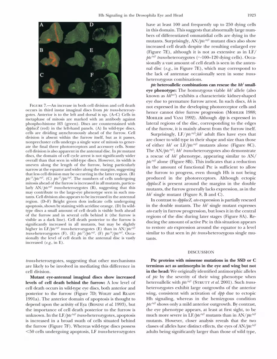
1923Hh Signaling in the Drosophila Eye and Head
have at least 100 and frequently up to 250 dying cellsin this domain. This suggests that abnormally large num-bers of differentiated ommatidial cells are dying in themutants. Surprisingly, AN/ptc tuf1 mutant discs also showincreased cell death despite the resulting enlarged eye(Figure 7E), although it is not as extensive as in LF/ptc tuf1 trans-heterozygotes (�100–120 dying cells). Occa-sionally a vast amount of cell death is seen in the anten-nal disc (e.g., in Figure 7E), which may correspond tothe lack of antennae occasionally seen in some trans-heterozygous combinations.
ptc heteroallelic combinations can rescue the hh1 small-eye phenotype: The homozygous viable hh1 allele (alsoknown as hhbar3) exhibits a characteristic kidney-shapedeye due to premature furrow arrest. In such discs, hh is
Figure 7.—An increase in both cell division and cell death not expressed in the developing photoreceptor cells andoccurs in third instar imaginal discs from ptc trans-heterozy- hence cannot drive furrow progression (Mohler 1988;gotes. Anterior is to the left and dorsal is up. (A–C) Cells in
Mohler and Vani 1992). Although dpp is expressed inmetaphase of mitosis are marked with an antibody againstlateral regions of the disc, corresponding to the edgesphospho-histone H3 (green). Discs are counterstained with
dpplacZ (red) in the left-hand panels. (A) In wild-type discs, of the furrow, it is mainly absent from the furrow itself.cells are dividing asynchronously ahead of the furrow. Cell Surprisingly, LF/ptc tuf1;hh1 adult flies have eyes thatdivision is absent within the furrow itself, but as it passes, are closer to wild type in their shape and size than thosenonprecluster cells undergo a single wave of mitosis to gener-
of either hh1 or LF/ptc tuf1 mutants alone (Figure 8C).ate the final three photoreceptors and accessory cells. SomeThe AN/ptc tuf1; hh1 trans-heterozygotes also demonstratecell division is also apparent in the antennal disc. In ptc mutant
discs, the domain of cell cycle arrest is not significantly wider a rescue of hh1 phenotype, appearing similar to AN/overall than that seen in wild-type discs. However, its width is ptc tuf1 alone (Figure 8B). This indicates that a reductionuneven along the length of the furrow, being particularly in the amount of functional Ptc is sufficient to allownarrow at the equator and wider along the margins, suggesting
the furrow to progress, even though Hh is not beingthat less cell division may be occurring in the latter region. (B)produced in the photoreceptors. Although ectopicptc13/ptctuf1. (C) ptc37/ptctuf1. The numbers of cells undergoing
mitosis ahead of the furrow is increased in all mutants, particu- dpplacZ is present around the margins in the doublelarly AN/ptctuf1 trans-heterozygotes (B), suggesting that this mutants, the furrow generally lacks expression, as in themay contribute to the large-eye phenotype seen in such mu- hh1 single mutant (Figure 8, B and C).tants. Cell division also appears to be increased in the antennal
In contrast to dpplacZ, ato expression is partially rescuedregion. (D–F) Bright green dots indicate cells undergoingin the double mutants. The hh1 single mutant expressesapoptosis, shown by staining with acridine orange. (D) In wild-
type discs a small amount of cell death is visible both ahead ato early in furrow progression, but loses it in the centralof the furrow and in several cells behind it (the furrow is regions of the disc during later stages (Figure 8A). Re-visible as a dark line). Cell death posterior to the furrow is ducing the amount of active Ptc in this situation appearssignificantly increased in all mutants, but may be slightly
to restore ato expression around the equator to a levelhigher in LF/ptctuf1 trans-heterozygotes (E) than in AN/ptctuf1
similar to that seen in ptc trans-heterozygous single mu-trans-heterozygotes (F). (E) ptc13/ptctuf1. (F) ptc16/ptctuf1. Occa-sionally the level of cell death in the antennal disc is vastly tants.increased (e.g., in E).
DISCUSSION
Ptc proteins with missense mutations in the SSD or Ctrans-heterozygotes, suggesting that other mechanismsterminus act as antimorphs in the eye and wing but notare likely to be involved in mediating this difference inin the head: We originally identified antimorphic allelescell division.of ptc by the severity of their wing phenotype whenMutant eye-antennal imaginal discs show increasedheteroallelic with ptc tuf1 (Strutt et al. 2001). Such trans-levels of cell death behind the furrow: A low level ofheterozygotes exhibit large outgrowths of the anteriorcell death occurs in wild-type eye discs, both anterior andwing, consistent with activation of dpp due to ectopicposterior to the furrow (Figure 7D; Wolff and ReadyHh signaling, whereas in the hemizygous condition1991a). The anterior domain of apoptosis is thought toptc tuf1 shows only a mild anterior outgrowth. By contrast,depend upon the activity of Eya (Bonini et al. 1993), butthe eye phenotype appears, at least at first sight, to bethe importance of cell death posterior to the furrow ismuch more severe in LF/ptc tuf1 mutants than in AN/ptc tuf1unknown. In the LF/ptc tuf1 trans-heterozygotes, apoptosismutants. However, closer analysis reveals that the twois increased in a broad swath of cells situated behindclasses of alleles have distinct effects, the eyes of AN/ptc tuf1the furrow (Figure 7F). Whereas wild-type discs possess
�50 cells undergoing apoptosis, LF trans-heterozygotes adults being significantly larger than those of wild type,

1924 C. Thomas and P. W. Ingham
Ptc does not act as a positive regulator of Smo inthe head: In the canonical Hh signaling pathway, Ptcfunctions as a negative regulator (Ingham et al. 1991)both by sequestering Hh ligand and by inhibiting Smoactivity (Hooper 1994; Alcedo et al. 1996; Chen andStruhl 1996). By contrast, Shyamala and Bhat (2002)presented evidence suggesting that Ptc and Smo act inconcert to promote growth in the head, a function thatis opposed by the activity of Hh. Our reexamination ofthis issue does not support such an atypical interactionbetween Ptc and Smo in the head; moreover, it suggeststhat the predominant role of Hh signaling in the headis in patterning rather than in proliferation.
Although many LF/ptc tuf1 mutants have reduced eyesand may lack antennae, there is often a concurrentincrease in actual head vertex size. Shyamala and Bhat(2002) do not specify whether the reduction in headsize they report in ptcGAL4 trans-heterozygotes is due to
Figure 8.—Trans-heterozygous ptc mutations rescue the hh1 a smaller vertex, a reduced eye size, or a lack of antennaladult-eye phenotype, but have less dramatic effects on the structures. However, our results suggest that they areexpression of different Hh target genes in the eye-antennal
likely observing an effect on the eye and antennae ratherdisc of hh1 mutants. Anterior is to the left and dorsal is up.than on the head vertex itself, which tends to exhibit(A) Flies homozygous for the hh1 allele exhibit narrow kidney-
shaped eyes due to premature arrest of the morphogenetic only mild bristle defects in such mutants. Their observa-furrow. dpplacZ expression is normal at the margins of third tion that removing one wild-type copy of smo in a ptcH84lacZ/instar eye-antennal discs (compare with Figure 6A), but absent ptcGAL4 background enhances the head defects led themin the furrow where it depends directly upon Hh signaling.
to suggest a novel positive interaction between Ptc andAto expression (green) is also absent in the central regionsSmo. However, if the reduction in Smo activity resultsof the disc and becomes downregulated as the furrow arrests.
Eye-antennal discs on the right are counterstained with the in a decrease in transcription of the mutant ptc alleles,neuronal marker HRP (red) to visualize developing photore- phenotypes caused primarily by a lack of Hh sequestra-ceptors. (B and C) The hh1 eye phenotype is rescued when tion will be enhanced by hemizygosity for smo. In trans-put into a ptc trans-heterozygote background. AN/ptctuf1;hh1
heterozygotes carrying AN alleles in combination withmutants appear similar to those carrying AN/ptctuf1 alone (B),the ptchdl allele (generated by Shyamala and Bhat), nobut the LF/ptctuf1 phenotype shows partial rescue with �50%
of such flies possessing eyes that are almost wild type in size head (or wing) defect is apparent (Shyamala and Bhat(C). In double mutants, the expression of dpplacZ is not re- 2002; our unpublished observations), suggesting thatstored in the furrow, although in some cases there does appear the ability of AN alleles to sequester Hh prevents severeto be some residual expression. However, Ato expression does
defects from occurring in the head. The lack of wingappear to be rescued in the trans-heterozygotes, although thephenotype also indicates that the Hh pathway is notdomain of expression is generally narrower and less intense
than that in wild type (compare with Figure 5A). Mutant ectopically activated in this allelic combination, im-genotypes are as follows: (B) ptc 13/ptc tuf1;hh1 and (C) ptc 16/ plying that the ptchdl allele must be able to inhibit Smoptc tuf1;hh1. A similar level of rescue is observed in ptc 14/ptc tuf1;hh1, quite effectively. If the predominant effect of the ptchdl
ptc 34/ptc tuf1;hh1, ptc 37/ptc tuf1;hh1, and ptc 47/ptc tuf1;hh1 mutants (notmutation is to compromise Hh sequestering activity, thisshown).may explain why ptchdl/LF trans-heterozygotes have suchsevere head defects compared with other LF trans-hetero-zygotes, whereas other tissues that are less sensitive torather than simply less reduced than those of LF/ptc tuf1
mutants. On the other hand, the head defects typical inefficient Hh sequestration are not affected as stronglyin such mutant animals.of AN/ptc tuf1 trans-heterozygotes do appear to be mild
versions of those seen in LF/ptc tuf1 mutants. Because We found that generating either ptc or smo clones inthe head vertex resulted in medio-lateral patterningantimorphic Ptc proteins are distinguished by their abil-
ity to sequester the Hh ligand (Johnson et al. 2000; defects. It is difficult to reconcile this with the smallhead phenotype observed with both ptc and smo clonesMartin et al. 2001; Strutt et al. 2001), this implies
that excessive Hh diffusion, rather than ectopic pathway by Shyamala and Bhat (2002), particularly as the eyesare often outgrown if they contain ptc clones, thus mak-activation due to Smo derepression, is the principal
cause of the head phenotypes such as ectopic ocelli. In ing the head appear larger overall. Inducing particularlylarge ptc or smo clones covering the entire vertex doessummary, it appears that the difference between allele
class phenotypes in wing, eye, and head reflects the differ- appear to lead to a reduction in head vertex size dueto a lack of specification of lateral or medial tissue,ent relative impact of cell-autonomous ectopic pathway
activation vs. excess Hh diffusion in the three different respectively. However, this happens extremely rarelyand it seems likely that the reduction is due primarilystructures.
1925Hh Signaling in the Drosophila Eye and Head
to the patterning defect and that the resulting lack of rescues the expression of ato, but not that of dpp. Thereare several possible explanations for these findings.proliferation is secondary. In addition, it is documentedFirst, ato is an indirect target of Hh signaling and thethat both ptc and smo clones located anteriorly to themediators of Hh activity in this context are unclear. Itanteroposterior boundary of the wing disc, where Hhis likely that other factors, in addition to those directlysignaling follows the canonical pathway, can result ininduced by Hh, are necessary for ato expression, anysimilar duplications of anterior wing tissue (Chen andone of which may be limiting. Second, dpp may respondStruhl 1996). This is due to the reduction in ptc tran-to lower levels of Hh pathway activation more than genesscription and hence to Hh sequestration that occursupstream of ato. In the wing disc, dpp is activated byin both cases, allowing Hh to traverse the clones andrelatively low levels of Hh anterior to the AP borderectopically activate signaling at their anterior edge. In(Ingham and Fietz 1995; Zecca et al. 1995), whereasconclusion, we find no evidence to suggest that Ptc actsother Hh target genes such as collier require a higheras a positive regulator of Smo in head development.level of pathway activation (Vervoort et al. 1999). InExpanding the range of Hh activity induces cell deathptc trans-heterozygotes some Ptc activity is retained andbehind the morphogenetic furrow: The eye disc con-hence the very highest level of Hh signaling cannot betains two domains of apoptosis: one immediately ante-reached. Third, Dpp in its role as an inducer of therior to the MF that is regulated by Eya and one posteriorpreproneural state can actually inhibit the expressionto the MF whose function is unknown (Wolff andof ato through activation of the proneural repressors hReady 1991a; Bonini et al. 1993). Our findings suggestand emc (Greenwood and Struhl 1999). In ptc trans-that the latter domain of cell death is promoted by Hhheterozygous discs, the domain of h expression doessignaling from photoreceptors. The milder increase inappear to be expanded, suggesting a possible explana-cell death seen in AN/ptc tuf1 trans-heterozygotes perhapstion for the observed downregulation of ato expression.reflects the efficient sequestration of Hh in such mu-A fourth possibility that has not been tested is that thetants, suggesting that the range of Hh diffusion may beincreased level of Hh signaling in ptc trans-heterozygoteimportant in influencing the proportion of cells thatdiscs results in an expansion of the domain of roughdie behind the MF. Normally, a large amount of cell(ro) expression. Ro is induced by high-level Hh signalingdeath, necessary for the elimination of two to threeat the posterior margin of the MF (Dominguez 1999),excess pigment cells per ommatidium, occurs in pupalbut, if expressed at excessive levels (as in the roDom mu-eye discs (Wolff and Ready 1991a). Cell death behindtant), causes a downregulation of ato expression (Cha-the MF may have a similar function, perhaps in remov-nut et al. 2000). Although a severe reduction in atoing cells mis-specified during differentiation. It is possi-expression such as that caused by roDom can result in
ble that Hh could regulate apoptosis through activationfurrow arrest, the significance of a mild downregulation
of a molecule at short range behind the MF. Alterna- of expression is unknown.tively, cell death may not depend directly upon Hh, but The two classes of mutants both show an increase inrather upon the presence of increased numbers of mis- dpplacZ expression relative to wild type. However, thespecified cells in ptc mutants that may result in cell death domain of ectopic expression differs significantly be-occurring to regulate differentiation effectively. tween allele types, suggesting a difference in the way in
The consequences of increased Hh signaling in the which the pathway is activated in the two classes of mutant.eye: The earliest known role of Hh in the eye imaginal Because LF ptc alleles cannot sequester Hh efficientlydisc is to help specify the dorso-ventral organizer (Cavo- (Strutt et al. 2001), the broad band of ectopic dpplacZdeassi et al. 1999). Incorrect DV specification compro- seen ahead of the MF may be caused by excessive dif-mises the function of N in promoting growth, resulting fusion of Hh anteriorly. In contrast, the AN/ptc tuf1
in small eyes (Papayannopoulos et al. 1998). However, trans-heterozygotes can sequester Hh efficiently andthe small-eye phenotype seen in LF/ptc tuf1 appears to be consequently demonstrate only phenotypes caused byunconnected to this process. Although some disorgani- autonomous ectopic pathway activation.zation of the equator is seen in ptc trans-heterozygotes, it Despite the rescue of adult-eye phenotype observedis not significant, and its presence in both classes of mutant in ptc/ptc tuf1;hh1 double mutants, the expression ofindicates that the effect, if any, on eye size is small. dpplacZ was not restored in the center of the disc. Since
The two major targets of Hh signaling during MF Dpp does not play a major role in MF progressionprogression are dpp and ato (Heberlein et al. 1993; Ma (Burke and Basler 1996; Greenwood and Struhlet al. 1993; Jarman et al. 1994). Our data indicate that 1999), the lack of expression in this situation may notalthough dpp is ectopically activated in ptc trans-heterozy- have a significant effect. Alternatively, excessive dpp ex-gotes, ato is not. This is unexpected as Hh signaling acti- pression at the margins may allow the protein to diffusevates the initial expression of ato (Jarman et al. 1994), medially into the disc, thus aiding MF progression inso an increase might be anticipated to expand the ato an unconventional way.expression domain into more anterior regions, while Dpp is known to have several functions in the eyemaintaining or even increasing the level of expression. disc, all of which, when modified, can influence the
final size and shape of the adult eye. However, despiteConversely, reducing the activity of ptc in the hh1 mutant
1926 C. Thomas and P. W. Ingham
Blackman, R. K., M. Sanicola, L. A. Raftery, T. Gillevet and W. M.the disparity between the patterns of dpplacZ expressionGelbart, 1991 An extensive 3� cis-regulatory region directs the
in the two types of trans-heterozygote, surprisingly little imaginal disk expression of decapentaplegic, a member of theTGF-� family in Drosophila. Development 111: 657–665.difference is detected downstream of Dpp. Although
Bonini, N. M., W. M. Leiserson and S. Benzer, 1993 The eyes absentectopic dpp expression has been shown to induce ec-gene: genetic control of cell survival and differentiation in the
topic MFs (Chanut and Heberlein 1997; Pignoni and developing Drosophila eye. Cell 72: 379–395.Bonini, N. M., Q. T. Bui, G. L. Gray-Board and J. M. Warrick, 1997Zipursky 1997), this does not occur in the ptc trans-
The Drosophila eyes absent gene directs ectopic eye formation inheterozygotes, presumably because the ectopic Dpp isa pathway conserved between flies and vertebrates. Development
either not high enough or not expressed in the right 124: 4819–4826.Borod, E. R., and U. Heberlein, 1998 Mutual regulation of decapen-place.
taplegic and hedgehog during the initiation of differentiation inIn addition to its effect on furrow initiation, Dpp isthe Drosophila retina. Dev. Biol. 197: 187–197.
also responsible for defining the eye field via inhibition Brook, W. J., and S. M. Cohen, 1996 Antagonistic interactions be-tween Wingless and Decapentaplegic responsible for dorsal-ven-of Wg and for inducing cell cycle arrest ahead of thetral pattern in the Drosophila leg. Science 273: 1373–1377.furrow (Royet and Finkelstein 1997; Horsfield et al.
Brown, N. L., C. A. Sattler, S. W. Paddock and S. B. Carroll,1998). Small-eye mutants do show an increased head 1995 Hairy and Emc negatively regulate morphogenetic furrow
progression in the Drosophila eye. Cell 80: 879–887.vertex size, suggesting that an eye-to-vertex fate changeBurke, R., and K. Basler, 1996 Hedgehog-dependent patterninghas occurred. However, ptc trans-heterozygotes do not in the Drosophila eye can occur in the absence of Dpp signaling.
display critical differences either from wild type or be- Dev. Biol. 179: 360–368.Campos-Ortega, J. A., and M. Waitz, 1978 Cell clones and patterntween allele classes in the distribution of Wg in second
formation: developmental restrictions in the compound eye ofinstar eye discs. In addition, dpp expression is actually Drosophila. Wihelm Roux’s Arch. Dev. Biol. 184: 155–170.expanded in eye discs of small-eye mutants, indicating Capdevila, J., M. P. Estrada, E. Sanchez-Herrero and I. Guerrero,
1994 The Drosophila segment polarity gene patched interactsthat processes other than Wg/Dpp antagonism must bewith decapentaplegic in wing development. EMBO J. 13: 71–82.involved in specification of eye vs. head domains. Carroll, S. B., and J. S. Whyte, 1989 The role of the hairy gene
Ectopic Dpp ahead of the furrow does not appear to during Drosophila morphogenesis: stripes in imaginal discs.Genes Dev. 3: 905–916.induce premature cell cycle arrest and therefore cannot
Carstea, E. D., J. A. Morris, K. G. Coleman, S. K. Loftus, D.explain the reduced-eye phenotypes observed in LF/ Zhang et al., 1997 Niemann-Pick C1 disease gene: homologyptc tuf trans-heterozygotes. However, when compared to to mediators of cholesterol homeostasis. Science 277: 228–231.
Cavodeassi, F., R. Diez Del Corral, S. Campuzano and M. Domin-wild type, ptc mutants do show an increase in cell divi-guez, 1999 Compartments and organising boundaries in thesions ahead of the furrow. We suggest that in addition to Drosophila eye: the role of the homeodomain Iroquois proteins.
an eye/head vertex specification defect, LF/ptc tuf1 trans- Development 126: 4933–4942.Chanut, F., and U. Heberlein, 1997 Role of decapentaplegic in initia-heterozygotes may exhibit a small-eye phenotype due
tion and progression of the morphogenetic furrow in the devel-to excessive cell death, despite some increase in cell oping Drosophila retina. Development 124: 559–567.
Chanut, F., A. Luk and U. Heberlein, 2000 A screen for dominantdivisions ahead of the furrow. Conversely, in DN/ptc tuf1
modifiers of ro(Dom), a mutation that disrupts morphogenetictrans-heterozygotes, increased proliferation could over-furrow progression in Drosophila, identifies groucho and hairless
compensate for increased cell death, leading to larger as regulators of atonal expression. Genetics 156: 1203–1217.Chen, R., M. Amoui, Z. Zhang and G. Mardon, 1997 Dachshundeyes. This suggests that the adult-eye phenotype is at
and Eyes absent proteins form a complex and function synergisti-least partially dependent upon a balance between cellcally to induce ectopic eye development in Drosophila. Cell 91:
division and cell death in the disc, in addition to an 893–903.Chen, R., G. Halder, Z. Zhang and G. Mardon, 1999 Signalingeye-to-head fate change.
by the TGF-� homolog decapentaplegic functions reiterativelyWe thank David Strutt, Andrew Jarman, Ken Howard, and Penny within the network of genes controlling retinal cell fate determi-
Rashbass for antibodies and/or Drosophila stocks. We are also grateful nation in Drosophila. Development 126: 935–943.Chen, Y., and G. Struhl, 1996 Dual roles for Patched in sequester-to Monica Kibart for maintenance of Drosophila stocks and John
ing and transducing Hedgehog. Cell 87: 553–563.Proctor for assisting with scanning electron microscopy. The confocalChin, D. J., G. Gil, D. W. Russell, L. Liscum, K. L. Luskey et al.,microscopy facility at the Centre for Developmental Genetics is funded
1984 Nucleotide sequence of a 3-hydroxy-3-methyl-glutaryl co-by a grant from Yorkshire Cancer Research. This analysis was sup-enzyme A reductase, a glycoprotein of endoplasmic reticulum.ported by a Wellcome Trust Program grant to P.W.I. Nature 308: 613–617.
Cho, K., and K. W. Choi, 1998 Fringe is essential for mirror symme-try and morphogenesis in the Drosophila eye. Nature 396: 272–276.
Choi, K. W., B. Mozer and S. Benzer, 1996 Independent determina-LITERATURE CITEDtion of symmetry and polarity in the Drosophila eye. Proc. Natl.
Alcedo, J., M. Ayzenzon, T. Von Ohlen, M. Noll and J. E. Hooper, Acad. Sci. USA 93: 5737–5741.1996 The Drosophila smoothened gene encodes a seven-pass Curtiss, J., and M. Mlodzik, 2000 Morphogenetic furrow initiationmembrane protein, a putative receptor for the Hedgehog signal. and progression during eye development in Drosophila: the rolesCell 86: 221–232. of decapentaplegic, hedgehog and eyes absent. Development 127:
Baker, W. K., 1978 A clonal analysis reveals early developmental 1325–1336.restrictions in the Drosophila head. Dev. Biol. 62: 422–463. Dokucu, M. E., S. L. Zipursky and R. L. Cagan, 1996 Atonal,
Baonza, A., and M. Freeman, 2001 Notch signalling and the initia- Rough and the resolution of proneural clusters in the developingtion of neural development in the Drosophila eye. Development Drosophila retina. Development 122: 4139–4147.128: 3889–3898. Dominguez, M., 1999 Dual role for Hedgehog in the regulation
Baonza, A., and M. Freeman, 2002 Control of Drosophila eye speci- of the proneural gene atonal during ommatidia development.Development 126: 2345–2353.fication by Wingless signalling. Development 129: 5313–5322.
1927Hh Signaling in the Drosophila Eye and Head
Dominguez, M., and J. F. de Celis, 1998 A dorsal/ventral boundary Masucci, J. D., R. J. Miltenberger and F. M. Hoffmann, 1990 Pat-tern-specific expression of the Drosophila decapentaplegic gene inestablished by Notch controls growth and polarity in the Drosoph-imaginal disks is regulated by 3� cis-regulatory elements. Genesila eye. Nature 396: 276–278.Dev. 4: 2011–2023.Dominguez, M., and E. Hafen, 1997 Hedgehog directly controls
McMahon, A. P., P. W. Ingham and C. J. Tabin, 2003 Develop-initiation and propagation of retinal differentiation in the Dro-mental roles and clinical significance of Hedgehog signaling.sophila eye. Genes Dev. 11: 3254–3264.Curr. Top. Dev. Biol. 53: 1–114.Gomez-Skarmeta, J., R. Diez Del Corral, E. De La Calle-Mustien-
McNeill, H., C. H. Yang, M. Brodsky, J. Ungos and M. A. Simon,nes, D. Ferres-Marco and J. Modolell, 1996 araucan and1997 mirror encodes a novel PBX-class homeoprotein that func-caupolican, two members of the novel Iroquois complex, encodetions in the definition of the dorsal-ventral border in the Drosoph-homeoproteins that control proneural and vein-forming genes.ila eye. Genes Dev. 11: 1073–1082.Cell 85: 95–105.
Mohler, J., 1988 Requirements for hedgehog, a segmental polarityGreenwood, S., and G. Struhl, 1999 Progression of the morphoge-gene, in patterning larval and adult cuticle of Drosophila. Genet-netic furrow in the Drosophila eye: the roles of Hedgehog, Deca-ics 120: 1061–1072.pentaplegic and the Raf pathway. Development 126: 5795–5808.
Mohler, J., and K. Vani, 1992 Molecular organisation and embry-Haynie, J. L., and P. J. Bryant, 1986 Development of the eye-onic expression of the hedgehog gene involved in cell-cell commu-antenna imaginal disc and morphogenesis of the adult head innication in segmental patterning in Drosophila. DevelopmentDrosophila melanogaster. J. Exp. Zool. 237: 293–308.115: 957–971.Hazelett, D. J., M. Bourouis, U. Walldorf and J. E. Treisman,
Mullor, J. L., and I. Guerrero, 2000 A gain-of-function mutant of1998 decapentaplegic and wingless are regulated by eyes absent andpatched dissects different responses to the Hedgehog gradient.eyegone and interact to direct the pattern of retinal differentiationDev. Biol. 228: 211–224.in the eye disc. Development 125: 3741–3751.
Munro, S., and M. Freeman, 2000 The Notch signalling regulatorHeberlein, U., T. Wolff and G. M. Rubin, 1993 The TGFß homologFringe acts in the Golgi apparatus and requires the glycosylrans-dpp and the segment polarity gene hedgehog are required forferase signature motif DXD. Curr. Biol. 10: 813–820.propagation of a morphogenetic wave in the Drosophila retina.
Nakano, Y., I. Guerrero, A. Hidalgo, A. Taylor, J. R. S. WhittleCell 75: 913–926.et al., 1989 A protein with several possible membrane-spanningHendzel, M. J., Y. Wei, M. A. Mancini, A. Van Hooser, T. Ranalli etdomains encoded by the Drosophila segment polarity patched.al., 1997 Mitosis-specific phosphorylation of histone H3 initiatesNature 341: 508–513.primarily within pericentric heterochromatin during G2 and
Panin, V. M., V. Papayannopoulos, R. Wilson and K. D. Irvine,spreads in an ordered fashion coincident with mitotic chromo-1997 Fringe modulates Notch-ligand interactions. Nature 387:some condensation. Chromasoma 106: 348–360.908–912.Hidalgo, A., 1989 Molecular cloning of ptc and analysis of its role
Papayannopoulos, V., A. Tomlinson, V. M. Panin, C. Rauskolbin intrasegmental patterning in D. melanogaster. Ph.D. Thesis,and K. D. Irvine, 1998 Dorsal-ventral signaling in the Drosoph-University of Oxford, Oxford.ila eye. Science 281: 2031–2034.Hooper, J. E., 1994 Distinct pathways for autocrine and paracrine
Pappu, K. S., R. Chen, B. W. Middlebrooks, C. Woo, U. HeberleinWingless signalling in Drosophila embryos. Nature 372: 461–464.et al., 2003 Mechanism of hedgehog signaling during DrosophilaHorsfield, J., A. Penton, J. Secombe, M. Hoffman and H. Richard-eye development. Development 130: 3053–3062.son, 1998 decapentaplegic is required for arrest in G1 phase dur-
Pignoni, F., and S. L. Zipursky, 1997 Induction of Drosophila eyeing Drosophila eye development. Development 125: 5069–5078.development by Decapentaplegic. Development 124: 271–278.Hua, X., A. Nohturfft, J. L. Goldstein and M. S. Brown, 1996
Pignoni, F., B. Hu, K. H. Zavitz, J. Xiao, P. A. Garrity et al., 1997Sterol resistance in CHO cells traced to point mutation in SREBPThe eye-specification proteins So and Eya form a complex andcleavage-activating protein. Cell 87: 415–426.regulate multiple steps in Drosophila eye development. Cell 91:Ingham, P. W., and M. J. Fietz, 1995 Quantitative effects of Hedge-881–891.hog and Decapentaplegic activity of the patterning of the Drosoph-
Ready, D. F., T. E. Hanson and S. Benzer, 1976 Development ofila wing. Curr. Biol. 5: 432–440. the Drosophila retina, a neurocrystalline lattice. Dev. Biol. 53:Ingham, P. W., and A. P. McMahon, 2001 Hedgehog signalling in 217–240.animal development: paradigms and principles. Genes Dev. 15: Royet, J., and R. Finkelstein, 1996 hedgehog, wingless and orthoden-3059–3087. ticle specify adult head development in Drosophila. DevelopmentIngham, P. W., A. M. Taylor and Y. Nakano, 1991 Role of the 122: 1849–1858.
Drosophila patched gene in positional signalling. Nature 353: 184– Royet, J., and R. Finkelstein, 1997 Establishing primordia in the187. Drosophila eye-antennal disc: the roles of decapentaplegic, wingless
Jarman, A. P., E. H. Grell, L. Ackerman, L. Y. Jan and Y. N. Jan, and hedgehog. Development 124: 4793–4800.1994 atonal is the proneural gene for Drosophila photorecep- Shyamala, B. V., and K. M. Bhat, 2002 A positive role for Patched-tors. Nature 369: 398–400. Smoothened signaling in promoting cell proliferation during
Johnson, R. L., L. Milenkovic and M. P. Scott, 2000 In vivo func- normal head development in Drosophila. Development 129:tions of the Patched protein: requirement of the C-terminus for 1839–1847.target gene inactivation but not Hedgehog sequestration. Mol. Speicher, S. A., U. Thomas, U. Hinz and E. Knust, 1994 The serrateCell 6: 467–478. locus of Drosophila and its role in morphogenesis of the wing
Lee, J. D., and J. E. Treisman, 2001 The role of Wingless signaling imaginal discs: control of cell proliferation. Development 120:in establishing the anteroposterior and dorsoventral axes of the 535–544.eye disc. Development 128: 1519–1529. Strutt, H., C. Thomas, Y. Nakano, D. Stark, B. Neave et al., 2001
Loftus, S. K., J. A. Morris, E. D. Carstea, J. D. Gu, C. Cummings Mutations in the sterol-sensing domain of Patched suggest a roleet al., 1997 Murine model of Niemann-Pick C disease: mutation for vesicular trafficking in Smoothened regulation. Curr. Biol.in a cholesterol homeostasis gene. Science 277: 232–235. 11: 608–613.
Ma, C., and K. Moses, 1995 wingless and patched are negative regula- Tomlinson, A., 1985 The cellular dynamics of pattern formationtors of the morphogenetic furrow and can affect tissue polarity in the eye of Drosophila. J. Embryol. Exp. Morphol. 89: 313–331.in the developing Drosophila compound eye. Development 121: Tomlinson, A., 1988 Cellular interactions in the developing Dro-2279–2289. sophila eye. Development 104: 183–193.
Ma, C., Y. Zhou, P. A. Beachy and K. Moses, 1993 The segment Tomlinson, A., and D. F. Ready, 1987 Neuronal differentiation inpolarity gene hedgehog is required for progression of the morpho- the Drosophila ommatidium. Dev. Biol. 120: 366–376.genetic furrow in the developing Drosophila eye. Cell 75: 927– Treisman, J. E., and G. M. Rubin, 1995 wingless inhibits morphoge-938. netic furrow movement in the Drosophila eye disc. Development
Martin, V., G. Carrillo, C. Torroja and I. Guerrero, 2001 The 121: 3519–3527.sterol-sensing domain of Patched protein seems to control Vegh, M., and K. Basler, 2003 A genetic screen for HedgehogSmoothened activity through Patched vesicular trafficking. Curr. targets involved in the maintenance of the Drosophila anteropos-
terior compartment boundary. Genetics 163: 1427–1438.Biol. 11: 601–607.

1928 C. Thomas and P. W. Ingham
Vervoort, M., M. Crozatier, D. Valle and A. Vincent, 1999 The tion in the Drosophila compound eye: the morphogenetic furrowCOE transcription factor Collier is a mediator of short-range and the second mitotic wave. Development 113: 841–850.Hedgehog-induced patterning of the Drosophila wing. Curr. Biol. Zecca, M., K. Basler and G. Struhl, 1995 Sequential organizing9: 632–639. activities of engrailed, hedgehog and decapentaplegic in the Dro-
Wolff, T., and D. F. Ready, 1991a Cell death in normal and rough sophila wing. Development 121: 2265–2278.eye mutants of Drosophila. Development 113: 825–839.
Wolff, T., and D. F. Ready, 1991b The beginning of pattern forma- Communicating editor: K. Anderson


