Embed Size (px)
Citation preview
www.elsevier.com/locate/ydbio
Developmental Biology 267 (2004) 387–398
Roles for Hedgehog signaling in androgen production and prostate
ductal morphogenesis
David M. Berman,a,b,c,*,1 Nishita Desai,d,e,1 Xi Wang,d,e Sunil S. Karhadkar,a,b,c
Melissa Reynon,d,e Cory Abate-Shen,b,f,g Philip A. Beachy,a,c and Michael M. Shend,e,*
aDepartment of Molecular Biology and Genetics, Johns Hopkins University School of Medicine, Baltimore, MD 21205, USAbDepartment of Pathology, Johns Hopkins University School of Medicine, Baltimore, MD 21205, USA
cHoward Hughes Medical Institute, Johns Hopkins University School of Medicine, Baltimore, MD 21205, USAdCenter for Advanced Biotechnology and Medicine, UMDNJ-Robert Wood Johnson Medical School, Piscataway, NJ 08854, USA
eDepartment of Pediatrics, UMDNJ-Robert Wood Johnson Medical School, Piscataway, NJ 08854, USAfDepartment of Medicine, UMDNJ-Robert Wood Johnson Medical School, Piscataway, NJ 08854, USA
gDepartment of Neuroscience and Cell Biology, UMDNJ-Robert Wood Johnson Medical School, Piscataway, NJ 08854, USA
Received for publication 8 September 2003, revised 14 November 2003, accepted 19 November 2003
Abstract
Previous studies have demonstrated that the Hedgehog (Hh) signaling pathway plays a critical role in the development and patterning of
many endodermally derived tissues. We have investigated the role of Sonic hedgehog (Shh) in formation of the prostate gland by examining
the urogenital phenotype of Shh mutant fetuses. Consistent with earlier work reporting an essential role for Shh in prostate induction, we have
found that Shh mutant fetuses display abnormal urogenital development and fail to form prostate buds. Unexpectedly, however, we have
discovered that this prostate defect could be rescued by three different methods: renal grafting, explant culture in the presence of androgens,
and administration of dihydrotestosterone (DHT) to pregnant mice, indicating that the prostate defect in Shh mutants is due to insufficient
levels of androgens. Furthermore, we find that the inhibition of Hh pathway signaling by treatment with cyclopamine does not block prostate
formation in explant culture, but instead produces morphological defects consistent with a role for Hh signaling in ductal patterning. Taken
together, our studies indicate that the initial organogenesis of the prostate proceeds independently of Shh, but that Shh or other Hh ligands
may play a role in subsequent events that pattern the prostate.
D 2003 Elsevier Inc. All rights reserved.
Keywords: Hedgehog; Endoderm; Organogenesis; Ductal morphogenesis; Tissue recombination; Androgens
tiation (reviewed in Grapin-Botton and Melton, 2000; Rob-
IntroductionOrganogenesis of gut-derived tissues in vertebrates is
mediated by epithelial–mesenchymal interactions that in-
duce regionalized expression of tissue-specific transcription
factors, which in turn regulate organ patterning and differen-
0012-1606/$ - see front matter D 2003 Elsevier Inc. All rights reserved.
doi:10.1016/j.ydbio.2003.11.018
* Corresponding authors. D.M. Berman is to be contacted at Depart-
ment of Pathology, Johns Hopkins University School of Medicine,
Baltimore, MD 21287. Fax: +1-410-614-8401. M.M. Shen, Center for
Advanced Biotechnology and Medicine, UMDNJ-Robert Wood Johnson
Medical School, 679 Hoes Lane, Piscataway, NJ 08854. Fax: +1-732-235-
5373.
E-mail addresses: [email protected] (D.M. Berman),
[email protected] (M.M. Shen).1 These authors made equal contributions.
erts, 2000). To date, however, most studies have focused on
patterning and differentiation of foregut- and midgut-derived
tissues, while relatively little is known about organogenesis
of hindgut derivatives, such as the prostate gland.
The formation of the mammalian prostate occurs through
epithelial budding from the urogenital sinus, which is derived
from an extension of the caudal hindgut that arises during
midgestation. In the mouse, prostate organogenesis occurs
toward the end of gestation, at approximately 17.5 days post
coitum (dpc), when prostatic epithelial buds emerge from the
urogenital sinus under the influence of unknown inductive
signals from the mesenchyme. During subsequent ductal
morphogenesis, occurring in the first 3 weeks of postnatal
development, the prostatic epithelial buds elongate and
canalize to form ducts, which branch extensively into the

D.M. Berman et al. / Developmental Biology 267 (2004) 387–398388
surrounding mesenchyme (Sugimura et al., 1986; Timms et
al., 1994). At maturity, the rodent prostate gland is a multi-
lobular tissue, consisting of anterior, dorsolateral, and ventral
lobes, which are arranged circumferentially around the blad-
der and display characteristic patterns of ductal branching,
histological features, and secretory protein production (Hay-
ashi et al., 1991; Sugimura et al., 1986). The identities of
these lobes are likely to be specified at early stages of prostate
formation, based at least in part on the position of the
emerging epithelial buds. Notably, the emerging prostatic
epithelial buds are marked by expression of the Nkx3.1
homeobox gene, which is dependent on epithelial–mesen-
chymal interactions during prostate formation, and represents
the earliest known marker for the prostatic epithelium (Bha-
tia-Gaur et al., 1999; Sciavolino et al., 1997).
The prostate gland also represents a model system for
studying the generation of sexual dimorphism, as its for-
mation, growth, and function are continually dependent on
androgen receptor signaling (reviewed in Cunha, 1994;
Cunha et al., 1987; Hayward et al., 1997). Notably, func-
tional androgen receptors are in the urogenital sinus mes-
enchyme during embryogenesis, whereas they are found in
both the mesenchyme and epithelium postnatally. Consis-
tent with this observation, tissue recombination studies
using mutant mice have demonstrated that androgen recep-
tors are initially required in the mesenchyme to produce
signals for prostate induction and growth, and only later in
the epithelium for the secretory function of differentiated
cell types.
Recent studies have shown that the Shh signaling path-
way plays a key role in mediating epithelial–mesenchymal
interactions during formation of many endodermal tissues.
For example, Shh expressed by ventral foregut endoderm
activates Hh pathway targets in adjacent primitive lung
mesenchyme through binding to and inactivating its trans-
porter-like receptor Patched (Ptc). Such binding alleviates
inhibition by Ptc of the seven-transmembrane protein
Smoothened (Smo), allowing activation of Hh target genes
through the Gli family of latent transcription factors. One
such target is Ptc itself, whose high-level expression illus-
trates Hh pathway activity that is required in developing
lung mesenchyme to support branching morphogenesis of
adjacent airway endoderm (Bellusci et al., 1997; Litingtung
et al., 1998).
In the case of the prostate gland, previous studies have
proposed that Shh maybe required not only for branching
morphogenesis, but also for the initial formation of prostatic
buds. Based on neutralizing antibody treatment of grafts and
pharmacological inhibition of Shh signaling in organ culture
assays (Lamm et al., 2002; Podlasek et al., 1999), this
potential role for Shh in the early outgrowth of prostatic
epithelial buds (prostate induction) is distinct from roles
described in other developing organs. For example, lung
buds form normally in mice with deficient Hh signaling, but
fail to undergo subsequent branching morphogenesis (Ho-
gan et al., 1997; Litingtung et al., 1998; Motoyama et al.,
1998). Likewise, hair follicle formation is initially normal in
mice lacking Shh function, but subsequent morphogenesis is
severely impaired (Chiang et al., 1999; St-Jacques et al.,
1998).
Consequently, we have investigated the role of Shh in
prostate formation through phenotypic analysis of mutant
mice. Although Shh mutant embryos fail to form prostate
glands, we find that the urogenital sinus tissues from these
mutants can form Nkx3.1-expressing prostatic tissue in
tissue recombination and organ culture assays. Furthermore,
our results obtained by treatment of prostatic organ cultures
with Shh protein or with the Hh pathway antagonist cyclop-
amine indicate that Hh signaling is not required for prostate
formation, but may instead play a role in prostatic ductal
morphogenesis.
Methods
Mouse strains and genotyping
Shh mutant mice were maintained in a CD1 outbred
strain background and genotyped as described (Chiang et
al., 1996). XY fetuses were identified using PCR primers
directed against Sry: 5VGAGAGCATGGAGGGCCAT 3Vand5VCCACTCCTCTGTGACACT 3V(Bowles et al., 1999).
The Nkx3.1tm2(lacZ)Mms allele will be described in further
detail in a separate study (Y.-P. Hu, M. Reynon, N.D., S.M.
Price, C.A.-S., M.M.S., in preparation). Nkx3.1tm2(lacZ)Mms
heterozygous embryos were obtained by mating of
Nkx3.1tm2(lacZ)Mms homozygous males with C57Bl/6
females.
Tissue recombination
Tissue recombinations and tissue grafts were performed
essentially as described (Cunha, 1994; Cunha and Donja-
cour, 1987). In brief, wild-type urogenital sinuses were
removed at 16.5 dpc, and dissected away from urethra,
ductus deferens, and ureters; for Shh mutants, urogenital
regions containing portions of bladder and hindgut were
used without extensive dissection. Epithelial and mesen-
chymal components were separated by treatment with 1%
trypsin at 4jC for 90 min, followed by mechanical disso-
ciation. Tissue recombinations were constructed by combi-
nation of dissociated mesenchyme and epithelium on 0.4%
agar plates, followed by incubation with DMEM/10% fetal
bovine serum overnight at 37j. Successful recombinations
were surgically implanted under the kidney capsule of male
nude mice the next day, and were harvested following 4
weeks of growth.
Explant culture
For organ culture, wild-type and Shh mutant urogenital
regions were collected as described above, but without

D.M. Berman et al. / Developmental
trypsinization. Organ culture of urogenital sinus rudiments
was performed essentially as described (Lopes et al., 1996).
In brief, explants were cultured in a serum-free medium
containing Ham’s F12/DMEM H-16 (50:50), 1 g/l glucose,
2 mM glutamine, nonessential amino acids, HEPES buffer,
10 Ag/ml insulin, 10 Ag/ml transferrin (Sigma) 50 Ag/ml
gentamycin, 50 U/ml penicillin, 50 Ag/ml streptomycin;
testosterone (4-Androsten-17heta-ol-3-one) was dissolved
in DMSO and used at concentration of 10�7 M. Explants
were cultured for 7 days at 37jC with 5% CO2 on Millicell
CM 0.4 Micron filters (Millipore, Bedford, MA), with
media changed every 48 h.
Shh-N terminal peptide (R&D Systems) was dissolved
in PBS/0.1% BSA and used at 30 nM concentration.
Cyclopamine purified from Veratrum extract and its inac-
tive analogs tomatidine (Sigma) and solanidine (ICN,
Costa Mesa, CA) were dissolved in MeOH (cyclopamine
and tomatidine) or EtOH (solanidine), and used at a
concentration of 5 AM.
For morphometric analysis, prostatic ducts were digitally
traced on photomicrographs of h-galactosidase-stainedexplants using Adobe Illustrator (Adobe Systems), and
two-dimensional areas were calculated using CADTools
(Hot Door, Inc.) plug-in software. Statistical analysis was
performed using Graphpad Prism (GraphPad Software, Inc.).
In situ hybridization, immunohistochemistry, and
b-galactosidase staining
Radioactive in situ hybridization on paraffin sections was
performed according to Berman et al. (1995), using 33P-
labelled riboprobes reverse-transcribed from a 1-kb EcoRI
fragment of the Nkx3.1 cDNA (Sciavolino et al., 1997).
After autoradiography, slides were stained with hematoxylin
and eosin and photographed under dark-field and bright-
field illumination. Dark-field images were false-colored
green and overlayed onto corresponding bright-field images
using Adobe Photoshop (Adobe Systems). Nonradioactive
in situ hybridization to cryosections was performed as
described (Sciavolino et al., 1997). For h-galactosidasestaining, explants were fixed in 2% paraformaldehyde and
stained as described (Ben-Arie et al., 2000). Immunohisto-
chemical staining was performed according to Kim et al.
(2002), using an antimouse Nkx3.1 polyclonal antiserum
(Kim et al., 2002), or anti-p63 monoclonal antibody (Santa
Cruz).
In utero virilization
Timed pregnant females from crosses between mice
heterozygous for the mutant Shh allele (Chiang et al.,
1996) were injected subcutaneously with triolein (ICN) in
20% EtOH vehicle, or with vehicle containing 50 mg/kg
dihydrotestosterone (Steraloids, Newport, RI) from 10.5–
17.5 dpc of gestation. Fetuses were harvested on 18.5 dpc
and processed for routine histology.
Real-time RT-PCR analysis
For mRNA quantitation, first-strand cDNAwas prepared
from organ cultures using Trizol reagent and SuperScript
reverse transcriptase (Invitrogen). Real-time PCR amplifi-
cation of first-strand cDNAwas performed using a Mx4000
quantitative PCR instrument (Stratagene) with LUXk fluo-
rogenic primers (Invitrogen), using the following primer
pairs: for Ptc, unlabeled 5VGGCCTTCGCTGTGGGATTA 3Vand FAM (6-carboxy-fluorescein)-labeled 5VCAACGCCA-CAGCTCCTCCACGTTG 3V; for h-actin, unlabeled
5VGGTTGGCCTTAGGGTTCAGG 3Vand JOE (6-carboxy-
4 V,5 V-dichloro-2 V,7 V-dimethoxy-f luorescein)- labeled
5VCACGCCACCTTCTACAATGAGCTGCGTG 3 V. Resultsfor Ptc levels were normalized to h-actin levels.
Biology 267 (2004) 387–398 389
Results
Urogenital phenotype of Shh mutant mice
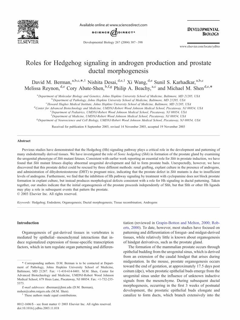
Previous studies have shown that components of the Hh
signaling pathway are expressed in the developing urogenital
system at stages before prostate formation (Lamm et al.,
2002; Podlasek et al., 1999). We have now found that Shh is
expressed in the urogenital sinus (UGS) epithelium at 16.5
dpc, in the basal layer immediately adjacent to the urogenital
mesenchyme (Fig. 1A); after prostatic buds emerge, Shh is
localized to the nascent epithelial buds (Lamm et al., 2002;
data not shown). In contrast, Ptc is expressed in the urogenital
mesenchyme adjoining the epithelium (Fig. 1B), indicating
that the Hh pathway is active in signaling from the epithelium
to the mesenchyme. Other downstream targets of the Hh
pathway are expressed in the urogenital mesenchyme at 16.5
dpc, including BMP4 and HIP, which encodes a soluble
inhibitor of Hh signaling (Chuang and McMahon, 1999)
(Figs. 1C,D).
To evaluate whether Shh is indeed required for prostate
formation, we examined the urogenital phenotype of male
Shh mutant fetuses that survived to 18.5 dpc. These
fetuses display severe cyclopia and limb defects, and
were identified as genotypically male by PCR using
primers directed against the Y-linked sex-determining
gene Sry. We found that XY Shh mutant fetuses have
severe morphological defects in the urogenital sinus,
including failure of prostatic budding by 18.5 dpc; we
also confirmed the anorectal defects previously described
for Shh mutants (Kimmel et al., 2000; Mo et al., 2001).
Histological sections of XY Shh mutants through the
urogenital sinus revealed profound disorganization of the
UGS and a complete absence of ductal budding immedi-
ately caudal to the bladder neck, indicating a lack of
prostate formation (n = 7) (Fig. 2). Seminal vesicle
differentiation was also undetectable in Shh mutant male
embryos, suggesting a broader requirement for Shh in
male phenotypic sexual differentiation. To confirm the
Fig. 1. Expression of Shh pathway components in 16.5 dpc urogenital sinus. In situ hybridization detection of Shh (A), Ptc (B), and the downstream targets
BMP4 (C) and HIP (D) in transverse sections caudal to the bladder, dorsal side up. Dashed lines indicate boundary between the urogenital epithelium (UGE)
and urogenital mesenchyme (UGM). Note that Shh expression is restricted to the urogenital epithelium, that Ptc and HIP are expressed primarily in urogenital
mesenchyme, and that BMP4 is exclusively expressed in mesenchyme. Scale bars correspond to 100 Am.
D.M. Berman et al. / Developmental Biology 267 (2004) 387–398390
absence of prostatic buds, we performed in situ hybrid-
ization to detect expression of Nkx3.1, which represents
the earliest known marker of prostatic ductal budding. No
Fig. 2. Failure of male sexual differentiation in 18.5 dpc Shh mutant embryos. P
show urogenital sinus (UGS) caudal to the bladder (BL) in both wild-type (A,C)
urogenital sinus epithelium (UGE) into urogenital sinus mesenchyme (UGM) in w
prostate (C). Prostate buds are absent in Shh mutant males (D). Also note prominen
in Shh mutants (D). Abbreviations: BL, bladder; DLP, dorsolateral prostate; SV, se
UGS, urogenital sinus; VP, ventral prostate.
evidence of Nkx3.1 expression was detected in XY Shh
mutants, consistent with the absence of prostatic ductal
budding (data not shown).
hotomicrographs of hematoxylin and eosin (H&E)-stained sagittal sections
and Shh mutant (B,D) males. Epithelial buds (arrows in C) evaginate from
ild-type males, giving rise to nascent ventral (VP) and dorsolateral (DLP)
t seminal vesicle (SV) development in wild-type males (C), which is absent
minal vesicle; UGE, urogenital epithelium; UGM, urogenital mesenchyme;
ental Biology 267 (2004) 387–398 391
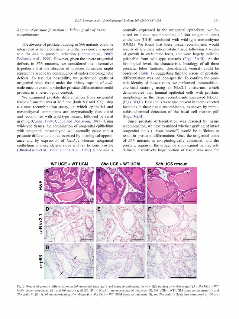
Rescue of prostate formation in kidney grafts of tissue
recombinants
The absence of prostate budding in Shh mutants could be
interpreted as being consistent with the previously proposed
role for Shh in prostate induction (Lamm et al., 2002;
Podlasek et al., 1999). However, given the severe urogenital
defects in Shh mutants, we considered the alternative
hypothesis that the absence of prostate formation might
represent a secondary consequence of earlier morphogenetic
defects. To test this possibility, we performed grafts of
urogenital sinus tissue under the kidney capsule of nude
male mice to examine whether prostate differentiation could
proceed in a heterologous context.
We examined prostate differentiation from urogenital
tissue of Shh mutants at 16.5 dpc (both XY and XX) using
a tissue recombination assay, in which epithelial and
mesenchymal components are enzymatically dissociated
and recombined with wild-type tissues, followed by renal
grafting (Cunha, 1994; Cunha and Donjacour, 1987). Using
wild-type tissues, the combination of urogenital epithelium
with urogenital mesenchyme will normally cause robust
prostatic differentiation, as assessed by histological appear-
ance and by expression of Nkx3.1, whereas urogenital
epithelium or mesenchyme alone will fail to form prostate
(Bhatia-Gaur et al., 1999; Cunha et al., 1987). Since Shh is
D.M. Berman et al. / Developm
Fig. 3. Rescue of prostatic differentiation in Shh urogenital sinus grafts and tissue
UGM tissue recombinant (B), and Shh mutant graft (C). (D–F) Nkx3.1 immunost
Shh graft (F). (G– I) p63 immunostaining of wild-type (G), Shh UGE + WT UGM
normally expressed in the urogenital epithelium, we fo-
cused on tissue recombinations of Shh urogenital sinus
epithelium (UGE) combined with wild-type mesenchyme
(UGM). We found that these tissue recombinants would
readily differentiate into prostatic tissue following 4 weeks
of growth in nude male hosts, and were largely indistin-
guishable from wild-type controls (Figs. 3A,B). At the
histological level, the characteristic histology of all three
prostatic lobes (anterior, dorsolateral, ventral) could be
observed (Table 1), suggesting that the rescue of prostatic
differentiation was not lobe-specific. To confirm the pros-
tatic identity of these tissues, we performed immunohisto-
chemical staining using an Nkx3.1 antiserum, which
demonstrated that luminal epithelial cells with prostatic
morphology in the tissue recombinants expressed Nkx3.1
(Figs. 3D,E). Basal cells were also present in their expected
locations in these tissue recombinants, as shown by immu-
nohistochemical detection of the basal cell marker p63
(Figs. 3G,H).
Since prostate differentiation was rescued by tissue
recombination, we next examined whether grafting of intact
urogenital sinus (‘‘tissue rescue’’) would be sufficient to
result in prostatic differentation. Since the urogenital sinus
of Shh mutants is morphologically abnormal, and the
prostatic region of the urogenital sinus cannot be precisely
defined, a relatively large portion of tissue was used for
recombinants. (A–C) H&E staining of wild-type graft (A), Shh UGE + WT
aining of wild-type (D), Shh UGE + WT UGM tissue recombinant (E), and
tissue recombinant (H), and Shh graft (I). Scale bars correspond to 100 Am.
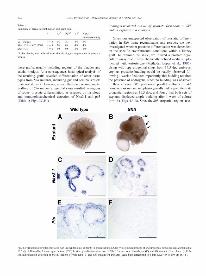
Table 1
Summary of tissue recombination and graft data
n APa DLPa VPa Nkx3.1
immunostaining
WT controls n = 2 2/2 2/2 2/2 2/2
Shh UGE + WT UGM n = 8 5/8 4/8 4/8 4/8
Shh UGS n = 5 5/5 5/5 3/5 5/5
a Lobe identity was inferred from the histological appearance of prostatic
tissues.
D.M. Berman et al. / Developmental392
these grafts, usually including regions of the bladder and
caudal hindgut. As a consequence, histological analysis of
the resulting grafts revealed differentiation of other tissue
types from Shh mutants, including gut and seminal vesicle
(data not shown). However, as with the tissue recombinants,
grafting of Shh mutant urogenital sinus resulted in regions
of robust prostate differentiation, as assessed by histology
and immunohistochemical detection of Nkx3.1 and p63
(Table 1; Figs. 3C,F,I).
Fig. 4. Formation of prostatic tissue in Shh urogenital sinus explants in organ cultu
16.5 dpc followed by 7 days organ culture. (C,D) In situ hybridization detection o
situ hybridization detection of Ptc in sections of wild-type (E) and Shh mutant (F
Androgen-mediated rescue of prostate formation in Shh
mutant explants and embryos
Given our unexpected observation of prostatic differen-
tiation in Shh tissue recombinants and rescues, we next
investigated whether prostatic differentiation was dependent
on the specific environmental conditions within a kidney
graft. To examine this issue, we utilized a prostate organ
culture assay that utilizes chemically defined media supple-
mented with testosterone (Methods; Lopes et al., 1996).
Using wild-type urogenital sinus from 16.5 dpc embryos,
copious prostatic budding could be readily observed fol-
lowing 1 week of culture; importantly, this budding required
the presence of androgens, since no budding was observed
in their absence. We performed parallel cultures of Shh
homozygous mutant and phenotypically wild-type littermate
urogenital regions at 16.5 dpc, and found that both sets of
explants displayed ample budding after 1 week of culture
(n = 15) (Figs. 4A,B). Since the Shh urogenital regions used
Biology 267 (2004) 387–398
re. (A,B) Whole-mount images of Shh urogenital sinus explants explanted at
f Nkx3.1 in sections of wild-type (C) and Shh mutant (D) explants. (E,F) In
) explants. Scale bars correspond to 1 mm (A,B) or to 100 Am (C–F).
D.M. Berman et al. / Developmental Biology 267 (2004) 387–398 393
for explant culture were highly abnormal, and contained
regions that might correspond to seminal vesicle and am-
pullary gland outgrowths, we confirmed that prostatic bud-
ding was present in these explants by detection of Nkx3.1
expression using in situ hybridization (n = 5) (Figs. 4C,D).
Importantly, in situ hybridization of adjacent sections with a
probe for Ptc demonstrated that Ptc expression could be
detected in mesenchyme adjoining Nkx3.1-expressing pros-
tatic epithelium in wild-type urogenital explants, but not in
mesenchyme from Shh mutant explants (Figs. 4E,F). This
result indicates that Hh pathway activity is absent in Shh
mutant urogenital sinus during prostatic differentiation, and
that compensatory expression of other hedgehog genes is
not responsible for the rescue of prostatic differentiation in
explants.
Given the ability to rescue prostatic differentiation by
explant culture of Shh mutant urogenital sinus, we consid-
ered the possibility that endogenous androgen levels were
insufficient for prostate development in Shh mutant fetuses
in vivo, but that exogenous high androgen levels in
explants and tissue recombinants could rescue this defect.
To test this possibility, we elevated fetal androgen levels
by administering DHT to pregnant mothers carrying prog-
eny from a Shh heterozygous intercross. We found that
DHT administration from 10.5 to 17.5 dpc could rescue
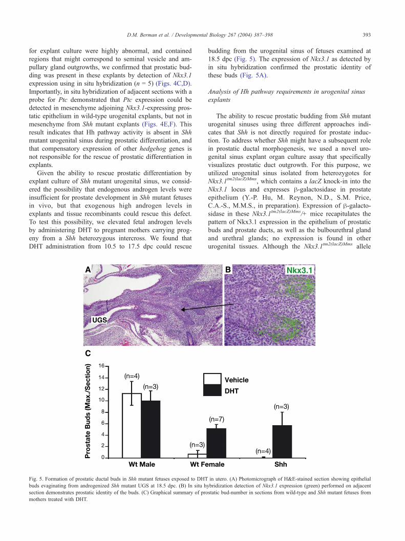
Fig. 5. Formation of prostatic ductal buds in Shh mutant fetuses exposed to DHT
buds evaginating from androgenized Shh mutant UGS at 18.5 dpc. (B) In situ h
section demonstrates prostatic identity of the buds. (C) Graphical summary of pro
mothers treated with DHT.
budding from the urogenital sinus of fetuses examined at
18.5 dpc (Fig. 5). The expression of Nkx3.1 as detected by
in situ hybridization confirmed the prostatic identity of
these buds (Fig. 5A).
Analysis of Hh pathway requirements in urogenital sinus
explants
The ability to rescue prostatic budding from Shh mutant
urogenital sinuses using three different approaches indi-
cates that Shh is not directly required for prostate induc-
tion. To address whether Shh might have a subsequent role
in prostatic ductal morphogenesis, we used a novel uro-
genital sinus explant organ culture assay that specifically
visualizes prostatic duct outgrowth. For this purpose, we
utilized urogenital sinus isolated from heterozygotes for
Nkx3.1tm2(lacZ)Mms, which contains a lacZ knock-in into the
Nkx3.1 locus and expresses h-galactosidase in prostate
epithelium (Y.-P. Hu, M. Reynon, N.D., S.M. Price,
C.A.-S., M.M.S., in preparation). Expression of h-galacto-sidase in these Nkx3.1tm2(lacZ)Mms/+ mice recapitulates the
pattern of Nkx3.1 expression in the epithelium of prostatic
buds and prostate ducts, as well as the bulbourethral gland
and urethral glands; no expression is found in other
urogenital tissues. Although the Nkx3.1tm2(lacZ)Mms allele
in utero. (A) Photomicrograph of H&E-stained section showing epithelial
ybridization detection of Nkx3.1 expression (green) performed on adjacent
static bud-number in sections from wild-type and Shh mutant fetuses from
D.M. Berman et al. / Developmental Biology 267 (2004) 387–398394
is null for Nkx3.1, there are no known phenotypic effects
for Nkx3.1 heterozygotes during prostatic budding or
ductal morphogenesis, although there is a moderate reduc-
tion in ductal tip number in Nkx3.1 homozygotes (Bhatia-
Gaur et al., 1999).
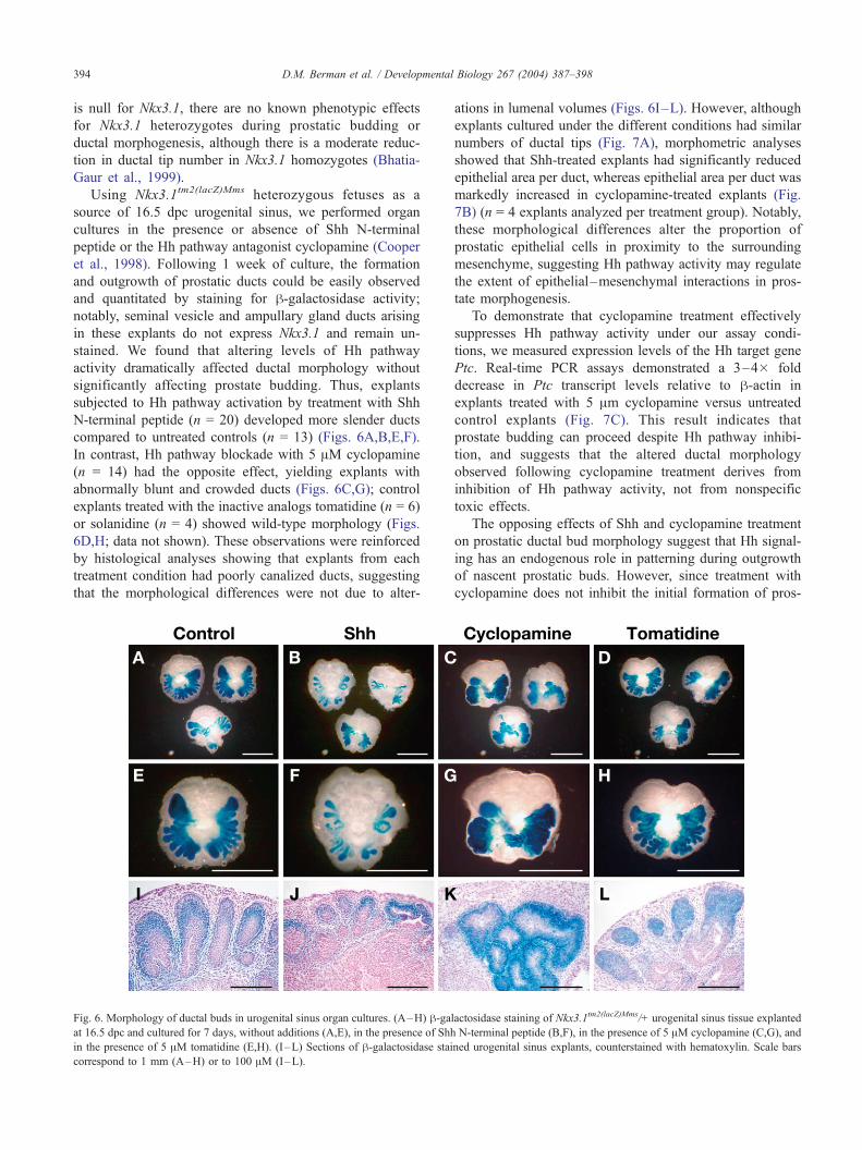
Using Nkx3.1tm2(lacZ)Mms heterozygous fetuses as a
source of 16.5 dpc urogenital sinus, we performed organ
cultures in the presence or absence of Shh N-terminal
peptide or the Hh pathway antagonist cyclopamine (Cooper
et al., 1998). Following 1 week of culture, the formation
and outgrowth of prostatic ducts could be easily observed
and quantitated by staining for h-galactosidase activity;
notably, seminal vesicle and ampullary gland ducts arising
in these explants do not express Nkx3.1 and remain un-
stained. We found that altering levels of Hh pathway
activity dramatically affected ductal morphology without
significantly affecting prostate budding. Thus, explants
subjected to Hh pathway activation by treatment with Shh
N-terminal peptide (n = 20) developed more slender ducts
compared to untreated controls (n = 13) (Figs. 6A,B,E,F).
In contrast, Hh pathway blockade with 5 AM cyclopamine
(n = 14) had the opposite effect, yielding explants with
abnormally blunt and crowded ducts (Figs. 6C,G); control
explants treated with the inactive analogs tomatidine (n = 6)
or solanidine (n = 4) showed wild-type morphology (Figs.
6D,H; data not shown). These observations were reinforced
by histological analyses showing that explants from each
treatment condition had poorly canalized ducts, suggesting
that the morphological differences were not due to alter-
Fig. 6. Morphology of ductal buds in urogenital sinus organ cultures. (A–H) h-gaat 16.5 dpc and cultured for 7 days, without additions (A,E), in the presence of Shh
in the presence of 5 AM tomatidine (E,H). (I –L) Sections of h-galactosidase stai
correspond to 1 mm (A–H) or to 100 AM (I–L).
ations in lumenal volumes (Figs. 6I–L). However, although
explants cultured under the different conditions had similar
numbers of ductal tips (Fig. 7A), morphometric analyses
showed that Shh-treated explants had significantly reduced
epithelial area per duct, whereas epithelial area per duct was
markedly increased in cyclopamine-treated explants (Fig.
7B) (n = 4 explants analyzed per treatment group). Notably,
these morphological differences alter the proportion of
prostatic epithelial cells in proximity to the surrounding
mesenchyme, suggesting Hh pathway activity may regulate
the extent of epithelial–mesenchymal interactions in pros-
tate morphogenesis.
To demonstrate that cyclopamine treatment effectively
suppresses Hh pathway activity under our assay condi-
tions, we measured expression levels of the Hh target gene
Ptc. Real-time PCR assays demonstrated a 3–4� fold
decrease in Ptc transcript levels relative to h-actin in
explants treated with 5 Am cyclopamine versus untreated
control explants (Fig. 7C). This result indicates that
prostate budding can proceed despite Hh pathway inhibi-
tion, and suggests that the altered ductal morphology
observed following cyclopamine treatment derives from
inhibition of Hh pathway activity, not from nonspecific
toxic effects.
The opposing effects of Shh and cyclopamine treatment
on prostatic ductal bud morphology suggest that Hh signal-
ing has an endogenous role in patterning during outgrowth
of nascent prostatic buds. However, since treatment with
cyclopamine does not inhibit the initial formation of pros-
lactosidase staining of Nkx3.1tm2(lacZ)Mms/+ urogenital sinus tissue explanted
N-terminal peptide (B,F), in the presence of 5 AM cyclopamine (C,G), and
ned urogenital sinus explants, counterstained with hematoxylin. Scale bars
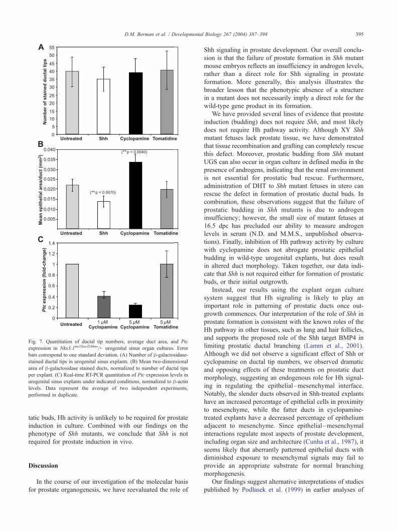
Fig. 7. Quantitation of ductal tip numbers, average duct area, and Ptc
expression in Nkx3.1tm2(lacZ)Mms/+ urogenital sinus organ cultures. Error
bars correspond to one standard deviation. (A) Number of h-galactosidase-stained ductal tips in urogenital sinus explants. (B) Mean two-dimensional
area of h-galactosidase stained ducts, normalized to number of ductal tips
per explant. (C) Real-time RT-PCR quantitation of Ptc expression levels in
urogenital sinus explants under indicated conditions, normalized to h-actinlevels. Data represent the average of two independent experiments,
performed in duplicate.
D.M. Berman et al. / Developmental Biology 267 (2004) 387–398 395
tatic buds, Hh activity is unlikely to be required for prostate
induction in culture. Combined with our findings on the
phenotype of Shh mutants, we conclude that Shh is not
required for prostate induction in vivo.
Discussion
In the course of our investigation of the molecular basis
for prostate organogenesis, we have reevaluated the role of
Shh signaling in prostate development. Our overall conclu-
sion is that the failure of prostate formation in Shh mutant
mouse embryos reflects an insufficiency in androgen levels,
rather than a direct role for Shh signaling in prostate
formation. More generally, this analysis illustrates the
broader lesson that the phenotypic absence of a structure
in a mutant does not necessarily imply a direct role for the
wild-type gene product in its formation.
We have provided several lines of evidence that prostate
induction (budding) does not require Shh, and most likely
does not require Hh pathway activity. Although XY Shh
mutant fetuses lack prostate tissue, we have demonstrated
that tissue recombination and grafting can completely rescue
this defect. Moreover, prostatic budding from Shh mutant
UGS can also occur in organ culture in defined media in the
presence of androgens, indicating that the renal environment
is not essential for prostatic bud rescue. Furthermore,
administration of DHT to Shh mutant fetuses in utero can
rescue the defect in formation of prostatic ductal buds. In
combination, these observations suggest that the failure of
prostatic budding in Shh mutants is due to androgen
insufficiency; however, the small size of mutant fetuses at
16.5 dpc has precluded our ability to measure androgen
levels in serum (N.D. and M.M.S., unpublished observa-
tions). Finally, inhibition of Hh pathway activity by culture
with cyclopamine does not abrogate prostatic epithelial
budding in wild-type urogenital explants, but does result
in altered duct morphology. Taken together, our data indi-
cate that Shh is not required either for formation of prostatic
buds, or their initial outgrowth.
Instead, our results using the explant organ culture
system suggest that Hh signaling is likely to play an
important role in patterning of prostatic ducts once out-
growth commences. Our interpretation of the role of Shh in
prostate formation is consistent with the known roles of the
Hh pathway in other tissues, such as lung and hair follicles,
and supports the proposed role of the Shh target BMP4 in
limiting prostatic ductal branching (Lamm et al., 2001).
Although we did not observe a significant effect of Shh or
cyclopamine on ductal tip numbers, we observed dramatic
and opposing effects of these treatments on prostatic duct
morphology, suggesting an endogenous role for Hh signal-
ing in regulating the epithelial–mesenchymal interface.
Notably, the slender ducts observed in Shh-treated explants
have an increased percentage of epithelial cells in proximity
to mesenchyme, while the fatter ducts in cyclopamine-
treated explants have a decreased percentage of epithelium
adjacent to mesenchyme. Since epithelial–mesenchymal
interactions regulate most aspects of prostate development,
including organ size and architecture (Cunha et al., 1987), it
seems likely that aberrantly patterned epithelial ducts with
diminished exposure to mesenchymal signals may fail to
provide an appropriate substrate for normal branching
morphogenesis.
Our findings suggest alternative interpretations of studies
published by Podlasek et al. (1999) in earlier analyses of
D.M. Berman et al. / Developmental Biology 267 (2004) 387–398396
Shh activity in prostate formation. Their previous work
showed that addition of an Affi-Gel bead soaked in Shh
neutralizing antibody could inhibit prostate formation from
15.5 dpc urogenital sinus grown in renal grafts, whereas we
observed essentially normal prostate morphology in grafts
grown from Shh mutant urogenital sinus. Furthermore,
Lamm et al. (2002) reported that organ culture of 14.5
dpc urogenital sinus in the presence of 10 AM cyclopamine
resulted in a 71% decrease in ductal tip number, whereas we
did not observe a significant change in ductal tip number in
explants cultured with 5 AM cyclopamine or with Shh N-
terminal peptide. It is conceivable that this discrepancy is
due to different sources and concentrations of cyclopamine,
or to differences in culture systems that could reveal an
effect of cyclopamine on ductal branching, rather than
inhibition of initial duct formation. In addition, we note
that our organ culture data are unlikely to be due to potential
toxicity of cyclopamine, since we observed apparently
opposite effects on prostate ductal morphology after addi-
tion of Shh peptide. Finally, our results suggest that the
abrogation of prostate morphogenesis in urogenital sinus
engrafted with a source of Hh neutralizing antibody (Pod-
lasek et al., 1999) may not reflect an inability to form
prostate tissue, but may instead indicate reduced viability of
the malformed organs that form under the influence of Hh
pathway blockade.
Our results are consistent with a recent study demon-
strating that addition of Shh to explant cultures of postnatal
rat ventral prostate leads to inhibition of ductal branching,
whereas cyclopamine treatment results in enlarged ductal
tips (Wang et al., 2003). Unlike our assay using embryonic
mouse urogenital sinus, this study used postnatal explants
that had already initiated prostate formation and ductal
morphogenesis, and were cultured in the absence of exog-
enous testosterone, which is required for normal prostate
growth in vivo. In addition, Freestone et al. (in press) have
performed similar organ culture experiments in the presence
of testosterone, and have also observed altered morphology
of rat ventral prostate ductal tips following cyclopamine
treatment. We note that the lack of detectable abnormalities
in the ductal morphology of Shh mutant prostates grown in
tissue recombinants or grafts in nude mice suggests either
that the requirement for Hh signaling in ductal morphogen-
esis is specific to in vitro culture systems, or that the kidney
and/or maturing prostate epithelium can complement defec-
tive Shh signaling, perhaps through expression of other Hh
ligands. While additional studies will be necessary to
distinguish between these possibilities, our results clearly
indicate that Shh is not required for prostate induction.
The androgen insufficiency of Shh XY mutant embryos
presumably reflects a direct or indirect role for Hh signaling
in the differentiation and/or function of testicular Leydig
cells, the primary site of androgen biosynthesis. One possi-
bility is that Shh is indirectly required for Leydig cell
differentiation due to its essential role in the pituitary–
gonadal axis during fetal development, since Shh is neces-
sary for formation of the pituitary gland (Treier et al., 2001).
Analyses of hypogonadal mutant mice, which are deficient
for gonadotropin-releasing hormone (GnRH), suggest that
pituitary gonadotrophs are not required for fetal develop-
ment of Leydig cells (O’Shaughnessy et al., 1998); in
contrast, Nkx2.1 mutant mice lack the pituitary gland and
have altered Leydig cell morphology and reduced testoster-
one levels (Pakarinen et al., 2002). A specific role for Hh
signaling in Leydig cell differentiation has been shown by
the findings that Desert hedgehog (Dhh) XY mutants have
profound defects in fetal Leydig cell differentiation (Clark et
al., 2000; Yao et al., 2002), as do fetal testes treated with
cyclopamine in organ culture (Yao and Capel, 2002). Other
potential explanations for the Shh XY androgen-deficient
phenotype include possible defects in export of testosterone
from the fetal testis or its delivery to the urogenital sinus
mesenchyme. Finally, Shh may be required for normal testes
development, which is supported by the observation that
Shh mutant fetuses display defective urogenital ridge devel-
opment and have rudimentary testes at 16.5 dpc (B. Capel,
personal communication; D.M.B., N.D., and M.M.S., un-
published observations).
Our findings also imply that expression of Nkx3.1 in the
prostate is not dependent on Shh function, since the prostatic
epithelium formed in Shh mutant grafts and explants display
abundant Nkx3.1 expression. Previous studies had suggested
that Nkx3.1 was downstream of Shh, based on the absence
of Nkx3.1 expression in Shh urogenital sinus (Schneider et
al., 2000); however, this observation is consistent with the
absence of prostate formation in Shh mutants, and with the
androgen-dependence of Nkx3.1 expression (Bieberich et
al., 1996; He et al., 1997; Prescott et al., 1998; Sciavolino et
al., 1997). In contrast, Nkx3.1 appears to be downstream of
Shh in the developing sclerotome during midgestation
embryogenesis, since Nkx3.1 expression in the ventromedial
region of nascent somites is dependent on the presence of
Shh signaling from the notochord (Kos et al., 1998).
The lack of a direct regulatory relationship between Hh
signaling and Nkx3.1 in normal prostate development is
consistent with their opposite roles in prostate cancer
progression, since Hedgehog pathway activity appears to
be elevated in human prostate carcinomas (Dahmane et al.,
2001; S.S.K., D.M.B., and P.A.B., in preparation; W.
Bushman, personal communication), whereas Nkx3.1 pro-
tein expression is decreased or absent in most human
carcinomas and in mouse models (Bowen et al., 2000;
Kim et al., 2002). Recently, we demonstrated a requirement
for Hh pathway activity in malignant growth of a variety of
tumors, and a relationship between levels of Hh pathway
activity and the rate of tumor growth (Berman et al., 2002,
2003; Watkins et al., 2003). In prostate cancer, the rate of
tumor growth and progression is strongly correlated to
Gleason grade, which evaluates branching patterns histo-
logically. It is therefore possible that further studies of Hh
signaling in prostate branching will lead to a better under-
standing of prostate carcinogenesis and tumor progression.
D.M. Berman et al. / Developmental Biology 267 (2004) 387–398 397
Acknowledgments
We thank Jeff Bush and Rajula Bhatia-Gaur for their help
in initiating this project, Nadia Abdallah, Keith Young,
Eileen Traband, and Ruoyu Gong for technical assistance;
Donald Coffey for advice and reagents; Andy McMahon for
providing plasmids; Jean Wilson for helpful discussions;
and Axel Thomson and Blanche Capel for sharing results
before publication. Work in the authors’ laboratories is
supported by grants from the NCI, NIDDK, CaP CURE,
and the Department of Defense Prostate Cancer Research
Program. P.A.B. is an Investigator of the Howard Hughes
Medical Institute. C.A.-S. and M.M.S. are members of the
NCI Mouse Models of Human Cancer Consortium.
References
Bellusci, S., Furuta, Y., Rush, M.G., Henderson, R., Winnier, G., Hogan,
B.L., 1997. Involvement of Sonic hedgehog (Shh) in mouse embryonic
lung growth and morphogenesis. Development 124, 53–63.
Ben-Arie, N., Hassan, B.A., Bermingham, N.A., Malicki, D.M., Arm-
strong, D., Matzuk, M., Bellen, H.J., Zoghbi, H.Y., 2000. Functional
conservation of atonal and Math1 in the CNS and PNS. Development
127, 1039–1048.
Berman, D.M., Tian, H., Russell, D.W., 1995. Expression and regulation of
steroid 5 alpha-reductase in the urogenital tract of the fetal rat. Mol.
Endocrinol. 9, 1561–1570.
Berman, D.M., Karhadkar, S.S., Hallahan, A.R., Pritchard, J.I., Eberhart,
C.G., Watkins, D.N., Chen, J.K., Cooper, M.K., Taipale, J., Olson, J.M.,
Beachy, P.A., 2002. Medulloblastoma growth inhibition by hedgehog
pathway blockade. Science 297, 1559–1561.
Berman, D.M., Karhadkar, S.S., Maitra, A., Montes De Oca, R., Gersten-
blith, M.R., Briggs, K., Parker, A.R., Shimada, Y., Eshleman, J.R.,
Watkins, D.N., Beachy, P.A., 2003. Widespread requirement for Hedge-
hog ligand stimulation in growth of digestive tract tumours. Nature 425,
846–851.
Bhatia-Gaur, R., Donjacour, A.A., Sciavolino, P.J., Kim, M., Desai, N.,
Young, P., Norton, C.R., Gridley, T., Cardiff, R.D., Cunha, G.R., Abate-
Shen, C., Shen, M.M., 1999. Roles for Nkx3.1 in prostate development
and cancer. Genes Dev. 13, 966–977.
Bieberich, C.J., Fujita, K., He, W.W., Jay, G., 1996. Prostate-specific and
androgen-dependent expression of a novel homeobox gene. J. Biol.
Chem. 271, 31779–31782.
Bowen, C., Bubendorf, L., Voeller, H.J., Slack, R., Willi, N., Sauter, G.,
Gasser, T.C., Koivisto, P., Lack, E.E., Kononen, J., Kallioniemi,
O.P., Gelmann, E.P., 2000. Loss of NKX3.1 expression in human
prostate cancers correlates with tumor progression. Cancer Res. 60,
6111–6115.
Bowles, J., Cooper, L., Berkman, J., Koopman, P., 1999. Sry requires a
CAG repeat domain for male sex determination in Mus musculus. Nat.
Genet. 22, 405–408.
Chiang, C., Litingtung, Y., Lee, E., Young, K.E., Corden, J.L., West-
phal, H., Beachy, P.A., 1996. Cyclopia and defective axial pattern-
ing in mice lacking Sonic hedgehog gene function. Nature 383,
407–413.
Chiang, C., Swan, R.Z., Grachtchouk, M., Bolinger, M., Litingtung, Y.,
Robertson, E.K., Cooper, M.K., Gaffield, W., Westphal, H., Beachy,
P.A., Dlugosz, A.A., 1999. Essential role for Sonic hedgehog during
hair follicle morphogenesis. Dev. Biol. 205, 1–9.
Chuang, P.T., McMahon, A.P., 1999. Vertebrate Hedgehog signalling
modulated by induction of a Hedgehog-binding protein. Nature 397,
617–621.
Clark, A.M., Garland, K.K., Russell, L.D., 2000. Desert hedgehog (Dhh)
gene is required in the mouse testis for formation of adult-type Leydig
cells and normal development of peritubular cells and seminiferous
tubules. Biol. Reprod. 63, 1825–1838.
Cooper, M.K., Porter, J.A., Young, K.E., Beachy, P.A., 1998. Teratogen-
mediated inhibition of target tissue response to Shh signaling. Science
280, 1603–1607.
Cunha, G.R., 1994. Role of mesenchymal–epithelial interactions in normal
and abnormal development of the mammary gland and prostate. Cancer
74, 1030–1044.
Cunha, G.R., Donjacour, A., 1987. Mesenchymal–epithelial interactions:
technical considerations. Prog. Clin. Biol. Res. 239, 273–282.
Cunha, G.R., Donjacour, A.A., Cooke, P.S., Mee, S., Bigsby, R.M., Hig-
gins, S.J., Sugimura, Y., 1987. The endocrinology and developmental
biology of the prostate. Endocr. Rev. 8, 338–362.
Dahmane, N., Sanchez, P., Gitton, Y., Palma, V., Sun, T., Beyna, M., Weiner,
H., Ruiz i Altaba, A., 2001. The Sonic Hedgehog-Gli pathway regulates
dorsal brain growth and tumorigenesis. Development 128, 5201–5212.
Freestone, S.H., Marker, P., Grace, O.C., Tomlinson, D.C., Cunha, G.R.,
Harnden, P., Thomson, A.A., 2003. Sonic hedgehog regulates prostatic
growth and epithelial differentiation. Dev. Biol. 264, 352–362.
Grapin-Botton, A., Melton, D.A., 2000. Endoderm development: from
patterning to organogenesis. Trends Genet. 16, 124–130.
Hayashi, N., Sugimura, Y., Kawamura, J., Donjacour, A.A., Cunha, G.R.,
1991. Morphological and functional heterogeneity in the rat prostatic
gland. Biol. Reprod. 45, 308–321.
Hayward, S.W., Rosen, M.A., Cunha, G.R., 1997. Stromal–epithelial inter-
actions in the normal and neoplastic prostate. Br. J. Urol. 79 (Suppl. 2),
18–26.
He, W.W., Sciavolino, P.J., Wing, J., Augustus, M., Hudson, P., Meissner,
P.S., Curtis, R.T., Shell, B.K., Bostwick, D.G., Tindall, D.J., Gelmann,
E.P., Abate-Shen, C., Carter, K.C., 1997. A novel human prostate-
specific, androgen-regulated homeobox gene (NKX3.1) that maps to
8p21, a region frequently deleted in prostate cancer. Genomics 43,
69–77.
Hogan, B.L., Grindley, J., Bellusci, S., Dunn, N.R., Emoto, H., Itoh, N.,
1997. Branching morphogenesis of the lung: new models for a classical
problem. Cold Spring Harbor Symp. Quant. Biol. 62, 249–256.
Kim, M.J., Cardiff, R.D., Desai, N., Banach-Petrosky, W.A., Parsons, R.,
Shen, M.M., Abate-Shen, C., 2002. Cooperativity of Nkx3.1 and Pten
loss of function in a mouse model of prostate carcinogenesis. Proc. Natl.
Acad. Sci. U. S. A. 99, 2884–2889.
Kimmel, S.G., Mo, R., Hui, C.C., Kim, P.C., 2000. New mouse models of
congenital anorectal malformations. J. Pediatr. Surg. 35, 227–230.
Kos, L., Chiang, C., Mahon, K.A., 1998. Mediolateral patterning of so-
mites: multiple axial signals, including Sonic hedgehog, regulate Nkx-
3.1 expression. Mech. Dev. 70, 25–34.
Lamm, M.L., Podlasek, C.A., Barnett, D.H., Lee, J., Clemens, J.Q., Heb-
ner, C.M., Bushman, W., 2001. Mesenchymal factor bone morphoge-
netic protein 4 restricts ductal budding and branching morphogenesis in
the developing prostate. Dev. Biol. 232, 301–314.
Lamm, M., Catbagan, W., Laciak, R., Barnett, D., Hebner, C., Gaffield, W.,
Walterhouse, D., Iannaccone, P., Bushman, W., 2002. Sonic hedgehog
activates mesenchymal Gli1 expression during prostate ductal bud for-
mation. Dev. Biol. 249, 349–366.
Litingtung, Y., Lei, L., Westphal, H., Chiang, C., 1998. Sonic hedgehog is
essential to foregut development. Nat. Genet. 20, 58–61.
Lopes, E.S., Foster, B.A., Donjacour, A.A., Cunha, G.R., 1996. Initiation
of secretory activity of rat prostatic epithelium in organ culture. Endo-
crinology 137, 4225–4234.
Mo, R., Kim, J.H., Zhang, J., Chiang, C., Hui, C.C., Kim, P.C., 2001.
Anorectal malformations caused by defects in sonic hedgehog signal-
ing. Am. J. Pathol. 159, 765–774.
Motoyama, J., Liu, J., Mo, R., Ding, Q., Post, M., Hui, C.C., 1998. Essen-
tial function of Gli2 and Gli3 in the formation of lung, trachea and
oesophagus. Nat. Genet. 20, 54–57.
O’Shaughnessy, P.J., Baker, P., Sohnius, U., Haavisto, A.M., Charlton,

D.M. Berman et al. / Developmental Biology 267 (2004) 387–398398
H.M., Huhtaniemi, I., 1998. Fetal development of Leydig cell activity
in the mouse is independent of pituitary gonadotroph function. Endo-
crinology 139, 1141–1146.
Pakarinen, P., Kimura, S., El-Gehani, F., Pelliniemi, L.J., Huhtaniemi, I.,
2002. Pituitary hormones are not required for sexual differentiation of
male mice: phenotype of the T/ebp/Nkx2.1 null mutant mice. Endocri-
nology 143, 4477–4482.
Podlasek, C.A., Barnett, D.H., Clemens, J.Q., Bak, P.M., Bushman, W.,
1999. Prostate development requires Sonic hedgehog expressed by the
urogenital sinus epithelium. Dev. Biol. 209, 28–39.
Prescott, J.L., Blok, L., Tindall, D.J., 1998. Isolation and androgen regu-
lation of the human homeobox cDNA, NKX3.1. Prostate 35, 71–80.
Roberts, D.J., 2000. Molecular mechanisms of development of the gastro-
intestinal tract. Dev. Dyn. 219, 109–120.
Schneider, A., Brand, T., Zweigerdt, R., Arnold, H., 2000. Targeted dis-
ruption of the Nkx3.1 gene in mice results in morphogenetic defects of
minor salivary glands: parallels to glandular duct morphogenesis in
prostate. Mech. Dev. 95, 163–174.
Sciavolino, P.J., Abrams, E.W., Yang, L., Austenberg, L.P., Shen, M.M.,
Abate-Shen, C., 1997. Tissue-specific expression of murine Nkx3.1 in
the male urogenital system. Dev. Dyn. 209, 127–138.
St-Jacques, B., Dassule, H.R., Karavanova, I., Botchkarev, V.A., Li, J.,
Danielian, P.S., McMahon, J.A., Lewis, P.M., Paus, R., McMahon,
A.P., 1998. Sonic hedgehog signaling is essential for hair development.
Curr. Biol. 8, 1058–1068.
Sugimura, Y., Cunha, G.R., Donjacour, A.A., 1986. Morphogenesis of
ductal networks in the mouse prostate. Biol. Reprod. 34, 961–971.
Timms, B.G., Mohs, T.J., Didio, L.J.A., 1994. Ductal budding and branch-
ing patterns in the developing prostate. J. Urol. 151, 1427–1432.
Treier, M., O’Connell, S., Gleiberman, A., Price, J., Szeto, D.P., Burgess,
R., Chuang, P.T., McMahon, A.P., Rosenfeld, M.G., 2001. Hedgehog
signaling is required for pituitary gland development. Development
128, 377–386.
Wang, B.E., Shou, J., Ross, S., Koeppen, H., De Sauvage, F.J., Gao, W.Q.,
2003. Inhibition of epithelial ductal branching in the prostate by sonic
hedgehog is indirectly mediated by stromal cells. J. Biol. Chem. 278,
18506–18513.
Watkins, D.N., Berman, D.M., Burkholder, S.G., Wang, B., Beachy, P.A.,
Baylin, S.B., 2003. Hedgehog signalling within airway epithelial pro-
genitors and in small-cell lung cancer. Nature 422, 313–317.
Yao, H.H., Capel, B., 2002. Disruption of testis cords by cyclopamine or
forskolin reveals independent cellular pathways in testis organogenesis.
Dev. Biol. 246, 356–365.
Yao, H.H., Whoriskey, W., Capel, B., 2002. Desert Hedgehog/Patched 1
signaling specifies fetal Leydig cell fate in testis organogenesis. Genes
Dev. 16, 1433–1440.


