Embed Size (px)
Citation preview

173
www.phcogcommn.org
Original Article
© Copyright 2015 EManuscript Services, India
Pharmacognosy CommunicationsVolume 5 | Issue 3 | Jul-Sep 2015
ABSTRACT
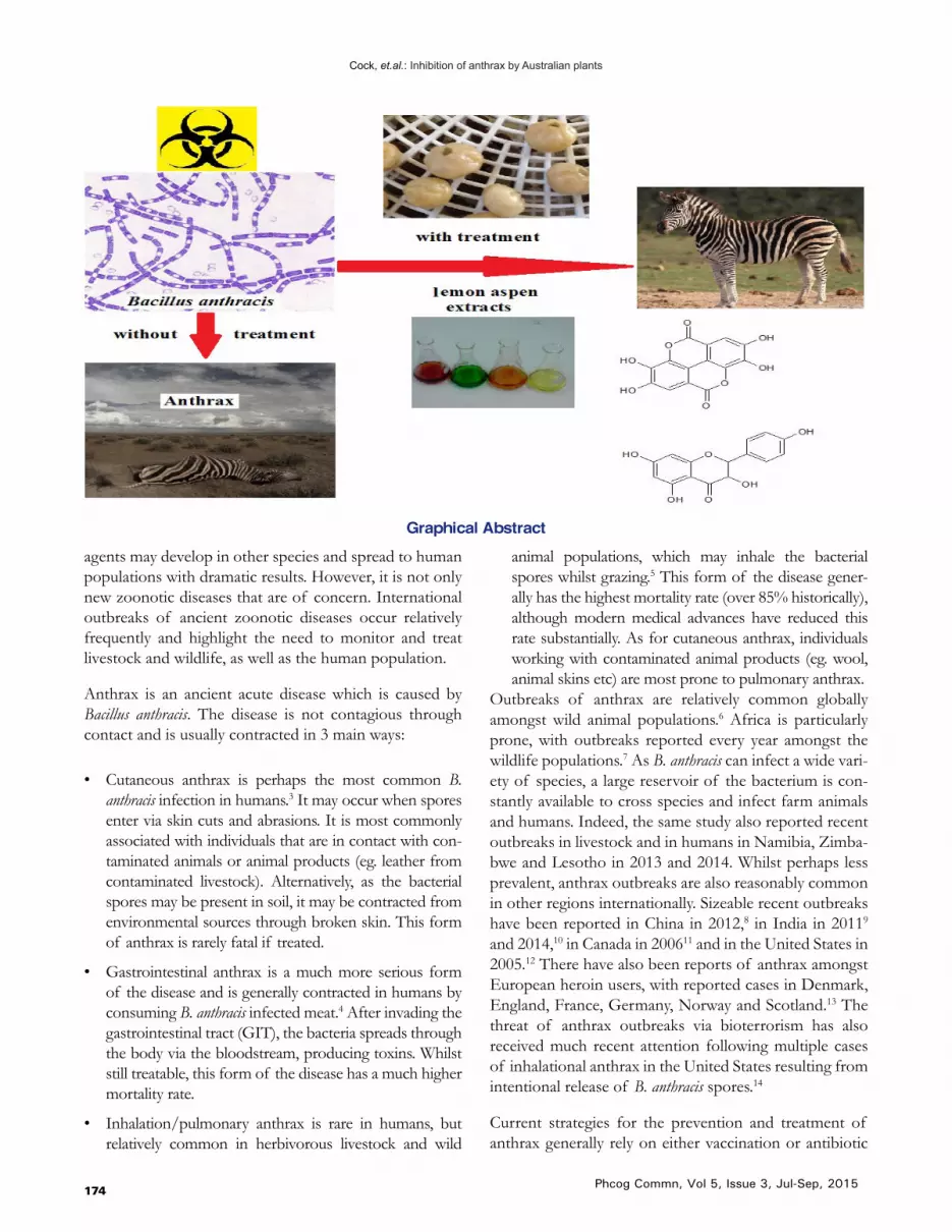
Introduction: Anthrax is severe acute disease caused by Bacillus anthracis infections. If untreated, it often results in mortality. High antioxidant plant extracts have documented therapeutic properties as general antiseptics, inhibiting the growth of a wide variety of bacterial species. This study examines the ability of selected high antioxidant Australian plant extracts to inhibit B. anthracis growth. Methods: Solvent extracts were prepared using various high antioxidant Australian fruits and herbs and investigated by disc diffusion assay for the ability to inhibit the growth of an environmental strain of B. anthracis. Their MIC values were determined to quantify and compare their efficacies. Toxicity was determined using the Artemia franciscana nauplii bioassay. The most potent extracts were analysed by non-targeted HPLC-QTOF mass spectroscopy (with screening against 3 compound databases) for the identification and characterisation of individual components in crude plant extracts. Results: Methanolic and aqueous extracts of several high antioxidant plant extract sdisplayed potent antibacterial activity in the disc diffusion assay against B. anthracis. The aqueous and methanolic extracts of lemon aspen, as well as the methanolic extracts of muntries, Illawarra plum and native tamarind were particularly potent growth inhibitors with MIC values<1000 µg/mL. Furthermore, all of these extracts were nontoxic in the Artemia fransiscana bioassay, with LC50 values substantially>1000 µg/mL. Non-biased phytochemical analysis of the lemon aspen aqueous and methanolic extracts putatively identified 85 compounds and highlighted several that may contribute to the ability of these extracts to inhibit the growth of B. anthracis. Conclusion: The low toxicity of several high antioxidant plant extracts and their potent inhibitory bioactivity against B. anthracis indicates their potential as medicinal agents in the treatment and prevention of anthrax. Lemon aspen is particularly worthy of further study.
Key words: Anthrax, Antioxidant, Bacillus anthracis, lemon aspen, Muntries, Metabolomic profiling, Syzygium, Zoonotic.
Growth inhibition of the zoonotic bacteria Bacillus anthracis by high antioxidant Australian plants: New leads for the prevention and treatment of anthraxMitchell Henry Wright1, Ben Matthews2, Anthony Carlson Greene1 and Ian Edwin Cock,1,3*
1School of Natural Sciences, Nathan Campus, Griffith University, 170 Kessels Rd, Nathan, Queensland 4111, Australia.2Smart Waters Research Centre, Griffith University, Gold Coast, Australia.3Environmental Futures Research Institute, Nathan Campus, Griffith University, 170 Kessels Rd, Nathan, Queensland 4111, Australia.
INTRODUCTION
The prevention and treatment of zoonotic diseases poses a unique set of difficulties not encountered for other
Corresponding AddressDr. Ian Edwin CockSchool of Natural Sciences and Environmental Futures Research Institute Nathan Campus, Griffith University, 170 Kessels Rd, Nathan, Queensland 4111, Australia.E-mail: [email protected] DOI: 10.5530/pc.2015.3.3
diseases. Many pathogens may be controlled by isolating the infected individual so that they cannot infect others and allowing the disease to run its course. Alternatively, targeted immunization programs may be useful to estab-lish herd immunity, allowing the disease to dissipate.1 Indeed, such approaches have allowed the eradication of the infectious disease smallpox.2 However, zoonotic pathogens may persist in a non-human reservoir until-they are able to infect new hosts. The recent outbreaks of Ebola in West Africa, Hendra virus in Australia and swine influenza globally, demonstrate how new infectious
Cock, et.al.: Inhibition of anthrax by Australian plants
174 Phcog Commn, Vol 5, Issue 3, Jul-Sep, 2015
agents may develop in other species and spread to human populations with dramatic results. However, it is not only new zoonotic diseases that are of concern. International outbreaks of ancient zoonotic diseases occur relatively frequently and highlight the need to monitor and treat livestock and wildlife, as well as the human population.
Anthrax is an ancient acute disease which is caused by Bacillus anthracis. The disease is not contagious through contact and is usually contracted in 3 main ways:
• Cutaneous anthrax is perhaps the most common B. anthracis infection in humans.3 It may occur when spores enter via skin cuts and abrasions. It is most commonly associated with individuals that are in contact with con-taminated animals or animal products (eg. leather from contaminated livestock). Alternatively, as the bacterial spores may be present in soil, it may be contracted from environmental sources through broken skin. This form of anthrax is rarely fatal if treated.
• Gastrointestinal anthrax is a much more serious form of the disease and is generally contracted in humans by consuming B. anthracis infected meat.4 After invading the gastrointestinal tract (GIT), the bacteria spreads through the body via the bloodstream, producing toxins. Whilst still treatable, this form of the disease has a much higher mortality rate.
• Inhalation/pulmonary anthrax is rare in humans, but relatively common in herbivorous livestock and wild
animal populations, which may inhale the bacterial spores whilst grazing.5 This form of the disease gener-ally has the highest mortality rate (over 85% historically), although modern medical advances have reduced this rate substantially. As for cutaneous anthrax, individuals working with contaminated animal products (eg. wool, animal skins etc) are most prone to pulmonary anthrax.
Outbreaks of anthrax are relatively common globally amongst wild animal populations.6 Africa is particularly prone, with outbreaks reported every year amongst the wildlife populations.7 As B. anthracis can infect a wide vari-ety of species, a large reservoir of the bacterium is con-stantly available to cross species and infect farm animals and humans. Indeed, the same study also reported recent outbreaks in livestock and in humans in Namibia, Zimba-bwe and Lesotho in 2013 and 2014. Whilst perhaps less prevalent, anthrax outbreaks are also reasonably common in other regions internationally. Sizeable recent outbreaks have been reported in China in 2012,8 in India in 20119 and 2014,10 in Canada in 200611 and in the United States in 2005.12 There have also been reports of anthrax amongst European heroin users, with reported cases in Denmark, England, France, Germany, Norway and Scotland.13 The threat of anthrax outbreaks via bioterrorism has also received much recent attention following multiple cases of inhalational anthrax in the United States resulting from intentional release of B. anthracis spores.14
Current strategies for the prevention and treatment of anthrax generally rely on either vaccination or antibiotic
Graphical Abstract

Cock, et.al.: Inhibition of anthrax by Australian plants
175Phcog Commn, Vol 5, Issue 3, Jul-Sep, 2015
administration. Effective vaccines have been available since the 19th century. However, severe adverse reactions may occur in approximately 1% of the population.15 Fur-thermore, B. anthracis vaccines are most useful as a pre-ventative measure and are generally of little use once the disease is contracted. Instead, large doses of intraveneous and/or oral antibiotics (eg. fluoroquinolones, penicillin, erythromycin, vancomycin) are used and are generally effective for treating anthrax infections.16 However, due to the potential development of super-resistant strains, it is important to search for new antibiotics with high effi-cacy.17 The search is ongoing by (a) the design and syn-thesis of new agents, and (b) re-searching the repertoire of natural resources for as yet unrecognised or poorly characterised antimicrobial agents. Furthermore, the development of new anti-B.anthracis products to disinfect contaminated sites, soils etc. would decrease the spread of anthrax and thus decrease the impact of the disease.
The antiseptic qualities of medicinal plants have been long recognised by many cultures. Recently there has been a revival of interest in herbal medications due to perceptions that there is often a lower incidence of adverse reactions to natural phytochemicals compared to synthetic pharmaceuticals. Antimicrobial plant extracts with high antioxidant contents are particularly attrac-tive as they may treat the symptoms of anthrax as well as blocking growth of the pathogen and thus have pleu-ripotent effects. Recent studies have demonstrated the potent inhibitory activity of several Australian plants with high antioxidant capacities against a wide panel of medicinally important bacteria.18-21 Furthermore, potent growth inhibition of the bacterial triggers of autoim-mune inflammatory diseases has also been reported for high antioxidant Australian fruits22-24 and culinary herbs.25 Despite this, many high antioxidant Australian plants are yet to be rigorously tested for the ability to inhibit the growth of pathogenic bacteria. The current study exam-ines the growth inhibitory activity of extracts of selected high antioxidant Australian plants against B. anthracis with the aim of determining new leads for the prevention and treatment of anthrax.
MATERIALS AND METHODS
Plant source and extraction
The Syzygium australe (brush cherry), Syzygium leuhman-nii (riberry), Davidsonia pruriens (Davidson’s plum) and Elaeoparpus angustifolius (blue quandong) plant materi-als used in this study were supplied and verified by the
Queensland Bush foods Association, Australia. Kunzea pomifera (muntries), Podocarpus elatus (Illawarra plum), Dip-loglottis australis (native tamarind), Acronychia acidula (lemon aspen), Citrus glauca (desert lime), Solanum aviculare (bush tomato), Acacia vivtoriae (wattle seed), Prostanthera rotundifo-lia (native thyme), Prostanthera incise (native sage), Ocimum tenuiflorum (native basil) and Mentha australis (river mint) were obtained from Taste of Australia Bush Food, Aus-tralia. Voucher samples have been stored in the School of Natural Sciences, Griffith University.The plant materials were thoroughly dried in a Sunbeam food dehydrator and the dried plant materials were stored at -30oC. Prior to use, the plant materials were thawed and freshly ground to a coarse powder. Individual 1 g quantities of the ground plant material were weighed into separate tubes and 50 mL of methanol or water were added. All solvents were obtained from Ajax and were AR grade. The ground plant materials were individually extracted in each solvent for 24 h at 4oC with gentle shaking. The extracts were sub-sequently filtered through filter paper (Whatman No. 54) under vacuum, followed by drying by rotary evaporation in an Eppendorf concentrator 5301. The resultant dry extract was weighed and redissolved in 10 mL deionised water (containing 1% DMSO).
Qualitative phytochemical studies
Phytochemical analysis of the extracts for the presence of saponins, phenolic compounds, flavonoids, polysteroids, triterpenoids, cardiac glycosides, anthraquinones, tan-nins and alkaloids was conducted by previously described assays.22-24
Antioxidant capacity
The antioxidant capacity of each sample was assessed using the DPPH free radical scavenging method26 with modifications. Briefly, a DPPH solution was prepared fresh as a 400 µM solution by dissolving DPPH (Sigma) in AR grade methanol (Ajax, Australia). The initial absor-bance of the DPPH solution was measured at 515 nm using a Molecular Devices, Spectra Max M3 plate reader and did not change significantly throughout the assay period. A 2 mL aliquot of each extract was dried by evap-oration and the residue resuspended in 2 mL of metha-nol. Each extract was added to a 96-well plate in amounts of 5, 10, 25, 50, 75 µL in triplicate. Methanol was added to each well to give a volume of 225 µL. A volume of 75 µL of the fresh DPPH solution was added to each well for a total reaction volume of 300 µL. A blank of each extract concentration, methanol solvent, and DPPH was also performed in triplicate. Ascorbic acid was prepared fresh and examined across the range 0-25 µg per well as a

Cock, et.al.: Inhibition of anthrax by Australian plants
176 Phcog Commn, Vol 5, Issue 3, Jul-Sep, 2015
Table 1: The mass of dried extracted material, the concentration after resuspension in deionised water, qualitative phytochemical screenings and antioxidant capacities of the plant extracts
Extr
act
Mas
s of
Drie
d Ex
trac
t (m
g)
Con
cent
ratio
n of
R
esus
pend
ed E
xtra
ct (µ
g/m
L)
Ant
ioxi
dant
Cap
acity
(µg
Asc
orbi
c A
cid
Equi
vale
ncy)
Tota
l Phe
nolic
s
Wat
er S
olub
le P
heno
lics
Wat
er In
solu
ble
Phen
olic
s
Car
diac
Gly
cosi
des
Sapo
nins
Trite
rpen
es
Poly
ster
oids
Alk
aloi
ds (M
ayer
Tes
t)
Alk
aloi
ds (W
agne
r Tes
t)
Flav
onoi
ds
Tann
ins
Free
Ant
hraq
uino
nes
Com
bine
d A
nthr
aqui
none
s
1 350 35 2.9 +++ +++ +++ - +++ ++ - - - +++ ++ - -
2 524 52.4 6.9 +++ +++ - - +++ ++ - - - +++ ++ - -
3 195 19.5 2.7 +++ ++ +++ - +++ ++ - - - ++ ++ ++ ++
4 314 31.4 6.8 +++ +++ +++ - +++ ++ - - - ++ ++ + ++
5 52 5.2 8.4 ++ - +++ - - - - - - + - ++ -
6 107 10.7 9.2 +++ - +++ - ++ - - - - + - - -
7 162 16.2 7.2 +++ - - + +++ ++ - - - +++ - - -
8 360 36 15.9 +++ - - + - ++ - - - +++ - - -
9 182 18.2 6.3 +++ ++ ++ + - ++ - - ++ +++ - - -
10 247 24.7 11.7 + - - + ++ ++ - - - +++ - - -
11 79 7.9 5.6 +++ ++ +++ - +++ ++ - - ++ +++ ++ - -
12 313 31.3 9.1 +++ - +++ - - ++ - - ++ ++ - - -
13 360 36 55 +++ +++ +++ - - + - - - +++ + - -
14 240 24 41 +++ +++ ++ - + - - - - +++ + - -
15 180 18 25 +++ +++ ++ - + + - - - ++ + - -
16 360 36 40 +++ +++ +++ - + + - - - +++ + - -
17 110 11 2.6 ++ - ++ - - - - - - + - - -
18 120 12 59 +++ +++ ++ - + + - - - +++ + - -
19 560 56 95 +++ +++ +++ - + + - - - +++ + - -
20 130 13 1.5 ++ - ++ - - - - - - - + - -
21 88 8.8 45 +++ +++ ++ - + + - - - ++ + - -
22 190 19 43 +++ +++ +++ - + + - - - ++ + - -
23 62 6.2 5.5 ++ - ++ - - - - - - ++ - - -
24 220 22 16 ++ +++ ++ - + + - - - +++ + - -
25 530 53 23 ++ +++ ++ - + + - - - ++ + - -
26 36 3.6 39 ++ ++ + - + - - - - +++ + - -
27 230 23 35 ++ ++ ++ - + - - - - +++ + - -
28 140 14 21 +++ ++ ++ - - + - - - ++ - - -
29 490 49 23 +++ ++ ++ - + + - - - +++ - - -
30 120 12 0.6 ++ ++ + ++ - ++ - - - +++ - - -
31 88 8.8 3.9 ++ +++ ++ - ++ ++ - - ++ +++ - - -
32 52 5.2 5.1 +++ +++ +++ ++ +++ ++ - - ++ +++ ++ ++ -
33 171 17.1 11.1 +++ +++ ++ ++ +++ ++ - - ++ +++ ++ - -
34 25 2.5 6.5 +++ ++ ++ +++ ++ - - - +++ ++ - -
35 109 10.9 12.3 +++ +++ +++ ++ +++ ++ - - + ++ ++ - -
36 108 10.8 6.8 +++ + ++ ++ +++ ++ - - + ++ + - -
37 192 19.2 11.7 +++ +++ +++ - +++ ++ - - ++ +++ ++ - -

Cock, et.al.: Inhibition of anthrax by Australian plants
177Phcog Commn, Vol 5, Issue 3, Jul-Sep, 2015
38 30 3 5 ++ ++ ++ ++ - ++ - - - +++ - - -
39 120 12 10.6 +++ +++ +++ ++ +++ ++ - - ++ +++ ++ + - +++ indicates a large response; ++ indicates a moderate response; + indicates a minor response; - indicates no response in the assay. 1 = aqueous muntries extract; 2 = methanolic muntries extract; 3 = aqueous Illawarra plum extract; 4 = methanolic Illawarra plum extract;5 = aqueous native tamarind extract; 6 = methanolic native tamarind extract; 7 = aqueous lemon aspen extract; 8 = methanolic lemon aspen extract; 9 = aqueous desert lime extract; 10 = methanolic desert lime extract; 11 = aqueous bush tomato extract; 12 = methanolic bush tomato extract; 13 = methanolic S. australe fruit extract; 14 = aqueous S. australe fruit extract; 15 =aqueous S. australe leaf extract; 16 = methanolic S. australe leaf extract;17 = S. australe fruit ethyl acetate extract; 18 = aqueous S. leuhmannii fruit extract; 19 = methanolic S. leuhmannii fruit extract; 20 =S. leuhmannii fruit ethyl acetate extract; 21 = aqueous S. leuhmannii leaf extract; 22 = methanolic S. leuhmannii leaf extract; 23 = S. leuhmannii leaf ethyl extract acetate; 24 = aqueous Davidson’s plum fruit extract; 25 = methanolic Davidson’s plum fruit extract; 26 = aqueous Davidson’s plum leaf extract; 27 = methanolic Davidson’s plum leaf extract; 28 = aqueous quandong extract; 29 = methanolic quandong extract; 30 = aqueous wattle seed extract; 31 = methanolic wattle seed extract; 32 = aqueous native thyme extract; 33 = methanolic native thyme extract; 34 = aqueous native sage extract; 35 = methanolic native sage extract; 36 = aqueous native basil extract; 37 = methanolic native basil extract; 38 = aqueous river mint extract; 39 = methanolic river mint extract.
reference and the absorbances were recorded at 515 nm. All tests were performed in triplicate and triplicate con-trols were included on each plate. The antioxidant capacity based on DPPH free radical scavenging ability was deter-mined for each extract and expressed as µg ascorbic acid equivalents per gram of original plant material extracted.
Antibacterial screening
Environmental Bacillus anthracis strain
An environmental strainof Bacillus anthracis was isolated and used in these studies. The bacterium was originally isolated from a water sample taken from Paralana hot springs (30°17’49”S, 139°44’15”E), South Australia. Iso-lation was achieved through successive culturing steps using a modified peptone/yeast extract (PYE) agar: 1 g/L peptone, 1.5 g/L yeast extract, 7.5 g/L NaCl, 1 g/L ammonium persulfate and 2.4 g/L HEPES buffer(pH 7.5). Incubation was at 30oC and the stock culture was subcultured and maintained in PYE media at 4oC. The media nutrient components were supplied by Oxoid Ltd. Sequence analysis of the environmental isolate generated a contig of 1428bp which was revealed to be 99.92% sim-ilar to B. anthracis by EzTaxon and designated as Bacillus anthracis strain PMO. The Gen Bank accession number for the 16S rRNA gene sequence for the isolate is KR003287.
Evaluation of antimicrobial activity
Antimicrobial activity of all plant extracts was determined using a modified disc diffusion assay.22-24 Briefly, 100 µL of the test bacteria were grown in 10 mL of fresh nutrient broth media until they reached a count of approximately 108 cells/mL. An amount of 100 µL of bacterial suspen-sion was spread onto nutrient agar plates. The extracts were tested for antibacterial activity using 5 mm sterilised filter paper discs. Discs were impregnated with 10 µL of the test sample, allowed to dry and placed onto inocu-lated plates. The plates were allowed to stand at 4oC for 2 h before incubation at 30oC for 24 h. The diameters of the inhibition zones were measured in millimetres. All measurements were to the closest whole millimetre. Each assay was performed in at least triplicate. Mean val-ues (± SEM) are reported in this study. Standard discs
of ampicillin (10 µg) and penicillin (2 µg) were obtained from Oxide Ltd. and served as positive controls for anti-bacterial activity. Filter discs impregnated with 10 µL of distilled water were used as a negative control.
Minimum inhibitory concentration (MIC) determination
The minimum inhibitory concentration (MIC) of the extracts was determined as previously described.22-24 Briefly, the plant extracts were diluted in deionised water and tested across a range of concentrations. Discs were impregnated with 10 µL of the test dilutions, allowed to dry and placed onto inoculated plates. The assay was per-formed as outlined above and graphs of the zone of inhi-bition versus concentration were plotted for each extract. Linear regression was used to calculate the MIC values.
Toxicity screening
Reference toxin for toxicity screening
Potassium dichromate (K2Cr2O7) (AR grade, Chem-Sup-ply, Australia) was prepared as a 4 mg/mL solution in dis-tilled water and was serially diluted in artificial seawater for use in the Artemia franciscana nauplii bioassay.
Artemia franciscana nauplii toxicity screening
Toxicity was tested using a modified Artemia franciscana nauplii lethality assay.22-24 Briefly, 400 µL of seawater con-taining approximately 43 (mean 43.2, n=155, SD 14.5) A. franciscana nauplii were added to wells of a 48 well plate and immediately used for bioassay. A volume of 400 µL of diluted plant extracts or the reference toxin were trans-ferred to the wells and incubated at 25 ± 1oC under artifi-cial light (1000 Lux). A negative control (400 µL seawater) was run in triplicate for each plate. All treatments were performed in at least triplicate. The wells were checked at regular intervals and the number of dead counted. The nauplii were considered dead if no movement of the appendages was observed within 10 seconds. After 24 h all nauplii were sacrificed and counted to determine the total % mortality per well. The LC50 with 95% confi-dence limits for each treatment was calculated using pro-bit analysis.

Cock, et.al.: Inhibition of anthrax by Australian plants
178 Phcog Commn, Vol 5, Issue 3, Jul-Sep, 2015
Non-targeted HPLC-MS QTOF analysis
Chromatographic separations were performed as previ-ously described.23,26 Briefly, 2 µL of sample was injected onto an Agilent 1290 HPLC system fitted with a Zorbax Eclipse plus C18 column (2.1x100 mm, 1.8 µm particle size). The mobile phases consisted of (A) ultrapure water and (B) 95:5 acetonitrile/water at a flow rate of 0.7 mL/min. Both mobile phases were modified with 0.1% (v/v) glacial acetic acid for mass spectrometry analysis in posi-tive mode and with 5 mM ammonium acetate for analysis in negative mode. The chromatographic conditions uti-lised for the study consisted of the first 5 min run iso-cratically at 5% B, a gradient of (B) from 5% to 100% was applied from 5 min to 30 min, followed by 3 min isocrati-cally at 100%. Mass spectrometry analysis was performed on an Agilent 6530 quadrapole time-of-flight spectrom-eter fitted with a Jetstream electrospray ionisation source in both positive and negative mode.
Data was analysed using the Mass hunter Qualitative analysis software package (Agilent Technologies). Blanks using each of the solvent extraction systems were ana-
lysed using the Find by Molecular Feature algorithm in the software package to generate a compound list of molecules with abundances greater than 10,000 counts. This was then used as an exclusion list to eliminate back-ground contaminant compounds from the analysis of the extracts. Each extract was then analysed using the same parameters using the Find by Molecular Feature function to generate a putative list of compounds in the extracts. Compound lists were then screened against three accurate mass databases; a database of known plant compounds of therapeutic importance generated specifically for this study (800 compounds); the Metlin metabolomics data-base (24,768 compounds); and the Forensic Toxicology Database by Agilent Technologies (7,509 compounds). Empirical formula for unidentified compounds was determined using the Find Formula function in the soft-ware package.
Statistical analysis
Data is expressed as the mean ± SEM of at least three independent experiments.
Figure 1: Growth inhibitory activity of high antioxidant Australian plant extracts against the B. anthracis environ-mental isolate measured as zones of inhibition (mm). 1=aqueous muntries extract; 2=methanolic muntries extract;
3=aqueous Illawarra plum extract; 4=methanolic Illawarra plum extract; 5=aqueous native tamarind extract; 6=metha-nolic native tamarind extract; 7=aqueous lemon aspen extract; 8=methanolic lemon aspen extract; 9=aqueous desert
lime extract; 10=methanolic desert lime extract; 11=aqueous bush tomato extract; 12=methanolic bush tomato ex-tract; 13=methanolic S. australe fruit extract; 14=aqueous S. australe fruit extract; 15=aqueous S. australe leaf extract;
16=methanolic S. australe leaf extract; 17=S. australe fruit ethyl acetate extract; 18=aqueous S. leuhmannii fruit extract; 19=methanolic S. leuhmannii fruit extract; 20=S. leuhmannii fruit ethyl acetate extract; 21 = aqueous S. leuhmannii leaf extract; 22=methanolic S. leuhmannii leaf extract; 23=S. leuhmannii leaf ethyl extract acetate; 24=aqueous Davidson’s plum fruit extract; 25=methanolic Davidson’s plum fruit extract; 26=aqueous Davidson’s plum leaf extract; 27=metha-
nolic Davidson’s plum leaf extract; 28=aqueous quandong extract; 29=methanolic quandong extract; 30=aqueous wattle seed extract; 31 = methanolic wattle seed extract; 32=aqueous native thyme extract; 33=methanolic native
thyme extract; 34=aqueous native sage extract; 35=methanolic native sage extract; 36=aqueous native basil extract; 37=methanolic native basil extract; 38=aqueous river mint extract; 39=methanolic river mint extract; 40=penicillin (2
µg); 41=ampicillin (10 µg). Results are expressed as mean zones of inhibition ± SEM

Cock, et.al.: Inhibition of anthrax by Australian plants
179Phcog Commn, Vol 5, Issue 3, Jul-Sep, 2015
RESULTS
Liquid extraction yields and qualitative phytochemical screening
Extraction of 1 g of the various dried Australian plant materials with the solvents yielded dried plant extracts ranging from 25 mg (native sage aqueous extract) to 524 mg (muntries methanolic extract) (Table 1). Methano-lic extracts generally gave relatively high yields of dried extracted material whilst the aqueous extracts had mod-erate to high yields for most species. The dried extracts were resuspended in 10 mL of deionised water (contain-ing 1% DMSO) resulting in the extract concentrations shown in Table 1.
Qualitative phytochemical studies showed that methanol generally extracted the widest range of phytochemicals (Table 1). It extracted high levels of phenolics (both water soluble and insoluble phenolics), flavonoids and sapo-nins, as well as moderate levels of triterpenoids. Muntries and Illawarra plum also showed moderate levels of tan-nins. The aqueous extracts generally extracted similar but slightly lower phytochemical profiles. Low to moderate levels of alkaloids were also noted for the methanolic and aqueous extracts of desert lime and bush tomato. Cardiac glycosides and alkaloids were detected in low to moderate levels in the methanolic and aqueous extracts of most of the culinary herbs.
Antioxidant content
Antioxidant capacity for the plant extracts (Table 1) ranged from 0.6 mg (aqueous wattle seed extract) to a high of 15.9 mg ascorbic acid equivalence per gram of dried plant material extracted (lemon aspen fruit methanolic extract). The methanol extracts generally had higher antioxidant capacities than the corresponding water extracts.
Antimicrobial activity
To determine the ability of the crude plant extracts to inhibit the growth of B. antharcis, aliquots (10 µL) of each extract were screened using a disc diffusion assay. The bacterial growth was strongly inhibited by 16 of the 39 extracts screened (41%) (Figure 1). The muntries metha-nolic extract appeared to be the most potent inhibitor of B. antharcis growth (as judged by zone of inhibition), with inhibition zones of 9.7 ± 0.8 mm. This compares favour-ably with the penicillin and ampicillin controls, with zones of inhibition of 8.3 ± 0.6 and 10.0 ± 0.7 respectively. Illawarra plum, lemon aspen, S. australe leaf, native sage and native basil all displayed good inhibition of B. anthar-cis growth, with ≥8 mm zones of inhibition. In general,
Table 2: Minimum inhibitory concentration (µg/mL) of the plant extracts and LC50 values (µg/mL) in the Artemia nauplii bioassay
Extract MIC LC50aqueous muntries extract - -
methanolic muntries extract 361 1965
aqueous Illawarra plum extract - 1956
methanolic Illawarra plum extract 883 1664
aqueous native tamarind extract - 1862
methanolic native tamarind extract 461 1595
aqueous lemon aspen extract 397 1872
methanolic lemon aspen extract 306 1500
aqueous desert lime extract - 3875
methanolic desert lime extract - -
aqueous bush tomato extract - 5372
methanolic bush tomato extract - 3467
methanolic S. australe fruit extract - 3310
aqueous S. australe fruit extract >10,000 1879
aqueous S. australe leaf extract - 244
methanolic S. australe leaf extract >10,000 294
S. australe fruit ethyl acetate extract - -
aqueous S. leuhmannii fruit extract - 478
methanolic S. leuhmannii fruit extract - 414
S. leuhmannii fruit ethyl acetate extract - -
aqueous S. leuhmannii leaf extract - 813
methanolic S. leuhmannii leaf extract 1428 450
S. leuhmannii leaf ethyl extract acetate 8,800 -
aqueous Davidson’s plum fruit extract >10,000 2883
methanolic Davidson’s plum fruit extract >10,000 6443
aqueous Davidson’s plum leaf extract 3,600 -
methanolic Davidson’s plum leaf extract >10,000 -
aqueous quandong extract - 3762
methanolic quandong extract - 5418
aqueous wattle seed extract - 6254
methanolic wattle seed extract - 5763
aqueous native thyme extract - -
methanolic native thyme extract - 3358
aqueous native sage extract 2,500 1831
methanolic native sage extract - 4015
aqueous native basil extract 535 2480
methanolic native basil extract >10,000 7185
aqueous river mint extract - -
methanolic river mint extract >10,000 2658
potassium dichromate - 186
seawater control - -Numbers indicate the mean MIC and LC50 values of triplicate determinations. - indicates no inhibition.

Cock, et.al.: Inhibition of anthrax by Australian plants
180 Phcog Commn, Vol 5, Issue 3, Jul-Sep, 2015
Figure 2: The lethality of the Australian plant extracts (2000 µg/mL) and the potassium dichromate control (1000 µg/mL) towards Artemia franciscana nauplii after 24 h exposure. 1=aqueous muntries extract; 2=methanolic muntries
extract; 3=aqueous Illawarra plum extract; 4=methanolic Illawarra plum extract;5=aqueous native tamarind extract; 6=methanolic native tamarind extract; 7=aqueous lemon aspen extract; 8=methanolic lemon aspen extract; 9=aqueous desert lime extract; 10=methanolic desert lime extract; 11=aqueous bush tomato extract; 12=methanolic bush tomato
extract; 13=methanolic S. australe fruit extract; 14=aqueous S. australe fruitextract; 15=aqueous S. australe leaf extract; 16=methanolic S. australe leaf extract; 17=S. australe fruit ethyl acetate extract; 18=aqueous S. leuhmannii fruit extract; 19=methanolic S. leuhmannii fruit extract; 20=S. leuhmannii fruit ethyl acetate extract; 21=aqueous S. leuhmannii leaf extract; 22=methanolic S. leuhmannii leaf extract; 23=S. leuhmannii leaf ethyl extract acetate; 24=aqueous Davidson’s plum fruit extract; 25=methanolic Davidson’s plum fruit extract; 26=aqueous Davidson’s plum leaf extract; 27=metha-
nolic Davidson’s plum leaf extract; 28=aqueous quandong extract; 29=methanolic quandong extract; 30=aqueous wattle seed extract; 31=methanolic wattle seed extract; 32=aqueous native thyme extract; 33=methanolic native thyme extract; 34=aqueous native sage extract; 35=methanolic native sage extract; 36=aqueous native basil extract; 37=meth-anolic native basil extract; 38=aqueous river mint extract; 39=methanolic river mint extract; 40=potassium dichromate
control; 41=seawater control. Results are expressed as mean % mortality ± SEM
the methanolic extracts were more potent inhibitors of B. antharcis growth than were their aqueous counterparts. Indeed, the muntries, Illawarra plum, native tamarind, S. australe leaf and S.leuhmannii leaf methanolic extracts were all good growth inhibitors, whilst the corresponding aqueous extracts did not inhibit B. antharcis growth at all.
The antimicrobial efficacy was further quantified by deter-mining the MIC values (Table 2). Several of the extracts were effective at inhibiting microbial growth, with MIC values against B. anthracis <1000 µg/mL (<10 µg impreg-nated in the disc). The lemon aspen extracts were particu-larly potent, with MIC values for both the aqueous and methanolic extracts 300-400 µg/mL (3-4 µg impregnated in the disc). The muntries, Illawarra plum and native tam-arind methanolic extracts, as well as the aqueous native basil extracts were similarly effective growth inhibitors. Although less potent, the aqueous S. Leuhmannii leaf, Davidson’s plum leaf and native sage aqueous extracts were also good anti-B. anthracis agents (MIC values 1500-3500 µg/mL). Several other extracts also inhibited B. anthracis growth when tested undiluted (Figure 1), although
with MIC values >5000 µg/mL, these were deemed to be of only low potency against B. anthracis.
Quantification of toxicity
All extracts were initially screened at 2000 µg/mL in the assay (Figure 2). For comparison, the reference toxin potassium dichromate (1000 µg/mL) was also tested in the bioassay. The potassium dichromate reference toxin was rapid in its onset of mortality, inducing nauplii death within the first 3 h of exposure and 100% mortality was evident following 4-5 h (results not shown). Most of the extracts displayed >50% mortality rates at 24h.
To further quantify the effect of toxin concentration on the induction of mortality, the extracts were serially diluted in artificial seawater to test across a range of concentrations in the Artemia nauplii bioassay. Table 2 shows the LC50 values of the extracts towards A. franciscana. No LC50 val-ues are reported for the muntries aqueous extracts, desert lime methanolic extract, S. australe leaf and S. leuhmannii fruit and leaf ethyl acetate extracts, the Davidson’s plum

Cock, et.al.: Inhibition of anthrax by Australian plants
181Phcog Commn, Vol 5, Issue 3, Jul-Sep, 2015
leaf methanolic and aqueous extracts, the native thyme aqueous extract, as well as the river mint aqueous extract as <50% mortality was seen for all concentrations tested. Significant toxicity was noted for several of the Syzygium spp. extracts, with LC50 values substantially <1000 µg/mL. All other extracts were determined to be nontoxic, with LC50 values substantially greater than 1000 µg/mL following 24 h exposure. Extracts with an LC50 of greater than 1000 µg/mL towards Artemia nauplii have been defined as being nontoxic.27
HPLC-MS QTOF analysis
As the lemon aspen extracts displayed the greatest potency in the B. anthracis growth inhibition assay, the methano-lic and aqueous extracts were deemed the most promis-
ing extracts for further phytochemical analysis. Optimised HPLC-MS parameters were developed and used to profile and compare the compound profiles from the aqueous and methanolic extractions of lemon aspen fruit. The resultant total compound chromatograms for the positive ion and negative ion chromatograms of the methanolic extract are presented in Figure 3a and Figure 3b respectively. The pos-itive ion chromatogram had a significantly greater number of mass signal peaks detected. However, the negative ion chromatogram had a higher base peak signal to noise ratio in the total ion chromatograms which may have hidden some peaks in the negative ionisation mode.
Both the positive and negative ion lemon aspen fruit chromatograms of the methanolic extract revealed
Figure 3: (a) Positive and (b) negative ion RP-HPLC total compound chromatograms (TCC) of 2 µL injections of lemon aspen methanolic extract
Figure 4: (a) Positive and (b) negative ion RP-HPLC total compound chromatograms (TCC) of 2 µL injections of lemon aspen aqueous extract

Cock, et.al.: Inhibition of anthrax by Australian plants
182 Phcog Commn, Vol 5, Issue 3, Jul-Sep, 2015
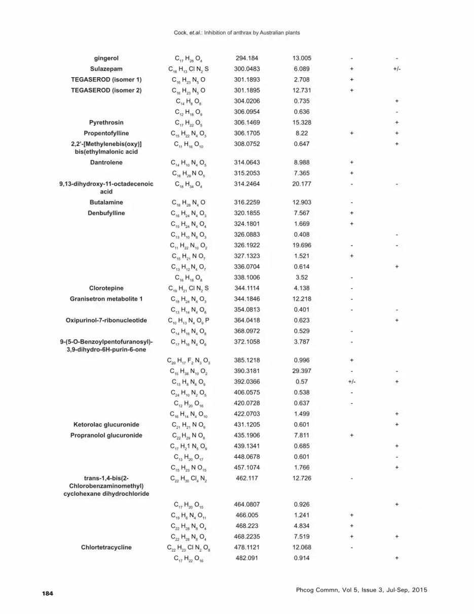
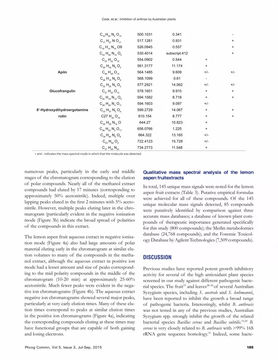
Table 3: Qualitative HPLC-MS QTOF analysis of the lemon aspen fruit methanolic and aqueous extracts, elucidation of empirical formulas and putative identification (where possible) of the compounds
Putative Identification Empirical Formula Molecular Mass Retention Time Methanol Extract
Aqueous Extract
C4 H6 O3 102.0328 0.478 -
Quinone C6 H4 O2 108.0217 1.603 + +
3-furanoic acid C5 H4 O3 112.0161 0.387 -
Purine C5 H4 N4 120.0436 32.377 +
phloroglucinol C6 H6 O3 126.0322 1.603 + +
2-Desoxy-D-ribose C5 H10 O4 134.0584 0.45 -
m-nitrotoluene C7 H7 N O2 137.047 0.521 +
(1S,5R)-4-hydroxy-6,7-dioxabicyclo[3.2.1]oct-2-en-8-
one (isomer 1)
C6 H6 O4 142.0269 3.647 +
(1S,5R)-4-hydroxy-6,7-dioxabicyclo[3.2.1]oct-2-en-8-
one (isomer 2)
C6 H6 O4 142.027 1.231 +
(E)-2-Methylglutaconic acid C6 H8 O4 144.0418 0.714 -
Xylitol C5 H12 O5 152.0699 0.44 -
C4 H4 N4 O3 156.0286 0.617 +
Oxanamide C8 H15 N O2 157.1109 0.614 + +
2-Oxoadipic acid C6 H8 O5 160.0376 1.229 +
ethyl (ethylperoxy) (oxo) acetate C6 H10 O5 162.0528 2.116 +/- -
C8 H4 O4 164.0111 3.106 +
C9 H11 N O2 165.0768 1.451 +
Ethyl 4-hydroxybenzoate C9 H10 O3 166.0635 0.956 -
(1S,5R)-4-Oxo-6,8-dioxabicyclo[3.2.1]oct-2-ene-2-
carboxylic acid
C7 H6 O5 170.0219 1.216 +
dehydroascorbic acid (oxidised vitamin C)
C6 H6 O6 174.0164 0.384 -
shikimic acid C7 H10 O5 174.0532 3.646 +
2-Hydroxyethyl salicylate C9 H10 O4 182.0578 0.742 -
C7 H8 O6 188.0326 1.219 +
citric acid C6 H8 O7 192.0268 0.749 - +/-
Hydroxy-7-methyl-4H,5H-pyrano[4,3-b]pyran-dione
C9 H6 O5 194.0221 2.001 + +
ferulic acid C10 H10 O4 194.0582 1.222 -
Feroxidin C11 H14 O3 194.0948 11.548 +
Cantharidin C10 H12 O4 196.074 1.309 -
C8 H10 O6 202.0478 3.645 +
calamenene (isomer 1) C15H22 202.1722 13.379 +
calamenene (isomer 2) C15 H22 202.1724 16.403 +
2,3-O-(Oxomethylene)hexopyranose
C7 H10 O7 206.0432 1.22 +/- +
Cyclazodone C12 H12 N2 O2 216.0901 5.567 + +
5-hydroxycalamenene C15 H22 O 218.1669 12.582 +
C8 H12 O7 220.0589 3.645 +/-
C9 H8 N4 O3 220.059 2.971 +
spathulenol (isomer 1) C15 H24 O 220.1828 13.575 +
spathulenol (isomer 2) C15 H24 O 220.1831 15.998 +

Cock, et.al.: Inhibition of anthrax by Australian plants
183Phcog Commn, Vol 5, Issue 3, Jul-Sep, 2015
spathulenol (isomer 3) C15 H24 O 220.1832 10.321 +
Gladiolic acid C11 H10 O5 222.0532 9.873 + +
C7 H13 N O7 223.0696 1.228 +
C13 H10 N2 O2 226.0746 8.511 +
Heptylheptanoate C14 H28 O2 228.2098 20.927 -
(2R,3S)-3-(3-Carboxylatopropanoyl)-
5-oxotetrahydro-2-furancarboxylate
C9 H8 O7 228.0251 1.23 +
Ozagrel C13 H12 N2 O2 228.09 7.84 + +
Heptylheptanoate C14 H28 O2 228.2093 20.899 -
Metomidate C13 H14 N2 O2 230.1059 7.704 + +
C9 H14 O7 234.0746 7.91 +
drimenin C15 H22 O2 234.1624 16.105 +
capsidiol (isomer 1) C15 H24 O2 236.1779 11.496 +
capsidiol (isomer 2) C15 H24 O2 236.178 17.371 +
capsidiol (isomer 3) C15 H24 O2 236.1781 14.516 +
C12 H19 N3 O2 237.1479 7.076 +
Kessyl alcohol C15 H26 O2 238.1936 14.479 +
C12 H5 N3 O3 239.0323 2.97 +
Isopentadecylic acid C15 H30 O2 242.2247 22.342 - -
C7 H4 N2 O8 243.999 1.227 +
LeuLeu C12 H24 N2 O3 244.1794 7.424 + +
nodakenetin C14 H14 O4 246.0897 12.068 +
Acetyltryptophane C13 H14 N2 O3 246.1007 5.157 +
Nitrefazole C10 H8 N4 O4 248.0538 0.737 +
C15 H26 O3 254.1881 16.722 +
7-palmitoleic acid C16 H30 O2 254.225 22.059 - -
11-keto pentadecanoic acid C15 H28 O3 256.2042 13.987 -
palmitic acid C16 H32 O2 256.241 23.786 - -
C10 H16 O8 264.085 0.64 -
Diftalone C16 H12 N2 O2 264.0913 10.429 +
Propacetamol C14 H20 N2 O3 264.1482 3.692 + +
C9 H14 O9 266.0642 0.737 +/-
Lauroylsarcosine C15 H29 N O3 271.2155 13.978 +
Lauroylsarcosine C15 H29 N O3 271.2155 17.008 +
C12 H11 N5 O3 273.0857 0.686 +
macowine C16 H19 N O3 273.1372 8.452 + +
2,15-dihydroxy-pentadecylic acid
C15 H30 O4 274.2148 9.96 -
C9 H13 N11 275.1351 0.864 +
Menthyl salicylate C17 H24 O3 276.1709 17.046 +
Queuine C12 H15 N5 O3 277.1171 0.556 +
7-hydroxy-10E,16-heptadecadien-8-ynoic acid
C17 H26 O3 278.1861 14.479 +
Dihydroartemisinin C15 H24 O5 284.1631 12.734 +/-
Hexyl dodecanoate C18 H36 O2 284.2723 26.727 -
N,N-Didemethylchlorpromazine C15 H15 Cl N2 S 290.0648 0.655 +
C9 H15 N11 O 293.146 0.863 +
Cock, et.al.: Inhibition of anthrax by Australian plants
184 Phcog Commn, Vol 5, Issue 3, Jul-Sep, 2015
gingerol C17 H26 O4 294.184 13.005 - -
Sulazepam C16 H13 Cl N2 S 300.0483 6.089 + +/-
TEGASEROD (isomer 1) C16 H23 N5 O 301.1893 2.708 +
TEGASEROD (isomer 2) C16 H23 N5 O 301.1895 12.731 +
C14 H8 O8 304.0206 0.735 +
C12 H18 O9 306.0954 0.636 -
Pyrethrosin C17 H22 O5 306.1469 15.328 +
Propentofylline C15 H22 N4 O3 306.1705 8.22 + +
2,2’-[Methylenebis(oxy)]bis(ethylmalonic acid
C11 H16 O10 308.0752 0.647 +
Dantrolene C14 H10 N4 O5 314.0643 8.988 +
C16 H29 N O5 315.2053 7.365 +
9,13-dihydroxy-11-octadecenoic acid
C18 H34 O4 314.2464 20.177 - -
Butalamine C18 H28 N4 O 316.2259 12.903 -
Denbufylline C16 H24 N4 O3 320.1855 7.567 +
C15 H24 N4 O4 324.1801 1.669 +
C13 H10 N8 O3 326.0883 0.408 -
C11 H22 N10 O2 326.1922 19.696 - -
C15 H21 N O7 327.1323 1.521 +
C13 H12 N4 O7 336.0704 0.614 +
C16 H18 O8 338.1006 3.52 -
Clorotepine C19 H21 Cl N2 S 344.1114 4.138 -
Granisetron metabolite 1 C18 H24 N4 O3 344.1846 12.218 -
C13 H14 N4 O8 354.0813 0.401 - -
Oxipurinol-7-ribonucleotide C10 H13 N4 O9 P 364.0418 0.623 +
C14 H16 N4 O8 368.0972 0.529 -
9-(5-O-Benzoylpentofuranosyl)-3,9-dihydro-6H-purin-6-one
C17 H16 N4 O6 372.1058 3.787 -
C20 H17 F2 N3 O3 385.1218 0.996 +
C15 H38 N10 O2 390.3181 29.397 - -
C13 H8 N6 O9 392.0366 0.57 +/- +
C24 H10 N2 O5 406.0575 0.538 -
C12 H20 O16 420.0728 0.637 -
C16 H14 N4 O10 422.0703 1.499 +
Ketorolac glucuronide C21 H21 N O9 431.1205 0.601 +
Propranolol glucuronide C22 H29 N O8 435.1906 7.811 +
C17 H21 N5 O9 439.1341 0.685 +
C13 H20 O17 448.0678 0.601 -
C15 H23 N O15 457.1074 1.766 +
trans-1,4-bis(2-Chlorobenzaminomethyl)
cyclohexane dihydrochloride
C22 H30 Cl4 N2 462.117 12.726 -
C17 H20 O15 464.0807 0.926 +
C19 H6 N4 O11 466.005 1.241 +
C22 H28 N8 O4 468.223 4.834 +
C22 H28 N8 O4 468.2235 7.519 + +
Chlortetracycline C22 H23 Cl N2 O8 478.1121 12.068 -
C17 H22 O16 482.091 0.914 +
Cock, et.al.: Inhibition of anthrax by Australian plants
185Phcog Commn, Vol 5, Issue 3, Jul-Sep, 2015
C18 H20 N4 O13 500.1031 0.341 -
C17 H27 N O17 517.1281 0.931 +
C19 H14 N10 O9 526.0945 0.557 +
C23 H50 N10 O4 530.4014 subscript.412 +
C23 H22 O16 554.0902 0.544 +
C29 H39 N9 O3 561.3177 11.174 +
Apiin C26 H28 O14 564.1485 9.609 +/- +/-
C26 H16 N8 O8 568.1099 0.61 -
C32 H35 N9 O2 577.2921 14.062 +/- +/-
Glucofrangulin C27 H30 O14 578.1651 9.915 + +
C25 H18 N14 O5 594.1582 8.718 + +
C28 H26 N4 O11 594.1603 9.097 +/-
8’-Hydroxydihydroergotamine C33 H37 N5 O6 599.2729 14.067 + +
rutin C27 H30 O16 610.154 8.777 + +
C29 H28 N18 O 644.27 10.823 +
C28 H20 N2 O17 656.0766 1.225 +
C34 H44 N6 O8 664.322 13.165 +/-
C53 H54 O2 722.4123 15.728 +/-
C31 H26 N24 734.2773 11.548 + + and - indicates the mass spectral mode in which that the molecule was detected.
numerous peaks, particularly in the early and middle stages of the chromatogram corresponding to the elution of polar compounds. Nearly all of the methanol extract compounds had eluted by 17 minutes (corresponding to approximately 50% acetonitrile). Indeed, multiple over lapping peaks eluted in the first 2 minutes with 5% aceto-nitrile. However, multiple peaks eluting later in the chro-matogram (particularly evident in the negative ionisation mode (Figure 3b) indicate the broad spread of polarities of the compounds in this extract.
The lemon aspen fruit aqueous extract in negative ionisa-tion mode (Figure 4a) also had large amounts of polar material eluting early in the chromatogram at similar elu-tion volumes to many of the compounds in the metha-nol extract, although the aqueous extract in positive ion mode had a lesser amount and size of peaks correspond-ing to the mid polarity compounds in the middle of the chromatogram (10-20 min) at approximately 25-60% acetonitrile. Much fewer peaks were evident in the nega-tive ion chromatograms (Figure 4b). The aqueous extract negative ion chromatograms showed several major peaks, particularly at very early elution times. Many of these elu-tion times correspond to peaks at similar elution times in the positive ion chromatograms (Figure 4a), indicating the corresponding compounds eluting at these times may have functional groups that are capable of both gaining and losing electrons.
Qualitative mass spectral analysis of the lemon aspen fruitextracts
In total, 145 unique mass signals were noted for the lemon aspen fruit extracts (Table 3). Putative empirical formulas were achieved for all of these compounds. Of the 145 unique molecular mass signals detected, 85 compounds were putatively identified by comparison against three accurate mass databases; a database of known plant com-pounds of therapeutic importance generated specifically for this study (800 compounds); the Metlin metabolomics database (24,768 compounds); and the Forensic Toxicol-ogy Database by Agilent Technologies (7,509 compounds).
DISCUSSION
Previous studies have reported potent growth inhibitory activity for several of the high antioxidant plant species screened in our study against different pathogenic bacte-rial species. The fruit19 and leaves28-30 of several Australian Syzygium species, including S. australe and S. leuhmannii, have been reported to inhibit the growth a broad range of pathogenic bacteria. Interestingly, whilst B. anthracis was not tested in any of the previous studies, Australian Syzygium spp. strongly inhibit the growth of the related bacterial species Bacillus cereus and Bacillus subtilis.19,30 B. cereus is very closely related to B. anthracis with >99% 16S rRNA gene sequence homology.31 Indeed, some bacte-

Cock, et.al.: Inhibition of anthrax by Australian plants
186 Phcog Commn, Vol 5, Issue 3, Jul-Sep, 2015
rial taxonomonists believe that B. anthracis, B. cereus, B. thuringiensis, B. mycoides, B. pseudomycoides and B. weinstepha-nensis should be classified as a single species under current standards (>97% 16S rRNA sequence homology) and are only classified as separate species as a result of the differ-ent diseases that they cause.32-34 Whilst B. subtilise has less similarity(>94% homology with B. anthracis 16S rRNA), it is still considered a closely related species.31 It is therefore perhaps not surprising that the Syzygium spp. extracts screened in our study displayed growth inhibitory activity towards B. anthracis.
Recently, we also reported growth inhibitory activity of several high antioxidant fruits22-24 and culinary herb extracts25 against some microbial triggers of selected autoimmune inflammatory diseases. The Illawarra plum, lemon aspen, desert lime, wattle seed, native thyme and rivermint were particularly potent inhibitors of the microbial triggers of rheumatoid arthritis and ankylos-ing spondylitis.24,25 All of the bacterial species associated with the onset of the autoimmune diseases screened in the previous studies are Gram negative and thus substan-tially different to the Gram positive B. anthracis screened
in this study. However, the previously reported inhibitory activity does indicate the presence of antibacterial com-ponents in these extracts and is consistent with the inhibi-tory activity reported in this study.
The same panel of high antioxidant plant extracts was also previously reported to inhibit the proliferation of CaCo2 and HeLa cancer cells.35,36 Although those stud-ies examined growth inhibition ineukaryotic cells, they are interesting as they report the phytochemical composi-tions of the bioactive extracts. A commonality between many of the inhibitory extracts was the relatively high lev-els of a number of tannin components including exifone (4-galloylpyrogallol), ellagic acid dehydrate, trimethylel-lagic acid, chebulic acid, corilagen, castalagin and che-bulagic acid. Gallotannins have been reported to inhibit the growth of a broad spectrum of bacterial species37 through a variety of mechanisms including binding cell surface molecules including lipotoichoic acid and proline-rich cell surface proteins,38,39 and by inhibiting glucosyl-transferase enzymes.40 Elligitannins are also highly potent inhibitors of bacterial growth, with MIC values as low as 62.5 µg/ml.37,39,41 Ellagitannins have also been reported
Figure 5: Chemical structures of lemon aspen fruit compounds detected in the methanolic and aqueous extracts: (a) ellagic acid; (b) trimethylellagic acid; (c) cantharidin; (d) calamanene; (e) hydroxycalamanene; (f) rutin; (g) luteolin; (h) diosmin; (i) dihydrokaempferol; (j) lauryl sarcosinate; (k) queuine; (l) dihydroxyarteminisin; (m) hydroxyethyl sa-
licylate; (n) ferulic acid; (o) gingerol; (p) chlorogenic acid

Cock, et.al.: Inhibition of anthrax by Australian plants
187Phcog Commn, Vol 5, Issue 3, Jul-Sep, 2015
to function via several antibiotic mechanisms including interaction with cytoplasmic oxidoreductases and by dis-rupting bacterial cell walls.37,39
The lemon aspen fruit extracts displayed the most potent growth inhibitory activity against B. anthracis of all the extracts screened in our study, with MIC values <400 µg/mL. Our study employed a metabolomics profil-ing approach to putatively identify as many of the phy-tochemical components as possible in the lemon aspen aqueous and methanolic extracts. We putatively identified several of the same tannins in the lemon aspen extracts as previously reported for other high antioxidant extracts. In particular, ellagic acid (Figure 5a) and trimethylellagic acid (Figure 5b) were detected in the lemon aspen extracts and are likely to contribute to their inhibition of B. anthra-cis growth. However, it is likely that other phytochemi-cal classes also contribute to this bioactivity. Alkaloids, anthraquionones, flavonoids, polyphenolics, phytosterols, saponins, stilbenes and terpenes have also been linked with antibacterial activity in different plant species and thus may be responsible (at least in part) for the bacterial growth inhibitory activities reported here.
Terpenoids and naphthalenes including cantharidin (Figure 5c), calamanene (Figure 5d) and hydroxycala-manene (Figure 5e) were also identified in the lemon aspen extracts. Terpenoids and naphthalenes have been previously reported to have potent broad spectrum anti-bacterial activity42 and therefore may contribute to the inhibitory activity against B. anthracis. Interestingly, can-tharidin, calamanene and hydroxycalamanene have also been shown to inhibit the proliferation of pancreatic can-cer cells via oxidative stress-independent cell cycle arrest and the induction of apoptosis.43,44 Furthermore, com-pounds structurally similar to calamanene and hydroxy-calamanene have also been associated with the induction of apoptosis in human lung cancer cell lines by upregulat-ing DR4 and DR5 cell death receptors and enhancing the activation of caspases 3, 7, 8 and 9,43,44 possibly account-ing for the anticancer activity previously reported for lemon aspen extracts.45
Flavonoids were also identified in the lemon aspen extracts. Many studies have reported potent antibacterial activities for a wide variety of flavonoids.46 Several flavo-noids including rutin (Figure 5 f), luteolin (Figure 5 g), diosmin (Figure 5 h) and dihydrokaempferol (Figure 5i)were putatively identified in our study. These flavonoids have been reported to be good inhibitors of bacterial growth, particularly of Gram positive bacteria.46 Whilst we were unable to find any reports of B. anthracis growth inhibitory activity of these flavonoids, they have been
reported to inhibit growth of the closely related species B. Cereus.47 Furthermore, a number of other phytochemicals including lauryl sarcosinate (Figure 5j), queuine (Figure 5k), dihydroxyarteminisin (Figure 5l), hydroxyethyl salicy-late (Figure 5m), ferulic acid (Figure 5n), gingerol (Fig-ure 5o), and chlorogenic acid (Figure 5p) were putatively identified in the lemon aspen extracts. Several recent stud-ies have demonstrated the potent antimicrobial activity of plant extracts with high levels of these components.18-24
In particular, Tasmannia lanceolata,20 Terminalia ferdinandi-ana21 and several Syzygium species19 have demonstrated potent antimicrobial activity against a wide panel of bac-teria. Thus, it is likely that multiple compounds within the lemon aspen extracts are contributing to the growth inhi-bition of B. anthracis.
An important consideration of any metabolomic tech-nique is that it will not detect all compounds in a complex mixture, but instead will only detect a portion of them. This is not necessarily a problem when a directed/biased study is undertaken to detect a particular compound or class of compounds and the separation and detection conditions can be optimised for the study. However, when the aim of the study is metabolomic profiling rather than metabolomic finger printing, the technique conditions must be chosen and optimised to separate and detect the largest amount of compounds, with the broadest possible physical and chemical characteristics. Generally, HPLC-MS is a good choice for such metabolomic profiling stud-ies as it generally detects a larger amount of compounds of varying polarities than the other commonly used tech-niques. However, this method is limited to studies of the mid-highly polar compounds and is not as useful for studies aimed at highly nonpolar compounds. Thus, many nonpolar phytosterols, saponins, stilbenes and terpenes which may contribute to the inhibitory activity of the lemon aspen extracts may escape detection by HPLC-MS. For this reason, future studies should also utilise GC-MS analysis to detect and identify the less polar compounds. Furthermore, mass spectral techniques are generally not capable on their own of differentiating between structural isomers. Further studies using a wider variety of tech-niques are required to confirm the identity of the com-pounds putatively identified here.
CONCLUSION
The results of this study demonstrate the potential of high antioxidant Australian plant extractsto block the growth of B. anthracis. Lemon aspen was particularly potent and thushas potential in the prevention and treat-

Cock, et.al.: Inhibition of anthrax by Australian plants
188 Phcog Commn, Vol 5, Issue 3, Jul-Sep, 2015
ment of anthrax. Further studies aimed at the purification of the bioactive components are needed to examine the mechanisms of action of these agents.
ACKNOWLEDGEMENTS
The authors would like to thank Agilent Technologies for the in-kind contribution of the commercially avail-
able databases to perform this study. Financial support for this work was provided by the Environmental Futures Research Institute and the School of Natural Sciences, Griffith University, Australia.
CONFLICT OF INTEREST
All authors declare no conflicts of interest.
REFERENCES
1. Anderson RM, May RM. Immunisation and herd immunity. The Lancet 1990; 335(8690): 641-5.
2. Fenner F. Global eradication of smallpox. Clinical Infectious Diseases 1982; 4(5): 916-30.
3. Doganay M, Metan G, Alp E. A review of cutaneous anthrax and its outcome. Journal of Infection and Public Health 2010; 3(3): 98-105.
4. Beatty ME, Ashford DA, Griffin PM, et al. Gastrointestinal anthrax: review of the literature. Archives of Internal Medicine 2003; 163(20): 2527-31.
5. Holty JEC, Bravata DM, Liu H, et al. Systematic review: a century of inhalational anthrax cases from 1900 to 2005. Annals of Internal Medicine 2006; 144(4): 270-80.
6. Dragon DC, Elkin BT, Nishi JS, et al. A review of anthrax in Canada and implications for research on the disease in the northern bison. Journal of Applied Microbiology 1999; 87(2): 208-13.
7. National Institute for Communicable Diseases. Communicable Diseases Communiqué 2014; 13(4): 1.
8. Zhang TL Cui LL, Li L, et al. Investigation of an outbreak of cutaneous anthrax in Banlu village, Lianyungang, China, 2012. Western Pacific Surveillance and Response Journal 2012: 3(4): 12-5. doi:10.5365/wpsar.2012.3.4.005
9. Reddy R, Parasadini G, Rao P, Uthappa CK, Murhekar MV. Outbreak of cutaneous anthrax in Musalimadugu village, Chittoor district, Andhra Pradesh, India, July-August. Journal of Infection in Developing Countries 2012; 6(10): 695-9.
10. Daw D. Anthrax outbreak causes quarantine of Indian village. BioPrepWatch (Chicago Illinois); http://bioprepwatch.com/biological-threats/anthrax-outbreak-causes-quarantine-of-indian-village/339832/ : retrieved 15/4/2015.
Highlights of Paper
Author Profile
• High antioxidant Australian plantextracts were potent inhibitors of Bacillus anthracis (the pathogenic agent of anthrax).
• The muntries, Illawarra plum and native tamarind extracts were particularly potent growth inhibitors with MIC values substantially < 1000 µg/mL.
• With the exception of Syzygium australe and Syzygium leuhmannii extracts, all inhibitory extracts were nontoxic.• LC-MS analysis of the most potent lemon aspen extracts identified 85 compounds and highlighted several that may
contribute to the ability of these extracts to inhibit the growth of B. anthracis.
• Dr Ian Cock leads a research team in the Environmental Futures Research Institute and the School of Natural Sciences at Griffith University, Australia. His research involves bioactivity and phyto-chemical studies into a variety of plant species of both Australian and international origin, including Aloe vera, South Asian and South American tropical fruits, as well as Australia plants including Scae-vola spinescens, Pittosporum phylliraeoides, Terminalia ferdinandiana (Kakadu plum), Australian Acacias, Syzygiums, Petalostigmas and Xanthorrhoea johnsonii (grass trees). This range of projects has resulted in approximately 150 scientific publications in a variety of peer reviewed journals.
11. Epp T, Waldner C, Argue CK. Case-control study investigating an anthrax outbreak in Saskatchewan, Canada – summer 2006. The Canadian Veterinary Journal 2010; 51(9): 973-8.
12. Mongoh MN, Dyer NW, Stoltenow CL, et al. Risk factors associated with anthrax outbreak in animals in North Dakota, 2005: A retrospective case-control study. Public Health Reports 2008; 123(3): 352-9.
13. Price EP, Seymour ML, Sarovich DS, et al. Molecular epidemoligical investigation of an anthrax out break among heroin users, Europe. Emerging Infectious Diseases 2012; 18(8): 1307-13. PMCid3414016.
14. Jernigan JA, Stephens DS, Ashford DA, et al. Bioterrorism-related inhalational anthrax: the first 10 cases reported in the United States. Emerging Infectious Diseases 2001; 7(6): 933-44.
15. Splino M, Patocka J, Prymula R, et al. Anthrax vaccines. Annals of Saudi Medicine 2005; 25(2): 143-9.
16. Vietri NJ, Purcell BK, Tobery SA, et al. A short course of antibiotic treatment is effective in preventing death from experimental inhalational anthrax after discontinuing antibiotics. Journal of Infectious Diseases 2009; 199(3): 336-41.
17. Hart CA, Beeching NJ. Prophylactic treatment of anthrax with antibiotics: Indiscriminate use of antibiotics will lead to resistance in organisms. British Medical Journal 2001; 323(7320): 1017.
18. Hart C, Ilanko P, Sirdaarta J, et al. Tasmanni astipitata as a functional food/natural preservative: Antimicrobial activity and toxicity. Phcog Commn. 2014; 4(4): 33-47.
19. Sautron C, Cock IE. Antimicrobial activity and toxicity of Syzygium australe and Syzygium leuhmanii fruit extracts. Phcog Commn. 2014; 4(1): 53-60.

Cock, et.al.: Inhibition of anthrax by Australian plants
189Phcog Commn, Vol 5, Issue 3, Jul-Sep, 2015
20. Winnett V, Boyer H, Sirdaarta J, Cock IE. The potential of Tasmannia lanceolata as a natural preservative and medicinal agent: Antimicrobial activity and toxicity. Phcog Commn. 2014; 4(1): 42-52.
21. Cock IE, Mohanty S. Evaluation of the antibacterial activity and toxicity of Terminalia ferdinandia fruit extracts. Phcog J. 2011; 3(20): 72-9.
22. Sirdaarta J, Matthews B, White A, et al. GC-MS and LC-MS analysis of Kakadu plum fruit extracts displaying inhibitory activity against microbial triggers of multiple sclerosis. Phcog Commn. 2015; 5(2): 100-15.
23. Courtney R, Sirdaarta J, Matthews B, et al. Tannin components and inhibitory activity of Kakadu plum leaf extracts against microbial triggers of autoimmune inflammatory diseases. Phcog J. 2015; 7(1): 18-31.
24. Maen A, Cock IE. Inhibitory activity of high antioxidant Australian native fruits against the bacterial triggers of selected autoimmune diseases. Phcog Commn. 2015; 5(1): 48-58.
25. Maen A, Cock IE. Inhibitory activity of Australian culinary herb extracts against the bacterial triggers of selected autoimmune diseases. Phcog Commn. 2015; 5(2): 130-9.
26. Arkhipov A, Sirdaarta J, Rayan P, et al. An examination of the antibacterial, antifungal, anti-Giardial and anticancer properties of Kigelia africana fruit extracts. Phcog Commn. 2014; 4(3): 62-76.
27. Cock IE, Ruebhart DR. Comparison of the brine shrimp nauplii bioassay and the ToxScreen-II test for the detection of toxicity associated with Aloe vera (Aloe barbadensis Miller) leaf extract. Pharmacognosy Research 2009; 1(2): 102-8.
28. Chikowe G, Mpala L, Cock IE. Antibacterial activity of selected Australian Syzygium species. Phcog Commn. 2013; 3(4): 77-83.
29. Cock IE. Antimicrobial activity of Syzygium australe and Syzygium leuhmannii leaf methanolic extracts. Phcog Commn. 2012; 2(2): 71-7.
30. Cock IE. Antibacterial activity of selected Australian native plant extracts. Internet Journal of Microbiology 2008; 4(2): 1-8.
31. Maughan H, Van der Auwera G. Bacillus taxonomy in the genomic era finds phenotypes to be essential though often misleading. Infection. Genetics and Evolution 2011; 11(5): 789-97.
32. Tourasse NJ, Helgason E, Okstad OA, et al. The Bacillus cereus group: novel aspects of population structure and genome dynamics. Journal of Applied Microbiology 2006; 101(3): 579–93.
33. Priest FG, Barker M, Baillie LW, et al. Population structure and evolution of the Bacillus cereus group. Journal of Bacteriology 2004; 186(23): 7959–70.
34. Rasko DA, Altherr MR, Han CS, et al. Genomics of the Bacillus cereus group of organisms. FEMS Microbiology Reviews 2005; 29(2): 303–29.
35. Jamieson N, Sirdaarta N, Cock IE. The anti-proliferative properties of Australian plants with high antioxidant capacities against cancer cell lines. Phcog Commn. 2014; 4(4): 71-82.
36. Sirdaarta J, Maen A, Rayan P, et al. HPLC-MS analysis of high antioxidant Australian fruits with antiproliferative activity against cancer cells. Pharmacognosy Magazine inpress; 2015.
37. Buzzini P, Arapitsas P, Goretti M, et al. Antimicrobial activity of hydrolysable tannins. Mini-Reviews in Medicinal Chemistry 2008; 8(12): 1179-87.
38. Wolinsky LE, Sote EO. Isolation of natural plaque-inhibiting substances from ‘Nigerian chewing sticks’. Caries Research 1984; 18(3): 216–225.
39. Hogg SD, Embery G. Blood-group-reactive glycoprotein from human saliva interacts with lipoteichoic acid on the surface of Streptococcus sanguis cells. Archives in Oral Biology 1982; 27(3): 261–8.
40. Wu-Yuan CD, Chen CY, Wu RT. Gallotannins inhibit growth, water-soluble glucan synthesis, and aggregation of Streptococci mutans. Journal of Dental Research 1988; 67(1): 51–55.
41. Machado TP, Pinto AV, Pinto MCFR, et al. In vitro activity of Brazilian medicinal plants, naturally occurring naphthoquinones and their analogues, against methicillin-resistant Staphylococcus aureus. International Journal of Antimicrobial Agents 2003; 21(3): 279-84.
42. Cock IE. The phytochemistry and chemotherapeutic potential of Tasmania lanceolata (Tasmanian pepper): A review. Phcog Commn. 2013; 3(4): 1-13.
43. Salminen A, Lehtonen M, Suuronen T, et al. Terpenoids: Natural inhibitors of NF-κB signalling with anti-inflammatory and anticancer potential. Cellular and Molecular Life Sciences 2008; 65(19): 2979-99.
44. Lu XG, Zhan LB, Feng BA, et al. Inhibition of growth and metastasis of human gastric cancer implanted in nude mice by d-limonene. World Journal of Gastroenterology 2004; 10(14): 2140–4.
45. Cock IE, Maen A. HPLC-MS analysis of high antioxidant Australian fruits with antiproliferative activity against cancer cells. Pharmacognosy Magazine in press; 2015.
46. Narayana KR, Reddy MS, Chaluvadi MR, et al. Bioflavonoids classification, pharmacological, biochemical effects and therapeutic potential. Indian Journal of Pharmacology 2001; 33(1): 2-16.
47. Arima H, Ashida H, Danno GI. Rutin-enhanced antibacterial activities of flavonoids against Bacillus cereus and Salmonella enteritidis. Bioscience, Biotechnology and Biochemistry 2002; 66(5): 1009-14.


